

images
1TEZSjF
1TEZSjF

Create successful ePaper yourself
Turn your PDF publications into a flip-book with our unique Google optimized e-Paper software.
clinical and research news<br />
Investigating Diffusion Tensor Imaging as a Non-Invasive<br />
Biomarker of Pediatric Kidney Transplant Health<br />
Jesse Courtier, MD, and Marsha Lee, MD<br />
Background<br />
In children with end-stage renal disease, kidney transplantation<br />
is the preferred choice for therapy, with overall lower<br />
long-term morbidity and mortality compared with dialysis.<br />
Annually, approximately 800 renal transplants are performed<br />
in the United States in children under 18 years of age.<br />
The monitoring of kidney transplant function in pediatric<br />
recipients is critical to optimize the longevity of their<br />
transplants and therefore their overall quality of life. Transplant<br />
rejection, however, cannot be easily determined<br />
using routine renal function laboratory tests such as BUN,<br />
serum creatinine or calculated eGFR, since rejection can<br />
be present even when these values are normal. Thus, renal<br />
transplant biopsies are routinely performed to screen for<br />
subclinical rejection and to evaluate for rejection in cases of<br />
elevated serum creatinine.<br />
Preliminary work has demonstrated the potential of<br />
magnetic resonance diffusion tensor imaging (MR-DTI) with<br />
quantified measurement of fractional anisotropy (FA) as<br />
a non-invasive method of assessing renal allograft function.<br />
Primarily used in neuroimaging, diffusion-weighted<br />
imaging (DWI) takes advantage of the differences between<br />
water molecular motion (Brownian motion) within various<br />
anatomic structures to generate contrast. In general, water<br />
molecules with restricted motion (e.g., within nerve tracts)<br />
appear hyper-intense compared to water molecules that are<br />
in free space (e.g., CSF). Diffusion tensor imaging (DTI) further<br />
assesses the directionality of water molecular motion.<br />
This has been used to tremendous advantage in neuroimaging<br />
where neuro-axonal tracts are longer than they are wide,<br />
which results in anisotropic diffusive properties and thus<br />
allows for mapping of specific axonal tracts (tractography).<br />
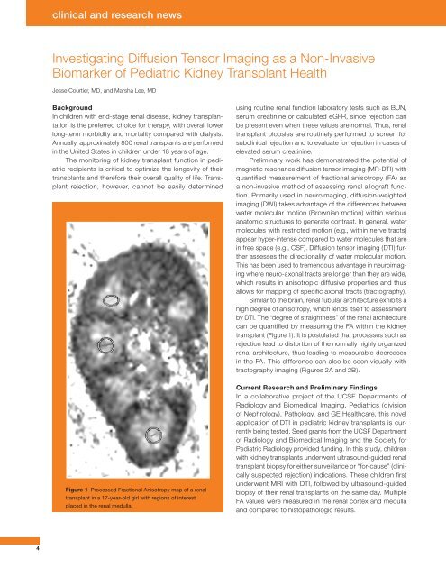
Similar to the brain, renal tubular architecture exhibits a<br />
high degree of anisotropy, which lends itself to assessment<br />
by DTI. The “degree of straightness” of the renal architecture<br />
can be quantified by measuring the FA within the kidney<br />
transplant (Figure 1). It is postulated that processes such as<br />
rejection lead to distortion of the normally highly organized<br />
renal architecture, thus leading to measurable decreases<br />
in the FA. This difference can also be seen visually with<br />
tractography imaging (Figures 2A and 2B).<br />
Figure 1 Processed Fractional Anisotropy map of a renal<br />
transplant in a 17-year-old girl with regions of interest<br />
placed in the renal medulla.<br />
Current Research and Preliminary Findings<br />
In a collaborative project of the UCSF Departments of<br />
Radiology and Biomedical Imaging, Pediatrics (division<br />
of Nephrology), Pathology, and GE Healthcare, this novel<br />
application of DTI in pediatric kidney transplants is currently<br />
being tested. Seed grants from the UCSF Department<br />
of Radiology and Biomedical Imaging and the Society for<br />
Pediatric Radiology provided funding. In this study, children<br />
with kidney transplants underwent ultrasound-guided renal<br />
transplant biopsy for either surveillance or “for-cause” (clinically<br />
suspected rejection) indications. These children first<br />
underwent MRI with DTI, followed by ultrasound-guided<br />
biopsy of their renal transplants on the same day. Multiple<br />
FA values were measured in the renal cortex and medulla<br />
and compared to histopathologic results.<br />
4