Editorial_lay:Layout 1 - BDIZ EDI
Editorial_lay:Layout 1 - BDIZ EDI
Editorial_lay:Layout 1 - BDIZ EDI

You also want an ePaper? Increase the reach of your titles
YUMPU automatically turns print PDFs into web optimized ePapers that Google loves.
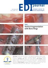
ISSN 1862-2879<br />
Issue 1/2009 Vol. 5<br />
<strong>EDI</strong> Journal<br />
European Journal for<br />
Dental Implantologists<br />
TOPIC<br />
Illusion of Nature<br />
Non-invasive esthetic rehabilitation<br />
»<strong>EDI</strong> News: The Future of Dentistry – IDS 2009 in Cologne · Preview of the 13 th <strong>BDIZ</strong> <strong>EDI</strong><br />
Symposium in Munich · Coming up: Third <strong>BDIZ</strong> <strong>EDI</strong> Mediterranean Symposium · Review<br />
of the Classes of Indications in Oral Implantology · Eighth OSIS <strong>EDI</strong> Congress in Jurata<br />
»European Law: Europe to Promote Healthcare Professions · Potential Abuse of the Process<br />
for the Recognition of Professional Qualifications »Case Studies: Computer-guided Flapless<br />
Surgery · Immediate and De<strong>lay</strong>ed “All-on-Six” Rehabilitation of the Atrophic Maxilla with<br />
Tilted Implants · Illusion of Nature »Product Studies: High-Tech Aesthetics · Optimizing the<br />
Implant Bed
© Nobel Biocare 2009<br />
NobelActive TM<br />
Taking a new direction in implants.<br />
Dual-function prosthetic<br />
connection<br />
Bone-condensing property<br />
Adjustable implant orientation<br />
for optimal final placement<br />
NobelActive equally satisfies<br />
surgical and restorative clinical<br />
goals. NobelActive thread design<br />
progressively condenses bone<br />
with each turn during insertion,<br />
which is designed to enhance initial<br />
stability. The sharp apex and cutting<br />
blades allow surgical clinicians<br />
to adjust implant orientation for<br />
optimal positioning of the prosthetic<br />
connection. Restorative clinicians<br />
benefit by a versatile and secure<br />
internal conical prosthetic connection<br />
with built-in Platform Shifting<br />
upon which they can produce<br />
excellent esthetic results. Based<br />
on customer feedback and market<br />
demands for NobelActive, the<br />
product assortment has been<br />
expanded – dental professionals will<br />
Built-in Platform Shifting<br />
High initial stability,<br />
even in compromised<br />
bone situations<br />
10 YEARS WITH<br />
TIUNITE® SURFACE<br />
New data confi rm<br />
long-term stability.<br />
now enjoy even greater flexi bility<br />
in prosthetic and implant selection.<br />
Nobel Biocare is the world leader<br />
in innovative evidence-based dental<br />
solutions.<br />
For more information, visit our<br />
website.<br />
www.nobelbiocare.com/nobelactive
This March, the International Dental Show (IDS) 2009 will<br />
open its gates in Cologne – an important international marketplace<br />
designed to offer guidance to promising new technologies<br />
and a “festival of innovations, trends and ideas”, but<br />
also a meeting place for the top p<strong>lay</strong>ers in the dental field<br />
and for dental care providers within the various national<br />
healthcare systems. Quality and innovation should be the<br />
determining factors for medical services in a competitive<br />
European market.<br />
In July 2008, the European Commission finally presented its<br />
promised proposal for a “Directive on the application of<br />
patient’s rights in cross-border healthcare”. The proposal is<br />
intended to respond to rapid changes in the social and economic<br />
fields. The proposed directive covers cross-border health<br />
services that patients may obtain outside their home country.<br />
Since 1998, the rulings by the European Court of Justice have<br />
consistently upheld the view that healthcare services are covered<br />
by the freedom to provide services, which is one of the<br />
four European fundamental freedoms (the other three being<br />
the freedom of movement of goods, the freedom of establishment<br />
and the freedom of capital movement). Unfortunately,<br />
only 30 percent of all EU citizens are aware that they have a<br />
right to obtain healthcare services in other EU member states.<br />
The healthcare system of the home state, whether organized<br />
directly by the state or on the insurance principle, must ensure<br />
that the patient is reimbursed – and that even if these services<br />
are provided within the home state.<br />
Reality, unfortunately, looks quite different. In Germany, the<br />
major p<strong>lay</strong>ers in statutory health care have turned the state<br />
into an instrument for erecting hurdles to discourage patients<br />
from opting for reimbursement. For example, patients are<br />
forced to pay a ten percent administrative surcharge, and any<br />
option for reimbursement will apply to all services for the<br />
entire year. For many patients, this acts as a clear deterrent.<br />
And now, on 31 March, the umbrella organizations of the<br />
healthcare insurers are planning to submit a “Practical Report”<br />
to the German parliament via the German Ministry of Health,<br />
Brave New World<br />
of Cross-border<br />
Medical Services?<br />
reporting on the small number of patients availing themselves<br />
of the options of Section 13 of the German Social Code, Book V,<br />
with the intention of having the entire section deleted. This<br />
section governs the procedure for cost reimbursement within<br />
the statutory health insurance system; it had been modified in<br />
2004, at the instigation of the EU, such that all patients now<br />
once again have the right to opt for cost reimbursement for<br />
their medical or dental treatment.<br />
As already stated, patients are often unaware of the reimbursement<br />
option; unfortunately, many German physicians<br />
and dentists shy away from informing their patients about this<br />
payment modality for outpatient medical treatment. Due to<br />
misguided ethical inhibitions, they want to avoid any impression<br />
of dishonesty that might arise from their pointing out the<br />
limits of the services and prescriptions available.<br />
It has been estimated that only one percent of all Europeans<br />
make use of cross-border healthcare services. The “EU Directive<br />
on the application of patient’s rights in cross-border healthcare”<br />
threatens to go under in the usual regulation mania.<br />
Brussels wants to establish European reference networks within<br />
the member states “to help realize the potential of European<br />
cooperation regarding highly specialized healthcare for<br />
patients and for healthcare systems from innovations in medical<br />
science and health technologies”. A similar approach is<br />
planned for the future development and operation of a network<br />
for health technology assessment. Member states are to<br />
be required to collect statistical data for monitoring the provision<br />
of cross-border healthcare, the modalities of the treatments<br />
provided, the providers, the patients, the cost and the<br />
outcomes and to provide them to the European Commission<br />
on an annual basis. Against the backdrop of this bureaucratic<br />
outrage, it is difficult to recognize the original good intentions<br />
of providing mobility to patients.<br />
Sincerely,<br />
Christian Berger, Kempten/Germany<br />
President of <strong>BDIZ</strong> <strong>EDI</strong><br />
<strong>EDI</strong><br />
<strong>Editorial</strong><br />
3
4 <strong>EDI</strong><br />
Table of Content<br />
<strong>EDI</strong> News<br />
The Future of Dentistry<br />
Kicking off IDS 2009 8<br />
Dental Hub IDS Cologne<br />
A short interview with <strong>BDIZ</strong> <strong>EDI</strong> president<br />
Christian Berger 9<br />
Meeting Point Implantology<br />
<strong>BDIZ</strong> <strong>EDI</strong> at IDS 2009 10<br />
Dramatic Developments in Many Areas<br />
New products at IDS 12<br />
20 Years of <strong>BDIZ</strong> <strong>EDI</strong><br />
Preview of the 13 th <strong>BDIZ</strong> <strong>EDI</strong> Symposium in Munich 22<br />
Update Implantology 2009<br />
Third <strong>BDIZ</strong> <strong>EDI</strong> Mediterranean Symposium 26<br />
Review of the Classes of Indications in Oral<br />
Implantology<br />
<strong>BDIZ</strong> <strong>EDI</strong> recommendations to the Consensus<br />
Conference Implantology 30<br />
Eighth OSIS <strong>EDI</strong> Congress in Jurata<br />
21 to 23 May in Poland 34<br />
Obituary: Ernst-Helmut Pruin and Willi Schulte 36<br />
Europe-Ticker 38<br />
European Law<br />
Europe to Promote Healthcare Professions<br />
Green Paper lists future challenges 40<br />
Potential Abuse of the Process for the Recognition<br />
of Professional Qualifications<br />
An ECJ decision and its consequences for the<br />
pursuit of the dentist profession 42<br />
Case Studies<br />
Computer-guided Flapless Surgery<br />
Complete prosthetic rehabilitation on immediately<br />
loaded transmucosal implants using a stereolithographic<br />
surgical guide 44<br />
Immediate and De<strong>lay</strong>ed “All-on-Six” Rehabilitation<br />
of the Atrophic Maxilla with Tilted Implants<br />
Literature review and clinical procedures with<br />
a twelve-month follow-up 62<br />
Illusion of Nature<br />
Non-invasive esthetic rehabilitation 72<br />
Computer-guided Flapless Surgery, page 58<br />
AGC copings for the maxillary abutments.<br />
Product Studies<br />
High-Tech Aesthetics<br />
Three years follow-up of a zirconia implant 76<br />
Optimizing the Implant Bed<br />
Bone splitting as a transversal augmentation<br />
method 82<br />
Business & Events<br />
International Implant Meeting<br />
ADI Biennial Congress 2009 86<br />
Technological Advances and Clinical Revolution<br />
8 th Biomet 3i Iberica Symposium 89<br />
Implantology Talks Spanish<br />
7 th Update Seminar on maxillofacial, head and<br />
neck surgery in Morelia, Mexico 90<br />
“Meet the Experts” in Berlin<br />
Quintessence Publishing – 60 years 92<br />
International Meeting on “Sheep Hill”<br />
Institute for Dental Implantology in Limburg 96<br />
Astra Tech Signs Dental Implant Research<br />
Agreement with University of Zürich 97<br />
Camlog under New Leadership 98<br />
New Camlog Distributor for Denmark 98<br />
Time to Set a New Standard<br />
Interview with AnnaKarin Lundgren, Astra Tech 100<br />
News and Views<br />
<strong>Editorial</strong>: Brave New World of Cross-border<br />
Medical Services? 3<br />
Imprint 6<br />
Product Reports 102<br />
Product News 113<br />
Calendar of Events 122<br />
Publishers Corner 122
The Final Piece Of The Aesthetic Puzzle<br />
The Encode ® Complete Restorative System<br />
Solving the aesthetic puzzle while staying productive<br />
is possible with the BIOMET 3i Encode Complete<br />
Restorative System.<br />
The Encode Complete Restorative System eliminates<br />
the need for an implant-level impression while<br />
delivering Patient Specific Restorations ® that have the<br />
appropriate margin height and natural emergence<br />
contours for each patient.<br />
To find out more about the final piece to the<br />
aesthetic puzzle, contact your BIOMET 3i<br />
Representative today.<br />
ENCODE ® Complete<br />
R E S T O R A T I V E S Y S T E M<br />
To Learn More About All Of The Superior Solutions<br />
BIOMET 3i Has To Offer, Contact Your Local Sales Representative Today.<br />
Europe, Middle East, Africa Headquarters: +34-93-470-55-00<br />
or visit us online at www.biomet3i.com<br />
Please Come<br />
And Visit Us At The<br />
IDS Meeting — Cologne 24–28 March: Hall 4.2 (Aisle G 030/J 039)<br />
• Custom Milled CAD/CAM Abutments Are<br />
Anatomically Designed To Each Patient Scenario<br />
• Robocast Technology Eliminates Implant-Level<br />
Impressions And Associated Component Inventory<br />
• Working Above The Gingiva Helps To Preserve<br />
Soft Tissue<br />
Titanium Zirconia*<br />
*Not Yet Commercially Available For Sale<br />
Encode and Patient Specific Restorations are registered trademarks of BIOMET 3i LLC.<br />
BIOMET is a registered trademark and BIOMET 3i and design are trademarks<br />
of BIOMET, Inc. ©2009 BIOMET 3i LLC. All rights reserved.
6<br />
<strong>EDI</strong><br />
Imprint<br />
<strong>EDI</strong><br />
European Journal for Dental Implantologists<br />
a <strong>BDIZ</strong> <strong>EDI</strong> publication<br />
published by teamwork media GmbH, Fuchstal<br />
Association: The European Journal for Dental Implantologists (<strong>EDI</strong>)<br />
is published in cooperation with <strong>BDIZ</strong> <strong>EDI</strong><br />
Publisher Board<br />
Members:<br />
Christian Berger<br />
Prof Dr Dr Joachim E. Zöller<br />
Dr Detlef Hildebrand, Dr Thomas Ratajczak<br />
Editor in Chief: Ralf Suckert, r.suckert@teamwork-media.de<br />
Editors: Anita Wuttke, Phone +49 89 72069-888, wuttke@bdizedi.org<br />
Simone Stark, Phone +49 8243 9692-34, s.stark@teamwork-media.de<br />
Scientific Board: Prof Dr Alberico Benedicenti, Genoa Dr Marco Degidi, Bologna<br />
Dr Eric van Dooren, Antwerp Prof Dr Rolf Ewers, Vienna<br />
Prof Dr Antonio Felino, Porto PD Dr Jens Fischer, Bern<br />
Dr Roland Glauser, Zurich Prof Dr Dr Ingrid Grunert, Innsbruck<br />
Dr Detlef Hildebrand, Berlin Dr Axel Kirsch, Filderstadt<br />
Prof Dr Ulrich Lotzmann, Marburg Prof Dr Edward Lynch, Belfast<br />
Dr Konrad Meyenberg, Zurich Prof Dr Georg Nentwig, Frankfurt<br />
Dr Jörg Neugebauer, Cologne Prof Dr Georgios Romanos, Rochester<br />
MDT Luc and Patrick Rutten, Tessenderlo Dr Henry and Maurice Salama, Atlanta<br />
Dr Ashok Sethi, London<br />
Prof Dr Dr Joachim Zöller, Cologne<br />
Ralf Suckert, Fuchstal<br />
All case reports and scientific documentations are peer reviewed by the international editorial board<br />
of “teamwork – Journal of Multidisciplinary Collaboration in Restorative Dentistry“<br />
Project Management<br />
& Advertising:<br />
Marianne Steinbeck, MS Media Service, Badstraße 5, D-83714 Miesbach,<br />
Phone +49 8025 5785, Fax +49 8025 5583, ms@msmedia.de, www.msmedia.de<br />
Publishers: teamwork media Verlags GmbH, Hauptstr. 1, D-86925 Fuchstal<br />
Phone +49 8243 9692-11, Fax +49 8243 9692-22<br />
service@teamwork-media.de, www.teamwork-media.de<br />
<strong>Layout</strong>: Sigrid Eisenlauer; teamwork media GmbH<br />
Printing: J. Gotteswinter GmbH; Munich<br />
Publication Dates: March, June, September, December<br />
Subscription Rates: Annual subscription: Germany € 40.- including shipping and VAT. All other countries € 58.- including shipping. Subscription<br />
payments must be made in advance. Ordering: in written form only to the publisher. Cancellation deadlines:<br />
in written form only, 8 weeks prior to end of subscription year. Subscription is governed by German law. Past issues<br />
are available. Complaints regarding nonreceipt of issues will be accepted up to 3months after date of publication.<br />
Current advertising rate list No. 1, from 1/01/05<br />
ISSN 1862-2879<br />
Payments: to teamwork media GmbH;<br />
Raiffeisenbank Fuchstal BRC 733 698 54 Account No.100 416746<br />
Copyright and<br />
Publishing Rights:<br />
All rights reserved. The magazine and all articles and illustrations therein are protected by copyright. Any utilization<br />
without the prior consent of editor and publisher is inadmissible and liable to prosecution. No part of this publication<br />
may be produced or transmitted in any form or by any means, electronic or mechanical including by photocopy, recording,<br />
or information storage and retrieval system without permission in writing from the publisher. With acceptance of<br />
manuscripts the publisher has the right to publish, translate, permit reproduction, electronically store in databases, produce<br />
reprints, photocopies and microcopies. No responsibility shall be taken for unsolicited books and manuscripts. Articles<br />
bearing symbols other than of the editorial department or which are distinguished by the name of the authors represent<br />
the opinion of the afore-mentioned, and do not have to comply with the views of <strong>BDIZ</strong> <strong>EDI</strong> or teamwork media<br />
GmbH. Responsibility for such articles shall be borne by the author. All information, results etc. contained in this publication<br />
are produced by the authors with best intentions and are carefully checked by the authors and the publisher. All<br />
cases of liability arising from inaccurate or faulty information are excluded. Responsibility for advertisements and other<br />
specially labeled items shall not be borne by the editorial department.<br />
Copyright: teamwork media GmbH . Legal Venue: Munich
WHATEVER<br />
YOUR PATIENTS<br />
NEED<br />
etkon – CADCAM technology by Straumann<br />
Straumann ® Emdogain<br />
Straumann ® Dental Implant System<br />
Straumann ® SLActive<br />
Straumann ® BoneCeramic<br />
SURGICAL, RESTORATIVE, AND REGENERATIVE SOLUTIONS BY STRAUMANN<br />
Whatever treatment is needed, Straumann offers the right solution to achieve optimal results.<br />
Straumann is dedicated to high quality products designed to meet biological principles. Our wide<br />
range of innovative products includes surgical, restorative and regenerative solutions as well as<br />
the latest in CADCAM technology.
8<br />
<strong>EDI</strong><br />
<strong>EDI</strong> News<br />
Kicking off IDS 2009<br />
The Future of Dentistry<br />
On 24 March 2009, IDS 2009 will open its gates in Cologne – an important international marketplace designed to offer guidance<br />
to promising new technologies and a “festival of innovations, trends and ideas”. IDS 2009 is sure to present new revelations<br />
for implantologists, manufacturers, dental technicians, dealers, dental receptionists, dental assistants, clinicians, practitioners,<br />
researchers, scientists and of course the political representatives of the dental sector.<br />
IDS at a glance<br />
Kicking off p. 8<br />
Positions p. 9<br />
The <strong>BDIZ</strong> <strong>EDI</strong> stand p. 10<br />
Implantology guide p. 12<br />
Approximately 1,800 exhibitors from 55 countries will<br />
be presenting themselves on 14 hectares/35 acres of<br />
floor space. 1,100 innovations have been announced.<br />
The 33 rd International Dental Show – IDS – is gearing<br />
up to get underway. It is organized by GFDI<br />
Gesellschaft zur Förderung der Dental-Industrie<br />
mbH, the commercial enterprise of the Association<br />
of German Dental Manufacturers (VDDI) and staged<br />
by Koelnmesse GmbH, Cologne.<br />
Entrance West<br />
3<br />
Entrance South<br />
4<br />
11<br />
Piazza<br />
Only a few steps to the Cologne Messe/<br />
Deutz train station<br />
The organizers are promising an all-new experience,<br />
a more contemporary atmosphere than two years<br />
ago. The Messeboulevard – main artery of the fairgrounds<br />
– will be guiding visitors easily and make for<br />
short trips between halls. At the same time, it is an<br />
exclusive service and shopping promenade, which<br />
begins at the new south entrance, the gateway to<br />
downtown Cologne and to the Cologne Messe/Deutz<br />
train station.<br />
This leading international dental trade show will<br />
be held in halls 3, 4, 10 and 11.<br />
Speaker’s Corner in Hall 3.1, right next to the south<br />
entrance, will be active and alive on all days of the<br />
trade show. Here, IDS exhibitors will be presenting<br />
new products, services and procedures. The first day<br />
10<br />
Entrance East<br />
Congress Centre East<br />
Roundabout
Christian Berger<br />
of the fair, 24 March, is reserved exclusively for dental<br />
retailers and importers. From 25 to 28 March, IDS will<br />
be open to the public.<br />
More than 200 new exhibitors are expected. Currently,<br />
the largest contingents hail from Germany,<br />
Italy, the U.S., Switzerland, South Korea and Great<br />
Britain. Morocco and Singapore will be participating<br />
for the first time.<br />
What is the current situation on the German and<br />
international implant markets?<br />
In early January, <strong>BDIZ</strong> <strong>EDI</strong> met with leading representatives<br />
of the large implant manufacturers. The<br />
global financial crisis has its implications on the<br />
implant market, especially since the patients themselves<br />
have to foot the bills for implantological treatment,<br />
without being reimbursed by insurance or<br />
healthcare funds. For this reason, there is a trend<br />
toward placing fewer implants and restoring them<br />
with less complex superstructures.<br />
Opening hours<br />
24 to 28 March<br />
Visitors: 9 am to 6 pm<br />
Exhibitors: 8 am to 7 pm<br />
<strong>EDI</strong><br />
<strong>EDI</strong> News<br />
<strong>BDIZ</strong> <strong>EDI</strong>, too, will be present at IDS again, this year<br />
with a joint stand with Ratajczak & Partners, the office<br />
of <strong>BDIZ</strong> <strong>EDI</strong> legal counsel Dr Thomas Ratjaczak.<br />
A short interview with <strong>BDIZ</strong> <strong>EDI</strong> president Christian Berger<br />
Dental Hub IDS Cologne<br />
IDS Cologne is an important hub for <strong>BDIZ</strong> <strong>EDI</strong> when it comes to keeping in touch with the protagonists of<br />
the dental industry, and particularly with implant manufacturers, producers of imaging systems, industry<br />
media and the representatives of professional associations and organizations. <strong>BDIZ</strong> <strong>EDI</strong> has been present<br />
and visible at IDS Cologne for many years. We have asked the president of the association some questions.<br />
What do you think are the main current trends in oral<br />
implantology?<br />
Current developments in imaging technologies<br />
will be of tremendous importance for diagnostics,<br />
treatment planning and treatment execution,<br />
although most of these procedures and equipment<br />
continue to be very expensive for an individual dental<br />
office to invest in. Not least because of the uncertain<br />
economic situation, the funds available to dentists<br />
for capital investment are limited. But we do expect<br />
that the new technologies will become more widespread,<br />
and greater production volumes will have to<br />
be reflected in lower prices. Innovations in the field of<br />
implant surfaces also open up encouraging perspectives<br />
in oral implantology. On the other hand, the day<br />
when implants will be replaced by teeth by genetic<br />
engineering is likely to be very far in the future.<br />
What does <strong>BDIZ</strong> <strong>EDI</strong> expect of IDS 2009?<br />
Financial crisis or not, IDS 2009 will still present a<br />
wealth of innovations, and a visit to this dental show will<br />
be worthwhile for any dentist. <strong>BDIZ</strong> <strong>EDI</strong> will be presenting<br />
the new edition of its accounting and billing manual,<br />
which will offer up-to-date and comprehensive guidance<br />
for dentists, independent of whether or when the<br />
new GOZ will take effect (GOZ is the German standard<br />
fee schedule for dentists, applicable to private patients,<br />
including patients with private health insurance).<br />
9
10<br />
<strong>EDI</strong><br />
<strong>EDI</strong> News<br />
Does the draft GOZ influence dentists’ curiosity?<br />
We certainly see a discrepancy here. On the one<br />
hand we have all these new technological options and<br />
innovations, and on the other hand we have the economic<br />
aspects. The valuations for oral implantology<br />
services overall have actually been noticeably reduced<br />
in the draft GOZ presented by the German Ministry of<br />
Health. Example: The old GOZ item 901 “Preparing a<br />
bone cavity for an endosseous implant” was effectively<br />
reduced by 110 points and GOZ 902 “Inserting a<br />
gauge to examine the bone cavity“ was reduced by<br />
805 points. Only very simple implant procedures have<br />
received a higher valuation in the new GOZ item 840,<br />
but this is only true if none of the ancillary treatment<br />
<strong>BDIZ</strong> <strong>EDI</strong> at IDS 2009<br />
Asking about the philosophy of a stand at a trade<br />
show – its art, its architecture, its craftsmanship –<br />
might be a bit highbrow. But of course the stand has<br />
a concept that reflects the principles of <strong>BDIZ</strong> <strong>EDI</strong>. The<br />
association represents competence, especially in the<br />
areas of science, clinical practice, the law and<br />
accounting and reimbursement procedures; it is<br />
member-driven and service-oriented and cherishes<br />
open communication channels with other professional<br />
organizations. It is not afraid of taking a stand<br />
and does so quickly and on the basis of sound<br />
research – whether we are talking about GOZ or<br />
about questions in dentistry. This is what the <strong>BDIZ</strong><br />
<strong>EDI</strong> stand had to represent visually.<br />
For the first time, <strong>BDIZ</strong> <strong>EDI</strong> has a joint stand with<br />
Ratajczak & Partners, the office of <strong>BDIZ</strong> <strong>EDI</strong> legal<br />
counsel Dr Thomas Ratjaczak. As an expert in medical<br />
law, Ratajczak has long been a competent partner for<br />
the <strong>BDIZ</strong> <strong>EDI</strong> board and members and an experienced<br />
and well-versed legal counsel. The Meeting Point<br />
Implantology unites and reflects the competence of<br />
both partners. There are no walls and no barriers, no<br />
steps have to be performed; these used to be billable<br />
separately but are now included. If we consider that<br />
implant-supported restorations mean a significant<br />
improvement in the quality of life for an increasingly<br />
aging population, we see that the German Ministry<br />
of Health completely disregards the treatment wishes<br />
and requirements of this segment of the population. A<br />
large number of services not previously contained in<br />
GOZ but build an analogy with other fee schedules has<br />
now be included in the new GOZ, but these, too, were<br />
devaluated – without any comments or rationale given<br />
by the authors of the draft.<br />
Mr Berger, thank you very much for this interview.<br />
Meeting Point Implantology<br />
It is widely known that <strong>BDIZ</strong> <strong>EDI</strong> is not prone to pursuing a closed-shop policy. This was amply demonstrated by the curricula<br />
modules in continuing education; and this was also frequently demonstrated by <strong>BDIZ</strong> <strong>EDI</strong> joining forces with other associations<br />
and organizations to stand up against impending trouble when it comes to the draft new German standard fee schedule for<br />
dentists, GOZ. This openness and transparency is physically reflected by the <strong>BDIZ</strong> <strong>EDI</strong> stand at IDS 2009.<br />
demarcation lines – openness and transparency predominate.<br />
The stand allows visitors to look again and<br />
look through, to walk in and walk through, rather<br />
than letting them wait at a counter. Only the shade<br />
of the carpet shows where <strong>BDIZ</strong> <strong>EDI</strong> ends and where<br />
R&P begins. The unifying element of the two worlds<br />
is the Dental Services Company (SZD), whose work<br />
closely involves both partners: SZD was founded as a<br />
wholly-owned subsidiary of <strong>BDIZ</strong> <strong>EDI</strong> to assist clinical<br />
The joint stand<br />
of <strong>BDIZ</strong> <strong>EDI</strong> and<br />
Ratajczak &<br />
Partners 2009.
12<br />
<strong>EDI</strong><br />
<strong>EDI</strong> News<br />
1 2<br />
3 4<br />
implantologists in matters relating to treatment<br />
plans and cost estimates as well as accounting and<br />
billing matters. Ratajczak & Partner is a cooperative<br />
partner of SZD.<br />
One important challenge in the implementation of<br />
this concept was to communicate the message to visi-<br />
New products at IDS<br />
Dramatic<br />
Deve lopments<br />
in Many Areas<br />
IDS 2009 will once again be presenting innovations<br />
– from stem-cell technology and human, bovine and<br />
vegetable bone replacement materials to innovative<br />
implant geometries and materials. Some of these<br />
developments open up new alternatives to classical<br />
procedures such as sinus floor elevation.<br />
tors that an uncompromising service orientation is the<br />
mission of both partners. This was achieved by a high<br />
level of transparency and openness, designed to pave<br />
the way for frank and rewarding discussions – between<br />
you, the board of <strong>BDIZ</strong> <strong>EDI</strong> and the representatives of<br />
R&P. Welcome to Hall 11.2, Aisle O, Stand 059!<br />
Progress has also been made in the field of implantological<br />
instruments such as atraumatic extraction<br />
forceps or improved round hollow osteotomes. Optimized<br />
procedures for preserving the alveolar process,<br />
for bone augmentation or for alveolar distraction<br />
osteogenesis will also be presented. A further focus<br />
will be on state-of-the-art bioengineering procedures<br />
for osteogenesis – including adult stem-cell technology.<br />
Manufacturers will be showing their latest<br />
developments for using bone marrow stem cells and<br />
other osteogenic factors and their integration into<br />
scaffolds (support membranes). Bioabsorbable rods<br />
or membranes, in some cases made of collagen or<br />
mucous material, will also be hot topics at IDS.<br />
High-resolution CT navigation procedures in combination<br />
with laser-scanned casts have now made<br />
their way into oral implantology. Used in conjunction<br />
with other diagnostic high-tech methods such as<br />
digital x-ray, oral implantologists can now work with<br />
extremely precise stereolithographic drilling templates<br />
that enable the preparation of the implant<br />
bed with a degree of precision hitherto unknown.<br />
Figs. 1 to 3<br />
The stand from<br />
different<br />
perspectives.<br />
Fig. 4<br />
The stand of<br />
<strong>BDIZ</strong> <strong>EDI</strong> at<br />
IDS 2007.
Carestream Health<br />
© Carestream Health, Inc., 2007.<br />
The Kodak trademark and trade dress are used under license from Kodak.<br />
Innovation, in reach.<br />
Announcing the new<br />
KODAK 9000 3D Extraoral Imaging System<br />
3D affordable now like never before.<br />
IDS 2009<br />
10.02 - T40/U41<br />
visit www.my90003d.com<br />
or call 00800 4567 7654
14<br />
<strong>EDI</strong><br />
<strong>EDI</strong> News<br />
CAD/CAM<br />
Only a few years ago, the computer-aided production<br />
of dental restorations was considered an exceptional<br />
procedure suited mostly for digital-technology<br />
enthusiasts. Today, however, these high-tech procedures<br />
predominate in prosthetics and, increasingly, in<br />
oral implantology: More than 25 million all-ceramic<br />
restorations have been manufactured with the help<br />
of CAD/CAM technology – and counting. “The influence<br />
of modern high-tech procedures has significantly<br />
changed the workflows in dental offices and<br />
laboratories. Dental users now have methods at their<br />
disposal that facilitate rapid design and production<br />
of crown and bridge frameworks as well as complex<br />
implant-supported superstructures”, said Dr Martin<br />
Rickert, chairman of the board of VDDI (Association of<br />
German Dental Manufacturers).<br />
Many companies in the dental industry have<br />
invested in this development. Ceramic materials such<br />
as zirconia are not the only possible materials; nonprecious<br />
alloys such as cobalt-chromium and titanium<br />
alloys, as well as pure titanium, are also increasingly<br />
used. However, processing them requires highly<br />
specialized hardware and software – starting with<br />
high-resolution, three-dimensional digital imaging<br />
using powerful CCD sensors and photodiodes, and<br />
proceeding to laser scanners (which today can handle<br />
up to 100,000 data points per second) and finally<br />
to special CAD programs that can turn the digital<br />
data generated from physical dies or casts into a virtual<br />
diagnostic cast. Even the occlusal characteristics<br />
of antagonists and adjacent teeth as well as the total<br />
pattern of contacts can now be generated by the<br />
computer.<br />
Ceramics and aesthetics<br />
Restorations with high-performance zirconia ceramics<br />
frameworks and built-up or overpressed ceramic<br />
veneers currently represent one of the most ambitious<br />
areas in prosthodontics. New materials are also<br />
constantly being developed in the area of aesthetic<br />
acrylic veneers. The latest composite materials offer<br />
previously unknown abrasion resistance as well as<br />
the necessary shade reliability. State-of-the-art<br />
veneers, which can now be manufactured from<br />
pressable ceramics, high-fusion veneering ceramics<br />
or composite, are of course also represented.<br />
Oral implantologists will rapidly encounter the disp<strong>lay</strong>s<br />
of planning software and guided-surgery<br />
approaches. There are clearly delimited as well as comprehensive<br />
approaches. Drilling templates can be produced<br />
in the laboratory or (based on exported data) by<br />
service laboratories. Telescopic prostheses can be<br />
reworked into temporary prostheses stabilized with<br />
the aid of mini-implants, in cases where an abutment<br />
has been lost. Implant manufacturers in particular will<br />
be presenting a large number of innovations.<br />
On 24 March 2009,<br />
IDS 2009 will open<br />
its gates in Cologne.<br />
Implant manufacturers<br />
will be<br />
presenting a<br />
large number of<br />
innovations at<br />
IDS 2009.
Ortothodontics and Implantology<br />
THE IMPLANT WITHOUT<br />
CONNECTING SCREW<br />
implant system
16<br />
<strong>EDI</strong><br />
<strong>EDI</strong> News<br />
Implantology guide<br />
Company Hall Aisle Stand<br />
ACTEON Germany GmbH 10.2 L + N 071/060<br />
Advanced Technology Research ATR s.r.l. 10.2 P 009<br />
AESCULAP AG 10.1 C + D 020/046/029<br />
Alpha-Bio tec. 04.2 G 020<br />
ANTHOGYR 11.1 C + D 040/041<br />
Aseptico, Inc. 10.2 T 015<br />
Astra Tech AB 03.2 A + C + E 010/019<br />
AVINENT IMPLANT SYSTEM, S.L. 0.41 E 048<br />
B.T.I. Deutschland GmbH 0.32 E + F 020/029<br />
BEGO Bremer Goldschlägerei<br />
Wilh. Herbst GmbH & Co. KG<br />
10.2 M + N 018/019/020/<br />
029/028<br />
BioComp Industries bv 0.41 E 081<br />
BioHorizons GmbH 0.41 C 050<br />
BIOMATLANTE SAS 11.3 D 073<br />
BIOMET 3i 04.2 G + J 030/039<br />
BIOTECK s.r.l. 11.2 L 052<br />
Bontempi Medizintechnik GmbH 04.1 D 100<br />
BPI Biologisch Physikalische Implantate GmbH & Co. KG 04.2 N 060<br />
B-Productions GmbH 10.2 R 068<br />
bredent medical GmbH & Co. KG 11.1 B + C 010/019<br />
BTI Biotechnology Institute, S.L. 03.2 E + F 020/029<br />
BTLock s.r.l. 11.1 H 016<br />
C. Hafner GmbH + Co. KG Gold- und Silberscheideanstalt 10.2 R 011<br />
CAMLOG Biotechnologies AG 11.3 A + B 010/019<br />
Carl Martin GmbH 10.2 N + O 020/021<br />
Carlo de Giorgi SRL. 11.2 L 051<br />
Clinical House Europe GmbH 04.1 A 021<br />
DentalTech Deutschland GmbH 03.2 D 040<br />
DENTATUS AB 10.1 J + K 050/051<br />
DENTAURUM IMPLANTS GmbH 10.1 F 014<br />
DENTECH CORPORATION 10.2 V 023<br />
Dentech Dental Instruments Manufacturer & Trade 04.2 G 008<br />
Dentegris Deutschland GmbH 11.2 K 051<br />
DENTIUM Co., Ltd. 04.1 C 010<br />
DENTSPLY Friadent GmbH 11.2 K + L + K + M 018/019/020/021<br />
DEPPELER S.A. 10.2 T 025<br />
DIO Corporation 04.1 E 019<br />
DOT GmbH 10.2 N 047<br />
Dr. Ihde Dental GmbH 10.2 O 069<br />
Dyna Dental Engineering b.v. 10.2 S + T 068/069<br />
EQUINOX M<strong>EDI</strong>CAL TECHNOLOGIES BV 10.1 C + D 030/031<br />
Gebrüder Martin GmbH & Co. KG 04.1 A 030<br />
General Implants GmbH 11.1 E 021
The leading light<br />
Surgical instruments with self-generating LED<br />
Operate by daylight quality light – and with a self-contained light source. The new<br />
W&H surgical instruments with LED produce a pure white light autonomously<br />
and are therefore compatible with all motors with ISO coupling. This is through<br />
the integrated generator, which supplies energy to the LEDs in the SI-11 LED G<br />
and WI-75 LED G handpieces. You will be amazed by how much you can see at a<br />
brightness of up to 31,000 Lux.<br />
W&H Dentalwerk, Ignaz-Glaser-Straße 53, 5111 Bürmoos, Austria wh.com<br />
PEOPLE HAVE PRIORITY<br />
Hall 10.1<br />
Aisle C /D 10/11
18<br />
<strong>EDI</strong><br />
<strong>EDI</strong> News<br />
Company Hall Aisle Stand<br />
Ghimas S.p.A. 04.1 C + D 070/071<br />
HADER SA 10.1 G + H 048/049<br />
Hager & Meisinger GmbH 10.1 G + H 028/029/030/<br />
039/040<br />
Hauschild & Co. KG 10.2 S + T 068/069<br />
Helmut Zepf Medizintechnik GmbH 10.1 C 041<br />
Heraeus Kulzer GmbH & Co KG 10.1 A + B + C 010/019<br />
HI-TEC IMPLANTS LTD 03.2 C + D 040/041<br />
Hu-Friedy Mfg. Co., Inc. 10.1 D + E 040/041<br />
IDI SYSTEM (IMPLANTS DIFFUSION INTERNATIONAL) 04.1 C 051<br />
IMTEC Europe GmbH 04.2 G 089<br />
Imtegra OHG 04.1 E 098<br />
Institut Straumann AG 04.2 G + J + K 080/089<br />
Intra-Lock International, Inc. 03.1 J + K 018/019<br />
IVS Solutions AG 11.3 G 041<br />
J+K Chirurgische Instrumente GmbH 10.1 K 061<br />
Jakobi Dental Instruments 04.2 M 031<br />
jmp-Dental GmbH 03.1 H 058<br />
K.S.I.-Bauer-Schraube GmbH 10.2 S 048<br />
Karl Hammacher GmbH 10.1 C 031<br />
KOHLER Medizintechnik GmbH & Co. KG 10.2 L 029<br />
Komet Gebr. Brasseler & Co KG 10.2 10.2 U + V 010/019<br />
LEADER ITALIA SRL 10.2 U 019<br />
LEONE s.p.a. 04.1 A 068<br />
m&k gmbh 10.2 O + P 040/041<br />
MATERIALISE DENTAL NV 04.2 J + K 040/041<br />
MECTRON S.P.A. 10.2 O + P 038/039/045<br />
med3D GmbH Implantology 10.2 R 011<br />
medentis medical GmbH 03.2 E + F 058/059<br />
MegaGen Implant Co., Ltd. 03.1 J 030<br />
Merz Dental GmbH 10.2 T + U 038/039<br />
Metoxit AG 04.1 C 021<br />
MIS Implant Technologies Ltd. 10.1 F + G 064/069<br />
MK1 Dental-Attachment GmbH 11.1 A 057<br />
MOZO-GRAU, S.L. 04.1 F 058<br />
Nemris GmbH & Co. KG 04.1 C 042<br />
NEOSS GmbH 11.1 A + B 030/031<br />
Nobel Biocare Deutschland GmbH 04.1 A + C 090/099/091<br />
NOUVAG AG 11.1 F 059<br />
OMNIA S.p.A. 04.1 D + E 090/091<br />
OSSTELL AB 03.2 C 061<br />
OSSTEM IMPLANT Co., Ltd. 04.1 A + C 008/010/019<br />
Otto Leibinger GmbH 04.2 G 100<br />
POLYDENTIA SA 10.2 R 050
www.adwork.de<br />
w.adwork.de<br />
Bionic Engineering Design transfer of optimal living<br />
nature solutions to technical products<br />
Are You<br />
Interested?<br />
Call your nearest BEGO Implant<br />
Systems Distributor<br />
or BEGO Implant Systems<br />
in Bremen, Germany<br />
www.bego-implantology.com<br />
BEGO Semados ®<br />
Mini-Implant<br />
GO FO FOR R GOLD. GO<br />
GOLD.<br />
BEGO Semados ®<br />
BEGO S Semados<br />
S-Implant S-Immplant<br />
BEGO Semados ®<br />
BEGO Sem<br />
RI-Implant RI-Impla<br />
BIONIC ENGINEERING<br />
DESIGNED IMPLANTS<br />
BEGO Semados ® patented Implants embody:<br />
Indication-optimised contour design<br />
Function-optimised and bacterial-tight implant-abutment-connection<br />
High purity and ultra-homogenous TIPurePlus-surface Polished rim for an infl ammation-free gingiva-attachment<br />
100 % German design – 100 % German manufacturing<br />
Value for money
20<br />
<strong>EDI</strong><br />
<strong>EDI</strong> News<br />
Company Hall Aisle Stand<br />
RESISTA Ing. Carlo Alberto Issoglio & C. S.R.L. 10.2 T 049<br />
Reuter Systems GmbH 11.3 D 050<br />
SAE DENTAL VERTRIEBS GmbH -INTERNATIONAL- 10.2 V 058<br />
Schlumbohm GmbH & Co. KG 10.2 U 030<br />
Schütz-Dental GmbH 10.1 G + H 010/019<br />
SIC invent AG 04.2 L + M 090/099<br />
Sirona Dental Systems GmbH 10.2 N + O + P 010/019/029/009<br />
Si-tec GmbH 04.1 C + E 030/039<br />
Southern Implants GmbH 04.2 J 020<br />
steco-system-technik GmbH & Co. KG 11.1 D 008<br />
Stoma Dentalsysteme GmbH & Co KG 10.2 U 011<br />
Straumann GmbH 04.2 G + J 080/089<br />
Sybron Implant Solutions GmbH 10.1 H 028<br />
T.B.R. GROUP SUDIMPLANT SA 11.2 K + L 048/049<br />
Talladium International 10.2 T 037<br />
Tecnoss Dental s.r.l. 10.1 B 031<br />
Thommen Medical AG 04.2 M 090<br />
Thommen Medical Deutschland GmbH 04.2 M 090<br />
Trinon Titanium GmbH 11.3 G 038<br />
Ustomed Instrumente Ulrich Storz GmbH & Co. KG 10.1 C 070<br />
Wegold Edelmetalle AG 04.1 C + E + C 028/029/030/039<br />
031/040/041<br />
Wieland Dental + Technik GmbH & Co. KG 10.1 F + G 020/029<br />
Wolf Dental 04.2 M 039<br />
YDM CORPORATION 04.1 D + E 068/069<br />
ziterion GmbH 04.2 G 040<br />
ZL Microdent-Attachment GmbH & Co. KG 10.1 H + J 058/059<br />
Z-Systems GmbH c/o Metalor Dental GmbH 04.1 C + D 080/089<br />
Important addresses<br />
Company Hall Aisle Stand<br />
<strong>BDIZ</strong> <strong>EDI</strong> 11.2 O 059<br />
BZÄK/DGZMK/KZBV 11.2 O + P 050/059<br />
DGOI 4.1 A 100<br />
DGZI 4.1 F 066<br />
DGCZ 11.2 R 053<br />
DAISY – Akademie und Verlag 11.2 S 009<br />
Deutscher Ärzte-Verlag 11.1 E + F 008/009<br />
Spitta Verlag 11.2 P + Q 020/029<br />
teamwork media Verlag 11.2 O 031<br />
Quintessenz Verlags-GmbH 11.2 L + N + O + P 060/008/009<br />
Zahnärztlicher Fachverlag/DZW 11.2 N 048/049<br />
Speaker's Corner 3.1
We found a gap<br />
– time to challenge old truths<br />
How do you get optimal long-term treatment<br />
outcomes for your patients? The standard norm<br />
regarding dental implant treatment success<br />
from 1986 does not reflect what is possible<br />
to achieve today. There are no reasons why<br />
the clinician or the patient should accept a<br />
marginal bone loss of up to 1.5 millimeters<br />
Mean marginal bone level change (mm)<br />
0<br />
- 0.2<br />
- 0.4<br />
- 0.6<br />
- 0.8<br />
-1.0<br />
-1.2<br />
-1.4<br />
-1.6<br />
based on a standard set 20 years ago. It has<br />
been proven in study after study that with<br />
the Astra Tech Implant System the mean<br />
marginal bone level reduction is only<br />
0.3 millimeters over five years.<br />
It is time to close the gap.<br />
Marginal Bone Maintenance with Astra Tech Implant System<br />
1 year 2 years 3 years 5 years<br />
Time period (yrs)<br />
Astra Tech AB, P.O. Box 14, SE-431 21 Mölndal, Sweden. Tel: +46 31 776 30 00. Fax: +46 31 776 30 10, www.astratechdental.com<br />
- 0.3<br />
Visit us at IDS<br />
Hall 3.2<br />
Astra Tech Implant System <br />
level*<br />
-1.5 Standard norm**<br />
* Astra Tech Implant System level based on data from more than 40 published articles presenting<br />
radiological data; literature search April 2008<br />
** Standard norm according to:<br />
Albrektsson T., et al., Int J Oral Maxillofac Implants 1986;1(1):11-25<br />
Albrektsson T. and Zarb G.A., Int J Prosthodont 1993;6(2):95-105<br />
Roos J., et al., Int J Oral Maxillofac Implants 1997;12(4):504-514<br />
How much bone loss are you willing to accept?<br />
Visit www.astratechdental.com and vote in the<br />
marginal bone maintenance campaign<br />
and find out more about the facts<br />
behind the figures.<br />
79146-USX-0902
22<br />
<strong>EDI</strong><br />
<strong>EDI</strong> News<br />
Preview of the 13 th <strong>BDIZ</strong> <strong>EDI</strong> Symposium in Munich<br />
20 Years of <strong>BDIZ</strong> <strong>EDI</strong><br />
Birthday time! On 9/10 October 2009, we will be celebrating 20 years of <strong>BDIZ</strong> <strong>EDI</strong> in Munich, the capital of Bavaria. This year’s<br />
<strong>BDIZ</strong> <strong>EDI</strong> symposium – the 13 th so far! – will offer a cavalcade of everything the association has to offer: information on scientific<br />
findings, the law, accounting and reimbursement procedures, and plenty of services for its members. The focus of the two-day<br />
symposium will be on imaging technologies and on oral implantology today and tomorrow. For 20 years now, <strong>BDIZ</strong> <strong>EDI</strong> has<br />
been a trailblazer for clinical oral implantology in Germany and, since 2004, throughout Europe.<br />
The history of <strong>BDIZ</strong> <strong>EDI</strong> is the history of oral implantology.<br />
When the German Federation of Dental<br />
Implantologists in Private Practice (Bundesverband<br />
der niedergelassenen implantologischen Zahnärzte,<br />
<strong>BDIZ</strong>) was founded in 1989 by practicing dentists,<br />
there were plenty of problems that required attention:<br />
It had barely been five years since oral implantology<br />
had obtained scientific recognition, and there<br />
were heated discussions on indications and payment<br />
and reimbursement issues. The clinical dentists who<br />
founded <strong>BDIZ</strong> did not want to watch the development<br />
of oral implantology in Germany from the sidelines.<br />
One of the reasons for founding <strong>BDIZ</strong> was that<br />
oral implantologists did not feel themselves represented<br />
in and by the dental professional bodies. The<br />
evaluation of implantological cases by dental experts<br />
unfamiliar with the new field was also viewed most<br />
critically. Who these pioneers were and how the history<br />
of the <strong>BDIZ</strong> <strong>EDI</strong> unfolded will be recounted in<br />
detail in our anniversary issue entitled “20 years of<br />
<strong>BDIZ</strong> <strong>EDI</strong>” to appear in September this year.<br />
Recent developments under scrutiny<br />
Just how rapidly and successfully oral implantology<br />
has grown since Formiggini presented the first screw<br />
implant in 1946 is evidenced by the fact that<br />
implants are today the restoration of choice for most<br />
patients and that between 10 and 15 percent of all<br />
dentists in Germany are clinically active in oral<br />
implantology today – and most of these are in turn<br />
members of the <strong>BDIZ</strong> <strong>EDI</strong>. The annual symposia of<br />
the <strong>BDIZ</strong> <strong>EDI</strong> reflect recent developments and put<br />
them to the test: immediate restoration and immediate<br />
loading, ceramic versus titanium and, last year,<br />
peri-implantitis as the most recent hot topic.<br />
In October 2009 in Munich, we will be focusing on<br />
3D diagnostics and computer-assisted oral implantology.<br />
For the first time this year, our association will<br />
be organizing this event jointly with an internationally<br />
renowned professional society that has made a<br />
name for itself in the field of computer-assisted<br />
20 years of<br />
<strong>BDIZ</strong> <strong>EDI</strong> will<br />
be celebrated<br />
in Munich.
The Sofitel<br />
Munich Bayerpost<br />
is located<br />
directly at the<br />
central train<br />
station.<br />
implantology. International speakers will be highlighting<br />
the chances and limits of the new technologies<br />
in terms of potential clinical benefits, usability<br />
issues, indications and contraindications and, not<br />
least, radiation levels.<br />
Revealing the secrets of 3D<br />
Once again this year, Prof Joachim Zöller, vice president<br />
of <strong>BDIZ</strong> <strong>EDI</strong>, will be in charge of the scientific program.<br />
As director both of the Department of Oral and<br />
Maxillofacial Plastic Surgery and of the Interdisciplinary<br />
Policlinic for Oral Surgery and Implantology at<br />
the University of Cologne, he wants to demystify the<br />
role and the potential of imaging technologies in oral<br />
implantology. Will state-of-the-art 3-D imaging really<br />
make the work of the implantologist simpler and<br />
more efficient? Where are the limits of periapical and<br />
panoramic radiography, and where does three-dimensional<br />
radiography come in? Questions are also certain<br />
to be asked on price-performance ratios and on<br />
the radiation levels of digital volume tomo graphy<br />
(DVT). We are looking forward to the experts’ reports!<br />
“GPS” for the oral cavity<br />
The GPS (Global Positioning System) helps motorists<br />
reach their destination by a safe and simple route<br />
and without traffic jams. In oral implantology, navi-<br />
gation systems can help position implants perfectly.<br />
Computer-assisted intraoral navigation helps minimize<br />
the uncertainties associated with impending<br />
implant surgery. The 13 th <strong>BDIZ</strong> <strong>EDI</strong> Symposium will<br />
forge the link between implantological diagnostics<br />
and clinically applicable achievements in computer<br />
science as they relate to the fields of oral surgery<br />
and general medicine. One of the tasks will be to<br />
identify where and how computer-assisted intraoral<br />
navigation can be integrated into the treatment<br />
workflow.<br />
Workshops<br />
Various manufacturer workshops on Friday will facilitate<br />
a more in-depth approach to the symposium's<br />
topics, especially with a view to 3-D technologies. In<br />
addition, <strong>BDIZ</strong> <strong>EDI</strong> will be offering its first motivational<br />
workshop specifically for dental assistants. You will<br />
find the program for the Munich symposium online,<br />
at www.bdizedi.org, Events.<br />
The dentist of the future<br />
Following the German federal elections in September,<br />
a symposium in Munich will be the first opportunity<br />
for a first post-election discussion of the situation<br />
of our profession. What will the dentist of the<br />
future be like? Will he or she be an employee or an<br />
entrepreneur? Which directions will political developments<br />
take? And what chances does the individual<br />
dentist in private practice have for economic survival<br />
within the German healthcare system? The Health<br />
Politics Forum on Friday will also address the German<br />
standard fee schedule for dentists, GOZ, with special<br />
emphasis on implantological issues.<br />
In the heart of Munich<br />
<strong>EDI</strong><br />
<strong>EDI</strong> News<br />
A 20 th anniversary is certainly reason enough to<br />
throw a party. Germany’s southernmost metropolis<br />
offers the perfect setting for this. The two-day symposium<br />
will be held right in the centre of Munich, in<br />
one of the newest hotels in town – Sofitel Munich<br />
Bayerpost, located directly at the central train station,<br />
is all new and stylish, offering the perfect<br />
ambience for a congress. Presentations will be held<br />
by international speakers; simultaneous interpreting<br />
will be available. The gala dinner will be held at<br />
a restaurant that is one of the highlights of the<br />
Munich restaurant scene. Ever heard of Lenbach?<br />
Well – we will not be revealing anything more at<br />
this time. More information will be available online<br />
soon – and of course in the next issue of the <strong>EDI</strong><br />
Journal.<br />
23
24<br />
<strong>EDI</strong><br />
<strong>EDI</strong> News<br />
Preliminary Program<br />
Friday, 9 October 2009<br />
9.00–12.00 am: Pre-congress workshops especially on<br />
3-D imaging systems and computer-guided technology<br />
1.00 pm: Health Politics Forum<br />
The dentist of the future – the future of dentists<br />
• Christian Berger, president of <strong>BDIZ</strong> <strong>EDI</strong><br />
• Dr Wolfgang Heubisch, Bavarian State Minister of<br />
Science, Research and the Art, Munich<br />
• Dr Peter Engel, president of the German Chamber<br />
of Dentists, Cologne<br />
• Professor Norbert Klusen, chief executive of the<br />
private health insurance company Techniker<br />
Krankenkasse, Hamburg<br />
Panel discussion<br />
• Carlos Gebauer, solicitor for insurance law,<br />
Duisburg<br />
• Dr Wilfried Beckmann, president of the association<br />
of German private dental practicioners, Gütersloh<br />
• Dr Thomas Ratajczak, legal advisor of <strong>BDIZ</strong> <strong>EDI</strong>,<br />
solicitor for health care and social law, Sindelfingen<br />
• Dr Willy Oggier, health economist, Switzerland<br />
Panel discussion<br />
<strong>BDIZ</strong> <strong>EDI</strong> General Meeting<br />
8.00 pm: Gala dinner<br />
Fax to:<br />
+49 228 9359 246<br />
or mail to<br />
<strong>BDIZ</strong> <strong>EDI</strong> Geschäftsstelle Bonn<br />
An der Esche 2<br />
53111 Bonn<br />
Germany<br />
Saturday, 10 October 2009<br />
Scientific program<br />
3D diagnostics and computer-assisted oral implantology<br />
• Chair: Professor Joachim E Zöller, Cologne<br />
• Welcome address: Christian Berger<br />
• Dr Guido Schiroli, Genua, Italy<br />
• Dr Scott Ganz, Fort Lee, New Jersey, USA<br />
• Dr Eduardo Anitua, Vitoria, Spain<br />
• Professor Andrzej Wojtowicz, Warsaw, Poland<br />
• Professor Antonio Felino, Porto, Portugal<br />
• Dr Detlef Hildebrand, Berlin, Germany<br />
• Dr Claudio Cacaci, Munich, Germany<br />
• Dr Paulo Malo, Lisbon, Portugal<br />
• Professor Hakan Özyuvaci, Istanbul, Turkey<br />
• Professor Stefan Haßfeld, Dortmund, Germany<br />
• Professor Vitomir Konstantinovic, Belgrade, Serbia<br />
Final discussion<br />
Professor Joachim E Zöller<br />
There will be a dental exhibition on both days.<br />
Please register the following persons for the 13th <strong>BDIZ</strong> <strong>EDI</strong> Symposium (Munich, 9/10 October 2009).<br />
I understand that the registration is binding.<br />
Please complete or tick as appropriate<br />
Family name —————————————————— Title ————— Given name ————————————————<br />
Street address —————————————————— Postal code/City —————————————————————<br />
Phone (home phone if necessary) —————— Fax ————————— E-mail ——————————————————–<br />
–––––––––––––––––––––––––––––––––––––––––––––––––––––––––––––––––––––––––––––––––––––––––––––––––––––––––––––––––––––––––––––––––––––––––––––––––––––––––––––––––––––––––<br />
Office seal<br />
Please enter my order for ____ tickets<br />
for the evening program on 9 October.<br />
I will be attending the General Meeting.<br />
Dentist/physician assistant<br />
Member <strong>BDIZ</strong> <strong>EDI</strong><br />
Non-member<br />
<strong>BDIZ</strong> <strong>EDI</strong> would like to refer you to its Terms and Conditions<br />
for the 13th <strong>BDIZ</strong> <strong>EDI</strong> Symposium.<br />
For rates and the symposium program, please visit www.bdizedi.org.<br />
Date/Signature ———————————————––––––——————————
Hall 3.2, E20/F29<br />
5 mm.<br />
6.5 mm.
26<br />
<strong>EDI</strong><br />
<strong>EDI</strong> News<br />
Third <strong>BDIZ</strong> <strong>EDI</strong><br />
Mediterranean Symposium<br />
Update<br />
Implantology<br />
2009<br />
Following the successful events in Montenegro and<br />
on Crete, <strong>BDIZ</strong> <strong>EDI</strong> is now heading for the Third <strong>BDIZ</strong><br />
<strong>EDI</strong> Mediterranean Symposium. “Update Implan-<br />
tology 2009: Diagnostics and treatment planning,<br />
therapy and recall, accounting and legal issues” –<br />
this is the title of the symposium. The venue will be<br />
Vouliagméni on the Saronic Gulf near Athens. The<br />
symposium will take place on 11 April 2009.<br />
<strong>BDIZ</strong> <strong>EDI</strong> will continue its proven concept to hold<br />
certain continuing education courses outside Germany<br />
in the year 2009. This concept helps promote<br />
the exchange of ideas within Europe. This year’s<br />
Mediterranean Symposium will be held in Vouliagméni,<br />
Greece. If you are looking to combine a topnotch<br />
dental education event with a family vacation,<br />
then the Third <strong>BDIZ</strong> <strong>EDI</strong> Mediterranean Symposium is<br />
the place to go.<br />
International exchange of ideas<br />
Attendees can expect a symposium characterized by<br />
top-notch international speakers dealing among<br />
other things with 3-D systems and exciting new<br />
The venue for the symposium and for recreation is the<br />
five-star Westin Astir Palace Hotel.<br />
topics in oral implantology as well as an exchange of<br />
ideas between the speakers and participants from all<br />
over the world. New technologies will be on the<br />
agenda, as will be user-friendliness, indications,<br />
radiation levels and, of course, interdisciplinary<br />
approaches.<br />
The symposium will be held jointly with the Greek<br />
dental publisher Omnipress, who will be responsible<br />
for the “Greek” part of the event. The venue for the<br />
symposium and for recreation is the five-star Westin<br />
Astir Palace Hotel, located directly on the coast only<br />
25 km from Athens. Vouliagméni, a luxury suburb of<br />
Athens, has been called the gem of the Athenian<br />
Riviera, enjoying more than 300 days of sunshine<br />
per year and dream beaches. During the Olympic<br />
Games of 2004 in Athens, Vouliagméni was the site<br />
of the Triathlon competitions.
The subgingival implant made of bio ceramic<br />
The alternative made of zirconia bio ceramic.<br />
Experience the premiere of the subgingival zit-vario z implant!<br />
Visit us at the IDS 2009: booth G 040, hall 4.2<br />
Phone [+49] 9842-9369-0 : www.ziterion.com<br />
zit-vario z<br />
IDS 2009<br />
booth G 040<br />
hall 4.2<br />
ziterion gmbh : Bahnhofstraße 3 : D-97215 Uffenheim : Phone [+49] 9842-9369-0 : Fax [+49] 9842-9369-10 : info@ziterion.com : www.ziterion.com
28<br />
✁<br />
<strong>EDI</strong><br />
<strong>EDI</strong> News<br />
11 April 2009<br />
3 rd Mediterranean Symposium of <strong>BDIZ</strong> <strong>EDI</strong><br />
Immediate function therapy – single teeth and edentulous jaw – when and how?<br />
Dr Stavros Pelekanos, Assistant Professor of the Department of Prosthodontics, University of Athens<br />
The TEAM-ATHENS-CONCEPT – Planning and implementation of implants in complex cases – a team approach<br />
Dr Ioannis Fakitsas, oral surgeon, Athens<br />
Reliable methods in biological bone tissue regeneration<br />
Prof Dr Dr Joachim E Zöller, University of Cologne<br />
Adequate planning – the challenge in maxillofacial implantology<br />
Prof Dr Vitomir Konstantinovic, University of Belgrade<br />
The TEAM-BERLIN-CONCEPT – Planning and implementation of implants in complex cases – a team approach<br />
Dr Detlef Hildebrand, Berlin<br />
Field report on Straumann Bone Level Implants<br />
Dr Holger Janssen, Berlin<br />
Decision making: Keeping the tooth or placing an implant?<br />
Christian Berger, oral surgeon, Kempten<br />
Relocation of endodontics in the new map of dentistry<br />
Dr Constantine Laghios, Athens<br />
Low-level laser in implantology – evidence-based medicine?<br />
Dr Julia Kenter-Berg, Cologne<br />
Platform-switching with Camlog case reports of the single-center pilot study<br />
Dr Ralf Masur, Bad Wörishofen<br />
Reflections of 3-D imaging on oral surgery and implantology<br />
Prof Dr Hakan Özyuvaci, Istanbul<br />
Implant and abutment materials: Effect on osseointegration and soft-tissue integration<br />
Dr Stratis Papazoglou, Assistant Professor, University of Athens<br />
Biologic basis and clinical techniques for ultimate esthetics around implants<br />
Dr Spyros Karatzas, Periodontist, Athens<br />
The symposium will be held in English.<br />
FAX your registration to<br />
<strong>BDIZ</strong> <strong>EDI</strong><br />
+ 49 89 720 69 023<br />
REGISTRATION<br />
Third <strong>BDIZ</strong> <strong>EDI</strong> Mediterranean Symposium (11 April 2009)<br />
Registration fee: Euro 145 per person for the one day symposium.<br />
Please note that travel and accommodation are not included. Please make your own reservations at the hotel<br />
(Phone: +30 210 8902-000).<br />
I will be attending the Third <strong>BDIZ</strong> <strong>EDI</strong> Mediterranean Symposium (11 April 2009).<br />
Name<br />
Street<br />
Country/Post Code/Town<br />
Phone<br />
Signature
Europe<br />
Sybron Implant Solutions GmbH<br />
Julius-Bamberger-Str. 8a<br />
28279 Bremen, Germany<br />
T 49.421.43939.0<br />
info@sybronimplants.de<br />
Extraordinary<br />
Introducing a new implant from one of the most respected names in dentistry - Sybron.<br />
Our new incorporates an extraordinary array of features<br />
proven to address immediate stability 1<br />
, preservation of crestal bone 2<br />
,<br />
and long-term aesthetics.<br />
Call us today to experience the Extraordinary for yourself!<br />
1 Surgical and Mechanical Techniques to Increase Stability of Dental Implants. Kharouf, Zeineb; Oh, Hyeong Cheol; Saito, Hanae; Cardaropoli, Giuseppe; Bral, Michael;<br />
Cho, Sang-Choon; Froum, Stuart; Tarnow, Dennis. Ashman Department of Periodontology and Implant Dentistry, New York University. Research presented at the AO Boston 2008.<br />
2 Implant Design and Its Effect on Preservation of Crestal Bone Levels. Jang, Bong-Joon; Pena, Maria Luisa; Kim, Mean Ji; Eskow, Robert; Elian, Nicolas; Cho, Sang-Choon;<br />
Froum, Stuart; Tarnow, Dennis. Ashman Department of Periodontology and Implant Dentistry, New York University. Research presented at the AO Boston 2008.<br />
Sybron – Celebrating over 100 years of dental excellence<br />
United Kingdom<br />
4 Flag Business Road<br />
Vicarage Farm Road<br />
Peterborough, PE1 5TX, UK<br />
T 008000 841 2131<br />
France<br />
16 Rue du Sergent Bobillot<br />
93100 Montreuil, France<br />
T 33.149.88.60.85<br />
Australia<br />
# 10, 112-118 Talavera Rd<br />
North Ryde, NSW 2113<br />
T 61.2.8870.3099<br />
HEADQUARTERS<br />
USA<br />
1717 West Collins Avenue<br />
Orange, California 92867<br />
T 714.516.7800<br />
Visit us at the IDS in<br />
Cologne: Hall 10.1,<br />
aisle H, booth 028<br />
www.sybronimplants.com
30<br />
<strong>EDI</strong><br />
<strong>EDI</strong> News<br />
<strong>BDIZ</strong> <strong>EDI</strong> recommendations to the Consensus Conference Implantology<br />
Review of the Classes of<br />
Indications in Oral Implantology<br />
<strong>BDIZ</strong> <strong>EDI</strong> has reviewed and updated the classes of indications for standard treatment situations in oral implantology.<br />
The classes of indications are designed to serve as guidelines for treatment providers, valuators, and reimbursement agents in<br />
their evaluation of cost estimates, invoices and expert opinions. The reviewed text was submitted to the other professional<br />
associations that make up the Consensus Conference Implantology and recommended for adoption.<br />
The classes of indications in oral implantology were<br />
last reviewed in 2002. They should not be construed<br />
as rigid prescriptions, but as treatment recommendations<br />
for the “normal” cases within each class.<br />
“The ideal treatment of tooth loss is to replace<br />
each individual tooth by an implant, with the exception<br />
of the third molar of each quadrant, which does<br />
not usually require replacement. But because the<br />
ideal treatment may not always be feasible for a variety<br />
of reasons (including anatomical, but also financial<br />
reasons), the following recommendations are<br />
being made for standard treatment situations.” This<br />
is the wording of the preamble of the classes of indications<br />
document as reviewed by <strong>BDIZ</strong> <strong>EDI</strong>.<br />
Since 2002, various new procedures, but also new<br />
types of implants, have become established in oral<br />
implantology. The review takes account of these<br />
developments. In anterior maxillary tooth loss, the<br />
advent of reduced-diameter implants mandated a<br />
re-evaluation. The recommendations for the partially<br />
edentulous jaw now take into consideration the<br />
prognosis of abutment teeth. The recommendations<br />
for the distally edentulous jaw now take into consideration<br />
tooth 7.<br />
As <strong>BDIZ</strong> <strong>EDI</strong> president Christian Berger motivates<br />
the review: “Our focus is on quality in dentistry. This<br />
means that oral implantology has to be based on<br />
up-to-date scientific evidence.”<br />
Classes of indications<br />
On 17 September 2008, <strong>BDIZ</strong> <strong>EDI</strong> reviewed and updated<br />
the classes of indications for standard treatment<br />
situations in oral implantology as follows.<br />
After our copy deadline...<br />
... the four other professional associations that<br />
make up the Consensus Conference Implantology<br />
amended the proposals made by <strong>BDIZ</strong> <strong>EDI</strong><br />
and jointly arrived at a more restrictive classification<br />
of indications. These organizations are: Professional<br />
Association of German Oral Surgeons<br />
(Berufsverband Deutscher Oralchirurgen, BDO),<br />
German Association of Oral Implantology<br />
(Deutsche Gesellschaft für Implantologie im<br />
Zahn-, Mund- und Kieferbereich e.V., DGI); German<br />
Association of Dental Implantology<br />
(Deutsche Gesellschaft für zahnärztliche Implantologie<br />
e.V., DGZI); German Association of Oral<br />
and Maxillofacial Surgery (Deutsche Gesellschaft<br />
für Mund-, Kiefer- und Gesichtschirurgie e.V.,<br />
DGMKG).
www.geistlich.com<br />
Better bone quality<br />
with Geistlich Bio-Gide ®<br />
100<br />
90<br />
80<br />
70<br />
60<br />
50<br />
40<br />
30<br />
20<br />
10<br />
0<br />
Bone density in % *<br />
8 weeks 16 weeks<br />
Control (neither bone substitute nor membrane)<br />
Geistlich Bio-Oss ®<br />
> better aesthetic result<br />
> higher implant survival rate<br />
55.0 75.6 65.5 89.9<br />
Geistlich Bio-Oss ® and Geistlich Bio-Gide ®<br />
*Kim M et al. Effect of Bone Mineral with or without Collagen Membrane in Ridge Dehiscence Defects Following<br />
Premolar Extraction. In Vivo. 2008; 22(2): 231-6. The use of Geistlich Bio-Gide ® resulted in significantly higher bone<br />
density vs. each group (p < 0.01).<br />
LEADING REGENERATION
32<br />
<strong>EDI</strong><br />
<strong>EDI</strong> News<br />
Class I:<br />
Single-tooth<br />
replacement<br />
Class II:<br />
Partially/<br />
distally<br />
edentulous jaw<br />
Class III:<br />
Edentulous jaw<br />
Preamble<br />
The ideal therapy of tooth loss is to replace each individual tooth by an implant, with<br />
the exception of the third molar of each quadrant, which does not usually require<br />
replacement. But because the ideal treatment may not always be feasible for a variety<br />
of reasons (including anatomical, but also financial reasons), the following recommendations<br />
are being made for standard treatment situations.<br />
Classes of indications for standard rehabilitative treatment<br />
in oral implantology<br />
Class I a: Anterior restorations<br />
If maxillary anterior teeth are missing and the adjacent teeth do not require treatment:<br />
� 1 implant for each missing tooth<br />
If mandibular anterior teeth are missing and the adjacent teeth do not require treatment:<br />
� 1 implant for each missing tooth, taking the specific anatomic situation into due<br />
account<br />
Class I b: Posterior restorations<br />
If individual teeth are missing from an otherwise complete dental arch and the<br />
adjacent teeth do not require treatment, each missing tooth should be replaced by<br />
an implant.<br />
Principle:<br />
When placing implants in partially edentulous jaws, the opposing dentition must be<br />
taken into due consideration at the treatment planning stage. The rules of conventional<br />
prosthodontics apply.<br />
- For a fixed restoration, 8 abutments are required in the maxilla and 6 abutments in<br />
the mandible. Natural abutments may be included if they are located in a favourable<br />
position from a structural point of view and have a favourable prognosis.<br />
- For a removable restoration, 6 abutments are required in the maxilla and 4 abutments<br />
in the mandible. Natural abutments may be included if they are located in a<br />
favourable position from a structural point of view and have a favourable prognosis.<br />
Class II a: Distally edentulous jaw (shortened arch)<br />
Tooth 8 missing No indication for implants<br />
Teeth 7 and 8 missing Indication for 1 implant<br />
Teeth 6 to 8 missing Indication for 1–2 implants<br />
Teeth 5 to 8 missing Indication for 2–3 implants<br />
Teeth 4 to 8 missing Indication for 3 implants<br />
Required to support a fixed restoration:<br />
In the edentulous maxilla � 8 implants<br />
In the edentulous mandible � 6 implants<br />
Required to support a removable restoration:<br />
In the edentulous maxilla � 6 implants<br />
In the edentulous mandible � 4 implants
3 rd Mediterranean Symposium<br />
of <strong>BDIZ</strong> <strong>EDI</strong><br />
3D-Implantology<br />
Vouliagmeni, Greece<br />
Hotel Westin Astir Palace*****<br />
Symposium 10-11 April 2009<br />
<strong>BDIZ</strong> <strong>EDI</strong> will continue its proven concept to hold certain continuing education courses outside Germany in the year 2009. This concept<br />
helps promote the exchange of ideas within Europe. This year’s Mediterranean Symposium will be held in Greece – in the vicinity<br />
of Athens. If you are looking for high-class continuing education event and at the same time a relaxing environment for the whole<br />
family, then this symposium will be right for you.<br />
The symposium with Greek and German renowned speakers will be highlighting the chances, but also the limits of new technologies<br />
in terms of usability, indications and contraindications, radiation levels and –especially – interdisciplinary aspects.<br />
<strong>BDIZ</strong> <strong>EDI</strong> offers in collaboration with OMNIPRESS:<br />
Two-day symposium on the topic of 3D-Implantology<br />
with top-notch Greek and German speakers<br />
WHEN: 10 – 11 April<br />
WHERE: Hotel Westin Astir Palace*****, Vouliagmeni. *****<br />
40, Apollonos str. Vouliagmeni, Tel: +30 210 8902000<br />
Registration fee: 145,- Euro (no later than 31 January 2009)<br />
Late registration fee: 175,- Euro<br />
Registration:<br />
OMNIPRESS<br />
48, Andritsainis STR<br />
111 46 – Galatsi<br />
Athens – Greece<br />
Tel: +30 210 22 22 637<br />
email: info@omnipress.gr<br />
web: www.omnipress.gr
34<br />
<strong>EDI</strong><br />
<strong>EDI</strong> News<br />
21 to 23 May in Poland<br />
Eighth OSIS <strong>EDI</strong> Congress in Jurata<br />
The National Polish Implantology Association OSIS <strong>EDI</strong> will be holding its Eighth Annual Congress in Jurata, Poland, 21 to 23 May 2009.<br />
Right from the start, its congresses have addressed<br />
interdisciplinary issues. For three days, international<br />
experts will be presenting clinical cases and scientific<br />
and technical innovations. The congress will take<br />
More Information<br />
Wydawrichwo Kwirtesencja<br />
ul. Rozana, 02-569 Warzawa<br />
Phone: +48 22 845 05 53, 845 69 70, 880 05 02<br />
Congress schedule<br />
Thursday, 21 May 6:30 pm –<br />
9:00 pm<br />
Friday, 22 May 9:00 am –<br />
3:00 pm<br />
3:00 pm –<br />
5:30 pm<br />
6:00 pm –<br />
8:00 pm<br />
Saturday, 23 May 9:00 am –<br />
6:00 pm<br />
6:00 pm –<br />
9:00 pm<br />
Interdisciplinary Workshops<br />
place in the Neptun Bryza hotels in Jurata, a small<br />
town on the shore of the Baltic Sea that has long<br />
been regarded as Poland’s most fashionable seaside<br />
resort, frequented by an artist elite – and even by the<br />
Polish president during his time off. Several five-star<br />
hotels dot the beaches.<br />
The opening ceremony at 9 am on Friday will be<br />
held by Professor Andrzej Wojtowicz, president of OSIS<br />
<strong>EDI</strong>. Congress attendees will receive an overview of<br />
dental developments in Europe.<br />
Session I: Trends in oral implantology<br />
Speakers: Professor Tomas Albrektsson (Sweden),<br />
Professor Markus Hürzeler, Dr Paolo Malo (Portugal) and others<br />
Session II: Experience in clinical procedures in implantology<br />
Speakers: Professor Sami Sandhaus (Switzerland),<br />
Professor Stanislaw Majewski, Professor Andrzej Wojtowicz (Poland)<br />
and others<br />
Special Workshop with Professor Markus Hürzeler:<br />
Esthetics in Oral Implantology<br />
Session: Optimized concepts in implant prosthetics<br />
Speakers: Dr Hubert Kubica, Dr Jean P Bernard,<br />
Dr Jörg Neugebauer (Germany), Professor Matthias Kern,<br />
Professor Andrzej Wojtowicz and others<br />
Hands-on Workshops<br />
Professor<br />
Andrzej<br />
Wojtowicz
The Innovation is in the Conditioning.<br />
Intuitive handling. More stability. Enhanced Osseointegration.<br />
APLIQUIQ ®<br />
Designed for function.<br />
Simply push the cartridge and shake<br />
to condition the implant just before<br />
placement.<br />
INICELL ®<br />
Evolution in surface technology.<br />
Provides exceptional wettability and 20% more<br />
bone-to-implant contact, for more stability and<br />
enhanced osseointegration.<br />
Join us for the world premiere of APLIQUIQ and INICELL at the International Dental Show (IDS)<br />
in Cologne, Germany, March 24-28, 2009. Experience the innovation at the Thommen Medical<br />
booth: Hall 4.2, corridor M, No. 090. Would you like to know more? Visit www.inicell.info<br />
Please check for availability of these products with your country representative office. INICELL ® and<br />
APLIQUIQ ® are registered trademarks of Thommen Medical AG and/or its affiliates. All rights reserved.<br />
SWISS PRECISION AND INNOVATION.<br />
www.thommenmedical.com
36<br />
<strong>EDI</strong><br />
<strong>EDI</strong> News<br />
Obituary<br />
Ernst-Helmut Pruin<br />
On 14 December 2008, Professor Ernst-Helmut Pruin died, 95 years old, at the end of a long and productive life.<br />
Ernst-Helmut Pruin was not only an unusually hardworking<br />
and successful dentist. He also made outstanding<br />
contributions to his profession above and<br />
beyond his clinical activities. When the formerly separate<br />
groups of academic and vocational dentists<br />
were united in the German state of Bremen in 1953,<br />
Pruin p<strong>lay</strong>ed an important role. The construction of<br />
Bremen’s first House of Dentists, the building where<br />
the legal organizations representing the profession<br />
presided, was also based on his plans.<br />
Starting in 1959, he was President of the Bremen<br />
Chamber of Dentists for 20 years. At a federal level,<br />
he contributed toward the development of the German<br />
standard schedule of dental fees (GOZ), the dentists’<br />
pension funds, the dentists’ group insurance<br />
contract, continuing-education regulations and the<br />
curricula for the training of dental assistants. As a<br />
physician in the army (Reserve Officer) with the rank<br />
of colonel, he worked for equality between dentists<br />
and physicians in the German armed forces. Upon his<br />
retirement, he was awarded the Golden Needle of<br />
Honour of German Dentists for his services.<br />
Willi Schulte<br />
In early December, Professor emeritus Willi Schulte,<br />
father of the Tübingen implant, died in Tübingen,<br />
almost 80 years old.<br />
With his thirst for research and his commitment for<br />
oral implantology, Professor Schulte was a trendsetter.<br />
His life was dedicated to academic research and education.<br />
Professor Schulte leaves behind a unique scientific<br />
legacy. A director of numerous multicentre scientific<br />
working groups and initiator of the only German<br />
special research institute on implantology at the University<br />
of Tübingen, he was considered a pioneer of<br />
this discipline. It was at his instigation and based on<br />
his studies that the Tübingen implant for immediate<br />
placement was developed, at the time an exciting<br />
Ernst-Helmut Pruin is rightly called one of the pioneers<br />
of dental implantology. From 1967 on, he investigated<br />
all the implant procedures then known outside<br />
Germany performing his own research and<br />
developing his own concepts such as the “needle<br />
street” in the anterior mandible that carried his<br />
name. He wrote the first textbook on oral implantology<br />
in the German language and passed on his<br />
knowledge to his fellow dentists in hundreds of presentations<br />
and seminars. He was a co-founder and<br />
teacher at Akademie Praxis und Wissenschaft, the<br />
training academy of the German Society of Oral, Dental<br />
and Craniomandibular Sciences, which made him<br />
an honorary member in 1991.<br />
Professor Pruin remained intellectually active and<br />
was a sought-after expert until very late in his life. He<br />
considered it his greatest success that dental implantology,<br />
once scorned by academia, now has its established<br />
place in the dental armamentarium, contributing<br />
to better patient care. We will hold his memory in<br />
our hearts with fondness and respect.<br />
Dr Peter Boehme, Bremen<br />
novelty because it used a new, tissue friendly implant<br />
material – alumina ceramics. Professor Schulte and his<br />
team developed the Periotest system, the first system<br />
to produce exact and comparable measurements of<br />
implant stability. His work set new standards and won<br />
him the highest international renown, as his honorary<br />
memberships in numerous professional associations<br />
all over the world bore witness to. For more than<br />
25 years, Willi Schulte also left his mark on professional<br />
politics in Germany on a state and federal level. A<br />
tangible expression of gratitude and the appreciation<br />
of his commitment was the gold medal of honour of<br />
the German Dental Association. He was also decorated<br />
with the Cross of Merit, First Class of the Federal<br />
Republic of Germany. AWU<br />
Professor<br />
Ernst-Helmut Pruin<br />
Professor<br />
Willi Schulte
It’s Time for a ReThink:<br />
High Quality at factory-direct Prices<br />
In times of financial constraints -<br />
look for innovation with the best value!<br />
Implant Direct sets new standards with high-quality implants with low prices<br />
and value added All-in-One Packaging for only 115 Euro per implant, including<br />
the corresponding prosthetic components. Besides the unique Spectra System,<br />
we offer compatible implant systems to Nobel Biocare, Straumann and Zimmer<br />
Dental. Make a decision today and choose a path of smart solutions and<br />
considerably more profit.<br />
*Registered trademarks of Straumann, Zimmer Dental and Nobel Biocare<br />
Spectra-System<br />
Five application-specific implants<br />
All-in-One Package: € 115<br />
Abutment: from € 65<br />
Hexagon Tri-Lobe Octagon<br />
Zimmer Dental *<br />
Legacy Line<br />
Implant: € 115<br />
Abutment: from € 65<br />
Nobel Biocare *<br />
RePlant Line<br />
RePlus Line<br />
ReActive Line<br />
All-in-One Package: from € 115<br />
Abutment: from € 65<br />
All-in-One Package: includes implant, abutment, cover screw, healing collar, comfort cap and transfer<br />
Straumann *<br />
SwissPlant Line<br />
All-in-One Package: € 135<br />
Abutment: from € 65<br />
Tollfree Infoline: 00800 4030 4030<br />
www.implantdirect.eu<br />
Europe’s No. 1 Online Provider for Dental Implants
38<br />
<strong>EDI</strong><br />
<strong>EDI</strong> News<br />
CED demands of Brussels:<br />
Tooth bleach not to be available<br />
over-the-counter<br />
The Council of European Dentists (CED) supports<br />
an expert opinion by the Scientific Committee on<br />
Consumer Products (SCCP) on tooth bleaches and<br />
demands that tooth-bleaching products, which have<br />
a hydrogen peroxide content of between 0.1 and<br />
6 percent and therefore pose a potential risk to consumers,<br />
should not be available over the counter.<br />
The CED welcomed the decision on the part of the<br />
European Commission to confirm the SCCP paper<br />
and its request to the SCCP to publish a final opinion.<br />
The CED suggested that the EU Commission<br />
should call for a vote on appropriate changes of<br />
the Cosmetics Directive as soon as possible, calling<br />
upon the Member States to work for a positive<br />
result. The SCCP paper, which dates back to December<br />
2007, points out that the potential risk might<br />
be reduced if the bleaching products were to be<br />
used only following a clinical examination and if<br />
the frequency and duration of their application<br />
were to be restricted.<br />
Source: CED<br />
Swedish Ministry of the Environment:<br />
Total ban on amalgam<br />
The Swedish government wants its total ban of all<br />
products containing mercury, to take effect on 1 June<br />
2009, to be viewed as a strong signal to other countries,<br />
according to a press release of the Swedish<br />
Ministry of the Environment of 15 January 2009. The<br />
press release explicitly mentions the use of dental<br />
amalgam fillings. Sweden consistently continues<br />
its policy, pursued since the 1990s, to implement a<br />
ban on the manufacture and sale of certain products<br />
containing mercury, including thermometers<br />
and other measuring devices and electronic components.<br />
Source: Press release of the Swedish Ministry<br />
of the Environment · adp Newsletter<br />
Europe-Ticker<br />
Czechia to assume the presidency of the<br />
EU Council: Healthcare to top Czech agenda<br />
In January 2009, the Czech Republic took over<br />
the presidency of the EU Council from France. Until<br />
its tenure ends in June, Czechia is planning<br />
on driving seven health initiatives, of which the<br />
most important one is the Directive on patient’s<br />
rights in cross-border healthcare, a draft version of<br />
which was proposed by the EU Commission last<br />
summer.<br />
The Directive specifically aims at simplifying the<br />
procedure for obtaining reimbursement for medical<br />
treatments performed abroad and making<br />
more information about the range of available<br />
services and patient’s rights in the event of treatment<br />
errors available to patients. The Czech government<br />
supports this proposal, as the agenda<br />
of the EU Presidency states. At the same time, however,<br />
increased patient mobility must not adversely<br />
influence the quality of healthcare and patient<br />
safety. In addition, the Czech presidency wants to<br />
address the issue of financial sustainability of the<br />
national healthcare systems and investigate how<br />
the available financial base of the health care system<br />
can be concentrated to meet the challenges of<br />
an aging population, medical progress and rising<br />
patient expectations.<br />
Source: Various media
Grand Coalition renewed in Austria:<br />
Aiming to heal the sick healthcare funds<br />
A Grand Coalition is once again ruling Austria. Its<br />
new Minister of Health is Alois Stöger of the Social<br />
Democratic Party. Following the failed healthcare<br />
reform and the physicians’ strikes last summer,<br />
Walter Dorner, president of the Austrian Medical<br />
Association, now hopes for a more cooperative effort.<br />
In a press release, Dorner called the new healthcare<br />
minister a pragmatist and a sufficiently experienced<br />
and competent person. Stöger is 48 years old, a toolmaker<br />
and lathe operator by training, a union functionary<br />
and, since mid-2005, a representative of the<br />
Upper Austrian Regional Healthcare Fund with more<br />
than a million policyholders. “My career has been<br />
dominated by education”, Stöger said about himself.<br />
Among other things, he completed a course of distance<br />
learning in the field of social practice. During<br />
the coalition negotiations, the Social Democrats and<br />
the Austrian People’s Party had agreed, among other<br />
things, to embark on rehabilitating the healthcare<br />
funds, which have run up a deficit of more than<br />
€ 1.2 billion.<br />
Source: Der Standard<br />
Ban on outside ownership:<br />
EU Commission sues France<br />
The EU Commission has sued France before the European<br />
Court of Justice for passing another ban on outside<br />
ownership for pharmacists and physicians: In<br />
France, biomedical laboratories must be operated by<br />
so-called clinical biologists, who are either pharmacists<br />
or physicians. Approximately 800 pharmacists<br />
carry this professional designation. No clinical biologists<br />
can own shares in more than two laboratories,<br />
while corporations may own at most a 25 percent<br />
share in each laboratory. According to the Directorate<br />
General for the Internal Market, these limitations are<br />
incompatible with the freedom of establishment. An<br />
international corporation had complained to the EU<br />
Commission, who in turn instigated infringement procedures<br />
against France in April 2006. On receiving the<br />
reply by the French government, the second admonitory<br />
letter to France was sent out as early as December<br />
2006, with a reply coming in February 2007. Following<br />
negotiations with the EU Commission, the<br />
French government promised to change the offending<br />
provisions by early 2009. Following a number<br />
of hearings, a bill was drafted and submitted to<br />
the French Parliament, whose members, however,<br />
refused to approve it, obviously trying to avoid having<br />
corporations enter the healthcare sector.<br />
Source: Various media/Apotheke Adhoc<br />
Barack Obama and healthcare:<br />
Health insurance for all?<br />
<strong>EDI</strong><br />
<strong>EDI</strong> News<br />
US President Barack Obama is planning on allowing the<br />
health protection scheme for socially disadvantaged<br />
children take effect. But not only that – he wants affordable<br />
health insurance for all. In 1993, Hillary Clinton,<br />
then head of a healthcare task force, had failed to gain<br />
Congress approval for a radical reform of the health<br />
care system, against resistance of the Republican Party<br />
and the corporate health insurers who make large profits<br />
with expensive private health insurance. So the status<br />
quo remained in place, with the result that 46 million<br />
Americans are not covered by health insurance.<br />
Companies that succumb to the recession leave workers<br />
behind who have not only lost their job but also their<br />
access to health care. For them, any illness is a major<br />
step in the direction of personal bankruptcy. The protection<br />
by the state-run programs Medicare and Medicaid,<br />
created in the 1960s by President Lyndon B. Johnson,<br />
afford only rudimentary protection as they apply only<br />
to pensioners, the disabled and the poorest of the poor.<br />
They do not offer any support to the majority of the<br />
population. Obama wants to remedy the deficiencies of<br />
Johnson’s social legislation while at the same time<br />
learning from Clinton’s mistake. Clinton had tried to<br />
implement mandatory health insurance, while Obama<br />
supports voluntary participation. Instead of polarizing,<br />
Obama wants to forge alliances. He proposes a middle<br />
road between a completely state-run and a completely<br />
privatized healthcare system, expanding the scope of<br />
the public programs and allowing them to compete<br />
with the offerings of the private sector. At the same<br />
time, it is planned to force even smaller employers to<br />
offer health insurance to their employees. The independent<br />
Tax Policy Center has estimated that Obama’s<br />
plan will reduce the number of uninsured Americans by<br />
30 million within ten years at a cost of $1.6 billion.<br />
Experts are critical as to whether this proposal has any<br />
chances of being realized amid the most serious economic<br />
crisis in decades.<br />
Source: Various media<br />
39
40 <strong>EDI</strong><br />
European Law<br />
Green Paper lists future challenges<br />
Europe to Promote<br />
Healthcare Professions<br />
With its “Green Paper on the EU Workforce for Health”, the<br />
European Commission is making another attempt to expand<br />
its competences in the healthcare sector. That despite the fact<br />
that the EC Treaty states that the principal responsibility for<br />
organizing and delivering healthcare services lies with the<br />
member states. A striking point is that the Commission seems<br />
to base its deliberations on a healthcare concept with a focus<br />
on employees rather than self-employed persons. It does not<br />
differentiate between healthcare professions.<br />
Those who want to look more closely at the hypothesis<br />
of the Green Paper will also have to read the White Paper<br />
“Together for health” published in autumn 2007. In this work,<br />
the Commission explains its strategic approach to Community<br />
healthcare policy and makes concrete suggestions. The strategy<br />
is based on common values and principles for the healthcare<br />
systems of the EU member states that are not defined by<br />
European law but by a series of decisions by the ministers of<br />
health. As the European Council stated in June 2006, these<br />
values and principles include universality, access to high quality<br />
care, distribution fairness/equality and solidarity. A common<br />
healthcare policy wants to “take the rights of citizens and<br />
patients as its starting point”.<br />
Demographic changes and the healthcare system<br />
In July 2008, therefore, the Commission presented a draft<br />
“Directive on the application of patient's rights in cross-border<br />
healthcare“ intended to increase patient mobility. It is currently<br />
being discussed by the European Parliament. Unlike this<br />
specific legislative act, the Green Paper on the EU healthcare<br />
workforce addresses the question how healthcare services<br />
can be provided for an aging European society. After all, the<br />
number of people over 65 years will grow by almost 67 million<br />
by 2060.<br />
In addition to demographics, the heterogeneity of the workforce<br />
– different levels of knowledge and expertise – and the<br />
low popularity of the healthcare professions with the younger<br />
generation constitute special challenges. This is compounded<br />
by the problems related to the migration to and from the EU,<br />
especially immigration from poorer countries. Seen in this<br />
light, the demand for a “high-quality workforce” is a quite<br />
ambitious goal.<br />
Green Paper calls for a “high-quality workforce”<br />
Whether a sufficient number of healthcare workers can be<br />
attracted in the future will depend on how successfully young<br />
workers can be recruited and held and how much is invested in<br />
existing healthcare staff. According to the Commission, this<br />
includes both a capacity increase in healthcare education facilities<br />
and intensive continuing education. But Brussels would<br />
not be Brussels if it did not immediately suggest the formation<br />
of a new authority: A “Monitoring Centre for the healthcare<br />
workforce” is to support the member states in planning the<br />
future development of the healthcare workforce. In addition,<br />
the flow of data is to be improved in order to obtain reliable<br />
indicators for the future demand for healthcare workers.<br />
Whether this will ultimately create as much as a single job<br />
remains to be seen.<br />
Despite the increasing demand for qualified personnel within<br />
the EU, the Commission is set to do everything to avoid a<br />
“brain drain” from developing countries to Europe. Two years<br />
ago, Brussels suggested applying ethical standards to the hiring<br />
of highly qualified healthcare workers from third countries<br />
in order to limit the negative influence of migration on precarious<br />
healthcare systems in developing countries.<br />
Relieving the burden on the self-employed<br />
Not until the very end does the Green Paper address the entrepreneurs<br />
for the creation of jobs in healthcare. Small and medium<br />
enterprises are the driving forces of growth and employment<br />
– this is as true in the healthcare sector as elsewhere.<br />
Improving the conditions for entrepreneurs in healthcare is<br />
therefore an important contribution toward increasing the<br />
healthcare workforce. Unfortunately, the Commission’s suggestions<br />
in this area are limited to “promoting entrepreneurship”<br />
and “examining the barriers to entrepreneurial<br />
activity in the health sector”.<br />
That is far too little to master the immense<br />
challenges facing the member states in the<br />
healthcare sector.<br />
Peter Knüpper, solicitor<br />
Joint practice of Dr Rehborn<br />
Munich, Germany
Lowest Vertical Height<br />
3.17 mm<br />
LOCATOR<br />
4.85 mm<br />
ERA<br />
5.82 mm<br />
Dal-Ro<br />
6.14 mm<br />
O-Ring<br />
6.22 mm<br />
EDS<br />
3.17 mm<br />
Allows Angles Up to 40°<br />
40°<br />
Self-aligning Feature<br />
To learn more about the LOCATOR visit our Web Site at www.zestanchors.com<br />
Benelux<br />
ProScan bvba<br />
+32 11 822 650<br />
Cyprus<br />
J & C Denmed Ltd.<br />
+357 25 584 441<br />
Denmark<br />
DanDental<br />
+45 4366 4444<br />
France<br />
Sodimed<br />
+33 490 85 2900<br />
Germany<br />
ProScan bvba<br />
+32 11 822 650<br />
Ireland<br />
Karma Dental Ltd.<br />
+35 31 837 6296<br />
Italy<br />
DIADENT Labs SRL<br />
+39 0438 53199<br />
Portugal<br />
Aceuropa<br />
+35 121 351 2160<br />
ZEST ANCHORS, INC.<br />
2061 Wineridge Place #100<br />
Escondido, CA 92029<br />
Phone: 760-743-7744<br />
Fax: 760-743-7975<br />
Spain<br />
Ancladen, S.L.<br />
+34 93 417 29 84<br />
Switzerland<br />
Medirel SA<br />
+41 91 604 5434<br />
United Kingdom<br />
General Medical<br />
+44 1380 734990<br />
Ventura-London<br />
+44 208 998 0879<br />
See the LOCATOR<br />
at IDS Cologne in Hall 4.2,<br />
Aisle G, Booth 71
42 <strong>EDI</strong><br />
European Law<br />
An ECJ decision and its consequences for the pursuit<br />
of the dentist profession<br />
Potential Abuse of the Process<br />
for the Re cognition of<br />
Professional Qualifications<br />
In its judgment dated 29 January 2009 pertaining to case<br />
C-311/06, Consiglio Nazionale degli Ingegneri (National Council<br />
of Engineers) vs. Ministerio della Giustizia (Ministry of Justice)<br />
and Marco Cavallera, the European Court of Justice has put a<br />
stop to a method for circumventing certain minimum requirements<br />
for pursuing a regulated profession in a Member State.<br />
Mr Cavallera is an Italian national. At the end of a three-year<br />
course of study, he obtained a degree in engineering from the<br />
University of Torino. According to Italian law, a person pursuing<br />
the profession of mechanical engineer, in addition to possessing<br />
a pertinent university diploma, must pass the State examination<br />
and be listed in the register of engineers, although the<br />
latter provision is a mere formality. As Mr Cavallera had not<br />
passed a State exam, he did not meet the requirements for<br />
taking up the profession of mechanical engineer in Italy.<br />
Mr Cavallera applied for homologation of his Italian qualification<br />
by the Spanish Ministry for Education and Sciences to<br />
have that Italian qualification treated as equivalent to the corresponding<br />
official Spanish university degree. That application<br />
was granted by the Ministry. As Spain – as opposed to Italy –<br />
does not require a State examination for a person to be entitled<br />
to pursue the profession of engineer, he was subsequently<br />
enrolled in the register of one of the Catalan Chambers of<br />
Engineers (Colegios de ingenieros técnicos industriales).<br />
Mr Cavallera was therefore entitled to pursue the regulated<br />
profession of industrial technical engineer in Spain.<br />
In turn, Mr Cavallera subsequently sought recognition of his<br />
Spanish certificate in Italy, in order to enable him to enrol in<br />
the Italian Register of Engineers. In particular, the Italian<br />
National Council of Engineers voted against this recognition.<br />
The Consiglio di Stato, as the competent court of appeal, decided<br />
to stay its proceedings because of legal doubts and to refer<br />
the problem to the ECJ for a preliminary ruling to determine<br />
whether a “reverse recognition” must be granted.<br />
The recognition of diplomas obtained in Member States<br />
is the subject of various European Directives. Engineering<br />
diplomas are covered by the Council Directive 89/48/EEC of<br />
21 December 1988 on a general system for the recognition of<br />
higher-education diplomas awarded on completion of professional<br />
education and training of at least three years’ duration.<br />
The first paragraph of Article 3 of that Directive gives any applicant<br />
who holds a “diploma”, within the meaning of that Directive,<br />
enabling him to pursue a regulated profession in a Member<br />
State, the right to pursue that profession in any other<br />
Member State.<br />
With its reference to the ECJ for a preliminary ruling, the Italian<br />
court sought guidance on whether that Directive can be<br />
construed to allow holders of university diplomas such as<br />
Mr Cavallera who do not meet the national requirements for<br />
pursuing a profession but who have obtained a certificate<br />
from a different Member State that attests the right to pursue<br />
that profession, to have their certificates recognized by their<br />
own Member State as sufficient qualification for taking up<br />
that profession. The ECJ rightly answered this question in the<br />
negative, putting a stop to potential abuses of the Directive.<br />
According to the court, the concrete case does not come within<br />
the scope of Directive 89/48/EEC, as the Spanish homologation<br />
certificate is not a “diploma” within the meaning of that<br />
Directive. A certificate attesting professional qualifications can<br />
only be treated as a “diploma” for the purposes of that Directive<br />
if those qualifications were acquired, wholly or in part,<br />
under the education system of the Member State that issued<br />
the certificate in question. The recognition in Spain of the university<br />
diploma obtained in Italy does not meet this requirement.<br />
In effect, the ECJ has stated that the definition of the concept<br />
of “diploma” set out in the Directive does not include a certificate<br />
issued by a Member State that does not attest any education<br />
or training covered by the education system of that Member<br />
State and is that not based on either an examination taken<br />
or professional experience acquired in that Member State.<br />
The judgement must be seen against the background of<br />
potential abuses of Directive 89/48/EEC. To accept that the<br />
Directive may be relied on in the circumstances underlying the<br />
judgement would be tantamount to allowing a homologation
procedure in another Member State to be used to gain access<br />
to a regulated profession that is otherwise denied pursuant to<br />
national legislation. The Directive embodies the principle that<br />
Member States must be able to reserve the option of fixing<br />
the minimum level of qualification necessary for the pursuit<br />
of a regulated profession to guarantee the quality of services<br />
provided in their territory. Directive 2005/36/EC on the recognition<br />
of professional qualifications that replaced Directive<br />
89/48/EEC on diplomas in 2005 also incorporates a similar<br />
principle in its eleventh recital.<br />
The central assertion made by the judgement is applicable<br />
to the regulations governing dentists (Directive 2005/36/EC).<br />
Here, too, circumvention of the qualification requirements for<br />
the pursuit of a regulated profession in a Member State must<br />
not be permitted. Consequently if an aspiring dentist holds a<br />
university diploma that does not entitle him to pursue the profession<br />
in his own Member State but acquires homologation of<br />
this diploma in another Member State in which the threshold<br />
for entering the profession has been set lower than in his own<br />
Member State, he is only entitled to pursue the profession only<br />
<strong>EDI</strong> 43<br />
European Law<br />
in the recognizing Member State. The prerequisites for pursuing<br />
the profession set by his own Member State remain unaffected.<br />
Shortening the overall education period would require the<br />
university diploma itself to have been obtained in another<br />
Member State. Unlike a homologation certificate, this university<br />
diploma documents attest to education and training covered<br />
by the education system of the Member State in question,<br />
making it a “diploma” pursuant to Directive 2005/36/EG, which<br />
must be recognized by the home Member State.<br />
Solicitor Dr Michael Knab<br />
Ratajczak & Partners<br />
Berlin · Cologne · Essen · Freiburg ·<br />
Meissen · Munich · Sindelfingen<br />
Posener Straße 1<br />
71065 Sindelfingen · Germany<br />
knab@rpmed.de
44 <strong>EDI</strong><br />
Case Studies<br />
Complete prosthetic rehabilitation on immediately loaded transmucosal implants<br />
using a stereolithographic surgical guide<br />
Computer-guided Flapless Surgery<br />
Matteo Danza, MD1 , Tania Sinderowsky2 , Francesco Carinci3 , Guido Schiroli, MD, DDS4 and<br />
Professor Stefano Fanali5 Over the past few years, numerous international companies have been working on the development of software for computer-<br />
assisted implant planning [1,2] as well as on systems that let the dentist transfer this information from the computer to the<br />
patient’s mouth [3,4]. Thanks to recent progress achieved in this field by Materialise (Louvain, Belgium), we now have a precise<br />
and complete treatment protocol at our disposal that facilitates not only the meticulous interactive planning of implant-<br />
supported restorations on the basis of image data obtained by computed tomography (CT), but also the implementation of<br />
the results in clinical reality, with customized surgical guides fabricated by a stereolithographic process [5].<br />
Numerous other branches of surgery have also benefited<br />
from the remarkable increase in computerrelated<br />
activities within oral implantology [6-8]. The<br />
methods were extensively studied by numerous<br />
authors within the framework of the European project<br />
initiated in 1997, called the PISA project. The main<br />
objective of this project was the testing and validation<br />
of potential clinical applications. The protocol<br />
was studied in all conceivable clinical situations: on<br />
complete or partial maxillary or mandibular dentures,<br />
with vertically or laterally inserted implants, in<br />
the edentulous jaw [9,10], with zygomatic implants<br />
[11], with conventionally loaded one-stage or twostage<br />
implants and with immediately loaded implants.<br />
The results were so convincing that these techniques<br />
are today routinely utilized clinically.<br />
The case presented here is a typical example of<br />
how the protocol is applied and outlines the procedures<br />
required to take advantage of these new technological<br />
tools.<br />
Case report<br />
A female patient approximately 50 years of age presented<br />
at our clinic for advice regarding her precarious<br />
masticatory situation, but above all for possible<br />
solutions of the problems she had in her social life.<br />
The patient was working in the health sector, in constant<br />
contact with the public, and she suffered from<br />
a serious anxious depression syndrome.<br />
As so often in these cases, the first part of the<br />
elicitation of the patient’s medical history revolved<br />
around her bad experiences that had kept her from<br />
consulting a dentist for several years. The patient<br />
otherwise was in good general health, but was<br />
receiving psychiatric medication for her depressive<br />
syndrome.<br />
Clinically, the patient presented with a highly precarious<br />
situation in the maxillary arch, with mobile<br />
provisional bridges that frequently loosened while<br />
the patient was speaking and with a generally compromised<br />
periodontal status with severe mobility of<br />
her residual teeth in both jaws. Furthermore, pronounced<br />
vertical and horizontal atrophy of the posterior<br />
regions was evident in both mandibular segments<br />
(Figs. 1 to 4).<br />
An orthopantomograph (Fig. 5) was taken to provide<br />
detailed information on which to base a treatment<br />
plan; it confirmed that the periodontal tissues<br />
of all teeth were compromised and that a comprehensive<br />
implantological rehabilitation of both jaws<br />
would be required.<br />
The patient accepted the proposal of implant-prosthetic<br />
treatment, but she indicated that she was<br />
opposed to any type of regenerative treatment, in<br />
particular to the bilateral augmentation of the maxillary<br />
sinus we had proposed and also to any vertical<br />
augmentation in the posterior mandible. Another<br />
condition was that the patient did not want to be<br />
subjected to extensive postsurgical waiting periods<br />
and definitely not to any periods, however short,<br />
without any restorations in place.<br />
The treatment we suggested in accordance with<br />
the patient’s requests after a meticulous examina-<br />
1 Dental School,<br />
University of<br />
Chieti, Italy<br />
2 Maxillofacial surgeon,<br />
free professional<br />
in Tel Aviv<br />
3 Maxillofacial Surgery,<br />
University of<br />
Ferrara, Italy<br />
4 Private practice,<br />
Genoa, Italy<br />
5 Head of the<br />
Department of<br />
Odontostoma -<br />
tologia II,<br />
University of<br />
Chieti, Italy
Figs. 1 to 4<br />
The clinical<br />
examination<br />
showed the<br />
precarious<br />
condition of the<br />
patients’ restorations<br />
and pronounced<br />
vertical<br />
and horizontal<br />
atrophy in both<br />
mandibular<br />
segments.<br />
1 2<br />
3 4<br />
tion consisted of complete implant-supported dentures<br />
for the maxilla and the mandible, using a computer-assisted<br />
approach to treatment planning and<br />
atraumatic surgery. The plan called for the placement<br />
of six implants each in the maxillary and mandibular<br />
arches, to be loaded immediately on placement with<br />
fixed denture prefabricated on the basis of data<br />
obtained from the SurgiGuide [12-17]. To be able to<br />
perform an atraumatic flapless procedure, it is necessary<br />
to transition through a phase with a conventional<br />
removable denture immediately after the extraction<br />
of the residual teeth, to remain in place during<br />
the entire time required for the soft tissue to heal.<br />
<strong>EDI</strong> 45<br />
Case Studies<br />
Fig. 5<br />
Baseline<br />
orthopantomograph.<br />
The patient embraced this project with enthusiasm<br />
and signed an informed consent form, and the<br />
treatment was begun immediately.<br />
The first step was a preparatory periodontal treatment<br />
to reduce the activity of periodontal disease<br />
and to improve the prognosis of the subsequent<br />
treatment steps.<br />
After four weeks and antibiotic preparation (amoxicillin<br />
3 g one hour before the procedure), we proceeded<br />
to extracting the residual maxillary teeth. The<br />
extractions were performed with the utmost caution,<br />
using the appropriate bone levers and syndesmotomes<br />
[18].
46 <strong>EDI</strong><br />
Case Studies<br />
6 7<br />
8 9<br />
10 11 12<br />
The post-extraction sockets were carefully cleaned<br />
by curettage and closed with wedges of native collagen<br />
(Gingistat, Vebas), then sealed with resorbable<br />
sutures (Figs. 6 and 7).<br />
Immediately following the extractions, a complete<br />
denture, rebased with a soft liner, was inserted, along<br />
with a partial denture for the mandible that served the<br />
dual purpose of stabilizing the maxillary denture and<br />
of rebalancing the occlusal plane (Figs. 8 and 9).<br />
After six weeks with the denture in situ and good<br />
healing progress below the maxillary denture after<br />
the later had been re-rebased with a rigid material,<br />
we duplicated the denture, constructing a radiopaque<br />
denture for scanning purposes (Figs. 10 to 13). This<br />
denture was the key element of the entire procedure;<br />
it encoded all the information required to implement<br />
the treatment plan and to fabricate a perfect mucosally<br />
supported SurgiGuide.<br />
The radiopaque denture p<strong>lay</strong>s an important role in<br />
that it allows the fixed restoration to be fabricated<br />
ahead of time, before the actual surgery, thus synchronizing<br />
the surgical and the prosthodontic phases<br />
of the treatment [5].<br />
13<br />
Figs. 10 to 13 The radiopaque denture for scanning with<br />
radiographic findings and occlusal augmentations.<br />
The radiopaque denture was executed in a mixture<br />
of acrylic resin and barium sulphate (BaSO4) with different<br />
mixing ratios that yielded two different<br />
degrees of opacity. In our case, the denture teeth<br />
were realized with a 30% admixture of BaSO4, while<br />
the denture base contained only 10% BaSO4. The gradient<br />
between different barium sulphate percentages<br />
was important during the segmentation phase<br />
to create the various guides/stents [23].<br />
Figs. 6 and 7<br />
Following the<br />
extractions of<br />
the teeth.<br />
Accurate sutures<br />
in place.<br />
Figs. 8 and 9<br />
The complete<br />
mandibular<br />
immediate provisional<br />
denture<br />
and the clinical<br />
situation following<br />
the tooth<br />
extractions.
16 17 18<br />
19 20 21<br />
22 23 24<br />
The following CT exam of the patient with the<br />
radiopaque denture in place (saving the data<br />
obtained to CD-ROM in DICOM format) allowed us<br />
to further plan the details of the treatment [24],<br />
which consisted of inserting six implants in the<br />
maxillary arch at positions 15, 13, 11, 21, 23 and 25. The<br />
distalmost implants were inclined distally to avoid<br />
the maxillary sinus and to create slightly distalized<br />
prosthetic abutments on which to construct a bio-<br />
<strong>EDI</strong> 47<br />
Case Studies<br />
Fig. 14 SimPlant screenshot giving an idea of the on-screen<br />
planning process. It shows, at a glance, the axial sections<br />
and (top right) the transaxial sections for image number<br />
nine, representing progressive vertical sections of the<br />
panoramic curve that was determined at the outset that<br />
the planning stage, the panoramic section that represents<br />
the plane intersecting the panoramic curve (continuous yellow<br />
line) and a three-dimensional model with the virtual<br />
treatment planning completed.<br />
Figs. 15 to 24 represent various aspects and details of computer-assisted treatment planning for the maxilla: with Figures 15 to 18<br />
showing the three-dimensional model of the maxilla by itself, the maxilla complete with the planned implants, a cranial view<br />
that demonstrates numerous details of the maxillary anatomy and a frontal-occlusal view with the representation of the<br />
radiopaque denture: Note the exact correspondence between the drill holes in the scanning denture and the extensions of the<br />
implants. Figure 19 shows the vertical planes calculated by SimPlant for this three-dimensional model. Figures 20 to 24 show<br />
various software options for creating sections at every level, with a three-dimensional visualization of the orientations of the<br />
planned implants and a characteristic depiction of the individual implants in this particular pyramid-shaped configuration.<br />
15<br />
mechanically stable fixed complete denture (Fig. 14).<br />
The SimPlant interactive dental implant treatment<br />
planning tool let us work three-dimensionally to<br />
select implants of the appropriate diameters and<br />
lengths as a function of the available vertical and<br />
transversal bone supply. In addition, the tool helped<br />
us optimize the directions, angulations and emergence<br />
profiles to accommodate the prosthetic<br />
aspects of the case [25] (Figs. 15 to 24).
48 <strong>EDI</strong><br />
Case Studies<br />
After scanning, the resultant data were transmitted<br />
to the rapid prototyping centre of Materialise,<br />
resulting in SurgiGuides with mucosal support for<br />
the flapless preparation of the implant sites to the<br />
appropriate diameters and lengths, as shown by the<br />
screenshot in Figure 25.<br />
The SurgiGuides (Figs. 26a to d) were first used in<br />
the laboratory for the fabrication of the abutments<br />
and the circular provisional denture; subsequently,<br />
they were used intraorally for the surgical procedure<br />
itself.<br />
26a 26b<br />
26c 26d<br />
Figs. 26a to d The SurgiGuides and the restorations fabricated prior to the actual surgical intervention. The SurgiGuides were<br />
placed on the master cast and permitted the preparation of implant sites identical to the intraoral implant beds. Based on<br />
laboratory analogues simulating the positions of the implants in the mouth, the corresponding abutments and a circular<br />
provisional denture were created in acrylic resin.<br />
Once the provisional denture had been completed,<br />
the patient was called in for the surgical appointment.<br />
At that time, eight weeks had passed since<br />
the extraction of the teeth, and the mucosal tissues<br />
exhibited excellent healing and epithelialization<br />
(Fig. 27a).<br />
The first 2-mm SurgiGuide was used to mark the<br />
tissues at the insertion sites and then served as a<br />
drilling stent for the pilot drill of the appropriate<br />
diameter, facilitating guided preparation of the<br />
implant bed (Figs. 27b to d).<br />
The preparation was successively enlarged and<br />
deepened using a sequence of three SurgiGuides and<br />
their corresponding drills until the projected depth of<br />
the implant beds was obtained (Figs. 28a to e).<br />
Fig. 25 List of the implant treatment stages as developed<br />
on-screen.<br />
27a<br />
Fig. 27a View of the soft tissue, two months after tooth<br />
extraction. Note the excellent re-epithelialization and the<br />
pronounced keratinization of the tissues healed under the<br />
prosthetic load.
Figs. 27b to d<br />
Insertion of the<br />
SurgiGuide in<br />
the oral cavity<br />
followed by<br />
tissue markings<br />
and circular<br />
mucotomies.<br />
Figs. 28a to e<br />
Various steps<br />
of the guided<br />
surgical<br />
procedure,<br />
followed by<br />
implant<br />
insertion.<br />
27b 27c<br />
27d<br />
28a 28b<br />
28c 28d<br />
28e<br />
<strong>EDI</strong> 49<br />
Case Studies
50 <strong>EDI</strong><br />
Case Studies<br />
29a 29b<br />
30a 30b<br />
31a 31b<br />
32a 32b<br />
The implants themselves (SFB, Alpha Bio Implant)<br />
were inserted, and all of them attained primary stability<br />
as defined by a torque of the micromotor controller<br />
set (Nobelpharma) of > 45 Ncm.<br />
Using a very simple, intelligent and efficient system,<br />
the Paraguide system (Alpha Bio Implant), we<br />
optimized the orientation of each implant’s hex<br />
for an ideal prosthetic emergence profile (Figs. 29a<br />
and b) and connected the previously fabricated abutments<br />
(Figs. 30a and b).<br />
Figures 31a and b show some details of abutment<br />
emergence profiles immediately after insertion and<br />
their occlusal relation to the antagonists. The status<br />
of the tissue bears witness to the advantages of computer-guided<br />
flapless surgery.<br />
To deliver the provisional restoration, some very<br />
minor modifications where required due to the excessive<br />
angulation of the distal implant at site 15, but the<br />
patient was able to leave the clinic with an implantsupported<br />
complete fixed provisional restoration in<br />
Figs. 29a and b<br />
The Paraguide<br />
system in place<br />
ensuring an<br />
optimized emergence<br />
profile of<br />
the prosthetic<br />
abutments.<br />
Figs. 30a and b<br />
These abutments<br />
were prefabricated<br />
before<br />
the actual surgery.<br />
They were<br />
connected to the<br />
implants and<br />
are ready for the<br />
placement of<br />
the provisional<br />
denture.<br />
Figs. 31a and b<br />
Status of the<br />
soft tissues<br />
immediately<br />
after connecting<br />
the abutments<br />
to the implants.<br />
Figs. 32a and b<br />
The immediate<br />
provisional<br />
denture delivered<br />
immediately<br />
following the<br />
surgical<br />
procedure.
Figs. 33a to c<br />
Postoperative<br />
radiological<br />
situation.<br />
Fig. 34<br />
Clinical situation at<br />
three weeks.<br />
Fig. 35<br />
Orthopantomograph<br />
taken six<br />
weeks after the<br />
surgical procedure.<br />
Figs. 36a to c<br />
Surprisingly<br />
healthy and<br />
stable soft tissues<br />
six weeks<br />
after implant<br />
placement.<br />
33a 33b 33c<br />
34 35<br />
36a<br />
36b<br />
36c<br />
<strong>EDI</strong> 51<br />
Case Studies<br />
place (Figs. 32a and b), following a procedure that she<br />
found hardly traumatic at all and which took only<br />
three hours, of which only the first 40 minutes had<br />
been dedicated to surgery. The remainder of the time<br />
had gone to the necessary prosthetic adjustments,<br />
during which the patient felt completely at ease and<br />
was even able to rise from her chair and chat with<br />
the treatment team.<br />
At the postoperative radiological follow-up (Figs.<br />
33a to c), the implants appeared well-placed and in<br />
accordance with the original treatment plan.<br />
The postoperative stage was uneventful; at the<br />
eight-day recall, the patient expressed her surprise<br />
over how much had been achieved and reported to<br />
be completely asymptomatic with no signs of inflammation.<br />
She was subsequently placed on weekly<br />
recalls, which were also uneventful (Fig. 34).<br />
At six weeks after the procedure, no problems of<br />
any kind had occurred, and the radiological follow-up<br />
was also reassuring (Fig. 35).<br />
Also at six weeks after the procedure, we took a position<br />
impression of the implants to fabricate a new provisional<br />
restoration capable of conditioning the soft<br />
tissues even better than the previous one. When we<br />
removed the first provisional, the healing process had<br />
already progressed beautifully, with the keratinized tissues<br />
being surprisingly stable (Figs. 36a to 37c).
52 <strong>EDI</strong><br />
Case Studies<br />
37a 37b<br />
37c<br />
38a 38b<br />
38c 39a<br />
39b 39c<br />
Based on the new impression, we fabricated six<br />
new abutments and a new resin provisional to serve<br />
the important purpose of tissue conditioning ahead<br />
of the insertion of the definitive restoration (Figs. 38a<br />
to 39c). At the time of inserting the new provisional<br />
for the maxilla, the complete mandibular immediate<br />
provisional denture had also been prepared and was<br />
ready for loading.<br />
Figs. 37a to c<br />
View of the<br />
transmucosal<br />
abutments after<br />
removal of the<br />
provisional abutments<br />
six weeks<br />
after implant<br />
placement.<br />
Figs. 38a to c<br />
The new<br />
abutments.<br />
Figs. 39a to c<br />
The new<br />
provisional.
Figs. 40a to d<br />
Tooth extraction,<br />
wound suturing<br />
and rebasing of<br />
the provisional<br />
lower complete<br />
denture.<br />
Fig. 41<br />
Soft tissue<br />
healing six<br />
weeks after<br />
tooth<br />
extraction.<br />
Figs. 42a and b<br />
The mandibular<br />
radiopaque<br />
(scanning)<br />
denture.<br />
40a 40b<br />
40c 40d<br />
42a 42b<br />
The procedure for the mandible was the same as<br />
that for the maxilla: extractions, provisional removable<br />
complete denture, scanning denture, treatment planning<br />
using SurgiGuide and finally the surgical procedure<br />
itself. We thus proceeded with the extraction of<br />
the mandibular teeth, socket cleaning, insertion of collagen,<br />
precision suturing and rebasing of the previously<br />
<strong>EDI</strong> 53<br />
Case Studies<br />
fabricated complete denture (Figs. 40a to d). After<br />
mandibular loading, the same six-week period was<br />
sufficient for the soft tissues to heal satisfactorily,<br />
and we could proceed with the duplication of the<br />
complete denture to obtain a radiopaque denture<br />
with the same characteristics as described above<br />
(Figs. 41 to 42b).
54 <strong>EDI</strong><br />
Case Studies<br />
43a 43b<br />
43c<br />
Having obtained the CT scan with the radiopaque<br />
denture in place, which gave us the DICOM data for<br />
the mandible that were then imported into SimPlant,<br />
we were able to execute the virtual treatment planning<br />
for the mandibular aspect of the procedure.<br />
A special trick as the radiopaque denture is prepared<br />
for scanning is to build up a small amount of<br />
acrylic resin without barium sulphate on top of the<br />
denture itself in order to avoid superimposition of<br />
the images of the two dental arches as the axial<br />
scanner passes the occlusal plane (Figs. 43a to c).<br />
Figs. 43a to c Occlusal augmentation of the radiopaque<br />
denture using pure acrylic resin.<br />
Figs. 44a to c Some of the many imaging possibilities available during the virtual planning stage. They demonstrate<br />
the amount of detail available for each individual implant and for the individual anatomic structures involved.<br />
Fig. 44a<br />
In this manner, we obtained a three-dimensional<br />
model of the mandible on which the implants could<br />
be inserted virtually using a number of tools offered<br />
by the software. Figures 44a to c illustrate some of<br />
the many imaging possibilities available during the<br />
virtual planning stage and demonstrate the amount<br />
of details available for each individual implant and<br />
for the individual anatomic structures involved.
Fig. 44b<br />
Figs. 45a to d<br />
The SurgiGuides<br />
were created by<br />
a stereolithographic<br />
process<br />
(RPT) on the<br />
basis of the<br />
scanned data.<br />
Fig. 44c<br />
45a 45b<br />
45c 45d<br />
The next step was to obtain the SurgiGuides in<br />
the same manner as described for the maxilla, with<br />
which we implemented the treatment plan in clinical<br />
<strong>EDI</strong> 55<br />
Case Studies<br />
reality (Figs. 45a to d). The mucosa-supported Surgi -<br />
Guides with access holes for stabilization screws<br />
greatly assisted in the surgical steps.
56 <strong>EDI</strong><br />
Case Studies<br />
46a 46b<br />
47a 47b<br />
48a 48b 48c<br />
48d 48e<br />
The SurgiGuide – first in the laboratory for fabricating<br />
of the provisional restorations, then intraorally for<br />
positioning the implants – allowed us to perform a<br />
guided insertion of six implants using a flapless<br />
transmucosal technique, followed by loading with a<br />
provisional screw-retained implant-supported bridge<br />
in occlusion with a provisional circular denture, also<br />
supported by six implants, executed two months<br />
before using the same computer-assisted technique.<br />
On placement of the SurgiGuide in the oral cavity,<br />
we were able to produce markings in the soft tissue<br />
indicating the future mucotomy sites (Figs. 46a to 47b).<br />
Having used additional SurgiGuides during implant<br />
bed preparation, we inserted the six mandibular<br />
implants planned and connected them to the screwretained<br />
circular denture that had been fabricated<br />
prior to surgery (Figs. 48a to 50d).<br />
Figs. 46a and b<br />
SurgiGuide with<br />
apertures 2 mm<br />
in diameter for<br />
marking the<br />
point of first<br />
access with a<br />
corresponding<br />
drill.<br />
Figs. 47a and b<br />
Markings on the<br />
mucosal tissue<br />
and circular<br />
mucotomies<br />
corresponding<br />
to the future<br />
implant sites.<br />
Figs. 48a to e<br />
The SurgiGuides;<br />
the images<br />
illustrate how<br />
the stents are<br />
connected to<br />
the mandibular<br />
bone.
Figs. 49a to c<br />
The implants<br />
have been<br />
placed, the provisional<br />
denture<br />
previously fabricated<br />
has been<br />
screw-connected<br />
to the implants<br />
and is now in<br />
occlusion with<br />
the implant-supported<br />
denture<br />
in the opposing<br />
jaw inserted two<br />
months before.<br />
Figs. 50a to d<br />
The definitive<br />
maxillary<br />
implant abutments:<br />
note the<br />
excellent healing<br />
status and<br />
keratinization of<br />
the peri-implant<br />
soft tissues.<br />
49a 49b<br />
49c<br />
50a 50b<br />
50c 50d<br />
<strong>EDI</strong> 57<br />
Case Studies
58 <strong>EDI</strong><br />
Case Studies<br />
51a 51b<br />
52a 52b<br />
The following prosthetic steps, performed after a<br />
period of two months of hard-tissue and soft-tissue<br />
maturation, finally culminated in the realization of the<br />
definitive metal-ceramic implant-prosthetic rehabilitation<br />
of both jaws with circular dentures supported<br />
by implants, cemented using passivization copings<br />
produced using the AGC electroplating technique [26].<br />
The total treatment period was approximately<br />
six months, during which the patient never reported<br />
any discomfort. Figures 51a to 54 document the principal<br />
phases of the prosthetic treatment and the<br />
resulting aesthetic and functional result. The patient<br />
was satisfied (Figs. 55a to 57).<br />
Fig. 54 Maxilla and mandible shown together.<br />
Figs. 51a and b<br />
AGC copings for<br />
the maxillary<br />
abutments.<br />
Figs. 52a and b<br />
Abutments and<br />
tissue maturation<br />
in the<br />
mandibular<br />
arch.<br />
Fig. 53<br />
AGC copings<br />
for the maxillary<br />
abutments.
55a 55b<br />
55c 55d<br />
Figs. 55a to d The definitive ceramically veneered rehabilitation and a detail view of the entering incisal papillae.<br />
56a<br />
56b<br />
Figs. 56a to g Clinical and radiological follow-up two years after surgery.<br />
56c 56d 56e<br />
56f 56g<br />
<strong>EDI</strong> 59<br />
Case Studies
60 <strong>EDI</strong><br />
Case Studies<br />
Fig. 57 Aesthetic impression of the definitive rehabilitation.<br />
Contact Address<br />
Matteo Danza, MD<br />
Via Carducci 83<br />
65122 Pescara<br />
ITALY<br />
Fax: +39 085 4225381<br />
References<br />
[1] Beckers L. Positive effect of SurgiGuides on total cost. Materialise<br />
Headlines 2003;1:3.<br />
[2] Sarment D, Al-Shammari K, Kazor C. Stereolithographic surgical<br />
templates for placement of dental implants in complex cases. Int J<br />
Periodontics Restorative Dent 2003;23:287-295.<br />
[3] Klein M, Abrams M. Computer guided surgery utilizing a computer<br />
milled surgical template. Pract Proced Aesthet Dent 200; 13:165-169.<br />
[4] Fortin T, Champleboux G, LorméeJ, Coudert J. Precise dental<br />
implant placement in bone using surgical guides in conjunction with<br />
medical imaging technique. J. Oral Implantol 2000;26:300-303.<br />
[5] Rosenfeld A, Mandelaris G, Tardieu P. Prosthetically directed<br />
implant placement using computer software to insure precise placement<br />
and predictable prosthetic outcomes. Part 1: Diagnostic, imaging,<br />
and collaborative accountability. Int J Periodontics Restorative<br />
Dent 2006;26:215-221.<br />
[6] Goffin J, Van Brussel K, Martens K, Vander Sloten J, Van Audekerkcle<br />
R, Smet MH. Three-dimensional computed tomography-based,<br />
personalized drill guide for posterior cervical stabilization at C1-C2<br />
Spine. 2001;26:1343-1347.<br />
[7] D’Urso PS, Earwaker WJ, Barker TM, Redmond MJ, Thompson RG,<br />
Effeny DJ, Tomlinson FH. Custom cranioplasty using stereolithography<br />
and acrylic. BcJ Plast Surg. 2000;53:200-204.<br />
[8] Gateno J, Allen ME, Teichgraeber JF, Messersmith ML. An vitro study<br />
of accuracy of a new protocol for planning distraction osteogenesis of<br />
the manible. J Oral Maxillofac Surg 2000;58:985-990.<br />
[9] Philippe B, Tardieu P. Endentement complet maxillaire avec atrophie<br />
osseouse. Prise en charge thérapeutique. A propos d’un cas<br />
Partie 1 Phase Chirurgicale. Principes thérapeutiqes et indications.<br />
Implant 2001;7:99-111.<br />
[10] Tardieu P, Philippe B. Endentement complet maxillaire avec atrophie<br />
osseouse terminale. Prise en cherge thérapeutique. A propos<br />
d’un cas Partie 2 Réalisation implantaire et prothetique. L’implantologie<br />
assistée par ordinateur. Implant 2001;7:199-210.<br />
[11] Wouters K, Vrielinck L, Wivell C, Dhsore E. Further development of<br />
drilling templates for the placement of regular dental implants and<br />
zygomatic fixtures, based on preoperative planning on CT images. In:<br />
Lemke HU, Vannier MV, Inamura K, Farman A, editor. Computer Assisted<br />
radiology. Berlin Germany Elsevier Science;2000:945-949.<br />
[12]. Erikson D, Chance D, Schmitt S, Mathis J. An opinion survey of<br />
reported benefit from the use of stereolithographic models. J Oral<br />
Maxillofac Surg 1999;57:1040-1043.<br />
[13] Swaelens B. Drilling templates for dental implantology. Phidias<br />
Newsletter 1990;3:10-12.<br />
Conclusions<br />
The case presented here nicely exemplifies the<br />
methods available today for linking surgical guides<br />
or stents to a computer-assisted treatment plan<br />
based on a three-dimensional virtual cast obtained<br />
by a high-resolution CT scan.<br />
These tools can achieve remarkable accuracy and<br />
a close correspondence between the treatment plan<br />
and the clinical positions of the implants.<br />
Even complex cases can be treated using this simple<br />
approach, with minimally invasive surgery and<br />
immediate loading with a previously fabricated denture.<br />
The method is highly reliable thanks to the software<br />
for computer-assisted implant planning used,<br />
the use of which we would like to help spread.<br />
A computer-assisted approach to oral implantology<br />
lets us perform complex implant-prosthetic<br />
rehabilitations in a minimum of time with a minimum<br />
of risk, offering functional and aesthetic predictability<br />
and great patient satisfaction.<br />
[14] Webb P. A review of rapid prototyping(RP) techniques in the medical<br />
and biomedical sector. J Med Eng Technol 2000;24:149-153.<br />
[15] Barker T, Earwaker W, Lisle D. Accuracy of stereolithographic models<br />
of human anatomy. Austalas Radiol 1994;38:106-111.<br />
[16] Sarment D, Sukovic P, Clinthorne N. Accuracy of implant placement<br />
with a stereolithographig surgical guides. Int J Oral Maxillofac<br />
Implants 2003;18:571-577.<br />
[17] Vrielinck l, Politis C, Schepers S, Pauwels M, Naert I. Image based<br />
planning and clinical validation of zygoma and pterygoid implant<br />
placement in patients with severe bone atrophy using customozed<br />
drill guides. Preliminary results from a prospective follow-up study. Int<br />
J Oral Maxillofac Surg 2003;32:7-14.<br />
[18] Fanali S, Filippello E, Danza M. Gl i Impianti Post-Estrattivi; Chirurgia<br />
e Clinica. Ed. Editrice Grasso Bologna 2001:45-46.<br />
[19] Mecall R, Rosenfeld A. The influence of residual ridge resorption<br />
patterns on fixture placement and tooth position. Part I. Int j Periodontic<br />
Restorative Dent 1991;11:137-141.<br />
[20] Mecall R, Rosenfeld A. The influence of residual ridge resorption<br />
pattern on fixture placement and tooth position. Part II. Presurgical<br />
determination of prosthesis type and deign. Int J Periodontic Restorative<br />
Dent 1992;12:32-51.<br />
[21] Mecall R, Rosenfeld A. The influence of residual ridge resorption<br />
pattern on fixture placement and tooth position. Part III. Presurgical<br />
Assessment of ridge augmentation requirements. Int J Periodontic<br />
Restorative Dent 1996;16:322-337.<br />
[22] Rosenfeld A, Mecall R. Using computerized tomography to develop<br />
realistic treatment objectives for the implant team. In: Nevins M,<br />
Mellonig JT (eds) Implant Therapy: Clinical Aproaches and evidence of<br />
Success. Chicago: Quintessence, 1998:37-42.<br />
[23] Tardieu P. Immediate smile. The state of the art. Materialise Headlines<br />
2003;3:1-3.<br />
[24] Mimics 8.1 Reference manual, Glen Burnie, MD: Materialise NV,<br />
2004.<br />
[25] Todd A, Cher M, Quintero G, Richardson A. Interpretation of linear<br />
and computer tomograms in the assessment of implant recipient<br />
sites. J Periodontl 1993;64:1243-1249.<br />
[26] Olsson M, Urde G, Anderson JB, Sennerby L. Early loading of maxillary<br />
fixed cross arch dental prostheses supported by six or eight oxidized<br />
titanium implants: Results after 1 year of loading, case series.<br />
Clin Implant Dent Relat Res 2003;5(suppl 1):81-85.
���������������������<br />
�������� �������������������������������������������<br />
Images courtesy of Dr. Julian Osorio.<br />
Are you looking for the best solution for cementretained<br />
implant prosthetics? Atlantis is the answer.<br />
By utilizing the unique Atlantis VAD (Virtual Abutment<br />
Design) software, the abutments are individually<br />
designed from the final tooth shape. The result is an<br />
abutment with outstanding function and esthetics.<br />
Visit us at IDS<br />
Hall 3.2<br />
Atlantis offers the following advantages:<br />
��������������������������������������<br />
���������������������������������������������������<br />
������������������������������������������������������<br />
�����������������������������������������������������������<br />
�������������<br />
��������������������������������������������������<br />
�������������������������������������������������������<br />
��������<br />
Atlantis is a real treasure within implant dentistry, and<br />
we are happy to share it with you.<br />
Astra Tech AB, P.O. Box 14, SE-431 21 Mölndal, Sweden. Tel: +46 31 776 30 00. Fax: +46 31 776 30 10, www.astratechdental.com<br />
79053-USX-0812
62 <strong>EDI</strong><br />
Case Studies<br />
Literature review and clinical procedures with a twelve-month follow-up<br />
Immediate and De<strong>lay</strong>ed “All-on-Six”<br />
Rehabilitation of the Atrophic<br />
Maxilla with Tilted Implants<br />
Dr Ilaria Franchini, Dr Luigi Daverio, Dr Roberto Castellaneta, Dr Maria Cristina Rossi,<br />
Dr Tiziano Testori and MDT Tiziano Tosini, Milano/Italy<br />
State-of-the-art implant treatment provides simple and individualized implant-supported restorations while reducing the<br />
number of surgical sessions and optimizing both function and aesthetics. But this requires a successful cooperation between<br />
the surgeon, the prosthodontist and the dental technician and a clear focus on the prosthetic rehabilitation.<br />
A careful and accurate diagnosis and precise planning<br />
of the prosthetic design render the rehabilitation<br />
of the edentulous patient predictable. Implantological<br />
and prosthetic treatment alternatives for the<br />
completely edentulous jaw differ between the maxilla<br />
and the mandible [1,2] (Tab. 1). Fixed implant-supported<br />
hybrid restorations as described by Brånemark<br />
require the insertion of four to six parallelized implants<br />
in the intraforaminal area. The literature reports<br />
implant success rates of 95 percent and prosthetic<br />
success rates of 100 percent over ten to 15 years [3,4].<br />
Fixed hybrid dentures require a bilateral posterior<br />
cantilever design that – depending on the anatomy of<br />
the anterior alveolar ridge (round or flat) and on the<br />
position of the genial foramen or the maxillary sinus –<br />
may reach or exceed a critical length of 15 mm and may<br />
impart serious loads on the implants, the im plant/<br />
denture connection and the peri-implant bone [5].<br />
Implant-supported fixed restorations for the edentulous<br />
jaw are often subject to anatomic limitations in the<br />
posterior regions, limitations that are presented by the<br />
mandibular canal and the genial foramen (mandible) or<br />
the maxillary sinus (maxilla). Posterior tilting of the distal<br />
implants reduces the length of the cantilever segments,<br />
allowing it to be lengthened without any sinus lift, bone<br />
augmentation or transposition of the mandibular nerve<br />
[6,7]. The tilting technique has three advantages:<br />
1. Added distal implant support with consequent<br />
shortening of the distal extension segment,<br />
2. Increased implant length, and<br />
3. Implant retention in the dense bone adjacent to<br />
the anterior sinus wall along with improved primary<br />
stability [8,9].<br />
From a biomechanical point of view, the distalization<br />
of the implant platform reduces the moments of<br />
force and improves load distribution.<br />
The insertion of six implants in the anterior maxilla,<br />
with the two distalmost implants tilted distally along<br />
the mesial wall of the maxillary sinus, allows the construction<br />
of a fixed hybrid prosthesis called “Marius<br />
bridge” by Fortin et al. [8], after the first patient rehabilitated<br />
with this type of fixed restoration. It combines<br />
the patient comfort of a fixed prosthesis with a partial<br />
reconstruction of the hard-tissue and soft-tissue<br />
anatomy without bone grafting [8]. The authors report<br />
97 percent survival rates for the implants and 100 percent<br />
survival rates for the prosthesis [9].<br />
While more and more scientific evidence indicates<br />
that immediate loading offers predictable results and<br />
achieves osseointegration in the mandible [10-12],<br />
the maxilla, because of its anatomy and morphology,<br />
historically causes problems in this respect [13,14].<br />
Rigid implant splinting protects the bone-implant<br />
interface from functional overload and prevents<br />
edentulous mandible edentulous maxilla<br />
2 implants<br />
& overdenture<br />
4–5 implants<br />
& fixed/removable hybrid<br />
implant-supported prosthesis<br />
6–8 implants<br />
& fixed implant-supported<br />
prosthesis<br />
4 implants<br />
& overdenture<br />
Tab. 1<br />
Alternative<br />
implant-supported<br />
prosthetic<br />
rehabilitations of<br />
the edentulous<br />
jaws.<br />
6 implants<br />
& fixed/removable hybrid<br />
implant-supported prosthesis<br />
6–10 implants<br />
& fixed implant-supported<br />
prosthesis
(n) author<br />
23<br />
28<br />
8<br />
27<br />
10<br />
12<br />
11<br />
21<br />
24<br />
20<br />
22<br />
29<br />
26<br />
18<br />
19<br />
25<br />
I = immediate<br />
loading<br />
D = de<strong>lay</strong>ed<br />
loading<br />
U = upright<br />
implant<br />
T = tilted<br />
implant<br />
y = year/s<br />
mo = month/s<br />
we = week/s<br />
Grunder,<br />
2001<br />
Testori et<br />
al., 2001<br />
Fortin et al.,<br />
2002<br />
Testori et<br />
al., 2002<br />
Testori et<br />
al., 2003<br />
Testori et<br />
al., 2004<br />
Testori et<br />
al., 2004<br />
Balshi et al.,<br />
2005<br />
Calandriello<br />
& Tomatis,<br />
2005<br />
Degidi et<br />
al., 2005<br />
Ostman et<br />
al., 2005<br />
Romanos et<br />
al., 2005<br />
Cannizzaro<br />
et al., 2007<br />
Capelli et<br />
al., 2007<br />
Daverio et<br />
al., 2007<br />
Testori et<br />
al., 2008<br />
followup<br />
mandible<br />
ma x illa I / D<br />
implant micromovement, facilitating predictable<br />
immediate loading in the maxilla [15].<br />
Immediate loading requires precise presurgical<br />
planning, a suitable device to transfer the prosthetic<br />
design from the cast to the radiograph, an appropriate<br />
surgical procedure and finally the use of a temporary<br />
restoration [16]. The cervical emergence profile of<br />
the prosthesis is the critical element. The space<br />
defined by the tooth crowns/implants/abutments/<br />
residual ridge determines the choice of the restorative<br />
solution. Immediate placement of a provisional<br />
prosthesis directly conditions the peri-implant marginal<br />
tissue for the desired emergence profile as early<br />
as during the first healing phase [17]. Other advantages<br />
of the immediate loading protocol include<br />
shorter treatment times, less postsurgical discomfort<br />
success bone loss BIC<br />
<strong>EDI</strong> 63<br />
Case Studies<br />
upright tilted prosthesis failure BIC U / T I D<br />
2 y x I 87,50% 100% similar<br />
4 mo x I 78-85%<br />
5 y x I/D 97% 100% early / 3y<br />
2 mo x I/D 64,20% 38,90%<br />
48 mo x I 98,90% 100% 3 we similar<br />
12-60<br />
mo<br />
x I 99,40% 100% similar<br />
8-65 mo x I 97,40% 100% 2 mo similar<br />
1-5 y x I 99% 100%<br />
1 y x I 96,70% 96,7% 100% similar<br />
5 y x I 98% 100% 6 mo<br />
12 mo x I/D<br />
99,2% /<br />
100%<br />
100% similar<br />
2-10 mo x x I 66,805%<br />
12 mo<br />
x - postex<br />
I 96,30% 100%<br />
40 mo x I 97,59% 100% 12-18 mo similar<br />
12 mo x I/D<br />
98,07% /<br />
100%<br />
100% 100% 2 mo similar<br />
12 mo x I 90,80% 100% 12-18 mo similar<br />
Tab. 2 Schematic revision of the literature references in the text (n) about immediate loading procedures and tilted implants<br />
disp<strong>lay</strong>ing implant and prosthetic success, peri-implant marginal bone resorption and bone-implant-contact (BIC).<br />
and the immediate rehabilitation of the masticatory<br />
function as well as of phonetic and aesthetic aspects.<br />
Immediate loading of the edentulous maxilla<br />
shows 87.5 to 98.9 percent implant success and 100<br />
percent prosthetic success, both with tilted and nontilted<br />
implants [18-25] and post-extraction (immediate)<br />
implants [26] (Tab. 2). Implant failures are above<br />
all early failures [10,11,12,19]. The resorption of the<br />
marginal bone around the implants is similar for<br />
immediate loading and de<strong>lay</strong>ed loading [10,11,12,22,<br />
23,24,25] and for tilted vs. non-tilted implants [18,19,<br />
24,25]. Histological examination shows bone-implant<br />
contact (BIC) of 64.2 percent and 85 percent after two<br />
and four months of immediate loading compared to<br />
38.9 percent after two months of de<strong>lay</strong>ed loading<br />
[27-29] (see Tab. 2).
64 <strong>EDI</strong><br />
Case Studies<br />
anamnesis<br />
clinical diagnostic<br />
phase<br />
(Figs. 1, 2)<br />
instrumental diagnostic<br />
phase<br />
(Figs. 3, 4, 5)<br />
I surgical phase<br />
(Figs. 6, 7, 8 9b, 10b, 11b)<br />
II surgical phase<br />
(Fig. 12b )<br />
I prosthetic phase<br />
(Figs. 9a, 10, 11a)<br />
II prosthetic phase<br />
(Figs. 12a, 13, 14, 15, 16, 17)<br />
maintenance phase<br />
& follow-up<br />
The aim of the present paper is to clinically examine<br />
two different “All-on-Six” rehabilitation approaches<br />
for moderately atrophic maxillae using tilted<br />
implants, to compare treatment durations and<br />
immediate vs. de<strong>lay</strong>ed loading and to evaluate<br />
implant success rates and marginal bone resorption.<br />
Materials and methods<br />
Two different clinical approaches were used (Tab. 3):<br />
for patient A, immediate post-extractive implants followed<br />
by immediate loading; for patient B, a traditional<br />
protocol with de<strong>lay</strong>ed implant insertion at the first<br />
surgical stage and implant uncovering at the second<br />
surgical stage followed by de<strong>lay</strong>ed loading. The<br />
patients each received six Camlog Root-Line implants<br />
(Camlog Biotechnologies AG, Switzerland). A careful<br />
anamnesis as well as the patient’s wishes both justified<br />
the two different approaches. Surgery on patient<br />
A, who presented with phobias, occurred in conscious<br />
sedation with the support of an anaesthetist. The fact<br />
that oral bisphosphonates (alendronate, 1 tablet/<br />
week) had been taken by patient B for the preceding<br />
two years precluded sophisticated regenerative tech-<br />
patient A patient B<br />
phobic patient<br />
diabetes I<br />
skeletal and soft tissue analysis<br />
inter-maxillary relation (1a)<br />
residual tooth elements<br />
incongruent fixed prosthesis (2a)<br />
cast model – facebow<br />
verification inter-maxillary relation<br />
articulator<br />
set-up (3a)<br />
provisional prosthesis<br />
-surgical<br />
device (4a)<br />
radiographic analysis (5a)<br />
dental extractions<br />
immediate – de<strong>lay</strong>ed implants (6a, 7a)<br />
--<br />
screwed provisional prosthesis (8a, 9a)<br />
immediate loading procedure (10a, 11a)<br />
cemented definitive prosthesis<br />
(12a, 13a, 14a, 15a, 16a, 17a)<br />
hygiene instruction & motivation<br />
individualized recall<br />
smoking (n>10)<br />
osteoporosis – oral bisphosphonate therapy<br />
skeletal and soft tissue analysis<br />
inter-maxillary relation (1b)<br />
edentulous maxilla – reduced resorption (2b)<br />
removable total prosthesis<br />
cast model – facebow<br />
registration inter-maxillary relation<br />
articulator<br />
wax-up (3b)<br />
provisional prosthesis (4b)<br />
esthetic-functional verification<br />
surgical device<br />
radiographic analysis (5b)<br />
-de<strong>lay</strong>ed<br />
implants (6b, 7b, 8b, 9b, 10b, 11b)<br />
implant exposure &<br />
creation attached gingiva (12b)<br />
screwed provisional prosthesis<br />
de<strong>lay</strong>ed loading procedure<br />
cemented definitive prosthesis<br />
(13b, 14b, 15b, 16b, 17b)<br />
niques, even in the absence of solid scientific evidence.<br />
A scrupulous clinical and instrumental analysis<br />
(Figs. 1 to 4) gave legitimation to a precision design for<br />
an implant-supported prosthetic rehabilitation and<br />
defined the surgical and technical aspects.<br />
The indication for a fixed rehabilitation cemented<br />
on custom abutments for both patients was justified<br />
by the intermaxillary relation and by adequate support<br />
by the oral and perioral soft tissues (see Figs. 1a<br />
and b). The bone volume of surgical interest was outlined<br />
by the lateral wall of the nose, by the anterior<br />
recess of the maxillary sinus and by the residual alveolar<br />
ridge. Significant bone resorption at the premolar<br />
level was not a part of this clinical indication. The<br />
tilted implant had to be of maximum length to<br />
exploit the whole length of the mesial wall of the<br />
maxillary sinus up to the lateral wall of the nose. The<br />
planning for the bilateral tilted implants was followed<br />
by the planning of the intermediate ones,<br />
respecting the implant axis and the inter-implant<br />
spaces according to the prosthetic design.<br />
The surgical phase began once the superstructure<br />
design had been precisely defined and a surgical<br />
stent was realized (Figs. 3 and 4).<br />
Tab. 3<br />
Schematic<br />
representation<br />
of the two<br />
different clinical<br />
procedures:<br />
immediate<br />
loading<br />
(patient A)<br />
and de<strong>lay</strong>ed<br />
loading<br />
(patient B),<br />
and their<br />
iconographic<br />
correspondence.
Figs. 1a and b<br />
Clinical analysis,<br />
frontal (a) and<br />
lateral (b) view.<br />
Figs. 2a and b<br />
Clinical analysis,<br />
occlusal view.<br />
Figs. 3a and b<br />
Set-up (b) and<br />
wax-up (a).<br />
Figs. 4a and b<br />
Surgical device (a)<br />
and provisional<br />
prosthesis (b).<br />
1a<br />
2a<br />
3a<br />
4a<br />
1b<br />
2b<br />
3b<br />
4b<br />
<strong>EDI</strong> 65<br />
Case Studies
66 <strong>EDI</strong><br />
Case Studies<br />
5a 5b<br />
6a 6b<br />
The radiographic and CT analyses facilitated exact<br />
surgical planning (Fig. 5). The bilateral surgical<br />
approach, where possible, is more conservative for the<br />
tissues and less traumatic for the patient. Raising a<br />
full-thickness flap through a crestal incision and<br />
mesial and distal releases expose the area of the maxillary<br />
sinus. A diagnostic antrostomy of the maxillary<br />
sinus, extended mesially adjacent to the anterior<br />
sinus wall, may help identify and control the correct<br />
implant location. According to the prosthetic design,<br />
the emergence of the implant should be placed in the<br />
second premolar area, with a 30 to 35 degree inclination<br />
from the vertical upright plane (Fig. 6a).<br />
Once the first tilted distal implant was placed, the<br />
surgical procedure continued according to the prosthetic<br />
design with the placement of the implant in the<br />
central or lateral incisal area, prosthetically guided with<br />
the help of the surgical stent (Figs. 6b, 7b and 8b). The<br />
intermediate implant is generally placed in the canine<br />
region. Root-form implants are particularly suitable for<br />
this kind of placement thanks to their geometry. The<br />
reduced apical shape of these implants allows their correct<br />
placement, avoiding contacts between them and<br />
bone perforations in the restricted apical zone of the<br />
maxillary base. Once the implant placement is completed,<br />
the sinus antrostomy can be sealed with collagen<br />
sponges without invading the sinus cavity in case<br />
of accidental perforation. Procedure B ends with suturing<br />
and a radio graphic check (Figs. 9b, 10b and 11b).<br />
The immediate-loading procedure A required recording<br />
the implant position immediately after placement<br />
using a custom stent/impression tray (Fig. 8a) that<br />
Figs. 5a and b<br />
Radiographic<br />
analysis (a)<br />
with surgical<br />
stent (b).<br />
Figs. 6a and b<br />
Tilted implant<br />
placed at height<br />
of the premolar (a)<br />
and upright<br />
implants placed in<br />
the premaxilla (b).<br />
7a 7b Figs. 7a and b<br />
Bilateral surgical<br />
approach.
Figs. 8a and b<br />
Registration of<br />
the implant<br />
position (a)<br />
and control of<br />
the implant<br />
surgical stent (b).<br />
Figs. 9a and b<br />
Provisional<br />
prosthesis (a)<br />
and sutures (b).<br />
Figs. 10a and b<br />
Positioning of<br />
the screwed provisionalprosthesis<br />
(a) and suture<br />
removal (b).<br />
Figs. 11a and b<br />
Radiographic<br />
control after<br />
surgery.<br />
8a<br />
9a 9b<br />
10a 10b<br />
11a 11b<br />
allows the assembly of the cast directly in the articulator<br />
and provisionalization by inserting abutments into<br />
the previously completed provisional full-arch restora-<br />
8b<br />
<strong>EDI</strong> 67<br />
Case Studies<br />
tion (Fig. 9a). The placement of the provisional screwretained<br />
prosthesis four hours after surgery ended<br />
with the radiographic check (Figs. 10a and 11a).
68 <strong>EDI</strong><br />
Case Studies<br />
12a 12b<br />
13a<br />
Figs. 13a and b<br />
Copying placement (a)<br />
and impression-taking (b).<br />
Once the implant had osseointegrated, procedure<br />
B required a second surgical phase to uncover the<br />
implants according to the standard rules of perio -<br />
dontology (Fig. 12b), followed by healing and maturation<br />
of the soft tissue aided by placement of a fixed<br />
temporary prosthesis that can be screw-retained or<br />
cemented depending on aesthetic requirements and<br />
the necessities of tissue conditioning.<br />
Procedure A required the removal of the temporary<br />
restoration and an impression (Figs. 12a and 13a). The<br />
fixed prostheses (A and B) were cemented on custom<br />
abutments (Figs. 14 to 16).<br />
The correct placement of the full-arch restorations,<br />
the degree of osseointegration and the peri-implant<br />
marginal bone resorption were periodically checked<br />
clinically and radiographically (Fig. 17). The marginal<br />
13b<br />
bone resorption is measured radiographically on<br />
orthopantomographs using a software-integrated<br />
and appropriately calibrated digital measurement<br />
program at the implant platform level, mesially and<br />
distally, at the times of implant placement, impression-taking,<br />
placement of the final prosthesis and<br />
during recalls (every six to twelve months).<br />
Results<br />
During the twelve-month follow-up period, no<br />
implant failures and no prosthetic complications<br />
were recorded. Both patients stated their complete<br />
satisfaction with their individual treatment options,<br />
methods and timing and with the functional and<br />
aesthetic result.<br />
Figs. 12a and b<br />
Healing and<br />
maturation of<br />
the soft tissue<br />
(a) and implant<br />
exposure (b).
Figs. 14a and b<br />
Positioning of<br />
the definitive<br />
individualized<br />
abutments,<br />
frontal view (a)<br />
and occlusal<br />
view (b).<br />
Figs. 15a and b<br />
Final prosthesis<br />
in situ,<br />
occlusal view.<br />
Figs. 16a and b<br />
Final prosthesis<br />
in situ,<br />
frontal view.<br />
Figs. 17a and b<br />
Radiographic<br />
outcome.<br />
14a 14b<br />
15a 15b<br />
16a 16b<br />
17a 17b<br />
Marginal bone resorption, measured mesially and<br />
distally on each implant, was within the physiological<br />
range of 0 to 1.4 mm, seemingly increasing from<br />
implant placement to impression-taking, but then<br />
decreasing and stabilizing over time, becoming less<br />
pronounced around tilted than around non-tilted<br />
implants, both with immediate and de<strong>lay</strong>ed loading.<br />
This agrees well with the data obtained from the literature.<br />
These preliminary results need more clinical<br />
confirmation and further investigation to achieve<br />
statistically significant results for scientific evidence.<br />
Discussion<br />
<strong>EDI</strong> 69<br />
Case Studies<br />
The rates of implant and prosthetic success recorded<br />
in the literature review [8,18,19] and in the clinical<br />
cases have shown that (1) the inclination of the<br />
implant axis relative to the surrounding bone and<br />
the occlusal plane is not a determinant for implant or<br />
prosthetic failure and that (2) implant tilting is not a<br />
determinant for marginal bone resorption. Implant<br />
treatment planning for the residual bone of the premaxilla<br />
cannot ignore the parameters of prosthetically
70 <strong>EDI</strong><br />
Case Studies<br />
guided implantology. An accurate diagnosis must<br />
precede the assembly of the casts in the articulator<br />
using a facebow and the creation of a custom set-up.<br />
A correct three-dimensional implant placement has<br />
to establish a suitable emergence profile of the<br />
implant platform into the prosthetic arch and appropriate<br />
tilting of the implant axis relative to the<br />
occlusal plane. The results from the literature (see<br />
Tab. 2) and from the clinical cases confirm the predictability<br />
of implant-supported fixed restorations<br />
for the edentulous maxilla with distally tilted<br />
implants, with both immediate and de<strong>lay</strong>ed loading,<br />
reducing the need for bone augmentation and postsurgery<br />
discomfort and shortening treatment times.<br />
Peri-implant measurements show a level of bone<br />
resorption similar to that described in the literature,<br />
and overlapping shapes, both in tilted and non-tilted<br />
implants and in immediate and de<strong>lay</strong>ed loading. Periimplant<br />
bone resorption is a physiological process<br />
that decreases with time and also depends on the<br />
individual response of the organism and on the<br />
patient’s habits and oral hygiene. Regular follow-ups<br />
must include customized maintenance programs<br />
and standard checks of the clinical and radiographic<br />
implant parameters.<br />
Conclusions<br />
The literature review and the clinical cases define a<br />
suitable protocol for an implant-supported rehabilitation<br />
of the completely edentulous maxilla. The placement<br />
of six implants in the pre-maxilla, two of them<br />
tilted distally along the maxillary sinus anterior wall,<br />
eliminates problems of bone atrophy in the posterior<br />
areas and renders advanced surgery with tissue grafts<br />
unnecessary, allowing the rehabilitation of the masticatory<br />
and phonetic function and aesthetics using a<br />
fixed implant-supported prosthesis. Literature data<br />
on tilted and non-tilted implants underline the overlapping<br />
of peri-implant resorption of the marginal<br />
bone, refuting the hypothesis that tilted implants are<br />
more prone to failure because of their angle to the<br />
bone crest, to the occlusal plane and to the main<br />
direction of functional load [6,7, 18,19].<br />
Contact Address<br />
Dr Ilaria Franchini<br />
Viale Vittorio Veneto 8<br />
20124 Milano<br />
ITALY<br />
Phone/Fax: +39 0220 46488<br />
ilaria.franchini@dentimplant.it<br />
References<br />
[1] Renouard F, Rangert B. Facteurs de Risques et Traitements Implantaires – Evaluation clinique et<br />
approche rationnelle. Quintessenz International, Paris, 1999.<br />
[2] Mericske-Stern RD, Taylor TD, Belser U. Management of the edentulous patient. Clin Oral Implants<br />
Res. 2000;11 Suppl 1:108-25.<br />
[3] Henry PJ, Bower RC, Wall CD. Rehabilitation of the edentulous mandible with osseointegrated dental<br />
implants: 10 year follow-up. Aust Dent J. 1995 Feb;40(1):1-9.<br />
[4] Lindquist LW, Carlsson GE, Jemt T. A prospective 15-year follow-up study of mandibular fixed prostheses<br />
supported by osseointegrated implants. Clinical results and marginal bone loss. Clin Oral<br />
Implants Res. 1996 Dec;7(4):329-36.<br />
[5] Shackleton JL, Carr L, Slabbert JC, Becker PJ. Survival of fixed implant-supported prostheses related<br />
to cantilever lengths. J Prosthet Dent. 1994 Jan;71(1):23-6.<br />
[6] Krekmanov L, Kahn M, Rangert B, Lindstrom H. Tilting of posterior mandibular and maxillary<br />
implants for improved prosthesis support. Int J Oral Maxillofac Implants. 2000 May-Jun;15(3):405-14.<br />
[7] Krekmanov L. Placement of posterior mandibular and maxillary implants in patients with severe<br />
bone deficiency: a clinical report of procedure. Int J Oral Maxillofac Implants. 2000 Sep-Oct;15(5):722-<br />
30.<br />
[8] Fortin Y, Sullivan RM, Rangert BR. The Marius implant bridge: surgical and prosthetic rehabilitation<br />
for the completely edentulous upper jaw with moderate to severe resorption: a 5-year retrospective<br />
clinical study. Clin Implant Dent Relat Res. 2002;4(2):69-77.<br />
[9] Malo P, Rangert B, Nobre M. "All-on-Four" immediate-function concept with Branemark System<br />
implants for completely edentulous mandibles: a retrospective clinical study. Clin Implant Dent Relat<br />
Res. 2003;5 Suppl 1:2-9.<br />
[10] Testori T, Del Fabbro M, Szumkler-Moncler S, Francetti L, Weinstein RL. Immediate occlusal loading<br />
of Osseotite implants in the completely edentulous mandible. Int J Oral Maxillofac Implants. 2003 Jul-<br />
Aug; 18(4): 544-51.<br />
[11] Testori T, Del Fabbro M, Galli F, Francetti L, Taschieri S, Weinstein RL. Immediate occlusal loading the<br />
same day or the day after implant placement: comparison of 2 different time frames in total edentulous<br />
lower jaws. J Oral Implantol. 2004; 30(5): 307-13.<br />
[12] Testori T, Meltzer A, Del Fabbro M, Zuffetti F, Troiano M, Francetti L, Weinstein RL. Immediate<br />
occlusal loading of Osseotite implants in the lower edentulous jaw. A multicenter prospective study.<br />
Clin Oral Implants Res. 2004 Jun; 15(3): 278-84.<br />
[13] Balshi TJ, Wolfinger GJ. Immediate loading of dental implants in the edentulous maxilla: case<br />
study of a unique protocol. Int J Periodontics Restorative Dent. 2003 Feb; 23(1): 37-45.<br />
[14] Bosse LP, Taylor TD. Problems associated with implant rehabilitation of the edentulous maxilla.<br />
Dent Clin North Am. 1998 Jan;42(1):117-27.<br />
[15] Cooper L, De Kok IJ, Reside GJ, Pungpapong, Rojas-Vizcaya F. Immediate fixed restoration of the<br />
edentulous maxilla after implant placement. J Oral Maxillofacial Surg 2005 Sep; 63(9 Suppl 2): 97-100.<br />
[16] Ganz SD. Presurgical planning with CT-derived fabrication of surgical guides. J Oral Maxillofacial<br />
Surg 2005 Sep; 63(9 Suppl 2): 59-71.<br />
[17] Gallucci GO, Mavropoulos A, Bernard JP, Belser UC. Influence of immediate implant loading on<br />
peri-implant soft tissue morphology in the edentulous maxilla. Int J Oral Maxillofac Implants 2007<br />
Jul-Aug; 22(4): 595-602.<br />
[18] Capelli M, Zuffetti F, Del Fabbro M, Testori T. Immediate rehabilitation of the completely edentulous<br />
jaw with fixed prostheses supported by either upright or tilted implants: a multicenter clinical<br />
report. Int J Oral Maxillofac Implants 2007 Jul-Aug; 22(4): 639-44.<br />
[19] Daverio L, Franchini I, Capelli M, Rossini M, Del Fabbro M, Testori T. Studio longitudinale prospettico<br />
nella riabilitazione del mascellare superiore totalmente edentulo con impianti inclinati distalmente.<br />
Implantologia Orale 2007; 3: 18-25.<br />
[20] Degidi M, Piattelli A, Felice P, Carinci F. Immediate functional loading of edentulous maxilla: a 5year<br />
retrospective study of 388 titanium implants. J Periodontol 2005 Jun; 76(6): 1016-24.<br />
[21] Balshi SF, Wolfinger GJ, Balshi TJ. A prospective study of immediate functional loading, following<br />
the Teeth in a Day protocol: a case series of 55 consecutive edentulous maxillas. Clin Implant Dent<br />
Relat Res 2005; 7(1): 24-31.<br />
[22] Ostman PO, Hellman M, Sennerky L. Direct implant loading in the edentulous maxilla using a<br />
bone density-adapted surgical protocol and primary implant stability criteria for inclusion. Clin<br />
Implant Dent Relat Res 2005; 7 Suppl 1: S60-9.<br />
[23] Grunder U. Immediate functional loading of immediate implants in edentulous arches: two-year<br />
result. Int J Periodontics Restorative Dent 2001 Dec; 21(6): 545-51.<br />
[24] Calandriello R, Tomatis M. Simplified treatment of the atrophic posterior maxilla via<br />
immediate/early function and tilted implants: a prospective 1-year clinical study. Clin Implant Dent<br />
Relat Res 2005; 7 Suppl 1: S1-12.<br />
[25] Testori T, Del Fabbro M, Capelli M, Zuffetti F, Francetti L, Weinstein RL. Immediate occlusal loading<br />
and tilted implants for the rehabilitation of the atrophic maxilla: 1-year interim results of a multicenter<br />
prospective study. Clin Oral Implants Res. 2008 Mar; 19(3): 227-32. Epub 2008 Jan 3.<br />
[26] Cannizzaro G, Leone M, Esposito M. Immediate functional loading of implants placed with flapless<br />
surgery in the edentulous maxilla: 1-year follow-up of a single cohort study. Int J Oral Maxillofac<br />
Implants 2007 Jan-Feb; 22(1): 87-95.<br />
[27] Testori T, Szmukler-Moncler S, Francetti L, Del Fabbro M, Scarano A, Piattelli A, Weinstein RL. Immediate<br />
loading of Osseotite implants: a case report and histological analysis after 4 months of occlusal<br />
loading. Int J Periodontics Restorative Dent 2001 Oct; 21(5): 451-9.<br />
[28] Testori T, Szmukler-Moncler S, Francetti L, Del Fabbro M, Trisi P, Weinstein RL. Healing of Osseotite<br />
implants under submerged and immediate loading conditions in a single patient: a case report and<br />
interface analysis after 2 months. Int J Periodontics Restorative Dent 2002 Aug; 22(4): 345-53.<br />
[29] Romanos GE, Testori T, Degidi M, Piattelli A. Histological and histomorphometric findings from<br />
retrieved immediately occlusally loaded implants in humans. J Periodontol 2005 Nov; 76(11): 326.
Together as one<br />
SCANORA® 3D low dose digital cone<br />
beam unit featuring real<br />
panoramic imaging.<br />
Now available with extra<br />
large fi eld-of-view.<br />
Eff ective - Ergonomic - Versatile<br />
www.soredex.com<br />
Eff ective<br />
Thanks to the focus on quality, ergonomy,<br />
workfl ow and low dose (ALARA*), with<br />
SCANORA® 3D SOREDEX can off er you the<br />
most effi cient and time saving tool to meet<br />
your daily diagnostic imaging tasks.<br />
Ergnomic<br />
Using a motorized chair not only improves<br />
patient comfort but ensures a stable and easy<br />
patient positioning while the ClearTouch<br />
control panel simplifi es the operation.<br />
Versatile<br />
Multi-Volume (60 x 60 mm, 100 x 75 mm,<br />
145 x 75 mm and 145 x 130 mm) and real<br />
Panoramic Imaging. With this “Two in One”<br />
concept SCANORA® sets a new benchmark<br />
to meet the daily diagnostic requirements of<br />
your professional environment.<br />
*ALARA principle - As Low As Reasonably<br />
Achievable dose<br />
Come to visit us at:<br />
AEEDC in Dubai, Booth 599-600, 613-614<br />
IDS in Cologne, Hall 1.2. Booth N050 /O051<br />
Manufactured by: SOREDEX<br />
Nahkelantie 160, Tuusula P.O.Box 148, FI-04301 TUUSULA, Finland<br />
info@soredex.com Tel.: +358 45 7882 2000
72 <strong>EDI</strong><br />
Case Studies<br />
Non-invasive esthetic<br />
rehabilitation<br />
Illusion<br />
of Nature<br />
Dr Roy Samuelsson, DDS and Svein Thorstensen, MDT, Oslo/Norway<br />
In modern restorative dentistry, natural and long-lasting esthetics join function and longevity as important treatment goals.<br />
Having this in mind and at the same time aiming for minimal invasiveness can put the clinician into an intriguing challenge.<br />
The simplest solution to replace a missing/failing<br />
tooth and/or to fulfill the patient’s esthetic preferences<br />
is often a conventional fixed dental prosthesis,<br />
perhaps combined with full crowns on neighboring<br />
teeth. In the short term, this would perhaps deliver<br />
good results, provided it is performed with the<br />
utmost precision and care for the surrounding soft<br />
tissues, something which is in fact rarely seen in<br />
everyday practice. However, if the patient was made<br />
aware of the future esthetic risk, the invasiveness,<br />
and destruction of sound tissues (note tooth 13 in the<br />
older FDP, Fig. 1), he or she might hesitate to go along<br />
with such treatment.<br />
Implant treatment does not necessitate the<br />
destruction of neighboring teeth and represents a<br />
non-invasive procedure in this respect. Nevertheless,<br />
in implant treatment as well as in conventional FDP,<br />
the supportive (or lack of) tissues will add to the<br />
esthetic challenge of achieving a pleasing end result<br />
when scrutinized with a critical eye. Augmentation<br />
may be called for, meaning additional surgical intervention<br />
that in turn adds to the risk of unexpected<br />
complications. The following case will illustrate ways<br />
of pleasing the patient with less invasiveness using<br />
techniques and materials that minimize the risk of<br />
complications and increase the likelihood of an<br />
esthetically pleasing outcome (Figs. 1 to 18).<br />
With acknowledgment to Hans R. Haanæs, DDS, Professor for Oral<br />
and Maxillofacial Surgery, Department of Oral Surgery, Faculty of<br />
Odontology, University of Oslo, Norway.<br />
Fig. 1 Acute consultation, tooth 11 is mobile and tender.<br />
Fig. 2 The clinical inspection reveals combined horizontal and vertical root fracture.
Fig. 3 Provisional restoration. A combined horizontal and<br />
vertical root fracture has caused bone destruction buccally<br />
and there is pus upon probing. Tooth 11 is untreatable and<br />
will be extracted orthodontically.<br />
Fig. 5 The implant is installed and a healing abutment placed.<br />
Fig. 7 Four months after implant installation. The implant<br />
shows good stability and healed soft tissues.<br />
Fig. 9 TempDesign in place before cutting it into a proper<br />
length. An acrylic crown form will be used together with cold<br />
cure acrylic.<br />
<strong>EDI</strong> 73<br />
Case Studies<br />
Fig. 4 After almost three months of extrusion excess hard<br />
and soft tissues are evident buccally and also good conditions<br />
for extraction and immediate installation of an<br />
implant.<br />
Fig. 6 The patient is provided with a bonded temporary<br />
restoration.<br />
Fig. 8 The healing abutment has created nice soft tissues<br />
which will be conditioned with a temporary construction in<br />
order to create an emergence profile.<br />
Fig. 10 The finished temporary crown ready for insertion.
74 <strong>EDI</strong><br />
Case Studies<br />
Fig. 11 A few months later the gingival margin has stabilized<br />
and we are ready for finalization of the restoration. The other<br />
teeth have been bleached.<br />
Fig. 13 The model is scanned and an Atlantis abutment in<br />
zirconia is produced and delivered to the technician.<br />
Fig. 15 Atlantis abutment in zirconia custom made for this<br />
specific case.<br />
Fig. 17 Finished treatment. An all ceramic crown is fabricated<br />
and cemented onto the Atlantis abutment.<br />
Fig. 12 An impression is taken at implant-level and the created<br />
emergence profile is transferred to the model.<br />
Fig. 14 Ready for try-in of the Atlantis abutment.<br />
Fig. 16 Try-in of abutment.<br />
Fig. 18 X-ray of the finished case.<br />
Contact Addresses<br />
Roy Samuelsson, DDS<br />
Bjerke Tannmedisin<br />
Trondheimsveien 275<br />
0589 Oslo · NORWAY<br />
roy.samuelsson@oslo.online.no<br />
Svein Thorstensen, MDT<br />
Dental Studio<br />
Strømsveien 96<br />
0663 Oslo · NORWAY<br />
post@dentalstudio.no
It has arrived.<br />
Osstell ® ISQ.<br />
A new implant stability meter from Osstell,<br />
faster and more user-friendly than ever.<br />
The fine dexterity and tactile skills of experienced dentists have<br />
always been the predominant tools for judging implant stability.<br />
The Osstell ® ISQ is a great electronic counterpart. This thirdgeneration<br />
instrument from Osstell is completely objective and<br />
totally accurate. Its user-friendly design and cutting-edge software<br />
allow you to get measurements faster and easier than ever.<br />
The unique digital probe uses resonance frequency analysis to<br />
measure implant stability. Supervising osseointegration has never<br />
been simpler.<br />
You can make optimal load decisions and detect any problems<br />
early on. And quality of treatment is always assured.<br />
The Osstell ISQ delivers precise stability measurements for<br />
every implant every time.<br />
The Osstell advantages:<br />
For more information or to place orders www.osstell.com<br />
��<br />
��<br />
��<br />
www.osstell.com<br />
Optimal load decisions<br />
Early warning of problems<br />
Quality assurance throughout<br />
Meet us at IDS in Cologne, Germany<br />
March 24 th - 28 th booth 3.2 C 61
76 <strong>EDI</strong><br />
Product Studies<br />
Three years follow-up of a zirconia implant<br />
High-Tech Aesthetics<br />
Rami Sarkis, DDS, MSc, Beirut/Lebanon<br />
A lost single maxillary anterior, unless replaced at an early stage, will sooner or later result in alveolar atrophy. This can cause<br />
serious problems from both a functional as well as an aesthetic point of view. When the root of a tooth is lost and the alveolar<br />
socket is nearly undamaged, it is important to prevent the collapse of the alveolar bone and the surrounding soft tissues by<br />
immediately placing a dental implant.<br />
The material of choice for dental implants is titanium.<br />
The grey color of titanium might present an aesthetic<br />
problem in the maxillary anterior region when the<br />
soft-tissue status and aesthetic parameters are<br />
unfavourable. Tooth-coloured implant materials that<br />
improve the aesthetic appearance while at the same<br />
time being highly biocompatible and withstanding<br />
the forces present in the oral cavity might be a solution.<br />
Today’s material of choice for dental implants<br />
with these properties is zirconia.<br />
Objective<br />
The objective of this case report is to evaluate the<br />
clinical outcome three years after immediate placement<br />
of a cylindrical zirconia 3Y-TZP dental implant<br />
with a rough surface and simultaneous use of boneaugmentation<br />
biomaterials in the anterior maxilla.<br />
Materials and methods<br />
A flapless surgical procedure was used for the extraction<br />
of a maxillary left central incisor fractured due to<br />
trauma, followed by immediate placement of a onepiece<br />
roughened-surface cylindrical zirconia implant<br />
(zit-z, ziterion, Uffenheim, Germany) in the undamaged<br />
socket and simultaneous use of a bone-augmentation<br />
biomaterial (Bio-Oss, Geistlich, Wolhusen,<br />
Switzerland) to fill voids and gaps between the<br />
implant surface and the surrounding alveolar bone<br />
on the coronal aspect. A zirconia all-ceramic crown<br />
was delivered after a healing period of six months<br />
under unloaded and protected conditions.<br />
Case report<br />
Fig. 1 The traumatized upper left central incisors 21 and 11.<br />
Fig. 2<br />
A horizontal<br />
root-level fracture<br />
on tooth 21 was<br />
detected.
Fig. 9<br />
Radiographs were taken immediately<br />
after implant placement. Note the<br />
condensed biomaterials at the coronal<br />
aspect and the bone morphology at the<br />
mid-apical aspect of the implant.<br />
The crown of the extracted tooth was<br />
modified to serve as a provisional<br />
restoration protecting the implant<br />
during the healing period.<br />
<strong>EDI</strong> 77<br />
Product Studies<br />
Fig. 3 Root of tooth 21 scheduled for extraction. Fig. 4 Careful extraction of the remaining part of the root.<br />
Fig. 5 The alveolar socket was large. No socket defect and no<br />
loss of buccal bone were observed. The direction of the<br />
preparation was moved toward the palatal side following the<br />
golden preparation rules for immediate implant placement.<br />
Fig. 7 The remaining bone defect was filled with a bone replacement<br />
material (Bio-Oss, Geistlich). The biomaterial was overfilled<br />
to the most coronal part of the polished neck of the im plant. No<br />
protective membrane was used in this clinical situation.<br />
Fig. 6 An immediate 3Y-TZP zirconia dental implant (zit-z,<br />
ziterion, 13 mm long, 4 mm in diameter) was inserted in the<br />
prepared site at a torque of 35 N.<br />
Fig. 8 The mucosa was carefully fitted around the neck of the<br />
implant using sutures, which were removed after ten days.<br />
Fig. 10<br />
Radiograph at<br />
six months. Note<br />
the perfect bone<br />
healing in the<br />
mid-apical part.<br />
With one-piece<br />
implants, special<br />
care must be<br />
taken to avoid<br />
premature<br />
loading.
78 <strong>EDI</strong><br />
Product Studies<br />
Fig. 11 The implant has a highly polished neck of 1.5 mm,<br />
allowing a tight connection of the circular collagen fibres<br />
and epithelium of the mucosa. Note the creeping attachment<br />
on the zirconia surface, which confirms the findings of<br />
several studies about the excellent affinity of the soft tissue<br />
and a certain degree of direct attachment to a zirconia surface.<br />
Fig. 13 A conventional impression of the implant (open tray<br />
technique) was taken. A CAD/CAM zirconia framework (Procera,<br />
Nobel Biocare, Göteborg, Sweden) was checked against<br />
the implant replica to evaluate its fit and marginal integrity.<br />
Discussion<br />
Fig. 14<br />
Radiographic<br />
examination of the<br />
adaptation of the<br />
zirconia coping on<br />
the pentagonal<br />
connector of the<br />
zirconia implant.<br />
The clinical images demonstrate that all-ceramic single-tooth<br />
restorations are characterized by appealing<br />
aesthetic results, high levels of biocompatibility and<br />
an excellent clinical aesthetic success rate. While zirconia<br />
implants have so far been used mostly experimentally<br />
and only few clinical cases are presented in<br />
the literature, the biocompatibility of zirconia seems<br />
very similar to that of titanium, and the restorative<br />
Fig. 12 Occlusal view of the pentagonal connector of a<br />
zit-z zirconia implant six months after implant placement.<br />
Fig. 15 Ceramic build-up <strong>lay</strong>ering and final zirconia crown.<br />
structures appear capable of withstanding occlusal<br />
forces over a long period. The zit-z implant design is<br />
based on a one-piece toughened zirconia implant.<br />
The implant is transmucosal and therefore exposed<br />
to the oral cavity immediately after placement. This<br />
necessitates special attention to prevent occlusal<br />
loads during the healing period in order to protect<br />
the bone apposition process to the implant surface.
16<br />
18 19<br />
<strong>EDI</strong> 79<br />
Product Studies<br />
Figs. 16 to 19 Pink aesthetics and emergence profile: The colour of the over<strong>lay</strong>ing oral mucosa is an “ideal pink”. The highly<br />
polished ceramic surface of the transmucosal part of the zirconia implant (coronal part of the abutment) contributes toward<br />
a healthy gingival margin. The ideal placement of the implant palatally to the adjacent teeth and the perfect vertical and<br />
horizontal position of the implant’s prosthetic neck work in favour of soft-tissue healing and an ideal emergence profile –<br />
both aesthetic prerequisites.<br />
20 21<br />
22 23<br />
Figs. 20 to 23 An excellent aesthetic and functional result was achieved with the zirconia crown.<br />
No clinically discernible bone resorption or soft-tissue recession was observed after two years.<br />
17<br />
Fig. 24<br />
Radiographs were<br />
taken immediately<br />
after implant<br />
placement, at the<br />
moment of the<br />
final evaluation<br />
and at the<br />
periodical clinical<br />
controls.
80 <strong>EDI</strong><br />
Product Studies<br />
Fig. 25 Clinical situation at three years follow-up.<br />
Conclusion<br />
The preliminary results of this clinical report show<br />
that cylindrical zirconia implants with roughened<br />
surfaces placed simultaneously with bone-augmentation<br />
materials can be a viable aesthetic alternative<br />
for immediate tooth replacement in the anterior<br />
maxilla. This approach will maintain the function<br />
and volume of the alveolar bone, as well as preserving<br />
References<br />
[1] Pirker W, Kocher A. Immediate non-submerged, root-analogue zirconia<br />
implant in single tooth replacement. Int J Oral Maxillofac Surg.<br />
2008 Mar; 37(3):293-5. Epub 2008 Feb 12.<br />
[2] Kohal RJ, Klaus G. A zirconia implant-crown system: a case report.<br />
Int J Periodontics Restorative Dent; 2004, 24: 147-153.<br />
[3] Oliva J, Oliva X, Oliva JD. One-year follow-up of first consecutive 100<br />
zirconia dental implants in humans: A comparison of two different<br />
rough surfaces. Int J Oral Max fac Impl. 2007; 22:430-435.<br />
[4] Atwood, DA. Some clinical factors related to the rate of resorption<br />
of residual ridges, Journal of Prosthetic Dentistry, 1966, 12, 441. Bustuc<br />
CM, ScauLTa.<br />
[5] Pietrokovski J, Massler M. Residual ridge remodelling after tooth<br />
extraction in monkeys. Journal of Prosthetic Dentistry, 1971, 26, 119.<br />
[6] Lebras A. Quelle zircone pour quelle prothèse dentaire ? Stratégie<br />
prothétique; 2003, (5): 351 362.<br />
[7] Heydecke G, Kohal R, Glaser R. Optimal esthetics in single tooth<br />
replacement with the Re-implant system: A case report, Int. J. Prosth,<br />
1999, 12, 184-189.<br />
[8] Kohal R. Loaded custom-made zirconia and titanium implants<br />
show similar osseointegration: An animal experiment. Journal of Periodontology,<br />
Vol. 75, No. 9, 2004, p. 1262-1268.<br />
Fig. 26 Radiographic examination at three years<br />
follow-up. Bone stability directly at the 1.5 mm<br />
polished neck limit.<br />
the contour of the mucosal soft tissues. Further<br />
research is needed to evaluate the long-term success<br />
rates for this technique. The treatment method presented<br />
would offer an optimal foundation for aesthetic<br />
restorations of anterior missing teeth using<br />
zirconia implants and all-ceramic zirconia crowns or<br />
bridges.<br />
Contact Address<br />
Rami Sarkis, DDS, MSc<br />
ramisarkis@hotmail.com<br />
[9] Hannick RHJ, Kelly PM, Muddle BC. Transformation toughening in<br />
zirconia-containing ceramics. J. Am. Ceram. Soc., 2000, 83, 3: 461-486.<br />
[10] Scarano A, Di Carlo E, Quaranta M, Piatelli A. Bone response to<br />
zirconia ceramic implants: an experimental study in rabbits. J Oral<br />
Implanto. 1, 2003; 29: 8-12.<br />
[11] Wenz HJ, Bartsch J, Wolfart S, Kern M: Osseointegration and clinical<br />
success of zirconia dental implants: a systematic review. Int J<br />
Prosthodontics. 2008 Jan-Feb; 21 (1) :27-36.<br />
[12] Glauser: Experimental zirconia abutments for implant supported<br />
single tooth restorations in esthetically demanding regions. 4 year<br />
results of a prospective clinical study. Int. J. Prosthodontic., 2004 May-<br />
June, 17, 3, 285-290.<br />
[13] Gahlert M, Gudehus T, Eichhorn S, Steinhauser E, Kniha H,<br />
Erhardt W: Biomechanical and histomophometric comparison<br />
between zirconia implants with varying surface textures and a titanium<br />
implant in the maxilla of miniature pigs. Clin. Oral. Impl. Res.<br />
18, 2007, 662-668.<br />
[14] Hisbergues M, Vendeville S, Vendeville P: Zirconia: Established<br />
facts and perspectives for a biomaterial in dental implantology. Pasteur<br />
Institute of Lille, Laboratory of Lactic Acid Bacteria and Mucosal<br />
Immunology, Lille, France. J Biomed Mater Res B Appl Biomater.<br />
2008, Jun 17.
Tools for professionals.<br />
True working results with the true all-in-one Orthopantomograph ®<br />
Volumetric Tomography – an advanced tool for implantology.<br />
VT is an imaging tool that provides digital tomography with reliable<br />
measurements and excellent image quality for implant site evaluation.<br />
Orthopantomograph ®<br />
OP200 VT – all the important imaging tools available<br />
in one unit. See it for yourself at www.vt-cube.com<br />
www.instrumentariumdental.com
82 <strong>EDI</strong><br />
Product Studies<br />
Bone splitting as a transversal augmentation method<br />
Optimizing the Implant Bed<br />
Dr Dr Andres Stricker, Konstanz/Germany<br />
Bone splitting is a method for transversal augmentation ahead of preparing an implant bed. The method presented in this<br />
article allows for a simultaneous procedure. Andres Stricker explains the classic steps of the method, including a variant that<br />
allows a better assessment of bone width in cases of severe atrophy.<br />
Modern implantology endeavours to restore structures<br />
destroyed by tooth loss. The implant insertion is<br />
no longer dictated by the existing bone support, but<br />
depends on the most favourable position in terms of<br />
prosthetics. Therefore many implantations must be<br />
accompanied by augmentation. The latter should<br />
ensure the long-term stability of the implant bed,<br />
however without overstretching the patience and<br />
pain tolerance of the patient over the period beginning<br />
with the healing phase and ending with the<br />
eventual result.<br />
Primary and secondary augmentation<br />
A narrow maxillary or mandibular alveolar process<br />
with a residual bone width of less than five millimetres<br />
is an indication for a transversal augmentation.<br />
After implant placement, the bony foundation must<br />
still be at least one millimetre wide around the<br />
inserted implants, both on the vestibular and on the<br />
lingual aspect.<br />
Various surgical techniques for transversal extension<br />
have been described in the literature. A distinction<br />
is made between primary/simultaneous and secondary,<br />
that is de<strong>lay</strong>ed, augmentation procedures. On<br />
the one hand we have block augmentation and horizontal<br />
distraction osteogenesis as secondary tech-<br />
Fig. 1 Preoperative OPG with sufficient vertical, but poor<br />
transversal bone support.<br />
niques, while on the other hand we have primary<br />
augmentation methods such as guided bone regeneration<br />
(GBR) or bone-splitting with or without interposition.<br />
The secondary techniques involve an initial<br />
block augmentation or distraction in the mandible<br />
before the actual implantation is carried out in a<br />
separate second step.<br />
The bone splitting method<br />
The simultaneous technique of bone splitting involves<br />
the following procedure (Fig. 1): The bone is split longitudinally,<br />
motor-assisted or manually, using rotary or<br />
ultrasound-powered instruments and then distracted<br />
by osteotomes. The implant is inserted in the resulting<br />
cavity. The voids between the implants can be filled<br />
with autologous bone or bone-replacement material.<br />
The vestibular lamella should be well-perfused, which<br />
can only be ensured if its pedicles remain.<br />
In the presence of borderline atrophies, however,<br />
this procedure is difficult to accomplish, since the<br />
bone width can not be reliably determined with the<br />
periosteum still in place. Any premature or uncontrolled<br />
fracturing increases the risk of failure (Fig. 2),<br />
which is why we argue in favour of a different<br />
method of bone splitting for such indications.<br />
Special procedure for borderline atrophies<br />
The procedure starts by preparing the mucoperiosteal<br />
flap with two- or threefold periosteal slits.<br />
This is done right at the beginning of the procedure<br />
because periosteal slitting at a later stage would<br />
cause stronger haemorrhaging into the wound area.<br />
Also, this method allows tension-free flap adaptation<br />
during wound closure. The vestibular lamella is<br />
completely disp<strong>lay</strong>ed and the transversal situation is<br />
examined. The residual transversal bone is split by
Fig. 2 Intraoperative situation with borderline transversal<br />
bone support.<br />
the Piezosurgery device as shown in Figure 3. The<br />
buccal lamella should be thinner than the lingual<br />
lamella. Ideally, the ratio between the lingual and<br />
buccal lamella should be two-thirds to one-third<br />
because it is much easier to secure the buccal segment<br />
against the lingual or palatal segment by<br />
means of an osteosynthesis screw in the unfortunate<br />
event of spontaneous fractures of the split<br />
bone segment.<br />
The Piezosurgery allows carrying out bone incisions<br />
in a bone-preserving manner. The operating method<br />
is perceived as more gentle by the patient, as forceful<br />
hitting movements against the patients jaw are<br />
avoided. The patient must be asked, before embarking<br />
on this procedure, whether he or she carries a pacemaker<br />
because the ultrasound vibrations would<br />
cause systemic complications in pacemaker patients.<br />
<strong>EDI</strong> 83<br />
Product Studies<br />
Fig. 3 Initial bone splitting using the Piezosurgery device.<br />
Fig. 4 Application of the Aesculap osteotome. Fig. 5 Situation after implant insertion.<br />
Fig. 6 Securing the membrane with nails.<br />
With the longitudinal ultrasonic incision completed,<br />
well-defined relief incisions are made in the mesial<br />
and distal region at a distance of at least 1 mm from<br />
the adjacent teeth so that these teeth are not damaged<br />
proximally and basally in the root region. Initial<br />
transversal widening is carried out with osteotomes<br />
(Aesculap, Tuttlingen, Germany) before the step<br />
osteotome is inserted at the predefined implantation<br />
site (Fig. 4).<br />
The step osteotome has to be applied such that the<br />
shorter peg with the larger planar surface sits on the<br />
vestibular side and the initial notch is created on the<br />
lingual side. Peg movement into the vestibular lamella<br />
follows 4 mm further down. This results in a preparation<br />
with a starting point for a pilot drill of 2 mm<br />
in diameter. Alternatively, in a maxilla with D-3 bone<br />
quality, the implant bed can be prepared with a bone<br />
condenser (see Figs. 11 and 12).<br />
This procedure involves a risk of bone trauma that<br />
may be caused by the movement and luxation of the<br />
buccal bone segment, leading to long-term absorption,<br />
which in turn could result in an increased rate<br />
of implant loss (Fig. 5). To compensate for this, we<br />
recommend strengthening the lamella with bonereplacement<br />
material and a membrane on the<br />
vestibular side, since a thicker buccal lamella can<br />
prevent absorption (Fig. 6).
84 <strong>EDI</strong><br />
Product Studies<br />
Fig. 7 Secured membrane prior to implantation of the<br />
replacement material.<br />
After that, the implants are inserted in the bone,<br />
taking care to ensure good primary stability, followed<br />
by securing the collagen membrane from the cortical<br />
lamella with a basal pin, as shown in Figure 7. The<br />
membrane is prepared and flipped buccally. The bonereplacement<br />
material, which is based on tricalcium<br />
sulphate and hydroxylapatite, is introduced as shown<br />
in Figure 8. The membrane is readapted lingually via<br />
the augmented buccal lamella (Fig. 9).<br />
The procedure concludes with the provision of a<br />
tension-free 5-0 suture (Fig. 10).<br />
After a healing phase of four to a maximum of six<br />
months – depending on the existing support bone of<br />
the implant bed – the healed implant is exposed. The<br />
restoration can follow two weeks later.<br />
Discussion<br />
The aim of the splitting technique is to create a new<br />
implant bed on the alveolar process. A lateral augmentation<br />
or distraction osteogenesis could be considered<br />
as alternatives.<br />
The lateral augmentation is an augmentation procedure<br />
that requires a long healing phase prior to<br />
implant insertion. This means that the secondary<br />
implant insertion is carried out three to six months<br />
after the initial bone transplant. Simultaneous<br />
Fig. 8 Insertion of the replacement material buccally.<br />
Fig. 9 Securing the membrane in the labial periosteal pouch. Fig. 10 Postoperative OPG.<br />
implantation in conjunction with bone transplants<br />
has been unsuccessful. For the lateral augmentation<br />
one can use either particulate materials or bone<br />
blocks. Particulate material requires mechanical<br />
retention by GBR membranes.<br />
Lateral block augmentation has the disadvantage<br />
of resorption, which can amount to 20 to 30 percent<br />
after twelve months.<br />
Bone splitting is carried out simultaneously in normal<br />
cases. The indication is soft bone that allows for<br />
osteocompression or a Grünholz fracture. Implants<br />
in the maxillary bone following interpositional augmentation<br />
have shown success rates of between<br />
86 and 99 percent.<br />
For this procedure, the buccal lamella is<br />
osteotomized longitudinally and carefully mobilized<br />
vestibularly, by analogy with a Grünholz fracture. The<br />
void between the buccal and lingual cortical lamellas<br />
is filled with autologous, allogenous or alloplastic<br />
materials.<br />
In the mandible, there is a very high risk of fracture<br />
of the osteotomized buccal segment, since due to the<br />
strong corticalization and mineralization of the buccal<br />
lamella, this bone is less flexible and therefore<br />
more prone to fracture. The splitting technique along<br />
the longitudinal axis thus has to be combined with<br />
relief incisions or relief cuts in the vertical direction.
Fig. 11 The Aesculap step osteotome.<br />
Fig. 12 Detail of the Aesculap step osteotome.<br />
In surgical terms, bone splitting following a slightly<br />
lingualized crestal incision of the mucoperiosteal<br />
flap is carried out to expose the width of the alveolar<br />
process. The true width can be clinically determined<br />
only by the vestibular denudation of the bone. It is<br />
important that the adaptation of the lingual periosteum<br />
is maintained. In some cases it is only barely<br />
exposed to find a pouch for the subsequent membrane<br />
insertion.<br />
85<br />
<strong>EDI</strong> 85<br />
Product Studies<br />
The step osteotome is applied immediately before<br />
implant placement to create the proper conditions<br />
for implant bed preparation with minimal ablative<br />
effect. After that, the standardized drills or – especially<br />
in the maxilla – osteotomes can be used to prepare<br />
the bone bed to the definitive implant diameter.<br />
To adapt the vestibular alloplastic material, the<br />
membrane should be basally secured with a pin to<br />
prevent muscular movements on the buccal side and<br />
to keep the material from migrating underneath the<br />
membrane. With this combination – a primary-stable<br />
implant over a stabilized augmentate at the secured<br />
vestibular lamella – first results have shown good<br />
implant bed stability and shorter treatment times to<br />
achieve the definitive result.<br />
Contact Address<br />
Dr Dr Andres Stricker<br />
Wessenbergstraße 6<br />
78462 Konstanz<br />
GERMANY<br />
Dental Showtime in Cologne!<br />
New products, interviews and videos live from our IDS Media Lounge*.<br />
Daily from March 24 th , 2009.<br />
MOVE YOUR MIND<br />
www.dental-online-community.de<br />
* Hall 11.2 / Stand O 31<br />
Background picture: „neuer Messeboulevard“ © Koelnmesse
86 <strong>EDI</strong><br />
Business & Events<br />
ADI Biennial Congress, 7 to 9 May 2009, ICC, Birmingham<br />
Organised by the ADI as part of its continuing postgraduate<br />
implant education programme, this major<br />
ADI event offers education for every member of the<br />
implant team. Clinical cases and research will be presented<br />
by leading implant exponents chosen for<br />
their ability to deliver generic material of scientific<br />
and educational value. Twelve key international<br />
speakers have been invited to present the plenary<br />
scientific programme with eleven international and<br />
national speakers presenting the specialised DCP<br />
programmes for the two full days 7 and 8 May. On<br />
Saturday, 9 May, delegates have a further opportunity<br />
to extend their knowledge by attending a choice of<br />
tailored industry workshops and masterclasses.<br />
Dental implantology is a team specialty – the 2009<br />
congress programme continues to uphold and<br />
endorse this ethos. The recent compulsory DCP registration<br />
recognises the important role p<strong>lay</strong>ed by the<br />
trained team. By attending the ADI Congress, every<br />
delegate will be assured of receiving verifiable CPD,<br />
crucial for individual maintenance on the GDC register.<br />
It has never been more timely for all professionals<br />
involved in the provision of implant treatment to<br />
keep up to date with current implant thinking and to<br />
ensure that each individual team member is competently<br />
trained to deliver the highest quality standard<br />
of care to patients.<br />
To complement the science, the ADI warmly welcomes<br />
all delegates to the complimentary reception<br />
International<br />
Implant Meeting<br />
<strong>EDI</strong> Journal readers are warmly invited to register for the UK’s major international<br />
implant meeting of 2009 – the ADI Biennial Scientific Team Congress and Healthcare<br />
Exhibition in Birmingham. The combination of high quality science and dedicated<br />
streamed programmes for the entire dental implant team has elevated the status of the<br />
ADI Congress to a prominent position in the world meeting arena. Added to which,<br />
innovative evening social events and an extensive technical exhibition attract over<br />
1,000 attendees every two years. This major event enables members, implant team staff<br />
and professional colleagues to renew friendships, discuss topical issues and catch up with<br />
the latest developments in this constantly growing specialty.<br />
at the Birmingham Museums and Art Gallery on the<br />
first night of the congress and urges all teams and<br />
industry colleagues to join them at the fabulous<br />
‘American Pie Party’ night on Friday, 8 May.<br />
Plenary Programme Speakers:<br />
Professor J Khoury, Germany<br />
Juergen Mehrhof, Germany<br />
Lambert J Stumpel, USA<br />
Matteo Chiapasco, Italy<br />
Saevar Petursson, Iceland<br />
Jan Kielhorn, Germany<br />
Professor Tomaso Vercellotti, Italy<br />
Kenneth S Hebel, Canada<br />
Paolo Trisi, Italy<br />
Thomas von Arx, Switzerland<br />
Ronald G Presswood, USA<br />
Professor Mark Thomason, UK SIS<br />
More Information and Registration<br />
Association of Dental Implantology UK<br />
98 South Worple Way East Sheen . London SW14 8ND . UK<br />
Phone: +44 20 8487-5555<br />
www.adi.org.uk
3RD M<strong>EDI</strong>TERRANEAN<br />
SYMPOSIUM OF <strong>BDIZ</strong> <strong>EDI</strong><br />
Update Implantology 2009<br />
Diagnosis and treatment planning,<br />
therapy and recall, accounting and legal issues<br />
Symposium: 11 April 2009<br />
Prof Dr Dr J.E. Zöller, Germany<br />
Prof Dr H. Özyuvaci, Turkey<br />
Prof Dr V. Konstantinovic, Serbia<br />
Dr S. Pelekanos, Greece<br />
Dr R. Masur, Germany<br />
Ch. Berger, Germany<br />
Dr D. Hildebrand, Germany<br />
Dr S. Papazoglou, Greece<br />
Dr C. Laghios, Greece<br />
Dr I. Fakitsas, Greece<br />
Dr S. Karatzas, Greece<br />
11 April 2009<br />
Vouliagmeni, Greece<br />
Westin Astir Palace Hotel*****<br />
Saturday, 11 April 2009<br />
9 - 9.15 a.m. Welcoming address Christian Berger, Yannis Roussis<br />
9.15 - 9.45 a.m. Immediate function therapy –<br />
single teeth and edentulous jaw – when and how?<br />
Dr Stavros Pelekanos, Asst. Prof University of Athens<br />
SCHEDULE<br />
OF EVENTS<br />
9.50 - 10.20 a.m. The TEAM-ATHENS-CONCEPT – Planning and implementation of implants in<br />
complex cases – a team approach Dr Ioannis Fakitsas, oral surgeon, Athens<br />
10.25 - 10.55 a.m. Reliable methods in biological bone tissue regeneration<br />
Prof Dr Dr Joachim E. Zöller, University of Cologne<br />
Coffee break<br />
11.20 - 11.50 a.m. Adequate planning – the challenge in maxillofacial implantology<br />
Prof Dr Vitomir Konstantinovic, University of Belgrade<br />
11.55 - 12.25 p.m. The TEAM-BERLIN-CONCEPT – Planning and Implementation of implants in<br />
complex cases – a team approach Dr Detlef Hildebrand, Berlin<br />
12.30 - 1 p.m. Field report on Straumann Bone Level Implants Dr Holger Janssen, Berlin<br />
Lunchtime<br />
2 - 2.30 p.m. Decision making: Keeping the tooth or placing an implant?<br />
Christian Berger, oral surgeon, Kempten<br />
2.35 - 3.05 p.m. Relocation of endodontics in the new map of dentistry<br />
Dr Constantine Laghios, Athens<br />
3.10 - 3.40 p.m. Low-level laser in implantology – evidence-based medicine?<br />
Dr Julia Kenter-Berg, Cologne<br />
Coffee break<br />
4.05 - 4.35 p.m. Platform-switching with Camlog case reports of the single-center pilot study<br />
Dr Ralf Masur, Bad Wörishofen<br />
4.40 - 5.10 p.m. Reflections of 3-D imaging on oral surgery and implantology<br />
Prof Dr Hakan Özyuvaci, Istanbul<br />
5.15 - 5.45 p.m. Implant and abutment materials: Effect on osseointegration and soft tissue<br />
integration Dr Stratis Papazoglou, Asst. Prof. University of Athens<br />
5.50 - 6.20 p.m. Biologic basis and clinical techniques for ultimate esthetics around implants<br />
Dr Spyros Karatzas, Periodontist, Athens<br />
6.20 - 6.30 p.m. Discussion and summary Prof Dr Dr Joachim E. Zöller<br />
The symposium will be held in English.
INTRODUCTION<br />
Christian Berger<br />
President of <strong>BDIZ</strong> <strong>EDI</strong><br />
Following the successful events in Montenegro and on Crete, <strong>BDIZ</strong> <strong>EDI</strong> is now heading for the Third <strong>BDIZ</strong> <strong>EDI</strong><br />
Mediterranean Symposium on the shore near Athens. „Update Implantology 2009: Diagnostics and treatment<br />
planning, therapy and recall, accounting and legal issues” – this is the title of the symposium. It will be held in Vouliagmeni on the<br />
Saronic Gulf near Athens. The symposium will take place on 11 April 2009 (the Western Easter week). The venue for the symposium and<br />
the recreation is the five-star Westin Astir Palace Hotel, located directly on the coast only 25 km from Athens. Vouliagmeni has been<br />
called the gem of the Athenian Riviera, enjoying more than 300 days of sunshine per year and dream beaches. If you are looking to combine<br />
a top-notch dental education event with a family vacation, then the Third <strong>BDIZ</strong> <strong>EDI</strong> Mediterranean Symposium is the place to go.<br />
Professor Dr Dr Joachim E. Zöller<br />
Head of Scientific Program<br />
Attendees can expect a symposium characterized by top-notch international speakers dealing among other<br />
things with 3-D systems and exciting new topics in oral implantology as well as an exchange of ideas between the<br />
speakers and participants from all over the world. New technologies will be on the agenda, as will be user-friendliness, indications,<br />
radiation levels and, of course, interdisciplinary approaches. <strong>BDIZ</strong> <strong>EDI</strong> will continue its proven concept to hold certain continuing education<br />
courses outside Germany in the year 2009. This concept also helps promote the exchange of ideas with oral implantologists<br />
around the Mediterranean Sea.<br />
WESTIN ASTIR PALACE<br />
During the Oympic Games of 2004 in Athens,<br />
Vouliagmeni was the site of the Triathlon competitions.<br />
An der Esche 2 · 53111 Bonn<br />
Telefon 02 28 / 9 35 92 44<br />
office@bdizedi.org<br />
www.bdizedi.org<br />
A white dream: the lobby of<br />
Westin Astir Palace Hotel<br />
Meeting point at night and day:<br />
swimming-pool and terrace<br />
The symposium will be organized by Bundesverband der implantologisch tätigen Zahnärzte in Europa / European Association of Dental<br />
Implantologists (<strong>BDIZ</strong> <strong>EDI</strong>) in collaboration with the Greek dental publisher Omnipress. The Symposium will be held in English.<br />
REGISTRATION<br />
3rd Mediterranean Symposium of <strong>BDIZ</strong> <strong>EDI</strong><br />
11 April 2009, Westin Astir Palace Hotel / Vouliagmeni, Greece<br />
Registration fee: Euro 145 per person for the one day symposium. I hereby register for the<br />
Third Mediterranean Symposium of <strong>BDIZ</strong> <strong>EDI</strong> (Vouliagmeni, 11 April 2009)<br />
Please note that travel and accommodation is not included. Please make your own<br />
reservations at the Hotel (phone: +30 210 89 02-000)<br />
Family name<br />
and given name ____________________________________________________________________________________________________<br />
Street address ____________________________________________________________________________________________________<br />
Postal code,<br />
city and country ____________________________________________________________________________________________________<br />
Contact/phone<br />
and E-Mail ____________________________________________________________________________________________________<br />
Date_______________________________Signature__________________________________________________________________<br />
PLEASE SEND VIA FAX TO: +49 89 720 69 023
8 th Biomet 3i Iberica Symposium<br />
Technological<br />
Advances<br />
and Clinical<br />
Revolution<br />
To accommodate the various workshops, this year’s<br />
symposium started a day earlier than usual. Thursday<br />
afternoon was dedicated to a workshop entitled<br />
Implantology for Beginners, led by Dr Manuel García<br />
Calderón and his team. Two workshops were held on<br />
Friday, the Advanced Dental Surgery workshop hosted<br />
by Prof Luis Blanco and Dr Juan López-Quiles and<br />
the Dental Practice Management Workshop held by<br />
Dr Manuel Menéndez.<br />
The Saturday workshops were dedicated to auxiliary<br />
personnel, one for dental hygienists held by<br />
Dr Ignacio Corral and Dr Pedro Buitrago and one for<br />
dental technicians held by August Bruguera and<br />
Justo Rubio. The workshop schedule concluded<br />
with the Guided Surgery workshop held by Dr José<br />
Carlos Moreno, Dr Damián Manzano and Dr Ángel<br />
Silván.<br />
On Thursday, the 1 st Seminar on Practice Management<br />
was held in parallel to the symposium, featur-<br />
ing prominent speakers such as Prof Nueno from<br />
IESE Business School. During this seminar, Biomet 3i<br />
presented the Unique Practice Positioning Program,<br />
UP3, developed to encourage and assist dental practices<br />
with their marketing efforts. It turned out to<br />
be a great success and generated high expectations<br />
among the audience.<br />
On Friday morning, the ordinary symposium sessions<br />
were kicked off by Dr Sclar with a presentation<br />
on how to achieve natural aesthetics with dental<br />
implants, including a live surgery broadcast from La<br />
Paz Hospital. Well-known international speakers on<br />
Friday and Saturday were Dr Ronnie Goené, Dr Mithridade<br />
Davarpanah and Dr Otto Zuhr. The list of outstanding<br />
national speakers included Dr Jose Antonio<br />
Arruti, Dr Manuel Berrazueta, Prof Luis Blanco,<br />
Dr Alfonso Borja, August Brugera, Dr Miguel Burgueño,<br />
Dr Jordi Cambra, Dr Juan Carlos de Vicente, Dr Javier<br />
González, Dr Jaime Jiménez, Dr Carlos Labaig, Dr Fernando<br />
Luengo, Dr Félix Mañes, Dr Luís Martín, Dr Victor<br />
Méndez, Dr Jose Manuel Navarro, Dr Manuel Neves,<br />
Dr Celsaltino Remedios and Justo Rubio. The presenters<br />
were Prof Mariano Sanz and Dr Jaime Gil. As at<br />
past symposia, Friday night’s “Pacha Party” brought<br />
together Biomet 3i staff and clients in celebration of<br />
a lively and instructive symposium.<br />
Biomet 3i has already begun to prepare for next<br />
year’s 9 th Iberica Symposium, which is sure to bring<br />
together the big family of implantologists and their<br />
teams again. STE<br />
<strong>EDI</strong> 89<br />
Business & Events<br />
The 8 th Biomet 3i Iberica Symposium – Madrid, 15/16 January 2009 – was held under the motto of “Technological Advances and<br />
Clinical Revolution”. The symposium drew an audience of almost 3000, making it one of the largest corporate events in implant<br />
dentistry in Europe. The symposium brought together professionals from all aspects of implant therapy: laboratory technicians,<br />
oral hygienists, restorative dentists and dental surgeons.
90 <strong>EDI</strong><br />
Business & Events<br />
7 th Update Seminar on maxillofacial, head and neck surgery in Morelia, Mexico<br />
Implantology Talks Spanish<br />
Like every other year, Professor Carlos Navarro Vila,<br />
Head of the Department of Maxillofacial Surgery at<br />
the Complutense University of Madrid and Chief of<br />
Service of the Gregorio Marañón University General<br />
Hospital in Madrid, organized an update seminar on<br />
surgical topics related to the field, in cooperation<br />
with the Latin-American medical associations.<br />
After Bogotá, Colombia for the previous Update Seminar,<br />
this year’s seminar was organized in collaboration<br />
with the Mexican Association of Oral and Maxillofacial<br />
Surgery with great support by its president,<br />
Dr Alejandro Martínez Garza. Throughout the years,<br />
the seminars have been a wonderful opportunity for<br />
an exchange of information between Spanish and<br />
Latin American dentists and have often yielded in<br />
closer international contacts in the individual and<br />
institutional levels – such as temporary residencies of<br />
foreign dentists in Spain or various lively contacts<br />
between associations, hospitals and individuals and<br />
publications.<br />
With over 700 dentists registered for the event<br />
– mostly from Mexico but also from other countries<br />
in the area such as Venezuela, Panama, or Cuba – and<br />
over 50 lecturers from Spain and from Mexico, the<br />
event offered something to everyone. A very intensive<br />
three days were filled with lectures from 8 am to<br />
8 pm, with topics organized by groups – oncology,<br />
traumatology, and implantology figuring prominently<br />
among them.<br />
In addition to Professor Navarro himself, renowned<br />
speakers from Spanish universities and from hospitals<br />
all around Spain held presentations – with<br />
names like Gregorio Marañón, La Paz, La Fe, Quirón<br />
and many others. Dentists working in Spain in private<br />
practice presented practical approaches to oral<br />
implantology for smaller clinics and dental offices.<br />
After three days of intensive lectures and social<br />
activities, attendees especially appreciated having<br />
had the opportunity to exchange information with<br />
international colleagues in their own language. It<br />
Organizers Professor Navarro and Dr Martinez.<br />
was noted that most general clinical procedures in<br />
implantology tend to be quite similar in all countries,<br />
but that a seminar of this type gave attendees the<br />
opportunity to learn from other, greatly experienced<br />
dentists and to hear about more complex cases in<br />
larger public hospitals in Spain. With the Internet and<br />
abundant publication activity, any dentist can learn<br />
much about current methods and procedures; on the<br />
other hand, there is rarely any opportunity to hear a<br />
first-hand account of spectacular TMJ treatments,<br />
mandibular reconstructions with fibula grafts and<br />
implants, cleft lip and palate, and more.<br />
The Spanish dentists were particularly impressed<br />
with the high level of experience and knowledge<br />
achieved by their Latin American colleagues.<br />
Compiling the proceedings of previous congresses<br />
over the years, the second reviewed, corrected, and<br />
extended edition of the Treatise on Oral and Maxillofacial<br />
Surgery was presented at the event. Edited by<br />
Professor Carlos Navarro Vila, the work was coordinated<br />
by Drs Fernando García Marín and Santiago<br />
Ochandiano, authorities in the field that have<br />
brought together the expertise of some of the most<br />
highly renowned Spanish-language specialists on<br />
both sides of the Atlantic. STE
PIEZON-MASTER-SURGERY.COM<br />
PIEZON MASTER SURGERY ® –<br />
THE WORLD OF DENTAL<br />
AND ORAL SURGERY NOW<br />
CELEBRATES A NEW HERO<br />
REVOLUTION IN<br />
YOUR HANDS For<br />
INNOVATIVE technology, easy<br />
to operate, multiple applications,<br />
exclusive instruments, great results,<br />
extensive experience – the Original<br />
Piezon Method now also available for<br />
periodontal surgery, implantology,<br />
oral and maxillary surgery.<br />
> Piezon Master<br />
Surgery<br />
UNIQUE in the realm of surgery –<br />
3-touch panel for intuitive user<br />
guidance.<br />
EXCLUSIVE design tailored to the<br />
new piezoceramic surgery handpiece<br />
– the new Swiss Instruments<br />
Surgery.<br />
REVOLUTIONARY in the smallest<br />
details – once you’ve experienced<br />
Piezon Master Surgery, you’ll ask<br />
yourself: Why didn’t we have this<br />
before?<br />
IDS<br />
HALL 10.2<br />
L10/M19<br />
more information,<br />
please visit www.piezon-master-surgery.com
92 <strong>EDI</strong><br />
Business & Events<br />
Quintessence Publishing – 60 years<br />
“Meet the Experts” in Berlin<br />
The world of dentistry met in Berlin on January 22 to 24 – and offered an impressive sight of the international dental scene:<br />
More than 3,000 attendees from 36 countries, 167 speakers from 25 countries and 125 exhibitors had come together to celebrate<br />
the 60 th anniversary of Quintessence Publishing. “Meet the Experts” – the congress participants were able to do this in six<br />
major auditoriums concurrently and in 26 workshops.<br />
This was a joint event that also encompassed the<br />
23 rd Berlin Dental Congress, the 19 th Berlin Congress<br />
for Dental Technicians, the 38 th German Congress for<br />
Dental Assistants, the 21 st Symposium of Practical<br />
Orthodontics, the 9 th Endodontic Symposium, the<br />
3 rd Periodontal Symposium, the 5 th International<br />
Annual Springtime Conference of the German Association<br />
of Esthetic Dentistry and the PreMaster Program<br />
2009 kickoff event of the Federal Association of<br />
Dental Students in Germany. Speakers from all over<br />
the world demonstrated a tremendous amount of<br />
commitment and presentational skills, as well as the<br />
highest levels of scientific achievement and technical<br />
expertise, covering the entire field of dentistry and<br />
dental technology from clinical presentations and<br />
current research and results all the way to innovations<br />
that will blaze the trail to our dental future.<br />
“Master Builders of Knowledge”<br />
A thousand guests attended the opening ceremony<br />
and the festive celebration of the 60 th Quintessence<br />
anniversary, including the presidents and chairs of<br />
numerous associations and professional societies<br />
and many CEOs representing the dental industry. The<br />
testimonial lecture, held by Professor Wolfgang A.<br />
Herrmann, president of the University of Excellence<br />
Munich, on the topic of “The Future of the University<br />
– The University of the Future” marked the high point<br />
of the evening. Publisher Dr H.W. Haase made it<br />
a point to thank all those who had so cogently<br />
expressed the spirit of this anniversary event in their<br />
messages. In his concluding remarks, Quintessence<br />
Junior Publisher C.W. Haase bridged the gap between<br />
the generations by emphasizing our debt to tradition<br />
and our obligation to the future: The “Master Builders<br />
of Knowledge,” he said – the international authors<br />
representing academic research and clinical practice –<br />
were the ones who gave the Quintessence Publishing<br />
Group, all of whose 14 member publishing houses<br />
were represented, its unmistakable high professional<br />
profile. As Klaus Wowereit, mayor of the German capital<br />
Berlin, said in his foreword to the printed program:<br />
“Berlin is now well on its way toward reclaiming<br />
its place as the centre of the German publishing<br />
industry.”<br />
Dr H. W. Haase At the hands of Dr Wolfgang Schmiedel, president of the Berlin<br />
Dental Chamber, Dr Erika Reihlen received the 2009 Ewald<br />
Harndt Medal for her contributions to the dental profession.
Dr Stefano Gracis Dr Ueli Grunder PD Dr Ronald E. Jung Dr Tidu Mankoo<br />
Award for work in the service of prevention<br />
The anniversary congress also encompassed the<br />
23 rd Berlin Dental Congress. At the hands of Dr Wolfgang<br />
Schmiedel, president of the Berlin Dental Chamber,<br />
and in the presence of the Presidents and Honorary<br />
Presidents of the German Dental Chamber,<br />
Berlin-based Dentist Dr Erika Reihlen received the<br />
2009 Ewald Harndt Medal for her outstanding contributions<br />
to the dental profession. In his laudation<br />
speech, Schmiedel praised Reihlen for her multifaceted<br />
involvement and committed honorary work in<br />
the service of dental health in children and adolescents,<br />
and particularly for her instrumental role in<br />
the development and establishment of professional<br />
group prevention in Germany. From 1985 to 1989,<br />
Reihlen had been Vice President of the working group<br />
on paediatric dentistry and prevention within the<br />
German Society of Oral, Dental and Craniomandibular<br />
Sciences (DGZMK). She had founded, assisted and<br />
finally directed the LAG Berlin over a period of<br />
17 years. In all her various capacities, she had always<br />
been an activist for dental prevention throughout<br />
Germany and beyond. In her acceptance speech,<br />
Reihlen reminded the audience that the development<br />
of group prevention and the promotion of dental<br />
health, which is still associated with a person’s educational<br />
background, is far from being completed; it<br />
was still necessary to fight for the preservation and,<br />
ultimately, expansion of the relevant programs.<br />
Aesthetics and implantology<br />
Given the enormous wealth of different presentations<br />
by high-profile speakers, it proved to be very<br />
difficult to decide where to go. Aesthetics and<br />
implantology? Or oral surgery? Or prosthodontics? Or<br />
endodontics? Dr Stefano Gracis of Milan, Italy, focused<br />
on the aesthetic success of implants from the<br />
prosthodontic point of view, presenting a variety of<br />
clinical cases to show solutions for situations with<br />
less-than-ideal implant positions. According to<br />
<strong>EDI</strong> 93<br />
Business & Events<br />
Gracis, who is the current President of the Italian<br />
Academy of Prosthetic Dentistry (AIOP), the chances<br />
to fabricate aesthetically satisfactory implant-supported<br />
restorations will largely depend on whether<br />
the supporting implants are optimally positioned<br />
and on how much supporting bone and soft tissue is<br />
available. Dr Ueli Grunder, Past President of the Swiss<br />
Society of Oral Implantology (SSOI) and Past President<br />
of the European Academy of Esthetic Dentistry<br />
(EAED), spoke on optimizing aesthetics in the partially<br />
edentulous jaw through implant-supported solutions.<br />
“If there is not enough bone in the buccal<br />
region to support the papilla, the papilla will be lost”,<br />
was Grunder’s simple credo. The determining factor<br />
for the long-term success of implant-supported solutions,<br />
he said, was to know the prosthodontic options<br />
ahead of performing the implant surgery, because<br />
any deviation from the ideal treatment will be final<br />
and cannot be remedied.<br />
Dr Ronald E. Jung, Vice Chairman of the Department<br />
of Fixed and Removable Prosthodontics and Dental<br />
Material Sciences, University of Zürich, Switzerland,<br />
let the audience participate in the decisions and<br />
strategies for the right selection of implant abutments.<br />
He said that there were two important choices:<br />
that between a prefabricated and a customized<br />
abutment and that between a ceramic and metal<br />
abutment. The success of aesthetic implantological<br />
treatment, he pointed out, begins with a correct<br />
assessment of the risks to be made during the first<br />
treatment session. Decisions on treatment timing,<br />
the choice of implants and the management of the<br />
soft and hard tissues must all be based on an individual<br />
risk analysis. Jung emphasized important strategies<br />
for choosing the right abutment based on the<br />
results of a number of scientific studies, including his<br />
own, arriving at the overall conclusion that titanium,<br />
alumina and zirconia have excellent properties from<br />
a biological point of view, but that the most important<br />
factor determining their choice was their clinical<br />
performance.
94 <strong>EDI</strong><br />
Business & Events<br />
Dr Siegfried Marquardt<br />
Important: Biological factors<br />
Dr Gaetano Calesini Dr Frederico Ferraris Prof Dr Andrzej Wojtowicz PD Dr Dr Karl Andreas<br />
Schlegel<br />
Dr Tidu Mankoo of Windsor, England, President of the<br />
EAED, presented contemporary implant concepts for<br />
the aesthetic zone as exemplified by complex pieces<br />
from his own clinic. According to Mankoo, biological<br />
factors with the potential to jeopardize the treatment<br />
outcome must be recognized and reflected<br />
during the treatment-planning phase. An interdisciplinary<br />
approach was the key to treatment success,<br />
he said, vividly demonstrating this by a number of<br />
clinical cases ranging from single-tooth implants to<br />
complex complete rehabilitations of an edentulous<br />
jaw. He named forced extrusion prior to tooth extraction<br />
as a key factor for optimizing soft-tissue aesthetics<br />
and advocated the use of inorganic bone replacement<br />
material of bovine origin for better stability<br />
and an abutment position 3 mm apically and 2 mm<br />
palatally from the definitive gingival margin.<br />
Possibilities and limits in implant aesthetics were<br />
demonstrated by Dr Siegfried Marquardt of Tegern see,<br />
Germany. Simple cases, he said, often distracted from<br />
the recognition that the overall number of problems<br />
had not decreased. Especially in connection with<br />
prosthodontic rehabilitations aiming at sophisticated<br />
aesthetics, oral implantology still presented an ample<br />
number of challenges despite all the progress made,<br />
said Marquardt. Presenting a number of everyday clinical<br />
cases, he showed how complicated it can be to<br />
View of the dental exhibition. An attentive audience.<br />
resolve the problems that present, at the same time<br />
demonstrating potential predictable and successful<br />
solutions. He also tried to answer the question as to<br />
exactly when an implant should be placed and<br />
whether bone resorption could be prevented by<br />
immediate implant placement. He admitted that his<br />
clinic also experienced a trend toward tissue recession<br />
following immediate placement. The determining<br />
factor, Marquardt said, was soft-tissue management:<br />
“Our clinic uses the punching technique for preimplantological<br />
soft-tissue prophylaxis.”<br />
Edentulous site enhancement (ESE)<br />
Dr Gaetano Calesini of Rome, Italy, spoke about old<br />
problems and new solutions in aesthetic oral implantology.<br />
He described the prerequisites for surgical<br />
and prosthodontic treatment using a systematic<br />
regenerative approach called “edentulous site<br />
enhancement” (ESE). This approach, he commented,<br />
generated a morphological reconstruction of the surrounding<br />
tissues, addressing the hard and soft tissues<br />
in a single surgical step and treating the placement<br />
of the implant itself as a part of the anatomical<br />
redesign. He called ESE a three-dimensional increase<br />
in the volume of bone and soft tissue to be achieved<br />
by and during implant placement – available in most<br />
clinical situations, independently of the implant system<br />
used.
Aesthetic restorations made of different metalfree<br />
materials were presented by Dr Frederico Ferraris<br />
of Alessandria, Italy. New developments in composite<br />
chemistry, he said, had resulted in materials that<br />
were much more satisfactory from an aesthetic point<br />
of view than those of only a few years ago. As innovative<br />
examples he cited refraction indices similar to<br />
that of natural enamel. Silica-based ceramic materials<br />
facilitated excellent aesthetic results thanks to<br />
their translucency. He mentioned polycrystalline<br />
ceramics, impervious to mechanical challenges, as<br />
one of the outstanding developments in recent years.<br />
He said that CAD/CAM technology greatly simplified<br />
the handling of this material, allowing treatment<br />
providers to realize a number of different restorative<br />
approaches using metal-free materials.<br />
60 th anniversary of Quintessence.<br />
Tissue engineering<br />
<strong>EDI</strong> 95<br />
Business & Events<br />
The aesthetic-implantological sessions continued on<br />
Saturday. The presentations by Professor Andrzej Wojtowicz<br />
of Warsaw, Poland, and by Dr Karl Andreas<br />
Schlegel, Erlangen, Germany, on tissue engineering<br />
should be mentioned by way of example. Wojtowicz<br />
reported on the efficacy of stem cells, PRP, CSF-1 and<br />
integrin-<strong>lay</strong>ering scaffolds on bone augmentation as<br />
exemplified by his own in-vivo and in-vitro studies.<br />
Within the framework of his results, he compared the<br />
efficacy of adult stem cells CD34+ and bone precursor<br />
cells with PRP, finding that there were no major<br />
differences between the two groups. Six months<br />
postoperatively, no differences were discernible<br />
between intact bone and augmented bone in either<br />
of the groups. Wojtowicz, who is president of OSIS<br />
<strong>EDI</strong>, the Polish partner association of <strong>BDIZ</strong> <strong>EDI</strong>, concluded<br />
that PRP had adapted to their environment<br />
more quickly than the stem cells.<br />
Schlegel spoke on “Growth factors, tissues off the<br />
shelf – future or vision?” He showed how biologically<br />
active techniques can be employed and for which<br />
groups of patients they are indicated, but he also<br />
pointed out the current limits of these techniques.<br />
These presentations and the entire Quintessence<br />
online education site are available to members of the<br />
<strong>BDIZ</strong> <strong>EDI</strong>. To register at www.quintessenz.tv, keep<br />
your <strong>BDIZ</strong> <strong>EDI</strong> membership number ready.<br />
AWU<br />
Aesculap Dental<br />
Ergoplant Bone Mill<br />
A-DE06010<br />
• small and handy<br />
• maximum bone yield<br />
• cutting disks for finer and<br />
coarser bone particles<br />
• easy to clean<br />
• easy mounting and<br />
demounting<br />
Professional Tools<br />
For Implantology<br />
Aesculap AG & Co. KG<br />
Am Aesculap-Platz<br />
78532 Tuttlingen<br />
Germany<br />
Phone +49 7461 95-2496<br />
Fax +49 7461 78980<br />
eMail dental@aesculap.de<br />
www.aesculap-dental.com
96 <strong>EDI</strong><br />
Business & Events<br />
Institute for Dental Implantology (IZI) in Limburg<br />
International Meeting<br />
on “Sheep Hill”<br />
The world assembled in Limburg: The Institute for Dental Implantology (IZI) at the Schafsberg (“Sheep Hill”) Health Center<br />
was the venue for a meeting of twenty dentists from eight countries. Dr Roland Streckbein (Limburg) and Dr Rainer Hassen -<br />
pflug (Diez) were able to welcome guests from Jordania, Libya, Syria, Malta, Cyprus and Greece as well as Sweden and<br />
Germany. For some time now, the implantological training classes of the IZI have been greatly appreciated by attendees from<br />
all over the world who not come to only familiarize themselves with the most recent professional standards and scientific<br />
findings, but also to visit picturesque Limburg and its vicinity.<br />
As a “trade centre” for the most recent findings of<br />
present-day dentistry, IZI holds an average of five<br />
scientific meetings each year, attracting more than<br />
200 physicians and dentists to the banks of the Lahn<br />
River. Each time, the focus is on a transfer of knowledge<br />
between research and clinical practice and the<br />
exchange of ideas between experts. Thanks to the<br />
cooperation with the Steinbeis University of Berlin<br />
and the German Association of Implantology<br />
(Deutsche Gesellschaft für Implantologie, DGI), the<br />
meetings are a recognized academic platform, especially<br />
for international guests.<br />
As early as in 2001, IZI inaugurated targeted training<br />
classes for continuing education for physicians,<br />
dentists and dental technicians at an academic level.<br />
Late last year the Institute found a permanent home<br />
at Schafsberg Health Center. At the time it had been<br />
praised by leading experts of universities and education<br />
centers as a center of competence in oral<br />
implantology in Germany and beyond. For example,<br />
Professor Johann Löhn, president of the Steinbeis<br />
University of Berlin, called IZI “a joint venture project<br />
in oral implantology that is unmatched not only in<br />
Germany”. STE<br />
State-of-the-art<br />
medical technology<br />
eye to eye with the<br />
Limburg Cathedral:<br />
Twenty dentists<br />
from eight countries<br />
had come to<br />
Limburg for a<br />
weekend to learn<br />
more about the<br />
most recent developments<br />
in dental<br />
diagnosis and<br />
therapy – and got<br />
the chance to discover<br />
the sights of<br />
Limburg...
“We are delighted to have entered<br />
into this collaboration,” says the<br />
President & CEO of Astra Tech,<br />
Peter Selley. “It gives us access to<br />
even more state-of-the-art competence<br />
in the field of implant<br />
dentistry and material sciences,<br />
as well as possibilities to enhance<br />
our training and educational<br />
activities at an international level.”<br />
Peter Selley continues, “Astra<br />
Tech has a strong focus on<br />
research and development and<br />
we invest significant resources in<br />
scientific and clinical documentation<br />
activities. This agreement<br />
shows how we can join forces<br />
with the expertise within the academic<br />
field in this very important<br />
area for mutual benefit.”<br />
Professor Christoph Hämmerle,<br />
Chairman of the Clinic for Fixed &<br />
Removable Prosthodontics and<br />
Dental Material Science at the<br />
<strong>EDI</strong><br />
Business & Events<br />
Astra Tech Signs<br />
Dental Implant<br />
Research Agreement<br />
with University<br />
of Zürich<br />
Astra Tech AB has signed a three-year agreement with the University<br />
of Zürich covering research projects in the field of implant dentistry<br />
and related sciences, including product development and international<br />
training and education.<br />
More Information<br />
Astra Tech AB<br />
www.astratech.com<br />
university’s highly regarded Center<br />
for Dental and Oral Medicine<br />
and Cranio-Maxillofacial Surgery,<br />
says: “To ensure optimal developments<br />
for the benefit of patients<br />
worldwide, it is important for us<br />
to have a fruitful collaboration<br />
with reliable industry partners<br />
who focus on sound scientific and<br />
clinical developments in the field<br />
of implant dentistry.”<br />
The first project at the University<br />
of Zürich is a clinical study evaluating<br />
the use of short implants<br />
to avoid the need for bone augmentation<br />
procedures. Bone grafting<br />
is a costly procedure, which<br />
lengthens total treatment time<br />
and increases the surgical exposure<br />
of the patient. Astra Tech has<br />
recently launched a short, 6 mm,<br />
implant and the study will generate<br />
data on its efficacy, particularly<br />
on its ability to replace bone<br />
grafting procedures.<br />
97<br />
Xpect more at IDS<br />
hall 10.2 booth L 29<br />
�������������������������������������<br />
������������<br />
��� ��� ��� ��� ��� ��� ��� ��� ��� ��� ��� ��� � � ����<br />
��������������������������������������������<br />
�����������������������������������������<br />
������������������������������������������������������������������������������������������������������������������������������������������<br />
����������������������������<br />
������������<br />
�����<br />
������������
98 <strong>EDI</strong><br />
Business & Events<br />
As of January 2009, Dr Michael Peetz has been<br />
appointed new CEO of Camlog Biotechnologies AG.<br />
He is exceptionally experienced and qualified to succeed<br />
Jürg Eichenberger. From 1990 to 2008, Dr Peetz<br />
held important executive positions with Geistlich<br />
Pharma AG, Wolhusen/Switzerland. As a Managing<br />
Director, Chief Operating Officer, and member of the<br />
Executive Board, he led Geistlich Biomaterials into the<br />
position of the world-wide leading provider of Regenerative<br />
Products and turned this division into a profitable<br />
and internationally significant business unit.<br />
Dr Peetz is also founder and a member of the<br />
Board of Directors of the Osteology Foundation and<br />
a member of its Scientific and Education Committees.<br />
New Camlog<br />
Distributor<br />
for Denmark<br />
Camlog Biotechnologies AG, Basel, Switzerland, has<br />
established an exclusive cooperation with EltiDent ApS,<br />
Greve, Denmark, with effect from January 2009.<br />
EltiDent ApS was founded in 2008 by Elsebeth Harbo<br />
and Tina Møller. The dynamic new company focuses<br />
on supplying Danish dental implant specialists with<br />
Camlog products and comprehensive Camlog services.<br />
Neither Elsebeth Harbo nor Tina Møller are newcomers<br />
Camlog under<br />
New Leadership<br />
After five years of successfully heading the Camlog Group, Jürg Eichenberger stepped<br />
down as Chief Executive Officer of Camlog Biotechnologies AG, Basel/Switzerland,<br />
at the end of last year. He maintains his function as Chairman of the Board of Directors of<br />
Camlog Holding AG. In the years from 2003 to 2008, decisively shaped by Jürg Eichenberger,<br />
the international Camlog Group has continuously developed above market average.<br />
He was the initiator of a series of world-wide recognized<br />
Osteology congresses with more than 2,500<br />
participants.<br />
Headed by the new CEO, the proven Camlog team<br />
will continue to reinforce and expand their inter -<br />
national market positions.<br />
More Information<br />
Camlog Biotechnologies AG<br />
Margarethenstrasse 38 . CH-4053 Basel . SWITZERLAND<br />
Phone: +41 61 5654100 . Fax: +41 61 5654101<br />
info@camlog.com . www.camlog.com<br />
to implant dentistry. On the contrary, together they<br />
stand for 20 years of expert experience in the Danish<br />
implant market and have achieved a solid reputation<br />
of customer-oriented professionalism, reliability and<br />
efficiency.<br />
Camlog and EltiDent are looking forward to bringing<br />
the whole range of Camlog Implant System<br />
advantages to Denmark for the benefit of dental<br />
implant experts and their patients.<br />
More Information<br />
Camlog Biotechnologies AG<br />
Margarethenstrasse 38<br />
CH-4053 Basel<br />
SWITZERLAND<br />
Phone: +41 61 5654100<br />
Fax: +41 61 5654101<br />
info@camlog.com<br />
www.camlog.com<br />
EltiDent ApS<br />
Udviklingspark Øresund<br />
Korskildelund 6<br />
DK-2670 Greve<br />
DENMARK<br />
Phone: +45 3691 5391<br />
info@eltident.com<br />
www.Eltident.com
All prices are exclusive of VAT or taxes<br />
Swiss Quality<br />
Micro Motorsystem MD-20<br />
For oral, jaw, and facial surgery, ear, nose,<br />
and throat medicine, and neurosurgery.<br />
1 contra-angle and 1 motor is included<br />
for the Dental Technology<br />
Contra Angle - No. 5054-5068<br />
4:1 / 16:1 / 32:1 / 64:1 / 128:1<br />
= sterilizable / autoclavable<br />
(valid for all items shown in this box)<br />
MD 20<br />
Contra Angle - No. 5050<br />
Power Transmission 1:1<br />
Handpiece 1:1 - No. 1710<br />
Angled Handpiece for ENT - No. 1960<br />
Mucotome - No. 1970<br />
Micro-Jigsaw - No. 5040<br />
Sagittal-Saw - No. 5110<br />
Oscillating Micro-Saw - No. 5090<br />
Contra Angle "Kirschner" No. 5160<br />
for surgery wires Ø 0.5-1.6 mm<br />
NEW with rapid clamp<br />
MD 10<br />
No. 2268 No. 2012<br />
3'030.– €<br />
from 350.– €<br />
295.– €<br />
290.– €<br />
570.– €<br />
895.– €<br />
810.– €<br />
810.– €<br />
810.– €<br />
775.– €<br />
MD-10<br />
Brushless Motor System for all implantings<br />
and surgical tasks under OR conditions.<br />
1 contra-angle 32:1 is included<br />
ENDO V ENDO III<br />
No. 1536 No. 1530<br />
TCM Endo V with Apex Locator<br />
Microprocessor-controlled motor system for<br />
the easy preparation of root canals.<br />
TCM 3000 BL<br />
No. 3285<br />
TC Motor 3000 BL with brushless Motor<br />
The microprocessor-controlled Motor System<br />
developed for oral, jaw, and facial surgery, and<br />
neurosurgery. aesthetic-plastic surgery and<br />
dermatology, for drilling, grinding and milling.<br />
NOUVAG<br />
®<br />
2'550.– €<br />
1'470.– €<br />
2'025.– €<br />
AF 30<br />
No. 1368<br />
Router AF-30<br />
Precision unit for measuring, blocking, and<br />
routing (router head accepts spindles, drawing<br />
tools, and measuring tools)<br />
NM-3000 - Motor System<br />
Universal and torque-controlled Motordrive<br />
for light grinding and routing tasks.<br />
NM 3000<br />
No. 1000<br />
2'650.– €<br />
965.– €<br />
800.– €<br />
NM-3000 - Universal Motor System<br />
for light grinding and routing tasks as a tabletop<br />
model with on/off foot pedal or vario-foot<br />
pedal, foot controller, or knee controller.<br />
Available with your choice of the micro-motor<br />
31 or motor handpiece 38.<br />
St.Gallerstrasse 23-25 Phone +41 71 846 66 00 Mail info@nouvag.com<br />
CH-9403 Goldach/Switzerland Fax +41 71 845 35 36 Web www.nouvag.com<br />
Please visit our new Website<br />
www.nouvag.com
100 <strong>EDI</strong><br />
Business & Events<br />
Why do you think it is necessary to set a new<br />
standard?<br />
The current industry standard was set in 1986 and<br />
much has happened since then. In 1986 a number of<br />
success criteria were set to measure and follow up<br />
the treatment in order to evaluate the safety, efficacy<br />
and predictability of dental implants. Originally the<br />
focus was on restoring function, but today the focus<br />
is just as much on esthetics and to facilitate treatment<br />
for both the clinician and the patient. Over the<br />
years both the methods and the products in implant<br />
treatment have developed and improved. We think<br />
that the standard norm should be updated and<br />
reflect what is possible to achieve today. There are no<br />
reasons why the clinician or the patient should settle<br />
with less than the best.<br />
What gives Astra Tech the self-confidence to call<br />
your findings a new standard in marginal bone level<br />
maintenance?<br />
We have always had a strong focus on research,<br />
science and documentation. Following the development<br />
regarding the maintenance of the marginal<br />
Interview with AnnaKarin Lundgren, DDS, PhD,<br />
Head of Scientific Management,<br />
Business Unit Dental, Astra Tech<br />
Time to Set a<br />
New Standard<br />
At their World Congress in Washington, DC in June 2008,<br />
Astra Tech broke the news that they are challenging the current<br />
standard for implant treatment success regarding the maintenance<br />
of marginal bone levels. The current industry standard, based on<br />
research on other implant systems, accepts a mean bone level<br />
reduction of about 1.5 mm after five years.<br />
bone level on our own system for many years, we<br />
always knew that the marginal bone level on Astra<br />
Tech Implant System has been extraordinary well<br />
maintained. We have now summarized it in an<br />
organized and structured manner, compared it to the<br />
standard norm, and we are ready to present our<br />
excellent data to the dental implant community. It<br />
turns out that the Astra Tech Implant System shows<br />
a marginal bone level reduction of only 0.3 millimeter<br />
or less over a five-year period. That is a performance<br />
indicator at least three times superior to the industry<br />
norm, which is a bone loss of 1.5 millimeter. This<br />
means that dental professionals should challenge<br />
old truths and demand more of their implant system,<br />
both in terms of documentation and results, all for<br />
the benefit of the patient.
<strong>EDI</strong> 101<br />
Business & Events<br />
We are talking millimeters, how important is it really?<br />
For the individual patient, a bone loss of 1 millimeter<br />
can be the difference between success and failure.<br />
If it is a compromised case with limited amount<br />
of bone, each millimeter is worth a lot for the final<br />
outcome. In the molar region, that millimeter can be<br />
the difference between being able to replace function<br />
or not. In the esthetic zone, 0.5 millimeter can be<br />
the difference between a healthy natural soft tissue<br />
and a black triangle between the teeth.<br />
Professor Tomas Albrektsson, Biomaterial Research at<br />
The Sahlgrenska Academy, University of Gothenburg,<br />
Sweden, was one of the authors of the scientific article<br />
from 1986 where the current standard norm was set;<br />
how did he react to your challenge?<br />
He agreed that it might be time to reconsider the<br />
old standard from 1986, and that a new standard<br />
should perhaps only allow 50 percent or less of the<br />
bone resorption currently accepted as a successful<br />
result.<br />
Are you going to promote a new consensus meeting<br />
to set a new standard?<br />
I don’t think that an implant company should do<br />
that. We can present our own documentation, but a<br />
consensus meeting and a new standard for implant<br />
treatment should be developed and set by the dental<br />
academic society.<br />
How did you come to the conclusion that Astra Tech<br />
Implant System only has a bone level reduction of<br />
0.3 millimeter or less over five years?<br />
The graph on page 100 illustrates our results<br />
and the standard norm. We summarized all our<br />
prospective radiographic studies on our implants<br />
with Conical Seal Design, Connective Contour and<br />
MicroThread with complete study cohorts and<br />
where a standard implant surgery had been performed.<br />
This resulted in twelve scientific articles,<br />
which the results are based on.<br />
How do you think that the competitors will react<br />
to this?<br />
Hopefully, they will examine and present their<br />
own documentation on marginal bone levels compared<br />
to the standard norm and to us, the Astra Tech<br />
standard. If they don’t have any documentation, it is<br />
about time that they get started.<br />
Dr Lundgren, thank you for the interview. STE
102 <strong>EDI</strong><br />
Product Reports<br />
Apliquiq and Inicell by Thommen Medical<br />
Conditioning Technology<br />
for More Implant Stability and<br />
Enhanced Osseointegration<br />
Apliquiq and Inicell, the innovation is in the conditioning. The centrepiece of the innovation is Apliquiq. This innovative applicator<br />
makes effective chairside surface conditioning fast and intuitive. In a matter of seconds, the conditioning agent hydroxylates the<br />
implant surface to achieve the super-hydrophilicity of the Inicell surface. This surface type is the result of consistent ongoing devel-<br />
opment based on Thommen Medical’s well-proven sandblasted and thermally acid-etched implant surface. The Inicell surface is dis-<br />
tinguished by excellent wettability, which enhances implant stability, and by improved osseointegration in the early healing phase.<br />
The combined use of air-abrading and thermal acidetching<br />
has become a standard method of processing<br />
implant surfaces. Thommen Medical can rely on over<br />
20 years of experience with this approach. The resultant<br />
implant surface has a microrough texture with the<br />
major advantage of offering an excellent functional<br />
and structural union at the implant-bone interface.<br />
Osseointegration is greatly influenced by the<br />
chemical properties of the implant surface. A major<br />
factor in successful implant healing is surface<br />
wettability. Commercial titanium implants tend<br />
to be hydrophobic, meaning that water is repelled<br />
from their surface (Fig. 1). Still, hydrophilic or super -<br />
hydrophilic surfaces are generally thought to be<br />
preferable, as they will normally offer spontaneous<br />
and thorough contact with physiological liquids. In<br />
addition, hydrophilic surfaces are believed to preserve<br />
both the function and the exchange of<br />
adsorbed proteins, which is conducive to the regenerative<br />
processes involved in osseointegration.<br />
The new Inicell surface has been developed by<br />
expanding on these physicochemical insights in a<br />
consistent fashion. It combines the advantages of<br />
established and well-documented microrough surface<br />
topographies obtained by sandblasting and<br />
acid-etching with the benefits of a significantly<br />
enhanced surface energy (Fig. 2).<br />
The superhydrophilic properties of the Inicell surface<br />
account for its high surface energy. Enossal surfaces<br />
thus created will get thoroughly wetted by physio-<br />
logical tissue liquids, most important among them<br />
being the blood inside the implant socket (Fig. 3).<br />
Inicell will enter into primary contact with its<br />
immediate environment of physiological protein<br />
solutions, tissue liquids and blood spontaneously<br />
and thoroughly. Indirect contact is established with<br />
the surrounding tissue. A different situation is<br />
encountered on conventional microrough surfaces.<br />
Due to the presence of non-wetted cavities, only<br />
part of the enossal surface is initially available for<br />
primary contact, which means that only part of<br />
the conventional implant surface is available for<br />
immediate integration with the physiological environment.<br />
Micrographs of a model substrate are<br />
provided to illustrate the primary contact offered<br />
by Inicell as compared to sandblasted acid-etched<br />
surfaces following contact with a physiological<br />
protein solution. The Inicell surface (Fig. 4, left)<br />
exhibits a complete and homogenous protein film,<br />
indicating full contact with the physiological protein<br />
solution; the reference surface (Fig. 4, right) shows<br />
incomplete contact and a non-homogenous protein<br />
film. The result suggests advantageous protein<br />
adsorption for Inicell.<br />
Fig. 1 Hydrophobic (water-repellent)<br />
implant.<br />
Fig. 2 Hydrophilic (spontaneously<br />
wetting) implant.
Fig. 4<br />
Microscopic<br />
image of model<br />
substrates<br />
covered with<br />
physiological<br />
protein solution.<br />
Left: Inicell<br />
surface, right:<br />
existing unconditioned<br />
surface<br />
of Thommen<br />
Medical.<br />
Fig. 3 Physiological primary contact: Inicell surface (left) and unconditioned surface of Thommen Medical (right).<br />
Images: Prof Uwe Eckelt, Dr Dr Bernd Stadlinger, Dr Roland Mai, University of Dresden.<br />
Fibrin network retention on Inicell surface. Initial blood contact<br />
leads to fibrin network formation. Fibrin network<br />
anchors directly to micro-rough surface – essential for successful<br />
bone formation and osseointegration 1 .<br />
( © Martin Oeggerli 2008 / www.Micronaut.ch)<br />
1 Lang, N. P.; Araùjo, M.; Karring, T. In Clinical Periodontology<br />
and Implant Dentistry, Bard, Lindhe, J.; Karring, T.; Lang, N. P.;<br />
Eds.; Blackwell: 2003; pp 867–896.<br />
Preliminary results of in-vivo studies demonstrate<br />
that, in comparison with acid-etched implant surfaces,<br />
the Inicell surface will promote osseointegration<br />
by allowing a faster rate of new bone formation<br />
around the implant. The same investigations have<br />
shown that reduced periods of implant healing are<br />
possible with implants featuring an Inicell surface.<br />
Protocols of this type can be implemented without<br />
compromising on reliability, which remains at the<br />
same high level also offered by conventional sandblasted<br />
and thermally acid-etched surfaces.<br />
Apliquiq, the first chairside implant<br />
conditioning system<br />
<strong>EDI</strong> 103<br />
Product Reports<br />
This applicator (pat. pending) makes effective chairside<br />
surface conditioning fast and intuitive. Apliquiq<br />
contains the implant and healing cap in a dry state,<br />
and the liquid conditioning agent in a sealed cartridge.<br />
Simply push the cartridge and shake the<br />
applicator to condition the implant and create the<br />
Inicell surface in seconds.<br />
First presentation worldwide at IDS 2009<br />
Apliquiq and Inicell will be introduced to the world in<br />
the spring of 2009. A first presentation of both innovations<br />
takes place at the International Dental Show<br />
(IDS) in Cologne, Germany, March 24 to 28, 2009.<br />
Visit the Thommen Medical booth: Hall 4.2, Corridor<br />
M, No. 090. Visit www.inicell.info for further information<br />
on Inicell and Apliquiq.<br />
More Information<br />
Thommen Medical AG<br />
Hauptstrasse 26d . 4437 Waldenburg . SWITZERLAND<br />
Phone: +41 61 9659020<br />
info@thommenmedical.com . www.thommenmedical.com<br />
The product information produced here editorially is based on information<br />
provided by the manufacturer and has not been checked by the editor<br />
for its accuracy.
104 <strong>EDI</strong><br />
Product Reports<br />
Kodak 9000C 3D by Carestream Health<br />
Completed X-ray System<br />
for the Dental Practice<br />
With Kodak 9000C 3D, Carestream Health has completed<br />
its modular extraoral X-ray system. The same applies<br />
for the new 9000 class flagship as for the X-ray units<br />
already on the market: Innovative technology and<br />
simple handling at an affordable price.<br />
The success story continues: Following the launch of<br />
the basic X-ray units Kodak 9000 and Kodak 9000 3D<br />
in November 2007, the Canadian-French medical<br />
company Carestream Health has now brought the<br />
Kodak 9000C 3D onto the market as a 3-in-1 solution<br />
for every dental practice. Since the basic systems have<br />
been met with a very positive response among the<br />
expert community and especially the Kodak 9000 3D<br />
has managed to establish itself in practices with<br />
big success within short time, Carestream Health<br />
anticipates even greater popularity for the complete<br />
X-ray system.<br />
The Kodak 9000C 3D system now offers three X-ray<br />
technologies in a single unit and is therefore ideally<br />
configured and designed for diagnostic needs, both<br />
for dentists and orthodontists, as well as for oral and<br />
maxillofacial surgeons: Panoramic, cephalometric and<br />
3D imaging are combined in a very small space. This<br />
makes the all-in-one alternative the most multifacetted<br />
solution in the 9000 series, satisfying almost<br />
all the doctor’s extraoral requirements.<br />
During the conception phase of the system line, the<br />
Carestream developers advanced a number of innovations<br />
to market maturity in their Paris lab, which<br />
impressively reflect the company’s philosophy and<br />
standards. The performance profile and the requirements<br />
were defined in collaboration with dentists<br />
and university hospitals: The Kodak 9000C 3D is<br />
designed to effectively support the doctor in diagnosis<br />
and treatment planning, helping them with<br />
patient information, as well as optimising and reducing<br />
treatment times through its fast and uncomplicated<br />
use.<br />
State-of-the-art solutions were<br />
found for all three imaging units,<br />
so the Kodak 9000C 3D system<br />
is set to be the reference in its<br />
class for years to come. Panorama<br />
imaging produces a complete<br />
denture overview with accurately<br />
adjustable focus areas and offers<br />
simple and precise positioning.<br />
The user-friendly operating interface<br />
simplifies practical use and<br />
diagnosis for the doctor. The faceto-face<br />
design – the doctor and<br />
patient see each other – helps<br />
improve communication. There are<br />
also two laser beams in the eareye<br />
and median-sagittal planes to<br />
help achieve correct positioning<br />
and obviate the need for repeated<br />
images.<br />
The Kodak 9000C 3D system combines panoramic,<br />
cephalometric and 3D imaging in a very small space.<br />
The X-ray unit can be set up in a tight<br />
space.
Highly Successful<br />
MIS Implant<br />
The MIS SEVEN implant has a highly advanced<br />
surface with a high rate of successful<br />
osseointegration (98%), which was validated by<br />
extensive worldwide research and clinical studies<br />
in cooperation with world-class universities and<br />
scientific research institutes. Its unique geometrical<br />
design gives the SEVEN implant the important<br />
features of simple, quick and safe insertion, high<br />
primary stability, and compatibility in the most<br />
complex cases in every area of the jaw.<br />
Innovation Movement by MIS...<br />
The MIS SEVEN implant is the only implant system in the world that comes with a specially<br />
designed and sterilized final drill, allowing a short and safe drilling procedure.<br />
MIS offers a wide range of innovative kits and<br />
accessories that provide creative and simple solutions<br />
for the varied challenges of implant dentistry. To learn<br />
more about MIS visit our website: www.mis-implants.com.<br />
INNOVATION<br />
© MIS Corporation. All Rights Reserved.<br />
0483, ISO 9001:2000, ISO 13485:2003<br />
Make it Simple
106 <strong>EDI</strong><br />
Product Reports<br />
A specially focussed field of view of 50 x 37 mm guarantees<br />
a high resolution image volume with a maximum resolution<br />
of 0.076 mm.<br />
Cephalometric imaging excels by virtue of its diversity<br />
of projections and software functions. “One-shot”<br />
technology reduces imaging time to one second, in<br />
contrast to conventional scan systems that require<br />
the patient to remain motionless for 8 to 14 seconds,<br />
and ensures high quality images. The innovative automatic<br />
fixed-point recognition function and the compact,<br />
user-friendly design round off the performance<br />
spectrum.<br />
Kodak 9000 3D imaging allows dynamic 3D examinations<br />
to be performed in less than two minutes. The<br />
low radiation exposure, true to the ALARA principle<br />
(As Low as Reasonably Achievable), with a maximum<br />
of 1.8 times a panorama image dose means the radiation<br />
exposure is up to thirty times lower than with<br />
comparable systems. In order to achieve this hitherto<br />
unparalleled low dose in the patient’s interest, the<br />
Carestream developers selected a specially focussed<br />
field of view of 50 x 37 mm, which guarantees a high<br />
resolution image volume with a maximum resolution<br />
of 0.076 mm and is also ideal for the most challenging<br />
dental applications, such as endodontics and individual<br />
implants.<br />
The key requirement in all development stages of<br />
the Kodak 9000 system was to only deploy technologies<br />
that guarantee outstanding image quality in<br />
every mode. This is why the system includes a high<br />
frequency generator, a CCD sensor and spinal column<br />
shadow compensation.<br />
The developers also had the principle of simplicity<br />
truly in mind for the software. Flexible, functional and<br />
above all comprehensive – these requirements had to<br />
be met. So the software is equipped as standard with<br />
an implantation planning module to identify the precise<br />
placement of the implant, to perform distance<br />
and angle measurements and to be able to mark the<br />
mandibular canal. In addition, the size and shape of<br />
implants can be disp<strong>lay</strong>ed as realistically as possible in<br />
the simulation. This presents the medical professional<br />
with all the necessary tools – additional implantation<br />
software is superfluous.<br />
Reliable and correct diagnoses become part of everyday<br />
life in the practice with the Kodak 9000 system.<br />
Particularly the combination of the various imaging<br />
modules in a single unit offers the doctor all available<br />
options for accessing the X-ray system necessary for<br />
individual treatment at any time. And: Thanks to a<br />
series of automated programs and computer-based<br />
sensor selection, the risk of damaging the most sensitive<br />
and expensive component of the unit is minimised.<br />
Another advantage of the 9000 class is its compact<br />
construction. The X-ray unit can be set up in a tight<br />
space and the installation itself is so uncomplicated<br />
that the system can be assembled in the dental practice<br />
within a day.<br />
The Kodak 9000 Extraoral X-ray Imaging System<br />
line will form the centrepiece of the Carestream<br />
Health presentations at the IDS (Hall 10.2, Aisle T,<br />
Stand 040). By way of another innovative novelty, the<br />
company unveils the large volume Kodak 9500 Digital<br />
Volume Tomography 3D system for full skull imaging<br />
on the first occasion in Europe.<br />
More Information<br />
Carestream Health Deutschland GmbH<br />
Hedelfinger Str. 60 . 70327 Stuttgart . GERMANY<br />
Phone: 00800 3456 6543<br />
europedental@cshdental.com<br />
www.kodakdental.com<br />
The product information produced here editorially is based on information<br />
provided by the manufacturer and has not been checked by the editor for<br />
its accuracy.
The new LEADING surface<br />
Predetermined geometry of the surface<br />
Interconnected cavities and pores<br />
Innovative production process:<br />
computer designed, laser created implant<br />
Immediate 3D organization of fibrin network<br />
L E A D E R<br />
I T A L<br />
I<br />
A<br />
LST Laser Sintered Titanium<br />
High adherence and cells activity<br />
High porosity of surface<br />
Fast bone growth (dark) inside cavities and<br />
pores of sintered titanium surface (clear)<br />
LEADER ITALIA S.r.l.<br />
Via Aquileja, 49 - 20092 Cinisello Balsamo MI - ITALY<br />
Ph +39 (0)2 618651 - Fax +39 (0)2 61290676<br />
www.leaderitalia.it - e-mail: export@leaderitalia.it<br />
24-28 March<br />
We will be in<br />
Cologne<br />
Hall 10.2<br />
Aisle U19
108 <strong>EDI</strong><br />
Product Reports<br />
Innovations by Bego Implant Systems<br />
New Products<br />
to Enrich the Market<br />
At IDS 2009, Bego Implant Systems will present a number of additions to its range of implant products.<br />
Locator system<br />
Since late 2008, the Locator abutment for Bego<br />
Semados S and RI implants has been available.<br />
Advanced methods for retaining an implant-supported<br />
cover denture may greatly improve a<br />
patient's masticatory function. The Bego Locator<br />
makes it easier for patients to insert the denture<br />
while at the same time reducing the wear and<br />
tear on the components. Given the wide selection<br />
of possible retention modes and angulations, nearly<br />
every case can be treated individually according to<br />
the needs of the patient.<br />
Implant-prosthetic kit<br />
The new and ergonomically improved Bego prosthetic<br />
kit consists of an ergonomically shaped aluminium<br />
insert to accommodate the patented Bego<br />
ratchet/torque spanner, two hex wrenches 1.25 mm<br />
(short/long), and one slot screwdriver (long). Additional<br />
tools can be positioned according to the specific<br />
requirements.<br />
Mini-/Osseo Plus<br />
The system-independent Osseo Plus instrument kit<br />
was developed in cooperation with Dr Roland<br />
Streckbein of Limburg, Germany. It allows a controlled<br />
preparation of the implant bed for subsequent<br />
insertion and can be utilized independent<br />
of the implant system used. The bone-spreading<br />
and bone-condensing tools included allow a minimally<br />
invasive preparation of the bone, greatly<br />
facilitating the procedure even in patients with<br />
complicated indications.<br />
Bego Semados Mini implant range<br />
Bego Implant Systems was the first company in<br />
the dental industry to develop and market highperformance<br />
based on bionic principles. Implants of<br />
around 3 mm and below in diameter now attain<br />
dynamic load values that are comparable with those<br />
achieved by standard implants with diameters of<br />
3.5 mm or above. The Bego Semados Mini implant<br />
system consists of the implant itself, a matching bar<br />
system made of Bego Wirobond MI alloy, a Wirobond<br />
MI abutment for restoring narrow single-tooth gaps<br />
in the anterior region and even a ball connector,<br />
offering a viable alternative to conventional and often<br />
demanding and expensive treatment approaches<br />
using standard implants.
New implant/abutment solutions<br />
For surgeons, the abutment line creates space for<br />
non-irritating acquisition of the soft tissue, stabilizes<br />
the tissue and helps prevent soft-tissue erosion.<br />
A new CAD/CAM abutment base allows users to<br />
provide CAD/CAM prosthetic solutions on Bego<br />
Semados implants using various popular scanning<br />
and production systems.<br />
Bego Academia Implantology<br />
In 2008, Bego Implant Systems opened Bego Aca -<br />
demia Implantology, a new institute for continuing<br />
education for dentists and dental technicians in<br />
Limburg. The institute offers state-of-the-art equipment.<br />
It is headed by Dr Roland Streckbein and his<br />
team, who run it together with the Institute for<br />
Dental Implantological Continuing Education (IZI FW).<br />
It offers microscope workbenches, 3-D planning stations,<br />
state-of-the-art digital volume tomography<br />
and a dental laboratory. Its closeness to the St. Vincenz<br />
hospital gives the institute access to the<br />
required clinical infrastructure.<br />
Cooperation between Bego Implant<br />
Systems and Osstell<br />
<strong>EDI</strong> 109<br />
Product Reports<br />
Bego Implant Systems has begun distributing the<br />
Osstell Mentor System for objective measurements<br />
of primary stability and osseointegration. The highly<br />
compact system takes its measurement by magnetic<br />
resonance process without physical contact. It is an<br />
ideal complement to the Bego Implant System range<br />
of products and services.<br />
More Information<br />
Bego Implant Systems GmbH & Co. KG<br />
Technologiepark Universität<br />
Wilhelm-Herbst-Straße 1<br />
28359 Bremen . GERMANY<br />
www.bego-implantology.com<br />
The product information produced here editorially is based on information<br />
provided by the manufacturer and has not been checked by the editor<br />
for its accuracy.
110 <strong>EDI</strong><br />
Product Reports<br />
Encode Complete Restorative System, Tapered Implant and Endobon<br />
Xenograft granules by Biomet 3i<br />
Enlarged Product Portfolio<br />
At IDS 2009, Biomet 3i will be presenting a wide range of innovative products for dental implantologists.<br />
Encode Complete Restorative System<br />
Biomet 3i, following an integral approach to bone<br />
and soft tissue preservation using simplified clinical<br />
procedures, is pleased to offer dental professionals<br />
a new restorative solution: The Encode Complete<br />
Restorative System eliminates implant-level impressions.<br />
The clinician simply makes a supragingival<br />
impression of the Encode healing abutment. Codes<br />
embedded on the occlusal surface of the abutment<br />
communicate implant depth, hex orientation and<br />
platform diameter and interface (Certain Internal<br />
Connection or External Connection). This information<br />
allows Biomet 3i technicians to create a patientspecific<br />
anatomic abutment with appropriate margin<br />
heights and a natural emergence profile, while a<br />
robot inserts the implant analogues into the master<br />
cast. The proprietary Robocast Technology delivers<br />
a patient-specific abutment packaged with a gold<br />
screw and a master cast ready for the fabrication of<br />
the final restoration. Encode abutments will be available<br />
in a titanium version for all Biomet 3i implants<br />
starting in March 2009. The launch of the zirconia<br />
abutment version is expected for summer 2009.<br />
To achieve maximum restorative flexibility and<br />
take advantage of the benefits of this technology, all<br />
that is required is replacing the regular healing abutments<br />
with the new Biomet 3i Encode<br />
healing abutments.<br />
The Biomet 3i Tapered Implant<br />
Each element of the Biomet 3i Tapered Implant system<br />
is designed for optimal performance and maximum<br />
primary stability.<br />
• With a true tapered shape that closely approximates<br />
the shape of a natural root and an innovative<br />
thread design: thread angle, depth and pitch<br />
produce a “bite in bone” response with high initial<br />
stability along the entire implant, while maximizing<br />
the initial bone-to-implant contact.<br />
• All the surgical instruments – quad-shaping drills,<br />
depth/direction indicators and bone taps – as well<br />
as the implants themselves have been meticulously<br />
engineered to ensure matching diameters,<br />
lengths, and macrogeometries.<br />
The combination of these features makes this<br />
implant the ideal solution for all cases where a high<br />
initial stability is desired, such as immediate and<br />
accelerated loading protocols, immediate placement<br />
in extraction sockets, or implants placed in simultaneous<br />
grafted sites or in soft bone.
Fig. 1 Preoperative periapical radiograph<br />
showing insufficient bone quantity in<br />
the maxillary left posterior quadrant.<br />
Endobon Xenograft granules<br />
The Endobon Xenograft granules are a Biomet 3i<br />
product for guided bone regeneration, supported by<br />
over ten years of clinical use and numerous clinical<br />
studies.<br />
Endobon Xenograft granules are bovine-derived<br />
hydroxyapatite granules that allow bone to grow<br />
directly on the ceramic surface and through the<br />
Fig. 2 Endobon Xenograft granules<br />
placed into the bone defects and sinus<br />
window.<br />
<strong>EDI</strong> 111<br />
Product Reports<br />
Fig. 3 Periapical radiograph, 15 months<br />
after grafting and nine months after<br />
implant placement.<br />
entire graft. The mineralized Endobon granules provide<br />
a non-resorbable, osseoconductive scaffold for<br />
the regeneration of bone defects and effective space<br />
maintenance. Endobon is fully deproteinated in a<br />
two-step high-temperature manufacturing process<br />
that eliminates bacteria, viruses and prions. Endobon<br />
has excellent handling characteristics for easy transfer<br />
to the defect site. Endobon is offered in three<br />
convenient configurations: 0.5, 1 and 2cc for better<br />
adaptation to the bone defect.<br />
More Information<br />
Biomet 3i<br />
www.biomet3i.com<br />
The product information produced here editorially is based on information<br />
provided by the manufacturer and has not been checked by the editor<br />
for its accuracy.<br />
Fast Bone regeneration with BONITmatrix®<br />
BONITmatrix® is a proven bone graft<br />
material with considerable advantages<br />
for surgeons and patients<br />
• Excellent biocompatibility due to nanostructuring<br />
• Integration into the natural bone remodelling<br />
• Completely synthetic material assures<br />
high safety<br />
Granulate and Package sizes:<br />
0,6 x 4 ,0 mm 0,25 g 0,6 x 4 ,0 mm 1,00 g<br />
0,6 x 4 ,0 mm 0,50 g 0,6 x 0,3 mm 0,25 g<br />
DOT GmbH<br />
Charles-Darwin-Ring 1a · D-18059 Rostock<br />
Germany · Tel: +49 (0)381-4 03 35-0 · Fax: -99<br />
sales@dot-coating.de · www.dot-coating.de medical implant solutions A-BM-030209<br />
0123<br />
Visit us: IDS 2009 Hall 10.2 / Booth No. N-047<br />
DOT is a leading supplier of<br />
innovative solutions for dental<br />
and orthopedic implants
112<br />
<strong>EDI</strong><br />
Product Reports<br />
The new zit-vario-z implant is the latest member of<br />
ziterion’s “one system – two worlds” implant family,<br />
consistently and purposefully developed as an integrated<br />
system since 2005. For the first time, a complete<br />
implant system is now on the market that has<br />
been developed with ceramic aspects in mind, consisting<br />
of identically designed transgingival and subgingival<br />
titanium and zirconia implants. This further<br />
underscores the position of ziterion in the vanguard<br />
of research, development and production when it<br />
comes to ceramic implants made of zirconia.<br />
In dynamic loading tests, the implant/abutment<br />
connections of subgingival titanium implants with a<br />
diameter of 4 mm have shown average loading<br />
capacities of 200 to 300 N after five million loading<br />
cycles. This value defines the lower loading limit at<br />
which implant fracture does not occur; it has been<br />
defined by the ISO 14801 standard for all implants.<br />
In developing the zit-vario-z subgingival zirconia<br />
bioceramic implant, ziterion has succeeded in more<br />
than doubling the strength of the connection<br />
between the implant and the abutment to 600 N.<br />
Fractures of zit-vario-z implants can be excluded for<br />
all practical purposes if the implant is placed as per<br />
the manufacturer’s recommendation.<br />
The parallel-walled implant design with its micro -<br />
threaded crestal segment and a well-defined rough<br />
surface replicates the proven designs of the existing<br />
ziterion implants made of ceramic or titanium.<br />
The conical yet antirotational implant/abutment<br />
connection with its integrated platform switch represents<br />
the state of the art in oral implantology.<br />
zit-vario-z by ziterion<br />
New Subgingival<br />
Ceramic Implant<br />
At IDS Cologne, ziterion GmbH will be celebrating<br />
the kick-off of the first subgingival ceramic implant<br />
made of zirconia: zit-vario-z, a true alternative to<br />
subgingival titanium implants.<br />
The abutment is permanently secured by a welldefined<br />
adhesive connection inside the implant. All<br />
available abutments are designed such that manual<br />
preparation is not required; the abutments can be<br />
restored using current CAD/CAM procedures.<br />
The zit implant system, whose scope has now been<br />
further broadened by the introduction of the zitvario-z<br />
implant, covers a broad range of indications<br />
across all segments of oral implantology. The zit<br />
implant system gives operators the convenience of<br />
a compatible single-source system characterized by<br />
great variability and flexibility.<br />
The system covers both low-cost standard restorative<br />
approaches and sophisticated and highly aesthetic<br />
rehabilitations. The zit implant system – “one<br />
system – two worlds” – offers excellent technical<br />
solutions for every situation using high-tech products<br />
made in Germany.<br />
Visit ziterion at IDS Cologne:<br />
Hall 04.2, Stand G40.<br />
More Information<br />
ziterion GmbH<br />
Bahnhofstraße 3 . 97215 Uffenheim . GERMANY<br />
Phone: +49 9842 9369-0<br />
info@ziterion.com . www.ziterion.com<br />
The product information produced here editorially is based on information<br />
provided by the manufacturer and has not been checked by the editor for<br />
its accuracy.
Mozo Grau New and Improved Products<br />
Products:<br />
Surgical kit system for the<br />
MG Inhex implant; set of<br />
abutments for the Inhex<br />
and Osseous implants<br />
Indication:<br />
Dental implantology<br />
Distribution:<br />
Mozo Grau<br />
San Felipe Neri 2<br />
47002 Valladolid<br />
SPAIN<br />
Phone: +34 983 211-312<br />
sales@mozo-grau.com<br />
www.mozo-grau.com<br />
Mozo Grau has launched a range<br />
of new and improved products,<br />
including a new surgical kit system<br />
for the MG Inhex implant<br />
and a set of new abutments for<br />
the Inhex and Osseous implants.<br />
With its user-friendly design,<br />
the new MG Inhex Basic Surgical<br />
kit is ideal for newcomers to oral<br />
implantology or to Mozo Grau<br />
MG Inhex implants. It fills the gap<br />
• Advanced Advannced<br />
Calibration<br />
Technology<br />
Teechnoology<br />
• Comprehensive<br />
Endo Endo<br />
Functionality<br />
Functiionality<br />
<strong>EDI</strong> 113<br />
Product News<br />
between the MG Inhex Complete set and the small<br />
Mini set, providing an intermediate and cost- effective<br />
solution. Clinicians are presented with an easy-tolearn<br />
and well-organized collection of tools. The logical<br />
color-coded surgical sequence allows them to<br />
concentrate on the procedure itself rather than on<br />
the tools. The system is suitable for placing MG Inhex<br />
implants of all sizes.<br />
The gold-based UCLA abutment for the MG Inhex<br />
implant is a welcome addition to the Mozo Grau range<br />
of precious-alloy abutments. This abutment ensures<br />
a perfect abutment-to-implant connection with a<br />
cast-to option for overdenture treatment or for multiple<br />
restorations. It ensures a perfect distribution of<br />
stress without any loss of precision, as the base is<br />
manufactured by Mozo Grau itself.<br />
The new 1-mm ball abutment for MG Osseous<br />
extends the indications of this system to include<br />
patients with very thin soft tissue, with the choice of<br />
heights now ranging from 1 mm to 5.5 mm, depending<br />
on the patient’s soft-tissue situation.<br />
��������������������������<br />
AEU-6000<br />
Powerful Features,<br />
Exceptional Value<br />
• Basic Handpiece Calibration<br />
• Basic Endo Functionality<br />
• Torque Control<br />
Implant: Auto-Stop<br />
Endo: Auto-Stop-Reverse<br />
• Programmable Preset Buttons<br />
Optional AHP-85MB 20:1 Handpieces Shown<br />
AEU-7000E<br />
AEUU<br />
7000E<br />
int@aseptico.com<br />
Contact<br />
AAseptico<br />
septico fo for or<br />
more<br />
Info Information<br />
ormation<br />
High-End High-EEnd<br />
Performance, Perfo<br />
ormance,<br />
Affordable Affffo<br />
ordable<br />
Price<br />
Hall 10.2<br />
T-15<br />
• Precision<br />
Torque Toorque<br />
Control Control<br />
Endo: Endoo:<br />
Auto-Stop-Reverse<br />
Implant: Implant:<br />
Auto-Stop<br />
• Fully Fullyy<br />
Customizable
114 <strong>EDI</strong><br />
Product News<br />
Trinon Titanium<br />
Reverse Guide Implant Technique<br />
Trinon Titanium’s reverse guide implant technique<br />
was developed to meet dentists’ wishes to use 3D<br />
planning methods and placement guides to optimize<br />
implant insertion. Most of today’s established systems<br />
are computer-assisted and require specific<br />
experience and expertise in the use of – often complicated<br />
– software systems.<br />
Trinon Titanium has developed a solution based on<br />
CT scans that does not require users to have any specific<br />
knowledge of software or computer technology.<br />
For this system, a rapid-prototyping (RPT) model of<br />
the patient’s jaw is produced based on CT scans.<br />
This model can be customized by the dentist or dental<br />
technician using common tools such as a thermoforming<br />
unit, stents, a plastic drill and pins to ensure<br />
parallel insertion. Most of these tools are re-usable,<br />
and the availability of guides for multiple implant<br />
diameters makes the system universally applicable for<br />
all implant systems.<br />
Zimmer Dental Inc. has announced the availability of<br />
its Zimmer Contour Angled Zirconia Abutment in the<br />
United States, Canada, Europe, Latin America and<br />
Australia.<br />
The 17° Angled Zirconia Abutments are an addition<br />
to the Zimmer Contour Zirconia Straight Abutment<br />
line. Engineered for use with its Tapered Screw-Vent<br />
implants, the Zimmer Contour Angled Zirconia Abutment<br />
provides clinicians with a convenient, off-theshelf<br />
restorative solution for an all ceramic, cementretained<br />
restoration satisfying patients’ esthetic<br />
demands in the anterior zone. Delivering a combination<br />
of strength, esthetics and simplicity the angled<br />
abutments have also the innovative titanium ring<br />
providing a stable titanium-to-titanium interface<br />
with the Tapered Screw-Vent implant.<br />
Using innovative laser-scanning<br />
technologies, a plaster cast of the<br />
patient’s teeth is scanned and digitized,<br />
and the data are integrated<br />
with the existing CT data. The resulting<br />
highly detailed cast of the<br />
patient’s jaw can be used for guidance<br />
during implant placement.<br />
The haptic qualities of the RPT<br />
cast give the dentist a good idea of<br />
the amount of available bone. The<br />
plastic cast with the insertion<br />
points marked is overlaid by a thermoforming<br />
foil through which the<br />
dentist drills holes into the RPT<br />
cast. To ensure parallelism, guide<br />
pins are placed inside the drill<br />
holes. The template is removed<br />
from the jaw and the guide sleeves<br />
are inserted, which accommodate<br />
the final drilling sleeves with the<br />
appropriate diameters. The reverse<br />
guide implant technique facilitates<br />
speedy and cost-efficient implant<br />
planning, with the added benefits<br />
of shorter operation times and<br />
lower risks to the patient.<br />
Product:<br />
Reverse guide implant technique<br />
Indication:<br />
Implant planning<br />
Zimmer Angled Zirconia Abutment<br />
Distribution:<br />
Trinon Titanium GmbH<br />
Augartenstr. 1<br />
76137 Karlsruhe<br />
GERMANY<br />
Phone: +49 721 932700<br />
trinon@trinon.com<br />
www.trinon.com<br />
Product:<br />
Angled Zirconia Abutment<br />
Indication:<br />
Abutment<br />
Distribution:<br />
Zimmer Dental Inc.<br />
USA<br />
Phone Germany: +49 761 15647-0<br />
Phone Spain: +34 93 84605-43<br />
Phone France: +33 1 451235-66<br />
Phone Italy: +39 043 85555-73<br />
Phone Israel: +972 3 612-4242<br />
www.zimmerdental.com
Camlog New Connection Geometry<br />
Product:<br />
New connection geometry<br />
Indication:<br />
Dental implantology<br />
Distribution:<br />
Camlog Biotechnologies AG<br />
Margarethenstrasse 38<br />
CH-4053 Basel<br />
SWITZERLAND<br />
Phone: +41 61 5654100<br />
info@camlog.com<br />
www.camlog.com<br />
��������� ������������ �� ���<br />
���������� �����<br />
The new Camlog connection<br />
geometry further optimizes the<br />
precision fit of Camlog implants<br />
and abutments. In the new<br />
Screw-Line implants, the existing<br />
round grooves have become<br />
square, on the appropriate abutments,<br />
the existing round cams<br />
have become square. This results<br />
in a high accuracy of transfer and<br />
positioning – decisive prerequisites<br />
for predictable and longtime<br />
reliable treatment results. By<br />
changing round to square, Camlog<br />
provides the basis for dentists<br />
to choose the option of platform<br />
switching with the new Screw-<br />
������ ������ ������������<br />
������� ������� ���� � ����� � ������������� ���� �<br />
����� ������ � �������<br />
���� ����� � �� �� � � �� � � �� � ��� ����� � �� �� � � �� � � ��<br />
����������������������� � ������ ������������������������<br />
<strong>EDI</strong> 115<br />
Product News<br />
Line implants. This will be one of the Camlog topics<br />
at the International Dental Show in Cologne. Visit us:<br />
Hall 11.3, Booth A10-B19.<br />
���������� �������� ������������<br />
������� ������� ������� ������� �������<br />
����� �������� ���� ��<br />
���� ������ ����� � �������<br />
����������� ����� ��������<br />
����������<br />
���� ��� ���� � ������<br />
��� �������� ������ ���<br />
��������� ������������ ��<br />
��� ���������� ������ �����<br />
������ ��� �� ����������<br />
��� ���� � ����� ������ ��<br />
����� ���������� ����������<br />
�������� ��� ��� ���������<br />
�������� ���� �� ���� �� ��<br />
������� ���� �� � ������<br />
������������ ��� �����<br />
���� ���������� ��� ���<br />
���������
116 <strong>EDI</strong><br />
Product News<br />
Omnia Mono Drape “No Panic“<br />
The new liquid-repellent and bacteria-repellent<br />
surgical drape “No Panic” by Omnia offers a triangular<br />
field access (11 x 10 cm) and comes with an<br />
integrated transparent plastic eye shield. It leaves<br />
the eyes free, allowing the patient to maintain visual<br />
contact with the operator during the procedure<br />
– whether in oral surgery, oral implantology or<br />
maxillofacial surgery. Thanks to the width of the<br />
drape (133 x 200 cm), it is suitable for advanced<br />
surgery. Two velcro loops can be used to secure surgical<br />
instruments or aspirators in place, preventing<br />
accidental contamination.<br />
Mikrona ChiroCart<br />
The tray system is an important factor when it comes<br />
to ensuring an economical workflow and impeccable<br />
in-office hygiene. Inspired by current discussions on<br />
new regulations for oral surgical facilities, the Swiss<br />
dental manufacturer Mikrona has been collaborating<br />
with Bien Air to present a mobile workspace concept<br />
at IDS 2009 that is specifically designed to meet the<br />
needs of dental implantologists and oral and maxillofacial<br />
surgeons.<br />
The new ChiroCart can be moved on wheels to exactly<br />
the desired and ergonomically ideal individual<br />
positioning. It saves time by placing everything up right<br />
at the surgeon’s fingertips, helps organize the workspace<br />
and simplifies the procedure itself. Its elegant<br />
and ergonomical design provides a hygienically perfect<br />
working environment that complies with the new<br />
requirements, optimizing in-office infection control.<br />
The new ChiroCart comes equipped with the new Chiropro<br />
L technology by BienAir, an innovation that is also<br />
first presented at IDS. The cart integrates a large number<br />
of powerful and innovative technical features:<br />
• Extremely powerful MX-LED<br />
micromotor<br />
• Novel contra-angle featuring<br />
internal and vertical irrigation<br />
• Powerful long-life LED<br />
• Seven preinstalled programs to<br />
accommodate the most important<br />
implant manufacturers plus<br />
one freely definable program<br />
• New peristaltic pump with oneway<br />
irrigation lines – patent<br />
pending<br />
The touch-screen control panel is<br />
connected to the movable swivel<br />
arm. The ChiroCart can be configured<br />
to match the individual<br />
preferences of the operator; it is<br />
also available in a broad range<br />
of shades. Combined with a<br />
Mikrona treatment unit designed<br />
to accommodate the needs of<br />
the oral surgeon, the ChiroCart is<br />
a compact and future-proof workspace<br />
system. You can take a look<br />
at the ChiroCart at IDS by visiting<br />
Mikrona in hall 11.3 at stands F-008<br />
and G-009 or BienAir in hall 10.1 at<br />
stands H-050 and J-051.<br />
Product:<br />
Mono Drape “No Panic”<br />
(Ref. 22.T1783 – Pack of 5)<br />
Indication:<br />
Surgical drape<br />
Distribution:<br />
Omnia S.p.A<br />
Via F. Delnevo 190<br />
43036 Fidenza (PR)<br />
ITALY<br />
Phone: +39 0524 527453<br />
info@omniasrl.com<br />
www.omniasrl.com<br />
Product:<br />
ChiroCart<br />
Indication:<br />
Mobile tray system<br />
Distribution:<br />
Mikrona Technologie AG<br />
Wigartestrasse 8<br />
CH-8957 Spreitenbach<br />
SWITZERLAND<br />
Phone: +41 56 418-4545<br />
www.mikrona.com
Soredex Scanora 3D Extra Large FoV<br />
Products:<br />
Scanora 3D extra large FoV and<br />
Scanora 3 software<br />
Indication:<br />
3D x-ray unit and software<br />
Distribution:<br />
Soredex<br />
P.O Box 148<br />
04301 Tuusula<br />
FINLAND<br />
Phone: +358 45 7882-2000<br />
info@soredex.com<br />
www.soredex.com<br />
Soredex has introduced a new<br />
optional large field-of-view in<br />
its Scanora 3D Cone Beam System.<br />
The new cylindrical FoV is<br />
145 mm in diameter and 130 mm<br />
in height. It considerably extends<br />
the diagnostic application area<br />
of the system. The new large FoV<br />
covers the entire dentomaxillo -<br />
facial area extending the diagnostic<br />
and treatment planning<br />
capabilities of Scanora 3D for<br />
orthodontic and orthognatic<br />
procedures, airway studies, and<br />
trauma cases.<br />
In addition, Soredex is also introducing<br />
a new version of the<br />
Scanora 3 software. It extends the<br />
compatibility of Scanora 3D with<br />
Soredex’s other digital systems<br />
www.omniasrl.com<br />
<strong>EDI</strong> 117<br />
Product News<br />
making it possible to integrate all Soredex digital<br />
intraoral and extraoral systems together.<br />
The large FoV and new Scanora 3 software are now<br />
available for all Scanora 3D systems. Upgrade kits for<br />
existing installations can also be purchased.<br />
Since our beginnings, we have always been focused on<br />
quality and innovation toward the battle against<br />
cross - contamination and infections.<br />
In the last 20 years, we have ensured safety and protection to you and your patients,<br />
with advanced and reliable products. Tools that represent the ideal solution for who is<br />
operating in dentistry, implantology/oral surgery and general surgery.<br />
With Omnia sure to be safe.<br />
OMNIA S.p.A.<br />
Via F. Delnevo, 190 - 43036 Fidenza (PR) Italy - Tel. +39 0524 527453 - Fax +39 0524 525230<br />
by
118 <strong>EDI</strong><br />
Product News<br />
Leader Italia Custom Implant<br />
For the first time in oral implantology, a fractured<br />
tooth root can now be substituted by a custom<br />
implant* manufactured of sintered titanium using<br />
the direct laser fabrication process (LST, Laser-Sintered<br />
Titanium). This innovative technology has been<br />
developed by Professor Manuel Silvetti together with<br />
a team at the University of Insubria-Varese (directors:<br />
Professors A. Macchi and C. Mangano) over the past<br />
few years, culminating in a process whereby a fractured<br />
root can be replaced by a perfectly matched<br />
copy made of sintered titanium.<br />
This technology has been adopted, further improved<br />
and refined by Leader Italia in its laboratories<br />
for the production of sintered titanium implants.<br />
Starting from a patient’s CT scan, a 3D model of the<br />
root to be replaced is created. Based on the virtual<br />
data of this 3D model, the titanium root is produced<br />
by sintering powdered metal nanoparticles with a<br />
focused laser beam. Immediately after extraction of<br />
the fractured tooth, the sintered titanium root that<br />
precisely matches the patient’s lost root and its extraction<br />
socket can be inserted.<br />
This experimental operation opens up new possibilities<br />
in dental surgery by facilitating the use of custom<br />
Aseptico AEU-6000<br />
Launched in 2007, the AEU-7000E-70V from Aseptico<br />
quickly became known for its high-end features. In<br />
response to a demand for a less expensive motor<br />
Aseptico has developed the AEU-6000. While the<br />
AEU-7000E-70V has many superior benefits, the<br />
AEU-6000 provides equivalent functionality found in<br />
other brands of implant motors at a lower price.<br />
Features of the AEU-6000 include:<br />
• A smaller footprint than the AEU-7000E-70V, but<br />
with a similar design, easy-to-read disp<strong>lay</strong> and<br />
intuitive user interface;<br />
• Autoclavable, brushless 40,000 rpm micromotor;<br />
• Basic calibration system to compensate for many variables<br />
in the contra angle at the time of treatment;<br />
• Performs the complete implant procedure using a<br />
20:1 reduction contra angle, such as the AHP-85MB<br />
and AHP-85MB-C Mont Blanc handpieces. It is also<br />
compatible with 1:1, 1:2, and 1:5 increaser handpieces<br />
for a multitude of dental applications;<br />
• After drilling, torque control allows the doctor to<br />
decrease the speed and increase the torque level<br />
for tapping or placement. Once the torque limit is<br />
restorations on custom implants.<br />
Leader Italia pioneers a completely<br />
new concept in oral implantology:<br />
the possibility to fabricate custom<br />
implants that can be inserted without<br />
the traumatic surgical procedure<br />
that is required today.<br />
*The custom AdHoc root is manufactured<br />
using the Silvetti-Combe<br />
process.<br />
reached, the motor will stop<br />
automatically;<br />
• Basic endo functionality with<br />
auto-stop-reverse;<br />
• Five programmable preset buttons<br />
for saving preferred ratio,<br />
speed, torque, irrigation, and<br />
motor direction settings;<br />
• A quiet, fully integrated irrigation<br />
pump that has a digital,<br />
adjustable flow rate;<br />
• Upgradeable software;<br />
• Automatic sensing power supply.<br />
The AEU-6000 includes the new<br />
AE-7PM On/Off Foot Control with<br />
360o activation. The optional AE-<br />
70V Multi-function Foot Pedal<br />
allows hands-free variable speed<br />
control, pump activation, preset<br />
number selection, and torque<br />
adjustment.<br />
Product:<br />
Custom implant manufactured of<br />
sintered titanium<br />
Indication:<br />
Dental implantology<br />
Distribution:<br />
Leader Italia S.r.l.<br />
Via Aquileja, 49<br />
20092 Cinisello Balsamo MI<br />
ITALY<br />
Phone: +39 02 618651<br />
export@leaderitalia.it<br />
www.leaderitalia.it<br />
Product:<br />
AEU-6000<br />
Indication:<br />
Surgical motor<br />
Distribution:<br />
Aseptico<br />
P.O. Box 1548<br />
Woodinville, WA 98072<br />
USA<br />
Phone: +1 425 487-3157<br />
int@aseptico.com<br />
www.aseptico.com
®<br />
Keystone Dental PrimaConnex Implant System introduces:<br />
Simplicity.<br />
Esthetics.<br />
PrimaConnex Zi-Abutment<br />
Flexibility.<br />
One or Two-Piece Implants<br />
Single Surgical Kit<br />
New: Zirconia Abutments -<br />
Combining Style and Substance<br />
Powerful Performance –<br />
in one System<br />
®<br />
PrimaConnex Esthetic Contour Zi Abutment System<br />
• Unique Esthetic Contour Abutment<br />
• Design follows the natural contour of the gingival tissue<br />
by a 4° and 6° tapered wall section<br />
• Available in straight or 15° angled<br />
• Used for single or multi-unit restorations<br />
PrimaConnexTi-Lobe<br />
•<br />
•<br />
•<br />
Patented internal 6-lobed connection<br />
Reduced load transfer<br />
Self-sealing coronal taper<br />
• 6 lobes for highly esthetic results<br />
For more information, please call +49-2222-92 94-0,“Zirconia” and ask for our free information<br />
package, or visit www.keystonedental.de.Your request will be forwarded to the local Keystone distributor.
120 <strong>EDI</strong><br />
Product News<br />
Zimmer Colla Wound Dressings<br />
Zimmer Dental Inc. has announced the availability of<br />
its absorbable collagen wound dressings in Europe.<br />
CollaTape, CollaCote, and CollaPlug Wound Dressing<br />
products are soft, pliable, nonfriable sponges suitable<br />
for a variety of surgical indications, and have significantly<br />
outperformed traditional methodology in<br />
clinical tests.<br />
CollaTape Dressings are a good choice for minor oral<br />
wounds, closure of grafted sites, and repair of Schneiderian<br />
Membranes. CollaCote Dressings are recommended<br />
for palatal donor sites and mucosal flaps, and<br />
CollaPlug Dressings are suitable for extraction and<br />
biopsy sites. Colla absorbable, collagen-based<br />
wound dressings offer clinicians a long list of<br />
benefits including bleeding control and stabilization<br />
of blood clots; wound bed protection;<br />
a matrix for tissue growth; absorption<br />
in ten to 14 days; and an accelerated<br />
wound healing process.<br />
Sybron Implant Solutions Cytoplast Membranes<br />
The Cytoplast TI 250 membrane by Sybron Implant<br />
Solutions is a non-resorbable and titanium reinforced<br />
membrane available in three dimensions. The<br />
reinforcement with titanium grade 1 increases the<br />
stability of this membrane and allows space preservation<br />
for augmentation. Indents within the surface<br />
of the membrane provide a structure which enlarges<br />
the available area for cell adhesion to 250 percent.<br />
A microporosity of less than 0.3 micron prevents an<br />
infiltration of bacteria as well as cells so that the<br />
membrane can remain exposed.<br />
In 1985, Colla Wound Dressings<br />
were the first collagen wound<br />
dressings to be approved by the<br />
FDA for use in dental surgery. Since<br />
that time, millions of Colla Wound<br />
Dressings have been implanted<br />
internationally. These products<br />
have now received CE approval<br />
with broad indications in dental<br />
implantology and oral surgery.<br />
In addition, the new resorbable<br />
membrane Cytoplast RTM has<br />
been included in the sales program.<br />
This membrane of highly<br />
purified (type 1) bovine achilles<br />
tendon allows tissue integration<br />
into the outer <strong>lay</strong>er thanks to the<br />
multiple <strong>lay</strong>er structure, thus preventing<br />
a direct migration of bacteria<br />
and epithelial cells. The special<br />
fiber alignment supports the<br />
tensile strength. The membrane is<br />
cell-occlusive, of optimal flexibility<br />
and enables an easy handling.<br />
Each side of the membrane can<br />
be placed on the defect. With the<br />
relatively long resorption time of<br />
26 to 38 weeks the membrane is<br />
suitable for the use in periodontal<br />
defects, sinus lift osteotomy and<br />
augmentation of soft tissue.<br />
Product:<br />
Colla Wound Dressings<br />
Indication:<br />
Wound dressing<br />
Distribution:<br />
Zimmer Dental Inc.<br />
USA<br />
Phone Germany: +49 761 15647-0<br />
Phone Spain: +34 93 84605-43<br />
Phone France: +33 1 451235-66<br />
Phone Italy: +39 043 85555-73<br />
Phone Israel: +972 3 612-4242<br />
www.zimmerdental.com<br />
Product:<br />
Cytoplast Membranes<br />
Indication:<br />
Membranes<br />
Distribution:<br />
Sybron Implant Solutions GmbH<br />
Julius-Bamberger-Str. 8a<br />
28279 Bremen<br />
GERMANY<br />
Phone: +49 421 43939-0<br />
info@sybronimplants.de<br />
www.sybronimplants.de
Get it!<br />
Fax +49 8243 9692-22<br />
teamwork media GmbH<br />
Hauptstr. 1,<br />
86925 FUCHSTAL<br />
GERMANY<br />
Phone +49 8243 9692-0<br />
Fax +49 8243 9692-22<br />
service@teamwork-media.de<br />
www.teamwork-media.de<br />
<strong>EDI</strong><br />
Yes, I sign up for a 1-year subscription to <strong>EDI</strong> Journal (4 issues) for only € 40,– (incl. shipping and VAT in<br />
Germany). For readers outside Germany the annual subscription is € 58,– . The subscription period will automatically<br />
renew for a successive one (1) year unless cancelled by the subscriber 8 weeks before the end of the<br />
related subscription term.<br />
Name<br />
Street<br />
Country / Post Code / Town<br />
Phone / Fax<br />
Date / 1. Signature<br />
I am aware of having the right to cancel this subscription order up until two weeks upon receipt of the order<br />
confirmation. In order to cancel it is sufficient to send a written note to: teamwork media GmbH, Hauptstr. 1,<br />
86925 Fuchstal, Germany. I confirm being aware of it with my 2. signature.<br />
Date / 2. Signature<br />
<strong>EDI</strong> Journal is the first and only European professional<br />
journal of its kind, written for all clinicians with distinct<br />
interest in dental implantology. This publication aims<br />
at uniting European dentistry in a common effort, to<br />
establish appropriate standards and to help open up<br />
new markets.<br />
The specific dental section of this periodical offers a<br />
wealth of original work, case reports, scientific research<br />
and other articles presented by authors from countries<br />
all over Europe, all helping to make this top-quality<br />
platform a truly international voice in the dental<br />
profession. Product innovations are covered in depth.<br />
And for the first time ever, dental implantologists are<br />
offered exhaustive information on important ancillary<br />
themes such as European standards, quality guidelines,<br />
legal issues, questions of remuneration and professional<br />
specialization.<br />
Information on upcoming events of importance to dental<br />
implantology and on training, continued education and<br />
professional growth opportunities are also regular<br />
features of <strong>EDI</strong> Journal.<br />
<strong>EDI</strong> Journal<br />
ISSN 1862-2879<br />
European Journal for<br />
Issue 1/2008 Vol. 4<br />
Dental Implantologists<br />
TOPIC<br />
Implantation in the<br />
Atrophied Jaw<br />
Placing implants in atrophic jaws without<br />
grafting procedures<br />
»<strong>EDI</strong> News: European Consensus Conference: Guidelines on Peri-implantitis · <strong>BDIZ</strong> <strong>EDI</strong><br />
Expert Symposium on Peri-implantitis in Cologne · European Committee Meeting with<br />
Focus on Postgraduate Education · Risk Management Topic at the Second Mediterranean<br />
Symposium of <strong>BDIZ</strong> <strong>EDI</strong> on Crete · Dental Implantology in Turkey »European Law:<br />
Prohibition of Age Discrimination »Case Studies: Implantation in the Atrophied Jaw ·<br />
“A Completely New Feeling!” »Product Studies: Healthy and Stable · The Reverse Abutment ·<br />
TINY Implants »Product Reports: Clinical Realities with Astra Tech BioManagement Complex<br />
Journal
122 <strong>EDI</strong><br />
Calendar of Events<br />
CALENDAR OF EVENTS<br />
2009 Event Location Date Details/Registration<br />
March 33 rd International Dental Show<br />
(IDS) 2009<br />
April 5 th Arab-German Implantology<br />
Meeting of DGZI<br />
3 rd Mediterranean Symposium<br />
of <strong>BDIZ</strong> <strong>EDI</strong><br />
Symposium: Membranes in GBR –<br />
Old Tradition or State of the Art?<br />
Cologne, Germany March 24–28, 2009 Koelnmesse GmbH<br />
Phone: +49 180 577-3577<br />
www.ids-cologne.de<br />
Damascus, Syria April 8–10, 2009 DGZI – International Section<br />
Phone: +962 6 553-3160<br />
www.dgzi-international.com<br />
Vouliagmeni, Greece April 11, 2009 Omnipress<br />
Phone: +30 210 2222-637<br />
info@omnipress.gr<br />
www.omnipress.gr<br />
Zurich, Switzerland April 24–25, 2009 Zahnmedizinische Fortbildung Zürichsee<br />
Phone: +41 44 72740-18<br />
info@zfz.ch<br />
BioHorizons Global Symposium Chicago, USA April 30–May 2, 2009 BioHorizons<br />
Phone: +1 866 872-9785<br />
www.biohorizons.com<br />
May AAO Annual Session Boston, USA May 1–5, 2009 American Association of Orthodontists<br />
www.aaomembers.org/mtgs/annual/<br />
2009/index.cfm<br />
ADI Biennial Implant Team Congress Birmingham, UK May 7–9, 2009 ADI<br />
Phone: +44 208 487-5555<br />
info@adi.org.uk<br />
www.adi.org.uk<br />
June Europerio 6 Stockholm, Sweden June 4–6, 2009 European Federation of Periodontology<br />
www.europerio6.net<br />
SimPlant Academy World<br />
Conference<br />
<strong>EDI</strong> – Information for Authors<br />
<strong>EDI</strong> – the interdisciplinary journal for prosthetic dental implantology is<br />
aimed at dentists (and technicians) interested in prosthetics implantology.<br />
All contributions submitted should be focused on this aspect in content<br />
and form. Suggested contributions may include:<br />
. Case studies<br />
. Original scientific research<br />
. Overviews<br />
Manuscript Submission<br />
Submissions should include the following:<br />
. two hard copies of the manuscript<br />
. a disk copy of the manuscript,<br />
. a complete set of illustrations<br />
Original articles will be considered for publication only on the condition<br />
that they have not been published elsewhere in part or in whole and are<br />
not simultaneously under consideration elsewhere.<br />
Manuscripts<br />
Pages should be numbered consecutively, starting with the cover page.<br />
The cover page should include the title of the manuscript and the name<br />
and degree for all authors. Also included should be the full postal address,<br />
telephone number, fax number, and electronic mail address of the contact<br />
author. The second page should contain an abstract that summarizes the<br />
article in approximately 100 words.<br />
Manuscripts can be organized in a manner that best fits the specific goals<br />
of the article, but should always include an introductory section, the body<br />
of the article and a conclusion.<br />
Figures and Tables<br />
Each article should contain a minimum of 20 and a maximum of 50 origi -<br />
nal color slides (35 mm) or digital photos, except in unusual circumstances.<br />
The slides will be returned to the author after publication. Slides<br />
should be numbered on the mount in the sequential numerical order in<br />
which they appear in the text (Fig. 1, Fig. 2, etc.).<br />
Editors Office: teamwork media GmbH . Hauptstr. 1 . 86925 Fuchstal/Germany<br />
Phone: +49 8243 9692-0 . Fax: +49 8243. 9692-22 . E-mail: service@teamwork-media.de<br />
Monterey, CA, USA June 25–27, 2009 SimPlant Academy<br />
www.simplantacademy.org<br />
Radiographs, charts, graphs, and drawn figures are also accepted.<br />
Figure legends should be brief one or two-line descriptions of each figure,<br />
typed on a separate sheet following the references. Legends should be<br />
numbered in the same numerical order as the figures.<br />
Tables should be typed on separate sheets and numbered consecutively,<br />
according to citation in the text. The title of the table and its caption<br />
should be on the same sheet as the table itself.<br />
References<br />
Each article should contain a minimum of 10 and a maximum of 30 references,<br />
except in unusual circumstances. Citations in the body of the text<br />
should be made in numerical order. The reference list should be typed on<br />
a separate sheet and should provide complete bibliographical information<br />
in the format exemplified below:<br />
[1] Albrektsson, T.: A multicenter report on osseointegrated oral implants. J<br />
Prosthet Dent 1988; 60, 75-82.<br />
[2] Hildebrand, H. F., Veron, Chr., Martin, P.: Nickel, chromium, cobalt dental<br />
alloys and allergic reactions: an overview. Biomaterials 10, 545-548, (1989)<br />
[3] Johanson, B., Lucas, L., Lemons, J.: Corrosion of copper, nickel and gold<br />
dental alloys: an in vitro and in vivo study. J Biomed Mater Res 23, 349,<br />
(1989)<br />
Review Process<br />
Manuscripts will be reviewed by three members of the editorial board.<br />
Authors are not informed of the identity of the reviewers and reviewers<br />
are not provided with the identity of the author. The review cycle will be<br />
completed within 60 days. Publication is expected within 9 months.<br />
Page Charges and Reprints<br />
There are no page charges. The publisher will cover all costs of production.<br />
The journal will provide the primary author with 20 tear sheets and a free<br />
copy of the journal issue in which the article appears.
CAMLOG<br />
AT THE IDS:<br />
HALL 11.3,<br />
BOOTH<br />
NO. A10-B19<br />
NEW IMPLANTS, NEW ABUTMENTS<br />
LIBERTY OF CHOICE<br />
NATURALLY FROM CAMLOG<br />
Reliable implants, patented Tube-in-Tube TM connection, and from now on as an option:<br />
platform switching. Camlog offers more. More information: www.camlog.com<br />
a perfect fit ©
NEW<br />
NEW HORIZONS WITH ANKYLOS ® C/X:<br />
DISCOVER<br />
the option of indexing<br />
With TissueCare<br />
Connection<br />
For more than 20 years, ANKYLOS ® with the hard and soft tissue maintaining<br />
tapered TissueCare Connection has stood for long-term red-white esthetics.<br />
Select now and only with ANKYLOS ® C/X:<br />
z Indexed abutments for simple and accurate placement<br />
z Non indexed abutments if free positioning is beneficial<br />
z Both with TissueCare Connection for optimum stability<br />
and rotation locking<br />
Curious on ANKYLOS ® C/X?<br />
http://ankylos.dentsply-friadent.com<br />
With TissueCare<br />
Connection AND index