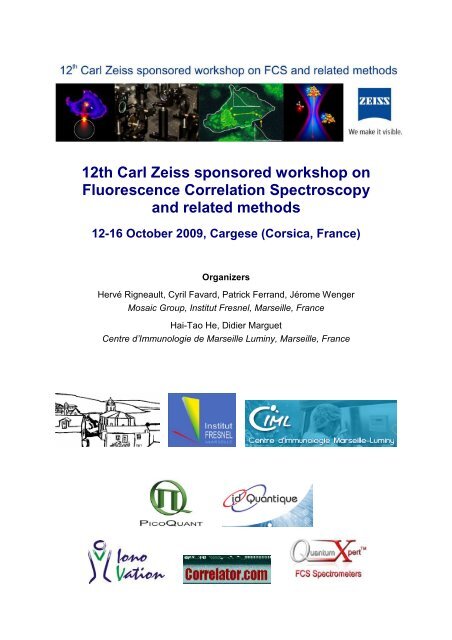
12th Carl Zeiss sponsored workshop on ... - Institut Fresnel
12th Carl Zeiss sponsored workshop on ... - Institut Fresnel
12th Carl Zeiss sponsored workshop on ... - Institut Fresnel

You also want an ePaper? Increase the reach of your titles
YUMPU automatically turns print PDFs into web optimized ePapers that Google loves.
<str<strong>on</strong>g>12th</str<strong>on</strong>g> <str<strong>on</strong>g>Carl</str<strong>on</strong>g> <str<strong>on</strong>g>Zeiss</str<strong>on</strong>g> <str<strong>on</strong>g>sp<strong>on</strong>sored</str<strong>on</strong>g> <str<strong>on</strong>g>workshop</str<strong>on</strong>g> <strong>on</strong><br />
Fluorescence Correlati<strong>on</strong> Spectroscopy<br />
and related methods<br />
12-16 October 2009, Cargese (Corsica, France)<br />
Organizers<br />
Hervé Rigneault, Cyril Favard, Patrick Ferrand, Jérome Wenger<br />
Mosaic Group, <strong>Institut</strong> <strong>Fresnel</strong>, Marseille, France<br />
Hai-Tao He, Didier Marguet<br />
Centre d’Immunologie de Marseille Luminy, Marseille, France
09:00<br />
09:20<br />
09:40<br />
10:00<br />
10:20<br />
10:40<br />
11:00<br />
11:20<br />
11:40<br />
12:00<br />
12:20<br />
12:40<br />
13:00<br />
13:20<br />
13:40<br />
14:00<br />
14:20<br />
14:40<br />
15:00<br />
15:20<br />
15:40<br />
16:00<br />
16:20<br />
16:40<br />
17:00<br />
17:20<br />
17:40<br />
18:00<br />
18:20<br />
18:40<br />
19:00<br />
19:20<br />
<str<strong>on</strong>g>12th</str<strong>on</strong>g> <str<strong>on</strong>g>Carl</str<strong>on</strong>g> <str<strong>on</strong>g>Zeiss</str<strong>on</strong>g> <str<strong>on</strong>g>sp<strong>on</strong>sored</str<strong>on</strong>g> <str<strong>on</strong>g>workshop</str<strong>on</strong>g> <strong>on</strong> FCS and related methods 2009<br />
M<strong>on</strong>day Tuesday Wednesday Thursday Friday<br />
12 Oct 13 Oct 14 Oct 15 Oct 16 Oct<br />
Bus transfer<br />
Ajaccio airport<br />
Shuttle 1<br />
Bus transfer<br />
Ajaccio airport<br />
Shuttle 2<br />
Registrati<strong>on</strong>s<br />
starting 08:20<br />
Opening<br />
Rigler<br />
Groves Petrasek<br />
Ham<strong>on</strong> Kuhnemuth<br />
Langowski Vereb Wennmalm<br />
Coffee break Coffee break Coffee break<br />
Engelborghs Enderlein Mueller<br />
Visser Orthaus Werner<br />
Wohland Galland Vukojevic<br />
Rigler Octeau Pfiffi<br />
Lunch Lunch Lunch<br />
Eggeling<br />
Jaffiol Del<strong>on</strong><br />
Wenger Gall<br />
Coffee break Coffee break<br />
Free Time<br />
Wiseman Brock<br />
Poster Sessi<strong>on</strong><br />
Widengren<br />
Sauer<br />
C<strong>on</strong>ference<br />
Dinner<br />
Weidtkamp-<br />
Peters<br />
Tramier<br />
Hendrix<br />
Closure<br />
Bus transfer<br />
Ajaccio airport
M<strong>on</strong>day October <str<strong>on</strong>g>12th</str<strong>on</strong>g><br />
Bus transfers from Ajaccio airport and installati<strong>on</strong> in Cargese<br />
Tuesday October 13th<br />
08h20 : Registrati<strong>on</strong> desk opening<br />
9h00-9h20 : H. Rigneault, Opening Introducti<strong>on</strong><br />
9h20-10h00 : R. Rigler, APD-Imaging:FCS in Single Cell Kinetics<br />
10h00-10h20 : J. Langovski, Generating mobility maps from two-dimensi<strong>on</strong>al fluorescence<br />
fluctuati<strong>on</strong> data<br />
10h20-10h40 : coffee break<br />
10h40-11h20 : Y. Engelborghs, short sampling time fluorescence correlati<strong>on</strong> spectroscopy<br />
reveals early oligomer formati<strong>on</strong> of alpha-synuclein<br />
11h20-11h40 : T. Visser, Global analysis of autocorrelati<strong>on</strong> functi<strong>on</strong>s and phot<strong>on</strong> counting<br />
distributi<strong>on</strong>s: some experimental tests involving EGFP and EGFP dimers<br />
11h40-12h00 : T. Wohland, Advances in Fluorescence Correlati<strong>on</strong> Spectroscopy:<br />
Quantitati<strong>on</strong> of Biomolecular Interacti<strong>on</strong>s and Imaging of Membrane Dynamics<br />
12h00-12h20 : P. Rigler, Towards single molecule enzymology of encapsulated enzymes<br />
12h20-14h00 : lunch break<br />
14h00-14h40 : C. Eggeling, Fluorescence Correlati<strong>on</strong> Spectroscopy at the nanoscale:<br />
Revealing more details with STED microscopy<br />
14h40-15h00 : R. Jaffiol, near-field fcs: driving biology bey<strong>on</strong>d the diffracti<strong>on</strong> limit<br />
15h00-15h20 : J. Wenger, Nanophot<strong>on</strong>ics to enhance FCS<br />
15h20-15h40 : coffee break<br />
15h40-16h20 : P. Wiseman, Recent Advances in Image Correlati<strong>on</strong> Spectroscopy and its<br />
Applicati<strong>on</strong>s in Cells<br />
16h30-18h30 : Poster Sessi<strong>on</strong>, finger food & drinks kindly supported by PicoQuant
Wednesday October 14th<br />
9h00-9h40 : J. Groves, Different acylati<strong>on</strong> motifs direct multiply orthog<strong>on</strong>oal co-localizati<strong>on</strong> of<br />
lipid anchored proteins in live cell membranes<br />
9h40-10h00 : Y. Ham<strong>on</strong>, Essential role for raft nanodomains in initiati<strong>on</strong> of T cell receptor<br />
signalling up<strong>on</strong> antigen recogniti<strong>on</strong><br />
10h00-10h20 : G. Vereb, ligand-stimulated egfr causes lipid domain-dependent higher order<br />
aggregati<strong>on</strong> and internalizati<strong>on</strong> of erbb2<br />
10h20-10h40 : coffee break<br />
10h40-11h20 : J. Enderlein, dual-focus fluorescence correlati<strong>on</strong> spectroscopy<br />
11h20-11h40 : S. Orthaus, New Methods for FCS: Fluorescence Lifetime<br />
Correlati<strong>on</strong> Spectroscopy and Two-Focus FCS<br />
11h40-12h00 : R. Galland, Multi-c<strong>on</strong>focal fluorescence correlati<strong>on</strong> spectroscopy for parallel<br />
multi-spot measurements in living cells<br />
12h00-12h20 : V. Octeau, Photothermal Absorpti<strong>on</strong> Correlati<strong>on</strong> Spectroscopy<br />
12h20-14h00 : lunch break<br />
14h00-17h20 : free time<br />
17h20-18h00 : J. Widengren, transient state m<strong>on</strong>itoring by fcs and related techniques<br />
18h00-18h40 : M. Sauer, super-resoluti<strong>on</strong> fluorescence imaging wth standard fluorophores<br />
19h00-21h00 : C<strong>on</strong>ference Dinner, kindly supported by <str<strong>on</strong>g>Carl</str<strong>on</strong>g> <str<strong>on</strong>g>Zeiss</str<strong>on</strong>g><br />
MicroImaging
Thursday October 15th<br />
9h00-9h40 : Z. Petrasek, Applicati<strong>on</strong>s of scanning FCS<br />
9h40-10h00 : R. Kühnemuth, laser scanning microscopy with avalanche photodiodes<br />
10h00-10h20 : S. Wennmalm, Inverse-Fluorescence Correlati<strong>on</strong> Spectroscopy<br />
10h20-10h40 : coffee break<br />
10h40-11h20 : J. Mueller, Hetero-Species Partiti<strong>on</strong> Analysis Revels Binding Curve and<br />
Stoichiometry of Protein Interacti<strong>on</strong>s in Living Cells<br />
11h20-11h40 : A. Werner, applicati<strong>on</strong>s of fluorescence correlati<strong>on</strong> spectroscopy in rna<br />
biochemistry<br />
11h40-12h00 : V. Vukojevic, functi<strong>on</strong>al synthetic hox genes: imaging the activity and<br />
quantifying transcripti<strong>on</strong> factor-dna interacti<strong>on</strong>s in live cells<br />
12h00-12h20 : D. Pfiffi, Improvement of Signal Strength and Photostability of Fluorophors by<br />
quenching triplet and radical states<br />
12h20-12h40 : Drinks and finger food, kindly supported by IdQuantique<br />
12h40-14h00 : lunch break<br />
14h00-14h40 : S. Weidtkamp-Peters, Multiparameter Fluorescence Imaging Spectroscopy<br />
including Fluorescence Correlati<strong>on</strong> Spectroscopy in living cells<br />
14h40-15h00 : A. Del<strong>on</strong>, measuring numbers of fluorophores labeling cDNA in soluti<strong>on</strong>, with<br />
fluorescence correlati<strong>on</strong> spectroscopy and c<strong>on</strong>tinuous photobleaching.<br />
15h00-15h20 : K. Gall, C<strong>on</strong>current Fluorescence Fluctuati<strong>on</strong> and<br />
Electrophysiological recordings of the Apoptosis Inducer PorB<br />
15h20-15h40 : coffee break<br />
15h40-16h20 : R. Brock, Quantitative Analyses of Protein Interacti<strong>on</strong> Networks<br />
16h20-16h40 : M. Tramier, Dual color GFP and mCherry Fluorescence Lifetime Correlati<strong>on</strong><br />
Spectroscopy for protein interacti<strong>on</strong>s in live cells<br />
16h40-17h00 : J. Hendrix, Cellular tunable focus FCS for the study of the molecular<br />
mechanism of LEDGF/p75 mediated chromatin tethering of HIV-1 integrase<br />
17h00-17h10 : K. Weisshart, C<strong>on</strong>cluding remarks<br />
Friday October 16th<br />
9h00 : shuttle bus departure to Ajaccio airport
Tuesday October 13 th
APD-Imaging:FCS in Single Cell Kinetics<br />
Rudolf Rigler<br />
Department of Medical Biochemistry and Biophysics ,Karolinska <strong>Institut</strong>et,Stockholm<br />
and Laboratory of Biomedical Optics , EPFL, Lausanne.(rudolf.rigler@epfl.ch)<br />
We have developed in collaborati<strong>on</strong> with <str<strong>on</strong>g>Carl</str<strong>on</strong>g> <str<strong>on</strong>g>Zeiss</str<strong>on</strong>g>.Jena the use of Avalanche Photo<br />
Diodes for background free detecti<strong>on</strong> and imaging of single molecule processes in<br />
individual cells (1,2) I will discuss the analysis of kinetic processes in various parts of<br />
the cells including diffusi<strong>on</strong> and chemical relaxati<strong>on</strong> taking membrane receptors<br />
as well as producti<strong>on</strong> of cytoplasmic and nuclear proteins and their interacti<strong>on</strong> with<br />
specific targets as examples . The use of Poiss<strong>on</strong> analysis (Poiss<strong>on</strong> Imaging)<br />
classical c<strong>on</strong>focal FCS . and scanning FCS will be discussed.<br />
(1)Vukojevic,V.et al. (2008) Quantitative single-molecule imaging by c<strong>on</strong>focal laser<br />
scanning microscopy.PNAS ,105,18176–18181<br />
(2) Rigler,R, (2009) FCS and Single Molecule Spectroscopy. Nobelsymposium 138<br />
:Single Molecule Spectroscopy in Chemistry,Physics and Biology.Sånga Säby.
Dynamics of supercoiled DNA studied by FCS and computer modeling<br />
Tomasz Wocjan 1 , Jan Krieger 1 , Oleg Krichevsky 2 and Jörg Langowski 1<br />
1<br />
Biophysics of Macromolecules, German Cancer Research Center, Im Neuenheimer Feld<br />
580, 69120 Heidelberg, Germany<br />
2<br />
Physics Department, Ben-Guri<strong>on</strong> University, Beer-Sheva 84105, Israel<br />
The organizati<strong>on</strong> of the genome in the cell nucleus is fundamental for cellular functi<strong>on</strong>:<br />
transcripti<strong>on</strong> and its regulati<strong>on</strong>, genome duplicati<strong>on</strong>, epigenetic effects and chromosome<br />
folding are all intricately c<strong>on</strong>nected to genome architecture. On the lowest level of genome<br />
organizati<strong>on</strong>, DNA folding is determined by its bending and twisting properties; superhelical<br />
DNA is a well-known model system to study these properties. Recent experiments by<br />
fluorescence correlati<strong>on</strong> spectroscopy (Shusterman et al., (1008) Phys Rev Lett. 100:098102)<br />
indicated a sensitivity of the m<strong>on</strong>omer mean square displacements in DNA circles towards<br />
superhelicity. To explain this phenomen<strong>on</strong>, we investigated the dynamics of a singlefluorophore-labeled<br />
pUC18 plasmid through a Brownian dynamics algorithm, followed by a<br />
simulati<strong>on</strong> of the fluorescence correlati<strong>on</strong> spectroscopy (FCS) process. Simulati<strong>on</strong>s with<br />
homogeneous DNA elasticity and local straight equilibrium are not sufficient to reproduce the<br />
observed behavior. But inserting permanently bent sequences into the DNA, which favor end<br />
loop formati<strong>on</strong>, caused a dependence of the calculated FCS correlati<strong>on</strong> curves <strong>on</strong> superhelical<br />
density. Furthermore, our simulati<strong>on</strong>s allow us to take into account the orientati<strong>on</strong> of the<br />
fluorophore in polarized excitati<strong>on</strong>, which might explain the observed appearance of a Rouselike<br />
regime at intermediate time scales.
SHORT SAMPLING TIME FLUORESCENCE CORRELATION<br />
SPECTROSCOPY REVEALS EARLY OLIGOMER FORMATION OF<br />
ALPHA-SYNUCLEIN<br />
Sangeeta Nath, Jelle Hendrix, Jessika Meuvis and Yves Engelborghs<br />
Laboratory of Biomolecular Dynamics, Department of Chemistry & BioSCENTer,<br />
University of Leuven, Celestijnenlaan 200 G, box 2403, B3001 Leuven, Belgium<br />
Yves.Engelborghs@fys.kuleuven.be<br />
C<strong>on</strong>focal microscopy not <strong>on</strong>ly reveals heterogeneity, when scanning cells, but also in<br />
soluti<strong>on</strong>. E.g. spike formati<strong>on</strong> can be observed when bright objects are formed. This<br />
analysis of heterogeneity in time can be quantitated to study protein-DNA interacti<strong>on</strong><br />
[1] or complex formati<strong>on</strong> [2]. Here we study the formati<strong>on</strong> of early oligomers of αsynuclein<br />
by applying Fluorescence Correlati<strong>on</strong> Spectroscopy (FCS). The idea is to<br />
use trace amounts (nM) of labeled protein in the presence of a large excess of<br />
unlabeled protein and follow the aggregati<strong>on</strong> process by measuring the reducti<strong>on</strong> in<br />
time of the diffusi<strong>on</strong> coefficient of the fluorescent species. Synuclein with an<br />
engineered cysteine (A140C) was labeled with Alexa488 and was used as a<br />
fluorescent probe in trace amounts (3-4 nM) in the presence of 100 μM unlabeled αsynuclein.<br />
The combinati<strong>on</strong> of short sampling times and repeated measurements<br />
produce a size distributi<strong>on</strong> of the oligomers. Initially, a sharp peak is obtained<br />
(diffusi<strong>on</strong> coefficient 114 ± 15 µm²/sec) corresp<strong>on</strong>ding to m<strong>on</strong>omers. Subsequently a<br />
distinct transient populati<strong>on</strong> appears, followed by the gradual formati<strong>on</strong> of broader<br />
sized distributi<strong>on</strong>s of higher oligomers. The formati<strong>on</strong> of the transient intermediate<br />
and the early oligomers is accompanied by a c<strong>on</strong>formati<strong>on</strong>al change, as visualised<br />
using FRET between the d<strong>on</strong>or labeled N-terminus and the acceptor labeled cysteine<br />
at positi<strong>on</strong> A140C. At l<strong>on</strong>ger time scales, further aggregati<strong>on</strong> leads to the formati<strong>on</strong><br />
of big aggregates that are moving too slow to c<strong>on</strong>tribute to the short term fluctuati<strong>on</strong>s.<br />
The overall aggregati<strong>on</strong> process can be followed by the decrease of the number<br />
c<strong>on</strong>centrati<strong>on</strong> of fast diffusing fluorescent species.<br />
[1] Vercammen J, Maertens G, Gerard M, De Clercq E, Debyser Z, Engelborghs Y.<br />
DNA-induced polymerizati<strong>on</strong> of HIV-1 integrase analyzed with fluorescence<br />
fluctuati<strong>on</strong> spectroscopy. J Biol Chem., 277, 38045, (2002)<br />
[2] Buyens K, Lucas B, Raemd<strong>on</strong>ck K, Braeckmans K, Vercammen J, Hendrix<br />
J,Engelborghs Y, De Smedt SC, Sanders NN. A fast and sensitive method for<br />
measuring the integrity of siRNA-carrier complexes in full human serum. J C<strong>on</strong>trol<br />
Release.,26, 67,(2008)
12 th <str<strong>on</strong>g>Carl</str<strong>on</strong>g> <str<strong>on</strong>g>Zeiss</str<strong>on</strong>g> <str<strong>on</strong>g>sp<strong>on</strong>sored</str<strong>on</strong>g> <str<strong>on</strong>g>workshop</str<strong>on</strong>g> <strong>on</strong> FCS and related methods (12-16 October<br />
2009, Cargese, Corsica, France)<br />
Global analysis of autocorrelati<strong>on</strong> functi<strong>on</strong>s and phot<strong>on</strong> counting distributi<strong>on</strong>s:<br />
some experimental tests involving EGFP and EGFP dimers<br />
Victor Skakun 1 and Ant<strong>on</strong>ie Visser 2<br />
1 Belarusian State University, Minsk, 220050 Belarus and 2 Microspectroscopy Centre,<br />
Wageningen University, 6703 HA Wageningen, The Netherlands<br />
It was shown previously that phot<strong>on</strong> counting histogram (PCH) analysis and dual-<br />
color fluorescence cross correlati<strong>on</strong> spectroscopy (FCCS) could m<strong>on</strong>itor fluorescently<br />
labeled receptor kinases at the single-molecule detecti<strong>on</strong> level in living plant cells<br />
(M.A. Hink et al. (2008) Biophys. J. 94, 1052-1062). Both PCH and fluorescence<br />
correlati<strong>on</strong> spectroscopy (FCS) analysis use the same experimental data, but each<br />
technique focuses <strong>on</strong> a different property of the signal. FCS measures the time-<br />
dependent decay of the fluorescence fluctuati<strong>on</strong>s yielding, for instance, molecular<br />
diffusi<strong>on</strong> coefficients, while PCH analysis calculates the amplitude distributi<strong>on</strong> of<br />
these fluctuati<strong>on</strong>s yielding the distributi<strong>on</strong> of molecular brightness (average<br />
fluorescence phot<strong>on</strong> count rate per molecule per sec<strong>on</strong>d). Both methods give<br />
informati<strong>on</strong> about the molecular c<strong>on</strong>centrati<strong>on</strong>. Via separate FCCS and PCH<br />
experiments some receptors were found to be present for 15-20% as homodimers,<br />
whereas no evidence was found for higher oligomeric complexes in other receptors.<br />
Realizing that FCS and PCH make use of the same experimental fluorescence<br />
fluctuati<strong>on</strong> traces it is obvious that a global analysis protocol must be developed that<br />
simultaneously recovers the relevant parameters. In this presentati<strong>on</strong> such an<br />
approach is described and tested with experimental fluorescence fluctuati<strong>on</strong> data of<br />
EGFP and dimeric EGFP in aqueous soluti<strong>on</strong> (pH 8.0).<br />
The algorithm for PCH analysis also allows correcting for out-of-focus emissi<strong>on</strong><br />
signals. This would pave the way to ‘visualize’ the n<strong>on</strong>-ideal brightness profile in<br />
cellular and other high-refractive-index systems.
Quantitative measurements of molecular interacti<strong>on</strong> in living organisms by<br />
Single Wavelength Fluorescence Cross-correlati<strong>on</strong> Spectroscopy (SW-<br />
FCCS)<br />
Thorsten Wohland<br />
Chemistry Department, Nati<strong>on</strong>al University of Singapore, 3 Science Drive 3, 117543 Singapore<br />
In the past biomolecular interacti<strong>on</strong>s have been measured mostly under in vitro<br />
c<strong>on</strong>diti<strong>on</strong>s because of the higher accuracy and ease of measurement. However, it has<br />
become clear in the last years that the cellular envir<strong>on</strong>ment has an important influence <strong>on</strong><br />
these interacti<strong>on</strong>s and when trying to understand cellular functi<strong>on</strong>s these factors have to<br />
be included. For that purpose we have developed single wavelength excitati<strong>on</strong><br />
fluorescence cross-correlati<strong>on</strong> spectroscopy (SW-FCCS) which allows the determinati<strong>on</strong><br />
of biomolecular interacti<strong>on</strong>s in vivo. Because SW-FCCS uses <strong>on</strong>ly a single laser line for<br />
excitati<strong>on</strong> it does not suffer from problems of excitati<strong>on</strong> volume overlap usually<br />
encountered in dual-color FCCS. And although that leads to lower count rates per<br />
particle detected, due to the excitati<strong>on</strong> of two-spectrally different fluorophores at n<strong>on</strong>ideal<br />
wavelength, it possesses a sufficiently high signal to noise ratio to allow the<br />
quantitati<strong>on</strong> of biomolecular interacti<strong>on</strong>s in living organisms. We dem<strong>on</strong>strate how SW-<br />
FCCS can be used to measure the dissociati<strong>on</strong> c<strong>on</strong>stants of Cdc42, a small Rho-GTPase,<br />
with different interacting molecules (N-WASP, IRSp53, IQGAP1) in live cells and<br />
zebrafish embryos and discuss the advantages and disadvantages of the method.
Towards single molecule enzymology of<br />
encapsulated enzymes<br />
Per Rigler 1 , Kai Hassler 2 , Mariusz Grzelakowski 1 , Ozana Onaca 1 , Karolina<br />
Langowska 1 , Wolfgang Meier 1<br />
1 Department Chemie, Universität Base1, Switzerland and<br />
2 Biomedical Imaging Group, EPFL, Lausanne, Switzerland<br />
Single molecular fluorescence spectroscopic studies of enzymes have paved the<br />
way for a better understanding of how enzymes work at the molecular scale. Here we<br />
show first results how single or few functi<strong>on</strong>al enzymes can be encapsulated in polymeric<br />
nanoc<strong>on</strong>tainers c<strong>on</strong>sisting of amphiphilic block copolymers of a polymethyloxazoline-polydimethylsiloxane-polymethyloxazoline<br />
(PDMS-PMOXA-PDMS) architecture.<br />
To add functi<strong>on</strong>ality to these nanoc<strong>on</strong>tainers a membrane protein (outer<br />
membrane protein F, OmpF) has been rec<strong>on</strong>stituted into the polymeric membrane and<br />
a biotinylated PMOXA-PDMS-PMOXA polymer is used allowing flux of substrate<br />
and product molecules through the above menti<strong>on</strong>ed membrane and to anchor the<br />
nanoc<strong>on</strong>tainers specifically to a planar substrate, respectively.<br />
Previously we have shown that it is possible to measure molecular turnover of single<br />
enzymes (horse radish peroxidase) at glass surfaces previously functi<strong>on</strong>alized with<br />
a biotin-derivatized polylysin-g-polyethyleneglycol polymer by using various fluorogenic<br />
substrates in combinati<strong>on</strong> with total internal fluorescence fluorescence correlati<strong>on</strong><br />
spectroscopy (TIR-FCS)[1, 2].<br />
Polymeric nanoc<strong>on</strong>tainers with rec<strong>on</strong>stituted OmpF have been reproducibly tethered<br />
to glass surfaces previously patterned by microc<strong>on</strong>tact printing using BSA/BSAbiotin.<br />
The enzyme acid phosphatase was encapsulated in the nanoreactors and the<br />
fluorogenic substrate ELF-97 was used as a fluorescent probe in order to determine<br />
enzyme kinetic parameters using both fluorimetry and c<strong>on</strong>focal fluorescence microscopy[3].<br />
The kinetic parameters obtained from the free, the encapsulated, and the surface-tethered<br />
encapsulated enzyme are compaired. Our results show that it will be possible<br />
in a near future to study enzyme kinetics at the single molecule level inside<br />
nanoreactors. This technique could potentially be used to develop new biosensors for<br />
screening enzymatic activity at an unprecedented sensitivity.<br />
[1.] Hassler, K., et al., Dynamic disorder in horseradish peroxidase observed with total internal<br />
reflecti<strong>on</strong> fluorescence correlati<strong>on</strong> spectroscopy. Optics Express 2007, 15, (9), 5366-<br />
5375.<br />
[2.] Hassler, K., et al., Total internal reflecti<strong>on</strong> fluorescence correlati<strong>on</strong> spectroscopy (TIR-<br />
FCS) with low background and high count-rate per molecule. Optics Express 2005, 13, (19),<br />
7415-7423.<br />
[3.] Broz, P., et al., Toward intelligent nanosize bioreactors: A pH-switchable, channelequipped,<br />
functi<strong>on</strong>al polymer nanoc<strong>on</strong>tainer. Nano Letters 2006, 6, (10), 2349-2353.
Fluorescence Correlati<strong>on</strong> Spectroscopy at the nanoscale: Revealing more details with<br />
STED microscopy<br />
Christian Eggeling *, Christian Ringemann * , Ver<strong>on</strong>ika Müller *, Rebecca Medda *, Günter<br />
Schwarzmann **, Birka Hein *, Andreas Schönle *, Stefan W. Hell *<br />
* Dep. Nanobiophot<strong>on</strong>ics, Max Planck <strong>Institut</strong>e for Biophysical Chemistry, Am Fassberg 11,<br />
37077 Göttingen, Germany<br />
** Kekulé-<strong>Institut</strong>e für Organische Chemie und Biochemie, Gerhard-Domagk-Strasse 1,<br />
53121 B<strong>on</strong>n, Germany<br />
ceggeli@gwdg.de<br />
Fluorescence Correlati<strong>on</strong> Spectroscopy (FCS) is a n<strong>on</strong>-invasive and very sensitive analysis<br />
technique, allowing for the disclosure of complex dynamical processes. FCS is usually<br />
combined with far-field (c<strong>on</strong>focal) microscopy. However, prominent (biological) problems<br />
can often not be solved due to the limited resoluti<strong>on</strong> of c<strong>on</strong>venti<strong>on</strong>al optical microscopy. For<br />
example, in a c<strong>on</strong>venti<strong>on</strong>al optical microscope FCS requires rather low c<strong>on</strong>centrati<strong>on</strong>s of the<br />
fluorescently labeled molecules, which has precluded its otherwise very promising applicati<strong>on</strong><br />
to systems where molecular c<strong>on</strong>centrati<strong>on</strong>s of several micro-mol per liter (µM) are required.<br />
Further, cholesterol-assisted lipid interacti<strong>on</strong>s such as the integrati<strong>on</strong> into lipid nanodomains<br />
(‘rafts’) are c<strong>on</strong>sidered to play a functi<strong>on</strong>al part in a whole range of membrane-associated<br />
processes, but their direct and n<strong>on</strong>-invasive observati<strong>on</strong> in living cells is impeded by the<br />
resoluti<strong>on</strong> limit of >200nm of a c<strong>on</strong>venti<strong>on</strong>al far-field fluorescence microscope. Using the<br />
superior spatial resoluti<strong>on</strong> of stimulated emissi<strong>on</strong> depleti<strong>on</strong> (STED) far-field microscopy [1],<br />
we dem<strong>on</strong>strate novel use of FCS at rather large fluorophore c<strong>on</strong>centrati<strong>on</strong> [2,3] or for the<br />
direct and n<strong>on</strong>-invasive detecti<strong>on</strong> of single diffusing lipid molecules in nanosized areas in the<br />
plasma membrane of living cells. Combining a (tunable) resoluti<strong>on</strong> of down to 30 nm with<br />
FCS, we obtain new details of molecular membrane dynamics. For example, unlike<br />
phosphoglycerolipids, sphingolipids or ‘raft’-associated proteins are transiently (~ 10 ms)<br />
trapped <strong>on</strong> the nanoscale in cholesterol-mediated molecular [4]. These results are corroborated<br />
by single-molecule tracking experiments.<br />
[1] S.W. Hell “Far-Field Optical Nanoscopy”, Science, 316, 1153, (2007).<br />
[2] L. Kastrup et al. “Fluorescence Fluctuati<strong>on</strong> Spectroscopy in Subdiffracti<strong>on</strong> Focal<br />
Volumes”, PRL, 94, 178104, (2005)<br />
[3] C. Eggeling et al. “Reversible photoswitching enables single-molecule fluorescence<br />
fluctuati<strong>on</strong> spectroscopy at high molecular c<strong>on</strong>centrati<strong>on</strong>”, MRT, 70, 1003, (2007)<br />
[4] C. Eggeling et al. “Direct observati<strong>on</strong> of the nanoscale dynamics of membrane lipids in a<br />
living cell”, Nature, 457, 1159, (2009)<br />
12 th <str<strong>on</strong>g>Zeiss</str<strong>on</strong>g> <str<strong>on</strong>g>sp<strong>on</strong>sored</str<strong>on</strong>g> FCS <str<strong>on</strong>g>workshop</str<strong>on</strong>g> – Cargèse, Corsica, France – <str<strong>on</strong>g>12th</str<strong>on</strong>g> -16th October 2009
NEAR-FIELD FCS: DRIVING BIOLOGY BEYOND THE DIFFRACTION LIMIT<br />
Winckler P, Jaffiol R, Plain J and Royer P<br />
Université de Technologie de Troyes, <strong>Institut</strong> Charles Delaunay FRE CNRS 2848,<br />
Laboratoire de Nanotechnologie et d’Instrumentati<strong>on</strong> Optique, LRC CEA,<br />
12 rue Marie Curie, BP2060, 10010 Troyes cedex, France<br />
c<strong>on</strong>tact: rodolphe.jaffiol@utt.fr<br />
For single molecule detecti<strong>on</strong> in soluti<strong>on</strong>, the issue is to get the probability of finding molecule<br />
in the observati<strong>on</strong> volume significantly inferior to unity. This can be achieved through<br />
diluting the dye of interest or limiting the detecti<strong>on</strong> to a very small volume. Classical fluorescence<br />
correlati<strong>on</strong> spectrosocpy (FCS), based <strong>on</strong> c<strong>on</strong>focal detecti<strong>on</strong>, presents two important<br />
limitati<strong>on</strong>s for in vivo studies. First, its requires to work with a nanomolar c<strong>on</strong>centrati<strong>on</strong> of<br />
molecules, very far from the biologically relevant c<strong>on</strong>centrati<strong>on</strong> (i.e µM, or higher). On the<br />
other hand, the background signal such as autofluorescence, is still often important. In order<br />
to overcome the limitati<strong>on</strong>s of classical FCS, we propose a new approach to reduce the detecti<strong>on</strong><br />
volume based <strong>on</strong> a n<strong>on</strong> radiative energy transfer. We present two applicati<strong>on</strong>s of our<br />
technique to make FCS and fluorescence imaging at the nanoscale, see Fig. 1.<br />
(A)<br />
(B)<br />
Fig. 1. (A) Picture of adhesi<strong>on</strong> areas between cells and substrate. Superpositi<strong>on</strong> of two<br />
widefield images, <strong>on</strong>e DIC picture in grey scale and <strong>on</strong>e fluorescence picture in red scale.<br />
(B) Typical autocorrelati<strong>on</strong> curve recorded <strong>on</strong> an cell adhesi<strong>on</strong> area.<br />
Red curve, fit according to a pure 2-D diffusi<strong>on</strong> of molecules.<br />
12 th <str<strong>on</strong>g>Zeiss</str<strong>on</strong>g> <str<strong>on</strong>g>sp<strong>on</strong>sored</str<strong>on</strong>g> FCS <str<strong>on</strong>g>workshop</str<strong>on</strong>g> – Cargèse, Corsica, France – <str<strong>on</strong>g>12th</str<strong>on</strong>g> -16th October 2009
Nanophot<strong>on</strong>ic structures to enhance single molecule fluorescence detecti<strong>on</strong><br />
J. Wenger, 1 D. Gérard, 1 H. Aouani, 1 H. Rigneault, 1 E. Devaux, 2 T.W. Ebbesen, 2 and S. Blair 3<br />
1 <strong>Institut</strong> <strong>Fresnel</strong>, CNRS, Aix-Marseille Université, Marseille, France<br />
2 ISIS, Université Louis Pasteur, CNRS, 8 allée G. M<strong>on</strong>ge, 67000 Strasbourg, France<br />
3 Electrical and Computer Engineering Department, University of Utah, Salt Lake City, USA<br />
Jerome.wenger@fresnel.fr<br />
The ability to produce nanometric devices with a resoluti<strong>on</strong> down to a few nanometers opens<br />
the way for promising applicati<strong>on</strong>s in nano- and bio-phot<strong>on</strong>ics. Such devices allow to locally<br />
c<strong>on</strong>fine the light, inducing large local electromagnetic fields.<br />
In this c<strong>on</strong>tributi<strong>on</strong>, we will focus <strong>on</strong> two fundamental questi<strong>on</strong>s that bear practical<br />
applicati<strong>on</strong>s, and we will show how nanophot<strong>on</strong>ic structures bring innovative answers :<br />
• How to detect <strong>on</strong>e single molecule in a highly c<strong>on</strong>centrated (µM) soluti<strong>on</strong> ?<br />
• How to enhance the optical signal detected per molecule ?<br />
As an illustrati<strong>on</strong>, we will focus <strong>on</strong> the use of nanometric apertures milled in a metallic film<br />
and dielectric (polystyrene) microspheres. Both structures are shown to enhance the<br />
fluorescence emissi<strong>on</strong> of single molecules, with an enhancement factor up to 25 as compared<br />
to the emissi<strong>on</strong> rate in open soluti<strong>on</strong> [1]. Besides, the observati<strong>on</strong> volume can be up to three<br />
orders of magnitude smaller than the diffracti<strong>on</strong> limit in optics, enabling efficient enzymatic<br />
reacti<strong>on</strong>s m<strong>on</strong>itoring at micromolar c<strong>on</strong>centrati<strong>on</strong>s for FCS or FCCS.<br />
The optimised fluorescence enhancement offers a gain in signal to noise ratio of about <strong>on</strong>e<br />
order of magnitude, corresp<strong>on</strong>ding to a 100-fold reducti<strong>on</strong> of the FCS experiment durati<strong>on</strong>.<br />
This evidences the feasibility of FCS analysis with fast integrati<strong>on</strong> times of about <strong>on</strong>e sec<strong>on</strong>d,<br />
opening the way to the m<strong>on</strong>itoring biochemical reacti<strong>on</strong>s at reduced time scales [2].<br />
[1] P.-F. Lenne, et al, Fluorescence fluctuati<strong>on</strong>s analysis in nanoapertures: physical c<strong>on</strong>cepts<br />
and biological applicati<strong>on</strong>s, HistoChem. Cell. Biol. 130, 795-805 (2008).<br />
[2] J. Wenger, et al, Nanoaperture-Enhanced Signal-to-Noise Ratio in Fluorescence<br />
Correlati<strong>on</strong> Spectroscopy, Anal. Chem. 81, 834-839 (2009).<br />
12 th <str<strong>on</strong>g>Zeiss</str<strong>on</strong>g> <str<strong>on</strong>g>sp<strong>on</strong>sored</str<strong>on</strong>g> FCS <str<strong>on</strong>g>workshop</str<strong>on</strong>g> – Cargèse, Corsica, France – <str<strong>on</strong>g>12th</str<strong>on</strong>g> -16th October 2009
Recent Advances in Image Correlati<strong>on</strong> Spectroscopy and its Applicati<strong>on</strong>s in Cells<br />
Paul W. Wiseman *,<br />
* McGill University<br />
Departments of Physics & Chemistry<br />
M<strong>on</strong>treal, QC Canada<br />
paul.wiseman@mcgill.ca<br />
This seminar will provide an introducti<strong>on</strong> for the n<strong>on</strong>-expert to various new methods of image<br />
correlati<strong>on</strong> spectroscopy (ICS) and their applicati<strong>on</strong> for measurements of protein transport and<br />
interacti<strong>on</strong>s in cells and neur<strong>on</strong>s. An overview of the history and background theory of these<br />
fluctuati<strong>on</strong> microscopy based methods will be presented al<strong>on</strong>g with a descripti<strong>on</strong> of the<br />
microscopy instrumentati<strong>on</strong> and computati<strong>on</strong>al resources required to perform such<br />
measurements. I will specifically cover more recent developments of ICS including spatiotemporal<br />
ICS (STICS), reciprocal or k-space ICS (kICS), and raster-scan ICS (RICS). The<br />
STICS porti<strong>on</strong> will focus <strong>on</strong> its applicati<strong>on</strong> to measuring vector maps of flows of adhesi<strong>on</strong><br />
and migrati<strong>on</strong> related proteins (actin, alpha-actinin, paxillin, integrins, talin) in living cells and<br />
discuss how two color cross-correlati<strong>on</strong> variants can be used to measure interacti<strong>on</strong>s via cotransport.<br />
The kICS part will discuss how this method can avoid complicati<strong>on</strong>s of complex<br />
photophysics of the fluorophore probe and even be used to extract informati<strong>on</strong> from probe<br />
blinking when quantum dot nanoparticles are used [1].<br />
9 µm/min<br />
0 µm/min<br />
5 µm<br />
Fig. 1. Velocity map of retrograde transport of alpha-actinin/EGFP in an MEF cell. Measured<br />
by TIRF microscopy and STICS analysis.<br />
[1] D. L. Kolin, P. W. Wiseman. Advances in image correlati<strong>on</strong> spectroscopy: measuring<br />
number densities, aggregati<strong>on</strong> states, and dynamics of fluorescently-labeled macromolecules<br />
in cells. Cell Biochem. Biophys. 49: 141-164 (2007)<br />
12 th <str<strong>on</strong>g>Zeiss</str<strong>on</strong>g> <str<strong>on</strong>g>sp<strong>on</strong>sored</str<strong>on</strong>g> FCS <str<strong>on</strong>g>workshop</str<strong>on</strong>g> – Cargèse, Corsica, France – <str<strong>on</strong>g>12th</str<strong>on</strong>g> -16th October 2009
Wednesday October 14 th
Different acylati<strong>on</strong> motifs direct multiply orthog<strong>on</strong>oal co-localizati<strong>on</strong> of lipid anchored<br />
proteins in live cell membranes<br />
J.T. Groves*, M.B. Forstner*, B.F. Lillemeier**, and M.M. Davis**<br />
* Howard Hughes Medical <strong>Institut</strong>e<br />
University of California Berkeley and Lawrence Berkeley Nati<strong>on</strong>al Laboratory<br />
** Stanford University<br />
C<strong>on</strong>tact author: JTGroves@lbl.gov<br />
A number of chemically distinct genetically-encoded acylati<strong>on</strong> modificati<strong>on</strong>s link soluble<br />
protein domains to cell membranes in vivo. It is widely speculated that these provide targeting<br />
mechanisms that direct protein organizati<strong>on</strong> in the membrane. Here, we use fluorescence<br />
cross correlati<strong>on</strong> spectroscopy to probe interacti<strong>on</strong>s am<strong>on</strong>g all combinatorial pairs of four<br />
comm<strong>on</strong> lipid anchoring motifs expressed in live primary T cells as fluorescent protein<br />
fusi<strong>on</strong>s. All homogeneous anchor combinati<strong>on</strong>s lead to str<strong>on</strong>g co-localizati<strong>on</strong> of the anchored<br />
proteins into small mobile clusters <strong>on</strong> the membrane surface. In c<strong>on</strong>trast, results from<br />
heterogeneous anchor pairings reveal mutual avoidance am<strong>on</strong>g each of the three cytoplasmic<br />
membrane leaflet anchors examined (palmityl:palmityl:myristoyl from Lck, myristoyl from<br />
Src, and gerenyl-gerenyl from RhoA). The extracellular leaflet GPI anchor co-localizes with<br />
the Lck anchor while avoiding the other anchors types. Both cholesterol depleti<strong>on</strong> and actin<br />
cytoskelet<strong>on</strong> disrupti<strong>on</strong> interfere with anchor-mediated clustering. These results indicate that<br />
protein acylati<strong>on</strong> can direct co-localizati<strong>on</strong> into at least three orthog<strong>on</strong>ally composed small<br />
dynamic clusters <strong>on</strong> the membrane surface. Additi<strong>on</strong>ally, at least <strong>on</strong>e of these cluster types<br />
exhibits trans-bilayer structure.<br />
Fig. 1. A small combinatorial library of different genetically encoded lipid anchor sequences<br />
fused to red or green fluorescent proteins is used to explore anchor co-localizati<strong>on</strong> in the<br />
membranes of live primary T cells.<br />
12 th <str<strong>on</strong>g>Zeiss</str<strong>on</strong>g> <str<strong>on</strong>g>sp<strong>on</strong>sored</str<strong>on</strong>g> FCS <str<strong>on</strong>g>workshop</str<strong>on</strong>g> – Cargèse, Corsica, France – <str<strong>on</strong>g>12th</str<strong>on</strong>g> -16th October 2009
Essential role for raft nanodomains in initiati<strong>on</strong> of T cell receptor signalling<br />
up<strong>on</strong> antigen recogniti<strong>on</strong><br />
Ham<strong>on</strong> Yannick, Bernard Anne-Marie, Salles Audrey, Hawchar Omar, Phelipot Marie-Claire,<br />
Guo Xiao-Jun, Sergé Arnauld, Mailfert Sébastien, Marguet Didier and He Hai-Tao<br />
Centre d’Immunologie de Marseille-Luminy (CIML),<br />
CNRS UMR 6102-INSERM UMR631-Université Aix-Marseille UMR6546,<br />
Case 906, 13288 Marseille Cedex 9, France<br />
C<strong>on</strong>tact: ham<strong>on</strong>@ciml.univ-mrs.fr<br />
[1] Ham<strong>on</strong>, Y., et al. Immun., Endoc. & Metab. Agents in Med. Chem. 2008, 8, p. 358-65.<br />
[2] Wawrezinieck, L., et al. Biophys J, 2005. 89(6): p. 4029-42.<br />
[3] Lenne, P.F., et al., EMBO J, 2006. 25(14): p. 3245-56.<br />
[4] Lasserre, R., et al., Nat Chem Biol, 2008. 4(9): p. 538-47.<br />
12 th <str<strong>on</strong>g>Zeiss</str<strong>on</strong>g> <str<strong>on</strong>g>sp<strong>on</strong>sored</str<strong>on</strong>g> FCS <str<strong>on</strong>g>workshop</str<strong>on</strong>g> – Cargèse, Corsica, France – <str<strong>on</strong>g>12th</str<strong>on</strong>g> -16th October 2009
LIGAND-STIMULATED EGFR CAUSES LIPID DOMAIN-DEPENDENT HIGHER<br />
ORDER AGGREGATION AND INTERNALIZATION OF ERBB2<br />
László<br />
Ujlaky-Nagy,<br />
Árpád Szöır,<br />
János Szöllısi,<br />
György Vereb<br />
University of Debrecen, Dept. of Biophysics and Cell Biology, H-4032 Debrecen (Hungary)<br />
vereb@dote.hu<br />
Members of the ErbB receptor tyrosine kinase family, ErbB1 (EGFR), ErbB2, ErbB3 and<br />
ErbB4, form homo- and heterodimers or higher oligomers with each other which promote cell<br />
survival and proliferati<strong>on</strong> [1]. Generally, overexpressi<strong>on</strong> of the ligandless ErbB2 is thought to<br />
drive homodimer formati<strong>on</strong> in the membrane of tumor cells. However, ErbB2 can also act as a<br />
heterodimerizing partner for other family members, and serve as a signal amplifier for their<br />
ligand-bound forms, which may have clinical relevance in tumors expressing multiple ErbBs<br />
[2]. We have used fluorescence correlati<strong>on</strong> spectroscopy to investigate molecular events of<br />
ErbB1-ErbB2 transactivati<strong>on</strong> in single living SKBR3 breast tumor cells. ErbB2 was labeled<br />
with A488 Fabs. Fluorescence fluctuati<strong>on</strong> traces were taken from single submicr<strong>on</strong> sized spots<br />
in the upper membrane of cells and used to c<strong>on</strong>struct fluorescence autocorrelati<strong>on</strong> spectra.<br />
These were fitted using a model accounting for triplet state, diffusi<strong>on</strong> of dissociated Fabs, as<br />
well as cytoplasmic and membrane (lateral, 2D) diffusi<strong>on</strong> of ErbB2 molecules or their<br />
aggregates. Correlati<strong>on</strong> spectra were obtained every 2.5 min for 1 hour. After 20 min, half of<br />
the samples were stimulated with 50 nM EGF to detect time-dependent changes of ErbB2<br />
diffusi<strong>on</strong>. C<strong>on</strong>trols showed a time dependent fluctuati<strong>on</strong> of membrane diffusi<strong>on</strong> times in the<br />
120-200 ms range (182±9 ms, mean±SEM). This was paralleled with internalizati<strong>on</strong> and<br />
recycling to the surface of receptors as measured by flow cytometry. Membrane diffusi<strong>on</strong> of<br />
ErbB2 was divided into at least two comp<strong>on</strong>ents: a faster comp<strong>on</strong>ent characterizing<br />
m<strong>on</strong>omers or smaller aggregates, and a slower comp<strong>on</strong>ent related to higher oligomers or<br />
larger clusters. The corresp<strong>on</strong>ding diffusi<strong>on</strong> times were 94±3 ms and 360±18 ms. Codiffusi<strong>on</strong><br />
of ErbB2 with ErbB2 up<strong>on</strong> labeling with a mixture of A488 and A647 fluorophors<br />
was detected by fluorescence cross-correlati<strong>on</strong> spectroscopy (FCCS) mainly for the slower<br />
comp<strong>on</strong>ent indicating that the faster comp<strong>on</strong>ent might be biased towards m<strong>on</strong>omers.<br />
Resp<strong>on</strong>ses to EGF stimulati<strong>on</strong> were vastly heterogeneous in timing, but manifested<br />
unanimously in an increase of characteristic average membrane diffusi<strong>on</strong> time to maximally<br />
550±119 ms. The average <strong>on</strong>set of resp<strong>on</strong>se was 5.5 min post stimulus and slowed-down<br />
diffusi<strong>on</strong> was maintained for ~30 min. EGF-induced transactivati<strong>on</strong> did not change the faster<br />
comp<strong>on</strong>ent (100±19 ms), but lead to the increased diffusi<strong>on</strong> times of the slower <strong>on</strong>e (from 360<br />
ms to 764±66 ms), at the same time decreasing its fracti<strong>on</strong>al proporti<strong>on</strong> from 0.55 to 0.2,<br />
while the fast comp<strong>on</strong>ent increased from 0.37 to 0.65. This may be an indirect indicati<strong>on</strong> that<br />
larger aggregates are needed for internalizati<strong>on</strong> and the receptors are recycled as m<strong>on</strong>omers or<br />
smaller clusters afterwards. C<strong>on</strong>sequences of the increased ErbB1-ErbB2 interacti<strong>on</strong> could be<br />
detected by microscopic fluorescence res<strong>on</strong>ance energy transfer as well as from increased<br />
ErbB1 and ErbB2 phosphorylati<strong>on</strong> in Western blots and were decreased by extracti<strong>on</strong> of<br />
membrane cholesterol with methyl-ß-cyclodextrine. This treatment also accelerated the<br />
diffusi<strong>on</strong> time of ErbB2 to 83±16 ms, indicating that the slower comp<strong>on</strong>ent (higher<br />
aggregates) corresp<strong>on</strong>ds to microscopic lipid rafts. Given the low expressi<strong>on</strong> of ErbB1 in<br />
SKBR3 cells, the mechanism of ErbB1-ErbB2 transactivati<strong>on</strong> can be interpreted as ErbB1<br />
serving as signal antennae that up<strong>on</strong> ligand binding recruit ErbB2 to form increasing size<br />
activated aggregates <strong>on</strong> lipid raft platforms that eventually get internalized.<br />
[1] Y. Yarden, M. X. Sliwkowski, Nat Rev Mol Cell Biol. 2 (2001) 127-137.<br />
[2] A. Citri., Y. Yarden, Nat Rev Mol Cell Biol. 7 (2006) 505-516.<br />
12 th <str<strong>on</strong>g>Zeiss</str<strong>on</strong>g> <str<strong>on</strong>g>sp<strong>on</strong>sored</str<strong>on</strong>g> FCS <str<strong>on</strong>g>workshop</str<strong>on</strong>g> – Cargèse, Corsica, France – <str<strong>on</strong>g>12th</str<strong>on</strong>g> -16th October 2009
DUAL-FOCUS FLUORESCENCE CORRELATION SPECTROSCOPY<br />
Anastasia Loman*, Claus B. Müller*, Iris v<strong>on</strong> der Hocht**, Thomas Detinger***,<br />
Ingo Gregor*, Jörg Enderlein*,<br />
* III. <strong>Institut</strong>e of Physics, Georg August University Göttingen, D-37077 Göttingen, Germany<br />
** Max-Planck <strong>Institut</strong>e of Biophysics, D-60438 Frankfurt, Germany<br />
*** Dept. of Chemistry and Biochemistry, UCLA, CA 90095, USA<br />
enderlein@physik3.gwdg.de<br />
Several decades ago, Magde, Els<strong>on</strong> and Webb invented an ingenuous method for measuring<br />
diffusi<strong>on</strong> coefficients of fluorescent molecules: Fluorescence Correlati<strong>on</strong> Spectroscopy (FCS)<br />
[1]. However, over the last couple of years, an increasing number of publicati<strong>on</strong>s have<br />
dem<strong>on</strong>strated the sensitivity of FCS measurements for even smallest changes in experimental<br />
c<strong>on</strong>diti<strong>on</strong>s such as cover slip thickness variati<strong>on</strong>, refractive index mismatch, or laser beam<br />
characteristics [2, 3]. One of the most disturbing observati<strong>on</strong>s was the dependence of FCS <strong>on</strong><br />
excitati<strong>on</strong> intensity due to optical saturati<strong>on</strong> of fluorescence [4]. This makes even comparative<br />
measurements problematic, because the photophysics and thus optical saturati<strong>on</strong> properties of<br />
fluorescence labels often change when they are chemically bound to a target molecule.<br />
Recently, we have introduced modificati<strong>on</strong> of c<strong>on</strong>venti<strong>on</strong>al FCS, which is called dual-focus<br />
FCS (2fFCS), that circumvents all the above menti<strong>on</strong>ed problems [5]. The achievable<br />
accuracy of the method was shown to be better than 5 % in absolute value [6]. Meanwhile, the<br />
method was used for systematically determining absolute diffusi<strong>on</strong> coefficients of reference<br />
dyes across the visible spectrum [7, 8]. In the presentati<strong>on</strong>, the method is described in detail,<br />
and various applicati<strong>on</strong>s of 2fFCS for protein biophysics as well as colloidal science will be<br />
given.<br />
References<br />
[1] Magde, D., E. Els<strong>on</strong>, and W.W. Webb, Thermodynamic fluctuati<strong>on</strong>s in a reacting system -<br />
measurement by fluorescence correlati<strong>on</strong> spectroscopy. Phys. Rev. Lett., 1972. 29: p. 705-<br />
708.<br />
[2] Enderlein, J., et al., Performance of fluorescence correlati<strong>on</strong> spectroscopy for measuring<br />
diffusi<strong>on</strong> and c<strong>on</strong>centrati<strong>on</strong>. ChemPhysChem 6, 2324-2336 (2005).<br />
[3] Enderlein, J., et al., Art and Artefacts of Fluorescence Correlati<strong>on</strong> Spectroscopy. Current<br />
Pharmaceutical Biotechnology 5, 155-161 (2004).<br />
[4] Gregor, I., D. Patra, and J. Enderlein, Optical Saturati<strong>on</strong> in Fluorescence Correlati<strong>on</strong><br />
Spectroscopy under C<strong>on</strong>tinuous-Wave and Pulsed Excitati<strong>on</strong>. ChemPhysChem 6, 164-170<br />
(2005).<br />
[5] Dertinger, T., et al., Two-Focus Fluorescence Correlati<strong>on</strong> Spectroscopy: A New Tool for<br />
Accurate and Absolute Diffusi<strong>on</strong> Measurements. ChemPhysChem 8, 433-443 (2007).<br />
[6] Dertinger, T., et al., The optics and performance of dual-focus fluorescence correlati<strong>on</strong><br />
spectroscopy. Opt. Express 16, 14535-14368 (2008).<br />
[7] Müller, C.B., et al., Precise measurement of diffusi<strong>on</strong> by multi-color dual-focus<br />
fluorescence correlati<strong>on</strong> spectroscopy. Eur. Phys. Lett 83, 46001 (2008).<br />
[8] Loman, A., et al., Comparis<strong>on</strong> of optical saturati<strong>on</strong> effects in c<strong>on</strong>venti<strong>on</strong>al and dual-focus<br />
fluorescence correlati<strong>on</strong> spectroscopy. Chem. Phys. Lett. 459, 18-21 (2008).<br />
12 th <str<strong>on</strong>g>Zeiss</str<strong>on</strong>g> <str<strong>on</strong>g>sp<strong>on</strong>sored</str<strong>on</strong>g> FCS <str<strong>on</strong>g>workshop</str<strong>on</strong>g> – Cargèse, Corsica, France – <str<strong>on</strong>g>12th</str<strong>on</strong>g> -16th October 2009
NEW METHODS FOR FCS: FLUORESCENCE LIFETIME CORRELATION<br />
SPECTROSCOPY AND TWO-FOCUS FCS<br />
S. Orthaus, B. Krämer, V. Buschmann, P. Kapusta, U. Ortmann, M. König, F. Koberling,<br />
A. Bülter, and R. Erdmann<br />
PicoQuant GmbH, Rudower Chaussee 29, 12489 Berlin, Germany<br />
orthaus@picoquant.com<br />
Ultrasensitive fluorescence detecti<strong>on</strong> and spectroscopy is important in many fields of<br />
fundamental research as well as chemo- and bioanalytical applicati<strong>on</strong>s. In recent years,<br />
technical improvements in photodetector sensitivity, microscope objective optics, and laser<br />
light sources have enhanced the capabilities for the detecti<strong>on</strong> of single molecules. This<br />
technique allows to visualize variati<strong>on</strong>s from molecule to molecule which would be hidden<br />
performing ensemble measurements. Today, time-resolved measurements permit to follow<br />
fluorescence dynamics of single molecules starting in the sub-nanosec<strong>on</strong>d range up to<br />
fluctuati<strong>on</strong>s in the sec<strong>on</strong>d range and bey<strong>on</strong>d. By exploiting the full informati<strong>on</strong> c<strong>on</strong>tent of<br />
such a multi-dimensi<strong>on</strong>al measurement, classical intensity based analysis schemes like FCS in<br />
c<strong>on</strong>focal microscopy can be significantly improved by sorting and weighting the detected<br />
phot<strong>on</strong>s. We will present actual instrumentati<strong>on</strong> and discuss recent applicati<strong>on</strong>s:<br />
� Fluorescence Lifetime Cross Correlati<strong>on</strong> Spectroscopy (FLCCS):<br />
Fluorescence cross correlati<strong>on</strong> spectroscopy (FCCS) is a superior tool to detect<br />
binding between two molecules, each marked with a different fluorophore, in liquid<br />
envir<strong>on</strong>ment. C<strong>on</strong>centrati<strong>on</strong>s ranging from pM to µM can be investigated. Only bound<br />
molecules moving together through the femtoliter detecti<strong>on</strong> volume c<strong>on</strong>tribute to the<br />
cross correlati<strong>on</strong> amplitude and are quantified.<br />
However, artefacts like spectral bleed through are compromising the detecti<strong>on</strong><br />
sensitivity of bound molecules. To overcome this limitati<strong>on</strong>, the combinati<strong>on</strong> of FCCS<br />
with fluorescence lifetime measurements allows to suppress bleed through as well as<br />
comm<strong>on</strong> parasitic c<strong>on</strong>tributi<strong>on</strong>s like Raman scattering and detector afterpulsing [1].<br />
� Two-Focus FCS (2fFCS):<br />
Small structural changes of molecules (e.g. proteins) can be investigated in their<br />
natural envir<strong>on</strong>ment by determining the diffusi<strong>on</strong> coefficient. The necessary accuracy<br />
for measuring the molecular hydrodynamic radius down to some ångström is met with<br />
Two-Focus FCS.<br />
Two orthog<strong>on</strong>ally polarized laser beams pulsed in an alternating fashi<strong>on</strong> (Pulsed-<br />
Interleaved Excitati<strong>on</strong>, PIE) are used to generate a robust dual foci geometry with a<br />
well known focal distance. This intrinsic length scale refines diffusi<strong>on</strong> studies in<br />
soluti<strong>on</strong> and allows to overcome various uncertainties formerly relying <strong>on</strong> the size and<br />
shape of the c<strong>on</strong>focal volume in single focus FCS. It also dramatically improves the<br />
accuracy of determining absolute diffusi<strong>on</strong> coefficients [2].<br />
References:<br />
[1] P. Kapusta, M. Wahl, A. Benda, M. Hof, J. Enderlein, “Fluorescence Lifetime Correlati<strong>on</strong><br />
Spectroscopy”, J. Fluorescence, 17, 043-048 (2007)<br />
[2] T. Dertinger, A. Loman, B. Ewers, C. B. Müller, B. Krämer, J. Enderlein, “The Optics and<br />
Performance of Dual-Focus Fluorescence Correlati<strong>on</strong> Spectroscopy”, Optics Express, 16,<br />
14353-14368 (2008)
Multi-c<strong>on</strong>focal fluorescence correlati<strong>on</strong> spectroscopy for parallel<br />
multi-spot measurements in living cells<br />
Rémi Galland 1 , Irène Wang 1 , Jacques Derouard 1 , Antoine Del<strong>on</strong> 1<br />
Catherine Souchier 2 , Yves Uss<strong>on</strong> 3<br />
(1) Univ. Grenoble I / CNRS, LSP, BP87, 38402 Saint Martin d’Hères Cedex 9, France<br />
(2) CRI U823 team 10, BP170, 38042 Grenoble Cedex 9, France<br />
(3) Univ. Grenoble I / CNRS, TIMC (IN3S)<br />
38706 La Tr<strong>on</strong>che Cedex, France<br />
Fluorescence correlati<strong>on</strong> spectroscopy (FCS), a method based <strong>on</strong> the statistical<br />
analysis of the fluctuati<strong>on</strong>s in the fluorescence signal collected in a small volume, provides a<br />
highly sensitive tool for studying c<strong>on</strong>centrati<strong>on</strong>s, mobility and interacti<strong>on</strong>s dynamics of<br />
molecules in living cells. However, FCS measurements are usually limited to a single<br />
c<strong>on</strong>focal volume, so that informati<strong>on</strong> can <strong>on</strong>ly be obtained from <strong>on</strong>e positi<strong>on</strong> at a time. To<br />
investigate the dynamics of cellular process, there is great interest in simultaneously<br />
m<strong>on</strong>itoring several locati<strong>on</strong>s within the cell. This requires developing parallel multi-spot<br />
strategies for both excitati<strong>on</strong> and detecti<strong>on</strong> that would allow flexible spatial c<strong>on</strong>figurati<strong>on</strong> of<br />
the spots locati<strong>on</strong>. We implemented a multi-c<strong>on</strong>focal FCS setup to study cellular resp<strong>on</strong>se to<br />
thermal stress. Multiple excitati<strong>on</strong> spots are created using a spatial light modulator (SLM) that<br />
displays programmable phase gratings to diffract the incoming laser beam. During an<br />
experiment, <strong>on</strong>e can then choose freely the number and positi<strong>on</strong> of the excitati<strong>on</strong> spots by<br />
simply pointing at interesting regi<strong>on</strong>s of the cell in study. Parallel detecti<strong>on</strong> is performed<br />
using an Electr<strong>on</strong>-multiplied CCD camera (EM-CCD). Each pixel of the camera acts as a<br />
standard pinhole, enabling c<strong>on</strong>focal detecti<strong>on</strong> in parallel. Although CCD cameras have a<br />
rather low frame rate due to sequential readout, applying a special readout mode makes it is<br />
possible to reach a time resoluti<strong>on</strong> compatible with the study of molecular mobility in cells.<br />
Performance and limitati<strong>on</strong>s of our multiplexed c<strong>on</strong>focal strategy will be presented and<br />
compared with standard FCS setups.
Photothermal Absorpti<strong>on</strong> Correlati<strong>on</strong> Spectroscopy<br />
Vivien Octeau*, Laurent Cognet*, Laurence Duchesne**, David Lasne*, Nicolas Schaeffer**,<br />
David G. Fernig**, and Brahim Lounis*<br />
* Centre de Physique Moléculaire Optique et Hertzienne, Université de Bordeaux and CNRS,<br />
351 cours de la Libérati<strong>on</strong>, Talence, F-33405, France<br />
** Centre for Nanoscale Science, School of Biological Sciences, Department of Chemistry,<br />
University of Liverpool, Liverpool, L69 7ZB, U.K<br />
v.octeau@cpmoh.u-bordeaux1.fr<br />
Fluorescence Correlati<strong>on</strong> Spectroscopy suffers from artifacts originating from the poor<br />
fluorophore photophysics: photobleaching, blinking, and saturati<strong>on</strong>. To circumvent these<br />
limitati<strong>on</strong>s we present here a new correlati<strong>on</strong> method called Photothermal Absorpti<strong>on</strong><br />
Correlati<strong>on</strong> Spectroscopy (PhACS) which relies <strong>on</strong> the absorpti<strong>on</strong> properties of tiny nanoobjects.<br />
PhACS is based <strong>on</strong> the photothermal heterodyne detecti<strong>on</strong> technique and measures<br />
akin FCS, the time correlati<strong>on</strong> functi<strong>on</strong> of the detected signals. Due to the excepti<strong>on</strong>al<br />
photostability of gold nanoparticles, PhACS has the advantage to give access to extremely<br />
slow dynamics. Applicati<strong>on</strong>s of this technique to the precise determinati<strong>on</strong> of the<br />
hydrodynamic sizes of different functi<strong>on</strong>alized gold nanoparticles are presented, highlighting<br />
the potential of this method.<br />
12 th <str<strong>on</strong>g>Zeiss</str<strong>on</strong>g> <str<strong>on</strong>g>sp<strong>on</strong>sored</str<strong>on</strong>g> FCS <str<strong>on</strong>g>workshop</str<strong>on</strong>g> – Cargèse, Corsica, France – <str<strong>on</strong>g>12th</str<strong>on</strong>g> -16th October 2009
TRANSIENT STATE MONITORING BY FCS AND RELATED TECHNIQUES<br />
Jerker Widengren<br />
Royal <strong>Institut</strong>e of Technology (KTH) , Dept. Appl Physics / Exp Biomol Physics,<br />
S-106 91 Stockholm (Sweden).<br />
jwideng@kth.se<br />
FCS offers a straightforward means to extract informati<strong>on</strong> from the populati<strong>on</strong> dynamics of<br />
transient dark states, generating fluorescence fluctuati<strong>on</strong>s superimposed <strong>on</strong> those due to<br />
translati<strong>on</strong>al diffusi<strong>on</strong> of the fluorescent molecules into and out of the detecti<strong>on</strong> volume. Here,<br />
two different approaches for transient state m<strong>on</strong>itoring by FCS will be presented.<br />
First, we will show how FCS can be used to m<strong>on</strong>itor local prot<strong>on</strong> exchange. The approach<br />
was used to investigate local prot<strong>on</strong> exchange at membrane surfaces, providing evidence for a<br />
prot<strong>on</strong> collecting antenna mechanism, where the membrane itself can increase the effective<br />
cross secti<strong>on</strong> of prot<strong>on</strong> uptake for fluorophores and membrane proteins [1,2]. The m<strong>on</strong>itoring<br />
approach is also applicable to locally m<strong>on</strong>itoring the exchange of other i<strong>on</strong>s, for instance the<br />
biologically important exchange of calcium i<strong>on</strong>s.<br />
Sec<strong>on</strong>d, FCS provides a c<strong>on</strong>venient way to m<strong>on</strong>itor photo-induced, l<strong>on</strong>g-lived transient dark<br />
states of fluorophores, such as triplet states, and states generated by photo-isomerizati<strong>on</strong> and<br />
photo-induced charge transfer. Photo-induced transient dark states are exhibited by practically<br />
all comm<strong>on</strong> fluorophores. Generally, their lifetimes are in the μs to ms time range typically<br />
rendering these states highly envir<strong>on</strong>ment sensitive. This sensitivity has however, to date <strong>on</strong>ly<br />
been sparsely exploited due to methodological c<strong>on</strong>straints. As a complement to FCS, we<br />
recently presented a c<strong>on</strong>cept based <strong>on</strong> modulati<strong>on</strong> of the excitati<strong>on</strong> light to extract informati<strong>on</strong><br />
about these states [3], which utilizes both the envir<strong>on</strong>mental sensitivity of these states and the<br />
sensitivity of fluorescence-based detecti<strong>on</strong>. Triplet state images of liposomes with different<br />
internal envir<strong>on</strong>ments were generated by a standard CLSM, resolving local envir<strong>on</strong>mental<br />
differences, not clearly distinguishable via other fluorescence parameters [4]. The c<strong>on</strong>cept can<br />
provide several new, useful and independent fluorescence-based parameters in biomolecular<br />
imaging. A similar modulati<strong>on</strong> of the excitati<strong>on</strong> intensity can also be used in Fluorescence<br />
Correlati<strong>on</strong> Spectroscopy (FCS) measurements [5], to suppress background and triplet state<br />
populati<strong>on</strong>s, and to reduce cross-talk in spectral cross-correlati<strong>on</strong> measurements [6].<br />
[1] Brändén M, Sandén T, Brzezinski P, Widengren J “Localized prot<strong>on</strong> microcircuits at the<br />
biological membrane-water interface” Proc. Natl. Acad. Sci. 103(52), 19766-19770 (2006)<br />
[2] Öjemyr L, Sandén T, Widengren J, Brzezinski P “Lateral Prot<strong>on</strong> Transfer between the<br />
Membrane and a Membrane Protein” Biochemistry, 48(10), 2173-2179 (2009)<br />
[3] Sandén T, Perss<strong>on</strong> G, Thyberg P, Blom H, Widengren J “M<strong>on</strong>itoring kinetics of highly<br />
envir<strong>on</strong>ment-sensitive states of fluorescent molecules by modulated excitati<strong>on</strong> and timeaveraged<br />
fluorescence intensity recording” Anal. Chem. 79(9), 3330-3341 (2007)<br />
[4] Sandén T, Perss<strong>on</strong> G and Widengren J “Transient state imaging for microenvir<strong>on</strong>mental<br />
m<strong>on</strong>itoring by laser scanning microscopy” Anal. Chem. . 80(24), 9589-9596 (2008)<br />
[5] Perss<strong>on</strong> G, Thyberg P, Widengren J “Modulated Fluorescence Correlati<strong>on</strong> Spectroscopy<br />
with complete time range informati<strong>on</strong>” Biophys J. 94, 977-985 (2008)<br />
[6] Perss<strong>on</strong> G, Sandén T, Sandberg A, Widengren J “Fluorescence cross-correlati<strong>on</strong><br />
spectroscopy of a pH-sensitive ratiometric dye for molecular prot<strong>on</strong> exchange studies” PCCP<br />
11(21), 4410-4418 (2009)<br />
12 th <str<strong>on</strong>g>Zeiss</str<strong>on</strong>g> <str<strong>on</strong>g>sp<strong>on</strong>sored</str<strong>on</strong>g> FCS <str<strong>on</strong>g>workshop</str<strong>on</strong>g> – Cargèse, Corsica, France – <str<strong>on</strong>g>12th</str<strong>on</strong>g> -16th October 2009
SUPER-RESOLUTION FLUORESCENCE IMAGING<br />
WITH STANDARD FLUOROPHORES<br />
Sebastian van de Linde*, Anindita Mukherjee*, Mark Schüttpelz**,<br />
,<br />
Steve Wolter**<br />
, Mike Heilemann**<br />
, Markus Sauer*<br />
**<br />
* Biotechnology & Biophysics, Julius-Maximilians-University Würzburg, Am Hubland,<br />
97074 Wuerzburg, Germany<br />
** Applied Laser Physics & Laser Spectroscopy, Bielefeld University, Universitaetsstr. 25,<br />
33615 Bielefeld, Germany<br />
sauer@physik.uni-bielefeld.de<br />
Fluorescence microscopy has become an essential tool in biological and biomedical sciences<br />
for imaging of living cells and tissues. In c<strong>on</strong>trast to other imaging techniques, such as<br />
electr<strong>on</strong> microscopy, fluorescence microscopy offers an advantage because it enables imaging<br />
even of dynamic processes in living cells. The weakness, however, has been the fact that the<br />
spatial resoluti<strong>on</strong> was limited to ~ 200 nm in the imaging plane for more than 100 years [1].<br />
Only recently methods emerged that enable subdiffracti<strong>on</strong> resoluti<strong>on</strong> imaging based <strong>on</strong><br />
photoswitchable or photoactivatable fluorescent proteins and organic fluorophores [2].<br />
Currently, most super-resoluti<strong>on</strong> approaches c<strong>on</strong>trol fluorescence emissi<strong>on</strong> of fluorophores by<br />
modulating the transiti<strong>on</strong> of fluorophores between a fluorescent (<strong>on</strong>) and n<strong>on</strong>-fluorescent (off)<br />
state. However, super-resoluti<strong>on</strong> fluorescence imaging with commercially available small<br />
organic fluorophores remains challenging.<br />
Here we introduce a novel and facile method that enables super-resoluti<strong>on</strong> imaging with<br />
standard Alexa Fluor and ATTO dyes. We dem<strong>on</strong>strate that these frequently used<br />
fluorophores which span the visible wavelength range from 480 to 700 nm can be switched<br />
reversibly between an <strong>on</strong>- and off-state under similar experimental c<strong>on</strong>diti<strong>on</strong>s. The recipe we<br />
developed comprises solely the additi<strong>on</strong> of thiol-c<strong>on</strong>taining reducing agents such as ßmercaptoethylamine<br />
(MEA), dithiothreitol (DTT), or glutathi<strong>on</strong>e (GSH) which efficiently<br />
populate a relatively stable n<strong>on</strong>-fluorescent state. Since oxygen does not have to be removed<br />
and reducing agents such as glutathi<strong>on</strong>e are present in the cytoplasm of cells in the µM to mM<br />
range the method we describe is very relevant for super-resoluti<strong>on</strong> imaging with molecular–<br />
scale resoluti<strong>on</strong> (~ 20 nm) even in living cells [3].<br />
[1] E. Abbe, Arch Mikrosk Anat 9, 413 (1873).<br />
[2] M. Heilemann, P. Dedecker, J. Hofkens, M. Sauer, Laser and Phot<strong>on</strong>ics Review 3, 180<br />
(2009).<br />
[3] M. Heilemann, S. van de Linde, A. Mukherjee, M. Sauer, Angew Chem Int Ed. (2009).<br />
12 th <str<strong>on</strong>g>Zeiss</str<strong>on</strong>g> <str<strong>on</strong>g>sp<strong>on</strong>sored</str<strong>on</strong>g> FCS <str<strong>on</strong>g>workshop</str<strong>on</strong>g> – Cargèse, Corsica, France – <str<strong>on</strong>g>12th</str<strong>on</strong>g> -16th October 2009
Thursday October 15 th
Applicati<strong>on</strong>s of scanning FCS<br />
Zdeněk Petrášek<br />
Biophysics group, Biotechnologisches Zentrum, Technische Universität Dresden, Tatzberg<br />
47/49, 01307 Dresden, Germany<br />
Scanning FCS (sFCS) is a modificati<strong>on</strong> of standard FCS where the probed volume is moved<br />
across the sample in a defined way during the measurement. This is usually realized in a c<strong>on</strong>focal<br />
laser scanning microscope by scanning the beam in linear or circular fashi<strong>on</strong>. Introducti<strong>on</strong> of<br />
scanning into the FCS measurements is motivated by different reas<strong>on</strong>s, all of which aim to reduce<br />
or remove particular limitati<strong>on</strong>s of standard FCS. These limitati<strong>on</strong>s include the problems<br />
encountered with slowly moving molecules, such as poor statistical accuracy or photobleaching,<br />
optical distorti<strong>on</strong>s of the measurement volume affecting the spatial calibrati<strong>on</strong> and the precisi<strong>on</strong><br />
of measurements, slow fluctuati<strong>on</strong>s of membranes disturbing the measurements <strong>on</strong> molecules<br />
diffusing within the membrane, etc. Focusing <strong>on</strong> a different problem then leads to different sFCS<br />
implementati<strong>on</strong>s. Here we present applicati<strong>on</strong>s of sFCS employing circular scan paths with large<br />
and small radii. While the former is applied when improvement of statistical accuracy and<br />
reducti<strong>on</strong> of photobleaching is desirable, the latter is particularly suitable in situati<strong>on</strong>s where<br />
robust measurements of diffusi<strong>on</strong> coefficients in the presence of optical distorti<strong>on</strong>s are the main<br />
goal.
LASER SCANNING MICROSCOPY WITH AVALANCHE PHOTODIODES<br />
Ralf Kühnemuth, Stefanie Weidtkamp-Peters, Suren Felekyan and Claus A.M. Seidel<br />
<strong>Institut</strong>e for Molecular Physical Chemistry, Heinrich-Heine-University, Düsseldorf, Germany<br />
ralf.kuehnemuth@uni-duesseldorf.de<br />
C<strong>on</strong>venti<strong>on</strong>al laser scanning microscopes (LSMs) equipped with photomultiplier detecti<strong>on</strong><br />
usually lack the sensitivity required for successful analysis of single molecule fluorescence or<br />
to perform sensitive fluorescence correlati<strong>on</strong> spectroscopy (FCS). Most modern LSMs offer<br />
access to a descanned fluorescence signal to attach external detectors, e.g. single phot<strong>on</strong><br />
sensitive avalanche photodiodes (APDs). In this presentati<strong>on</strong> the performance of such a<br />
system will be compared to other c<strong>on</strong>focal setups. To that end a procedure based <strong>on</strong> FCS to<br />
quantitatively assess the overall sensitivity of the instruments has been developed and will be<br />
dem<strong>on</strong>strated.<br />
High sensitivity as provided by APDs allows for multiparameter fluorescence detecti<strong>on</strong><br />
(MFD). MFD is a technique that simultaneously records all informati<strong>on</strong> observable in a<br />
fluorescence experiment, i.e. polarizati<strong>on</strong>, color, phot<strong>on</strong> arrival time and spatial origin. This is<br />
particularly useful in single molecule studies, where sequential measurements of different<br />
properties are affected by temporal variati<strong>on</strong>s in heterogeneous or unstable samples, but also<br />
in low light level investigati<strong>on</strong>s where the number of available phot<strong>on</strong>s is very limited. In<br />
MFD a single raw data set delivers fluorescence decay histograms, anisotropy decay<br />
histograms, intensity ratios and time traces or fluorescence correlati<strong>on</strong> curves for individual<br />
molecules or sub-ensembles with spatial resoluti<strong>on</strong> close to the diffracti<strong>on</strong> limit.<br />
We will show first results using biological and n<strong>on</strong>biological samples to dem<strong>on</strong>strate the<br />
performance of the extended LSM. For that purpose also the analysis and presentati<strong>on</strong> of<br />
multiparameter data will be discussed.
Inverse- Fluorescence Correlati<strong>on</strong> Spectroscopy<br />
Stefan Wennmalm, Lei Xu, Per Thyberg and Jerker Widengren<br />
Royal <strong>Institut</strong>e of Technology, Applied Physics,<br />
Experimental Biomolecular Physics Group<br />
106 91 Stockholm, Sweden<br />
C<strong>on</strong>tact: stewen@kth.se<br />
Using Fluorescence Correlati<strong>on</strong> Spectroscopy (FCS) for analysis of biomolecules requires<br />
labelling. An alternative approach that circumvents the need for labelling is inverse-FCS<br />
(iFCS), where the signal from a medium surrounding the particles of interest is analyzed, as<br />
opposed to a signal from the particles themselves. As unlabeled particles traverse the<br />
detecti<strong>on</strong> volume, part of the medium will be displaced whereby fluorescence fluctuati<strong>on</strong>s are<br />
generated. This allows for analysis of n<strong>on</strong>-labelled particles or potentially biomolecules. We<br />
have tested iFCS by measuring <strong>on</strong> unlabeled polystyrene beads of 800, 400, 200 and 100 nm<br />
diameter, in a surrounding medium of highly c<strong>on</strong>centrated alexa 488-fluorophores. Our aim is<br />
now to enhance the sensitivity of iFCS and thereby allow for analysis of unlabelled<br />
biomolecules of ≤ 10 nm diameter.<br />
A<br />
B<br />
Fig. 1. Carto<strong>on</strong> showing the principle of inverse-FCS: A) An unperturbed, high, fluorescence<br />
signal is measured when the detecti<strong>on</strong> volume is absent from particles. B) The signal is<br />
reduced up<strong>on</strong> the entrance of a particle. C) The fluorescence signal is restored as the particle<br />
leaves the detecti<strong>on</strong> volume.<br />
12 th <str<strong>on</strong>g>Zeiss</str<strong>on</strong>g> <str<strong>on</strong>g>sp<strong>on</strong>sored</str<strong>on</strong>g> FCS <str<strong>on</strong>g>workshop</str<strong>on</strong>g> – Cargèse, Corsica, France – <str<strong>on</strong>g>12th</str<strong>on</strong>g> -16th October 2009<br />
C
Hetero-Species Partiti<strong>on</strong> Analysis Revels Binding Curve and Stoichiometry of Protein<br />
Interacti<strong>on</strong>s in Living Cells<br />
Joachim D. Mueller *, Bin Wu** and Yan Chen*<br />
* School of Physics and Astr<strong>on</strong>omy, University of Minnesota, Minneapolis, MN 55455, USA<br />
** Current Address: Albert Einstein College of Medicine, 1300 Morris Park Avenue, Br<strong>on</strong>x,<br />
NY, 10461<br />
mueller@physics.umn.edu<br />
Measuring the binding curve and stoichiometry of protein complexes in living cells is a<br />
prerequisite for quantitative modeling of cellular processes. Dual-color fluorescence<br />
fluctuati<strong>on</strong> spectroscopy provides a general framework for detecting protein interacti<strong>on</strong>s.<br />
However, quantitative characterizati<strong>on</strong> of protein hetero-interacti<strong>on</strong>s remains a difficult task.<br />
To address this challenge we introduce hetero-species partiti<strong>on</strong> (HSP) analysis for measuring<br />
protein hetero-interacti<strong>on</strong>s of the type D + nA ↔ DAn. HSP directly identifies the heterointeracting<br />
species from the sample mixture and determines the binding curve and<br />
stoichiometry in the cellular envir<strong>on</strong>ment. The method is applied to measure the liganddependent<br />
binding curve of the nuclear receptor retinoic X receptor to the coactivator<br />
transcripti<strong>on</strong> intermediate factor 2. The binding stoichiometry of this protein system has not<br />
been directly measured yet. A previous study using protein fragments observed a higher<br />
binding stoichiometry than biologically expected. We address this difference in stoichiometry<br />
by measuring the binding curves of the full-length proteins in living cells. This study provides<br />
proof-of-principle experiments that illustrate the potential of HSP as a general and robust<br />
analysis tool for the quantitative characterizati<strong>on</strong> of protein hetero-interacti<strong>on</strong>s in living cells.<br />
12 th <str<strong>on</strong>g>Zeiss</str<strong>on</strong>g> <str<strong>on</strong>g>sp<strong>on</strong>sored</str<strong>on</strong>g> FCS <str<strong>on</strong>g>workshop</str<strong>on</strong>g> – Cargèse, Corsica, France – <str<strong>on</strong>g>12th</str<strong>on</strong>g> -16th October 2009
APPLICATIONS OF FLUORESCENCE CORRELATION SPECTROSCOPY<br />
IN RNA BIOCHEMISTRY<br />
Arne Werner *, Victor V. Skakun **, Petr V. K<strong>on</strong>arev ***, Patrick Ziegelmüller ****, Dmitri<br />
I. Svergun *** and Ulrich Hahn ****<br />
* Ludwigs-Maximilian University, Department of Chemistry, <strong>Institut</strong>e for Physical Chemistry,<br />
Munich, Germany<br />
** Belarusian State University, Department of Systems Analysis, Minsk, Belarus<br />
*** EMBL Outstati<strong>on</strong>, c/o DESY, Hamburg, Germany and <strong>Institut</strong>e of Crystallography,<br />
Russian Academy of Sciences, Moscow, Russia<br />
**** Hamburg University, Department of Chemistry, <strong>Institut</strong>e for Biochemistry and<br />
Molecular Biology, Hamburg, Germany<br />
Arne.Werner@cup.uni-muenchen.de<br />
An in vitro assay based <strong>on</strong> fluorescence correlati<strong>on</strong> spectroscopy (FCS) is presented which<br />
allows to investigate the kinetic behavior of human Dicer by different diffusi<strong>on</strong> coefficients of<br />
product and substrate. To investigate the linearity of the enzyme assay, FCS data of defined<br />
soluti<strong>on</strong>s of two mobility species were analysed by different fitting routines, fitting<br />
autocorrelati<strong>on</strong> curves individually and globally. The optimised data analysis approach<br />
allowed to m<strong>on</strong>itor product formati<strong>on</strong> with high accuracy.<br />
As RNA does not emit fluorescence in the visible spectrum by itself, chemical or indirect<br />
labeling is required to detect RNA by a fluorescence signal. The suitability of a<br />
sulforhodamine B binding RNA aptamer to m<strong>on</strong>itor RNA dynamics at the single molecule<br />
level was tested. Additi<strong>on</strong>ally, molecular size and oligomerizati<strong>on</strong> state of the fluorophore-<br />
RNA complexes could be characterized by FCS, FCCS and SAXS [1].<br />
[1] Arne Werner, Petr V. K<strong>on</strong>arev, Dmitri I. Svergun and Ulrich Hahn, 'Characterisati<strong>on</strong> of a<br />
fluorophore binding RNA aptamer by Fluorescence Correlati<strong>on</strong> Spectroscopy and Small<br />
Angle X-Ray Scattering’, Analytical Biochemistry, 389, 52-62 (2009)
FUNCTIONAL SYNTHETIC HOX GENES: IMAGING THE ACTIVITY AND<br />
QUANTIFYING TRANSCRIPTION FACTOR-DNA INTERACTIONS IN LIVE<br />
CELLS<br />
Vukojević V 1 , Papadopoulos DK 2 , Terenius L 1 , Gehring WJ 2 , Rigler R 3,4<br />
1 Department of Clinical Neuroscience, Karolinska <strong>Institut</strong>et, Stockholm, Sweden<br />
2 Department of Cell Biology, Biozentrum, University of Basel, Basel, Switzerland<br />
3 Department of Medical Biochemistry and Biophysics, Karolinska <strong>Institut</strong>et, Stockholm,<br />
Sweden<br />
4 Laboratory of Biomedical Optics, Swiss Federal <strong>Institut</strong>e of Technology, Lausanne,<br />
Switzerland<br />
Vladana.Vukojevic@ki.se<br />
Homeotic genes encode transcripti<strong>on</strong> factors that specify body segments al<strong>on</strong>g the embry<strong>on</strong>ic<br />
anteroposterior axis [1]. Now, 26 years after the discovery of the Homeodomain (HD), the<br />
molecular mechanisms underlying Hox-mediated transcripti<strong>on</strong> remain ill-defined and we still<br />
do not understand how transcripti<strong>on</strong> factors, and Hox proteins in particular, find their specific<br />
target sequences in a large eukaryotic genome. In order to address this questi<strong>on</strong>, we have<br />
designed synthetic Sex Combs Reduced (Scr) genes and investigated their functi<strong>on</strong> genetically<br />
and quantitatively using advanced fluorescence imaging and Fluorescence Correlati<strong>on</strong><br />
Spectroscopy, techniques with single-molecule sensitivity [2,3]. We were able to visualize<br />
and quantify physiologically relevant levels of Scr-HD molecules in live cells, characterize<br />
their dynamics and interacti<strong>on</strong>s with nuclear DNA in different nuclear compartments, discern<br />
specific from n<strong>on</strong>-specific DNA–Scr-HD interacti<strong>on</strong>s, determine their corresp<strong>on</strong>ding<br />
dissociati<strong>on</strong> c<strong>on</strong>stants (Kd) and study the kinetics of these interacti<strong>on</strong>s in live cells.<br />
[1] Gehring W.J. (1987). Homeo boxes in the study of development. Science 236, 1245-1252.<br />
[2] Vukojević V, Heidkamp M, Ming Y, Johanss<strong>on</strong> B, Terenius L, Rigler R. “Quantitative<br />
single-molecule imaging by C<strong>on</strong>focal Laser Scanning Microscopy”, Proc Natl Acad Sci USA,<br />
105, 18176-18181, (2008).<br />
[3] Vukojević V, Pramanik A, Yakovleva T, Rigler R, Terenius L,Balkanin G. “Study of<br />
molecular events in cells by fluorescence correlati<strong>on</strong> spectroscopy” Cell Mol Life Sci 62,<br />
535–550, (2005).<br />
12 th <str<strong>on</strong>g>Zeiss</str<strong>on</strong>g> <str<strong>on</strong>g>sp<strong>on</strong>sored</str<strong>on</strong>g> FCS <str<strong>on</strong>g>workshop</str<strong>on</strong>g> – Cargèse, Corsica, France – <str<strong>on</strong>g>12th</str<strong>on</strong>g> -16th October 2009
Improvement of Signal Strength and Photostability of Fluorophors by<br />
quenching triplet and radical states<br />
Pfiffi D. * , Kalinin S. * , Dörr D. * , Kühnemuth R. * , Overmann S. * , Bier B. ** , Chmyrov A. *** ,<br />
Widengren J. *** , Müller T. J. J. ** , Schaper K. ** and Seidel C. A. M. * ;<br />
[*]Lehrstuhl für Molekulare Physikalische Chemie<br />
Heinrich-Heine-Universität Düsseldorf<br />
Universitätsstr. 1, D 40225 Düsseldorf<br />
[**]<strong>Institut</strong> für Organische Chemie und Makromolekulare Chemie<br />
Heinrich-Heine-Universität Düsseldorf<br />
Universitätsstr. 1, D 40225 Düsseldorf<br />
[***]Department of Applied Physics and Experimental Biomolecular Physics<br />
Royal <strong>Institut</strong>e of Technology<br />
SE-106 91 Stockholm Sweden<br />
Daniela-pfiffi@uni-duesseldorf.de<br />
In single molecule spectroscopy the signal strength and photostability of the fluorophore is<br />
often limited by the populati<strong>on</strong> of l<strong>on</strong>g-living triplet states and photoi<strong>on</strong>isati<strong>on</strong> due to the high<br />
excitati<strong>on</strong> power.<br />
In this study, we focused <strong>on</strong> the effects of triplet and radical state quenchers <strong>on</strong> the<br />
photophysical processes of Rhodamine 123. Fluorescence Correlati<strong>on</strong> Spectroscopy and<br />
photobleaching experiments allow us to investigate the timescales of the different excited-<br />
state reacti<strong>on</strong>s of the fluorophore and give new insights in the triplet, radical and<br />
photobleaching kinetics of the dye. Adjustment of the quencher’s redox properties allows<br />
addressing special electr<strong>on</strong> transfer pathways of the flurophore in the singlet, triplet and<br />
radical state and provides a new way of reducing dark states of the dye via electr<strong>on</strong> transfer.<br />
By using novel quenchers with low lying triplet energies the triplet state populati<strong>on</strong> of<br />
rhodamine is quenched efficiently with rate c<strong>on</strong>stants in the range of 2.5 – 3.6 × 10 9 M -1 s -1 .<br />
Furthermore, the quencher shows a remarkable increase of the reducti<strong>on</strong> rate from the radical<br />
dark state of Rhodamine 123. Molecules with both, triplet and radical quenching properties,<br />
increase the dye brightness in comparis<strong>on</strong> to a pure triplet quencher by a factor of five.
Multiparameter Fluorescence Imaging Spectroscopy including Fluorescence Correlati<strong>on</strong><br />
Spectroscopy in living cells<br />
Stefanie Weidtkamp-Peters *, Ralf Kühnemuth*, Suren Felekyan* and Claus A. M. Seidel*<br />
* <strong>Institut</strong>e for Molecular Physical Chemistry, Heinrich-Heine-University Duesseldorf,<br />
Universitaetsstr. 1, 40225 Duesseldorf, Germany<br />
Stefanie.Weidtkamp-Peters@uni-duesseldorf.de<br />
The main goal of molecular cell biology is to identify and m<strong>on</strong>itor the interacting network of<br />
proteins and other molecules in a cell with respect to different c<strong>on</strong>diti<strong>on</strong>s to understand the<br />
biological processes <strong>on</strong> a molecular level.<br />
Fluorescence microscopy and spectroscopy are sensitive and n<strong>on</strong> invasive methods to gain<br />
insight into processes going <strong>on</strong> in living cells but most approaches in advanced fluorescence<br />
microscopy techniques focus <strong>on</strong>ly <strong>on</strong> single aspects of these processes, e.g. protein<br />
interacti<strong>on</strong>, dynamic behaviour of proteins or structure, size and compositi<strong>on</strong> of molecular<br />
complexes.<br />
In c<strong>on</strong>trast Multiparameter Fluorescence Imaging Spectroscopy (MFIS) is a holistic approach<br />
used to m<strong>on</strong>itor simultaneously a variety of fluorescence parameters in c<strong>on</strong>focal fluorescence<br />
microscopy. As the phot<strong>on</strong>s are registered <strong>on</strong>e by <strong>on</strong>e, MFIS allows for fully parallel<br />
recording of Fluorescence Correlati<strong>on</strong>/ Cross Correlati<strong>on</strong> Spectroscopy (FCS/ FCCS),<br />
fluorescence lifetime and pixel/ image informati<strong>on</strong> over time periods of hours with<br />
picosec<strong>on</strong>ds accuracy.<br />
The multidimensi<strong>on</strong>al analysis of correlated changes of several parameters measured by<br />
FRET, FCS, fluorescence lifetime and anisotropy increases the robustness of the analysis<br />
significantly. The ec<strong>on</strong>omic use of phot<strong>on</strong> informati<strong>on</strong> allows <strong>on</strong>e especially in live cell<br />
studies to keep the expressi<strong>on</strong> levels of fluorescent fusi<strong>on</strong> proteins as low as possible (down to<br />
the single-molecule level).<br />
Therefore in c<strong>on</strong>trast to c<strong>on</strong>venti<strong>on</strong>al FCS measurements taken from regi<strong>on</strong>s of interest in a<br />
cell the MFIS approach provides informati<strong>on</strong> <strong>on</strong> c<strong>on</strong>centrati<strong>on</strong>s and mobilities of proteins or<br />
other molecules by correlating the fluorescence signal in each single pixel of an MFIS image.<br />
[1] Weidtkamp-Peters, S., S. Felekyan, A. Bleckmann, R. Sim<strong>on</strong>, W. Becker, R. Kühnemuth<br />
and C. A. M. Seidel. "Multiparameter fluorescence image spectroscopy to study molecular<br />
interacti<strong>on</strong>s." Photochem Photobiol Sci 8(4): 470-80 (2009).<br />
12 th <str<strong>on</strong>g>Zeiss</str<strong>on</strong>g> <str<strong>on</strong>g>sp<strong>on</strong>sored</str<strong>on</strong>g> FCS <str<strong>on</strong>g>workshop</str<strong>on</strong>g> – Cargèse, Corsica, France – <str<strong>on</strong>g>12th</str<strong>on</strong>g> -16th October 2009
MEASURING NUMBERS OF FLUOROPHORES LABELING cDNA IN SOLUTION,<br />
WITH FLUORESCENCE CORRELATION SPECTROSCOPY AND CONTINUOUS<br />
PHOTOBLEACHING.<br />
Antoine Del<strong>on</strong> * , IrèneWang * , Emeline Lambert *** , Silva Mache *** , Régis Mache *** , Jacques<br />
Derouard * , Vincent Motto-Ros ** , Rémi Galland *<br />
* Univ. Grenoble I / CNRS, LSP, BP 87, 38402 Saint Martin d'Hères<br />
** Univ. Ly<strong>on</strong> I / CNRS, LaSIM, 69622 Villeurbanne Cedex<br />
*** Univ. Grenoble I / CNRS, PCV, BP 53, 38041 Saint Martin d'Hères<br />
rgalland@spectro.ujf-genoble.fr<br />
We developed a Total Internal Reflecti<strong>on</strong> Fluorescence Microscopy experiment in order to<br />
detect very small amount of cDNA strand labeled with Alexa647 fluorophores. However the<br />
interpretati<strong>on</strong> of this kind of measurement needs to accurately characterize the labeling of the<br />
cDNA strand, i.e. to know precisely the number of fluorescent bases per cDNA strand and<br />
their brightness.<br />
In the present study we present different approaches that aim at determining the brightness<br />
and the number of Alexa647 molecules labeling the C bases of two sequences of cDNA,<br />
corresp<strong>on</strong>ding to two transcripts of different sizes, a short and a l<strong>on</strong>g transcript (123 and 306<br />
base l<strong>on</strong>g, with resp. 45 and 74 dCTP deoxynucleotides). In each case, the dCTP-Alexa647<br />
labeled bases have been incorporated during reverse-transcripti<strong>on</strong>.<br />
Two kinds of experiments have been performed and combined: c<strong>on</strong>tinuous photobleaching<br />
and Fluorescence Correlati<strong>on</strong> Spectroscopy (together with the factorial cumulant analysis<br />
method).<br />
C<strong>on</strong>tinuous photobleaching measurement was realized in exciting small quantities of cDNA<br />
strand c<strong>on</strong>fined in micrometric well under c<strong>on</strong>tinuous illuminati<strong>on</strong>. As a result we show that<br />
there is almost no interacti<strong>on</strong>s between fluorophores <strong>on</strong> the same cDNA strand and that the<br />
photobleaching cross secti<strong>on</strong> of Alexa incorporated in cDNA strand is about half that of free<br />
Alexa in aqueous soluti<strong>on</strong>.<br />
On the other side, Fluorescence Correlati<strong>on</strong> Spectroscopy measurement enable us to measure<br />
the c<strong>on</strong>centrati<strong>on</strong>, the diffusi<strong>on</strong> c<strong>on</strong>stant and especially the brightness of the labeled cDNA<br />
strand and the free Alexa. We thus observe that the cDNA strand brightness is about twice the<br />
brightness of free Alexa providing informati<strong>on</strong> about the interplay between the radiative, n<strong>on</strong><br />
radiative and photobleaching decay rate in relati<strong>on</strong> with their photobleaching cross secti<strong>on</strong>.<br />
We then studied the brightness and c<strong>on</strong>centrati<strong>on</strong> evoluti<strong>on</strong> of labeled cDNA strand during<br />
the soluti<strong>on</strong> photobleaching. Factorial cumulant analysis of the fluorescent fluctuati<strong>on</strong>s put<br />
into evidence the fact that the brightness of cDNA strands is not uniform, due to the<br />
distributi<strong>on</strong> of the number of Alexa647 fluorophores labeling cDNA. We thus propose that<br />
the number of fluorescent labels per cDNA follows a Poiss<strong>on</strong> distributi<strong>on</strong>, with a mean value<br />
of about 2 for the l<strong>on</strong>g cDNA strands.
C<strong>on</strong>current Fluorescence Fluctuati<strong>on</strong> and Electrophysiological recordings of the<br />
Apoptosis Inducer PorB<br />
Alf H<strong>on</strong>igmann*, Claudius Walter* , ** Richard Wagner* and Karsten Gall**<br />
* Universität Osnabrück, Dept. Biophysics, Barbarastr. 13, 49069 Osnabrück, Germany<br />
** I<strong>on</strong>ovati<strong>on</strong> GmbH, Westerbreite 7, 49084 Osnabrück, Germany<br />
karsten.gall@i<strong>on</strong>ovati<strong>on</strong>.com<br />
A detailed understanding of the molecular mechanisms of bacterial infecti<strong>on</strong>s is crucial for the<br />
development of successful strategies against antibiotic resistance. The porin PorB of Neisseria<br />
g<strong>on</strong>orrhoeae or Neisseria meningitidis is an intriguing bacterial factor owing to its ability to<br />
translocate from the outer bacterial membrane into host cell membranes where it modulates<br />
the infecti<strong>on</strong> process. After prol<strong>on</strong>ged infecti<strong>on</strong> of epithelial cells with pathogenic Neisseria<br />
species programmed cell death is induced. The underlying mechanism includes the<br />
translocati<strong>on</strong> of the porin into the inner mitoch<strong>on</strong>drial membrane which leads to a complete<br />
breakdown of the membrane potential, release of cytochrome c and subsequent proteolytic<br />
activati<strong>on</strong> of caspase 3, a main 'executi<strong>on</strong>er' molecule for apoptosis.<br />
In order to gain new insights into the individual properties of PorB from Neisseria<br />
g<strong>on</strong>orrhoeae we used a I<strong>on</strong>ovati<strong>on</strong> Bilayer Explorer to determine the diffusi<strong>on</strong> c<strong>on</strong>stant and<br />
the molecular brightness of this protein in a lipid bilayer and in the surrounding buffer while<br />
simultaneously observing channel activities under a c<strong>on</strong>trolled membrane potential. The<br />
diffusi<strong>on</strong> c<strong>on</strong>stant was used to determine the oligomeric state of electrically active PorB in the<br />
bilayer.<br />
FIDA data directly and hydrodynamic calculati<strong>on</strong>s with the results derived from FCS<br />
experiments support the homotrimeric structure of the membrane-bound PorB and give a<br />
trimer radius of approx. 4 nm. With the innovative c<strong>on</strong>cept of the I<strong>on</strong>ovati<strong>on</strong> Bilayer<br />
Explorer, the electrophysiological activity can directly be correlated to this oligomeric state of<br />
PorB.<br />
Fig. 1. Schematic diagram of the I<strong>on</strong>ovati<strong>on</strong><br />
Bilayer Slide. Electrophysiological<br />
measurements and c<strong>on</strong>focal observati<strong>on</strong> can<br />
be c<strong>on</strong>ducted simultaneously.<br />
Fig. 2. Detecti<strong>on</strong> unit of the I<strong>on</strong>ovati<strong>on</strong><br />
Bilayer Explorer (Photoart) with a fully<br />
automated liquid handling and perfusi<strong>on</strong> unit<br />
<strong>on</strong> a standard inverted microscope.<br />
12 th <str<strong>on</strong>g>Zeiss</str<strong>on</strong>g> <str<strong>on</strong>g>sp<strong>on</strong>sored</str<strong>on</strong>g> FCS <str<strong>on</strong>g>workshop</str<strong>on</strong>g> – Cargèse, Corsica, France – <str<strong>on</strong>g>12th</str<strong>on</strong>g> -16th October 2009
Quantitative Analyses of Protein Interacti<strong>on</strong> Networks<br />
Yi-Da<br />
Chung,<br />
Ivo R.<br />
Ruttekolk,<br />
Petra H.<br />
Bovée-Geurts,<br />
Michael D.<br />
Sinzinger,<br />
Roland Brock<br />
Dept. of Biochemistry, Nijmegen Centre for Molecular Life Sciences, Radboud University<br />
Nijmegen Medical Centre, PO Box 9101, 6500 HB Nijmegen, The Netherlands<br />
r.brock@ncmls.ru.nl<br />
Quantitative analyses still are a major challenge in cell biology. In order to understand how an<br />
extracellular signal is translated into a cellular resp<strong>on</strong>se, quantitative knowledge <strong>on</strong> the<br />
number of signaling molecules inside a cell, the fracti<strong>on</strong> of molecules that actually participate<br />
in signal transducti<strong>on</strong> and the temporal and spatial organizati<strong>on</strong> of the signaling machinery is<br />
required.<br />
Here, we present a combinati<strong>on</strong> of fluorescence correlati<strong>on</strong> spectroscopy (FCS) in cell lysates<br />
and in intact cells, c<strong>on</strong>focal laser scanning microscopy and quantitative Western Blots to<br />
address this problem. We focused our analyses <strong>on</strong> interacti<strong>on</strong>s of the transmembrane<br />
scaffolding protein Linker for the Activati<strong>on</strong> of T cells (LAT) and down-stream signaling<br />
proteins interacting with LAT. These complexes form up<strong>on</strong> activati<strong>on</strong> of T cell receptors in T<br />
lymphocytes and are linked to the initiati<strong>on</strong> of an immune resp<strong>on</strong>se.<br />
For the quantificati<strong>on</strong> of intracellular protein expressi<strong>on</strong> we developed a protocol in which<br />
transiently expressed fusi<strong>on</strong> proteins with fluorescent proteins serve as an internal standard. In<br />
cell lysates, fusi<strong>on</strong> proteins are quantitated with FCS and the relative amount of fusi<strong>on</strong> protein<br />
and endogenous protein determined by Western Blots. Furthermore, we account for different<br />
folding efficiencies of individual fluorescent protein fusi<strong>on</strong> proteins. This protocol overcomes<br />
the requirement for the purificati<strong>on</strong> of recombinant proteins associated with standard<br />
quantitative Western Blots.<br />
In cells expressing a LAT-GFP c<strong>on</strong>struct and a red fluorescent mCherry-fusi<strong>on</strong> protein of a<br />
down-stream signaling protein next to the n<strong>on</strong>-fluorescent endogenous proteins, stimulati<strong>on</strong>dependent<br />
interacti<strong>on</strong>s were quantitated through a combinati<strong>on</strong> of c<strong>on</strong>focal laser scanning<br />
microscopy using single phot<strong>on</strong> detectors for fluorescence detecti<strong>on</strong>, fluorescence correlati<strong>on</strong><br />
spectroscopy and cross-correlati<strong>on</strong> spectroscopy. Owing to the sensitivity of the detecti<strong>on</strong><br />
methods, interacti<strong>on</strong>s could be quantitated for cells expressing <strong>on</strong>ly low levels of fusi<strong>on</strong><br />
proteins, thereby avoiding potential artifacts associated with the overexpressi<strong>on</strong> of fusi<strong>on</strong><br />
proteins. Through integrati<strong>on</strong> of optical tweezers, c<strong>on</strong>tacts of T cells with antigen-presenting<br />
cells could be established in a highly c<strong>on</strong>trolled fashi<strong>on</strong>, enabling these measurements with<br />
precise timing c<strong>on</strong>trol for preselected cells.<br />
In summary, the results present an answer to the experimental challenges associated with<br />
quantitative intracellular analyses. This approach critically depends <strong>on</strong> the ability of FCS to<br />
determine absolute c<strong>on</strong>centrati<strong>on</strong>s of fluorescent molecules, both in vitro and in cells.<br />
12 th <str<strong>on</strong>g>Zeiss</str<strong>on</strong>g> <str<strong>on</strong>g>sp<strong>on</strong>sored</str<strong>on</strong>g> FCS <str<strong>on</strong>g>workshop</str<strong>on</strong>g> – Cargèse, Corsica, France – <str<strong>on</strong>g>12th</str<strong>on</strong>g> -16th October 2009
DUAL COLOR GFP AND MCHERRY FLUORESCENCE LIFETIME<br />
CORRELATION SPECTROSCOPY FOR PROTEIN INTERACTIONS IN LIVE<br />
CELLS<br />
Marc Tramier, Sergi Padilla-Parra, Imene Bouhlel, Valerie Doye and Maïté Coppey-Moisan<br />
<strong>Institut</strong> Jacques M<strong>on</strong>od, Cell Biology Program, UMR 7592 CNRS/Université Paris 7<br />
tramier@ijm.jussieu.fr<br />
The combinati<strong>on</strong> of TCSPC-FLIM and FCS (FLCS) [1, 2] takes advantage of time resolved<br />
detecti<strong>on</strong> to differentiate between two different FCS c<strong>on</strong>tributi<strong>on</strong>s. Each fluorescent<br />
comp<strong>on</strong>ent coming from the sample (e.g. green, red, n<strong>on</strong> correlated background…) can be<br />
resolved <strong>on</strong> the basis of their lifetime signatures. The time delay measured for each phot<strong>on</strong><br />
(which gives the fluorescence decay for each species excited with a pulsed laser) combined<br />
with the “macroscopic” arrival time c<strong>on</strong>stitutes the basis of the FLCS approach and is<br />
potentially a very useful tool in biology.<br />
When performing FCCS experiments, <strong>on</strong>e must take care of a known artifact that corrupts<br />
cross-correlati<strong>on</strong> data: the cross-talk effect [3]. In the case of using green/red fluorophores<br />
(e.g. EGFP and mCherry) some phot<strong>on</strong>s coming from the EGFP emissi<strong>on</strong> are counted in the<br />
red detector, and hence the cross-correlati<strong>on</strong> functi<strong>on</strong> treats them as if green and red phot<strong>on</strong>s<br />
were actually correlated, hence they c<strong>on</strong>tribute to the calculati<strong>on</strong> of the cross-correlati<strong>on</strong><br />
curve. A way of getting rid of the bleed-through is the so-called switching method [4, 5]. In<br />
this method two alternating laser beams are c<strong>on</strong>trolled by an AOM, the synchr<strong>on</strong>ized<br />
correlati<strong>on</strong> signals in both detectors are calculated as cross-correlati<strong>on</strong> functi<strong>on</strong>s with no<br />
cross-talk.<br />
Here, we have used another way based <strong>on</strong> FLCS to overcome the above referred problem. By<br />
using the scanning c<strong>on</strong>focal time-resolved microscope Picoquant MicroTime 200 (Picoquant,<br />
Germany), a blue pulsed laser at 470 nm and a c<strong>on</strong>tinuous green laser at 561 nm are directly<br />
focused <strong>on</strong>to live cells expressing GFP and mCherry fused proteins. The time trace<br />
corresp<strong>on</strong>ding to the green detector c<strong>on</strong>tains <strong>on</strong>ly the informati<strong>on</strong> related to the correlated<br />
phot<strong>on</strong>s coming from the GFP (excited with the pulsed laser and therefore with a decay form).<br />
The time trace corresp<strong>on</strong>ding to the red detector c<strong>on</strong>tains both: uncorrelated phot<strong>on</strong>s coming<br />
from mCherry, and correlated phot<strong>on</strong>s (fluorescence decay) coming from the GFP. Using<br />
FLCCS, we apply the decay filter to the data coming from the green detector and next, we<br />
apply the background filter to the red detector; this way, <strong>on</strong>ly “green decay” phot<strong>on</strong>s and red<br />
un-correlated phot<strong>on</strong>s will be used in the final cross-correlati<strong>on</strong> analysis. We dem<strong>on</strong>strate our<br />
approach by using cells expressing GFP and mCherry separately or in tandem as negative and<br />
positive c<strong>on</strong>trol, respectively. First results to m<strong>on</strong>itor interacti<strong>on</strong> of nuclear pore proteins<br />
during mitosis using this methodology will be also presented.<br />
[1] Böhmer M, Wahl M, Rahn H-J, Erdmann R, Enderlein J. “Time-resolved fluorescence correlati<strong>on</strong><br />
spectroscopy”, Chem Phys Lett, 353:439-445, (2002).<br />
[2] Kapusta P, Wahl M, Benda A, Hof M, Enderlein J. “Fluorescence lifetime correlati<strong>on</strong> spectroscopy”, J<br />
Fluoresc, 17:43-48, (2007).<br />
[3] Bacia K, Schwille P. “Practical guidelines for dual-color fluorescence crosscorrelati<strong>on</strong> spectroscopy”, Nat<br />
Protoc, 2:2842-2856, (2007).<br />
[4] Müller BK, Zaychikov E, Bräuchle C, Lamb DC. “Pulsed interleaved excitati<strong>on</strong>”, Biophys J, 89:3508-3522,<br />
(2005).<br />
[5] Takahashi Y, Nishimura J, Suzuki A, Ishibashi K, Kinjo M, Miyawaki A. “Crosstalk-free fluorescence crosscorrelati<strong>on</strong><br />
spectroscopy by the switching method”, Cell Struct Funct, 33:143-150, (2008).<br />
12 th <str<strong>on</strong>g>Zeiss</str<strong>on</strong>g> <str<strong>on</strong>g>sp<strong>on</strong>sored</str<strong>on</strong>g> FCS <str<strong>on</strong>g>workshop</str<strong>on</strong>g> – Cargèse, Corsica, France – <str<strong>on</strong>g>12th</str<strong>on</strong>g> -16th October 2009
Cellular tunable focus FCS for studying the molecular mechanism of<br />
LEDGF/p75 mediated chromatin tethering of HIV-1 integrase<br />
J. Hendrix 1 , Z. Debyser 2 & Y. Engelborghs 1<br />
1 Laboratory of Biomolecular Dynamics, Katholieke Universiteit Leuven<br />
2 Laboratory of Molecular Virology and Gene Therapy, Katholieke Universiteit Leuven<br />
Human transcripti<strong>on</strong>al co-activator LEDGF/p75 is hijacked by HIV-1 integrase (IN) during<br />
the replicati<strong>on</strong> of HIV. Little is still known about the molecular complex of these two proteins<br />
in the living cell. In this work we first studied the cellular chromatin interacti<strong>on</strong> of eGFPtagged<br />
LEDGF/p75 with tunable-focus fluorescence correlati<strong>on</strong> spectroscopy (TF-FCS) and<br />
show that LEDGF/p75 is in equilibrium between a free Brownian moti<strong>on</strong> and a very slow<br />
movement <strong>on</strong> the chromatin. Being dependent <strong>on</strong> the size of the laser focus, this slow<br />
movement represents a c<strong>on</strong>tinuous associati<strong>on</strong>-dissociati<strong>on</strong>-reassociati<strong>on</strong> process that is<br />
governed by diffusi<strong>on</strong>. C<strong>on</strong>centrati<strong>on</strong>-dependent c<strong>on</strong>tinuous photobleaching measurements<br />
(CP) furthermore revealed the existence of high-affinity chromatin binding sites. Next, we coexpressed<br />
mRFP-tagged IN and c<strong>on</strong>firmed its intracellular interacti<strong>on</strong> with LEDGF/p75 by<br />
fluorescence cross-correlati<strong>on</strong> spectroscopy (FCCS). Interestingly, CP and fluorescence<br />
recovery after photobleaching (FRAP) indicated that the affinity of this complex for<br />
chromatin is excepti<strong>on</strong>ally high. By two-phot<strong>on</strong> fluorescence lifetime imaging (2P-FLIM) we<br />
verified if the cellular stoichiometry was altered when the proteins were expressed together.<br />
We believe that this work is useful for the understanding and targeting of HIV-replicati<strong>on</strong>.
A DUAL SPOT LASER SCANNING FCS SYSTEM<br />
P. Ferrand*, M. Pianta*, A. Kress*, H. Rigneault*, D. Marguet**<br />
* <strong>Institut</strong> <strong>Fresnel</strong>, CNRS, Aix-Marseille université, Ecole Centrale Marseille, Campus de St<br />
Jérôme, 13013 Marseille, France<br />
** Centre d'Immunologie de Marseille Luminy, CNRS, INSERM, Aix-Marseille université,<br />
Parc scientifique de Luminy, case 901, 13288 Marseille, France<br />
patrick.ferrand@fresnel.fr<br />
Fluorescence Correlati<strong>on</strong> Spectroscopy (FCS) is a powerful for cell biology since it allows to<br />
quantify a wide range of phenomena such as photophysical, photochemical, interacti<strong>on</strong>,<br />
diffusi<strong>on</strong>, and transport property of specifically labelled molecules. In order to overcome the<br />
inherent drawbacks of local measurements (bleaching due to the c<strong>on</strong>tinuous exposure to the<br />
excitati<strong>on</strong>, absence of c<strong>on</strong>trol of drift, etc.), many variants of FCS have been proposed that use<br />
a scanned observati<strong>on</strong> volume. Bey<strong>on</strong>d the spatial informati<strong>on</strong>, they usually provide also a<br />
better flexibility in terms of timescale, a lower bleaching, and a better statistics [1]. We have<br />
developed a fluorescence multipurpose system made by the combinati<strong>on</strong> of two fully<br />
independent custom c<strong>on</strong>focal laser scanning microscopes. Thus, two excitati<strong>on</strong> spots can be<br />
generated independently within the three dimensi<strong>on</strong>s of the sample, each <strong>on</strong>e being associated<br />
to a detecti<strong>on</strong> channel. The system was designed in order to provide the highest versatility for<br />
a multimodal analysis [2]. A complete protocol to c<strong>on</strong>trol the locati<strong>on</strong>, size and shape of the<br />
measurement volumes has been developed, as well as a method which allows m<strong>on</strong>itoring<br />
independently the excitati<strong>on</strong> and collecti<strong>on</strong> efficiency distributi<strong>on</strong>. The system can operate<br />
either two simultaneous independent single spot measurements (including imaging, FCS,<br />
Phot<strong>on</strong> Counting Histogram, scanning FCS, etc.) or <strong>on</strong>e real-time cross-analysis between two<br />
distant locati<strong>on</strong>s.<br />
Fig. 1. Example of dual spot measurement <strong>on</strong> a COS7 cell. From ref. 2.<br />
This project was funded by the French Agence Nati<strong>on</strong>ale de la Recherche under c<strong>on</strong>tract<br />
ANR-05-BLAN-0337-02 and Regi<strong>on</strong> PACA.<br />
[1] For a review, see D. L. Kolin, P. W. Wiseman, Cell Biochem. Biophys., 49, 141 (2007).<br />
[2] P. Ferrand, M. Pianta, A. Kress, A. Aillaud, H. Rigneault, D. Marguet, "A versatile dual<br />
spot laser scanning c<strong>on</strong>focal microscopy system for advanced fluorescence correlati<strong>on</strong><br />
spectroscopy analysis in living cell", Rev. Sci. Instrum. 80, 083702 (2009).<br />
12 th <str<strong>on</strong>g>Zeiss</str<strong>on</strong>g> <str<strong>on</strong>g>sp<strong>on</strong>sored</str<strong>on</strong>g> FCS <str<strong>on</strong>g>workshop</str<strong>on</strong>g> – Cargèse, Corsica, France – <str<strong>on</strong>g>12th</str<strong>on</strong>g> -16th October 2009
POSTERS
Portable FCS setups with latex microspheres<br />
H. Aouani, 1 F. Deiss, 2 D. Gérard, 1,3 P. Ferrand, 1 N. Sojic, 2 J. Wenger, 1 and H. Rigneault 1<br />
1 <strong>Institut</strong> <strong>Fresnel</strong>, CNRS, Aix-Marseille Université, Marseille, France<br />
2 <strong>Institut</strong> des Sciences Moléculaires, Université Bordeaux 1, ENSCPB, CNRS, Pessac, France<br />
3 LNIO, <strong>Institut</strong> Charles Delaunay, CNRS, Université de Technologie de Troyes, France<br />
Jerome.wenger@fresnel.fr<br />
We dem<strong>on</strong>strate the use of dielectric microspheres as cost-effective tools to develop efficient<br />
and portable setups for Fluorescence Correlati<strong>on</strong> Spectroscopy (FCS). Two implementati<strong>on</strong>s<br />
are discussed : (i) with a low numerical aperture lens to form a disposable microscope<br />
objective, and (ii) with an etched optical fiber to form an endoscope. Both designs have a<br />
sensitivity high enough to detect the fluorescence fluctuati<strong>on</strong>s from standard single molecules.<br />
First, we explore the combinati<strong>on</strong> of a latex microsphere with a low NA lens to form a high<br />
performance optical system, enabling the detecti<strong>on</strong> of single molecules. Viable FCS<br />
experiments at c<strong>on</strong>centrati<strong>on</strong>s 1-1000 nM with different objectives costing less than $40 are<br />
dem<strong>on</strong>strated [1]. This offers an alternative to the c<strong>on</strong>venti<strong>on</strong>al microscope objectives.<br />
Sec<strong>on</strong>d, we report the use of an optical fiber combined with a latex microsphere to perform<br />
FCS analysis. The sensitivity of the technique is dem<strong>on</strong>strated at the single molecule level<br />
thanks to a phot<strong>on</strong>ic nanojet effect [2]. This offers new opportunities for reducing the bulky<br />
microscope setup and extending FCS to remote or in vivo applicati<strong>on</strong>s.<br />
Fig. 1. (a) experimental setup, (b) end of the optical fiber with the microsphere, (c) electr<strong>on</strong><br />
microscope image of the optical fiber bundle etched with polystyrene microspheres.<br />
[1] Wenger J., Gérard D., Aouani H., Rigneault H., Disposable Microscope Objective Lenses<br />
for Fluorescence Correlati<strong>on</strong> Spectroscopy Using Latex Microspheres, Anal. Chem. 80,<br />
6800-6804 (2008).<br />
[2] H. Aouani, et al, Optical-fiber-microsphere for remote fluorescence correlati<strong>on</strong><br />
spectroscopy, submitted (2009).<br />
12 th <str<strong>on</strong>g>Zeiss</str<strong>on</strong>g> <str<strong>on</strong>g>sp<strong>on</strong>sored</str<strong>on</strong>g> FCS <str<strong>on</strong>g>workshop</str<strong>on</strong>g> – Cargèse, Corsica, France – <str<strong>on</strong>g>12th</str<strong>on</strong>g> -16th October 2009
FCCS in yeast: accounting for problems in live cell measurements<br />
Mathias Bayer*, Gabor Csucs *, Yves Barral *<br />
* ETH Zurich<br />
mathias.bayer@lmc.biol.ethz.ch<br />
The aim of my project is to establish Fluorescence Cross-Correlati<strong>on</strong> Spectroscopy in budding<br />
yeast (S. Cerevisiae). More specifically, proteins involved in the asymmetric c<strong>on</strong>structi<strong>on</strong> of<br />
the spindle pole body during cell divisi<strong>on</strong> are under investigati<strong>on</strong>.<br />
It has been shown that a number of different proteins (Kar 9, Bim1, Cdc28 and others) may<br />
play a role in this asymmetric localizati<strong>on</strong>, however their exact interplay is not known. By<br />
labeling the proteins pairwise with GFP and mCherry and quantifying their interacti<strong>on</strong>s using<br />
FCCS we hope to get a better understanding of the procedure. Our preliminary results (and<br />
data from the literature) have shown that such experiments are feasible but difficult.<br />
My work focuses <strong>on</strong> the experimental problems encountered when measuring in vivo.<br />
The small cell volume limits the pool of available fluorescent proteins and causes visible<br />
photobleaching, resulting in short possible measuring times.<br />
Cellular autofluorescence as well as possible cell damage due to high laser power limit the<br />
signal-to-noise ratio in live cells.<br />
We seek to develop methods granting semi-automatic determinati<strong>on</strong> of correlati<strong>on</strong> curves.<br />
This would enable us to significantly increase the amount of collected data and therefore<br />
improve the statistics to gain more reliable results.<br />
12 th <str<strong>on</strong>g>Zeiss</str<strong>on</strong>g> <str<strong>on</strong>g>sp<strong>on</strong>sored</str<strong>on</strong>g> FCS <str<strong>on</strong>g>workshop</str<strong>on</strong>g> – Cargèse, Corsica, France – <str<strong>on</strong>g>12th</str<strong>on</strong>g> -16th October 2009
Systematic biomarker discovery of glioma<br />
Kyunghee Byun 1 , KiYoung Lee 1,2,3 , Le Thi H<strong>on</strong>g Nhung 1 , Han-Yu Chuang 2,4,5 , Chan-Gi<br />
Pack 6 , Sun Ha Paek 7 , Hye Young Shin 7 , Trey Ideker 2,4,5 , and B<strong>on</strong>ghee Lee 1*<br />
1. Center for Genomics and Proteomics, Lee Gil Ya Cancer and Diabetes <strong>Institut</strong>e, Gach<strong>on</strong><br />
University of Medicine and Science, Inche<strong>on</strong> 406-840, Korea, 2. Department of<br />
Bioengineering, University of California San Diego, La Jolla, California 92093, USA, 3.<br />
Department of Biomedical Informatics, Ajou University School of Medicine, San 5,<br />
W<strong>on</strong>che<strong>on</strong>, Ye<strong>on</strong>gt<strong>on</strong>g, Suw<strong>on</strong> 443-749, Korea, 4. Divisi<strong>on</strong> of Genetics, University of<br />
California San Diego, La Jolla, California 92093, USA, 5. Bioinformatics Program,<br />
University of California San Diego, La Jolla, California 92093, USA, 6. Cellular Systems<br />
Modeling Team, RIKEN Advanced Science <strong>Institut</strong>e, 2-1 Hirosawa, Wako-shi, Saitama 351-<br />
0198, Japan, 7. Department of Neurosurgery, Seoul Nati<strong>on</strong>al University College of Medicine,<br />
Seoul, Korea<br />
C<strong>on</strong>tact_author_khbyun@gach<strong>on</strong>.ac.kr, bhlee@gach<strong>on</strong>.ac.kr<br />
ABSTRACT<br />
Systems biology is the study of an organism, viewed as an integrated and interacting network<br />
of genes, proteins and biochemical reacti<strong>on</strong>s which give rise to life. Instead of analyzing<br />
individual comp<strong>on</strong>ents or aspects of the organism, systems biologists focus <strong>on</strong> all the<br />
comp<strong>on</strong>ents and the interacti<strong>on</strong>s am<strong>on</strong>g them, all as part of <strong>on</strong>e system using computer-based<br />
or special algorithms-based analysis. Here we introduce a novel method for predicting<br />
genome-wide c<strong>on</strong>diti<strong>on</strong>-specific subcellular locati<strong>on</strong>s of human proteins using <strong>on</strong>ly limited<br />
and c<strong>on</strong>diti<strong>on</strong>-unspecified known locati<strong>on</strong>s. With systems biological analysis of glioma, the<br />
key target genes which involved in the c<strong>on</strong>trol of the tumoregenesis process can be estimated.<br />
After systemic molecular biological experiments for target genes with immunohistochemical<br />
staining, FCCS, Olink system, western blotting, siRNA and growth/proliferati<strong>on</strong> inhibiti<strong>on</strong><br />
assay technique to determine protein binding sites <strong>on</strong> DNA and molecular imaging, the gene<br />
regulatory mechanism can be investigated and validated functi<strong>on</strong>ally based <strong>on</strong> key<br />
mechanisms. We discover that the relocated but still interacting PSPN and GFRa4 in<br />
endoplasmic reticulum do not interact with ret proto-<strong>on</strong>cogene (RET), whereas the three<br />
proteins are highly interact to each other in plasma membrane in normal brain tissues. The<br />
novel relocati<strong>on</strong> of each gene discovered here can be a biomarker in glioma.<br />
12 th <str<strong>on</strong>g>Zeiss</str<strong>on</strong>g> <str<strong>on</strong>g>sp<strong>on</strong>sored</str<strong>on</strong>g> FCS <str<strong>on</strong>g>workshop</str<strong>on</strong>g> – Cargèse, Corsica, France – <str<strong>on</strong>g>12th</str<strong>on</strong>g> -16th October 2009
FLUORESCENCE CORRELATION SPECTROSCOPY ON NANO-FAKIR<br />
SURFACES WITH TWO-PHOTON EXCITATION<br />
J. Delahaye*, S. Grésill<strong>on</strong>*, E. Fort*, N. Sojic** and S. Lévêque-Fort***<br />
* Centre d’Imageries Plasm<strong>on</strong>iques Appliquées, <strong>Institut</strong> Langevin, ESPCI ParisTech, CNRS<br />
UMR 7587, 10 rue Vauquelin, 75 231 Paris Cedex 05, France<br />
**<strong>Institut</strong> des Sciences Moléculaires, UMR 5255 CNRS, Université Bordeaux 1 – ENSCPB,<br />
16 Avenue Pey-Berland, 33607 Pessac Cedex, France<br />
***Laboratoire de Photophysique Moléculaire, Université Paris sud, bat 210, 91405 Orsay,<br />
France<br />
julie.delahaye@espci.fr<br />
Single biomolecule behaviour can reveal crucial informati<strong>on</strong> about processes not<br />
accessible by ensemble measurements. It thus represents a real biotechnological challenge.<br />
Comm<strong>on</strong> optical microscopy approaches require pico- to nano-molar c<strong>on</strong>centrati<strong>on</strong>s in order<br />
to isolate an individual molecule in the observati<strong>on</strong> volume. However, biologically relevant<br />
c<strong>on</strong>diti<strong>on</strong>s often involve micromolar c<strong>on</strong>centrati<strong>on</strong>s. These impose a drastic reducti<strong>on</strong> of the<br />
c<strong>on</strong>venti<strong>on</strong>al observati<strong>on</strong> volume by at least three orders of magnitude. This c<strong>on</strong>finement is<br />
also crucial for mapping subwavelength heterogeneities in cells which play an important role<br />
in many biological processes.<br />
We propose an original approach that couples Fluorescence Correlati<strong>on</strong> Spectroscopy<br />
(FCS), powerful tool to retrieve essential informati<strong>on</strong> <strong>on</strong> single molecular behaviour, and<br />
nanostructured plasm<strong>on</strong>ic substrates with str<strong>on</strong>g field enhancements and c<strong>on</strong>finements at their<br />
surface. These electromagnetic singularities at nanometer scale, called “hotspots”, are the<br />
result of the unique optical properties of surface plasm<strong>on</strong>s. They provide an elegant way for<br />
studying single-molecule dynamics at high c<strong>on</strong>centrati<strong>on</strong>s by reducing dramatically the<br />
excitati<strong>on</strong> volume and enhancing the fluorophore signal by several orders of magnitude.<br />
Fig. 1 Scanning electr<strong>on</strong> micrographs of a part of the nanotip array<br />
The nanostructured substrates are obtained by electrochemistry of an optical fibre bundle<br />
covered by a metallic thin film (see Figure 1) [1]. This technique is versatile and highly<br />
reproducible. It permits to create “nano-fakir surfaces” which cover a wide range of surface<br />
topographies to tune and tailor the hotspots density, localisati<strong>on</strong> and size.<br />
We present two-phot<strong>on</strong> FCS results <strong>on</strong> these nano-fakir substrates using fluorescent<br />
nanobeads. We show i) a dramatic reducti<strong>on</strong> of the observati<strong>on</strong> volume and ii) the ability to<br />
perform at high c<strong>on</strong>centrati<strong>on</strong> measurements, which is promising for biological applicati<strong>on</strong>s.<br />
[1] V. Guieu, F. Lagugné-Labarthet, L. Servant et al., “Ultrasharp Optical-Fiber Nanoprobe<br />
Array for Raman Local-Enhancement Imaging” Small, 4, 96-99 (2008).
Full correlati<strong>on</strong> FCS (fcFCS) to investigate the bulk polymerizati<strong>on</strong> of styrene<br />
Maren Dorfschmid, Andreas Zumbusch, Dominik Wöll 1<br />
1 Zukunftskolleg/Department of Chemistry, Universität K<strong>on</strong>stanz, 78457 K<strong>on</strong>stanz, Germany<br />
E-mail: dominik.woell@uni-k<strong>on</strong>stanz.de<br />
Radical polymerizati<strong>on</strong> represents a challenging topic for basic kinetic research. Proceeding<br />
from dilute to semi-dilute and finally c<strong>on</strong>centrated polymer soluti<strong>on</strong>s, the mobility of m<strong>on</strong>omers,<br />
polymer chains and other solutes becomes more and more restricted. This restricti<strong>on</strong> starts to<br />
dominate polymerizati<strong>on</strong> kinetics as so<strong>on</strong> as polymerizati<strong>on</strong> or terminati<strong>on</strong> rates become diffusi<strong>on</strong>c<strong>on</strong>trolled.<br />
In previous work, we have c<strong>on</strong>centrated <strong>on</strong> studying the decrease of translati<strong>on</strong>al<br />
diffusi<strong>on</strong> coefficients of single perylendiimide (PDI) molecules by fluorescence correlati<strong>on</strong><br />
spectroscopy (FCS) and wide-field fluorescence microscopy (WFM).[1]<br />
Here, we present our latest results <strong>on</strong> rotati<strong>on</strong>al and translati<strong>on</strong>al moti<strong>on</strong> of different sized<br />
PDIs during bulk polymerizati<strong>on</strong> of styrene. Rotati<strong>on</strong>al diffusi<strong>on</strong> coefficients in dilute soluti<strong>on</strong>s<br />
were determined by fluorescence anisotropy measurements. When rotati<strong>on</strong>al diffusi<strong>on</strong> times clearly<br />
exceed the fluorescence lifetime (ca. 5 ns for the PDI used), a situati<strong>on</strong> which is reached at some<br />
point during increasing m<strong>on</strong>omer-to-polymer c<strong>on</strong>versi<strong>on</strong>, this method cannot be applied anymore.<br />
However, in this case, rotati<strong>on</strong>al diffusi<strong>on</strong> can be observed by fcFCS with correlati<strong>on</strong>s down to<br />
nanosec<strong>on</strong>ds.[2,3] The crosscorrelati<strong>on</strong> curves show antibunching, rotati<strong>on</strong>al and translati<strong>on</strong>al<br />
moti<strong>on</strong>. Triplet c<strong>on</strong>tributi<strong>on</strong>s do not appear due to the low ISC rate of the PDI. With fcFCS we were<br />
able to follow rotati<strong>on</strong>al moti<strong>on</strong> of the probing dye over an extended range of m<strong>on</strong>omer-to-polymer<br />
c<strong>on</strong>versi<strong>on</strong>. Thus, a direct comparis<strong>on</strong> between rotati<strong>on</strong>al and translati<strong>on</strong>al moti<strong>on</strong>, both observed<br />
by the same fcFCS experiment, is possible. This knowledge will be used to gain insight into<br />
changes in mobility during radical polymerizati<strong>on</strong> and how they depend <strong>on</strong> the polymerizati<strong>on</strong><br />
c<strong>on</strong>diti<strong>on</strong>s.<br />
References<br />
[1] D. Wöll, H. Uji-i, T. Schnitzler, J. Hotta, P. Dedecker, A. Herrmann, F. C. De<br />
Schryver, K. Müllen, J. Hofkens, Angew. Chem. Int. Ed. 2008, 47, 783.<br />
[2] M. Wahl, H.-J. Rahn, I. Gregor, R. Erdmann, J. Enderlein, Rev. Sci. Instr. 2007, 78,<br />
033106.<br />
[3] S. Felekyan, R. Kühnemuth, V. Kudryavtsev, C. Sandhagen, W. Becker, C. A. M.<br />
Seidel, Rev. Sci. Instr. 2005, 76, 083104.
Latex microspheres to enhance fluorescence correlati<strong>on</strong> spectroscopy<br />
bey<strong>on</strong>d the diffracti<strong>on</strong> limit<br />
D. Gérard, 1,2 J. Wenger, 1 A. Devilez, 1 H. Aouani, 1 N. B<strong>on</strong>od, 1 B. Stout, 1 E. Popov, 1 and H.<br />
Rigneault 1<br />
1 <strong>Institut</strong> <strong>Fresnel</strong>, CNRS, Aix-Marseille Université, Marseille, France<br />
2 Laboratoire de Nanotechnologie et d’Instrumentati<strong>on</strong> Optique, <strong>Institut</strong> Charles Delaunay,<br />
CNRS, Université de Technologie de Troyes, Troyes, France<br />
Jerome.wenger@fresnel.fr<br />
We propose a simple and low-cost method to simultaneously decrease the c<strong>on</strong>focal<br />
observati<strong>on</strong> volume and enhance the fluorescence brightness [1]. A micr<strong>on</strong>-sized dielectric<br />
sphere is set in the focus of a high numerical aperture objective to further focus the excitati<strong>on</strong><br />
beam and enhance the fluorescence collecti<strong>on</strong> (Figure 1).<br />
When a dielectric microsphere is illuminated by a tightly focused Gaussian beam, it overfocuses<br />
light in a focal spot whose dimensi<strong>on</strong>s are sub-wavelength in the three directi<strong>on</strong>s of<br />
space [2]. In order to experimentally investigate this effect at the single-molecule scale, we<br />
use fluorescence correlati<strong>on</strong> spectroscopy. We show that in the vicinity of a 2µm polystyrene<br />
sphere, the collected fluorescence signal from Alexa-Fluor 647 molecules is enhanced by a<br />
factor of 5. This factor stems from a simultaneous enhancement of the local excitati<strong>on</strong><br />
intensity and of the collecti<strong>on</strong> efficiency of the setup [3]. Compared to c<strong>on</strong>focal microscopy<br />
with high numerical aperture, we also m<strong>on</strong>itor a detecti<strong>on</strong> volume reducti<strong>on</strong> of <strong>on</strong>e order of<br />
magnitude below the diffracti<strong>on</strong> limit.<br />
Fig. 1: Left: Schematic view of the experimental setup. Right: observati<strong>on</strong> volume reducti<strong>on</strong><br />
and fluorescence rate enhancement versus microsphere diameter.<br />
[1] D. Gerard, et al, “Str<strong>on</strong>g electromagnetic c<strong>on</strong>finement near dielectric microspheres to<br />
enhance single-molecule fluorescence,” Opt. Express 16, 15297 (2008).<br />
[2] A. Devilez, et al, “Three-dimensi<strong>on</strong>al subwavelength c<strong>on</strong>finement of light with dielectric<br />
microspheres,” Opt. Express 17, 2089 (2009).<br />
[3] Gérard D., et al, Efficient excitati<strong>on</strong> and collecti<strong>on</strong> of single molecule fluorescence close to<br />
a dielectric microsphere, J. Opt. Soc. Am. B 26, 1473-1478 (2009).<br />
12 th <str<strong>on</strong>g>Zeiss</str<strong>on</strong>g> <str<strong>on</strong>g>sp<strong>on</strong>sored</str<strong>on</strong>g> FCS <str<strong>on</strong>g>workshop</str<strong>on</strong>g> – Cargèse, Corsica, France – <str<strong>on</strong>g>12th</str<strong>on</strong>g> -16th October 2009
FCS STUDY OF ATP DIFFUSION IN CARDIOMYOCYTES AND ISOLATED<br />
MITOCHONDRIA<br />
M<strong>on</strong>ge C. *, Grichine A.**, Robert-Nicoud M.** and Saks V* , ***<br />
* Laboratory of Fundamental and Applied Bioenergetics, U884, Inserm/UJF-Grenoble I<br />
** Optical microscopy – Cell imaging platform, IAB U823, Inserm/UJF-Grenoble I, France<br />
*** Laboratory of Bioenergetics, Nati<strong>on</strong>al <strong>Institut</strong>e of Chemical Physics and Biophysics,<br />
Tallinn, Est<strong>on</strong>ia<br />
Alexei.Grichine@ujf-grenoble.fr<br />
The aim of this research project is to investigate the intracellular diffusi<strong>on</strong> and metabolic<br />
microcompartmentati<strong>on</strong> of ATP by using fluorescence correlati<strong>on</strong> spectroscopy (FCS). While<br />
the mechanisms of ATP synthesis in mitoch<strong>on</strong>dria and the biochemistry of MgATPases are<br />
well understood, the questi<strong>on</strong> of how ATP diffuses from mitoch<strong>on</strong>dria to all sites of ATP<br />
c<strong>on</strong>sumpti<strong>on</strong> is still a subject of debates [1]. A precise descripti<strong>on</strong> of the ATP diffusi<strong>on</strong><br />
process within cardiomyocytes is clearly needed for a correct understanding of the<br />
mechanisms of regulati<strong>on</strong> of mitoch<strong>on</strong>drial respirati<strong>on</strong> and energy fluxes in the cells in vivo.<br />
The method of FCS was used here to study the diffusi<strong>on</strong> kinetics of the ATP fluorescent<br />
analogue ATP-Alexa 647 in experimental systems with increased complexity: first in soluti<strong>on</strong>,<br />
then in isolated mitoch<strong>on</strong>dria and finally in permeabilised cardiomyocytes.<br />
The autocorrelati<strong>on</strong> curve in soluti<strong>on</strong> was successfully fit by the <strong>on</strong>e-comp<strong>on</strong>ent Brownian<br />
diffusi<strong>on</strong> model (Ddiff 240 µ 2 /s). However, pr<strong>on</strong>ounced multicomp<strong>on</strong>ent autocorrelati<strong>on</strong> was<br />
revealed both in isolated mitoch<strong>on</strong>dria and cardiomyocytes (Fig.1). At least three<br />
characteristic ranges of autocorrelati<strong>on</strong> were identified using a multicomp<strong>on</strong>ent free diffusi<strong>on</strong><br />
model and their relative amplitudes were related to the physiological state of the cells.<br />
Tested separately, the anomalous diffusi<strong>on</strong> model with two comp<strong>on</strong>ents was also able to fit<br />
the curves but it did yield “sub-” and “super-” diffusi<strong>on</strong> factors, that were difficult to interpret.<br />
A<br />
G(t)<br />
2<br />
1.9<br />
1.8<br />
1.7<br />
1.6<br />
1.5<br />
1.4<br />
1.3<br />
1.2<br />
1.1<br />
1<br />
0.001 0 0.01 0 0.1 0 1 10<br />
100 1000<br />
Correlati<strong>on</strong> time (ms)<br />
Mt (- OxPh) B<br />
Mt (+ OxPh)<br />
Mt (+ OxPh + Atr)<br />
Mitomed soluti<strong>on</strong><br />
Fig. 1. Permeabilised cardiomyocyte incubated with the fluorescent ATP analogue (A) and<br />
normalized autocorrelati<strong>on</strong> curves measured under different c<strong>on</strong>diti<strong>on</strong>s (B). OxPh - oxidative<br />
phosphorylati<strong>on</strong>, Atr – atractyloside, Mt – mitoch<strong>on</strong>dri<strong>on</strong>, Scale bar - 10 µm.<br />
The relative c<strong>on</strong>tributi<strong>on</strong> of correlati<strong>on</strong> comp<strong>on</strong>ents was found to be sensitive to the<br />
disorganisati<strong>on</strong> of the ordered structure of the cardiomyocyte, the functi<strong>on</strong>al state of the<br />
mitoch<strong>on</strong>dria and the presence of respirati<strong>on</strong> inhibitors thus indicating the existence of ATP<br />
pools with different restricted diffusi<strong>on</strong> properties.<br />
[1] Saks V., Beraud N., and T. Wallimann “Metabolic compartmentati<strong>on</strong> – a system level<br />
property of muscle cells”, Int. J. Mol. Sci., 9, 751-767, (2008).<br />
12 th <str<strong>on</strong>g>Zeiss</str<strong>on</strong>g> <str<strong>on</strong>g>sp<strong>on</strong>sored</str<strong>on</strong>g> FCS <str<strong>on</strong>g>workshop</str<strong>on</strong>g> – Cargèse, Corsica, France – <str<strong>on</strong>g>12th</str<strong>on</strong>g> -16th October 2009
Measuring FRET via Acceptor triplet states.<br />
Heike Hevekerl, Andriy Chmyrov and Jerker Widengren<br />
Experimental Biomolecular Physics,<br />
Albanova University Center<br />
Royal <strong>Institut</strong>e of Technology<br />
10690 Stockholm, Sweden<br />
hevekerl@kth.se<br />
Fluorescence res<strong>on</strong>ance energy transfer (FRET) offers a versatile means to m<strong>on</strong>itor intra- and<br />
intermolecular distances in biomolecular research. The FRET signal can be recorded by<br />
several independent parameters, typically via the d<strong>on</strong>or lifetime or the ratio of the d<strong>on</strong>or and<br />
acceptor fluorescence intensities. Here we present a new method for determining FRET<br />
efficiencies based <strong>on</strong> the m<strong>on</strong>itoring of the triplet state populati<strong>on</strong> of the acceptor dyes. From<br />
FCS measurements, it is shown how the triplet state populati<strong>on</strong> of the acceptor dye can be<br />
used to distinguish between d<strong>on</strong>or-acceptor pairs of different separati<strong>on</strong> lengths. We show<br />
how the approach can offer a unique sensitivity at low FRET efficiencies and a good signalto-noise-ratio<br />
for high FRET efficiencies, altogether covering a very broad range of d<strong>on</strong>oracceptor<br />
distances. The approach can be realized based <strong>on</strong> a relatively simple instrumentati<strong>on</strong>.<br />
The readout of the acceptor triplet state populati<strong>on</strong>s can be performed by FCS or by<br />
modulated excitati<strong>on</strong> in time or space. The latter allows triplet state imaging by a standard<br />
LSM to be performed, and also opens for a wide field readout in a massive parallell fashi<strong>on</strong><br />
[1,2]."<br />
[1] Tor Sandén, Gustav Perss<strong>on</strong>, Per Thyberg, Hans Blom, Jerker Widengren “M<strong>on</strong>itoring<br />
Kinetics of Highly Envir<strong>on</strong>ment Sensitive States of Fluorescent Molecules by Modulated<br />
Excitati<strong>on</strong> and Time-Averaged Fluorescence Intensity Recording, Anal. Chem., 79, 3330,<br />
2007.<br />
[2] Tor Sandén, Gustav Perss<strong>on</strong>, Jerker Widengren, “Transient State Imaging for<br />
Microenvir<strong>on</strong>mental M<strong>on</strong>itoring by Laser Scanning Microscopy”, Anal. Chem., 80, 9569,<br />
2008.<br />
12 th <str<strong>on</strong>g>Zeiss</str<strong>on</strong>g> <str<strong>on</strong>g>sp<strong>on</strong>sored</str<strong>on</strong>g> FCS <str<strong>on</strong>g>workshop</str<strong>on</strong>g> – Cargèse, Corsica, France – <str<strong>on</strong>g>12th</str<strong>on</strong>g> -16th October 2009
SATURATION SPECTROSCOPY WITH A FCS SETUP: ARTIFACTS RELATED<br />
TO THE SIZE VARIATION OF THE OBSERVATION VOLUME<br />
Jaffiol R*, Winckler P* and Royer P*<br />
*Université de Technologie de Troyes, <strong>Institut</strong> Charles Delaunay FRE CNRS 2848,<br />
Laboratoire de Nanotechnologie et d’Instrumentati<strong>on</strong> Optique, LRC CEA,<br />
12 rue Marie Curie, BP2060, 10010 Troyes cedex, France<br />
c<strong>on</strong>tact: rodolphe.jaffiol@utt.fr<br />
Saturati<strong>on</strong> Spectroscopy combined with Fluorescence Correlati<strong>on</strong> Spectroscopy (FCS) c<strong>on</strong>stitute<br />
an important method to characterize photophysical parameters of fluorescent molecules.<br />
However, experimental observati<strong>on</strong>s show an anomalous decrease of the brightness (i.e. the<br />
count rate per molecules) at high excitati<strong>on</strong> regime, typically when the laser intensity Ie is<br />
higher than the saturati<strong>on</strong> intensity Is of the molecules. The fundamental reas<strong>on</strong> of this decrease<br />
of the brightness is actually related to a size increase of the observati<strong>on</strong> volume with<br />
the excitati<strong>on</strong> power, as shown in Fig 1. Although several recent researches have explored the<br />
influence of the fluorescence saturati<strong>on</strong> in FCS [1,2], no accurate evaluati<strong>on</strong> of the size variati<strong>on</strong><br />
of the observati<strong>on</strong> volume, and FCS parameters such as the diffusi<strong>on</strong> time and the number<br />
of molecule, was investigated. In this work, we propose to compare experimental FCS<br />
studies with a quantitative theoretical model to characterize the influence of the size modificati<strong>on</strong><br />
of the observati<strong>on</strong> volume <strong>on</strong> fluorescence saturati<strong>on</strong> spectroscopy.<br />
Fig. 1. Calculated observati<strong>on</strong> volume for different values of the laser excitati<strong>on</strong> (a scalar approach<br />
was used, as describe in [3]). R is the ratio of the laser excitati<strong>on</strong> Ie to the saturati<strong>on</strong><br />
intensity Is, R= Ie ⁄ Is.<br />
[1] A. Nagy, J. Wu, K. M. Berland “Characterizing observati<strong>on</strong> volumes and the role of excitati<strong>on</strong><br />
saturati<strong>on</strong> in <strong>on</strong>e-phot<strong>on</strong> fluorescence fluctuati<strong>on</strong> spectroscopy”, J. of Biomedical Optics,<br />
10 (4), 044015, (2005).<br />
[2] I. Gregor, D. Patra, J. Enderlein “Optical saturati<strong>on</strong> in FCS under c<strong>on</strong>tinuous wave and<br />
pused excitati<strong>on</strong>”, ChemPhysChem, 6, 164-170, (2005).<br />
[3] R. Jaffiol, Y. Blancquaert, A. Del<strong>on</strong>, J. Derouard “Spatial fluorescence cross-correlati<strong>on</strong><br />
spectroscopy”, Appl Opt, 45 (6), 1225-1235, (2006).<br />
12 th <str<strong>on</strong>g>Zeiss</str<strong>on</strong>g> <str<strong>on</strong>g>sp<strong>on</strong>sored</str<strong>on</strong>g> FCS <str<strong>on</strong>g>workshop</str<strong>on</strong>g> – Cargèse, Corsica, France – <str<strong>on</strong>g>12th</str<strong>on</strong>g> -16th October 2009
ANALYSIS OF MEMBRANE-BINDING PROTEIN DIFFUSION USING<br />
MULTIPOINT TOTAL INTERNAL REFLECTION FLUORESCENCE<br />
CORRELATION SPECTROSCOPY (MP-TIR-FCS)<br />
Kinjo M *<br />
* Laboratory of Molecular Cell Dynamics, Faculty of Advanced Life Science,<br />
Hokkaido University, 001-0021, JAPAN<br />
kinjo@sci.hokudai.ac.jp<br />
Fluorescence correlati<strong>on</strong> spectroscopy (FCS) is a unique method to determine the diffusi<strong>on</strong>al<br />
properties and number of fluorescent molecules in the aqueous c<strong>on</strong>diti<strong>on</strong>. The method has<br />
single-molecule sensitivity by m<strong>on</strong>itoring the fluctuati<strong>on</strong>s of fluorescence intensity in a very<br />
tiny detecti<strong>on</strong> area. In spite of their wide range of applicati<strong>on</strong>s, FCS measurements are<br />
restricted to m<strong>on</strong>itoring at <strong>on</strong>ly <strong>on</strong>e volume element defined by both a focused laser beam and<br />
a pinhole. To overcome this restricti<strong>on</strong>, we developed a multipoint FCS system which was<br />
based <strong>on</strong> an objective-type total internal reflecti<strong>on</strong>-FCS (TIR-FCS) (1).<br />
Intercellular signaling pathways are initiated by the binding of a ligand to its receptor <strong>on</strong> the<br />
cell surface to regulate cell activities. Therefore, studying the formati<strong>on</strong> and separati<strong>on</strong> of<br />
molecular complexes <strong>on</strong> the cell membrane is also necessary for understanding of cellular<br />
functi<strong>on</strong> as well. To analyze the diffusi<strong>on</strong> properties of membrane bound proteins in very thin<br />
structures such the plasma membrane, we combined FCS with total internal reflecti<strong>on</strong><br />
fluorescence microscopy (TIR-FCS). Furthermore, we c<strong>on</strong>structed multipoint TIR-FCS (M-<br />
TIR-FCS) in order to elucidate the structure of cell membranes and dynamics of membranebound<br />
molecules in live cells (2).<br />
However, the area of the measurement is restricted to <strong>on</strong>ly the surface, that is, the plasma<br />
membrane. Therefore, the next purpose of our research is further development of a multipoint<br />
FCS system that has three-dimensi<strong>on</strong>al capacities and can analyze the molecular complexes<br />
and molecular interacti<strong>on</strong>s at any point in a living cell.<br />
References<br />
(1) Ohsugi Y., Saito, K., Tamura M. and Kinjo M. “Lateral mobility of membrane-binding<br />
proteins in living cells measured by total internal reflecti<strong>on</strong> fluorescence correlati<strong>on</strong><br />
spectroscopy”, Biophys J, 91, 3456-3464, (2006).<br />
(2) Ohsugi Y, Kinjo M. “Multipoint fluorescence correlati<strong>on</strong> spectroscopy with total internal<br />
reflecti<strong>on</strong> fluorescence microscope.” J Biomed Opt.;14(1):014030. (2009)<br />
12 th <str<strong>on</strong>g>Zeiss</str<strong>on</strong>g> <str<strong>on</strong>g>sp<strong>on</strong>sored</str<strong>on</strong>g> FCS <str<strong>on</strong>g>workshop</str<strong>on</strong>g> – Cargèse, Corsica, France – <str<strong>on</strong>g>12th</str<strong>on</strong>g> -16th October 2009
Immunostimulatory CpG-DNA studied by FCS and AFM<br />
Di<strong>on</strong>ne C.G. Klein 1,2 , Eicke Latz 3 , Bjørn T. Stokke 2 , and Terje Espevik 1<br />
1. Dept. of Cancer Research and Molecular Medicine, NTNU, N-7489, Tr<strong>on</strong>dheim, Norway<br />
2. Dept. of Physics, Norwegian University of Science and Technology<br />
NTNU, N-7491,Tr<strong>on</strong>dheim, Norway<br />
3. Dept. of Medicine, Div. of Infectious Diseases and Immunology, University of<br />
Massachusetts Medical School, Worcester, MA 01605, USA<br />
di<strong>on</strong>ne.c.g.klein@ntnu.no<br />
Toll-like receptors (TLR) are resp<strong>on</strong>sible for the immediate resp<strong>on</strong>se of the innate<br />
immune system against invading pathogens. TLR recognize evoluti<strong>on</strong>ary c<strong>on</strong>served microbial<br />
patterns such as glycolipids and bacterial DNA. We focus <strong>on</strong> TLR9, which binds<br />
unmethylated CpG-DNA, which is ten times more comm<strong>on</strong> in bacterial DNA than in human<br />
DNA. Synthetic oligodeoxynucleotides (ODN) that stimulate TLR9 have been developed and<br />
are currently being used in human clinical trials for cancer therapy and as vaccine adjuvant.<br />
These immunostimulatory ODN can be divided in three classes that have a different<br />
stimulatory effect. A-class ODN stimulates interfer<strong>on</strong> (IFN) producti<strong>on</strong>; while B-class ODN<br />
activates B-cells, and C-class ODN does both. The sec<strong>on</strong>dary structure of ODN in these three<br />
classes is different. A-class has poly-G ends that self-associate into quadruplex structures via<br />
Hoogsteen base pairing, and c<strong>on</strong>tains a palindromic sequence. B-class is linear and does not<br />
c<strong>on</strong>tain a palindrome. C-class c<strong>on</strong>tains the same palindromic sequence as A-class, but lacks<br />
the poly-G tails. [1, 2]<br />
Recently, we showed that TLR9 activati<strong>on</strong> by class B CpG-DNA involves a ligandinduced<br />
c<strong>on</strong>formati<strong>on</strong>al change in the pre-formed dimeric TLR9 receptor [3]. Still unresolved,<br />
however, is how the three different classes of CpG-DNA elicit different immune resp<strong>on</strong>ses.<br />
We have investigated two different ODN classes using fluorescence correlati<strong>on</strong><br />
spectroscopy and atomic force microscopy. Our AFM images of A-class CpG-DNA c<strong>on</strong>firm<br />
the nanoparticle structures that were seen by Costa et al. [4], while B-class does not form<br />
these structures. The FCS experiments also indicate higher-order structure formati<strong>on</strong> of Aclass.<br />
The diffusi<strong>on</strong> coefficients derived from our experiments are compared to literature<br />
values and theoretical predicti<strong>on</strong>s based <strong>on</strong> either a rigid rod model for A-class, or a globular<br />
model for B-class.<br />
The difference in higher order structure of the different classes of CpG-ODN most<br />
probably c<strong>on</strong>tributes to the difference in immune resp<strong>on</strong>se.<br />
[1] Hartmann, G.; Krieg, A. M. J. Immunol. 2000, 164, 944-952.<br />
[2] Krug, A.; Rothenfusser, S.; Hornung, V.; Jahrsdörfer, B.; Blackwell, S.; Ballas, Z. K.;<br />
Endres, S.; Krieg, A. M.; Hartmann, G. Eur. J. Immunol. 2001, 31, 2154-2163.<br />
[3] Latz, E.; Verma, A.; Visintin, A.; G<strong>on</strong>g, M.; Sirois, C. M.; Klein, D. C. G.; M<strong>on</strong>ks, B. G.;<br />
McKnight, C. J.; Lamphier, M. S.; Duprex, W. P.; Espevik, T.; Golenbock, D. T. Nature<br />
Immunology 2007, 8, 772-779.<br />
[4] Costa, L. T.; Kerkmann, M.; Hartmann, G.; Endres, S.; Bisch, P. M.; Heckl, W. M.;<br />
Thalhammer, S. Biochem. Biophys. Res. Commun. 2004, 313, 1065-1072.<br />
12 th <str<strong>on</strong>g>Zeiss</str<strong>on</strong>g> <str<strong>on</strong>g>sp<strong>on</strong>sored</str<strong>on</strong>g> FCS <str<strong>on</strong>g>workshop</str<strong>on</strong>g> – Cargèse, Corsica, France – <str<strong>on</strong>g>12th</str<strong>on</strong>g> -16th October 2009
1.6<br />
1.4<br />
1.2<br />
1.0<br />
0.8<br />
Observati<strong>on</strong> of rotati<strong>on</strong>al diffusi<strong>on</strong> of GFP-tagged MHC molecule in living cells with<br />
polarized FCS<br />
Alla Kress *, P. Ferrand*, S. Brasselet*, H. Rigneault*, H. T. He** et D. Marguet**<br />
* <strong>Institut</strong> <strong>Fresnel</strong>, CNRS UMR6133, Univ. Aix-Marseille III, Marseille, France<br />
** Centre d’Immunologie de Marseille Luminy, CNRS, Case 906, 13288 Marseille, France<br />
alla.kress@fresnel.fr<br />
The MHC (major histocompatibility complex) – encoded class I and class II membrane<br />
glycoproteins are involved in the complex procedure of recognising a viral invasi<strong>on</strong>. This<br />
process involves presenting an antigen <strong>on</strong> cell’s surface via MHC membrane protein [1].<br />
Despite of intensive research, the recogniti<strong>on</strong> process of antigen loaded MHC molecules via T-<br />
Cell receptors (TCR) is poorly understood. The questi<strong>on</strong> remains to which extend the peptide-<br />
MHC molecules act together in order to trigger the TCR signalling.<br />
FCS polarisati<strong>on</strong> methods are expected to give informati<strong>on</strong> <strong>on</strong> the formati<strong>on</strong> and size of<br />
molecular clusters in living cells. In this work we have implemented a polarized FCS<br />
technique, in order to explore MHC clusters formati<strong>on</strong>. The rotati<strong>on</strong>al diffusi<strong>on</strong> of GFP-tagged<br />
H2Ld protein H2LdGFPin and H2LdGFPout expressed in COS7 cells is compared. The<br />
H2LdGFPin has the GFP attached to its cytoplasmic tail and is c<strong>on</strong>sidered to be very flexible<br />
in its moti<strong>on</strong>s. The H2LdGFPout has its GFP attached within the amino acid sequence of the<br />
H2Ld protein and is c<strong>on</strong>sidered to be rather rigid, which makes is accessible to polarized<br />
measurements [2]. Our results show that polarized FCS is able to discriminate the rotati<strong>on</strong>al<br />
dynamics behavior of these two systems.<br />
10 -4<br />
10 -3<br />
10 -2<br />
10 -1<br />
τ [ms]<br />
H2LdGFPout<br />
○ Autocorrelati<strong>on</strong><br />
● Crosscorrelati<strong>on</strong><br />
10 0<br />
10 1<br />
10 2<br />
10 3<br />
Auto (○)- and crosscorrelati<strong>on</strong> (●) of H2LdGFPout (right) and H2LdGFPin (left). On short<br />
time scales of several hundreds ns the anticorrelati<strong>on</strong> of H2LdGFPin is less pr<strong>on</strong>ounced than<br />
for H2LdGFPout. The data have been recorded with circular polarized excitati<strong>on</strong> and crosscorrelated<br />
x, y detecti<strong>on</strong> channels.<br />
[1]. R. N. Germain, Cell, Vol. 76, 287-299 (1994)<br />
[2]. J. V. Rocheleau, M. Edidin, and D. W. Pist<strong>on</strong>, Biophysical Journal, Vol. 84, 4078–4086,<br />
(2003)<br />
1.4<br />
1.3<br />
1.2<br />
1.1<br />
1.0<br />
0.9<br />
0.8<br />
12 th <str<strong>on</strong>g>Zeiss</str<strong>on</strong>g> <str<strong>on</strong>g>sp<strong>on</strong>sored</str<strong>on</strong>g> FCS <str<strong>on</strong>g>workshop</str<strong>on</strong>g> – Cargèse, Corsica, France – <str<strong>on</strong>g>12th</str<strong>on</strong>g> -16th October 2009<br />
10 -4<br />
10 -3<br />
10 -2<br />
10 -1<br />
τ [ms]<br />
H2LdGFPin<br />
○ Autocorrelati<strong>on</strong><br />
● Crosscorrelati<strong>on</strong><br />
10 0<br />
10 1<br />
10 2<br />
10 3
The influence of chitosan structure <strong>on</strong> the intracellular trafficking and dissociati<strong>on</strong> of<br />
chitosan-pDNA polyplexes studied by FCS.<br />
Sylvie Lelu *, Sabina Strand**, Nina reitan*, and Catharina Davies*<br />
* Department of Physics, Norwegian University of Science and Technology, Tr<strong>on</strong>dheim,<br />
Norway<br />
** Department of Biotechnology, Norwegian Biopolymer Laboratory (NOBIPOL),<br />
Norwegian University of Science and Technology, Tr<strong>on</strong>dheim, Norway<br />
Sylvie.lelu @ntnu.no<br />
Chitosan is a biodegradable material obtained by partial or full de-acetylati<strong>on</strong> of a marine<br />
biopolymer, chitin. As a cati<strong>on</strong>ic and biodegradable polysaccharide presenting a low<br />
cytotoxicity, chitosan is a promising n<strong>on</strong>viral gene delivery vector. Previous studies have<br />
shown that although more than 85% of a populati<strong>on</strong> of cells internalizes chitosan-plasmid<br />
DNA polyplexes, transfecti<strong>on</strong> efficiency is str<strong>on</strong>gly dependent <strong>on</strong> chitosan intrinsic properties.<br />
Modifying chitosan molecular weight, degree of acetylati<strong>on</strong>, structure, functi<strong>on</strong>ality, as well as<br />
chitosan/DNA ratio leads to the formati<strong>on</strong> of polyplexes of different structures [1]. These<br />
structures might be internalized by the cells via different uptake mechanisms which may<br />
ultimately result in varied transfecti<strong>on</strong> efficiencies [2, 3]. Hence studying the intracellular<br />
traffic of chitosan-pDNA polyplexes as a functi<strong>on</strong> of the intrinsic properties of chitosan might<br />
help to improve chitosan/DNA gene delivery system.<br />
Fluorescence Correlati<strong>on</strong> Spectroscopy FCS combined with C<strong>on</strong>focal Laser Scanning<br />
Microscopy CLSM allows detecti<strong>on</strong> of single free and partially bound particles into a very<br />
small and carefully chosen cellular volume. Hence CLSM was used to observe chitosanpDNA<br />
polyplexes intracellularly, while FCS (LSM 510 Meta C<strong>on</strong>focor 2, <str<strong>on</strong>g>Carl</str<strong>on</strong>g> <str<strong>on</strong>g>Zeiss</str<strong>on</strong>g>,<br />
Germany) enabled us to study dissociati<strong>on</strong> of the polyplexes by measuring the apparent<br />
c<strong>on</strong>centrati<strong>on</strong>s and diffusi<strong>on</strong> times of polyplexes inside the cells. In order to identify the<br />
critical steps impeding gene transfer, this study focused <strong>on</strong> two different linear and fully<br />
deacetylated chitosan oligomers leading to high and low transfecti<strong>on</strong> efficacy in HEK 293<br />
cells. Chitosan with degree of polymerisati<strong>on</strong> DPn 31 and 88 were used, and chitosan DPn31<br />
had 4 times higher transfecti<strong>on</strong> efficacy than chitosan DPn88. Cells were incubated with<br />
chitosan labeled with alexa fluor 647 and the oligomers were either free or complexed with<br />
pDNA.<br />
The experimental autocorrelati<strong>on</strong> curve was fitted to a two-comp<strong>on</strong>ent model. The shorter<br />
diffusi<strong>on</strong> times of alexa fluor 647 labeled chitosan-pDNA polyplexes seem to indicate the<br />
presence of free chitosan both inside cells cytoplasm and nuclei. Bound DPn 31 chitosan tends<br />
to accumulate into cells cytoplasm while bound DPn88 seems to c<strong>on</strong>centrate into cells nuclei<br />
24 hours post transfecti<strong>on</strong>. Labeling DNA instead of chitosan might help to get more insights<br />
regarding the mechanisms of transport of polyplexes inside the cells.<br />
[1] Reitan et al., “Characterizing DNA c<strong>on</strong>densati<strong>on</strong> by structurally different chitosans of<br />
variable gene transfer efficacy”, Biomacromolecules, 10(6), 1508, (2009).<br />
[2] Issa et al., “Targeted gene delivery with trisaccharide-substituted chitosan oligomers in<br />
vitro and after lung dministrati<strong>on</strong> in vivo”, Journal of c<strong>on</strong>trolled release, 115, 103, (2006).<br />
[3] Strand et al., “Tailoring of Chitosans for Gene Delivery: Novel Self-Branched<br />
Glycosylated Chitosan Oligomers with Improved Functi<strong>on</strong>al Properties”, Biomacromolecules<br />
9(11), 3268, (2008)<br />
12 th <str<strong>on</strong>g>Zeiss</str<strong>on</strong>g> <str<strong>on</strong>g>sp<strong>on</strong>sored</str<strong>on</strong>g> FCS <str<strong>on</strong>g>workshop</str<strong>on</strong>g> – Cargèse, Corsica, France – <str<strong>on</strong>g>12th</str<strong>on</strong>g> -16th October 2009
Selective compartmentalizati<strong>on</strong> of the death receptor Fas<br />
Mailfert S*, Chakrabandhu K**, Huault S**, Stebe E**, Hueber AO** and Marguet D*<br />
* Centre d'Immunologie de Marseille Luminy, Université de la Méditerranée,<br />
CNRS UMR 6102, INSERM U631<br />
** <strong>Institut</strong>e of Developmental Biology and Cancer Research CNRS UMR 6543<br />
C<strong>on</strong>tact mailfert@ciml.univ-mrs.fr<br />
The Fas receptor-ligand system is known as <strong>on</strong>e of the key regulators of apoptosis and is<br />
particularly important for the maintenance of lymphocyte homeostasis. The apoptosis<br />
signaling of Fas is triggered through ligati<strong>on</strong> by its ligand (FasL), either as membraneassociated<br />
FasL <strong>on</strong> FasL-expressing cells or as cross-linked soluble FasL. Following ligand<br />
engagement, Fas rapidly recruits Fas-associated death domain protein (FADD) and<br />
procaspase-8 to form the death-inducing signaling complex (DISC), which leads to activati<strong>on</strong><br />
of a caspase cascade and ultimately cell death. Although the nature of molecular events<br />
downstream of the DISC formati<strong>on</strong> is rather well documented, eth knowledge of initial events,<br />
particularly at the plasma membrane level, governing the formati<strong>on</strong> of this complex remains<br />
poorly defined.<br />
To understand the molecular mechanisms that might govern the fate of Fas receptors at the<br />
cell surface up<strong>on</strong> FasL engagement, we first investigated the role of potential posttranslati<strong>on</strong>al<br />
lipid modificati<strong>on</strong>s of the Fas cytoplasmic domain. We established the FCS<br />
diffusi<strong>on</strong> laws [1] of Fas under different experimental c<strong>on</strong>diti<strong>on</strong>s. We dem<strong>on</strong>strate that Fas is<br />
palmitoylated and that this post-translati<strong>on</strong>al modificati<strong>on</strong> of the receptor is essential for the<br />
redistributi<strong>on</strong> of Fas into lipid rafts and its associati<strong>on</strong> to ezrin and actin cytoskelet<strong>on</strong> [2].<br />
Moreover, studies have indicated endocytosis as a means for coordinating the type, extent, and<br />
timing of signals of surface receptors. This is the case for Fas, of which efficient cell death<br />
signal requires its internalizati<strong>on</strong> and this process involves the interacti<strong>on</strong> between Fas and its<br />
lipidic envir<strong>on</strong>ment including its targeting by palmitoylati<strong>on</strong> to the cholesterol-,<br />
glycosphingolipid-rich membrane microdomains (rafts). A glycosphingolipid has an<br />
oligosaccharide headgroup linked to a ceramide backb<strong>on</strong>e. Many glycosphingolipids are<br />
involved in the sorting and trafficking of proteins and lipids. Recently, a comm<strong>on</strong> motif with<br />
glycosphingolipid-binding specificity has been identified in proteins that share little sequence<br />
homology. We report the identificati<strong>on</strong> of such a motif in Fas and its role in the selecti<strong>on</strong> of<br />
the internalizati<strong>on</strong> route of the receptor, which defines its mode of signal [3].<br />
[1] Lenne PF*, Wawrezinieck L*, C<strong>on</strong>ch<strong>on</strong>aud F, Wurtz O, B<strong>on</strong>ed A, Guo XJ, Rigneault H,<br />
He HT, Marguet D Dynamic molecular c<strong>on</strong>finement in the plasma membrane by<br />
microdomains and the cytoskelet<strong>on</strong> meshwork EMBO J 25:3245-56 (2006)<br />
[2] Chakrabandhu K, Hérincs Z, Huault S, Dost B, Peng L, C<strong>on</strong>ch<strong>on</strong>aud F, Marguet D, He<br />
HT, Hueber AO Palmitoylati<strong>on</strong> is required for efficient Fas cell death signaling EMBO J<br />
26:209-20 (2007)<br />
[3] Chakrabandhu K, Huault S, Garmy N, Fantini J, Stebe E, Mailfert S, Marguet D,<br />
Hueber AO The extracellular glycosphingolipid-binding motif of Fas defines its<br />
internalizati<strong>on</strong> route, mode and outcome of signals up<strong>on</strong> activati<strong>on</strong> by ligand Cell Death<br />
Differ 15:1824-37 (2008)<br />
12 th <str<strong>on</strong>g>Zeiss</str<strong>on</strong>g> <str<strong>on</strong>g>sp<strong>on</strong>sored</str<strong>on</strong>g> FCS <str<strong>on</strong>g>workshop</str<strong>on</strong>g> – Cargèse, Corsica, France – <str<strong>on</strong>g>12th</str<strong>on</strong>g> -16th October 2009
TIME-RESOLVED CONFOCAL FLUORESCENCE MICROSCOPY: A GENERALIZED<br />
APPROACH ENABLES NEW DIRECTIONS FOR FLIM, FRET AND FCS<br />
S. Orthaus*, F. Koberling*, P. Kapusta*, B. Krämer*, B. Ewers* and R. Erdmann*<br />
Steffen Rüttinger**, Julie L. Fiore*** and David J. Nesbitt***<br />
*PicoQuant GmbH, Rudower Chaussee 29, 12489 Berlin, Germany<br />
**Physikalisch-Technische Bundesanstalt, Berlin, Germany<br />
***JILA, NIST and University of Colorado, USA<br />
orthaus@picoquant.com<br />
Today, fluorescence dynamics in single molecules can be followed <strong>on</strong> timescales from subnanosec<strong>on</strong>ds<br />
to sec<strong>on</strong>ds and even bey<strong>on</strong>d with a universal approach of time-resolved<br />
measurement. The underlying technique (Time-Tagged Time-Resolved (TTTR) Recording)<br />
allows <strong>on</strong>e to simultaneously record timing and fluorescence intensity informati<strong>on</strong>, both<br />
spectrally and spatially, <strong>on</strong> a single phot<strong>on</strong> basis. We apply phot<strong>on</strong> sorting and weighting schemes<br />
determined from the nanosec<strong>on</strong>d phot<strong>on</strong> arrival times to extend and improve single-molecule<br />
fluorescence methodologies which up to now comm<strong>on</strong>ly utilize <strong>on</strong>ly intensity-based analysis,<br />
namely FCS and FRET.<br />
In Fluorescence Lifetime Correlati<strong>on</strong> Spectroscopy (FLCS) phot<strong>on</strong> weighting provides superior<br />
suppressi<strong>on</strong> of comm<strong>on</strong> parasitic c<strong>on</strong>tributi<strong>on</strong>s, e.g., Raman scattering and detector after-pulsing.<br />
Bey<strong>on</strong>d this improvement of traditi<strong>on</strong>al FCS, FLCS also offers the possibility to accurately<br />
determine diffusi<strong>on</strong> properties of different species <strong>on</strong>ly requiring that the species differ in their<br />
fluorescence lifetimes, i.e., the need for multicolor labeling is eliminated [1].<br />
In 2 Focus FCS (2fFCS) the nanosec<strong>on</strong>d timing informati<strong>on</strong> is used to identify the spatial origin<br />
of the phot<strong>on</strong>s by combining Pulsed Interleaved Excitati<strong>on</strong> (PIE) with time-gated detecti<strong>on</strong> [2].<br />
By using two orthog<strong>on</strong>ally polarized and delayed pulsed laser diodes it is possible to design a<br />
robust dual–foci geometry where the well known focal distance define an intrinsic length scale to<br />
study diffusi<strong>on</strong> in soluti<strong>on</strong>. This enables <strong>on</strong>e to overcome various uncertainties and limitati<strong>on</strong>s of<br />
single-focus FCS that arise because <strong>on</strong>e must rely <strong>on</strong> knowledge of the size and shape of the<br />
c<strong>on</strong>focal volume. Thereby, 2fFCS dramatically improves the accuracy of measuring absolute<br />
diffusi<strong>on</strong> coefficients.<br />
In additi<strong>on</strong> to this, Pulsed Interleaved Excitati<strong>on</strong> can be used to identify artifacts and subpopulati<strong>on</strong>s<br />
in single-pair FRET measurements. Nanosec<strong>on</strong>d time-resolved detecti<strong>on</strong> offers a<br />
complementary approach to d<strong>on</strong>or/acceptor intensity based methods for calculating FRET<br />
efficiencies via quenching of the d<strong>on</strong>or lifetime. We compare fluorescence intensity- and lifetimebased<br />
techniques for extracting FRET efficiencies in RNA folding studies of a single tertiary<br />
binding motif, the GAAA tetraloop and its receptor.<br />
[1] Benda A., Hof. M., Wahl M., Patting M., Erdmann R., Kapusta P., Rev. Sci. Instr., Vol.76,<br />
033106 (2005)<br />
[2] Dertinger Th., Pacheco V., v<strong>on</strong> der Hocht I., Hartmann R., Gregor I., Enderlein J,<br />
ChemPhysChem, Vol.8, p.433 (2007)
Investigati<strong>on</strong>s of membrane order effects protein diffusi<strong>on</strong> using phase-sensitive probes<br />
and fluorescence correlati<strong>on</strong> spectroscopy<br />
D.M. Owen,* D. Williams<strong>on</strong>,* M. Cebecauer,** P.M.W. French,** A.I. Magee** and K.<br />
Gaus*<br />
* University of New South Wales, Sydney, Australia<br />
** Imperial College L<strong>on</strong>d<strong>on</strong>, L<strong>on</strong>d<strong>on</strong>, UK<br />
dylan.owen@unsw.edu.au<br />
Membrane lipid microdomains – lipid rafts – are believed to be important structures in the<br />
organizati<strong>on</strong> of cellular signaling events at the plasma membrane. They are thought to be<br />
highly-ordered membrane domains enriched in saturated phospholipids, sphingolipids and<br />
cholesterol [1]. We have developed advanced fluorescence microscopy methods, used<br />
together with the order-sensitive membrane probes Laurdan and di-4-ANEPPDHQ to image<br />
order distributi<strong>on</strong>s in live cells. These include two-phot<strong>on</strong> and c<strong>on</strong>focal microscopy, multichannel<br />
total internal reflecti<strong>on</strong> fluorescence (TIRF) microscopy and fluorescence lifetime<br />
imaging (FLIM) using time-correlated single phot<strong>on</strong> counting (TCSPC) [2]. We mainly focus<br />
<strong>on</strong> the T cell immunological synapse in which lipid rafts have been proposed to have an<br />
important regulatory role and we detect a distal ring of high membrane order at the synapse<br />
peripheral regi<strong>on</strong> [3].<br />
The membrane protein LAT (linker for activati<strong>on</strong> of T cells) has been shown to be raft<br />
associated and critical for T cell activati<strong>on</strong>. Using fluorescent fusi<strong>on</strong> c<strong>on</strong>structs and mutants of<br />
LAT, we investigate the correlati<strong>on</strong> between LAT diffusi<strong>on</strong>, membrane order and the<br />
underlying actin cytoskelet<strong>on</strong>. This is d<strong>on</strong>e using a combinati<strong>on</strong> of quantitative order imaging<br />
and simultaneous fluorescence correlati<strong>on</strong> spectroscopy measurements of LAT acquired using<br />
TCSPC.<br />
12 th <str<strong>on</strong>g>Zeiss</str<strong>on</strong>g> <str<strong>on</strong>g>sp<strong>on</strong>sored</str<strong>on</strong>g> FCS <str<strong>on</strong>g>workshop</str<strong>on</strong>g> – Cargèse, Corsica, France – <str<strong>on</strong>g>12th</str<strong>on</strong>g> -16th October 2009
Receptor-ligand interacti<strong>on</strong> studies in plants by FCS, FRAP and FLIM<br />
Yv<strong>on</strong>ne Stahl *, Stephanie Grabowski**, Stefanie Weidtkamp-Peters**, Ralf Kühnemuth**,<br />
Claus Seidel** and Rüdiger Sim<strong>on</strong> *<br />
* <strong>Institut</strong>e for Genetics, Heinrich-Heine University, Düsseldorf, Germany<br />
** <strong>Institut</strong>e for Molecular Physical Chemistry, Heinrich-Heine University, Düsseldorf,<br />
Germany<br />
Yv<strong>on</strong>ne.Stahl@uni-duesseldorf.de<br />
Plants possess at the tip of the shoot and the root meristems that harbor pluripotent stem cells<br />
from which all cells of the plant body derive. Intercellular signaling processes mediated by<br />
small peptide ligands play important roles in the necessary dynamic but also tight regulati<strong>on</strong><br />
of the transiti<strong>on</strong> from stem cell fate to differentiati<strong>on</strong>. The stem cells in the Arabidopsis<br />
thaliana shoot apical meristem express and secrete CLAVATA3 (CLV3) dodecapeptide and<br />
this signal is transmitted by the membrane localized leucine rich repeat (LRR) receptor kinase<br />
CLAVATA1 (CLV1) to the subjacent organizing cells. This signaling eventually leads to a<br />
negative feedback loop adjusting stem cell homeostasis in the shoot. A direct binding of<br />
CLV3 to the ectodomain of CLV1 was shown using radio labeled ligand and CLV1<br />
extracellular domain [1]. Recently we discovered, based <strong>on</strong> genetic evidence, that a similar<br />
regulati<strong>on</strong>, c<strong>on</strong>sisting of a CLV3-related peptide (CLE40) and a membrane localized receptor<br />
kinase (ACR4), also exists in the root meristem [2]. We therefore want to proof and analyze<br />
ligand-receptor interacti<strong>on</strong> and putative complex formati<strong>on</strong> in transiently expressing tobacco<br />
leaves and Arabidopsis. We are currently establishing experimental setups using fluorescently<br />
labeled receptors and ligands for these analyses using fluorescent correlati<strong>on</strong> spectroscopy<br />
(FCS), fluorescence recovery after photobleaching (FRAP) and fluorescent lifetime imaging<br />
(FLIM) techniques.<br />
A<br />
B<br />
correlati<strong>on</strong> amplitude<br />
1.10<br />
1.08<br />
1.06<br />
1.04<br />
1.02<br />
FCS curve<br />
fit<br />
1.00<br />
1E-4 1E-3 0.01 0.1 1 10 100 1000<br />
correlati<strong>on</strong> time [ms]<br />
Fig. 1. A) CLV1-GFP expressing tobacco leaf cell B) FCS point measurement <strong>on</strong> protoplasted<br />
CLV1-GFP expressing tobacco leaf cell (inset shows protoplast used for measurement).<br />
[1] Ogawa, M, Shinohara H, Sakagami Y, Matsubayashi Y “Arabidopsis CLV3 peptide<br />
directly binds CLV1 ectodomain” Science, 319, 294, (2008).<br />
[2] Stahl Y, Wink RH, Ingram GC, Sim<strong>on</strong> R “A Signaling Module C<strong>on</strong>trolling the Stem Cell<br />
Niche in Arabidopsis Root Meristems” Curr Biol, 19, 909–914, (2009).<br />
12 th <str<strong>on</strong>g>Zeiss</str<strong>on</strong>g> <str<strong>on</strong>g>sp<strong>on</strong>sored</str<strong>on</strong>g> FCS <str<strong>on</strong>g>workshop</str<strong>on</strong>g> – Cargèse, Corsica, France – <str<strong>on</strong>g>12th</str<strong>on</strong>g> -16th October 2009
Nucleosome structural variati<strong>on</strong>s characterized by single molecule FRET<br />
Katalin Toth 1 , Alexander Gansen 1 , Alessandro Valeri 2 , Vera Böhm 1 , Claus Seidel 2 , Jörg<br />
Langowski 1<br />
1 Abt. Biophysik der Makromoleküle, Deutsches Krebsforschungszentrum,<br />
Im Neuenheimer Feld 580, D-69120 Heidelberg, Germany<br />
2 Lehrstuhl für Molekulare Physikalische Chemie, Heinrich Heine Universität,<br />
Universitätsstrasse 1, Geb 26.32, D-40225 Düsseldorf, Germany<br />
The nucleosome has a central role in the compacti<strong>on</strong> of genomic DNA and the c<strong>on</strong>trol of<br />
DNA accessibility for transcripti<strong>on</strong> and replicati<strong>on</strong>. We studied the effect of DNA sequence<br />
and selective hist<strong>on</strong>e acetylati<strong>on</strong> <strong>on</strong> the structure, stability and disassembly of the<br />
m<strong>on</strong><strong>on</strong>ucleosomes. Quantitative single molecule FRET measurements between dyes attached<br />
to different parts of the nucleosome permitted us to detect the equilibrium between several<br />
subpopulati<strong>on</strong>s of rec<strong>on</strong>stituted nucleosomes in soluti<strong>on</strong>. We obtained that the heterogeneity<br />
and stability of the samples are correlated with each other and influenced both by the DNA<br />
sequence and the hist<strong>on</strong>e acetylati<strong>on</strong>. The path of the linker DNA is more sensitive to all<br />
studied effects than the DNA <strong>on</strong> the core. Intermediates of the disassembly pathway were<br />
identified and characterized.
Nanoaperture-enhanced FCS : understanding the physical origin of<br />
fluorescence enhancement<br />
J. Wenger, 1 D. Gérard, 1 H. Aouani, 1 H. Rigneault, 1 E. Devaux, 2 T.W. Ebbesen, 2 and S. Blair 3<br />
1 <strong>Institut</strong> <strong>Fresnel</strong>, CNRS, Aix-Marseille Université, Marseille, France<br />
2 ISIS, Université Louis Pasteur, CNRS, 8 allée G. M<strong>on</strong>ge, 67000 Strasbourg, France<br />
3 Electrical and Computer Engineering Department, University of Utah, Salt Lake City, USA<br />
Jerome.wenger@fresnel.fr<br />
Subwavelength apertures milled in a metallic film can significantly enhance the fluorescence<br />
emissi<strong>on</strong> rate of molecules diffusing inside of them [1]. This phenomen<strong>on</strong> stems from the<br />
electromagnetic enhancement of the excitati<strong>on</strong> field inside the aperture, and also from<br />
modificati<strong>on</strong>s of the emissi<strong>on</strong> properties of the emitter (quantum efficiency and emissi<strong>on</strong><br />
diagram). Understanding the relative weight of these effects in the total fluorescence signal is<br />
a crucial knowledge to design nanostructures for high-efficiency single-molecule analysis.<br />
In this communicati<strong>on</strong> we present a comprehensive study of the fluorescence emissi<strong>on</strong> of dyes<br />
freely diffusing inside circular apertures milled in a 200 nm thick gold film with diameters<br />
from 80 up to 350 nm [2]. Fluorescence correlati<strong>on</strong> spectroscopy experiments (Fig. 1) allow a<br />
quantitative estimati<strong>on</strong> of the fluorescence emissi<strong>on</strong> enhancement factor at the singlemolecule<br />
scale. Moreover, we present an experimental method to discriminate the excitati<strong>on</strong><br />
and emissi<strong>on</strong> c<strong>on</strong>tributi<strong>on</strong>s to the enhancement factor [3].<br />
Fig. 1: (Left) Schematic view of the experimental setup: the emissi<strong>on</strong> from dyes diffusing in a<br />
nanoaperture is detected through a c<strong>on</strong>focal microscope in epi-fluorescence<br />
c<strong>on</strong>figurati<strong>on</strong>. (Right) Physical c<strong>on</strong>tributi<strong>on</strong>s to nanoaperture enhanced fluorescence,<br />
plotted versus the aperture diameter and normalized to the open soluti<strong>on</strong> case.<br />
[1] H. Rigneault, J. Capoulade, J. Dintinger, J. Wenger, N. B<strong>on</strong>od, E. Popov, T.W. Ebbesen,<br />
and P.-F. Lenne, Phys. Rev. Lett. 95, 117401 (2005).<br />
[2] D. Gérard, J. Wenger, N. B<strong>on</strong>od, E. Popov, H. Rigneault, F. Mahdavi, S. Blair, J.<br />
Dintinger, and T.W. Ebbesen, Phys. Rev. B 77, 045413 (2008).<br />
[3] J. Wenger, D. Gérard, J. Dintinger, O. Mahboub, N. B<strong>on</strong>od, E. Popov, T. W. Ebbesen, and<br />
H. Rigneault, Opt. Express 16, 3008 (2008).<br />
12 th <str<strong>on</strong>g>Zeiss</str<strong>on</strong>g> <str<strong>on</strong>g>sp<strong>on</strong>sored</str<strong>on</strong>g> FCS <str<strong>on</strong>g>workshop</str<strong>on</strong>g> – Cargèse, Corsica, France – <str<strong>on</strong>g>12th</str<strong>on</strong>g> -16th October 2009
DISTRIBUTION OF PLASMA MEMBRANE MICROFLUIDITY AT THE SINGLE<br />
CELL LEVEL<br />
Winckler P*, Jaffiol R*, Deturche R*, Morjani H**, Jeanness<strong>on</strong> P** and Royer P*<br />
* Université de Technologie de Troyes, <strong>Institut</strong> Charles Delaunay FRE CNRS 2848, Laboratoire<br />
de Nanotechnologie et d’Instrumentati<strong>on</strong> Optique, LRC CEA, 12 rue Marie Curie,<br />
BP2060, 10010 Troyes cedex, France<br />
** Université de Reims Champagne-Ardennes, Faculté de Pharmacie, Matrice Extracellulaire<br />
Dynamique Cellulaire UMR CNRS 6237, 51 rue Cognacq-Jay, 51096 Reims cedex,<br />
France<br />
c<strong>on</strong>tact: rodolphe.jaffiol@utt.fr<br />
Fluorescence Correlati<strong>on</strong> Spectroscopy (FCS) has been used to explore the plasma membrane<br />
microfluidity of multidrug-resistant cancer cells. FCS measurements together with diffusi<strong>on</strong>time<br />
distributi<strong>on</strong> analysis were performed at the single cell level [1,2]. It enabled us to reveal<br />
the distributi<strong>on</strong> law of the plasma membrane fluidity. The method was c<strong>on</strong>ducted <strong>on</strong> a large<br />
number of living cells, which allowed us to get an accurate view of plasma membrane microviscosity.<br />
Thus, the diffusi<strong>on</strong>-time distributi<strong>on</strong> obtained for MCF-7 cells is asymmetric, as<br />
shown in Fig.1. This typical distributi<strong>on</strong> of diffusi<strong>on</strong>-time, which is also observed for different<br />
cancer cell lines, can be clearly approximate by the well-known Gumbel distributi<strong>on</strong>,<br />
Fig.1.<br />
Fig. 1. Normalized diffusi<strong>on</strong>-time distributi<strong>on</strong> of MCF7 cell’s plasma membrane (the diffusi<strong>on</strong>-time<br />
distributi<strong>on</strong> is centered around the mean with = 0, and the standard deviati<strong>on</strong><br />
is normalized to unity, ⌠⎮d = 1). This distributi<strong>on</strong> was obtained from a populati<strong>on</strong> of 45 different<br />
cells. The blue curve corresp<strong>on</strong>d to a fit by the Gumbel distributi<strong>on</strong>.<br />
[1] H. Jankevics et al., “Diffusi<strong>on</strong>-time distributi<strong>on</strong> analysis reveals characteristic liganddependent<br />
interacti<strong>on</strong> patterns of nuclear receptor in living cells,” Biochemistry 44, 11676–<br />
11683 (2005).<br />
[2] C. Boutin et al., “High heterogeneity of plasma membrane microfluidity in multidrugresistant<br />
cancer cells” Journal of Biomedical Optics 14(3), 034030 (2009).<br />
12 th <str<strong>on</strong>g>Zeiss</str<strong>on</strong>g> <str<strong>on</strong>g>sp<strong>on</strong>sored</str<strong>on</strong>g> FCS <str<strong>on</strong>g>workshop</str<strong>on</strong>g> – Cargèse, Corsica, France – <str<strong>on</strong>g>12th</str<strong>on</strong>g> -16th October 2009
Beta-2a facilitated trafficking of L-type calcium channel Cav1.2 as revealed by<br />
fluorescence correlati<strong>on</strong> spectroscope in insulin-secreting cells.<br />
Enming Zhang*, Erik Renström*<br />
*Islets Pathophysiology, Department of Clinical Science, Lund University, Malmo,<br />
20502, Sweden.<br />
E-mail: enming.zhang@med.lu.se<br />
Background<br />
Ca 2+ signals c<strong>on</strong>trolled by Cav1.2 play important roles as trnsducing signals from glucose<br />
metabolism to insulin secreti<strong>on</strong> in pancreatic beta islet cells. Cav1.2 trafficking between<br />
cytosol and plasma membrane, has emerged as a rapid process that can resp<strong>on</strong>d to<br />
abnormal extracellular envir<strong>on</strong>ments such as quick glucose rising. In the early type-2<br />
diabetes, the rapid resp<strong>on</strong>se to high c<strong>on</strong>centrati<strong>on</strong> glucose is lost in beta-cells. If the<br />
resp<strong>on</strong>se lack is related to Cav1.2 trafficking has still not been elucidated.<br />
Methods<br />
Insulin-secreting INS-1 cells were cultured <strong>on</strong> glass coverslips (
An attempt to quantify tat peptide’s uptake at therapeutic c<strong>on</strong>centrati<strong>on</strong>s using<br />
Fluorescence Correlati<strong>on</strong> Spectroscopy and Image Correlati<strong>on</strong> Spectroscopy<br />
techniques<br />
Egor Zindy , Benjamin Staley, Alain Pluen and David Clarke<br />
School of Pharmacy and Pharmaceutical Sciences, Manchester University<br />
Egor.Zindy@manchester.ac.uk<br />
Due an apparent ability to enter cells in an energy independent manner, cellpenetrating<br />
peptides (CPPs) are increasingly being used as vectors for the delivery of<br />
macromolecules into cells. But 20 years <strong>on</strong>, their mechanism of entry is still<br />
disputed 1 . Additi<strong>on</strong>ally cellular uptake and delivery have been studied at relatively<br />
high c<strong>on</strong>centrati<strong>on</strong>s (micromolar), while therapeutic doses more likely to be in the<br />
nanomolar range. Thus, we hypothesised that taking advantage of Fluorescence<br />
Correlati<strong>on</strong> Spectroscopy (FCS) and Image Correlati<strong>on</strong> Spectroscopy (ICS) should<br />
help to understand the delivery mechanisms of Tat peptide, the most comm<strong>on</strong>ly<br />
studied CPP, at therapeutic doses.<br />
Tat-TAMRA peptide (at bulk c<strong>on</strong>centrati<strong>on</strong>s lower than 500nM) was incubated for<br />
<strong>on</strong>e hour with Caco-2 cells. Subcellular distributi<strong>on</strong> was analysed with c<strong>on</strong>focal<br />
microscopy revealing three localised areas -discrete, punctuate and cytoplasmic<br />
regi<strong>on</strong>s- sampled initially with FCS. To improve our understanding at the cytoplasmic<br />
level, ICS 2,3 was implemented to parallel and correlate FCS. ICS and FCS were<br />
applied initially to a soluti<strong>on</strong> of 20nm fluorescent beads of known diameter and<br />
c<strong>on</strong>centrati<strong>on</strong> to test the influence of parameters such as image and pixel size, pixel<br />
dwell time, number of images and need for image rejecti<strong>on</strong> criteria. Then Tat-<br />
TAMRA peptide presence and molecular c<strong>on</strong>centrati<strong>on</strong>s were determined in the<br />
different areas of Caco-2 cells using ICS and FCS.<br />
Our results indicate that tat-TAMRA peptide is observed at both 250 and 500nM<br />
c<strong>on</strong>centrati<strong>on</strong>s in all three areas sampled. The amount in cytoplasm appears to be<br />
linearly dependant with c<strong>on</strong>centrati<strong>on</strong>. On the other hand, in the punctuate and<br />
discrete areas, the amount of Tat-TAMRA peptide did not appear to be c<strong>on</strong>centrati<strong>on</strong><br />
dependent.<br />
To c<strong>on</strong>clude, FCS and ICS have shown they can provide valuable informati<strong>on</strong> <strong>on</strong> the<br />
delivery of peptides at therapeutic levels.<br />
References:<br />
1. Lee, H. L.; Dubikovskaya, E. A.; Hwang, H.; Semy<strong>on</strong>ov, A. N.; Wang, H.;<br />
J<strong>on</strong>es, L. R.; Twieg, R. J.; Moerner, W. E.; Wender, P. A. Journal of the American<br />
Chemical Society 2008, 130, 9364-9370.<br />
2. Kolin, D. L.; Wiseman, P. W. Cell Biochemistry and Biophysics 2007, 49,<br />
141-164.<br />
3. Hebert, B.; Costantino, S.; Wiseman, P. W. Biophysical Journal 2005, 88,<br />
3601-3614.
Industrial Partners
C<strong>on</strong>foCor 3 – Laser Scanning detecti<strong>on</strong> module<br />
Bringing sensitivity to your samples!<br />
The C<strong>on</strong>foCor 3 was designed for highest light efficiency to analyze molecules <strong>on</strong>e at a time.<br />
Outstanding features include:<br />
• Full integrati<strong>on</strong> of C<strong>on</strong>foCor functi<strong>on</strong>s into the LSM ZEN 710 platform for highest<br />
flexibility.<br />
• Online calculati<strong>on</strong> and display of correlati<strong>on</strong> functi<strong>on</strong>s for fast data analysis.<br />
• Full automati<strong>on</strong> of all processes for highest reproducibility and stability.<br />
• C-Apochromat 40x / 1.2W NIR-VIS-UV objective for highest sensitivity and excellent<br />
color correcti<strong>on</strong>.<br />
• Automatic pinhole adjustment for perfect alignment of optical beam.<br />
• Iris field stop in the excitati<strong>on</strong> beam path for adjusting the c<strong>on</strong>focal volume size.<br />
• TWIN gate beam splitter for excellent laser suppressi<strong>on</strong>.<br />
And there are many more for you to discover. Experience a performance you will never want<br />
to miss for your future work.
The C<strong>on</strong>foCor 3 will be your tool of choice when it comes to quantitative imaging and<br />
spectroscopy. It provides:<br />
• Phot<strong>on</strong> counting imaging with low background and highest sensitivity in point<br />
detecti<strong>on</strong>.<br />
• FCS and FCCS for evaluati<strong>on</strong> of diffusi<strong>on</strong>, binding and kinetic processes.<br />
• RICS for obtaining spatial informati<strong>on</strong> <strong>on</strong> diffusi<strong>on</strong> and binding processes within your<br />
cell in <strong>on</strong>e glance.<br />
• Photo counting histogram to analyze oligomerizati<strong>on</strong> states of molecules.<br />
• Export functi<strong>on</strong>s to analyze your data with other evaluati<strong>on</strong> software.<br />
All what it takes, is to select the measurement positi<strong>on</strong> and than let the system perform at its<br />
best.<br />
Specimen: HepG2 cell expressing EGFP-HP1. The protein binds chromatin. Correlati<strong>on</strong><br />
analysis shows two molecule classes having diffusi<strong>on</strong> coefficients of 5.8 µm²/s (unbound) and<br />
0.2 µm²/s (transiently bound to chromatin). Kindly provided by Karolin Klement and Peter<br />
Hemmerich, Fritz-Lipmann-<strong>Institut</strong>e, Jena, Germany
for PicoQuant<br />
FLIM, FRET & FCS PICOQUANT GmbH<br />
PicoQuant GmbH is a research and development company, founded in 1996 and based in the Technology Park<br />
Berlin-Adlershof, Germany. A north american subsidiary, PicoQuant Phot<strong>on</strong>ics North America Inc., was<br />
established in April 2008. The company is leading in the field of Single Phot<strong>on</strong> Counting Applicati<strong>on</strong>s. The<br />
product line includes pulsed light sources, phot<strong>on</strong> counting instrumentati<strong>on</strong> and fluorescence lifetime systems.<br />
PicoQuant offers a range of compact and easy to use picosec<strong>on</strong>d diode laser systems with wavelengths from<br />
ultra-violet to infrared. PicoQuant also developed new technology for Time-Correlated Single Phot<strong>on</strong> Counting<br />
(TCSCP). Combining these technologies, the outstanding range of time-resolved spectrometers was born. A<br />
variety from compact systems to high-end modular lifetime spectrometers and finally complete c<strong>on</strong>focal timeresolved<br />
microscopes and upgrade kits for Laser Scanning Microscopes is available.<br />
The combinati<strong>on</strong> of pulsed laers and TCSPC electr<strong>on</strong>ics allows new applicati<strong>on</strong>s, such as:<br />
Fluorescence Lifetime Correlati<strong>on</strong> Spectroscopy (FLCS)<br />
FLCS is based <strong>on</strong> a statistical separati<strong>on</strong> of different intensity c<strong>on</strong>tributi<strong>on</strong>s, performed <strong>on</strong> a single phot<strong>on</strong> level. It<br />
can be used to e.g. correct for detector afterpulsing without the need for crosscorrelati<strong>on</strong>, to remove influence<br />
from scattered or background light or to separate the FCS c<strong>on</strong>tributi<strong>on</strong>s of two dyes measured from a mixture,<br />
based <strong>on</strong> their different fluorescence lifetimes.<br />
Time-gated FCS<br />
FLCS allows to separate the diffusi<strong>on</strong> characteristics of two<br />
dyes measured from a mixture based <strong>on</strong> their different<br />
fluorescence lifetimes. The example shows the measured<br />
FCS curve of a soluti<strong>on</strong> c<strong>on</strong>taining Atto655 and Cy5<br />
molecules in aqueous buffer. By a statistical evaluati<strong>on</strong> which<br />
takes the different decay characteristics of the dyes into<br />
account, the individual autocorrelati<strong>on</strong> curves of Atto655 and<br />
Cy5 can be rec<strong>on</strong>structed. As expected, the increase of the<br />
correlati<strong>on</strong> amplitude at ìs lag times is caused by Cy5 <strong>on</strong>ly<br />
(cis-trans isomerizati<strong>on</strong> blinking).<br />
An even more advanced method uses not <strong>on</strong>ly <strong>on</strong>e, but two laterally shifted, slightly overlapping laser foci at a<br />
fixed and known distance. This measurement scheme, termed two-focus-FCS (2fFCS), is capable of measuring<br />
absolute values of diffusi<strong>on</strong> coefficients without referencing against a sample with known diffusi<strong>on</strong> coefficient,<br />
like it is often d<strong>on</strong>e in c<strong>on</strong>venti<strong>on</strong>al FCS.<br />
Inserting a time-gate prior to auto-or cross-correlati<strong>on</strong> analysis<br />
can be used to suppress the influence of scattered light<br />
c<strong>on</strong>tributi<strong>on</strong>s and allows e.g. a more accurate c<strong>on</strong>centrati<strong>on</strong><br />
measurement.<br />
Cross correlati<strong>on</strong><br />
8<br />
6<br />
4<br />
2<br />
0<br />
Sample: Atto 488 in water, c<strong>on</strong>centrati<strong>on</strong>: (0.3±0.2) nmol/l<br />
apply time gate<br />
withou a<br />
t g<br />
Calculated c<strong>on</strong>centrati<strong>on</strong>s:<br />
4.0 nmol/l<br />
0.4 nmol/l<br />
with time gate<br />
time te<br />
0,001 0,01 0,1 1 10<br />
Lag time [ms]
0 Positi<strong>on</strong> [µm] 20<br />
6 [ns]<br />
Fluorescence decay time<br />
11<br />
Classificati<strong>on</strong> of<br />
diffusing molecules<br />
Cell biology<br />
PicoQuant systems can be used to measure fluorescence<br />
lifetime and fluorescence anisotropy.<br />
Example of an anisotropy measurement of a fluorescein<br />
labeled substrate with and without the presence of a binding<br />
enzyme. Up<strong>on</strong> binding to the enzyme, the mobility<br />
of the substrate changes, which can be clearly seen in<br />
the anisotropy curves.<br />
Frequency [events]<br />
350<br />
300<br />
250<br />
200<br />
150<br />
100<br />
50<br />
with PIE<br />
without PIE<br />
0<br />
0,0 0,2 0,4 0,6 0,8 1,0<br />
FRET efficiency [g =1]<br />
Single Molecule Spectroscopy<br />
PicoQuant offers soluti<strong>on</strong>s to upgrade Laser Scanning Microscopes towards<br />
time-resolved measurements for Fluorescence Lifetime Imaging<br />
in cell biology<br />
Fluorescence Lifetime Image of a part of the membrane of a living hepatocyte<br />
cell, stained with the dye NBD, whose lifetime is depending <strong>on</strong> the<br />
hydrophobicity of the envir<strong>on</strong>ment (courtesy of Dr. Tannert, Humboldt-<br />
University Berlin, Molecular Biophysics). Every pixel shows the result of<br />
an single exp<strong>on</strong>ential fit to the fluorescence decay built from all phot<strong>on</strong>s<br />
c<strong>on</strong>tained in this pixel.<br />
(PIE)-FRET<br />
PicoQuant offers a complete single molecule spectroscopy microscope<br />
for Fluorescence Lifetime Imaging (FLIM) and Fluorescene Correlati<strong>on</strong><br />
Spectroscopy (FCS).<br />
Raster-scanned image of immobilized single molecules, measured<br />
with the MicroTime 200. Polarizati<strong>on</strong>-resolved fluorescence of isolated,<br />
single Cy5 molecules <strong>on</strong> top of a standard glass coverslip. The<br />
collected fluorescence light was detected with two SPAD detectors.<br />
The image c<strong>on</strong>tains all molecules which exhibit either a predominant<br />
parallel (yellow) or perpendicular (blue) polarized emissi<strong>on</strong>.<br />
PicoQuant offers specialized systems for Förster Res<strong>on</strong>ance<br />
Energy Transfer measurements using Pulsed-Interleaved<br />
Excitati<strong>on</strong> (PIE).<br />
With PIE the acceptor molecule is excited idenpendently<br />
from the d<strong>on</strong>or to prove ist excistance. These measurements<br />
can e.g. be used to identify incomplete FRET pairs, which lead<br />
to additi<strong>on</strong>al peaks at low FRET efficiencies.<br />
0 Positi<strong>on</strong> [µm] 6<br />
I (I) 0 I (II)<br />
Fluoescence intensity
Inverse time-resolved c<strong>on</strong>focal microscope<br />
�<br />
�<br />
�<br />
�<br />
�<br />
�<br />
�<br />
PICOQUANT GmbH<br />
Picosec<strong>on</strong>d diode lasers inside a compact fibre coupling unit for<br />
excitati<strong>on</strong>, wavelengths between 375 nm and 900 nm<br />
Multi-channel driver for Pulsed Interleaved Excitati<strong>on</strong><br />
2D and 3D high resoluti<strong>on</strong> piezo scanning<br />
Up to 4 detectors (PMTs or SPADs)<br />
Time-Correlated Single Phot<strong>on</strong> Counting for<br />
highest sensitivity and resoluti<strong>on</strong><br />
Advanced measurement and analysis software<br />
Exit ports for further extensi<strong>on</strong>s<br />
Add temporal resoluti<strong>on</strong> to your Laser Scanning Microscope<br />
�<br />
�<br />
�<br />
�<br />
�<br />
�<br />
Picosec<strong>on</strong>d diode lasers for excitati<strong>on</strong><br />
One or two channel detecti<strong>on</strong><br />
PMTs or SPADs<br />
Time-Correlated Single Phot<strong>on</strong> Counting<br />
Advanced data analysis software<br />
Supported LSMs:<br />
FluoView FV1000<br />
FluoView FV300<br />
A1,<br />
C1, C1si<br />
PicoQuant GmbH<br />
Rudower Chaussee 29 (IGZ)<br />
D-12489 Berlin<br />
Germany<br />
TCS SP5<br />
TCS SP2<br />
LSM 710<br />
LSM 510<br />
Applicati<strong>on</strong> Notes for download -<br />
www.picoquant.com/scientific.htm<br />
�<br />
�<br />
�<br />
�<br />
�<br />
�<br />
�<br />
�<br />
�<br />
Fluorescence (Förster) Res<strong>on</strong>ance Energy Transfer (FRET)<br />
FRET analysis using Pulsed-Interleaved Excitati<strong>on</strong> (PIE)<br />
Fluorescence Lifetime Imaging (FLIM)<br />
Two-phot<strong>on</strong> excitati<strong>on</strong> using the MicroTime 200<br />
Time-gated Fluorescence Correlati<strong>on</strong> Spectoscopy<br />
Fluorescence Lifetime Correlati<strong>on</strong> Spectroscopy (FLCS)<br />
FLCS analysis using the SymPhoTime<br />
Quantitative Fluorescence Correlati<strong>on</strong> Spectroscopy<br />
Two-Focus Fluorescence Correlati<strong>on</strong> Spectroscopy (2fFCS)<br />
nd<br />
2 Short Course <strong>on</strong> Time-Resolved Microscopy<br />
�<br />
�<br />
�<br />
�<br />
�<br />
16-18 February 2010, Berlin-Adlershof, Germany<br />
http://www.picoquant.com/_mic-course.htm<br />
Topics: Introducti<strong>on</strong> to Microscopy, Hardware for Time-Resolved Microscopy,<br />
FCS, FLIM, FRET, Steady-State Microscopy Techniques<br />
Course instructors: Jörg Enderlein, Paul French, Johan Hofkens, Fred Wouters, ...<br />
Hands-On experimentati<strong>on</strong> and lab dem<strong>on</strong>strati<strong>on</strong> by: Leica, Nik<strong>on</strong>, Olympus and PicoQuant<br />
MicroTime 200<br />
LSM Upgrade Kit<br />
Ph<strong>on</strong>e +49-(0)30-6392-6560<br />
Telefax +49-(0)30-6392-6561<br />
Email info@picoquant.com<br />
WWW http://www.picoquant.com
id Quantique<br />
Optical Instrumentati<strong>on</strong> Activities<br />
Delivering Single Phot<strong>on</strong> Soluti<strong>on</strong>s Today<br />
id Quantique, a leader in phot<strong>on</strong> counting technologies, is providing innovative soluti<strong>on</strong>s for industrial, commercial<br />
and research applicati<strong>on</strong>s. The company’ s product offering includes:<br />
id100: Phot<strong>on</strong> counter Module in the VIS spectrum.<br />
> Best- in-class timing resoluti<strong>on</strong> of typically 40ps.<br />
> Very low dark count rate down to less than 1 Hz.<br />
id101: Phot<strong>on</strong> counter in the VIS spectrum (OEM executi<strong>on</strong>).<br />
> World smalest phot<strong>on</strong> counter.<br />
id150: Miniature 8-channel phot<strong>on</strong> counter for OEM applicati<strong>on</strong>s in the VIS spectrum.<br />
> The <strong>on</strong>ly multichannel solid-state single phot<strong>on</strong> detector <strong>on</strong> the market.<br />
id201: Single phot<strong>on</strong> detecti<strong>on</strong> module - Near infrared range (Telecom: wavelength).<br />
>Adjustable phot<strong>on</strong> detecti<strong>on</strong> probability: 10 , 15%, 20%, 25% and user-defined.<br />
>Adjustable gate width: 2.5 ns, 5 ns, 20ns, 50ns, 100ns and userdefined.<br />
id400: Single phot<strong>on</strong> detecti<strong>on</strong> module for 1064 nm .<br />
>The world's first phot<strong>on</strong> counter for 1064nm.<br />
> Detecti<strong>on</strong> efficiency of up to 30%.<br />
> Gated and free-running modes of operati<strong>on</strong>.<br />
> Internal or external gating modes.<br />
id300: Short-pulse laser source, externally triggered, that generates sub-nanosec<strong>on</strong>d laser pulses.<br />
> DFB or Fabry-Perot lasers.<br />
These products are used in a wide range of applicati<strong>on</strong>s, from medical and envir<strong>on</strong>mental instrumentati<strong>on</strong> to<br />
quantum optics, aerospace and defense applicati<strong>on</strong>s.<br />
id Quantique is headquartered in Geneva, Switzerland. For more informati<strong>on</strong>,<br />
visit www.idquantique.com.<br />
id Quantique<br />
Ch. de la Marbrerie, 3 1227 Carouge Switzerland<br />
Tel: +41 (0)22 301 83 71 Fax: +41 (0)22 301 83 79<br />
sales@idquantique.com<br />
www.idquantique.com<br />
May, 2009. Copyright.
Informati<strong>on</strong> Security Products<br />
Quantis: Quantum Random Number Generator<br />
Quantis is a physical random number generator exploiting an elementary<br />
quantum optics process. Phot<strong>on</strong>s - light particles - are sent <strong>on</strong>e by <strong>on</strong>e <strong>on</strong>to<br />
a semi-transparent mirror and detected. The exclusive events (reflecti<strong>on</strong> -<br />
transmissi<strong>on</strong>) are associated to "0" - "1" bit values. The max. random bit rate<br />
is up to 16 Mbit/s (Quantis-PCI-4). (PCI Versi<strong>on</strong>) (USB Versi<strong>on</strong>) (OEM Versi<strong>on</strong>)<br />
Clavis2:<br />
high-speed encrypti<strong>on</strong> combined with<br />
quantum key distributi<strong>on</strong><br />
Thanks to its world-class experience in quantum optics, id Quantique<br />
has developed the ultimate tool for quantum cryptography research.<br />
Whether the goal is novel protocol investigati<strong>on</strong>, quantum network<br />
implementati<strong>on</strong> or cryptographic study, the id3100 Clavis2 system allows<br />
quick experimental platform preparati<strong>on</strong> and validati<strong>on</strong>. The Clavis2<br />
system is designed by researchers for researchers.<br />
Cerberis: high-speed encrypti<strong>on</strong> combined with<br />
quantum key distributi<strong>on</strong><br />
id Quantique’s Cerberis soluti<strong>on</strong> offers a radically new approach to network<br />
security, combining the sheer power of high-speed layer 2 encrypti<strong>on</strong><br />
appliance (Centauris) with the unc<strong>on</strong>diti<strong>on</strong>al security of quantum key<br />
distributi<strong>on</strong> (QKD) technology.<br />
The Centauris appliances perform high-speed encrypti<strong>on</strong> based <strong>on</strong> the<br />
standardizedAdvanced Encrypti<strong>on</strong> Standard (AES) (See below).<br />
Additi<strong>on</strong>al encrypti<strong>on</strong> appliances can be added to a QKD server at any<br />
time, without network interrupti<strong>on</strong>. This allows for a scalable deployment,<br />
adding more encrypti<strong>on</strong> appliances whenever necessary to increase the<br />
bandwidth or to add additi<strong>on</strong>al protocols.<br />
Centauris: layer 2 encrypti<strong>on</strong> engine<br />
Centauris is a high performance layer 2 encrypti<strong>on</strong> engine. Those<br />
appliances are designed to support multi-protocol infrastructures such as<br />
ATM, SONET, Fibre channel and Ethernet. Centauris product ranges have<br />
a throughput from 2Mbps up to 10Gbps.<br />
The appliances are accredited to the highest internati<strong>on</strong>al levels, including<br />
FIPS-140 and Comm<strong>on</strong> Criteria EAL-4.<br />
Centauris is upgradable to be c<strong>on</strong>nected to a QKD Server in order to obtain<br />
the Cerberis soluti<strong>on</strong>.<br />
id Quantique<br />
Ch. de la Marbrerie, 3 1227 Carouge Switzerland<br />
Tel: +41 (0)22 301 83 71 Fax: +41 (0)22 301 83 79<br />
sales@idquantique.com<br />
www.idquantique.com<br />
(Cerberis)<br />
(Centauris)<br />
May, 2009. Copyright. �
High c<strong>on</strong>tent electrophysiology<br />
� State-of-the-art electrophysiology comp<strong>on</strong>ents<br />
� Proprietary mechanical devices<br />
� Complete, ready-to-use soluti<strong>on</strong>s for i<strong>on</strong> channel,<br />
transporter and bio-membrane research<br />
High definiti<strong>on</strong> microscopy<br />
Individual microscopy projects in the field of:<br />
� High resoluti<strong>on</strong> microscopy<br />
� Fluorescence spectroscopy<br />
� Single molecule detecti<strong>on</strong><br />
Services around your research<br />
� C<strong>on</strong>tract research<br />
� Training classes<br />
� Customized c<strong>on</strong>sulting<br />
I<strong>on</strong>ovati<strong>on</strong> GmbH<br />
Westerbreite 7 +49 (541) 9778-660<br />
49084 Osnabrück c<strong>on</strong>tact@i<strong>on</strong>ovati<strong>on</strong>.com<br />
Germany www.i<strong>on</strong>ovati<strong>on</strong>.com<br />
the bilayer company
���� �����<br />
Bilayer Explorer<br />
� An automated bilayer add-<strong>on</strong><br />
������� �������� Autocorrelati<strong>on</strong> Functi<strong>on</strong> G(�) G(�) G(�)<br />
Probability<br />
1E-6<br />
0 5 10 15 20 25 30<br />
Phot<strong>on</strong>s counts in 40µs<br />
2<br />
0<br />
-2<br />
-4<br />
0 5 10 15 20 25 30<br />
Residues<br />
3,5<br />
3,0<br />
2,5<br />
2,0<br />
1,5<br />
1,0<br />
1E-3 0,01 0,1 1 10 100 1000<br />
2<br />
1<br />
0<br />
-1<br />
Correlati<strong>on</strong> time ��<br />
� [ms]<br />
[ms]<br />
-2<br />
1E-3 0,01 0,1 1 10 100 1000<br />
1<br />
0,1<br />
0,01<br />
1E-3<br />
1E-4<br />
1E-5<br />
1<br />
2<br />
PorB in Bilayer<br />
Fit<br />
PorB in Buffer<br />
Fit<br />
in 250mM KCl<br />
Fit<br />
in Bilayer<br />
Fit<br />
for inverted microscopes<br />
that ...<br />
� allows simultaneous electrical and<br />
optical recordings<br />
� achieves single molecule sensitivity<br />
in c<strong>on</strong>ductance and brightness<br />
�<br />
�<br />
the bilayer company<br />
produces and validates the bilayer<br />
takes data at the push of a butt<strong>on</strong><br />
� provides a low volume bilayer<br />
system with fast perfusi<strong>on</strong><br />
capabilities<br />
� offers flexible experiment c<strong>on</strong>trol<br />
I<strong>on</strong>ovati<strong>on</strong> GmbH<br />
Westerbreite 7<br />
49084 Osnabrück<br />
Germany<br />
+49 (541) 9778-660<br />
c<strong>on</strong>tact@i<strong>on</strong>ovati<strong>on</strong>.com<br />
www.i<strong>on</strong>ovati<strong>on</strong>.com
CORRELATOR.COM<br />
15 COLMART WAY, BRIDGEWATER, NJ 08807, U.S.A.<br />
+1(908)-725-1244, info@correlator.com , Skype ID: Correlatormaker<br />
Dear Colleagues,<br />
My name is Jixiang Zhu.<br />
Correlator.com is my trade name. My<br />
credentials include a Ph.D. in physics<br />
from City University of New York, and<br />
a research experience at Princet<strong>on</strong><br />
University <strong>on</strong> dynamic light scattering.<br />
Using single chip programmable logic<br />
devices, I started manufacturing Flex<br />
series correlator 13 years ago. I have<br />
enjoyed working with researchers<br />
worldwide since then.<br />
The Flex series correlator is designed<br />
specifically for FCS applicati<strong>on</strong>s. The<br />
correlator computes multiple correlati<strong>on</strong><br />
functi<strong>on</strong>s simultaneously.<br />
Simultaneously, the device records<br />
phot<strong>on</strong> counting histogram as well as a<br />
complete phot<strong>on</strong> history at a high<br />
resoluti<strong>on</strong>.<br />
Besides the correlators advertised here, I<br />
am happy to custom design a correlator<br />
for a specific applicati<strong>on</strong>. I have a<br />
variety of technology available in house.<br />
This includes up to 32-channel correlator<br />
<strong>on</strong> <strong>on</strong>e board, correlator with amplifier<br />
and discriminator. If you have any<br />
questi<strong>on</strong>s, please feel free to talk to me.<br />
Best regards<br />
Jixiang<br />
Standard correlator, model # Flex03LQ<br />
Specificati<strong>on</strong>s:<br />
1. Single (channel AxA or AxB)<br />
correlati<strong>on</strong> functi<strong>on</strong>, MT64<br />
multiple tau resoluti<strong>on</strong> at<br />
resoluti<strong>on</strong>s as low as 1.56ns.<br />
2. Dual (two auto AxA, BxB or<br />
two cross AxB, BxA) correlati<strong>on</strong><br />
functi<strong>on</strong>, MT32 multiple tau at<br />
resoluti<strong>on</strong>s as low as 1.56ns.<br />
3. Quad (four arbitrary combinati<strong>on</strong><br />
correlati<strong>on</strong> functi<strong>on</strong>s) MT16<br />
multiple tau at resoluti<strong>on</strong>s as low<br />
as 1.56ns.<br />
4. Histogram with user selectable<br />
sample times.<br />
5. Opti<strong>on</strong> to save a complete copy<br />
of phot<strong>on</strong> history at a resoluti<strong>on</strong><br />
as high as 640Mhz.<br />
6. 2 channel Imaging applicati<strong>on</strong><br />
synchr<strong>on</strong>ized to a TTL pulse<br />
7. Ready to run software and DLL<br />
compatible with Labview, C++,<br />
Visual Basic in Windows.<br />
Low cost correlator, model # Flex02-12<br />
Single (channel AxA or AxB)<br />
correlati<strong>on</strong> functi<strong>on</strong>, MT64 Multiple<br />
tau resoluti<strong>on</strong> at 12.5ns resoluti<strong>on</strong>.
General Informati<strong>on</strong>
C<strong>on</strong>tacts<br />
<strong>Institut</strong> d’Etudes Scientifiques de<br />
Cargèse<br />
Menasina<br />
F-20130 CARGÈSE<br />
Fax +33 (0)4 95 26 80 45<br />
Scientific Directi<strong>on</strong><br />
Mme Giovanna Chimini<br />
chimini@ciml.univ-mrs.fr<br />
School organizati<strong>on</strong> Logistics and transports<br />
Mme Dominique D<strong>on</strong>zella Melle Nathalie Bedjai<br />
+33 (0)4 95 26 80 40 +33 (0)4 95 26 80 44<br />
dominique.d<strong>on</strong>zella@iesc.univ-corse.fr nathalie.bedjai@iesc.univ-corse.fr<br />
Computers, video and web site Invoicing<br />
M. Pierre-Eric Grossi Mme Brigitte Cassegrain<br />
+33 (0)4 95 26 80 42 +33 (0)4 95 26 80 48<br />
pierre-eric.grossi@iesc.univ-corse.fr brigitte.cassegrain@iesc.univ-corse.fr
The <strong>Institut</strong>e – Facilities<br />
The <strong>Institut</strong>e is split into several buildings. Wireless Internet c<strong>on</strong>nexi<strong>on</strong> covers all these<br />
buildings. Online free access to bibliographic databases and to a broad spectrum of scientific<br />
journals is available for participants.<br />
In the core building are located the comm<strong>on</strong> areas, restaurant and administrati<strong>on</strong>. Uphill,<br />
separate edifices host the auditorium, offices equipped with free access computers and<br />
meeting rooms for didactic needs.
Last name First name E-mail Affiliati<strong>on</strong> City Country<br />
AOUANI HEYKEL heykel.aouani@fresnel.fr INSTITUT FRESNEL MARSEILLE FRANCE<br />
Bacia Kirsten kb@halomem.de HALOmem, Martin-<br />
Luther-Universität Halle<br />
Bayer Mathias mathias.bayer@lmc.biol.eth<br />
z.ch<br />
Halle (Saale) Germany<br />
ETH Zurich Zurich Switzerland<br />
Blair Steve blair@ece.utah.edu University of Utah Salt Lake City USA<br />
Brock Roland r.brock@ncmls.ru.nl<br />
the<br />
netherlands<br />
Radboud University Nijmegen<br />
Byun Kyunghee khbyun@gach<strong>on</strong>.ac.kr Gach<strong>on</strong> university of<br />
medicine and science<br />
Inche<strong>on</strong> Korea<br />
Chan-Gi Back cback@riken.jp RIKEN Wako Japan<br />
Delahaye Julie julie.delahaye@espci.fr <strong>Institut</strong> Langevin, ESPCI Paris France<br />
DELON Antoine adel<strong>on</strong>@ujf-grenoble.fr Université Joseph<br />
Fourier et CNRS<br />
Dorfschmid Maren maren.dorfschmid@unik<strong>on</strong>stanz.de<br />
Eggeling Christian ceggeli@gwdg.de<br />
Enderlein Joerg joerg.enderlein@physik3.g<br />
wdg.de<br />
Engelborghs Yves yves.engelborghs@fys.kule<br />
uven.be<br />
Zumbusch Group,<br />
University of K<strong>on</strong>satnz<br />
Saint Martin<br />
d\'Hères<br />
France<br />
K<strong>on</strong>stanz Germany<br />
Germany<br />
Max-Planck-<strong>Institut</strong> für biophysikalische Chemie<br />
Germany<br />
Georg August University Göttingen<br />
Belgium<br />
Katholieke Universiteit Leuven<br />
Favard Cyril cyril.favard@fresnel.fr <strong>Institut</strong> <strong>Fresnel</strong>, CNRS Marseille France<br />
Ferrand Patrick patrick.ferrand@fresnel.fr <strong>Institut</strong> <strong>Fresnel</strong> Marseille France<br />
Friedländer elza elza.friedlaender@gmail.co<br />
m<br />
Gall Karsten karsten.gall@i<strong>on</strong>ovati<strong>on</strong>.co<br />
m<br />
UK Erlangen Erlangen Germany<br />
I<strong>on</strong>ovati<strong>on</strong> GmbH Osnabrueck Germany
Galland Rémi rgalland@spectro.ujfgrenoble.fr<br />
Laboratoire de<br />
Spectromètrie Physique<br />
- CNRS<br />
GERARD Davy davy.gerard@utt.fr Universite de<br />
Technologie de Troyes<br />
Grichine Alexei alexei.grichine@ujfgrenoble.fr<br />
Groves Jay jtgroves@lbl.gov<br />
<strong>Institut</strong>e Albert B<strong>on</strong>niot<br />
CRI U823 UJF<br />
HAMON Yannick ham<strong>on</strong>@ciml.univ-mrs.fr<br />
University of California<br />
Centre d\'Immunologie<br />
de Marseille Luminy<br />
(CIML)<br />
He Hai-Tao he@ciml.univ-mrs.fr<br />
Hendrix Jelle jelle.hendrix@chem.kuleuv<br />
en.be<br />
Hevekerl Heike hevekerl@kth.se Royal Intitute of<br />
Technology<br />
St Martin<br />
d\'Hères<br />
France<br />
Troyes FRANCE<br />
La Tr<strong>on</strong>che France<br />
USA<br />
MARSEILLE FRANCE<br />
Marseille France<br />
Centre d'immunologie de Marseille Luminy<br />
Katholieke Universiteit<br />
Leuven<br />
Heverlee Belgium<br />
Stockholm Sweden<br />
jaffiol Rodolphe rodolphe.jaffiol@utt.fr UTT-LNIO Troyes France<br />
Kinjo Masataka kinjo@sci.hokudai.ac.jp Faculty of Advanced<br />
Life Science, Hokkaido<br />
University<br />
Klein Di<strong>on</strong>ne di<strong>on</strong>ne.c.g.klein@ntnu.no Norwegian University of<br />
Science and<br />
Technology<br />
Sapporo JAPAN<br />
Tr<strong>on</strong>dheim Norway<br />
Kress Alla alla.kress@fresnel.fr <strong>Institut</strong> <strong>Fresnel</strong> MARSEILLE France<br />
Kühnemuth Ralf ralf.kuehnemuth@uniduesseldorf.de<br />
<strong>Institut</strong>e for Molecular<br />
Physical Chemistry,<br />
Heinrich Heine<br />
Langowski Jörg jl@dkfz.de<br />
University<br />
German Cancer<br />
Research Center<br />
Lelu Sylvie Sylvie.lelu@ntnu.no Norwegian University of<br />
Science and<br />
Technology<br />
Düsseldorf Germany<br />
Heidelberg Germany<br />
Tr<strong>on</strong>dheim Norway<br />
MAILFERT Sébastien mailfert@ciml.univ-mrs.fr CIML MARSEILLE France<br />
Marguet Didier marguet@ciml.univ-mrs.fr<br />
Morjani Hamid hamid.morjani@univreims.fr<br />
Marseille France<br />
Centre d'immunologie de Marseille Luminy<br />
University of Reims<br />
MEDyC CNRS<br />
UMR6237<br />
Reims France
Muller Joachim mueller@physics.umn.edu<br />
OCTEAU Vivien v.octeau@cpmoh.ubordeaux1.fr<br />
USA<br />
University of Minnesota<br />
CPMOH - Groupe<br />
Nanophot<strong>on</strong>ique -<br />
Université Bordeaux 1<br />
TALENCE FRANCE<br />
Orthaus Sandra orthaus@picoquant.com PicoQuant GmbH Berlin Germany<br />
Papadopoulos Dimitrios d.papadopoulos@unibas.ch Biozentrum, University<br />
of Basel<br />
Petrasek Zdenek zdenek.petrasek@biotec.tudresden.de<br />
Pfiffi Daniela daniela-pfiffi@uniduesseldorf.de<br />
Basel Switzerland<br />
Dresden Germany<br />
Technische Universität Dresden<br />
Lehrstuhl für Molekulare Düsseldorf Germany<br />
Physikalische Chemie<br />
Rigler Per per.rigler@unibas.ch University of Basel Basel Switzerland<br />
Rigler Rudolf rudolf.rigler@bluewin.ch<br />
Lausanne Switzerland<br />
Ecole Polytechnique Fédérale de Lausanne<br />
Rigneault Herve herve.rigneault@fresnel.fr <strong>Institut</strong> <strong>Fresnel</strong>, CNRS Marseille France<br />
Sauer Markus sauer@physik.unibielefeld.de<br />
Stahl Yv<strong>on</strong>ne Yv<strong>on</strong>ne.Stahl@uniduesseldorf.de<br />
Universität Bielefeld<br />
Heinrich-Heine<br />
University<br />
Tóth Katalin kt@dkfz.de German Cancer<br />
Research Center<br />
Germany<br />
Duesseldorf Germany<br />
Heidelberg Germany<br />
Tramier Marc tramier@ijm.jussieu.fr Jacques M<strong>on</strong>od <strong>Institut</strong>e Paris France<br />
Vereb György vereb@dote.hu University of Debrecen<br />
MHSC Dept. of<br />
Biophysics and Cell<br />
visser t<strong>on</strong> t<strong>on</strong>.visser@wur.nl<br />
Biology<br />
microspectroscopy<br />
centre, wageningen<br />
university<br />
Debrecen Hungary<br />
wageningen the<br />
netherlands<br />
Vukojevic Vladana Vladana.Vukojevic@ki.se Karolinska <strong>Institut</strong>e Stockholm Sweden<br />
Walter Claudius Walter_C@biologie.uniosnabrueck.de<br />
Weidtkamp-<br />
Peters<br />
Stephanie Stefanie.Weidtkamp-<br />
Peters@uni-duesseldorf.de<br />
University Osnabrueck Osnabrueck Germany<br />
Heinrich-Heine-Universität<br />
Germany
Weisshart Klaus weisshart@zeiss.de<br />
Jena Germany<br />
<str<strong>on</strong>g>Carl</str<strong>on</strong>g> <str<strong>on</strong>g>Zeiss</str<strong>on</strong>g> MicroImaging GmbH<br />
Wenger Jerome jerome.wenger@fresnel.fr <strong>Institut</strong> <strong>Fresnel</strong>, CNRS Marseille France<br />
Wennmalm Stefan stewen@kth.se Royal <strong>Institut</strong>e of<br />
Technology<br />
Werner Arne Arne.Werner@cup.unimuenchen.de<br />
Widengren Jerker jwideng@kth.se<br />
Ludwigs Maximilians<br />
University<br />
Stockholm Sweden<br />
Munich Germany<br />
Sweden<br />
KTH Royal <strong>Institut</strong>e of Technology<br />
WINCKLER Pascale pascale.winckler@utt.fr Laboratoire de<br />
Nanotechnologie et<br />
d\'Instrumentati<strong>on</strong><br />
TROYES France<br />
Wiseman Paul Paul.Wiseman@McGill.ca<br />
Optique<br />
Canada<br />
Wohland Thorsten chmwt@nus.edu.sg<br />
McGill University<br />
Nati<strong>on</strong>al University<br />
Singapore<br />
Wöll Dominik dominik.woell@unik<strong>on</strong>stanz.de<br />
Zhang Enming enming.zhang@med.lu.se Lund University<br />
Diabetes Center<br />
Zindy Egor Egor.Zindy@manchester.ac<br />
.uk<br />
Singapore Singapore<br />
Universität K<strong>on</strong>stanz K<strong>on</strong>stanz Germany<br />
School of Pharmacy<br />
and Pharmaceutical<br />
Sciences University of<br />
Manchester<br />
Malmo Sweden<br />
Manchester United<br />
Kingdom
About Mosaic:<br />
Science@Mosaic<br />
<strong>Institut</strong> <strong>Fresnel</strong> - Mosaic Group<br />
Domaine Universitaire de St Jérôme<br />
13397 Marseille cedex 20<br />
http://www.fresnel.fr/mosaic/<br />
info-mosaic@fresnel.fr<br />
We are physicists and biologists who put effort together to unravel molecular mechanisms in the cell<br />
machinery and tissues’ organizati<strong>on</strong>.<br />
Our expertise in physics, phot<strong>on</strong>ics, instrumentati<strong>on</strong> and signal processing together with the input of<br />
biologists deeply involved in our work enables innovative investigati<strong>on</strong>s at the Physics / Biology interface.<br />
Biological issues:<br />
- Nanoscale organizati<strong>on</strong> and dynamics of living cell membranes and signaling processes (CIML collaborati<strong>on</strong>)<br />
- Cell dynamics and tissue morphogenesis (IBDML collaborati<strong>on</strong>)<br />
Optical and Phot<strong>on</strong>ic investigati<strong>on</strong>s:<br />
- Ultra sensitive microscopy and single molecule detecti<strong>on</strong> in bio-samples (cells, tissues, biochips)<br />
- Phot<strong>on</strong>ics structures to enhance molecular signals and improved spatial resoluti<strong>on</strong><br />
- Nanoholes in metal films<br />
- Mirrors and Microcavities<br />
- Improving resoluti<strong>on</strong> in far field microscopy<br />
- Point Spread Functi<strong>on</strong> (PSF) shaping with interferences<br />
- Wavefr<strong>on</strong>t shaping for imaging in scattering media<br />
- N<strong>on</strong> linear microscopy<br />
- Coherent Anti-stokes Raman Scattering (CARS) microscopy<br />
- Pulse shaping and polarizati<strong>on</strong> c<strong>on</strong>trol for n<strong>on</strong> linear imaging and spectroscopy<br />
- Cell handling and manipulati<strong>on</strong> with optical tweezers<br />
- Laser nanodissecti<strong>on</strong> in live cells<br />
- Microfabricati<strong>on</strong> by means of the micro-stereolithography technique<br />
Physical issues:<br />
- Molecular diffusi<strong>on</strong> in membranes and c<strong>on</strong>strained envir<strong>on</strong>ment<br />
- Molecular imaging in cells and tissues<br />
- Cell mechanics<br />
MOSAIC bio-partners<br />
Centre d’Immunologie de Marseille Luminy<br />
CIML, CNRS UMR 6102, INSERM U 631, Univ. Méditerranée<br />
Parc Scientifique de Luminy –Marseille<br />
<strong>Institut</strong> de Biologie du Développement de Marseille Luminy,<br />
IBDML, CNRS UMR 6216, , Univ. Méditerranée<br />
Parc Scientifique de Luminy – Marseille<br />
D. Marguet, H. T. He group<br />
T. Lecuit group