pdf (French) - Institut Fresnel
pdf (French) - Institut Fresnel
pdf (French) - Institut Fresnel

You also want an ePaper? Increase the reach of your titles
YUMPU automatically turns print PDFs into web optimized ePapers that Google loves.
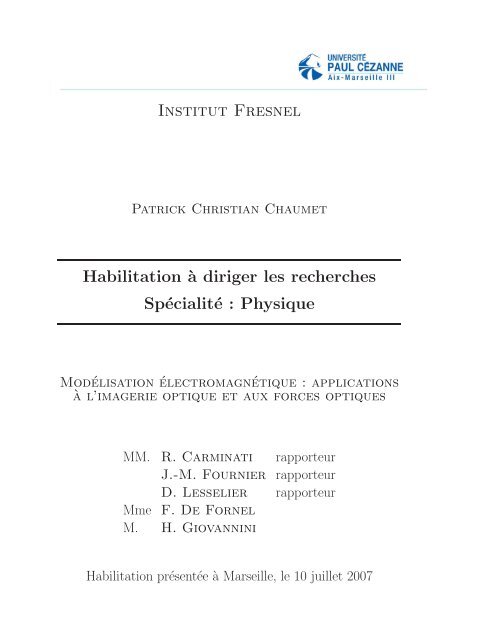
<strong>Institut</strong> <strong>Fresnel</strong><br />
Patrick Christian Chaumet<br />
Habilitation à diriger les recherches<br />
Spécialité : Physique<br />
Modélisation électromagnétique : applications<br />
à l’imagerie optique et aux forces optiques<br />
MM. R. Carminati rapporteur<br />
J.-M. Fournier rapporteur<br />
D. Lesselier rapporteur<br />
Mme F. De Fornel<br />
M. H. Giovannini<br />
Habilitation présentée à Marseille, le 10 juillet 2007
Table des matières<br />
Liste des figures vii<br />
I Curriculum Vitæ 1<br />
II Liste des publications 9<br />
Articles publiés dans des revues internationales 11<br />
Conférences 15<br />
III Résumé de mes activités d’enseignement 17<br />
IV Résumé de mes activités de recherche et d’encadrement 21<br />
Introduction générale 23<br />
1 Amélioration, et généralisation à des structures complexes, de la méthode<br />
des dipôles couplés 25<br />
1.1 Introduction . . . . . . . . . . . . . . . . . . . . . . . . . . . . . . . . . . . . 25<br />
1.2 Le principe de la méthode des dipôles couplés . . . . . . . . . . . . . . . . . 26<br />
1.3 Diffraction par une structure bi-périodique avec et sans la présence d’un<br />
défaut . . . . . . . . . . . . . . . . . . . . . . . . . . . . . . . . . . . . . . . 27<br />
1.3.1 Diffraction par une structure bi-périodique . . . . . . . . . . . . . . 27<br />
1.3.2 Diffraction par une structure bi-périodique en présence de défauts . 28<br />
1.4 Augmentation de la précision de la CDM . . . . . . . . . . . . . . . . . . . 28<br />
1.4.1 Correction de champ local . . . . . . . . . . . . . . . . . . . . . . . . 30<br />
1.4.2 Intégration de la susceptibilité linéaire du champ . . . . . . . . . . . 30<br />
1.5 Conclusion . . . . . . . . . . . . . . . . . . . . . . . . . . . . . . . . . . . . 31<br />
2 Forces dues à la lumière : forces optiques 33<br />
2.1 Introduction . . . . . . . . . . . . . . . . . . . . . . . . . . . . . . . . . . . . 33<br />
2.2 Calcul des forces optiques avec la CDM . . . . . . . . . . . . . . . . . . . . 34<br />
2.3 Forces optiques exercées sur une sphère en interaction avec un substrat plan 35<br />
2.4 Lien optique entre deux sphères . . . . . . . . . . . . . . . . . . . . . . . . . 36<br />
iii
iv TABLE DES MATIÈRES<br />
2.5 Pinces optiques : nano-manipulation . . . . . . . . . . . . . . . . . . . . . . 37<br />
2.6 Piégeage avec un cristal photonique . . . . . . . . . . . . . . . . . . . . . . . 39<br />
2.7 Pièges multiples . . . . . . . . . . . . . . . . . . . . . . . . . . . . . . . . . . 40<br />
2.8 Micro-moteurs : couples optiques . . . . . . . . . . . . . . . . . . . . . . . . 41<br />
2.9 Conclusion . . . . . . . . . . . . . . . . . . . . . . . . . . . . . . . . . . . . 41<br />
3 Sondage électromagnétique 43<br />
3.1 Introduction . . . . . . . . . . . . . . . . . . . . . . . . . . . . . . . . . . . . 43<br />
3.1.1 Généralités . . . . . . . . . . . . . . . . . . . . . . . . . . . . . . . . 43<br />
3.1.2 Quelques mots sur la résolution . . . . . . . . . . . . . . . . . . . . . 44<br />
3.1.3 La microscopie optique à haute résolution . . . . . . . . . . . . . . . 44<br />
3.2 Formulation du problème . . . . . . . . . . . . . . . . . . . . . . . . . . . . 45<br />
3.3 Approche qualitative rapide . . . . . . . . . . . . . . . . . . . . . . . . . . . 47<br />
3.4 Caractérisation de un ou de plusieurs objets inconnus . . . . . . . . . . . . 47<br />
3.4.1 Objets en espace homogène . . . . . . . . . . . . . . . . . . . . . . . 48<br />
3.4.2 Objets déposés sur un substrat plan . . . . . . . . . . . . . . . . . . 49<br />
3.4.3 Objets au-dessus d’un réseau . . . . . . . . . . . . . . . . . . . . . . 50<br />
3.5 Réalisation expérimentale . . . . . . . . . . . . . . . . . . . . . . . . . . . . 53<br />
3.5.1 Dispositif expérimental . . . . . . . . . . . . . . . . . . . . . . . . . 53<br />
3.5.2 Les premiers résultats . . . . . . . . . . . . . . . . . . . . . . . . . . 54<br />
3.6 Conclusion . . . . . . . . . . . . . . . . . . . . . . . . . . . . . . . . . . . . 56<br />
4 Etude de la fluorescence en espace confiné 57<br />
4.1 Introduction . . . . . . . . . . . . . . . . . . . . . . . . . . . . . . . . . . . . 57<br />
4.2 Utilisation d’une molécule fluorescente comme sonde locale . . . . . . . . . 58<br />
4.3 Impureté interstitielle dans un cristal . . . . . . . . . . . . . . . . . . . . . . 59<br />
4.4 Rayonnement d’une molécule fluorescente . . . . . . . . . . . . . . . . . . . 59<br />
4.4.1 Source fluorescente dans un cristal photonique . . . . . . . . . . . . 59<br />
4.4.2 Molécule fluorescente dans une ouverture nanométrique . . . . . . . 60<br />
4.5 Conclusion . . . . . . . . . . . . . . . . . . . . . . . . . . . . . . . . . . . . 61<br />
Conclusion générale et perspectives 63<br />
Références bibliographiques 67<br />
V Annexes 71<br />
1 Equivalence entre la CDM et la méthode des moments 73<br />
1.1 Les équations de Maxwell et tutti quanti . . . . . . . . . . . . . . . . . . . . 73<br />
1.2 La méthode des moments . . . . . . . . . . . . . . . . . . . . . . . . . . . . 74<br />
1.3 Comment retrouver la CDM à partir de la méthode des moments . . . . . . 75<br />
2 Remarques sur la résolution 77<br />
2.1 Diffraction par un trou circulaire : tache d’Airy . . . . . . . . . . . . . . . . 77<br />
2.2 Critère de Rayleigh . . . . . . . . . . . . . . . . . . . . . . . . . . . . . . . . 77<br />
2.3 Critère de Dawes . . . . . . . . . . . . . . . . . . . . . . . . . . . . . . . . . 78<br />
2.4 Critère de Sparrow . . . . . . . . . . . . . . . . . . . . . . . . . . . . . . . . 78<br />
2.5 Critère d’Abbe . . . . . . . . . . . . . . . . . . . . . . . . . . . . . . . . . . 79<br />
2.6 Conclusion . . . . . . . . . . . . . . . . . . . . . . . . . . . . . . . . . . . . 79
TABLE DES MATIÈRES v<br />
3 Minimisation de la fonction coût par la méthode des gradients conjugués 81<br />
3.1 Formulation du problème . . . . . . . . . . . . . . . . . . . . . . . . . . . . 81<br />
3.2 La méthode des gradients conjugués . . . . . . . . . . . . . . . . . . . . . . 82<br />
4 Publications incluses dans le tapuscript 85<br />
Phys. Rev. E 70, 036606 (2004) . . . . . . . . . . . . . . . . . . . . . . . . . . . . 86<br />
Phys. Rev. B 67, 165404 (2003) . . . . . . . . . . . . . . . . . . . . . . . . . . . . 92<br />
Phys. Rev. B 72, 205437 (2005) . . . . . . . . . . . . . . . . . . . . . . . . . . . . 97<br />
Astrophysical J. 607, 873 (2004) . . . . . . . . . . . . . . . . . . . . . . . . . . . 105<br />
Opt. Lett. 27, 2118 (2002) . . . . . . . . . . . . . . . . . . . . . . . . . . . . . . . 111<br />
Opt. Lett. 25, 1065-1067 (2000) . . . . . . . . . . . . . . . . . . . . . . . . . . . . 114<br />
Phys. Rev. B 61, 14119 (2000) . . . . . . . . . . . . . . . . . . . . . . . . . . . . 117<br />
Phys. Rev. B 62, 11185 (2000) . . . . . . . . . . . . . . . . . . . . . . . . . . . . 126<br />
Phys. Rev. B 64, 035422 (2001) . . . . . . . . . . . . . . . . . . . . . . . . . . . . 133<br />
Phys. Rev. Lett. 88, 123601 (2002) . . . . . . . . . . . . . . . . . . . . . . . . . . 140<br />
Phys. Rev. B 66, 195405 (2002) . . . . . . . . . . . . . . . . . . . . . . . . . . . . 144<br />
Phys. Rev. B 71, 045425 (2005) . . . . . . . . . . . . . . . . . . . . . . . . . . . . 155<br />
Phys. Rev. B 69, 245405 (2004) . . . . . . . . . . . . . . . . . . . . . . . . . . . . 162<br />
Opt. Lett. 29, 2740 (2004) . . . . . . . . . . . . . . . . . . . . . . . . . . . . . . . 169<br />
J. Opt. Soc. Am. A. 22, 1889 (2005) . . . . . . . . . . . . . . . . . . . . . . . . . 172<br />
J. Opt. Soc. Am. A. 23, 586 (2006) . . . . . . . . . . . . . . . . . . . . . . . . . . 181<br />
Phys. Rev. Lett. 97, 243901 (2006) . . . . . . . . . . . . . . . . . . . . . . . . . . 191<br />
Phys. Rev A 63, 023819-11 (2001) . . . . . . . . . . . . . . . . . . . . . . . . . . 195<br />
Opt. Lett. 27, 430 (2002) . . . . . . . . . . . . . . . . . . . . . . . . . . . . . . . 206
Table des figures<br />
1.1 Principe de la CDM : l’objet à étudier (à gauche) est discrétisé en un ensemble<br />
de petits dipôles (à droite). . . . . . . . . . . . . . . . . . . . . . . . 26<br />
1.2 (a) Schéma de la configuration : plots d’argent de largeur a = 30 nm et de<br />
hauteur h = 10 nm, disposés sur une maille carrée de période p = 100 nm<br />
sur un substrat de verre. θ = 50 ◦ est l’angle d’incidence, λ = 600 nm, et le<br />
champ est polarisé TM. (b) Module du champ à 20 nm au-dessus du substrat<br />
de verre. Les carrés blancs représentent la position des pavés d’argent. . . . 28<br />
1.3 Images de champ proche obtenues à z = 100 nm, λ = 600 nm, pour une<br />
structure bi-périodique en présence d’un défaut. La structure bi-périodique<br />
est constituée de pavés de silicium tels que a = 50 nm (notation de la<br />
Fig. 1.2). Pour les six premières images k 0 est parallèle à l’axe x avec<br />
u = (100,0) nm et v = (0,100) nm. Pour les deux dernières images [(g)<br />
et (h)], k 0 est comme montré sur les figures avec u = (100,0) nm et<br />
v = (50,86) nm. Sur chaque figure le carré en trait plein représente le motif<br />
du réseau et u, v les vecteurs de base de celui-ci. (a), (c) (e) et (g) sont<br />
obtenus pour θ = 0 ◦ . (b), (d), (f) et (h) sont obtenus pour θ = 50 ◦ . (a),<br />
(b), (g) et (h) le carré en pointillé représente la lacune dans la structure<br />
bi-périodique. (c) et (d) le défaut est un pavé en argent de même taille que<br />
ceux de silicium. (e) et (f) le carré en pointillé est un cube de silicium qui<br />
a été déplacé. . . . . . . . . . . . . . . . . . . . . . . . . . . . . . . . . . . . 29<br />
1.4 Module du champ à l’extérieur et à l’intérieur d’un slab. Le slab est défini<br />
par 0 ≤ z ≤ 50 nm délimité par les lignes verticales. L’onde plane incidente<br />
(λ=400 nm) arrive de la gauche avec un angle de θ=50 ◦ polarisée en TM.<br />
La permittivité relative du slab est : ǫ=20. . . . . . . . . . . . . . . . . . . . 31<br />
2.1 (a) Force subie par une sphère diélectrique dans un faisceau gaussien quand<br />
elle est située hors axe. (b) Piégeage optique d’une sphère diélectrique située<br />
sur l’axe d’un faisceau gaussien. . . . . . . . . . . . . . . . . . . . . . . . . . 34<br />
2.2 Les traits pleins correspondent à la polarisation suivant x, et les traits pointillés<br />
à la polarisation perpendiculaire à x. (a) Potentiel de piégeage sur la<br />
sphère B normalisé à kbT en fonction de la distance entre les deux sphères.<br />
La hauteur des barres représente 3kbT : si le puits de potentiel est supérieur<br />
à cette valeur alors le piégeage est considéré comme stable. (b) Force suivant<br />
x exercée sur la sphère B. . . . . . . . . . . . . . . . . . . . . . . . . . . . . 36<br />
vii
viii TABLE DES FIGURES<br />
2.3 Plusieurs nano-particules de natures différentes sont déposées sur un substrat<br />
plan en verre. La pointe au-dessus de la surface balaye celle-ci afin<br />
d’obtenir une image optique de l’échantillon étudié, ce qui permet de localiser<br />
et caractériser les différentes particules en présence. . . . . . . . . . . . 38<br />
2.4 Force optique s’exerçant sur une sphère en verre en fonction de la distance<br />
pointe-sphère : L’irradiance du faisceau incident est de 0.05 W/µm 2 avec un<br />
angle d’incidence de θ = 43 ◦ > θc = 41.8 ◦ et λ = 500 nm. La pointe est en<br />
tungstène avec un rayon de courbure à son apex de 10 nm. (a) Polarisation<br />
TM. (b) Polarisation TE. . . . . . . . . . . . . . . . . . . . . . . . . . . . . 39<br />
2.5 Exemples de micro-moteurs. 36 . . . . . . . . . . . . . . . . . . . . . . . . . . 41<br />
3.1 L’objet ou les objets (deux cubes sur le schéma proposé) sont éclairés avec L<br />
différents angles d’incidence et le champ électromagnétique (module+phase)<br />
est mesuré sur la surface Γ en M points d’observations. En pointillé est défini<br />
le domaine d’investigation Ω dans lequel l’objet est supposé être. . . . . . . 46<br />
3.2 (c) Schéma de la configuration étudiée : la taille du domaine d’investigation<br />
est de 2λ × 2λ × 2.2λ, les objets sont des cubes de côté λ/4 éclairés par<br />
16 ondes planes se propageant dans la direction des z positifs avec θ0 ∈<br />
[−80 ◦ ,80 ◦ ]. Le champ électromagnétique est mesuré au-dessus de la surface<br />
avec θd ∈ [−80 ◦ ,80 ◦ ]. (a) Reconstruction de la partie réelle de la permittivité<br />
relative. (b) Reconstruction de la partie imaginaire de la permittivité relative. 48<br />
3.4 Cubes de côté λ/4 espacés de λ/3 disposés suivant l’axe z. (a) Carte de la<br />
permittivité relative dans le plan (x,z). (b) Carte de la permittivité relative<br />
dans le plan (x,y). (c) Coupe de la permittivité relative selon z à x = y = 0.<br />
En trait plein le profil réel de la permittivité relative. . . . . . . . . . . . . . 49<br />
3.3 Cubes de côté λ/4 espacés d’une distance c = λ/7 disposés suivant l’axe<br />
x. (a) Carte de la permittivité relative dans le plan (x,z). (b) Carte de la<br />
permittivité relative dans le plan (x,y). (c) Coupe de la permittivité relative<br />
selon x à z = y = 0. En trait plein le profil réel de la permittivité relative. . 49<br />
3.5 (a) Schéma de la configuration utilisée. (b), (c), et (d) Carte de la permittivité<br />
relative dans le plan (x,y) à z = λ/40 et dans le plan (x,z) à y = 0. (b)<br />
Seules les ondes évanescentes sont utilisées : θ0 ∈ [−80 ◦ , −43 ◦ ] ∪ [80 ◦ ,43 ◦ ].<br />
(c) Seules les ondes évanescentes sont utilisées mais le champ diffracté est<br />
bruité. (d) Ondes propagatives + évanescentes (θ0 ∈ [−80 ◦ ,80 ◦ ]) avec le<br />
champ diffracté bruité. . . . . . . . . . . . . . . . . . . . . . . . . . . . . . . 50<br />
3.6 (a) Deux objets dipolaires (cubes de 20 nm de côté et de permittivité relative<br />
2.25) sont déposés sur un réseau caractérisé par p = 100 nm, m = 66.7 nm,<br />
l = 6.66 nm, h = 6.75 nm, εs = 2.25, εa = 4.41, εl = −8.4537 + 0.6984i<br />
(argent). Le réseau est éclairé par 8 ondes planes polarisées TM tel que<br />
θ0 = 80 ◦ et λ = 500 nm dans le vide. Le champ diffracté est mesuré sur 80<br />
points d’observations tel que montré sur la figure. (b) Carte de permittivité<br />
relative reconstruite dans le plan (x,y) à une altitude de 12 nm par rapport<br />
au substrat nanostucturé. Le champ diffracté a été corrompu avec 10% de<br />
bruit. Les carrés noirs représentent la vraie position des cubes et en rouge<br />
les motifs du réseau. . . . . . . . . . . . . . . . . . . . . . . . . . . . . . . . 51
TABLE DES FIGURES ix<br />
3.7 (a) Configuration étudiée. (b) Coupes réalisées pour différentes approximations<br />
dans la méthode d’inversion. En bleu le profil réel de la permittivité<br />
relative. (c), (d) et (e) Carte de permittivité relative dans le plan (x,y) à<br />
12 nm au-dessus de la surface : (c) inversion en utilisant l’approximation de<br />
Born. (d) inversion en supposant l’objet en espace homogène. (e) inversion<br />
en supposant l’objet sur un substrat plan. (f) Carte de permittivité relative<br />
dans le plan (x,z). L’inversion est effectuée en supposant l’objet sur un<br />
substrat plan. . . . . . . . . . . . . . . . . . . . . . . . . . . . . . . . . . . . 52<br />
3.8 Configuration expérimentale pour mesurer le champ diffracté en module<br />
et phase dans le cas de la transmission. En rouge le champ incident qui<br />
éclaire l’objet, en bleu le champ diffracté par l’objet et en vert le champ<br />
de référence qui vient interférer avec le champ incident (sur un pixel de la<br />
caméra) et le champ diffracté par l’objet. . . . . . . . . . . . . . . . . . . . 53<br />
3.9 Module du champ diffracté enregistré sur la caméra CCD. Le maximum d’intensité<br />
correspondant au faisceau incident (spéculaire). Les cercles montrent<br />
la plage de fréquences spatiales accessibles à travers la mesure du champ<br />
diffracté. (a) Champ diffracté en incidence normale. (b) Somme de (f), (c),<br />
(d) et (e) chacune étant enregistrée avec une incidence de θ = 25 ◦ avec<br />
φ = 0 ◦ , φ = 90 ◦ , φ = 180 ◦ et φ = 270 ◦ respectivement. . . . . . . . . . . . . 55<br />
3.10 (a) Reconstruction de l’objet obtenue avec 8 incidences. La palette de couleur<br />
à droite de la figure exprime la hauteur en nm. (b) Coupe en y = 0. (c)<br />
Coupe verticale sur le troisième objet : une incidence : courbes rouges (-.) ;<br />
quatre incidences : courbes bleues (- -) ; huit incidences :courbes noires : (-). 55<br />
4.1 Molécule fluorescente émettant dans le domaine du visible. En pointillé le<br />
spectre de la source excitatrice, et en trait plein le spectre rayonné par le<br />
source. . . . . . . . . . . . . . . . . . . . . . . . . . . . . . . . . . . . . . . . 58<br />
4.2 La molécule fluorescente est attachée à une pointe formée d’un matériau<br />
diélectrique. Celle-ci balaye la surface (en verre), où l’objet à étudier est<br />
déposé, à une hauteur constante de 40 nm. La molécule rayonne à λ =<br />
488 nm, et le taux d’émission spontanée en fonction de la position de la<br />
molécule est représenté. (a) Le moment dipolaire associé à la transition est<br />
selon l’axe z. (b) Le moment dipolaire associé à la transition est selon l’axe x. 59<br />
4.3 (g) Schéma d’une micro cavité H2. L’épaisseur du slab est de 250 nm avec<br />
un indice de 3.17. La période du réseau est de 535 nm, avec un diamètre<br />
pour les cylindres de 178 nm. Le point rouge représente la position de la<br />
source. (a)-(f) Module du champ électrique (en unité arbitraire) au-dessus<br />
de la cavité pour une longueur d’onde d’émission de λ = 1405 nm pour les<br />
figures (a)-(c) et λ = 1410 nm pour les figures (d)-(f). Le résultat a été<br />
moyenné sur toutes les orientations du dipôle dans le plan (x,y). Les cartes<br />
de champ sont calculées pour différentes hauteurs z : (a) et (d) : z=50 nm;<br />
(b) et (e) : z=100 nm ; (c) et (f) : z=500 nm . . . . . . . . . . . . . . . . . 60<br />
4.4 (a) Une couche métallique percée d’un trou est déposée sur un substrat de<br />
verre. Un laser excite une molécule fluorescente placée dans le trou et la<br />
puissance rayonnée par celle-ci est alors mesurée. (b) Une structure plus<br />
compliquée peut être ajoutée afin que la lumière rayonnée par la source soit<br />
plus focalisée. . . . . . . . . . . . . . . . . . . . . . . . . . . . . . . . . . . . 61<br />
A 2.1 Intensité totale (+) en fonction de x pour deux sources de même intensité,<br />
incohérentes et ponctuelles. . . . . . . . . . . . . . . . . . . . . . . . . . . . 78
x TABLE DES FIGURES<br />
A 2.2 Même légende que Fig. A 2.1. . . . . . . . . . . . . . . . . . . . . . . . . . 78<br />
A 2.3 Même légende que Fig. A 2.1. . . . . . . . . . . . . . . . . . . . . . . . . . 78
Première partie<br />
Curriculum Vitæ<br />
1
Curriculum Vitæ<br />
Nom patronymique : Chaumet Prénom : Patrick<br />
Né le 24 janvier 1971 à Paray-Le-Monial Nationalité : Française<br />
Situation de famille : célibataire Email : patrick.chaumet@fresnel.fr<br />
Tel. professionnel : 04-91-28-27-91 Fax : 04-91-67-44-28<br />
Formation universitaire<br />
1998, Doctorat en physique.<br />
Obtenu à Dijon, au laboratoire de physique de l’université de Bourgogne. Le titre de<br />
ma thèse était “Diffusion d’une Onde Electromagnétique par des Structures Arbitraires :<br />
Application à l’Emission de Lumière en STM” et a été encadré par J.-P Dufour. Le jury<br />
était constitué de :<br />
Rapporteurs : M. Jacques Baudon<br />
M. Richard Berndt<br />
M. Yves Borensztein<br />
Examinateurs : Mme Frédérique de Fornel<br />
M. Michel Loëte<br />
Directeur de thèse : M. Jean-Paul Dufour<br />
1994, DEA interaction matière et rayonnement.<br />
Obtenu à l’université de Dijon. Le stage avait été encadré par J.-P Dufour au<br />
laboratoire de physique de l’université de Bourgogne.<br />
Parcours professionnel<br />
Depuis le 01/10/2000, maître de conférence de l’université d’Aix-Marseille III.<br />
Du 01/03/1999 au 30/09/2000, post-doctorant à l’<strong>Institut</strong>o de Ciencia de Materiales<br />
de Madrid sous la responsabilité de M. Nieto-Vesperinas.<br />
Du 01/09/1998 au 30/02/1999, ATER à l’université de Bourgogne.<br />
Du 01/09/1997 au 01/09/1998, demi ATER à l’université de Bourgogne.<br />
Du 01/09/1994 au 01/09/1997, doctorant à l’université de Bourgogne (allocatairemoniteur).<br />
3
4 CURRICULUM VITÆ<br />
Collaborations extérieures<br />
A. Rahmani, LEOM, Ecole Centrale de Lyon.<br />
M. Nieto-Vesperinas, ICMM CSIC, Madrid.<br />
G. Bryant, NIST, Gaithersburg (USA).<br />
N. Sojic, université de Bordeaux I.<br />
Encadrements<br />
C. Billaudeau, 2003-2004, DEA.<br />
R. Lencrerot, 2004-2005, DEA.<br />
F. Drsek, 2005-2008, thèse.<br />
h-index : 11<br />
Activités de recherche<br />
Octobre 2000 jusqu’à ce jour en tant que Maître de conférences de l’université<br />
d’Aix-Marseille III.<br />
• Confinement et exaltation électromagnétique pour biopuce<br />
Ce projet financé par une ANR a pour but d’exploiter les effets de confinement et<br />
d’exaltation des ouvertures nanométriques mis à jour dans le projet Nanospot (ACI 2003)<br />
combiné au savoir faire de la jeune entreprise GENEWAVE (Palaiseau) sur l’exaltation de<br />
la fluorescence dans le contexte des biopuces. Outre les aspects de nanophotonique liés à<br />
la conception et à la réalisation des ouvertures, nous prenons en compte les spécificités de<br />
l’hybridation sur puce pour la définition des ouvertures (nombre, taille, espacement).<br />
• Nano-imagerie bioanalytique<br />
Ce projet financé par une ANR se propose de développer de nouveaux outils nanostructurés<br />
pour l’imagerie cellulaire et la bioanalyse hautement parallèle. Notre démarche<br />
s’articule suivant le triptyque : nanofabrication / étude des propriétés nouvelles liées à la<br />
taille nanométrique / applications bioanalytiques. La participation d’un partenaire industriel<br />
reconnu permet d’allier des objectifs fondamentaux (études sur cellules isolées, sur<br />
modèles de peaux reconstruites) à des opportunités d’applications in vivo. Nous proposons<br />
de réaliser un réseau de nano-cavités métalliques qui serviront comme nano-pinces<br />
optiques. Ces nano-pinces permettront ainsi de manipuler, de déplacer et d’immobiliser<br />
des populations différentes de particules modifiées par des systèmes de reconnaissance<br />
biotique (antigène-anticorps ou brins d’ADN). Les applications visées sont tournées vers<br />
l’immunodosage et les biopuces à ADN.<br />
• Création de micro ou nano-moteurs grâce aux forces optiques<br />
Le problème dans le cas de nano ou micro-machines est la source de puissance (moteur)<br />
permettant de les faire fonctionner. Un moteur est un système à qui on fournit de l’énergie<br />
et qui nous redonne un mouvement mécanique généralement sous la forme d’un axe en<br />
rotation. Les forces optiques, si l’objet étudié présente une dissymétrie, peuvent entraîner<br />
une rotation de celui-ci. Cet objet peut alors servir de moteur pour une machine plus
CURRICULUM VITÆ 5<br />
complexe. Avec C. Billaudeau (étudiant en DEA) nous avons développé une méthode<br />
pour calculer le couple optique.<br />
• Imagerie optique à haute résolution<br />
Ce projet est animé par A. Sentenac, chargé de recherche au CNRS à l’institut <strong>Fresnel</strong>,<br />
dans le cadre d’une ACI jeunes chercheurs. L’objectif de ce projet est de proposer une<br />
nouvelle méthode de microscopie optique, en champ lointain, permettant de restituer la<br />
forme de l’objet et sa permittivité relative, et ce avec un pouvoir de résolution supérieur<br />
à celui imposé par la limite de diffraction. Notre technique d’imagerie s’apparente à celles<br />
développées dans le domaine des micro-ondes. L’échantillon est éclairé par un faisceau laser<br />
sous différentes incidences et le champ diffracté est détecté selon différents angles d’observations.<br />
Je me suis plus particulièrement intéressé au développement des algorithmes<br />
de résolution du problème inverse afin de remonter aux caractéristiques de l’objet, et ce à<br />
trois dimensions. Afin de pouvoir pleinement appréhender le problème je suis actuellement<br />
en CRCT (2004-2006). De plus c’est exactement sur ce thème que j’ai encadré R.<br />
Lencrerot (DEA) et que j’encadre F. Drsek (début de thèse).<br />
• Forces optiques, application à la nano-manipulation (pinces optiques)<br />
Ces dernières années ont vu l’utilisation des forces optiques pour confiner et contrôler,<br />
dans les trois dimensions de l’espace, des particules avec des tailles allant de la dizaine<br />
de nanomètres à plusieurs micromètres. Nous avons montré récemment, en étroite collaboration<br />
avec A. Rahmani (LEOM, Ecole Centrale de Lyon) et M. Nieto-<br />
Vesperinas (ICMM, Madrid), que ces forces optiques pouvaient servir à manipuler des<br />
objets de taille nanométrique. Ceux-ci sont déposés sur un substrat plan transparent, puis<br />
la pointe d’un microscope optique de champ proche est utilisée pour repérer pour finalement<br />
manipuler de manière sélective ces particules. Notons que la collaboration avec M.<br />
Nieto-Vesperinas fait suite à un stage post-doctoral de 1 an et demi dans son laboratoire.<br />
• Contrôle de l’émission spontanée<br />
Quand un environnement modifie, en un point de l’espace, la distribution spatiale et<br />
spectrale des modes électromagnétiques, il modifie aussi l’émission spontanée d’une source<br />
placée à cet endroit. Je m’intéresse à la dynamique des sources de photons dans des environnements<br />
variés tels que des cristaux photoniques. Ceci a été effectué en collaboration<br />
avec A. Rahmani (LEOM, Ecole Centrale de Lyon).<br />
• Amélioration de la convergence de la méthode des dipôles couplés<br />
Il existe de nombreuses méthodes pour calculer la diffraction de la lumière par des<br />
objets de forme arbitraire. La méthode que j’utilise consiste à discrétiser la matière en<br />
un ensemble de petits éléments disposés sur une maille cubique. Sous l’action d’une onde<br />
incidente chacun des éléments de discrétisation va acquérir un moment dipolaire qui va<br />
dépendre de l’onde incidente et de son couplage avec tous les autres dipôles. En collaboration<br />
avec A. Rahmani (LEOM, Lyon) et G. Bryant (NIST, Gaithersburg,<br />
USA) nous avons proposé plusieurs possibilités pour améliorer la convergence de la méthode.<br />
La collaboration avec G. Bryant m’a amené à travailler durant cinq semaines<br />
aux Etats-Unis (NIST, Washington) au sein de son équipe.<br />
• Extension de la méthode des dipôles couplés à des configurations complexes<br />
La méthode des dipôles couplés (CDM) était limitée aux objets finis, je me suis donc<br />
intéressé à étendre la CDM aux structures périodiques et plus récemment aux structures<br />
périodiques en présence d’objets apériodiques. Ceci voit son application dans mes autres<br />
thèmes de recherche.
6 CURRICULUM VITÆ<br />
Mars 1999/septembre 2000, stage post-doctoral à Madrid dans le groupe<br />
dirigé par M. Nieto-Vesperinas<br />
• Forces optiques pour des objets sur des substrats et lien optique<br />
Lors de mon stage post-doctoral à l’université Autonoma de Madrid dans l’équipe<br />
dirigée par M. Nieto-Vesperinas, j’ai étudié les forces optiques sur des objets nano et<br />
micro-métriques. L’étude a porté essentiellement sur les forces optiques créées par des<br />
ondes évanescentes et l’influence du substrat sur lequel étaient déposées les particules. Les<br />
liens optiques entre les particules, créés par la lumière, ont aussi été étudiés.<br />
1994/février 1999, thèse et ATER au laboratoire de physique de l’université<br />
de Bourgogne<br />
• Emission de lumière en microscopie tunnel électronique (STM)<br />
Mon sujet de thèse portait sur la microscopie tunnel électronique (STM). Il consistait à<br />
caractériser la nature chimique des éléments sous la pointe d’un STM à partir de l’émission<br />
de lumière de la jonction tunnel.<br />
• Microscopie de champ proche optique<br />
La méthode développée pour étudier l’émission de lumière en STM m’a permis aussi<br />
d’étudier le champ proche optique. Notamment la formation des images en microscopie de<br />
champ proche et la durée de vie de molécules fluorescentes.<br />
Activités d’enseignement<br />
Depuis octobre 2000 en qualité de Maître de Conférence de l’université<br />
d’Aix-Marseille III<br />
J’effectue, en moyenne, chaque année 220 heures (équivalent TD) à l’IUT de Saint<br />
Jérôme dans le département Mesures Physiques excepté durant les deux années universitaires<br />
2004/2005 et 2005/2006 du CRCT.<br />
• Travaux dirigés en optique géométrique destinés aux étudiants de première année.<br />
• Travaux dirigés en optique ondulatoire destinés aux étudiants de première année.<br />
• Travaux dirigés en mécanique du point destinés aux étudiants de première année.<br />
• Travaux dirigés en optronique destinés aux étudiants de deuxième année.<br />
• Travaux pratiques en optique géométrique destinés aux étudiants de première<br />
année.<br />
• Travaux pratiques en optique ondulatoire destinés aux étudiants de première<br />
année.<br />
• Travaux pratiques en optronique destinés aux étudiants de deuxième année.<br />
1997/1999, ATER à l’université de Dijon<br />
• Travaux dirigés en optique géométrique destinés aux étudiants de premier cycle<br />
(DEUG B).<br />
• Travaux pratiques en optique géométrique et ondulatoire destinés aux étudiants<br />
de premier<br />
cycle (DEUG B et DEUG A).<br />
• Travaux pratiques en électricité destinés aux étudiants de premier cycle (DEUG<br />
A).<br />
1994/1997, moniteur à l’université de Dijon
CURRICULUM VITÆ 7<br />
• Travaux dirigés en optique géométrique destinés aux étudiants de premier cycle<br />
(DEUG B).<br />
• Travaux pratiques en optique géométrique et ondulatoire destinés aux étudiants<br />
de premier<br />
cycle (DEUG B et DEUG A).<br />
• Travaux dirigés en mécanique quantique destinés aux étudiants de deuxième cycle.
8 CURRICULUM VITÆ
Deuxième partie<br />
Liste des publications<br />
9
Articles publiés dans des revues<br />
internationales à comité de lecture<br />
1. P. C. Chaumet, K. Belkebir et A. Sentenac,<br />
Numerical study of grating-assisted optical diffraction tomography<br />
Phys. Rev. A accepté.<br />
2. P. C. Chaumet, B. Pouligny, R. Dimova et N. Sojic,<br />
Optical tweezers in interaction with an apertureless probe<br />
J. Appl. Phys. accepté.<br />
3. A. Sentenac, C.-A. Guérin, P. C. Chaumet, F. Drsek, H. Giovannini, N. Bertaux<br />
et M. Holschneider,<br />
Influence of multiple scattering on the resolution of an imaging system : a Cramer-<br />
Rao analysis.<br />
Opt. Express 15, 1340 (2007).<br />
4. P. C. Chaumet et C. Billaudeau,<br />
Coupled dipole method to compute optical torque : Application to a micropropeller.<br />
J. Appl. Phys. 101, 023106 (2007).<br />
5. A. Sentenac, P. C. Chaumet, et K. Belkebir,<br />
Beyond the Rayleigh criterion : Grating assisted far-field optical diffraction tomography.<br />
Phys. Rev. Lett. 97, 243901 (2006). [annexe 4 page 191]<br />
6. P. C. Chaumet,<br />
Fully vectorial highly non paraxial beam close to the waist.<br />
J. Opt. Soc. Am. A 23, 3197 (2006).<br />
7. E. Popov, M. Neviere,J. Wenger, P-F. Lenne, H. Rigneault, P. C. Chaumet,<br />
N. Bonod, J. Dintinger, and T. Ebbesen,<br />
Field enhancement in single subwavelength apertures,<br />
J. Opt. Soc. Am. A. 23, 1084 (2006). J. Opt. Soc. Am. A.<br />
8. P. C. Chaumet, A. Rahmani, M. Nieto-Vesperinas,<br />
Local-field enhancement in an optical force metallic nanotrap : Application to singlemolecule<br />
spectroscopy.<br />
App. Opt. 45, 5185 (2006).<br />
9. A. Rahmani et P. C. Chaumet,<br />
Optical Trapping near a Photonic Crystal.<br />
Opt. Express 14, 6353 (2006).<br />
11
12 ARTICLES<br />
10. F. Bordas, N. Louvion, S. Callard, P. C. Chaumet, and A. Rahmani,<br />
Coupled dipole method for radiation dynamics in finite photonic crystal structures.<br />
Phys. Rev. E. 73, 056601 (2006).<br />
11. P. C. Chaumet, K. Belkebir, and R. Lencrerot,<br />
Three-dimensional optical imaging in layered media,<br />
Opt. Exp. 14, 3415 (2006).<br />
12. K. Belkebir, P. C. Chaumet, A. Sentenac,<br />
Influence of multiple scattering on three-dimensional imaging with optical diffraction<br />
tomography.<br />
J. Opt. Soc. Am. A. 23, 586 (2006). [annexe 4 page 181]<br />
13. P. C. Chaumet, A. Sentenac,<br />
Numerical simulation of the electromagnetic field scattered by defects in a doubleperiodic<br />
structure.<br />
Phys. Rev. B 72, 205437-8 (2005). [annexe 4 page 97]<br />
14. P. C. Chaumet, A. Rahmani, A. Sentenac, and G. W. Bryant,<br />
Efficient computation of optical forces with the coupled dipole method.<br />
Phys. Rev. E 72, 046708-6 (2005).<br />
15. K. Belkebir, P. C. Chaumet, A. Sentenac,<br />
Superresolution in total-internal reflection tomography.<br />
J. Opt. Soc. Am. A. 22, 1889-1897 (2005). [annexe 4 page 172]<br />
16. E. Popov, N. Bonod, M. Nevière, H. Rigneault, P.-F. Lenne, and P. C. Chaumet,<br />
Surface plasmon excitation on a single subwavelength hole in a metallic sheet.<br />
Appl. opt. 44, 2332-2337 (2005).<br />
17. P. C. Chaumet, A. Rahmani, and M. Nieto-Vesperinas,<br />
Photonic force spectroscopy on metallic and absorbing nanoparticles.<br />
Phys. Rev. B 71, 045425-7 (2005). [annexe 4 page 155]<br />
18. P. C. Chaumet, K. Belkebir, A. Sentenac,<br />
Superresolution of three-dimensional optical imaging by use of evanescent waves.<br />
Opt. Lett. 29, 2740-2742 (2004). [annexe 4 page 169]<br />
19. P. C. Chaumet, A. Sentenac, and A. Rahmani,<br />
Coupled dipole method for scatterers with large permittivity.<br />
Phys. Rev. E 70, 036606-6 (2004). [annexe 4 page 86]<br />
20. P. C. Chaumet, K. Belkebir, and A. Sentenac,<br />
Three-dimensional sub-wavelength optical imaging using the coupled dipole method.<br />
Phys. Rev. B 69, 245405-7 (2004). [annexe 4 page 162]<br />
21. A. Rahmani, P. C. Chaumet, and G. W. Bryant,<br />
On the Importance of Local-Field Corrections for Polarizable Particles on a Finite<br />
Lattice : Application to the Discrete Dipole Approximation.<br />
Astrophysical J. 607, 873-878 (2004). [annexe 4 page 105]<br />
22. M. Nieto-Vesperinas, P. C. Chaumet, and A. Rahmani,<br />
Near-field photonic forces.<br />
Phil. Trans. Roy. Soc. Lond. A 362, 719-737 (2004).<br />
23. P. C. Chaumet,<br />
Comment on“Trapping force, force constant, and potential depths for dielectric spheres<br />
in the presence of spherical aberrations”.<br />
Appl. Opt. 43, 1825-1826 (2004).
ARTICLES 13<br />
24. P. C. Chaumet, A. Rahmani, and G. W. Bryant,<br />
Generalization of the coupled dipole method to periodic structure.<br />
Phys. Rev. B 67, 165404-5 (2003). [annexe 4 page 92]<br />
25. A. Rahmani, P. C. Chaumet, and G. W. Bryant,<br />
Coupled dipole method with an exact long-wavelength limit and improved accuracy at<br />
finite frequencies.<br />
Opt. Lett. 27, 2118-2120 (2002). [annexe 4 page 111]<br />
26. P. C. Chaumet, A. Rahmani, and M. Nieto-Vesperinas,<br />
Selective nanomanipulation using optical forces.<br />
Phys. Rev. B 66, 195405-11 (2002). [annexe 4 page 144]<br />
27. P. C. Chaumet, A. Rahmani, and M. Nieto-Vesperinas,<br />
Optical trapping and manipulation of nano-object with an apertureless probe.<br />
Phys. Rev. Lett. 88, 123601-4 (2002). [annexe 4 page 140]<br />
28. A. Rahmani, P. C. Chaumet, and G. W. Bryant,<br />
Local-field correction for an interstitial impurity in a crystal.<br />
Opt. Lett. 27, 430-432 (2002). [annexe 4 page 206]<br />
29. P. C. Chaumet, and M. Nieto-Vesperinas,<br />
Optical binding of particles with or without the presence of a flat dielectric surface.<br />
Phys. Rev. B 64, 035422-7 (2001). [annexe 4 page 133]<br />
30. A. Rahmani, P. C. Chaumet, et F. de Fornel,<br />
Enrironment-induced modification of spontaneous emission : Single-molecule nearfield<br />
probe.<br />
Phys. Rev A 63, 023819-11 (2001). [annexe 4 page 195]<br />
31. P. C. Chaumet, and M. Nieto-Vesperinas,<br />
Electromagnetic force on a metallic particle in the presence of a dielectric surface.<br />
Phys. Rev. B 62, 11185-11191 (2000). [annexe 4 page 126]<br />
32. P. C. Chaumet, and M. Nieto-Vesperinas,<br />
Time-averaged total force on a dipolar sphere in an electromagnetic field.<br />
Opt. Lett. 25, 1065-1067 (2000). [annexe 4 page 114]<br />
33. P. C. Chaumet, and M. Nieto-Vesperinas,<br />
Coupled dipole method determination of the electromagnetic force on a particle over<br />
a flat dielectric substrate.<br />
Phys. Rev. B 61, 14119-14127 (2000). [annexe 4 page 117]<br />
34. P. C. Chaumet, and A. Rahmani,<br />
Comment on “Physical picture for light emission in scanning tunneling microscopy”.<br />
Phys. Rev. Lett. 84, 3498-3401 (2000).<br />
35. P. C. Chaumet, A. Rahmani, F. de Fornel, and J.-P. Dufour,<br />
Evanescent light scattering : The validity of the dipole approximation.<br />
Phys. Rev. B 58, 2310-2315 (1998).<br />
36. P. C. Chaumet, and J.-P. Dufour,<br />
Electric potential and field between two different spheres.<br />
J. of Electrostatics 43, 145-159 (1998).<br />
37. A. Rahmani, P. C. Chaumet, F. de Fornel, and C. Girard,<br />
Field propagator of a dressed junction : Fluorescence lifetime calculations in a confined<br />
geometry.<br />
Phys. Rev A 56, 3245-3254 (1997).
Proceeding et Conférences avec<br />
publication des actes<br />
1. A. Sentenac, P. C. Chaumet et K. Belkebir, Grating-assisted optical diffraction<br />
tomography with near-field resolution. Focus On Microscopy (FOM 2007) avril 2007<br />
Valencia (Espagne).<br />
2. A. Sentenac, K. Belkebir et P. C. Chaumet : Reconstruction procedures in<br />
structured illumination fluorescent microscopy. Focus On Microscopy (FOM 2007)<br />
avril 2007 Valencia (Espagne).<br />
3. H. Giovannini, D. Konan, A. Sentenac, F. Drsek, P. C Chaumet, K. Belkebir,<br />
V. Lauer, et F Maffezzini : Far-field reflection microscope using optical diffraction<br />
tomography-application to profilometry. Focus On Microscopy (FOM 2007) avril<br />
2007 Valencia (Espagne).<br />
4. P. C. Chaumet, K. Belkebir, F. Drsek, H. Giovannini, A. Sentenac : Nanoscopy<br />
with Grating-Assisted Optical Diffraction Tomography. Annual Review of<br />
Progress in Applied Computational Electromagnetics, mars 2007 Verona (Italie).<br />
5. F. Drsek, H. Giovannini, P. Chaumet, K. Belkebir, A. Sentenac : Optical<br />
diffraction tomography in reflection. PSIP 2007 - Physics in Signal and Image Processing<br />
- Mulhouse 31 jan.-2 fev. 2007.<br />
6. A. Sentenac, C.-A. Guérin, P. C. Chaumet, F. Drsek, N. Bertaux, H. Giovannini,<br />
M. Holschneider : Influence of multiple scattering on the resolution of<br />
an imaging system, a Cramér-Rao analysis. PSIP 2007 - Physics in Signal and Image<br />
Processing - Mulhouse 31 jan.-2 fev. 2007.<br />
7. A. Sentenac, P. C. Chaumet, K. Belkebir :Grating-assisted optical diffraction<br />
tomography with nearfield resolution. The 88 th Eastern Forum of Science and Technology<br />
Challenges and Opportunities in Nano-Optics Jan 5-9, 2007, Fudan University,<br />
Shanghai, China.<br />
8. A. Sentenac, P. C. Chaumet, K. Belkebir : Grating-assisted optical diffraction<br />
tomography with nearfield resolution, EOS Annual Meeting 2006 : Paris, France,<br />
16-19 October 2006.<br />
9. P. C. Chaumet,K. Belkebir, A. Sentenac : High resolution three-dimensional<br />
imaging with optical diffraction tomography. Mediterranean Microwave Symposium<br />
(MMS), Genova, Italy, 19-21 september 2006.<br />
10. A. Sentenac, K. Belkebir, P. C. Chaumet : Inversion techniques in structured<br />
illumination fluorescent microscopy. International Symposium - Optical Analysis of<br />
Biomolecular Machines, Berlin, Germany, 13-16 july 2006.<br />
15
16 CONFÉRENCES<br />
11. J.-M. Geffrin, C. Eyraud, P. Sabouroux, P. C. Chaumet, H. Tortel, and H.<br />
Giovannini : Validation of 3D Scattering Measurements, IEEE AP-S International<br />
Symposium and USNC/URSI National Radio Science Meeting, 9-14 July 2006.<br />
12. K. Belkebir, P. C. Chaumet, and A. Sentenac : Optical imaging of sub-wavelength<br />
objects, 2005 IEEE AP-S International Symposium and USNC/URSI National<br />
Radio Science Meeting, 3-8 July 2005.<br />
13. P. C. Chaumet, K. Belkebir, and A. Sentenac : High resolution three-dimensional<br />
imaging with optical diffraction tomography, Focus on Microscopy 2005, Friedrich<br />
Schiller University Jena, Germany, 20-23 mars 2005.<br />
14. K. Belkebir, P. C. Chaumet, A. Sentenac : Three-dimensional sub-wavelength<br />
optical imaging using the Coupled Dipole Method, Mediterranean Microwave Symposium<br />
(MMS), Marseille, 1-3 juin 2004.<br />
15. P. C. Chaumet, K. Belkebir, and A. Sentenac : High resolution imaging with<br />
optical diffraction tomography, role of the evanescent waves, PIERS, Pisa, Italy,<br />
28-31 march 2004.<br />
16. P. C. Chaumet, K. Belkebir, and A. Sentenac : Sub-wavelength imaging with<br />
diffraction tomography, EOS Topical Meeting - Advanced Imaging Techniques, Delft,<br />
The Netherlands, 20-23 October 2003.<br />
17. A. Rahmani, P. C. Chaumet, and G. W. Bryant : Modeling Nano-optics : towards<br />
a more physical formulation of the coupled dipole method, CLEO/QELS Long Beach,<br />
USA, May 2002.<br />
18. A. Rahmani, P. C. Chaumet, and G. W. Bryant : Local-field correction for a<br />
nanosource in a crystal, CLEO/QELS Long Beach, USA, May 2002.<br />
19. M. Nieto-Vesperinas, J. Ricardo Arias-Gonzales, P. C. Chaumet, and M.<br />
Lester, Nanoparticles on surfaces : resonances and optical forces, Trends in Nanotechnology<br />
- TNT Toledo 16-20 October 2000.<br />
20. J. Ricardo Arias-Gonzales, P. C. Chaumet, and M. Nieto-Vesperinas, Nanoparticles<br />
on surfaces : Resonance and optical forces. International school of physics<br />
“Enrico Fermi”, 27 June-7 July 2000.<br />
21. A. Rahmani, P. C. Chaumet, and F. de Fornel : Modification of spontaneous<br />
emission : Single-molecule near-field probe, NFO6 Twente, Netherland, August 2000.<br />
22. P. C. Chaumet and A. Rahmani : Limite d’une description dipolaire pour des<br />
objets métalliques, Congrès National de la S.F.P., Paris, France, 7-11 July 1997.<br />
23. A. Rahmani and P. C. Chaumet : Fluorescence en optique de champ proche,<br />
Congrès National de la S.F.P., Paris, France 7-11 July 1997.<br />
24. P. C. Chaumet : Photoémission en STM, Journées Franco-Algériennes sur la Matière<br />
Condensée, Dijon, France, 9-12 septembre 1996.<br />
25. P. C. Chaumet and J.-P. Dufour, and F. de Fornel : Photoémission inverse en<br />
microscopie tunnel électronique, Congrès National de la S.F.P., Marseille, France,<br />
4-8 Septembre 1995.
Troisième partie<br />
Résumé de mes activités<br />
d’enseignement<br />
17
Résumé de mes activités<br />
d’enseignement<br />
Généralités<br />
Je suis maître de conférence à l’université d’Aix-Marseille III depuis le 1er octobre 2000.<br />
J’enseigne au département mesures physiques de l’institut universitaire de technologie<br />
(IUT) de Marseille. Le département mesures physiques de l’IUT a l’avantage d’être une<br />
formation pluridisciplinaire, très appréciée dans les secteurs les plus divers. Ceci permet<br />
aux étudiants d’exercer directement leurs compétences tant dans l’encadrement technique<br />
industriel (grandes entreprises, PME, PMI), que dans des secteurs plus spécialisés tels le<br />
médical ou le paramédical, les bureaux d’études, les laboratoires de recherche ou dans<br />
certaines professions technico-commerciales.<br />
Après l’IUT mesures physiques de très nombreuses poursuites d’études sont accessibles<br />
à la majorité des étudiants : écoles d’ingénieurs (ENSI, INSA, réseaux EIFFEL et ARCHI-<br />
MÈDE ...), licences classiques et technologiques, IUP/Masters, ... sans oublier les diplômes<br />
à l’étranger (Bachelor of Sciences par exemple...).<br />
Horaires<br />
Années 2000 à 2004 :<br />
Durant ces trois années scolaires, j’ai effectué en moyenne 220 heures (équivalent TD).<br />
Années 2004 à 2006 :<br />
Pour les années scolaires 2004/2005 et 2005/2006 j’ai obtenu un congé pour recherche<br />
et conversion thématique (CRCT) avec un service d’enseignement réduit à 96 heures équivalent<br />
TD.<br />
Enseignement réalisé<br />
L’enseignement au département mesures physiques de l’IUT se fait aussi bien en travaux<br />
pratiques qu’en travaux dirigés.<br />
• Travaux dirigés et pratiques en optique géométrique destinés aux étudiants de première<br />
année :<br />
-Etude des lois de la réflexion et de la réfraction sur une interface séparant deux milieux<br />
d’indices différents.<br />
19
20 ENSEIGNEMENT<br />
-Dioptres sphériques et lentilles minces (convergents et divergents) : relations de conjugaison<br />
et application aux instruments d’optique (télescope, microscope, appareil photographique,...).<br />
Notions d’objet et d’image.<br />
-Miroir convexe, concave, et plan.<br />
-Mise en évidence des aberrations chromatiques et géométriques (sphéricité, coma,<br />
astigmatisme, courbures de champ, distorsions).<br />
• Travaux dirigés et pratiques en optique ondulatoire destinés aux étudiants de première<br />
année :<br />
-Concept d’onde électromagnétique, notation complexe.<br />
-Etude de la diffraction en champ lointain : par une fente, un trou rectangulaire et<br />
circulaire.<br />
-Notions d’interférométrie : Interféromètre de Fizeau, de Michelson.<br />
• Travaux dirigés en mécanique du point destinés aux étudiants de première année :<br />
-Définition de la vitesse et de l’accélération moyenne et instantanée.<br />
-Deuxième loi de Newton.<br />
-Changement de repère en translation et rotation (accélération de corriolis).<br />
-Conservation de la quantité de mouvement (choc élastique et inélastique).<br />
-Couple, moment cinétique, moment d’inertie, conservation du moment cinétique.<br />
• Travaux dirigés et pratiques en optronique destinés aux étudiants de deuxième année :<br />
-Polarisation de la lumière, coefficient de <strong>Fresnel</strong>, angle de Brewster.<br />
-Fibres optiques.<br />
-Récepteurs (photodiodes).<br />
Co-responsable des stages<br />
Les étudiants à la fin de leur formation de deux ans, se doivent de finir par un stage en<br />
entreprise de 11 semaines. Depuis 2006 je suis co-responsable de l’organisation des stages<br />
avec M. Brutin.<br />
Visites de stage<br />
Lors du stage en entreprise effectué par les élèves, chaque professeur, afin de contrôler<br />
l’insertion et le sérieux de l’élève va le voir au sein de l’entreprise.<br />
En moyenne je vois chaque année trois ou quatre élèves pendant leur stage.
Quatrième partie<br />
Résumé de mes activités de<br />
recherche et d’encadrement<br />
21
Introduction générale<br />
C’est pas parce qu’on a rien à dire qu’il faut fermer sa gueule.<br />
M. Audiard<br />
est la branche de la physique qui étudie tout ce qui est en rapport<br />
avec la lumière au sens large du terme, i.e., quelle que soit la longueur<br />
L’optique<br />
d’onde considérée. Il y a cinquante ans, l’optique était considérée comme une<br />
science achevée grâce aux équations de Maxwell, qui constituent les postulats de base de<br />
l’électromagnétisme avec l’expression de la force électromagnétique de Lorentz, les travaux<br />
de M. Planck et A. Einstein, qui introduisent le concept de photon, et Louis de Broglie<br />
en 1924, qui réussit à concilier la dualité onde-corpuscule. Or, depuis la dernière guerre<br />
mondiale, avec l’avènement du laser nous assistons à un bouleversement de l’optique. Le<br />
laser nous a ouvert notamment des domaines de l’optique jusqu’alors inconnus tels que<br />
la microscopie de champ proche optique, la spectroscopie à haute résolution, l’optique<br />
intégrée, l’optique non linéaire, pour ne citer que quelques exemples.<br />
Cette partie présente mes activités de recherche qui se situent toutes dans le domaine<br />
de l’optique. Elles ont été réalisées d’une part lors de mon stage post-doctoral à Madrid<br />
à l’<strong>Institut</strong>o de Ciencia de Materiales de Madrid, où j’ai alors travaillé au département<br />
de la matière condensée au sein du groupe de M. Nieto-Vesperinas pour la période 1999-<br />
2000, puis d’autre part, depuis que je suis à l’<strong>Institut</strong> <strong>Fresnel</strong> de Marseille (2000-2007) en<br />
tant que maître de conférence au sein des équipes SEMO (Sondage ElectroMagnétique et<br />
Optique) et CLARTE (Contrôle de la Lumière et Analyse du Rayonnement : Traitement<br />
Electromagnétique).<br />
Ma recherche couvre a priori des domaines assez variés de l’optique, tels que la fluorescence,<br />
le sondage électromagnétique, et les forces optiques, mais ils ont deux dénominateurs<br />
communs. Le premier et le plus évident, est bien sûr le concept d’onde évanescente, mis<br />
en évidence par Newton, i qui est de première importance dans les études que j’ai réalisées.<br />
Le deuxième est la méthode employée pour résoudre les différents problèmes posés.<br />
Le premier chapitre est consacré à la méthode que j’utilise pour calculer la diffusion<br />
d’une onde électromagnétique par des objets de forme et de permittivité relative arbitraires.<br />
Cette méthode, dite méthode des dipôles couplés (CDM), est utilisée aussi bien<br />
pour étudier la fluorescence, que les forces optiques ou réaliser du sondage électromagné-<br />
i Soit un prisme éclairé avec un angle d’incidence supérieur à l’angle limite à la réflexion totale. En<br />
posant une lentille sphérique sur la face du prisme on s’aperçoit alors qu’une partie de la lumière est<br />
transmise à travers la lentille dans le second milieu.<br />
23
24 INTRODUCTION GÉNÉRALE<br />
tique. Je vais donc expliciter les différents développements que je dus apporter à la CDM<br />
pour pouvoir l’appliquer aux configurations souhaitées.<br />
Le second chapitre aborde les forces optiques. Après une courte introduction pour poser<br />
les principes et l’utilité des forces optiques dans la physique moderne, je vais expliciter<br />
les différents travaux que j’ai effectués sur le sujet. Je vais notamment me focaliser sur<br />
le résultat majeur qui est la nano-manipulation, i.e., comment repérer puis déplacer des<br />
objets de taille nanométrique déposés sur un substrat plan.<br />
Le troisième chapitre est dédié au sondage électromagnétique. Cela consiste à partir du<br />
seul champ diffracté par un objet (ou des objets) à pouvoir le (les) localiser et si possible<br />
le (les) caractériser (forme, permittivité relative,...). Ce travail réalisé au sein de l’équipe<br />
SEMO a pour but de fabriquer un microscope avec une résolution très fortement inférieure<br />
au critère de Rayleigh.<br />
Le quatrième chapitre quant à lui est consacré au phénomène de la fluorescence, c’est<br />
à dire la durée de vie d’un atome (ou d’une molécule) dans son état excité en fonction de<br />
l’environnement dans lequel il est situé.<br />
Quelques notes sur la rédaction<br />
– Le cadre dans lequel a été effectué un travail donné est à chaque fois précisé :<br />
collaboration nationale ou internationale, localisation des différentes personnes, et<br />
le ou les laboratoires impliqués.<br />
– Avant d’aborder les quatre chapitres consacrés à ma recherche, il faut souligner qu’ils<br />
s’adressent à un public relativement large, et donc que chaque paragraphe présente<br />
une idée ayant amené à une, ou plusieurs publications, en évitant au maximum les<br />
équations absconses. J’ai essayé d’exposer le travail effectué et la physique sousjacente<br />
sans qu’il soit nécessaire d’aller regarder les publications liées au travail<br />
expliqué. Néanmoins, les lecteurs intéressés par les détails abstrus, et les grandes<br />
équations alambiquées pourront se reporter aux annexes.<br />
– Les références bibliographiques associées à chaque chapitre concernent mes articles<br />
et les articles les plus fondateurs sur le sujet. Pour avoir une bibliographie plus<br />
complète il faut se reporter aux annexes.<br />
– A noter que les chapitres ne sont pas rédigés à la première personne du singulier,<br />
mais à la première personne du pluriel. Ceci est dû au fait que le travail de recherche<br />
que j’ai effectué n’a pas été réalisé seul, comme l’attestent les auteurs de ma liste de<br />
publication.
Chapitre 1<br />
Amélioration, et généralisation à<br />
des structures complexes, de la<br />
méthode des dipôles couplés<br />
Sommaire<br />
Ceux qui ne savent rien en savent toujours autant<br />
que ceux qui n’en savent pas plus qu’eux.<br />
Pierre Dac<br />
1.1 Introduction . . . . . . . . . . . . . . . . . . . . . . . . . . . . . . 25<br />
1.2 Le principe de la méthode des dipôles couplés . . . . . . . . . . 26<br />
1.3 Diffraction par une structure bi-périodique avec et sans la<br />
présence d’un défaut . . . . . . . . . . . . . . . . . . . . . . . . . 27<br />
1.3.1 Diffraction par une structure bi-périodique . . . . . . . . . . . . 27<br />
1.3.2 Diffraction par une structure bi-périodique en présence de défauts 28<br />
1.4 Augmentation de la précision de la CDM . . . . . . . . . . . . . 28<br />
1.4.1 Correction de champ local . . . . . . . . . . . . . . . . . . . . . . 30<br />
1.4.2 Intégration de la susceptibilité linéaire du champ . . . . . . . . . 30<br />
1.5 Conclusion . . . . . . . . . . . . . . . . . . . . . . . . . . . . . . . 31<br />
1.1 Introduction<br />
Il existe de nombreuses méthodes permettant d’étudier la diffraction d’une onde<br />
électromagnétique par un objet de forme et de permittivité relative arbitraires. Nous<br />
n’allons par faire ici une liste exhaustive de ces méthodes, mais le lecteur intéressé<br />
peut se reporter à l’article de F. M. Kahnert qui détaille les forces et les faiblesses des<br />
méthodes les plus usuelles. 1<br />
La méthode que nous utilisons s’appelle la méthode des dipôles couplés (CDM). Cette<br />
méthode, dite volumique car le champ diffracté est obtenu à partir d’une intégrale dont<br />
le support est le volume de l’objet considéré, a été introduite par E. M. Purcell et C. R.<br />
Pennypacker en 1973 pour étudier la diffusion de la lumière par des grains dans le milieu<br />
interstellaire. 2 La CDM a été par la suite étendue à des objets en présence d’un substrat<br />
plan ou dans un système multicouche, voir par exemple Ref. [3]. Nous nous sommes attachés<br />
ces dernières années, à d’une part étendre la CDM à des géométries plus complexes<br />
(réseaux avec ou sans défaut), et d’autre part à augmenter sa précision. Ces améliorations<br />
25
26 1.2 Le principe de la méthode des dipôles couplés<br />
Fig. 1.1 : Principe de la CDM : l’objet à étudier (à gauche) est discrétisé en un ensemble<br />
de petits dipôles (à droite).<br />
confèrent à ce chapitre un côté un peu technique, mais elles voient leurs applications dans<br />
les chapitres suivants. Mais avant d’étudier plus en détails les dernières avancées de la<br />
CDM, rappelons d’abord son principe.<br />
1.2 Le principe de la méthode des dipôles couplés<br />
Soit un objet de forme et de permittivité relative arbitraires dans un espace homogène,<br />
que nous supposerons ici être le vide. Cet objet est soumis à une onde électromagnétique<br />
incidente de longueur d’onde λ (k0 = 2π/λ). Le principe de la CDM consiste à représenter<br />
l’objet en un ensemble de N petits cubes d’arête a [par petits, nous entendons plus petits<br />
que la longueur d’onde dans l’objet : a ≪ λ/ √ ε (Fig. 1.1)]. Chacun des petits cubes sous<br />
l’action de l’onde incidente va se polariser, et donc acquérir un moment dipolaire, dont<br />
la valeur va dépendre du champ incident et de son interaction avec ses voisins. Le champ<br />
local à la position d’un dipôle localisé en ri, E(ri), est, en l’absence de lui-même, la somme<br />
de l’onde incidente et du champ rayonné par les N − 1 autres dipôles :<br />
E(ri) = E0(ri) +<br />
N<br />
j=1,i=j<br />
T(ri,rj)α(rj)E(rj). (1.1)<br />
E0 est le champ incident, T la susceptibilité linéaire du champ en espace homogène, i−ii−<br />
4 et α la polarisabilité de chaque élément de discrétisation obtenue à partir de la relation<br />
de Claussius-Mossotti. Notons que la polarisabilité α, pour respecter le théorème optique,<br />
se doit de contenir un terme dit de réaction de rayonnement. 5,6 L’Eq. (1.1) est vraie pour<br />
i = 1, · · · ,N, et représente donc un système de 3N équations linéaires à résoudre, les<br />
champs locaux, E(ri), étant les inconnus. Une fois le système d’équations linéaires résolu,<br />
le champ diffusé par l’objet à une position r arbitraire, est obtenu en faisant la somme de<br />
tous les champs rayonnés par chacun des dipôles :<br />
<br />
i ik0r<br />
T (ri, rj) = e 3 r r<br />
E(r) =<br />
N<br />
T(r,rj)α(rj)E(rj). (1.2)<br />
j=1<br />
r2 1<br />
− I r3 − ik0<br />
r2 <br />
+ I − r r<br />
r2 2<br />
k0 avec I la matrice unité et r = ri − rj.<br />
r<br />
ii 2<br />
Notons qu’entre T et la fonction de Green il y a un facteur multiplicatif (−k0).
1.3 Diffraction par une structure bi-périodique avec et sans la présence d’un défaut 27<br />
Quand l’objet est en présence d’un substrat plan, ou dans un multicouche, il suffit de<br />
remplacer T, par la susceptibilité linéaire du champ du système de référence.<br />
Nous venons de présenter la CDM telle que l’ont présentée E. M. Purcell and C.<br />
R. Pennypacker. 2 Notons qu’une autre méthode très proche de la CDM existe. Cette<br />
méthode, dite méthode des moments, part de l’équation intégrale de Lippman Schwinger,<br />
est, moyennant quelques hypothèses, strictement identique à la CDM. La démonstration<br />
de l’équivallence entre ces deux méthodes étant un peu technique, elle est explicitée dans<br />
l’annexe 1 page 73.<br />
Les avantages de la CDM sont qu’elle est applicable à des objets de forme arbitraire,<br />
inhomogène (chose difficilement réalisable dans le cas de méthode surfacique), et anisotrope<br />
(la polarisabilité associée aux éléments de discrétisation devient alors tensorielle). La<br />
condition d’onde sortante est automatiquement satisfaite à travers la susceptibilité linéaire<br />
du champ. Notons enfin, que seul l’objet est discrétisé, contrairement aux méthodes de<br />
différences finies et éléments finis. 1<br />
L’inconvénient majeur de la CDM consiste en une croissance rapide du temps de calcul<br />
avec l’augmentation du nombre d’éléments de discrétisation, i.e., l’augmentation de la<br />
taille du système d’équations linéaires à résoudre. Il existe des moyens pour accélérer la<br />
résolution d’un système d’équations linéaires de très grande taille, telle que la méthode<br />
des gradients conjugués, mais malgré tout, des valeurs de N > 10 6 en espace homogène<br />
sont difficiles à traiter.<br />
1.3 Diffraction par une structure bi-périodique avec et sans<br />
la présence d’un défaut<br />
1.3.1 Diffraction par une structure bi-périodique<br />
La CDM, étant une méthode volumique, ne peut s’appliquer a priori, que dans le cas<br />
de structure de taille finie. En fait, nous avons montré récemment, que si l’objet est une<br />
structure bi-périodique sur un substrat plan (ou en espace homogène), il est quand même<br />
possible d’utiliser la CDM si l’éclairement est réalisé avec une onde plane. Dans ce cas, le<br />
champ en tout point de l’espace est quasi périodique :<br />
E(r + mu + m ′ v) = E(r)e ik 0.(mu+m ′ v) , (1.3)<br />
avec (m,m ′ ) ∈ Z 2 , k 0 la composante du vecteur d’onde du champ incident (k0) parallèle<br />
au substrat, et u, v les vecteurs de base générant la structure bi-périodique. La conséquence<br />
de l’Eq. (1.3) est que le champ local, à chaque position de discrétisation de l’ensemble de<br />
la structure, est aussi quasi-périodique, et donc le nombre d’inconnues se réduit au nombre<br />
d’éléments servant à discrétiser le motif décrivant le réseau bi-périodique. La susceptibilité<br />
linéaire du champ est quant à elle modifiée pour tenir compte de la périodicité de la<br />
structure, mais elle reste simple à calculer, voir annexe 4 page 92. 8 La CDM périodisée est<br />
illustrée avec la Fig. 1.2(b) qui montre la carte de champ proche obtenue avec le réseau<br />
décrit Fig. 1.2(a).<br />
Cadre de ce travail : Ce travail est le fruit d’une collaboration<br />
internationale entre A. Rahmani (alors au National <strong>Institut</strong>e of<br />
Standards and Technology (NIST), USA), et G. W. Bryant (NIST,<br />
USA). Ce travail fait suite à ma visite de cinq semaines au NIST<br />
dans l’équipe “Quantum Processes and Metrology” dirigée par G.<br />
W. Bryant.
28 1.4 Augmentation de la précision de la CDM<br />
E 0<br />
k 0<br />
θ<br />
v<br />
z<br />
u<br />
Ag<br />
p<br />
a<br />
substrat (verre)<br />
h<br />
y (nm)<br />
x<br />
150<br />
100<br />
50<br />
0<br />
0 50 100 150<br />
x (nm)<br />
Fig. 1.2 : (a) Schéma de la configuration : plots d’argent de largeur a = 30 nm et de<br />
hauteur h = 10 nm, disposés sur une maille carrée de période p = 100 nm sur un substrat<br />
de verre. θ = 50 ◦ est l’angle d’incidence, λ = 600 nm, et le champ est polarisé TM. (b)<br />
Module du champ à 20 nm au-dessus du substrat de verre. Les carrés blancs représentent<br />
la position des pavés d’argent.<br />
1.3.2 Diffraction par une structure bi-périodique en présence de défauts<br />
S’il existe de nombreuses méthodes permettant de traiter une structure bi-périodique,<br />
voir par exemple Ref. [9], il est par contre plus délicat de calculer la diffraction par une<br />
structure bi-périodique en présence d’un ou plusieurs défauts. Nous avons étendu la CDM<br />
à ce type de configuration. Le principe en soi est simple : rappelons que la CDM est<br />
entièrement basée sur la discrétisation de l’objet diffractant d’une part (dans le cas présent<br />
un défaut dans la structure bi-périodique), la susceptibilité linéaire du champ du système<br />
de référence d’autre part (ici la structure bi-périodique sur le substrat plan), et pour finir<br />
le champ incident dans ce même système de référence. Celui-ci est obtenu facilement grâce<br />
au travail présenté § 1.3.1, et la susceptibilité linéaire du champ du réseau plus le substrat<br />
est quant à elle obtenue en la décomposant en une série de Fourier. iii− 10 Il est alors aisé<br />
à partir des Eqs. (1.1) et (1.2), en remplaçant E0 et T de l’espace homogène par ceux du<br />
réseau bi-périodique, de calculer le champ diffracté par le défaut, voir annexe 4 page 97. 10<br />
La Fig. 1.3 présente quelques configurations étudiées. A noter qu’avec cette méthode, nous<br />
pouvons aussi bien rajouter un ou plusieurs défauts, que créer une lacune dans la structure<br />
bi-périodique.<br />
Cadre de ce travail : Ce travail a été réalisé au sein de l’<strong>Institut</strong><br />
<strong>Fresnel</strong> en collaboration avec A. Sentenac.<br />
1.4 Augmentation de la précision de la CDM<br />
La précision du résultat de la CDM est directement reliée à la taille des éléments de<br />
discrétisation. Plus celle-ci est petite, plus la CDM gagne en précision. Malheureusement,<br />
pour un objet donné, diminuer la taille de la discrétisation, revient à augmenter la valeur<br />
iii Joseph Fourier (1768-1830) : mathématicien et physicien français<br />
4<br />
3.5<br />
3
1.4 Augmentation de la précision de la CDM 29<br />
y (µ m)<br />
y (µ m)<br />
y (µ m)<br />
a) b)<br />
c)<br />
y (µ m)<br />
0.2<br />
0.1<br />
0<br />
−0.1<br />
−0.2<br />
0.2<br />
0.1<br />
u<br />
v<br />
−0.2 0 0.2<br />
x (µ m)<br />
0<br />
−0.1<br />
−0.2<br />
0.2<br />
0.1<br />
0<br />
−0.1<br />
−0.2<br />
0.2<br />
0.1<br />
0<br />
−0.1<br />
−0.2<br />
k 0||<br />
u<br />
u<br />
u<br />
v<br />
−0.2 0 0.2<br />
x (µ m)<br />
v<br />
−0.2 0 0.2<br />
x (µ m)<br />
v<br />
−0.2 0 0.2<br />
x (µ m)<br />
1.5<br />
1.45<br />
1.4<br />
1.35<br />
1.5<br />
1.45<br />
1.4<br />
1.55<br />
1.5<br />
1.45<br />
1.4<br />
1.5<br />
1.45<br />
1.4<br />
y (µ m)<br />
y (µ m)<br />
e) f)<br />
y (µ m)<br />
g) h)<br />
y (µ m)<br />
0.2<br />
0.1<br />
0<br />
−0.1<br />
−0.2<br />
0.2<br />
0.1<br />
−0.1<br />
−0.2<br />
0.2<br />
0.1<br />
−0.1<br />
−0.2<br />
v<br />
u<br />
−0.2 0 0.2<br />
x (µ m)<br />
−0.2 0 0.2<br />
x (µ m)<br />
−0.2 0 0.2<br />
x (µ m)<br />
0<br />
k 0||<br />
u<br />
v<br />
−0.2 0 0.2<br />
x (µ m)<br />
Fig. 1.3 : Images de champ proche obtenues à z = 100 nm, λ = 600 nm, pour une<br />
structure bi-périodique en présence d’un défaut. La structure bi-périodique est constituée<br />
de pavés de silicium tels que a = 50 nm (notation de la Fig. 1.2). Pour les six premières<br />
images k 0 est parallèle à l’axe x avec u = (100, 0) nm et v = (0, 100) nm. Pour les deux<br />
dernières images [(g) et (h)], k 0 est comme montré sur les figures avec u = (100, 0) nm<br />
et v = (50, 86) nm. Sur chaque figure le carré en trait plein représente le motif du réseau<br />
et u, v les vecteurs de base de celui-ci. (a), (c) (e) et (g) sont obtenus pour θ = 0 ◦ .<br />
(b), (d), (f) et (h) sont obtenus pour θ = 50 ◦ . (a), (b), (g) et (h) le carré en pointillé<br />
représente la lacune dans la structure bi-périodique. (c) et (d) le défaut est un pavé en<br />
argent de même taille que ceux de silicium. (e) et (f) le carré en pointillé est un cube de<br />
silicium qui a été déplacé.<br />
d)<br />
0.2<br />
0.1<br />
0<br />
−0.1<br />
−0.2<br />
0<br />
u<br />
u<br />
v<br />
v<br />
3.8<br />
3.6<br />
3.4<br />
3.2<br />
3<br />
2.8<br />
5<br />
4.5<br />
4<br />
3.5<br />
3<br />
3.8<br />
3.6<br />
3.4<br />
3.2<br />
3<br />
2.8<br />
3.8<br />
3.6<br />
3.4<br />
3.2<br />
3<br />
2.8
30 1.4 Augmentation de la précision de la CDM<br />
de N. Comme dit au § 1.2, quand N devient grand il est numériquement difficile de<br />
résoudre le système d’équations linéaires représenté par l’Eq. (1.1). Il est donc intéressant<br />
d’augmenter la précision de la CDM à N fixé.<br />
1.4.1 Correction de champ local<br />
La CDM a pour principe de discrétiser l’objet à étudier en un ensemble de petits<br />
cubes, et à chacun de ces petits cubes est associée une polarisabilité. Différentes expressions<br />
de la polarisabilité pour les petits cubes constituant l’objet ont été établies, mais<br />
toutes sont basées sur la relation de Claussius-Mossotti. 11 Rappelons que la relation de<br />
Claussius-Mossotti est établie pour une sphère en espace homogène dans un champ statique<br />
uniforme. 12 En isolant un des éléments de discrétisation, il est clair que d’une part celui-ci<br />
n’est pas dans un espace homogène et que d’autre part le champ local qui s’applique à<br />
celui-ci n’est pas homogène, étant la somme du champ incident et du champ rayonné par<br />
tous les autres dipôles. L’idée est donc de tenir compte de l’environnement pour modifier<br />
la polarisabilité de chaque élément de discrétisation, et donc introduire une polarisabilité<br />
effective. 13,14<br />
Soit une sphère dans un champ statique : ce problème peut être résolu exactement,<br />
et le champ macroscopique à l’intérieur de celle-ci est donc parfaitement défini. Si nous<br />
discrétisons cette sphère avec la CDM, nous pouvons calculer le champ macroscopique à la<br />
position de chaque élément de discrétisation. En égalant le champ macroscopique calculé<br />
rigoureusement et celui obtenu via la CDM, nous pouvons en déduire une polarisabilité<br />
effective pour chacun des éléments de la discrétisation. Ensuite, pour tenir compte du fait<br />
que notre objet est éclairé par une onde électromagnétique en e −iωt il suffit de rajouter<br />
la réaction de rayonnement à cette nouvelle polarisabilité (voir annexe 4 page 105 et<br />
page 111). 13,14<br />
L’inconvénient de cette méthode est qu’il faut obtenir rigoureusement le champ macroscopique<br />
pour un objet soumis à un champ électrostatique uniforme. Ceci n’est possible<br />
analytiquement que dans le cas où le facteur de champ local est constant dans l’objet, i.e.,<br />
pour des ellipsoïdes (la sphère étant un cas particulier) et des slabs. Figure 1.4 présente le<br />
cas du slab possédant une forte permittivité relative. Le calcul exact est en trait discontinu,<br />
le calcul avec la CDM usuelle avec les symboles carrés (old CDM), et la CDM avec la<br />
polarisabilité effective avec les symboles triangles (new CDM) : il est clair que non seulement<br />
cette nouvelle méthode augmente la précision de la CDM de manière conséquente,<br />
mais en plus elle fait disparaître les oscillations du champ à l’intérieur du slab.<br />
Cadre de ce travail : Ce travail a été réalisé en collaboration avec<br />
A. Rahmani et G. W. Bryant (NIST) lors de ma visite de 5 semaines<br />
au NIST.<br />
1.4.2 Intégration de la susceptibilité linéaire du champ<br />
La méthode présentée § 1.4.1 est astreinte à des géométries particulières de l’objet.<br />
Dans ce paragraphe nous établissons un autre moyen d’augmenter la précision de la CDM,<br />
mais sans contrainte quant à la forme de l’objet.<br />
En fait la CDM telle que nous l’avons présentée § 1.2 fait appel à deux approximations.<br />
La première consiste à supposer le champ uniforme sur une maille de discrétisation<br />
(l’utilité de cette approximation est détaillée dans l’annexe 1 page 73) . Cette approximation<br />
si la maille est très petite vis-à-vis de la longueur d’onde dans le milieu considéré est<br />
parfaitement licite. La deuxième approximation est de supposer la susceptibilité linéaire
1.5 Conclusion 31<br />
Champ macroscopique normalise<br />
1<br />
0.8<br />
0.6<br />
0.4<br />
0.2<br />
Exact<br />
New CDM<br />
Old CDM<br />
0<br />
−20 0 20 40 60 80<br />
z (nm)<br />
Fig. 1.4 : Module du champ à l’extérieur et à l’intérieur d’un slab. Le slab est défini<br />
par 0 ≤ z ≤ 50 nm délimité par les lignes verticales. L’onde plane incidente (λ=400 nm)<br />
arrive de la gauche avec un angle de θ=50 ◦ polarisée en TM. La permittivité relative du<br />
slab est : ǫ=20.<br />
du champ uniforme sur une maille, i.e., T(ri,r) = T(ri,rj) quelle que soit la position r<br />
appartenant à la maille j (Ceci permet de ne pas calculer l’intégration de la susceptibilité<br />
linéaire du champ sur la maille, voir annexe 1 page 73). Si nous prenons deux mailles<br />
contiguës et petites, il est clair que la décroissance en 1/r3 de la susceptibilité linéaire du<br />
champ, ne permet plus de supposer celle-ci uniforme. Pour tenir compte de cette variation<br />
il suffit d’intégrer la susceptibilité linéaire du champ sur la maille : <br />
Vj T(ri,r)dr. Cette<br />
modification permet d’améliorer la convergence de la CDM surtout quand la permittivité<br />
relative de l’objet est très forte (annexe 4 page 86). 7<br />
Cadre de ce travail : Ce travail a été réalisé en collaboration avec<br />
A. Rahmani (Lyon) et A. Sentenac. Ce travail montre la parfaite<br />
équivalence entre la CDM et la méthode des moments.<br />
1.5 Conclusion<br />
Ce chapitre consacré à la CDM donne les récents avancements que nous avons réalisés<br />
en ce qui concerne la CDM : d’une part des avancées réalisées au niveau de la précision de<br />
la CDM (à travers la correction de facteur de champ local cf § 1.4.1, ou l’intégration de la<br />
susceptibilité linéaire du champ sur une maille cf § 1.4.2), et d’autre part au niveau des<br />
géométries que celle-ci peut désormais traiter : réseau bi-périodique (cf § 1.3.1), et défaut<br />
en présence d’un réseau bi-périodique (cf § 1.3.2).
Chapitre 2<br />
Forces dues à la lumière : forces<br />
optiques<br />
Sommaire<br />
The Force will be with you, always.<br />
Obi-Wan Kenobi “Star War”<br />
2.1 Introduction . . . . . . . . . . . . . . . . . . . . . . . . . . . . . . 33<br />
2.2 Calcul des forces optiques avec la CDM . . . . . . . . . . . . . . 34<br />
2.3 Forces optiques exercées sur une sphère en interaction avec<br />
un substrat plan . . . . . . . . . . . . . . . . . . . . . . . . . . . . 35<br />
2.4 Lien optique entre deux sphères . . . . . . . . . . . . . . . . . . 36<br />
2.5 Pinces optiques : nano-manipulation . . . . . . . . . . . . . . . . 37<br />
2.6 Piégeage avec un cristal photonique . . . . . . . . . . . . . . . . 39<br />
2.7 Pièges multiples . . . . . . . . . . . . . . . . . . . . . . . . . . . . 40<br />
2.8 Micro-moteurs : couples optiques . . . . . . . . . . . . . . . . . . 41<br />
2.9 Conclusion . . . . . . . . . . . . . . . . . . . . . . . . . . . . . . . 41<br />
2.1 Introduction<br />
de la lumière sur la matière est connue depuis fort longtemps. En<br />
effet en 1616, Johannes Kepler,<br />
L’action i observant le mouvement d’une comète<br />
constate alors que les cristaux de glace et les poussières constituant la queue<br />
sont repoussés dans la direction opposée à celle du soleil, ceci l’amenant à supposer que la<br />
lumière solaire exerçait sur ces particules une poussée, qui sera appelée plus tard pression de<br />
radiation. ii Mais cette force due à la lumière est très faible, et il a fallu attendre l’avènement<br />
des lasers, pour que dans les années 70, Arthur Ashkin montre que la lumière pouvait<br />
déplacer des particules de taille micrométrique, voire même des atomes. 15,16<br />
A partir des articles fondateurs de Arthur Ashkin, se sont développées des techniques<br />
de piégeage optique et de manipulation de petites particules neutres par des lasers. Les<br />
applications biologiques de ces techniques sont nombreuses : fabrication de pinces optiques<br />
i Johannes Kepler (1571-1630) : astronome et physicien allemand<br />
ii En toute rigueur une comète a une queue ionique de couleur bleue pâle, et une queue de poussière<br />
de couleur blanche. C’est bien sûr la queue de poussière qui subit la pression de radiation. Ce processus<br />
étant relativement lent, l’éjection est aussi affectée par le mouvement de la comète et la queue s’en trouve<br />
courbée. La queue ionique, quant à elle, est constituée d’atomes ionisés lors du dégazage de la comète. Sa<br />
trajectoire rectiligne (direction opposée au Soleil) est due à l’interaction des ions avec le vent solaire.<br />
33
34 2.2 Calcul des forces optiques avec la CDM<br />
a)<br />
Faisceau<br />
Gaussien<br />
F<br />
F<br />
2<br />
1<br />
F<br />
net<br />
b)<br />
Faisceau<br />
Gaussien<br />
Fgradient<br />
F diffusion<br />
Fig. 2.1 : (a) Force subie par une sphère diélectrique dans un faisceau gaussien quand<br />
elle est située hors axe. (b) Piégeage optique d’une sphère diélectrique située sur l’axe<br />
d’un faisceau gaussien.<br />
pour manipuler des virus au sein d’une cellule in vitro, mesure des propriétés mécaniques<br />
des différentes parties constituant la cellule, 17,18 ...<br />
Le principe de base des forces optiques est représenté Fig. 2.1 où une sphère diélectrique<br />
est immergée dans un liquide tel que l’indice de la sphère est plus fort que l’indice du<br />
liquide. Considérons une paire de rayons arrivant sur la sphère [Fig. 2.1(a)] et négligeons<br />
en première approximation les réflexions de ces rayons sur les interfaces. Par conservation<br />
de la quantité de mouvement, le rayon du haut dévié vers le bas donne une force F1 dirigée<br />
vers le haut, et le rayon du bas dévié vers le haut une force F2 dirigée vers le bas. Le rayon<br />
lumineux du haut étant plus intense que celui du bas, la force totale Fnet s’exerçant sur la<br />
sphère, la pousse vers le centre du faisceau gaussien. iii En additionnant tous ces rayons par<br />
paires sur toute la surface de la sphère exposée au faisceau gaussien, nous obtenons une<br />
force qui peut se décomposer en deux parties : une composante qui pousse la sphère vers<br />
l’axe du faisceau gaussien, dite force due au gradient du champ, et une autre composante<br />
qui pousse la sphère dans l’axe du faisceau gaussien, dite force de diffusion (pression de<br />
radiation). La Fig. 2.1(b) montre alors comment il est possible de faire un piège optique<br />
à partir d’un faisceau gaussien. Il suffit que la force due au gradient du champ (qui attire<br />
la sphère dans la zone la plus étroite du faisceau) et la force de diffusion (qui pousse la<br />
sphère loin du waist) s’équilibrent.<br />
Mais les théories qui existaient pour calculer les forces optiques, il y a une dizaine<br />
d’années, n’étaient pas nombreuses et c’est pourquoi nous nous sommes intéressés à ce<br />
domaine. En effet, il existait des modèles basés sur l’approximation dipolaire ou l’approximation<br />
géométrique (tel que nous l’avons montré au paragraphe ci-dessus) mais très peu<br />
sur une résolution rigoureuse des équations de Maxwell. Nous nous sommes donc intéressés<br />
au cas où la taille de la particule est dans le domaine résonnant, i.e., la taille de la<br />
particule est de l’ordre de grandeur de la longueur d’onde d’illumination, configuration<br />
dans laquelle la CDM est parfaitement adaptée.<br />
2.2 Calcul des forces optiques avec la CDM<br />
Soit un objet en espace homogène soumis à un champ électromagnétique incident. La<br />
CDM discrétise l’objet à étudier en un ensemble de petits cubes, assimilables à des dipôles<br />
ponctuels sous l’action du champ incident. A partir de l’expression de la force de Lorentz,<br />
iii Johann Carl Friedrich Gauss (1777-1855) : mathématicien allemand.
2.3 Forces optiques exercées sur une sphère en interaction avec un substrat plan 35<br />
il est alors aisé de connaître la force F exercée par un champ électromagnétique (E,B)<br />
sur un des dipôles p formant l’objet :<br />
F = (p.∇)E + 1<br />
p × B. (2.1)<br />
c<br />
L’Eq. (2.1) fait intervenir le champ électrique local et magnétique local. En utilisant la relation<br />
de Maxwell-Ampère, B = c<br />
iω ∇×E, la force à laquelle est soumis le dipôle peut s’exprimer<br />
uniquement en fonction du champ électrique et de sa dérivée (annexe 4 page 114). 19<br />
Le champ électrique à la position de chaque dipôle se calcule grâce à l’Eq. (1.1), et sa dérivée<br />
est obtenue en dérivant l’Eq. (1.1), moyennant quelques précautions. 20 Il est alors<br />
facile de calculer la force sur chaque dipôle et par conséquent la force totale à laquelle est<br />
soumis l’objet. Mais dans le domaine des fréquences correspondant au domaine du visible,<br />
ν ≈ 1014 Hz, il convient d’effectuer la moyenne temporelle de la force. A noter que pour un<br />
calcul correct des forces optiques avec la CDM, il est d’une importance vitale de prendre<br />
en compte la réaction de rayonnement afin de respecter le théorème optique. 19 Notons que<br />
les résultats obtenus au chapitre 1, § 1.4.1 et § 1.4.2, peuvent s’appliquer ce qui augmente<br />
alors la précision sur le calcul des forces optiques. 20<br />
Nantis du formalisme nécessaire pour le calcul des forces optiques, nous désirions alors<br />
faire de la nanomanipulation, mais auparavant il nous fallait connaître parfaitement l’effet<br />
de l’interaction de l’objet à manipuler avec le substrat sur lequel il était déposé, ainsi que<br />
les interactions dans le cas d’objets multiples.<br />
Cadre de ce travail : Le travail sur les forces optiques a débuté<br />
lors de mon stage post-doctoral dans l’équipe de M. Nieto-Vesperinas<br />
à l’<strong>Institut</strong>o de Ciencia de Materiales de Madrid (mars 1999) et<br />
continue jusqu’à ce jour au sein de l’institut <strong>Fresnel</strong>.<br />
2.3 Forces optiques exercées sur une sphère en interaction<br />
avec un substrat plan<br />
Dans le cas d’un objet posé sur une surface plane, la diffusion multiple entre celui-ci<br />
et le substrat doit être prise en compte. Ceci est facile à réaliser avec la CDM à travers<br />
la susceptibilité linéaire du champ associée à la surface. Le cas de sphères diélectriques<br />
et métalliques posées sur un substrat plan dans les cas d’un éclairement propagatif ou<br />
évanescent a donc été étudié (annexe 4 page 117 et page 126). Principalement notons<br />
que :<br />
– dans le cas de sphères de petite taille vis-à-vis de la longueur d’onde, l’interaction de<br />
la sphère avec elle même, via le substrat, donne toujours une force attirant celle-ci<br />
contre le substrat, et ce quelle que soit la nature de l’éclairement et de la particule.<br />
21,22<br />
– il est habituellement dit que lorsqu’un objet est dans un faisceau gaussien les forces<br />
optiques l’attirent vers les zones où l’intensité est la plus forte. En fait ceci n’est vrai<br />
que si l’objet est diélectrique. Dans le cas d’une sphère métallique cette assertion n’est<br />
pas toujours vérifiée. Par exemple dans le cas d’une sphère d’argent, en choisissant<br />
bien la longueur d’onde d’illumination, les forces optiques peuvent attirer la sphère<br />
vers les zones de plus faible intensité. Nous avons étudié cet effet et l’influence du<br />
substrat dans ce cas là.
36 2.4 Lien optique entre deux sphères<br />
U/k b T<br />
F x (pN)<br />
10<br />
5<br />
0<br />
−5<br />
0.0<br />
−0.5<br />
−1.0<br />
a)<br />
b)<br />
z x<br />
A O<br />
B<br />
E 0<br />
0 1 2<br />
x (µm)<br />
Fig. 2.2 : Les traits pleins correspondent à la polarisation suivant x, et les traits pointillés<br />
à la polarisation perpendiculaire à x. (a) Potentiel de piégeage sur la sphère B normalisé<br />
à kbT en fonction de la distance entre les deux sphères. La hauteur des barres représente<br />
3kbT : si le puits de potentiel est supérieur à cette valeur alors le piégeage est considéré<br />
comme stable. (b) Force suivant x exercée sur la sphère B.<br />
Cadre de ce travail : L’étude des forces optiques s’exerçant sur<br />
un objet en interaction avec un substrat a entièrement été réalisée<br />
à Madrid avec M. Nieto-Vesperinas.<br />
2.4 Lien optique entre deux sphères<br />
Quand il y a plusieurs objets dans un même piège optique, il y a une interaction entre<br />
ceux-ci. On peut se demander si le couplage électromagnétique entre ces particules ne<br />
pourrait pas produire un nouvel état de la matière, c’est-à-dire, une disposition particulière<br />
des objets étudiés qui serait ordonnée par ces interactions. En 1989, Burns et al. ont<br />
montré expérimentalement que quand deux sphères étaient dans le point de focalisation<br />
de deux lentilles cylindriques, la distance entre les deux sphères prenait des valeurs particulières.<br />
23,24 Nous nous sommes penchés sur ce problème en étudiant l’interaction entre<br />
deux sphères éclairées par une onde plane, quand les sphères sont en espace homogène<br />
ou disposées sur un substrat plan. 25 Nous détaillons ici le résultat le plus marquant, obtenu<br />
pour deux sphères en verre d’un rayon de 100 nm, immergées dans de l’eau, éclairées<br />
comme décrit par la vignette de la Fig. 2.2(b). L’intensité du laser est de 0.2 W/µm 2 avec<br />
λ = 632.8 nm dans le vide. Figure 2.2(a) montre le potentiel de piégeage suivant l’axe x,<br />
normalisé par rapport à kbT (T est la température de l’eau et kb la constante de Boltzmann<br />
iv ) pour la sphère B tandis que la Fig. 2.2(b) trace la force s’exerçant sur cette même<br />
sphère. Notons que les puits de potentiel sont espacés d’une longueur d’onde, en parfait<br />
accord avec les expériences 23 et ce quelle que soit la polarisation du faisceau incident. Ce<br />
résultat peut se comprendre intuitivement en assimilant les sphères à des dipôles : quand<br />
iv Ludwig Eduard Boltzmann (1844-1906) : physicien autrichien.<br />
k
2.5 Pinces optiques : nano-manipulation 37<br />
le dipôle associé à la sphère B (induit par le champ incident en première approximation)<br />
est en phase (respectivement en opposition de phase) avec le champ rayonné par le dipôle<br />
associé à la sphère A, alors la sphère B est attirée (respectivement repoussée) par la sphère<br />
A [Fig. 2.2(a)]. Ce qui donne des positions d’équilibre espacées chacune d’une longueur<br />
d’onde pour les deux polarisations. Les puits de potentiels supérieurs à 3kbT [barres verticales<br />
sur la Fig. 2.2(a)] dans le cas de la polarisation perpendiculaire à x, nous donne 2<br />
ou 3 positions d’équilibre stable où le mouvement brownien v ne devrait pas perturber le<br />
piégeage optique. Dans le cas de l’autre polarisation (champ électrique suivant l’axe x), il<br />
conviendrait d’augmenter l’intensité du faisceau incident pour que les positions d’équilibre<br />
stable ne soient pas perturbées par le mouvement brownien. Cette différence d’amplitude<br />
entre les deux polarisations vient du fait (toujours en associant un dipôle à chacune des<br />
sphères) qu’un dipôle rayonne un champ d’intensité maximum perpendiculairement à son<br />
orientation. Notons que quand les sphères sont en contact, le comportement est par contre<br />
différent suivant la polarisation : quand le champ est polarisé suivant x les deux sphères<br />
s’attirent (car le dipôle associé à la sphère B est en phase avec le champ créé en B par<br />
le dipôle associé à la sphère en A) et quand le champ est polarisé perpendiculairement à<br />
x les deux sphères se repoussent (car le dipôle associé à la sphère B est en opposition de<br />
phase avec le champ créé en B par le dipôle associé à la sphère en A). Pour plus de détails<br />
voir annexe 4 page 133. 25<br />
Cadre de ce travail : L’étude des liens optiques entre deux sphères<br />
en interaction a commencé à Madrid avec M. Nieto-Vesperinas<br />
et a été terminé lors de mon installation à Marseille.<br />
2.5 Pinces optiques : nano-manipulation<br />
Les pinces optiques sont habituellement utilisées pour la manipulation d’objets biologiques<br />
17 ou des sphères de forte taille (plusieurs dizaines de longueurs d’onde en diamètre).<br />
26 Nous nous sommes plus particulièrement intéressés à la manipulation d’objets de<br />
taille nanométrique. Un premier article en 1997 montrait une telle possibilité en utilisant<br />
une pointe en or éclairée par une onde plane. 27 L’exaltation du champ électromagnétique à<br />
l’apex de la pointe formait alors un piège très localisé. Mais le principal inconvénient de ce<br />
piège était de travailler dans l’eau, et il n’y avait alors pas de possibilité de sélectionner les<br />
particules à piéger, celles-ci étant fortement soumises au mouvement brownien : il fallait<br />
donc attendre qu’une particule tombe dans le piège.<br />
Pour éviter le problème dû au mouvement brownien nous avons décidé de placer les<br />
particules dans l’air, celles-ci étant déposées sur un substrat plan (Fig. 2.3). Pour localiser<br />
celles-ci nous avons choisi d’utiliser un microscope de champ proche optique sans<br />
ouverture. 28 Ce microscope est constitué d’une pointe métallique (souvent en tungstène)<br />
balayant la surface, sur laquelle les objets à étudier sont posés. vi Dans la configuration<br />
choisie le substrat est éclairé par en dessous en réflexion totale avec deux ondes contrapropagatives<br />
non cohérentes. Le flux lumineux alors diffusé par la pointe lors de son balayage<br />
est collecté, vii et nous obtenons une image optique de l’échantillon, ce qui permet<br />
v Robert Brown (1773-1858) : botaniste britannique<br />
vi Pour être plus précis, la pointe oscille perpendiculairement à la surface avec une amplitude d’oscillation<br />
d’environ 100 nm, et une fréquence de quelques kilohertz. L’amplitude d’oscillation est maintenue constante<br />
par un système d’asservissement, ce qui nous donne accès à la topographie de l’échantillon.<br />
vii La lumière diffusée par la pointe est collectée par un objectif de microscope et dirigée vers un photomultiplicateur.<br />
Une détection synchrone démodule le signal à la fréquence de vibration de la pointe, et<br />
permet d’extraire le signal de champ proche du fond continu de champ lointain.
38 2.5 Pinces optiques : nano-manipulation<br />
Fig. 2.3 : Plusieurs nano-particules de natures différentes sont déposées sur un substrat<br />
plan en verre. La pointe au-dessus de la surface balaye celle-ci afin d’obtenir une image<br />
optique de l’échantillon étudié, ce qui permet de localiser et caractériser les différentes<br />
particules en présence.<br />
donc de localiser les particules de différentes natures déposées sur le substrat (voir annexe 4<br />
page 140). 29<br />
Figure 2.4 montre l’évolution de la force optique quand la pointe métallique du microscope<br />
approche une sphère de taille nanométrique a = 10 nm déposée sur un substrat<br />
de verre. En mode TE la sphère n’est pas du tout attirée par la pointe [Fig. 2.4(b)], alors<br />
qu’en mode TM quand la pointe est suffisamment proche de la sphère (z < 25 nm), l’exaltation<br />
du champ électromagnétique à l’extrémité de la pointe, crée une force positive qui<br />
va attirer et soulever la sphère : Fig. 2.4(a) et sa vignette.<br />
La procédure pour manipuler des nano-particules est la suivante :<br />
– le faisceau incident est polarisé en mode TE et la pointe balaye le substrat pour<br />
faire une image optique de l’échantillon, afin de localiser et sélectionner la particule<br />
souhaitée.<br />
– la pointe est amenée au-dessus de la particule sélectionnée, et le faisceau incident est<br />
polarisé en mode TM. La pointe s’approche alors de la particule pour la capturer.<br />
– la pointe soulève la particule et peut la déplacer à sa nouvelle position.<br />
– en repassant en polarisation TE la particule est de nouveau posée sur le substrat.<br />
Le grand avantage de cette procédure par rapport à la proposition de Novotny et al. 27<br />
est la possibilité de pouvoir choisir la particule à piéger, et de la déplacer à l’endroit<br />
désiré. Notons que les particules à manipuler peuvent être aussi bien diélectriques 30 que<br />
métalliques, 31 comme montré annexe 4 page 144 et page 155. Si la sphère en verre est<br />
remplacée par une sphère de même taille mais en or, avec un éclairement correspondant à<br />
la résonance plasmon de celle-ci, la force exercée par la pointe sur la particule quand elles<br />
sont en contact est alors proche de 200 pN, soit 50 fois plus qu’avec une particule en verre.<br />
A noter que la configuration étudiée pour manipuler de petites particules, peut aussi<br />
être utilisée pour contrôler l’exaltation (en amplitude et en fréquence) du champ électromagnétique<br />
autour de la particule. En effet en jouant sur la nature de la pointe, de la
2.6 Piégeage avec un cristal photonique 39<br />
F z (pN)<br />
F z (pN)<br />
4<br />
3<br />
2<br />
1<br />
0<br />
−1<br />
−0.02<br />
−0.03<br />
−0.04<br />
b)<br />
a)<br />
0.08<br />
0.04<br />
−0.04<br />
0 20 40<br />
0 20 40<br />
distance sphere−pointe(nm)<br />
Fig. 2.4 : Force optique s’exerçant sur une sphère en verre en fonction de la distance<br />
pointe-sphère : L’irradiance du faisceau incident est de 0.05 W/µm 2 avec un angle d’incidence<br />
de θ = 43 ◦ > θc = 41.8 ◦ et λ = 500 nm. La pointe est en tungstène avec un rayon<br />
de courbure à son apex de 10 nm. (a) Polarisation TM. (b) Polarisation TE.<br />
sphère métallique sélectionnée, et de la distance pointe sphère, l’exaltation du mode de<br />
plasmon de la cavité ainsi créée peut être contrôlée. Cette possibilité voit une application<br />
immédiate dans la spectroscopie SERS (Surface Enhanced Raman Spectroscopy) qui fournit<br />
une analyse détaillée de la structure chimique d’un objet (dans notre cas une molécule<br />
placée entre la pointe et la particule viii ). 32<br />
Cadre de ce travail : L’étude de la nano-manipulation a été réalisée<br />
à Marseille en collaboration avec M. Nieto-Vesperinas et A.<br />
Rahmani.<br />
2.6 Piégeage avec un cristal photonique<br />
Nous avons montré très récemment que le confinement de la lumière induit par un cristal<br />
photonique pouvait être exploité pour piéger des particules nanométriques. La configuration<br />
que nous avons étudiée correspond au cristal photonique présenté Fig. 4.3 mais<br />
dans le cadre d’une cavité H1 (un trou cylindrique est enlevé à la structure périodique).<br />
La présence de cette cavité crée une très forte exaltation du champ électromagnétique et<br />
par conséquence de forts gradients de l’intensité du champ. Ces forts gradients permettent<br />
alors d’avoir un potentiel de piégeage proche de la cavité avec une profondeur suffisante<br />
pour que le mouvement brownien ne sorte pas la particule du piège. 33<br />
viii La section efficace de diffusion Raman d’une molécule est souvent faible, et son signal ne peut être<br />
détecté sans utiliser un processus d’exaltation efficace de la diffusion. Dans notre cas c’est le mode de cavité<br />
qui crée une exaltation du champ.<br />
0
40 2.7 Pièges multiples<br />
Cadre de ce travail : L’étude du piégeage de nanoparticules au<br />
voisinage d’un cristal photonique a été réalisé en collaboration avec<br />
A. Rahmani.<br />
2.7 Pièges multiples<br />
Une des branches des forces optiques prenant de l’importance ces dernières années est<br />
celle des pièges multiples : il s’agit de piéger plusieurs dizaines de particules en même<br />
temps. Les premiers à avoir abordé la notion de piège multiple sont Dholakia et al. 34<br />
mais les particules piégées sont de taille largement plus grande que la longueur d’onde. Il<br />
serait intéressant de piéger plusieurs milliers de petites particules métalliques.<br />
Pour ce faire, un projet financé par une ANR a vu le jour fin 2005. Il a pour but de<br />
développer de nouveaux outils nanostructurés pour l’imagerie cellulaire et la bioanalyse<br />
hautement parallèle. Notre démarche s’articule suivant le triptyque : nanofabrication /<br />
étude des propriétés nouvelles liées à la taille nanométrique / applications bioanalytiques.<br />
Dans un premier temps, l’objectif est de réaliser l’imagerie en champ proche optique en<br />
utilisant un microréseau constitué de 3000 nanosondes. 35 Etant donné la taille caractéristique<br />
des cellules biologiques visées, cette approche permettra de sonder simultanément<br />
deux échelles (composants sub-cellulaires et intercellulaires) et de corréler les informations<br />
spectroscopiques ainsi collectées. La participation d’un partenaire industriel reconnu permet<br />
d’allier des objectifs fondamentaux (études sur cellules isolées, sur modèles de peaux<br />
reconstruites) à des opportunités d’applications in vivo. Dans un second temps, nous proposons<br />
de réaliser un réseau de nano-cavités métalliques qui serviront comme nano-pinces<br />
optiques. Ces nano-pinces permettront ainsi de manipuler, de déplacer et d’immobiliser<br />
des populations différentes de particules modifiées par des systèmes de reconnaissance<br />
biotique (antigène-anticorps ou brins d’ADN). L’étape de détection aura lieu par voie<br />
optique ou par voie électrochimique dans l’environnement confiné de la nano-cavité ; ce<br />
confinement induisant une exaltation du signal. Les applications visées sont tournées vers<br />
l’immunodosage et les biopuces à ADN. ix L’intérêt de cette approche est de réaliser un<br />
nano-système analytique complètement intégré permettant l’immobilisation sélective et la<br />
détection hautement parallèle, à l’échelle nanométrique, de biomolécules.<br />
Mon travail sur ce projet consiste à étudier la possibilité de piéger des particules<br />
nanométriques avec une pointe sans ouverture (métallique) ou avec ouverture et surtout<br />
comment amener ces particules au voisinage de ces nano-pièges.<br />
Cadre de ce travail Ce projet fait donc l’objet d’un financement<br />
ANR ayant pour titre “NANO-IMAGERIE BIOANALYTIQUE”.<br />
Ce projet ANR lancé par N. Sojic (LACReM, Bordeaux) fait intervenir<br />
différents laboratoires tels que le LPCM (Talence), DRFMC-<br />
CEA (Grenoble), CRPP (Pessac), l’université de Southampton, le<br />
Max Planck <strong>Institut</strong>, ainsi qu’un partenaire industriel (l’Oreal x ).<br />
ix Une biopuce est un petit outil d’analyse et de diagnostic d’environ 1 cm 2 . Elle se présente sous la<br />
forme d’un support en verre ou en silicium sur lequel sont fixés des protéines ou des milliers de fragments<br />
d’ ADN ou d’ ARN. Une biopuce permet d’identifier en un temps record un grand nombre de gènes (puces<br />
à ADN ou à ARN) et de protéines (puces à protéines) ou d’en étudier le fonctionnement.<br />
x Parce que je le vaux bien!
2.8 Micro-moteurs : couples optiques 41<br />
Fig. 2.5 : Exemples de micro-moteurs. 36<br />
2.8 Micro-moteurs : couples optiques<br />
Ces dernières décennies ont vu le champ d’application des micro-systèmes, et même<br />
des nano-systèmes, devenir de plus en plus étendu. C’est ainsi que ces dix dernières années<br />
nous avons vu apparaître des micro-moteurs dont la source de puissance était un faisceau<br />
laser : la rotation est engendrée par les forces induites par la lumière (voir Fig. 2.5). Pour<br />
l’instant, les études sont essentiellement expérimentales et portent surtout sur des moteurs<br />
qui ont une taille de l’ordre de quelques microns, 37 domaine où l’optique géométrique peut<br />
s’appliquer. 38 Mais l’étude de la rotation d’objets nanométriques commence à prendre de<br />
l’ampleur, et les applications dans ce cas peuvent être diverses, tels que les nanomoteurs,<br />
ou l’étude de la viscosité et de l’élasticité d’un fluide. 39<br />
A partir des travaux réalisés par Draine 40 nous avons récemment établi les expressions<br />
rigoureuses pour obtenir le couple optique à partir de la CDM. Nantis de ces expressions<br />
nous avons calculé le couple exercé sur un objet ayant une forme s’apparentant à celle<br />
d’une aile d’avion. 41 Nous en sommes au début de l’étude sur les couples optiques, mais<br />
les perspectives sont nombreuses et il serait intéressant d’étudier les couples optiques avec<br />
soit des faisceaux de structure plus complexe, tels que des faisceaux de bessel, gaussien<br />
fortement focalisé, ou avec des matériaux possédant une forte anisotropie ou des propriétés<br />
rotatoires.<br />
Cadre de ce travail : La mise en place de la théorie et le début<br />
de l’étude sur le couple optique ont été effectués lors du stage de<br />
DEA de C. Billaudeau.<br />
2.9 Conclusion<br />
Cela fait maintenant plusieurs années que je consacre une partie de mes activités<br />
de recherche aux forces optiques. Nous avons étudié les forces optiques exercées sur des<br />
particules en présence d’un substrat et notamment la possibilité de nano manipulations<br />
sélectives de celles-ci.
Sommaire<br />
Chapitre 3<br />
Sondage électromagnétique<br />
La théorie, c’est quand on sait tout et que rien ne fonctionne.<br />
La pratique, c’est quand tout fonctionne et que personne ne sait pourquoi.<br />
Ici, nous avons réuni théorie et pratique :<br />
Rien ne fonctionne... et personne ne sait pourquoi !<br />
A. Einstein<br />
3.1 Introduction . . . . . . . . . . . . . . . . . . . . . . . . . . . . . . 43<br />
3.1.1 Généralités . . . . . . . . . . . . . . . . . . . . . . . . . . . . . . 43<br />
3.1.2 Quelques mots sur la résolution . . . . . . . . . . . . . . . . . . . 44<br />
3.1.3 La microscopie optique à haute résolution . . . . . . . . . . . . . 44<br />
3.2 Formulation du problème . . . . . . . . . . . . . . . . . . . . . . . 45<br />
3.3 Approche qualitative rapide . . . . . . . . . . . . . . . . . . . . . 47<br />
3.4 Caractérisation de un ou de plusieurs objets inconnus . . . . . 47<br />
3.4.1 Objets en espace homogène . . . . . . . . . . . . . . . . . . . . . 48<br />
3.4.2 Objets déposés sur un substrat plan . . . . . . . . . . . . . . . . 49<br />
3.4.3 Objets au-dessus d’un réseau . . . . . . . . . . . . . . . . . . . . 50<br />
3.5 Réalisation expérimentale . . . . . . . . . . . . . . . . . . . . . . 53<br />
3.5.1 Dispositif expérimental . . . . . . . . . . . . . . . . . . . . . . . 53<br />
3.5.2 Les premiers résultats . . . . . . . . . . . . . . . . . . . . . . . . 54<br />
3.6 Conclusion . . . . . . . . . . . . . . . . . . . . . . . . . . . . . . . 56<br />
3.1 Introduction<br />
3.1.1 Généralités<br />
Le chapitre 1 a été consacré à la diffusion d’une onde électromagnétique par un<br />
objet de permittivité relative et de forme arbitraires. C’est ce que nous définissons<br />
comme étant le problème direct, i.e., l’objet étant connu, il faut calculer en<br />
un ou plusieurs points d’observations donnés, le champ diffracté. Il est alors possible de<br />
définir le problème inverse : connaissant le champ diffracté en différents points d’observations,<br />
il s’agit de “trouver l’objet” qui a créé ce champ diffracté. Le terme “trouver l’objet”<br />
est mis entre guillemets, car il peut avoir différentes significations suivant la problématique<br />
posée :<br />
– détection : repérer le nombre de diffuseurs.<br />
– localisation : connaître la position des diffuseurs.<br />
43
44 3.1 Introduction<br />
– contour : déterminer la forme des objets inconnus.<br />
– caractérisation : accéder à la forme et à la permittivité relative des diffuseurs.<br />
Le problème inverse tel que nous l’avons défini correspond donc au sondage de la<br />
matière par des ondes électromagnétiques. Il existe de nombreux domaines d’application<br />
du sondage électromagnétique (recherche de mines antipersonnelles, détection de barres<br />
d’acier dans le béton,...), mais nous allons plus particulièrement nous focaliser sur l’une<br />
d’entre elle, qui est la microscopie optique à haute résolution.<br />
3.1.2 Quelques mots sur la résolution<br />
Avant d’aborder la microscopie optique à haute résolution, nous devons d’abord définir<br />
dans le domaine de la microscopie optique classique i ce qu’on entend par le terme de<br />
résolution (la résolution étant le pouvoir séparateur de l’instrument d’optique considéré).<br />
La définition la plus commune de la résolution se fait à travers le critère de Rayleigh : ii−iii<br />
la résolution d’un instrument c’est l’écart angulaire minimal entre deux objets lumineux<br />
ponctuels incohérents, pour que l’observateur puisse les distinguer l’un de l’autre avec<br />
l’instrument considéré : iv<br />
∆θ = 1.22 λ<br />
, (3.1)<br />
a<br />
où λ est la longueur d’onde utilisée et a est le diamètre instrumental. L’Eq.(3.1), dans le<br />
cas d’un microscope travaillant dans l’air, donne :<br />
d = 1.22λ<br />
2sin α ⇒ dminimum = 0.61λ, (3.2)<br />
où d est la plus petite distance pour laquelle deux objets peuvent être séparés, et α l’angle<br />
de collection de la lentille objectif. Au mieux α = π/2, soit un pouvoir de séparation<br />
de 0.61λ. Dans le domaine du visible pour des microscopes optiques en transmission la<br />
résolution est donc typiquement de 300 nm. Notons que dans le cadre d’un microscope<br />
en champ proche optique, type PSTM par exemple, 42 il est de coutume de dire que son<br />
pouvoir de résolution est largement inférieur au critère de Rayleigh, vu que des objets<br />
séparés de quelques dizaines de nanomètres sont discernables. Mais nous étudions alors<br />
des objets qui n’ont plus un caractère ponctuel, et qui de plus sont fortement couplés de<br />
par la distance qui les sépare. Il est alors difficile dans ces conditions de définir un critère<br />
de résolution, celle-ci variant suivant la diffusion multiple entre les objets. 43<br />
3.1.3 La microscopie optique à haute résolution<br />
Les progrès rapides de la science sur les structures de taille nanométrique, dans le<br />
domaine de la biologie, des matériaux, de la microélectronique, ont provoqué un intérêt<br />
croissant pour les techniques d’imagerie à haut pouvoir de résolution. La microscopie<br />
électronique, la microscopie à force atomique produisent des images dont la résolution<br />
est inférieure à une dizaine de nanomètres. Cependant, ce sont des techniques coûteuses,<br />
i Notons que l’invention du microscope optique remonte à 1595. Le hollandais Zacharias Janssen profite<br />
de ses compétences de fabriquant de lentilles pour inventer un système optique qui va bouleverser la<br />
biologie : le microscope. Il est alors équipé de deux lentilles convexes dans un ensemble de tubes coulissants.<br />
Grâce aux modifications apportées par Antoine van Leeuwenhoek et Robert Hooke, le microscope permettra<br />
notamment à ce dernier d’aboutir à la découverte de la cellule en 1665.<br />
ii John William Strutt Lord Rayleigh (1842-1919).<br />
iii Le critère de Rayleigh n’est pas l’unique critère existant, voir annexe 2.<br />
iv Cela correspond au moment où le maximum de la tache de diffraction de la première source coïncide au<br />
premier minimum de la tache de diffraction pour la seconde source, pour plus de précision voir annexe 2.
3.2 Formulation du problème 45<br />
difficiles à mettre en œuvre et elles ne permettent pas de faire, sans intrusion, une cartographie<br />
tridimensionnelle de l’objet. Or, de plus en plus, les nano-structures manufacturées<br />
deviennent complexes selon les trois dimensions de l’espace. Le besoin en techniques<br />
d’imagerie tridimensionnelle non intrusives ayant un pouvoir de résolution inférieur à une<br />
centaine de nanomètres est donc patent, pour la caractérisation et le contrôle de ces nouveaux<br />
composants, mais aussi, de manière générale, pour l’analyse des structures internes<br />
d’objets semi-transparents. Les microscopes optiques traditionnels n’atteignent pas ce niveau<br />
de résolution et ne permettent pas de restituer la carte de permittivité relative de<br />
l’objet en trois dimensions.<br />
La technique d’imagerie que nous sommes en train de développer s’apparente à de la<br />
tomographie par diffraction : l’échantillon étudié est éclairé par un faisceau laser sous différentes<br />
incidences successives, et le champ diffracté est alors mesuré en différents points<br />
d’observations, c’est-à-dire qu’il nous faut mesurer le module et la phase du champ diffracté.<br />
Notons que si la mesure de la phase du champ diffracté dans le domaine des microondes<br />
est chose facile à réaliser, dans le domaine du visible les fréquences sont telles<br />
(≈ 10 14 Hz) qu’un montage interférométrique est nécessaire pour l’obtention de la phase.<br />
Contrairement à un microscope conventionnel, où l’image de l’objet est construite analogiquement<br />
par l’action des lentilles sur le champ diffracté, nous utilisons des algorithmes<br />
de résolution du problème inverse afin de remonter à la carte de permittivité relative de<br />
l’objet.<br />
Cadre de ce travail : Ce chapitre, entièrement dédié aux méthodes<br />
d’inversions tridimensionnelles, est le fruit d’une collaboration<br />
avec K. Belkebir et A. Sentenac. Ce projet, initié par A. Sentenac,<br />
est financé par une ACI jeune chercheur sur le thème de la microscopie<br />
optique à haute résolution. Ma forte implication sur ce<br />
projet a motivé d’une part ma demande de CRCT pour les années<br />
2004-2006, et d’autre part, mon intégration dans l’équipe SEMO<br />
en 2007. En 2004 j’ai encadré R. Lencrerot en DEA sur le sondage<br />
électromagnétique, et c’est sur le thème de la microscopie à haute<br />
résolution que je coencadre actuellement la thèse de F. Drsek avec<br />
H. Giovannini et A. Sentenac ; A. Sentenac et moi-même nous focalisant<br />
plus sur l’aspect théorique et H. Giovannini plus sur le côté<br />
expérimental.<br />
3.2 Formulation du problème<br />
L’objet étudié (en espace homogène, sur un substrat ou dans une configuration plus<br />
compliquée) est éclairé successivement par L différentes ondes planes. Pour chacune de ces<br />
ondes planes incidentes le champ diffracté, f l, est mesuré en amplitude et en phase sur<br />
une surface Γ comprenant M points d’observations (Fig. 3.1). Ce champ mesuré expérimentalement,<br />
qui dans notre cas est simulé théoriquement, s’écrit très simplement à partir<br />
des Eqs. (1.1)-(1.2) :<br />
El = E0,l + Apl (3.3)<br />
fl = Bpl, (3.4)<br />
avec l = 1, · · · ,L, A la matrice contenant toutes les susceptibilités linéaires du champ<br />
permettant d’accéder au champ dans l’objet, et B la matrice permettant de calculer le<br />
champ diffusé par l’objet. A partir du champ diffracté pour chaque angle d’incidence, il
46 3.2 Formulation du problème<br />
E<br />
Ω<br />
k<br />
0<br />
θ<br />
0<br />
c<br />
O<br />
z<br />
Illumination<br />
Fig. 3.1 : L’objet ou les objets (deux cubes sur le schéma proposé) sont éclairés avec L différents<br />
angles d’incidence et le champ électromagnétique (module+phase) est mesuré sur<br />
la surface Γ en M points d’observations. En pointillé est défini le domaine d’investigation<br />
Ω dans lequel l’objet est supposé être.<br />
nous faut donc retrouver les caractéristiques de l’objet. Pour ce faire nous définissons un<br />
domaine Ω, que nous appellerons domaine d’investigation, dans lequel nous savons que<br />
l’objet est situé (Fig. 3.1). La méthode que nous suivons est alors une démarche itérative.<br />
A partir d’une estimation initiale, située dans Ω, nous allons“changer l’objet”pas à pas, de<br />
façon à ce que le champ diffracté par celui-ci ressemble de plus en plus à celui mesuré. Pour<br />
ce faire nous définissons une fonction coût qui est la différence entre le champ diffracté par<br />
l’objet estimé, f e l , et le champ effectivement mesuré f l :<br />
F =<br />
θ d<br />
y<br />
a<br />
x<br />
k 0<br />
Γ<br />
L<br />
|f e l − fl| 2 . (3.5)<br />
l=1<br />
La minimisation de cette fonction coût peut se faire par différentes techniques. Pour plus<br />
de détails sur les principales méthodes de minimisations, voir Ref. 44 où ces méthodes sont<br />
confrontées à des données expérimentales dans le domaine des micro-ondes. Notons que<br />
dans le cadre de ce tapuscrit l’éclairement se fera toujours dans la direction des z positifs<br />
(exception faite du § 3.5.2) où θ0 est l’angle formé par k0 (vecteur d’onde du champ<br />
incident) et l’axe z, et la mesure du champ diffracté toujours au-dessus de l’objet où θd<br />
est l’angle formé par kd (vecteur d’onde du champ diffracté) et l’axe z. Ceci correspond<br />
donc typiquement à la configuration utilisée par un microscope optique en transmission.<br />
E
3.3 Approche qualitative rapide 47<br />
Quelques mots sur la sphère d’Ewald v<br />
Dans le cas où le diffuseur est petit vis-à-vis de la longueur d’onde, par exemple une<br />
sphère de diamètre λ/10, le champ diffusé par celle-ci peut s’écrire, en utilisant l’Eq. (3.4)<br />
et les propriétés de la susceptibilité linéaire du champ, comme :<br />
f l(r) ∝ eikdr<br />
r E⊥ 0 ˜χ(kd − k0). (3.6)<br />
Le champ diffracté par l’objet au point d’observation r est donc proportionnel à la transformée<br />
de Fourier de la susceptibilité linéaire χ prise en kd − k0, E ⊥ 0 étant la composante<br />
du champ incident perpendiculaire à la direction d’observation. En faisant varier la direction<br />
du champ incident et la position d’observation, il est donc possible d’accéder à un<br />
certain domaine spectral de ˜χ. Par exemple, dans la configuration d’un microscope optique<br />
en transmission, quand l’angle d’incidence est tel que θ0 ∈ [−90 ◦ ;90 ◦ ] nous obtenons<br />
k0,x ∈ [−k0;k0] et k0,z ∈ [0;k0]. Quant au champ diffracté, si les points d’observations<br />
sont tels que θd ∈ [−90 ◦ ;90 ◦ ] nous obtenons kd,x ∈ [−k0;k0] et kd,z ∈ [0;k0]. Le support<br />
spectral accessible pour ˜χ est alors de [−2k0;2k0] suivant la direction transversale et de<br />
[−k0;k0] dans la direction longitudinale. vi Normalement avec un tel domaine de fréquence<br />
la résolution transverse est de 0.3λ, mais il convient de tempérer ce résultat. En effet, avoir<br />
accès à un large domaine spectral n’assure en rien l’existence des hautes fréquences, ou de<br />
leurs signatures plus élevées par rapport au bruit. Par exemple, pour un objet dipolaire<br />
déposé sur un substrat plan, il est toujours possible de mettre un point d’observation à<br />
θd ≈ 90 ◦ , mais il n’y aura pas de champ diffracté...<br />
3.3 Approche qualitative rapide<br />
La méthode initialement développée est basée sur une minimisation rapide de la fonction<br />
coût F. Dans ce cas la minimisation de la fonction coût se fait sur les dipôles, p l.<br />
Quand les dipôles sont obtenus, en utilisant l’Eq. (3.3), le champ à l’intérieur de l’objet<br />
est calculé, pour finalement en déduire la permittivité relative (annexe 4 page 162). 45<br />
Comme la minimisation se fait sur les moments dipolaires, et non sur la polarisabilité,<br />
cette méthode ne permet que de la localisation. Néanmoins elle présente l’avantage d’être<br />
très rapide (typiquement quelques minutes) ce qui peut être utile en biologie où un résultat<br />
immédiat est parfois souhaité.<br />
Etudions par exemple le cas où différents objets (absorbants ou non) sont immergés<br />
au sein d’un système multicouche. La Fig. 3.2(c) indique les données optogéométriques du<br />
problème, tandis que les Figs. 3.2(a) et 3.2(b) montrent la reconstruction pour la partie<br />
réelle et la partie imaginaire de la permittivité relative, respectivement. Figures 3.2(a)<br />
et 3.2(b) localisent clairement la position des différents diffuseurs et montrent si ceux-ci<br />
sont absorbants ou non. 46 A noter que la méthode développée dans ce paragraphe peut<br />
être fortement améliorée avec de l’information a priori, par exemple en connaissant la<br />
permittivité relative des objets inconnus. 45<br />
3.4 Caractérisation de un ou de plusieurs objets inconnus<br />
Dans le cas où nous désirons trouver la forme et la permittivité relative de un ou de<br />
plusieurs objets inconnus, plusieurs méthodes non linéaires, évitant donc l’approximation<br />
v Paul Peter Ewald (1888-1985) : physicien allemand.<br />
vi Si l’incident et la position d’observation tournent tout autour de l’objet alors le domaine spectral<br />
accessible est de [−2k0;2k0] dans toutes les directions de l’espace.
48 3.4 Caractérisation de un ou de plusieurs objets inconnus<br />
z/ λ<br />
a) b)<br />
Re( ε) Im( ε)<br />
x/ λ<br />
z/ λ<br />
x/ λ<br />
c)<br />
ε =1<br />
ε =2.25<br />
ε =1.7<br />
ε =2.25<br />
ε =1.7<br />
ε =2.25<br />
ε =1.7<br />
ε =1.7+0.5i<br />
Fig. 3.2 : (c) Schéma de la configuration étudiée : la taille du domaine d’investigation<br />
est de 2λ × 2λ × 2.2λ, les objets sont des cubes de côté λ/4 éclairés par 16 ondes planes<br />
se propageant dans la direction des z positifs avec θ0 ∈ [−80 ◦ , 80 ◦ ]. Le champ électromagnétique<br />
est mesuré au-dessus de la surface avec θd ∈ [−80 ◦ , 80 ◦ ]. (a) Reconstruction de<br />
la partie réelle de la permittivité relative. (b) Reconstruction de la partie imaginaire de la<br />
permittivité relative.<br />
de Born, vii−viii existent, tels que le gradient conjugué, le gradient modifié et des méthodes<br />
hybrides. 47–49 La méthode que nous avons choisie est une méthode des gradients conjugués<br />
où la variable de minimisation est la polarisabibilité (donc la permittivité relative à travers<br />
la relation de Claussius-Mossotti). Dans ce cas, à chaque itération la polarisabilité de<br />
chaque élément de discrétisation est mise à jour. Pour plus de détails sur cette méthode des<br />
gradients conjugués le lecteur peut se repporter à l’annexe 3. Notons que l’inconvénient<br />
principale de cette méthode est qu’à chaque itération, il nous faut recalculer le champ<br />
auto-cohérent dans le domaine d’investigation Ω, et donc résoudre le système d’équations<br />
linéaires représenté par l’Eq. (3.3). Le nombre d’itérations étant important, la résolution<br />
de l’Eq. (3.3) prend un temps considérable et l’inversion demande alors plusieurs heures.<br />
Nous donnons ci-dessous les résultats les plus marquants obtenus par cette méthode.<br />
3.4.1 Objets en espace homogène<br />
Soit deux objets de côté λ/4 séparés par une distance c = λ/7 suivant l’axe x, de<br />
permittivité relative ε = 2.25 éclairés par 16 ondes planes tel que θ0 ∈ [−80 ◦ ,80 ◦ ]. Le<br />
champ est mesuré en 65 points sur une calotte sphérique disposée au-dessus de l’objet tel<br />
que θd ∈ [−80 ◦ ,80 ◦ ]. Figure 3.3 présente la reconstruction obtenue après convergence. Il est<br />
clair que la permittivité relative est bien retrouvée et que les deux objets sont parfaitement<br />
séparés alors qu’une distance de seulement λ/7 les sépare, ce qui correspond à une distance<br />
centre à centre d’environ 0.4λ. Notons que le domaine des fréquences spectrales auquel nous<br />
avons accès, avec l’illumination choisie, est de [−2k0;2k0] dans la direction x.<br />
vii L’approximation de Born consiste à supposer que le champ dans l’objet correspond au champ incident :<br />
l’Eq. (3.3) s’écrit alors : El = E0,l.<br />
viii Max Born (1882-1970) : physicien allemand, puis britannique.<br />
λ/2<br />
λ/2
3.4 Caractérisation de un ou de plusieurs objets inconnus 49<br />
z / λ<br />
z / λ<br />
0.8<br />
0.4<br />
0.0<br />
−0.4<br />
−0.8<br />
−0.8 −0.4 0.0<br />
x / λ<br />
0.4 0.8<br />
y / λ<br />
0.8<br />
0.4<br />
0.0<br />
−0.4<br />
−0.8<br />
−0.8 −0.4 0.0<br />
x / λ<br />
0.4 0.8<br />
3.0<br />
2.5<br />
2.0<br />
1.5<br />
1.0<br />
0.5<br />
Relative permittivity [.]<br />
3.0<br />
2.5<br />
2.0<br />
1.5<br />
1.0<br />
0.5<br />
−0.8 −0.4 0.0<br />
z / λ<br />
0.4 0.8<br />
(a) (b) (c)<br />
Fig. 3.4 : Cubes de côté λ/4 espacés de λ/3 disposés suivant l’axe z. (a) Carte de la<br />
permittivité relative dans le plan (x, z). (b) Carte de la permittivité relative dans le plan<br />
(x, y). (c) Coupe de la permittivité relative selon z à x = y = 0. En trait plein le profil<br />
réel de la permittivité relative.<br />
0.8<br />
0.4<br />
0.0<br />
−0.4<br />
−0.8<br />
−0.8 −0.4 0.0<br />
x / λ<br />
0.4 0.8<br />
y / λ<br />
0.8<br />
0.4<br />
0.0<br />
−0.4<br />
−0.8<br />
−0.8 −0.4 0.0<br />
x / λ<br />
0.4 0.8<br />
3.0<br />
2.5<br />
2.0<br />
1.5<br />
1.0<br />
0.5<br />
Relative permittivity [.]<br />
3.0<br />
2.5<br />
2.0<br />
1.5<br />
1.0<br />
0.5<br />
−0.8 −0.4 0.0<br />
x / λ<br />
0.4 0.8<br />
(a) (b) (c)<br />
Fig. 3.3 : Cubes de côté λ/4 espacés d’une distance c = λ/7 disposés suivant l’axe x. (a)<br />
Carte de la permittivité relative dans le plan (x, z). (b) Carte de la permittivité relative<br />
dans le plan (x, y). (c) Coupe de la permittivité relative selon x à z = y = 0. En trait<br />
plein le profil réel de la permittivité relative.<br />
Les deux objets sont toujours éclairés de la même façon mais ils sont cette fois ci alignés<br />
suivant l’axe z avec une distance c = λ/3, ce qui correspond à une distance centre à centre<br />
d’environ 0.6λ. Figure 3.4 montre une bonne séparation des objets, mais la résolution est<br />
moindre suivant la direction z que suivant la direction x. Ceci s’explique facilement à<br />
partir de l’éclairement choisi, qui donne un domaine de fréquences spectrales de seulement<br />
[−k0;k0] pour la direction z, soit deux fois moindre que suivant la direction x.<br />
3.4.2 Objets déposés sur un substrat plan<br />
Dans le cadre de l’ACI jeune chercheur “microscopie à haute résolution” nous nous<br />
intéressons plus particulièrement à des objets déposés sur une surface. En éclairant par<br />
des ondes évanescentes, i.e., θinc > θc, où θc est l’angle limite à la réflexion totale, nous<br />
avons alors accès à de plus hautes fréquences spatiales. En regardant plus particulièrement<br />
la sphère d’Ewald, et en supposant que l’éclairement se fait uniquement en réflexion<br />
interne et que les points d’observation sont au-dessus du substrat, la projection des vecteurs<br />
d’onde suivant l’axe x montre que le domaine spectral maximum est maintenant de<br />
[−(n + 1)k0;(n + 1)k0], avec n l’indice optique de la surface. Avec n = 1.5 le domaine<br />
spectral est augmenté de 25% par rapport à la même configuration dans l’espace homo-
50 3.4 Caractérisation de un ou de plusieurs objets inconnus<br />
a)<br />
z/λ<br />
0.5<br />
0.4<br />
0.3<br />
0.2<br />
0.1<br />
a<br />
Ω<br />
Illumination<br />
−0.4 −0.2 0<br />
x/λ<br />
0.2 0.4 0.6<br />
Z<br />
z/λ<br />
0.5<br />
0.4<br />
0.3<br />
0.2<br />
0.1<br />
c) d)<br />
y/λ<br />
0.2<br />
0<br />
−0.2<br />
c<br />
Γ<br />
b)<br />
−0.4 −0.2 0<br />
x/λ<br />
0.2 0.4 0.6<br />
z/λ<br />
0.5<br />
0.4<br />
0.3<br />
0.2<br />
0.1<br />
−0.4 −0.2 0<br />
x/λ<br />
0.2 0.4 0.6<br />
y/λ<br />
−0.4 −0.2 0<br />
x/λ<br />
0.2 0.4 0.6<br />
−0.2<br />
y/λ<br />
0.2<br />
0<br />
0.2<br />
0<br />
−0.2<br />
−0.4 −0.2 0<br />
x/λ<br />
0.2 0.4 0.6<br />
−0.4 −0.2 0<br />
x/λ<br />
0.2 0.4 0.6<br />
Fig. 3.5 : (a) Schéma de la configuration utilisée. (b), (c), et (d) Carte de la permittivité<br />
relative dans le plan (x, y) à z = λ/40 et dans le plan (x, z) à y = 0. (b) Seules les<br />
ondes évanescentes sont utilisées : θ0 ∈ [−80 ◦ , −43 ◦ ] ∪ [80 ◦ , 43 ◦ ]. (c) Seules les ondes<br />
évanescentes sont utilisées mais le champ diffracté est bruité. (d) Ondes propagatives +<br />
évanescentes (θ0 ∈ [−80 ◦ , 80 ◦ ]) avec le champ diffracté bruité.<br />
gène. Mais attention, avoir un domaine spectral plus large, ne signifie par forcément avoir<br />
une meilleure résolution, car il faut encore avoir de la lumière diffractée pour les angles<br />
correspondants aux hautes fréquences.<br />
La configuration étudiée est décrite Fig. 3.5(a). Les reconstructions présentées dans la<br />
Fig. 3.5(b) montrent que quand seules les ondes évanescentes sont utilisées la séparation<br />
des deux cubes est parfaitement accomplie. Mais si nous bruitons le champ diffracté ix f l<br />
pour se rapprocher d’avantage de l’expérience la Fig. 3.5(c) montre que la reconstruction<br />
est très sensible au bruit : phénomène bien connu de la sensibilité au bruit des hautes fréquences<br />
spatiales. Pour éviter ce problème il convient de stabiliser la procédure d’inversion<br />
en introduisant des fréquences plus basses, i.e., des ondes propagatives dans l’éclairement.<br />
Dans ce cas nous avons θ0 ∈ [−80 ◦ ,80 ◦ ] et la résolution est celle donnée par les ondes évanescentes,<br />
et la robustesse au bruit est due aux ondes propagatives [Fig. 3.5(d)]. Pour plus<br />
de précision sur l’influence du bruit, ou sur l’influence du substrat dans la reconstruction,<br />
voir annexe 4 page 169 et page 172. 50,51<br />
3.4.3 Objets au-dessus d’un réseau<br />
Afin d’améliorer la résolution latérale, il faut que la composante parallèle à la surface<br />
du vecteur d’onde incident soit la plus grande possible. L’idée est donc d’utiliser, non pas<br />
un substrat qui a forcément un indice limité (n = 2.4 au maximum dans le domaine du<br />
visible), mais un réseau pour éclairer les objets [Fig. 3.6(a)]. 52 Le champ diffracté par un<br />
ix Pour plus de précision sur la méthode utilisée pour bruiter le champ diffracté, voir Ref. 51.
3.4 Caractérisation de un ou de plusieurs objets inconnus 51<br />
a) b)<br />
y (nm)<br />
60<br />
50<br />
40<br />
30<br />
20<br />
10<br />
0<br />
−10<br />
0 20 40 60<br />
x (nm)<br />
Fig. 3.6 : (a) Deux objets dipolaires (cubes de 20 nm de côté et de permittivité relative<br />
2.25) sont déposés sur un réseau caractérisé par p = 100 nm, m = 66.7 nm, l = 6.66 nm,<br />
h = 6.75 nm, εs = 2.25, εa = 4.41, εl = −8.4537+ 0.6984i (argent). Le réseau est éclairé<br />
par 8 ondes planes polarisées TM tel que θ0 = 80 ◦ et λ = 500 nm dans le vide. Le champ<br />
diffracté est mesuré sur 80 points d’observations tel que montré sur la figure. (b) Carte de<br />
permittivité relative reconstruite dans le plan (x, y) à une altitude de 12 nm par rapport au<br />
substrat nanostucturé. Le champ diffracté a été corrompu avec 10% de bruit. Les carrés<br />
noirs représentent la vraie position des cubes et en rouge les motifs du réseau.<br />
réseau éclairé en incidence normale (à une dimension) s’écrit :<br />
avec γ =<br />
<br />
k 2 0 −<br />
E(x,z) =<br />
∞<br />
j=−∞<br />
Aje i2π<br />
p jx e iγz , (3.7)<br />
<br />
2π<br />
p j<br />
2 . Si seul l’ordre 1 est non nul (A1 = 0 et Aj = 0 pour j = 1)<br />
alors la composante du vecteur d’onde incident suivant x est égale à 2π<br />
p . Avec p = λ/5,<br />
soit dans les 100 nm pour λ = 500 nm, nous obtenons kx = 5k0. Le rayon de la sphère<br />
d’Ewald est alors augmenté et de par ce fait, la résolution latérale s’accroît. A noter que<br />
le champ au-dessus du réseau est alors fortement évanescent ce qui n’est pas un problème<br />
car les objets étudiés sont très petits vis-à-vis de la longueur d’onde, et ne sont donc pas<br />
très hauts.<br />
La géométrie que nous avons choisie pour étudier la résolution de notre configuration<br />
est représentée Fig. 3.6(a) : deux dipôles sont disposés sur un substrat constitué d’une<br />
faible couche d’argent déposée sur un substrat de verre (n =1.5) sur laquelle est disposé<br />
un réseau formé par une couche de verre (n =1.5) avec des plots d’indice n =2.1. En<br />
optimisant les différents paramètres (angle d’incidence, indices optiques, taille des pavés,<br />
épaisseurs) le champ incident au-dessus du réseau n’est plus constitué que des ordres (0,0)<br />
et (1,0). Les très hautes fréquences spatiales contenues dans le champ incident permettent<br />
de distinguer les deux dipôles séparés de seulement 50 nm, soit λ/10 [Fig. 3.6(b)]. Notons<br />
que pour effectuer le problème direct il convient de calculer le champ diffracté par des<br />
objets en présence d’un réseau. Pour ce faire nous utilisons les outils développés § 1.3.1,<br />
et § 1.3.2 pour le calcul du champ diffracté. Pour le problème inverse nous appliquons les<br />
procédures d’inversion développées dans ce chapitre, mais il serait très long en temps de<br />
calcul dans l’inversion de prendre en compte le réseau : en effet cela demanderait à chaque<br />
itération de résoudre l’Eq. (3.3). Comme dans ce cas les objets sont très petits vis-à-vis de<br />
la longueur d’onde, l’inversion est réalisée avec l’approximation de Born.<br />
1.6<br />
1.5<br />
1.4<br />
1.3<br />
1.2<br />
1.1<br />
1<br />
0.9
52 3.4 Caractérisation de un ou de plusieurs objets inconnus<br />
y(nm)<br />
y(nm)<br />
y(nm)<br />
a) b)<br />
50<br />
0<br />
−50<br />
−100<br />
−100 −50 0 50<br />
x(nm)<br />
2<br />
1.5<br />
1<br />
Re(ε)<br />
c) d)<br />
y(nm) y(nm)<br />
e) f)<br />
50<br />
0<br />
−50<br />
−100<br />
−100 −50 0 50<br />
x(nm)<br />
2<br />
1.5<br />
1<br />
z(nm) z<br />
3<br />
2.5<br />
2<br />
1.5<br />
1<br />
0.5<br />
−150 −100 −50 0 50<br />
y(nm)<br />
x(nm)<br />
50<br />
0<br />
−50<br />
−100<br />
−100 −50 0 50<br />
x(nm)<br />
80<br />
60<br />
40<br />
20<br />
−100 −50 0<br />
x(nm) x<br />
50<br />
Fig. 3.7 : (a) Configuration étudiée. (b) Coupes réalisées pour différentes approximations<br />
dans la méthode d’inversion. En bleu le profil réel de la permittivité relative. (c), (d) et<br />
(e) Carte de permittivité relative dans le plan (x, y) à 12 nm au-dessus de la surface :<br />
(c) inversion en utilisant l’approximation de Born. (d) inversion en supposant l’objet en<br />
espace homogène. (e) inversion en supposant l’objet sur un substrat plan. (f) Carte de<br />
permittivité relative dans le plan (x, z). L’inversion est effectuée en supposant l’objet sur<br />
un substrat plan.<br />
2<br />
1.5<br />
1<br />
2<br />
1.5<br />
1
3.5 Réalisation expérimentale 53<br />
Objet etudie<br />
faisceau incident<br />
Objectif de microscope<br />
Fibre optique<br />
Modulateur<br />
de phase<br />
cube separateur<br />
champ de reference<br />
LASER<br />
Fig. 3.8 : Configuration expérimentale pour mesurer le champ diffracté en module et<br />
phase dans le cas de la transmission. En rouge le champ incident qui éclaire l’objet, en<br />
bleu le champ diffracté par l’objet et en vert le champ de référence qui vient interférer<br />
avec le champ incident (sur un pixel de la caméra) et le champ diffracté par l’objet.<br />
Le prochain objet étudié est un tore à section carrée qui a pour diamètre extérieur<br />
110 nm et diamètre intérieur 55 nm et une permittivité relative de 2.25 [Fig. 3.7(a)]. Cet<br />
objet a été choisi car, au vu de sa taille, il est évident que la variation du champ incident<br />
sur le support de l’objet est importante. De plus le trou à l’intérieur est suffisamment<br />
petit (≈ λ/10) pour que celui-ci soit invisible à un microscope optique classique. Pour<br />
effectuer l’inversion plusieurs niveaux d’approximation ont été étudiés, toujours pour éviter<br />
de résoudre exactement l’Eq. (3.3). Pour la Fig. 3.7(c) l’inversion a été faite en utilisant<br />
l’approximation de Born, Fig. 3.7(d) en supposant l’objet en espace homogène, et quant<br />
à la Fig. 3.7(e) en prenant le substrat en compte mais le réseau est simulé par une couche<br />
homogène. La Fig. 3.7(b) rassemble les profils obtenus avec les différentes approximations.<br />
Il est clair que l’approximation de Born donne un résultat qui ne représente pas l’objet<br />
réel, ce qui était attendu du fait de la taille de l’objet [Fig. 3.7(c)]. Par contre le fait<br />
de négliger la présence du substrat dans les algorithmes de reconstruction permet quand<br />
même d’avoir une très bonne estimation de la permittivité relative de l’objet [Fig. 3.7(d)]<br />
et prendre en compte le substrat permet juste d’avoir un objet reconstruit un peu plus<br />
homogène comme montré Figs. 3.7(e) et 3.7(f). A noter que la Fig. 3.7(f) montre que le<br />
trou du tore se voit moins bien au fur et à mesure que l’observation s’éloigne du substrat<br />
nanostructuré. Ceci n’est pas surprenant vu que la résolution est directement reliée aux<br />
ondes évanescentes, qui décroissent très vite quand on s’éloigne du substrat.<br />
Beaucoup de travail reste à faire sur ce sujet notamment une étude de l’influence du<br />
bruit sur la mesure du champ diffracté, mais aussi l’influence des défauts de construction<br />
du réseau (plots manquants ou mal placés, période mal connue), car il est évident que tout<br />
est conditionné par la connaissance du champ incident au-dessus du réseau nano structuré.<br />
3.5 Réalisation expérimentale<br />
3.5.1 Dispositif expérimental<br />
Comme précisé § 3.1.3 la partie expérimentale est effectuée par F. Drsek que je coencadre<br />
avec H. Giovannini et A. Sentenac. La mesure de la phase est réalisée par un dispositif<br />
semblable à celui inventé par V. Lauer, 53 mais permettant de travailler en transmission<br />
(Fig. 3.8) et en réflexion.<br />
L’objet étudié est éclairé par un faisceau laser (en rouge sur la Fig. 3.8) dont l’incidence<br />
peut varier (le système contrôlant l’incidence n’est pas représenté sur la Fig. 3.8). Le champ<br />
Camera CCD
54 3.5 Réalisation expérimentale<br />
diffracté par l’objet (en bleu sur la Fig. 3.8) est dirigé, via un système de lentilles, sur une<br />
caméra CCD. La valeur du module du champ diffracté se déduit directement de la mesure<br />
de l’intensité sur la caméra CCD.<br />
La mesure de la phase quant à elle est plus délicate, surtout dans le domaine du visible,<br />
et demande de passer par un système de mesure basé sur les interférences. Dans le cas de<br />
la Fig. 3.8, l’hypothèse doit être faite que le champ incident, après traversée des lentilles,<br />
se focalise en un point de la caméra CCD, et est peu perturbé par le champ diffracté<br />
par l’objet. Cette approximation est parfaitement légitime vu que nous voulons étudier<br />
des particules de tailles nanométriques. Nous superposons au champ diffracté par l’objet<br />
un champ (rayon vert sur la Fig. 3.8) de référence issu de la même source que le champ<br />
incident : les deux ondes sont isochrones. Nantis de cette hypothèse, la mesure de l’intensité<br />
résultant de l’interférence entre, d’une part, le champ de référence et le champ incident et<br />
d’autre part, entre ce même champ de référence et le champ diffracté par l’objet, permet<br />
via un modulateur de phase, de remonter à la phase du champ diffracté par l’objet par<br />
rapport au champ incident. En fait le schéma est un peu plus compliqué que ce qui est<br />
représenté Fig. 3.8, d’une part pour permettre la mesure dans le cas de la réflexion et<br />
d’autre part pour pouvoir faire varier l’angle d’incidence.<br />
3.5.2 Les premiers résultats<br />
Pour les premières expériences, nous avons choisi d’étudier un échantillon constitué<br />
d’une surface plane réfléchissante dans laquelle trois pistes ont été gravées. Le dispositif<br />
expérimental est donc utilisé en réflexion. Figure 3.9(a) montre l’enregistrement sur la<br />
caméra CCD du module du champ diffracté par l’objet dans le cas de l’incidence normale.<br />
Ensuite quatre enregistrements sont effectués avec une incidence de θ = 25 ◦ (θ est l’angle<br />
entre la normale à la surface et le faisceau incident) mais avec différents φ [φ = 0 ◦ , φ = 90 ◦ ,<br />
φ = 180 ◦ et φ = 270 ◦ , respectivement Figs. 3.9(f), 3.9(c), 3.9(d) et 3.9(e)]. Ces quatre enregistrements,<br />
ayant un éclairement différent, permettent d’accéder à des fréquences spatiales<br />
différentes. En sommant ces quatre images (en faisant attention d’ajuster les phases de<br />
manière adéquate), une nouvelle image du champ diffracté est formée [Fig. 3.9(b)], avec<br />
une plage de fréquences spatiales plus large qu’avec une seule incidence (le cercle noir étant<br />
bien plus grand que chacun des cercles blancs pris séparément).<br />
Moyennant quelques approximations, la phase de la transformée de Fourier du champ<br />
diffracté est reliée au profil de l’échantillon. x Figure 3.10(a) montre la reconstruction<br />
de l’objet dans le cas ou huit incidences sont utilisées : l’objet est formé de trois pistes<br />
verticales qui apparaissent dans un cercle correspondant au diaphragme placé avant le<br />
cube séparateur. Figures 3.10(b) et 3.10(c) montrent les coupes obtenues suivant les axes<br />
x et y, à partir d’une seule incidence (courbes rouges : -.) , quatre incidences (courbes<br />
bleues : - -) et huit incidences (courbes noires : -).<br />
L’observation au microscope à force atomique de notre objet donne une profondeur de<br />
120 nm pour les pistes avec une largeur de 5 µm. Quel que soit le nombre d’incidences<br />
utilisées pour la reconstruction, nous obtenons bien une profondeur de 120 nm avec une<br />
largeur entre 5 et 6 µm, ce qui nous permet de dire que nous faisons du quantitatif. Mais<br />
il est clair que l’augmentation de la plage de fréquences spatiales du champ diffracté, en<br />
augmentant le nombre des incidences, permet de renforcer la qualité de la reconstruction.<br />
Cette première réalisation expérimentale est très encourageante, et il serait intéressant<br />
x φ(x,y) = 2k0 cos θh(x,y) avec φ(x,y) la phase de la transformée de Fourier du champ diffracté, h(x, y)<br />
le profil de l’échantillon et θ l’angle d’incidence des différents éclairements.
3.5 Réalisation expérimentale 55<br />
(a)<br />
(b)<br />
(c) (d)<br />
(e) (f)<br />
Fig. 3.9 : Module du champ diffracté enregistré sur la caméra CCD. Le maximum d’intensité<br />
correspondant au faisceau incident (spéculaire). Les cercles montrent la plage de<br />
fréquences spatiales accessibles à travers la mesure du champ diffracté. (a) Champ diffracté<br />
en incidence normale. (b) Somme de (f), (c), (d) et (e) chacune étant enregistrée<br />
avec une incidence de θ = 25 ◦ avec φ = 0 ◦ , φ = 90 ◦ , φ = 180 ◦ et φ = 270 ◦ respectivement.<br />
h (nm)<br />
150<br />
100<br />
50<br />
0<br />
−50<br />
−30 −20 −10 0 10 20<br />
x (µ m)<br />
h (nm)<br />
150<br />
100<br />
50<br />
0<br />
−50<br />
−20 −10 0 10 20<br />
x (µ m)<br />
(a) (b) (c)<br />
Fig. 3.10 : (a) Reconstruction de l’objet obtenue avec 8 incidences. La palette de couleur<br />
à droite de la figure exprime la hauteur en nm. (b) Coupe en y = 0. (c) Coupe verticale<br />
sur le troisième objet : une incidence : courbes rouges (-.); quatre incidences : courbes<br />
bleues (- -); huit incidences :courbes noires : (-).
56 3.6 Conclusion<br />
d’effectuer la reconstruction de l’objet avec des méthodes plus précises, par exemple avec<br />
l’approximation de Kirchoff. xi<br />
3.6 Conclusion<br />
Dans ce chapitre dédié au sondage électromagnétique, nous avons évité au maximum les<br />
équations et juste donné le principe de base de la reconstruction des cartes de permittivité<br />
relative. En effet les différents algorithmes d’inversion sont assez complexes et difficilement<br />
compréhensibles par le profane. Nous nous sommes donc focalisés sur les résultats obtenus<br />
et sur notre démarche pour aboutir à un microscope optique à haute résolution, qui est<br />
la finalité de notre étude. Mais comme le montrent les § 3.4.3 et § 3.5, il reste encore du<br />
travail à réaliser, aussi bien théorique qu’expérimental, pour atteindre les objectifs que<br />
nous nous sommes fixés.<br />
A noter que l’étude commencée sur le réseau afin d’obtenir une sphère d’Ewald de plus<br />
grand rayon, peut aussi s’appliquer dans le cas de microscopes à fluorescence. 54<br />
xi Gustav Robert Kirchoff (1824-1887) : physicien allemand.
Chapitre 4<br />
Etude de la fluorescence en espace<br />
confiné<br />
Sommaire<br />
In fact, the mere act of opening the box will determine the state of the cat,<br />
although in this case there were three determinate states the cat could be in :<br />
these being Alive, Dead, and Bloody Furious.<br />
Schrodinger’s Moggy explained (Terry Pratchett, Lords and Ladies).<br />
4.1 Introduction . . . . . . . . . . . . . . . . . . . . . . . . . . . . . . 57<br />
4.2 Utilisation d’une molécule fluorescente comme sonde locale . . 58<br />
4.3 Impureté interstitielle dans un cristal . . . . . . . . . . . . . . . 59<br />
4.4 Rayonnement d’une molécule fluorescente . . . . . . . . . . . . 59<br />
4.4.1 Source fluorescente dans un cristal photonique . . . . . . . . . . 59<br />
4.4.2 Molécule fluorescente dans une ouverture nanométrique . . . . . 60<br />
4.5 Conclusion . . . . . . . . . . . . . . . . . . . . . . . . . . . . . . . 61<br />
4.1 Introduction<br />
Ce chapitre est consacré à la durée de vie et du rayonnement d’une molécule<br />
fluorescente dans un environnement plus ou moins complexe. Mais avant de<br />
détailler le travail réalisé dans ce domaine de recherche, voyons déjà la fluorescence<br />
d’une molécule en espace homogène : une molécule fluorescente (ou atome, ion,<br />
boîte quantique) recevant une radiation va absorber celle-ci, et passer à un niveau excité.<br />
Après une relaxation partielle, avec par exemple perte de chaleur par échange avec le milieu<br />
ambiant, la molécule va retourner à l’état fondamental par une émission lumineuse.<br />
En raison de la perte énergétique lors de la relaxation partielle, le spectre de fluorescence<br />
est décalé vers le rouge, par rapport à la fréquence de l’onde incidente, i voir Fig. 4.1.<br />
En fait, l’explication de l’émission spontanée d’un photon par une molécule fluorescente<br />
est purement quantique : l’émission spontanée résulte du couplage de la molécule aux<br />
modes électromagnétiques. Quand un environnement modifie, en un point de l’espace, la<br />
distribution spatiale et spectrale des modes électromagnétiques, il modifie alors la façon<br />
dont la molécule se couple au champ électromagnétique, et de par ce fait son émission<br />
spontanée, voir par exemple les expériences fondatrices de Selényi, puis Drexhage. 55,56<br />
i A propos de la fluorescence on parle parfois de “lumière froide”.<br />
57
58 4.2 Utilisation d’une molécule fluorescente comme sonde locale<br />
Intensite relative<br />
2 493 530<br />
1<br />
0<br />
450 500 550 600 650<br />
Longueur d’onde (nm)<br />
Fig. 4.1 : Molécule fluorescente émettant dans le domaine du visible. En pointillé le<br />
spectre de la source excitatrice, et en trait plein le spectre rayonné par le source.<br />
L’intérêt pour l’émission spontanée grandit d’année en année de par la multiplication<br />
des applications de celle-ci : création d’un laser sans seuil, source à un photon (très utile<br />
pour la cryptographie 57 ), source de lumière localisée, sonde locale, 58 ...Il convient donc de<br />
pouvoir calculer le taux d’émission spontanée ii dans des environnements complexes. Mais il<br />
est très délicat dans des géométries non canoniques de calculer celui-ci avec le formalisme<br />
quantique. Néanmoins, il est possible de montrer que le taux d’émission spontanée est<br />
directement relié à la susceptibilité linéaire du champ électrique, qui elle peut se calculer<br />
par une approche classique. iii Le calcul de la durée de vie d’une molécule fluorescente peut<br />
donc se faire par la CDM, mais au lieu de calculer le champ électrique diffusé par l’objet<br />
étudié, c’est la susceptibilité linéaire du champ qui est calculée. 59<br />
4.2 Utilisation d’une molécule fluorescente comme sonde locale<br />
Depuis les travaux fondateurs de Courjon, 60 les microscopes optiques de champ proche<br />
se sont développés de manière très importante. Ces microscopes utilisent une sonde de<br />
petite dimension (typiquement une fibre optique amincie à son extrémité ayant un rayon<br />
de courbure au minimum de 50 nm, ou une pointe métallique d’un rayon de courbure de<br />
10 nm dans le cas de microscope apertureless) qui balaye la surface pour capter ou diffuser<br />
les ondes évanescentes. 61,62 La limite de résolution de ces microscopes est principalement<br />
due à la taille finie de la pointe, qui donne une image convoluée de la topographie de la<br />
surface avec celle-ci. La grande sensibilité d’une molécule fluorescente à son environnement<br />
proche, et son caractère ponctuel, en fait donc une candidate idéale comme sonde locale<br />
de champ proche optique. 63 Figure 4.2 représente le taux d’émission spontanée d’une<br />
molécule quand celle-ci balaye une surface sur laquelle sont présents des objets. Quand le<br />
ii Le taux d’émission spontanée est l’inverse de la durée de vie.<br />
iii Le taux d’émission spontanée est relié à la partie imaginaire de la susceptibilité linéaire du champ.
4.3 Impureté interstitielle dans un cristal 59<br />
Y (nm)<br />
500<br />
0<br />
−500<br />
(a)<br />
−600 −400 −200 0<br />
X (nm)<br />
200 400 600<br />
5<br />
4.8<br />
4.6<br />
4.4<br />
4.2<br />
4<br />
3.8<br />
Y (nm)<br />
−600 −400 −200 0<br />
X (nm)<br />
200 400 600<br />
Fig. 4.2 : La molécule fluorescente est attachée à une pointe formée d’un matériau diélectrique.<br />
Celle-ci balaye la surface (en verre), où l’objet à étudier est déposé, à une hauteur<br />
constante de 40 nm. La molécule rayonne à λ = 488 nm, et le taux d’émission spontanée<br />
en fonction de la position de la molécule est représenté. (a) Le moment dipolaire associé<br />
à la transition est selon l’axe z. (b) Le moment dipolaire associé à la transition est selon<br />
l’axe x.<br />
moment dipolaire associé à la transition est orienté selon l’axe z [Fig. 4.2(a)] les objets<br />
sur le substrat plan sont parfaitement reconnus. Ceci nous donne donc un microscope avec<br />
une résolution latérale en dessous de 20 nm (annexe 4 page 195). Dans le cas où le moment<br />
dipolaire associé à la transition est orienté selon l’axe x [Fig. 4.2(b)] il est plus délicat de<br />
reconnaître les objets.<br />
Cadre de ce travail : Ce travail est le fruit d’une collaboration<br />
entre A. Rahmani alors au NIST, et F. de Fornel à Dijon (équipe<br />
champ proche optique).<br />
4.3 Impureté interstitielle dans un cristal<br />
L’influence de l’environnement sur les propriétés électrodynamiques d’une nano source<br />
est un point important de l’optique moderne. Il est donc essentiel de connaître la correction<br />
de champ local d’une impureté interstitielle. Dans le cas d’un cristal possédant une symétrie<br />
cubique celle-ci peut être calculée avec la CDM. Le calcul est assez complexe, et le lecteur<br />
intéressé peut se référer à l’annexe 4 page 206. 64<br />
Cadre de ce travail : Ce travail a été réalisé lors de ma visite de<br />
5 semaines au NIST (USA) dans le laboratoire de G. W. Bryant en<br />
collaboration avec A. Rahmani et G. W. Bryant.<br />
4.4 Rayonnement d’une molécule fluorescente<br />
4.4.1 Source fluorescente dans un cristal photonique<br />
Dans ce paragraphe nous nous intéressons à la dynamique d’une source de photons<br />
(par exemple un “quantum dot”) située dans un cristal photonique, ou plus précisément<br />
du contrôle de cette dynamique en façonnant le paysage électromagnétique environnant la<br />
500<br />
0<br />
−500<br />
(b)<br />
1.38<br />
1.36<br />
1.34<br />
1.32<br />
1.3<br />
1.28<br />
1.26
60 4.4 Rayonnement d’une molécule fluorescente<br />
(a) (d)<br />
(b)<br />
(c)<br />
(e)<br />
(f)<br />
Fig. 4.3 : (g) Schéma d’une micro cavité H2. L’épaisseur du slab est de 250 nm avec un<br />
indice de 3.17. La période du réseau est de 535 nm, avec un diamètre pour les cylindres<br />
de 178 nm. Le point rouge représente la position de la source. (a)-(f) Module du champ<br />
électrique (en unité arbitraire) au-dessus de la cavité pour une longueur d’onde d’émission<br />
de λ = 1405 nm pour les figures (a)-(c) et λ = 1410 nm pour les figures (d)-(f). Le résultat<br />
a été moyenné sur toutes les orientations du dipôle dans le plan (x, y). Les cartes de champ<br />
sont calculées pour différentes hauteurs z : (a) et (d) : z=50 nm; (b) et (e) : z=100 nm;<br />
(c) et (f) : z=500 nm<br />
source. Figure 4.3(g) montre la structure que nous avons choisie d’étudier (cavité H2 iv ). La<br />
CDM permet alors de calculer les cartes de champ proche et de champ lointain au-dessus<br />
de la cavité H2 pour différentes positions de la source dans la cavité H2. 66<br />
Les cartes de champ proche présentées ont été retrouvées expérimentalement 67 et<br />
montrent que la CDM nous permet d’étudier la dynamique d’une source, i.e., sa durée<br />
de vie en même temps que son rayonnement champ proche et champ lointain. 66<br />
Cadre de ce travail : Ce projet est mené essentiellement à l’école<br />
centrale de Lyon au LEOM (financé par une ACI jeune chercheur)<br />
et ma participation avec A. Rahmani porte sur l’utilisation de la<br />
CDM pour simuler les cartes de champ proche optique obtenues<br />
expérimentalement.<br />
4.4.2 Molécule fluorescente dans une ouverture nanométrique<br />
Ce projet vise à atteindre, dans un contexte industriel, la détection par marquage<br />
fluorescent de multiples molécules uniques, sur format puce à ADN, en détection hautement<br />
parallèle. Il exploite les effets de confinement et d’exaltation des ouvertures nanométriques.<br />
68 Pour ce faire il convient de bien connaître la diffraction par une ouverture<br />
nanométrique 69 telle que représentée Fig. 4.4(a) puis d’accéder au diagramme de rayonnement<br />
d’une molécule située dans ce trou. Pour avoir un diagramme de rayonnement<br />
iv Pour plus de précision sur les cristaux photoniques voir Ref. 65.<br />
g)
4.5 Conclusion 61<br />
a)<br />
Fig. 4.4 : (a) Une couche métallique percée d’un trou est déposée sur un substrat de verre.<br />
Un laser excite une molécule fluorescente placée dans le trou et la puissance rayonnée par<br />
celle-ci est alors mesurée. (b) Une structure plus compliquée peut être ajoutée afin que la<br />
lumière rayonnée par la source soit plus focalisée.<br />
très focalisé il est alors possible d’adjoindre au trou une structure plus compliquée comme<br />
montrée Fig. 4.4(b).<br />
Cadre de ce travail : Ce projet financé par une ANR est initié<br />
par H. Rigneault de l’institut <strong>Fresnel</strong>. L’exploitation des effets de<br />
confinement et d’exaltation des ouvertures nanométriques mis à jour<br />
dans le projet Nanospot (ACI 2003) est combinée au savoir-faire de<br />
la jeune entreprise GENEWAVE (Palaiseau) sur l’exaltation de la<br />
fluorescence, dans le contexte des biopuces. Outre les aspects de<br />
nanophotonique liés à la conception et réalisation des ouvertures,<br />
nous prenons en compte les spécificités de l’hybridation sur puce<br />
pour la définition des ouvertures (nombre, taille, espacement).<br />
4.5 Conclusion<br />
A l’origine la fluorescence n’était pas un de mes principaux axes de recherche, mais<br />
une collaboration étroite avec A. Rahmani m’a initié à ce sujet. Maintenant, comme le<br />
montrent les § 4.4.1 et § 4.4.2 cette thématique à travers différentes collaborations tend à<br />
prendre de l’ampleur.<br />
De plus les résultats obtenus § 4.4.1 permettent de faire un lien entre la fluorescence<br />
et le piégeage optique présenté chapitre 2. En effet les cavités H2 ou H1 présentant un<br />
fort facteur de qualité créent donc une forte exaltation locale du champ électrique, ce qui<br />
permettrait alors de piéger une ou des particules.<br />
b)
Conclusion générale et<br />
perspectives<br />
- Oui ce sera la fin du spectacle, le cri de la spontanéité. Tu diras ce qui te<br />
passera par la tête.<br />
- Mais il y a des fois où il n’y passe rien!<br />
Uderzo et Goscinny, “Astérix et le chaudron”<br />
Dans ce présent tapuscript j’ai donc présenté les travaux réalisés depuis l’obtention<br />
de mon doctorat. Comme souligné dans l’introduction, le dénominateur<br />
commun de mes travaux de recherche est la méthode des dipôles couplés<br />
que nous avons améliorée au fil du temps, soit en gagnant en précision (convergence plus<br />
rapide), soit en permettant d’étudier des géométries plus complexes. Dans tous les cas ces<br />
améliorations trouvent leurs applications dans les domaines que j’ai étudiés, fluorescence,<br />
forces optiques, et sondage électromagnétique. Les perspectives à long terme concernent<br />
mes deux principaux axes de recherche, i.e., les forces optiques et le sondage électromagnétique.<br />
• En ce qui concerne les forces optiques, plusieurs pistes s’ouvrent au vu des compétences<br />
et des moyens de développement acquis au cours de ces dernières années. Toutes<br />
les études que j’ai réalisées jusqu’à ce jour ont été faites dans le cas du régime harmonique.<br />
Plus précisément, dans la littérature à ma connaissance un seul article étudie la<br />
force s’exerçant sur un atome lorsque celui-ci est soumis à une impulsion lumineuse et<br />
ce en négligeant la pression de radiation. 70 Il serait dont intéressant de regarder la force<br />
optique exercée sur un objet (ou des objets pour voir la conséquence de la diffusion multiple)<br />
dans le cas d’une impulsion et ce sans approximation. Ceci permettrait notamment<br />
d’étudier la force d’Abraham et de décrire une expérience permettant de mettre celle-ci<br />
en évidence. 71,72 Pour un dipôle ce terme est directement relié à la dérivée par rapport<br />
au temps du vecteur de Poynting (qui, dans le cas du régime harmonique, a une moyenne<br />
temporelle nulle) : le cas extrêmement simple du dipôle en régime impulsionnel devra donc<br />
être étudié en premier de par son côté didactique et permettra de voir l’importance de ce<br />
nouveau terme par rapport aux termes plus connus que sont la pression de radiation et la<br />
force due au gradient du champ. Pour ce faire il convient de passer la méthode des dipôles<br />
couplés en régime temporel. Ceci paraît délicat à réaliser car la CDM est a priori purement<br />
63
64 CONCLUSION GÉNÉRALE ET PERSPECTIVES<br />
basée sur le régime harmonique (contrairement à des méthodes comme la FDTD, éléments<br />
finis qui discrétisent dès le départ le temps et l’espace), mais à l’aide de transformées de<br />
Fourier (ou de Laplace) il est possible de palier à cet inconvénient.<br />
Une autre voie à étudier concerne le couple optique. En effet la force optique peut<br />
engendrer un couple optique pour peu que l’objet et/ou l’éclairement soient bien choisis.<br />
Les premières études réalisées ont consisté à analyser l’effet de la géométrie d’un objet sur<br />
le couple optique. Bien sûr il existe une infinité de possibilité sur la forme d’un objet, mais<br />
j’ai encore peu regardé l’influence du faisceau éclairant l’objet (faisceau gaussien, faisceau<br />
de Bessel,...). De plus l’effet d’une permittivité relative anisotrope, et/ou présentant des<br />
propriétés rotatoires pourrait renforcer le couple optique. Il serait donc intéressant de<br />
regarder l’influence de ces différents paramètres dans le but d’optimiser des micro-moteurs<br />
par exemple.<br />
De plus le projet ANR sur la nano imagerie bioanalytique ouvre la voie à la notion<br />
de piège multiple. Cette voie de recherche est en train de prendre de l’importance au<br />
sein de la communauté mondiale, mais avec des objets piégés de plusieurs micromètres.<br />
Dans notre cas cela concernera plutôt des objets (particules d’or par exemple) de quelques<br />
dizaines de nanomètres piégés via un réseau de fibres optiques. Pour ce faire, il conviendra<br />
de simuler le piégeage optique d’une particule proche ou contre l’apex d’une pointe, avec<br />
et sans ouverture. La difficulté dans cette configuration consiste à amener la particule<br />
dans un piège optique de dimension sub longueur d’onde à un endroit bien précis, en effet<br />
le mouvement brownien pour de petites particules est très dérangeant. Une possibilité<br />
consiste à mettre beaucoup de particules et d’attendre qu’une particule daigne tomber<br />
dans le piège, mais cette méthode n’est pas très sélective. Une autre possibilité, sous<br />
étude actuellement, est d’abord de piéger la particule par un faisceau gaussien (piégeage<br />
parfaitement maîtrisé de nos jours), et d’utiliser ce piège optique intermédiaire pour amener<br />
la particule proche de l’extrémité de la pointe. Cette étude nous permettra alors de donner<br />
aux expérimentateurs les caractéristiques des faisceaux lasers à utiliser pour le premier<br />
piégeage (suivant la nature et la taille de la particule) et la procédure à suivre pour<br />
aboutir au piège final, sub-longueur d’onde, souhaité.<br />
• Pour le sondage électromagnétique, il convient de développer aussi bien le côté expérimental<br />
que théorique, les deux étant intimement liés au sein de l’équipe SEMO.<br />
Pour la partie expérimentale, à court terme plusieurs travaux s’offrent à nous. En premier<br />
lieu il convient d’affiner le montage expérimental proposé Fig. 3.8 : la manipulation<br />
est en cours d’automatisation afin de permettre l’enregistrement avec un nombre d’incident<br />
important, mais pour l’instant seul le montage en réflexion est utilisé dans le cas d’objets<br />
réfléchissants évidemment. Pour l’instant, comme dit § 3.5.2, nous avons uniquement utilisé<br />
des algorithmes basés sur l’approximation de Fraunhofer (de simples transformées de<br />
Fourier du champ diffracté permettant de relier la phase au profil de l’échantillon) pour<br />
retrouver le profil des échantillons, mais si les angles d’incidence deviennent grands (supérieurs<br />
à 25 ◦ ) il convient d’envisager des algorithmes autres, basés par exemple, sur la<br />
résolution rigoureuse de la diffraction par une surface parfaitement conductrice (méthode<br />
surfacique). De telles surfaces peuvent bien sûr être observées par d’autres techniques<br />
(microscope électronique, scanning tunneling microscope, atomic force microscope), c’est<br />
pourquoi nous envisagerons très rapidement par exemple des objets en verre sur des surfaces<br />
métalliques. Dans ce cas retrouver la forme des objets en verre et quantifier leurs<br />
indices est un défi que seul notre processus expérimental, avec des algorithmes d’inversion<br />
évolués, peut relever.
CONCLUSION GÉNÉRALE ET PERSPECTIVES 65<br />
A moyen terme il serait particulièrement intéressant de passer la manipulation dans le<br />
cas de l’éclairement en transmission pour étudier des objets transparents (enterrés ou non)<br />
afin de confronter les algorithmes d’inversion présentés Chap. 3 à la réalité. Cette première<br />
étape validée il sera alors possible de passer à la microscopie à très haute résolution. Pour<br />
ce faire il sera très important d’optimiser et de réaliser un réseau bi-périodique, comme<br />
décrit § 3.4.3. Ceci nécessitera une collaboration avec des laboratoires possédant un grand<br />
savoir-faire quant à la réalisation de tels objets, par exemple le groupe d’Optique des<br />
nanoStructures Semi-conductrices du Laboratoire de Photonique et de Nanostructures.<br />
A noter qu’en théorie il reste encore un travail important à faire, car nous n’avons pas<br />
encore étudié de façon sérieuse les effets sur la reconstruction des bruits de mesure et plus<br />
particulièrement de l’influence de défauts dans le réseau (plots manquants, déplacés, plus<br />
gros ou plus petits que prévu, ...) et de la méconnaissance de la phase à l’origine. Ce<br />
dernier point est le plus délicat et demandera sûrement une optimisation de la position du<br />
réseau en même temps que la minimisation de la fonction coût qui permet de trouver la<br />
carte de permittivité relative.<br />
Notons que l’utilisation d’un réseau comme substrat nanostructuré pour parvenir à la<br />
très haute résolution en microscopie en champ lointain peut aussi s’appliquer en microscopie<br />
de fluorescence.<br />
Une autre façon de mettre les algorithmes que nous avons développés à l’épreuve de<br />
l’expérience, est d’utiliser la chambre anéchoïque mise à disposition de l’équipe SEMO.<br />
Cette chambre anéchoïque permet, dans le domaine des micro-ondes, des mesures du<br />
champ diffracté, amplitude plus phase, pour des objets tridimensionnels. Il serait donc<br />
intéressant de retrouver expérimentalement les résultats présentés § 3.4.1 sur les objets<br />
en espace homogène. Pour l’instant nous en sommes au début de l’étude et nous avons<br />
commencé une comparaison entre le champ diffracté expérimental et théorique, puis réalisé<br />
nos premières inversions sur des objets simples (cubes, sphères). L’excellent accord<br />
obtenu entre les champs diffractés calculés théoriquement et ceux mesurés expérimentalement<br />
était encourageant (cet accord entre les champs expérimentaux et théoriques est<br />
essentiellement dû à un travail important des expérimentateurs qui ont diminué le bruit<br />
de mesure de manière drastique) et nous a permis de lancer des inversions pour deux<br />
sphères jointives et deux cubes identiques. Là encore, les résultats sont très bons et nous<br />
permettent d’envisager des inversions avec de multiples objets de forme non canonique.<br />
Plus généralement, en ce qui concerne la partie théorique, le sondage électromagnétique<br />
tridimensionnel en est encore à ses balbutiements dans l’équipe SEMO (nous n’avons pour<br />
l’instant que regarder des méthodes d’inversion basées sur un simple gradient conjugué),<br />
par contre le cas bidimensionnel est parfaitement maîtrisé, avec une panoplie d’algorithmes<br />
très évolués (méthode du gradient modifié, méthode hybride, ...). En première étape, il<br />
serait donc souhaitable de passer à ces méthodes hautement performantes dans le cadre<br />
de la CDM de façon à gagner en précision et surtout en rapidité. En effet la méthode pour<br />
l’instant utilisée nécessite un grand nombre d’itérations pour aboutir à la convergence, ce<br />
qui demande alors un temps de calcul non négligeable (plusieurs heures) : il serait donc<br />
souhaitable de diminuer le temps de calcul. Par la suite le champ d’étude s’élargirait alors<br />
à des configurations plus complexes ; notamment utiliser des impulsions lumineuses plutôt<br />
qu’un régime harmonique. Le caractère multifréquence du régime temporel, donne avec les<br />
fréquences basses une bonne résistance au bruit, et avec les hautes fréquences une bonne<br />
résolution. 73 Pour ce faire il convient bien sûr de passer la CDM en régime temporel ce<br />
qui rejoint en partie l’étude sur les forces optiques dans le régime temporel. Par la suite,<br />
la méthode de la décomposition de l’opérateur de retournement temporel pourrait être
66 CONCLUSION GÉNÉRALE ET PERSPECTIVES<br />
appliquée afin d’obtenir une meilleure reconstruction dans le cadre d’objets en présence de<br />
bruit de structure. A noter que dans ce cas là encore, la chambre anéchoïque permet de faire<br />
des mesures expérimentales dans le domaine des micro-ondes et donc une confrontation<br />
entre la théorie et l’expérience est envisageable.<br />
Bien sûr les perspectives décrites ci-dessus ne constituent que la partie émergée de<br />
l’iceberg, i.e., la partie ludique. En effet, il sera indispensable pour parvenir aux objectifs<br />
fixés de réaliser des innovations et du développement relatifs à la CDM et aux méthodes<br />
d’inversion.
Bibliographie<br />
La bibliographie se fait après<br />
et non avant d’aborder un sujet de recherche.<br />
Jean Perrin<br />
Bibliographie liée au chapitre 1 : Amélioration, et généralisation<br />
à des structures complexes, de la méthode des<br />
dipôles couplés<br />
[1] F. M. Kahnert, J. Quant. Spect. Rad. Trans. 79-80, 775 (2003).<br />
[2] E. M. Purcell and C. R. Pennypacker, Astrophys. J. 186, 705 (1973).<br />
[3] A. Rahmani, P. C.Chaumet, F. de Fornel, and C. Girard, Phys. Rev A 56, 3245<br />
(1997).<br />
[4] J. D. Jackson, Classical Electrodynamics, Wiley 2ème ed. 1975.<br />
[5] B. T. Draine, Astrophys. J. 333, 848 (1988).<br />
[6] P. C. Chaumet, Appl. Opt. 43, 1825 (2004).<br />
[7] {Annexe page 86} P. C. Chaumet, A. Sentenac, and A. Rahmani, Phys. Rev. E<br />
70, 036606 (2004).<br />
[8] {Annexe page 92} P. C. Chaumet, A. Rahmani, and G. W. Bryant, Phys. Rev. B<br />
67, 165404 (2003).<br />
[9] E. Popov, M. Nevière, B. Gralak, et G. Tayeb, J. Opt. Soc. Am. A 19, 33 (2002).<br />
[10] {Annexe page 97} P. C. Chaumet, A. Sentenac, Phys. Rev. B 72, 205437 (2005).<br />
[11] B. T. Draine, et J. Goodman, Astrophys. J. 405, 685 (1993).<br />
[12] J. A. Stratton, Electromagnetic theory, McGraw-Hill, New-York, (1941).<br />
[13] {Annexe page 105} A. Rahmani, P. C. Chaumet, and G. W. Bryant, Astrophysical<br />
J. 607, 873 (2004).<br />
[14] {Annexe page 111} A. Rahmani, P. C. Chaumet, et G. W. Bryant, Opt. Lett. 27,<br />
2118 (2002).<br />
67
68 RÉFÉRENCES BIBLIOGRAPHIQUES<br />
Bibliographie liée au chapitre 2 : Forces dues à la lumière :<br />
forces optiques<br />
[15] A. Ashkin, Phys, Rev. Lett. 24 156 (1970).<br />
[16] A. Ashkin, Phys. Rev. Lett. 25 1321 (1970).<br />
[17] A. Ashkin, et J. M. Dziedzic, Science 235, 1517 (1987).<br />
[18] A. Ashkin, et J. M. Dziedzic, Proc. Natl. Acad. Sci. USA Cell Biology 86, 7914<br />
(1989).<br />
[19] {Annexe page 114} P. C. Chaumet, et M. Nieto-Vesperinas, Opt. Lett. 25, 1065-<br />
1067 (2000).<br />
[20] P. C. Chaumet, A. Rahmani, A. Sentenac, et G. W. Bryant, Phys. Rev. E 72, 046708<br />
(2005).<br />
[21] {Annexe page 117} P. C. Chaumet, et M. Nieto-Vesperinas, Phys. Rev. B 61,<br />
14119 (2000).<br />
[22] {Annexe page 126} P. C. Chaumet, et M. Nieto-Vesperinas, Phys. Rev. B 62,<br />
11185 (2000)<br />
[23] M. Burns, J.-M. Fournier, et J. Golovchenko, Phys. Rev. Lett. 63, 1233 (1989).<br />
[24] M. Burns, J.-M. Fournier, et J. A. Golovchenko, Science 249, 749-754 (1990).<br />
[25] {Annexe page 133} P. C. Chaumet, et M. Nieto-Vesperinas, Phys. Rev. B 64,<br />
035422-7 (2001).<br />
[26] R. Holmlin, M. Schiavoni, C. Chen, S. Smith, M. Prentiss, and G. Whitesides, Angew.<br />
Chem. Int. Ed. 39, 3503, (2000)<br />
[27] L. Novotny, R. X. Bian, and X. Sunney Xie, Phys. Rev. Lett. 79 645 (1997).<br />
[28] G. A. Wurtz, N. M. Dimitrijevic, et G. Wiederrecht, Jpn. J. Appl. Phys. 41 L351<br />
(2002).<br />
[29] {Annexe page 140} P. C. Chaumet, A. Rahmani, et M. Nieto-Vesperinas, Phys.<br />
Rev. Lett. 88, 123601 (2002).<br />
[30] {Annexe page 144} P. C. Chaumet, A. Rahmani, et M. Nieto-Vesperinas, Phys.<br />
Rev. B 66, 195405 (2002).<br />
[31] {Annexe page 155} P. C. Chaumet, A. Rahmani, et M. Nieto-Vesperinas, Phys.<br />
Rev. B 71, 045425 (2005).<br />
[32] P.C. Chaumet, A. Rahmani, et M. Nieto-Vesperinas, App. Opt. 45, 5185 (2006).<br />
[33] A. Rahmani et P.C. Chaumet, Opt. Express 14, 6353 (2006).<br />
[34] V. Garcès-Chàvez, D. McGloin, H. Melville, W. Sibbet, et K. Dholakia, Nature, 419<br />
145 (2002).<br />
[35] A. Chovin, P. Garrigue, I. Manek-Hönninger, N. Sojic Nano Letters 4, 1965 (2004).<br />
[36] P. Galajda et P. Ormos, Appl. Phys. Lett. 78, 249 (2001).
RÉFÉRENCES BIBLIOGRAPHIQUES 69<br />
[37] E. Higurashi, H. Ukita, H. Tanaka, and O. Ohguchi, Appl. Phys. Lett. 64, 2209<br />
(1994).<br />
[38] R. C. Gauthier, Appl. Phys. Lett. 67, 2269 (1995).<br />
[39] A. I. Bishop, T. A. Nieminen, N. R. Heckenberg, and H. Rubinsztein-Dunlop, Phys.<br />
Rev. A 68, 033802 (2003).<br />
[40] B. T. Draine, and J. C. Weingartner, Astrophys. J. 470, 551 (1996).<br />
[41] P. C.chaumet et C. Billaudeau, J. Appl. Phys. 101, 023106 (2007)<br />
Bibliographie liée au chapitre 3 : Sondage électromagnétique<br />
[42] F. de Fornel, Evanescent Waves, Springer series in Optical Sciences, vol. 73 (Springer<br />
Verlag, Berlin, 2001).<br />
[43] A. Sentenac, C.-A. Guérin, P. C. Chaumet, F. Drsek, H. Giovannini, N. Bertaux et<br />
M. Holschneider, Opt. Express. 15, 1340 (2007).<br />
[44] Inverse problem, 21, pages S1-S178, Eds. K. Belkebir, et M. Saillard (2005).<br />
[45] {Annexe page 162} P. C. Chaumet, K. Belkebir, et A. Sentenac, Phys. Rev. B<br />
69, 245405 (2004).<br />
[46] P. C. Chaumet, K. Belkebir, et R. Lencrerot, Opt. Exp. 14, 3415 (2006).<br />
[47] A. G. Tijhuis, K. Belkebir, A. C. S. Litman, et B. P. Hon, IEEE Trans. Geosci.<br />
Remote Sens. 39, 1316 (2001).<br />
[48] R. E. Kleinman et A. G. Tijhuis, J. Comput. Appl. Math. 42, 17 (1992).<br />
[49] K. Belkebir et A. G. Tijhius, Inverse Probl. 17, 1671 (2001).<br />
[50] {Annexe page 169} P. C. Chaumet, K. Belkebir, A. Sentenac, Opt. Lett. 29, 2740<br />
(2004).<br />
[51] {Annexe page 172} K. Belkebir, P. C. Chaumet, A. Sentenac, J. Opt. Soc. Am.<br />
A. 22, 1889 (2005).<br />
[52] {Annexe page 191} A. Sentenac, P. C. Chaumet, et K. Belkebir, Phys. Rev. Lett.<br />
97, 243901 (2006).<br />
[53] V. Lauer, J. Microsc. 205, 165 (2002).<br />
[54] J. T. Frohn, H. F. Knapp, et A. Stemmer, PNAS 97, 7232 (2000).<br />
Bibliographie liée au chapitre 4 : Etude de la fluorescence<br />
en espace confiné<br />
[55] P. Selényi, Phys. Rev. 56, 477 (1939).<br />
[56] K. H. Drexhage, J. Lumin. 1,2, 693 (1970).
70 RÉFÉRENCES BIBLIOGRAPHIQUES<br />
[57] A. Beveratos, R. Brouri, T. Gacoin, A. Villing, J.-P. Poizat, et P. Grangier, Phys.<br />
Rev. Lett. 89, 187901 (2002).<br />
[58] J. Michaelis, C. Hettich, J. Mlynek et V. Sandoghdar, Nature 405, 325 (2000).<br />
[59] A. Rahmani, Ph.D. thesis, Université de Bourgogne, France (1998).<br />
[60] D. Courjon, K. Sarayeddine, M. Spajer, Opt. Comm. 71, 23 (1989).<br />
[61] F. de Fornel, Evanescent Waves, Springer series in Optical Sciences, vol. 73 (Springer<br />
Verlag, Berlin, 2001).<br />
[62] J.-J. Greffet, R. Carminati, Progress in surface science 56 133 (1997).<br />
[63] {Annexe page 195} A. Rahmani, P. C. Chaumet, et F. de Fornel, Phys. Rev A<br />
63, 023819-11 (2001).<br />
[64] {Annexe page 206} A. Rahmani, P. C. Chaumet, et G. W. Bryant, Opt. Lett. 27,<br />
430 (2002).<br />
[65] J.-M. Lourtioz, H. Benisty, V. Berger, J.-M. Gérard, D. Maystre et A. Tchelnokov,<br />
Les cristaux photoniques, ou la lumière en cage, par Ed. Hermès Science Publications<br />
(Collection technique et scientifique des télécommunications), 2003.<br />
[66] F. Bordas, N. Louvion, S. Callard, P. C. Chaumet, et A. Rahmani, Phys. Rev. E.<br />
73, 056601 (2006).<br />
[67] N. Louvion, D. Gérard, J. Mouette, F. de Fornel, C. Seassal, X. Letartre, A. Rahmani,<br />
et S. Callard, Phys. Rev. Lett. 94, 113907 (2005).<br />
[68] H. Rigneault, J. Capoulade, J. Dintinger, J. Wenger, N. Bonod, E. Popov, T. W.<br />
Ebbesen, et P.-F. Lenne, Phys. Rev. Lett. 95, 117401 (2005).<br />
[69] E. Popov, N. Bonod, M. Nevière, H. Rigneault, P.-F. Lenne, et P.C. Chaumet, Appl.<br />
opt. 44, 2332 (2005).<br />
Bibliographie liée à la conclusion et à l’annexe 2<br />
[70] J. P. Gordon, Phys. Rev. A 8, 14 (1975).<br />
[71] S. Antoci and L. Mihich, Eur. Phys. J. D 3, 205-210 (1998).<br />
[72] S. Antoci and L. Mihich, arXiv :physics/9808002 (1998).<br />
[73] A. Dubois, J.-M. Geffrin, K. Belkebir et M. Saillard, Appl. Phys. Lett. 88,<br />
164104,(2006).<br />
[74] S. Grill, et E. H. K. Stelzer, J. Opt. Soc. Am. A 16, 2658 (1999).<br />
[75] M. Shahram, et P. Milanfar, IEEE Trans. Image Proc. 13, 677 (2004).<br />
[76] J.-M. Vigoureux, Annales de la Fondation Louis de Broglie, 28, 525 (2003).
Cinquième partie<br />
Annexes<br />
71
Chapitre 1<br />
Equivalence entre la CDM et la<br />
méthode des moments<br />
Sommaire<br />
Une chose ne peut jamais être identique qu’avec elle-même,<br />
jamais avec une autre.<br />
Ainsi deux choses peuvent-elles certes être équivalentes,<br />
mais jamais identiques.<br />
Ulrich Wickert<br />
1.1 Les équations de Maxwell et tutti quanti . . . . . . . . . . . . . . 73<br />
1.2 La méthode des moments . . . . . . . . . . . . . . . . . . . . . . 74<br />
1.3 Comment retrouver la CDM à partir de la méthode des moments<br />
. . . . . . . . . . . . . . . . . . . . . . . . . . . . . . . . . . . 75<br />
1.1 Les équations de Maxwell et tutti quanti<br />
Dans ce chapitre nous allons montrer que la méthode des moments, moyennant<br />
quelques hypothèses, est équivalente à la CDM telle que l’ont introduite E. M.<br />
Purcell et C. R. Pennypacker en 1973. 2 Pour ce faire nous allons commencer<br />
par le commencement, i.e. les équations de Maxwell en espace homogène : i<br />
∇ × E = − 1 ∂<br />
(H + 4πM)<br />
c ∂t<br />
(A 1.1a)<br />
∇.(H + 4πM) = 0 (A 1.1b)<br />
∇ × H = 1 ∂<br />
(D + 4πP )<br />
c ∂t<br />
(A 1.1c)<br />
∇.(D + 4πP ) = 0, (A 1.1d)<br />
avec P la polarisation électrique et M l’aimantation magnétique. En combinant ces quatre<br />
équations nous pouvons écrire :<br />
(∇ 2 + k 2 0)E = −4π[k 2 0P + ∇(∇.P)] − 4πik0∇ × M (A 1.2a)<br />
(∇ 2 + k 2 0)H = −4π[k 2 0M + ∇(∇.M)] + 4πik0∇ × P. (A 1.2b)<br />
i Il n’aura pas échappé au lecteur averti que je travaille en CGS, d’où les 4π qui traînent un peu partout !<br />
73
74 1.2 La méthode des moments<br />
Pour le cas qui nous intéresse dans l’immédiat le terme de source se résume à un unique<br />
dipôle électrique placé en r0, soit :<br />
P (r) = pδ(r − r0) (A 1.3)<br />
M(r) = 0. (A 1.4)<br />
Le milieu choisi étant non magnétique, seule la solution de l’Eq. (A 1.2a) nous intéresse,<br />
et celle-ci peut s’écrire, en tout point r différent de r0, sous la forme suivante :<br />
E(r) = [k 2 0p + ∇(∇.p)]eik0|r−r0|<br />
|r − r0| = T(r,r0)p. (A 1.5)<br />
T(r,r0) est le tenseur de susceptibilité linéaire du champ ii et s’écrit :<br />
T(r,r0) = e ik0|r−r0|<br />
<br />
1 ik0<br />
× (3r.r − I)<br />
−<br />
|r − r0| 3 |r − r0| 2<br />
<br />
k<br />
+ (I − r.r)<br />
2 <br />
0<br />
, (A 1.6)<br />
|r − r0|<br />
avec r = (r − r0).(r − r0) t /|r − r0| 2 , t symbole de la transposition, I tenseur identité et<br />
k0 le nombre d’onde dans l’espace homogène. La dépendance en 1/|r − r0| correspond au<br />
rayonnement d’un dipôle électrique en champ lointain, tandis que le terme en 1/|r − r0| 3<br />
correspond au champ électrique créé par un dipôle électrique dans le régime électrostatique.<br />
1.2 La méthode des moments<br />
Un objet (non magnétique) dans l’espace homogène, de permittivité relative ε0, est<br />
soumis à une onde incidente E0(r). Le champ en tout point r peut alors se calculer à<br />
partir de l’équation volumique intégrale suivante :<br />
<br />
E(r) = E0(r) + T(r,r ′ )P (r ′ )dr ′ , (A 1.7)<br />
où E0(r) est solution de l’Eq. (A 1.2a) homogène (cela correspond au champ électrique<br />
qu’il y aurait dans tout l’espace en l’absence de l’objet) et T(r,r ′ )P(r ′ )dr ′ est une<br />
solution particulière de l’Eq. (A 1.2a) (l’intégration se fait a priori sur tout l’espace). En<br />
utilisant le lien entre le champ et la polarisation [P(r ′ ) = χ(r ′ )E(r ′ ) = ε(r′ )−ε0<br />
4π E(r′ )]<br />
nous obtenons la fameuse équation de Lippmann-Schwinger :<br />
<br />
E(r) = E0(r) + T(r,r ′ ) ε(r′ ) − ε0<br />
E(r<br />
4π<br />
′ )dr ′ , (A 1.8)<br />
avec ε(r ′ ) la permittivité relative de l’objet. Cette équation est auto-cohérente car elle fait<br />
apparaître le champ recherché dans le membre de gauche et de droite à travers l’intégration.<br />
A noter que l’intégration ne s’effectue que sur le support de l’objet, la susceptibilité étant<br />
nulle en dehors de celui-ci. L’Eq. (A 1.8) peut être vue comme une généralisation du<br />
principe de Huygens-<strong>Fresnel</strong>, car le champ en tout point r est la somme des champs<br />
rayonnés par tous les dipôles constituant l’objet. C’est l’Eq. (A 1.8) qui constitue la base de<br />
la méthode des moments. La première étape consiste à connaître le champ macroscopique à<br />
l’intérieur de l’objet que nous noterons Em. Pour ce faire, l’objet est par exemple découpé<br />
ii A noter que e ik 0 |r−r 0 |<br />
|r−r0|<br />
est la fonction de Green de l’espace homogène.
1.3 Comment retrouver la CDM à partir de la méthode des moments 75<br />
en N petits cubes de coté d (“petit”signifiant plus petit que la longueur d’onde dans l’objet<br />
étudié), ce qui permet d’écrire le champ au centre de la maille cubique i comme :<br />
Em(ri) = E0(ri) +<br />
N<br />
<br />
j=1<br />
Vj<br />
T(ri,r ′ ) ε(r′ ) − ε0<br />
Em(r<br />
4π<br />
′ )dr ′ , (A 1.9)<br />
où ri repère la position du centre du cube i et <br />
signifie que l’intégration est effectuée<br />
Vj<br />
sur le volume de la maille j. Si la discrétisation de l’objet est suffisamment fine alors la<br />
permittivité relative sur chacun des petits cubes peut être supposée comme uniforme ainsi<br />
que le champ électrique ce qui permet d’écrire :<br />
N<br />
Em(ri) = E0(ri) + T(ri,r ′ )dr ′<br />
<br />
ε(rj) − ε0<br />
Em(rj). (A 1.10)<br />
4π<br />
j=1<br />
Vj<br />
En écrivant l’Eq. (A 1.10) pour i = 1, · · · ,N nous obtenons un système d’équations<br />
linéaires à résoudre. Ceci constitue la méthode des moments. Ensuite pour accéder au<br />
champ diffusé par l’objet en une position r en dehors de l’objet, il suffit de reprendre<br />
l’Eq. (A 1.10) :<br />
E(r) = E0(r) +<br />
N<br />
j=1<br />
Vj<br />
T(r,r ′ )dr ′<br />
<br />
ε(rj) − ε0<br />
Em(rj). (A 1.11)<br />
4π<br />
1.3 Comment retrouver la CDM à partir de la méthode des<br />
moments<br />
Pour retrouver la CDM il convient de faire l’hypothèse que le tenseur de susceptibilité<br />
linéaire du champ est constant sur une maille j pour i = j. Pour simplifier, faisons aussi<br />
l’hypothèse que quand i = j, nous avons T(ri,r ′ ) = − 4π<br />
3ε0 Iδ(r′ − ri). iii Il faut bien noter<br />
que cette hypothèse n’est en rien obligatoire pour retrouver la CDM, mais cela facilitera<br />
les expressions dans le cadre de cette annexe. Les lecteurs qui désirent plus de précisions<br />
quant aux calculs rigoureux de l’intégration du tenseur sur la maille qui contient le terme<br />
source, peuvent se reporter à l’annexe 4 page 86. Nanti de ces hypothèses l’Eq. (A 1.10)<br />
se réduit à :<br />
Em(ri) = E0(ri)<br />
+<br />
N<br />
T(ri,rj) ε(rj) − ε0<br />
d<br />
4π<br />
3 Em(rj) − ε(ri) − ε0<br />
Em(ri).<br />
3ε0<br />
(A 1.12)<br />
j=1,i=j<br />
iii L’expression du terme de contact dépend de la forme prise pour la discrétisation : la CDM utilise la<br />
plupart du temps une discrétisation cubique avec une collocation au centre. Pour plus de précision, voir la<br />
note de bas de page iv, page suivante.
76 1.3 Comment retrouver la CDM à partir de la méthode des moments<br />
En réunissant dans le membre de gauche le champ macroscopique estimé en ri et en<br />
posant iv que E(ri) = Em(ri) ε(ri) + 2ε0<br />
3ε0<br />
E(ri) = E0(ri) +<br />
N<br />
j=1,i=j<br />
l’expression finale est :<br />
T(ri,rj) ε(rj) − ε0 3d<br />
ε(rj) + 2ε0<br />
3ε0 4π E(rj), (A 1.13)<br />
où E(ri) est le champ local à la position ri et correspond au champ rayonné en ri par<br />
les N − 1 autres dipôles, en l’absence de la maille i. Il est clair que cette expression est<br />
strictement équivalente à l’Eq. (1.1) de la page 26 où α était l’expression de la polarisabilité<br />
de Claussius-Mossotti. v L’Eq. (A 1.13) s’écrit pour i = 1, · · · ,N et reste à résoudre un<br />
système d’équations linéaires. Notons que plus l’objet est grand, ou plus la permittivité<br />
relative de l’objet est grande, plus le nombre d’éléments nécessaires pour discrétiser l’objet<br />
deviendra important. Dans ce cas pour résoudre un grand système d’équations linéaires<br />
nous utilisons une méthode de résolution itérative. Une fois les champs Em(ri) obtenus<br />
pour tout i = 1, · · · ,N, le champ diffusé par l’objet s’obtient de manière équivalente à<br />
l’Eq. (A 1.11) :<br />
E(r) = E0(r) +<br />
N<br />
j=1<br />
T(r,rj) ε(rj) − ε0 3d<br />
ε(rj) + 2ε0<br />
3ε0 4π E(rj), (A 1.14)<br />
L’expression ε(rj)−ε0 3d<br />
ε(rj)+2ε0<br />
3ε0 4π E(rj) correspond au dipôle induit à la maille j, et le champ<br />
diffusé par l’objet est donc la somme de tous les champs diffusés par les dipôles constituant<br />
l’objet.<br />
iv<br />
La relation entre le champ local et le champ macroscopique peut se retrouver à partir d’un simple<br />
raisonnement en électrostatique : une sphère plongée dans un champ électrique uniforme E0 se polarise.<br />
. Le champ électrique macroscopique à<br />
Ces charges de polarisation créent un champ dépolarisant, − P<br />
3ε0<br />
l’intérieur de la sphère, Em, est la somme du champ incident et du champ dépolarisant : Em = E0 − P<br />
3ε0 =<br />
E0− ε−ε0<br />
3ε0<br />
Em. La relation entre le champ appliqué et le champ macroscopique s’écrit alors : E0 = Em ε+2ε0<br />
3ε0 .<br />
A noter que le champ dépolarisant est uniforme dans la sphère, et que dans le cas d’un cube il a exactement<br />
la même valeur que celui de la sphère uniquement en son centre. C’est pourquoi avec le maillage cubique<br />
de notre objet et la collocation au centre de la maille, le facteur de dépolarisation reste le même que celui<br />
d’une sphère.<br />
v Une intégration plus précise du tenseur faisant apparaître la réaction de rayonnement dans l’expression<br />
de la polarisabilité. 7
Sommaire<br />
Chapitre 2<br />
Remarques sur la résolution<br />
Il faut tenir à une résolution parce qu’elle est bonne,<br />
et non parce qu’on l’a prise.<br />
La Rochefoucauld<br />
2.1 Diffraction par un trou circulaire : tache d’Airy . . . . . . . . . 77<br />
2.2 Critère de Rayleigh . . . . . . . . . . . . . . . . . . . . . . . . . . 77<br />
2.3 Critère de Dawes . . . . . . . . . . . . . . . . . . . . . . . . . . . . 78<br />
2.4 Critère de Sparrow . . . . . . . . . . . . . . . . . . . . . . . . . . 78<br />
2.5 Critère d’Abbe . . . . . . . . . . . . . . . . . . . . . . . . . . . . . 79<br />
2.6 Conclusion . . . . . . . . . . . . . . . . . . . . . . . . . . . . . . . 79<br />
2.1 Diffraction par un trou circulaire : tache d’Airy<br />
d’un objet ponctuel, donnée par un instrument d’optique n’est pas<br />
ponctuelle. Le phénomène de diffraction, dû à la nature ondulatoire de la<br />
L’image<br />
lumière, implique la formation d’un disque (disque d’Airy i ou tache de diffraction)<br />
entouré d’anneaux concentriques. En fait, l’intensité lumineuse d’une source ponctuelle<br />
sur un écran d’observation donné, satisfait la relation récurrente suivante :<br />
<br />
I(α) ∝ 2 J1(παd/λ)<br />
2 , (A 2.1)<br />
παd/λ<br />
avec J1 la fonction de Bessel de première espèce, λ la longueur d’onde de la source, d le<br />
diamètre instrumental, et α l’angle entre la direction d’observation et la direction centrale,<br />
i.e. l’axe optique du système. Cette fonction présente un maximum à α = 0 et pour des α<br />
grands oscille comme (sin x/x) 2 avec x = παd/λ. La fonction J1 présente donc des minima,<br />
et le premier minimum de J1 est obtenu pour x = 3.83.<br />
2.2 Critère de Rayleigh<br />
La diffraction est le phénomène limitant le pouvoir de résolution de l’instrument. Il est<br />
évident que quand deux sources de même intensité, incohérentes, sont très proches l’une de<br />
l’autre, alors la mesure de l’intensité totale (qui dans ce cas est la somme de l’intensité de<br />
chacune des ondes, car celles-ci sont incohérentes) ne permet pas forcément de les séparer.<br />
i Sir George Biddell Airy, astronome anglais (1801-1892).<br />
77
78 2.3 Critère de Dawes<br />
Le critère de Rayleigh consiste à dire<br />
que les deux sources sont séparables si<br />
le minimum de la tache d’Airy d’une<br />
source correspond au maximum de la<br />
tache d’Airy de l’autre source, ce qui<br />
implique que α = 1.22λ/d. Dans le cas<br />
d’une longueur d’onde dans le vert, en<br />
exprimant le diamètre instrumental en<br />
millimètres et α en secondes d’arc, nous<br />
obtenons α = 140/d. Figure A 2.1 présente<br />
l’intensité totale (+), résultant de<br />
la superposition des intensités de chacune<br />
des sources en fonction de x.<br />
2.3 Critère de Dawes<br />
Le critère de Dawes ii a quant à<br />
lui été déterminé par Dawes empiriquement,<br />
en observant des systèmes<br />
d’étoiles binaires ayant une magnitude<br />
de 6. Notons que ce critère est utilisé<br />
couramment en astronomie, photographie,<br />
mais plus rarement en microscopie.<br />
L’écart angulaire minimum pour<br />
séparer deux étoiles est α = 115/d<br />
avec les mêmes conventions qu’au § 2.2<br />
(Fig. 2.2).<br />
Il est clair que le minimum entre les<br />
I<br />
0.25<br />
0.2<br />
0.15<br />
0.1<br />
0.05<br />
0<br />
−5 0 5<br />
x<br />
Fig. A 2.1 : Intensité totale (+) en fonction<br />
de x pour deux sources de même intensité,<br />
incohérentes et ponctuelles.<br />
I<br />
0.25<br />
0.2<br />
0.15<br />
0.1<br />
0.05<br />
0<br />
−5 0 5<br />
x<br />
Fig. A 2.2 : Même légende que Fig. A 2.1.<br />
deux pics est plus faible avec ce critère qu’avec le critère de Rayleigh.<br />
2.4 Critère de Sparrow<br />
Sparrow a pour sa part défini son<br />
critère dans le domaine de l’astronomie,<br />
mais il est quelquefois utilisé en microscopie.<br />
D’après Sparrow il est encore<br />
possible de séparer deux objets pour peu<br />
que l’intensité totale, résultant de la superposition<br />
des deux intensités, présente<br />
un plat sur le sommet, ou pour être plus<br />
précis que la dérivée seconde de l’intensité<br />
totale s’annule (Fig. A 2.3). Dans<br />
ce cas l’écart angulaire minimum pour<br />
I<br />
0.25<br />
0.2<br />
0.15<br />
0.1<br />
0.05<br />
0<br />
−5 0 5<br />
x<br />
Fig. A 2.3 : Même légende que Fig. A 2.1.<br />
séparer deux étoiles est α = 108/d. Le critère de Sparrow, des trois critères utilisés en<br />
astronomie, est le moins exigeant.<br />
ii William Rutter Dawes, astronome britannique (1799-1868).
2.5 Critère d’Abbe 79<br />
2.5 Critère d’Abbe<br />
Une autre manière de définir la résolution a été établie par Abbe en 1873. iii Celui-ci<br />
établit son raisonnement en assimilant l’objet à un fin réseau de période d éclairé par une<br />
source monochromatique de longueur d’onde λ. Pour caractériser le réseau il faut qu’au<br />
moins l’ordre 1 ou -1 soit propagatif, et ceci s’obtient si d ≥ λ/2. Abbe en déduisit que<br />
deux objets séparés de moins de λ/2, ne pouvaient être distingués, ce qui rejoint le critère<br />
de Rayleigh, cf § 3.1.2.<br />
2.6 Conclusion<br />
Dans cette annexe nous avons présenté quelques critères de résolution, mais la liste n’est<br />
pas exhaustive (critère de Houston où la résolution est définie par rapport à la largeur à<br />
mi-hauteur du pic central [α = 117/d], critère de Schuster qui stipule que deux points<br />
objets sont séparés s’il n’y a aucun recouvrement des lobes centraux de leurs taches de<br />
diffraction [α = 280/d], ...). A noter, au vu de tous les critères que nous avons rappelés,<br />
que la définition de la résolution peut sembler un peu arbitraire. Pour avoir une définition<br />
plus physique, le principe d’incertitude de Heisenberg iv est une possibilité et il permet<br />
alors d’établir, hors approximation paraxiale, la résolution latérale et longitudinale. 74<br />
Mais toutes les définitions des critères présentés en cette annexe se font toujours en<br />
l’absence de bruit de mesure. Dans ce cas nous connaissons exactement le champ électromagnétique<br />
(module + phase) diffracté par l’objet sur une surface donnée, et l’objet<br />
peut être alors entièrement caractérisé, ce qui implique une résolution infinie. Ceci n’est<br />
bien sûr pas possible, car toute mesure est nécessairement entachée de bruit, et il est<br />
évident que la résolution va dépendre très fortement de celui-ci. Dans ce cas une analyse<br />
statistique de la résolution du système doit être envisagée, via par exemple la borne de<br />
Cramer-Rao. 75 C’est l’approche que nous avons étudiée récemment, ce que nous a permis<br />
en plus d’appréhender l’influence de la diffusion multiple sur la résolution. 43<br />
A noter que si pour de nombreux physiciens cette barrière de λ/2 paraissait infranchissable,<br />
Abbe quant à lui n’a jamais considéré que son critère était une limite infranchissable,<br />
et écrivait ainsi en 1876 : “Il se peut que dans l’avenir, l’esprit humain découvre<br />
des processus et des forces permettant de franchir ce mur qui nous paraît actuellement<br />
infranchissable. Je pense personnellement que cela se fera. Mais en même temps, je crois<br />
que quel qu’il soit, l’outil qui nous permettra d’étudier l’infiniment petit de manière plus<br />
efficace que notre microscope actuel, n’aura en commun avec lui que le nom.” 76<br />
iii Ernst Abbe, physicien opticien allemand (1840-1905).<br />
iv Werner Karl Heisenberg, Physicien allemand (1901-1976).
80 2.6 Conclusion
Chapitre 3<br />
Minimisation de la fonction coût<br />
par la méthode des gradients<br />
conjugués<br />
Sommaire<br />
Il n’y a que le premier pas qui coûte.<br />
Marie du Deffand<br />
3.1 Formulation du problème . . . . . . . . . . . . . . . . . . . . . . . 81<br />
3.2 La méthode des gradients conjugués . . . . . . . . . . . . . . . . 82<br />
3.1 Formulation du problème<br />
Comme décrit au chapitre 3, § 3.2 et § 3.4, dans la configuration que nous<br />
étudions, le problème inverse se résume à trouver la distribution de permittivité<br />
relative (dans un domaine d’investigation donné) qui minimise la fonction coût<br />
suivante : i<br />
F =<br />
L<br />
||f e l − fl|| 2<br />
l=1<br />
L<br />
l=1<br />
||f e l ||2<br />
= W<br />
L<br />
||f e l − fl|| 2 . (A 3.1)<br />
||.|| est une norme L2 effectuée sur les M points d’observation. f e l<br />
périmentalement pour l’incidence l à tous les points d’observation, et fl le champ diffracté<br />
calculé pour une distribution de permittivité relative donnée dans le domaine d’investigation<br />
pour une incidence l. fl se calcule à partir des deux relations suivantes :<br />
f l = BαEl<br />
l=1<br />
est le champ mesuré ex-<br />
(A 3.2)<br />
El = E0,l + AαEl. (A 3.3)<br />
L’Eq. (A 3.3) permet de connaître le champ dans le domaine d’investigation pour une<br />
distribution donnée de permittivité relative (α étant relié à la permittivité relative par la<br />
i Nous avons ajouté à l’Eq. (A 3.1) un facteur de normalisation par rapport à l’Eq. (3.5) du chapitre 3,<br />
mais cela ne change rien au problème posé.<br />
81
82 3.2 La méthode des gradients conjugués<br />
relation de Claussius-Mossotti) et l’Eq. (A 3.2) d’en calculer le champ diffracté. A et B<br />
sont des matrices définies au chapitre 3 § 3.2.<br />
3.2 La méthode des gradients conjugués<br />
Nous désirons donc minimiser la fonction coût F par rapport au paramètre d’intérêt<br />
qu’est la polarisabilité. Pour ce faire le domaine d’investigation est discrétisé en N petits<br />
cubes, puis nous allons construire une suite satisfaisant la relation suivante :<br />
αn = αn−1 + andn, (A 3.4)<br />
où la polarisabilité à l’étape n est déduite de la polarisabilité à l’étape n − 1 plus une<br />
correction. Cette correction est constituée de deux termes : un scalaire an et une direction<br />
de descente dn. A noter que cette suite est définie pour tout point de discrétisation du<br />
domaine d’investigation (αn et dn sont donc de dimension N), mais que le paramètre an<br />
est global.<br />
Définissons l’erreur résiduelle hl,n comme l’écart entre le champ diffracté expérimentalement<br />
et le champ diffracté par la distribution de polarisabilité obtenue à l’étape n et<br />
ce pour l’incidence l :<br />
la fonction coût s’écrivant alors à l’étape n comme :<br />
hl,n = f e l − BαnEl,n, (A 3.5)<br />
Fn = W<br />
L<br />
l=1<br />
||hl,n|| 2 . (A 3.6)<br />
En utilisant l’expression donnée par l’Eq. (A 3.4) et la définition de la fonction coût<br />
Eq. (A 3.6), celle-ci peut se réécrire comme une fonction de an :<br />
Fn(an) = W<br />
L <br />
||hl,n−1|| 2 + |an| 2 ||BdnEl,n−1|| 2 − 2anRe 〈hl,n−1|BdbEl,n−1〉 , (A 3.7)<br />
l=1<br />
où 〈.〉 défini un produit scalaire sur le domaine des points d’observation. Le paramètre an<br />
est choisi de telle façon qu’il minimise la fonction coût, i.e. l’Eq. (A 3.7). La fonction coût<br />
étant dans ce cas un polynôme de degré deux en an, le minimum est unique et s’obtient<br />
pour :<br />
an =<br />
L<br />
l=1<br />
〈BdbEl,n−1|hl,n−1〉<br />
L<br />
l=1<br />
||BdnEl,n−1|| 2<br />
. (A 3.8)<br />
Pour la direction de descente il existe plusieurs possibilités, la plus simple étant le gradient<br />
de la fonction coût par rapport au paramètre d’intérêt (le gradient correspondant à la<br />
direction pour laquelle la dérivée directionnelle est maximale), mais ce choix peut parfois<br />
se révéler inefficace (par exemple quand les iso-paramètres forment des vallées étroites).<br />
Pour remédier à ce problème nous avons choisi la méthode de gradient conjugué de type<br />
Polak et Ribière qui combine le gradient considéré et la direction de descente précédente :<br />
dn = gn + γndn−1. (A 3.9)
3.2 La méthode des gradients conjugués 83<br />
gn, comme dit précédemment, est le gradient de la fonction coût par rapport à la polarisabilité,<br />
en supposant que le champ interne du domaine d’investigation ne change pas.<br />
Nous allons donc commencer par calculer la dérivée de F par rapport à la polarisabilité<br />
dans la direction u :<br />
F(α + tu) − F(α)<br />
∇αF = lim<br />
t→0 t<br />
W<br />
= lim<br />
t→0 t<br />
= lim<br />
t→0<br />
= 2W<br />
= 2W<br />
L<br />
l=1<br />
(A 3.10)<br />
L<br />
[〈hl|hl〉 − 〈hl − BtuE|hl − BtuE〉] (A 3.11)<br />
l=1<br />
W<br />
t<br />
t 〈hl|BuE〉 + t 〈BuE|hl〉 + O(t 2 ) <br />
(A 3.12)<br />
L<br />
Re[〈hl|BuE〉] (A 3.13)<br />
l=1<br />
L <br />
Re E ∗ B † <br />
hl|u . (A 3.14)<br />
l=1<br />
Nous cherchons alors la valeur de u qui maximise la dérivée de la fonction coût, ce qui à<br />
l’étape n nous donne comme gradient gn :<br />
gn = −W<br />
L<br />
E ∗ l,n−1B† hl,n−1. (A 3.15)<br />
l=1<br />
B † est l’adjoint de B soit dans notre cas la matrice transposée conjuguée de B. Le coefficient<br />
scalaire γn est quant à lui défini par la relation suivante :<br />
γn = 〈gn|gn − gn−1〉<br />
||gn−1|| 2 . (A 3.16)<br />
Pour compléter la procédure d’inversion donnée dans le chapitre de cette annexe, il convient<br />
encore de définir l’étape n = 0. La polarisabilité initiale, α0, est donnée par une rétropropagation,<br />
ii avec γ0 = 0 ce qui donne une direction de descente égale au gradient comme<br />
point de départ.<br />
ii Pour plus de détails sur cette procédure le lecteur intéressé peut se reporter à l’annexe 4 page 181.
Sommaire<br />
Chapitre 4<br />
Publications incluses dans le<br />
tapuscript<br />
Publish or perish<br />
Vox populi!<br />
Phys. Rev. E 70, 036606 (2004) . . . . . . . . . . . . . . . . . . . . . . 86<br />
Phys. Rev. B 67, 165404 (2003) . . . . . . . . . . . . . . . . . . . . . . 92<br />
Phys. Rev. B 72, 205437 (2005) . . . . . . . . . . . . . . . . . . . . . . 97<br />
Astrophysical J. 607, 873 (2004) . . . . . . . . . . . . . . . . . . . . . . 105<br />
Opt. Lett. 27, 2118 (2002) . . . . . . . . . . . . . . . . . . . . . . . . . 111<br />
Opt. Lett. 25, 1065-1067 (2000) . . . . . . . . . . . . . . . . . . . . . . 114<br />
Phys. Rev. B 61, 14119 (2000) . . . . . . . . . . . . . . . . . . . . . . . 117<br />
Phys. Rev. B 62, 11185 (2000) . . . . . . . . . . . . . . . . . . . . . . . 126<br />
Phys. Rev. B 64, 035422 (2001) . . . . . . . . . . . . . . . . . . . . . . 133<br />
Phys. Rev. Lett. 88, 123601 (2002) . . . . . . . . . . . . . . . . . . . . 140<br />
Phys. Rev. B 66, 195405 (2002) . . . . . . . . . . . . . . . . . . . . . . 144<br />
Phys. Rev. B 71, 045425 (2005) . . . . . . . . . . . . . . . . . . . . . . 155<br />
Phys. Rev. B 69, 245405 (2004) . . . . . . . . . . . . . . . . . . . . . . 162<br />
Opt. Lett. 29, 2740 (2004) . . . . . . . . . . . . . . . . . . . . . . . . . 169<br />
J. Opt. Soc. Am. A. 22, 1889 (2005) . . . . . . . . . . . . . . . . . . . 172<br />
J. Opt. Soc. Am. A. 23, 586 (2006) . . . . . . . . . . . . . . . . . . . . 181<br />
Phys. Rev. Lett. 97, 243901 (2006) . . . . . . . . . . . . . . . . . . . . 191<br />
Phys. Rev A 63, 023819-11 (2001) . . . . . . . . . . . . . . . . . . . . 195<br />
Opt. Lett. 27, 430 (2002) . . . . . . . . . . . . . . . . . . . . . . . . . . 206<br />
85
86 Phys. Rev. E 70, 036606 (2004)<br />
Coupled dipole method for scatterers with large permittivity<br />
Patrick C. Chaumet<br />
<strong>Institut</strong> <strong>Fresnel</strong> (UMR 6133), Universitié d’Aix-Marseille III, Av. Escadrille Normandie-Niemen, F-13397 Marseille Cedex 20, France<br />
Anne Sentenac<br />
<strong>Institut</strong> <strong>Fresnel</strong> (UMR 6133), Université d’Aix-Marseille III, Av. Escadrille Normandie-Niemen, F-13397 Marseille Cedex 20, France<br />
Adel Rahmani<br />
Laboratoire d’Electronique, Optoélectronique et Microsystèmes, UMR CNRS 5512, Ecole Centrale de Lyon 36,<br />
avenue Guy de Collongue, F-69134 Ecully Cedex, France<br />
(Received 31 March 2004; published 17 September 2004)<br />
In the coupled dipole method, a three-dimensional scattering object is discretized over a lattice into a set of<br />
polarizable units that are coupled self-consistently. Starting from the volume integral equation for the field, we<br />
show that performing the integration of the free-space field susceptibility tensor over the lattice cell dramatically<br />
improves the accuracy of the method when the permittivity of the object is large. This integration, done<br />
without any approximation, allows us to define a prescription for the polarizability used in the coupled dipole<br />
method. Our derivation is not restricted to any particular shape of the scatterer or to a cubic discretization<br />
lattice.<br />
DOI: 10.1103/PhysRevE.70.036606 PACS number(s): 42.25.Fx, 42.25.Bs, 41.20.q<br />
I. INTRODUCTION<br />
The scattering of an electromagnetic wave by an arbitrary,<br />
three-dimensional scatterer is a complex problem of central<br />
importance in optics and photonics. Aside from a few particular<br />
systems, the interaction of electromagnetic waves<br />
with an arbitrary object eludes an analytical representation<br />
and numerical methods are needed. Many such methods have<br />
been developed and we refer the reader to the detailed review<br />
by Kahnert [1] where the strengths and weaknesses of each<br />
method are discussed.<br />
In this article we consider one such three-dimensional<br />
scattering approach called the coupled dipole method<br />
(CDM). This method was introduced by Purcell and Pennypacker<br />
[2] to study the scattering of light by interstellar<br />
grains with arbitrary shapes. It has been used to compute<br />
cross sections [3], optical forces [4–6], near-field light scattering<br />
[7], and spontaneous emission [8]. The theoretical<br />
foundation of the CDM relies on the fact that when an object<br />
interacts with an electromagnetic field it develops a polarization.<br />
If one considers a small enough volume inside the object,<br />
the induced polarization is uniform within this volume,<br />
and hence that small region can be represented by an electric<br />
dipole with the appropriate polarizability. Therefore, any object<br />
can be discretized as a collection of dipolar subunits. In<br />
this article we show that accounting for finite size effects for<br />
the subunits significantly improves the description of light<br />
scattering by arbitrary objects. Our formulation of the CDM<br />
is obtained through the integration of the field susceptibility<br />
tensor over the volume associated with the dipole, hence<br />
defining a different form for the polarizability. The formulation<br />
of the CDM derived here improves the accuracy of the<br />
method when dealing with scatterers with large permittivities,<br />
a situation where the conventional CDM performs<br />
poorly.<br />
In Sec. II we describe our formulation of the CDM. In<br />
Sec. III we test the accuracy of this formulation of the polar-<br />
PHYSICAL REVIEW E 70, 036606 (2004)<br />
izability and compare it to previous prescriptions. Finally, in<br />
Sec. IV we present our conclusions. The details of the computation<br />
of the integrated field susceptibility tensor are given<br />
in the Appendix.<br />
II. THEORY<br />
Let E 0 be the electric field associated with an electromagnetic<br />
wave impinging on an arbitrary object (for the sake<br />
of simplicity we will assume that the object is nonmagnetic).<br />
The incident field induces a polarization inside the object.<br />
The self-consistent electric field inside the object reads<br />
Er, = E0r, + G<br />
V<br />
Jr,r,r,Er,dr, 1<br />
where the integration is performed over the volume of the<br />
object. Er, is the macroscopic field inside the object,<br />
r, is the linear susceptibility of the object which we<br />
will suppose to be homogeneous, i.e.,<br />
− 1<br />
r, = = , 2<br />
4<br />
where is the relative permittivity of the object, and<br />
G J r,r, is the free-space electric field susceptibility tensor,<br />
which can be written as [9]<br />
GJ r,r, = eik0R3 R R<br />
R2 − I<br />
+I J −<br />
R R<br />
R 2 <br />
k 0 2<br />
J 1<br />
R3 − ik0 R2 R − 4<br />
3 IJ R, 3<br />
with R=r−r, k 0 the modulus of the wave vector in vacuum,<br />
and I J the unit tensor. To solve Eq. (1) numerically we dis-<br />
1539-3755/2004/70(3)/036606(6)/$22.50 70 036606-1<br />
©2004 The American Physical Society
Phys. Rev. E 70, 036606 (2004) 87<br />
CHAUMET, SENTENAC, AND RAHMANI PHYSICAL REVIEW E 70, 036606 (2004)<br />
cretize the object into a set of N subunits arranged on a cubic<br />
lattice (for simplicity, but our approach holds for any arbitrary<br />
orthogonal lattice); hence Eq. (1) becomes<br />
Er, = E0 N<br />
r, + G<br />
j=1 Vj<br />
Jr,r,Er,dr. 4<br />
To solve Eq. (1) numerically we need to make some approximations.<br />
The first one is usually to assume that the electromagnetic<br />
field is uniform over one subunit, which is a good<br />
approximation if the subunit is smaller than the wavelength<br />
inside the object. Then Eq. (4) can be written as<br />
Eri, = E0 N<br />
ri, + G<br />
j=1 Vj<br />
Jr i,r,drEr j,.<br />
Equation (5) is the starting point of our method. In the original<br />
form of the CDM, another approximation is made: the<br />
field susceptibility tensor is taken to be constant over any<br />
subunit. This entails that<br />
Er i, = E 0 r i, + <br />
N<br />
j=1,ji<br />
G J r i,r j,Er j,<br />
5<br />
− 1<br />
− Eri,. 6<br />
3<br />
If we factorize the terms corresponding to the index i, we get<br />
E l r i, = E 0 r i, + <br />
with<br />
N<br />
j=1,ji<br />
0 3<br />
j =<br />
GJ 0 l ri,r j, j E rj, 7<br />
− 1<br />
4 + 2 Vj. 8<br />
Equation (8) is the Clausius-Mossotti relation for the polarizability<br />
of the cubic subunit j and E l =+2/3E is the<br />
local field expressed in terms of the macroscopic field. Equation<br />
(7) is the original form of the CDM introduced by Purcell<br />
and Pennypacker [2]. The problem with this formulation<br />
is that if one computes the cross section using Eq. (8) one<br />
gets an incorrect result. This is due to the fact that the optical<br />
theorem is not satisfied. A radiation reaction term must be<br />
introduced in the expression of the polarizability to satisfy<br />
the optical theorem [3,10]:<br />
0 0<br />
j = j /1 − 2/3ik0 j . 9<br />
The importance of including the radiation reaction term in<br />
the polarizability is also discussed in detail in Refs. [5,11]<br />
Several ideas have been put forward to improve the accuracy<br />
of the CDM [12–15]. These approaches have in common that<br />
they all start from the Clausius-Mossotti relation and simply<br />
add a finite-frequency correction. They also suppose that the<br />
field susceptibility tensor is constant over any given subunit.<br />
Recently, it was pointed out that the Clausius-Mossotti relation<br />
may not hold for every subunit; rather, for each subunit<br />
the polarizability should be related to its local environment<br />
[16,17]. However, so far this approach has been restricted to<br />
special geometries.<br />
In this paper we propose a formulation of the scattering<br />
process that accounts for the geometry of the scatterer without<br />
being restricted to a particular set of shapes. Going back<br />
to Eq. (5), instead of the usual approach described previously,<br />
we compute the volume integration of the free-space<br />
susceptibility numerically. Equation (5) thus becomes<br />
Eri, = E0ri, N<br />
+ G<br />
j=1,ji Vj<br />
Jr i,r,drEr j,<br />
+ G<br />
Vi<br />
Jr i,r,drEr i,. 10<br />
The integrated tensor is defined as G J int r i,r j,<br />
= Vj G J r i,r,dr. We have isolated the diagonal term i<br />
= j as, whereas the case i j is easy to perform numerically,<br />
the diagonal term needs particular attention. To compute the<br />
last term of Eq. (10), G J int r i,r i,, a Weyl expansion of the<br />
tensor is performed. After a tedious derivation we can write<br />
this term as<br />
=0<br />
GJ intri,ri, = 16<br />
IJ<br />
k0 2 iw − k01 − e 0/2 2 iw − w0e 0/2 /2<br />
sinkpcos /2sinkpsin/2 2 ddw0 w0 =0<br />
w0 kpcos sin <br />
2 2 2 −/2 /2<br />
k0 − k0 + e sinkecos/2sinkesin/2 + =0 2 dd , 11<br />
=0 kecos sin <br />
2 2 2 2 with kp=k 0−w0<br />
, ke=k 0+<br />
, and the lattice spacing.<br />
Equation (11) does not contain any approximations. To our<br />
knowledge this is the first time that GJ intri,r i, has been<br />
expressed in an exact form which can be computed numeri-<br />
036606-2<br />
cally in an efficient way. Further details on the derivation of<br />
Eq. (11) are given in the Appendix.<br />
When tends toward 0, we find that<br />
lim →0G J int r i,r i,=−4/3I J which is the depolarization
88 Phys. Rev. E 70, 036606 (2004)<br />
COUPLED DIPOLE METHOD FOR SCATTERERS WITH… PHYSICAL REVIEW E 70, 036606 (2004)<br />
factor computed at the center of a cube as given by Yaghjian<br />
[18]. After some work, Eq. (10) can be written in the form<br />
introduced by Purcell and Pennypacker [2]:<br />
Elri, = E0 N<br />
GJ intri,r j,<br />
ri, + <br />
jE j=1,ji Vj lrj,, 12<br />
where the local field is defined by<br />
Elri, =I J − GJ int − 1<br />
ri,ri, Eri,. 13<br />
4<br />
Notice that when G J int r i,r i, reduces to −4/3I J we recover<br />
the definition of the local field that we derived previously.<br />
The polarizability of the subunit j is now expressed as<br />
0 G<br />
j = j 1 − intrj,r j, + 4/3<br />
V j<br />
−1<br />
0<br />
j . 14<br />
In the present study the subunit has a cubic shape and the<br />
medium is isotropic; hence the polarizability is a scalar. In<br />
Eq. (12), the quantity jE l r j,=pr j, is the dipole<br />
moment of subunit j induced by the incident field and the<br />
field scattered by all other subunits. Working with the local<br />
field is very convenient if one wants to evaluate the optical<br />
forces [5,6]. Yet there exist other approaches, such as the<br />
method of moments (MOM), that use the macroscopic field<br />
instead. In most three-dimensional implementations, the<br />
MOM simply amounts to solving Eq. (6), which is formally<br />
equivalent to the CDM [12,19] Eq. (7). However, only in the<br />
special case of two-dimensional objects in a stratified medium,<br />
has the integration of the field susceptibility tensor<br />
over the subunits been proposed [20].<br />
If we perform a Taylor expansion of the imaginary part<br />
of Gintri,r i, with respect to k0, we obtain<br />
ImGint 3<br />
ri,r i,/V i2/3k 0.<br />
Hence the radiation reaction<br />
term that is usually added to the Clausius-Mossotti polarizability<br />
appears naturally in our formulation. This radiation<br />
reaction term represents the damping of the dipole by its<br />
self-field. As the dipole oscillates, it generates an electric<br />
field ERR at its location. The part of the electric field that is in<br />
quadrature with the dipole oscillations performs work on the<br />
dipole which dampens its oscillations. For a point dipole we<br />
3<br />
have ERR=i2/3k 0p [9]. In our case, due to the finite size of<br />
the subunit, the radiation reaction field is directly connected<br />
to the integration of the imaginary part of the free-space field<br />
susceptibility: ERR=iImG J intri,r i,/V ip.<br />
We have performed the quadrature over a cubic subunit,<br />
but the quadrature can be done over a parallelepiped (see the<br />
Appendix). In that case GJ intri,r i, would always be a diagonal<br />
tensor but the elements of the diagonal can be different<br />
[see Eq. (A8) of the Appendix]. Such a lattice geometry<br />
would, for instance, be useful to study the scattering of light<br />
by an object with significantly different extensions in the<br />
three directions of space.<br />
III. RESULTS<br />
To test the accuracy resulting from the integration of the<br />
tensor we study the scattering of light by a homogeneous<br />
036606-3<br />
sphere, as an analytic solution is known in the form of a Mie<br />
series, but we emphasize that the method presented here does<br />
not depend on the shape of the scatterer. We will compare<br />
our results to the extinction, absorption, and scattering efficiencies<br />
computed after using the Mie solution. The computation<br />
of the cross sections with the CDM is performed using<br />
Eq. (3.01) of Ref. [3] for the extinction, and Eq. (3.06) of<br />
Ref. [3] for the absorption, i.e.,<br />
C ext = 4k 0<br />
C abs = 4k 0<br />
N<br />
E 0 2 j=1<br />
N<br />
E 0 2 j=1<br />
ImE 0 r j, · p * r j,, 15<br />
pr j, 2Im 1<br />
j − 2<br />
3 k 0 3. 16<br />
The scattering cross section is obtained as the difference between<br />
the extinction and absorption cross sections [3]. If we<br />
use the polarizability defined by Eq. (14), Eq. (15) is always<br />
used for obtaining the extinction cross section, but the absorption<br />
cross section is now computed as<br />
C abs = 4k 0<br />
N<br />
E 0 2 j=1<br />
E l r j, 2 Im j<br />
− ImG int r j,r j, j 2 , 17<br />
where ImG int r j,r j, represents the energy lost by a radiating<br />
dipole. We will compare the present formulation (IT<br />
for integrated tensor) of the polarizability to other known<br />
prescriptions. We will consider the usual CDM with radiation<br />
reaction (RR) correction [Eq. (9)], the lattice dispersion<br />
relation (LDR) [15], and the polarizability defined by<br />
Lakhtakia (LAK) [12]. Lakhtakia defines the polarizability<br />
by integrating the field susceptibility over a spherical region<br />
of the same volume as the cubic subunit [19]. In that case the<br />
polarizability is analytical. To check the validity of the approximation<br />
done by Lakhtakia, we will compute the cross<br />
sections with Eq. (14) for the definition of the polarizability,<br />
i.e., by integrating only the diagonal elements of the tensor<br />
(IDT).<br />
The sphere is discretized into N=2320 subunits. We<br />
present in Figs. 1 and 2 the relative error in percent between<br />
Mie and the different method used versus nk 0, where n is<br />
the refractive index of the object: n= (nk 0 large corresponds<br />
to a large value of ).<br />
In Fig. 1 we have taken =2.25+i. We first observe that<br />
the integration of the diagonal term only (IDT) leads to a<br />
result very close to that obtained with the polarizability of<br />
Lakhtakia. Thus, the approximation made by Lakhtakia by<br />
replacing the cube by a sphere is valid. Then, we notice that<br />
the integration of the field susceptibility tensor for all terms<br />
(IT) yields an overall slightly more accurate cross section.<br />
In Fig. 2 we perform the same calculation but for a large<br />
relative permittivity: =10+10i. The IDT result is still close<br />
to the one given by the Lakhtakia method. We see that the IT<br />
method is better for all the cross sections from small values<br />
of nk 0 until nk 00.3. The fact that the IT method is<br />
better for small nk 0 is always true irrespective of the<br />
value of the relative permittivity (in Fig. 1 due to the small
Phys. Rev. E 70, 036606 (2004) 89<br />
CHAUMET, SENTENAC, AND RAHMANI PHYSICAL REVIEW E 70, 036606 (2004)<br />
FIG. 1. Scattering properties of a pseudosphere with N=2320<br />
and =n 2 =2.25+i. Relative error (in percent) for the extinction,<br />
absorption, and scattering cross sections between different methods<br />
and the exact Mie result. The relative error is plotted versus the<br />
normalized lattice spacing nk 0, where k 0 is the free-space wave<br />
vector of the incident light. Dotted line, radiation reaction correction<br />
(RR); dash-dotted line, lattice dispersion relation (LDR);<br />
dashed line, polarizability is defined by integrating only the diagonal<br />
element of the tensor (IDT); solid line, computation is done by<br />
integrating all the elements of the field susceptibilities tensor (IT).<br />
The curves with the symbol + pertain to calculations using the<br />
polarizability defined by Lakhtakia.<br />
value of the relative permittivity this is true only until<br />
nk 0=0.1). When the size of the subunit is very small<br />
compared to the wavelength, the field susceptibility tensor<br />
G J r j,r j, for r j in the vicinity of r i varies as 1/r j−r i 3 ;<br />
hence the approximation of a uniform field susceptibility tensor<br />
over the subunit does not hold. Therefore, the integration<br />
of G J r j,r j, allows us to go beyond this approximation.<br />
For any given value of nk 0, different relative permittivities<br />
correspond to different sizes of the subunit, the size decreasing<br />
when the relative permittivity increases. This is the<br />
reason why the difference between the IT and the other results<br />
is more visible in Fig. 2 where the relative permittivity<br />
is larger.<br />
FIG. 2. Same as Fig. 1 but for =n 2 =10+10i.<br />
036606-4<br />
FIG. 3. Relative error (in percent) for the extinction cross section<br />
for nk 0=0.02 versus Re for three different values of<br />
Im. Solid line, radiation reaction correction (RR); dashed line,<br />
computation done by integrating all the elements of the field susceptibility<br />
tensor (IT).<br />
To be more general, Fig. 3 presents the error on the extinction<br />
cross section for a given nk 0=0.02, versus the<br />
real part of the relative permittivity for three different imaginary<br />
part of the relative permittivity: Im=2, 5, and 10. We<br />
compare the IT result to the standard calculation accounting<br />
only for the radiation reaction term (RR). Note that all the<br />
other prescriptions of the polarizability are equivalent to RR<br />
for this small value of nk 0. Figure 3 shows clearly that the<br />
IT method leads to a better estimate of the cross section. This<br />
is particularly true when the polarizability has a small imaginary<br />
part (2) and a large real part (50). In that case the<br />
relative error is about 200% for RR whereas the IT calculation<br />
gives a relative error below 15%. We can even see some<br />
oscillations in the RR method due to morphological resonances,<br />
which are usually hard to describe with the CDM<br />
when the imaginary part of the permittivity is small. These<br />
oscillations are not present in the IT calculation, which demonstrates<br />
the robustness of our present prescription for the<br />
polarizability. In fact, irrespective of Im, the IT method<br />
gives the same relative error for Re=50, less than 15%.<br />
Notice that for the sake of computation time we use a small<br />
number N of subunits; however, increasing N would decrease<br />
the relative error.<br />
IV. CONCLUSION<br />
In conclusion we have derived a prescription for the polarizability<br />
in which the interaction of each subunit with itself<br />
is treated by accounting for its finite volume, through the<br />
integration of the full field susceptibility tensor over the subunit.<br />
This integration is performed without any approximation.<br />
In doing this we validate the approximations used by<br />
Lakhtakia in his derivation of a polarizability with finite-size<br />
effects. Our derivation is, however, more general and can be<br />
applied to a formulation of the coupled dipole method with a<br />
noncubic orthogonal lattice. It should be possible to extend<br />
our approach to a case where the subunits have arbitrary
90 Phys. Rev. E 70, 036606 (2004)<br />
COUPLED DIPOLE METHOD FOR SCATTERERS WITH… PHYSICAL REVIEW E 70, 036606 (2004)<br />
shapes; however, in such a case the polarizability tensor may<br />
not be diagonal. We showed that integrating the full field<br />
susceptibility tensor increases the accuracy of the CDM<br />
when the size of the subunit is small compared to the wavelength<br />
in the medium. These results are particularly dramatic<br />
when the relative permittivity becomes large, a situation<br />
where the conventional formulation of the CDM performs<br />
very poorly. Finally, we emphasize that the approach presented<br />
here is not restricted to any particular geometry of the<br />
scatterer.<br />
ACKNOWLEDGMENT<br />
A.R. thanks the Ecole Centrale de Lyon BQR program for<br />
funding.<br />
APPENDIX: COMPUTATION OF THE DIAGONAL TERM<br />
OF THE FREE-SPACE FIELD SUSCEPTIBILITY<br />
A Weyl expansion of the tensor G J yields [21]<br />
G J r i,r, = i<br />
with<br />
2 k<br />
dkxdky M<br />
w0 J<br />
e ik x x i −x+k y y i −y+iw 0 z i −z − 4r i − r<br />
MJ = k0 2 2<br />
− kx kxky A1<br />
− kxky − w0kx 2 2<br />
k0 − ky − w0ky , A2<br />
− w 0k x − w 0k y k 2 <br />
2 2 2 2 2<br />
where =sgnzi−z, w0=k 0−k<br />
, and k =kx +ky.<br />
Using Eq.<br />
(A1), the integrated tensor over a subunit is defined as<br />
G J int r i,r i, = Vi<br />
d 3 r i<br />
2 k<br />
dkxdky M<br />
w0 J<br />
e ik x x i −x+k y y i −y+iw 0 z i −z − 4 A3<br />
= i<br />
2 k<br />
dkxdky M<br />
w0 J Vi<br />
d 3 r<br />
e ik x x i −x+k y y i −y+iw 0 z i −z − 4.<br />
It is easy to perform the integration over the spatial coordinates<br />
using as the spacing lattice of the subunit (V i= 3 ).<br />
Incidentally, one can note that the nondiagonal terms of the<br />
integrated tensor vanish, and that the components xx, yy, and<br />
zz are physically identical. Hence, with M zz=k 2 , we obtain<br />
int<br />
Gzz ri,ri, = 4<br />
k<br />
sinkx/2sinky/2 dkxdky kxky k2<br />
w 0 2 − 1 + eiw 0 /2 <br />
− 4. A4<br />
The problem in computing this integral numerically is that<br />
036606-5<br />
the integral converges very slowly, as the integrand varies as<br />
1/k when k x,k y tend to infinity. To solve this problem of<br />
convergence we use the following relation:<br />
I = − 4<br />
k sinkx/2sinky/2 dkxdky = − 4. A5<br />
kxky Now if we move to polar coordinates dk xdk y=kdkd, we<br />
have<br />
int<br />
Gzz ri,ri, = 4<br />
<br />
k=0 2<br />
kdkd 2 2 iw 2 − k0 + k e 0/2 =0 w0 sink cos /2sink sin/2<br />
<br />
k2 . A6<br />
cos sin<br />
This new integrand converges as 1/ k 3 when k tends to infinity;<br />
hence a fast convergence is obtained. However, when k<br />
=k 0 we have w 0=0 and the integrand is not defined. This<br />
value corresponds to the transition from propagating to evanescent<br />
modes (kk 0). We change the variable of integration<br />
to w 0 and use kdk=−w 0dw 0. Using parity considerations we<br />
finally get<br />
int<br />
Gzz ri,ri, = 16<br />
0<br />
w0 =k0 /2<br />
=0<br />
i<br />
+ w0 =0<br />
<br />
sink cos /2sink sin/2<br />
d<br />
k2cos sin<br />
k0 2 − k2eiw0/2 dw0. A7<br />
w0 Equation (A7) is the sum of two integrals. The first one is the<br />
integration over the propagating mode w 0=0, ... ,k 0 and<br />
the second one that over the evanescent mode w 0<br />
=0, ... ,i. Therefore Eq. (A7) is defined irrespective of the<br />
values of w 0, k, and . With a little algebra, Eq. (A7) can be<br />
written as Eq. (11) where for the sake of clarity we have<br />
separated the two integrals.<br />
If the subunit is not a cube but a parallelepiped x y<br />
z, a similar derivation yields<br />
int<br />
Gzz ri,ri, = 16<br />
0<br />
w0 =k0 /2<br />
=0<br />
i<br />
+ w0 =0<br />
<br />
d sink cos x/2sink sin y/2<br />
k 2 cos sin <br />
k0 2 − k2eiw0z /2<br />
dw0, A8<br />
w0 int int<br />
where Gxxri,r i, and Gyyri,r i, are obtained by permutation<br />
of the indices x,y,z.
Phys. Rev. E 70, 036606 (2004) 91<br />
CHAUMET, SENTENAC, AND RAHMANI PHYSICAL REVIEW E 70, 036606 (2004)<br />
[1] F. M. Kahnert, J. Quant. Spectrosc. Radiat. Transf. 79-80, 775<br />
(2003)<br />
[2] E. M. Purcell and C. R. Pennypacker, Astrophys. J. 186, 705<br />
(1973)<br />
[3] B. T. Draine, Astrophys. J. 333, 848 (1988).<br />
[4] B. T. Draine and J. C. Weingartner, Astrophys. J. 470, 551<br />
(1996).<br />
[5] P. C. Chaumet and M. Nieto-Vesperinas, Opt. Lett. 25, 1065<br />
(2000).<br />
[6] P. C. Chaumet and M. Nieto-Vesperinas, Phys. Rev. B 61,<br />
14119 (2000).<br />
[7] A. Liu, A. Rahmani, G. W. Bryant, L. Richter, and S. Stranick,<br />
J. Opt. Soc. Am. A 18, 704 (2001).<br />
[8] A. Rahmani, P. C. Chaumet, and F. de Fornel, Phys. Rev. A<br />
63, 023819 (2001).<br />
[9] J. D. Jackson, Classical Electrodynamics, 2nd ed. (John Wiley,<br />
New York, 1975).<br />
[10] A. Wokaun, J. P. Gordon, and P. F. Liao, Phys. Rev. Lett. 48,<br />
036606-6<br />
957 (1982).<br />
[11] P. C. Chaumet, Appl. Opt. 43, 1825 (2004).<br />
[12] A. Lakhtakia, Int. J. Mod. Phys. C 3, 583 (1992).<br />
[13] K. Klumme and J. Rahola, Astrophys. J. 425, 653 (1994).<br />
[14] C. E. Dungey and C. F. Bohren, J. Opt. Soc. Am. A 8, 81<br />
(1991).<br />
[15] B. T. Draine and J. Goodman, Astrophys. J. 405, 685 (1993).<br />
[16] A. Rahmani, P. C. Chaumet, and G. W. Bryant, Opt. Lett. 27,<br />
2118 (2002).<br />
[17] A. Rahmani, P. C. Chaumet, and G. W. Bryant, Astrophys. J.<br />
607, 873 (2004)<br />
[18] A. D. Yaghjian, Proc. IEEE 68, 248 (1980).<br />
[19] A. Lakhtakia and G. Mulholland, J. Res. Natl. Inst. Stand.<br />
Technol. 98, 699 (1993).<br />
[20] F. Pincemin, A. Sentenac, and J.- J. Greffet, J. Opt. Soc. Am. A<br />
11, 1117 (1994).<br />
[21] G. S. Agarwal, Phys. Rev. A 11, 230 (1975).
92 Phys. Rev. B 67, 165404 (2003)<br />
Generalization of the coupled dipole method to periodic structures<br />
Patrick C. Chaumet*<br />
<strong>Institut</strong> <strong>Fresnel</strong> (Unité Mixte de Recherche 6133), Faculté des Sciences et Techniques de St Jérôme,<br />
Avenue Escadrille Normandie-Niemen, F-13397 Marseille cedex 20, France<br />
Adel Rahmani<br />
Laboratoire d’Electronique, Optoélectronique et Microsystèmes, UMR CNRS ECL 5512,<br />
36 avenue Guy de Collongue, F-69134 Ecully, France<br />
Garnett W. Bryant<br />
Atomic Physics Division, National <strong>Institut</strong>e of Standards and Technology, Gaithersburg, Maryland 20899-8423<br />
Received 27 November 2002; published 2 April 2003<br />
We present a generalization of the coupled dipole method to the scattering of light by arbitrary periodic<br />
structures. This formulation of the coupled dipole method relies on the same direct-space discretization scheme<br />
that is widely used to study the scattering of light by finite objects. Therefore, all the knowledge acquired<br />
previously for finite systems can be transposed to the study of periodic structures.<br />
DOI: 10.1103/PhysRevB.67.165404 PACS numbers: 78.67.n, 42.25.Fx, 42.79.Dj<br />
I. INTRODUCTION<br />
In its original form, the coupled dipole method CDM<br />
was developed for the study, in free space, of the scattering<br />
of light by an object with finite dimensions. 1,2 The method<br />
was subsequently extended to deal with objects near a<br />
substrate 3,4 or inside a multilayer system. 5 The principle of<br />
the method is always the same: the object is represented by a<br />
cubic array of N polarizable subunits, each with a size small<br />
enough compared to the spatial variations of the electromagnetic<br />
field for the dipole approximation to apply. If the CDM<br />
could be extended to deal with local scatterers near periodic<br />
structures, the CDM could then also be used, for example, to<br />
study light scattering by objects near surface gratings or by<br />
defects or cavities in photonic crystals. The first step toward<br />
such an extension is to develop a form of the CDM capable<br />
of describing periodic structures efficiently. In this paper, we<br />
present a generalization of the CDM to arbitrary periodic<br />
structures.<br />
II. SELF-CONSISTENT FIELD FOR A PERIODIC<br />
STRUCTURE<br />
We consider a plane substrate occupying the region z<br />
0. For a single object on the substrate, the self-consistent<br />
field at the ith subunit at location r i is given by<br />
Eri ,E 0ri , Sr i ,rj ,<br />
j1<br />
N<br />
Fr i ,r j , jEr j ,, 1<br />
where E 0(r i ,) is the initial field at r i in the absence of the<br />
scattering object. Note that none of the subunits lies in the<br />
plane z0. The tensors F and S are the field susceptibilities<br />
linear responses associated with the free space 6 and the<br />
substrate. 7 Note that the diagonal term of the free-space susceptibility<br />
F(r i ,r i ,)is excluded from the sum in Eq. 1<br />
PHYSICAL REVIEW B 67, 165404 2003<br />
because this term is automatically taken into account in the<br />
dynamic polarizability i() see, for instance, Ref. 8. The<br />
dynamic polarizability accounts for radiation reaction as<br />
well. 2,9 The self-consistent field E(r i ,) is found by solving<br />
the symmetric linear system formed by writing Eq. 1 for<br />
i1, . . . ,N. The total field at position r is computed as<br />
Er,E 0r,<br />
N<br />
j1<br />
Sr,r j ,Fr,r j , jEr j ,. 2<br />
This conventional form of the CDM is well adapted to deal<br />
with localized objects. If, instead of a single object, one<br />
wants to study a periodic structure created by the repetition<br />
of the object over a lattice located above the substrate, Eq.<br />
1 becomes<br />
Er i ,E 0r i , j1<br />
N<br />
<br />
Sr i ,r¯ jmunv,<br />
m,n<br />
Fr i ,r¯ jmunv, j<br />
Er¯ jmunv,. 3<br />
The vectors u and v are the basis vectors of the lattice Fig.<br />
1. The index i runs over all the subunits of the structure. r i<br />
is the position of subunit i. The sum over j is restricted to the<br />
N subunits of a single object with position r¯ j inside the object.<br />
The number of subunits is now infinite, and therefore so<br />
is the size of the linear system to be solved. One solution<br />
would be to truncate the infinite sum and solve the system<br />
for a large but finite number of objects, but this is impractical<br />
because the sums over the lattice converge very slowly. This<br />
problem can be circumvented by using a plane-wave decomposition<br />
of the incident field. In the case of plane-wave<br />
propagating or evanescent illumination, the field above thesurface<br />
can be written as we note by k 0 the projection of<br />
vector k 0 on a plane parallel to the surface<br />
0163-1829/2003/6716/1654045/$20.00 67 165404-1<br />
©2003 The American Physical Society
Phys. Rev. B 67, 165404 (2003) 93<br />
CHAUMET, RAHMANI, AND BRYANT PHYSICAL REVIEW B 67, 165404 2003<br />
FIG. 1. Example of a periodic structure created by the repetition<br />
of an object over a lattice parallel to a substrate.<br />
E 0r¯ imunv,E 0r¯ i ,expik 0•munv, 4<br />
where k 0 is the wave vector in free space. Because of the<br />
periodicity of the system and the translational invariance of<br />
the field susceptibilities, the self-consistent field satisfies the<br />
same relation as the incident field Eq. 4, and at any subunit<br />
Eq. 3 can be written as<br />
Er i ,E 0r i , j1<br />
Fr i ,r¯ jmunv,<br />
N<br />
<br />
Sr i ,r¯ jmunv,<br />
m,n<br />
expik 0•munv jEr¯ j ,. 5<br />
The self-consistent field on the right-hand side of Eq. 5 is<br />
independent of (m,n) and can be taken out of the infinite<br />
sum. Hence, the sum over subunits in Eq. 5 only involves<br />
j1, . . . ,N, that is, the number of subunits in a unit cell,<br />
which we choose to be the cell for which mn0. Moreover,<br />
because of the translational symmetry of the selfconsistent<br />
field, we only need to find E in one cell. Once the<br />
self-consistent field is found in the central cell, the field in<br />
any other cell is obtained by multiplying by the appropriate<br />
phase factor. Thus we only have to solve a linear system of<br />
the same size as the one describing a single object. The major<br />
issue in solving Eq. 5 is to compute efficiently the infinite,<br />
slowly convergent sums without performing a truncation<br />
of the sums. This is possible owing to the translational<br />
invariance of the field susceptibilities in a plane parallel to<br />
the surface. The dependence on (r¯ i ,r¯ j ,) can be written as<br />
( i j ,z i ,z j ,) with i j(r¯ ir¯ j) . Hence, the infinite sums<br />
of Eq. 5 become<br />
<br />
K Sr¯ i ,r¯ jmunv,Fr¯ i ,r¯ jmunv,<br />
m,n<br />
expik 0•munv<br />
<br />
dr r munvexpik 0•r <br />
m,n<br />
S i jr ,z i ,z j ,F i jr ,z i ,z j ,. 6<br />
We define the two-dimensional Fourier transform as<br />
Fb(r )dr b(r )exp(ir •h ), and its inverse as<br />
F 1 B(h )1/(2) 2 dh B(h )exp(ir •h ). Using the<br />
Parseval-Plancherel theorem, Eq. 6 becomes<br />
where<br />
and<br />
K 1<br />
2 2<br />
<br />
dhM h munvk 0<br />
m,n<br />
FS i jr ,z i ,z j ,F i jr ,z i ,z j ,, 7<br />
u2v yxˆv xyˆ/u xv yv xu y<br />
v2u yxˆu xyˆ/u xv yv xu y<br />
are the basis vectors of the reciprocal lattice, and M<br />
(2) 2 /(u xv yv xu y). xˆ and yˆ are the basis vectors of the<br />
coordinate system. Using the angular spectrum representations<br />
W and G of tensors S and F, Eq. 7 becomes 6,7<br />
with<br />
and<br />
0<br />
2<br />
kx w0 p<br />
2<br />
k <br />
Wk ,k k y 2 2<br />
k0s 2<br />
k w0<br />
kxk y<br />
2<br />
w0k <br />
kxk y<br />
w 0 2 2<br />
pk 0s 2<br />
k <br />
K i<br />
2 M <br />
expimunvk 0• i j<br />
m,n<br />
Wmunvk 0 ,k0expiw 0z iz j<br />
Gmunvk 0 ,k0expiw 0z iz j, 8<br />
0<br />
2 2<br />
k0kx w0 Gk ,k<br />
k <br />
xk y<br />
w0 k x<br />
kxk y<br />
w0 k x<br />
2 2<br />
k0ky w0 k y<br />
k , y<br />
2<br />
k <br />
w0 9<br />
2<br />
w0k <br />
w 0 2 2<br />
pk 0s kx p<br />
k y 2 w0 p<br />
k x 2 2<br />
k0s 2<br />
k w0<br />
k y p<br />
2<br />
pk <br />
k x p k y p <br />
165404-2<br />
w 0 , 10
94 Phys. Rev. B 67, 165404 (2003)<br />
GENERALIZATION OF THE COUPLED DIPOLE METHOD . . . PHYSICAL REVIEW B 67, 165404 2003<br />
where sgn(z iz j), k munvk 0k xxˆk yyˆ, and<br />
w 0 is the component along z of the wave vector k 0, i.e.,<br />
w 0(k 0 2 kx 2 ky 2 ) 1/2 . p and s are the <strong>Fresnel</strong> reflection<br />
coefficients for the substrate. Sums involving different susceptibility<br />
tensors free space or surface will have a different<br />
behavior, due to the different arguments of the exponential<br />
terms (z iz j and z iz j). They will be computed<br />
separately.<br />
For the surface term, the convergence of the sum is ensured<br />
by the exponential term. As m and n increase, the magnitude<br />
of k increases and the nature of the plane wave<br />
changes from propagating to evanescent. Because z iz j<br />
never vanishes, and because the subunits are never exactly<br />
on the surface, this exponential term is always present and<br />
ensures the rapid convergence of the sum. Hence, the periodic<br />
field susceptibility associated to the surface is computed<br />
in the reciprocal space.<br />
For the free-space part, the argument of the exponential<br />
term is z iz j and the rapid convergence of the sums is not<br />
as trivial. We use the method introduced to derive the Green<br />
function of a two-dimensional 2D square grating. 10 We<br />
consider two cases. The first case pertains to the interaction<br />
between elements from different ‘‘layers’’ of the lattice, and<br />
corresponds to the case z iz j . This case is similar to the<br />
surface case where the rapid convergence of the sum is ensured<br />
by the exponential term. Accordingly, this sum is also<br />
computed in the reciprocal space.<br />
In the second case z iz j and the exponential term disappears<br />
making the convergence of the sum in the reciprocal<br />
space slow. Therefore, another strategy is needed to compute<br />
this term efficiently. We cast the free-space part of the infinite<br />
sum in two different forms. Let a(r ,z iz j) be the sum<br />
expressed in direct space the F terms in Eq. 6, and let<br />
A(k ,z iz j) be the sum in reciprocal space the G terms in<br />
Eq. 8. Note that these two sums represented the same<br />
quantity sum of free-space terms expressed in two forms,<br />
one in direct space and the other in reciprocal space. By<br />
combining these two forms we can improve the convergence<br />
of the sum in the case z iz j . When z iz j , we write the<br />
sum as<br />
ar ,0Ak ,har ,0ar ,h, 11<br />
where h is an offset parameter. We emphasize that A(k ,h)<br />
and a(r ,h) represent the same sum expressed in reciprocal<br />
and direct space, respectively. The auxiliary sum in the reciprocal<br />
space A(k ,h) can be computed efficiently owing<br />
to the presence of an exponentially decreasing term. The<br />
difference of direct-space sums a(r,0)a(r ,h) goes as<br />
2<br />
1/r and can also be computed efficiently. With Eq. 11 we<br />
can ensure a rapid convergence of the sums in a discretization<br />
plane despite the absence of an exponentially decreasing<br />
term in the original sum.<br />
To improve further on the convergence of the sums both<br />
the sums in direct space and reciprocal space for the field<br />
susceptibility of free space and the surface, we use Shanks’<br />
transformation. 11 The principle is the following: consider the<br />
n<br />
sum S i1ai<br />
. Let us define the partial sum Sn i1ai<br />
and a new sequence es1(S n) such that<br />
165404-3<br />
FIG. 2. Intensity of the electric field above a dielectric substrate<br />
in the direction of the x axis with a 2D grating of parallelepipeds.<br />
The inset shows the geometry used. The solid line is for an isolated<br />
parallelepiped. The other curves are obtained for the 2D grating<br />
with a100 nm dotted line, a200 nm dot dashed line, a<br />
1000 nm dashed line.<br />
e s1S ne s1S n1<br />
for s1, . . . ,n1 with<br />
1<br />
e sS n1e sS n<br />
12<br />
1<br />
e0S nS n , and e1S n<br />
. 13<br />
e0S n1e 0S n<br />
The new sequence formed by the even-order terms e2p(S n)<br />
converges toward S faster than the original sum S<br />
<br />
i1ai<br />
.<br />
Because we are dealing with double sums over m and n),<br />
one way to evaluate the double sums would be to apply<br />
successively Shanks’ accelerator to the inner n and outer<br />
m sums as suggested in Ref. 12. The problem with this<br />
approach is that in our case, the convergence of the inner<br />
sum over n) can be very slow for high values of m outer<br />
sum. A better solution consists in defining one element l of<br />
the Shanks series as the sum over ml,l for n<br />
l, . . . ,l and nl,l for ml1, . . . ,l1. This strategy<br />
gets rid of the inner/outer sum problem and results in a<br />
faster convergence and an easier implementation of the<br />
Shanks’ algorithm.<br />
Note that there is another way of computing efficiently<br />
the free-space term. As we did earlier, when we introduced a<br />
parameter h, it is possible to split the infinite sum (F) terms<br />
in Eq. 6 in two parts; one in the direct space and one in the<br />
reciprocal space, where these two sums converge quickly<br />
owing to a damping function. 13,14 The convergence is the<br />
best when h/(u xv yv xu y). Poppe et al. introduced<br />
this method to study the optical response of an atomic monolayer;<br />
the period of the structure was therefore very small<br />
compared to the wavelength.<br />
Once the periodic susceptibility tensors are known, we<br />
solve the linear system of Eq. 5 to find the self-consistent
Phys. Rev. B 67, 165404 (2003) 95<br />
CHAUMET, RAHMANI, AND BRYANT PHYSICAL REVIEW B 67, 165404 2003<br />
TABLE I. Computation time in seconds for the coefficients of<br />
the linear system Eq. 6 used to solve Eq. 5. N is the number of<br />
subunits. CDM is the time for the classical CDM for one parallelepiped.<br />
CDM 1 is the time for the periodic CDM when the freespace<br />
contribution is computed with Eq. 8, and CDM 2 is the time<br />
for the periodic CDM when the free-space contribution is computed<br />
with Ref. 13. The infinite sums of the series are stopped when the<br />
relative error is less than 10 3 (10 6 ).<br />
N 32 256 500 1372<br />
CDM 2 18 39 137<br />
a CDM 1 0.3 2 4 17 10 34 43 116<br />
100 nm CDM 2 0.2 0.4 2.7 5.5 7 13 29 54<br />
a CDM 1 0.7 2 12 30 30 75 119 300<br />
200 nm CDM 2 0.4 1 7 16 18 40 72 158<br />
a CDM 1 5.7 16 96 281 246 684 949 4020<br />
1 m CDM 2 5.6 16 96 276 233 674 900 2460<br />
field at each site. Once the field at all subunits is known, the<br />
scattered field at any position r, above, below, or inside the<br />
periodic structures is readily computed through Eq. 5 with<br />
the exchange r↔r i . Notice that the new linear system is no<br />
longer symmetric. This is due to the fact that the elements of<br />
the system depend on the incident plane wave via the exponential<br />
term in Eq. 5.<br />
III. EXAMPLE: SCATTERING BY A PERIODIC<br />
STRUCTURE LYING ON A SUBSTRATE<br />
To illustrate the method we consider the case of a dielectric<br />
substrate the relative permittivity is 2.25 on which lies<br />
a 2D grating of parallelepipeds with the same permittivity.<br />
The structure is illuminated in TM polarization from the substrate<br />
side by total internal reflection at an angle of incidence<br />
45°; then<br />
k 0 2<br />
sin 2.25,0 .<br />
The wavelength in vacuum is 632.8 nm, and the basis<br />
vectors of the lattice u(a,0), v(0,a). The parallelepipeds<br />
have a square base of 4040 nm 2 , and a height of 20 nm<br />
see inset in Fig. 2. In Fig. 2, we present the intensity of the<br />
electric field, normalized to the incident field, along the x<br />
axis, 60 nm above the dielectric substrate for different value<br />
of a. The curves are obtained for N256, hence the size of<br />
the subunit is 555 nm 3 but convergence is already<br />
achieved for N32). Notice that the solid line is for an<br />
isolated parallelepiped on the substrate, i.e., the electric field<br />
is computed with the conventional CDM. 4 When a is small,<br />
the computed curves for the electric field are notably different<br />
from the single object case. This denotes a strong coupling<br />
between parallelepipeds. Conversely, for large a (a<br />
1000 nm), the curve is very similar to the curve for an<br />
isolated parallelepiped.<br />
Table I presents the computation time for the coefficients<br />
of the linear system Eq. 6 used to solve Eq. 5, for different<br />
values of N, and three values of a. The factor h has an<br />
165404-4<br />
important influence on the computation time, therefore we<br />
have chosen the optimal value of h for each case. As a reference,<br />
we use the conventional CDM to compute the field<br />
for a single parallelepiped. 15<br />
Table I shows three computation times: CDM is the time<br />
for the classical CDM for one parallelepiped. CDM 1 is the<br />
time for the periodic CDM when the free-space contribution<br />
is computed with Eq. 8, and CDM 2 is the time for the<br />
periodic CDM when the free-space contribution is computed<br />
with Ref. 13. CDM 2 is faster than CDM 1 for small periods.<br />
For a1 m, the computation times are similar. For larger<br />
periods, CDM 2 fails to converge to the reference result because<br />
the method of Ref. 13 used to compute the free-space<br />
term does not work well for large a. We note that the computation<br />
time increases with a. This is mainly due to the<br />
surface term. The convergence of the series depends on the<br />
term expiw 0(z iz j). In our case the moduli of the vectors of<br />
the reciprocal basis are uv2/a. Hence when a<br />
decreases, the modulus of the vector basis increases, w 0 becomes<br />
imaginary for smaller values of (m,n), and the exponential<br />
term produces a stronger damping. Obviously, when<br />
N increases, the computation time increases due to the increased<br />
number of subunits involved. But there is another<br />
effect of the surface term. As the size of the subunit becomes<br />
smaller (N increases, there are more subunits close to the<br />
substrate with a small value of z iz j and a slower exponential<br />
decay. When we compare the classical CDM to the periodic<br />
CDM, we see that for a smaller than 200 nm the computation<br />
time of the periodic CDM is shorter. When the size<br />
of the period becomes larger than the wavelength used, the<br />
convergence becomes slower.<br />
IV. CONCLUSION<br />
In conclusion we have generalized the coupled dipole<br />
method CDM to periodic structures. We have discussed<br />
explicitly the case of a three-dimensional structure, periodic<br />
in two directions, placed on a substrate. However, the principle<br />
of the approach described here applies to a broad range<br />
of configurations with one-, two-, or three-dimensional structures.<br />
The main advantage of this formulation is that it relies<br />
on the same straightforward, direct-space discretization<br />
scheme that is used for a single localized object. Therefore,<br />
all the knowledge acquired previously in CDM modeling of<br />
finite systems can be transposed to the study of periodic<br />
structures. 16 Optical anisotropy, for instance, can be included<br />
by taking the appropriate permittivity tensor. Also, as shown<br />
here, the symmetry of the periodic lattice can be arbitrary.<br />
Here, we have considered the case of plane-wave illumination.<br />
In the case of arbitrary illumination, each spectral component<br />
of the incident field must be treated individually. An<br />
interesting extension of the present work would be to merge<br />
the periodic CDM and the conventional CDM into a single<br />
approach to light scattering. This would be particularly useful<br />
in dealing with localized defects in periodic structures or<br />
the interaction between a near-field probe microscope tip,<br />
fluorescing particle, . . . and a periodic system. The periodic<br />
generalization of the coupled dipole method can also be used<br />
to draw a better physical picture of the local-field corrections<br />
that appear during the multiple scattering of light by a discrete<br />
set of scatterers. 17
96 Phys. Rev. B 67, 165404 (2003)<br />
GENERALIZATION OF THE COUPLED DIPOLE METHOD . . . PHYSICAL REVIEW B 67, 165404 2003<br />
* Email address: pchaumet@loe.u-3mrs.fr<br />
1<br />
E.M. Purcell and C.R. Pennypacker, Astrophys. J. 186, 705<br />
1973.<br />
2<br />
B.T. Draine, Astrophys. J. 333, 848 1988; B.T. Draine and J.<br />
Goodman, ibid. 405, 685 1993; B.T. Draine and P.J. Flatau, J.<br />
Opt. Soc. Am. A 11, 1491 1994 and references therein.<br />
3<br />
R. Schmehl, B.M. Nebeker, and E.D. Hirleman, J. Opt. Soc. Am.<br />
A 14, 3026 1997.<br />
4<br />
P.C. Chaumet and M. Nieto-Vesperinas, Phys. Rev. B 61, 14 119<br />
2000; 62, 11 185 2000; 64, 035422 2001.<br />
5<br />
A. Rahmani, P.C. Chaumet, and F. de Fornel, Phys. Rev. A 63,<br />
023819 2001.<br />
6<br />
A. Rahmani and G.W. Bryant, Opt. Lett. 25, 433 2000.<br />
7<br />
G.S. Agarwal, Phys. Rev. A 11, 230 1975; 12, 1475 1975.<br />
8<br />
A. Lakhtakia, Int. J. Mod. Phys. C 3, 583 1992.<br />
9<br />
P.C. Chaumet and M. Nieto-Vesperinas, Opt. Lett. 25, 1065<br />
2000.<br />
10<br />
R.E. Jorgenson and R. Mittra, IEEE Trans. Antennas Propag. 38,<br />
165404-5<br />
633 1990.<br />
11<br />
D. Shanks, J. Math. Phys. 34, 1 1955.<br />
12<br />
S. Singh and R. Singh, IEEE Trans. Med. Imaging 39, 1226<br />
1991.<br />
13<br />
G.P.M. Poppe, C.M.J. Wijers, and A. van Silfhout, Phys. Rev. B<br />
44, 7917 1991.<br />
14<br />
Note that there are typographical errors in Ref. 13. In Eq. A15,<br />
the argument of the exponential term of the first sum should<br />
have a plus sign. In Eq. A19, the argument of the erfc function<br />
should have a minus sign.<br />
15<br />
We use a 750-MHz monoprocessor PC.<br />
16<br />
L. Salomon, F.D. Grillot, A.V. Zayats, and F. de Fornel, Phys.<br />
Rev. Lett. 86, 1110 2001; L. Martín-Moreno, F.J. García-Vidal,<br />
H.J. Lezec, K.M. Pellerin, T. Thio, J.B. Pendry, and T.W. Ebbesen,<br />
ibid. 86, 1114 2001.<br />
17<br />
A. Rahmani and G.W. Bryant, Phys. Rev. A 65, 033817 2002;<br />
A. Rahmani, P.C. Chaumet, and G.W. Bryant, Opt. Lett. 27, 430<br />
2002.
Phys. Rev. B 72, 205437 (2005) 97<br />
Numerical simulations of the electromagnetic field scattered by defects in a double-periodic<br />
structure<br />
Patrick C. Chaumet and Anne Sentenac<br />
<strong>Institut</strong> <strong>Fresnel</strong> (UMR 6133), Université Paul Cézanne, Avenue Escadrille Normandie-Niemen, F-13397 Marseille cedex 20, France<br />
Received 21 June 2005; revised manuscript received 29 September 2005; published 23 November 2005<br />
We have developed a rigorous numerical method that permits the simulation of the electromagnetic field<br />
scattered by an aperiodic object in presence of a double-periodic structure grating. Our volume integral<br />
formulation, which is an extension of the classic coupled dipole method, is versatile and can address inhomogeneous<br />
objects and gratings of any shape. The electromagnetic field is calculated both in the near-field and<br />
far-field region. In this latter case we propose an efficient technique based on the reciprocity theorem.<br />
DOI: 10.1103/PhysRevB.72.205437 PACS numbers: 42.25.Fx, 41.20.q, 02.70.Dh<br />
I. INTRODUCTION<br />
Development of numerical tools that simulate the electromagnetic<br />
field inside complex structures is crucial for the<br />
understanding of physical phenomena and the design of new<br />
components. The study of the interaction between the electromagnetic<br />
field and an object placed in the vicinity of a<br />
planar periodic structure has many applications. For example,<br />
it permits one to evaluate the field enhancement inside<br />
microcavities in photonic crystal slabs, to calculate the<br />
scattering by grating defects, or to simulate the radiation pattern<br />
of sources in a structured planar waveguide. 1<br />
Several numerical techniques have been proposed to solve<br />
the Maxwell equations without any approximation except<br />
that necessary for the numerical implementation. Among<br />
these are the finite difference time domain method FDTD,<br />
the finite element method FEM, the multiple multipole<br />
method MMP, the volume integral equation such as the<br />
coupled dipole method CDM or method of moment<br />
MoM, the surface integral equation. 2 However, despite the<br />
progress of the computing capabilities, these techniques are<br />
usually adapted to specific configurations to limit the calculation<br />
cost. Hence the evaluation of the field scattered by a<br />
bounded three-dimensional object immersed in an homogeneous<br />
space will not be addressed in the same way as that of<br />
the field scattered by an infinite periodic structure or<br />
grating. 3<br />
In our configuration, the structure is an aperiodic object<br />
defect in the presence of a double-periodic structure grating,<br />
hence it is neither periodic nor bounded and few simulation<br />
tools are adapted to this problem. If one assumes that<br />
the defect is duplicated periodically with a period much<br />
larger than that of the grating, codes adapted to periodic<br />
structures, such as the differential or coupled-wave<br />
methods, 4 can be used. This supercell technique gives an<br />
accurate result if the defect period is large enough so that the<br />
coupling between adjacent objects can be neglected. In general<br />
this leads to a large number of unknowns in the Fourier<br />
field representation and a very high computation cost. FDTD<br />
techniques with periodic boundary conditions can also be<br />
proposed. 5 In this case, the discretized domain must be large<br />
enough so that the influence of the defect at the edges is<br />
negligible. 6,7<br />
PHYSICAL REVIEW B 72, 205437 2005<br />
In this paper we propose an efficient method that simulates<br />
the field scattered by a defect in a two-dimensional<br />
periodic structure without invoking the supercell technique.<br />
Our approach is an extension of the coupled dipole method<br />
or equivalently the method of moment. 8 One first calculates<br />
the field susceptibility tensor of the grating which gives the<br />
field scattered by a dipole in the presence of the doubleperiodic<br />
structure. In this work, the tensor is obtained<br />
through a volume integral formulation which can address<br />
inhomogeneous gratings of any shapes. Then, the aperiodic<br />
object is considered as a collection of dipoles whose exciting<br />
field is obtained by solving a self-consistent linear system<br />
involving the field susceptibility tensor of the grating. Last,<br />
when the local field inside the aperiodic object is known, the<br />
scattered field is evaluated in the near-field and far-field regions.<br />
Since the CDM is a volume integral equation method,<br />
it can address any arbitrary shaped, inhomogeneous, anisotropic<br />
defect and periodic structure. Another important advantage<br />
of this technique is that its numerical cost lies essentially<br />
in the calculation of the field susceptibility tensor.<br />
Once the latter is evaluated and memorized, one can easily<br />
study the scattering by various objects for example, different<br />
kinds of microcavities. The additional numerical effort<br />
will be the same as that obtained if the objects were in a<br />
homogeneous medium.<br />
Our paper is organized as follows: In Sec. II A the principles<br />
of the coupled dipole method are developed. The calculation<br />
of the field susceptibility tensor of the doubleperiodic<br />
structure is given in Sec. II B, and the field scattered<br />
in far field is investigated in Sec. II C. In Sec. II D we show<br />
how this method can be used to calculate the scattering by a<br />
lacuna of the grating. Section III is devoted to some numerical<br />
results and comparisons with other techniques, and we<br />
draw our conclusion in Sec. IV.<br />
II. COMPUTATION OF THE DIFFRACTED FIELD BY A<br />
DEFECT IN A PERIODIC STRUCTURE<br />
A. Formalism of the problem<br />
In its original form, the coupled dipole method CDM<br />
was developed to study the free-space scattering of light by<br />
an object with finite dimensions. 9,10 The method was subse-<br />
1098-0121/2005/7220/2054378/$23.00 205437-1<br />
©2005 The American Physical Society
98 Phys. Rev. B 72, 205437 (2005)<br />
P. C. CHAUMET AND A. SENTENAC PHYSICAL REVIEW B 72, 205437 2005<br />
FIG. 1. Geometry of the system: double-periodic structure with<br />
basis vector u and v. The base cell of the periodic structure is<br />
discretized in N subunits. In this periodic structure a defect is introduced,<br />
and is discretized in K subunits. The double-periodic structure<br />
and the defect are above a flat substrate.<br />
quently extended to deal with objects near a substrate 11,12 or<br />
inside a multilayer system. 13 Recently the CDM has been<br />
extended to planar periodic structures or gratings. 3 Now we<br />
consider the scattering by an aperiodic object deposited on a<br />
grating Fig. 1. More precisely, in our configuration, a planar<br />
homogeneous substrate occupies the region z0 and the<br />
grating and object occupy the region z0. The grating is<br />
made of a motif or base cell that is duplicated periodically<br />
on the substrate. The whole structure is illuminated by a<br />
plane wave which comes either from the substrate or the<br />
superstrate. In the coupled dipole method, the objects are<br />
represented by a cubic array of polarizable subunits, each<br />
with a size small enough compared to the spatial variations<br />
of the electromagnetic field for the dipole approximation to<br />
apply. Hence, in our configuration, the local electric field at<br />
the ith subunit at location r i is given by the self-consistent<br />
equation,<br />
N<br />
Er i = E 0 r i + j=1<br />
<br />
<br />
m,n=−<br />
Hr i,r¯ j + mu + nv<br />
+ Fr i,r¯ j + mu + nv pr¯ jEr¯ j + mu + nv<br />
K<br />
+ Hri,r k + Fri,r kdrkEr k, 1<br />
k=1<br />
<br />
where m,n=− means that we perform a double sum, i.e.,<br />
<br />
m=−n=−. The index i runs over all the subunits of the<br />
structure grating and defect. F is the free-space field<br />
susceptibility14 and H represents the field susceptibility associated<br />
with the surface. 13 The aperiodic object or defect<br />
is discretized into K subunits located at rk with k=1,…,K<br />
and polarizability drk. The elementary motif of the grating<br />
restricted to one period is discretized into N subunits placed<br />
at r¯ j with j=1,…,N and polarizability pr¯ j. The whole<br />
grating is described by an infinite set of dipoles, placed at<br />
r¯ j+mu+nv with m,nZ 2 . u,v is the period of the grating<br />
with u=u xi+u yj and v=v xi+v yj. The expression of the<br />
polarizability of each subunit is given by<br />
205437-2<br />
0 3 0<br />
d,pri = d,pri/1 − 2/3ik0d,pri, 2<br />
where the lower index d, p denotes the polarizability associated<br />
with the defect or the period respectively. k 0 is the<br />
modulus of the wave vector of the electromagnetic field in<br />
0<br />
vacuum, and d,pri satisfies the Clausius-Mossotti relation :<br />
0<br />
ri = 3d 3<br />
d,p d,pri − 1<br />
. 3<br />
4 d,pri + 2<br />
d,p<br />
In Eq. 3 dd,p is the spacing of the object discretization<br />
defect or grating and d,pri stands for its relative permit-<br />
3 0<br />
tivity. The term 2/3ik 0d,pri<br />
in Eq. 2 is related to the<br />
radiative reaction term and is essential to satisfy the optical<br />
theorem. 15,16 Note that the the contact term Fri,r i in Eq.<br />
1 is equal to zero inasmuch as we are dealing with the local<br />
electric field. 8 In Eq. 1, due to the grating, the number of<br />
subunits is infinite, and therefore so is the size of the linear<br />
system to be solved. One solution would be to truncate the<br />
infinite sum, and solve the system for a large but finite number<br />
of objects, but this is impractical because the sums over<br />
the lattice converge very slowly.<br />
This problem can be circumvented by taking the substrate<br />
and the grating as the reference system of the problem. To<br />
this aim, one introduces the field susceptibility tensor of the<br />
double periodic structure G, such that Gr,rpr is the<br />
electric field at r radiated by a dipole pr placed at r in<br />
presence of the grating. With this tool, one can calculate the<br />
local field inside the aperiodic object through a selfconsistent<br />
integral equation. Namely, the local field is the<br />
sum of the field that would exist in absence of the defect<br />
Eperr this field can be obtained with any classic grating<br />
method; in Appendix A and B we provide a means to calculate<br />
it with the CDM formalism, plus the field radiated in<br />
the presence of the grating by the K dipoles forming the<br />
aperiodic object. The self-consistent equation giving the local<br />
field inside the aperiodic object reads<br />
K<br />
Eri = Eperri + Gri,r kdrkEr k. 4<br />
k=1<br />
Equation 4 is a linear system to solve whose size is<br />
3K3K. Once the local field Er i is known at each r i, for<br />
i=1,…,K the electric field can be computed everywhere<br />
outside the aperiodic object through the equation<br />
K<br />
Er = Eperr + Gr,r kdrkEr k. 5<br />
k=1<br />
From Eqs. 4 and 5, it appears that the main difficulty of<br />
the CDM is to calculate the field susceptibility tensor of the<br />
periodic structure.<br />
B. Field susceptibility tensor of a double-periodic structure<br />
Very few techniques have been proposed to calculate the<br />
field susceptibility tensor of a periodic structure. In Ref. 1<br />
one calculates the field scattered by a dipole placed in a<br />
grating with an S-matrix approach and the use of the Fourier<br />
modal method. This method is efficient when the field inside
Phys. Rev. B 72, 205437 (2005) 99<br />
NUMERICAL SIMULATIONS OF THE… PHYSICAL REVIEW B 72, 205437 2005<br />
the periodic structure can be represented adequately by a<br />
small number of Fourier coefficients, i.e., when the period of<br />
the grating is small. In this paper, we use a volume integral<br />
approach which is efficient when the grating motif can be<br />
described by a small number of dipoles, regardless of the<br />
period. The field susceptibility tensor Gr,r which gives<br />
the field in r radiated by a dipole placed in r in presence of<br />
the grating is written as the sum of the field radiated by the<br />
dipole in presence of the substrate alone with the field radiated<br />
by the infinite number of subunits periodically placed on<br />
the substrate and constituting the grating:<br />
N<br />
Gr,r = Sr,r + j=1<br />
<br />
<br />
m,n=−<br />
Sr,r¯ j + mu + nv<br />
pr¯ jGr¯ j + mu + nv,r, 6<br />
with Sr,r=Fr,r+Hr,r and u,v are the basis vectors<br />
of the grating. It is worth noting here that one could<br />
replace the field susceptibility tensor of the surface by the<br />
field susceptibility tensor of a multilayer. It amounts to<br />
changing the <strong>Fresnel</strong> reflection and transmission coefficients<br />
present in the expression of H by that of the multilayer. In<br />
the same way, we can remove the substrate with H=0.<br />
To compute Gr,r we define a new tensor as<br />
<br />
G perr,r,k ª <br />
p,q=−<br />
Gr + pu + qv,r<br />
expik . pu + qv, 7<br />
where the tensor G per is pseudoperiodic with G perr+mu<br />
+nv,r,k =G perr,r,k exp−ik .mu+nv. Similarly,<br />
we introduce a pseudoperiodic tensor associated with the surface:<br />
<br />
S perr,r,k ª <br />
p,q=−<br />
Sr + pu + qv,r<br />
expik . pu + qv. 8<br />
Notice that this new tensor S per, as detailed in Refs. 3,17, can<br />
be computed very efficiently. Due to the translational invariance<br />
of the substrate S per has some properties, notably<br />
we have S perr+ pu+qv,r,k =S perr,−pu−qv+r,k <br />
=S perr,r,k exp−ik .pu+qv.<br />
Introducing Eq. 6 in Eq. 7 and using the definition of<br />
Eq. 8 and the property of S per, Eq. 7 can be written as<br />
Gperr,r,k = Sperr,r,k + Sperr,r¯ j,k j=1<br />
N<br />
pr¯ jG perr¯ j,r,k . 9<br />
To obtain G perr,r,k from Eq. 9, one needs to compute<br />
G perr¯ j,r,k which is the solution of the following selfconsistent<br />
equation:<br />
205437-3<br />
Gperr¯ k,r,k = Sperr¯ k,r,k + Sperr¯ k,r¯ j,k j=1<br />
N<br />
pr¯ jG perr¯ j,r,k . 10<br />
Equation 10 is a linear system of equation of size 3N<br />
3N where G perr¯ k,r,k are the unknowns. Once this system<br />
is solved, G perr,r,k can be obtained for any position<br />
r and r.<br />
Now, the pseudoperiodic G perr,r,k Eq. 7 can be<br />
cast into a Fourier series whose elements are given by the<br />
following integral:<br />
Gr,r + mu + nv = 1<br />
S S<br />
G perr,r,k <br />
exp− ik . mu + nvdk 11<br />
where S means that the integration is performed over the<br />
first Brillouin zone of the grating, defined by the two following<br />
vectors:<br />
U = 2v yi − v xj/u xv y − v xu y, 12<br />
V = 2− u yi + u xj/u xv y − v xu y, 13<br />
and S=UV. Hence taking m,n=0,0 Eq. 11 leads to<br />
the following expression for the field tensor susceptibility of<br />
the double periodic structure:<br />
Gr,r = 1<br />
S S<br />
G perr,r,k dk . 14<br />
The tensor G is then obtained through Eq. 14 which is<br />
discretized for numerical purposes as<br />
Gr,r 1<br />
MM l=0<br />
M−1 M−1<br />
<br />
l=0<br />
Gperr,r, l l<br />
U +<br />
M M V,<br />
15<br />
with M ,M a natural positive number. Note that this discretization<br />
has a physical meaning. Indeed, the field susceptibility<br />
tensor obtained with Eq. 15 is doubly periodical<br />
with periods equal to Mu and Mv. In the other term, it gives<br />
the field in r radiated by an infinite set of dipoles placed at<br />
r+lMu+lMv where l,lZ 2 . The larger M ,M the<br />
better the approximation for the tensor. Hence we are faced<br />
with the same problem of convergence as that encountered in<br />
a supercell method. 6 At this point, it is worth stressing that<br />
the field radiated by a dipole in the presence of a grating is<br />
the sum of the field radiated by the dipole in the presence of<br />
the bare substrate given by S plus the field radiated by<br />
each subunits forming the grating. To minimize the influence<br />
of the field radiated by the discretization-induced dipoles we<br />
calculate the field susceptibility tensor by injecting Eq. 9 in<br />
Eq. 14, while using Eq. 8. We obtain<br />
Gr,r = Sr,r + 1<br />
S S<br />
N<br />
Sperr,r¯ j,k j=1<br />
pr¯ jG perr¯ j,r,k dk , 16
Sr,r +<br />
100 Phys. Rev. B 72, 205437 (2005)<br />
P. C. CHAUMET AND A. SENTENAC PHYSICAL REVIEW B 72, 205437 2005<br />
1<br />
MM l=0<br />
M−1 M−1<br />
<br />
l=0<br />
N<br />
S perr,r¯ j,<br />
j=1<br />
l l<br />
U +<br />
M M V<br />
pr¯ jGperr¯ j,r, l l<br />
U +<br />
M M V, 17<br />
where G perr¯ j,r,k is obtained from Eq. 10. With this<br />
formulation, the calculation of S is independent of the discretization<br />
due to the numerical procedure. Hence, at the<br />
observation point, the field radiated by the dipole via the bare<br />
substrate is accurately accounted for. The unwanted contribution<br />
of the discretization-induced dipoles located at r<br />
+lMu+lMv is solely felt through multiple scattering with<br />
the subunits of the grating. It is worth noting that the field<br />
radiated in r by the discretization-induced dipoles will be<br />
significantly smaller than that radiated by the “real” dipole<br />
placed at r if r−rminr−r−Mu,r−r−Mv.<br />
C. Evaluation of the field in the far-field zone<br />
The method presented in Sec. II B to calculate the field<br />
susceptibility tensor is not efficient if the position of observation<br />
r is located in the far-field zone. Indeed, to minimize<br />
the contribution of the discretization-induced dipoles, Eq.<br />
17, one should have rminMu,Mv, which is impossible<br />
when r is very large compared to the wavelength.<br />
In this case, it is much simpler and efficient to use the reciprocity<br />
theorem to calculate the tensor. The latter states that,<br />
whatever the configuration under study and, in particular, in<br />
presence of the grating the field Er k created by a dipole<br />
p placed at r is related to the field Er created by a dipole<br />
p placed at r k through the relation 18<br />
pr k · Er k = pr · E kr. 18<br />
Now, the field scattered by the dipole p placed at r in far<br />
field and impinging on the defect embedded in the grating<br />
can be written as<br />
E 0 r k = p r eik 0 r−r k <br />
r − r k p r eik 0 r<br />
r e−ik 0 .r k, 19<br />
where p r means that we take only the component of the<br />
dipole moment perpendicular to the vector r and k 0=k 0r/r.<br />
Hence E 0 can be assimilated to an incident plane wave with<br />
magnitude e ik 0r /r. To compute the field Erk we use Eqs.<br />
5 and A1 where the incident field E 0 is replaced by the<br />
expression given by Eq. 19. Once Er k is evaluated for<br />
the two fundamental polarizations it is easy to compute E kr<br />
from Eq. 18. Finally, to obtain the field diffracted by the<br />
object in the presence of the grating in far field, we add the<br />
field contribution of each dipole forming the object as<br />
K<br />
Er = Eperr + Ekr. 20<br />
k=1<br />
D. Particular case where the defect is a lacuna in the<br />
double-periodic structure<br />
The method that we have presented can be used to create<br />
a lacuna in the double-periodic structure. In this case, the<br />
205437-4<br />
aperiodic object must have exactly the same discretization as<br />
that of the grating motif and the same polarizability with<br />
opposite sign: dr¯ j =− pr¯ j+mu+mv for j=1,…,N it is<br />
obvious from Eq. 1 that the resulting structure will be a<br />
grating with a missing motif. For example, the field scattered<br />
by a grating whose central motif has been suppressed,<br />
i.e., m,n=0,0 which implies r¯ j =r¯ j, is given by<br />
N<br />
Er = Eperr + Gr,r¯ kdr¯ kEr¯ k, 21<br />
k=1<br />
N<br />
Er¯ k = Eperr¯ k + Gr¯ k,r¯ ldr¯ lEr¯ l, 22<br />
l=1<br />
where E perr¯ k is obtained with Eq. A1 and the field susceptibility<br />
tensor of the grating G is given by Eq. 17. In Eq.<br />
21 the sum over k represents the field scattered by the<br />
defect with the negative polarizability, i.e., the difference between<br />
the field scattered by the periodic structure minus the<br />
field scattered by the structure with the lacuna. Obviously,<br />
with the same technique, it is also possible to displace or to<br />
change the nature of one base cell of the grating.<br />
III. NUMERICAL RESULTS<br />
In this section, we present some numerical results and we<br />
check the convergence of our method. We consider a doubleperiodic<br />
structure made of silicon cubes of width a deposited<br />
along a square lattice of period u=p,0 and v=0, p on a<br />
glass substrate. The aperiodic object is a cube of silver with<br />
the same width a, placed at the center of the square cell see<br />
Figs. 2a and 2b. The substrate is illuminated from the<br />
substrate, with a TM polarized plane wave with angle of<br />
incidence see Figs. 2b, and the magnitude of the incident<br />
field is set to 1. The angle of incidence can be chosen so<br />
as to illuminate the grating in total internal reflection. In the<br />
first example, we have taken a=50 nm, p=200 nm, and <br />
=600 nm. The relative permittivity of the material are taken<br />
from Palik’s handbook. 19 Figures 2c and 2d show the<br />
modulus of the electromagnetic field calculated at an altitude<br />
h=100 nm for an angle of incidence =0 and =50 , respectively.<br />
These figures show clearly the coupling between<br />
the silver defect and the neighboring silicon cubes and the<br />
limits of its influence. Within a few periods away from the<br />
defect, the field is not affected by its presence. In this example,<br />
the calculation has been done for M =M=11, and<br />
one can wonder if the convergence is obtained. In the next<br />
example we study the influence of the number of modes<br />
M ,M on the accuracy of the results. In Figs. 3a and 3b,<br />
we plot the near field along the dotted line shown in Figs.<br />
2a and 2b z=h=100 nm, y=p/2=100 nm for different<br />
values of M and M. Note that this line overhangs the defect.<br />
It is the place where the field is the most affected by the<br />
presence of the object. It is observed that, when the observation<br />
point is just above the defect, the calculation with M<br />
=M=3 is very close to that obtained with M =M=5 and<br />
M =M=11 the convergence rate is quick. On the other<br />
hand, if the observation point moves away from the defect,
Phys. Rev. B 72, 205437 (2005) 101<br />
NUMERICAL SIMULATIONS OF THE… PHYSICAL REVIEW B 72, 205437 2005<br />
FIG. 2. a and b top view and side view of the geometry of<br />
the system: the double-periodic structure is formed by pads in silicon<br />
with size a 3 , and a square periods p. The defect is a silver pad<br />
with the same size a 3 , and the wavelength of the TM polarized<br />
illumination is =600 nm. c Near field intensity for a=50 nm,<br />
p=200 nm, h=100 nm, and =0 ° . d Near-field intensity for a<br />
=50 nm, p=200 nm, h=100 nm, and =50 ° .<br />
the value obtained for M =M=3 departs from that given<br />
with M =M=5 and M =M=11. This result is in agreement<br />
with the previous discussion on the influence of the “parasite<br />
dipoles” which increases when the observation point is far<br />
from the object. Note that, as expected, in the converged<br />
cases, as the point of observation moves away from the defect,<br />
the intensity comes closer to that obtained for the grating<br />
without the defect.<br />
If the observation point is in far field Figs. 3c and 3d<br />
the sensibility to the number of modes M and M is very<br />
small due to the accurate evaluation of the far field with the<br />
reciprocity theorem. In this case, the mode numbers influences<br />
solely the calculation of the field inside the defect and<br />
the convergence of this near-field calculation is obtained<br />
with a relatively small number of modes. For comparison<br />
purpose, we also plot in the solid line the field scattered by<br />
the defect alone on the substrate. In this example, we observe<br />
that the grating does not modify the field scattered by the<br />
defect in far field. This means that the coupling between the<br />
defect silver pad and the silicon pads of the grating is weak.<br />
In Fig. 4 the same study is conducted for a smaller period of<br />
the grating, p=100 nm, so that a stronger coupling is expected.<br />
The same observations as that done for the previous<br />
Figs. 3a and 3b can be done for Figs. 4a and 4b except<br />
that the convergence is obtained for a significantly higher<br />
number of modes M =M=21. This was to be expected since<br />
the period of the grating is smaller. In the far-field case, Figs.<br />
4c and 4d, we observe that the field scattered by the defect<br />
in the presence of the grating differs strongly from that<br />
scattered by the defect alone on the substrate; this implies a<br />
strong coupling between the defect and the grating. Note<br />
that, contrary to the near-field calculation, the convergence is<br />
205437-5<br />
FIG. 3. We present the modulus of the field in the near-field a<br />
and b and in the far-field zone c and d. Computations done<br />
with M =M=3 are in the dashed line, for M =M=5 in the dotdashed<br />
line, and for M =M=11 in the dotted line. a and b<br />
modulus of the field in the near-field zone: z=h=100 nm, and y<br />
= p/2=100 nm. In plain line the field obtained without the defect<br />
a =0 . b =50 . c and d modulus of the field in the far-field<br />
zone y=0 and x 2 +z 2 =1 m, and in the solid line the field scattered<br />
by the defect without the double-periodic structure c =0 . d <br />
=50 .<br />
almost reached for M =M=3. The evaluation of the far-field<br />
amplitude does not necessitate an accurate calculation of the<br />
near field. Indeed, the propagation in vacuum is a low-pass<br />
filter, so that the high-frequency components of the near field<br />
that are the most difficult to calculate accurately are suppressed.<br />
Hence very few modes are necessary to evaluate the<br />
far-field scattered by the defect.<br />
In Fig. 5, we study the number of duplicated motifs that<br />
are necessary to simulate the presence of an infinite grating<br />
and we compare the results to that of our method. The structure<br />
under study is the same as that of Fig. 4 and it is illuminated<br />
under normal incidence and in total internal reflection<br />
configuration =50 . We use the classic CDM 12 to<br />
simulate the field scattered by an object consisting in a silver<br />
cube surrounded by a finite number of silicon cubes, i.e., we<br />
<br />
M<br />
truncate the sums in Eq. 1: m,n=− max<br />
m,n=−Mmax.<br />
Note that<br />
the linear system that has to be solved in this case is quite
102 Phys. Rev. B 72, 205437 (2005)<br />
P. C. CHAUMET AND A. SENTENAC PHYSICAL REVIEW B 72, 205437 2005<br />
FIG. 4. We present the modulus of the field in the near-field a<br />
and b and in the far-field zone c and d. Computations done<br />
with M =M=3 are indicated with crosses not represented in a<br />
and b, for M =M=5 in the dashed line, for M =M=11 in the<br />
dot-dashed line, and M =M=21 in the dotted line. a and b<br />
modulus of the field in near field zone: z=h=100 nm, and y=p/2<br />
=50 nm. In the solid line the field obtained without the defect. a<br />
=0 . b =50 . c and d modulus of the field in the far-field<br />
zone y=0 and x 2 +z 2 =1 m, and in the solid line the field scattered<br />
by the defect without the double-periodic structure c =0 . d <br />
=50 .<br />
large for example, 1600 base cells for M max=20. In Fig. 5<br />
we compare the near field above the defect, in the presence<br />
of the infinite grating and for various truncated gratings. The<br />
number of periods, M max, is successively equal to 0 object<br />
alone, dotted line, M max=5 dashed line, and M max=20<br />
solid line. The field scattered by the defect in the presence<br />
of the infinite structure is represented by the bold line. For<br />
the angle of incidence =50 , Fig. 4, we observe that more<br />
than M max=20 is necessary to reproduce the influence of the<br />
infinite grating. Under normal incidence, the convergence is<br />
reached more easily but still, five periods are not enough to<br />
give a good estimation of the field. This numerical test permits<br />
the validation of our method and it shows the difficulty<br />
of replacing the infinite grating by a truncated one. Note that<br />
the time of computation with the classic CDM and the trun-<br />
205437-6<br />
FIG. 5. The configuration used for these curves is the same as<br />
the one used in Fig. 4. In the solid line the computation is done as<br />
in Fig. 4 with M =M=11. The other curves are obtained by truncating<br />
the infinite sum in Eq. 1. The curve in the dotted line is<br />
obtained for the object alone M max=0, in the dot-dashed line with<br />
M max=5, and in the dashed line with M max=20 M max is defined in<br />
the text. a =50 . b =0 .<br />
cated grating with M max=20 is 25 times larger than that of<br />
our technique.<br />
In Fig. 6 we give different examples of the wide possibilities<br />
of our method. We keep the same grating as that of<br />
Fig. 2 with p=100 nm and we change the nature of the defect.<br />
All the calculations are performed with M =M=11<br />
since Fig. 4 has shown that convergence had been reached in<br />
this case. In Figs. 6a and 6b we remove a silicon cube we<br />
create a lacuna with an aperiodic object with negative polarizability,<br />
opposite to that of the grating, dr¯ j=− Sir¯ j. In<br />
Figs. 6c and 6d the silicon cube at the center of the image<br />
is replaced by a silver cube dr¯ j=− Sir¯ j+ Agr¯ j. In<br />
Figs. 6e and 6f the silicon cube at the center of the image<br />
is moved at the position p/2u+p/2v; the method consists<br />
in that case to first cancel one silicon cube by creating a<br />
lacuna then adding a cube in silicon at the new position.<br />
Note that all these different examples are easily computed<br />
once the field susceptibility tensor of the grating is known.<br />
When the illumination is normal to the substrate in Figs.<br />
6a, 6c, and 6e, the incident electric field is directed<br />
along the x axis. Assuming that the field direction in the<br />
structure is close to that of the transmitted incident field, one<br />
can explain the pattern of the near-field intensity by invoking<br />
the continuity of the field displacement D=E along the x<br />
axis, where is either the permittivity of vacuum or that of<br />
silicon, and the continuity of E along the y axis. As expected,<br />
the field minimum is found inside the silicon pads,<br />
and, by continuity, this low field is retrieved along lines oriented<br />
along the y axis. The minimum of intensity above the<br />
silicon pads neighboring the lacuna on the left and right can<br />
also be explained by invoking the continuity of the field<br />
displacement. In Fig. 6c, the central silicon pad has been<br />
replaced by a silver pad. The resulting map of intensity is<br />
close to that of the unperturbed grating. Indeed, at this wavelength<br />
the permittivity of silver is close to that of silicon in
Phys. Rev. B 72, 205437 (2005) 103<br />
NUMERICAL SIMULATIONS OF THE… PHYSICAL REVIEW B 72, 205437 2005<br />
FIG. 6. Near-field images obtained from a double-periodic<br />
structure as described in Fig. 2 with p=100 nm, a=50 nm, k 0<br />
along the x axis for the first six images, and with u=100,0 nm,<br />
v=50,86 nm, k 0 as showed in the figure for images g and h.<br />
The square in the solid line represents the base cell and u and v<br />
represent the basis vector of the double-periodic structure. We have<br />
taken h=100 nm, and =600 nm, and the pads are in silicon. a,<br />
c, e, and g are obtained for =0°. b, d, f, and h are<br />
obtained for =50°. a, b, g, and h the square in dashed line<br />
represents a lacuna in the double-periodic structure. c and d the<br />
square in the dashed line is in silver. e and f the square in dashed<br />
line is a pad of silicon that has been displaced in the doubleperiodic<br />
structure.<br />
absolute value Ag=−13+0.9i instead of Si=15+0.2i.<br />
Hence one expects the intensity inside the silver pad to be<br />
similar to that existing in a silicon pad. As a consequence,<br />
the intensity map is not strongly modified by the presence of<br />
this defect in the grating. In Figs. 6b, 6d, and 6f, the<br />
incident angle is =50°, hence the transmitted incident field<br />
is mostly directed along the z axis and it decays exponentially<br />
quicker in air than in silicon. This might explain why<br />
the field above the lacuna is smaller than the field above the<br />
silicon pads in all these plots. The enhancement of the field<br />
above the silver pad can be due to the fact that the polarizability<br />
of the silver pad is bigger than that of the silicon pad,<br />
205437-7<br />
so that the radiated field by the defect is stronger than that<br />
radiated by a base cell of the grating. In Figs. 6g and 6h,<br />
we have changed the grating and taken a triangular lattice<br />
u=100,0 nm and v=50,86 nm. The defect consists in<br />
a lacuna at the origin. The illumination is chosen so that<br />
there is an angle of 10° between the x axis and k 0. To explain<br />
the main features of the intensity map, the same comment<br />
as that given for Figs. 6a and 6b holds also in this<br />
case.<br />
IV. CONCLUSION<br />
We have proposed a rigorous numerical technique based<br />
on the principles of the coupled dipole method, that permits<br />
the simulation of the field scattered by an object embedded<br />
in a planar periodic structure. We have presented an efficient<br />
means to calculate the field radiated by a dipole in the presence<br />
of a grating both in near and far field and studied carefully<br />
the convergence of the technique. The advantage of our<br />
method is that it addresses any kind of inhomogeneous objects<br />
and gratings in particular the object can be a lacuna.<br />
Moreover, in this paper, the periodic structure was deposited<br />
on flat homogeneous substrate, but the latter could easily be<br />
replaced by a multilayer that supports guided waves, for<br />
example. Once the field susceptibility tensor of the grating<br />
is known, the computation cost of the method depends solely<br />
on the size of the object compared to the wavelength. It is<br />
thus possible to study rapidly many kinds of defects. This<br />
method should be very useful for designing optical planar<br />
components using cavities in slices of two-dimensional photonic<br />
crystal. 20,21<br />
APPENDIX A: FIELD DIFFRACTED BY THE GRATING<br />
IN ABSENCE OF THE DEFECT<br />
Once the field susceptibility tensor of the double-periodic<br />
structure is known for any pair of points r,r, the field can<br />
be evaluated at any position r by solving the linear system<br />
Eq. 4 and using Eq. 5. The last step is then to evaluate the<br />
field that would exist in the absence of the defect, E per. The<br />
incident beam being a plane wave, E per can be obtained by<br />
any grating methods. 3 With our formulation E per is simply<br />
given through<br />
N<br />
Eperr¯ j = E0r¯ j + Sperr¯ j,r¯ l,k0 pr¯ lEperr¯ l,<br />
l=1<br />
A1<br />
with j=1,…,N. To obtain Eq. A1 we have used the fact<br />
that the substrate is illuminated by a plane wave whose wave<br />
vector k 0, projected onto the u,v plane is k 0 so that<br />
E 0 r¯ i + mu + nv = E 0 r¯ iexpik 0 . mu + nv. A2<br />
The linear system of size 3N3N represented by Eq. A1<br />
is easy to solve, and then the electric field due to the doubleperiodic<br />
structure can be computed at any arbitrary position<br />
r:
104 Phys. Rev. B 72, 205437 (2005)<br />
P. C. CHAUMET AND A. SENTENAC PHYSICAL REVIEW B 72, 205437 2005<br />
N<br />
Eperr = E0r + Sperr,r¯ j,k0 pr¯ jEperr¯ j. A3<br />
j=1<br />
More details on the numerical evaluation of E per can be<br />
found in Ref. 3.<br />
APPENDIX B: EFFICIENT COMPUTATION OF THE MAP<br />
OF E per„r…<br />
In this appendix we propose an efficient way to compute<br />
E perr for many positions above the grating with the<br />
coupled dipole method. This technique can be useful to obtain<br />
the map of the field at different altitudes for near-field<br />
microscopy experiments, for example. Using the previous<br />
appendix, we calculate the field on the surface of the base<br />
cell at a constant altitude z above the grating. Bearing in<br />
mind the pseudoperiodicity of the field, we cast the latter into<br />
a Fourier series,<br />
1H. Rigneault, F. Lemarchand, and A. Sentenac, J. Opt. Soc. Am.<br />
A 17, 1048 2000.<br />
2F. M. Kahnert, J. Quant. Spectrosc. Radiat. Transf. 79-80, 775<br />
2003.<br />
3P. C. Chaumet, A. Rahmani, and G. W. Bryant, Phys. Rev. B 67,<br />
165404 2003.<br />
4E. Popov, M. Nevière, B. Gralak, and G. Tayeb, J. Opt. Soc. Am.<br />
A 19, 33 2002, and references therein.<br />
5C. T. Chan, Q. L. Yu, and K. M. Ho, Phys. Rev. B 51, 16635<br />
1995.<br />
6R. D. Meade, A. M. Rappe, K. D. Brommer, J. D. Joannopoulos,<br />
and O. L. Alherhand, Phys. Rev. B 48, 8434 1993; 55, 15942<br />
1993.<br />
7M. Okano, A. Chutinan, and S. Noda, Phys. Rev. B 66, 165211<br />
2002.<br />
8P. C. Chaumet, A. Sentenac, and A. Rahmani, Phys. Rev. E 70,<br />
036606 2004.<br />
9E. M. Purcell and C. R. Pennypacker, Astrophys. J. 186, 705<br />
1973.<br />
10B. T. Draine, Astrophys. J. 333, 848 1988; B. T. Draine and J.<br />
Goodman, ibid. 405, 685 1993, and references therein.<br />
11R. Schmehl, B. M. Nebeker, and E. D. Hirleman, J. Opt. Soc. Am.<br />
with<br />
205437-8<br />
<br />
E perr ,z = e ik 0·r <br />
m,n=−<br />
E m,nk 0,ze imU+nV·r , B1<br />
1<br />
Em,nk0,z =<br />
Eperr,ze u v Cell<br />
−ik0+mU+nV·r dr.<br />
B2<br />
Once the modes E m,nk 0,z are known, one obtains the field<br />
above the double-periodic structure at any altitude z above<br />
the grating with<br />
<br />
E perr ,z = e ik 0·r <br />
m,n=−<br />
2<br />
with m,n=k 0−k0+mU+nV<br />
21/2 .<br />
E m,nk 0,ze imU+nV·r + m,n z−z ,<br />
B3<br />
A 14, 3026 1997.<br />
12P. C. Chaumet and M. Nieto-Vesperinas, Phys. Rev. B 61, 14119<br />
2000; 62, 11185 2000; 64, 035422 2001.<br />
13A. Rahmani, P. C. Chaumet, and F. de Fornel, Phys. Rev. A 63,<br />
023819 2001.<br />
14J. D. Jackson, Classical Electrodynamics, 2nd ed. John Wiley,<br />
New York, 1975, p. 395.<br />
15P. C. Chaumet and M. Nieto-Vesperinas, Opt. Lett. 25, 1065<br />
2000.<br />
16P. C. Chaumet, Appl. Opt. 43, 1825 2004.<br />
17G. P. M. Poppe, C. M. J. Wijers, and A. van Silfhout, Phys. Rev.<br />
B 44, 7917 1991.<br />
18S. Seely and A. D. Poularikas, Electromagnetics Classical and<br />
Modern Theory and Applications, Marcel Dekker, New York,<br />
1979.<br />
19Handbook of Optical Constants of Solids, edited by E. D. Palik<br />
Academic Press, New York, 1985.<br />
20C. Sauvan, P. Lalanne, and J. P. Hugonin, Phys. Rev. B 71,<br />
165118 2005.<br />
21N. Louvion, D. Gérard, J. Mouette, F. de Fornel, C. Seassal, X.<br />
Letartre, A. Rahmani, and S. Callard, Phys. Rev. Lett. 94,<br />
113907 2005.
Astrophysical J. 607, 873 (2004) 105<br />
The Astrophysical Journal, 607:873–878, 2004 June 1<br />
# 2004. The American Astronomical Society. All rights reserved. Printed in U.S.A.<br />
ON THE IMPORTANCE OF LOCAL-FIELD CORRECTIONS FOR POLARIZABLE PARTICLES ON<br />
A FINITE LATTICE: APPLICATION TO THE DISCRETE DIPOLE APPROXIMATION<br />
Adel Rahmani<br />
Laboratoire d’Electronique, Optoélectronique et Microsystèmes-UMR CNRS-ECL 5512 36, Avenue Guy de Collongue,<br />
F-69134 Ecully, France; adel.rahmani@ec-lyon.fr<br />
Patrick C. Chaumet<br />
<strong>Institut</strong> <strong>Fresnel</strong> (UMR 6133), Faculté des Sciences et Techniques de St Jérôme, Avenue Escadrille Normandie-Niemen,<br />
F-13397 Marseille Cedex 20, France; pchaumet@loe.u-3mrs.fr<br />
and<br />
Garnett W. Bryant<br />
Atomic Physics Division, National <strong>Institut</strong>e of Standards and Technology, Gaithersburg, MD 20899-8423; garnett.bryant@nist.gov<br />
Received 2003 November 27; accepted 2004 February 16<br />
ABSTRACT<br />
We investigate the influence of local-field effects on the electromagnetic response of a collection of dipoles. We<br />
derive the local-field corrected static polarizability for a collection of dipoles in the case of a scatterer with uniform<br />
depolarization. We then use this correction within the discrete dipole approximation to study the scattering of an<br />
electromagnetic wave by a spherical particle. The local-field correction leads to a new formulation of the discrete<br />
dipole approximation that is exact in the long-wavelength limit and more accurate at finite frequencies. We also<br />
discuss the feasibility of a generalization of the local-field correction to arbitrary scatterers.<br />
Subject headings: dust, extinction — scattering<br />
1. INTRODUCTION<br />
The scattering of electromagnetic waves by irregular objects<br />
can be addressed from many viewpoints. For simple or special<br />
configurations, analytical solutions can be found. However, in<br />
the majority of cases the scattering problem is not amenable<br />
to an analytic solution and numerical methods must be used.<br />
Many computational approaches exist, relying on different<br />
strategies (Mishchenko et al. 2000; Kahnert 2003). Among<br />
these methods one finds volume integral methods, of which<br />
the discrete dipole approximation (DDA) is a discretized<br />
version. The DDA was introduced by Purcell & Pennypacker<br />
(1973) to study the scattering of light by interstellar dust<br />
grains with arbitrary shapes. Dust grains can alter the electromagnetic<br />
signature of stars and galaxies, some wavelengths<br />
being attenuated or, conversely, strengthened by the scattering<br />
process (Draine 2003). An accurate description of the scattering<br />
of light by arbitrary dust grains is therefore an essential<br />
part of the astrophysics of the interstellar medium. The theoretical<br />
foundation of the DDA stems from a simple observation.<br />
When an object interacts with an electromagnetic field, it<br />
develops a polarization. If one considers a small enough<br />
volume inside the object, the induced polarization will be uniform<br />
within this volume and hence that small region can be<br />
represented by an electric dipole. Accordingly, in the DDA<br />
the scatterer is discretized over a cubic lattice and its electromagnetic<br />
properties are described by those of a collection<br />
of coupled electric dipoles. Therefore, the central quantity in<br />
the DDA is the electric polarizability associated with the dipoles<br />
(polarizable regions forming the scatterer).<br />
The original formulation of the DDA (Purcell & Pennypacker<br />
1973) used the Clausius-Mossotti (CM) polarizability. However,<br />
the CM polarizability is only exact in the long-wavelength<br />
(static) regime, and for an isolated dipole in free-space (or a<br />
dipole in an infinite lattice). Consequently, problems such as<br />
the violation of the optical theorem arise when the DDA is<br />
873<br />
used at finite frequencies, a critical issue for the calculation of<br />
absorption cross sections or optical forces and torques (Draine<br />
& Weingartner 1996; Chaumet & Nieto-Vesperinas 2000;<br />
Chaumet et al. 2002). Subsequent formulations of the DDA<br />
improved on the CM polarizability by accounting for retardation<br />
and propagation effects (Draine & Flatau 1994 and references<br />
therein). For instance, Draine (1988) introduced a radiationreaction<br />
correction to the CM polarizability, thereby ensuring<br />
that the optical theorem is satisfied (i.e., the total electromagnetic<br />
energy is conserved). Later, Draine & Goodman (1993) introduced<br />
the lattice dispersion relation (LDR) correction to derive<br />
a polarizability such that the lattice would reproduce the propagation<br />
properties of a continuum. Other forms of the polarizability<br />
were also proposed in order to improve the performance<br />
of the DDA at finite frequencies (Dungey & Bohren 1991;<br />
Lakhtakia 1992). A point worth emphasizing is that although<br />
several prescriptions exist for the polarizability, they all reduce<br />
to the Clausius-Mossotti expression in the long-wavelength<br />
(static) limit. In other words, it has been widely accepted that<br />
the CM polarizability was the correct starting point and that<br />
any improvement of the DDA in describing electromagnetic<br />
scattering has to come from an improvement of how dynamic<br />
(i.e., finite frequency) effects are accounted for.<br />
However, in his study of light scattering by spherical particles<br />
Draine (1988) pointed out some discrepancies between<br />
the DDA and the exact Mie calculation in the long-wavelength<br />
limit. These discrepancies are most noticeable for large values<br />
of jn 1j (n being the complex refractive index of the sphere),<br />
and are not due to a mere convergence issue since increasing<br />
the number of dipoles does not solve the problem. In the longwavelength<br />
regime radiative corrections are irrelevant; therefore,<br />
the problem pointed out by Draine suggests that the<br />
conventional form of the DDA overlooks some fundamental<br />
issues that exist in the long-wavelength regime.<br />
While this long-wavelength anomaly may hinder highprecision<br />
scattering calculations at any wavelength (the
106 Astrophysical J. 607, 873 (2004)<br />
874 RAHMANI, CHAUMET, & BRYANT<br />
Vol. 607<br />
finer the discretization, the closer to the long-wavelength<br />
limit), it will have particularly dramatic consequences in the<br />
infrared (IR) part of the electromagnetic spectrum (1 m <<br />
wavelength < 300 m). Indeed, consider the scattering of<br />
electromagnetic radiation by interstellar dust particles. In the<br />
IR region, two effects will contribute to the dramatic increase<br />
of the long-wavelength anomaly. First, for a given size of the<br />
scatterer as the wavelength gets larger one moves deeper into<br />
the long-wavelength regime. The second effect results from<br />
the large refractive index (and the usually concomitant large<br />
absorption) that most materials exhibit as the wavelength<br />
increases, which demands a fine discretization in the DDA and<br />
hence ensures again that the long-wavelength regime will be<br />
attained. These two effects can drastically reduce the accuracy<br />
of the DDA, putting the method in a delicate situation. A<br />
coarse discretization will fail at describing accurately the<br />
scattering properties of the particle. But on the other hand, a<br />
fine discretization will hit the long-wavelength anomaly. The<br />
only way to overcome this problem is by understanding the<br />
physics of the scattering of electromagnetic waves by a collection<br />
of dipoles in the long-wavelength regime.<br />
In this paper, we address the problem of the static polarizability<br />
for a dipole in a finite lattice, by deriving an expression<br />
of the polarizability that takes into account the particular<br />
environment of each dipole. Because the correction is made<br />
on the static polarizability, the usual radiative corrections,<br />
such as the LDR, still apply. We illustrate the relevance of<br />
local-field corrections by computing the scattering properties<br />
of a spherical particle and comparing them to the exact Mie<br />
results. We also discuss the possibility of extending this approach<br />
to arbitrary scatterers.<br />
2. DERIVATION OF A STATIC POLARIZABILITY THAT<br />
ACCOUNTS FOR LOCAL-FIELD EFFECTS<br />
The idea behind our derivation is quite simple. When a static<br />
electric field is applied to a collection of small polarizable<br />
particles, each particle will develop an induced dipole moment<br />
that will depend on the applied field but also on the field<br />
resulting from all other induced dipoles. This local-field effect<br />
will be responsible for all particle not having an identical polarization,<br />
which in other terms means that not all particles will<br />
have the same effective polarizability. The simplest way to see<br />
this is to consider a slab of matter discretized over a cubic lattice<br />
with infinite extension along directions x and y, and a finite<br />
thickness, larger than a few layers, along z. From symmetry<br />
considerations, a dipole near the center of the slab should have<br />
equal polarizabilities along directions x, y, and z. On the other<br />
hand, for a dipole at the surface of the slab one should expect<br />
that the response (polarizability) to an applied field within the xy<br />
plane will differ from the response to a field applied along z. We<br />
now express this idea in a more formal way. For the sake of<br />
clarity we reproduce here the derivation of the static polarizability<br />
as described in Rahmani et al. (2002). However, we will<br />
make an essential distinction between two classes of scattering<br />
objects that was not made in Rahmani et al. (2002). The first<br />
class, with uniform depolarization (response of the material due<br />
to polarization charges when an electric field is applied), is<br />
discussed in the next section. The second class, with nonuniform<br />
depolarization, will be discussed in x 3.2.<br />
2.1. Class A: Objects with Uniform Depolarization<br />
From a mathematical viewpoint, the first class of objects<br />
comprises shapes for which the depolarization tensor is<br />
uniform over the volume of the object. The depolarization<br />
tensor L can be viewed as a geometrical factor whose value is<br />
determined by the shape of the object (Yaghjian 1980). From a<br />
physical viewpoint, these are objects that respond to a uniform<br />
static electric field by exhibiting a uniform electric polarization<br />
(not necessarily parallel to the initial field). This class<br />
includes objects such as slabs, infinite cylinders, spheres,<br />
and spheroids. Consider a class A homogeneous scatterer with<br />
permittivity (assumed to be scalar for simplicity), whose<br />
electromagnetic properties are approximated by a set of N<br />
electric dipoles with electric polarizability i (i ¼ 1; N ), arranged<br />
on a cubic lattice with spacing d. The self-consistent<br />
local field at subunit i is<br />
E 0<br />
i<br />
E loc<br />
i (!) ¼ E 0<br />
X<br />
i (!) þ<br />
j 6¼ i<br />
Fi j(!) j (!)E loc<br />
j (!): ð1Þ<br />
(!) is the incident field at subunit i, F(!) is the free-space<br />
field susceptibility (Green tensor), and ! is the angular frequency<br />
of the electromagnetic wave. The sum over j runs over<br />
all the subunits forming the scatterer. Note that the term j ¼ i<br />
is not included in the sum; this term is automatically dealt with<br />
by accounting for the finite volume of the dipoles.<br />
We make the electrostatic approximation (! ¼ 0; the angular<br />
frequency will be omitted in the equations henceforth)<br />
and consider a uniform applied field E 0 . In the case where the<br />
macroscopic field E m is uniform over the lattice, it can be<br />
related to the applied field through the (uniform) depolarization<br />
tensor L such that<br />
1 þ<br />
1<br />
4 L E m ¼ E 0 ; ð2Þ<br />
where 1 is the identity tensor. Once the macroscopic field is<br />
introduced in equation (1) we obtain<br />
E loc<br />
i<br />
¼ 1 þ<br />
1<br />
4 L E m þ X<br />
j 6¼ i<br />
By definition, the local-field tensor +i satisfies<br />
Fi j j E loc<br />
j : ð3Þ<br />
E loc<br />
i ¼ +i E m : ð4Þ<br />
Using the fact that the (uniform) polarization can be written as<br />
P ¼ i loc<br />
E<br />
d 3 i ¼<br />
1<br />
4 E m ¼<br />
4<br />
1<br />
1 loc<br />
+ i E i ; ð5Þ<br />
we can express the polarizability in terms of the local-field<br />
tensor:<br />
i ¼<br />
4<br />
1<br />
+ 1<br />
i d 3 : ð6Þ<br />
The local-field tensor is derived by using equations (4)–(6) to<br />
express the local field in terms of the macroscopic field in<br />
equation (3), leading to<br />
+i ¼ 1 þ<br />
1 X<br />
L þ<br />
4<br />
j6¼i<br />
Fij<br />
1<br />
d<br />
4<br />
3 : ð7Þ<br />
The local-field corrected static polarizability (LFCSP) of<br />
equation (6) reduces to the CM expression only for a single,
Astrophysical J. 607, 873 (2004) 107<br />
No. 2, 2004<br />
CORRECTIONS IN THE DISCRETE DIPOLE APPROXIMATION 875<br />
Fig. 1.—Scattering properties of a pseudosphere with N ¼ 17904 and ¼ n 2 ¼ 5 þ 5i. The incident wave vector is along direction ½1 1 1Š of the lattice. The<br />
electric field is polarized along direction ½2 ¯1 ¯1Š of the lattice. From top to bottom the figure shows extinction, absorption, and scattering efficiencies (Qext; Qabs, and<br />
Qsca; top panel ) as given by Mie as a function of the normalized lattice spacing jnjkd, where k is the free-space wave vector of the incident light; and the relative<br />
error in the DDA computed values of the extinction, absorption, and scattering efficiencies (lower three panels). The static polarizability (dashed lines) is defined<br />
according to the Clausisu-Mossotti relation. The static polarizability (solid lines) is defined according to eq. (6). A dynamic correction is applied to both forms of the<br />
static polarizability. (a) Lattice dispersion relation ( LDR ); (b) radiation reaction.<br />
isolated dipole in free space, or a dipole in an infinite lattice<br />
(for any practical purpose, a dipole more than a few lattice<br />
sites away from any interface can in general be considered as<br />
immersed in an infinite lattice).<br />
We emphasize that our correction affects the static polarizability,<br />
i.e., the LFCSP prescription is used as a replacement<br />
of the CM expression. Consequently, previously derived corrections<br />
that account for radiation-reaction (Draine 1988) or<br />
propagation effects (Draine & Goodman 1993) can be applied<br />
to the LFCSP.<br />
Rahmani et al. (2002) illustrated the relevance of the LFCSP<br />
by computing the field inside and outside a slab. Note that the<br />
slab was actually treated as a three-dimensional problem using<br />
a generalization of the DDA to periodic systems (Chaumet<br />
et al. 2003). This made it possible to use the conventional,<br />
three-dimensional expressions for the field susceptibility and<br />
the polarizability. Using the example of a slab, we showed<br />
that accounting for the influence of the local environment of<br />
the dipoles on their electromagnetic response led to a more<br />
accurate estimate of the macroscopic field inside the slab as<br />
well as a more accurate calculation of reflected and transmitted<br />
fields.<br />
However, it is of interest to test the prescription of equations<br />
(6) and (7) on a fully three-dimensional scatterer. In<br />
the next section, we consider the case of a spherical scatterer.<br />
2.2. Example: Light Scattering by a Homogeneous<br />
Spherical Particle<br />
The case of a homogeneous spherical scatterer is of particular<br />
interest because an analytic solution is known in the form<br />
of a Mie series. We shall use the Mie result as our reference<br />
in the computation of the scattering properties of spheres. The<br />
Mie scattering numerical code we use is the Bohren-Huffman<br />
Mie scattering subroutine modified by Draine. Our DDA<br />
light-scattering code is derived from our DDA spontaneous<br />
emission code (Rahmani et al. 2001; Rahmani & Bryant 2002)<br />
by solving for the electric field instead of the field susceptibility.<br />
The local field at each lattice site is found by<br />
solving a linear system using the QMR iterative solver developed<br />
by Freund & Nachtigal (1991). For free-space computations,<br />
we perform matrix-vector multiplications using the<br />
Temperton fast Fourier transform routine (FFT; Temperton<br />
1992) as implemented in the DDSCAT code of Draine and<br />
Flatau. 1<br />
For a homogeneous sphere with permittivity , the depolarization<br />
tensor L is constant within the volume of the sphere<br />
and equal to 4 =3 (note that our definition of L and that of<br />
Yaghjian 1980 differ by a factor 4 ). Following equation (7),<br />
the local-field tensor becomes<br />
+i ¼<br />
þ 2<br />
3 þ<br />
1<br />
d<br />
4<br />
3 X<br />
j 6¼ i<br />
Fi j; ð8Þ<br />
where the sum involves the static limit of the free space field<br />
susceptibility. It is of interest to note that d 3 times the sum is<br />
a finite lattice sum that depends only on the relative position<br />
of the dipoles on the lattice. Therefore, the sum pertaining to<br />
a homogeneous sphere with a given number N of dipoles need<br />
not be computed again if the wavelength of the incident light,<br />
the permittivity of the sphere, the lattice spacing or any other<br />
parameter is changed.<br />
We plot in Figures 1, 2, and 3 the relative error (using Mie<br />
as a reference) for the extinction, absorption, and scattering<br />
efficiencies, as a function of jnjkd where n is the complex<br />
refractive index of the sphere, k the magnitude of the wave<br />
1 DDSCAT is a software package developed by Draine and Flatau that<br />
applies the DDA to calculate scattering and absorption of electromagnetic<br />
waves by targets with arbitrary geometries and complex refractive index.<br />
DDSCAT is available at http://www.astro.princeton.edu/~draine/DDSCAT<br />
.6.0.html. Note that the routines we use come from ver. 5.10 of DDSCAT.
108 Astrophysical J. 607, 873 (2004)<br />
876 RAHMANI, CHAUMET, & BRYANT<br />
Vol. 607<br />
Fig. 2.—Same as Fig. 1, but for (a) silicate at 20 m ( ¼ n 2 ¼ 2:3 þ 4i) and (b) silicon carbide at 12 m ( ¼ n 2 ¼ 9 þ 20i). The radiation-reaction correction<br />
is used in both cases.<br />
vector in vacuum of the incident light, and d the DDA lattice<br />
spacing. The scatterer is a pseudosphere composed of N ¼<br />
17; 904 dipoles. The plots in all the figures are for an incident<br />
field with a wave vector parallel to direction ½1 1 1Š of the<br />
lattice and polarized along direction ½2 ¯1 ¯1Š. Other combinations<br />
of propagation direction and polarization lead to similar<br />
results for the effectiveness of the LFCSP prescription. The<br />
extinction and absorption efficiencies are calculated from the<br />
respective cross sections (Draine 1988, eqs. [3.02] and [3.06]).<br />
The scattering efficiency is computed as the difference of the<br />
two other efficiencies after we check that it yields a value in<br />
agreement with what can be found by a direct computation<br />
(Draine 1988, eq. [3.07]).<br />
A good assessment of the relevance of the LFCSP prescription<br />
can be made by considering a scatterer with j 2<br />
1j > 1. We consider in Figures 1a and 1b a sphere with permittivity<br />
¼ 5 þ 5i. The two figures differ by the nature of the<br />
radiative correction. On Figure 1a, the LDR prescription is<br />
used (in addition to the ‘‘static prescription’’; either CM or<br />
LFCSP), whereas the radiation-reaction corrective term is used<br />
in Figure 1b. We consider two dynamic correction terms<br />
merely to illustrate the fact that the LFCSP leads to an improvement<br />
of the accuracy of the DDA irrespective of the<br />
specifics of the additional correction procedure used to account<br />
for finite frequency effects. The overall effect of the LFCSP is<br />
to bring the DDA computation closer to the Mie result. In<br />
Fig. 3.—Same as Fig. 1, but for graphite at 20 m. (a) Electric field parallel to c-axis, ¼ n 2 ¼ 3 þ 1:4i; (b) electric field perpendicular to c-axis, ¼<br />
n 2 ¼ 34 þ 140i. The radiation-reaction correction is used.
Astrophysical J. 607, 873 (2004) 109<br />
No. 2, 2004<br />
CORRECTIONS IN THE DISCRETE DIPOLE APPROXIMATION 877<br />
Fig. 4.—(a): Reflected, internal, and transmitted electric field for a multilayered slab. The polarization of the incident light is shown on the figures. Solid line:<br />
exact result; dashed line with crosses: DDA using CM; dashed line with diamonds: DDA with LFCSP. The radiation-reaction prescription is applied to both forms of<br />
the static polarizability. (b): Plot of the xx (squares) and the zz (circles) components of the LFCSP normalized to the CM result for the structure of Fig. 3a. The<br />
dashed vertical lines mark the interfaces.<br />
particular, the tendency of the DDA to overestimate the absorption<br />
is noticeably moderated by the use of the LFCSP.<br />
Another striking feature is that the local-field corrected DDA<br />
converges toward the exact result, whereas the conventional<br />
DDA (using the CM polarizability) converges toward a result<br />
with a relative error of a few percent in the long-wavelength<br />
limit (jnjkd ! 0). We emphasize that this does not depend on<br />
the permittivity of the sphere; the local-field correction of<br />
equation (8) was designed precisely to yield the exact result in<br />
the static limit. This point is illustrated further in the next<br />
figures. As we mentioned previously, the DDA might have<br />
great difficulties at describing accurately scattering processes<br />
in the infrared region of the spectrum. Figures 2a and 2b<br />
pertain to a spherical particle with permittivities ¼ n 2 ¼<br />
2:3 þ 4i and ¼ n 2 ¼ 9 þ 20i, respectively. These roundedoff<br />
values are close to the permittivity of silicate around 20 m<br />
and that of silicon carbide around 12 m (Laor & Draine<br />
1993). Figures 3a and 3b pertain to a spherical particle with<br />
permittivities ¼ n 2 ¼ 3 þ 1:4i and ¼ n 2 ¼ 34 þ 140i,<br />
respectively, which corresponds to graphite around 20 m with<br />
the electric field parallel (Fig. 3a) or perpendicular (Fig. 3b) to<br />
the c-axis (Laor & Draine 1993).<br />
One can see that, irrespective of the permittivity, the LFCSP<br />
prescription leads to a higher accuracy of the DDA. Most<br />
striking is the fact that the DDA using our new prescription<br />
always converges toward the exact result in the longwavelength<br />
limit. Moreover, we see again that the LFCSP<br />
lessens the tendency of the DDA to overestimates absorption.<br />
This is most noticeable in Figure 3b, where the imaginary part<br />
of the permittivity is large.<br />
These examples demonstrate the dramatic effect that the<br />
local-field correction has on the computation of the electromagnetic<br />
properties of a sphere with the DDA, despite the<br />
fact that the correction is significant only for the dipoles at<br />
the surface of the sphere. In the next section, we discuss the<br />
possibility of a generalization of this approach to arbitrary<br />
scatterers.<br />
3. SCATTERERS WITH ARBITRARY SHAPE<br />
3.1. Class A: Objects with Uniform Depolarization<br />
For this class of objects, the LFCSP is readily found using<br />
equation (7) with the appropriate value of L in place of the<br />
4 =3 value for a sphere. For objects from this class, or<br />
sometimes a collection of objects as illustrated in Figure 4a,<br />
the use of the LFCSP prescription will make the DDA converge<br />
toward the exact result in the long-wavelength limit and<br />
should improve significantly the performances of the DDA<br />
at finite frequencies, not only for far-field computations but for<br />
internal fields as well. We emphasize again that the LFCSP<br />
will differ from the CM polarizability only near interfaces. To<br />
illustrate this point, we plot in Figure 4b the xx (squares) and<br />
zz (circles) components of the LFCSP, normalized to the CM<br />
result, for the multilayered structure of Figure 4a. Figure 4b<br />
shows that only the dipoles at the interfaces have optical<br />
responses that depart notably from the CM prescription.<br />
3.2. Class B: Objects with Nonuniform Depolarization<br />
This class comprises objects that would exhibit a nonuniform<br />
polarization when placed in a uniform static field. One of<br />
the simplest examples of such an object is a dielectric cube.<br />
For this class of objects, the derivation described above or in<br />
Rahmani et al. (2002) is not valid. 2<br />
While investigating the possibility of extending the LFCSP<br />
to class B objects, we found that Karam (1997) derived the<br />
general expression for the macroscopic field at position r<br />
inside an arbitrary object placed in a static field E 0 . The<br />
macroscopic field reads<br />
E m (r) ¼ E 0 (r)<br />
Z<br />
( 1)<br />
4 S<br />
ˆR ds 0<br />
jr r 0 j 2 Em (r 0 ); ð9Þ<br />
where is the permittivity of the object, ˆR ¼ (r r0 )=jr r0j, and the integration is performed over the surface S enclosing<br />
the object. Note that when the macroscopic field is uniform,<br />
equation (9) reduces to equation (2), with the depolarization<br />
tensor given by<br />
Z<br />
L ¼<br />
S<br />
ˆR ds 0<br />
jr r0 : ð10Þ<br />
2<br />
j<br />
2 Note that because the necessity of a uniform depolarization was not<br />
explicit, eq. (8) in Rahmani et al. (2002) is misleading as it does not include<br />
the simplifications that result from the depolarization tensor being uniform.<br />
However, it is clear that eq. (8) is not valid for a class B object since it would<br />
yield a nonsymmetric static polarizability; a physical impossibility.
878<br />
110 Astrophysical J. 607, 873 (2004)<br />
Therefore, the obstacle in deriving the LFCSP for a class B<br />
objects lies in the fact that a simple term of proportionality<br />
between the incident and the macroscopic field cannot be<br />
found. However, it will be of great interest to investigate<br />
the possibility of estimating numerically the surface effect<br />
of equation (9) in order to derive a more appropriate expression<br />
of the static polarizability when the DDA is used to<br />
represent class B objects. We are currently working to extend<br />
the LFCSP to this class of objects.<br />
4. CONCLUSION<br />
We have considered an electric dipole on a lattice and we<br />
have shown that its electrostatic response (static polarizability)<br />
depends on its local environment (local-field effect).<br />
From this observation, we have derived a new prescription<br />
for the static polarizability to be used in the DDA in place of<br />
the Clausius-Mossotti expression. Our general derivation is<br />
valid for any value of the refractive index and always converging<br />
to the exact result in the long-wavelength limit. We<br />
Chaumet, P. C., & Nieto-Vesperinas, M. 2000, Opt. Lett., 25, 1065<br />
Chaumet, P. C., Rahmani, A., & Bryant, G. W. 2003, Phys. Rev. B, 67, 165404<br />
Chaumet, P. C., Rahmani, A., & Nieto-Vesperinas, M. 2002, Phys. Rev. Lett.,<br />
88, 126601<br />
Draine, B. T. 1988, ApJ, 333, 848<br />
———. 2003, ARA&A, 41, 241<br />
Draine, B. T., & Flatau, P. J. 1994, J. Opt. Soc. Am. A, 11, 1491<br />
Draine, B. T., & Goodman, J. 1993, ApJ, 405, 685<br />
Draine, B. T., & Weingartner, J. C. 1996, ApJ, 470, 551<br />
Dungey, C. E., & Bohren, C. F. 1991, J. Opt. Soc. Am. A, 8, 81<br />
Freund, R. W., & Nachtigal, N. M. 1991, Numer. Math., 60 315<br />
Kahnert, F. M. 2003, J. Quant. Spectrosc. Radiat. Tranfer, 79, 775<br />
RAHMANI, CHAUMET, & BRYANT<br />
REFERENCES<br />
also showed that, at finite frequencies, the usual radiative<br />
corrections to the polarizability can still be used. As a result,<br />
when the DDA is used to model light scattering by a<br />
spherical scatterer, the accuracy of the computation is globally<br />
enhanced and the long-wavelength problem pointed out<br />
by Draine (1988) is resolved. Moreover, our prescription<br />
improves significantly the description of strongly absorbing<br />
material. The derivation of the static polarizability presented<br />
in this work is valid for any scatterer whose depolarization<br />
tensor, and by extension macroscopic field, is uniform. For<br />
other shapes, there is no simple factor of proportionality<br />
between the macroscopic field and the incident field, which<br />
hinders the derivation of a static polarizability that account<br />
for local-field effects. However, as we have shown, the localfield<br />
correction can lead to significant improvements in the<br />
modeling of scattering and internal properties of scatterers,<br />
particularly in the infrared region. Therefore, even an<br />
approximate solution of the local-field problem is worth<br />
investigating.<br />
Karam, M. A. 1997, Appl. Opt., 36, 5238<br />
Lakhtakia, A. 1992, Int. J. Mod. Phys. C, 34, 583<br />
Laor, A., & Draine, B. T. 1993, ApJ, 402, 441<br />
Mishchenko, M. I., Hovenier, J. W., & Travis, L. D. 2000, Light Scattering by<br />
Nonspherical Particles (San Diego: Academic Press)<br />
Purcell, E. M., & Pennypacker, C. R. 1973, ApJ, 186, 705<br />
Rahmani, A., & Bryant, G. W. 2002, Phys. Rev. A, 65, 033817<br />
Rahmani, A., Chaumet, P. C., & Bryant, G. W. 2002, Opt. Lett., 27, 2118<br />
Rahmani, A., Chaumet, P. C., & de Fornel, F. 2001, Phys. Rev. A, 63, 023819<br />
Temperton, C. 1992, SIAM J. Sci. Stat. Comput., 13, 676<br />
Yaghjian, A. D. 1980, Proc. IEEE, 68, 248
Opt. Lett. 27, 2118 (2002) 111<br />
2118 OPTICS LETTERS / Vol. 27, No. 23 / December 1, 2002<br />
Coupled dipole method with an exact long-wavelength limit<br />
and improved accuracy at finite frequencies<br />
Adel Rahmani<br />
Atomic Physics Division, National <strong>Institut</strong>e of Standards and Technology, Gaithersburg, Maryland 20899-8423<br />
Patrick C. Chaumet<br />
<strong>Institut</strong> <strong>Fresnel</strong> (Unité Mixte de Recherche 6133), Faculté des Sciences et Techniques de St. Jérôme,<br />
Avenue Escadrille Normandie-Niemen, F-13397 Marseille Cedex 20, France<br />
Garnett W. Bryant<br />
Atomic Physics Division, National <strong>Institut</strong>e of Standards and Technology, Gaithersburg, Maryland 20899-8423<br />
Received September 6, 2002<br />
We present a new formulation of the coupled dipole method that accounts for local-field effects and is exact<br />
in the long-wavelength limit. This formulation also leads to improved accuracy of the description of lightscattering<br />
processes at finite frequencies. © 2002 Optical Society of America<br />
OCIS codes: 260.2110, 290.0290.<br />
Since its introduction in the early seventies 1 the<br />
discrete dipole approximation, also called the coupled<br />
dipole method (CDM), has been used extensively to<br />
study many problems, such as light scattering by<br />
arbitrary objects, 2 – 4 near-field molecular probes, 5 optical<br />
forces, 6 and nanosources in microcavities. 7 The<br />
versatility of the method resides in the simplicity of<br />
its formalism. An arbitrary scatterer can always<br />
be viewed as a collection of polarizable subunits.<br />
Provided that the sizes of the subunits are small<br />
enough compared with the local spatial variations<br />
of the field, the dipole approximation applies, and<br />
each subunit can be represented by a polarizability a.<br />
Several formulations of the CDM exist. They differ<br />
by the polarizabilities used to describe the optical<br />
response of the subunits. Nevertheless, the polarizabilities<br />
always reduce to the Clausius–Mossotti<br />
(CM) expression in the long-wavelength limit. As it<br />
is often pointed out, for a uniform excitation the CM<br />
expression for the polarizability of a small particle<br />
is exact in the long-wavelength limit. However, as<br />
demonstrated by Draine, 2 when the CM polarizability<br />
is used within the CDM to describe a small particle as<br />
a collection of dipoles, one does not get the exact result<br />
in the long-wavelength limit. The reason for this is<br />
that the common assumption that all dipoles must<br />
have the same optical response, irrespective of their<br />
environment, is flawed. Indeed, near interfaces, one<br />
must account for the particular local environment of<br />
each dipole.<br />
In this Letter we present a form of the CDM that<br />
is exact in the long-wavelength regime. This form is<br />
achieved by derivation of a self-consistent local-field<br />
correction that accounts for the particular environment<br />
of each subunit. Accordingly, the performance<br />
of the CDM at finite frequencies is improved as well.<br />
We start with a general derivation of the self-consistent<br />
local-field correction and of the corresponding<br />
polarizability. Consider an arbitrary scatterer discretized<br />
over a cubic lattice with spacing d. The selfconsistent<br />
local field at subunit i is given by<br />
Ei loc Ei 0 1 X<br />
FijajEj<br />
jfii<br />
loc , (1)<br />
where Ei 0 is the incident field at subunit i and F<br />
is the free-space field susceptibility, 8 i.e., FijajEj loc ,<br />
gives the electric field at location i produced by subunit<br />
j. The sum over j runs over all the subunits<br />
j fi i that form the scatterer. Our aim is to find<br />
the exact local-field factor in the long-wavelength limit<br />
(electrostatic approximation) from which we will infer<br />
the polarizability. After making the electrostatic<br />
approximation and introducing macroscopic field E m ,<br />
which is related to the incident field by E m 1 1 Liei 2<br />
14p Ei 0 , with depolarization tensor Li, we find<br />
that Eq. (1) becomes<br />
Ei loc µ<br />
1 1 Li<br />
∂<br />
ei 2 1<br />
4p<br />
Ei m 1 X<br />
FijajEj<br />
jfii<br />
loc , (2)<br />
where 1 is the identity tensor and ei, assumed to be<br />
scalar for simplicity, is the permittivity of subunit i.<br />
By definition, local-field tensor Li satisfies<br />
Ei loc LiEi m . (3)<br />
Using the fact that the polarization can be written as<br />
0146-9592/02/232118-03$15.00/0 © 2002 Optical Society of America<br />
Pi ai<br />
d 3 Ei loc , (4)<br />
ei 2 1<br />
4p Ei m , (5)<br />
ei 2 1<br />
4p Li 21 Ei loc , (6)
112 Opt. Lett. 27, 2118 (2002)<br />
we can express the polarizability in terms of the<br />
local-field tensor:<br />
ai ei 2 1<br />
4p Li 21 d 3 . (7)<br />
We derive the local-field tensor by use of Eqs. (3)–(7)<br />
to express the local field in terms of the macroscopic<br />
field in Eq. (2), leading to<br />
Li Ci 1 X<br />
Fij<br />
jfii<br />
where the tensors Ci are defined as<br />
ej 2 1<br />
4p d3 Cj 21 Ci, (8)<br />
Ci 1 1 ei 2 1<br />
4p Li. (9)<br />
The depolarization tensor, Li, is essentially a geometrical<br />
factor whose value is determined by the shape of the<br />
object. 9 Note that to arrive at Eq. (8) we assumed that<br />
the initial, static field E 0 was uniform (static limit of<br />
a plane wave, for instance). If this were not the case,<br />
there would simply be an additional term in the sum<br />
on the right-hand side of Eq. (8) to account for the variation<br />
of E 0 from one subunit to another.<br />
The polarizability of Eq. (7) reduces to the CM<br />
expression only for a single, isolated dipolar scatterer<br />
or a subunit lying away from any interface. In<br />
the general case the polarizability accounts for the<br />
environment of the subunit via the local-field tensor<br />
[Eq. (8)]. Note that because of its dependence on the<br />
environment the polarizability can be anisotropic even<br />
for an isotropic material (scalar e). We emphasize<br />
that Eq. (7) replaces the CM expression. This means<br />
that the usual radiation reaction correction 2 can now<br />
be applied to Eq. (7). All the results presented below<br />
include the radiation-reaction correction.<br />
To illustrate the method presented above, we consider<br />
a slab (infinite along directions x and y) of material<br />
with permittivity e and thickness t along direction<br />
z. This configuration has an analytic solution that<br />
will be our reference. Note that for a slab (as well as<br />
for ellipsoids and infinite cylinders 10 ) Li is uniform over<br />
the slab. For more-complex geometries Li is not constant<br />
and can easily be calculated numerically. 9 ) We<br />
emphasize that although the geometry of a slab allows<br />
a two-dimensional treatment we describe the slab as<br />
a three-dimensional object, using an extension of the<br />
CDM to periodic structures. 11 This approach allows<br />
us to use three-dimensional expressions for the polarizabilities<br />
and will make it easier to apply the present<br />
approach to more-complex scatterers. The case of a<br />
slab makes the physical content of Eq. (7) obvious. A<br />
subunit near the surface of the slab will respond (be<br />
polarized) differently depending on the polarization of<br />
the incident field, because of the subunit’s particular<br />
environment near the interface. However, a subunit<br />
that lies within the bulk of the slab will be have an<br />
isotropic response.<br />
We focus on the behavior of the new formulation<br />
of the CDM at finite frequencies. Indeed, in the<br />
long-wavelength limit, Eq. (7) is exact by design. We<br />
first consider a dielectric slab e 20, t 50 nm<br />
December 1, 2002 / Vol. 27, No. 23 / OPTICS LETTERS 2119<br />
illuminated by a plane wave, with wavelength<br />
l 400 nm and angle of incidence u 50 ± , polarized<br />
in the xz plane. The slab occupies the region<br />
0 # z # 50 nm and is discretized into 20 layers. We<br />
choose a large value of e to emphasize the effects<br />
of the new formulation of the CDM. We compare<br />
in Fig. 1 the x component of the macroscopic field<br />
outside and inside the slab, for the old (conventional)<br />
and new forms of the CDM, with the exact result.<br />
The old CDM predicts strong, nonphysical oscillations<br />
of the field inside the slab. These oscillations have<br />
been pointed out before, although they have not<br />
been explained. 12 – 14 They result from the erroneous<br />
assumption that the bulk local-field correction can be<br />
used for every subunit. Indeed, the assumption that<br />
each subunit of the CDM is well described by the CM<br />
polarizability and experiences a local-field correction<br />
e 1 23 is incorrect near interfaces. However,<br />
the new form of the CDM reduces drastically the<br />
oscillations of the macroscopic field inside the slab.<br />
As a result, the ref lected and the transmitted fields<br />
are improved as well. We emphasize that the new<br />
local-field correction is most important at interfaces.<br />
In general, the new polarizability differs significantly<br />
from the CM value only for the first two or three<br />
layers near an interface.<br />
To illustrate further the inf luence of the local-field<br />
correction on the calculation of the scattered field,<br />
we present in Figs. 2 and 3 the relative error (in<br />
percent) of the transmitted field as a function of the<br />
number of layers used to discretize the slab. The<br />
incident light is polarized in the xz plane or along<br />
y, and we represent the x or the y components of<br />
the field. The z component has a behavior similar<br />
to the x component. The inclusion of local-field<br />
corrections [Eq. (7)] improves significantly the convergence<br />
of the CDM for dielectric (Fig. 2), metals<br />
(Fig. 3), and strongly absorbing media (not shown).<br />
Because the optical response of each subunit accounts<br />
for its local environment, the induced dipole<br />
moment at each subunit is calculated more precisely,<br />
Fig. 1. Macroscopic field outside and inside a dielectric<br />
slab. The slab occupies the region 0 # z # 50 nm delimited<br />
by the vertical lines. The plane wave l 400 nm<br />
is incident from the left of the figure. Angle u 50 ± , permittivity<br />
of the slab e 20 (see text for details).
Opt. Lett. 27, 2118 (2002) 113<br />
2120 OPTICS LETTERS / Vol. 27, No. 23 / December 1, 2002<br />
Fig. 2. Relative error of the field transmitted through a<br />
slab. Results for the x and the y components are plotted<br />
as a function of the number of discretization layers. The<br />
parameters of the calculation are e 20, t 50 nm, u <br />
50 ± , and l 600 nm.<br />
Fig. 3. Same as Fig. 2 but for an aluminum slab and parameters<br />
e 234.5 1 8.5i, t 50 nm, u 50 ± , and l <br />
488 nm.<br />
and therefore the error on the scattered field is<br />
reduced.<br />
In summary, we have presented a new form of the<br />
CDM in which the local field is derived so that the<br />
CDM is exact in the long-wavelength limit. The polarizability<br />
of the subunits is expressed in terms of a<br />
local-field tensor that is derived self-consistently and<br />
accounts for the local environment of each subunit.<br />
Accordingly, the convergence of the CDM at finite frequencies<br />
is drastically improved, and fewer subunits<br />
are needed for a given accuracy. In this Letter we explicitly<br />
considered a slab to make the physical basis<br />
for the new form of the CDM clear. It will be interesting<br />
to consider more-complex geometries. Indeed,<br />
calculations for spherical scatterers show that the performance<br />
of the CDM for three-dimensional scatterers<br />
can be significantly improved.<br />
A. Rahmani’s e-mail address is adel.rahmani@<br />
ec-lyon.fr.<br />
References<br />
1. E. M. Purcell and C. R. Pennypacker, Astrophys. J.<br />
186, 705 (1973).<br />
2. B. T. Draine, Astrophys. J. 333, 848 (1988).<br />
3. B. T. Draine and J. Goodman, Astrophys. J. 405, 685<br />
(1993).<br />
4. B. T. Draine and P. J. Flatau, J. Opt. Soc. Am. A 11,<br />
1491 (1994), and references therein.<br />
5. A. Rahmani, P. C. Chaumet, and F. de Fornel, Phys.<br />
Rev. A 63, 023819 (2001).<br />
6. P. C. Chaumet, A. Rahmani, and M. Nieto-Vesperinas,<br />
Phys. Rev. Lett. 88, 123601 (2002).<br />
7. A. Rahmani and G. W. Bryant, Phys. Rev. A 65, 033817<br />
(2002).<br />
8. J. D. Jackson, Classical Electrodynamics, 2nd ed. (Wiley,<br />
New York, 1980).<br />
9. A. D. Yaghjian, Proc. IEEE 68, 248 (1980).<br />
10. J. A. Stratton, Electromagnetic Theory (McGraw-Hill,<br />
New York, 1941).<br />
11. P. C. Chaumet, A. Rahmani, and G. W. Bryant have<br />
prepared a manuscript called “Generalization of the<br />
coupled dipole method to periodic structures.”<br />
12. N. Kar and A. Bagchi, Solid State Commun. 31, 645<br />
(1980).<br />
13. W. L. Mochán and R. G. Barrera, Phys. Rev. Lett. 55,<br />
1192 (1985).<br />
14. G. P. M. Poppe and C. M. J. Wijers, Physica B 167, 221<br />
(1990).
114 Opt. Lett. 25, 1065-1067 (2000)<br />
August 1, 2000 / Vol. 25, No. 15 / OPTICS LETTERS 1065<br />
Time-averaged total force on a dipolar sphere in an<br />
electromagnetic field<br />
P. C. Chaumet and M. Nieto-Vesperinas<br />
<strong>Institut</strong>o de Ciencia de Materiales de Madrid, Consejo Superior de Investigaciones Cientificas, Campus de Cantoblanco,<br />
Madrid 28049, Spain<br />
Received February 11, 2000<br />
We establish the time-averaged total force on a subwavelength-sized particle in a time-harmonic-varying field.<br />
Our analysis is not restricted to the spatial dependence of the incident field. We discuss the addition of the<br />
radiative reaction term to the polarizability to deal correctly with the scattering force. As an illustration, we<br />
assess the degree of accuracy of several previously established polarizability models. © 2000 Optical Society<br />
of America<br />
OCIS codes: 260.2110, 260.0260.<br />
In the past few years there has been an increase of<br />
interest in the manipulation of small particles by<br />
means of the Lorentz force. For the subwavelength<br />
radius of a sphere the total force that is due to a light<br />
wave is usually split into two parts from the use of<br />
the dipole approximation (cf. Ref. 1): a gradient force<br />
p?=E, which is essentially due to interaction of the<br />
particle-induced dipole moment p with the electric<br />
field E and scattering and absorbing forces 1 /c p 3 B,<br />
where B is the magnetic vector, p 5 ≠p≠t, and c is<br />
the speed of light in vacuum. It has been customary,<br />
after Ref. 1, to express the gradient force Fgrad as (see,<br />
e.g., Ref. 2)<br />
Fgrad 12a0=E 2 , (1)<br />
where a0 is the particle polarizability that satisfies the<br />
Clausius–Mossotti equation<br />
a0 a 3 e 2 1<br />
e 1 2 , (2)<br />
where a is the particle radius and e denotes the dielectric<br />
permittivity. On the other hand, the absorbing<br />
and scattering forces are written in the approximation<br />
of small spheres through the absorbing Cabs and scattering<br />
Cscat cross sections as<br />
F jEj2<br />
8p Cabs 1 Cscat k<br />
k , (3)<br />
where k represents the light vector k jkj. When<br />
one is using the expression of these cross sections in the<br />
dipole approximation, only the first term of their Taylor<br />
expansion versus the size parameter, x 2pal, is<br />
usually considered. 3<br />
At the optical frequencies involved in many experiments,<br />
however, only the time average of the<br />
electromagnetic force is observed. In this Letter we<br />
establish the form of the time-averaged total force on a<br />
particle without restriction on the spatial dependence<br />
of the electromagnetic field. Further, we discuss some<br />
of the consequences of this new relation. For timeharmonic<br />
electromagnetic waves, 4 we write Er, t <br />
ReE0 exp2ivt, Br, t ReB0 exp2ivt, and<br />
pr, t Rep0 exp2ivt; E0, B0, and p0 are complex<br />
functions of position in space, and Re denotes the<br />
real part. Then the time average of the total force is<br />
F 1<br />
4T<br />
Z T2<br />
2T/2<br />
∑<br />
p 1 p?=E 1 E 1 1<br />
c p 1 p 3 B 1 B <br />
∏<br />
dt, (4)<br />
where denotes the complex conjugate. On performing<br />
the integral and using E0, B0, and p0, we find that<br />
Eq. (4) yields, for each ith Cartesian component of the<br />
averaged total force,<br />
F i ∑<br />
12Re p0j≠ j E i 0 1 1<br />
c eijk p0j B0k <br />
∏<br />
(5)<br />
for i 1, 2, 3, where eijk is the Levi–Civita tensor.<br />
Using the relations B0 c/iv = 3 E0, p0 aE0, and<br />
p0 2ivp0, one gets for Eq. (5)<br />
F i 12ReaE0j≠ j E i 0 1 e ijk eklmE0j≠ l E m 0 .<br />
(6)<br />
On taking into account that e ijk eklm d i l d j m 2 d i md j<br />
l<br />
one can finally express F i as<br />
F i 12ReaE0j≠ i E j<br />
0 . (7)<br />
Equation (7) is the main result of this Letter. It represents<br />
the total averaged force exerted by an arbitrary<br />
time-harmonic electromagnetic field on a small<br />
particle.<br />
In this connection, Ref. 5 establishes the average<br />
force on an object represented by a set of dipoles<br />
when the electromagnetic field is a plane wave.<br />
We note that in this case Eq. (7) reduces to just<br />
Eq. (3), in agreement with the results reported in<br />
Ref. 5. However, as we illustrate next, Eq. (7) allows<br />
one to apply the coupled-dipole method (CDM) to<br />
more-complex configurations such as that of a small<br />
particle in front of a dielectric surface, under arbitrary<br />
illumination (see Ref. 6 for a discussion of the<br />
coupled-dipole method for large particles). Also, the<br />
0146-9592/00/151065-03$15.00/0 © 2000 Optical Society of America
Opt. Lett. 25, 1065-1067 (2000) 115<br />
1066 OPTICS LETTERS / Vol. 25, No. 15 / August 1, 2000<br />
absence of the magnetic field B0 in Eq. (7) eases the<br />
computations.<br />
Conversely, when Eq. (2) for the polarizability is introduced<br />
into Eq. (7), one obtains for the ith component<br />
of the time-averaged optical force<br />
F i 12a0 ReE0j≠ i E j<br />
0 <br />
14a0 Re≠ i jE0j 2 14a0≠ i jE0j 2 , (8)<br />
which is just the gradient force. Notice the factor<br />
14 (see, e.g., Ref. 7) instead of 12, which often<br />
appears for nonaveraged fields in the literature (see,<br />
for example, Refs. 2, 8, and 9). In agreement with<br />
the remarks in Ref. 10, the scattering force, Eq. (3),<br />
vanishes, and thus F reduces to the gradient force.<br />
Therefore, the static expression of a0 from Eq. (2)<br />
must be replaced with an added damping term. This<br />
was done by Draine, 10 who, with the help of the optical<br />
theorem, obtained<br />
a a 01 2 23ik 3 a0. (9)<br />
The existence of the imaginary term for a in Eq. (9) is<br />
essential for deriving the correct value for the averaged<br />
total force that is due to a time-varying field.<br />
As an illustration, let the field that illuminates the<br />
particle be the beam whose electric vector is<br />
Ex exp2x 2 2expikz 2 vt, Ey 0,Ez 0.<br />
(10)<br />
Using Eqs. (2) and (10) in Eq. (7), we find<br />
Fx 2a02x exp2x 2 , (11a)<br />
Fz 0. (11b)<br />
On the other hand, if the correct polarizability,<br />
Eq. (9), is introduced with Eqs. (10) into Eq. (7), the<br />
total force is then expressed as<br />
Fx 12Re2ax exp2x 2 <br />
2a02x exp2x 2 <br />
1 1 49k 6 a 2 0<br />
Fz 12k exp2x 2 Re2ia<br />
, (12a)<br />
exp2x2k4a 2 03<br />
1 1 49k6a 2 . (12b)<br />
0<br />
For a particle with a radius a ,, l, e.g., a 10 nm,<br />
at wavelength l 632.8 nm and e 2.25, the factor<br />
1 1 49k6a 2 0 is very close to 1 (notice in passing<br />
that the expression used for a in Ref. 11 makes<br />
this factor unity). Thus we can see that, in contrast<br />
with Eqs. (11), the correct form for the polarizability,<br />
Eq. (9), leads to a total force given by Eqs. (12a) and<br />
(12b), which can be associated with the gradient and<br />
scattering components, namely, with the time average<br />
of Eq. (1) and Eq. (3) with Cabs 0, respectively.<br />
In the case of an absorbing sphere, the dielectric constant<br />
becomes complex, and so is a0. Then, Eqs. (12)<br />
with a ,, l become<br />
Fx 212Rea0x exp2x 2 , (13a)<br />
Fz exp2x2 k 4 ja0j 2<br />
3<br />
1 k exp2x2 <br />
2<br />
Ima0. (13b)<br />
The imaginary part of a0 does not contribute to<br />
the component Fx, that is, to the gradient force,<br />
Eq. (13a). On the other hand, the absorbing and<br />
scattering force, Eq. (13b), exactly coincides with the<br />
expression obtained from Eq. (3).<br />
We next illustrate the above arguments with numerical<br />
calculations that permit us to assess the degree<br />
of accuracy of several previously established polarizability<br />
models. We first compare the relative difference<br />
between the force obtained from the exact Mie<br />
calculation and the most-typical polarizability models,<br />
namely, those of Lakhtakia 12 (LAK) and Dungey and<br />
Bohren 13 (DB) and the Clausius–Mossotti relation with<br />
the radiative reaction term 10 (CM-RR), versus the radius<br />
a of a sphere illuminated by a propagating plane<br />
wave in free space (Fig. 1). Next, when this sphere is<br />
illuminated by an evanescent wave created by total internal<br />
ref lection on a dielectric surface, the component<br />
of the force perpendicular to the incident wave vector<br />
(Fig. 2) is compared with the result derived from the<br />
CDM. 6 All curves are represented up to a l10.<br />
The percent relative difference in Fig. 2 is defined as<br />
100 3 Fref 2 FpolFref, where pol denotes the force<br />
obtained from the corresponding method used for the<br />
polarizability (LAK, DB, or CM-RR) and ref stands for<br />
Fig. 1. (a) Relative difference between the force computed<br />
by the exact Mie calculation and by the dipole<br />
approximation: thin curve, CM-RR; thick curve, LAK;<br />
dashed curve, DB. The sphere is glass e 2.25<br />
illuminated by an incident propagating plane wave<br />
l 600 nm. (b) Same as (a) but for a silver sphere<br />
l 400 nm, e 24 1 i0.7.<br />
Fig. 2. (a) Relative difference between the component of<br />
the force perpendicular to the incident wave vector obtained<br />
by the CDM and by the dipole approximation: thin curve,<br />
CM-RR; thick curve, LAK; dashed curve, DB. The sphere<br />
is glass e 2.25 illuminated by an incident evanescent<br />
wave l 600 nm. (b) Same as (a) but for a silver sphere<br />
l 400 nm, e 24 1 i0.7.
116 Opt. Lett. 25, 1065-1067 (2000)<br />
the force derived from the Mie calculation when the<br />
incident wave is propagating and from the CDM when<br />
the incident wave is evanescent.<br />
We first consider a dielectric sphere (glass, e 2.25)<br />
illuminated at l 600 nm [Figs. 1(a) and 2(a)].<br />
We observe that, for an incident propagating wave<br />
[Fig. 1(a)], the result from the CM-RR relation is<br />
better than that of DB, and this, in turn, is better<br />
than the result from LAK. The force over a dielectric<br />
particle given by the exact Mie calculation is<br />
F Cscat1 2 cos ujEj 2 8p, and that obtained from<br />
the dipole approximation is F 12jEj 2 Re2ia.<br />
When the DB model is used, a 32ia1k 3 , where<br />
a1 is the first Mie coefficient, and hence, 4p Re2ia<br />
is the scattering cross section for an electric dipole.<br />
However, when Eq. (9) for the CM-RR is employed,<br />
4p Re2ia constitutes only the first term of the<br />
Taylor expansion of the scattering cross section versus<br />
the size parameter x. This is why Cscat is underestimated<br />
when it is calculated from the CM-RR model.<br />
Therefore the DB model should be better. However,<br />
in both cases the factor cos u has not been taken into<br />
account in the dipole approximation, and thus both<br />
results overestimate the force. Hence, this factor<br />
cos u produces a balance, making the CM-RR result<br />
closer to the Mie solution. In the case of an incident<br />
evanescent wave [Fig. 2(a)], the DB and CM-RR<br />
results are very close together; this is due to the fact<br />
that the real parts of both polarizabilities are very<br />
close to each other. One can see that the LAK result,<br />
as with a propagating wave, is far from the correct<br />
solution.<br />
As a second example, we consider a metallic sphere<br />
(silver) illuminated at l 400 nm e 24 1 i0.7.<br />
We now observe that for an incident propagating<br />
wave [Fig. 1(b)] the DB model yields the<br />
best result. The force can be exactly written as<br />
F Cext 1 Cscatcos ujEj 2 8p. Notice that now<br />
Cscatcos u is of the sixth order in x in comparison<br />
with Cext. Since Cext ~ Rea1 in the electric dipole<br />
limit, the DB formulation appears to be the best.<br />
Also, for incident evanescent waves [Fig. 2(b)], the<br />
DB formulation gives the most accurate solution.<br />
However, for a metallic sphere, the relative permittivity<br />
greatly depends on the wavelength used.<br />
Hence, it is difficult to establish a generalization of<br />
these results. We checked and found that, for a gold<br />
or silver sphere in free space in the visible, the DB<br />
formulation is often the best.<br />
August 1, 2000 / Vol. 25, No. 15 / OPTICS LETTERS 1067<br />
In summary, we have established the average total<br />
force on a small particle in a time-harmonic-varying<br />
field of arbitrary form and thus clarified the use of<br />
this finding in the interpretation of experiments as<br />
well as of previous theoretical works. For instance, we<br />
showed that Eq. (7) is not just the gradient force as<br />
stated previously (see, e.g., Ref. 14). Also, this general<br />
expression shows the importance of the radiative reaction<br />
term in the polarizability of the sphere as put<br />
forward by other authors. In the derivation of Eq. (7)<br />
we make no assumptions about the surrounding environment.<br />
It is necessary only to know both the electric<br />
field and its derivative at the position of the sphere,<br />
and thus Eq. (7) permits easy handling of illuminating<br />
evanescent fields. An immediate important consequence<br />
is that it allows one to assess the adequacy of<br />
several polarizability models.<br />
This work was supported by Dirección General de<br />
Investigacíon Científica y Técnica, grant PB 98-0464,<br />
and the European Union. M. Nieto-Vesperinas’s<br />
e-mail address is mnieto@.icmm.csic.es.<br />
References<br />
1. J. P. Gordon, Phys. Rev. A 8, 14 (1973).<br />
2. A. Ashkin, J. M. Dziedzic, J. E. Bjorkholm, and S. Chu,<br />
Opt. Lett. 11, 288 (1981).<br />
3. H. C. van de Hulst, Light Scattering by Small Particles<br />
(Dover, New York, 1981).<br />
4. M. Born and E. Wolf, Principles of Optics (Pergamon,<br />
Oxford, 1975), Sec. 1.4.3.<br />
5. B. T. Draine and J. C. Weingartner, Astrophys. J. 470,<br />
551 (1996).<br />
6. P. C. Chaumet and M. Nieto-Vesperinas, Phys. Rev. B<br />
61, 14119 (2000).<br />
7. Y. Harada and T. Asakura, Opt. Commun. 124, 529<br />
(1996).<br />
8. P. W. Smith, A. A. Ashkin, and W. J. Tomlison, Opt.<br />
Lett. 6, 284 (1986).<br />
9. P. W. Smith, P. J. Maloney, and A. Ashkin, Opt. Lett.<br />
7, 347 (1982).<br />
10. B. T. Draine, Astrophys. J. 333, 848 (1988).<br />
11. K. Klumme and J. Rahola, Astrophys. J. 425, 653<br />
(1994).<br />
12. A. Lakhtakia, Int. J. Mod. Phys. C 34, 583 (1992).<br />
13. C. E. Dungey and C. F. Bohren, J. Opt. Soc. Am. A 8,<br />
81 (1991).<br />
14. K. Visscher and G. J. Brakenhoff, Optik 89, 174 (1992).
Phys. Rev. B 61, 14119 (2000) 117<br />
PHYSICAL REVIEW B VOLUME 61, NUMBER 20<br />
15 MAY 2000-II<br />
Coupled dipole method determination of the electromagnetic force on a particle<br />
over a flat dielectric substrate<br />
P. C. Chaumet and M. Nieto-Vesperinas<br />
<strong>Institut</strong>o de Ciencia de Materiales de Madrid, Consejo Superior de investigaciones Cientificas,<br />
Campus de Cantoblanco, Madrid 28049, Spain<br />
Received 13 October 1999; revised manuscript received 13 December 1999<br />
We present a theory to compute the force due to light upon a particle on a dielectric plane by the coupled<br />
dipole method. We show that, with this procedure, two equivalent ways of analysis are possible, both based on<br />
Maxwell’s stress tensor. The interest in using this method is that the nature and size or shape of the object can<br />
be arbitrary. Even more, the presence of a substrate can be incorporated. To validate our theory, we present an<br />
analytical expression of the force due to the light acting on a particle either in presence, or not, of a surface.<br />
The plane wave illuminating the sphere can be either propagating or evanescent. Both two- and threedimensional<br />
calculations are studied.<br />
I. INTRODUCTION<br />
The demonstration of mechanically acting upon small<br />
particles with radiation pressure was done by Ashkin. 1,2 A<br />
consequence of these works was the invention of the optical<br />
tweezer for nondestructive manipulation of suspended<br />
particles 3 or molecules and other biological objects. 4–6 Recently,<br />
these studies have been extended to the nanometer<br />
scale, 7–12 and multiple particle configurations based on optical<br />
binding have been studied. 13–17 Also, the effect of evanescent<br />
waves created by total internal reflection on a dielectric<br />
surface on which particles are deposited was studied in<br />
Ref. 18. However, the only theoretical interpretation of such<br />
a system is given in Refs. 19 and 20. In Ref. 19 no multiple<br />
interaction of the light between the particles and the dielectric<br />
surface was taken into account. On the other hand, in<br />
Ref. 20 a multiple scattering numerical method was put forward<br />
limited to a two-dimensional 2D configuration.<br />
It is worth remarking here that several previous theoretical<br />
works on optical forces usually employ approximations<br />
depending on the radius of the particle; if the particle is<br />
electrically small it has been usual to split the force into three<br />
parts: the gradient, scattering, and absorbing forces. 21 However,<br />
a rigorous and exact calculation requires the use of<br />
Maxwell’s stress tensor. We shall use it in this paper. Some<br />
work has been done in free space, 7,22 or for a spherical particle<br />
over a dielectric surface illuminated by a Gaussian<br />
beam. 23<br />
We shall present, therefore, a detailed theoretical analysis<br />
in three dimensions of how the optical force is built on the<br />
multiple interaction of light with the particle and the dielectric<br />
surface. This will be done whatever its size, shape, or<br />
permittivity. To this end, we shall make use of the coupled<br />
dipole method CDM, whose validity was studied in detail<br />
in Ref. 25.<br />
In Sec. II we present the CDM, and two possibilities that<br />
arise with this method to compute the force by means of<br />
Maxwell’s stress tensor. Concerning the first possibility, in<br />
Sec. II A we use Maxwell’s stress tensor directly and perform<br />
the surface integrations. As regards the second possi-<br />
bility, we present in Sec. II B the dipole approximation on<br />
each subunit of discretization for the numerical calculations.<br />
Since, however, these methods are somewhat cumbersome<br />
from a numerical point of view, we have introduced in Sec.<br />
III an analytical calculation for the force due to the light on a<br />
electrically small particle in the presence of the surface. Results<br />
are illustrated in three dimensions in Sec. III A a little<br />
sphere and in two dimensions in Sec. III B a small cylinder.<br />
In Sec. IV we compute the force with the CDM and<br />
validate these calculations on electrically small particles by<br />
means of the analytical solution presented in Sec. III. After<br />
this validation of the CDM on little particles, we present in<br />
Sec. IV C calculations on larger particles.<br />
II. ELECTROMAGNETIC FORCE COMPUTED<br />
WITH THE COUPLED DIPOLE METHOD<br />
The CDM was introduced by Purcell and Pennypacker in<br />
1973 for studying the scattering of light by nonspherical dielectric<br />
grains in free space. 26 This system is represented by<br />
a cubic array of N polarizable subunits. The electric field<br />
E(r i ,) at each subunit position r i can be expressed as<br />
Er i ,E 0r i , j1<br />
N<br />
Sr i ,r j ,<br />
Tr i ,r j , jEr j ,, 1<br />
where E 0(r i ,) is the field at the position r i in the absence<br />
of the scattering object, T is the linear response to a dipole in<br />
free space, 27 and S represents the linear response of a dipole<br />
in the presence of a surface in front of which the particle is<br />
placed see Fig. 1. We take the weak form of the CDM, as<br />
in our configuration the strong form does not adapt. 28 However,<br />
in fact for very small discretization subunits, the difference<br />
of results derived from the strong and weak forms is not<br />
significant. The derivation of S is extensively developed in<br />
Refs. 29 and 30. j(), the polarizability of the subunit j, is<br />
expressed as:<br />
j j 0 /12/3ik0 3 j 0 , 2<br />
0163-1829/2000/6120/141199/$15.00 PRB 61 14 119 ©2000 The American Physical Society
118 Phys. Rev. B 61, 14119 (2000)<br />
14 120 P. C. CHAUMET AND M. NIETO-VESPERINAS<br />
PRB 61<br />
FIG. 1. Geometry of the configuration considered in this paper:<br />
sphere, or cylinder, of radius a on a dielectric flat surface. The<br />
relative permittivity is 2.25 both for the sphere or the cylinder<br />
and the surface. The wavelength used is 632.8 nm in vacuum<br />
and the incident wave vector k is in the XZ plane.<br />
where k 0k 0/c (k 0 being the incident wave vector of<br />
the electromagnetic field in vacuum and j 0 () is given by<br />
the Clausius-Mossotti relation:<br />
j 0 3d 3<br />
1<br />
. 3<br />
4 2<br />
In Eq. 3 d is the spacing of lattice discretization and ()<br />
stands for the relative permittivity of the object. Let us remark<br />
that the polarizability is expressed according to Eq. 2<br />
as defined by Draine. 31 The term (2/3)ik 0 3 j 0 () is the radiative<br />
reaction term, necessary for the optical theorem to be<br />
satisfied and for a correct calculation of forces via the<br />
CDM. 32<br />
Once the values of E(r i ,) are obtained by solving the<br />
linear system, Eq. 1 whose size is 3N3N), it is easy to<br />
compute the field at an arbitrary position r:<br />
Er,E 0r, j1<br />
N<br />
Sr,r j ,<br />
Tr,r j , jEr j ,. 4<br />
The computation of the force also requires the magnetic field<br />
radiated by the scattering object. We obtain it through Faraday’s<br />
equation, H(r,)c/(i)“E(r,).<br />
A. Force computed with Maxwell’s stress tensor<br />
The force F on an object due to the electromagnetic<br />
field 33 is computed from Maxwell’s stress tensor: 34<br />
F1/8Re Er,•nE*r,<br />
S<br />
Hr,•nH*r,1/2Er, 2<br />
Hr, 2 ndr , 5<br />
where S is a surface enclosing the object, n is the local outward<br />
unit normal, the asterisk denotes the complex conjugate,<br />
and Re represents the real part of a complex number.<br />
Let us notice that Eq. 5 is written in CGS units for an<br />
object in vacuum, and so will be given all forces presented in<br />
Sec. IV. To apply Eq. 5 with the CDM, we must first solve<br />
Eq. 1 to obtain E(r i ,) at each dipole position, and then,<br />
through Eq. 4 and the Faraday equation, the electromagnetic<br />
field is computed at any position r of S. This enables us<br />
to numerically perform the two-dimensional quadrature involved<br />
in Eq. 5.<br />
B. Force determined via the dipolar approximation<br />
Let us consider a small spherical particle with a radius<br />
smaller than the wavelength. Then the u component of the<br />
force can be written in the dipole approximation: 35,32<br />
F ur 01/2Re v1<br />
3<br />
p vr 0 , E v *r 0 ,<br />
u , u1,2,3<br />
where r 0 is the position of the center of the sphere and u and<br />
v stand for the components along x, y, or z. We discretize<br />
the object into N small dipoles p(r i ,) (i1, . . . ,N) so that<br />
it is possible to compute the force on each dipole from Eq.<br />
6. Hence, to obtain the total force on the particle it suffices<br />
to sum the contributions F(r i) from all of the dipoles. To use<br />
this method it is necessary to know E v(r i ,)/u at each<br />
discretization subunit. On performing the derivative of Eq.<br />
1 we obtain<br />
Er,<br />
r rr i<br />
E 0r,<br />
N<br />
r <br />
rr<br />
j1<br />
i<br />
Tr,r j , rr i<br />
<br />
r Sr,r j ,<br />
6<br />
jEr j ,. 7<br />
Thus, the derivative of the field at r i requires that of<br />
E 0(r i ,) and that of T and S for all pairs (r i ,r j). Hence we<br />
now have two tensors with 27 components each. It is important<br />
to notice that the derivative of the field at r i has been<br />
directly computed from just the field at this position r i , so it<br />
is not computed in a self-consistent manner. To have the<br />
required self-consistence for the derivative, it is necessary to<br />
perform in Eq. 1 a multipole expansion up to second order.<br />
Then, this equation must be written up to the quadrupole<br />
order after taking its derivative. As a result, we obtain a<br />
linear system whose unknowns are both the electric field and<br />
its derivative. The disadvantage of this method is that the<br />
size of the linear system increases up to 12N12N and requires<br />
the computation of the second derivative of T and S<br />
81 components. More information about the CDM by using<br />
the multipole expansion can be found in Ref. 25.<br />
In what follows, we shall denote CDM-A the force computed<br />
directly from Maxwell’s stress tensor Eq. 5 and<br />
CDM-B the force obtained on using the field derivative Eq.<br />
6. The advantages of these two methods is that they are not<br />
restricted to a particular shape of the object to be discretized.<br />
Furthermore, this object can be inhomogeneous, metallic, or<br />
in a complex system whenever it is possible to compute its<br />
linear response to a dipole.
Phys. Rev. B 61, 14119 (2000) 119<br />
PRB 61 COUPLED DIPOLE METHOD DETERMINATION OF THE . . .<br />
14 121<br />
III. FORCE ON A DIPOLAR PARTICLE<br />
A. The three-dimensional case: A sphere<br />
Equation 1 with N1, taking the surface into account,<br />
gives for the field at the position r 0(x 0 ,y 0 ,z 0) of the<br />
sphere of a radius a:<br />
Er 0 ,ISr 0 ,r 0 , 1 E 0r 0 ,, 8<br />
where I is the unit tensor and () the polarizability of the<br />
sphere according to Eq. 2 with 0()a 3 ()<br />
1/()2. We notice that S is purely diagonal and<br />
depends only on the distance z 0 between the center of the<br />
sphere and the surface see Fig. 1. We also assume that the<br />
sphere is near the surface, and hence S can be used in the<br />
static approximation (k 00, we shall discuss the validity of<br />
this approximation in Sec. IV. Therefore, the components of<br />
this tensor become S xxS yy/(8z 0 3 ) and Szz<br />
/(4z 0 3 ), with (1)/(1) representing the<br />
<strong>Fresnel</strong> coefficient of the surface. Since we consider the object<br />
in the presence of a surface with a real relative permittivity,<br />
is real. As shown by Fig. 1, the light incident wave<br />
vector k 0 lies in the XZ plane. Therefore, there is no force in<br />
the Y direction. On using Eqs. 6 and 8, and assuming the<br />
incident field E 0 above the surface to be a plane wave either<br />
propagating or evanescent, depending on the illumination<br />
angle , the components of the force on the sphere can be<br />
written as<br />
F x Re<br />
2 4z 0 3 ikx* 2E 0x 2<br />
8z 0 3 E 0z 2<br />
4z 0 3 , 9<br />
Re<br />
2 FzE 0x<br />
2 8z 0 3 ik z*<br />
<br />
3<br />
8z 0 12z0 2 2 3<br />
8z 0 2<br />
for p polarization and<br />
Re<br />
2 E 0z<br />
2 4z 0 3 ik z*<br />
<br />
3<br />
4z 0 6z 0 2 2 3<br />
4z 0 2 10<br />
Re<br />
2 FxE 0y<br />
2 8z 0 3 ik x*<br />
3 , 11<br />
8z 0 Re<br />
2 FzE 0y<br />
2 8z 0 3 ik z*<br />
<br />
3<br />
8z 0 12z0 2 2 3<br />
8z 0 2 12<br />
for s polarization. We see that the advantage of working with<br />
the static approximation is that an analytic form of the force<br />
is obtained. To see the effect of the incident field only i.e.,<br />
without interaction with the surface, we can put z 0→ or<br />
0 in Eqs. 9–12. The forces are then expressed as<br />
Re<br />
2 FxE 0<br />
2 ik x*, 13<br />
Re<br />
2 FzE 0<br />
2 ik z*, 14<br />
with E 0 2 E 0y 2 for s polarization and E 0 2 E 0x 2<br />
E 0z 2 for p polarization. Equations 13 and 14 show a<br />
spherical symmetry, and hence the results both in p and s<br />
polarization are the same.<br />
If we look at Fig. 1, we see that the incident field above<br />
the surface always has k x real, but k z can be either real<br />
propagating wave or imaginary evanescent wave when <br />
c , where c is the critical angle defined as sin c1).<br />
Hence, all forces in the X direction have the form<br />
A Re(ik x)*, where A is always a positive number. In<br />
using Eq. 2 we find that Re(ik x)*(2/3) 0 2 k0 3 kx we<br />
have assumed that (4/9)k 0 6 0 2 1; in fact, this expression is<br />
about 6.610 7 for a10 nm, 632.8 nm, and <br />
2.25; thus this approximation is perfectly valid. Hence,<br />
whatever the field, either propagating or evanescent, and<br />
whether the system is in the presence of a surface or in free<br />
space, the force in the X direction is always along the incident<br />
field.<br />
From Eq. 14 and from the discussion above, it is easy to<br />
see that in the absence of interfaces the force is positive for a<br />
propagating incident wave (k z real. In the case of an evanescent<br />
incident wave, k zi with 0, and hence the<br />
force becomes F z 0E 0 2 /2; namely, the sphere is attracted<br />
towards the higher intensity field. Concerning the<br />
force along the Z direction, its sign will depend on the nature<br />
of the field and the interaction of the sphere with the surface.<br />
We shall discuss this in Sec. IV A.<br />
B. The two-dimensional case: A cylinder<br />
For a cylinder with its axis at (x 0 ,z 0), parallel to the Y<br />
axis Fig. 1, the electric field at its center is obtained by an<br />
equation similar to Eq. 8, but with a different polarizability.<br />
With the help of Refs. 36 and 37 we write this polarizability:<br />
1<br />
1 0 <br />
1ik 0 2 1 0 /2 with 1 0 1<br />
1<br />
2 0 <br />
a 2<br />
2 ,<br />
15<br />
a 0<br />
2 with 2 0 21 1ik 02 2<br />
4 .<br />
16<br />
The subscripts i1 and 2 correspond to the field perpendicular<br />
and parallel to the axis of the cylinder, respectively. The<br />
linear response in the presence of a surface in the two dimensional<br />
case is given in Ref. 38 for s-polarization and in Ref.<br />
39 for p polarization. Since we address a cylinder with a<br />
small radius a and near the surface, we use the static approximation,<br />
and then S xxS zz/(2z 0 2 ) and Syy0. In the<br />
same way as seen before, the force is written as<br />
for s polarization and<br />
Re<br />
2 FxE 0<br />
2 2ik x*, 17<br />
Re<br />
2 FzE 0<br />
2 2ik z* 18
120 Phys. Rev. B 61, 14119 (2000)<br />
14 122 P. C. CHAUMET AND M. NIETO-VESPERINAS<br />
PRB 61<br />
TABLE I. Force on a sphere of radius a10 nm in free space. Numerical results for different number of<br />
subunits N in the CDM-A, CDM-B. Comparison of calculation with the dipolar approximation and Mie’s<br />
calculation. % Mie is the relative difference in percent between the exact Mie calculation and the method<br />
used.<br />
CDM-A CDM-B Dipole approx. Mie<br />
Force N % Mie Force N % Mie Force % Mie Force<br />
2.811910 22 81 0.46 2.833810 22 81 1.24 2.802710 22 0.13 2.799110 22<br />
2.818110 22 912 0.68 2.824310 22 912 0.91<br />
2.815110 22 1791 0.57 2.819410 22 1791 0.73<br />
2.815110 22 2553 0.57 2.818610 22 2553 0.70<br />
Re<br />
2 FxE 0<br />
2 2z 0 2 1ik x*<br />
2 , 19<br />
2z 01 Re<br />
2 FzE 0<br />
2 2z 0 2 1ik z*<br />
2<br />
2z 01 2z 0 1 2 2<br />
2z 01 2 20<br />
for p polarization. E 0 2 E 0y 2 for s polarization and<br />
E 0 2 E 0x 2 E 0z 2 for p polarization. If, again, z 0→ or<br />
0 and there is no interaction between the cylinder and<br />
the surface, then we find the same equations as those established<br />
for the sphere with only a replacement of by 1 or<br />
2, depending on the polarization. Concerning the force<br />
along the X direction, we have the same effect as for the<br />
sphere, namely, F x has the sign of k x .<br />
IV. NUMERICAL RESULTS AND DISCUSSION<br />
In this section we present numerical results on forces acting<br />
on either an electrically small sphere or a small cylinder.<br />
Theses forces are normalized in the form F u /E 0 2 , where<br />
F u is the u component of the force and E 0 stands for the<br />
modulus of the incident field at the center of either the sphere<br />
or the cylinder. All calculations are done for a body in glass<br />
(2.25), at a wavelength of 632.8 nm, in front of a flat<br />
surface (2.25) illuminated from the glass side by internal<br />
reflection Fig. 1.<br />
A. Results for an electrically small sphere<br />
We have first checked our CDM calculation by comparing<br />
it with the well known Mie scattering results for a sphere in<br />
free space illuminated by a plane wave. 40 The force is<br />
FMie 1<br />
8 E 0 2 C extcos C sca k0 , 21<br />
k0 where C ext denotes the extinction cross section, C sca the<br />
scattering cross section, and cos the average of the cosine<br />
of the scattering angle. Calculations are done for a sphere of<br />
radius a10 nm.<br />
Table I compares the force obtained from the CDM on<br />
using, without any approximation, either the method developed<br />
in Sec. II A CDM-A or that from Sec. II B CDM-B,<br />
and from the dipolar approximation presented in Sec. III A,<br />
with the Mie calculation % Mie is the relative difference<br />
in percent between the Mie result and the other corresponding<br />
method. For an incident field with E 094825 V/m,<br />
which corresponds to a power of 1.19 mW distributed on a<br />
surface of 10 m 2 , the force on the sphere in MKSA units<br />
is 2.799110 22 N. One can see that for both CDM-A and<br />
CDM-B the convergence is reached even for a coarse discretization,<br />
and hence either one of the two CDM approaches<br />
can be used. As regards the dipolar approximation, we conclude<br />
that it is perfectly valid to use it for a sphere of radius<br />
a10 nm (a/0.016). Notice that in this article we prefer<br />
the Clausius-Mossotti relation with the radiative reaction<br />
term to the polarizability defined by Dungey and Bohren 24 as<br />
the force obtained in free space for an electrically small<br />
sphere is less accurate than the one obtained from the<br />
Clausius-Mossotti relation.<br />
Now that we have validated our methods both analytic<br />
and CDM we proceed to take the surface into account. It<br />
should be remarked that with the CDM-A it is not possible to<br />
compute the force when the sphere is on the surface. This is<br />
because for an observation point very close to the sphere, the<br />
electromagnetic field values are affected by the discretization<br />
of the sphere, and so the field is not correctly computed. An<br />
empirical criterion that we have found 25 is that the electric<br />
field must be computed at least at a distance d from the<br />
sphere, but this criterion depends on the relative permittivity.<br />
For more precision about the dependence of the criterion and<br />
the relative permittivity one can look to Ref. 31. With the<br />
CDM-B this problem does not occur because with this approach<br />
it is not necessary to obtain the field outside the<br />
sphere.<br />
In all figures shown next, we plot the force versus the<br />
distance z between the sphere or the cylinder, see Sec. IV B<br />
FIG. 2. Normalized force in the Z direction on the sphere of a<br />
10 nm versus distance Z. The angle of incidence of illumination<br />
is 42° in p polarization. The full line represents the exact calculation<br />
with CDM-B, the dashed line corresponds to the static<br />
approximation with CDM-B, and the dotted line is the calculation<br />
without interaction between the sphere and the surface.
Phys. Rev. B 61, 14119 (2000) 121<br />
PRB 61 COUPLED DIPOLE METHOD DETERMINATION OF THE . . .<br />
14 123<br />
FIG. 3. Normalized force in the Z direction on a sphere of radius<br />
a10 nm. The full line corresponds to the dipole approximation,<br />
the dashed line to the CDM-A, and the dotted line to the CDM-B.<br />
The angle of incidence is 0°. The inset shows the force near z<br />
50 nm. We show the zero force and the force computed from<br />
Mie’s limit with Eq. 21.<br />
and the plane notice that we represent by z 0 the distance<br />
between the center of the sphere, or cylinder, and the plane.<br />
The calculation using the dipole approximation, as well as<br />
the CDM A or B, has been done with the static approximation<br />
for the linear response of a dipole in the presence of a<br />
surface SALRS. However, the distance between the sphere<br />
and the surface goes generally up to 100 nm. In order to<br />
justify the study of the force at distances about 100 nm between<br />
the sphere and the plane through a calculation done in<br />
the static approximation, we plot in Fig. 2 the normalized<br />
force F z for p polarization, with a sphere of radius a<br />
10 nm, at an angle of incidence 42°, without any approximation<br />
with the CDM-A namely, taking into account<br />
all retardation effects with the SALRS, and with the approximation<br />
in which no interaction between the sphere and<br />
the surface is considered. The difference between SALRS<br />
and the exact calculation is less than 1.5%. This is in fact<br />
logical. Near the surface, the SALRS is correct, far from the<br />
surface. However, S in the exact calculation is significantly<br />
different from S derived from a static approximation. Nevertheless,<br />
for distances larger than z30 nm the curves overlap<br />
because the sphere does not ‘‘feel’’ the substrate at this<br />
distance. This is manifested by a difference of only 2% between<br />
the exact calculation result and that computed without<br />
addressing the surface horizontal line.<br />
Figure 3 shows the normalized force for light at an angle<br />
of incidence 0°. The curves corresponding to CDM-A<br />
and CDM-B are similar, and the dipole approximation ap-<br />
pears slightly above when the sphere is close to the surface.<br />
This may seem strange at first sight in view of the good<br />
results presented in Table I we will discuss it later. We can<br />
see that although the illuminating wave is propagating, if the<br />
sphere is near the surface, it is attracted towards it, opposite<br />
to the propagation direction. To understand this, we look at<br />
Eq. 12, established with the dipole approximation with the<br />
values kx0, kzk 0, and E0z0, which corresponds to <br />
6 2<br />
0°. After some approximations namely (4/9)k 001, which implies 2 2<br />
0, the force can be written:<br />
Fz E 0 2 6<br />
64z0 . 22<br />
2<br />
3<br />
2 4 0 3<br />
8z 0 2 0k0/3 4 32z0 The factor before the parentheses of Eq. 22 corresponds to<br />
the intensity of the field at the position of the sphere. The<br />
first term in the parentheses of this equation is due to the<br />
light scattering on the particle as in free space and is always<br />
positive. The second term in the parentheses is always<br />
negative as 0. Therefore, the relative weight of the two<br />
terms in Eq. 22 determines the direction of F z . F z given by<br />
Eq. 22, becomes zero for<br />
z 0 4 91<br />
32k 0 4 1 . 23<br />
Hence, in our example we find z 057 nm. Below the value<br />
of Eq. 23 the force is attractive towards the surface, and<br />
above this value the sphere is pushed away. This is seen in<br />
the inset of Fig. 3 which enlarges those details. We find F z<br />
0 at z47 nm namely at z 0(4710) nm57 nm,<br />
which is exactly the same value previously found. Physically,<br />
the attraction of the sphere is due to the second term of<br />
Eq. 22, which corresponds to the interaction of the dipole<br />
with its own evanescent field reflected by the surface. Now<br />
we can explain the discrepancy between the dipole approximation<br />
and the CDM as regards the good results obtained in<br />
free space. In fact, when the computation is done in free<br />
space the field can be considered uniform over a range of<br />
20 nm. However, in an evanescent field, the applied field is<br />
not uniform inside the sphere and the Clausius-Mossotti relation<br />
is less adequate. Hence the dipole approximation departs<br />
more from the exact calculation. However, when the<br />
sphere is out from the near field zone, the three methods<br />
match well see the inset of Fig. 3. We can also see in the<br />
TABLE II. Force on a finite cylinder of radius a10 nm in free space. The discretization interval is d4 nm. Numerical results are<br />
presented for different lengths L of the cylinder for both CDM-A and CDM-B. Comparison is made with both the dipolar approximation and<br />
Mie’s calculation. % Mie is the relative difference between the exact Mie calculation for an infinite cylinder and the method used.<br />
Calculations are done for the field perpendicular to the axis of the cylinder.<br />
CDM-A CDM-B Dipole approx. Mie<br />
Force L nm % Mie Force L nm % Mie Force % Mie Force<br />
2.154010 13 197 24 2.162510 13 197 24 2.843310 13 0.27 2.835410 13<br />
2.990610 13 391 5.47 3.001310 13 391 5.85<br />
2.790710 13 777 1.58 2.800010 13 777 1.25<br />
2.866110 13 1164 1.08 2.875610 13 1164 1.42<br />
2.834710 13 1551 0.03 2.843910 13 1551 0.30
122 Phys. Rev. B 61, 14119 (2000)<br />
14 124 P. C. CHAUMET AND M. NIETO-VESPERINAS<br />
PRB 61<br />
FIG. 4. Normalized force in the Z direction acting on the sphere<br />
with a10 nm. The angle of incidence 42° is larger than the<br />
critical angle c41.8°. The full line corresponds to the dipole<br />
approximation, the dashed line to the CDM-A, and the dotted line<br />
to the CDM-B. Curves without symbols are for p polarization, and<br />
those with symbol are for s polarization.<br />
inset of Fig. 3 that these three curves tend towards the Mie<br />
limit because at large distance there is no interaction with the<br />
surface.<br />
Figure 4 shows the z component of the normalized force<br />
when the incident wave illuminated at 42°41.8° c .<br />
Then, for s polarization we can write Eq. 12 as<br />
F z<br />
E 0y 2<br />
8z 0 3 2 4z 0 3 0 08z 0 3 6z0 2 0 2 .<br />
24<br />
It is easy to see that for a dielectric sphere both the first and<br />
second terms within the brackets of Eq. 24 are always<br />
negative. Hence, the sphere is always attracted towards the<br />
surface the same reasoning can be done for p polarization.<br />
Near the surface the force becomes larger because of the<br />
interaction of the sphere with its own evanescent field. We<br />
notice that the normalized force becomes constant at larger z.<br />
This constant reflects the fact that the force decreases as<br />
e 2z from the surface.<br />
B. Results for a small cylinder<br />
Let us now address an infinite cylinder. Since the CDM<br />
method used here works in three dimensions, we have computed<br />
the force on a finite length cylinder. In order to verify<br />
this approximation, we once again compare the force, obtained<br />
in free space from the CDM with different cylinders<br />
lengths, with that from a calculation done with the dipole<br />
approximation established in Sec. III B, and that from an<br />
exact calculation for an infinite cylinder 40 i.e., the well<br />
known 2D version for cylinders of the Mie calculation for<br />
spheres. We consider a radius of the cylinder, a10 nm,<br />
with the same spacing lattice as for the case of the sphere,<br />
namely 81 subunits. We have seen that this value of d<br />
4 nm gives consistent results. In all cases we compute the<br />
force per unit length of the cylinder.<br />
The first case addressed is with the electric field perpendicular<br />
to the axis of the cylinder (p polarization. The results<br />
are given in the Table II. The second case considered is<br />
with the electric field parallel to the axis of the cylinder (s<br />
polarization in Table III.<br />
We notice that the dipole approximation gives the correct<br />
results for p polarization, but it is worse for s polarization. If<br />
we compare CDM-A and CDM-B, we see that they both give<br />
the same results. But we also see that the length of the cylinder<br />
has a great influence, although up to a different extent<br />
according to whether we deal with p or s polarization. For p<br />
polarization, the simulation of an infinite cylinder becomes<br />
correct at L/2 and for s polarization only it is so at L<br />
2. This can be understood by the fact that in p polarization<br />
the electric field is continuous at the end of the cylinder;<br />
thus the end does not have a large influence on the field<br />
computed around the cylinder. However, in s polarization the<br />
field is discontinuous at the end of the cylinder and then the<br />
field will strongly vary around this end and so will do the<br />
force. This is why in s polarization it is necessary to consider<br />
cylinders with large lengths in order to avoid edge effects.<br />
Now let us address the presence of the plane surface to compute<br />
the force. We consider the cylinder length L<br />
1551 nm. Like for the sphere, we address both 0°<br />
Fig. 5, and 42° Fig. 6. The curves from CDM-B stop at<br />
z10 nm due to the disadvantage previously noted.<br />
Concerning Fig. 5, if we focus on Fz for p polarization,<br />
we can write this force approximated from Eq. 20 by<br />
Fz 4z 0 4 E 0 2<br />
. 25<br />
2<br />
2z 01 2<br />
0 2<br />
<br />
0 2 3 1 <br />
1 k0/4 3 4z 0<br />
Equation 25 is of the same form as Eq. 22. Hence, the<br />
same consequence is derived: near the surface the cylinder is<br />
attracted towards the plane surface. But far from the plane<br />
the cylinder is pushed away because at this distance the cylinder<br />
cannot interact with itself. As with the sphere, we can<br />
compute the distance z 0 at which the force is null,<br />
z 0 3 1<br />
k 0 3 1 , 26<br />
which in our illustration leads to z 050 nm. Although we<br />
do not present now an enlargement with details of Fig. 5, we<br />
TABLE III. The same as in Table II but for the electric field parallel to the axis of the cylinder.<br />
CDM-A CDM-B Dipole. approx. Mie<br />
Force L nm % Mie Force L nm % Mie Force % Mie Force<br />
0.564910 12 197 63 0.216310 12 197 86 1.501510 12 2.31 1.537010 12<br />
0.998610 12 391 35 1.002110 12 391 35<br />
1.305910 12 777 15.0 1.310310 12 777 14.7<br />
1.397110 12 1164 9.10 1.401810 12 1164 8.80<br />
1.443010 12 1551 6.12 1.447910 12 1551 5.80
Phys. Rev. B 61, 14119 (2000) 123<br />
PRB 61 COUPLED DIPOLE METHOD DETERMINATION OF THE . . .<br />
14 125<br />
FIG. 5. Normalized force in the Z direction on a cylinder with<br />
radius a10 nm, 632.8 nm, and 2.25. The light angle of<br />
incidence is 0°. The full line corresponds to the dipole approximation,<br />
the dashed line to the CDM-A, and the dotted line to the<br />
CDM-B.<br />
have found the value z 0(4010) nm50 nm. The cylinder<br />
in p polarization has the same behavior as the sphere.<br />
However, in s polarization there is a difference. Then the<br />
force obtained from the dipolar approximation is always constant<br />
because there is no interaction with the surface. This is<br />
clear from Eq. 19, and it is due to the fact that in the<br />
electrostatic limit S yy tends to zero, and then there is no<br />
influence of the surface on the cylinder. This is a consequence<br />
of the continuity of both the field and its derivative of<br />
both the plane and the cylinder. 38 Therefore, the cylinder<br />
does not feel the presence of the plane. As the wave is propagating,<br />
the force is positive, thus pushing the cylinder away<br />
from the plane with magnitude values given by Table III.<br />
Notice that the force obtained from CDM-B, when the cylinder<br />
is in contact with the surface, becomes negative in s<br />
polarization. This is due to the diffraction of the field at the<br />
end of the cylinder, which induces a component perpendicular<br />
to the plane, and therefore an attractive force.<br />
In the case represented in Fig. 6, as for the sphere, we<br />
observe a force always attractive (F z0) whatever the polarization.<br />
For p polarization we have exactly the same behavior<br />
as for the sphere. However, for s polarization the normalized<br />
force is always constant whatever the distance<br />
between the cylinder and the surface, due to the same reason<br />
as before, namely, S yy0. Only when the cylinder is on the<br />
surface can we see from the CDM-B calculation that the<br />
force is slightly more attractive for the same reason previously<br />
quoted.<br />
C. Results for a sphere beyond the Rayleigh regime<br />
Let us now consider a sphere of radius a100 nm. This<br />
size is far from the Rayleigh scattering regime (/3). As in<br />
FIG. 6. Normalized force in the Z direction on the same cylinder<br />
as described in Fig. 5 but with an angle of incidence 42° larger<br />
than the critical angle c41.8°. The full line corresponds to the<br />
dipole approximation, the dashed line to the CDM-A, and the dotted<br />
line to the CDM-B.<br />
previous cases, we first validate our method with the aid of<br />
Mie’s calculation in free space. Table IV shows the results.<br />
As before, as d decreases, the CDM results tend to the Mie<br />
calculation, the error never exceeding 1.7%. Now, we address<br />
the presence of a flat dielectric surface. The forces, to<br />
be shown next, are computed with CDM-B only since the<br />
particle can be in contact with the surface.<br />
In Fig. 7 we present the case for 0°. We have plotted<br />
two curves: the exact calculation and the SALRS done with<br />
N1791. In the inset of Fig. 7, we see that even near the<br />
surface SALRS is not good. This is due to the large radius of<br />
the sphere; then the discretization subunits on the top of the<br />
sphere are at 100 nm from the surface, and thus the effects of<br />
retardation are now important. The SALRS calculation also<br />
shows that at a distance of 200 nm which corresponds to the<br />
size of the sphere: 2a200 nm) the sphere does not ‘‘feel’’<br />
the surface, as manifested by the fact that then the curve<br />
obtained from this computation reaches the Mie scattering<br />
limit previously obtained in Table IV cf. the full horizontal<br />
line in the inset. Hence, we conclude that evanescent waves<br />
are absent from the interaction process at distances beyond<br />
this limit. From the exact calculation we obtain a very low<br />
force near the surface, due to the interaction of the sphere<br />
with itself. This effect vanishes beyond z50 nm where<br />
oscillations of the force F z take place with period /2. As<br />
these oscillations do not occur in the SALRS, this means that<br />
they are due to interferences from multiple reflections between<br />
the surface and the sphere. As expected, they decrease<br />
as the sphere goes far from the surface.<br />
TABLE IV. Force on a sphere of radius a100 nm in free space. Numerical results are for different<br />
number of subunits N in CDM-A and CDM-B. Comparison with Mie’s calculation also given.<br />
CDM-A CDM-B Mie<br />
Force N (d in nm % Mie Force N (d in nm % Mie Force<br />
2.135510 16 280 25 1.31 2.143910 16 280 25 1.71 2.108010 16<br />
2.135310 16 912 17 1.30 2.140210 16 912 17 1.53<br />
2.133210 16 1791 13 1.20 2.136710 16 1791 13 1.37<br />
2.131210 16 4164 10 1.11 2.133310 16 4224 10 1.21
124 Phys. Rev. B 61, 14119 (2000)<br />
14 126 P. C. CHAUMET AND M. NIETO-VESPERINAS<br />
PRB 61<br />
FIG. 7. Normalized force in the Z direction on a sphere with<br />
radius a100 nm, 632.8 nm, and 2.25. The light angle of<br />
incidence is 0°. The full line corresponds to the exact calculation<br />
with CDM-B, and the dashed line represents the static approximation.<br />
Figure 8 shows the force computed with an angle of incidence<br />
42°. We plot the exact calculation full line and<br />
the SALRS dashed line both for p polarization no symbol<br />
and s polarization ( symbol. Once again, we see that the<br />
SALRS is not adequate even near the surface. On the other<br />
hand, in the exact calculation, the two polarizations show<br />
oscillations of the force F z with period /2. However, there<br />
is a large difference of magnitude of these oscillations between<br />
the two polarizations see inset of Fig. 8. To understand<br />
this difference, we must recall that the sphere is a set of<br />
dipoles. When a dipole is along the Z direction there is no<br />
propagating wave in this direction. But if the dipole is oriented<br />
in the X or Y ) direction, its radiation is maximum in<br />
the Z direction. However, in s polarization all dipoles are,<br />
approximately, parallel to the surface, so there is an important<br />
radiation from the dipole in the Z direction and consequently<br />
between the sphere and the surface.<br />
V. CONCLUSIONS<br />
In this paper we have presented exact three-dimensional<br />
calculations based on the coupled dipole method and an analytical<br />
expression for the force on either a sphere or an infinite<br />
cylinder, both in front of a flat dielectric surface. The<br />
results for electrically small bodies show that, whatever the<br />
polarization, in the case of a sphere, and in p polarization for<br />
the cylinder, the force always has the same behavior:<br />
namely, in the case of illumination under total internal reflection,<br />
the particle is always attracted towards the surface.<br />
A surprising result in the case when the illuminating beam is<br />
perpendicular to the surface and the object remains stuck to<br />
1<br />
A. Ashkin, Phys. Rev. Lett. 24, 56 1970.<br />
2<br />
A. Ashkin, Phys. Rev. Lett. 25, 1321 1970.<br />
3<br />
A. Ashkin, J.M. Dziedzic, J.E. Bjorkholm, and S. Chu, Opt. Lett.<br />
11, 288 1986.<br />
4<br />
A. Ashkin, J.M. Dziedzic, and T. Yamane, Nature London 330,<br />
769 1987.<br />
5<br />
S.M. Block, D.F. Blair, and H.C. Berg, Nature London 338, 514<br />
1989.<br />
6<br />
A. Ashkin, Proc. Natl. Acad. Sci. USA 94, 4853 1997.<br />
FIG. 8. Normalized force in the Z direction on a sphere with<br />
radius a100 nm, 632.8 nm, and 2.25. The light angle of<br />
incidence is 42° c . The full line corresponds to the exact<br />
calculation with CDM-B, and the dashed line to the static approximation.<br />
The curves without symbol are in p polarization, and those<br />
with the symbol in s polarization.<br />
the surface is that the force is attractive due the interaction of<br />
the particle with itself, and therefore this object keeps stuck<br />
to the surface. However, when the object is far from the<br />
surface, the force becomes repulsive, as one would have expected.<br />
For s polarization, the cylinder does not ‘‘feel’’ the presence<br />
of the substrate. This is more noticeable for a propagating<br />
wave, namely, at angles of incidence lower than the critical<br />
angle. However, when an evanescent wave is created by<br />
total internal reflection, the force is attractive under s polarization.<br />
The scope of the static calculation for this configuration<br />
has been validated. We have also shown the advantage of<br />
having an analytical form that shows the contribution of the<br />
incident field on the particle, as well as that of the force<br />
induced by the sphere or cylinder on itself, thus yielding a<br />
better understanding of the physical process involved.<br />
For bigger spheres, we have observed somewhat different<br />
effects of the forces. Under the action of evanescent waves,<br />
the force is always attractive, but it always becomes repulsive<br />
when it is due to propagating waves. Unlike the case of<br />
the small sphere, there is no point of zero force.<br />
ACKNOWLEDGMENTS<br />
This work has been supported by the European Union,<br />
Grant No. ERBFMRXCT 98-0242 and by the DGICYT,<br />
Grant No. PB 98-0464. The authors would like to thank J.-J<br />
Greffet and R. Carminati for helpful and valuable discussions.<br />
One of us, P. C., is particularly grateful to A. Rahmani<br />
for many constructive discussions.<br />
7<br />
L. Novotny, R.X. Bian, and X. Sunney Xie, Phys. Rev. Lett. 79,<br />
645 1997.<br />
8<br />
M. Tanaka, and K. Tanaka, J. Opt. Soc. Am. A 15, 101 1998.<br />
9<br />
M. Renn, and R. Pastel, J. Vac. Sci. Technol. B 16, 3859 1998.<br />
10<br />
K. Svoboda and S. Block, Opt. Lett. 19, 930 1994.<br />
11<br />
T. Sugiura, T. Okada, Y. Inouye, O. Nakamura, and S. Kawata,<br />
Opt. Lett. 22, 1663 1997.<br />
12<br />
R. Omori, T. Kobayashi, and A. Suzuki, Opt. Lett. 22, 816<br />
1997.
Phys. Rev. B 61, 14119 (2000) 125<br />
PRB 61 COUPLED DIPOLE METHOD DETERMINATION OF THE . . .<br />
14 127<br />
13<br />
M. Burns, J.-M. Fournier, and J. Golovchenko, Phys. Rev. Lett.<br />
63, 1233 1989.<br />
14<br />
M. Gu and P. Ke, Appl. Phys. Lett. 75, 175 1999.<br />
15<br />
M.I. Antonoyiannakis and J.B. Pendry, Phys. Rev. B 60, 2363<br />
1999.<br />
16<br />
M.I. Antonoyiannakis, and J.B. Pendry, Europhys. Lett. 40, 613<br />
1997.<br />
17<br />
M. Bayer, T. Gutbrod, A. Forchel, T.L. Reinecke, P.A. Knipp,<br />
A.A. Dremin, V.D. Kulakovskii, and J.P. Reithmaier, Phys. Rev.<br />
Lett. 81, 2582 1997.<br />
18<br />
S. Kawata and T. Sugiura, Opt. Lett. 17, 772 1992.<br />
19<br />
E. Almaas and I. Brevik, J. Opt. Soc. Am. B 12, 2429 1995.<br />
20<br />
M. Lester and M. Nieto-Vesperinas, Opt. Lett. 24, 936 1999.<br />
21<br />
K. Visscher and G.J. Brakenhoff, Optik Stuttgart 89, 174<br />
1992.<br />
22<br />
J.P. Barton, D.R. Alexander, and S.A. Scaub, J. Appl. Phys. 66,<br />
4594 1989.<br />
23<br />
S. Chang, J.H. Jo, and S.S. Lee, Opt. Commun. 108, 133 1994.<br />
24<br />
C.E. Dungey and C.F. Bohren, J. Opt. Soc. Am. A 8, 81 1991.<br />
25<br />
P.C. Chaumet, A. Rahmani, F. de Fornel, and J.-P. Dufour, Phys.<br />
Rev. B 58, 2310 1998.<br />
26<br />
E.M. Purcell and C.R. Pennypacker, Astrophys. J. 186, 705<br />
1973.<br />
27<br />
J.D. Jackson, Classical Electrodynamics, 2nd ed. Wiley, New<br />
York, 1975, p. 395.<br />
28<br />
A. Rahmani private communication.<br />
29<br />
G.S. Agarwal, Phys. Rev. A 11, 230 1975; 12, 1475 1975.<br />
30<br />
A. Rahmani, P.C. Chaumet, F. de Fornel, and C. Girard, Phys.<br />
Rev. A 56, 3245 1997.<br />
31<br />
B.T. Draine, Astrophys. J. 333, 848 1988.<br />
32<br />
P.C. Chaumet and M. Nieto-Vesperinas unpublished.<br />
33<br />
In fact F is the time averaged force on the particle.<br />
34<br />
J.A. Stratton, Electromagnetic Theory McGraw-Hill, New York,<br />
1941.<br />
35<br />
J.P. Gordon, Phys. Rev. A 8, 14 1973.<br />
36<br />
A. Lakhtakia, Int. J. Mod. Phys. C 34, 583 1992.<br />
37<br />
A.D. Yaghjian, Proc. IEEE 68, 248 1980.<br />
38<br />
F. Pincemin, A. Sentannac, and J.-J. Greffet, J. Opt. Soc. Am. A<br />
11, 1117 1994.<br />
39<br />
J.-J. Greffet, Opt. Commun. 72, 274 1989.<br />
40<br />
H.C. van de Hulst, Light Scattering by Small Particles Dover,<br />
New York, 1981.
126 Phys. Rev. B 62, 11185 (2000)<br />
PHYSICAL REVIEW B VOLUME 62, NUMBER 16<br />
15 OCTOBER 2000-II<br />
Electromagnetic force on a metallic particle in the presence of a dielectric surface<br />
P. C. Chaumet and M. Nieto-Vesperinas<br />
<strong>Institut</strong>o de Ciencia de Materiales de Madrid, Consejo Superior de Investigaciones Científicas,<br />
Campus de Cantoblanco, Madrid 28049, Spain<br />
Received 5 April 2000<br />
By using a method, previously established, to calculate electromagnetic fields, we compute the force of light<br />
upon a metallic particle. This procedure is based on both Maxwell’s stress tensor and the couple dipole method.<br />
With these tools, we study the force when the particle is over a flat dielectric surface. The multiple interaction<br />
of light between the particle and the surface is fully taken into account. The wave illuminating the particle is<br />
either evanescent or propagating depending an whether or not total internal reflection takes place. We analyze<br />
the behavior of this force on either a small or a large particle in terms of the wavelength. A remarkable result<br />
obtained for evanescent field illumination is that the force on a small silver particle can be either attractive or<br />
repulsive depending on the wavelength. This behavior also varies as the particle becomes larger.<br />
I. INTRODUCTION<br />
Since the first demonstration of particle manipulation by<br />
the action of optical forces, 1,2 optical tweezers 3 and other<br />
configurations of light beams have been established to hold<br />
suspended particles like molecules, 4 or more recently, dielectric<br />
spheres. 5–7 Also, the possibilities of creating microstructures<br />
by optical binding and resonance effects have been<br />
discussed 8–12 as well as the control of particles by evanescent<br />
waves. 13,14 Only a few works exist on the interpretation,<br />
prediction, and control of the optical force acting on a small<br />
particle on a plane surface. To our knowledge, the only theoretical<br />
works dealing with this subject are those of Refs.<br />
15–17. In Ref. 15 no multiple interaction of the light between<br />
the particle and the dielectric surface is considered. On<br />
the other hand, Ref. 16 deals with a two-dimensional 2D<br />
situation. Only recently in Ref. 17 the full 3D case with<br />
multiple scattering was addressed for dielectric particles.<br />
This paper, extends the study of Ref. 17 to metallic particles<br />
and, as such, this is the first theoretical study of light<br />
action on a metallic particle. We shall therefore present a<br />
rigorous procedure to evaluate the electromagnetic force in<br />
three dimensions. Further, we shall analyze how this force<br />
depends on the wavelength, distance between the particle<br />
and the surface, angle of incidence whether the excitation is<br />
a plane propagating or an evanescent wave, and on the excitation<br />
of plasmons on the sphere. We shall make use of the<br />
couple dipole method previously employed, whose validity<br />
was analyzed in detail in Ref. 17.<br />
In Sec. II we introduce a brief outline on the method used<br />
to compute the optical force on a particle. We also write its<br />
expression from the dipole approximation for a metallic<br />
sphere in the presence of a surface. Then, in Sec. III A we<br />
present the results and discussion obtained in the limit of a<br />
small sphere, and in Sec. III B we analyze the case of larger<br />
spheres compared to the wavelength.<br />
II. COMPUTATION OF THE OPTICAL FORCES<br />
The coupled dipole method CDM was introduced by<br />
Purcell and Pennypacker in 1973. 18 In this paper we use this<br />
procedure together with Maxwell’s stress tensor 19 in order to<br />
compute the optical forces on a metallic object in the presence<br />
of a surface. Since we developed this method in a previous<br />
paper, 17 we shall now outline only its main features. It<br />
should be remarked that all calculations next will be written<br />
in cgs units for an object in vacuum.<br />
The system under study is a sphere, represented by a cubic<br />
array of N polarizable subunits, above a dielectric flat<br />
surface. The field at each subunit can be written:<br />
Er i ,E 0r i , j1<br />
N<br />
Sr i ,r j ,<br />
Tr i ,r j , jEr j , 1<br />
where E 0(r i ,) is the field at the position r i in the absence<br />
of the scattering object, and T and S are the field susceptibilities<br />
associated to the free space 20 and the surface, 21,22<br />
respectively. i() is the polarizability of the ith subunit.<br />
Like in Ref. 17 we use the polarizability of the Clausius-<br />
Mossotti relation with the radiative reaction term given by<br />
Draine: 23<br />
<br />
0<br />
12/3ik 0 3 0<br />
, 2<br />
where 0 holds the usual Clausius-Mossotti relation 0<br />
a 3 (1)/(2). 17,24 In a recent paper, 25 we have shown<br />
the importance to compute the optical forces taking into account<br />
the radiative reaction term in the equation for the polarizability<br />
of a sphere. For a metallic sphere, the polarizability<br />
is written as 01(2/3)ik 0 3 0 */D with D1<br />
(4/3)k 0 3 Im(0)(4/9)k 0 6 0 2 , where the asterisk stands<br />
for the complex conjugate and I m denotes the imaginary<br />
part.<br />
The force 26 at each subunit is 25<br />
F kr i1/2Re iE ilr i ,<br />
<br />
k El *<br />
r, , 3<br />
rri 0163-1829/2000/6216/111857/$15.00 PRB 62 11 185 ©2000 The American Physical Society
Phys. Rev. B 62, 11185 (2000) 127<br />
11 186 P. C. CHAUMET AND M. NIETO-VESPERINAS<br />
PRB 62<br />
FIG. 1. Geometry of the configuration considered. Sphere of<br />
radius a on a dielectric flat surface (2.25). The incident wave<br />
vector k is in the XZ plane.<br />
where k and l stand for the components along either x, y or<br />
z, and Re denotes the real part. The object is a set of N small<br />
dipoles so that it is possible to compute the force on each one<br />
from Eq. 3. Hence, to obtain the total force on the particle,<br />
it suffices to sum the contributions F(r i) on each dipole.<br />
Being the object under study a small sphere located at<br />
r 0(0,0,z 0) see Fig. 1, we can employ the dipole approximation,<br />
and hence use directly Eq. 3 with N1. Within the<br />
static approximation for the field susceptibility associated to<br />
the surface SAFSAS that is to say k 00), we have found<br />
an analytical expression for E(r 0 ,) that yields the force<br />
components: 17<br />
F x Re<br />
2 4z 0 3 ikx* 2E 0x 2<br />
8z 0 3 E 0z 2<br />
4z 0 3 , 4<br />
Re<br />
2 FzE 0x<br />
2 8z 0 3 ik z*<br />
<br />
3<br />
8z 0 12z0 2 2 3<br />
8z 0 2<br />
for p-polarization, and<br />
Re<br />
2 E 0z<br />
2 4z 0 3 ik z*<br />
<br />
3<br />
4z 0 6z 0 2 2 3<br />
4z 0 2 . 5<br />
Re<br />
2 FxE 0y<br />
2 8z 0 3 ik x*<br />
3 , 6<br />
8z 0 Re<br />
2 FzE 0y<br />
2 8z 0 3 ik z*<br />
<br />
3<br />
8z 0 12z0 2 2 3<br />
8z 0 2 . 7<br />
for s-polarization, with (1)/(1) being the <strong>Fresnel</strong><br />
coefficient of the surface. We have assumed a dielectric surface,<br />
hence is real.<br />
From Fig. 1 is easy to see that k x is always real whatever<br />
the angle , hence we can write Eqs. 4 and 6 for metallic<br />
particles as<br />
F x E 0x 2 64z 0 6<br />
8z 0 3 2 E 0z 2 16z 0 6<br />
4z 0 3 2 k x Im 0/2D<br />
k xk 0 3 0 2 /3D 8<br />
for p polarization, and<br />
F x E 0y 2 64z 0 6<br />
8z 0 3 2 k x Im 0/2D<br />
k xk 0 3 0 2 /3D 9<br />
for s polarization. In Eq. 8 the factor in front of<br />
k x Im( 0)/(2D)k xk 0 3 0 2 /(3D) for the two polarizations<br />
constitutes the field intensity at z 0. The first term within<br />
these square brackets corresponds to the absorbing force<br />
whereas the second represents the scattering force on the<br />
sphere. We see from Eqs. 8 and 9 that F x always has the<br />
sign of k x . Notice that it is not possible to write a general<br />
equation for the force along the Z direction, as k z will be<br />
either real or imaginary, according to the angle of incidence.<br />
III. RESULTS AND DISCUSSION<br />
All forces calculated in this section are in cgs units with<br />
the modulus of the incident field normalized to unity.<br />
A. Small particles<br />
We first address a small isolated silver particle with radius<br />
a10 nm. In this case we can use the dipole approximation,<br />
hence we consider Eqs. 4–7 with 0. Figure 2a presents<br />
the polarizability modulus ( 0) of the sphere. The<br />
maximum of the curve corresponds to the plasmon resonance,<br />
i.e., when the dielectric constant is equal to 2 in<br />
Drude’s model. Notice that in this model the dielectric constant<br />
is real, on using experimental values, 27 the dielectric<br />
constant is complex and the resonance is not exactly at<br />
Re()2 but slightly shifted. In Fig. 2b we plot the real<br />
part of the polarizability Re( 0), and Fig. 2c shows its<br />
imaginary part Im( 0). Figure 2d represents the force in<br />
free space computed from an exact Mie calculation full line<br />
and by the dipole approximation from Eqs. 4–7 with <br />
0 dashed line and Eq. 2 for . In this case, the dipole<br />
approximation slightly departs from Mie’s calculation between<br />
350 nm and 375 nm. We can compute the polarizability<br />
from the first Mie coefficient a 1 given by Dungey and<br />
Bohren DB. 28 Therefore, the electric-dipole polarizability<br />
is 3ia 1 /(2k 0 3 ). 29 The symbol in Fig. 2d corresponds<br />
to the DB polarizability and it is exactly coincident with the<br />
Mie calculation. When the optical constant of the metallic<br />
sphere is close to the plasmon resonance, the calculation<br />
from the Clausius-Mossotti relation with the radiative reaction<br />
term, departs from the exact calculation, even for a<br />
small radius. We shall next use the polarizability of DB.<br />
Analytical calculations will always be done with Eq. 2 to<br />
get simple expressions, and thus a better understanding of<br />
the physics involved. The curve of the force obtained by
128 Phys. Rev. B 62, 11185 (2000)<br />
PRB 62 ELECTROMAGNETIC FORCE ON A METALLIC . . .<br />
11 187<br />
FIG. 2. From top to bottom: the first three curves represent the<br />
modulus, the real part, and the imaginary part of the polarizability<br />
of a silver sphere with radius a10 nm versus the wavelength. The<br />
fourth curve is the force on this particle in free space. Plain line:<br />
Mie calculation, dashed line: polarizability of Clausius-Mossotti<br />
relation with the radiative reaction term, symbol : DB<br />
polarizability.<br />
Mie’s calculation has exactly the same shape as the imaginary<br />
part of the polarizability, this is due to the fact that for<br />
a small metallic sphere the absorbing force is larger than the<br />
scattering force.<br />
Next, we consider the small sphere on a dielectric plane<br />
surface as shown by Fig. 1. Illumination takes place from the<br />
dielectric side with 0 °, hence in vacuum k zk 0 and k x<br />
0. Figure 3 represents the force in the Z direction from Eq.<br />
7 versus z for different wavelengths. Far from the surface<br />
the force tends to the Mie limit. Near the surface the force<br />
decreases, and, depending on the wavelength, it can become<br />
negative. For a better understanding of this force we write<br />
Eq. 7 as<br />
FIG. 3. Force along the Z direction on a silver sphere with a<br />
10 nm versus distance z in the dipole approximation. The angle<br />
of incidence is 0 °. With the static approximation no symbol<br />
and in an exact calculation for k. Dashed line 360 nm, plain<br />
line 270 nm, and dotted line 500 nm. The inset shows details<br />
of the zero force.<br />
F z 64z 0 6 E 0 2<br />
8z 0 3 2 k 0<br />
2 Im 0 k 0 4<br />
3 0 2 3 0 2 <br />
32z 0 4 ,<br />
10<br />
having made the approximation 0, and D1 since<br />
we have a small sphere compared to the wavelength (k 0a<br />
1). The factor in front of the bracket corresponds to the<br />
intensity of the field at z 0. The first and the second terms<br />
within brackets represent the interaction between the dipole<br />
moment associated to the sphere and the incident field: the<br />
first term is the absorbing force whereas the second one corresponds<br />
to the scattering force, hence these forces are always<br />
positive. The third term is due to the interaction between<br />
the dipole and the field radiated by the dipole and<br />
reflected by the surface. We can consider this term as a gradient<br />
force exerted on the sphere due to itself via the surface.<br />
Hence, this force is always negative whatever the relative<br />
permittivity . Since this term is proportional to 1/z 0 4 , it becomes<br />
more dominant as the sphere approaches the surface,<br />
hence the force decreases. To derive the point z 0 at which the<br />
force vanishes, if such a point exists, let us assume the scattering<br />
force smaller than the absorbing force, then from Eq.<br />
10 the zero force is<br />
z 0 4 3 0 2<br />
16k 0 Im 0<br />
1<br />
. 11<br />
2<br />
This equation always has a solution. We find z 0 for the three<br />
wavelengths used to be: 270 nm, z 07.9 nm, <br />
360 nm, z 012.8 nm, 500 nm, and z 021.6 nm.<br />
Now, z 0 the location of the center of the sphere must be<br />
larger than the radius a, or else, the sphere would be buried<br />
in the surface. Therefore the first of those values of z 0 is not<br />
possible. Hence, the force is always positive. Thus, the distance<br />
between the sphere and the surface is z2.8 nm and<br />
11.6 nm for 360 nm and 500 nm, respectively. These<br />
values are very close to those shown in the inset of Fig. 2.<br />
Notice that near the plasmon resonance for the sphere (<br />
360 nm) both 0 and Im( 0) are maxima, hence the<br />
force is very large when the sphere is far from the surface<br />
and, due to the third term of Eq. 10, which depends of<br />
0 2 , the decay of this force is very fast. We have used the<br />
SAFSAS at large distance. We plot in Fig. 2 with crosses the<br />
force obtained from an exact calculation for S with the dipole<br />
approximation. As we see, these crosses coincide with<br />
those curves obtained with the SAFSAS whatever the distance<br />
z. Yet, we obtain for dielectric spheres the following:<br />
near the surface the SAFSAS is valid, whereas far from the<br />
surface the sphere does not feel its presence, and, thus,<br />
whether using the tensor susceptibility associated to the surface<br />
in its exact form, or within the static approximation, has<br />
no influence.<br />
We next consider the surface illuminated at angle of incidence<br />
larger than the critical angle: 50 °41.8 ° c .<br />
Now the transmitted electromagnetic wave above the surface<br />
is evanescent. We plot in Fig. 4 from Eqs. 4–7 the force<br />
on the sphere for the two polarizations in the Z direction<br />
versus the wavelength, and in the X direction in Fig. 5, when<br />
the sphere is located at z 030 nm. In Fig. 4 we see that the<br />
force in the Z direction is also either positive or negative. In<br />
a previous work, 17 we have observed that the force on a
Phys. Rev. B 62, 11185 (2000) 129<br />
11 188 P. C. CHAUMET AND M. NIETO-VESPERINAS<br />
PRB 62<br />
FIG. 4. Force along the Z direction on a silver sphere with a<br />
10 nm versus the wavelength in the dipole approximation. The<br />
angle of incidence is 50 °. Plain line: p polarization and dashed<br />
line: s polarization.<br />
small dielectric sphere is always attractive when the sphere is<br />
located in an evanescent wave. This is no longer the case for<br />
a metallic sphere. To understand this difference, and as the<br />
two polarizations have the same behavior, we take the analytical<br />
solution for F z with k zi (0) for s polarization.<br />
Then Eq. 7 can be written:<br />
F z<br />
E 0y 2<br />
8z 0 3 2<br />
Re<br />
2 8z 0 3 8z 0 3 *<br />
12z 0 2 2 12<br />
On using the approximation D1 and 0 we obtain<br />
Fz 64z0 6 E 0y 2<br />
3<br />
8z 0 2 Re 0<br />
2<br />
0 2 3<br />
16z0 3 0 2 4 13<br />
32z0 FIG. 5. Force along the X direction on a silver sphere with a<br />
10 nm versus the wavelenght in the dipole approximation. The<br />
angle of incidence is 50 °. Plain line: p polarization and dashed<br />
line: s polarization.<br />
As shown by Fig. 3, when the sphere is located at z 0<br />
30 nm, the influence of the surface becomes negligible,<br />
thus we can use Eq. 13 with the hypothesis that z 0 is large.<br />
Hence, F zE 0y 2 Re( 0)/2. This is the gradient force<br />
due to the incident field, and therefore due to the interaction<br />
between the dipole associated to the sphere and the applied<br />
field. This force exactly follows the behavior of Re( 0) cf.<br />
Fig. 2. When Re( 0) is negative, the dipole moment of the<br />
sphere oscillates in opposition to the applied field and so the<br />
sphere is attracted towards the weaker field. Notice that the<br />
same phenomenon is used to build an atomic mirror: for<br />
frequencies of oscillation higher than the atomic frequency<br />
of resonance, the induced dipole oscillates in phase opposition<br />
with respect to the field. The atom then undergoes a<br />
force directed towards the region of weaker field. 30 For p<br />
polarization, the force can be written F z(E 0x 2<br />
E 0z 2 ) Re( 0)/2. As the modulus of the field becomes<br />
more predominant in p polarization, the magnitude of the<br />
force becomes more important. We now search more carefully<br />
the change of sign in the force. Writing i for<br />
the relative permittivity, we get<br />
Re 0a 3122<br />
2 2 2<br />
, 14<br />
Im 0a 3<br />
3<br />
2 2 2<br />
15<br />
If the damping is weak, then the change of sign of F z happens<br />
both for 1 and at the plasmon resonance for the<br />
sphere, i.e., 2. Between these two values, the gradient<br />
force is positive. In fact, the limiting values of the positive<br />
gradient force are always strictly in the interval 2,1 due<br />
to damping. For example, the force vanishes at 352 nm<br />
with 1.910.6i and 317 nm with 0.660.95i.<br />
We notice that the change of sign happens steeply at the<br />
plasmon resonance since then the denominator of the real<br />
part of the polarizability becomes very weak see Eq. 14,<br />
hence the zero force is surrounded by the two maxima of the<br />
force one positive and the other negative. At 317 nm<br />
the change of sign is smoother, as in that case, the denominator<br />
is far from zero. The third case, 259 nm, lies between<br />
those two previous cases as the damping of the relative<br />
permittivity is important: 1.651.12i. We have<br />
also investigated the cases of gold and copper spheres, where<br />
a plasmon easily takes place, but we found a change of sign<br />
not possible for F z for these two materials as the damping is<br />
then too important: if 3/2, then Re( 0) is always positive<br />
whatever . However, if the particle is embedded in a<br />
liquid with a relative permittivity 2, then it is possible to get<br />
Re()0 for gold. Notice that if is close to c , then <br />
0 and so we only have the third term of Eq. 13, then the<br />
force is always negative whatever the wavelength. In Fig. 5<br />
we see, as previously, that the force in the X direction has the<br />
sign of k x and, as the absorbing force is the most predominant<br />
one, the curve has the same shape as the imaginary part<br />
of the polarizability cf. Fig. 2. In that case, the maximum of<br />
the force F x is at the plasmon resonance see Eq. 15<br />
namely, at 354 nm.
130 Phys. Rev. B 62, 11185 (2000)<br />
PRB 62 ELECTROMAGNETIC FORCE ON A METALLIC . . .<br />
11 189<br />
FIG. 6. Force along the Z direction on a silver sphere with a<br />
10 nm versus distance z in the dipole approximation. The angle<br />
of incidence is 50 °. Without symbol: p polarization, with cross<br />
: s polarization. Plain line: 340 nm, dashed line: <br />
260 nm, and dotted line: 317.5 nm.<br />
Figure 6 shows the force in the Z direction versus z for<br />
both s symbol and p polarization without any symbol<br />
at three different wavelengths (265,340,317.5 nm) for<br />
50 °. The behavior is the same for both polarizations,<br />
only appearing as a difference of magnitude, this is due to<br />
the component of the field perpendicular to the surface in p<br />
polarization. All curves manifest that near the surface the<br />
force is attractive, this is due, as seen before, with a propagating<br />
wave, to the term of /z 0 4 in Eqs. 10 and 13 which<br />
is always negative, irrespective of the kind of wave above<br />
the surface. At 317.5 nm dotted line, we have Re()<br />
0 which is why the force very quickly goes to zero when z<br />
grows. The two other cases correspond to Re()0 (<br />
260 nm) and Re()0 (340 nm) and far from the<br />
surface the force tends to zero. As the force is proportional to<br />
E 0 2 , we have F ze (2z) .<br />
Notice, that when the sphere is close to the surface, the<br />
Casimir-Polder force 31 may be not negligible. In fact, as the<br />
light force depends on the intensity of the incident beam, in<br />
practice, a comparison of the two forces must be done for<br />
each specific configuration under study. In the case of a<br />
small sphere, either dielectric or metallic, in front of a dielectric<br />
plane surface, one can look at the discussion of Ref. 32.<br />
B. Large particles<br />
It is difficult to obtain convergence of the CDM calculations<br />
when the relative permittivity of the medium to be<br />
discretized is large. This imposes a very fine sampling. In<br />
this section, we use the range 250-355 nm for the wavelength,<br />
the real part of the relative permittivity being small.<br />
In that case, the difference between the force upon a sphere<br />
of radius a100 nm, in free space, calculated from the<br />
CDM and that obtained from the exact calculation 33 is less<br />
than 7% at the plasmon resonance, and outside this range it<br />
is less than 4%. In this range of wavelengths, we have a good<br />
convergence of the CDM, and in addition, this is the most<br />
interesting case as Re() crosses three times the axis<br />
Re()0 in this interval of wavelengths. We do not take<br />
FIG. 7. Force along the Z direction on a silver sphere versus<br />
distance z with 0 and a100 nm for the following wavelengths:<br />
Plain line: 255 nm, dashed line: 300 nm, and dotted<br />
line: 340 nm. Dots correspond to computed points.<br />
into account the Casimir-Polder force yet, but it can be computed<br />
in first approximation from Ref. 34.<br />
In Fig. 7 we plot the force in the Z direction for an incident<br />
propagating wave (0 °) versus the distance between<br />
the sphere and the surface at three different wavelengths: <br />
255 nm, 300 nm, and 340 nm. The calculations are done<br />
without any approximation. The curves have a similar magnitude<br />
and behavior at the three wavelengths. The forces<br />
present oscillations due to the multiple reflection of the radiative<br />
waves between the sphere and the surface, hence the<br />
period of these oscillations is /2. The magnitude of these<br />
oscillations depends on the reflectivity of the sphere, so the<br />
higher the <strong>Fresnel</strong> coefficient is, the longer these oscillations<br />
are. As expected, they are less remarkable when the sphere<br />
goes far from the surface. We notice that the decay of the<br />
force when the metallic sphere gets close to the surface is not<br />
comparable to that on a dielectric sphere see Ref. 17. This<br />
is due to strong absorbing and scattering forces on the metallic<br />
sphere in comparison to the gradient force induced by<br />
the presence of the dielectric plane.<br />
In Fig. 8 we plot for 50 ° the normalized force in the Z<br />
direction, i.e., F z /E 0 2 , E 0 being the field at z 0 in the absence<br />
of the sphere. We relate two important facts at this<br />
angle of incidence. First, the decay of the force when the<br />
sphere is near the surface is more important in p polarization.<br />
Notice that with the CDM it is not possible to numerically<br />
split the scattering, absorbing, and gradient forces. Therefore,<br />
when the sphere is large we shall argue on the set of<br />
dipoles forming it. In p polarization, due to the z component<br />
of the incident field, the dipoles also have a component perpendicular<br />
to the surface, and this is larger than in s polarization.<br />
Hence, in agreement with Fig. 6, due to this z component,<br />
the attraction of the sphere towards the surface is<br />
larger for p polarization. Second, all forces are positive when<br />
the sphere is far from the surface except for 300 nm in p<br />
polarization. At this wavelength, for a small sphere, the force<br />
is negative for both s and p polarization, hence we can assume<br />
an effect due to the size of the sphere. Just to see this<br />
effect, we present in Fig. 9, the force in the Z direction versus<br />
the radius a, on a sphere located at z 0100 nm for an<br />
angle of incidence 50 ° but without taking into account
Phys. Rev. B 62, 11185 (2000) 131<br />
11 190 P. C. CHAUMET AND M. NIETO-VESPERINAS<br />
PRB 62<br />
FIG. 8. Force along the Z direction on a silver sphere with a<br />
100 nm versus distance z with 50 ° for the following wavelengths:<br />
Plain line: 255 nm, dashed line: 300 nm, and dotted<br />
line: 340 nm. symbol : s-polarization and without symbol:<br />
p-polarization.<br />
the multiple interaction with the surface i.e., S0). We take<br />
the previous wavelength of Fig. 8 (255 nm, 300 nm, and<br />
340 nm more the wavelength at the plasmon resonance, <br />
351.5 nm, where Re( 0)0. For small radius, we observe<br />
the same behavior as in the previous section for an incident<br />
evanescent wave. These curves show a dependence proportional<br />
to the cube of the radius as Re( 0)a 3 . At the plasmon<br />
resonance, the force is slightly positive as there is no<br />
gradient force, but only weak absorbing and scattering<br />
forces. But as shown by Fig. 9, when the radius grows, in s<br />
polarization at 300 nm, the force sign changes and it becomes<br />
positive around 82 nm as p polarization keep the same<br />
behavior. This confirms the fact that the positive force obtained<br />
in Fig. 8 for 300 nm is only a size effect. For the<br />
cases 255 nm, and 340 nm, the gradient force is positive<br />
in the Z direction, like the scattering and absorbing forces.<br />
Hence, the force, is always positive whatever the radius.<br />
Nevertheless, for 300 nm, there is a negative gradient<br />
force, the two other forces being positive. As previously<br />
said, it is not possible to know the relative contribution of the<br />
different forces, but since the dipoles are mainly oriented<br />
along the direction of the incident electric field, namely, parallel<br />
to the surface for s polarization, and in the plane of<br />
incidence for p polarization, we can assume that due to the<br />
field radiative part in the normal direction, which is larger in<br />
s polarization, the absorbing and scattering forces acting on<br />
each subunit in the sphere, become relevant when its radius<br />
increases, thus counterbalancing the negative gradient force.<br />
At the plasmon resonance 351.5 nm, when the radius<br />
grows, the absorbing and scattering forces become larger, but<br />
then as no gradient force exists, the force is lower than those<br />
obtained at 255 nm and 340 nm. In fact, the curve shown<br />
at 340 nm is the one showing the largest force contribution,<br />
due to the onset of the plasmon resonance, thus 0<br />
and Im( 0) are near their maximum, and Re( 0) is close to<br />
its minimum. In this case, the gradient force is maximum and<br />
positive.<br />
IV. CONCLUSIONS<br />
We have presented a theoretical study of the optical<br />
forces acting upon a metallic particle on a dielectric plane<br />
FIG. 9. Force along the Z direction on a silver sphere located at<br />
z 0100 nm, with 50 °, versus the radius a for plain line: <br />
255 nm, dashed line: 300 nm, dotted line: 340 nm, and<br />
thick line: 351.5 nm. Symbol : s-polarization and without<br />
symbol: p-polarization. The interaction between the sphere and the<br />
surface is not taken into account.<br />
surface either illuminated at normal incidence or under total<br />
internal reflection. This paper is done both with the coupled<br />
dipole method and Maxwell’s stress tensor. We observe that<br />
when the incident wave is propagating, the difference between<br />
the force acting on a dielectric sphere and that on a<br />
metallic sphere stems from the absorbing force. Due to this<br />
contribution, the force upon a small silver sphere close to the<br />
dielectric surface can be positive in spite of the gradient<br />
force. The opposite happens with a dielectric sphere. The<br />
main difference between the two cases dielectric and metallic<br />
arises however on illumination under total internal reflection.<br />
In that case, the effect on a small silver sphere is<br />
completely different to that observed on a dielectric sphere.<br />
Depending on the wavelength, the gradient force due to the<br />
incident field can be either repulsive or attractive. The<br />
change of sign happens both at the plasmon resonance and<br />
when becomes close to one. In the interval between these<br />
two values the gradient force is positive. The explanation is<br />
very similar to that on the effect used to build an atomic<br />
mirror. At a wavelength where the gradient force on a small<br />
sphere is negative, we see that when the sphere radius grows,<br />
the force along the Z direction stays negative for p polarization,<br />
and it becomes positive for s polarization due to the size<br />
effect. Nevertheless, at any arbitrary wavelength and angle of<br />
incidence, as the sphere approaches close to the surface, the<br />
attraction of the surface on the sphere increases, repulsive<br />
forces diminish and even can change sign, eventually becoming<br />
attractive at certain wavelengths. Attractive forces, on the<br />
other hand, increase their magnitude.<br />
ACKNOWLEDGMENTS<br />
This work was supported by the European Union and Dirección<br />
General de Investigación Cientifíca y Técnica. P. C.<br />
acknowledges financial support from a TMR contract of the<br />
European Union.
132 Phys. Rev. B 62, 11185 (2000)<br />
PRB 62 ELECTROMAGNETIC FORCE ON A METALLIC . . .<br />
11 191<br />
1<br />
A. Ashkin, Phys. Rev. Lett. 24, 156 1970.<br />
2<br />
A. Ashkin, Phys. Rev. Lett. 25, 1321 1970.<br />
3<br />
A. Ashkin, J. M. Dziedzic, J. E. Bjorkholm, and S. Chu, Opt.<br />
Lett. 11, 288 1986.<br />
4<br />
A. Ashkin and J. M. Dziedzic, Science 235, 1517 1987.<br />
5<br />
S. D. Collins, R. J. Baskin, and D. G. Howitt, Appl. Opt. 38, 6068<br />
1999.<br />
6<br />
R. C. Gauthier and A. Frangioudakis, Appl. Opt. 39, 26 2000.<br />
7<br />
K. Taguchi, K. Atsuta, T. Nakata, and M. Ikeda, Opt. Commun.<br />
176, 43 2000.<br />
8<br />
M. Burns, J.-M. Fournier, and J. Golovchenko, Phys. Rev. Lett.<br />
63, 1233 1989.<br />
9<br />
M. I. Antonoyiannakis, and J. B. Pendry, Phys. Rev. B 60, 2363<br />
1999.<br />
10<br />
M. I. Antonoyiannakis and J. B. Pendry, Europhys. Lett. 40, 613<br />
1997.<br />
11<br />
M. Bayer, T. Gutbrod, A. Forchel, T. L. Reinecke, P. A. Knipp,<br />
A. A. Dremin, V. D. Kulakovskii, and J. P. Reithmaier, Phys.<br />
Rev. Lett. 81, 2582 1998.<br />
12<br />
L. Novotny, R. X. Bian, and X. Sunney Xie, Phys. Rev. Lett. 79,<br />
645 1997.<br />
13<br />
S. Kawata and T. Sugiura, Opt. Lett. 17, 772 1992.<br />
14<br />
T. Sugiura, T. Okada, Y. Inouye, O. Nakamura, and S. Kawata,<br />
Opt. Lett. 22, 1663 1997.<br />
15<br />
E. Almaas and I. Brevik, J. Opt. Soc. Am. B 12, 2429 1995.<br />
16<br />
M. Lester and M. Nieto-Vesperinas, Opt. Lett. 24, 936 1999.<br />
17<br />
P. C. Chaumet and M. Nieto-Vesperinas, Phys. Rev. B 61, 14 119<br />
2000.<br />
18<br />
E. M. Purcell and C. R. Pennypacker, Astrophys. J. 186, 705<br />
1973.<br />
19<br />
J. A. Stratton, Electromagnetic Theory McGraw-Hill,<br />
New-York, 1941.<br />
20<br />
J. D. Jackson, Classical Electrodynamics, 2nd ed. Wiley,<br />
New York, 1975, p. 395.<br />
21<br />
G. S. Agarwal, Phys. Rev. A 11, 230 1975; 12, 1475 1975.<br />
22<br />
A. Rahmani, P. C. Chaumet, F. de Fornel, and C. Girard, Phys.<br />
Rev. A 56, 3245 1997.<br />
23<br />
B. T. Draine, Astrophys. J. 333, 848 1988.<br />
24 3 3 a 3d /(4) where d is the spacing of lattice discretization. In<br />
the case of the dipole approximation a is the radius of the<br />
sphere.<br />
25<br />
P. C. Chaumet and M. Nieto-Vesperinas, Opt. Lett. to be published.<br />
26<br />
In fact F is not exactly the force but moreever the time-averaged<br />
force.<br />
27<br />
Handbook of Optical Constants of Solids, edited by E. D. Palik<br />
Academic Press, New York, 1985.<br />
28<br />
C. E. Dungey and C. F. Bohren, J. Opt. Soc. Am. A 8, 81 1991.<br />
29<br />
W. T. Doyle, Phys. Rev. B 39, 9852 1989.<br />
30<br />
L. Cognet, V. Savalli, G. Zs. K. Horvath, D. Holleville, R.<br />
Marani, C. I. Westbrook, N. Westbrook, and A. Aspect, Phys.<br />
Rev. Lett. 81, 5044 1998; A. Landragin, J.-Y. Courtois, G.<br />
Labeyrie, N. Vansteenkiste, C. I. Westbrook, and A. Aspect,<br />
ibid. 77, 1464 1996.<br />
31<br />
H. B. G. Casmir and D. Polder, Phys. Rev. 73, 360 1948.<br />
32<br />
L. H. Ford, Phys. Rev. A 58, 4279 1998.<br />
33<br />
H. C. van de Hulst, Light Scattering by Small Particles Dover,<br />
New York, 1981.<br />
34<br />
S. K. Lamoreaux, Phys. Rev. Lett. 78, 5 1997; 81, 5475 1998.
Phys. Rev. B 64, 035422 (2001) 133<br />
Optical binding of particles with or without the presence of a flat dielectric surface<br />
P. C. Chaumet and M. Nieto-Vesperinas<br />
<strong>Institut</strong>o de Ciencia de Materiales de Madrid, Consejo Superior de Investigaciones Cientificas,<br />
Campus de Cantoblanco Madrid 28049, Spain<br />
Received 29 November 2000; published 28 June 2001<br />
Optical fields can induce forces between microscopic objects, thus giving rise to different structures of<br />
matter. We study theoretically these optical forces between two spheres, either isolated in water, or in the<br />
presence of a flat dielectric surface. We observe different behavior in the binding force between particles at<br />
large and at small distances in comparison with the wavelength from each other. This is due to the great<br />
contribution of evanescent waves at short distances. We analyze how the optical binding depends on the size<br />
of the particles, the material composing them, the wavelength, and, above all, the polarization of the incident<br />
beam. We also show that depending on the polarization the force between small particles at small distances<br />
changes its sign. Finally, the presence of a substrate surface is analyzed, showing that it only slightly changes<br />
the magnitudes of the forces, but not their qualitative nature, except when one employs total internal reflection,<br />
in which case the particles are induced to move together along the surface.<br />
DOI: 10.1103/PhysRevB.64.035422 PACS numbers: 78.70.g, 03.50.De, 42.50.Vk<br />
I. INTRODUCTION<br />
Some time ago, it was demonstrated that optical fields can<br />
produce forces on neutral particles; 1,2 since then this mechanical<br />
action has been used in optical tweezers 3 and more<br />
recently in optical force microscopy, 4,5 as well as in manipulating<br />
molecules 6 and dielectric spheres. 7–9 In addition, the<br />
possibility of binding objects 10 through optical forces and<br />
thus creating microstructures, in on- or off-resonant<br />
conditions, 11–13 was pointed out.<br />
In this paper we wish to undertake a detailed study of<br />
optical forces on neutral particles, based on a rigorous analysis<br />
that we have carried out 14–16 in a full three-dimensional<br />
configuration by using the coupled dipole method of Purcell<br />
and Pennypacker 17 via the Maxwell stress tensor. 18 Specifically,<br />
we study the forces induced by light between two<br />
spheres, either isolated in solution, or in the presence of a flat<br />
dielectric surface. We shall monitor the nature, either attractive<br />
or repulsive, of the light induced force between the<br />
spheres, according to the wavelength, polarization of the incident<br />
wave, and size and composition of the spheres.<br />
In Sec. II we outline the calculation method employed to<br />
determine the optical binding forces; then, in Sec. III we<br />
present results for spheres either isolated in water Sec.<br />
III A or suspended in this liquid in the presence of a flat<br />
dielectric interface Sec. III B.<br />
II. METHOD USED FOR COMPUTING<br />
THE OPTICAL BINDING<br />
In a previous article 14 we showed the possibility of computing<br />
the optical forces on a sphere with the coupled dipole<br />
method. 17 For the computation of the optical binding between<br />
particles, we now use the same procedure; thus we<br />
shall only outline the main equations and the changes introduced<br />
in them to address the presence of multiple objects.<br />
Let K objects be above a flat dielectric surface. Each object<br />
is discretized into N k subunits, with k1, . . . ,K. Following<br />
the procedure of Ref. 17, the field at the (i,k)th sub-<br />
PHYSICAL REVIEW B, VOLUME 64, 035422<br />
unit, namely, the ith subunit of the kth object, can be written<br />
as<br />
Er i k ,E0r i k , l1<br />
K<br />
Nl k l<br />
j1<br />
Sr i ,rj ,<br />
Tr i k ,rj l , j l Erj l ,, 1<br />
l<br />
where j is the polarizability of the ( j,l) subunit, T is the<br />
linear response to a dipole in free space, 19 and S represents<br />
the linear response of a dipole in the presence of a<br />
surface. 20,21 The value of the electric field at each subunit<br />
position is obtained by solving the linear system Eq. 1 written<br />
for all subunits, so that the size of the system to solve is<br />
K Nk . Once the electric field is obtained, the component<br />
k1<br />
of the total averaged force on the (i,k)th subunit can be<br />
deduced from both the field and its derivative at its position<br />
r i k : 15<br />
F ur i k 1/2Re v1<br />
3<br />
p vr i k , E v *r i k ,<br />
u u1,2,3,<br />
where u,v stand for x,y,z, and p(r i k ,) is the electric dipole<br />
of the (i,k)th subunit due to the incident field and all the<br />
other subunits. Note that the derivative of the field can be<br />
obtained from the derivative of Eq. 1. 14 Then the following<br />
relation can be written:<br />
N k<br />
2<br />
Fk k Fri , 3<br />
i1<br />
where F k is the total force on the kth object due to both the<br />
incident field and the multiple interaction with the surface<br />
and the other K1 objects. If the kth object is a sphere small<br />
compared to the wavelength, the dipole approximation can<br />
be made; hence N k1. We also remark that, in what follows,<br />
when we represent the normalized force, this means<br />
F/(4 0E i 2 ), where 0 is the permittivity of vacuum and<br />
E i 2 denotes the intensity of the incident beam.<br />
0163-1829/2001/643/0354227/$20.00 64 035422-1<br />
©2001 The American Physical Society
134 Phys. Rev. B 64, 035422 (2001)<br />
P. C. CHAUMET AND M. NIETO-VESPERINAS PHYSICAL REVIEW B 64 035422<br />
FIG. 1. The most complex geometry considered in this paper:<br />
two spheres of radius r on a dielectric flat surface. The spheres are<br />
embedded in water with w1.69, and the relative permittivity of<br />
the surface is s2.25. The incident wave vector k 0 is in the XZ<br />
plane, and is the angle of incidence.<br />
III. RESULTS AND DISCUSSION<br />
In Fig. 1 we represent the more complex geometry that<br />
we shall consider in this work. Two spheres either dielectric<br />
or metallic are embedded in water ( w1.69). Illumination<br />
with an incident plane wave takes place in the XZ plane at an<br />
angle of incidence . When a dielectric flat surface at z0 is<br />
used, we consider it separating glass ( s2.25) at z0 from<br />
water (z0).<br />
A. Particles in water<br />
In this section we do not address yet the presence of the<br />
surface ( s w1.69), i.e., S(r i k ,rj l ,)0 in Eq. 1, and<br />
the angle of incidence is 0°. Even in the absence of a<br />
surface, we make reference to the polarization and thus we<br />
shall always use the terms p polarization and s polarization<br />
when the electric field vector is in the XZ plane and along<br />
the Y axis, respectively.<br />
We begin with the simplest case, i.e., the radius r of the<br />
two particles is small compared to the wavelength employed.<br />
As previously said, we then use the dipole approximation.<br />
We study, first, the case of two identical spheres with 1<br />
22.25 and radius r10 nm at a wavelength 632.8<br />
nm in vacuum. Figure 2 represents the force along the X<br />
direction on the sphere B at different positions of this sphere<br />
on the X axis. The sphere A remains fixed. We have plotted<br />
only the force exerted on sphere B, since by symmetry the<br />
force along the X axis on sphere A is opposite to that on B.<br />
We observe two facts: first, the oscillation of the force when<br />
the spheres are far from each other, and, second, the strong<br />
force, either attractive or repulsive, when the spheres are<br />
very close to each other, depending on the polarization. For a<br />
better understanding of the physical process, using Eq. 1<br />
and its derivative for two dipolar objects, we can analytically<br />
determine, through Eq. 2, the force on the spheres. Then,<br />
on using the fact that there is a plane wave in the Z direction<br />
the incident wave is E 0i e ik 0 z , where ix or y depending on<br />
the polarization of the incident field the force on the second<br />
sphere can be written as<br />
F xr 2 1<br />
2 Re 2E ir 2 , 1 *E i *r 1 , <br />
x T ii *r 2 ,r 1 ,<br />
,<br />
4<br />
035422-2<br />
FIG. 2. Normalized force in the X direction on sphere B versus<br />
distance x between the centers of the spheres. Both spheres are of<br />
glass ( 1 22.25), with r10 nm. The angle of incidence of the<br />
illuminating plane wave is 0° and the wavelength 632.8 nm<br />
in vacuum. The full line corresponds to p polarization, and the<br />
dashed line represents s polarization. a Force for short distances<br />
between the spheres; the symbols () correspond to the values<br />
from the nonretarded approximation for p polarization (s polarization.<br />
b Force in far field; the symbols () represent the values<br />
from the nonretarded approximation in far field for p polarization (s<br />
polarization.<br />
where ix for p polarization and iy for s polarization of<br />
the incident field. Notice that to obtain the force on sphere A<br />
the indices 1 and 2 must be permuted. But, even in this<br />
simple case, the exact analytical solution of Eq. 4 is not<br />
easy to interpret. Hence, we make in Eq. 1 the approximation<br />
that the term T(r i k ,rj l ,) j l is smaller than 1 we will<br />
discuss this approximation further. Now, if we use the hypothesis<br />
that the two spheres are identical ( 1 2), Eq. 4<br />
becomes<br />
Fxr 2 1<br />
2 1E 0i 2 Re <br />
x Tiir 2 ,r1 , . 5<br />
At short distances we can make the nonretarded approximation<br />
(k 00) and, as shown in the Appendix, we have that<br />
F x(r 2)3 1E 0x 2 /a 4 in p polarization and F x(r 2)<br />
(3/2) 1E 0y 2 /a 4 in s polarization. The points with the<br />
symbols and ) obtained with this approximation are<br />
shown in Fig. 2a and fit the curves obtained correctly without<br />
any approximation, as seen in this figure. Thus, they<br />
validate the approximation T(r i k ,rj l ,) j l 1 previously<br />
made. Only when the spheres are very close to each other<br />
does this approximation slightly depart from the exact calculation<br />
due to the increase of the free space susceptibility. In<br />
fact, this approximation assumes the dipole associated with<br />
the spheres to be due only to the incident field, which is a<br />
good assumption when the polarizabilities are small, as for<br />
glass spheres. It is now easy to physically understand from<br />
Eq. 5 the reason for this either attractive or repulsive force.<br />
As the spheres are small, the scattering force is negligible 22<br />
and thus only the gradient force remains, due to the interaction<br />
between the dipole associated with sphere B, and to the<br />
variation of the field created by sphere A at the position of
Phys. Rev. B 64, 035422 (2001) 135<br />
OPTICAL BINDING OF PARTICLES WITH OR . . . PHYSICAL REVIEW B 64 035422<br />
sphere B. In p polarization, the field due to sphere A at the<br />
position of sphere B and the dipole of sphere B are in phase;<br />
hence sphere B is pushed to the higher intensity region,<br />
namely, toward sphere A. In s polarization, as the field due to<br />
sphere A at the position of sphere B and the dipole of sphere<br />
B are in opposite phase, sphere B is pushed to the lower<br />
intensity region, namely, far from sphere A. One can observe<br />
a similar effect in an atom mirror, 23 or on a small silver<br />
particle in an evanescent field. 16<br />
On the other hand, in the far field we obtain, from the<br />
Appendix, the force upon sphere B as F x(r 2)<br />
1E 0x 2 k 0 2 cos(k0a)/a 2 in p polarization and F x(r 2)<br />
1E 0y 2 k 0 3 sin(k0a)/(2a) in s polarization, with k 0<br />
2 w/. The same explanation as before can be used for<br />
the sign of the force: following the phase relationship between<br />
the dipole and the field due to sphere A, the force is<br />
either positive or negative; hence, the oscillations of the<br />
force F x take place with period / w. The phase difference<br />
/(4 w) that appears in the far field between the oscillations<br />
of s and p polarization comes from the difference between<br />
the derivatives of the components xx and yy of the<br />
free space susceptibility. We observe that the force in p polarization<br />
decreases faster than in s polarization; this is due to<br />
the absence of a propagating field along the X axis in the far<br />
field. The magnitude of the force differs by a factor of 10 4<br />
between far field and near field. This is due to the strong<br />
interaction between the spheres through the evanescent<br />
waves.<br />
We can make an analogy in the near field with molecular<br />
physics. If we look at the dipole moments of the two spheres,<br />
we compare our system of forces with the interaction between<br />
two molecules. In p polarization, as the dipole moments<br />
are aligned and antisymmetric, they produce an attractive<br />
force analogous to that between two orbitals p z , giving<br />
rise to a bonding state u . In s polarization, the dipole moments<br />
are parallel and symmetric, so we have antibonding<br />
states g * , where * means that the two spheres cannot be<br />
bound.<br />
We represent in Fig. 3 the force along the Z direction. In<br />
this case the scattering force is predominant. The interaction<br />
between the spheres is now directly responsible for the oscillation<br />
of the force. Notice that when the spheres are far<br />
from each other, as the interaction between the spheres becomes<br />
weak when the distance increases, the force tends<br />
toward the scattering force upon one sphere due to the incident<br />
field. As this force is not responsible for optical binding,<br />
we are not going to discuss it further.<br />
More interesting is the case of two different small<br />
spheres, one B being dielectric and the other A being<br />
metallic silver. The first fact easily observed from Eq. 4<br />
pertains to the nonretarded case; as the derivative of the free<br />
space susceptibility is real, the forces on spheres A and B are<br />
equal but of opposite sign to each other this is no longer the<br />
case in the far field. In Fig. 4 we represent the force on<br />
sphere B at short distances from sphere A in comparison<br />
with the wavelength, for 365 nm, 388.5 nm, and 600<br />
nm. For p polarization, we observe that the force at the<br />
wavelength 365 nm has a rather strange behavior as it is<br />
035422-3<br />
FIG. 3. Normalized force in the Z direction on sphere B versus<br />
distance x between the centers of the spheres. Both spheres are of<br />
glass with r10 nm, 0°, and 632.8 nm in vacuum. The full<br />
line corresponds to p polarization, whereas the dashed line represents<br />
s polarization.<br />
positive, and only when the spheres are almost in contact<br />
does this force change and become similar to those at the<br />
other wavelengths. With the approximation T(r i k ,rj l ,) j l<br />
1, the force in the nonretarded approximation can be written<br />
Fxr 21/2 T iir 2 ,r1 ,<br />
E 0i x<br />
2 2 Re 1. 6<br />
We observe that the sign of the force depends on Re( 1).<br />
When Re( 1)0 (600 nm, which is the common case,<br />
the dipole associated with the silver sphere is in phase with<br />
the applied field, so everything happens as for the dielectric<br />
sphere. Conversely, when Re( 1)0 (365 nm the dipole<br />
is in opposite phase to the applied field, and hence the<br />
force becomes positive. But when the spheres are almost in<br />
contact the approximation T(r i k ,rj l ,) j l 1 is no longer<br />
valid as shown when Re( 1)0 (388.5 nm, in which<br />
case the force is not null but negative. This is due to the fact<br />
that the polarizability of the silver sphere is large and hence<br />
the approximation is no longer valid for short distances.<br />
Physically, this represents the contribution of the metallic<br />
FIG. 4. Nonretarded approximation normalized force in the X<br />
direction on sphere B versus distance x between the centers of the<br />
spheres. The sphere A is of silver and the sphere B is of glass with<br />
r10 nm. 365 nm full line, 388.5 nm dotted line, and 600<br />
nm dashed line. Without symbols, p polarization; with symbol ,<br />
s polarization.
136 Phys. Rev. B 64, 035422 (2001)<br />
P. C. CHAUMET AND M. NIETO-VESPERINAS PHYSICAL REVIEW B 64 035422<br />
sphere to the electric field acting on the dielectric sphere,<br />
which is larger than that of the incident field; hence the dipoles<br />
associated with the two spheres are in phase and the<br />
force is attractive. Notice that the change of sign occurs both<br />
at the plasmon resonance Re( 1)2 w and when<br />
Re( 1) w . When 1 is between these two values, the real<br />
part of the polarizability is negative. For a more complete<br />
discussion of this, one can see Ref. 16. Similar reasoning<br />
applies for s polarization. If we now make the analogy previously<br />
done with the molecular orbitals, then depending on<br />
the wavelength in s polarization we shall obtain either antibonding<br />
states g * or bonding states u .<br />
In the far field, for p polarization, on using Eq. 4 and the<br />
approximation T(r i k ,rj l ,) j l 1, we can write the force on<br />
the sphere B as<br />
F xr 2Re 1 * 2cosk 0a<br />
and the force on sphere A as<br />
Im 1 *2sink 0ak 0 2 E0x 2 /a 2 7<br />
F xr 1Re 1 * 2cosk 0a<br />
Im 1 * 2sink 0ak 0 2 E0x 2 /a 2 . 8<br />
As the spheres are small, we can take only the gradient force<br />
as this is now the predominant one; then 2 is real. 15 Therefore,<br />
the forces on spheres A and B for the wavelength <br />
600 nm, where Im( 1) is weak, are opposite to each other<br />
as for two identical spheres. But at 388.5 nm, where<br />
Re( 1)0, the forces on the two spheres are completely<br />
identical.<br />
It should be remarked that, if the laser intensity of the<br />
incident light is assumed to be 0.2 W/m 2 , 24 the optical<br />
forces for these small spheres are not strong enough to create<br />
optical binding, since then the Brownian motion remains the<br />
dominant force. In this respect, the interest of the case of<br />
these small spheres is mainly the interpretative value it yields<br />
for the underlying physics. However, for larger radius in<br />
comparison to the wavelength, the forces become larger and<br />
so does the trapping potential. In Figs. 5b and 6b we plot<br />
the force along the X axis for two dielectric spheres glass<br />
with radius r100 nm and 200 nm, respectively. We observe<br />
that with the intensity used previously (0.2 W/m 2 )<br />
the magnitude of the force is now enough to optically bind<br />
both spheres. We compute the potential energy of the optical<br />
trap by integration of the force we take the potential energy<br />
as null when the second sphere is at infinity. As the two<br />
spheres are identical, the potential energy is the same for<br />
both. The efficiency of the trapping force requires it to be<br />
larger than the force due to the Brownian motion; hence the<br />
depth of the potential wells of the trap should be larger than<br />
k bT, T being the temperature of water and k b the Boltzmann<br />
constant. Considering T290 K, then k bT410 21 J. We<br />
plot in Figs. 5a and 6a the potential normalized to the<br />
value k bT. We adopt the criterion that the trap is efficient<br />
when the potential well is larger that 3k bT. Hence the bars<br />
plotted at the bottom of the wells in Figs. 5a and 6a<br />
correspond to the value 3. We see from Fig. 5 that for p<br />
035422-4<br />
FIG. 5. Two glass spheres of radius r100 nm, 0°, and <br />
632.8 nm in vacuum. The laser intensity of the incident light is<br />
0.2 W/m 2 . Full line curves are for p polarization. Dashed line<br />
curves correspond to s-polarization. a Potential of sphere B normalized<br />
to k bT versus distance between the centers of the spheres.<br />
The height of the bars corresponds to a normalized potential equal<br />
to 3. b Force in the X direction versus the distance between the<br />
spheres.<br />
polarization the trap is not feasible except when the spheres<br />
are in contact. For s polarization, we have three equilibrium<br />
positions spaced out by one wavelength. This behavior is<br />
explained by the previous results on small spheres, and in<br />
agreement with experiments. 10 When the size of the sphere is<br />
close to one wavelength, we see from Fig. 6 that the depth of<br />
the potential well is larger than in the previous case. In p<br />
polarization there is no possibility of sticking the spheres<br />
together, but now we have one potential minimum of stable<br />
position. Thus, we observe that it is easier to trap particles<br />
when their radii are large, in agreement with the experiments<br />
of Burns et al. 10<br />
Notice that the gravity force is 6.1610 5 pN and<br />
4.9310 4 pN for the spheres of radii r100 nm and 200<br />
nm, respectively. Another force that exists between the<br />
spheres is the Casimir-Polder force F c . To our knowledge,<br />
F c has often been studied either between two plates or between<br />
a sphere and a plate, 25 but it has never been established<br />
between two spheres. In the nonretarded approximation<br />
and the dipole approximation for the spheres, F c is<br />
reduced to the dispersion force London’s force which is<br />
inversely proportional to the seventh power of the distance<br />
between the dipoles see, for example, Ref. 26. Hence, only<br />
when the spheres are in contact, or at distances smaller than<br />
FIG. 6. Same as Fig. 5 but with a radius r200 nm.
Phys. Rev. B 64, 035422 (2001) 137<br />
OPTICAL BINDING OF PARTICLES WITH OR . . . PHYSICAL REVIEW B 64 035422<br />
TABLE I. Minimum radius in nm to get one minimum position of the potential for two identical spheres.<br />
The following cases are addressed: glass sphere and silver sphere both off and on plasmon resonance. The<br />
criterion of stability used is that the potential well depth must be larger that 3k bT.<br />
Spheres Glass (632.8 nm Silver (394 nm Silver (314 nm<br />
Limiting radius in nm (p pol. 123 33 180<br />
Limiting radius in nm (s pol. 85 21 50<br />
the wavelength from each other, might this force be of the<br />
same magnitude as the optical forces. However, the fast decay<br />
of this force at distances larger than the wavelength prevents<br />
it from perturbing the optical trap.<br />
In Table I we give some examples of the limiting radius<br />
to get optical trapping for two identical spheres embedded in<br />
water notice that for p polarization we do not mean optical<br />
trapping when the spheres are stuck in contact, i.e., the<br />
minimum radius to obtain one potential minimum or stable<br />
position for the two spheres using the same criterion as before<br />
namely, U3k bT). We should remark that this is the<br />
limiting radius only to obtain the first stable position; if we<br />
want to get more stable positions, as in Ref. 10, the radius<br />
must be larger. As mentioned before, the table shows that the<br />
optical trapping is easier for s polarization. For the silver<br />
sphere, the value 394 nm corresponds to the plasmon<br />
resonance, and that of 314 nm is for a wavelength out of<br />
resonance. At the plasmon resonance the polarizability is<br />
largest, so it is easier to perform optical binding at this wavelength.<br />
As a last instance, we now consider two spheres in free<br />
space, with radii r100 nm, one being of glass and the other<br />
of silver, illuminated by a plane wave at 388.5 nm. We<br />
plot in Fig. 7 the potential energy for the two polarizations<br />
and the two spheres, since now this magnitude depends on<br />
the sphere material. We then observe that it is not possible to<br />
obtain a stable equilibrium since the potentials of the two<br />
spheres are now different. This result is explainable from the<br />
previous calculation on small spheres of silver and glass,<br />
FIG. 7. The sphere A is of glass and the sphere B of silver with<br />
r100 nm, 388.5 nm. The laser intensity of the incident light is<br />
0.2 W/m 2 . Plot of the potential normalized to k bT for the two<br />
spheres versus the distance between them. The full line is for p<br />
polarization and dashed line corresponds to s polarization. The potential<br />
of sphere A is without symbols, and the potential of sphere B<br />
is with symbol .<br />
035422-5<br />
since then the forces were always opposed to each other at<br />
this wavelength. In fact, as the spheres are now large, the<br />
forces at this wavelength are not exactly in opposition, due to<br />
the larger scattering and absorbing force. Hence it is possible<br />
to obtain points where the potential of the two spheres is<br />
minimum. This happens when the forces on each sphere are<br />
the same and positive. In that case, the two spheres move in<br />
the direction of the positive X axis while keeping constant<br />
the distance between them.<br />
B. Particles in water on a dielectric flat surface<br />
In this section we consider a flat dielectric surface upon<br />
which the spheres are suspended in water, as shown by Fig.<br />
1. We compute the force along the X axis on sphere B when<br />
both spheres are dielectric glass, with 0° Fig. 8. We<br />
now observe that for both large and small spheres the force<br />
has a behavior similar to that acting on dielectric spheres<br />
isolated in water. When the spheres are in contact, the force<br />
on sphere B is the same as in the absence of the interface,<br />
whereas when the spheres are far from each other this force<br />
is slightly smaller than that without the interface. This means<br />
that optical binding is more difficult to perform when the<br />
spheres are on a surface than when they are far from interfaces.<br />
Also, there is a change in the period of oscillation due<br />
to interaction between the spheres via the light reflected by<br />
the surface. However, in the case when one of the spheres is<br />
metallic silver, we observe the same behavior of the forces<br />
when the surface is present as without it. Thus, as previously<br />
observed for dielectric spheres, there appears only a shift in<br />
the oscillation and magnitude of the forces.<br />
FIG. 8. Force in the X direction upon sphere B when the spheres<br />
are placed on a flat dielectric surface. 0°, 632.8 nm. The full<br />
line represents the force in the presence of the surface, and the<br />
dashed line corresponds to the force computed without the interface.<br />
The curves with symbols denote s polarization, and those<br />
without symbols correspond to p polarization. a Spheres of glass<br />
with r10 nm. b Spheres of glass with r100 nm.
138 Phys. Rev. B 64, 035422 (2001)<br />
P. C. CHAUMET AND M. NIETO-VESPERINAS PHYSICAL REVIEW B 64 035422<br />
FIG. 9. Two glass spheres of radius r100 nm in vacuum, in<br />
front of a flat dielectric surface. 50°, 632.8 nm. The laser<br />
intensity of the incident light is 0.2 W/m 2 . Full line curves are for<br />
p polarization. Dashed line curves correspond to s polarization. a<br />
Potential of interaction between the two spheres, normalized to k bT,<br />
versus distance between the centers of the spheres. b Force in the<br />
X direction against distance between the spheres. The curve of force<br />
on sphere A is without symbols, and the force on sphere B is<br />
marked with symbol .<br />
In Fig. 9 we investigate the potential and optical force on<br />
two glass spheres in front of a dielectric surface, illuminated<br />
by total internal reflection (50°). Figure 9b shows that<br />
the force component along the X axis always pushes the<br />
spheres in the direction of the wave vector component parallel<br />
to the surface. Hence, it is not possible to obtain a stable<br />
equilibrium with the two spheres remaining fixed. But if we<br />
compute the potential of the two spheres together Fig. 9a,<br />
we observe some minima, indicating that the system can acquire<br />
internal equilibrium, namely, the relative positions of<br />
the spheres can be kept fixed. Hence, when both spheres<br />
move impelled by the evanescent wave propagating along<br />
the surface, their velocity remains parallel to this surface,<br />
while the distance between them keeps some particular values<br />
given by the positions of the potential minima cf. Fig.<br />
9a. Notice that the force on the second sphere in both<br />
polarizations has no oscillation; a very similar behavior was<br />
observed by Okamoto and Kawata. 24 The computational prediction<br />
of similar collective movements in systems of more<br />
than two spheres will involve long computing times of their<br />
relative positions by potential energy minimization.<br />
IV. CONCLUSION<br />
We have studied the optical binding between two spheres<br />
embedded in water, in either the presence or absence of a flat<br />
dielectric interface. We have presented results for different<br />
sizes and illumination conditions. Some of them agree with<br />
1<br />
A. Ashkin, Phys. Rev. Lett. 24, 156 1970.<br />
2<br />
A. Ashkin, Phys. Rev. Lett. 25, 1321 1970.<br />
3<br />
A. Ashkin, J. M. Dziedzic, J. E. Bjorkholm, and S. Chu, Opt.<br />
Lett. 11, 288 1986.<br />
4<br />
A. R. Clapp, A. G. Ruta, and R. B. Dickinson, Rev. Sci. Instrum.<br />
035422-6<br />
previous experiments 10 for two identical spheres; however,<br />
when they are composed of different materials, the force<br />
between them may have quite different behavior, depending<br />
on the wavelength of the light employed. In future work, it<br />
would be interesting to investigate the effect of light on several<br />
spheres in water in order to build up particle arrays.<br />
However, the vertical force that pushes the spheres away<br />
from the substrate constitutes a hindrance to this aim. This<br />
work shows, however, that this problem can be avoided by<br />
illuminating the system under total internal reflection at the<br />
substrate interface. Then the spheres will be stuck to the<br />
surface by the gradient force due to the transmitted evanescent<br />
wave. The horizontal force of this surface wave on the<br />
sphere, which pushes them along the interface, can be compensated<br />
by means of a second counterpropagating evanescent<br />
wave, created by an additional beam. Notice, in addition,<br />
that if both surface waves are mutually coherent, the<br />
resulting standing wave pattern can introduce further structure<br />
in the resulting potential wells seen by the spheres.<br />
ACKNOWLEDGMENTS<br />
This work was supported by the European Union and the<br />
Direccion General de Investigacion Cientifica y Tecnica,<br />
Grant No. PB98 0464.<br />
APPENDIX: DERIVATIVE OF THE FREE SPACE<br />
SUSCEPTIBILITY<br />
The derivative of the free space susceptibility used in this<br />
paper is<br />
<br />
x Txxx,x 0 6a<br />
, A1<br />
5 a<br />
<br />
x Tyyx,x 0 <br />
x Tzzx,x 0 3a<br />
in the nonretarded case, and<br />
a 5<br />
A2<br />
<br />
x Txxx,x 0 , 2ak 0 2<br />
a3 eik 0a , A3<br />
<br />
x Tyyx,x 0 , <br />
x Tzzx,x 0 , iak 0 3<br />
a 2 eik 0 a A4<br />
in the far field, where a(xx 0) and aa. x is the abscissa<br />
of the observation point and x 0 that of the dipole position.<br />
70, 2627 1999.<br />
5 A. C. Dogarin and R. Rajagopalan, Langmuir 16, 2770 2000.<br />
6 A. Ashkin and J. M. Dziedzic, Science 235, 1517 1987.<br />
7 S. D. Collins, R. J. Baskin, and D. G. Howitt, Appl. Opt. 38, 6068<br />
1999.
Phys. Rev. B 64, 035422 (2001) 139<br />
OPTICAL BINDING OF PARTICLES WITH OR . . . PHYSICAL REVIEW B 64 035422<br />
8<br />
R. C. Gauthier and A. Frangioudakis, Appl. Opt. 39, 26 2000.<br />
9<br />
K. Taguchi, K. Atsuta, T. Nakata, and M. Ikeda, Opt. Commun.<br />
176, 43 2000.<br />
10<br />
M. Burns, J.-M. Fournier, and J. Golovchenko, Phys. Rev. Lett.<br />
63, 1233 1989.<br />
11<br />
M. I. Antonoyiannakis and J. B. Pendry, Phys. Rev. B 60, 2363<br />
1999.<br />
12<br />
M. I. Antonoyiannakis and J. B. Pendry, Europhys. Lett. 40, 613<br />
1997.<br />
13<br />
M. Bayer, T. Gutbrod, A. Forchel, T. L. Reinecke, P. A. Knipp,<br />
A. A. Dremin, V. D. Kulakovskii, and J. P. Reithmaier, Phys.<br />
Rev. Lett. 81, 2582 1997.<br />
14<br />
P. C. Chaumet and M. Nieto-Vesperinas, Phys. Rev. B 61, 14 119<br />
2000.<br />
15<br />
P. C. Chaumet and M. Nieto-Vesperinas, Opt. Lett. 25, 1065<br />
2000.<br />
16<br />
P. C. Chaumet and M. Nieto-Vesperinas, Phys. Rev. B 62, 11 185<br />
2000.<br />
17<br />
E. M. Purcell and C. R. Pennypacker, Astrophys. J. 186, 705<br />
1973.<br />
035422-7<br />
18<br />
J. A. Stratton, Electromagnetic Theory McGraw-Hill, New York,<br />
1941.<br />
19<br />
J. D. Jackson, Classical Electrodynamics, 2nd ed. John Wiley,<br />
New York, 1975, p. 395.<br />
20<br />
G. S. Agarwal, Phys. Rev. A 11, 230 1975; 12, 1475 1975.<br />
21<br />
A. Rahmani, P. C. Chaumet, and F. de Fornel, Phys. Rev. A, 63,<br />
023819 2001; A. Rahmani, P. C. Chaumet, F. de Fornel, and<br />
C. Girard, Phys. Rev. A 56, 3245 1997.<br />
22<br />
For a small sphere, the gradient and scattering forces are proportional<br />
to a3 and a6 , respectively.<br />
23<br />
L. Cognet, V. Savalli, G. Zs. K. Horvath, D. Holleville, R. Marani,<br />
C. I. Westbrook, N. Westbrook, and A. Aspect, Phys. Rev.<br />
Lett. 81, 5044 1998; A. Landragin, J.-Y. Courtois, G. Labeyrie,<br />
N. Vansteenkiste, C. I. Westbrook, and A. Aspect, ibid. 77,<br />
1464 1996.<br />
24<br />
K. Okamoto and S. Kawata, Phys. Rev. Lett. 83, 4534 1999.<br />
25<br />
G. L. Klimchitskaya, U. Mohideen, and V. M. Mostepanko, Phys.<br />
Rev. A 61, 062107 2000.<br />
26<br />
P. W. Milonni, Phys. Rev. A 53, 3484 1996.
140 Phys. Rev. Lett. 88, 123601 (2002)<br />
Optical Trapping and Manipulation of Nano-objects with an Apertureless Probe<br />
Patrick C. Chaumet, 1 Adel Rahmani, 2 and Manuel Nieto-Vesperinas 3<br />
1 <strong>Institut</strong> <strong>Fresnel</strong> (UMR 6133), Faculté des Sciences et Techniques de St Jérôme, F-13397 Marseille Cedex 20, France<br />
2 Atomic Physics Division, National <strong>Institut</strong>e of Standards and Technology, Gaithersburg, Maryland 20899-8423<br />
3 <strong>Institut</strong>o de Ciencia de Materiales de Madrid, CSIC, Campus de Cantoblanco, Madrid 28049, Spain<br />
(Received 10 July 2001; published 8 March 2002)<br />
We propose a novel way to trap and manipulate nano-objects above a dielectric substrate using an<br />
apertureless near-field probe. A combination of evanescent illumination and light scattering at the probe<br />
apex is used to shape the optical field into a localized, three-dimensional optical trap. We use the<br />
coupled-dipole method and the Maxwell stress tensor to provide a self-consistent description of the<br />
optical force, including retardation and the influence of the substrate. We show that small objects can be<br />
selectively captured and manipulated under realistic conditions.<br />
DOI: 10.1103/PhysRevLett.88.123601 PACS numbers: 42.50.Vk, 03.50.De, 68.65.–k, 78.70.–g<br />
Since the seminal work of Ashkin [1] on radiation pressure,<br />
the possibility to exploit the mechanical action of<br />
optical fields to trap and manipulate neutral particles has<br />
spawned a wide range of applications. From atomic and<br />
nonlinear physics to biology, optical forces have provided<br />
a convenient way to control the dynamics of small particles<br />
(see Ref. [1] for a review). Optical tweezers, for example,<br />
have proved useful not only for trapping particles but also<br />
for assembling objects ranging from microspheres to biological<br />
cells [2]. However, most of these manipulations<br />
involve particles whose size is between one and several<br />
micrometers. While for much smaller particles, such as<br />
atoms or molecules, the scanning tunneling microscope<br />
provides a powerful tool for manipulation and engineering<br />
[3]; dealing with neutral particles of a few nanometers<br />
requires new experimental approaches.<br />
The idea of using a metallic probe to trap small particles<br />
was reported by Novotny et al. [4]. Their calculations<br />
showed that strong field enhancement from light<br />
scattering at a gold tip could generate a trapping potential<br />
deep enough to overcome Brownian motion and capture a<br />
nanometric particle in water (a related work by Okamoto<br />
and Kawata demonstrates theoretically the trapping of a<br />
nanometric sphere in water near an aperture probe [5]).<br />
This technique should be delicate to implement in practice<br />
for three reasons. First, before the particle can be captured,<br />
Brownian fluctuations will have a disruptive effect.<br />
Second, radiation pressure from the illuminating laser will<br />
impart momentum to the particle. Therefore, one would<br />
have to capture a moving object. Third, it will be rather<br />
difficult to use a near-field probe to find in water a particle<br />
a few nanometers in size. One might wait for a particle to<br />
wander in the trapping region, but such an operating mode<br />
does not allow for selective capture.<br />
In this Letter, we propose an experimental scheme to selectively<br />
capture and manipulate nanoparticles in vacuum<br />
or air above a substrate, using the tungsten probe of an<br />
apertureless near-field microscope. The particles are not<br />
in a liquid environment, hence there is no Brownian motion<br />
and the apertureless probe can be used as a near-field<br />
optical probe to localize and select the particles [6,7]. This<br />
is an important asset when different particles have to be<br />
placed according to a specific pattern or when interactions<br />
between particles are investigated.<br />
We first consider a spherical particle with radius 10 nm<br />
placed in air on a substrate (Fig. 1). Unless it is stated<br />
otherwise the permittivity of both the particle and the substrate<br />
is e 2.25. The particle is illuminated (wavelength<br />
500 nm) by two counterpropagating evanescent waves<br />
created by the total internal reflection of plane waves at<br />
the substrate/air interface (angle u . uc 41.8 ± with<br />
p e sinuc 1). These two waves have the same polarization<br />
and a random phase relation [8]. This symmetric<br />
illumination ensures that the lateral force vanishes when<br />
vacuum<br />
TM<br />
E<br />
k<br />
θ>θ<br />
c<br />
TE<br />
Z<br />
O<br />
−θ<br />
Y<br />
X<br />
k’<br />
Tip (W)<br />
sphere (glass)<br />
E<br />
glass<br />
FIG. 1. Scheme of the configuration. A dielectric sphere<br />
(radius 10 nm) on a flat dielectric surface is illuminated under<br />
total internal reflection. A tungsten probe is used to create an<br />
optical trap.<br />
123601-1 0031-90070288(12)123601(4)$20.00 © 2002 The American Physical Society 123601-1
Phys. Rev. Lett. 88, 123601 (2002) 141<br />
the sphere is just below the tip, thus avoiding that the<br />
sphere be pushed away from the tip.<br />
We study the interactions between the particle and a<br />
tungsten probe (commonly used in apertureless microscopy)<br />
with a radius at the apex of 10 nm. The theory<br />
used to compute the optical forces has been presented in<br />
detail elsewhere [9]. Here we give a succinct summary. We<br />
use the coupled-dipole method [10,11] to model the light<br />
scattering and find the electromagnetic field inside the tip<br />
and the particle. Note that this procedure takes into account<br />
the multiple scattering between the sphere, the tungsten tip,<br />
and the substrate. We then use the Maxwell stress tensor<br />
technique to derive the optical forces [12]. We emphasize<br />
that the stress tensor approach is exact and does not rely on<br />
any assumption regarding the nature of the field (whether<br />
evanescent or propagating) or of the objects.<br />
All forces are computed for an irradiance of<br />
0.05 Wmm 2 (this corresponds, for example, to a 5 W<br />
laser beam focused over an area of 100 mm 2 . Figure 2<br />
shows the z component of the force experienced by the<br />
sphere versus the vertical position of the tip above the<br />
sphere, for both TE and TM polarizations and for two<br />
angles of illumination (u 43 ± and u 50 ± ). As the<br />
tip moves closer to the sphere, the evolution of the force<br />
for the two polarizations is radically different. For TM<br />
illumination there is a large enhancement of the field near<br />
the apex of the probe because of the discontinuity of the<br />
z component of the electric field across the air/tungsten<br />
boundary [4]. This enhancement is responsible for the<br />
force being positive at short distances. Note that, for the<br />
sphere, the force of gravity is on the order of 10 27 pN.<br />
The z component of the force experienced by the sphere<br />
(when the tip is in contact with the sphere) is about 3 pN<br />
which is 10 8 times its weight. Hence, gravity can be<br />
neglected. Figure 2a shows that for TM polarization the<br />
force is larger for u 43 ± than for u 50 ± . This is<br />
related to the slower decay of the evanescent wave for the<br />
smaller angle, which results in a weaker coupling between<br />
the sphere and the substrate. Moreover, the slower the<br />
decay of the evanescent field, the larger the field that<br />
reaches the tip, and the larger the (positive) gradient force<br />
F z (pN)<br />
F z (pN)<br />
4.0<br />
3.0<br />
2.0<br />
1.0<br />
0.0<br />
−1.0<br />
−0.02<br />
−0.03<br />
−0.04<br />
−0.05<br />
a)<br />
b)<br />
F z (pN)<br />
0.1<br />
0.0<br />
− 0.1<br />
0 10 20 30<br />
0 10 20 30<br />
tip−sphere distance (nm)<br />
FIG. 2. z component of the force experienced by the sphere<br />
versus the distance between the tip and the sphere. Solid lines:<br />
u 43 ± ; dashed lines: u 50 ± . (a) TM polarization; (b) TE<br />
polarization. [See text for explanation of inset in (a)].<br />
caused by the field enhancement at the apex of the probe.<br />
As a consequence, when the tip approaches the sphere,<br />
the sign reversal (negative to positive) of the z component<br />
of the force occurs at a larger distance for u 43 ±<br />
(z 21 nm) than for u 50 ± (z 13 nm). This is<br />
shown in the inset of Fig. 2a. On the other hand, for TE<br />
polarization (Fig. 2b), the magnitude of the z component<br />
of the force increases while the force remains negative<br />
(directed toward the substrate and away from the tip) as the<br />
tip gets closer to the particle. This prevents any trapping.<br />
Our calculations show that this behavior is caused by the<br />
decrease of the field inside the upper part of the sphere<br />
which, in turn, causes a decrease of the gradient force.<br />
Another way of explaining this is to note that, because the<br />
apex of the tip and the sphere are small compared to the<br />
wavelength, they can be approximated by two dipoles. For<br />
TE polarization these dipoles are essentially parallel to the<br />
substrate and, as shown in Ref. [13], two parallel dipoles<br />
tend to repel each other. Since the magnitude of the dipole<br />
is stronger for u 43 ± (due to the larger intensity of the<br />
field), the repulsive force is also stronger. Note that, for<br />
a silver tip at the plasmon resonance frequency, the force<br />
acting on the sphere is positive for both polarizations. At<br />
the frequency considered here, tungsten behaves like an<br />
absorbing dielectric and the force is positive only for TM<br />
polarization.<br />
To fully assess the probe-particle coupling we study<br />
the evolution of the force experienced by the particle as<br />
the probe is moved laterally, the sphere remaining fixed.<br />
The components of the force acting on the sphere are plotted<br />
in Figs. 3 and 4 for two distances between the tip and<br />
the substrate (20 and 31 nm), and for two angles of illumination<br />
(43 ± and 50 ± ). For TM polarization (Fig. 3a), the<br />
z component of the force is negative when the tip is far<br />
from the particle. When the tip gets closer, the particle<br />
starts experiencing a positive force along z. The change<br />
of sign occurs between jxj 30 nm for u 43 ± and<br />
z 20 nm, and jxj 7 nm for u 50 ± and z 31 nm.<br />
F z (pN)<br />
F z (pN)<br />
2.5<br />
1.5<br />
0.5<br />
−0.5<br />
−0.01<br />
−0.03<br />
a)<br />
b)<br />
−0.05<br />
−00 −100 0 100 200<br />
position of the probe along Ox (nm)<br />
FIG. 3. Force along z experienced by the sphere as a function<br />
of the lateral position of the probe. The sphere is at the origin.<br />
(a) TM polarization; (b) TE polarization. Thick lines: u 43 ± ;<br />
thin lines: u 50 ± . The tip is either 20 nm (solid lines) or<br />
31 nm (dashed lines) above the substrate.<br />
123601-2 123601-2
F x (pN)<br />
F x (pN)<br />
0.5<br />
0.0<br />
−0.5<br />
0.00<br />
142 Phys. Rev. Lett. 88, 123601 (2002)<br />
a)<br />
b)<br />
−0.01<br />
−200 −100 0 100 200<br />
position of the probe along Ox (nm)<br />
FIG. 4. Same as Fig. 3 for the force along x.<br />
For TE polarization the force is negative (Fig. 3b). Similarly,<br />
we plot in Fig. 4 the lateral force. The symmetry<br />
of the force plot is a consequence of the symmetric illumination.<br />
For TM polarization (Fig. 4a) this force tends<br />
to push the particle toward the tip. For example, if the<br />
tip is located at x 10 nm, we can see in Fig. 4a that<br />
the x component of the force experienced by the sphere<br />
is positive, hence the lateral force pushes the sphere toward<br />
the tip. If the tip is located at x 210 nm, the x<br />
component of the force is negative and, again, it pushes<br />
the sphere toward the tip. Therefore, when the tip and<br />
the particle are close enough for the z component of the<br />
force to be positive and to lift the particle off the surface,<br />
the lateral force actually helps bring the particle into<br />
the trap. Again TE polarization gives a different result.<br />
The lateral force always pushes the particle away from the<br />
tip. However, since the magnitude of the (downward) z<br />
component of the force is larger than the x component by a<br />
factor of 5, we expect that the sphere is not displaced when<br />
the tip is scanned over it under TE illumination. Note that<br />
apertureless probes are often used in tapping mode when<br />
imaging a surface. This mode minimizes the lateral motion<br />
imparted to the object by the optical force.<br />
We have shown that a tungsten probe can be used to<br />
trap efficiently a nanometric object above a surface using<br />
TM illumination. For nanomanipulation it is important to<br />
assess the stability of the trap as the probe lifts the particle<br />
off the substrate. Figure 5 shows the z component of the<br />
force when the sphere is fixed at the apex of the tip and<br />
the tip is moved vertically (solid and dashed curves). The<br />
optical force remains larger than the weight of the particle<br />
over a range of several tens of nanometers. The particle can<br />
therefore be manipulated vertically as well as horizontally.<br />
Note that the evolution of the force with the distance to<br />
the substrate is linear rather than exponential. The particle<br />
experiences a negative gradient force due the exponential<br />
decay of the intensity of the illumination. At the same<br />
time, the particle suffers a positive gradient force due to<br />
the field enhancement at the tip apex, which also decreases<br />
exponentially with z because this enhancement depends<br />
on the intensity of the evanescent illuminating light. The<br />
competition between these two contributions results in a<br />
F z (pN)<br />
6.0<br />
5.0<br />
4.0<br />
3.0<br />
2.0<br />
1.0<br />
0.0<br />
0 10 20 30 40 50<br />
nano−object to surface distance (nm)<br />
FIG. 5. z component of the force experienced by a particle<br />
as a function of the distance between the particle and the substrate<br />
under TM illumination. The particle is placed at the apex<br />
of the probe. Solid line: sphere (e 2.25, radius: 10 nm,<br />
u 43 ± ). Dashed line: sphere (e 2.25, radius: 10 nm,<br />
u 50 ± ). Circles: sphere (e 2.25, radius: 30 nm, u 50 ± ).<br />
Triangles (force 30.1): sphere of gold (e 22.81 1 3.18i, radius:<br />
10 nm, u 50 ± ). Squares: cube (e 2.25, size: 20 nm,<br />
u 50 ± ).<br />
weak decrease of the trapping force as the particle is moved<br />
away from the substrate. If we change the nature, size, or<br />
shape of the object the magnitude of the force changes but<br />
the conclusions are qualitatively the same (Fig. 5, curves<br />
with symbols).<br />
It is fundamental to know whether our procedure also<br />
works if several particles are clustered together. We consider<br />
a set of three spheres (radius 10 nm, permittivity<br />
2.25) aligned along x. The probe is placed above the<br />
middle sphere. We account for the multiple scattering between<br />
the three spheres, the substrate, and the tip; therefore<br />
the optical binding experienced by the spheres [13]<br />
is included in our description. For this configuration, TE<br />
illumination again does not lead to trapping. For TM illumination,<br />
we plot in Fig. 6 the z component of the force<br />
experienced by the middle sphere and by those on the sides<br />
as a function of the vertical distance between the probe and<br />
the middle sphere. For an angle of incidence u 43 ± , the<br />
z component of the force, although largest for the middle<br />
F z (pN)<br />
F z (pN)<br />
3.5<br />
2.5<br />
1.5<br />
0.5<br />
−0.5<br />
0.00<br />
a)<br />
b)<br />
−0.05<br />
0 10 20 30<br />
tip−sphere distance (nm)<br />
FIG. 6. Force along z versus the vertical position of the probe<br />
for three spheres aligned along x. The probe is centered over the<br />
middle sphere. Solid lines: u 43 ± . Dashed lines: u 50 ± .<br />
(a) Force on the middle sphere; (b) force experienced by the<br />
spheres on the sides.<br />
123601-3 123601-3
Phys. Rev. Lett. 88, 123601 (2002) 143<br />
F z (pN)<br />
2<br />
1<br />
0<br />
0 20 40 60<br />
sphere−surface distance (nm)<br />
FIG. 7. Force along z as experienced by the middle sphere<br />
(solid line) versus vertical position of the middle sphere trapped<br />
at the apex probe (u 50 ± ). Solid line: sphere with a 10 nm<br />
radius; circles: sphere with a 20 nm radius.<br />
sphere, is positive for all the spheres. This could be a problem<br />
if one wanted to manipulate only one particle. The<br />
central particle can be selectively trapped by increasing the<br />
angle of incidence of the illuminating beams to tighten the<br />
trap in the x direction (Fig. 4). In Fig. 6 we see that for<br />
u 50 ± the optical force induced by the probe is positive<br />
only for the middle sphere. This remains true for three<br />
spheres aligned along y. Figure 7 shows the extraction of<br />
the middle sphere by the tip. Our calculation shows that the<br />
vertical force experienced by the two side spheres remains<br />
negative when the probe moves away from the substrate.<br />
Therefore, the spheres on the sides do not hinder the capture<br />
of the middle sphere.<br />
In Fig. 7, the curve with circles shows that the selective<br />
trapping works as well when the middle sphere is twice<br />
as big as the others. Once the chosen particle has been<br />
trapped, it can be moved above the substrate. Our calculations<br />
show that the presence of other particles on the<br />
substrate does not destroy the trap, provided that during the<br />
manipulation the trapped sphere is kept at least 5 nm above<br />
the spheres that are on the substrate. We have checked that,<br />
if the optical binding causes the side spheres to move laterally<br />
toward the middle sphere, increasing the angle of<br />
illumination still creates a negative (downward) force on<br />
the two side spheres, and the selective capture of the middle<br />
sphere is not hampered.<br />
The procedure to trap a small object with a tungsten tip is<br />
therefore the following: TE illumination is used while the<br />
tip scans the surface in tapping mode or in constant-height<br />
mode if the area under investigation is small enough. Such<br />
modes avoid the displacement of the particle by the tip.<br />
Once an object has been selected, the probe is placed above<br />
the object and the polarization of the illumination is rotated<br />
to TM. The probe is then brought down over the particle<br />
and captures it. The probe can then move the particle<br />
above the substrate, both horizontally and vertically, to a<br />
new position where it can be released by switching back to<br />
TE polarization (note that if, for some reason, one wishes<br />
to move the particle over distances larger than the size of<br />
the illumination spot, one could move the sample with the<br />
piezoelectric device once the sphere is trapped at the apex<br />
of the tip). The lack of trapping under TE illumination is<br />
actually an important advantage during the imaging (selection)<br />
and release phases of the manipulation. Indeed,<br />
under TE illumination, when the tip is above a particle it<br />
actually increases the downward optical force, which contributes<br />
to prevent the tip from sweeping the particle away.<br />
In conclusion, we propose a new method to trap and<br />
manipulate nanometric particles in air above a substrate,<br />
using an apertureless tungsten probe. The probe is used to<br />
scatter two counterpropagating evanescent waves, generating<br />
an optical trap at the apex. We showed that an object<br />
of a few nanometers can be selectively trapped and manipulated.<br />
An interesting extension of this work will be a<br />
study of the influence of different illuminations (e.g., focused<br />
beam) and a systematic study of the influence of the<br />
nature of the particle and the tip on the trapping mechanism.<br />
For example, the strong spectral dependence of the<br />
electromagnetic response of metal particles (or resonance<br />
excitation of both dielectric and metallic particles) could<br />
lead to a material selective trapping.<br />
P. C. C. and A. R. thank Stéphanie Emonin for many<br />
fruitful discussions.<br />
[1] A. Ashkin, Proc. Natl. Acad. Sci. U.S.A. 94, 4853 (1997).<br />
[2] R. Holmlin et al., Angew. Chem., Int. Ed. Engl. 39, 3503<br />
(2000); E. R. Dufresne et al., Rev. Sci. Instrum. 72, 1810<br />
(2001).<br />
[3] S. Hla et al., Phys. Rev. Lett. 85, 2777 (2000); T. W.<br />
Fishlock et al., Nature (London) 404, 743 (2000); H. C.<br />
Manoharan et al., Nature (London) 403, 512 (2000).<br />
[4] L. Novotny et al., Phys. Rev. Lett. 79, 645 (1997).<br />
[5] K. Okamoto and S. Kawata, Phys. Rev. Lett. 83, 4534<br />
(1999).<br />
[6] F. Zenhausern et al., Science 269, 1083 (1995); R. Bachelot<br />
et al., Opt. Lett. 20, 1924 (1995).<br />
[7] F. de Fornel, Evanescent Waves, Springer series in Optical<br />
Sciences Vol. 73 (Springer-Verlag, Berlin, 2001).<br />
[8] Because typical laser coherence times are small (e.g.,<br />
200 ps for an argon laser), a simple calculation shows<br />
that a glass sphere with a radius of 10 nm, trapped by an<br />
optical force of about 4 pN, will see spatial fluctuations in<br />
the 30 pm range, due to the laser phase fluctuation. Therefore,<br />
in real cases the trapped particule will only feel the<br />
time-averaged trapping potential, without actually being<br />
perturbed by the laser fluctuations.<br />
[9] P. C. Chaumet and M. Nieto-Vesperinas, Phys. Rev. B 61,<br />
14 119 (2000); 62, 11 185 (2000); Opt. Lett. 25, 1065<br />
(2000).<br />
[10] E. M. Purcell and C. R. Pennypacker, Astrophys. J. 186,<br />
705 (1973); B. T. Draine, Astrophys. J. 333, 848 (1988).<br />
[11] P. C. Chaumet et al., Phys. Rev. B 58, 2310 (1998);<br />
A. Rahmani et al., Phys. Rev. A 63, 023819 (2001).<br />
[12] J. A. Stratton, Electromagnetic Theory (McGraw-Hill,<br />
New York, 1941).<br />
[13] P. C. Chaumet and M. Nieto-Vesperinas, Phys. Rev. B 64,<br />
035422 (2001).<br />
123601-4 123601-4
144 Phys. Rev. B 66, 195405 (2002)<br />
Selective nanomanipulation using optical forces<br />
Patrick C. Chaumet<br />
<strong>Institut</strong> <strong>Fresnel</strong> (UMR 6133), Faculté des Sciences et Techniques de St Jérôme, Av. Escadrille Normandie-Niemen,<br />
F-13397 Marseille cedex 20, France<br />
Adel Rahmani<br />
Laboratoire d’Electronique, Optoélectronique et Microsystèmes-UMR CNRS 5512-Ecole Centrale de Lyon,<br />
36 avenue Guy de Collongue, BP 163, F-69131 Ecully Cedex, France<br />
Manuel Nieto-Vesperinas<br />
<strong>Institut</strong>o de Ciencia de Materiales de Madrid, Consejo Superior de Investigaciones Cientificas,<br />
Campus de Cantoblanco Madrid 28049, Spain<br />
Received 27 June 2002; published 13 November 2002<br />
We present a detailed theoretical study of the recent proposal for selective nanomanipulation of nanometric<br />
particles above a substrate using near-field optical forces P.C. Chaumet et al., Phys. Rev. Lett. 88, 123601<br />
2002. Evanescent light scattering at the apex of an apertureless near-field probe is used to create an optical<br />
trap. The position of the trap is controlled on a nanometric scale via the probe, and small objects can be<br />
selectively trapped and manipulated. We discuss the influence of the geometry of the particles and the probe on<br />
the efficiency of the trap. We also consider the influence of multiple scattering among the particles on the<br />
substrate and its effect on the robustness of the trap.<br />
DOI: 10.1103/PhysRevB.66.195405 PACS numbers: 03.50.De, 78.70.g, 42.50.Vk<br />
I. INTRODUCTION<br />
Thirty years ago, it was demonstrated by Ashkin that optical<br />
fields produce a net force on neutral particles. 1,2 Since<br />
then, it has been shown that it was possible to exploit the<br />
mechanical action of optical fields in a wide range of applications.<br />
From atomic and nonlinear physics to biology, optical<br />
forces have provided a convenient way to manipulate,<br />
nondestructively, small particles in a liquid environment. 3–6<br />
These optical forces can also be used to create microstructures<br />
by optical binding 7 or measure the van der Waals force<br />
between a dielectric wall and an atom. 8 But one of the most<br />
interesting applications of optical forces is optical tweezers.<br />
They have proved useful not only for trapping particles, but<br />
also for assembling objects ranging from microspheres to<br />
biological cells 9,10 notice that in Ref. 10 the trapped spheres<br />
are 50 times larger than the wavelength used in the experiment.<br />
More recently, optical tweezers have been used to<br />
transport Bose-Einstein condensates over a large distance. 11<br />
However, most of these manipulations involve objects whose<br />
size is of the order of one to several micrometers. While for<br />
much smaller objects, such as atoms or molecules, the scanning<br />
tunneling microscope provides a powerful tool for manipulation<br />
and engineering, 12 dealing with neutral particles<br />
of a few nanometers requires new experimental approaches.<br />
A novel approach was presented recently, where an apertureless<br />
near-field probe is used to create localized optical<br />
traps and allow for the selectively capture and manipulation<br />
of nanoparticles in vacuum or air above a substrate. 13 In this<br />
paper we analyze in detail the scheme presented in Ref. 13<br />
and we discuss the interplay of the different physical processes<br />
that contribute to the force experienced by the particles<br />
including van der Waals forces. The particles are not<br />
in a liquid environment; hence there is no Brownian motion<br />
PHYSICAL REVIEW B 66, 195405 2002<br />
which would otherwise induce a disruptive force for small<br />
particles and the apertureless probe can be used as a nearfield<br />
optical probe to localize and select the particles. 14,15<br />
In Sec. II we describe briefly the method used to compute<br />
the optical forces. In Sec. III A we study the optical force<br />
experienced by a sphere in presence of a tungsten tip. First<br />
we explain the principle of the manipulation of a nano-object<br />
with the apertureless probe and then we look at the influence<br />
of the different parameters of the system geometry of the<br />
tip, size of the nanoparticle, illumination on the trapping. In<br />
Sec. III B the presence of many particles on the substrate is<br />
investigated to study the influence of neighbors on the manipulation<br />
of a particle. Finally in Sec. IV we present our<br />
conclusions. In Appendix A we underline the importance of<br />
using total internal reflection to get an efficient optical trap at<br />
the tip apex, and in Appendix B we compare the optical force<br />
with the other forces present in this system gravitational<br />
force, van der Waals force, electrostatic force, and capillary<br />
force.<br />
II. COMPUTATION OF THE OPTICAL FORCES<br />
The theory used to compute the optical forces has been<br />
presented previously. 16 We use the couple dipole method<br />
CDM. Here we only recall the main steps. First, the<br />
coupled dipole method 17,18 is used to derive the field inside<br />
the different objects probe and particles. Each object is<br />
discretized into dipolar subunits and the field at each subunit<br />
satisfies the following self-consistent equation:<br />
Er i ,E 0r i , j1<br />
N<br />
Sr i ,r j ,<br />
Tr i ,r j , jEr j ,. 1<br />
0163-1829/2002/6619/19540511/$20.00 66 195405-1<br />
©2002 The American Physical Society
Phys. Rev. B 66, 195405 (2002) 145<br />
CHAUMET, RAHMANI, AND NIETO-VESPERINAS PHYSICAL REVIEW B 66, 195405 2002<br />
FIG. 1. Scheme of the configuration. A dielectric sphere radius<br />
10 nm on a flat dielectric surface is illuminated under total internal<br />
reflection. A tungsten probe is used to create an optical trap.<br />
j() is the dynamic polarizability of subunit j, 19 T is the<br />
field linear response to a dipole in free space, 20,21 and<br />
S the field linear response to a dipole in the presence of a<br />
substrate. 22,23 Note that the field obtained in Eq. 1 takes<br />
into account all the multiple interactions between the particles,<br />
the substrate, and the tip. The second step is to derive<br />
the optical forces experienced by each subunit. Once the<br />
electric field is known, the component of the total force 24 on<br />
the ith subunit is given by<br />
F ur i1/2Re v1<br />
3<br />
p vr i , Ev r i ,*<br />
u , 2<br />
where u or v stand for either x, y, or z, and p(r i ,) is the<br />
electric dipole moment of the ith subunit. 25 Notice that the<br />
derivative of the field is obtained by differentiating Eq. 1.<br />
To compute the force exerted by the light on any given object,<br />
one has to sum the force experienced by each dipole<br />
forming the object. The main advantage of using the CDM to<br />
compute the optical force is that retardation and multiple<br />
scattering between the objects, the tip, and the substrate are<br />
accounted for.<br />
III. RESULTS<br />
We consider a particle in glass, placed either in air or<br />
vacuum, with relative permittivity 2.25 and a radius a,<br />
above a dielectric substrate. The particle is illuminated by<br />
two evanescent waves created by total internal reflection at<br />
the substrate/air interface angle of incidence c41.8°<br />
with sin c1 where 2.25 is the relative permittivity<br />
of the substrate. The importance of illuminating the particle<br />
on the substrate with evanescent waves is explained in Appendix<br />
A. The two evanescent waves are counterpropagating,<br />
i.e., k k , with the same polarization and a random<br />
phase relation Fig. 1. As discussed later, this is to ensure a<br />
symmetric lateral force. The optical trap is created by the<br />
interaction of the incident waves with a tungsten probe with<br />
a radius of curvature at the apex r.<br />
195405-2<br />
FIG. 2. z component of the force experienced by the sphere vs<br />
the distance between the tip and the sphere. a TM polarization.<br />
The inset is an enhancement of a near the sign reversal. b TE<br />
polarization. The arrow indicates the direction along which the tip is<br />
moved.<br />
Notice that all forces are computed for an irradiance of<br />
0.05 W/m 2 , which corresponds, for a laser with a power of<br />
5 W, to a beam focused over an area of 100 m 2 .<br />
A. Isolated particle<br />
1. Principle of the manipulation<br />
In order to foster understanding of the selective trapping<br />
scheme, we start by studying the interaction between a single<br />
sphere with radius a10 nm and a tungsten tip a tip often<br />
used in apertureless microscopy as they are not expensive<br />
and easy to prepare with a radius at the apex r10 nm,<br />
which is a typical size for tips used in experiments. The<br />
illumination wavelength is 514.5 nm. Figure 2 shows the<br />
z component of the force experienced by the sphere versus<br />
the vertical position of the tip above the sphere, for both TE<br />
and TM polarizations. The illumination angle is close to the<br />
critical angle, 43°. As the tip gets closer to the sphere,<br />
the evolution of the force is radically different for the two<br />
polarizations. The sphere experiences mainly three gradient<br />
forces because the sphere is small, the scattering force is<br />
negligible, and since the relative permittivity is real, absorption<br />
does not contribute to the force. The three forces are,<br />
first, the negative gradient force due to the evanescent incident<br />
field notice that for a dielectric sphere, the gradient<br />
force always pushes the sphere toward the region of high<br />
field intensity; as the evanescent field decays in the direction<br />
of z positive, the gradient force is negative; second, the<br />
negative gradient force due to the interaction of the sphere<br />
with itself via the substrate this force can be understood as<br />
the interaction between the dipole associated with the sphere<br />
and the field at the dipole location, radiated by this dipole<br />
and reflected by the surface; this force is always negative<br />
whatever the dielectric constant of the sphere 16 ; and third,<br />
the gradient force resulting from the interaction between the<br />
probe and sphere. This last gradient force can be either positive<br />
or negative. For TM illumination, there is a large enhancement<br />
of the field near the apex of the probe due to the
146 Phys. Rev. B 66, 195405 (2002)<br />
SELECTIVE NANOMANIPULATION USING OPTICAL FORCES PHYSICAL REVIEW B 66, 195405 2002<br />
FIG. 3. x component of the force if the symmetric illumination<br />
is not used. a TM polarization. b TE polarization.<br />
discontinuity across the air/tungsten boundary. 26 This enhancement<br />
generates a positive gradient force which, at short<br />
distances, counterbalances the two negative contributions<br />
due to the interaction of the particle with itself via the substrate<br />
and the incident evanescent waves. The inset in Fig.<br />
2a shows that the force experienced by the sphere changes<br />
sign when the tip is 25 nm away from the sphere. On the<br />
other hand, for TE polarization Fig. 2b, as the tip gets<br />
closer to the particle, the magnitude of the z component of<br />
the force increases while the force remains negative directed<br />
toward the substrate and away from the tip, hence preventing<br />
any trapping. This due to a decrease of the field at the tip<br />
apex for this polarization one can see the electromagnetic<br />
field around a gold tip apex in Ref. 26, thus giving a third<br />
negative contribution to the gradient force. Because the apex<br />
of the tip and the sphere are small compared to the wavelength,<br />
the nature of the interaction between the tip and<br />
sphere can be understood by considering the tip and sphere<br />
as two dipoles. In TM polarization, these two dipoles have<br />
two components, parallel and perpendicular to the substrate.<br />
As shown in Ref. 27 two aligned dipoles tend to attract each<br />
other and two parallel dipoles tend to repel each other. For<br />
the same magnitude of the two components parallel and<br />
perpendicular the attractive force due to the component perpendicular<br />
to the substrate is twice that of the repulsive force<br />
due to the parallel component. Hence in TM polarization the<br />
sphere is attracted by the tip. For the TE polarization, however,<br />
the two dipoles are essentially parallel to the substrate<br />
and the sphere experiences a negative gradient force. Notice<br />
that if we only use a single laser beam, a lateral force would<br />
appear as shown in Fig. 3. For TE polarization, the lateral<br />
force is very small in the fN range and negative, showing<br />
that it is mainly due to the gradient force arising from the<br />
presence of the tip the radiation pressure from the incident<br />
field always gives a force in the direction of the wave vector,<br />
hence in this case a positive force. When the sphere is in<br />
contact with the substrate, the lateral force (x component is<br />
weaker than the z component of the force by a factor of 40.<br />
As the static friction coefficient is 1 glass on glass, the<br />
sphere cannot slide along the surface. Indeed, as shown by<br />
195405-3<br />
Kawata and Sugiura, 28 for the sphere to slide on the substrate,<br />
its radius has to be large enough for radiation pressure<br />
to overcome the gradient force. For TM polarization the lateral<br />
force may have a disruptive effect as it tends to push the<br />
sphere away from the tip, particularly when F z becomes<br />
positive. In order to avoid this problem, we introduce a second,<br />
counterpropagating, evanescent wave with a random<br />
phase relation with respect to the first wave. In this way, the<br />
sphere experiences no lateral force when it is right underneath<br />
the tip. Note that due to the coherence time of the laser<br />
e.g., 200 ps for an argon laser, one can suspect that the<br />
sphere experiences spatial fluctuations. We compute these<br />
spatial fluctuations for a glass sphere with a radius a<br />
10 nm, trapped by an optical force F4 pN. From the<br />
second law of Newton the distance covered by the sphere,<br />
during the time of coherence, is equal to Ft 2 /2m8 pm.<br />
Therefore, in any realistic configuration, the trapped particle<br />
will only be sensitive to the time-averaged trapping potential,<br />
without actually being perturbed by the laser fluctuations. If<br />
the sphere is larger, its sensitivity to the spatial fluctuation<br />
becomes even smaller due to its larger weight.<br />
To assess fully the probe-particle coupling we need to<br />
study the evolution of the force experienced by the particle,<br />
as the probe is moved laterally. The coordinates (x,y) represent<br />
the lateral position of the sphere. The tip is at 0,0 see<br />
Fig. 4a. Figure 4 shows the z component of the force when<br />
the tip is 25 nm above the substrate for TE and TM polarizations<br />
and for an angle of incidence 43°. For TM polarization,<br />
Fig. 4b represents the magnitude of the z component<br />
of the force. We see that when the tip is far from the<br />
particle the force is negative: the sphere does not feel the tip.<br />
As the tip gets closer, the particle starts to experience a positive<br />
force along z. The change of sign of the z component of<br />
the force occurs when the sphere is about 30 nm away laterally<br />
from the tip. Below this distance the sphere is in the area<br />
of the enhancement of the field at the tip apex and the gradient<br />
force changes sign; hence the sphere is attracted toward<br />
the tip. The region where F z0 is represented by a solid<br />
closed curve in Fig. 4b. If the tip is farther away from the<br />
substrate, the zero-force curve becomes smaller because of<br />
the dependence of the force on the tip-surface distance Fig.<br />
2. If we approximate the zero-force curve by a circle, the<br />
radius of the circle is about 7 nm when the tip is 31 nm<br />
above the substrate and as shown by Fig. 2 it vanishes when<br />
the tip is 45 nm above the substrate. Figure 4c, which<br />
pertains to TE polarization, shows that the z component of<br />
the force is always negative and smaller in magnitude by a<br />
factor of 100 than the force associated with the TM polarization.<br />
Note that the force becomes stronger more negative<br />
when the sphere gets closer the tip. Figure 5 represents the<br />
lateral force (F ) experienced by the sphere when the tip<br />
scans the surface the arrows represent the direction of the<br />
force experienced by the sphere at the origin of the arrow,<br />
and the length of the arrows shows the magnitude of this<br />
force. We only consider an area of 40 nm around the origin<br />
as the lateral force decreases very quickly away from the tip.<br />
In Fig. 5a, the vectors show that the sphere is attracted by<br />
the tip; hence the lateral force pushes the sphere toward the<br />
tip. Therefore, when the tip and particle are close enough to
Phys. Rev. B 66, 195405 (2002) 147<br />
CHAUMET, RAHMANI, AND NIETO-VESPERINAS PHYSICAL REVIEW B 66, 195405 2002<br />
FIG. 4. z component of the force vs the position of the sphere<br />
(a10 nm) when the tip is located at the origin. a Sketch of the<br />
configuration. b TM polarization. The thick line represents the<br />
case F z0. c TE polarization. FIG. 5. Parallel component of the force vs the position of the<br />
each other for the z component of the force to be positive<br />
the zero force F z0 is always represented by the black<br />
circle and large enough to lift the particle off the surface, the<br />
lateral force actually helps bringing the particle in the trap.<br />
This effect is due to the symmetric illumination. Again TE<br />
polarization gives a different result. Figure 5b shows that<br />
the lateral force pushes the particle away from the tip. However,<br />
since the magnitude of the downward z component of<br />
the force is larger than the x component by a factor of 5, we<br />
expect that the sphere is not displaced when the tip is<br />
scanned over it under TE illumination. Note that apertureless<br />
probes are often used in the tapping mode when imaging a<br />
surface. This mode minimizes the lateral motion imparted to<br />
the object by the optical force.<br />
We have shown that a tungsten probe can be used to trap<br />
efficiently a nanometric object above a surface using TM<br />
195405-4<br />
sphere when the tip is located at the origin. a TM polarization. The<br />
thick line represents the case F z0. b TE polarization.<br />
illumination. For nanomanipulation purposes it is important<br />
to assess the stability of the trap as the probe lifts the particle<br />
off the substrate. Figure 6 shows the z component of the<br />
force when the sphere is located at the apex of the tip and the<br />
tip is moved vertically. For the TM polarization Fig. 6a<br />
the optical force remains positive over a large distance, at<br />
least 200 nm. The particle can therefore be manipulated vertically<br />
as well as horizontally. The stability of the trap when<br />
the tip-particle pair is away from the substrate prevents any<br />
disruptive interaction with the surface roughness as shown in<br />
the next section. Note that the evolution of the force versus<br />
the distance to the substrate is linear rather than exponential.<br />
The particle experiences a negative gradient force due the
148 Phys. Rev. B 66, 195405 (2002)<br />
SELECTIVE NANOMANIPULATION USING OPTICAL FORCES PHYSICAL REVIEW B 66, 195405 2002<br />
FIG. 6. z component of the force experienced by the sphere as a<br />
function of the distance between the sphere and substrate. The<br />
sphere is placed at the apex of the probe. 43°. a TM polarization.<br />
b TE polarization.<br />
exponential decay of the intensity of the illumination. At the<br />
same time, the particle experiences a positive gradient force<br />
due to the field enhancement at the tip apex, which also<br />
decreases exponentially with z because this enhancement depends<br />
on the intensity of the evanescent illuminating light.<br />
The competition between these two contributions results in a<br />
weak decrease of the trapping force as the particle is moved<br />
away from the substrate.<br />
As described in Ref. 13, the procedure to trap a small<br />
object with a tungsten tip is the following: first TE illumination<br />
is used while the tip scans the surface in tapping mode<br />
or in constant-height mode if the area under investigation is<br />
small enough. Such modes avoid the displacement of the<br />
particle by the tip. Once an object has been selected, the<br />
probe is placed above the object and the polarization of the<br />
illumination is rotated to TM. The probe is then brought<br />
down over the particle and captures it. The probe can then<br />
move the particle above the substrate, both horizontally and<br />
vertically, to a new position note that if, for some reason,<br />
one wishes to move the particle over distances larger than the<br />
size of the illumination spot, one could move the sample<br />
with a piezoelectric device once the sphere is trapped at the<br />
apex of the tip. As shown by Fig. 6b, as the z component<br />
of the force in TE polarization is always negative, the nanoparticle<br />
can be released by switching back to TE polarization.<br />
The lack of trapping under TE illumination is actually<br />
an important advantage during both the imaging selection<br />
and release phases of the manipulation. Indeed, under TE<br />
illumination, when the tip is above a particle, it actually increases<br />
the downward optical force, which contributes to<br />
prevent the tip from sweeping the particle away.<br />
2. Efficiency of the manipulation scheme<br />
In the previous section we have established the possibility<br />
to manipulate selectively a nanoparticle above a flat dielectric<br />
substrate. Now we study the influence of the different<br />
parameters of the system tip radius at the apex, angle of<br />
incidence, etc. on the efficiency of the trap.<br />
a. Influence of the illumination. So far we have considered<br />
an angle of incidence 43°, which corresponds to a slow de-<br />
195405-5<br />
FIG. 7. z component of the force experienced by the sphere as a<br />
function of the distance between the tip and sphere and the substrate<br />
and angle of incidence. a TM polarization. b TE polarization.<br />
cay of the evanescent field above the substrate. Figures 7 and<br />
8 show the influence of the angle of incidence on the capability<br />
of the tip to manipulate the nano-object. Figure 7<br />
shows the evolution of the z component of the force as the<br />
tip gets closer the surface versus the angle of incidence for<br />
both polarizations. In Fig. 7a TM polarization one can see<br />
that for a fixed distance between the tip and substrate, the<br />
larger the angle of incidence, the smaller the magnitude of<br />
the force experienced by the trapped object. Note that as the<br />
angle of incidence increases, the initial evanescent field<br />
decays faster. Accordingly, a weaker field reaches the tip and<br />
FIG. 8. z component of the force experienced by the sphere as a<br />
function of the distance between the sphere and substrate with <br />
43° for TM polarization.
Phys. Rev. B 66, 195405 (2002) 149<br />
CHAUMET, RAHMANI, AND NIETO-VESPERINAS PHYSICAL REVIEW B 66, 195405 2002<br />
the enhancement of the field at the tip apex is less important,<br />
leading to a smaller positive gradient force. As a consequence,<br />
when the tip approaches the sphere, the change of<br />
sign for the z component of the force occurs for smaller<br />
tip-sphere distances at larger angles of incidence cf. the<br />
thick line in Fig. 7a which represents the level curve F z<br />
0]. For example, for 43°,z25 nm, and for <br />
73°, z8 nm. This means that it is easier to manipulate<br />
the sphere when the angle of incidence is close to the critical<br />
angle. Notice that for TE polarization Fig. 7b, when the<br />
angle of incidence increases, the magnitude of the negative z<br />
force decreases. The explanation for this evolution is similar<br />
to that of the TM polarization case: the incident field that<br />
reaches the apex of the tip is weaker for large angles of<br />
incidence. Accordingly, the interaction between the tip and<br />
particle becomes weaker as the angle increases, and the magnitude<br />
of the repulsive force decreases. Figure 8 represents<br />
the z force experienced by the sphere when it is located at the<br />
apex of the tip versus the angle of incidence and distance<br />
between the trapped sphere and substrate. The force along z<br />
decays more rapidly for larger angles of incidence. The exponential<br />
decay of the incident field is stronger when the<br />
angle of incidence is far from the critical angle. As the positive<br />
force along z is due to the enhancement of the field at the<br />
apex of the tip and it depends on the value of the incident<br />
evanescent field at the tip apex, the z component of the force<br />
follows the same behavior as the incident field. Note that the<br />
influence of the wavelength is easy to infer. The initial field<br />
decays as e z with 2( sin 2 1)/, where is the<br />
wavelength in vacuum. When increases, decreases;<br />
hence the exponential decay is slower and the manipulation<br />
is easier to perform.<br />
b. Influence of the geometry. In the previous section, both<br />
the radius of the tip, r, and the radius of the sphere, a, were<br />
10 nm. In this paragraph we study the influence of these two<br />
geometrical parameters. Figure 9 shows the evolution of the<br />
force along z versus a for both polarizations and two angles<br />
of incidence. The tip (r10 nm) is in contact with the<br />
sphere. One might expect a force proportional to a 3 as the<br />
gradient force is proportional to the real part of the polarizability,<br />
hence to the volume of the sphere. Actually, this behavior<br />
is only observed for the TE polarization Fig. 9b.<br />
For the TM polarization, when a increases, so does the distance<br />
between the tip and substrate. Thus we have competition<br />
between the increase of the gradient force due to a larger<br />
a and the decrease of the field enhancement at the tip apex<br />
due to a larger distance between the tip and substrate. For the<br />
smallest angle (43°), because the decrease of the field is<br />
slow, the force starts by increasing linearly for small values<br />
of a. When a increases, the enhancement of the field at the<br />
tip decreases and the positive gradient force due to this enhancement<br />
vanishes. The competition between these two effects<br />
leads to a maximum of the force for a40 nm. For the<br />
largest angle (60°) the incident field decays rapidly in<br />
that case, and for a larger than 40 nm the z force experienced<br />
by the sphere becomes negative. Thus it would not be possible<br />
to manipulate a larger particle. For TE polarization the<br />
z component of the force varies roughly as a 3 . This implies<br />
that the main contribution to the force is due to the incident<br />
195405-6<br />
FIG. 9. z component of the force experienced by the sphere as a<br />
function of its radius. Plain line 43°, and dashed line 60°.<br />
a TM polarization. b TE polarization. The symbol pertains to<br />
results for when the tip is not taken into account in the computation.<br />
field: the tip has a very weak influence on the force experienced<br />
by the sphere. To check this argument we plot the<br />
force along z without the presence of the tip line with the<br />
‘‘’’ symbol in Fig. 9b. These curves show that for the<br />
largest angle (60°) the above argument is true; only<br />
when the radius is small can we see a slight shift of the force<br />
due to the presence of the tip see inset in Fig. 9b. For the<br />
smallest angle (43°) the presence of the tip shifts the<br />
force curves toward negative values, as explained in the previous<br />
section. This holds even for a very large radius of the<br />
sphere because of the slow evanescence of the incident field.<br />
We must now check that for large radii it is always possible<br />
to manipulate the sphere above the substrate. Figure 10<br />
shows the z component of the force at two different angles of<br />
incidence for two different radii: a30 nm and 50 nm. One<br />
can see that it is possible to lift the spheres up to 200 nm<br />
above the surface without any problem, even for the case <br />
60° with a30 nm which corresponds to a radius close to<br />
the limiting case Fig. 9a shows F z0 for a40 nm if <br />
60°]. In that case, the force is small and the trap is less<br />
robust than for smaller angles of illumination. Therefore, the<br />
trapping scheme presented here works over a wide range of<br />
particle sizes. Notice that although we could not compute the<br />
largest radius that we could manipulate at 43° because it<br />
would require too many subunits, we can estimate that<br />
spheres with a radius up to around 90 nm can be trapped and<br />
manipulated.
150 Phys. Rev. B 66, 195405 (2002)<br />
SELECTIVE NANOMANIPULATION USING OPTICAL FORCES PHYSICAL REVIEW B 66, 195405 2002<br />
FIG. 10. z component of the force experienced by the sphere in<br />
TM polarization. Solid line: a30 nm,43°. Dashed line:<br />
a30 nm,60°. Crosses: a50 nm,43°.<br />
Another relevant geometric parameter is the radius of curvature<br />
at the apex of the tip. It is easy to see the importance<br />
of this parameter for our optical trap because the enhancement<br />
of the field at the tip apex depends directly on this<br />
radius. Figure 11 shows the z component of the force versus<br />
r for a particle with radius a10 nm for two different angles<br />
of incidence. One can see that the z component of the force<br />
depends strongly on the radius of the tip apex. For 43°<br />
the squares are the CDM results and show a decay of the<br />
force for larger radii. The solid line is a fit of the form<br />
a 0 /ra 1 where a 0 and a 1 are the parameters of the fit. This<br />
form is associated to the 1/r dependence of the z force,<br />
which is found irrespective of the angle of incidence see<br />
circles in Fig. 11.<br />
B. Many particles on the surface<br />
In the preceding section we studied the case of an isolated<br />
sphere to illustrate how to select and manipulate a nanoobject<br />
above a surface. It is nevertheless important to know<br />
whether the proposed manipulation scheme would still work<br />
if several particles are clustered together. We consider a set<br />
FIG. 11. z component of the force experienced by the sphere for<br />
TM polarization vs the radius of the tip apex. a10 nm. Solid line<br />
fit of the form a 0 /ra 1) and squares CDM results for 43°<br />
and circles CDM results for 60°.<br />
195405-7<br />
FIG. 12. z component of the force experienced by the sphere in<br />
TM polarization. Solid line 43°, dashed line 60°. a Force<br />
experienced by the middle sphere. b Force experienced by the side<br />
spheres.<br />
of three spheres radius 10 nm, permittivity 2.25 aligned<br />
along the x axis. The probe is placed above the middle<br />
sphere. We account for the multiple scattering between the<br />
three spheres, the substrate, and the tip. The optical binding<br />
induced among the spheres 27 is also included in our description.<br />
For this configuration again, TE illumination does not<br />
permit trapping. For TM illumination, we plot in Fig. 12 the<br />
z component of the force experienced by the middle sphere<br />
and by those on the sides as a function of the vertical distance<br />
between the probe and middle sphere. For an angle of<br />
incidence 43°, as the tip gets closer to the middle sphere,<br />
the z component of the force, although the strongest for the<br />
middle sphere, remains positive for the two side spheres.<br />
This could be a problem if one wanted to manipulate only<br />
one particle among several. The central particle can be selectively<br />
trapped by increasing the angle of incidence of the<br />
illuminating beams to tighten the trap in the x direction. In<br />
Fig. 12 we see that for 60° the optical force induced by<br />
the probe is positive only for the middle sphere. This remains<br />
true for three spheres aligned along y. Figure 13 shows the<br />
extraction of the middle sphere by the tip. Our calculation<br />
FIG. 13. z component of the force experienced by the sphere in<br />
TM polarization. Solid line 43°, dashed line 60°. With symbol<br />
‘‘’’: force experienced by the middle sphere. With no symbol:<br />
force experienced by the side spheres.
Phys. Rev. B 66, 195405 (2002) 151<br />
CHAUMET, RAHMANI, AND NIETO-VESPERINAS PHYSICAL REVIEW B 66, 195405 2002<br />
FIG. 14. z component of the force experienced by the sphere in<br />
TM polarization with 60°. The radius of the side spheres is 10<br />
nm. The radius of the middle sphere is a10 nm dot-dashed line,<br />
a17 nm solid line, and a28 nm dashed line. a Force experienced<br />
by the middle sphere. b Force experienced by the side<br />
spheres.<br />
shows that the vertical force experienced by the two side<br />
spheres remains negative when the probe moves away from<br />
the substrate for an angle of incidence 60°. Therefore,<br />
the spheres on the sides do not hinder the capture of the<br />
middle sphere if the angle of incidence is adequately chosen.<br />
Notice that this computation is done for three identical<br />
spheres. We show in Fig. 14 that if the middle sphere is<br />
larger than the other two, it is still possible to trap the middle<br />
sphere without disturbing the side spheres. Figure 14a<br />
shows the z component of the force experienced by the<br />
middle sphere for three different radii: a10, 17, and 28<br />
nm. The side spheres have a fixed radius: a10 nm. When<br />
the middle sphere is lifted off the surface by the tip angle of<br />
incidence 60°) the force along z is always positive; hence<br />
the manipulation of the middle sphere is not disturbed by the<br />
side spheres. Figure 14b shows the evolution of the z force<br />
experienced by the sides spheres during the extraction of the<br />
middle sphere. This force is always negative; therefore the<br />
side spheres are not attracted by the tip. Moreover, as the<br />
radius of the middle sphere increases, the z component of the<br />
force becomes larger while being negative, thus excluding<br />
the possibility of having the side spheres captured by the tip.<br />
This reflects the fact that as the radius of the middle sphere<br />
increases, so does the distance between the side spheres and<br />
the apex of the tip. Hence as the tip is farther from the sides<br />
sphere its influence is weaker.<br />
In Fig. 15 we present the case where the middle sphere is<br />
smaller than the side spheres. First for an angle of incidence<br />
close to the critical angle (43°) the three spheres experience<br />
a positive force when the tip approaches the substrate.<br />
If we look carefully see the inset, we see that the z component<br />
of the force becomes positive for the side spheres before<br />
the middle sphere experiences a positive force. Hence it is<br />
impossible to only manipulate the middle sphere. If the angle<br />
of incidence is increased to the value 60° to make the<br />
trap smaller, we show that the z component of the force<br />
becomes positive for the middle sphere first. However, when<br />
the tip is in contact with the middle sphere the z force is<br />
positive for all three spheres. Notice that to get a smaller trap<br />
one can increase the angle of incidence but in that case the<br />
195405-8<br />
FIG. 15. z component of the force experienced by the sphere in<br />
TM polarization. The radius of the middle sphere is 10 nm. The<br />
radius of the side sphere is a17 nm. Solid line: force experienced<br />
by the middle sphere. Dashed line: force experienced by the side<br />
spheres. Curve with no symbol: 43°. Curve with symbol ‘‘’’:<br />
60°. The inset is a magnification of what happens close to the<br />
sign reversal for the force along z.<br />
force would be very small compare to the other force in the<br />
system see Appendixes A and B. Therefore, in this case it is<br />
impossible to capture selectively the middle sphere. The solution<br />
would be to first move the side spheres aways to isolate<br />
the smaller middle sphere, and only after would it be<br />
possible to trap it. Notice that when the tip is far from the<br />
surface the negative force is stronger for the largest spheres.<br />
This is due to the gradient force proportional to a 3 .<br />
In the previous section we showed that a tungsten probe<br />
can be used to trap efficiently a nanometric object above a<br />
surface using TM illumination. By moving the tip laterally, it<br />
is possible to transport the selected particle in a precise manner.<br />
However, we must check that the electromagnetic field<br />
scattered by another particle on the substrate would not disturb<br />
the trap during the transport. Figure 16 shows the evolution<br />
of the force experienced by the sphere trapped by the<br />
tip when a second sphere is on the surface as shown by Fig.<br />
16a both spheres have a radius of 10 nm. Figure 16b<br />
shows that the z component of the force on the trapped<br />
sphere is not altered by the presence of the other sphere.<br />
When the two spheres are 30 nm apart (h50 nm), the<br />
force along z does not depend on the position of the tip.<br />
Figure 16c shows that the lateral force is more sensitive<br />
and we can see some oscillations when the two spheres are<br />
close to each other. However, this is not really a problem<br />
since even if the two spheres are only 30 nm apart, the lateral<br />
force is 1000 times smaller than the force along z, and therefore,<br />
would not hinder the optical trapping. We have studied<br />
the case where the tip and trapped sphere scan the surface at<br />
h20 nm. When the tip is located at the origin, the two<br />
spheres are in contact the trapped sphere and the sphere on<br />
the substrate and the z component of the force becomes<br />
negative, of the order of 5 pN, and the lateral force when<br />
the spheres are close to each other is about 1 pN. In this<br />
configuration the trapped sphere can escape and thus be lost<br />
by the tip. In summary, if the distance between the two<br />
spheres is larger than 3 times the radius of the sphere on the<br />
surface, then the trapped sphere is not disturbed.
152 Phys. Rev. B 66, 195405 (2002)<br />
SELECTIVE NANOMANIPULATION USING OPTICAL FORCES PHYSICAL REVIEW B 66, 195405 2002<br />
FIG. 16. Force experienced by a sphere (a10 nm) trapped at<br />
the apex of the tungsten tip, when the tip scans the substrate at<br />
different height h, with another particle (a10 nm) on the surface<br />
localized at the origin. Solid line: h50 nm. Dot-dashed line: h<br />
100 nm. Dashed line: h200 nm. a sketch of the configuration.<br />
b z component of the force. c x component of the force.<br />
IV. CONCLUSION<br />
We have presented a detailed study of a trapping scheme<br />
that allows one to trap and nanomanipulate, in a selective<br />
manner, nanometric particles in air above a substrate. The<br />
substrate is illuminated under total internal reflection by two<br />
laser beams which create two counterpropagating evanescent<br />
waves. An apertureless tungsten probe is used to scatter these<br />
two waves and generate a localized optical trap. An object of<br />
a few nanometers can be selectively brought into the trap and<br />
manipulated with the probe. An important advantage of this<br />
scheme is the possibility to use the probe to localize the<br />
particles upon the surface. Using TE polarization the tip can<br />
scan the surface in tapping mode or constant-height mode,<br />
and it allows one to acquire an optical near-field image of the<br />
surface. Because in TE polarization the z component of the<br />
optical force is directed toward the substrate, there is no risk<br />
of displacing the particles during the imaging phase. Then,<br />
just by switching to TM polarization, we can manipulate the<br />
particles. As we showed, even if many particles are clustered,<br />
varying the angle of incidence still makes it possible to manipulate<br />
selectively only one particle.<br />
An interesting extension of this work will be a study of<br />
the influence of different illuminations e.g., focused beam<br />
and a study when the particle is either absorbing or metallic.<br />
In that case the optical force has two contributions: the gradient<br />
force and the momentum transfer from the laser to the<br />
particle due to absorption. For metallic particles, the strong<br />
spectral dependence of the electromagnetic response or the<br />
resonances in the response of dielectric and metallic particles<br />
could lead to new effects. For example, at some wave-<br />
195405-9<br />
FIG. 17. z component of the force experienced by the sphere vs<br />
the angle of incidence. The vertical line is plotted for c in the<br />
case of total internal reflection. The solid line pertains to the substrate<br />
illuminated from below the surface, and the dashed line pertains<br />
to an illumination from above the surface. a TM polarization.<br />
b TE polarization.<br />
length the gradient force on a silver particle vanishes, and<br />
only the absorbing force remains. Such phenomena could<br />
lead to a material-selective trapping. It will also be interesting<br />
to explore the possibility of trapping a small gold particle,<br />
a few nanometers in size, and use it as a highly localized<br />
probe for topographic or spectroscopic studies. 29,30<br />
APPENDIX A: IMPORTANCE OF THE EVANESCENT<br />
ILLUMINATION<br />
In this Appendix we show that the choice of total internal<br />
reflection illumination is the most adequate to get a strong<br />
optical force. Figure 17 shows the z component of the force<br />
when the angle of incidence is varied between 0 and 90° for<br />
an illumination either from above or from below the surface.<br />
For an illumination from below the surface internal reflection<br />
and for TM polarization, the largest force is obtained<br />
for c . The magnitude of the force decreases exponentially<br />
when increases. For smaller than the critical angle,<br />
the force is small and even negligible for smaller than 20°.<br />
For TE polarization the minimum of the z component of the<br />
force is obtained for 43° c . This negative force is important<br />
as it prevents the tip from displacing the particle<br />
during scans. If the surface is illuminated from above, for<br />
TM polarization the z components of the force remain<br />
weaker than the force obtained with an evanescent wave, and<br />
for TE polarization the negative force is very weak compared<br />
to those obtained with an evanescent wave above the surface.<br />
Hence to trap and manipulate a nano-object it is best to<br />
choose an angle of incidence close to but larger than the<br />
critical angle.<br />
APPENDIX B: DISCUSSION ON THE ROLE OF FORCES<br />
OTHER THAN THE OPTICAL FORCE<br />
In an actual experiment, there would be other forces attracting<br />
the sphere toward the substrate. These forces are<br />
mainly four: van der Waals, electrostatic, capillary, and
Phys. Rev. B 66, 195405 (2002) 153<br />
CHAUMET, RAHMANI, AND NIETO-VESPERINAS PHYSICAL REVIEW B 66, 195405 2002<br />
gravitational forces. One must therefore compare their effect<br />
with that of the optical forces in order to assess the robustness<br />
of the scheme.<br />
1. van der Waals force<br />
The van der Waals force 31 between two particles which is<br />
the Casimir force in the nonretarded case 32 can be described<br />
as a short-range force, derived from the Lennard-Jones 6-12<br />
potential, in the form<br />
F w AS<br />
6h<br />
2 z6<br />
4h 6 1 , B1<br />
h being the distance between the two particles, A the Hamaker<br />
constant (A60 zJ for glass, and about 200 zJ for<br />
tungsten, z corresponds to the separation of lowest energy<br />
between two particles i.e., the position of the minimum of<br />
the Lennard-Jones potential, which we have estimated at 0.5<br />
nm, and S is the Derjaguin geometrical factor related to the<br />
mutual curvature of the two particles. Notice that the van der<br />
Waals force is maximum when zh.<br />
If we compute the van der Waals force between the glass<br />
sphere and glass surface, we have Sa (a radius of the<br />
sphere; hence the force is F w0.3 pN. This force is not a<br />
problem as the optical force when the tip is in contact with<br />
the sphere is larger than 1 pN. In addition we have to consider<br />
the van der Waals force between the apex of the tip and<br />
the sphere when they are in contact. We have estimated it for<br />
the case where the radius of the sphere and the curvature of<br />
the tip apex are equal to 10 nm, with Sa/25 nm,A<br />
60200 zJ, for which we obtain F w0.27 pN. Hence<br />
the two van der Waals forces are of comparable magnitude<br />
and cancel each other when the particle is in contact with<br />
both the substrate and tip. Therefore the van der Waals force<br />
does not hamper the manipulation of the particle. A problem<br />
can arise when we want to release the sphere from the trap,<br />
as switching back to TE polarization may not create a strong<br />
enough repulsive force. There are many ways to avoid this<br />
problem. One is to approach the tip-sphere system to the<br />
surface to benefit from the van der Waals force between the<br />
substrate and the sphere. Another solution is to choose an<br />
angle of incidence close to the critical angle to increase the<br />
magnitude of the repulsive force. Of course, one could also<br />
increase the intensity of the incident field.<br />
Notice that the van der Waals force is computed here for<br />
perfectly smooth bodies. In reality this force should be<br />
weaker. Indeed, for a surface roughness about 2 nm, the van<br />
der Waals force is reduced by a factor of 10. 33<br />
2. Electrostatic force<br />
The electrostatic force Coulomb force between an electrically<br />
charged sphere and an uncharged plane can be expressed<br />
as 33<br />
195405-10<br />
Fe 1<br />
0 1 a2 2 , B2<br />
being the charge surface density (10 3 C m 2 in very dry<br />
conditions, 08.8510 12 F m 1 the permittivity of<br />
vacuum, and the relative permittivity of the dielectric substrate.<br />
For a10 nm and 2.25, we get F e0.013 pN.<br />
This force is clearly negligible compared to the optical force.<br />
And moreover this force will be weaker for a conductor. 33<br />
3. Capillary force<br />
If there is water on the surface, there will be a capillary<br />
force which can be expressed as 33<br />
F c2a, B3<br />
being the surface tension of water, with 72<br />
10 3 N m 1 . For a radius of the sphere a10 nm we get<br />
F c4.5 pN. This force is of the same order as the optical<br />
force; hence it is necessary to work in a dry environment in<br />
order to reduce this capillary force.<br />
The force of gravity is<br />
4. Gravitational force<br />
F gmg 4<br />
3 a3 g, B4<br />
where g10 m s 2 is the gravitational acceleration and <br />
2500 kg m 3 is the density of glass. If the radius is equal<br />
to 10 nm, we find F g0.1 aN, and the z component of the<br />
optical force experienced by the sphere is larger by a factor<br />
of 10 7 ) than the gravitational force. Hence gravity can be<br />
neglected.<br />
5. Conclusion<br />
In conclusion, in a dry environment no capillary force<br />
only the van der Waals force could perturb the release of the<br />
particle, but as we mentioned previously, this force becomes<br />
weaker when roughness is taken into account. Note, however,<br />
that these four forces do not depend on the illumination<br />
whereas the optical forces depends on the intensity of the<br />
incident field. Therefore, one solution to avoid any disruptive<br />
contribution from the van der Waals force is to increase the<br />
power of the laser beam. For example, with the power used<br />
by Okamoto and Kawata, 34 which corresponds to an irradiance<br />
of 0.2 W/m 2 , the optical force is multiplied by a<br />
factor of 4 compared to the computation presented in this<br />
paper. Another way of increasing the optical force is to<br />
choose another material for the probe. For example, at a<br />
wavelength of 450 nm, a silver tip when in contact with the<br />
sphere generates an optical force 6 times stronger than that<br />
created by a tungsten tip at 514.5 nm.
154 Phys. Rev. B 66, 195405 (2002)<br />
SELECTIVE NANOMANIPULATION USING OPTICAL FORCES PHYSICAL REVIEW B 66, 195405 2002<br />
1<br />
A. Ashkin, Phys. Rev. Lett. 24, 156 1970.<br />
2<br />
A. Ashkin, Phys. Rev. Lett. 25, 1321 1970.<br />
3<br />
A. Ashkin, J. M. Dziedzic, J. E. Bjorkholm, and S. Chu, Opt. Lett.<br />
11, 288 1986.<br />
4<br />
A. Ashkin, J. M. Dziedzic, and T. Yamane, Nature London 330,<br />
769 1987.<br />
5<br />
S. M. Block, D. F. Blair, and H. C. Berg, Nature London 338,<br />
514 1989.<br />
6<br />
A. Ashkin, Proc. Natl. Acad. Sci. U.S.A. 94, 4853 1997.<br />
7<br />
M. Burns, J.-M. Fournier, and J. Golovchenko, Phys. Rev. Lett.<br />
63, 1233 1989.<br />
8<br />
A. Landragin, J.-Y. Courtois, G. Labeyrie, N. Vansteenkiste, C. I.<br />
Westbrook, and A. Aspect, Phys. Rev. Lett. 77, 1464 1996.<br />
9<br />
R. Holmlin, M. Schiavoni, C. Chen, S. Smith, M. Prentiss, and G.<br />
Whitesides, Angew. Chem. Int. Ed. Engl. 39, 3503 2000; E. R.<br />
Dufresne, G. C. Spalding, M. T. Dearing, S. A. Sheets, and D. G.<br />
Grier, Rev. Sci. Instrum. 72, 1810 2001; E. R. Dufresne and<br />
David G. Grier, ibid. 69, 1974 1998.<br />
10<br />
M. P. Macdonald, L. Paterson, K. Volke-Sepulveda, J. Arlt, W.<br />
Sibbet, and K. Dholakia, Science 296, 1101 2002.<br />
11<br />
T. L. Gustavson, A. P. Chikkatur, A. E. Leanhardt, A. Görlitz, S.<br />
Gupta, D. E. Pritchard, and W. Ketterle, Phys. Rev. Lett. 88,<br />
020401 2002.<br />
12<br />
S. Hla, L. Bartels, G. Meyer, and K. Rieder, Phys. Rev. Lett. 85,<br />
2777 2000; T. W. Fishlock, A. Oral, R. G. Edgell, and J. B.<br />
Pethica, Nature London 404, 743 2000; H. C. Manoharan, C.<br />
P. Lutz, and D. M. Eigler, ibid. 403, 512 2000.<br />
13<br />
P. C. Chaumet, A. Rahmani, and M. Nieto-Vesperinas, Phys. Rev.<br />
Lett. 88, 123601 2002.<br />
14<br />
F. Zenhausern, Y. Martin, and H. K. Wickramasinghe, Science<br />
269, 1083 1995; R. Bachelot, P. Gleyzes, and A. C. Boccara,<br />
Opt. Lett. 20, 1924 1995.<br />
195405-11<br />
15<br />
F. de Fornel, Evanescent Waves, Springer Series in Optical Sciences,<br />
Vol. 73 Springer-Verlag, Berlin, 2001.<br />
16<br />
P. C. Chaumet and M. Nieto-Vesperinas, Phys. Rev. B 61, 14 119<br />
2000; 62, 11 185 2000.<br />
17<br />
E. M. Purcell and C. R. Pennypacker, Astrophys. J. 186, 705<br />
1973.<br />
18<br />
P. C. Chaumet, A. Rahmani, F. de Fornel, and J.-P. Dufour, Phys.<br />
Rev. B 58, 2310 1998.<br />
19<br />
B. T. Draine, Astrophys. J. 333, 848 1988.<br />
20<br />
J. D. Jackson, Classical Electrodynamics, 2nd ed. John Wiley,<br />
New York, 1975, p. 395.<br />
21<br />
A. Rahmani and G. W. Bryant, Opt. Lett. 25, 433 2000.<br />
22<br />
G. S. Agarwal, Phys. Rev. A 11, 230 1975; 12, 1475 1975.<br />
23<br />
A. Rahmani, P. C. Chaumet, and F. de Fornel, Phys. Rev. A 63,<br />
023819 2001.<br />
24<br />
In fact the optical force is the time-averaged force as shown in<br />
Ref. 25.<br />
25<br />
P. C. Chaumet and M. Nieto-Vesperinas, Opt. Lett. 25, 1065<br />
2000.<br />
26<br />
L. Novotny, R. X. Bian, and X. Sunney Xie, Phys. Rev. Lett. 79,<br />
645 1997.<br />
27<br />
P. C. Chaumet and M. Nieto-Vesperinas, Phys. Rev. B 64,<br />
035422 2001.<br />
28<br />
S. Kawata and T. Sugiura, Opt. Lett. 17, 772 1992.<br />
29<br />
O. Sqalli, M. P. Bernal, P. Hoffmann, and F. Marquis-Weible,<br />
Appl. Phys. Lett. 76, 2134 2000.<br />
30<br />
T. Kalkbrenner, M. Ramstein, J. Mlynek, and V. Sandoghdar, J.<br />
Microsc. 202, 72 2001.<br />
31<br />
A. Feiler, I. Larson, P. Jenkins, and P. Attard, Langmuir 16,<br />
10 269 2000.<br />
32<br />
H. B. G. Casimir and D. Polder, Phys. Rev. 73, 360 1948.<br />
33<br />
F. Arai unpublished.<br />
34<br />
K. Okamoto and S. Kawata, Phys. Rev. Lett. 83, 4534 1999.
Phys. Rev. B 71, 045425 (2005) 155<br />
Photonic force spectroscopy on metallic and absorbing nanoparticles<br />
Patrick C. Chaumet<br />
<strong>Institut</strong> <strong>Fresnel</strong> (UMR 6133), Université d’Aix-Marseille III, Avenue Escadrille Normandie-Niemen,<br />
F-13397 Marseille Cedex 20, France<br />
Adel Rahmani<br />
Laboratoire d’Electronique, Optoélectronique et Microsystèmes, UMR CNRS, 5512 Ecole Centrale de Lyon 36,<br />
avenue Guy de Collongue, F-69134 Ecully Cedex, France<br />
Manuel Nieto-Vesperinas<br />
<strong>Institut</strong>o de Ciencia de Materiales de Madrid, Consejo Superior de Investigaciones Cientificas, Campus de Cantoblanco,<br />
Madrid 28049, Spain<br />
Received 18 March 2004; revised manuscript received 9 July 2004; published 26 January 2005<br />
We present a detailed study of the optical trapping and manipulation of nanoparticles with complex permittivity<br />
using an apertureless near-field probe. We use a three-dimensional, self-consistent description of the<br />
electromagnetic scattering processes that accounts for retardation and the intricate many-body interaction<br />
between the substrate, the particle, and the probe. We analyze the influence of absorption on the optical force.<br />
For metals we describe how the optical force spectrum is influenced by the optical response of the metal, and<br />
in particular by plasmon resonances. We find that the optical force spectrum can provide an intrinsic signature<br />
of the particle composition which can be used to achieve a material-selective trapping and nanomanipulation.<br />
DOI: 10.1103/PhysRevB.71.045425 PACS numbers: 78.70.g, 03.50.De, 42.50.Vk<br />
I. INTRODUCTION<br />
Since the pioneering work of Ashkin, 1,2 the use of optical<br />
forces has led to the development of optical tweezers techniques<br />
which use light to manipulate dielectric particles<br />
larger than the wavelength. 3–5 On the other hand, schemes to<br />
trap and manipulate nanometric objects in a controlled and<br />
selective manner are still scarce. In a previous article, 6 we<br />
proposed a scheme to selectively manipulate dielectric nanoobjects<br />
on a flat dielectric substrate in air or vacuum. This<br />
manipulation was achieved using the scattering of evanescent<br />
waves at the apex of an apertureless near-field probe.<br />
However, that study was restricted to lossless dielectric particles.<br />
The case of metallic particles is more complex due to the<br />
imaginary part of the relative permittivity and the existence<br />
of plasmon resonances. For large particles compared to the<br />
wavelength of illumination, a two-dimensional optical trap<br />
can be created under special conditions, i.e., using TEM00 *<br />
and TEM01 laser modes. 7,8 But it is only very recently that<br />
large metallic particle have been trapped in three<br />
dimension. 9,10 In the case of small particles, i.e., particles<br />
smaller than the wavelength Rayleigh particles, the optical<br />
trapping can be achieved in three dimensions. 11–13 The large<br />
interest of small metallic particles is their plasmon resonance<br />
which produces an enhancement of the optical force experienced<br />
by the particle. This can be used in photonic force<br />
microscopy14 or to trap single molecules to study surfaceenhanced<br />
Raman scattering. 15<br />
In this article, using the selective trapping scheme proposed<br />
in Refs. 6 and 16, we extend the analysis of our optical<br />
trap to the selective manipulation of absorbing and metallic<br />
particles. The optical trapping of dielectric particles is the<br />
result of the gradient force only, as the scattering force is<br />
PHYSICAL REVIEW B 71, 045425 2005<br />
negligible, compared to this gradient force, for small particles.<br />
However, absorbing and metallic particles, due to the<br />
nonzero imaginary part of their relative permittivity, experience<br />
an absorbing force of the same order of magnitude as<br />
the gradient force. We shall investigate the consequences of<br />
this force on the stability of the optical trap. Moreover, we<br />
shall address the effect of the plasmon resonance of small<br />
particles on the optical forces.<br />
Since the details of the computation of optical forces have<br />
been described previously, 17–19 only a brief account of the<br />
theory is given in Sec. II. Section III A presents the results<br />
for absorbing dielectric particles e.g., quantum dots. Section<br />
III B addresses the case of metallic particles. The influence<br />
of plasmon resonances on the optical trapping is discussed,<br />
with a particular emphasis on the case of silver<br />
particles which exhibit an intricate behavior. In Sec. III C we<br />
study the possibility of scanning the surface in tapping mode<br />
with a metallic particle trapped at the tip apex. Finally, we<br />
present our conclusions in Sec. IV.<br />
II. CALCULATION OF THE OPTICAL FORCES<br />
We use the coupled dipole method CDM to compute the<br />
optical forces. The method consists in discretizing the object<br />
under study as a set of polarizable subunits arranged on a<br />
cubic lattice. As the incident field E 0 is time-harmonicdependent,<br />
we shall omit the frequency dependence. The<br />
field at each subunit is obtained by the following selfconsistent<br />
relation:<br />
N<br />
Eri = E0ri + Tri,r j + Sri,r jr jEr j, 1<br />
j=1<br />
where r j is the polarizability of subunit j, T is the electric<br />
field linear susceptibility in free space, 20 and S denotes the<br />
1098-0121/2005/714/0454257/$23.00 045425-1<br />
©2005 The American Physical Society
156 Phys. Rev. B 71, 045425 (2005)<br />
CHAUMET, RAHMANI, AND NIETO-VESPERINAS PHYSICAL REVIEW B 71, 045425 2005<br />
FIG. 1. Schematic of the configuration. A sphere on a flat dielectric<br />
surface is illuminated under total internal reflection, i.e.,<br />
c with sin c=1. A tungsten probe is used to create an<br />
optical trap. The illumination can be done either in TM polarization<br />
as drawn in the figure or in TE polarization.<br />
field linear susceptibility in the presence of a substrate. 21 The<br />
polarizability contains the radiative reaction term which ensures<br />
that the optical theorem is satisfied. 22,23 Since we are<br />
dealing with optical frequencies, we need to compute the<br />
time-averaged force. This net force on one subunit is 23<br />
F ur i = 1/2Re v<br />
p vr i Ev r i *<br />
u , 2<br />
where u or v stands for either x, y, or z, pr i is the electric<br />
dipole moment of the ith subunit, and denotes the complex<br />
conjugate. Notice that the derivative of the electromagnetic<br />
field is obtained by differentiation of Eq. 1 with respect to<br />
the vector r i. Hence, the net optical force on the object is<br />
obtained as<br />
N<br />
F = Fri. 3<br />
i=1<br />
III. RESULTS<br />
Consider a particle with complex relative permittivity<br />
= r+i i placed in air above a flat dielectric substrate relative<br />
permittivity =2.25. The particle is illuminated by two<br />
evanescent waves created by the total internal reflection of<br />
plane waves at the substrate/air interface c=41.8° with<br />
sin c=1. These two waves are counterpropagating evanescent<br />
waves, i.e., k =−k , with the same polarization and a<br />
random-phase relation Fig. 1, see Ref. 16 for the more details.<br />
The irradiance due to the laser illuminating the surface<br />
is taken to be 0.05 W/m 2 . 16<br />
A. Absorbing particles<br />
In this section we consider an absorbing particle with radius<br />
a=10 nm and Re=2.25. We shall study the influence<br />
of the complex relative permittivity on the trapping and na-<br />
045425-2<br />
FIG. 2. z component of the force experienced by an absorbing<br />
sphere Re=2.25 vs the imaginary part of the relative permittivity.<br />
The tungsten tip is in contact with the sphere and the angle of<br />
incidence is =43°. a TM polarization. b TE polarization.<br />
nomanipulation process. The trapping is performed with a<br />
tungsten tip of radius at the apex r=10 nm. The wavelength<br />
is =514 nm. The angle of incidence for the illumination is<br />
chosen to be =43°; this is the angle at which the optical<br />
potential is deepest. 16 We recall briefly here the procedure<br />
used to manipulate a particle; the procedure has been described<br />
in detail for dielectric particles in Refs. 6 and 16 and<br />
it remains the same in the case of absorbing or metallic particles.<br />
In TE polarization, the probe scans the surface to localize<br />
the particles. In that case the optical force is always<br />
negative. Then the polarization is switched to TM and the<br />
optical force becomes positive when the tip is close a few<br />
nanometers to the particle. Once the particle is trapped at<br />
the apex of the tip, it can be manipulated and subsequently<br />
released by switching back to TE polarization.<br />
Figure 2 shows the z component of the force experienced<br />
by the sphere, for the two polarizations, when the imaginary<br />
part of the relative permittivity increases Fig. 2a, transverse<br />
magnetic TM polarization, and Fig. 2b, transverse<br />
electric TE polarization and the tip is in contact with the<br />
sphere. For both polarizations, the magnitude of the force is<br />
enhanced when the imaginary part of the relative permittivity<br />
increases. For a better understanding of this effect, let us<br />
decompose the polarizability of a small sphere compared<br />
to the wavelength of illumination into its real and imaginary<br />
parts, 24<br />
Re = a3 2<br />
r − 1r + 2 + i r + 22 2 , 4<br />
+ i Im = a 3<br />
3 i<br />
r + 22 2 , 5<br />
+ i where a is the radius of the sphere. For our geometry the z<br />
component of the optical force on the small sphere is mainly<br />
due to the gradient force 25 which is proportional to the real<br />
part of the polarizability of the sphere. From Eq. 4 it is<br />
obvious that when i increases, the real part of the polarizability<br />
increases and so does the gradient force, hence the<br />
force becomes more positive for TM polarization, and more<br />
negative for TE polarization. Note that as i becomes very<br />
large, Re tends toward a finite limit.
Phys. Rev. B 71, 045425 (2005) 157<br />
PHOTONIC FORCE SPECTROSCOPY ON METALLIC AND… PHYSICAL REVIEW B 71, 045425 2005<br />
FIG. 3. Lateral x component of the force experienced by an<br />
absorbing sphere Re=2.25 vs the imaginary part of the relative<br />
permittivity if the symmetric illumination is not used. The tungsten<br />
tip is in contact with the sphere, =43°. a TM polarization. b TE<br />
polarization.<br />
Figure 3 shows the lateral force experienced by the sphere<br />
for both polarizations if only one laser beam is used for the<br />
illumination. The force is plotted for the case k 0 illumination<br />
from the left side of Fig. 1. We note that for both<br />
polarizations, the lateral optical force is never zero and therefore<br />
it should be difficult to trap a particle with a single beam<br />
illumination. The symmetric illumination prevents the sphere<br />
from escaping from the optical trap. 19 For TM polarization,<br />
the increase of the absorption only gives an enhancement of<br />
the lateral force as the two forces—the gradient force and the<br />
absorbing force—are both positive the gradient force is<br />
positive because the maximum of the intensity of the electromagnetic<br />
field is obtained slightly to the right of the tip<br />
apex due to the nonsymmetric illumination. But for TE polarization<br />
the behavior is different because the sign of the<br />
lateral force changes. When the absorbing part of the relative<br />
permittivity vanishes, only the negative gradient force remain<br />
the scattering force is negligible due to the small size<br />
of the particle compared to the wavelength of illumination.<br />
Conversely to the TM polarization, for TE polarization the<br />
minimum of the intensity of the electromagnetic field is obtained<br />
slightly to the left of the tip apex which produces a<br />
negative gradient force. But when the imaginary part of the<br />
polarizability increases, there appears an absorbing force.<br />
This absorbing force comes from the incident evanescent<br />
wave which propagates parallel to the substrate. Due to the<br />
choice of illumination, this force is positive. Because the<br />
lateral gradient force is weak, it is easily outweighed by the<br />
absorbing force which yields the main contribution. This is<br />
why a large, positive lateral force exists in this case.<br />
Since we have shown Fig. 3 the importance of using a<br />
symmetric illumination, all the following computations will<br />
be done in that case. Figure 4 shows the z component of the<br />
force when the sphere is lifted by the probe the sphere is<br />
placed at the tip apex. We have plotted the results for three<br />
values of the relative permittivity: a lossless dielectric sphere<br />
with =2.25 which is our reference, and two absorbing<br />
spheres with =2.25+3i and =12.5+i, respectively. A comparison<br />
of the lossless sphere case solid line and the absorbing<br />
case dashed line shows that a nonzero imaginary<br />
part of the relative permittivity increases the force experi-<br />
045425-3<br />
FIG. 4. z component of the force experienced by the sphere as a<br />
function of the distance between the sphere and the substrate for<br />
TM polarization. The sphere is placed at the apex of the probe.<br />
=43°. Solid line =2.25, dashed line =2.25+3i, dot-dashed line<br />
=12.5+i.<br />
enced by the sphere, and hence it helps trap and manipulate<br />
the particle. In the case of =12.5+i, due to the large real<br />
part of the polarizability, the z component of the force is<br />
clearly stronger than in the two previous cases. But a closer<br />
look at the curves shows that the z component of the force is<br />
not directly proportional to Re. We have for =2.25,<br />
Re=0.3a 3 ; for =2.25+3i, Re=0.5a 3 ; and for <br />
=12.5+i, Re=0.8a 3 . Between the lossless dielectric<br />
sphere and the sphere with =12.5+i, the real part of the<br />
polarizability is multiplied by 2.6 and the force by a factor 8.<br />
The proportionality relation only applies for a sphere in free<br />
space. However, due to the strong interaction between the<br />
sphere and the tungsten tip, there is a strong enhancement of<br />
the field around the tip apex when the relative permittivity of<br />
the particle increases. This effect improves the efficiency of<br />
the trapping and, therefore, the larger the real part of the<br />
polarizability, the easier the manipulation. Notice that although<br />
the radius of the trapped sphere is about 10 nm, we<br />
have checked that the trapping is achieved up to a radius of<br />
85 nm for the absorbing sphere =3+i. Hence the approach<br />
presented here could, for instance, allow one to manipulate<br />
quantum-dot nanocrystals and place them into a specific<br />
configuration to study dot-dot interactions, or one<br />
nanocrystal could be isolated to study single-dot properties. 26<br />
We have not plotted the force experienced by the sphere<br />
versus the distance between the sphere and the tip, nor did<br />
we plot the evolution of the optical force as the tip scans the<br />
surface above the substrate, because these results are similar<br />
to those obtained for dielectric particles. 16 However, we emphasize<br />
that it is particularly important to use a symmetric<br />
illumination with absorbing particles to avoid the disruptive<br />
effect of the lateral force.<br />
B. Metallic particles<br />
In this section, we consider metallic particles which exhibit<br />
plasmon resonances, i.e., gold, silver, and copper particles.<br />
We shall start by illustrating the convergence of the<br />
CDM calculation for a sphere in free space. This problem<br />
can be solved exactly in the form of a Mie series which we
158 Phys. Rev. B 71, 045425 (2005)<br />
CHAUMET, RAHMANI, AND NIETO-VESPERINAS PHYSICAL REVIEW B 71, 045425 2005<br />
FIG. 5. Force experienced by a sphere with radius a=10 nm in<br />
free space vs the wavelength of illumination. Solid line: Mie calculation.<br />
Dotted line: CDM with d=4 nm. Dashed line: CDM with<br />
d=0.95 nm. a Without symbol: gold sphere; circles: copper<br />
sphere. b Silver sphere.<br />
shall use as a reference. The comparison to the Mie result is<br />
done for the three metals studied in this work; the sphere has<br />
a radius a=10 nm. We plot in Fig. 5a the optical force for<br />
the gold and copper spheres. The Mie results are plotted with<br />
a solid line. The other curves pertain to the CDM. With a<br />
dotted line we plot the result computed with d=4 nm, and<br />
with a dashed line the force computed with d=0.95 nm. It is<br />
obvious that when d decreases, the force computed with the<br />
CDM converges toward the Mie result. With d=4 nm, the<br />
error in the magnitude of the force is small we slightly overestimate<br />
the optical force, hence a lattice spacing of<br />
d=4 nm is sufficient for our purpose of studying the general<br />
physical process of optical trapping and manipulation of metallic<br />
particles. For a silver sphere, as shown in Fig. 5b,<br />
again the CDM converge towards the Mie result when d<br />
decreases. However, the convergence is slower than for gold<br />
or copper. To get a quantitatively accurate representation of<br />
the resonance, one should take the smaller discretization, but<br />
for the sake of computation time and memory, we shall take<br />
d=4 nm for the silver as well. Notice that the comparison<br />
with Mie is for a sphere in free space. In a more complex<br />
geometry, such as ours, the variation of the field is most<br />
likely stronger. However, because the sphere is very small<br />
and it is discretized, we are taking into account the variation<br />
of the field inside the sphere, and we can assume that the<br />
degree of convergence of the CDM remains the same in our<br />
complex configuration.<br />
Notice that beside the dipole mode, a metallic sphere can<br />
support quadrupole or higher electromagnetic modes. Because<br />
the sphere is discretized in small subunits, we actually<br />
take into account all the multipolar modes of the sphere as<br />
described in Ref. 27, hence our force calculation takes into<br />
account the multipolar response of the sphere. However, for<br />
a small metallic sphere, as in our case, the effect of modes<br />
higher than the dipole mode is negligible. One can see in<br />
Ref. 28 that for a small metallic sphere in an evanescent<br />
incident field, the plasmon resonance computed with the<br />
dipole approximation or with the CDM gives an accurate<br />
result. The multipole expansion only changes the magnitude<br />
of the scattered field, and therefore no important physics is<br />
overlooked by treating the sphere as a dipole.<br />
045425-4<br />
FIG. 6. z component of the force experienced by a sphere of<br />
radius a=10 nm vs the wavelength of illumination. The tungsten tip<br />
is in contact with the sphere. The solid line pertains to a gold<br />
sphere, the dotted line a copper sphere, and the dashed line a dielectric<br />
sphere. a TM polarization. b TE polarization.<br />
Figure 6 shows the z component of the force experienced<br />
by a sphere versus the wavelength of illumination for both<br />
polarizations when the tip is in contact with the sphere and<br />
the sphere is lying on the glass substrate see Fig. 1. The<br />
solid line pertains to a gold sphere, while the dotted line is<br />
related to a copper sphere. In both cases the sphere has a<br />
radius a=10 nm. Since the relative permittivity of the tungsten<br />
tip depends on the wavelength, the dashed line corresponds<br />
to the case of a dielectric sphere with constant permittivity,<br />
and shows the influence of the tungsten tip only.<br />
We see that in TM polarization, the z component of the force<br />
experienced by the dielectric sphere increases with the wavelength<br />
. This is due to the fact that the imaginary part of the<br />
relative permittivity of tungsten increases slowly with . For<br />
TE polarization, as we have no enhancement of the field at<br />
the tip apex, the force is less sensitive to the slow variation<br />
of the relative permittivity of tungsten. For the gold sphere,<br />
and for TM polarization, we find a very strong enhancement<br />
of the z component of the force for =643 nm<br />
F z=180 pN. Notice that the plasmon resonance for a small<br />
gold sphere is about =520 nm. This difference is due to the<br />
strong coupling between the tungsten tip and the sphere. In<br />
fact, we do not have the plasmon resonance of one sphere but<br />
a plasmon mode of the cavity formed by the sphere and the<br />
apex of the tip, which redshifts the resonance of the sphere. 29<br />
Outside the resonance of the plasmon mode, the force is 20<br />
times weaker, but stays stronger than in the case of a dielectric<br />
sphere. For TE polarization, the z component of the force<br />
is always negative. We can point out that the force varies<br />
little with the wavelength of illumination. Only around<br />
=520 nm do we observe a decrease of the force by a factor<br />
of 2. This minimum corresponds to the plasmon resonance of<br />
the sphere alone. This is due to the lack of field enhancement<br />
at the tip apex for TE polarization, which produces a weak<br />
coupling between the tip and the sphere. For the copper<br />
sphere we have the same behavior; for TM polarization the<br />
maximum is at =605 nm, hence the plasmon resonance of<br />
the copper sphere is redshifted by the presence of the tip. As<br />
for TE polarization, the minimum of the force is found again<br />
at the plasmon resonance of a single sphere, i.e.,<br />
=585 nm.
Phys. Rev. B 71, 045425 (2005) 159<br />
PHOTONIC FORCE SPECTROSCOPY ON METALLIC AND… PHYSICAL REVIEW B 71, 045425 2005<br />
FIG. 7. Same as Fig. 6 but for a silver sphere. a TM polarization.<br />
b TE polarization.<br />
Figure 7 pertains to a silver sphere a=10 nm. At first the<br />
results for the force spectrum are intriguing. For TM polarization,<br />
a maximum of the z component of the force is found<br />
at =480 nm the plasmon resonance of a silver sphere is<br />
around =360 nm while the force is close to zero at<br />
=324 nm. For TE polarization, we get a range of wavelengths<br />
over which a weak positive force along z is obtained,<br />
unlike anything we found for gold, copper, or absorbing dielectric<br />
particles. To explain the behavior of the silver<br />
sphere, let us recall that the gradient force experienced by a<br />
small particle in the z direction can be written as<br />
F z = Re zE 2 /2. 6<br />
Therefore, the sign of F z depends on the sign of Re. In the<br />
case of a dielectric, gold, or copper particle, we have<br />
Re0, which produces, for an incident field evanescent<br />
along the z direction, a negative gradient force. For the silver<br />
sphere, we have Re0 in the 316–351 nm spectral<br />
range. Therefore, within that range, the sphere experiences<br />
from the incident evanescent field a force directed toward<br />
regions of lower intensity of the field, that is, away from the<br />
substrate note that at =316 and =351 nm the gradient<br />
force on the sphere vanishes as the dipole associated to the<br />
sphere is in quadrature with the phase of the incident field.<br />
Hence, for silver, the incident evanescent field produces a<br />
positive optical force. On the other hand, the interaction between<br />
the sphere and the substrate always leads to a negative<br />
optical force, irrespective of the dielectric constant of the<br />
sphere. 17,18 This force can be understood as the interaction<br />
between the dipole associated to the sphere and the reflected<br />
field at the dipole location. For a dipole close to a lossless<br />
dielectric substrate, the reflected field at the location of the<br />
dipole is always in phase with the dipole. This is why the<br />
force due to the reflected field is always negative. This is<br />
easy to understand as the scattered field at the location of the<br />
dipole can be written as<br />
Er i = Sr i,r ipr i, 7<br />
where pr i is the dipole moment associated with the sphere.<br />
Since the dipole is close to the substrate, we can use the<br />
static approximation wherein Sb/z 3 with z the distance<br />
from the dipole to the substrate and b a real positive<br />
constant. 17–19 Hence Er i is in phase with pr i and there-<br />
045425-5<br />
FIG. 8. z component of the force when the sphere is manipulated<br />
with the tip. The particle is made of silver solid line, gold<br />
dashed line, and copper dot-dashed line. Curves with the “”<br />
marker pertain to forces computed at the plasmon resonance wavelength<br />
Au=640 nm, Cu=605 nm, Ag=480 nm. Curves without<br />
a marker are computed away from the plasmon resonance<br />
Au=400 nm, Cu=400 nm, Ag=300 nm.<br />
fore the gradient force is always negative irrespective of the<br />
dipole moment i.e., the nature of the sphere. Notice that<br />
this force is strongest when the dipole moment associated to<br />
the sphere is largest, as is the case near the plasmon resonance.<br />
As a result, for the silver sphere with a TE polarized<br />
incident field, the interaction between the tip and the sphere<br />
is weak and the only significant contribution to the optical<br />
force experienced by the sphere comes from the interaction<br />
with the substrate. Accordingly, a strong negative force is<br />
found at =385 nm, which is close to the plasmon resonance<br />
of the sphere alone. On the other hand, around =320 nm<br />
we have Re0 and the sphere is attracted toward lowintensity<br />
regions, yielding a positive gradient force. However,<br />
the modulus of the polarizability is small, which entails<br />
a small negative force due to the interaction of the sphere<br />
with itself via the substrate. The net force in that case is close<br />
to zero. For TM polarization, the explanation is more complex.<br />
Due to the field enhancement at the apex of the tip<br />
there is a positive contribution to the optical force, but the<br />
force remains weak at =320 nm as Re0 at this wavelength,<br />
hence the sum of all the contributions to the gradient<br />
force vanishes. We observe a redshift of the maximum of the<br />
curve which is now at =480 nm as the plasmon resonance<br />
is now a plasmon mode of the cavity shaped by the tip and<br />
the sphere. We mentioned previously that when Re0<br />
for example, at =340 nm, the sphere is pushed toward<br />
low-intensity regions. Therefore, the enhancement of the<br />
field at the tip apex in TM polarization should create, in that<br />
case, a negative optical force on the sphere, however this is<br />
not the case. This is due to the fact that strong evanescent<br />
components of the field exist near the tip. As shown in Ref.<br />
18 see, for instance, Eq. 13, in the presence of an evanescent<br />
field, a sphere close to a dielectric surface tungsten at<br />
the wavelengths we consider in this paper behaves as an<br />
absorbing dielectric will experience a gradient force that<br />
will pull it towards the surface irrespective of the nature of<br />
the sphere.<br />
In Fig. 8, we present the z component of the force experienced<br />
by a gold, silver, or copper sphere at different wave-
160 Phys. Rev. B 71, 045425 (2005)<br />
CHAUMET, RAHMANI, AND NIETO-VESPERINAS PHYSICAL REVIEW B 71, 045425 2005<br />
lengths in and out of plasmon resonance as the sphere is<br />
trapped and manipulated by the tip. The behaviors of the<br />
three spheres are similar and show that it is easier to manipulate<br />
the particle at the plasmon resonance. Notice that the<br />
resonance for the three metallic spheres occurs at different<br />
wavelengths, and more generally, the behavior of the force<br />
versus the wavelength is different. By using the dependence<br />
of the force on the spectral response of the sphere, it would<br />
be possible to perform a material-selective trapping. For example,<br />
at =325 nm the z component of the force on a silver<br />
sphere is close to zero and for this same wavelength the force<br />
on the gold sphere is around 12 pN. Hence only the gold<br />
sphere would be trapped and manipulated at this wavelength.<br />
Finally, we can mention that the decay of the z component of<br />
the force with the sphere-substrate distance is different for<br />
each curve. This is due to the use of a different wavelength in<br />
each case, thus the decay of the evanescent field is different.<br />
When we have many spheres clustered together, for example<br />
three identical spheres in contact and aligned following<br />
the x axis, we have shown in a previous article that, in<br />
the case of dielectric spheres, it is possible to adapt the angle<br />
of incidence so as to capture, say, the middle sphere. 16 In the<br />
case of metallic spheres it is more complex, as the behavior<br />
depends on the wavelength. For three gold spheres, our calculations<br />
show that depending on the wavelength, it may or<br />
may not be possible only to capture the middle sphere.<br />
C. Apertureless probe with a metallic sphere at the tip apex<br />
We have seen in the previous section that a metallic<br />
sphere can be trapped and manipulated at the apex of a tungsten<br />
tip. By keeping the same polarization TM, it should be<br />
possible to scan the surface in taping mode, i.e., the tip vibrating<br />
perpendicularly to the substrate, in the so-called apertureless<br />
scanning near-field optical microscopy mode. 30<br />
Typically, the frequency used in this mode is f =4 kHz with a<br />
magnitude for the oscillations h=10−200 nm. 30 We take<br />
h=100 nm. Assuming a sinusoidal oscillation,<br />
zt=h sint/2 with =2f, we can compute the z component<br />
of the inertial force experienced by the sphere<br />
during this oscillation: Ft=md 2 zt/dt 2 =−mh 2 sint/2.<br />
Hence the maximum force experienced by the sphere is<br />
mh 2 /2=25 aN for a gold sphere and 14 aN for a silver<br />
1 A. Ashkin, Phys. Rev. Lett. 24, 156 1970.<br />
2 A. Ashkin, Phys. Rev. Lett. 25, 1321 1970.<br />
3 R. Holmlin, M. Schiavoni, C. Chen, S. Smith, M. Prentiss, and G.<br />
Whitesides, Angew. Chem., Int. Ed. 39, 3503 2000; E. R.<br />
Dufresne, G. C. Spalding, M. T. Dearing, S. A. Sheets, and D.<br />
G. Grier, Rev. Sci. Instrum. 72, 1810 2001; E. R. Dufresne<br />
and David G. Grier, ibid. 69, 1974 1998; R. L. Eriksen, V. R.<br />
Darias, and J. Glückstad, Opt. Express 10, 597 2002; J. Leach,<br />
G. Sinclair, P. Jordan, J. Courtal, M. J. Padgett, J. Cooper, and Z.<br />
J. Laczik, ibid. 12, 220 2004.<br />
4 M. P. Macdonald, L. Paterson, K. Volke-Sepulveda, J. Arlt, W.<br />
045425-6<br />
sphere. This maximum is always smaller than the optical<br />
forces over the distance h see Fig. 8, even when the particle<br />
is manipulated with a wavelength of illumination out of the<br />
plasmon resonance, hence it would be possible to scan the<br />
surface while keeping the sphere at the tip apex, in spite of<br />
the oscillations of the tip. The advantage of scanning the<br />
surface with a small metallic sphere as a nanoprobe is to<br />
improve the quality of the imaging owing to the increase of<br />
the scattered field, as shown in Refs. 31 and 32. Accordingly,<br />
at a given operating wavelength, one can choose the nature<br />
of the particle trapped at the tip apex such that it yields the<br />
largest enhancement of the scattered field. This enhancement<br />
yields a better resolution of the near-field microscope. Also,<br />
following the resolution wanted one can change also the size<br />
of the particle trapped, as the smaller the probe is, the better<br />
would be the resolution.<br />
IV. CONCLUSION<br />
In conclusion, we have presented a detailed study of the<br />
nanomanipulation of particles with a complex relative permittivity,<br />
using an apertureless near-field probe. We considered<br />
both absorbing dielectric and metallic particles. In the<br />
case of absorbing dielectric particles, such as quantum dots,<br />
the presence of an imaginary part in the relative permittivity<br />
increases the efficiency depth of the optical trap and helps<br />
manipulate the particle. In the case of metallic particles, the<br />
manipulation is improved if the wavelength of illumination<br />
corresponds to the plasmon resonance of the cavity formed<br />
by the tip and the particle. We showed that depending on the<br />
nature of the metallic sphere, the characteristics of the optical<br />
force spectrum are very different. This suggests the possibility<br />
of achieving a material selective manipulation. Similarly,<br />
the present approach can be applied to the optical manipulation<br />
of semiconductor nanoparticles radius smaller than<br />
100 nm, under an excitonic resonance condition which can<br />
increase the optical force as much as four orders of magnitude<br />
more than in the absence of excitation. 33<br />
ACKNOWLEDGMENTS<br />
P.C.C. thanks Kamal Belkebir for many fruitful discussions.<br />
A.R. thanks the Ecole Centrale de Lyon BQR program<br />
for funding. M.N-V acknowledges grants from the Spanish<br />
MCYT and from the EU.<br />
Sibbet, and K. Dholakia, Science 296, 1101 2002.<br />
5M. J. Lang and S. M. Block, Am. J. Phys. 71, 201 2003.<br />
6P. C. Chaumet, A. Rahmani, and M. Nieto-Vesperinas, Phys. Rev.<br />
Lett. 88, 123601 2002.<br />
7S. Sato, Y. Harada, and Y. Waseda, Opt. Lett. 19, 1807 1994.<br />
8H. Furukawa and I. Yamaguchi, Opt. Lett. 23, 216 1998.<br />
9M. Gu and D. Morrisk, J. Appl. Phys. 91, 1606 2002.<br />
10S. A. Tatarkova, A. E. Carruthers, and K. Dholakia, Phys. Rev.<br />
Lett. 89, 283901 2002.<br />
11K. Svoboda and S. M. Block, Opt. Lett. 19, 930 1994.<br />
12L. Novotny, R. X. Bian, and X. Sunney Xie, Phys. Rev. Lett. 79,
Phys. Rev. B 71, 045425 (2005) 161<br />
PHOTONIC FORCE SPECTROSCOPY ON METALLIC AND… PHYSICAL REVIEW B 71, 045425 2005<br />
645 1997.<br />
13A. Rohrbach, H. Kress, and E. H. K. Stelzer, Opt. Lett. 28, 411<br />
2003.<br />
14J. R. Arias-Gonzàlez, M. Nieto-Vesperinas, and M. Lester, Phys.<br />
Rev. B 65, 115402 2002.<br />
15H. Xu and M. Käll, Phys. Rev. Lett. 89, 246802 2002.<br />
16P. C. Chaumet, A. Rahmani, and M. Nieto-Vesperinas, Phys. Rev.<br />
B 66, 195405 2002.<br />
17P. C. Chaumet and M. Nieto-Vesperinas, Phys. Rev. B 61, 14119<br />
2000.<br />
18P. C. Chaumet and M. Nieto-Vesperinas, Phys. Rev. B 62, 11185<br />
2000.<br />
19P. C. Chaumet and M. Nieto-Vesperinas, Phys. Rev. B 64,<br />
035422 2001.<br />
20J. D. Jackson, Classical Electrodynamics, 2nd ed. John Wiley,<br />
New York, 1975, p. 395.<br />
21A. Rahmani, P. C. Chaumet, and F. de Fornel, Phys. Rev. A 63,<br />
023819 2001.<br />
22P. C. Chaumet, Appl. Opt. 43, 1825 2004.<br />
23P. C. Chaumet and M. Nieto-Vesperinas, Opt. Lett. 25, 1065<br />
045425-7<br />
2000.<br />
24For the sake of simplicity, the radiation-reaction term is omitted.<br />
25For a sphere above a surface in an evanescent field, the z component<br />
of the force is the result of the gradient force only; see Refs.<br />
17 and 18.<br />
26P. Michler, A. Imamoğlu, M. D. Mason, P. J. Carson, G. F.<br />
Strouse, and S. K. Buratto, Nature London 406, 968 2000.<br />
27C. Bourrely, P. Chiappetta, T. Lemaire, and B. Torresani, J. Opt.<br />
Soc. Am. A 9, 1336 1992.<br />
28P. C. Chaumet, A. Rahmani, F. de Fornel, and J.-P Dufour, Phys.<br />
Rev. B 58, 2310 1998.<br />
29P. C. Chaumet, Ph.D. thesis, Université de Bourgogne, France<br />
1998.<br />
30G. Wurtz, R. Bachelot, and P. Royer, Eur. Phys. J.: Appl. Phys. 5,<br />
269 1999.<br />
31M. Gu and P. C. Ke, Opt. Lett. 24, 74 1999.<br />
32T. Kalkbrenner, M. Ramstein, J. Mlynek, and V. Sandoghdar, J.<br />
Microsc. 202, 72 2001.<br />
33T. Iida and H. Ishihara, Phys. Rev. Lett. 90, 057403 2003.
162 Phys. Rev. B 69, 245405 (2004)<br />
Three-dimensional subwavelength optical imaging using the coupled dipole method<br />
P. C. Chaumet, K. Belkebir, and A. Sentenac<br />
<strong>Institut</strong> <strong>Fresnel</strong> (UMR 6133), Université d’Aix-Marseille, Av. Escadrille Normandie-Niemen,<br />
F-13397 Marseille Cedex 20, France<br />
(Received 6 November 2003; revised manuscript received 4 March 2004; published 18 June 2004)<br />
We simulate a three-dimensional optical diffraction tomography experiment in which the scattered field from<br />
an unknown object is measured for various observation and incident angles. We propose a fast inversion<br />
scheme based on the coupled dipole method that enables us to reconstruct the three-dimensional map of<br />
permittivity of the object from the far-field data. We show that a power of resolution of /4 can be expected<br />
and that the method is robust to noise.<br />
DOI: 10.1103/PhysRevB.69.245405 PACS number(s): 42.30.Wb, 42.25.Fx<br />
I. INTRODUCTION<br />
There has been considerable interest in the development<br />
of methods which extend the spatial resolution of optical<br />
microscopy beyond the classical diffraction limit and provide<br />
information on the three-dimensional structure of the sample.<br />
Among their potential applications, one quotes in-vivo cell<br />
imaging or the optical control of wafers in semiconductor<br />
industries.<br />
The existing imaging systems with a resolution smaller<br />
than several tens of nanometers, such as atomic force microscopes<br />
or optical near-field microscopy, 1 are plagued by the<br />
need of approaching a probe in the vicinity (a few nanometers)<br />
of the object. Moreover, they are generally limited to<br />
surface imaging although, recently, an inversion procedure<br />
has been proposed to extract, from the near-field data, the<br />
three-dimensional map of permittivity of the sample. 2<br />
Optical far-field microscopes, on the other hand, permit<br />
one to obtain three-dimensional images in a convenient, noninvasive<br />
way. Their transverse resolution is limited by the<br />
Rayleigh criterion, /2NA, where NA=n sin is the numerical<br />
aperture of the objective, with n the index of refraction<br />
of the medium surrounding the sample and the halfangle<br />
of the collection cone of the lenses. 3,4 Enlarging the<br />
numerical aperture can be done by immersing the objective<br />
in a liquid with a high refractive index or by using a hemispherical<br />
prism to collect the scattered light, as in subsurface<br />
microscopy. 5 However, the distance of separation of the best<br />
microscopes working with visible light and immersed objectives<br />
does not exceed 200 nm in the transverse plane and<br />
500 nm in the axial direction.<br />
Optical diffraction tomography (ODT) is another far-field<br />
imaging technique that permits one to retrieve the internal<br />
structure of semitransparent objects. Although its principles<br />
have been established more than 30 years ago, 6 it is only<br />
recently that ODT has addressed with success the issue of<br />
three-dimensional imaging with subwavelength resolution. 7<br />
It consists of successively illuminating the sample under different<br />
directions and collecting the diffracted field for many<br />
scattered angles. Then, a numerical inversion procedure is<br />
developed to reconstruct the three-dimensional map of permittivity<br />
of the sample from the measured data. ODT differs<br />
from classic microscopy in that many different illumination<br />
PHYSICAL REVIEW B 69, 245405 (2004)<br />
configurations are used and that both the phase and amplitude<br />
of the scattered field are detected. This last point is the<br />
main problem of ODT from an experimental point of view.<br />
Indeed, it is very difficult to control the phase of the incident<br />
field in an optical experiment, even more so when many<br />
different incident directions are required. Yet, under the assumption<br />
that the sample does not alter much the transmitted<br />
specular beam, it is possible to compensate the noncontrolled<br />
incident phase-shift and thus to obtain the absolute phase of<br />
the scattered field for all illuminations. 7<br />
Several elaborate nonlinear inversion schemes have been<br />
proposed, mainly in the acoustic and microwave domains, in<br />
the simplified case of two-dimensional geometries. These<br />
techniques, that account for multiple scattering and incorporate<br />
some a priori information on the scatterer, may possess<br />
a power of resolution smaller than /7NA. 8 Yet, their numerical<br />
cost is high and their extension to the threedimensional<br />
vectorial problem remains problematic. In this<br />
latter case, the few proposed inversion schemes are usually<br />
based on Born approximation and they often neglect the vectorial<br />
nature of the field. 7,9–11<br />
In this paper, we simulate an ODT experiment and investigate<br />
the power of resolution of the inversion technique. In<br />
Sec. II A, we describe briefly the coupled dipole method<br />
(CDM) that enables one to calculate the field scattered by a<br />
three-dimensional object. In Sec. II B, we propose a fast linear<br />
inversion scheme based on the CDM. In Sec. III, we<br />
present reconstructions of various objects and study the robustness<br />
of the inversion scheme to uncorrelated and correlated<br />
noise. Finally, in Sec. IV we conclude on the advantages<br />
and drawbacks of our method.<br />
II. THEORY<br />
A. Formulation of the forward scattering problem<br />
The coupled dipole method (CDM) was introduced by<br />
Purcell and Pennypacker in 1973 for studying the scattering<br />
of light by nonspherical dielectric grains in free space. 12 The<br />
object under study is represented by a cubic array of N polarizable<br />
subunits. The electromagnetic field at each subunit<br />
can be expressed with the following self-consistent equation:<br />
0163-1829/2004/69(24)/245405(7)/$22.50 69 245405-1<br />
©2004 The American Physical Society
Phys. Rev. B 69, 245405 (2004) 163<br />
P. C. CHAUMET, K. BELKEBIR, AND A. SENTENAC PHYSICAL REVIEW B 69, 245405 (2004)<br />
Er i, = E inc r i,+ <br />
N<br />
j=1,ji<br />
Tr i,r j,r j,Er j,, 1<br />
where E inc r i, is the incident field at the position r i in the<br />
absence of the scattering object, T is the linear response to a<br />
dipole in free space, 13 and r j, is the polarizability of the<br />
subunit j. As the electromagnetic wave is time harmonic dependent<br />
we can omit in the expressions. The polarizability<br />
is written as<br />
with<br />
<br />
rj =<br />
0rj 1 − 2/3ik3 0 , 2<br />
rj 0r j = 3d3 1r j − 0 , 3<br />
4 1r j + 20 where d is the spacing of lattice discretization, 1r j the<br />
relative permittivity of the object, and 0 and k the relative<br />
permittivity and the wave number of the homogeneous medium<br />
which contains the object, respectively. The material is<br />
isotropic so that 1r j and the polarizability are scalar. The<br />
radiative reaction term in the polarizability, Eq. (2), is important<br />
if one wants to compute the optical forces 14–16 or the<br />
extinction cross section of an object. 17 But the correction<br />
brought by the radiative reaction term, if we are interested<br />
only by the scattered field, is small and can be neglected in<br />
our study. Hence the polarizability reduces to the Clausius-<br />
Mossotti expression, Eq. (3). 18 In that case, for a nonabsorbing<br />
object, the polarizability is real.<br />
Once Eq. (1) is solved, the total field scattered by the<br />
object at an arbitrary position r is given by<br />
N<br />
Er = Eincr + Tr,r jr jEr j. 4<br />
j=1<br />
We can write Eq. (1) in the condensed form<br />
E = E inc + A p, 5<br />
where the overline on E indicates a vector 3N which gathers<br />
the electric field at each subunit of the object:<br />
E=E xr 1,E yr 1,E zr 1, ... ,E xr N,E yr N,E zr N. pr i<br />
= 0 r iEr i is the dipole moment of the subunit i and A is a<br />
square matrix (whose size is 3N3N) which contains the<br />
field tensors susceptibilities Tr i,r j.<br />
In an ODT experiment, the scattered field is detected at M<br />
observation points for L successive illuminations. Denoting<br />
by f l the vector of the scattered field at each observation<br />
points for the lth illumination, we can write the far-field<br />
equation, Eq. (4), in the following condensed form:<br />
f l = B p l, 6<br />
where l=1, ¯ ,L, and B is a matrix whose size is 3M 3N.<br />
B contains the tensors field susceptibilities, Tr k,r j where r j<br />
denotes a point in the discretized object, j=1, ... ,N, while r k<br />
is an observation point, k=1, ... ,M. Note that B does not<br />
depend on the angle of incidence.<br />
245405-2<br />
B. Formulation of the inverse scattering problem<br />
We assume that an unknown object is confined in a<br />
bounded box (test domain or an investigating domain) and<br />
illuminated successively by l=1, ... ,L electromagnetic exci-<br />
inc<br />
tation El=1,. . .,L.<br />
For each excitation l, the scattered field fl is<br />
measured on a surface at M points. The inverse scattering<br />
problem is now stated as finding the permittivity distribution<br />
1 inside the investigating area such that the associated<br />
scattered field matches the measured field fl=1,. . .,L. Many accurate<br />
iterative techniques have been developed to solve this<br />
inverse problem. In these methods, starting from an initial<br />
guess, one adjusts the parameter of interest gradually by<br />
minimizing a cost functional involving the measured<br />
scattered-field data. Two main approaches can be found in<br />
the literature. In the first one, 19–22 the linearized methods, the<br />
field in the test domain is considered fixed. This field is the<br />
solution of the forward problem, Eq. (5), for the best available<br />
estimation of the permittivity at each iteration step, or it<br />
is the reference field if the Born approximation is assumed.<br />
In the second approach, 23,24 typically the modified gradient<br />
method, the field inside the test domain is an unknown<br />
that is obtained, together with the permittivity, by the minimization<br />
procedure. A hybrid method25,26 that combines the<br />
ideas from the two approaches has also been developed. All<br />
these methods deal with two-dimensional inverse scattering<br />
problems. In three-dimensions, most techniques use a linear<br />
inversion based on Born approximation9,11 and are restricted<br />
to the scalar case. Recently, a more advanced method,<br />
namely the contrast source inversion (CSI) method, 27 has<br />
been introduced for solving the full vectorial threedimensional<br />
problem. 28,29 In the CSI method the induced dipoles<br />
are reconstructed iteratively by minimizing at each iteration<br />
step a cost functional involving both the far-field<br />
equation, Eq. (6), and the domain-field equation, Eq. (5).<br />
Inspired by the CSI method, we propose in the present<br />
paper a simpler iterative scheme. The dipoles are reconstructed<br />
using a reduced cost functional involving only the<br />
far-field equation. We build up a sequence pl,n according to<br />
the following recursive relation:<br />
p l,n = p l,n−1 + l,nd l,n, 7<br />
where d l,n is an updating direction and will be specified later<br />
in the paper. The weighting scalar number l,n is determined<br />
at each iteration step by minimizing the cost functional F n<br />
that represents the discrepancy between the data (measurements)<br />
and the scattered field corresponding to the best available<br />
estimate of the object p l,n. The cost functional F n is<br />
defined as<br />
L<br />
F np l,n = W l=1<br />
where W is a normalizing coefficient,<br />
W =<br />
fl − B 2<br />
pl,n, 8<br />
1<br />
L<br />
2 fl l=1<br />
9
164 Phys. Rev. B 69, 245405 (2004)<br />
THREE-DIMENSIONAL SUBWAVELENGTH OPTICAL… PHYSICAL REVIEW B 69, 245405 (2004)<br />
and Q is the deduced norm from the inner product of two<br />
vectors R,Q defined on . This inner product reads as<br />
R,Q = R<br />
rk * rk · Qrk, 10<br />
where R * denotes the complex conjugate of R.<br />
Substituting p l,n from Eq. (7) in Eq. (8) leads to a polynomial<br />
expression of the weighting coefficients l,n. The cost<br />
functional F np l,n is then reduced to a simple cost function<br />
F n l,n with respect to L scalar coefficients l,n. The minimization<br />
is accomplished according to a Polak-Ribière conjugate<br />
gradient procedure. 30<br />
As updating direction d l,n the authors took the Polak-<br />
Ribière conjugate gradient direction<br />
with<br />
d l,n = g l,n;p + l,nd l,n−1, 11<br />
l,n = gl,n;p¯,g l,n;p¯− gl,n−1;p¯ <br />
2 , 12<br />
gl,n−1;p¯ <br />
where ·,· is the same inner product as Eq. (10) but acting<br />
on vectors defined on .<br />
The vector function g l;p¯ is the gradient of the cost functional<br />
F with respect to p¯ l evaluated for the n−1th quantities.<br />
This gradient reads as<br />
g l,n;p¯= W B † f l − B p l,n−1, 13<br />
where B † is the transpose complex conjugate matrix of the<br />
matrix B .<br />
Once the sources p l are reconstructed, one can determine<br />
the fields E l inside using Eq. (5). The polarizability 0 at<br />
the position r j is then given by<br />
0 r j =<br />
L<br />
El rj · plr j<br />
l=1<br />
L<br />
Elr j<br />
l=1<br />
2<br />
. 14<br />
Notice that, if the material under the test is assumed to be<br />
without losses, 1 is real, the polarizability may be rewritten<br />
as<br />
L<br />
0 r j = Re l=1<br />
<br />
El rj · plr j<br />
L<br />
Elr j<br />
l=1<br />
2 . 15<br />
The permittivity 1 distribution is determined easily using<br />
Eq. (3).<br />
As initial estimate for p l the authors took the estimate<br />
obtained by the back-propagation procedure. This technique<br />
is described for the two-dimensional problem in Refs. 26, 31,<br />
and 32. The extension to the three-dimensional problem is<br />
straightforward and therefore does not need to be presented<br />
herein.<br />
245405-3<br />
FIG. 1. Sketch of the illumination and detection configuration of<br />
the ODT experiment. The points regularly placed on the half sphere<br />
are the observation points in far-field.<br />
III. RESULTS<br />
In this section, we simulate an ODT experiment and we<br />
present reconstructed maps of permittivity for various objects.<br />
A. Isolated object<br />
We first consider a unique homogeneous object, with relative<br />
permittivity 1, embedded in a homogeneous medium<br />
with relative permittivity 0. We define = 1/ 0 and the<br />
wavelength in the homogeneous medium.<br />
Figure 1 is a sketch of the experimental configuration.<br />
The unknown object is drawn in a plain line while the box in<br />
dashed line indicates the domain of investigation taken in the<br />
reconstruction procedure. We take L=31 incident angles, and<br />
M =65 observation points. The latter are regularly placed on<br />
a half sphere with radius 400 , above the x,y plane so that<br />
one can consider that the scattered field is detected in farfield<br />
along 65 directions in a cone of half-angle 80°. Each<br />
electromagnetic excitation is a plane wave with wave vector<br />
k belonging to the x,z plane with k z0. The incident angle<br />
with respect to the z-axis i varies from −80° to 80°. In this<br />
example, the polarization of the incident electric field lies in<br />
the x,z plane as shown in Fig. 1. We have done the same<br />
study with an incident polarization parallel to the x,y plane<br />
and obtained very similar results.<br />
The object under study is a cube whose size is aaa<br />
with a=/4, and =2.25. The scattered field at each observation<br />
point and for each incident angle is calculated with<br />
the CDM [Eqs. (5) and (6)]. The direct problem is solved<br />
with a discretization in N=125 subunits with width d<br />
=/20. Notice that we avoid using the same discretization in<br />
the forward and the inverse problems, hence the reconstruction<br />
procedure is applied to a test domain discretized into<br />
subunits of width d=/10.<br />
Figure 2 shows the maps of the relative permittivity obtained<br />
after inversion. In Figs. 2(a), 2(c), and 2(e) the size of<br />
the test domain is 1.81.81.8 (6859 subunits) while<br />
in Figs. 2(b), 2(d), and 2(f) the size of the test domain is
Phys. Rev. B 69, 245405 (2004) 165<br />
P. C. CHAUMET, K. BELKEBIR, AND A. SENTENAC PHYSICAL REVIEW B 69, 245405 (2004)<br />
FIG. 2. Reconstructed map of permittivities for two sizes of the<br />
test domain. The left side is computed when the size is 1.8<br />
1.81.8 and the right side when the size is 333. (a)<br />
and (b) are the maps of the relative permittivity in the plane x,z<br />
for y=0. (c) and (d) are the maps of the relative permittivity in the<br />
plane x,y for z=0. (e) and (f) are the relative permittivity vs x for<br />
y=z=0. The bold square indicates the position of the cube.<br />
333 (29 791 subunits). The center of the cube [indicated<br />
by the bold square in Figs. 2(a)–2(d)] is located at<br />
/8,0,0. The time of computation for the inversion of the<br />
smallest problem N=6859 subunits is less than 7 min on a<br />
modern computer (Opteron at 2 GHz). Most of the calculation<br />
time 80% is taken by the resolution of Eq. (5) which<br />
permits one to obtain the electromagnetic field from the dipoles<br />
in the test domain. This step could be avoided by assuming<br />
that the field is close to the incident field, thus resorting<br />
to the Born approximation. In this case, the method is<br />
close to the singular value decomposition technique presented<br />
in Ref. 9. We note that, whatever the size of the box<br />
used for the reconstruction, we always localize the position<br />
of the object. Yet, the value of the relative permittivity is<br />
greatly underestimated and this effect is stronger when the<br />
size of the test domain is increased. Indeed, we note that with<br />
our simple inversion scheme, the dipoles in the test domain<br />
are never equal to zero. Hence we get a kind of “dilution” of<br />
the object which reduces the value found for the relative<br />
permittivity of the object. To get a physical insight into this<br />
phenomenon, we define the “optical volume” of the cube, in<br />
analogy with the optical path, as a 3 −1, and we call V the<br />
volume of the test domain and ¯ the average of the relative<br />
reconstructed permittivity obtained inside V. When the algorithm’s<br />
convergence is obtained we find that V¯−1a 3 <br />
−1. When the volume V increases, ¯ diminishes and the<br />
relative permittivity at the location of the object is lower.<br />
This systematic behavior suggests a possible posttreatment to<br />
improve the quality of the reconstruction.<br />
If one knows the value of the relative permittivity of the<br />
object under study, which is often the case, one can put the<br />
values of the weaker dipoles at zero and iterate the process<br />
245405-4<br />
FIG. 3. (a) Map of the relative permittivity in the x,y plane<br />
(z=0). (b) Map of the relative permittivity in the x,z plane<br />
(x=0). (c) Relative permittivity vs z for y=0, x=/8. (d) Relative<br />
permittivity vs x for y=z=0.<br />
until the known relative permittivity is reached. In fact this<br />
posttreatment amounts to reducing the size of the test domain<br />
and permits one to avoid the dilution effect. As the mesh of<br />
the test box does not fit exactly the volume of the unknown<br />
object, we stop the iterative process when the average of the<br />
relative reconstructed permittivity is equal to 0.7. Figure 3<br />
shows the images obtained after the posttreatment. We see<br />
that in the x,y plane [Figs. 3(b) and 3(d)] the object is<br />
perfectly localized and the reconstructed shape is close to the<br />
real one. On the other hand, the reconstruction of the object<br />
deteriorates in the x,z or y,z plane. This lack of accuracy<br />
in the z direction is due to the illumination and collection<br />
configuration of our experiment. In transmission diffraction<br />
tomography, the incident field comes from below the sample<br />
while the detectors are placed above the sample. In this case,<br />
the portion of the Ewald sphere that is covered with the<br />
far-field data is two times smaller in the axial direction than<br />
in the transverse plane. 7 Hence the resolution is twice as<br />
large along the z axis than in the x,y plane. The same<br />
phenomenon is observed with classic optical transmission<br />
microscopes. To improve the axial resolution, it is necessary<br />
to illuminate and collect the diffracted light from both sides<br />
of the sample. This has been done in the 4 microscope 33<br />
and the same resolution is obtained in the transverse and<br />
axial directions in that case.<br />
We now study the robustness of our algorithm with respect<br />
to noise. We corrupt the scattered far-field data,<br />
f l=1,. . .,L, by an additive uncorrelated noise on each component<br />
of the electric field at each position of observation,<br />
f˜ l v rk = f l v rk + uAe i , 16<br />
where v stands for the components along x, y, or z, A<br />
=maxf l=1,. . .,L, and k=1, ... ,M. is a random number<br />
taken for each component of the positions of observation and<br />
angles of incidence with uniform probability density in
166 Phys. Rev. B 69, 245405 (2004)<br />
THREE-DIMENSIONAL SUBWAVELENGTH OPTICAL… PHYSICAL REVIEW B 69, 245405 (2004)<br />
FIG. 4. (a) and (b) map of the relative permittivity in the x,y<br />
plane at z=0. (c) and (d) Map of the relative permittivity in the<br />
x,z plane at x=0. (a) and (c) correspond to u=0.1 and (b) and (d)<br />
to u=0.3. (e) Relative permittivity vs x at y=z=0: plain line u=0,<br />
dashed line with crosses u=0.1, and point-dashed line with squares<br />
u=0.3.<br />
0,2, and u is a real number smaller than unity that monitors<br />
the noise level. We take the same object and the same<br />
experimental conditions as those of Figs. 2(a) and 2(c). In<br />
Fig. 4 we present the reconstructed map of permittivity in the<br />
x,y and x,z planes for u=10% and 30%, and in Fig. 4(e)<br />
the relative permittivity along the x axis for the three different<br />
values of u=0 (no noise), 0.1, and 0.3. We note that in<br />
Fig. 4(e) the two curves u=0 and u=0.1 are confounded, and<br />
the curves obtained for u=0.3 depart from the others only at<br />
both extremities of the investigation line. If we look carefully<br />
at the map of the relative permittivity, Figs. 4(b) and<br />
4(d), we see that the perturbation due to the noise appears<br />
essentially at the edge of the test domain and more particularly<br />
at the corner of the box. This behavior is found whatever<br />
the size of the investigation box. The perturbation induced<br />
by the noise is thus easily eliminated with the<br />
posttreatment proposed in Fig. 3 albeit with some precautions.<br />
We introduce a second test domain, smaller than the<br />
original one, and we apply the regularization only within this<br />
second zone. Thus the dipoles located at the edges of the first<br />
test domain are never forced to zero. Their role is to absorb<br />
the noise so that it does not perturb the second test domain.<br />
We finally obtain an image very close to that of Fig. 3. The<br />
observed robustness of the inversion scheme must, however,<br />
be qualified. Indeed, the noise we have chosen is uncorrelated<br />
and can be regarded as a high frequency function added<br />
to the scattered amplitudes. Since the far-field data are basically<br />
linked to the induced dipoles through a Fourier transform,<br />
the noise perturbation will be interpreted as the inter-<br />
245405-5<br />
FIG. 5. (a)–(c) Map of the relative permittivity in the x,y<br />
plane z=0. (a) =0, =/3. (b) =0, =2/3. (c) =/3, <br />
=/3. (d) Relative permittivity vs x for y=z=0. Solid line is without<br />
noise, crosses correspond to =/3 (a), and circles to <br />
=2/3 (b).<br />
ference pattern of dipoles as far as possible, i.e., at the<br />
extremity of the test domain. Thus the reconstruction of<br />
small centered objects is not affected.<br />
Unfortunately, it is most likely that the experimental noise<br />
will be correlated. Indeed, due to the envisaged experimental<br />
setup, one can expect systematic cumulative errors on the<br />
phase measurements as one moves away from the specular<br />
direction. We have investigated the behavior of the inversion<br />
technique in the presence of correlated noise on the phase, in<br />
the form,<br />
f˜ l v rk = f l v rke i with = g + a, 17<br />
where v=x, y, or z, l=1, ... ,L, and k=1, ... ,M. g is a<br />
Gaussian noise with mean 0 and standard deviation while<br />
a is a correlated noise defined as a=/2k d−k/k<br />
where k d is the wave vector of the scattered field. In our<br />
experimental configuration, Fig. 1, the most important error<br />
on the phase, max a, occurs when k dx =−k x and i<br />
= ±80°.<br />
We observe in Fig. 5(b) that this kind of noise has a small<br />
effect on the map of the permittivity, even when the value of<br />
the phase error reaches 2/3 for the furthest directions of<br />
observation. In fact, its main effect is to move the location of<br />
the object in the x direction, Fig. 5(d). This phenomenon is<br />
directly linked to the fact that the object is not at the center<br />
of the test box. The second effect is to decrease the value of<br />
and create some object ghosts. If we add an uncorrelated<br />
Gaussian noise, , to the correlated noise, we observe in Fig.<br />
5(c) the same behavior as for the white noise previously<br />
studied: in addition to the lateral shift, a perturbation at the<br />
edge of the test domain appears. Clearly, a correlated noise<br />
cannot be eliminated with a simple post-treatment. However,<br />
as it stands, the reconstruction scheme is able to provide a<br />
rather accurate map of permittivity even with phase errors<br />
reaching 2/3 in certain directions.<br />
B. Many objects<br />
We have also checked the efficiency of the inverse technique<br />
when many objects are embedded in the homogeneous
Phys. Rev. B 69, 245405 (2004) 167<br />
P. C. CHAUMET, K. BELKEBIR, AND A. SENTENAC PHYSICAL REVIEW B 69, 245405 (2004)<br />
FIG. 6. Two identical cubes =2.25 with a=/4 separated by<br />
/4 along the x direction: the center of the first cube is located at<br />
−/4,0,0 and of second cube at /4,0,0. (a)–(c) Map of the<br />
relative permittivity in the plane x,y with z=0. The bold square<br />
represents the position of the cubes. (a) Reconstruction with no<br />
noise. (b) Reconstruction with 30% of noise. (c) Reconstruction<br />
with the regularization used in Fig. 3. (d) Relative permittivity vs x<br />
at y=z=0: plain line with crosses for u=0, dashed line with circles<br />
for u=0.1, and point-dashed line with squares for u=0.3.<br />
medium. If the objects are far from each other, we first take<br />
a large test domain with a loose mesh (for example, d=/2)<br />
in order to localize the objects. Then, we reduce the domain<br />
of investigation to small boxes surrounding every detected<br />
site (we can take many separated test domains) and we diminish<br />
the spacing of the mesh in each box. This technique is<br />
a simple extension of the posttreatment described in the previous<br />
section and it can be applied indifferently to objects far<br />
apart or close to each other.<br />
To investigate the power of resolution of our imaging system,<br />
we take two small cubes of width a=/4 and separated<br />
by /4 along the x direction. The conditions of illumination<br />
and discretization are the same as those used in Figs. 3–5.<br />
Figures 6(a)–6(c) show the map of the relative reconstructed<br />
permittivity in the x,y plane at z=0 with or without<br />
noise and with posttreatment. All the conclusions drawn<br />
for an isolated object apply in the case of several objects.<br />
First, the perturbation due to the white noise, Eq. (16), appears<br />
only at the edge of the investigation box and does not<br />
alter the reconstruction of the permittivity. Then, in Figs. 6(a)<br />
and 6(b), the two objects are localized, yet the separation<br />
between the two cubes is not frankly marked [see Fig. 6(d)<br />
plain line with crosses]. Last, if we use the posttreatment,<br />
Fig. 6(c), the map of the reconstructed relative permittivity<br />
perfectly fits the real shape of the objects in the x,y plane.<br />
The reconstruction in the x,y plane, not shown, displays<br />
the same lack of accuracy as that observed in Fig. 3(a). More<br />
precisely, we have checked that the resolution is twice as<br />
large along the z axis than that along the x axis, by studying<br />
the image of two cubes separated by /4 and /2 along the<br />
z axis. As expected, we were not able to distinguish the two<br />
245405-6<br />
objects in the first case whereas the separation was clearly<br />
visible in the second one.<br />
It is worth noting that the reconstructed image depends on<br />
the illumination configuration. In our case the incident beam<br />
is varied in the x,z plane and the centers of the cubes<br />
belong to the x axis. A simple analysis of the portion of the<br />
Ewald sphere covered with these illuminations shows that<br />
this configuration is the best for obtaining details along the x<br />
axis. Indeed, if the plane of incidence is changed to the y,z<br />
plane, the inversion procedure does not allow one to distinguish<br />
the two objects. Now, in general, the orientation of the<br />
objects is unknown. Thus the solution consists of illuminating<br />
the sample with at least two orthogonal planes of incidence,<br />
i.e., the x,z and the y,z planes. In this case, the<br />
separation between the two cubes is less pronounced than<br />
that presented in Fig. 6(d) but the posttreatment gives the<br />
same maps of permittivity as those presented in Fig. 6(c).<br />
Finally, to investigate further the power of resolution of<br />
our technique, we have taken two cubes separated by /7<br />
along the x axis. The inversion procedure, even with the<br />
posttreatment, failed to reconstruct the two objects. In this<br />
case, it is necessary to implement more sophisticated inversion<br />
procedures (such as those developed in 2D 8 ).<br />
IV. CONCLUSION<br />
We have simulated a three-dimensional transmission diffraction<br />
tomography experiment applied to the optical domain.<br />
The inversion algorithm, based on the coupled dipole<br />
method, gives the map of permittivity of the object from the<br />
far-field amplitudes. This technique does not possess all the<br />
refinements of nonlinear inversion procedures but it is simple<br />
to implement and its numerical cost is reasonable inasmuch<br />
as it does not require one to inverse any matrix. Moreover, it<br />
appears quite robust to white noise or phase errors. Taking<br />
into account the weakness of the algorithm, we have proposed<br />
a posttreatment that significantly improves the images.<br />
We show that it is possible to distinguish two objects separated<br />
by /4 with relatively few illumination and collection<br />
angles. The main interest of this simple inversion technique<br />
is its versatility. We intend to extend it to configurations in<br />
which the object is placed in the vicinity of a substrate. This<br />
can be achieved by adding to the tensor of the free-space<br />
susceptibility the tensor of the surface susceptibility. 34 In a<br />
more general way if the object is in an environment where<br />
we can compute the tensor susceptibility (for example, a<br />
multilayer system 35 or a grating 36 ) the method presented here<br />
can be applied.<br />
ACKNOWLEDGMENTS<br />
This work was supported by a grant of the Ministère de la<br />
Recherche, ACI 02 2 0225, and the Conseil Général des<br />
Bouches du Rhône and the Conseil Régional PACA. The<br />
authors would like to thank Frédéric Forestier for the computer<br />
science support.
168 Phys. Rev. B 69, 245405 (2004)<br />
THREE-DIMENSIONAL SUBWAVELENGTH OPTICAL… PHYSICAL REVIEW B 69, 245405 (2004)<br />
1F. de Fornel, in Evanescent Waves, Vol. 73 of Springer Series in<br />
Optical Sciences (Springer-Verlag, Berlin, 2001).<br />
2P. S. Carney and J. C. Schotland, Appl. Phys. Lett. 77, 2798<br />
(2000).<br />
3O. Haeberlé, C. Xu, A. Dieterlen, and S. Jacquey, Opt. Lett. 26,<br />
1684 (2001).<br />
4J. Enderlein, Opt. Lett. 25, 634 (2000).<br />
5S. B. Ippolito, B. B. Goldberg, and M. S. Ünlü, Appl. Phys. Lett.<br />
78, 4071 (2001).<br />
6E. Wolf, Opt. Commun. 1, 153 (1969).<br />
7V. Lauer, J. Microsc. 205, 165 (2002).<br />
8K. Belkebir and A. Sentenac, J. Opt. Soc. Am. A 20, 1223<br />
(2003).<br />
9P. S. Carney, V. A. Markel, and J. C. Schotland, Phys. Rev. Lett.<br />
86, 5874 (2001).<br />
10E. Wolf, Opt. Commun. 1, 153 (1969).<br />
11S. Kawata, O. Nakamura, and S. Minami, J. Opt. Soc. Am. A 4,<br />
292 (1987).<br />
12A. M. Purcell and C. R. Pennypacker, Astrophys. J. 186, 705<br />
(1973).<br />
13J. D. Jackson, Classical Electrodynamics, 2nd ed. (Wiley, New<br />
York, 1975).<br />
14P. C. Chaumet and M. Nieto-Vesperinas, Phys. Rev. B 61, 14 119<br />
(2000).<br />
15P. C. Chaumet and M. Nieto-Vesperinas, Phys. Rev. B 62, 11 185<br />
(2000).<br />
16P. C. Chaumet and M. Nieto-Vesperinas, Opt. Lett. 25, 1065<br />
(2000).<br />
17B. T. Draine, Astrophys. J. 333, 848 (1988).<br />
18A. Lakhtakia, Int. J. Mod. Phys. C 3, 583 (1992).<br />
19W. C. Chew and Y. M. Wang, IEEE Trans. Med. Imaging 9, 218<br />
(1990).<br />
245405-7<br />
20N. Joachimowicz, C. Pichot, and J.-P. Hugonin, IEEE Trans. Antennas<br />
Propag. 39, 1742 (1991).<br />
21A. G. Tijhuis, Wave Motion 11, 151 (1989).<br />
22A. G. Tijhuis, K. Belkebir, A. C. S. Litman, and B. P. de Hon,<br />
IEEE Trans. Geosci. Remote Sens. 39, 1316 (2001).<br />
23R. E. Kleinman and P. M. van den Berg, J. Comput. Appl. Math.<br />
42, 17 (1992).<br />
24R. E. Kleinman and P. M. van den Berg, Radio Sci. 28, 877<br />
(1993).<br />
25K. Belkebir and A. G. Tijhuis, Inverse Probl. 17, 1671 (2001).<br />
26K. Belkebir, S. Bonnard, F. Pezin, P. Sabouroux, and M. Saillard,<br />
J. Electromagn. Waves Appl. 14, 1637 (2000).<br />
27P. M. van den Berg and R. E. Kleinman, Inverse Probl. 13, 1607<br />
(1997).<br />
28A. Abubakar, P. M. van den Berg, and B. J. Kooij, IEICE Trans.<br />
Electron. E83-C, 1864 (2000).<br />
29A. Abubakar and P. M. van den Berg, Inverse Probl. 18, 495<br />
(2002).<br />
30W. H. Press, B. P. Flannery, S. A. Teukolski, and W. T. Vetterling,<br />
Numerical Recipes: The Art of Scientific Computing (Cambridge<br />
University Press, Cambridge, England, 1986).<br />
31L. Souriau, B. Duchêne, D. Lesselier, and R. E. Kleinman, Inverse<br />
Probl. 12, 463 (1996).<br />
32R. E. Kleinman and P. M. van den Berg, Radio Sci. 29, 1157<br />
(1994).<br />
33M. Schrader and S. Hell, J. Appl. Phys. 84, 4033 (1998).<br />
34P. C. Chaumet, A. Rahmani, F. de Fornel, and J.-P. Dufour, Phys.<br />
Rev. B 58, 2310 (1998).<br />
35A. Rahmani, P. C. Chaumet, and F. de Fornel, Phys. Rev. A 63,<br />
023819 (2001).<br />
36P. C. Chaumet, A. Rahmani, and G. W. Bryant, Phys. Rev. B 67,<br />
165404 (2003).
Opt. Lett. 29, 2740 (2004) 169<br />
2740 OPTICS LETTERS / Vol. 29, No. 23 / December 1, 2004<br />
Superresolution of three-dimensional optical imaging by use of<br />
evanescent waves<br />
Patrick C. Chaumet, Kamal Belkebir, and Anne Sentenac<br />
<strong>Institut</strong> <strong>Fresnel</strong>, Centre National de la Recherche Scientifique, Unité Mixte de Recherche (UMR 6133), Campus de Saint Jérôme,<br />
Case 162, Université d’Aix-Marseille I & III, 13397 Marseille Cedex, France<br />
Received July 8, 2004<br />
We simulate a three-dimensional optical diffraction tomography experiment in which superresolution is<br />
achieved by illuminating the object with evanescent waves generated by a prism. We show that accounting for<br />
multiple scattering between the object and the prism interface is mandatory to obtain superresolved images.<br />
Because the Born approximation leads to poor results, we propose a nonlinear inversion method for retrieving<br />
the map of permittivity of the object from the scattered far field. We analyze the sensitivity to noise of<br />
our algorithm and point out the importance of using incident propagative waves together with evanescent<br />
waves to improve the robustness of the reconstruction without losing the superresolution. © 2004 Optical<br />
Society of America<br />
OCIS codes: 180.6900, 110.6960, 290.3200.<br />
There has been considerable interest in the development<br />
of optical microscopes with lateral resolution<br />
below the usual Rayleigh criterion, l2NA, where<br />
l is the wavelength of the illumination and NA<br />
is the numerical aperture of the imaging system.<br />
The resolution has been improved well below the<br />
classical limit in optical near-field microscopy1 by<br />
bringing a probe within a few nanometers of the<br />
sample. In far-field fluorescence microscopy an<br />
important amelioration has been obtained by taking<br />
advantage of nonlinear effects 2 or by use of numerical<br />
postprocessing with strong prior information on the<br />
fluorescent sources. In classical far-field microscopes<br />
the NA has been increased with immersed lenses,<br />
hemispherical prisms, 3 or the placement of several<br />
objectives on opposite sides of the sample. 4,5 It has<br />
also been proposed to illuminate the sample with<br />
many structured illuminations and to mix the different<br />
images through simple arithmetics. 6 This last<br />
technique is, in principle, close to optical diffraction<br />
tomography (ODT), in which the sample is illuminated<br />
under various angles of incidence, the phase and<br />
intensity of the diffracted far-field is detected along<br />
several directions of observation, 7 and a numerical<br />
procedure is used to retrieve the map of permittivity<br />
of the object from the far-field data. 8<br />
In general,<br />
the inversion methods are based on the Born approximation,<br />
so that there is a linear relationship<br />
between the scattered field and the permittivity of the<br />
object. 9 Experimental and theoretical studies have<br />
shown that using several illuminations permits one<br />
to exceed the classical diffraction limit by a factor<br />
of 2. 6,8 To ameliorate further the resolution of the<br />
system, it has been proposed to illuminate the sample<br />
with evanescent waves through a prism in total internal<br />
ref lection. 10 Actually, superresolution is obtained<br />
if the objects are close to the surface of the prism or<br />
even deposited onto it. 11<br />
In this case the inf luence of<br />
the interface cannot be neglected, as was done in the<br />
pioneering work on this technique. 10 In this Letter we<br />
simulate accurately a full-vectorial three-dimensional<br />
ODT experiment in total internal ref lection configuration.<br />
We point out that multiple scattering<br />
between the object and the interface is not negligible.<br />
Hence, contrary to what happens for configurations<br />
without an interface, the Born approximation leads<br />
to nonsatisfactory results even for objects that are<br />
small with respect to the wavelength. We thus propose<br />
a full-vectorial nonlinear inversion method and<br />
investigate its power of resolution. Last, we show<br />
that the robustness of the reconstruction with respect<br />
to noise can be significantly increased by use of both<br />
propagative- and evanescent-wave illumination beams.<br />
The geometry of the problem is depicted in Fig. 1.<br />
Assume that an unknown three-dimensional object<br />
is entirely confined in a bounded box V , 3 (in-<br />
vestigation domain) and illuminated successively by<br />
l 1,...,L electromagnetic waves E inc<br />
l1,...,L. For each<br />
excitation l the scattered field fl is measured at M<br />
points on a surface G located outside the investigation<br />
domain V. We use the coupled dipole method<br />
Fig. 1. Sketch of the ODT experiment.<br />
0146-9592/04/232740-03$15.00/0 © 2004 Optical Society of America
170 Opt. Lett. 29, 2740 (2004)<br />
(CDM) to model the scattered field by an arbitrary<br />
three-dimensional object deposited on a semi-infinite<br />
medium. 12 In this method the scatterer is discretized<br />
into a cubic lattice of N subunits with spacing d that<br />
are considered radiating dipoles. The self-consistent<br />
local field at a subunit i inside V can be written as<br />
Elri [ V E inc<br />
NX $<br />
l ri 1 Gri,rjarjElrj, (1)<br />
j1<br />
where $ G is the field susceptibility tensor that takes<br />
into account the substrate 12 and i 1,...,N. arj <br />
denotes the polarizability of subunit j depending on d<br />
through the Clausius–Mossotti relation. 12 The scattered<br />
field on G reads as<br />
E d l rm [ G <br />
NX $<br />
Grm,rjarj Elrj. (2)<br />
j1<br />
For each illumination the forward scattering problem,<br />
Eqs. (1) and (2), can be reformulated symbolically as<br />
E d l GfaraEl, (3)<br />
El E inc<br />
l 1 GnearaEl , (4)<br />
where Gfar and Gnear are matrices that contain the<br />
field susceptibility tensor and are 3N 3 3M and<br />
3N 3 3N, respectively. The inverse scattering<br />
problem consists of finding the permittivity distribution<br />
´ inside investigation area V such that the<br />
associated scattered field matches measured field<br />
fl1,...,L. To solve this nonlinear and ill-posed inverse<br />
scattering problem, we propose an iterative<br />
approach as described for the two-dimensional case<br />
in Ref. 13. In this approach, starting from an initial<br />
guess, the parameter of interest (the polarizability<br />
distribution and subsequently the permittivity) is<br />
adjusted gradually by minimizing a cost functional<br />
F a involving the discrepancy between the data fl<br />
and the scattered field that is predicted by the model<br />
through Eqs. (3) and (4). In fact, the inverse problem<br />
is stated as an optimization problem in which for each<br />
iteration step n the cost functional<br />
LX<br />
¡ LX<br />
Fnan <br />
(5)<br />
kfl 2 GfaranEl,nk<br />
l1<br />
2 G<br />
kflk<br />
l1<br />
2 G<br />
is minimized by the Polak–Ribière conjugate gradient<br />
procedure as in Ref. 13. When the extended Born<br />
approximation 14 is assumed, local field El is approxi-<br />
mated by incident field E inc<br />
l . When multiple scattering<br />
is taken into account, local field E is the solution<br />
of Eq. (4) for the best available estimation of the polar-<br />
izability distribution, i.e.,<br />
En,l <br />
∑<br />
I 2 Gnearan21<br />
∏ 21<br />
E inc<br />
l , (6)<br />
with I being the identity matrix.<br />
We check the performance of the inverse procedure<br />
on synthetic data by simulating an ODT experiment<br />
December 1, 2004 / Vol. 29, No. 23 / OPTICS LETTERS 2741<br />
with the CDM. Consider two cubes of side l4 and of<br />
relative permittivity 2.25 separated by l10, deposited<br />
on a semi-infinite medium with a relative permittivity<br />
of ´sub 2.25 (as depicted in Fig. 1). The superstrate<br />
is a vacuum, whereas the substrate is made of glass.<br />
The object is illuminated by 16 plane waves coming<br />
from the substrate, whose wave vectors and electric<br />
field are either in the x,z plane or in the y,z plane.<br />
Let u inc<br />
l be the angle of incidence with respect to the<br />
z axis corresponding to the lth illumination. For the<br />
total internal ref lection experiment, all the incident<br />
plane waves are totally ref lected at the interface; hence<br />
u inc<br />
l [ 280, 243 S 80,43 deg. The amplitude and<br />
phase of the scattered fields are detected at 65 points<br />
regularly distributed on a half-sphere G (see Fig. 1).<br />
The radius of the sphere is 400l so that only data from<br />
far-field components are considered. Azimuthal angle<br />
of observation u, defined as the angle between the diffracted<br />
wave vector and thez axis, ranges from 280 ± to<br />
80 ± . In all the examples the synthetic data are computed<br />
with a mesh size of l40, which differs from that<br />
used in the inversion, l20. In all the reported results<br />
we display the map of the reconstructed relative permittivity<br />
distribution after enough iterations for the<br />
cost function to reach a plateau. During the minimization<br />
process the value of the relative permittivity<br />
was enforced not to exceed 2.25; thus the convergence<br />
was obtained within 100 iterations. We first investigate<br />
the efficiency of the extended Born approximation<br />
14 (worse results were obtained with the standard<br />
Born approximation). In Figs. 2(a) and 2(b) we plot<br />
the top and side views of the reconstructed relative permittivity<br />
in the x,y and x,z planes with the linear<br />
inversion method. We observe that the two cubes are<br />
not resolved and that the relative permittivity quickly<br />
saturates at 2.25, especially in the vicinity of the interface.<br />
This result can be explained easily by studying<br />
the behavior of the local field inside a small dielectric<br />
sphere placed above a substrate and illuminated under<br />
normal incidence in transmission. When the distance<br />
between the object and the interface tends to zero, the<br />
amplitude of the local field increases and departs from<br />
the transmitted incident field. Thus, although the extended<br />
Born approximation underestimates the local<br />
Fig. 2. Reconstructed relative permittivity distribution<br />
with only evanescent-wave illumination: (a), (b) with the<br />
extended Born approximation; (c), (d) with the nonlinear<br />
inverse scheme.
Opt. Lett. 29, 2740 (2004) 171<br />
2742 OPTICS LETTERS / Vol. 29, No. 23 / December 1, 2004<br />
Fig. 3. Reconstructed relative permittivity distribution<br />
with the nonlinear inverse algorithm from corrupted data<br />
with noise: (a), (b) with evanescent waves; (c), (d) with<br />
evanescent and propagative waves.<br />
field, the inversion method compensates this error by<br />
overestimating the polarizability of the dipoles close<br />
to the interface. The failure of the linear inversion<br />
method is clearly due to the presence of the interface.<br />
Indeed, we checked the linear inversion method with<br />
the same two cubes separated by l7 and illuminated<br />
by propagative plane waves only. When objects are<br />
deposited on the interface, the reconstructed map of<br />
the permittivity is similar to that obtained in Figs. 2(a)<br />
and 2(b), whereas, when the objects are in vacuum,<br />
they are correctly resolved. Hence it is mandatory to<br />
account for the multiple scattering between the objects<br />
and the interface in the inverse problem. In Figs. 2(c)<br />
and 2(d) we plot the maps of the relative permittivity<br />
obtained by taking into account the multiple scattering<br />
effect, i.e., the self-consistent Eq. (4) for the local<br />
field is solved at each iteration step. The improvement<br />
in the resolution and accuracy is manifest. The<br />
ability to resolve two cubes separated by l10 is due to<br />
both the nonlinear inversion method that accounts for<br />
the multiple scattering and the set of incident evanescent<br />
plane waves that maximizes the radius of the<br />
Ewald sphere that can be covered in such a configu-<br />
ration. 11 When only propagative waves are used, i.e.,<br />
u inc<br />
l [ 243,43 deg, the two cubes are not resolved.<br />
In Fig. 3 we checked the robustness of the inverse<br />
method by adding an uncorrelated noise to the scattered<br />
far-field data. The noise amplitude is 20% of<br />
the maximum of the scattered field for all the illuminations.<br />
We note, by comparing Figs. 3(a) and 3(b)<br />
with Figs. 2(c) and 2(d), that the reconstructed map<br />
of the permittivity is strongly affected by the noise.<br />
The same algorithm, used in a homogeneous configuration,<br />
shows a better robustness to noise. In our opinion<br />
this is because the incident waves that illuminate<br />
the objects are evanescent. Indeed, the convergence<br />
of iterative inverse schemes deteriorates when the frequency<br />
of the illumination is increased. 15 Now, one<br />
can consider evanescent waves as high-frequency illumination.<br />
It is possible to circumvent the sensitivity<br />
of the reconstruction to noise by using both evanescent<br />
and propagative waves as illumination. Indeed,<br />
it has been shown that low-frequency illumination, although<br />
yielding poorly resolved images, ameliorates<br />
the convergence of iterative inversion schemes. 15<br />
In<br />
Figs. 3(c) and 3(d) we plot the reconstructed map of<br />
the permittivity obtained when u inc<br />
l [ 280,80 deg<br />
is used to build the set of data. This result has to<br />
be compared with Figs. 3(a) and 3(b), in which only<br />
evanescent waves are used. The robustness of the inversion<br />
algorithm is clearly improved. Note that the<br />
resolution is not deteriorated by the use of propagative<br />
waves together with evanescent waves.<br />
In conclusion, we have presented a realistic optical<br />
diffraction tomography experiment that can image<br />
three-dimensional objects with a resolution much<br />
higher than the one reached with classical microscopes.<br />
The superresolution is attained by illuminating<br />
the sample with evanescent waves and taking<br />
into account the multiple scattering between the<br />
objects and the interface in the inversion procedure.<br />
We stress that the Born approximation leads to poor<br />
results in this configuration and that adding incident<br />
propagative waves to the evanescent waves permits<br />
one to improve the robustness with respect to noise of<br />
the reconstruction.<br />
The authors acknowledge a project grant from<br />
the Ministère de la Recherche Français, ACI jeune<br />
chercheur 2115, and support from the Conseil Régional<br />
Provence-Alpes-Côte d’Azur and Conseil général CG13.<br />
K. Belkebir’s e-mail address is kamal.belkebir@<br />
fresnel.fr.<br />
References<br />
1. J.-J. Greffet and R. Carminati, Prog. Surf. Sci. 56, 133<br />
(1997).<br />
2. M. Dyba and S. Hell, Phys. Rev. Lett. 88, 163901<br />
(2002).<br />
3. S. B. Ippolito, B. B. Goldberg, and M. S. Ünlü, Appl.<br />
Phys. Lett. 78, 4071 (2001).<br />
4. O. Haeberlé, A. Dieterlen, and S. Jacquey, Opt. Lett.<br />
26, 1684 (2001).<br />
5. J. Enderlein, Opt. Lett. 25, 634 (2000).<br />
6. M. Gustafsson, J. Microsc. 198, 82 (2000).<br />
7. N. Destouches, C. A. Guérin, M. Lequime, and<br />
H. Giovannini, Opt. Commun. 198, 233 (2001).<br />
8. V. Lauer, J. Microsc. 205, 165 (2002).<br />
9. E. Wolf, Opt. Commun. 1, 153 (1969).<br />
10. P. S. Carney and J. C. Schotland, Opt. Lett. 26, 1072<br />
(2001).<br />
11. K. Belkebir and A. Sentenac, J. Opt. Soc. Am. A 20,<br />
1223 (2003).<br />
12. P. C. Chaumet, A. Rahmani, F. de Fornel, and J.-P.<br />
Dufour, Phys. Rev. B 58, 2310 (1998).<br />
13. K. Belkebir and A. G. Tijhuis, Inverse Probl. 17, 1617<br />
(2001).<br />
14. T. M. Habashy, R. W. Groom, and B. R. Spies, J. Appl.<br />
Phys. 98, 1759 (1993).<br />
15. A. G. Tijhuis, K. Belkebir, A. Litman, and B. de Hon,<br />
IEEE Trans. Geosci. Remote Sens. 39, 1316 (2001).
172 J. Opt. Soc. Am. A. 22, 1889 (2005)<br />
Belkebir et al. Vol. 22, No. 9/September 2005/J. Opt. Soc. Am. A 1889<br />
Superresolution in total internal reflection<br />
tomography<br />
Kamal Belkebir, Patrick C. Chaumet, and Anne Sentenac<br />
<strong>Institut</strong> <strong>Fresnel</strong>, UMR-CNRS 6133, Campus de Saint Jérôme, case 162, Université d’Aix-Marseille I & III,<br />
13397 Marseille Cedex 20, France<br />
Received October 28, 2004; revised manuscript received March 3, 2005; accepted March 15, 2005<br />
We simulate a total internal reflection tomography experiment in which an unknown object is illuminated by<br />
evanescent waves and the scattered field is detected along several directions. We propose a full-vectorial threedimensional<br />
nonlinear inversion scheme to retrieve the map of the permittivity of the object from the scattered<br />
far-field data. We study the role of the solid angle of illumination, the incident polarization, and the position of<br />
the prism interface on the resolution of the images. We compare our algorithm with a linear inversion scheme<br />
based on the renormalized Born approximation and stress the importance of multiple scattering in this particular<br />
configuration. We analyze the sensitivity to noise and point out that using incident propagative waves<br />
together with evanescent waves improves the robustness of the reconstruction. © 2005 Optical Society of<br />
America<br />
OCIS codes: 180.6900, 110.6960, 290.3200.<br />
1. INTRODUCTION<br />
There is considerable interest in developing optical microscopes<br />
presenting a lateral resolution below the usual<br />
Rayleigh criterion /2NA, where is the wavelength of<br />
the illumination and NA is the numerical aperture of the<br />
imaging system, while retaining the convenience of farfield<br />
illumination and collection. Among the various ways<br />
to ameliorate the resolution, it has been proposed to illuminate<br />
the sample with many structured illuminations,<br />
namely standing waves, and to mix the different images<br />
through simple arithmetic. 1 This technique is very close<br />
to optical diffraction tomography, in which the sample is<br />
illuminated under various angles of incidence, the phase<br />
and intensity of the diffracted far field is detected along<br />
several directions of observation, 2–4 and a numerical procedure<br />
is used to retrieve the map of the permittivity distribution<br />
of the object from the far-field data. 3 Experimental<br />
and theoretical studies have shown that using several<br />
illuminations permits one to exceed the classical diffraction<br />
limit by a factor of 2. 1,3<br />
Recently, the diffraction tomography approach has<br />
been applied to total internal reflection microscopy. 5,6 In<br />
total internal reflection tomography (TIRT), the sample is<br />
illuminated with different evanescent waves through a<br />
prism in total internal reflection. The use of incident evanescent<br />
waves permits circumvention of the diffraction<br />
limit, as in near-field microscopy, without the inconvenience<br />
of bringing a probe close to the sample. 7 A resolution<br />
of /7 has been observed in standing-wave total internal<br />
reflection fluorescent microscopy. 8 Note that<br />
superresolution in the TIRT is obtained only if the objects<br />
under test are close to the surface of the prism or even deposited<br />
on it. 9<br />
In all microscopy techniques using several successive<br />
illuminations, one needs a numerical procedure to combine<br />
the different images and extract the map of the rela-<br />
1084-7529/05/091889-9/$15.00 © 2005 Optical Society of America<br />
tive permittivity distribution of the object from the scattered<br />
far field. In general, one assumes that the object is a<br />
weak scatterer so that there is a linear relationship between<br />
the scattered field and the relative permittivity of<br />
the object, that is, one assumes that the Born approximation<br />
is valid. 5,10 In this case, the transverse resolution<br />
limit can be inferred from simple considerations on the<br />
portion of the Ewald sphere that is covered by the<br />
experiment. 3 It is limited by /2n i+n d for configurations<br />
in which the incident waves propagate in a medium of refractive<br />
index n i while the diffracted waves propagate in a<br />
medium of refractive index n d. 3<br />
However, the Born approximation restricts the field of<br />
application of these imaging techniques to weakly scattering<br />
objects whose dielectric contrast with the surrounding<br />
medium does not exceed 0.1, typically immersed biological<br />
samples. In particular, it cannot be used for imaging<br />
manufactured nanostructures or integrated circuits,<br />
where the dielectric contrast can reach several unities.<br />
Developing reconstruction procedures that account for<br />
multiple scattering, in the framework of TIRT, is thus<br />
mandatory for a wide domain of applications. It is all the<br />
more interesting in that it has been shown recently, in a<br />
classical optical tomography configuration, that the presence<br />
of multiple scattering permits one to improve the<br />
resolution limit beyond that classically foreseen with the<br />
study of the Ewald sphere. 11,12<br />
In this paper, we simulate accurately a TIRT experiment,<br />
and we stress the role of the interface and of the<br />
multiple scattering. We propose a full-vectorial nonlinear<br />
inversion method, and we investigate its power of resolution<br />
with respect to the nature—propagative, evanescent,<br />
s-polarized (TE polarization), p-polarized (TM<br />
polarization)—of the illuminations. We compare our reconstruction<br />
procedure with a linear inversion technique<br />
based on the renormalized Born approximation. Last, we
J. Opt. Soc. Am. A. 22, 1889 (2005) 173<br />
1890 J. Opt. Soc. Am. A/Vol. 22, No. 9/September 2005 Belkebir et al.<br />
show that the robustness of the reconstruction with respect<br />
to noise can be significantly increased by using both<br />
propagative and evanescent wave illumination beams.<br />
2. FORMULATION OF THE FORWARD<br />
SCATTERING PROBLEM<br />
The coupled dipole method (CDM) was introduced by Purcell<br />
and Pennypacker in 1973 to study the scattering of<br />
light by nonspherical dielectric grains in free space. 13 In<br />
the configuration presented in this article, the objects are<br />
deposited on a flat dielectric substrate, but the principle<br />
stays the same. The objects under study are represented<br />
by a cubic array of N polarizable subunits, and the local<br />
field at each subunit of discretization is expressed with<br />
the following self-consistent equation:<br />
Eri = Einc N<br />
ri + T<br />
j=1,ji<br />
Iri,r jrjEr j<br />
N<br />
+ S<br />
j=1<br />
Iri,r jrjEr j, 1<br />
where E inc r i denotes the incident field at the position r i.<br />
The quantity r j represents the polarizability of the<br />
subunit j. According to the Clausius–Mossotti expression,<br />
the polarizability distribution can be written as<br />
rj = 3d3 rj − 1<br />
, 2<br />
4 rj + 2<br />
where d is the spacing of the lattice discretization and<br />
r j is the relative permittivity of the object. In Eq. (2),<br />
the radiative reaction term is not taken into account in<br />
the expression of the polarizability, 14 the weak form of the<br />
CDM being accurate enough for the present study. 15 T I is<br />
the field linear response to a dipole in free space, also<br />
called the free-space field susceptibility (see Appendix A).<br />
S I is the field linear response to a dipole in the presence of<br />
a substrate, also called the surface field susceptibility. 16<br />
The elements of this tensor are reported in Appendix B.<br />
Once the linear system represented by Eq. (1) is solved,<br />
the scattered field in the far-field zone, E d r, can be computed<br />
at an arbitrary position r with<br />
N<br />
Edr = T<br />
j=1<br />
Idr,r j + SId r,rjr jErj. 3<br />
T Id is the field linear response of a dipole in the far field,<br />
and hence it corresponds to the term that decays as 1/r<br />
−r j in the expression of T I . The surface field susceptibility<br />
S Id is the field linear response to a dipole in the presence<br />
of a substrate when the observation is in the far field<br />
zone. In that case, the tensor can be written in a simple<br />
analytical form that can be computed rapidly. The expression<br />
of S Id is given in Appendix C.<br />
The self-consistent equation (1) can be rewritten in a<br />
more condensed form as<br />
E = E inc + A p, 4<br />
where A is a square matrix of size 3N3N and contains<br />
all the tensors G I r i,r j=S I r i,r j+T I r i,r j. We have<br />
E = E xr 1,E yr 1,E zr 1, ... ,E zr N,<br />
E inc = E x inc r1,E y inc r1,E z inc r1, ... ,E z inc rN,<br />
p = p xr 1,p yr 1,p zr 1, ... ,p zr N,<br />
where E and E inc denote the local field and the incident<br />
field, respectively. The dipole moment p is related to the<br />
local electric field through pr i=r iEr i. Calculating<br />
rigorously the local field (4) is time-consuming, especially<br />
for a large number of subunits. Hence it is advantageous<br />
to first check the validity of the renormalized Born approximation<br />
E E inc . 5<br />
In a TIRT experiment, the scattered field is collected at M<br />
observation points for L successive illuminations. Let E l d<br />
be the scattered field corresponding to the lth illumination;<br />
then we can rewrite the far-field equation (3) in the<br />
following condensed form:<br />
E l d = B pl, 6<br />
where l=1, ... ,L and B is a matrix of size 3M3N. The<br />
matrix B contains the tensors G I d r k,r j=T Id r k,r j<br />
+S Id r k,r j, where r j, j=1, ... ,N, denotes a point in the<br />
discretized object and r k, k=1, ... ,M, is an observation<br />
point. Note that B does not depend on the angle of<br />
incidence.<br />
3. FORMULATION OF THE INVERSE<br />
SCATTERING PROBLEM<br />
Most reconstruction procedures proposed in the framework<br />
of three-dimensional optical tomography have been<br />
developed under the Rytov or the Born approximation. In<br />
this case, the amplitude of the plane wave with wave vector<br />
k d, diffracted by an object illuminated by a plane wave<br />
with wave vector k inc, is proportional to the Fourier transform<br />
of the dielectric contrast r−1 taken at k d−k inc.<br />
Thus, if the Fourier space is accurately described by taking<br />
a sufficient number of incident and observation<br />
angles, it is possible to obtain the map of permittivity of<br />
the object by performing a three-dimensional inverse Fourier<br />
transform of the diffracted field. 3,10 However, in general,<br />
the set of measurements and illuminations is discrete<br />
and limited, and there are missing cones in the<br />
Fourier space representation. When the Born approximation<br />
is assumed, Eq. (6) is linear with respect to the polarizability<br />
distribution . In this case, the incomplete linear<br />
system linking the permittivity to the measured far<br />
field can be solved in the least-mean-squares sense by using<br />
backpropagation algorithms, 4 conjugate gradient<br />
techniques, 17 or singular value decomposition. 5,9,18 Note<br />
that these techniques require the assumption that the unknown<br />
object is entirely confined in a bounded box (test
174 J. Opt. Soc. Am. A. 22, 1889 (2005)<br />
Belkebir et al. Vol. 22, No. 9/September 2005/J. Opt. Soc. Am. A 1891<br />
domain or domain of investigation). This a priori information<br />
can be used to increase the resolution of the inversion.<br />
To avoid the Born approximation, several nonlinear reconstruction<br />
procedures such as the conjugate gradient,<br />
modified gradient, and hybrid methods 19–21 have been developed,<br />
especially in the microwave domain. In these iterative<br />
methods, the field in the scattering domain is no<br />
longer assumed to be the incident field. The modified gradient<br />
method consists in updating simultaneously the<br />
contrast of permittivity as well as the total field inside the<br />
investigating domain by minimizing a cost functional<br />
involving the residual errors of both Eqs. (4) and (6). In<br />
the conjugate gradient method, the total field inside is<br />
considered at each iteration step as a fixed solution of Eq.<br />
(4) for the best available estimation of the contrast permittivity,<br />
and the contrast permittivity is determined by<br />
minimizing a cost functional involving the sole residual<br />
error of Eq. (6). The hybrid method combines ideas of the<br />
two approaches mentioned above. Due to their computational<br />
cost, very few have been extended to the vectorial<br />
three-dimensional case. Recently, it has been proposed to<br />
reconstruct the induced dipoles, pr=rEr in the test<br />
domain, by minimizing a cost functional involving the linear<br />
far-field equation (6), then calculating the field inside<br />
the box with Eq. (4), and deducing the permittivity<br />
through the polarizability. With adequate postprocessing,<br />
this technique led to satisfactory results for objects with a<br />
moderate dielectric constant. 22 In a more advanced<br />
method, namely the contrast source inversion<br />
method, 23,24 the induced dipoles are reconstructed iteratively<br />
by minimizing at each iteration step a cost functional<br />
involving both the far-field equation (6) and the<br />
near-field equation (4). In the present algorithm, the polarizability<br />
in the test domain, r, is modified so as to<br />
minimize a cost functional involving Eq. (6). At each step<br />
of the iterative procedure, the local field is obtained by<br />
solving Eq. (4) for the available estimation of the polarizability.<br />
In the present section, we briefly present this nonlinear<br />
reconstruction procedure, extended to the stratified<br />
case.<br />
The geometry of the problem is depicted in Fig. 1. The<br />
object under test is assumed to be confined in an investigating<br />
domain R 3 and illuminated successively by l<br />
inc<br />
=1, ... ,L electromagnetic excitations El=1,. . .,L.<br />
For each<br />
excitation l, the scattered field fl is measured on a surface<br />
at M points and located outside the investigating domain<br />
. The inverse scattering problem is stated as finding<br />
the permittivity distribution inside the investigating<br />
area such that the associated scattered field matches<br />
the measured field fl=1,. . .,L.<br />
The sequence n is built up according to the following<br />
recursive relation:<br />
n = n−1 + a nd n, 7<br />
where the updated polarizability n is deduced from the<br />
previous one, n−1, by adding a correction a nd n. This correction<br />
is composed of two terms: a scalar weight a n and<br />
an updating direction d n. Once the updating direction d n<br />
is found (this step will be specified later in the paper), the<br />
scalar weight a n is determined by minimizing the cost<br />
functional F n n involving the residual error h l,n on the<br />
Fig. 1. Illumination and detection configuration of the TIRT experiment.<br />
The observation points are regularly placed on the<br />
half-sphere (with a radius of 400). The illumination is as represented<br />
by the arrows, corresponding to a plane wave propagating<br />
toward the positive values of z. For the TIRT experiments,<br />
the authors took as illumination 16 plane waves in both the<br />
planes x,z and y,z, either in p or s polarization. The angle between<br />
the incident wave vector and the z axis ranges over<br />
−80 to 80 deg.<br />
scattered field computed from observation equation (6):<br />
h l,n = f l − B nE l,n, 8<br />
with E l,n being the total electric field that would be<br />
present in if the polarizability distribution were n−1,<br />
i.e., solution of the forward problem with n−1. This field<br />
can be written symbolically from Eq. (4) as<br />
E n,l = I − A n−1 −1 E l inc , 9<br />
with I being the identity matrix.<br />
The cost functional F n n mentioned above that is<br />
minimized at each iteration step reads as<br />
F n n =<br />
L<br />
2 hl,n l=1<br />
L<br />
2 fl l=1<br />
L<br />
2<br />
= Wl=1 hl,n, 10<br />
where the subscript is included in the norm · and later<br />
the inner product ·· in L 2 to indicate the domain of<br />
integration.<br />
Note that substituting the expression of the polarizability<br />
n derived from Eq. (7) into Eq. (10) leads to a polynomial<br />
expression with respect to the scalar coefficient a n.<br />
Thus the minimization of the cost functional F n n is reduced<br />
to the minimization of a simple cost function<br />
F n n. Moreover, for the particular case of a dielectric<br />
material, i.e., the polarizability is real, the cost function<br />
F na n takes the following form:<br />
L<br />
2 2<br />
Fnan = Wl=1 hl,n−1 + anB 2<br />
dnEl,n − 2a n Reh l,n−1B d nE l,n . 11<br />
In this case, the unique minimum of F na n is reached for
J. Opt. Soc. Am. A. 22, 1889 (2005) 175<br />
1892 J. Opt. Soc. Am. A/Vol. 22, No. 9/September 2005 Belkebir et al.<br />
a n =<br />
L<br />
<br />
l=1<br />
Reh l,n−1B d nE l,n <br />
L<br />
B<br />
l=1<br />
2<br />
dnEl,n . 12<br />
As updating direction d n, the authors take the Polak–<br />
Ribière conjugate gradient direction<br />
d n = g n; + nd n−1, 13<br />
where g n is the gradient of the cost functional F n with respect<br />
to the polarizability assuming that the total fields<br />
E l do not change.<br />
L<br />
g n; = − W l=1<br />
*<br />
El,n · B † hl,n−1, 14<br />
in which u * denotes the complex conjugate of u and B †<br />
represents the transpose complex conjugate matrix of the<br />
matrix B .<br />
The scalar coefficient n is defined as in the Polak–<br />
Ribière conjugate gradient method 25 :<br />
n = gn;gn; − gn−1; . 15<br />
2<br />
gn−1; To complete the inverse scheme, we need to specify the<br />
initial guess. As initial estimate for 0, the authors take<br />
the estimate obtained by the backpropagation procedure.<br />
This technique is described in detail for the twodimensional<br />
problem in Refs. 26–28. The extension to the<br />
three-dimensional problem is described in Appendix D.<br />
4. NUMERICAL EXPERIMENTS<br />
We check the performance of the inverse procedure on<br />
synthetic data by simulating a TIRT experiment with the<br />
CDM. We consider two cubes of side a=/4, of relative<br />
permittivity 2.25, separated by a distance c=/10, deposited<br />
on a semi-infinite medium of relative permittivity<br />
s=2.25 (as depicted in Fig. 1). The superstrate is<br />
vacuum, while the substrate is made of glass. The object<br />
is illuminated by 16 plane waves coming from the substrate:<br />
eight plane waves in the x,z plane and eight<br />
plane waves in the y,z plane. The plane waves can be p<br />
or s polarized. Let l inc be the angle of incidence with respect<br />
to the z axis corresponding to the lth illumination.<br />
The amplitude and phase of the scattered fields are detected<br />
at M=65 points regularly distributed on a halfsphere<br />
(see Fig. 1). The radius of the sphere is 400, so<br />
that only far-field component data are considered. The<br />
Fig. 2. Left side: map of the relative permittivity in the plane<br />
x,y just above the substrate, i.e., z=/40. Right side: map of the<br />
relative permittivity in the plane x,z for y=0. We have a=/4,<br />
c=/10, s=2.25, inc −80,80 deg, and p-polarized incident<br />
waves.<br />
azimuthal angle of observation, , defined as the angle between<br />
the diffracted wave vector and the z axis, ranges<br />
from −80 to 80 deg. In all examples, the synthetic data<br />
are computed with a mesh size of d=/40, which differs<br />
from the one used in the inversion, d=/20. In all reported<br />
results, the investigation domain is a box of size<br />
1.250.750.6 surrounding the cubes, except in Fig.<br />
5, where the size of the box is 1.250.751.2. We display<br />
the map of the reconstructed relative permittivity<br />
distribution after enough iterations for the cost function<br />
to reach a plateau. During the minimization process, the<br />
value of the relative permittivity was enforced not to exceed<br />
2.25; thus the convergence was obtained within 100<br />
iterations.<br />
In Fig. 2, we plot the map of relative permittivity obtained<br />
with the nonlinear inversion procedure scheme.<br />
The left side corresponds to the map of the relative permittivity<br />
in the plane x,y just above the substrate, i.e.,<br />
at z=/40, and the right side corresponds to the map of<br />
the relative permittivity in the plane x,z for y=0. The<br />
incident field is p polarized, and the objects are illuminated<br />
with both propagative and evanescent waves: inc<br />
−80,−80 deg. Note that, except for Fig. 6, we consider<br />
only p-polarized illuminations. One can see that the two<br />
objects are perfectly resolved and that the permittivity<br />
level saturates at 2.25 inside the cubes. The map of permittivity<br />
displayed in Fig. 2, obtained with the nonlinear<br />
algorithm, without any noise on the synthetic data and in<br />
a “complete” configuration, with evanescent and propagative<br />
illuminations, can be considered a reference for all<br />
the following reconstructions.<br />
A. Influence of the Substrate<br />
In many numerical simulations of TIRT, 5,9 the object is<br />
assumed to be immersed in a homogeneous medium and<br />
illuminated by evanescent waves. Yet, the generation of<br />
an evanescent wave necessitates the presence of an interface<br />
close to the object. Hence this approach amounts to<br />
neglecting the influence of the interface on the field scattered<br />
by the object. The main advantage of this assumption<br />
is that, the free-space susceptibility tensor being a<br />
convolution operator with a simple analytical formulation<br />
in the direct space, the calculations are greatly simplified<br />
in the inversion procedure. Thus it is worth comparing<br />
the reconstructed maps of permittivity obtained by first<br />
neglecting and then taking into account the interface in<br />
the inversion procedure. In Fig. 3(a), we neglect the interface<br />
in both the near- and far-field equations (4) and (6),<br />
respectively, that are solved at each iteration of the reconstruction<br />
algorithm. This is done by suppressing the susceptibility<br />
tensor of the interface, i.e., S Id =S I =0. We observe<br />
that the image is strongly deteriorated as compared<br />
with Fig. 2, and the two cubes are not resolved anymore.<br />
In Fig. 3(b), we neglect the susceptibility tensor of the interface<br />
in the near-field calculations only S I =0. The reconstruction<br />
appears better than that in Fig. 3(a) but still<br />
less accurate than that obtained in Fig. 2. These numerical<br />
simulations show that accounting for the interface is<br />
mandatory, especially for the far-field calculations. This<br />
can be explained rather easily by comparing the field radiated<br />
by a dipole in free space with that radiated by a
176 J. Opt. Soc. Am. A. 22, 1889 (2005)<br />
Belkebir et al. Vol. 22, No. 9/September 2005/J. Opt. Soc. Am. A 1893<br />
Fig. 3. Influence of the interface in the inverse scattering problem:<br />
(a) map of the relative permittivity when the interaction between<br />
the objects and the substrate is not taken into account<br />
S Id =S I =0, (b) map of the relative permittivity when the sub-<br />
strate is taken into account only in the far-field zone S I =0.<br />
Fig. 4. Influence of the illuminations: (a) map of the relative<br />
permittivity with only propagative wave illuminations<br />
inc−40,40 deg, (b) same as (a) but with evanescent wave il-<br />
inc<br />
luminations l −80,−4380,43 deg.<br />
dipole placed in the vicinity of a plane interface. In the<br />
first case, the scattered far field radiated in the plane normal<br />
to the polarization of the dipole is constant whatever<br />
the direction of observation. In the second case, it tends to<br />
zero at grazing angles. Thus, accounting for the interface<br />
is most important for accurate modeling of the scattered<br />
far field. On the other hand, the error caused by neglecting<br />
the multiple scattering between the object and the interface<br />
in the evaluation of the field inside the object can<br />
be overlooked, in a first approximation, when both the diopter<br />
and the sample are dielectric with moderate permittivities.<br />
The advantage of this approximation is that solving<br />
the near-field equation (4) with the free-space<br />
susceptibility tensor yields an important time gain due to<br />
the convolution properties of the operator. 29<br />
B. Influence of Evanescent Illumination<br />
We show in Figs. 4(a) and 4(b) the reconstructed maps of<br />
permittivity obtained when the incident angles belong to<br />
−40,40 and −80,4343,80 deg, respectively. When<br />
the objects are illuminated by propagative waves only, it<br />
is impossible to distinguish the two cubes. On the con-<br />
trary, if evanescent waves are used, one obtains an accurate<br />
reconstruction of the target, with sharp edges. Indeed,<br />
the transverse resolution is better the higher the<br />
spatial frequencies in the x,y plane of the incident plane<br />
waves. 9 When propagative waves are solely used, the<br />
horizontal components of the incident wave vectors are<br />
bounded by k 0, while they reach nk 0, where n is the refractive<br />
index of the prism, in the evanescent illumination<br />
configuration.<br />
Figure 5 checks the influence of the position of the objects<br />
with respect to the interface in a complete illumination<br />
configuration inc−80,80 deg. The cube centers<br />
are placed 0.6 above the prism. We observe that the reconstructed<br />
map of permittivity is close to that obtained<br />
when only propagative waves are used. Indeed, due to the<br />
exponential decay of the incident evanescent waves along<br />
the z axis, the far field scattered by the object when it is<br />
illuminated by an evanescent wave is negligible as compared<br />
with that scattered by the object when it is illuminated<br />
by a propagative wave. As a result, the weight of<br />
the evanescent illuminations in the cost functional is insufficient<br />
to elicit new information as compared with that<br />
given by propagative waves alone.<br />
C. Influence of the Polarization<br />
In Fig. 6, we plot the map of permittivity obtained under<br />
the complete illumination configuration but with<br />
s-polarized plane waves. The reconstructed image is close<br />
to that obtained with p-polarized waves (Fig. 2), although<br />
we observe that the edges are less accurately defined and<br />
the permittivity levels inside the cubes do not saturate in<br />
the same way. The p-polarized illuminations yield more<br />
accurate reconstructions than the s-polarized ones. This<br />
conclusion is not surprising inasmuch as the modulus of<br />
the incident evanescent waves at the surface of the prism<br />
is greater in p polarization than in s polarization. The<br />
weight of the evanescent illuminations in the cost functional<br />
being smaller in s polarization than in p polarization,<br />
the high-frequency features of the object are less defined.<br />
Fig. 5. Influence of the position of the sample with respect to<br />
the interface. This figure is the same as Fig. 2, except that the<br />
centers of the cubes are located at z0.6 from the interface.<br />
Fig. 6. Reconstruction of the permittivity using s-polarized<br />
wave illumination. The parameters are the same as those for<br />
Fig. 2.
J. Opt. Soc. Am. A. 22, 1889 (2005) 177<br />
1894 J. Opt. Soc. Am. A/Vol. 22, No. 9/September 2005 Belkebir et al.<br />
Fig. 7. Map of the relative permittivity in using the renormalized<br />
Born approximation: (a) with only propagative waves<br />
inc−40,40 deg, (b) with both propagative and evanescent<br />
waves inc inc<br />
−80,80 deg, (c) with only evanescent waves l −80,−4380,43 deg.<br />
It is worth noting that one could enhance the highfrequency<br />
information by increasing artificially the<br />
weight of the far-field data stemming from the evanescent<br />
illuminations in the cost function. This remark holds also<br />
for the experiment depicted in Fig. 5. However, this technique<br />
can be applied only if the signal-to-noise ratio is<br />
high enough for the data obtained with evanescent illuminations<br />
to be meaningful.<br />
D. Using the Renormalized Born Approximation<br />
In this subsection, we present reconstructions obtained<br />
with a linear inversion technique based on the renormalized<br />
Born approximation. The latter consists in replacing<br />
the local near field given by Eq. (4) by the incident one. It<br />
is more accurate than the classical Born approximation,<br />
since it accounts for the static depolarization that occurs<br />
inside any dielectric, as shown in Appendix A. Under this<br />
approximation, one does not need to solve Eq. (4), and the<br />
computation time is greatly reduced. Note that, bearing<br />
in mind the remarks made in Subsection 4.A, the far-field<br />
equation (3) is calculated with the tensor of susceptibility<br />
that accounts for the interface. We observe in Fig. 7 that,<br />
whatever the incident illuminations (propagative, evanescent,<br />
or both), the reconstructed maps of permittivity do<br />
not permit the resolution of the two cubes, and the permittivity<br />
is overestimated close to the interface. This last<br />
point can be explained by noting that the local field inside<br />
a small sphere increases when the distance between the<br />
sphere and the interface decreases. 30 The renormalized<br />
Born approximation thus underestimates the field inside<br />
the objects, especially close to the interface. The inversion<br />
procedure compensates this error by overestimating the<br />
polarizability of the dipoles close to the interface.<br />
E. Robustness with Respect to Noise<br />
In this subsection, we analyze the robustness of our inversion<br />
scheme when an uncorrelated noise is added to the<br />
scattered field. This noise can be related to the detector<br />
background noise or to uncontrolled dust scattering. We<br />
corrupt each component of the scattered field as<br />
Ref ˜ l;vr k = Ref l;vr k + uA r l;v, 16<br />
Imf ˜ l;vr k = Imf l;vr k + uA i l;v, 17<br />
where v stands for the component along x, y, or z. l;v and<br />
l;v are random numbers with uniform probability density<br />
in −1,1, and u is a real number smaller than unity that<br />
monitors the noise level. Ar=maxRefl;v−minRefl;v, and Ai=maxImfl;v−minImfl;v. Figure 8 shows the<br />
effect of the noise on the reconstructed maps of relative<br />
permittivity when the noise level u is equal to 20% for different<br />
configurations of illumination. Figure 8(a) shows<br />
the reconstruction when only evanescent waves are used,<br />
inc<br />
i.e., l −80,−4380,43 deg. We observe that the reconstruction<br />
is relatively deteriorated by the presence of<br />
noise. This can be due to the fact that, in this configuration,<br />
the scattered far field obtained for the most evanescent<br />
incident waves, inc= 80 deg, is totally blurred by the<br />
noise, whose level is related to the most important farfield<br />
intensity, i.e., that obtained for inc=43 deg. Thus, although<br />
these data do not reveal any information, their intensity<br />
becomes comparable with that obtained with<br />
moderate evanescent incident waves and they strongly<br />
perturb the reconstruction. In Fig. 8(b), we plot the map<br />
of permittivity obtained from noisy data in a complete<br />
configuration, containing both evanescent and propagative<br />
waves. Through the propagative waves, we have<br />
added low-spatial-frequency data that are less deteriorated<br />
by the noise and have suppressed several incident<br />
evanescent waves. We observe that the reconstruction becomes<br />
less sensitive to the noise and that, most important,<br />
the superresolution stemming from the use of evanescent<br />
illumination is still present. Hence it seems that<br />
using propagative together with evanescent wave illuminations<br />
permits one to increase the robustness to noise of<br />
Fig. 8. Robustness of the inverse scattering algorithm with respect<br />
to uncorrelated noise: (a) map of the relative permittivity<br />
using only evanescent wave illuminations<br />
inc<br />
l −80,<br />
−4380,43 deg, (b) same as (a) but with both evanescent and<br />
propagative wave illuminations inc −80,80 deg.
178 J. Opt. Soc. Am. A. 22, 1889 (2005)<br />
Belkebir et al. Vol. 22, No. 9/September 2005/J. Opt. Soc. Am. A 1895<br />
the inversion procedure while retaining the superresolution.<br />
5. CONCLUSION<br />
We have simulated a realistic total internal reflection tomography<br />
(TIRT) experiment. We have proposed a fullvectorial<br />
nonlinear inversion scheme to retrieve the map<br />
of permittivity of the objects from the scattered far field.<br />
We have shown that it is possible to resolve two cubes of<br />
width /4 separated by /10 deposited on a prism made<br />
of glass =2.25. We have investigated the power of resolution<br />
of our reconstructions with respect to the incident<br />
solid angle, the polarization, and the distance between<br />
the objects and the interface. We have presented a linear<br />
inversion method based on the renormalized Born approximation<br />
and pointed out that the presence of the interface<br />
restricts considerably the field of application of<br />
such a technique. Last, we have checked the influence of<br />
noise on the reconstructions. We have shown that adding<br />
propagative incident waves, together with evanescent<br />
ones, increases the robustness of our inversion procedure.<br />
To ameliorate the spatial resolution of the TIRT, it is necessary<br />
to generate evanescent waves with a large tangential<br />
wave vector. Unfortunately, in optics, the highest refractive<br />
index of the prism is about 2. Thus the resolution<br />
of the TIRT will be two times better than that of a standard<br />
tomography technique. To go further, one can modify<br />
the substrate so as to support high-frequency evanescent<br />
waves such as thin-film surface plasmons. Work in this<br />
direction is in progress.<br />
APPENDIX A: BORN APPROXIMATION AND<br />
RENORMALIZED BORN APPROXIMATION<br />
For the sake of simplicity, we explain the difference between<br />
the Born approximation and the renormalized<br />
Born approximation for an object in free space. Adding an<br />
interface does not change the explanation. The selfconsistent<br />
electric field inside the object can be obtained<br />
from the following integral equation:<br />
Emr = Eincr +V<br />
TI r,rrE mrdr, A1<br />
where E m r denotes the macroscopic field inside the object<br />
and r=r−1/4 is the linear susceptibility<br />
of the object. T I is the free-space susceptibility tensor, 31<br />
given by<br />
TI R R<br />
r,r = expik0R3 R2 1<br />
− I R3 − ik0 +I R R<br />
−<br />
R2 k0 2<br />
− R 4<br />
IR, A2<br />
3<br />
R 2<br />
where R=r−r, R=R, k 0 is the free-space wave number,<br />
and I is the unit tensor. To solve Eq. (A1), we discretize<br />
the object into N subunits, arranged on a cubic lattice.<br />
The size d of the elementary cell is small enough that the<br />
macroscopic field can be considered constant over it (typically,<br />
the cell side is one tenth of the wavelength in the<br />
object). The discretized Eq. (A1) reads as<br />
Emri = Einc N<br />
ri + T<br />
j=1,ji<br />
Iri,r jrjd3E mrj − ri − 1<br />
E<br />
3<br />
mri. A3<br />
If we factorize the terms corresponding to index i, we get<br />
an equation for the local field,<br />
Eri = Einc N<br />
ri + T<br />
j=1,ji<br />
Iri,r jrjEr j, A4<br />
where the macroscopic field and the local field are linked<br />
by the relation<br />
+ 2<br />
3 Em r i = Er i. A5<br />
Equation (A4) is the usual form of the CDM introduced by<br />
Purcell and Pennypacker. 13<br />
The so-called Born approximation consists in the hypothesis<br />
that the macroscopic field inside the object is<br />
close to the incident field, i.e., Eq. (A1) is reduced to<br />
E m r i=E inc r i. The renormalized Born approximation<br />
amounts to assimilating the local field inside the object to<br />
the incident field; hence Eq. (A4) is reduced to Er i<br />
E inc r i. In this case, the relation between the macroscopic<br />
field and the incident field reads as<br />
E m r i 3<br />
+ 2 Einc r i. A6<br />
This approximation is thus different from the classical<br />
Born approximation. It is more accurate than the latter,<br />
especially when the dielectric contrast between the object<br />
and the surrounding medium is high. 32<br />
APPENDIX B: EXPRESSION OF THE<br />
SURFACE FIELD SUSCEPTIBILITY<br />
In this appendix, we express the elements of the tensor of<br />
the surface field susceptibility S I . In general, the tensor in<br />
the presence of an interface normal to the z axis is given,<br />
in Cartesian coordinates, under the Weyl development. 18<br />
It requires the numerical calculation of a two-dimensional<br />
Fourier transform over the conjugate variables of x and y.<br />
Here we propose an alternative expression of the surface<br />
field susceptibility, in cylindrical coordinates, that necessitates<br />
a single numerical integration.<br />
The dyadic tensor has nine components, but the symmetry<br />
of the surface suggests some relations between the<br />
elements of the tensor. Hence only four integrals are<br />
needed to derive all the elements. Let the angle be defined<br />
by
J. Opt. Soc. Am. A. 22, 1889 (2005) 179<br />
1896 J. Opt. Soc. Am. A/Vol. 22, No. 9/September 2005 Belkebir et al.<br />
a = x − x 0 2 + y − y 0 2 1/2 ,<br />
sin = x − x 0/a,<br />
cos = y − y 0/a, B1<br />
where x,y,z is the position of the observation point and<br />
x 0,y 0,z 0 the location of the dipole. Let us also define the<br />
<strong>Fresnel</strong> reflection coefficients s and p for s- and<br />
p-polarized plane or evanescent waves:<br />
p = w1 − sw0 , s =<br />
w1 + sw0 w1 − w0 , B2<br />
w1 + w0 2 2 2 2 2 2 with w1=sk 0−u and w0=k0 −u . u is the modulus of the<br />
wave vector parallel to the surface. SI can be written as 33<br />
SI I1 + cos2I2 r,r0 = − sin2I2 − sin I3 − sin2I2 I1 − cos2I2 − cos I3 sin I3 cos I3, I4 with<br />
I 1 = i<br />
I 2 = i<br />
2−0<br />
k 0<br />
+0<br />
i<br />
dw 0J 0auexpiw 0z + z 0<br />
B3<br />
k 0 2 s − w 0 2 p, B4<br />
2−0<br />
k 0<br />
+0<br />
i<br />
dw 0J 0auexpiw 0z + z 0<br />
− k 0 2 s − w 0 2 p, B5<br />
I 3 =−0<br />
k 0<br />
I 4 = i−0<br />
k 0<br />
+0<br />
i<br />
+0<br />
i<br />
dw 0J 1auexpiw 0z + z 0 pw 0u,<br />
B6<br />
dw 0J 0auexpiw 0z + z 0 pu 2 .<br />
B7<br />
The functions J 0, J 1, and J 2 are Bessel functions of the<br />
first kind and are zero, first, and second order, respectively.<br />
Equations (B4)–(B7) correspond to the sum of two<br />
integrals: One is evaluated over the propagative waves,<br />
and the second over the evanescent waves. When a=0, S I<br />
becomes diagonal.<br />
APPENDIX C: EXPRESSION IN THE<br />
FAR-FIELD ZONE OF THE SURFACE<br />
FIELD SUSCEPTIBILITY<br />
When the observation point is in the far field, one can use<br />
the method of stationary phase 34 for computing the surface<br />
field susceptibility tensor S Id :<br />
SId r,r0 = k0 2<br />
<br />
r expik0xx − x0 + yy − y0 + zz + z0/r xz p − y2<br />
2 xy<br />
s<br />
r2 xz<br />
p + s 2<br />
r<br />
xy<br />
<br />
2 z2<br />
r 2 p + s yz<br />
2 z2<br />
2<br />
r<br />
− xz<br />
r 2 p − yz<br />
p − x2<br />
2 s<br />
r 2 p<br />
yz<br />
r 2 p<br />
r 2 p − 2<br />
r 2 p,<br />
C1<br />
with r=x 2 +y 2 +z 2 1/2 and =x 2 +y 2 1/2 . The <strong>Fresnel</strong> reflection<br />
coefficients are given by<br />
p = sr2 − 21/2 − sz sr2 − 21/2 + sz , s = sr2 − 21/2 − z<br />
sr2 − 21/2 . C2<br />
+ z<br />
If x=y=0, then<br />
S Id z,z 0 = k 0 2<br />
z expik p 0 0<br />
0z + z0 0 p 0 C3<br />
0 0 0.<br />
The analytical form of S Id permits a quick computation of<br />
the matrix B .<br />
APPENDIX D: BACKPROPAGATION<br />
PROCEDURE TO GENERATE AN INITIAL<br />
GUESS TO THE ITERATIVE INVERSE<br />
SCATTERING ALGORITHM<br />
We present here the derivation of an initial guess that is<br />
used to start the iterative scheme described in Section 3.<br />
First, we determine, for each illumination l, an estima-<br />
init<br />
tion of the dipole distribution pl lying in the investigating<br />
domain by backpropagating the measured fields fl into :<br />
p l init = lB † f l, D1<br />
where B † denotes the transpose complex conjugate matrix<br />
of the matrix B . The scalar weight l is determined by<br />
minimizing the cost function M l describing the discrepancy<br />
between the data f l and those that would be obtained<br />
with p l init :<br />
M l = f l − B p l init 2 = fl − lB B † f l 2 . D2<br />
Writing down the necessary condition M/ l=0 for M to<br />
be a minimum leads to an analytical expression of l:<br />
l = BB † flfl BB † . D3<br />
2<br />
fl Once the estimation of the dipoles is determined, an esti-<br />
init<br />
mation of the total field El in the investigating domain<br />
can be derived either by assuming the Born approximation<br />
or by applying Eq. (4):
180 J. Opt. Soc. Am. A. 22, 1889 (2005)<br />
Belkebir et al. Vol. 22, No. 9/September 2005/J. Opt. Soc. Am. A 1897<br />
E l init = El inc + A pl init . D4<br />
Finally, the initial guess for the polarizability distribution<br />
init at a position r inside is deduced from p l init and El init<br />
as follows:<br />
L<br />
initr = Re init init<br />
p<br />
l=1 l r · El *<br />
r<br />
L<br />
init 2 E<br />
l=1 l r <br />
. D5<br />
K. Belkebir’s e-mail address is kamal.belkebir<br />
@fresnel.fr.<br />
REFERENCES<br />
1. M. Gustafsson, “Surpassing the lateral resolution limit by a<br />
factor of two using structured illumination microscopy,” J.<br />
Microsc. 198, 82–87 (2000).<br />
2. N. Destouches, C. A. Guérin, M. Lequime, and H.<br />
Giovannini, “Determination of the phase of the diffracted<br />
field in the optical domain. Application to the<br />
reconstruction of surface profiles,” Opt. Commun. 198,<br />
233–239 (2001).<br />
3. V. Lauer, “New approach to optical diffraction tomography<br />
yielding a vector equation of diffraction tomography and a<br />
novel tomographic microscope,” J. Microsc. 205, 165–176<br />
(2002).<br />
4. T. Wedberg and J. Stammes, “Experimental examination of<br />
the quantitative imaging properties of optical diffraction<br />
tomography,” J. Opt. Soc. Am. A 12, 493–500 (1995).<br />
5. D. Fischer, “Subwavelength depth resolution in near-field<br />
microscopy,” Opt. Lett. 25, 1529–1531 (2000).<br />
6. P. So, H. Kwon, and C. Dong, “Resolution enhancement in<br />
standing-wave total-internal reflection microscopy: a point<br />
spread function engineering approach,” J. Opt. Soc. Am. A<br />
18, 2833–2845 (2001).<br />
7. F. de Fornel, Evanescent Waves, Vol. 73 of Springer Series<br />
in Optical Sciences (Springer Verlag, 2001).<br />
8. G. Cragg and P. So, “Standing wave total-internal reflection<br />
microscopy,” Opt. Lett. 25, 46–48 (2000).<br />
9. P. S. Carney and J. C. Schotland, “Three-dimensional totalinternal<br />
reflection microscopy,” Opt. Lett. 26, 1072–1074<br />
(2001).<br />
10. E. Wolf, “Three-dimensional structure determination of<br />
semi-transparent objects from holographic data,” Opt.<br />
Commun. 1, 153–156 (1969).<br />
11. K. Belkebir and A. Sentenac, “High resolution optical<br />
diffraction microscopy,” J. Opt. Soc. Am. A 20, 1223–1229<br />
(2003).<br />
12. P. C. Chaumet, K. Belkebir, and A. Sentenac,<br />
“Superresolution of three-dimensional optical imaging by<br />
use of evanescent waves,” Opt. Lett. 29, 2740–2742<br />
(2004).<br />
13. E. M. Purcell and C. R. Pennypacker, “Scattering and<br />
absorption of light by nonspherical dielectric grains,”<br />
Astrophys. J. 186, 705–714 (1973).<br />
14. P. C. Chaumet and M. Nieto-Vesperinas, “Time-averaged<br />
total force on a dipolar sphere in an electromagnetic field,”<br />
Opt. Lett. 25, 1065–1067 (2000).<br />
15. A. Lakhtakia, “Strong and weak forms of the method of<br />
moments and the coupled dipole method for scattering of<br />
time-harmonic electromagnetics fields,” Int. J. Mod. Phys.<br />
C 3, 583–603 (1992).<br />
16. P. C. Chaumet, A. Sentenac, and A. Rahmani, “Coupled<br />
dipole method for scatterers with large permittivity,” Phys.<br />
Rev. E 70, 036606 (2004).<br />
17. S. Kawata, O. Nakamura, and S. Minami, “Optical<br />
microscope tomography. I. Support constraint,” J. Opt. Soc.<br />
Am. A 4, 292–297 (1987).<br />
18. P. S. Carney and J. C. Schotland, “Theory of total-internalreflection<br />
tomography,” J. Opt. Soc. Am. A 20, 542–547<br />
(2003).<br />
19. A. G. Tijhuis, K. Belkebir, A. C. S. Litman, and B. P. de<br />
Hon, “Theoretical and computational aspects of 2-D inverse<br />
profiling,” IEEE Trans. Geosci. Remote Sens. 39,<br />
1316–1330 (2001).<br />
20. R. E. Kleinman and P. M. van den Berg, “A modified<br />
gradient method for two-dimensional problems in<br />
tomography,” J. Comput. Appl. Math. 42, 17–35 (1992).<br />
21. K. Belkebir and A. G. Tijhuis, “Modified 2 gradient method<br />
and modified Born method for solving a two-dimensional<br />
inverse scattering problem,” Inverse Probl. 17, 1671–1688<br />
(2001).<br />
22. P. C. Chaumet, K. Belkebir, and A. Sentenac, “Threedimensional<br />
subwavelength optical imaging using the<br />
coupled dipole method,” Phys. Rev. B 69, 245405 (2004).<br />
23. A. Abubakar, P. M. van den Berg, and B. J. Kooij, “A<br />
conjugate gradient contrast source technique for 3D profile<br />
inversion,” IEICE Trans. Electron. E83-C, 1864–1874<br />
(2000).<br />
24. A. Abubakar and P. M. van den Berg, “The contrast source<br />
inversion method for location and shape reconstructions,”<br />
Inverse Probl. 18, 495–510 (2002).<br />
25. W. H. Press, B. P. Flannery, S. A. Teukolski, and W. T.<br />
Vetterling, Numerical Recipes: The Art of Scientific<br />
Computing (Cambridge U. Press, 1986).<br />
26. K. Belkebir, S. Bonnard, F. Pezin, P. Sabouroux, and M.<br />
Saillard, “Validation of 2D inverse scattering algorithms<br />
from multi-frequency experimental data,” J. Electromagn.<br />
Waves Appl. 14, 1637–1667 (2000).<br />
27. K. Belkebir, R. E. Kleinman, and C. Pichot, “Microwave<br />
imaging—location and shape reconstruction from<br />
multifrequency scattering data,” IEEE Trans. Microwave<br />
Theory Tech. 45, 469–476 (1997).<br />
28. R. E. Kleinman and P. M. van den Berg, “Two-dimensional<br />
location and shape reconstruction,” Radio Sci. 29,<br />
1157–1169 (1994).<br />
29. J. J. Goodman, B. T. Draine, and P. J. Flatau, “Application<br />
of fast-Fourier-transform techniques to the discrete-dipole<br />
approximation,” Opt. Lett. 16, 1198–1200 (1991).<br />
30. F. Pincemin, A. Sentenac, and J.-J. Greffet, “Near-field<br />
scattered by a dielectric rod below a metallic surface,” J.<br />
Opt. Soc. Am. A 11, 1117–1127 (1994).<br />
31. J. D. Jackson, Classical Electrodynamics, 2nd ed. (Wiley,<br />
1975).<br />
32. T. M. Habashy, R. W. Groom, and B. R. Spies, “Beyond the<br />
Born and Rytov approximations—a nonlinear approach to<br />
electromagnetic scattering,” J. Geophys. Res. [Solid Earth]<br />
98, 1759–1775 (1993).<br />
33. A. Rahmani, P. C. Chaumet, F. de Fornel, and C. Girard,<br />
“Field propagator of a dressed junction: fluorescence<br />
lifetime calculations in a confined geometry,” Phys. Rev. A<br />
56, 3245–3254 (1997).<br />
34. M. Born and E. Wolf, Principles of Optics (Pergamon,<br />
1959).
J. Opt. Soc. Am. A. 23, 586 (2006) 181<br />
586 J. Opt. Soc. Am. A/Vol. 23, No. 3/March 2006 Belkebir et al.<br />
Influence of multiple scattering on<br />
three-dimensional imaging<br />
with optical diffraction tomography<br />
Kamal Belkebir, Patrick C. Chaumet, and Anne Sentenac<br />
<strong>Institut</strong> <strong>Fresnel</strong> (UMR 6133), Université d’Aix-Marseille I & III, Avenue Escadrille Normandie–Niemen,<br />
F-13397 Marseille cedex 20, France<br />
Received May 3, 2005; revised July 8, 2005; accepted August 23, 2005<br />
Optical diffraction tomography is an imaging technique that permits retrieval of the map of permittivity of an<br />
object from its scattered far field. Most reconstruction procedures assume that single scattering is dominant so<br />
that the scattered far field is linearly linked to the permittivity. In this work, we present a nonlinear inversion<br />
method and apply it to complex three-dimensional samples. We show that multiple scattering permits one to<br />
obtain a power of resolution beyond the classical limit imposed by the use of propagative incident and diffracted<br />
waves. Moreover, we stress that our imaging method is robust with respect to correlated and uncorrelated<br />
noise. © 2006 Optical Society of America<br />
OCIS codes: 180.6900, 110.6960, 290.3200.<br />
1. INTRODUCTION<br />
In an optical diffraction tomography (ODT) experiment,<br />
the unknown object is illuminated under several angles of<br />
incidence and the diffracted field is collected along many<br />
directions of observation. In contrast to optical microscopy,<br />
in which lenses are used to image the object, ODT<br />
relies entirely on a numerical procedure to retrieve the<br />
three-dimensional map of permittivity of the sample. For<br />
a long time, this technique has been limited to the study<br />
of absorbing objects whose typical length scale is much<br />
larger than the wavelength. In the short-wavelength<br />
limit, the reconstruction algorithms are based on a geometrical<br />
model of propagation similar to techniques developed<br />
in x-ray tomography, and sole intensity measurements<br />
are necessary for retrieving the three-dimensional<br />
(3D) variations of the absorption in the sample. When the<br />
features of the object of interest are of the same order as<br />
the wavelength, the physical optics approximation is no<br />
longer valid and a more precise model of the electromagnetic<br />
scattering is necessary. In this case, most inverse<br />
procedures require amplitude and phase measurements.<br />
The latter can be obtained with a phase-shifting interferometry<br />
setup, as proposed by Lauer 1 or Destouches et al. 2<br />
The inversion procedures used in ODT experiments are<br />
usually based on the Rytov or the Born approximation under<br />
which the 3D Fourier components of the 3D scatterer<br />
are obtained from the two-dimensional (2D) Fourier components<br />
of the scattered field 3 by varying the angle of the<br />
incident plane wave. The reconstruction of the map of permittivity<br />
is then performed with a simple Fourier transform.<br />
To compensate for the missing cones in the Fourier<br />
space due to the limited solid angle of collection and illumination<br />
and the discrete number of measures, reconstruction<br />
procedures using interpolation techniques,<br />
backpropagation algorithms, and least-squares minimizations<br />
have been developed. 4,5 These methods are justified<br />
when there is a linear relationship between the scattered<br />
field and the Fourier components of the permittivity, i.e.,<br />
under the weak-scattering approximation.<br />
Recently, a 3D linear inversion procedure based on the<br />
reconstruction of the induced currents in the object has<br />
been proposed to address imaging of objects with moderate<br />
dielectric contrast and size. 6 Combined with an appropriate<br />
posttreatment, it leads to the resolution of two<br />
cubes of permittivity =2.25 and of side /4 separated by<br />
/4 in the transverse plane, or /2 in the axial direction,<br />
with relatively few illumination and observation<br />
directions.<br />
In this work, we consider the same experimental configuration,<br />
and we propose a nonlinear inversion scheme<br />
that takes into account the multiple-scattering effect. Although<br />
the presented inverse scheme does not use any<br />
regularization technique nor postprocessing procedure including<br />
prior information on the sample, it yields a higher<br />
resolution than that reached in the previous work of the<br />
authors. 6 In Subsection 2.A, we sketch the coupled-dipole<br />
method that is used to simulate the experiment and in<br />
Subsection 2.B we describe the inversion procedure. In<br />
Section 3, we present several reconstructions from synthetic<br />
data and investigate the axial and transverse resolution<br />
and the role of multiple scattering. We analyze the<br />
sensitivity of the reconstruction to correlated and uncorrelated<br />
noise, and we point out the ability of our technique<br />
to image complex 3D objects. In Section 4 we<br />
present our conclusions.<br />
2. THEORY<br />
A. Formulation of the Forward-Scattering Problem<br />
The coupled-dipole method (CDM) was introduced by Purcell<br />
and Pennypacker in 1973 for studying the scattering<br />
of light by nonspherical, dielectric grains in free space. 7<br />
The object under study is represented by a cubic array of<br />
N polarizable subunits. The monochromatic electromag-<br />
1084-7529/06/030586-10/$15.00 © 2006 Optical Society of America
182 J. Opt. Soc. Am. A. 23, 586 (2006)<br />
Belkebir et al. Vol. 23, No. 3/March 2006/J. Opt. Soc. Am. A 587<br />
netic field at each subunit can be expressed with the following<br />
self-consistent equation:<br />
Eri = Einc N<br />
ri + <br />
j=1,ji<br />
T I r i,r jr jEr j, 1<br />
where E inc r i denotes the incident field at the position r i,<br />
i.e., the total electric field that would be observed in the<br />
absence of the scattering object. T I describes the linear response<br />
of a dipole in free space 8 and r j is the polarizability<br />
of the subunit j. According to the Clausius–<br />
Mossotti expression, 9 the polarizability distribution <br />
may be written as<br />
rj = 3d3 rj − 0 , 2<br />
4 rj + 20 where d is the spacing of lattice discretization and r j<br />
the relative permittivity of the object. The relative permittivity<br />
of the homogeneous background medium is denoted<br />
by 0. This expression of the polarizability corresponds<br />
to the weak form of the CDM and is accurate<br />
enough for the present study. However, in a different<br />
topic, such as optical force analysis 10–12 or extinctioncross-section<br />
modeling, 13 one needs to take into account<br />
the radiative reaction term. The material under test is assumed<br />
to be isotropic. Hence, the relative permittivity<br />
r j and subsequently the polarizability are both scalars.<br />
Once Eq. (1) is solved, the scattered field E d r at an arbitrary<br />
position r exterior to the object is given by<br />
N<br />
E d r = j=1<br />
T I r,r jr jEr j. 3<br />
For the sake of simplicity Eq. (1) is rewritten in a more<br />
condensed form as<br />
E = E inc + A p, 4<br />
where A is a square matrix of size 3N3N and contains<br />
all the field susceptibilities T I r i,r j. Further<br />
E = E xr 1,E yr 1,E zr 1, ... ,E zr N,<br />
E inc = E x inc r1,E y inc r1,E z inc r1, ... ,E z inc rN,<br />
p = p xr 1,p yr 1,p zr 1, ... ,p zr N,<br />
where E and Einc denote the total and the incident electric<br />
field, respectively. The dipole moment p is related to<br />
the electric field as pri=r iEri. In an ODT experiment, the scattered field is collected<br />
at M observation points for L successive illuminations.<br />
d<br />
Let El be the scattered field corresponding to the lth illumination.<br />
We can then rewrite the far-field Eq. (3) in the<br />
condensed form<br />
E l d = B pl, 5<br />
where l=1, ... ,L, and B is a matrix of size 3M3N. The<br />
matrix B contains the field susceptibilities T I r k,r j,<br />
where r j denotes a point in the discretized object with j<br />
=1, ... ,N, while r k is an observation point with k<br />
=1, ... ,M. Note that B does not depend on the angle of<br />
incidence.<br />
B. Formulation of the Inverse-Scattering Problem<br />
The geometry of the problem investigated in this paper is<br />
illustrated in Fig. 1. We assume that an unknown 3D object<br />
is entirely confined in a bounded box R 3 (test domain<br />
or an investigating domain) and illuminated succes-<br />
inc<br />
sively by l=1, ... ,L electromagnetic excitations El=1,. . .,L.<br />
For each excitation l, the scattered field fl is measured at<br />
M points on a surface that is located outside the investigating<br />
domain .<br />
The inverse-scattering problem is stated as finding the<br />
permittivity distribution inside the investigation domain<br />
such that the associated scattered field matches<br />
the measured field fl=1,. . .,L. Many accurate iterative techniques<br />
have been developed to solve this inverse problem.<br />
In these methods, starting from an initial guess, one adjusts<br />
the parameter of interest gradually by minimizing a<br />
cost functional involving the measured scattered-field<br />
data. Two main approaches can be found in the literature.<br />
In the first one, 14–17 the linearized method, the field in the<br />
test domain is considered fixed. This field is the solution<br />
of the forward problem—the solution of Eq. (4)—for<br />
the best available estimation of the permittivity at each<br />
iteration step, or it is the reference field if the Born approximation<br />
is assumed.<br />
In the second approach, 18,19 typically the modified gradient<br />
method, the field inside the test domain is an un-<br />
Fig. 1. Sketch of the illumination and detection configuration of<br />
the ODT experiment. The observation points are regularly placed<br />
on the half-sphere (with a radius of 400 ). The illumination is<br />
as represented by the arrows, which denote a plane wave propagating<br />
toward positive z. For the ODT experiments, the authors<br />
took as illumination 16 plane waves in both planes x,z and<br />
y,z. The angle between the incident wave vector and the z axis<br />
ranges over −80° to 80°. See text for more detail.
J. Opt. Soc. Am. A. 23, 586 (2006) 183<br />
588 J. Opt. Soc. Am. A/Vol. 23, No. 3/March 2006 Belkebir et al.<br />
known that is obtained, together with the permittivity, by<br />
the minimization procedure. A hybrid method 20–22 that<br />
combines the ideas from the two approaches has also been<br />
developed. All these methods deal with 2D inversescattering<br />
problems. In three dimensions, most techniques<br />
use a linear inversion based on the Born<br />
approximation 4,23 and are restricted to the scalar case.<br />
Recently, a more advanced method, namely the<br />
contrast-source-inversion (CSI) method, 24 has been introduced<br />
for solving the full vectorial 3D problem. 25,26 In the<br />
CSI method the induced dipoles are reconstructed iteratively<br />
by minimizing at each iteration step a cost functional<br />
involving both far-field Eq. (5) and domain-field Eq.<br />
(4). Here, we also present an iterative approach to solving<br />
this nonlinear and ill-posed inverse-scattering problem in<br />
which at each iteration step the forward problem, Eq. (4),<br />
is solved for the available estimation of the polarizability<br />
. Thus, the field inside the test domain is considered<br />
fixed at each iteration step. The sequence n is built up<br />
according to the recursive relation<br />
n = n−1 + a nd n, 6<br />
where the updated polarizability n is deduced from the<br />
previous one n−1 by adding a correction. This correction<br />
is composed of two terms: a scalar weight a n and a search<br />
direction d n. Once the updating direction d n is found (this<br />
will be specified below), the scalar weight a n is determined<br />
by minimizing the cost functional F n n involving<br />
the residual error h l,n on the scattered field computed<br />
from observation Eq. (5),<br />
h l,n = f l − B nE l, 7<br />
with E l being the total electric field that would be present<br />
in if the polarizability distribution were . This field<br />
can be written symbolically from Eq. (4) as<br />
E l = I − A −1 E l inc , 8<br />
with I being the identity matrix.<br />
The cost functional F n n mentioned above reads as<br />
F n n =<br />
L<br />
2<br />
h<br />
l=1<br />
l,n L<br />
2<br />
f<br />
l=1 l L<br />
2<br />
= Wl=1 hl,n, 9<br />
where the subscript is included in the norm. · and<br />
later the inner product ·· in L 2 to indicate the domain of<br />
integration.<br />
Note that substituting the expression of the polarizability<br />
n derived from Eq. (6) in Eq. (9) and approximating<br />
the actual field E l by the field that would be present in<br />
the investigating domain for the best available estimate<br />
of the polarizability , i.e.,<br />
E l E l,n−1 = I − A n−1 −1 E l inc ,<br />
leads to a polynomial expression with respect to the scalar<br />
coefficient a n. Thus the minimization of the cost functional<br />
F n n is reduced to a minimization of a simple cost<br />
function F n n. Moreover, for the particular case of dielectric<br />
material, i.e., the polarizability is real, the cost<br />
function F n n takes the form<br />
L<br />
2 2<br />
Fnan = Wl=1 hl,n−1 + anB 2<br />
dnEl,n−1 − 2a n Reh l,n−1B d nE l,n−1 . 10<br />
In this case, the unique minimum of F n n is reached for<br />
a n =<br />
L<br />
l=1 L<br />
B<br />
l=1<br />
2<br />
dnEl,n−1 Rehl,n−1B dnEl,n−1 . 11<br />
As updating direction d n, the authors take<br />
d n = g n; + nd n−1, 12<br />
where g n is the gradient of the cost functional F n with respect<br />
to the polarizability assuming that the total fields<br />
E l do not change:<br />
L<br />
g n; = − W l=1<br />
*<br />
El,n−1 · B † hl,n−1, 13<br />
in which u * denotes the complex conjugate of u and B †<br />
represents the transposed complex conjugate matrix of<br />
the matrix B .<br />
The scalar coefficient n is defined as in the Polak–<br />
Ribière conjugate-gradient method 27 by<br />
n = g n;g n; − g n−1; <br />
g n−1; 2 . 14<br />
To complete the inverse scheme, we need to specify the<br />
initial guess. As initial estimate for 0 the authors take<br />
the estimate obtained by the back-propagation procedure.<br />
This technique is described in detail for the 2D problem in<br />
Refs. 21 and 28–30. The extension to the 3D problem is<br />
straightforward and therefore does not need to be presented<br />
here.<br />
3. NUMERICAL RESULTS<br />
In this section we report some examples of reconstruction<br />
of targets from synthetic data for different configurations<br />
simulating ODT experiments. In all examples, the synthetic<br />
data were computed using the CDM in which the<br />
mesh size /20 of the scattering domain is different<br />
from that used in the inversion /10, where stands for<br />
the wavelength of the incident field in the background<br />
medium. The scattered fields are evaluated at 65 points<br />
regularly distributed on half-sphere (see Fig. 1). The radius<br />
of the sphere is 400 so that only far-field component<br />
data are considered and the diffracted field at the observation<br />
point can be considered a plane wave with wave<br />
vector k d. The azimuthal angle of observation, defined as<br />
the angle between the diffracted wave vector and the z<br />
axis, ranges from =−80° to 80°. The incident fields con-
184 J. Opt. Soc. Am. A. 23, 586 (2006)<br />
Belkebir et al. Vol. 23, No. 3/March 2006/J. Opt. Soc. Am. A 589<br />
sist of 16 plane waves. Eight plane waves have their wave<br />
vector k and their electric field in the x,z plane (which<br />
corresponds to the p polarization), while the others have<br />
their wave vector and field in the y,z plane. The angle of<br />
incidence, defined as the angle between the incident wave<br />
vector and the z axis, varies from inc =−80° to 80°. In all<br />
the reconstructions, the investigation domain is a box of<br />
side 1.6 surrounding the object, except in Figs. 9 and 10<br />
below where the side of is 2. The reconstructed permittivity<br />
is plotted after enough iterations for the cost<br />
function to reach a plateau.<br />
A. Image of a Single Scatterer; Role of Multiple<br />
Scattering<br />
In most imaging techniques in optics, such as far-field or<br />
near-field microscopes, the resolution is obtained by<br />
studying the “impulsional response” of the system, i.e.,<br />
the image of a dipole (namely, a sphere or cube small<br />
enough that the electromagnetic field can be assumed to<br />
be constant over its volume). The latter is a function of<br />
the three variables of space, called the point-spread function<br />
(PSF). It presents a peak at the dipole position whose<br />
width at midheight along the x,y,z axis is commonly used<br />
to determine the transverse and axial power of resolution<br />
of the imaging technique. In a standard optical microscope<br />
in transmission, the width of the PSF is roughly<br />
0.6/NA transversally and 2n/NA 2 axially, where NA<br />
=n sin is the numerical aperture of the system, n being<br />
the index of the propagation medium and being the<br />
half-aperture of the imaging optics–objective.<br />
Defining the resolution of an imaging system from its<br />
response to a point source is relevant if one can assume<br />
that the image of a collection of dipoles is the convolution<br />
of the PSF with the dipole distribution. While this assumption<br />
is clearly justified in fluorescence microscopy, in<br />
which the sources radiate incoherently, it can be questioned<br />
in coherent microscopy or tomography, especially<br />
when multiple scattering is present.<br />
To point out this difficulty, we have studied the image<br />
of a cube of width /20 and permittivity =2.25 as obtained<br />
with the nonlinear inversion scheme. Because of<br />
its small width and moderate permittivity, the object can<br />
Fig. 2. Reconstructed permittivity of a single cube of permittivity<br />
=2.25 of widths /20 (dashed curve) and /4 (solid curve).<br />
Upper figure, plot of the relative permittivity along the x axis;<br />
lower figure, plot along the z axis. The legend on the left is for the<br />
solid curve (large cube), on the right for the dashed curve (small<br />
cube).<br />
be assimilated to a radiating dipole and its image can be<br />
considered as the PSF of our system. In Fig. 2 we compare<br />
the reconstructed permittivity of the dipole along the x<br />
and z axis to that of a cube of width /4 and same permittivity.<br />
The most important feature of Fig. 2 is that the<br />
width of the permittivity peak of the larger cube along the<br />
z axis is smaller than that of the dipole.<br />
To confirm this surprising result, we have applied our<br />
nonlinear inversion scheme to two dipoles whose centers<br />
are separated by 0.6, and we compare the reconstructed<br />
map of permittivity to that of two cubes of width /4<br />
whose centers are separated by the same distance. In Fig.<br />
3(a) we display a map of the reconstructed permittivity of<br />
the dipoles while in Fig. 3(b) we plot the normalized reconstructed<br />
permittivity contrast −1/max−1 along<br />
the z axis for the two dipoles and the two cubes. We observe<br />
that the two dipoles are not resolved while the<br />
cubes are easily distinguished. In our opinion, the presence<br />
of multiple scattering and the use of a nonlinear inversion<br />
scheme is responsible for the better resolution of<br />
the image of the two cubes. This observation calls in question<br />
the notion of resolution as usually defined by the PSF<br />
and it points up the difficulty of defining it in a nonlinear<br />
imaging system.<br />
One can get a physical insight into the role of multiple<br />
scattering with the following arguments. Consider an object<br />
defined by its permittivity contrast with the background<br />
medium (vacuum), r=r−1. The object is illuminated<br />
by a plane wave with wave vector k. The far<br />
field diffracted along the direction defined by the<br />
wavevector kd can be assimilated to a plane wave with<br />
amplitude Ekd,k. Assuming the Born approximation,<br />
the latter is proportional to ˜ kd−k where ˜ is the 3D<br />
Fourier transform of . 31 Hence, under the singlescattering<br />
approximation, the far-field amplitudes are directly<br />
linked to the Fourier transform of the permittivity<br />
contrast. By studying the spatial frequency domain, or<br />
the portion of the Ewald sphere that is covered by the experiment,<br />
one can estimate the limit of the resolution of<br />
the imaging system. In our configuration, the boundaries<br />
of the accessible spatial frequencies are roughly<br />
−2k0,2k 0 in the x,y plane and −k0,k 0 along the z<br />
axis. Consequently, the expected widths at midheight of<br />
the dipole image are about /4 along the x axis and /2<br />
along the z axis. The better result observed in Fig. 2 is<br />
due to the a priori information of the location of the dipole<br />
in a relatively small investigation box. 6 Note that the PSF<br />
of a tomography experiment is naturally smaller than<br />
that of a microscope with the same NA 1 because of the use<br />
of multiple illuminations.<br />
When multiple scattering is present, the classical Fourier<br />
analysis no longer holds. Indeed, in this case, the amplitude<br />
of the far field diffracted in the kd direction carries<br />
information on the Fourier transform of the<br />
permittivity for all spatial frequencies. More precisely, by<br />
iterating the Born series, it is shown that the second order<br />
of Ekd,k can be cast in the form<br />
Bkd,k,k ˜ kd−k ˜ k−kdk. 32 Thus, it should be<br />
possible to obtain a better resolution than that classically<br />
expected from considerations of the single-scattering approximation.<br />
Note that the presence of multiple scatter-
J. Opt. Soc. Am. A. 23, 586 (2006) 185<br />
590 J. Opt. Soc. Am. A/Vol. 23, No. 3/March 2006 Belkebir et al.<br />
Fig. 3. (a) Map in the y=0 plane of the reconstructed permittivity of two dipoles (cubes of width /20, =2.25) separated by 0.6 along<br />
the z axis. (b) Normalized reconstructed permittivity contrast −1/max−1 versus z/ for x=y=0: dashed curve, two dipoles separated<br />
by 0.6 along the z axis; solid curve, two cubes of width /4, =2.25, whose centers are separated by the same distance as the<br />
dipoles, 0.6, along the z axis.<br />
Fig. 4. Two cubes of side a=/4 separated by a distance c=/7 along the x axis. (a) and (b) show reconstructed maps of permittivities<br />
with a test domain of size 1.61.61.6 3 ; the square in dashed line indicates the position of the actual cubes: (a) map of the relative<br />
permittivity in the plane x,z for y=0; (b) map of the relative permittivity in the plane x,y for z=0. (c) Relative permittivity versus x<br />
for y=z=0 (dashed curve) and the actual profile (solid curve).<br />
ing is linked to the size of the objects and to their dielectric<br />
contrast. Thus, it is to be expected that the power of<br />
resolution of a nonlinear imaging system depends on<br />
these two parameters. This will be confirmed in Subsection<br />
3.B.<br />
B. Spatial Separation of Two Scatterers<br />
To check the resolution along the x axis or z axis, we have<br />
taken two cubes of side a=/4 and permittivity =2.25<br />
that are placed either along the x axis and separated by a<br />
distance c=/7 or along the z axis and separated by a distance<br />
c=/3 (the centers of the cubes are separated by c<br />
+/4).<br />
We have first tried the linear inversion scheme presented<br />
in Ref. 6, which is based on the reconstruction of<br />
the induced dipoles inside the test domain. Even with a<br />
posttreatment, the method failed to distinguish the two<br />
cubes, either in the x or z directions. We have also<br />
checked an inversion procedure based on the extended<br />
Born approximation. This approximation yields a better<br />
estimation of the internal electric field than the standard<br />
Born approximation, Ref. 33, and it permits one to skip<br />
the resolution of Eq. (8) during the iterative process. This<br />
method allowed us to resolve the two objects placed along<br />
the x axis, though with an estimation of the relative per-<br />
mittivity significantly smaller than the actual one, but<br />
failed in resolving the two cubes placed along the z axis.<br />
On the other hand, the full nonlinear scheme was successful<br />
in retrieving accurately the location, permittivity,<br />
and size of the cubes in both configurations, as can be<br />
seen in the views of the reconstructed permittivity maps<br />
in the x,y and x,z planes, Figs. 4 and 5. Not surprising,<br />
when multiple scattering is present a nonlinear inversion<br />
scheme is more efficient than a linear one. The<br />
slight shift of the positions of the cubes in Fig. 5 along the<br />
positive z axis can possibly be explained by the nonsymmetric<br />
configuration of the illumination and collection<br />
and the shadowing effect between the cubes. This shift<br />
vanishes when the illumination is symmetric with respect<br />
to the x,y plane, or when the separation of the cubes is<br />
increased.<br />
We now check the influence of the permittivity of the<br />
objects on the reconstruction. In Figs. 6(a)–6(c) we plot<br />
the reconstructed permittivity versus z/ for x=y=0 of<br />
two cubes of width /4 separated by c=/3 along the z<br />
axis, with permittivities =1.01, 2.25, and 4, respectively.<br />
We first observe that the retrieved value of the permittivity<br />
is correct in the three cases. This shows that our imaging<br />
system permits the characterization of the sample.<br />
Second, we find, as expected, that the greater the permittivity,<br />
the better the resolution of the two cubes. In par-
186 J. Opt. Soc. Am. A. 23, 586 (2006)<br />
Belkebir et al. Vol. 23, No. 3/March 2006/J. Opt. Soc. Am. A 591<br />
ticular, when =1.01, the Born approximation is valid<br />
and the inversion scheme fails to resolve the two cubes. In<br />
our opinion, this example emphasizes the role of multiple<br />
scattering in the resolution.<br />
C. Robustness against Noise<br />
In this section we analyze the robustness of our inversion<br />
algorithm with respect to different kinds of noise. First,<br />
the scattered far-field data, f l=1,. . .,L, are corrupted with<br />
uncorrelated noise on each component of the electric field,<br />
and for each observation point<br />
Ref ˜ l;vr k = Ref l;vr k + uA r l;v, 15<br />
Imf ˜ l;vr k = Imf l;vr k + uA i l;v, 16<br />
where v stands for the component along x, y, or z. l;v and<br />
l;v are random numbers with uniform probability density<br />
in −1,1, and u is a real number smaller than unity that<br />
monitors the noise level:<br />
A r = maxRef l;v − minRef l;v l=1,. . .,L;v,<br />
A i = maxImf l;v − minImf l;v l=1,. . .,L;v.<br />
Figure 7 shows the effect of noise on the reconstructed<br />
maps of relative permittivity versus the noise level u. The<br />
reconstructed objects are always perfectly localized in<br />
both planes x,z and x,y whatever the value of u [5% or<br />
15%; see Figs. 7(a), 7(b), 7(d), and 7(e)]. The separation<br />
between the two cubes is still visible, the only effect of the<br />
uncorrelated noise being an increase of the relative permittivity.<br />
As shown in Figs. 7(c) and 7(f), when u increases,<br />
the maximum of the relative permittivity increases.<br />
In fact, in the case presented A r and A i are both<br />
positive, hence the intensity of the noisy scattered field,<br />
averaged over the observation domain , is larger than<br />
the intensity of the uncorrupted field. In our opinion the<br />
consequence is a larger relative permittivity in the reconstruction<br />
to counterbalance the increase in this intensity.<br />
The previous noise was uncorrelated but it is most<br />
likely that experimental noise will be correlated. Indeed,<br />
because of the envisaged experimental setup, we have<br />
suspected cumulative errors on the phase measurements<br />
as one moves away from the specular direction. Hence, to<br />
be faithful to the experimental setup, we have chosen a<br />
correlated noise of the form<br />
f˜<br />
l;vrk = fl;vrkei l;v,<br />
g a<br />
l;v = l;v + l , 17<br />
where v denotes the component x, y, or z; l=1, ... ,L; and<br />
g<br />
k=1, ... ,M. l;v is a Gaussian noise with mean 0 and<br />
a<br />
standard deviation while l is a correlated noise de-<br />
a<br />
fined as l =kd−k/k/2. In the experimental configu-<br />
Fig. 5. Two cubes of size a=/4 separated by a distance c=/3 along the z axis. (a) and (b) show reconstructed maps of permittivities<br />
for a test domain sized 1.61.61.6 3 ; the square in dashed line indicates the position of the actual cubes: (a) map of the relative<br />
permittivity in the plane x,z for y=0; (b) map of the relative permittivity in the plane x,y for z=0. (c) Comparison between the reconstructed<br />
relative permittivity (dashed curve) and the actual profile (solid curve) versus z for x=y=0.<br />
Fig. 6. Two cubes of side a=/4 separated by a distance c=/3 along the z axis for different permittivities. We display the permittivity<br />
versus z/ for x=y=0. In solid is the actual profile, in dashed curve with diamond symbols, the reconstruction obtained with the nonlinear<br />
inversion scheme. The actual permittivity of the two cubes is (a) 1.01, (b) 2.25, (c) 4.0.
J. Opt. Soc. Am. A. 23, 586 (2006) 187<br />
592 J. Opt. Soc. Am. A/Vol. 23, No. 3/March 2006 Belkebir et al.<br />
Fig. 7. The objects are the same as those in Fig. 4, and we have kept the same representation, but with uncorrelated noise on the<br />
scattered field. The upper figures (a), (b), (c) are obtained for a level of noise u=5%, the lower figures (d), (e), (f) for a stronger noise u<br />
=15%.<br />
Fig. 8. Same as Fig. 7 but the noise consists now of multiplying the scattered field by a phase factor of the form e i as specified in Eq.<br />
(17). The term of Gaussian noise is of a standard deviation =5°, and for the correlated phase a we have chosen =10°. For the upper<br />
figures =10°, for the lower figures, =−10°.
188 J. Opt. Soc. Am. A. 23, 586 (2006)<br />
Belkebir et al. Vol. 23, No. 3/March 2006/J. Opt. Soc. Am. A 593<br />
ration, Fig. 1, the most important error on the phase,<br />
max a , occurs when k d;x=−k x, or k d;y=−k y, and the<br />
maximum angle of incidence is inc = ±80°.<br />
We note that the correlated noise, Fig. 8, has more impact<br />
than the uncorrelated one on the reconstructed map<br />
of permittivity. In the x,y plane, the localization, size,<br />
and value of permittivity are still accurate [Figs. 8(b) and<br />
8(e)]. The two cubes are resolved without any doubt since<br />
the reconstructed relative permittivity vanishes between<br />
them as shown in Figs. 8(c) and 8(f). The main changes<br />
appear in the x,z plane where we observe a shift of the<br />
center of the cubes along the z axis. We note that the shift<br />
is different, following the sign of . If is positive (negative)<br />
the reconstructed cubes are shifted in the direction<br />
of negative (positive) z. In fact, when is positive the<br />
phase factor a is also positive.<br />
With our model, the maximum error on the phase occurs<br />
for the scattered fields far from the specular direction,<br />
i.e., for the data richest in information on the position<br />
of the objects. For the scattered field far from the<br />
specular direction, the added phase a can be interpreted<br />
as an increase of the optical path. Since the points of observation<br />
are above the plane x,y the phase error yields<br />
a shift of the objects in the direction of negative z. Conversely,<br />
when is negative the error on the phase translates<br />
the two cubes in the direction of positive z. One<br />
notes that whereas for 0 the value of the relative permittivity<br />
is close to the actual one, this is not the case for<br />
0: The reconstructed relative permittivity is weaker.<br />
Unfortunately, we did not find a complete explanation of<br />
the underestimation of the permittivity when 0. How-<br />
ever, it is obvious that the two values of cannot lead to<br />
the same result. This is due to the nonsymmetrical measurement<br />
configuration: The illumination and the observation<br />
points are located on opposite sides of the x,y<br />
plane.<br />
D. Multiple Scatterers<br />
In the previous cases, we considered a simple target made<br />
of only two cubes. The number of data was 6516<br />
=1040 (this number should be multiplied by a factor of 2<br />
since we are considering the complex amplitude of the<br />
Table 1. Coordinates of the Center of the Nine<br />
Cubes „a=/4… Embedded in an Investigation<br />
Domain of Volume 8 3a<br />
Scatterer<br />
Coordinates Relative Permittivity<br />
x/ y/ z/ Fig. 9 Fig. 10<br />
1 −0.575 −0.375 −0.675 2.25 1.5<br />
2 0.675 −0.375 −0.675 2.25 1.5<br />
3 −0.325 −0.375 −0.425 2.25 2.25<br />
4 0.675 −0.375 −0.075 2.25 1.5<br />
5 −0.575 −0.375 0.575 2.25 2.25<br />
6 −0.175 −0.375 0.575 2.25 2.25<br />
7 0.575 −0.375 0.575 2.25 2.25<br />
8 −0.325 0.575 −0.425 2.25 1.5<br />
9 0.675 0.575 −0.075 2.25 2.25<br />
a Maps of the reconstructed relative permittivity by our nonlinear inversion algorithm<br />
are presented in Figs. 9 and 10.<br />
Fig. 9. Nine cubes of side a=/4 distributed in a test domain of volume 8 3 (see Table 1 for their positions). (a), (b), (c), (d) are the<br />
reconstructed maps of the relative permittivities: (a) map in the x,z plane for y/=−0.375, (b) map in the x,z plane for y/=0.575, (c)<br />
map in the x,y plane for z/=−0.675, (d) map in the x,y plane for z/=0.575. (e) Relative permittivity versus x/ along the horizontal<br />
line plotted in (a). (f) Relative permittivity versus z/ along the vertical line plotted in (a).
J. Opt. Soc. Am. A. 23, 586 (2006) 189<br />
594 J. Opt. Soc. Am. A/Vol. 23, No. 3/March 2006 Belkebir et al.<br />
Fig. 10. Same as Fig. 9 but the cubes have different relative permittivities as detailed in Table 1.<br />
electric field) while that of the unknowns, i.e., the polarizability<br />
of each cell, was about 3375. Hence, the number<br />
of measurements and unknowns were close to each other<br />
and, finally, only few polarizabilities departed from that of<br />
the background medium. One can wonder what would<br />
happen if the test domain were larger, with several objects,<br />
and the number of unknowns larger than that of the<br />
measurements. Hence, in the last example, we consider<br />
an investigation domain of side 2 V=8 3 which<br />
yields 8000 unknowns (note that if the relative permittivity<br />
were complex the number of unknowns would be multiplied<br />
by a factor of 2) while the number of data remains<br />
equal to 1040. The sample consists of nine cubes of side<br />
a=/4, distributed in the box as specified in Table 1.<br />
Figure 9 shows, in different planes, comparisons between<br />
the reconstructed relative permittivities and the<br />
actual ones. One can note that, even with many objects<br />
and a number of unknowns larger than the number of<br />
measurements, the reconstructed maps localize without<br />
any doubt the positions of the objects [see Figs. 9(a)–9(d)].<br />
Satisfactory reconstructed profiles have been obtained.<br />
These profiles are plotted in dashed curves in Figs. 9(e)<br />
and 9(f), which show also that the presence of many objects<br />
does not alter the power of resolution along the x or<br />
z axis. The two cubes that are separated by a distance of<br />
/7 along the x axis (cubes 5 and 6 of Table 1) and the two<br />
cubes separated by /3 along the z axis (cubes 2 and 4)<br />
are accurately resolved. In Fig. 9(f) one can notice the<br />
same shift along the z axis as that observed in Fig. 5.<br />
The nonlinear scheme is also able to characterize unknown<br />
objects by giving a correct estimation of their permittivity.<br />
In Fig. 10 we plot the map of permittivity of a<br />
target made of several cubes placed at the same positions<br />
as in Fig. 9 but presenting different permittivities (see<br />
Table 1). The scattered far-field data are corrupted with<br />
the uncorrelated noise described in Eqs. (15) and (16)<br />
with u=5%. We observe that the location, size, and permittivity<br />
of each cube are accurately retrieved. We have<br />
also checked the robustness of the reconstruction to correlated<br />
noise, Eq. (17), and obtained satisfactory results.<br />
4. CONCLUSION<br />
We have proposed a three-dimensional nonlinear inversion<br />
scheme that permits one to retrieve the map of permittivity<br />
of unknown objects from their scattered far field<br />
in an optical diffraction tomography experiment in transmission.<br />
The efficiency of the algorithm, based on the<br />
coupled-dipole method, has been checked successfully on<br />
complex targets made of many cubes positioned on various<br />
planes of a box. We have shown that, for objects small<br />
compared to the wavelength and with moderate dielectric<br />
constant, accounting for multiple scattering in the reconstruction<br />
procedure improves the image significantly.<br />
Moreover, we have pointed out that the presence of multiple<br />
scattering permits one to obtain a power of resolution<br />
beyond that classically expected. Moreover, our algorithm<br />
is robust to both correlated and uncorrelated noise.<br />
Last, our method can be extended without conceptual difficulties<br />
to configurations that are closer to realistic experiments,<br />
for example, objects deposited on a known substrate<br />
or buried inside a semi-infinite medium. This can<br />
be done by adding to the tensor of the free-space field susceptibility<br />
the tensor of the environment. 34–36<br />
ACKNOWLEDGMENTS<br />
This work was supported by a grant of the Ministère de la<br />
Recherche, ACI 02 2 0225, and the Conseil Général des
190 J. Opt. Soc. Am. A. 23, 586 (2006)<br />
Belkebir et al. Vol. 23, No. 3/March 2006/J. Opt. Soc. Am. A 595<br />
Bouches du Rhône and the Conseil Régional PACA. The<br />
authors thank Frédéric Forestier for the computer science<br />
support. Kamal Belkebir’s e-mail address is<br />
kamal.belkebir@fresnel.fr.<br />
REFERENCES<br />
1. V. Lauer, “New approach to optical diffraction tomography<br />
yielding a vector equation of diffraction tomography and a<br />
novel tomographic microscope,” J. Microsc. 205, 165–176<br />
(2002).<br />
2. N. Destouches, C. A. Guérin, M. Lequime, and H.<br />
Giovannini, “Determination of the phase of the diffracted<br />
field in the optical domain. Application to the<br />
reconstruction of surface profiles,” Opt. Commun. 198,<br />
233–239 (2001).<br />
3. E. Wolf, “Three-dimensional structure determination of<br />
semi-transparent objects from holographic data,” Opt.<br />
Commun. 1, 153–156 (1969).<br />
4. S. Kawata, O. Nakamura, and S. Minami, “Optical<br />
microscope tomography. I. Support constraint,” J. Opt. Soc.<br />
Am. A 4, 292–297 (1987).<br />
5. P. S. Carney and J. C. Schotland, “Three-dimensional totalinternal<br />
reflection microscopy,” Opt. Lett. 26, 1072–1074<br />
(2001).<br />
6. P. Chaumet, K. Belkebir, and A. Sentenac, “Threedimensional<br />
subwavelength optical imaging using the<br />
coupled dipole method,” Phys. Rev. B 69, 245405 (2004).<br />
7. E. M. Purcell and C. R. Pennypacker, “Scattering and<br />
absorption of light by nonspherical dielectric grains,”<br />
Astrophys. J. 186, 705–714 (1973).<br />
8. J. D. Jackson, Classical Electrodynamics, 2nd ed. (Wiley,<br />
1975).<br />
9. A. Lakhtakia, “Strong and weak forms of the method of<br />
moments and the coupled dipole method for scattering of<br />
time-harmonic electromagnetics fields,” Int. J. Mod. Phys.<br />
C 3, 583–603 (1992).<br />
10. P. C. Chaumet and M. Nieto-Vesperinas, “Coupled dipole<br />
method determination of the electromagnetic force on a<br />
particle over a flat dielectric substrate,” Phys. Rev. B 61,<br />
14119–14127 (2000).<br />
11. P. C. Chaumet and M. Nieto-Vesperinas, “Electromagnetic<br />
force on a metallic particle in the presence of a dielectric<br />
surface,” Phys. Rev. B 62, 11185–11191 (2000).<br />
12. P. C. Chaumet and M. Nieto-Vesperinas, “Time-averaged<br />
total force on a dipolar sphere in an electromagnetic field,”<br />
Opt. Lett. 25, 1065–1067 (2000).<br />
13. B. T. Draine, “The discrete dipole approximation and its<br />
application to interstellar graphite grains,” Astrophys. J.<br />
333, 848–872 (1988).<br />
14. W. C. Chew and Y. M. Wang, “Reconstruction of twodimensional<br />
permittivity distribution using the distorted<br />
wave Born iterative method,” IEEE Trans. Med. Imaging 9,<br />
218–235 (1990).<br />
15. N. Joachimowicz, C. Pichot, and J.-P. Hugonin, “Inverse<br />
scattering: An iterative numerical method for<br />
electromagnetic imaging,” IEEE Trans. Antennas Propag.<br />
39, 1742–1751 (1991).<br />
16. A. G. Tijhuis, “Born-type reconstruction of material<br />
parameters of an inhomogeneous, lossy dielectric slab from<br />
reflected-field data,” Wave Motion 11, 151–173 (1989).<br />
17. A. G. Tijhuis, K. Belkebir, A. C. S. Litman, and B. P. de<br />
Hon, “Theoretical and computational aspects of 2-D inverse<br />
profiling,” IEEE Trans. Geosci. Remote Sens. GE–39,<br />
1316–1330 (2001).<br />
18. R. E. Kleinman and P. M. van den Berg, “A modified<br />
gradient method for two-dimensional problems in<br />
tomography,” J. Comput. Appl. Math. 42, 17–35 (1992).<br />
19. R. E. Kleinman and P. M. van den Berg, “An extended<br />
range-modified gradient technique for profile inversion,”<br />
Radio Sci. 28, 877–884 (1993).<br />
20. K. Belkebir and A. G. Tijhuis, “Modified 2 gradient method<br />
and modified Born method for solving a two-dimensional<br />
inverse scattering problem,” Inverse Probl. 17, 1671–1688<br />
(2001).<br />
21. K. Belkebir, S. Bonnard, F. Pezin, P. Sabouroux, and M.<br />
Saillard, “Validation of 2D inverse scattering algorithms<br />
from multi-frequency experimental data,” J. Electromagn.<br />
Waves Appl. 14, 1637–1667 (2000).<br />
22. K. Belkebir and A. Sentenac, “High resolution optical<br />
diffraction microscopy,” J. Opt. Soc. Am. A 20, 1223–1229<br />
(2003).<br />
23. P. S. Carney, V. A. Markel, and J. C. Schotland, “Near-field<br />
tomography without phase retrieval,” Phys. Rev. Lett. 86,<br />
5874–5877 (2001).<br />
24. P. M. van den Berg and R. E. Kleinman, “A contrast source<br />
inversion method,” Inverse Probl. 13, 1607–1620 (1997).<br />
25. A. Abubakar, P. M. van den Berg, and B. J. Kooij, “A<br />
conjugate gradient contrast source technique for 3D profile<br />
inversion,” IEICE Trans. Electron. E83-C, 1864–1874<br />
(2000).<br />
26. A. Abubakar and P. M. van den Berg, “The contrast source<br />
inversion method for location and shape reconstructions,”<br />
Inverse Probl. 18, 495–510 (2002).<br />
27. W. H. Press, B. P. Flannery, S. A. Teukolski, and W. T.<br />
Vetterling, Numerical Recipes: The Art of Scientific<br />
Computing (Cambridge U. Press, 1986).<br />
28. K. Belkebir, R. E. Kleinman, and C. Pichot, “Microwave<br />
imaging—Location and shape reconstruction from<br />
multifrequency scattering data,” IEEE Trans. Microwave<br />
Theory Tech. 45, 469–476 (1997).<br />
29. L. Souriau, B. Duchêne, D. Lesselier, and R. E. Kleinman,<br />
“Modified gradient approach to inverse scattering for<br />
binary objects in stratified media,” Inverse Probl. 12,<br />
463–481 (1996).<br />
30. R. E. Kleinman and P. M. van den Berg, “Two-dimensional<br />
location and shape reconstruction,” Radio Sci. 29,<br />
1157–1169 (1994).<br />
31. J. Daillant and A. Gibaud, X-Ray and Neutron Reflectivity<br />
Lecture Notes in Physics (Springer-Verlag, 1999), p. 130.<br />
32. C.-A. Guérin and A. Sentenac, “Second-order perturbation<br />
theory for scattering from heterogeneous rough surfaces,”<br />
J. Opt. Soc. Am. A 21, 1251–1260 (2004).<br />
33. T. M. Habashy, R. W. Groom, and B. R. Spies, “Beyond the<br />
Born and Rytov approximations–A nonlinear approach to<br />
electromagnetic scattering,” J. Geophys. Res., [Solid Earth]<br />
98, 1759–1775 (1993).<br />
34. P. C. Chaumet, A. Rahmani, F. de Fornel, and J.-P. Dufour,<br />
“Evanescent light scattering: The validity of the dipole<br />
approximation,” Phys. Rev. B 58, 2310–2315 (1998).<br />
35. A. Rahmani, P. C. Chaumet, and F. de Fornel,<br />
“Environment-induced modification of spontaneous<br />
emission: Single-molecule near-field probe,” Phys. Rev. A<br />
63, 023819 (2001).<br />
36. P. C. Chaumet, A. Rahmani, and G. W. Bryant,<br />
“Generalization of the coupled dipole method to periodic<br />
structure,” Phys. Rev. B 67, 165404 (2003).
Phys. Rev. Lett. 97, 243901 (2006) 191<br />
PRL 97, 243901 (2006)<br />
Beyond the Rayleigh Criterion: Grating Assisted Far-Field Optical Diffraction Tomography<br />
Anne Sentenac, Patrick C. Chaumet, and Kamal Belkebir<br />
<strong>Institut</strong> <strong>Fresnel</strong> (UMR 6133), Université Paul Cézanne and Université de Provence, F-13397 Marseille Cedex 20, France<br />
(Received 5 July 2006; published 11 December 2006)<br />
We propose an optical imaging system, in which both illumination and collection are done in far field,<br />
that presents a power of resolution better than one-tenth of the wavelength. This is achieved by depositing<br />
the sample on a periodically nanostructured substrate illuminated under various angles of incidence. The<br />
superresolution is due to the high spatial frequencies of the field illuminating the sample and to the use of<br />
an inversion algorithm for reconstructing the map of relative permittivity from the diffracted far field.<br />
Thus, we are able to obtain wide-field images with near-field resolution without scanning a probe in the<br />
vicinity of the sample.<br />
DOI: 10.1103/PhysRevLett.97.243901 PACS numbers: 42.30.Wb, 42.25.Fx<br />
Developing optical imaging systems with sub-100 nm<br />
resolution stirs a considerable interest especially in the<br />
nanotechnology and biology domains. The barrier to be<br />
broken is the well-known Rayleigh criterion that states that<br />
two radiating dipoles are distinguishable on the image of<br />
their far-field intensity if their interdistance is greater than<br />
0:6 where is the wavelength of radiation. This intrinsic<br />
limitation, caused by light diffraction, applies, in particular,<br />
to classic optical microscopes [1]. Shortening the<br />
wavelength of radiation with immersion techniques [2],<br />
the use of hemispherical prisms [3,4] or the excitation of<br />
surface plasmons [5,6] has permitted the increase of the<br />
power of resolution of these systems. Yet, due to the<br />
narrow range of available transparent material in optics,<br />
the power of resolution remains close to 150 nm for a freespace<br />
wavelength of 500 nm. A similar result has been<br />
obtained with the so-called silver superlens imaging technique<br />
[7]. The breakthroughs in high resolution optical<br />
imaging have been obtained in near-field optical microscopy<br />
by scanning a probe in subwavelength proximity to<br />
the sample [8], and more recently, in far-field fluorescence<br />
microscopy by taking advantage of nonlinear effects [9]<br />
and the use of structured illumination [10,11]. Yet, these<br />
spectacular ameliorations of the resolution are achieved at<br />
the expense of a certain readiness or versatility of utilization,<br />
probe scanning, or need for fluorescent objects.<br />
In this Letter, we propose an optical imaging system<br />
dedicated to nonfluorescent objects that presents a<br />
nanometer-scale resolution without resorting to probe<br />
scanning or nonlinear phenomena. Our approach relies<br />
on diffraction tomography principles, a relatively new<br />
imaging technique in optics which is closely related to<br />
that developed in the microwave or acoustic domains. In<br />
an optical diffraction tomography (ODT) experiment, the<br />
sample is illuminated by a laser beam along different<br />
successive directions of incidence and the amplitude and<br />
phase of the far field is detected for many angles of<br />
observations. Note that the problem of measuring the phase<br />
of the diffracted field in this configuration has been solved<br />
experimentally with an astute interferometric setup by<br />
[12]. The image, or map of relative permittivity of the<br />
P H Y S I C A L R E V I E W L E T T E R S week ending<br />
15 DECEMBER 2006<br />
sample, is then numerically synthesized from the diffracted<br />
far-field data. The inversion algorithm uses the fact that,<br />
under single scattering approximation, when a sample is<br />
illuminated by a monochromatic plane wave with wave<br />
vector k inc , its diffracted far field in the direction given by<br />
the wave vectork diff yields the spatial Fourier transform of<br />
the sample permittivity, ^" k inc k diff . Hence, in an ideal<br />
optical diffraction tomography system with all possible<br />
angles of incidence and detection, the resolution is about<br />
0:3 , already half that obtained with a classical microscope<br />
[12]. From these considerations, it is easily seen that the<br />
power of resolution of ODT is all the better than the spatial<br />
frequency of the field incident on the sample is high.<br />
Hence, it has been proposed to deposit the objects on a<br />
prism and to illuminate them under total internal reflection<br />
[13]. In total internal reflection tomography (TIRT), the<br />
highest spatial frequency of the incident field in the plane<br />
of the substrate isn= , wherenis the index of refraction of<br />
the prism. Recent numerical simulations have shown that,<br />
in this case, a resolution of 0:6 = n 1 can be expected<br />
[14]. Thus, to reach a power of resolution comparable to<br />
that of near-field optical microscopes or saturated fluorescent<br />
microscopes, it is necessary to overcome the bounds<br />
imposed by the available refraction indices in the optical<br />
domain to generate high-spatial-frequency incident fields.<br />
For example, this can be obtained with a combination of<br />
ODT and near-field microscopy by scanning a diffractive<br />
element in the near zone of the sample [15]. Yet, this<br />
technique is still hampered by the need of moving an object<br />
close to the sample. To the contrary, our approach does not<br />
borrow any features to near-field microscopy inasmuch as<br />
the near-field resolution is reached with a classical far-field<br />
ODT setup [12,14] by depositing the sample on a periodically<br />
nanostructured substrate, see Fig. 1.<br />
We consider a biperiodic grating that is illuminated from<br />
below by a plane wave with wave vector k inc k inc<br />
k ;kinc z<br />
where, the subscript k indicates the projection onto the<br />
plane of the substrate while the subscript z indicates the<br />
projection onto the normal of the substrate plane. The field<br />
above the grating, for z>0, can be written as a Rayleigh<br />
series,<br />
0031-9007=06=97(24)=243901(4) 243901-1 © 2006 The American Physical Society
192 Phys. Rev. Lett. 97, 243901 (2006)<br />
PRL 97, 243901 (2006)<br />
FIG. 1 (color online). Geometry of the imaging system. the<br />
objects are deposited on a nanostructured substrate and successively<br />
illuminated from below by eight plane waves. The incident<br />
angle with respect to Oz is always 80 while the incident<br />
angle with respect toOx varies with a step of 45 . The far field is<br />
detected above the substrate along 64 directions equally spaced<br />
within a cone of half-angle 80 . The sample is made of two<br />
cubes with side =20 and centers interdistance =10.<br />
E ref k inc<br />
k ;r k;z X<br />
K2W<br />
E K k inc<br />
k<br />
K kinc<br />
ei k<br />
r k ik zz ; (1)<br />
where W denotes the reciprocal space of the periodic<br />
structure, kz k2 0 jK kinc k j2 1=2 with imaginary part<br />
ofkz positive, and the dependence inexp i!t withk0 2 = !=c is omitted. From Eq. (1) it is easily seen that<br />
provided that EK does not decay too quickly with increasing<br />
K, the field incident on the sample will present high<br />
spatial frequencies. Now, to use the simple relationship<br />
between the diffracted far field and the Fourier coefficient<br />
of the sample permittivity, it is preferable that one order,<br />
denoted by EKc , be predominant in Eq. (1). One solution<br />
consists in using the periodic nanostructuration as a grating<br />
coupler for high-frequency surface modes such as those<br />
supported by metallic films. Indeed, it is well known that<br />
metallic layers support surface plasmons whose wave vectors<br />
kp are all the larger than their thickness is small [16].<br />
These plasmons can be coupled, via the grating, to a freespace<br />
incident plane wave if there exists one vector of the<br />
reciprocal space Kc such that<br />
k p k inc<br />
k K c: (2)<br />
Unfortunately, contrary to long-range plasmons, plasmons<br />
whose wave vector modulus is much larger than the freespace<br />
wave numberk 0 present high losses. Hence, they are<br />
difficult to excite even with an optimized periodic perturbation.<br />
In all the structures we have studied, the field<br />
amplitude E Kc , which bears the signature of the plasmon<br />
excitation, turned out to be comparable to the specular<br />
transmitted amplitudeE 0. Hence, the relationship between<br />
P H Y S I C A L R E V I E W L E T T E R S week ending<br />
15 DECEMBER 2006<br />
243901-2<br />
the far-field data and the permittivity is not as simple as in<br />
the usual ODT, since the object is illuminated simultaneously<br />
by several plane waves. Moreover, due to the<br />
interferences, the field intensity at the surface of the grating<br />
is strongly inhomogeneous. To avoid the formation of blind<br />
and bright spots, it is necessary to check that the average of<br />
the illumination over all the incidences is roughly homogeneous<br />
within one period of the grating. This can be<br />
obtained with a weak periodic modulation. Bearing these<br />
requirements in mind, we designed a substrate consisting<br />
in a 7 nm silver film, deposited on a glass prism, and<br />
surmounted by a 7 nm layer of SiO2. This last layer is<br />
etched periodically with square holes that are filled with<br />
Ta2O5. Hence, the nanostructure is a phase grating with<br />
relatively small contrast nSiO2 1:5, nTa2O 2:1 and<br />
5<br />
presents flat interfaces. The square period d of the bidimensional<br />
grating is 100 nm while the side of the holes is<br />
67 nm and the free-space wavelength of illumination<br />
500 nm. The short-range plasmon that can be excited with<br />
this structure, taking nsilver 0:12 2:91i has a wave<br />
number close to 6k0, much larger than that obtained with<br />
usual plasmon-assisted sensors. The incident wave vector<br />
is taken in theOxz plane with an angle of incidence of 80<br />
2<br />
that roughly satisfies Eq. (2) withKc ^x. d To excite the<br />
plasmon, the incident polarization is also in theOxz plane.<br />
We have used eight different illuminations by rotating the<br />
incident plane of 45 about thezaxis. In Fig. 2 we plot the<br />
modulus of the field just above the grating for various<br />
angles of incidence and we verify that, eventually, the<br />
grating surface is illuminated rather homogeneously.<br />
We have simulated a grating-assisted diffraction tomography<br />
experiment with a rigorous numerical method based<br />
on the coupled dipole technique. This tool permits one to<br />
calculate the field diffracted by an aperiodic object depos-<br />
FIG. 2 (color online). Modulus of the field above the grating at<br />
the altitude z 10 nm for various incident angles. The projection<br />
of the incident wave vector onto the Oxy plane is represented<br />
by an arrow. One observes that the bright and dark spots<br />
vary with the incident direction.
Phys. Rev. Lett. 97, 243901 (2006) 193<br />
PRL 97, 243901 (2006)<br />
ited on a periodically structured substrate [17]. The diffracted<br />
field is evaluated (both in amplitude and phase) for<br />
64 directions of observation homogeneously spaced in a<br />
cone of half-angle 80 above the substrate for each eight<br />
illuminations. The unavoidable experimental noise is taken<br />
into account by corrupting the simulated data with an<br />
additive complex uniform noise with an amplitude equal<br />
to 10% of the diffracted field modulus. Then, the permittivity<br />
distribution " of the sample is determined by minimizing<br />
the cost functional [14],<br />
X<br />
F " kF kinc ;kdiff E kinc ;kdiff ;" k2 ; (3)<br />
k inc ;k diff<br />
whereFis the ‘‘experimental’’ data andEis the simulated<br />
field radiated by the best available estimated permittivity".<br />
More precisely, we assume that the sample is included in a<br />
bounded domain above the grating (see Fig. 1) and " is<br />
deduced iteratively at the nodes of a regular meshing of<br />
with a conjugate gradient algorithm. To speed up the<br />
inversion procedure, E is evaluated under the extended<br />
Born approximation [14] or by replacing the nanostructured<br />
top layer by a homogeneous film.<br />
FIG. 3 (color online). Map in the (x, y) plane at altitude z<br />
12 nm of the real part of the permittivity for various investigation<br />
domains obtained with the inversion procedure from the<br />
diffracted far-field data. (a)–(c) The objects are deposited on the<br />
nanostructured substrate. (e)–(g) The objects are deposited on<br />
the same substrate with the nanostructured top layer replaced by<br />
an homogeneous film with same thickness and permittivity 2.25.<br />
When present, the positions of the high index motif of the grating<br />
are indicated with squares. The investigation domain volume is<br />
6 6 =2 in (a) and (e), =2 =2 =5 in (b) and (f),<br />
and =5 =5 =10 in (c) and (g).<br />
P H Y S I C A L R E V I E W L E T T E R S week ending<br />
15 DECEMBER 2006<br />
243901-3<br />
In a first example, we study the image of two dipolelike<br />
objects, namely, two cubes of glass with side =20. The<br />
interdistance between the cube centers is taken equal to<br />
=10, thus much smaller than the Rayleigh criterion. To<br />
assess the role of the grating in the enhanced resolution, we<br />
compare the reconstructed permittivity obtained when the<br />
objects are deposited on the nanostructured substrate to<br />
that obtained when the nanostructured top layer is replaced<br />
by a homogeneous film with the same thickness and permittivity<br />
2.25. The inversion procedures in these two configurations<br />
differ solely by the use of different Green<br />
tensors for calculating the illumination and the far field<br />
[14,17]. In Fig. 3, we present the map of permittivity<br />
obtained with the inversion algorithms for different domains<br />
of investigation. In Figs. 3(a) and 3(d) the domain<br />
covers6 6 =2 with a mesh side about =2. The<br />
reconstructed relative permittivity points out the ability of<br />
our technique to localize the objects whatever the substrate.<br />
Then, we diminish the size of the investigation<br />
domain to =2 =2 =5 and we reconstruct the<br />
permittivity over a meshing of side =60, see Figs. 3(b)<br />
and 3(e). We observe already that the reconstructed permittivity<br />
is more accurate when the objects are deposited<br />
on the grating than when they are deposited on the homogeneous<br />
metallic multilayer. To ameliorate further the<br />
resolution, we focus in the zone of interest, indicated by<br />
the white lines in Figs. 3(b) and 3(e), while keeping the<br />
same mesh size. Indeed, decreasing the data-to-unknown<br />
ratio generally allows one to improve the reconstruction.<br />
With the nanostructured substrate, Fig. 3(c), the two cubes<br />
are now clearly distinguishable while, with the layered<br />
substrate, Fig. 3(f) the image is a nonsense that depends<br />
FIG. 4 (color online). (a),(c) Map in the (x, y) plane at altitude<br />
z 12 nm of the real part of the permittivity for different<br />
positions of the objects with respect to the grating.<br />
(b),(d) idem (a),(c) but with a smaller investigation domain<br />
indicated with white lines in (a),(c). Ten different positions<br />
and orientations of the objects have been studied. Sole the worst<br />
and best reconstructions are presented.
194 Phys. Rev. Lett. 97, 243901 (2006)<br />
PRL 97, 243901 (2006)<br />
FIG. 5 (color online). Plain lines in (a),(b) [(c),(d)] indicate the<br />
real shape (permittivity profile) of the object. (a),(b) The<br />
C-shaped object is deposited on the homogeneous layered substrate.<br />
Map in theOxy plane at altitude z 4 nm of the real part<br />
of the reconstructed permittivity for a C-shaped object deposited<br />
on an homogeneous layered substrate, (b) plot of the estimated<br />
permittivity along the dash line in (a). (c) and (d) same as (a) and<br />
(b) but the nanostructured layer is replaced by an homogeneous<br />
film as in Fig. 3.<br />
strongly on the noise. This focusing technique can be used<br />
to obtain wide-field images of more complex samples.<br />
First, the objects scattered on the substrate are localized<br />
with a coarse meshing, then, several disjoined investigation<br />
domains with smaller meshes are generated about their<br />
location to improve the resolution.<br />
Since the nanostructured substrate is not invariant by<br />
translation, it is necessary to check the influence of the<br />
relative position of the objects with respect to the grating<br />
on the resolution. In Fig. 4, we present the worst and the<br />
best images that we have obtained for ten different positions<br />
of the cubes. It is always possible to distinguish<br />
precisely the two cubes. The width at midheight of the<br />
peaks is about =12. This resolution is in agreement with<br />
the criterion obtained in the TIRT configuration<br />
0:6 = n p 1 , where n p is the effective index of the<br />
plasmon, k p n pk 0 6k 0.<br />
In a second example, Fig. 5, we present the reconstructed<br />
permittivity of a C-shaped object with a relative<br />
permittivity of 2.25. In Figs. 5(a) and 5(b) the object is<br />
deposited on the homogeneous multilayer. One observes<br />
that the shape of the object is not retrieved and that the<br />
value of the permittivity is badly estimated. On the contrary,<br />
when the object is deposited on the nanostructured<br />
substrate, Figs. 5(c) and 5(d), the topography of the object<br />
is well described and the permittivity is accurately esti-<br />
P H Y S I C A L R E V I E W L E T T E R S week ending<br />
15 DECEMBER 2006<br />
243901-4<br />
mated. The role of the grating is again clearly<br />
demonstrated.<br />
We have shown the interest and the feasibility of a<br />
grating-assisted optical diffraction tomography experiment<br />
for obtaining wide-field, highly resolved images. In our<br />
imaging system, the grating is used as a delocalized probe<br />
in order to generate evanescent waves with high spatial<br />
frequencies at each point of the substrate. The reconstruction<br />
algorithm is simple and fast since linear inversion<br />
using the extended Born approximation is sufficient in<br />
most cases. The image resolution is not limited by the<br />
wavelength of illumination but depends on the ability of<br />
the grating to generate electromagnetic field with high<br />
spatial frequencies. Hence, it is linked essentially to the<br />
period of the grating. The proposed nanostructured substrate<br />
can be realized with present nanolithography techniques<br />
but gratings with period smaller than 100 nm<br />
yielding better resolution could be envisaged in the future.<br />
Last, our system can easily be adapted to fluorescent<br />
imaging with structured illumination without saturation.<br />
[11,18].<br />
This work was supported by a grant of the Ministère de<br />
la Recherche, No. ACI 02 2 0225, and the Conseil Général<br />
des Bouches du Rhône and the Conseil Régional PACA.<br />
The authors would like to thank Frédéric Forestier for the<br />
computer science support.<br />
[1] N. Streibl, J. Opt. Soc. Am. A 2, 121 (1985).<br />
[2] D. Courjon, K. Sarayeddine, and M. Spajer, Opt.<br />
Commun. 71, 23 (1989).<br />
[3] S. B. Ippolito, B. B. Goldberg, and M. S. Unlu, Appl. Phys.<br />
Lett. 78, 4071 (2001).<br />
[4] G. E. Cragg and P. T. C. So, Biophys. J. 78, 248a (2000).<br />
[5] B. Rothenhäusler and W. Knoll, Nature (London) 332, 615<br />
(1988).<br />
[6] I. I. Smolyaninov, J. Elliott, A. V. Zayats, and C. Davis,<br />
Phys. Rev. Lett. 94, 057401 (2005).<br />
[7] N. Fang, H. Lee, C. Sun, and X. Zhang, Science 308, 534<br />
(2005).<br />
[8] J.-J. Greffet and R. Carminati, Prog. Surf. Sci. 56, 133<br />
(1997).<br />
[9] M. Dyba and S. Hell, Phys. Rev. Lett. 88, 163901 (2002).<br />
[10] M. Gustafsson, Proc. Natl. Acad. Sci. U.S.A. 102, 13 081<br />
(2005).<br />
[11] R. Heintzmann, T. Jovin, and C. Cremer, J. Opt. Soc. Am.<br />
A 19, 1599 (2002).<br />
[12] V. Lauer, J. Microsc. 205, 165 (2002).<br />
[13] P. S. Carney and J. C. Schotland, J. Opt. Soc. Am. A 20,<br />
542 (2003).<br />
[14] K. Belkebir, P. C. Chaumet, and A. Sentenac, J. Opt. Soc.<br />
Am. A 22, 1889 (2005).<br />
[15] D. Marks and P. S. Carney, Opt. Lett. 30, 1870 (2005).<br />
[16] E. N. Economou, Phys. Rev. 182, 539 (1969).<br />
[17] P. C. Chaumet and A. Sentenac, Phys. Rev. B 72, 205437<br />
(2005).<br />
[18] M. Gustafsson, J. Microsc. 198, 82 (2000).
Phys. Rev A 63, 023819-11 (2001) 195<br />
Environment-induced modification of spontaneous emission: Single-molecule near-field probe<br />
Adel Rahmani<br />
Atomic Physics Division, National <strong>Institut</strong>e of Standards and Technology, Gaithersburg, Maryland 20899-8423<br />
Patrick C. Chaumet<br />
<strong>Institut</strong>o de Ciencia de Materiales de Madrid, Consejo Superior de Investigaciones Cientificas, Campus de Cantoblanco,<br />
Madrid 28049 Spain<br />
Frédérique de Fornel<br />
Groupe d’Optique de Champ Proche, LPUB, CNRS UMR 5027, Faculté des Sciences Mirande, Boîte Postale 47870,<br />
F-21078 Dijon Cedex, France<br />
Received 20 June 2000; published 18 January 2001<br />
The modification of lifetime experienced by a fluorescent molecule placed in an arbitrary environment is<br />
investigated theoretically within the framework of linear response theory. We present a complete description of<br />
the interaction of the particle with arbitrary structures on a plane substrate or inside a cavity. The theory is<br />
based on a self-consistent scattering procedure in which retardation effects and contributions from both homogeneous<br />
and evanescent modes of the electromagnetic field are included. The decay rate variations are<br />
computed and the concept of single-molecule near-field probe is discussed.<br />
DOI: 10.1103/PhysRevA.63.023819 PACS numbers: 42.50.Ct, 32.70.Cs, 32.70.Jz, 07.79.Fc<br />
I. INTRODUCTION<br />
Since the pioneering work of Drexhage 1, the study of<br />
fluorescence emission in finite geometries has emphasized<br />
the influence of the environment on the dynamics of the<br />
fluorescent particle 2–4. In finite geometries, the fluorescence<br />
lifetime, or the spontaneous emission rate, differs from<br />
the free-space value because the presence of matter near the<br />
decaying particle modifies the boundary conditions imposed<br />
on the electromagnetic field 5. If, for a fluorescent molecule,<br />
we adopt the picture of a dipole interacting with its<br />
surroundings through its field, it is the reflected field which<br />
conveys back to the molecule information concerning its environment.<br />
While this interpretation has a classical flavor, it<br />
nevertheless remains consistent with the quantummechanical<br />
aspects of the source-field interactions as both<br />
vacuum and radiation modes conform to the same laws and,<br />
hence, are modified in a same way when particular boundary<br />
conditions are imposed 6,7. While exact treatments have<br />
been proposed for simple geometries 3,8, the influence of<br />
complex structures on the molecular lifetime is usually dealt<br />
with by resorting to a perturbative approach for the electromagnetic<br />
field, and/or by neglecting retardation effects, as it<br />
is the case, for instance, for a substrate with shallow roughness<br />
9–15. In the two-dimensional case, Bian et al. presented<br />
a nonperturbative treatment using the method of finite<br />
difference in time domain 16. They computed the lifetime<br />
and the classical frequency shift for a dipole on a substrate<br />
as a function of its position relative to the tip of a scanning<br />
near-field optical microscope. Their calculation showed a<br />
great sensitivity of the dynamics of the dipole with respect to<br />
its position beneath the tip consistent with experimental observations<br />
17–19. Another numerical study of lifetime<br />
modification was presented by Girard et al. 20, however,<br />
these authors based their work on a misconception of the<br />
coupling of a two level atom with radiation, as they ignored<br />
PHYSICAL REVIEW A, VOLUME 63, 023819<br />
the fundamental relation between the free-space decay rate of<br />
spontaneous emission and the atom polarizability, leading to<br />
an unsound model. The three-dimensional problem was addressed<br />
by Novotny who studied the influence of a scanning<br />
near-field optical microscope tip, represented by an aluminum<br />
disk-shaped object, on the fluorescence lifetime of a<br />
dipole lying on a substrate 21. His calculation, using a<br />
semianalytical method derived from the multiple multipole<br />
method 22, showed that the orientation of the dipole was<br />
critical, especially when the dipole was located close to the<br />
rim of the object.<br />
The great sensitivity of a fluorescent molecule to its environment,<br />
makes it an interesting candidate for an elementary<br />
near-field probe 23–27. In order to assess the potential of<br />
such a single-molecule probe, it is crucial to describe properly<br />
the coupling of the molecule to its environment. The<br />
purpose of this work is thus to study, in three dimensions, the<br />
modification of the fluorescence decay rate of a molecule by<br />
arbitrary structures, including ones too large to be used in a<br />
Born-type perturbative approach. The formalism presented<br />
here is based on a knowledge of the dynamical electromagnetic<br />
response of a plane surface or cavity to both homogeneous<br />
and evanescent modes of the field. The dressed, retarded<br />
field susceptibility pertaining to the environment is<br />
derived through a self-consistent procedure. The decay rate<br />
of the molecule is then computed according to linear response<br />
theory. Our paper is organized as follows. Using linear<br />
response theory, we relate in Sec. II the spontaneous<br />
emission rate to the field susceptibility. Using the theory of<br />
Agarwal 28,29, we then derive the exact retarded field susceptibility<br />
tensor associated with a surface interface between<br />
two media. The relevant quantity in the problem of<br />
spontaneous emission is the field correlation function. Since<br />
this correlation function can be related to the field linear<br />
susceptibility through the fluctuation-dissipation theorem<br />
28, this approach, while rigorously quantal, avoids an explicit<br />
quantization of the field. In Sec. III, we insert this<br />
tensor into the self-consistent procedure of the coupled di-<br />
1050-2947/2001/632/02381911/$15.00 63 023819-1<br />
©2001 The American Physical Society
196 Phys. Rev A 63, 023819-11 (2001)<br />
RAHMANI, CHAUMET, AND de FORNEL PHYSICAL REVIEW A 63 023819<br />
pole method CDM and compute the dressed fieldsusceptibility<br />
accounting for many-body interactions between<br />
the fluorescent particle, a plane surface, and an<br />
arbitrary object deposited on it, and we compute the decay<br />
rate for a transition dipole moment with arbitrary orientation.<br />
Since our calculation includes retardation effects, we are able<br />
to treat the case of extended structures. Both lossless dielectric<br />
and absorbing materials dielectric or metallic are considered.<br />
Section IV is devoted to the application of the CDM<br />
to the study of decay rate modification near microstructures<br />
placed on a plane substrate. Several examples are considered<br />
dielectrics and metals to foster understanding of the interactions<br />
between the molecular source and the environment.<br />
Particularly, we illustrate the different coupling mechanisms<br />
involved in the alteration of the dynamics of the molecular<br />
source. The molecular dynamics is studied further in Sec. V,<br />
where the possibility of a single-molecule near-field probe is<br />
demonstrated. Finally, our conclusions are presented in Sec.<br />
VI.<br />
II. EXACT RETARDED FIELD SUSCEPTIBILITY OF A<br />
FLAT SURFACE<br />
Since Purcell’s prediction 5, the study of the influence<br />
of finite geometries on spontaneous emission gave rise to a<br />
large body of work, where both the viewpoint of classical<br />
1,3,4 and quantum 26–32 electrodynamics were adopted<br />
see the recent reviews by Haroche 6 and Barnes 33. For<br />
our purpose, the choice of the formalism classical versus<br />
quantum-mechanical to describe the particle has little consequence<br />
on the final conclusion, at least as far as the decay<br />
rate, or its inverse the lifetime, is concerned level shifts are<br />
discussed in the Appendix. Note that this is true because we<br />
consider a weak coupling regime irreversible decay. We<br />
might either consider an excited two-level atom and compute<br />
its electric-dipole transition rate from the excited to the<br />
ground state separated by an energy ) according to Fermi’s<br />
golden rule, or a classical oscillating dipole whose oscillations<br />
at frequency ) are damped or enhanced by the<br />
field reflected by the environment. Both approaches lead to<br />
the same result 26. We will use the shorthand ‘‘dipole’’ to<br />
identify the decaying particle, and whenever it will be appropriate,<br />
we shall exploit the intuitive character of the classical<br />
interpretation. In linear-response theory, the linear field<br />
susceptibility is the response of the electromagnetic field to<br />
an elementary excitation 28,34–36. Let the dipole (r 0 ,)<br />
be located at r 0(x 0 ,y 0 ,z 0), z 00 in vacuum permittivity<br />
01) above a substrate permittivity s() for z0. The<br />
electric field at position r(x,y,z) above the interface is<br />
given by<br />
Er,Tr,r 0 ,r 0 ,, 1<br />
where T, the total field-susceptibility tensor, consists of F,<br />
the free-space 37 and S, the surface field susceptibilities:<br />
Tr,r 0 ,Fr,r 0 ,Sr,r 0 ,. 2<br />
To derive the damping rates, we need to find the dissipative<br />
part of the force acting upon the dipole. This damping force<br />
023819-2<br />
consists of the part of the electric field which is /2 out-ofphase<br />
with respect to the dipole’s oscillations. From linearresponse<br />
theory we can write the normalized damping decay<br />
rate as<br />
<br />
1<br />
0 l<br />
3q<br />
3<br />
2k 0 ImS llr 0 ,r0 ,, 3<br />
where Im(X) is the imaginary part of X, k 02/ ( is the<br />
wavelength, in vacuum, of the electric dipole transition, and<br />
q denotes the intrinsic quantum yield of the molecule. For<br />
simplicity we will take q1. The subscript l stands for one<br />
of the three Cartesian coordinates x,y,z. The quantity 0 is<br />
the free-space decay rate, it includes the contribution from F<br />
so that only the surface contribution remains in the expression<br />
of the normalized decay rate Eq. 3. The next step<br />
consists in deriving the surface field susceptibility S through<br />
a rigorous treatment of the boundary-value problem at the<br />
interface. Starting from the field emitted by a dipole in free<br />
space, one must perform a plane wave expansion of the field<br />
and consider the interaction of each mode either homogeneous<br />
or evanescent with the surface 28. At the end one<br />
can write the components of the surface field susceptibility<br />
as 26<br />
Sxxr,r 0 , i k0 2 0<br />
0<br />
i<br />
dw 0 pw 0 2 sk 0 2 <br />
e iw 0 (zz 0 ) J 0Rk 0 2 w0 2 cos2<br />
pw 0 2 sk 0 2 e iw 0 (zz 0 ) J2Rk 0 2 w0 2 ,<br />
Sxyr,r 0 , i<br />
2 sin2<br />
k0 i 0 0<br />
dw 2 2<br />
0 pw 0sk 0 e iw 0 (zz 0 ) 2 2<br />
J2Rk 0w0 , 5<br />
k0 i<br />
Sxzr,r 0 ,i cos 0 0<br />
dw 2 2<br />
0k 0w0 2 2 iw J 0Rk 0w0 e 0 (zz 0 ) w0 p , 6<br />
k0 i<br />
Szzr,r 0 ,i 0 0<br />
dw 2 2<br />
0k 0w0 <br />
4<br />
J 0Rk 0 2 w0 2 e iw 0 (zz 0 ) p , 7<br />
where p and s are the <strong>Fresnel</strong> coefficients for p-polarized<br />
transverse magnetic and s-polarized transverse electric<br />
homogeneous or evanescent waves falling onto the interface.<br />
The distance R(xx 0) 2 (yy 0) 2 , is the angle defined<br />
by cos (xx 0)/R, and w 0 2 k0 2 kx 2 ky 2 . The functions<br />
J 0 and J 2 are Bessel functions of the first kind 38.<br />
The other components of this tensor can be deduced by symmetry.<br />
The respective contributions from homogeneous and<br />
evanescent modes of the field are readily identified by looking<br />
at the domains of integration of the two integrals. The
Phys. Rev A 63, 023819-11 (2001) 197<br />
ENVIRONMENT-INDUCED MODIFICATION OF . . . PHYSICAL REVIEW A 63 023819<br />
decay rates can then be computed by inserting the expression<br />
of S in Eq. 3. Here both homogeneous and evanescent<br />
parts of the surface field susceptibility are likely to contribute<br />
to the decay rate, unlike the free-space case where evanescent<br />
modes of the field are absent from the dissipative process<br />
39.<br />
III. MANY-BODY INTERACTIONS AND<br />
SELF-CONSISTENT FIELD SUSCEPTIBILITY<br />
In the preceding section we considered the case of a dipole<br />
above a bare surface. Let us now consider the situation<br />
where an object is present near or on the surface. In such a<br />
case, the response of the environment to the dipole is not<br />
given by S anymore. One possible way to describe the influence<br />
of the object on the scattering process is to use the<br />
coupled dipole method 40 where the object is described as<br />
a collection of N polarizable subunits with dynamical polarizabilities<br />
i() located at positions r i (i1,N) on a cubic<br />
lattice. Let the molecule be located at r 0, the self-consistent<br />
field susceptibility associated with the dressed surface is thus<br />
given by a set of N linear equations<br />
SDri ,r0 ,Fr i ,r0 ,Sr i ,r0 , Fr i ,rj ,<br />
j1<br />
Sr i ,r j , jS Dr j ,r 0 ,, 8<br />
for i1,N. We shall assume that the optical properties of the<br />
object is isotropic so that the polarizability is scalar. The<br />
dynamical polarizability of each unit of discretization is<br />
given by 41–43<br />
i<br />
CM <br />
, 9<br />
3 CM 12/3ik 0 <br />
where CM () is the expression for the polarizability derived<br />
from the Clausius-Mossotti relation<br />
CM 3d3<br />
4<br />
N<br />
1<br />
, 10<br />
2<br />
with () the permittivity of the object and d the lattice<br />
spacing of the discretization.<br />
Since the decay rate of the dipole is related to the dissipative<br />
part of the field susceptibility tensor at the position of<br />
the dipole, we seek to find S D(r 0 ,r 0 ,). After solving the<br />
self-consistent equation 8 for S D(r i ,r 0 ,) (i1,N), we<br />
can write 26,27<br />
SDr0 ,r0 ,Sr 0 ,r0 , Fr 0 ,ri ,<br />
i1<br />
N<br />
Sr 0 ,r i , iS Dr i ,r 0 ,,<br />
11<br />
where the free-space term F(r 0 ,r 0 ,) has been discarded<br />
because the contribution of this self-term is included in the<br />
free-space decay rate.<br />
023819-3<br />
A convenient way to understand the physical content of<br />
Eq. 11 is to think in terms of electric field 44. To do so<br />
we introduce the dipole moment and cast Eq. 11 in the<br />
form<br />
S Dr 0 ,r 0 ,r 0 ,<br />
Sr 0 ,r 0 ,r 0 ,<br />
N<br />
Fr 0 ,ri ,Sr 0 ,ri , i<br />
i1<br />
S Dr i ,r 0 ,r 0 ,. 12<br />
The right-hand side of Eq. 12 can be decomposed as<br />
follows.<br />
The term S(r 0 ,r 0 ,)(r 0 ,) represents the radiation reaction<br />
effect caused by the interaction of the molecule with<br />
its own field reflected by the substrate.<br />
The term S D(r i ,r 0 ,)(r 0 ,) represents the selfconsistent<br />
field induced by the molecule at the position of the<br />
ith polarizable element of the object this element being<br />
coupled to the N1 others.<br />
Finally, as a whole, the sum in Eq. 12 represents the<br />
reaction of all the elements of the object, self-consistently<br />
coupled to the substrate, on the molecule.<br />
The spontaneous emission rates are still given by Eq. 3<br />
except that the field susceptibility of the bare surface (S) is<br />
now to be replaced by the field susceptibility of the dressed<br />
surface (S D).<br />
IV. SPONTANEOUS-EMISSION RATES<br />
We shall now use the theory described in the preceding<br />
sections to compute the decay rate of a fluorescent molecule.<br />
The configuration is the following. The molecule is located<br />
in vacuum at a distance z 0 from a surface (x,y) plane. The<br />
structures considered are parallelepipeds with square base<br />
bb and height h. Other shapes can be considered see, for<br />
instance, Refs. 43–47 but because of the cubic symmetry<br />
of the CDM discretization procedure, for an equal volume,<br />
they would require a larger number of elements to be described<br />
properly. Moreover this choice will allow us to study<br />
the effect of sharp edges and corners on the molecular fluorescence<br />
lifetime. In the following we will always plot the<br />
normalized decay rate inverse of the normalized lifetime,<br />
the normalization being performed with respect to the molecule<br />
in vacuum as a function of the molecule position in<br />
the plane zz 0. Except for Fig. 2 the parallel dipole is chosen<br />
to be oriented along x. The values of the permittivity for<br />
metals are taken from Ref. 48.<br />
A. Dielectrics<br />
We first consider the case of a dielectric substrate ( s<br />
2.25), the wavelength of the transition is 612 nm. Deposited<br />
on the substrate are three dielectric parallelepipeds (<br />
2.25) with dimensions h50 nm and b100, 200, and<br />
250 nm, respectively Fig. 1. This example will serve to<br />
illustrate an important feature which stems from the near-
198 Phys. Rev A 63, 023819-11 (2001)<br />
RAHMANI, CHAUMET, AND de FORNEL PHYSICAL REVIEW A 63 023819<br />
FIG. 1. Normalized decay rate above three dielectric objects<br />
see text placed on a dielectric substrate ( s2.25, 612 nm,<br />
d25 nm, z 090 nm. a Dipole moment along z. b Dipole<br />
moment along x.<br />
field nature of the interaction between the molecule and its<br />
environment. In this regime the role of evanescent modes of<br />
the field is essential 49. Indeed, it is through these modes<br />
that the molecule couples to the high spatial frequencies<br />
edges, corners of the objects. On the other hand, the fast<br />
damping associated with these modes entails that the larger<br />
the distance between the molecule and the object, the softer<br />
the spatial variations of the lifetime, because the highly evanescent<br />
modes of the field that would interact with the sharpest<br />
features of the object decay so fast that they vanish before<br />
they could be scattered or absorbed in the material in the<br />
case of a lossy material. Moreover, the orientation of the<br />
molecule dipole moment influences strongly the decay rate.<br />
The variations observed on Fig. 1a, for the perpendicular<br />
with respect to the substrate orientation of the dipole moment<br />
of the molecule, reflects more closely the actual shape<br />
of the adsorbed structure as the object gets larger. The case<br />
of a parallel dipole along x) is different as the rotational<br />
symmetry around the z axis is broken and boundary effects<br />
prevail Fig. 1b. The strong edge effect is a consequence<br />
of the normal component of the electric field being discon-<br />
023819-4<br />
FIG. 2. Normalized decay rate above a dielectric object (h<br />
50 nm, b250 nm, d25 nm, 2.25, z 090 nm, on a dielectric<br />
substrate ( s2.25) as a function of the position along the x<br />
axis (y0) and of the wavelength of emission. The dipole moment<br />
is along z a or y b.<br />
tinuous see Ref. 26. Particularly, this effect is responsible<br />
for an inhibition (/ 01) of spontaneous emission above<br />
the smallest pad. As the pad gets larger, the edges recede<br />
away from each other and the decay rate above the center of<br />
the pad tends towards that of a molecule located z 0h away<br />
from a bare surface. The sensitivity of the decay rate on the<br />
emission wavelength is illustrated further on Fig. 2. Note that<br />
this is a purely ‘‘geometrical’’ effect as we assume that the<br />
permittivity of the object is constant over the whole spectrum<br />
(2.25). Figure 2a shows the variation of the perpendicular<br />
decay rate along direction x, as a function of the<br />
wavelength of the light emitted by the dipole, for an object<br />
with h50 nm and b250 nm, and a distance dipole substrate<br />
of z 090 nm. As we mentioned, for any given distance,<br />
the ability of the dipole to ‘‘see’’ the spatial variation<br />
of the object depends on the wavelength of the emission; the<br />
larger the wavelength and the more uniform the decay rate<br />
over the object. One can also note that the substrate contribution<br />
to the decay rate increases with the wavelength. This<br />
is a reflection of the growing importance of frustration of
Phys. Rev A 63, 023819-11 (2001) 199<br />
ENVIRONMENT-INDUCED MODIFICATION OF . . . PHYSICAL REVIEW A 63 023819<br />
FIG. 3. Normalized decay rate above two dielectric objects left:<br />
2.25 and right: 2.250.1i) placed on a dielectric substrate<br />
( s2.25, 612 nm, d25 nm, z 090 nm. a Dipole moment<br />
along z. b Dipole moment along x.<br />
evanescent waves as z 0 / decreases. This near-field effect is<br />
somewhat more difficult to observe in the case of a parallel<br />
dipole Fig. 2b because, for this orientation, it is concealed<br />
by the reflection of the propagating (1/r) component of the<br />
field.<br />
We shall now consider an absorbing dielectric. The consequence<br />
of absorption is illustrated on Fig. 3. The two objects<br />
are identical except for the imaginary part of the permittivity.<br />
Note how retardation makes the decay rate<br />
oscillates away from the parallelepipeds. Aside from orientational<br />
features, the effect of absorption is to increase the<br />
decay rates. Indeed, recall that the normalized decay rate<br />
represents the losses of the molecule; absorption in the material<br />
provides the molecule with an additional decay channel.<br />
Unlike the mechanism of frustration, absorption affects<br />
all the evanescent modes of the field emitted by the decaying<br />
particle; as a result this decay channel dominates the molecular<br />
losses at very short distances. As the dipole gets closer to<br />
a lossless medium, its lifetime tends towards a finite value<br />
because the mechanism of frustration affects only those eva-<br />
023819-5<br />
nescent modes whose component of the wave vector parallel<br />
to the interface is between k 0 and k 0. The case of an<br />
absorbing medium is different. The closer the dipole to the<br />
absorbing surface and the more evanescent modes reach the<br />
material and are absorbed which makes the lifetime go to<br />
zero of course, the lifetime does not become zero because<br />
the present description in terms of dipole and flat surface<br />
would actually become invalid for distances smaller than a<br />
few Å).<br />
B. Metals<br />
If at first we disregard the object, the influence of a metallic<br />
surface on the decay rate of the dipole will significantly<br />
depend on the wavelength of the transition, and since we are<br />
in a near-field regime, on the distance between the molecule<br />
and the surface. The interaction of a dipole with a flat metallic<br />
surface is addressed in detail in Refs. 3,4,9,27, and<br />
references therein, and we will here merely underline the<br />
major features. First, an important contribution to the decay<br />
rate arises from the excitation of surface plasmon on the<br />
metallic surface. It can be shown that this contribution might<br />
prevail over any other process even at fairly large a few<br />
hundreds nm distances from the surface 50. For small<br />
separations, absorption dominates; the coupling to surface<br />
modes is, however, more efficient when the wavelength of<br />
the dipole transition is close to the surface plasmon wavelength.<br />
One can also note that when the dipole is close to the<br />
surface, retardation becomes negligible and the perpendicular<br />
decay rate is twice the parallel decay rate as expected<br />
limit k 00 in Eqs. 4 and 7. In summary, the case of a<br />
metallic substrate is a combination of reflection, frustration,<br />
absorption, and surface mode excitation 27.<br />
Thus, when metals are involved, the decay rate variations<br />
will strongly depend on the wavelength of the dipole transition.<br />
Since the purpose of this work is to give a general<br />
picture of the near-field coupling of a decaying particle to its<br />
environment, in the following, we shall restrict ourselves to a<br />
few situations that will help us illustrate the various physical<br />
processes involved. Let us now consider the same objects as<br />
on Fig. 3, but located on a gold substrate. The decay rate<br />
maps are shown on Fig. 4. The differences in the influence of<br />
the two objects on the molecule are attenuated by the presence<br />
of the gold substrate. While the overall variation of the<br />
decay rate for a perpendicular dipole Fig. 4a strongly resembles<br />
the one observed for a dielectric substrate, the situation<br />
is quite different for the parallel dipole Fig. 4b. The<br />
presence of the metallic substrate leads, for the emission<br />
wavelength considered, to a strong damping when the molecule<br />
is above one of the objects.<br />
Actually, when the molecule is above one of the objects,<br />
the coupling mechanism can be understood with a simple<br />
model. The width of the objects is large enough to allow the<br />
decay rate to get fairly close to the limit value it would have,<br />
had the molecule been placed above a multilayered structure<br />
composed of a gold substrate and a dielectric layer of thickness<br />
h. To compute the decay rate in such an environment,<br />
we replace the <strong>Fresnel</strong> coefficients appearing in Eqs. 4–7<br />
by those of a multiple interface system 51. We can then<br />
study the evolution of the decay rates for a molecule located
200 Phys. Rev A 63, 023819-11 (2001)<br />
RAHMANI, CHAUMET, AND de FORNEL PHYSICAL REVIEW A 63 023819<br />
FIG. 4. Same as Fig. 3 but with a substrate of gold.<br />
FIG. 5. Rate evolution for a multilayer system as a function of<br />
the thickness t of a dielectric layer (2.25,612 nm) deposited<br />
on top of a gold substrate. The distance between the dipole and<br />
the substrate is 90 nm. Solid line, dipole moment perpendicular to<br />
the surface, dashed line, dipole moment parallel.<br />
023819-6<br />
FIG. 6. Normalized decay rate above two dielectric objects left:<br />
2.25 and right: 5) placed on a gold substrate (520 nm,<br />
d25 nm, z 090 nm. a Dipole moment along z. b Dipole<br />
moment along x.<br />
at distance z 0 from the gold substrate, as a dielectric layer<br />
(2.25) is grown on top of it Fig. 5. It becomes clear that<br />
the strong damping experienced by the molecule over the<br />
parallelepipeds comes from this multilayer effect. Note that,<br />
as we shall see on the next example, the enhancement of the<br />
decay rate, as the dielectric layer is grown, does not translate<br />
a mere effect of frustration of the near field of the dipole as<br />
the thickness of the dielectric increases.<br />
The previous example shows that a small difference between<br />
the imaginary parts of the permittivities of the two<br />
structures has little consequence on the decay rate of the<br />
molecule; at least for the distances we just considered. In<br />
order to have a better insight let us consider a case where the<br />
difference between the permittivities of two nonabsorbing<br />
objects is larger 2.25 and 5. The substrate is still gold, and<br />
the wavelength of the transition is chosen to be 520 nm. The<br />
variations of the decay rates for this case are shown on Fig.<br />
6. The decay rate variations are now significantly different<br />
for the two structures. Particularly, one can see that the interaction<br />
of the molecule with the objects does not amount to<br />
a mere mechanism of frustration of the near-field of the mol-
Phys. Rev A 63, 023819-11 (2001) 201<br />
ENVIRONMENT-INDUCED MODIFICATION OF . . . PHYSICAL REVIEW A 63 023819<br />
FIG. 7. Same as Fig. 5 but with 520 nm. Solid line: <br />
2.25, dipole along z. Dot-dashed line: 2.25, dipole along x.<br />
Dotted line: 5, dipole along z. Dashed line: 5, dipole<br />
along x.<br />
ecule. The decay rate is more enhanced over the parallelepiped<br />
with the lowest permittivity. Once again, this feature can<br />
be explained by considering a multilayer configuration<br />
Fig. 7. Despite its simplicity, this simple model is extremely<br />
valuable in that it provides us with an easy way to<br />
understand the behavior of the molecule above the objects.<br />
However, one should bear in mind that the good agreement<br />
between the value of the decay rates when the molecule is<br />
centered above one object, and the one given by a simple<br />
infinite multilayer model will hold as long as the lateral<br />
dimensions of the object are large enough.<br />
The next two cases will allow us to illustrate further the<br />
sensitivity to the environment of the molecular response. For<br />
a transition wavelength of 520 nm, we consider the silver/<br />
gold pair. In the first case an object made of gold is placed<br />
on a silver substrate Fig. 8. In the second case, the roles of<br />
the two metals are exchanged Fig. 9. From this single example,<br />
it is obvious that not only the magnitude, but also the<br />
qualitative variations of the decay rates depend drastically on<br />
the nature of the materials forming the environment of the<br />
FIG. 8. Normalized decay rate above a gold structure (<br />
520 nm, d12.5 nm, z 090 nm placed on a silver substrate.<br />
Dipole moment along z.<br />
023819-7<br />
FIG. 9. Normalized decay rate above a silver structure (<br />
520 nm, d12.5 nm, z 090 nm placed on a gold substrate.<br />
Dipole moment along z.<br />
molecule see also Ref. 27. Nevertheless, a general conclusion<br />
may be drawn. Spatially localized perturbations of<br />
the environment of the molecule can couple to the evanescent<br />
modes pervading the near field of the molecule. Hence,<br />
although these evanescent modes do not contribute to the<br />
dissipation of energy for a molecule in free space 39, their<br />
scattering by the environment provides the molecule with an<br />
additional decay channel in addition to frustration, absorption,<br />
and reflection.<br />
V. SINGLE-MOLECULE NEAR-FIELD PROBE<br />
In the preceding section we saw the great diversity of the<br />
molecular response; the way the molecule couples to its environment<br />
depends on the geometry as well as on the nature<br />
of the environment. On the one hand, such diversity might be<br />
construed as a hindrance for an easy correspondence between<br />
the environment and the molecular response; a correspondence<br />
crucial if the molecule were to be used as a near-field<br />
probe. On the other hand, the fact that different objects yield<br />
radically different molecular responses can be viewed as an<br />
asset for two different material could, at least in principle, be<br />
unambiguously distinguished. To assess the potential for imaging<br />
of a single-molecule probe in a somewhat more transparent<br />
way, let us consider a structure of no particular symmetry.<br />
We are interested in the ability for the molecular<br />
decay rate to reflect the symmetry of the structure. For simplicity<br />
we model the structure by a single layer of 410<br />
coupled dipoles. The height of the structure is 20 nm and its<br />
permittivity is 2.25 same as the permittivity of the substrate.<br />
The probe molecule is scanned over the structure at<br />
constant height 40 nm. If the scanning distance were to be<br />
shortened, the size of the dipoles forming the structure would<br />
simply have to be reduced. Moreover, we suppose that the<br />
molecule is attached to a dielectric material with permittivity<br />
2.25, which consists of a semi-infinite medium occupying<br />
the half space z40.5 nm. This geometry corresponds to a<br />
worst case scenario since for any material of finite lateral<br />
extend, multiple scattering would be less important. The de-
202 Phys. Rev A 63, 023819-11 (2001)<br />
RAHMANI, CHAUMET, AND de FORNEL PHYSICAL REVIEW A 63 023819<br />
FIG. 10. Single-molecule near-field image (488 nm) in a<br />
cavity configuration see text for a dielectric substrate (z 0<br />
40 nm). The dipole moment is along z a or x b.<br />
cay rate is computed by deriving the self-consistent field<br />
susceptibility of a dressed cavity 26,27 in a fashion similar<br />
to the one described in Sec. II, but with the surface now<br />
replaced by a three-media junction. The results presented on<br />
Fig. 10 show that the decay rate variations are strongly correlated<br />
to the actual shape of the sample only in the case of a<br />
molecule transition moment oriented along the z axis. This is<br />
a logical result as, for the perpendicular z case the molecular<br />
probe response is symmetric in the scanning plane, while<br />
in the parallel x case, the true features of the sample are<br />
concealed by strong edge effects which are a reflection of the<br />
fact that the normal component of the electric field is discontinuous<br />
across an interface. Hence, in the case of a perpendicular<br />
dipole moment, there exists a rather obvious correspondence<br />
between the decay rate variations and the actual<br />
shape of the sample. The same applies in the case of a metallic<br />
substrate Fig. 11. This correspondence holds for more<br />
extended structures 27. Hence, provided that the dipole<br />
moment of the probe is oriented perpendicular to the substrate,<br />
a single molecule could in principle act as a localized<br />
near-field probe. On the other hand, for parallel orientation,<br />
one obtains a probe sensitive to the frontiers between do-<br />
023819-8<br />
FIG. 11. Same as Fig. 10 but for a gold substrate.<br />
mains with different permittivities. One should also note that<br />
since the dynamics of the molecular probe depends on the<br />
optical properties of the environment, the decay rate would<br />
also be affected if the molecule were scanned over a flat<br />
sample no topographic features exhibiting a strong optical<br />
contrast.<br />
VI. CONCLUSION<br />
We have presented a theoretical model for the study of<br />
near-field modification of spontaneous emission. The<br />
coupled dipole method CDM, allied with a rigorous derivation<br />
of the self-consistent electric field linear susceptibility<br />
which includes retardation effects, allowed us to address the<br />
electromagnetic coupling between an elementary source<br />
molecule or atom and its environment. Our approach is not<br />
restricted to a perturbative scattering regime or to a particular<br />
type of environment. Once the electromagnetic response<br />
susceptibility of a simple environment is determined, in our<br />
case a substrate or a cavity, the flexibility of the CDM allows<br />
one to consider general structures.<br />
Using this approach, we have computed the decay rates<br />
for a single molecule represented by an oscillating dipole, as<br />
a function of its position above various structures deposited
Phys. Rev A 63, 023819-11 (2001) 203<br />
ENVIRONMENT-INDUCED MODIFICATION OF . . . PHYSICAL REVIEW A 63 023819<br />
on a substrate. We have considered both the dielectric and<br />
the metallic case. While the details of the molecular response<br />
depend on the very nature of its environment, we have<br />
shown that the orientation of the dipole moment of the probe<br />
is crucial as in the parallel case dipole in the scanning plane<br />
the near-field decay rate map is dominated by features stemming<br />
from the discontinuity of the normal component of the<br />
electric field across interfaces 26. Also, the scattering of the<br />
evanescent modes by sharp features of the sample provides<br />
the molecule with an additional decay channel. Accordingly,<br />
the decay rates are enhanced around corners or sharp features<br />
in general. This result is consistent with previous experimental<br />
and theoretical investigations 16–19,21. For a better<br />
assessment of the potential of a single molecule for use as a<br />
near-field probe, we placed the molecule in a cavity, thus<br />
maximizing multiple scattering, and we considered a geometrically<br />
complex sample. This example demonstrated that,<br />
provided that the probe’s dipole moment is conveniently oriented<br />
in the cases considered in this paper this corresponds<br />
to a moment perpendicular to the scanning plane, the decay<br />
rate near-field map, is indeed strongly correlated to the actual<br />
geometry of the sample. Of course, in the case of a sample<br />
composed of several materials whose optical responses are<br />
radically different, especially in the case of metals, such a<br />
simple correspondence between decay rate variations and<br />
sample geometry is likely to be lost. As in any near-field<br />
interaction, the distance between the probe and the sample<br />
will also influence the decay rate variations. As the distance<br />
decreases increases, the identification of the sample<br />
through its near-field signature on the decay rate of the probe<br />
becomes easier more difficult. More detailed studies are<br />
presented in Ref. 27.<br />
To conclude, let us state that the theoretical validation of<br />
the concept of a single-molecule near-field probe should not<br />
occult the difficulties encumbering the road to an experimental<br />
realization of such a single-molecule near-field probe<br />
some of these aspects are discussed in Refs. 27,15. However,<br />
the main idea behind an efficient near-field probe is<br />
localization and while using a single-molecule might prove<br />
to be difficult, if one could gather several of these molecules<br />
in a small volume, coherent coupling between these molecules<br />
could well permit the realization of a multimolecular<br />
probe without compromising the near-field sensitivity. Yet<br />
another possibility might be to isolate spectrally a molecule<br />
embedded in a host crystal by taking advantage of the inhomogeneous<br />
spectral broadening as was done recently to create<br />
a single molecule near-field source 52. Also, let us mention<br />
that as pointed out by Agarwal 53, the effect of the<br />
environment on the field can be viewed as the breaking of<br />
the isotropy of the mode distribution of the vacuum state of<br />
the field. This is another way of explaining the dependence<br />
of the decay rate on the orientation of the transition moment<br />
of the molecule. However, as Agarwal demonstrated, this<br />
anisotropy of the vacuum also has the interesting property<br />
that it can lead to interferences in the decay from two close<br />
lying states to a common state, even if the dipole moments of<br />
the two states are orthogonal. Such interference effects<br />
could, in principle, also be studied with a single-molecule<br />
near-field probe.<br />
023819-9<br />
FIG. 12. Same as Fig. 1a but for the normalized frequency<br />
shift / 0.<br />
APPENDIX: FREQUENCY SHIFTS AND MULTILEVEL<br />
ATOM<br />
In this paper we use a classical treatment of the interaction<br />
between the molecule and the field. As we mentioned,<br />
owing to the classical nature of the field linear susceptibility,<br />
this classical approach is, for the decay rate, equivalent to a<br />
quantum-electrodynamics calculation where the molecule<br />
would be represented as a two-level system 54. Indeed, the<br />
normalized decay rates computed in this way are identical to<br />
those that would stem from a quantum-mechanical description,<br />
only their derivation in a classical framework is simpler<br />
for complex geometries. For a multilevel system, the principle<br />
of the calculation is the same, one merely needs to add<br />
the contributions from all the allowed transitions to the decay<br />
rate. The case of the frequency shifts is different, however.<br />
The difference lies in the fact that part of the level shift is<br />
purely quantum mechanical. Let us assume that the molecular<br />
polarizability for a level a has the form<br />
2<br />
a<br />
n na an na<br />
2 2 nai . A1<br />
It can then be shown 30 that the shift for level a consists of<br />
the two contributions<br />
<br />
vdW a<br />
E a S 2 r 0 ,r0 ,i id,<br />
A2<br />
0<br />
E a RR n<br />
an na ReSr 0 ,r 0 , an an,<br />
A3<br />
where is the usual Heaviside step function. The first term<br />
van der Waals or Casimir-Polder according to whether or<br />
not retardation is included, pertains to the polarization of the
204 Phys. Rev A 63, 023819-11 (2001)<br />
RAHMANI, CHAUMET, AND de FORNEL PHYSICAL REVIEW A 63 023819<br />
molecule by vacuum fluctuations in the presence of the environment.<br />
This term has no classical analog. The second<br />
term, on the other hand, has an obvious classical interpretation.<br />
It corresponds to the shift induced by radiation reaction,<br />
i.e., the dispersive effect of the component of the field reflected<br />
back by the environment and in phase with the dipole<br />
source oscillations. Also, unlike the preceding contribution,<br />
the derivation of this term does not require an explicit form<br />
of the polarizability. In the case of a transition from an excited<br />
state a to the stable ground state b, the classical frequency<br />
shift is given by E a RR /. Actually, to get a classical<br />
expression, this shift should be normalized to the free space<br />
1 K.H. Drexhage, J. Lumin. 1,2, 693 1970; K.H. Drexhage,<br />
Progress in Optics XII, edited by E. Wolf North-Holland,<br />
Amsterdam, 1974, p. 163.<br />
2 C.K. Carniglia, L. Mandel, and K.H. Drexhage, J. Opt. Soc.<br />
Am. 62, 479 1972.<br />
3 R.R. Chance, A. Prock, and R. Silbey, J. Chem. Phys. 60, 2184<br />
1974; 60, 2744 1974; Adv. Chem. Phys. 37, 1 1978.<br />
4 R.E. Kunz and W. Lukosz, J. Opt. Soc. Am. 67, 1607 1977;<br />
67, 1615 1977; R.E. Kunz and W. Lukosz, Opt. Commun.<br />
20, 195 1977; W. Lukosz, Phys. Rev. B 21, 4814 1980; 22,<br />
3030 1980; J. Opt. Soc. Am. 71, 744 1981; W. Lukosz and<br />
M. Meier, Opt. Lett. 6, 251 1981; Ch. Fattinger and W. Lukosz,<br />
J. Lumin. 31,32, 933 1984; M. Lieberherr, Ch. Fattinger,<br />
and W. Lukosz, Surf. Sci. 189,190, 954 1987.<br />
5 E.M. Purcell, Phys. Rev. 69, 681 1946.<br />
6 S. Haroche, in Fundamental Systems in Quantum Optics, edited<br />
by J. Dalibard, J. M. Raimond, and J. Zinn-Justin North-<br />
Holland, Amsterdam, 1992, pp. 767–940.<br />
7 P.W. Milonni, The Quantum Vacuum Academic, San Diego,<br />
1994.<br />
8 H. Chew, Phys. Rev. A 38, 3410 1988.<br />
9 H. Metiu, Prog. Surf. Sci. 17, 153 1984.<br />
10 P.T. Leung and T.F. George, Phys. Rev. B 36, 4664 1987.<br />
11 D. Barchiesi, T. Pagnot, C. Pieralli, and D. Van Labeke, Proc.<br />
SPIE 2384, 90 1995.<br />
12 W.L. Blacke and P.T. Leung, Phys. Rev. B 56, 12 625 1997.<br />
13 D. Pastré, P. Grossel, and M. Troyon, J. Phys. D 31, 2718<br />
1998; Opt. Commun. 156, 92 1998.<br />
14 C. Henkel and V. Sandoghdar, Opt. Commun. 158, 250<br />
1998.<br />
15 G. Parent, D. Van Labeke, and D. Barchiesi, J. Opt. Soc. Am.<br />
A 16, 896 1999.<br />
16 R.X. Bian, R.C. Dunn, X.S. Xie, and P.T. Leung, Phys. Rev.<br />
Lett. 75, 4772 1995.<br />
17 X.S. Xie and R.C. Dunn, Science 265, 361 1994.<br />
18 W.P. Ambrose, P.M. Goodwin, J.C. Martin, and R.A. Keller,<br />
Science 265, 364 1994.<br />
19 J.K. Trautman and J.J. Macklin, Chem. Phys. 205, 221 1996.<br />
20 C. Girard, O.J.F. Martin, and A. Dereux, Phys. Rev. Lett. 75,<br />
3098 1995.<br />
21 L. Novotny, Appl. Phys. Lett. 69, 3806 1996.<br />
22 C. Hafner, The Generalized Multipole Technique for Computational<br />
Electromagnetics Artech, Boston, 1990.<br />
023819-10<br />
decay rate 0 to yield, in the case of a two-level atom, the<br />
classical result see Ref. 55<br />
<br />
<br />
0 3<br />
3<br />
4k 0 ReS zzr 0 ,r0 ,, A4<br />
where a term 0 /(8) negligible compared to 1 has been<br />
dropped out. Therefore, the classical shift can be calculated<br />
by simply computing the real part of the field susceptibility<br />
in the self-consistent procedure described in Sec. III. An example<br />
is provided in Fig. 12.<br />
23 R. Kopelman, K. Lieberman, A. Lewis, and W. Tan, J. Lumin.<br />
48-49, 871 1991.<br />
24 E. Betzig and R.J. Chichester, Science 262, 1422 1993.<br />
25 S.K. Sekatskii and V.S. Letokhov, Appl. Phys. B: Lasers Opt.<br />
63, 525 1996.<br />
26 A. Rahmani, P.C. Chaumet, F. de Fornel, and C. Girard, Phys.<br />
Rev. A 56, 3245 1997.<br />
27 A. Rahmani, Ph.D. thesis, Université de Bourgogne, France,<br />
1998; A. Rahmani and F. de Fornel, Emission Photonique en<br />
Espace Confiné Eyrolles, Paris, 2000.<br />
28 G.S. Agarwal, Phys. Rev. A 11, 230 1975.<br />
29 G.S. Agarwal, Phys. Rev. A 12, 1475 1975.<br />
30 J.M. Wylie and J.E. Sipe, Phys. Rev. A 30, 1185 1984; 32,<br />
2030 1985.<br />
31 H. Nha and W. Jhe, Phys. Rev. A 54, 3505 1996.<br />
32 M.S. Yeung and T.K. Gustafson, Phys. Rev. A 54, 5227<br />
1996.<br />
33 W.L. Barnes, J. Mod. Opt. 45, 661 1998.<br />
34 J. Dalibard, J. Dupont-Roc, and C. Cohen-Tannoudji, J. Phys.<br />
Paris 43, 1617 1982.<br />
35 C. Cohen-Tannoudji, J. Dupont-Roc, and G. Grynberg, Introduction<br />
à l’Électrodynamique Quantique InterEdition, Paris,<br />
1987, pp. 223, 354–358; English translation: Photons and Atoms:<br />
Introduction to Quantum Electrodynamics Wiley-<br />
Interscience, New York, 1989.<br />
36 C. Cohen-Tannoudji, J. Dupont-Roc, and G. Grynberg, Processus<br />
d’Interaction Entre Photons et Atomes InterEdition,<br />
Paris, 1988, complément A IV ; English translation: Atom-<br />
Photon Interactions: Basic Processes and Applications<br />
Wiley-Interscience, New York, 1992.<br />
37 J.D. Jackson, Classical Electrodynamics, 2nd ed. Wiley, New<br />
York, 1975, p. 395.<br />
38 I.S. Gradshteyn and I.M. Ryzhik, Table of Integrals, Series<br />
and Products, Corrected and Enlarged Edition Academic,<br />
New York, 1980.<br />
39 A. Rahmani and Garnett W. Bryant, Opt. Lett. 25, 433 2000.<br />
40 E.M. Purcell and C.R. Pennypacker, Astrophys. J. 186, 705<br />
1973.<br />
41 F. Hynne and R.K. Bullough, J. Phys. A 5, 1272 1972.<br />
42 A. Wokaun, J.P. Gordon, and P.F. Liao, Phys. Rev. Lett. 48,<br />
957 1982.<br />
43 B.T. Draine, Astrophys. J. 333, 848 1988; B.T. Draine and<br />
P.J. Flatau, J. Opt. Soc. Am. A 11, 1491 1994.
Phys. Rev A 63, 023819-11 (2001) 205<br />
ENVIRONMENT-INDUCED MODIFICATION OF . . . PHYSICAL REVIEW A 63 023819<br />
44 P.C. Chaumet, A. Rahmani, F. de Fornel, and J.P. Dufour,<br />
Phys. Rev. B 58, 2310 1998.<br />
45 R. Schmehl, B.M. Nebeker, and E.D. Hirleman, J. Opt. Soc.<br />
Am. A 14, 3026 1997.<br />
46 P.C. Chaumet and M. Nieto-Vesperinas, Phys. Rev. B 61, 14<br />
119 2000.<br />
47 P. C. Chaumet and M. Nieto-Vesperinas, Opt. Lett. 25, 1065<br />
2000.<br />
48 Handbook of Optical Constants of Solids, edited by E. D. Palik<br />
Academic Press, New York, 1985.<br />
49 F. de Fornel, Evanescent Waves, Vol. 73 of Springer Series in<br />
023819-11<br />
Optical Sciences Springer Verlag, Berlin, 2000.<br />
50 W.H. Weber and C.F. Eagen, Opt. Lett. 4, 236 1979.<br />
51 M. Born and E. Wolf, Principles of Optics, 6th ed. Cambridge<br />
University Press, Cambridge, 1980.<br />
52 J. Michaelis, C. Hettich, J. Mlynek, and V. Sandoghdar, Nature<br />
London 405, 325 2000.<br />
53 G.S. Agarwal, Phys. Rev. Lett. 84, 5500 2000.<br />
54 For a discussion of level shifts for a two-level atom see G.<br />
Barton, J. Phys. B 7, 2134 1974, and Ref. 29.<br />
55 R.R. Chance, A. Prock, and R. Silbey, Phys. Rev. A 12, 1448<br />
1975.
206 Opt. Lett. 27, 430 (2002)<br />
430 OPTICS LETTERS / Vol. 27, No. 6 / March 15, 2002<br />
Local-field correction for an interstitial impurity in a crystal<br />
Adel Rahmani, Patrick C. Chaumet,* and Garnett W. Bryant<br />
Atomic Physics Division, National <strong>Institut</strong>e of Standards and Technology, Gaithersburg, Maryland 20899-8423<br />
Received December 13, 2001<br />
The local-field correction experienced by an interstitial impurity in a crystal with cubic symmetry is derived<br />
by use of a rigorous, self-consistent, semimicroscopic description of spontaneous emission in a microcavity.<br />
We compute the local-field factor for various positions of the impurity inside the crystal. Furthermore, we<br />
demonstrate that the local-field factor can be computed from a simple electrostatic model as a rapidly converging<br />
lattice sum. We show that the agreement between the predictions of this simple model and the rigorous<br />
calculations is remarkable, opening the way to a simple, general theory of a local-field effect for an impurity<br />
in a crystal with arbitrary symmetry. © 2002 Optical Society of America<br />
OCIS codes: 260.2510, 270.5580.<br />
The inf luence of the local environment on the electromagnetic<br />
properties of a nanosource is of crucial importance<br />
in modern optics and optoelectronics. As optical<br />
devices become smaller, the nature of the local-field<br />
correction experienced by a nanosource (atom, molecule,<br />
quantum dot) becomes a central issue. In this<br />
Letter we present a self-consistent calculation of the<br />
local-field correction experienced by an interstitial impurity<br />
source in a crystal with cubic symmetry. The<br />
local-field correction is obtained from a rigorous description<br />
of spontaneous emission 1 that includes the<br />
Purcell effect and the dipole–dipole coupling between<br />
the source and the host.<br />
Consider a two-level atom that is located at r0 in a<br />
microcavity and has an electric dipole transition moment<br />
along direction s x, y, z. The normalized decay<br />
rate is given by 2,3<br />
Gs<br />
G0<br />
1 1 2<br />
2k0 3 Im$ F ssr0,r0; v, (1)<br />
where G0 and k0 vc are the decay rate and the<br />
wave vector in free space, respectively, and $ F is the<br />
electric-field susceptibility that represents the electromagnetic<br />
response of the cavity. Finding the field susceptibility<br />
related to the expressions for the decay rate<br />
amounts to finding the electric field reflected back to<br />
the source by the cavity. To do so we use the coupled<br />
dipole method (CDM), which was developed for<br />
study of the scattering of light by particles with arbitrary<br />
shapes. 4 – 12 In the CDM the scatterer is discretized<br />
over a cubic lattice. Details of the theory are<br />
given elsewhere. 1,13,14 Local-field correction L is defined<br />
as 13,14<br />
G L 2 Gcav 1 dG, (2)<br />
where Gcav represents the effect of the cavity geometry<br />
(ref lection at the boundaries of the cavity). The term<br />
dG represents the dissipative part of the dipole–dipole<br />
interaction between the source and the polarizable elements<br />
of the host medium (for a lossless medium, dG <br />
0). When we consider a spherical cavity, we can use<br />
the theory of Chew 15 to compute Gcav . By comparing<br />
this result and the CDM result, we can find jLj<br />
from Eq. (2). 13,14 Therefore the CDM allows us to compute<br />
rigorously the retarded, self-consistent local-field<br />
correction.<br />
To gain a better insight into the rigorous scattering<br />
theory of the local-field correction it is useful to have a<br />
simple model to describe the local-field correction experienced<br />
by an impurity source in a crystal. Here<br />
we show how the local-field factor can be derived as<br />
a rapidly converging sum. In what follows, we consider<br />
an infinite crystal. However, it is implicit that<br />
all derivations are to be performed for a slab with finite<br />
thickness in the z direction. The limit of an infinite<br />
crystal is taken once all the relevant lattice sums have<br />
been transformed into an absolutely convergent series<br />
by the planewise summation technique. 17<br />
Consider an infinite cubic lattice of point dipoles<br />
(polarizability a) with lattice spacing d. We neglect<br />
retardation (electrostatic approximation) and assume<br />
that a uniform external fieldE ext creates a uniform polarization<br />
P in the lattice. At any interstitial location<br />
in the lattice the local field is the sum of the external<br />
(applied) field and the field that is due to all the dipoles<br />
of the lattice (the case of a substitutional impurity is<br />
discussed in Refs. 13 and 14):<br />
E loc E ext 1 E dip . (3)<br />
Let a,b,c be the coordinates, normalized by d, of<br />
the field point where we evaluate the local field inside a<br />
unit cell. We have 212 , x # 12, where x a, b, c<br />
and a,b,c fi 0,0,0.<br />
The total field created at an interstitial location r<br />
by the dipoles of the lattice is<br />
E dip r X<br />
i<br />
$<br />
F 0r,ri;0 a<br />
Eloc<br />
d3 dipri, (4)<br />
where the sum runs over the infinite lattice. $ F 0d3 is<br />
the vacuum field susceptibility. 18 Because all dipoles<br />
experience the same local field, E loc<br />
dip, we can write (the<br />
dependence on r is omitted henceforth)<br />
E dip a<br />
d3 $ loc<br />
AEdip. (5)<br />
Tensor $ A contains all the lattice sums (sums over the<br />
lattice of the vacuum field susceptibility) for all dipole<br />
0146-9592/02/060430-03$15.00/0 © 2002 Optical Society of America
Opt. Lett. 27, 430 (2002) 207<br />
components. For instance, element i,j of the tensor<br />
gives the lattice sum along direction i that is due to<br />
a lattice of dipoles oriented along direction j. All the<br />
elements of $ A can be converted to rapidly converging<br />
sums that are readily computed by the general approach<br />
of De Wette and Schacher. 19 Polarization P<br />
can be expressed as<br />
P a e 2 1<br />
Eloc<br />
d3 dip <br />
4p Emacro , (6)<br />
where E macro is the macroscopic field. Using the continuity<br />
relations at an interface for the electric field<br />
and the electric induction, we also have<br />
with<br />
$ hE macro E ext , (7)<br />
2 3<br />
1 0 0<br />
$ h 4 0 1 0 5 , (8)<br />
0 0 e<br />
where e is the permittivity. This expression for $ h<br />
relates to our definition of the infinite crystal as the<br />
limit for infinite thickness of a slab normal to direction<br />
z. Equation (3) becomes<br />
E loc $ hE macro 1<br />
e 2 1 $ macro<br />
AE . (9)<br />
4p<br />
By definition, local-field factor $ L satisfies<br />
Using Eqs. (9) and (10), we obtain<br />
E loc $ LE macro . (10)<br />
$ e 2 1 $<br />
L A 1 $ h . (11)<br />
4p<br />
The local-field factor is obtained at once from Eq. (11).<br />
For example, the local field factor in direction i is<br />
jLij <br />
√ 3X<br />
jLijj<br />
j1<br />
2<br />
! 12<br />
. (12)<br />
To foster understanding we first consider special<br />
high-symmetry cases. At certain locations of the<br />
lattice there is no mixing of polarization in the contribution<br />
of the dipoles to the local field, and the<br />
tensors $ A and $ L are diagonal. For example, an<br />
impurity source with a transition moment along z and<br />
located at a,b,0 or 0,0,c will experience a local-field<br />
correction<br />
with<br />
L Lz <br />
e 2 1<br />
4p Azz 1 e, (13)<br />
March 15, 2002 / Vol. 27, No. 6 / OPTICS LETTERS 431<br />
Azz <br />
Sn <br />
`X<br />
m2`<br />
`X<br />
k,l2`<br />
3m 1 c 2 S5/2 2 S3/2, (14)<br />
k 1 a 2 1 l 1 b 2 1 m 1 c 2 2n . (15)<br />
Note that Azz is a geometrical factor that is independent<br />
of e. In Table 1 we list the values of dipole sum<br />
Azz and local-field factor Lz at various locations inside<br />
the unit cell. The dipole sum satisfies 17 Azza,b,c 1<br />
Azzc,a,b 1 Azzb,c,a 28p.<br />
Because the local-field factor in our CDM treatment<br />
of spontaneous emission is obtained from the decay<br />
rate (power losses) and not from the field, the CDM<br />
approach actually yields jLj. We plot in Figs. 1 and<br />
2 the local-field factor found with the CDM (the direction<br />
of the transition moment of the source defines<br />
direction z). The dashed lines are the results computed<br />
from Table 1. The agreement is remarkable.<br />
Note that when c 0 (source in a lattice plane and<br />
oriented normally to it) L 0 for certain values of e,<br />
which would correspond to an inhibition of spontaneous<br />
emission by local-field effects. The reason for the excellent<br />
agreement between the rigorous CDM scattering<br />
theory and the simple quasi-static model is that<br />
the CDM calculation of the local field in a crystal is<br />
Table 1. Dipole Sum and Local-Field Factor at<br />
Various Locations in the Lattice<br />
Location<br />
a b c Dipole Sum Azz Local-Field Factor Lz<br />
12 0 0 223.4186008171 20.864e 1 1.864<br />
12 12 0 217.0453117498 20.356e 1 1.356<br />
0 0 12 21.7044604055 2.727e 2 1.727<br />
0 12 12 24.04371473946 0.678e 1 0.322<br />
14 0 0 273.5999217727 24.857e 1 5.857<br />
0 0 14 122.067102317 10.71e 2 9.710<br />
12 12 12 28.37758040957 e 1 23<br />
Fig. 1. Absolute value of the local-field factor versus permittivity<br />
e for an interstitial source: symbols, self-consistent<br />
CDM calculation; dashed curves, results from Table 1<br />
[Eq. (13)]. The numbers in parentheses give the a,b,c<br />
coordinates of the source in the lattice. The transition moment<br />
of the source defines the z direction.
208 Opt. Lett. 27, 430 (2002)<br />
432 OPTICS LETTERS / Vol. 27, No. 6 / March 15, 2002<br />
Fig. 2. Same as Fig. 1 but at different locations in the<br />
lattice.<br />
Fig. 3. Same as Fig. 1 but here we have computed the<br />
solid curves by using the generalized local-field factor<br />
[Eq. (12)].<br />
meaningful only if k0d ,, 1. When this condition is<br />
satisfied, because only the dipoles closest to the source<br />
make a significant contribution to the local-field, 13,14<br />
the phase associated with retardation remains small<br />
and has a negligible effect on the local-field correction.<br />
Whereas in the special cases of Table 1 the local-field<br />
factor depends linearly on the permittivity, we emphasize<br />
that in the general case the linearity of the<br />
local-field factor with respect to the permittivity is only<br />
asymptotic (for large e). This condition is illustrated<br />
in Fig. 3, in which we compare Eq. (12) and our rigorous,<br />
self-consistent calculation for sources at some arbitrary<br />
location in the lattice. Again, the agreement<br />
is excellent.<br />
In conclusion, we have used a rigorous, self-consistent<br />
scattering theory of spontaneous emission in<br />
microcavities to derive the local-field correction experienced<br />
by an interstitial impurity source in a crystal.<br />
We also presented a simple but general theory of localfield<br />
correction based on rapidly converging sums,<br />
whose validity we have demonstrated. Although we<br />
explicitly considered a cubic lattice here, this simple<br />
model of a local-field effect can be extended to crystals<br />
with lower symmetry by use of the appropriate lattice<br />
sums. 19<br />
Correspondence should be addressed to G. W. Bryant<br />
at garnett.bryant@nist.gov.<br />
*Permanent address, <strong>Institut</strong> <strong>Fresnel</strong> (Unité Mixte<br />
de Recherche 6133), Faculté des Sciences et Techniques<br />
de St. Jérôme, F-13397 Marseille Cedex 20, France.<br />
References<br />
1. A. Rahmani, P. C. Chaumet, and F. de Fornel, Phys.<br />
Rev. A 63, 0238-19 (2001).<br />
2. G. S. Agarwal, Phys. Rev. A 11, 230 (1975).<br />
3. G. S. Agarwal, Phys. Rev. A 12, 1475 (1975).<br />
4. E. M. Purcell and C. R. Pennypacker, Astrophys. J.<br />
186, 705 (1973).<br />
5. B. T. Draine, Astrophys. J. 333, 848 (1988).<br />
6. B. T. Draine and P. J. Flatau, J. Opt. Soc. Am. A 11,<br />
1491 (1994), and references therein.<br />
7. P. C. Chaumet, A. Rahmani, F. de Fornel, and J.-P.<br />
Dufour, Phys. Rev. B 58, 2310 (1998).<br />
8. P. C. Chaumet and M. Nieto-Vesperinas, Phys. Rev. B<br />
61, 141-19 (2000).<br />
9. P. C. Chaumet and M. Nieto-Vesperinas, Phys. Rev. B<br />
62, 111-85 (2000).<br />
10. P. C. Chaumet and M. Nieto-Vesperinas, Opt. Lett. 25,<br />
1065 (2000).<br />
11. P. C. Chaumet and M. Nieto-Vesperinas, Phys. Rev. B<br />
64, 0354-22 (2001).<br />
12. A. Liu, A. Rahmani, G. W. Bryant, L. Richter, and S.<br />
Stranick, J. Opt. Soc. Am. A 18, 704 (2001).<br />
13. A. Rahmani and G. W. Bryant, “Spontaneous emission<br />
in microcavity electrodynamics,” Phys. Rev. A (to be<br />
published).<br />
14. A. Rahmani and G. W. Bryant, Phys. Status Solidi B<br />
224, 807 (2001).<br />
15. H. Chew, J. Chem. Phys. 87, 1355 (1987).<br />
16. H. Chew, Phys. Rev. A 38, 3410 (1988).<br />
17. B. R. A. Nijboer and F. W. De Wette, Physica 24, 422<br />
(1958).<br />
18. A. Rahmani and G. W. Bryant, Opt. Lett. 25, 433<br />
(2000).<br />
19. F. W. De Wette and G. E. Schacher, Phys. Rev. 137,<br />
A78 (1965).
Résumé<br />
Le travail présenté dans ce tapuscript correspond à mon travail en tant que post-doc,<br />
durant 18 mois au sein de l’<strong>Institut</strong>o de Ciencia de Materiales de Madrid, et maître de<br />
conférence à l’université d’Aix-Marseille III durant ces 7 dernières années. Mes domaines<br />
de recherche portent sur :<br />
– les forces optiques. Depuis l’observation de la queue des comètes, il est connu que<br />
les objets peuvent être mis en mouvement par la lumière. Nous avons étudié ces<br />
forces optiques pour des objets (de taille mésoscopique ou nanoscopique) déposés ou<br />
proche d’un substrat plan. Nous avons notamment montré qu’il était possible dans<br />
la configuration d’un microscope de champ proche optique sans ouverture, éclairé en<br />
réflexion totale interne, de manipuler sélectivement des nano-particules diélectriques<br />
ou métalliques.<br />
– le sondage électromagnétique. Des objets inconnus sont éclairés successivement avec<br />
différents faisceaux incidents et à partir du champ électromagnétique diffracté par ces<br />
objets en champ lointain, à l’aide d’algorithmes d’inversion, nous pouvons en déduire<br />
leurs positions et leurs permittivités relatives. Nous avons notamment montré qu’en<br />
mettant des nano-objets sur un réseau nanostructuré, alors une résolution inférieure<br />
à λ/10 était possible, battant ainsi largement le critère de Rayleigh.<br />
– la fluorescence : Nous avons étudié comment la dynamique d’une source fluorescente<br />
était modifiée par son environnement. Nous avons alors appliqué cette étude<br />
pour montrer qu’une molécule fluorescente pouvait être utilisée comme une sonde de<br />
champ proche révélant ainsi la géométrie de l’échantillon avec une résolution latérale<br />
de 20 nm.<br />
Tous ces sujets de recherche ont bien sûr en commun d’être de l’optique, mais surtout<br />
un domaine où les ondes évanescentes ont toute leur importance.<br />
Mots clés : Forces optiques ; sondage électromagnétique ; fluorescence.