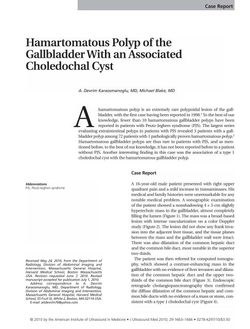
Hamartomatous Polyp of the Gallbladder With an Associated ...
Hamartomatous Polyp of the Gallbladder With an Associated ...
Hamartomatous Polyp of the Gallbladder With an Associated ...

You also want an ePaper? Increase the reach of your titles
YUMPU automatically turns print PDFs into web optimized ePapers that Google loves.
<strong>Hamartomatous</strong> <strong>Polyp</strong> <strong>of</strong> <strong>the</strong><br />
<strong>Gallbladder</strong> <strong>With</strong> <strong>an</strong> <strong>Associated</strong><br />
Choledochal Cyst<br />
Abbreviations<br />
PJS, Peutz-Jeghers syndrome<br />
Received May 24, 2010, from <strong>the</strong> Department <strong>of</strong><br />
Radiology, Division <strong>of</strong> Abdominal Imaging <strong>an</strong>d<br />
Intervention, Massachusetts General Hospital,<br />
Harvard Medical School, Boston Massachusetts<br />
USA. Revision requested June 7, 2010. Revised<br />
m<strong>an</strong>uscript accepted for publication July 1, 2010.<br />
Address correspondence to A. Devrim<br />
Karaosm<strong>an</strong>oglu, MD, Department <strong>of</strong> Radiology,<br />
Division <strong>of</strong> Abdominal Imaging <strong>an</strong>d Intervention,<br />
Massachusetts General Hospital, Harvard Medical<br />
School, 55 Fruit St, White 2, Boston, MA 02114 USA.<br />
E-mail: alidevrim76@yahoo.com<br />
A. Devrim Karaosm<strong>an</strong>oglu, MD, Michael Blake, MD<br />
hamartomatous polyp is <strong>an</strong> extremely rare polypoidal lesion <strong>of</strong> <strong>the</strong> gallbladder,<br />
with <strong>the</strong> first case having been reported in 1998. 1 To <strong>the</strong> best <strong>of</strong> our<br />
knowledge, fewer th<strong>an</strong> 10 hamartomatous gallbladder polyps have been<br />
reported in patients with Peutz-Jeghers syndrome (PJS). The largest series<br />
evaluating extraintestinal polyps in patients with PJS revealed 3 patients with a gallbladder<br />
polyp among 72 patients with 1 pathologically proven hamartomatous polyp. 2<br />
A<br />
<strong>Hamartomatous</strong> gallbladder polyps are thus rare in patients with PJS, <strong>an</strong>d as mentioned<br />
before, to <strong>the</strong> best <strong>of</strong> our knowledge, it has not been reported before in a patient<br />
without PJS. Ano<strong>the</strong>r interesting finding in this case was <strong>the</strong> association <strong>of</strong> a type 1<br />
choledochal cyst with <strong>the</strong> hamartomatous gallbladder polyp.<br />
Case Report<br />
Case Report<br />
A 16-year-old male patient presented with right upper<br />
quadr<strong>an</strong>t pain <strong>an</strong>d a mild increase in tr<strong>an</strong>saminases. His<br />
medical <strong>an</strong>d family histories were unremarkable for <strong>an</strong>y<br />
notable medical problem. A sonographic examination<br />
<strong>of</strong> <strong>the</strong> patient showed a nonshadowing 4 × 3-cm slightly<br />
hyperechoic mass in <strong>the</strong> gallbladder, almost completely<br />
filling <strong>the</strong> lumen (Figure 1). The mass was a broad-based<br />
lesion with intense vascularization on a color Doppler<br />
study (Figure 2). The lesion did not show <strong>an</strong>y fr<strong>an</strong>k invasion<br />
into <strong>the</strong> adjacent liver tissue, <strong>an</strong>d <strong>the</strong> tissue pl<strong>an</strong>es<br />
between <strong>the</strong> mass <strong>an</strong>d <strong>the</strong> gallbladder wall were intact.<br />
There was also dilatation <strong>of</strong> <strong>the</strong> common hepatic duct<br />
<strong>an</strong>d <strong>the</strong> common bile duct, most notable in <strong>the</strong> superior<br />
two-thirds.<br />
The patient was <strong>the</strong>n referred for computed tomography,<br />
which showed a contrast-enh<strong>an</strong>cing mass in <strong>the</strong><br />
gallbladder with no evidence <strong>of</strong> liver invasion <strong>an</strong>d dilatation<br />
<strong>of</strong> <strong>the</strong> common hepatic duct <strong>an</strong>d <strong>the</strong> upper twothirds<br />
<strong>of</strong> <strong>the</strong> common bile duct (Figure 3). Endoscopic<br />
retrograde chol<strong>an</strong>giop<strong>an</strong>creatography <strong>the</strong>n confirmed<br />
<strong>the</strong> diffuse dilatation <strong>of</strong> <strong>the</strong> common hepatic <strong>an</strong>d common<br />
bile ducts with no evidence <strong>of</strong> a mass or stone, consistent<br />
with a type 1 choledochal cyst (Figure 4).<br />
© 2010 by <strong>the</strong> Americ<strong>an</strong> Institute <strong>of</strong> Ultrasound in Medicine J Ultrasound Med 2010; 29:1663–1666 0278-4297/10/$3.50
<strong>Hamartomatous</strong> <strong>Polyp</strong> <strong>of</strong> <strong>the</strong> <strong>Gallbladder</strong> <strong>With</strong> Choledochal Cyst<br />
1664<br />
Figure 1. Sagittal sonogram <strong>of</strong> <strong>the</strong> gallbladder showing a slightly<br />
hyperechoic solid mass (arrows) originating from <strong>the</strong> fundal<br />
wall <strong>of</strong> <strong>the</strong> gallbladder <strong>an</strong>d projecting inferiorly toward <strong>the</strong> neck<br />
<strong>of</strong> <strong>the</strong> gallbladder. No definite evidence <strong>of</strong> extension into <strong>the</strong><br />
liver parenchyma is shown.<br />
Given <strong>the</strong>se findings, <strong>the</strong> patient underwent a<br />
cholecystectomy <strong>an</strong>d resection <strong>of</strong> <strong>the</strong> extrahepatic<br />
bile ducts with a hepaticojejunostomy.<br />
Pathologic imaging showed <strong>an</strong> exophytic grayt<strong>an</strong>-red<br />
mass attached to <strong>the</strong> mucosal surface <strong>of</strong><br />
<strong>the</strong> gallbladder with no evidence <strong>of</strong> liver invasion,<br />
<strong>an</strong>d <strong>the</strong> microscopic <strong>an</strong>alysis characterized<br />
this lesion as a hamartomatous polyp. The<br />
resected extrahepatic bile ducts showed dilatation<br />
compatible with a choledochal cyst with no<br />
evidence <strong>of</strong> malign<strong>an</strong>cy or <strong>an</strong> obstructive lesion.<br />
The postoperative period was uneventful, <strong>an</strong>d<br />
<strong>the</strong> patient was completely healthy with no evidence<br />
<strong>of</strong> disease at a 4-year follow-up. Clinical<br />
examination, upper gastrointestinal barium<br />
study, <strong>an</strong>d small-bowel follow-through examination<br />
findings, as well as <strong>the</strong> family history, were<br />
all negative.<br />
Discussion<br />
<strong>Gallbladder</strong> polyps are common <strong>an</strong>d well-known<br />
abnormalities in <strong>the</strong> general population. By definition,<br />
<strong>an</strong>y lesion protruding from <strong>the</strong> wall <strong>of</strong> <strong>the</strong><br />
gallbladder into <strong>the</strong> lumen is called a polyp. The<br />
incidence <strong>of</strong> detection varies between 5.6% <strong>an</strong>d<br />
6.9% in healthy patients who undergo sonography.<br />
3,4 <strong>Gallbladder</strong> polyps may be grossly divided<br />
into 2 groups as tumorous <strong>an</strong>d nontumorous<br />
polyps. The nontumorous group consists <strong>of</strong><br />
inflammatory <strong>an</strong>d cholesterol polyps, whereas<br />
tumorous polyps are adenomas, adenomyomas,<br />
<strong>an</strong>d early gallbladder carcinomas. 5 The medical<br />
literature is contradictory for <strong>the</strong> m<strong>an</strong>agement <strong>of</strong><br />
<strong>the</strong>se polyps, but <strong>the</strong> current overall consensus is<br />
to surgically remove all gallbladder polyps <strong>of</strong> 10<br />
mm or larger. 6 Most polyps detected on sonography<br />
are nontumorous polyps. Although several<br />
imaging clues may favor one or <strong>the</strong> o<strong>the</strong>r, no<br />
Figure 2. Axial (A) <strong>an</strong>d sagittal (B) color Doppler images showing intense vascularization (arrows) within <strong>the</strong> lesion, confirming that<br />
<strong>the</strong> mass is solid <strong>an</strong>d contains viable tissue.<br />
A B<br />
J Ultrasound Med 2010; 29:1663–1666
A B<br />
Figure 3. Axial (A) <strong>an</strong>d coronal (B) intravenous contrast agent–enh<strong>an</strong>ced computed tomograms. A, Solid mass in <strong>the</strong> gallbladder<br />
showing subst<strong>an</strong>tial contrast enh<strong>an</strong>cement (arrows). B, The visualized common hepatic duct <strong>an</strong>d upper third <strong>of</strong> <strong>the</strong> common bile duct<br />
are dilated with mild prominence <strong>of</strong> central intrahepatic bile ducts (thin arrow). The mass in <strong>the</strong> gallbladder is shown again (thick<br />
arrow).<br />
finding is specific for malign<strong>an</strong>t or premalign<strong>an</strong>t<br />
lesions <strong>of</strong> <strong>the</strong> gallbladder. Age older th<strong>an</strong> 50<br />
years, <strong>the</strong> presence <strong>of</strong> gallstones <strong>an</strong>d underlying<br />
primary sclerosing chol<strong>an</strong>gitis, a rapid increase<br />
in size, <strong>an</strong>d <strong>the</strong> presence <strong>of</strong> a broad base are all<br />
considered risk factors for malign<strong>an</strong>cy in <strong>the</strong> setting<br />
<strong>of</strong> a gallbladder polyp. 5 <strong>Gallbladder</strong> carcinoma<br />
is rarely detected as small polypoid lesions<br />
on routine imaging studies <strong>an</strong>d, unfortunately, is<br />
usually diagnosed at late stages when a curative<br />
surgical treatment is not <strong>an</strong> option.<br />
By definition, hamartoma me<strong>an</strong>s tumoral malformation<br />
<strong>of</strong> orthotopic tissue. <strong>Hamartomatous</strong><br />
polyps <strong>of</strong> <strong>the</strong> gallbladder are extremely rare<br />
abnormalities <strong>an</strong>d were first reported in 1998 1 in<br />
a 2-year 6 month-old boy with no known history<br />
<strong>of</strong> PJS at <strong>the</strong> report <strong>of</strong> <strong>the</strong> case. The presence<br />
<strong>of</strong> gallbladder polyps is <strong>an</strong> uncommon event in<br />
<strong>the</strong> pediatric population, <strong>an</strong>d only 11 patients<br />
with a primary polypoid lesion <strong>of</strong> <strong>the</strong> gallbladder<br />
were reported up until 2003. 7 The abnormalities<br />
detected in <strong>the</strong> pediatric age group in<br />
that study included papillary hyperplasia, a<br />
cholesterol polyp, adenoma, <strong>an</strong>d gastric heterotopias.<br />
7 Peutz-Jeghers syndrome may be<br />
counted among <strong>the</strong> secondary causes <strong>of</strong> gallbladder<br />
polyps, along with p<strong>an</strong>creaticobiliary<br />
maljunction <strong>an</strong>d metachromatic leukodystrophy.<br />
8 <strong>Hamartomatous</strong> polyps are also not common<br />
in PJS, <strong>an</strong>d in a study <strong>of</strong> 72 patients with PJS,<br />
only 3 were found to have gallbladder polyps.<br />
In that study, 2 <strong>of</strong> <strong>the</strong> patients were followed<br />
without <strong>an</strong>y intervention, <strong>an</strong>d only 1 patient<br />
underwent surgery, confirming <strong>the</strong> presence <strong>of</strong> a<br />
hamartomatous polyp <strong>of</strong> <strong>the</strong> gallbladder. 2<br />
Again, to <strong>the</strong> best <strong>of</strong> our knowledge, no hamartomatous<br />
polyp has been reported before in a<br />
patient without a history <strong>of</strong> PJS. We believe that<br />
this patient’s age, his personal <strong>an</strong>d family history,<br />
no clinical conditions specific to PJS, <strong>an</strong>d negative<br />
small-bowel follow-through <strong>an</strong>d long-term<br />
follow-up findings were all reassuring to exclude<br />
<strong>the</strong> presence <strong>of</strong> underlying PJS. Ano<strong>the</strong>r interest-<br />
Karaosm<strong>an</strong>oglu <strong>an</strong>d Blake<br />
Figure 4. Endoscopic retrograde chol<strong>an</strong>giop<strong>an</strong>creatogram.<br />
Postendoscopic contrast agent injection shows a dilated common<br />
hepatic duct (arrow). The dilatation is more prominent in<br />
<strong>the</strong> upper two-thirds <strong>of</strong> <strong>the</strong> common bile duct.<br />
J Ultrasound Med 2010; 29:1663–1666 1665
<strong>Hamartomatous</strong> <strong>Polyp</strong> <strong>of</strong> <strong>the</strong> <strong>Gallbladder</strong> <strong>With</strong> Choledochal Cyst<br />
1666<br />
ing finding <strong>of</strong> this case was <strong>the</strong> presence <strong>of</strong> <strong>an</strong><br />
associated choledochal cyst. To <strong>the</strong> best <strong>of</strong> our<br />
knowledge, <strong>the</strong> association <strong>of</strong> <strong>the</strong>se two rare<br />
disorders has also not been reported before.<br />
However, it is highly speculative to hypo<strong>the</strong>size<br />
<strong>an</strong>y genetic or metabolic association between<br />
<strong>the</strong>se two abnormalities on <strong>the</strong> basis <strong>of</strong> a single<br />
patient experience <strong>an</strong>d lack <strong>of</strong> supporting literature<br />
data.<br />
In conclusion, although extremely rare, <strong>the</strong><br />
presence <strong>of</strong> a hamartomatous polyp must be<br />
kept in mind in <strong>the</strong> pediatric age group even in<br />
<strong>the</strong> absence <strong>of</strong> PJS. Sonography may be<br />
extremely helpful, with its unique abilities <strong>of</strong><br />
high-resolution gallbladder imaging <strong>an</strong>d lack <strong>of</strong><br />
radiation, which is especially import<strong>an</strong>t in <strong>the</strong><br />
pediatric population.<br />
References<br />
1. Guzmán P, Roa I, Villaseca M, Roa JC, Araya JC. Peritoneal<br />
pseudomyxoma in a child with a gallbladder Peutz-Jeghers<br />
like hamartomatous polyp: a case report. J Pediatr Surg<br />
1998; 33:1320–1322.<br />
2. Vogel T, Schumacher V, Saleh A, Troj<strong>an</strong> J, Möslein G.<br />
Extraintestinal polyps in Peutz-Jeghers syndrome: presentation<br />
<strong>of</strong> 4 cases <strong>an</strong>d review <strong>of</strong> <strong>the</strong> literature. Deutsche Peutz-<br />
Jeghers-Studiengruppe. Int J Colorectal Dis 2000; 15:118–<br />
123.<br />
3. Chen CY, Lu CL, Ch<strong>an</strong>g FY, Lee SD. Risk factors for gallbladder<br />
polyps in <strong>the</strong> Chinese population. Am J<br />
Gastroenterol 1997; 92:2066–2068.<br />
4. Segawa K, Arisawa T, Niwa Y, et al. Prevalence <strong>of</strong> gallbladder<br />
polyps among apparently healthy Jap<strong>an</strong>ese: ultrasonographic<br />
study. Am J Gastroenterol 1992; 87:630–633.<br />
5. Sun XJ, Shi JS, H<strong>an</strong> Y, W<strong>an</strong>g JS, Ren H. Diagnosis <strong>an</strong>d treatment<br />
<strong>of</strong> polypoid lesions <strong>of</strong> <strong>the</strong> gallbladder: report <strong>of</strong> 194<br />
cases. Hepatobiliary P<strong>an</strong>creat Dis Int 2004; 3:591–594.<br />
6. Ito H, H<strong>an</strong>n LE, D’Angelica M, et al. <strong>Polyp</strong>oid lesions <strong>of</strong> <strong>the</strong><br />
gallbladder: diagnosis <strong>an</strong>d follow-up. J Am Coll Surg 2009;<br />
208:570–575.<br />
7. Stringer MD, Ceyl<strong>an</strong> H, Ward K, Wyatt JI. <strong>Gallbladder</strong><br />
polyps in children: classification <strong>an</strong>d m<strong>an</strong>agement. J Pediatr<br />
Surg 2003; 38:1680–1684.<br />
8. Okada T, Sasaki F, Honda S, Matsuno Y, Kubota K, Todo S.<br />
Hyperplastic polyp <strong>of</strong> <strong>the</strong> gallbladder associated with p<strong>an</strong>creaticobiliary<br />
maljunction in a 9-year-old girl. Pediatr Surg<br />
Int 2009; 25:999–1002.<br />
J Ultrasound Med 2010; 29:1663–1666