Research Report 2003 - Max-Planck-Institut für molekulare Genetik
Research Report 2003 - Max-Planck-Institut für molekulare Genetik
Research Report 2003 - Max-Planck-Institut für molekulare Genetik

You also want an ePaper? Increase the reach of your titles
YUMPU automatically turns print PDFs into web optimized ePapers that Google loves.
<strong>Max</strong>-<strong>Planck</strong>-<strong>Institut</strong><br />
<strong>für</strong> <strong>molekulare</strong> <strong>Genetik</strong><br />
<strong>Research</strong> <strong>Report</strong> <strong>2003</strong><br />
<strong>Max</strong> <strong>Planck</strong> <strong>Institut</strong>e<br />
for Molecular Genetics, Berlin
<strong>Research</strong> <strong>Report</strong> <strong>2003</strong><br />
Published by the <strong>Max</strong> <strong>Planck</strong> <strong>Institut</strong>e for Molecular Genetics (MPIMG),<br />
Berlin, Germany, October <strong>2003</strong><br />
Editorial Board Bernhard Herrmann, Hans Lehrach,<br />
H.-Hilger Ropers, Martin Vingron<br />
Coordination<br />
& lay-out: Patricia Béziat<br />
Photos: Katrin Ullrich, MPIMG<br />
Printing &<br />
technical support: Thomas Didier, Meta Data<br />
Contact: <strong>Max</strong> <strong>Planck</strong> <strong>Institut</strong>e for Molecular Genetics<br />
Ihnestr. 63 - 73<br />
D-14195 Berlin<br />
Phone: ++49 / 30 / 8413 - 0<br />
Fax: ++49 / 30 / 8413 - 1394<br />
Email: info@molgen.mpg.de<br />
For further information about the MPIMG please see our website:<br />
http://www.molgen.mpg.de
Table of Contents<br />
The <strong>Max</strong> <strong>Planck</strong> <strong>Institut</strong>e for Molecular Genetics<br />
• Organigram . . . . . . . . . . . . . . . . . . . . . . . . . . . . . . . . . . . . . . . . . . . 4<br />
• Mission . . . . . . . . . . . . . . . . . . . . . . . . . . . . . . . . . . . . . . . . . . . . . . .5<br />
• Development of the institute. . . . . . . . . . . . . . . . . . . . . . . . . . . . . . .5<br />
• <strong>Research</strong> concept . . . . . . . . . . . . . . . . . . . . . . . . . . . . . . . . . . . . . . . 5<br />
Department of Vertebrate Genomics<br />
• Introduction . . . . . . . . . . . . . . . . . . . . . . . . . . . . . . . . . . . . . . . . . . . 7<br />
• Protein Structure Factory . . . . . . . . . . . . . . . . . . . . . . . . . . . . . . . . 12<br />
• Mass Spectrometry Group . . . . . . . . . . . . . . . . . . . . . . . . . . . . . . . 15<br />
• Bioinformatics Group . . . . . . . . . . . . . . . . . . . . . . . . . . . . . . . . . . 19<br />
• Mouse, Medaka & MHC Group. . . . . . . . . . . . . . . . . . . . . . . . . . . 25<br />
• Genetic Variation, Haplotypes & Genetics of Complex Disease Group . . 30<br />
• Oligofingerprinting / Cell Arrays Group . . . . . . . . . . . . . . . . . . . . 32<br />
• Kinetic Modeling Group . . . . . . . . . . . . . . . . . . . . . . . . . . . . . . . . 36<br />
• In vitro Ligand Screening Group. . . . . . . . . . . . . . . . . . . . . . . . . . 39<br />
• Neurodegenerative Disorders Group . . . . . . . . . . . . . . . . . . . . . . . 43<br />
• Automation Group . . . . . . . . . . . . . . . . . . . . . . . . . . . . . . . . . . . . . 45<br />
• Evolution & Development Group . . . . . . . . . . . . . . . . . . . . . . . . . 52<br />
• Gene Traps & Microarrays - Molecular Analysis of Heart Failure Group . 56<br />
• Protein Group . . . . . . . . . . . . . . . . . . . . . . . . . . . . . . . . . . . . . . . . 61<br />
• Cardiovascular Genetics Group . . . . . . . . . . . . . . . . . . . . . . . . . . . 65<br />
• Genomic Sequencing & Gene Function in Complex Diseases Group . . . 68<br />
• Chromosome 21 Group . . . . . . . . . . . . . . . . . . . . . . . . . . . . . . . . . 71<br />
Department of Human Molecular Genetics<br />
• Introduction . . . . . . . . . . . . . . . . . . . . . . . . . . . . . . . . . . . . . . . . . . 75<br />
• Neurochemistry Group & Mouse Lab . . . . . . . . . . . . . . . . . . . . . . 81<br />
• Clinical Genetics. . . . . . . . . . . . . . . . . . . . . . . . . . . . . . . . . . . . . . . 84<br />
• Chromosome Rearrangement & Disease . . . . . . . . . . . . . . . . . . . . 87<br />
• DNA Microarrays . . . . . . . . . . . . . . . . . . . . . . . . . . . . . . . . . . . . . . 91<br />
• Neurobiology Group . . . . . . . . . . . . . . . . . . . . . . . . . . . . . . . . . . . .94<br />
• Cytology Group . . . . . . . . . . . . . . . . . . . . . . . . . . . . . . . . . . . . . . . 98<br />
• Biochemistry of Inherited Brain Disorders . . . . . . . . . . . . . . . . . 101<br />
• Familial Mental Retardation . . . . . . . . . . . . . . . . . . . . . . . . . . . . 104<br />
Department of Computational Molecular Biology<br />
• Introduction . . . . . . . . . . . . . . . . . . . . . . . . . . . . . . . . . . . . . . . . . 109<br />
• Gene Structure & Array Design Group . . . . . . . . . . . . . . . . . . . . 112<br />
• Protein Families & Evolution Group . . . . . . . . . . . . . . . . . . . . . . 115<br />
• Algorithms Group . . . . . . . . . . . . . . . . . . . . . . . . . . . . . . . . . . . . 118<br />
• Protein Function Analysis Group . . . . . . . . . . . . . . . . . . . . . . . . . 122<br />
• Computational Diagnostics Group . . . . . . . . . . . . . . . . . . . . . . . . 124<br />
• Transcriptional Regulation Group . . . . . . . . . . . . . . . . . . . . . . . . 128<br />
MPI for Molecular Genetics<br />
<strong>Research</strong> <strong>Report</strong> <strong>2003</strong><br />
1
2<br />
General Information<br />
Department of Developmental Genetics<br />
• Scientific development and future orientation . . . . . . . . . . . . . . . 131<br />
Emeritus Group General Molecular Genetics<br />
• Biographical notes . . . . . . . . . . . . . . . . . . . . . . . . . . . . . . . . . . . . 137<br />
<strong>Research</strong> Group Development & Disease<br />
• Overview . . . . . . . . . . . . . . . . . . . . . . . . . . . . . . . . . . . . . . . . . . . 141<br />
Junior <strong>Research</strong> Groups / Otto-Warburg Laboratory<br />
• Endocrine Regulation of C. elegans Development & Aging . . . . 147<br />
• Gene Silencing in Saccharomyces cerevisiae . . . . . . . . . . . . . . . . 152<br />
• Molecular Control of Skeletal Development . . . . . . . . . . . . . . . . 157<br />
Ribosome Group<br />
• Ribosomal RNA Structure . . . . . . . . . . . . . . . . . . . . . . . . . . . . . . 163<br />
• Ribosome Crystallography . . . . . . . . . . . . . . . . . . . . . . . . . . . . . . 166<br />
• Ribosomal Function . . . . . . . . . . . . . . . . . . . . . . . . . . . . . . . . . . . 170<br />
Miscellaneous <strong>Research</strong> Groups<br />
• Phage & Conjugation Group . . . . . . . . . . . . . . . . . . . . . . . . . . . . 175<br />
• Microscopy Group . . . . . . . . . . . . . . . . . . . . . . . . . . . . . . . . . . . . 180<br />
• High-throughput Technologies Group . . . . . . . . . . . . . . . . . . . . . 182<br />
• Analysis of Protein Evolution Group . . . . . . . . . . . . . . . . . . . . . 186<br />
Administration & <strong>Research</strong> Support<br />
• Administration . . . . . . . . . . . . . . . . . . . . . . . . . . . . . . . . . . . . . . . 189<br />
• Technical Management & Workshops . . . . . . . . . . . . . . . . . . . . . 191<br />
• Analytics & Computing . . . . . . . . . . . . . . . . . . . . . . . . . . . . . . . . 192<br />
• Animal Facility . . . . . . . . . . . . . . . . . . . . . . . . . . . . . . . . . . . . . . 194<br />
• Library . . . . . . . . . . . . . . . . . . . . . . . . . . . . . . . . . . . . . . . . . . . . .195<br />
• Graphics / Photo . . . . . . . . . . . . . . . . . . . . . . . . . . . . . . . . . . . . . 196
MPI for Molecular Genetics<br />
<strong>Research</strong> <strong>Report</strong> <strong>2003</strong><br />
3
4<br />
General Information
The <strong>Max</strong> <strong>Planck</strong> <strong>Institut</strong>e for<br />
Molecular Genetics<br />
Mission<br />
<strong>Research</strong> at the MPIMG concentrates on genome analysis of man and other organisms to<br />
contribute to a global understanding of many of the biological processes in the organism,<br />
and to provide a basis to elucidate the mechanism behind many human diseases. It is the<br />
overall goal of the combined efforts of all MPIMG’s groups to gain new insights into the<br />
development of diseases on a molecular level, thus contributing to the development of<br />
cause-related new medical treatments.<br />
Development of the institute<br />
The <strong>Max</strong> <strong>Planck</strong> <strong>Institut</strong>e for Molecular Genetics (MPIMG) was founded in 1964 with<br />
the appointment of Heinz-Günther Wittmann and Heinz Schuster as heads of department,<br />
followed by the appointment of Thomas Trautner in 1965. At this time, the research of the<br />
institute was focussing on DNA replication and gene regulation in bacteria, bacterial phage<br />
and fungi (departments Schuster and Trautner) and on the structure, function and evolution<br />
of ribosomes which were central to the work of H.-G. Wittmann.<br />
In 1970, the three departments, as well as<br />
four independent junior research groups (the<br />
future Otto Warburg Laboratories) moved<br />
into the new premises of the institute situated<br />
in the Ihnestraße, Berlin-Dahlem. After<br />
the sudden death of H.G. Wittmann in<br />
1990 and the retirement of H. Schuster in<br />
1995, the appointments of Hans Lehrach<br />
(1994, Dept. of Vertebrate Genomics), and<br />
Hans-Hilger Ropers (Dept. of Human Molecular<br />
Genetics, full-time since 1997) induced<br />
a major shift in the scientific orientation<br />
of the institute. Following the retirement<br />
of T. Trautner in 2000, Martin Vingron<br />
was appointed as head of the new Department of Computational Molecular Biology. At<br />
the same time, Stefan Mundlos was jointly appointed by the Humboldt University of<br />
Berlin and the <strong>Max</strong> <strong>Planck</strong> Society as head of the <strong>Institut</strong>e of Medical Genetics at the<br />
Charité and of an independent research group at the MPIMG. Together with the Free<br />
University of Berlin, Bernhard Herrmann has been appointed professor at the university<br />
and director at the institute in <strong>2003</strong>, forming the fourth department.<br />
Currently three junior research groups work at the institute. A newly created junior<br />
research group will start in 2004 when the others are due to leave.<br />
<strong>Research</strong> concept<br />
Genome research, the systematic study of genes and genomes, has changed the way in<br />
which research in molecular genetics is pursued. The focus and composition of the MPI<br />
for Molecular Genetics reflects this development. Large scale genome research (Dept.<br />
Lehrach) generates the tools and information to understand the function of most or all<br />
genes of man and other organisms. Human molecular genetics (Dept. Ropers) searches<br />
for disease genes and their biological function. Computational molecular biology (Dept.<br />
MPI for Molecular Genetics<br />
<strong>Research</strong> <strong>Report</strong> <strong>2003</strong><br />
5
6<br />
General Information<br />
Vingron) exploits the generated data to better understanding of biological and disease<br />
processes. The newly established Dept. of Developmental Genetics (Dept. Herrmann)<br />
uses the systematic functional analysis for understanding developmental mechanisms.<br />
The institute pursues a number of large scale projects. Probably the most prominent<br />
national project is the German National Genome Network (NGFN), where all departments<br />
of the institute participate and collaborate with each other. Other prominent<br />
projects include a number of EU projects, participation in several projects of the German<br />
Ministry of Science as well DFG “Sonderforschungsbereiche”.<br />
With this involvement in national and international research projects as well as by<br />
virtue of the research output of the institute, the MPIMG is perceived internationally<br />
as a stronghold of genome and genetics research in Germany. The publications coming<br />
from the institute document the international competitiveness of the institute. Maintaining<br />
this status in the future will require continuing technological innovation, close<br />
co-operation with the universities and, in particular, their medical schools, and ongoing<br />
integration between genome research and genetics, as well as between experimental<br />
and computational biological research. These are the means by which research<br />
excellence shall be maintained and further strengthened in the future.<br />
Bernhard Herrmann, Director<br />
Hans Lehrach, Director<br />
H.-Hilger Ropers, Director<br />
Martin Vingron, Director
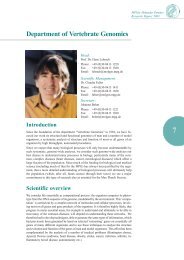
Department of Vertebrate Genomics<br />
Introduction<br />
Since the foundation of the department “Vertebrate Genomics” in 1994, we have focused<br />
our work on structural and functional genomics of man and a number of model<br />
organisms, a systematic analysis of structure and function of most or all genes of an<br />
organism by high throughput, automated procedures.<br />
Since we expect that many biological processes will only become understandable by<br />
such systematic, genome-wide analyses, we consider such genome-wide analyses our<br />
best chance to understand many processes in biology, particularly many of the common,<br />
complex diseases (heart diseases, cancer, neurological diseases) which affect a<br />
large fraction of the population. Since much of the funding for biological and medical<br />
science (including much of that for the MPG) has always been justified by the argument,<br />
that a more detailed understanding of biological processes will ultimately help<br />
the population (which, after all, funds science through their taxes) we see a major<br />
commitment to this type of research also as essential for the <strong>Max</strong> <strong>Planck</strong> Society.<br />
Scientific overview<br />
Head:<br />
Prof. Dr. Hans Lehrach<br />
Phone: +49 (0)30-8413 1220<br />
Fax: +49 (0)30-8413 1380<br />
Email: lehrach@molgen.mpg.de<br />
Scientific Management:<br />
Dr. Claudia Falter<br />
Phone: +49 (0)30-8413 1411<br />
Fax: +49 (0)30-8413 1380<br />
Email: falter@molgen.mpg.de<br />
Secretary:<br />
Johanna Belart<br />
Phone: +49 (0)30-8413 1221<br />
Fax: +49 (0)30-8413 1380<br />
Email: belart@molgen.mpg.de<br />
We consider life essentially as computational process: the organism computes its phenotype<br />
from the DNA sequence of its genome, modulated by the environment. This ‘computation’<br />
is carried out by a complex network of molecular (and cellular) processes, involving<br />
most or all genes and gene products of the organism. It is therefore highly likely, that<br />
progress in many essential areas, for example to understand and ultimately to be able to<br />
treat many of the common diseases, will depend on understanding these networks. We<br />
therefore had to develop techniques, able to generate the same types of information, which<br />
had previously been generated by hand on selected ‘interesting’ genes on essentially all<br />
genes of many different organisms and to use these techniques to analyze the structure<br />
and evolution and function of the genes of man and model organisms. This effort has been<br />
complemented by the analysis of a number of medical problems (Huntington chorea,<br />
Apeced, Downs syndrome, heart disease, obesity, stroke, cancer, infection, arthritis, inflammatory<br />
bowel disease, autoimmunity etc.)<br />
MPI for Molecular Genetics<br />
<strong>Research</strong> <strong>Report</strong> <strong>2003</strong><br />
7
Department of Vertebrate Genomics<br />
8<br />
Analysis of genomic sequences<br />
Mapping and sequencing projects<br />
A major effort during the last 6 years has been directed at mapping of chromosomes<br />
(Yaspo, Sudbrak) and entire genomes (Himmelbauer, Schalkwyk*, Knoblauch*), and<br />
the determination of genomic sequences, culminating in the completion of the sequence<br />
of chromosome 21 (Yaspo, Reinhardt), the working draft of the human genome and<br />
finally the recent publication of the essentially completed genome sequence. Since<br />
then, we have increasingly focused on the use of genomic sequencing to understand<br />
the evolution of regions of the human genome (Yaspo, Sudbrak, Reinhardt). In one of<br />
these projects, the sequencing of chimp chromosome 22, the equivalent of human<br />
chromosome 21, has been completed in collaboration with groups in Germany, Japan,<br />
China, Taiwan and South Korea. The results, currently being prepared for publication,<br />
provide fascinating insights into the biological differences between man and our closest<br />
relative. The sequence of the rat MHC has been completed, and submitted for<br />
publication (Himmelbauer, Reinhardt). To try to understand more distant evolutionary<br />
processes, we have continued to work on the genomic sequence of Oikopleura, a chordate<br />
with an exceptionally small genome (app. 70 Mb). In a first whole genome shotgun<br />
phase, a coverage of approximately 1.1 x has been achieved. To minimize the<br />
effect of the high polymorphism rate, we have now shifted to BAC based sequencing,<br />
with the progress limited by lack of funding (Reinhardt, Yaspo).<br />
Genetic analysis<br />
Genotype-phenotype correlations in man will increasingly require the application of<br />
more effective genotyping tools. For this, new protocols for SNP genotyping (Gut*,<br />
Sauer) and microsequencing (Nordhoff*) based on mass spectroscopy have been developed.<br />
An alternative optical approach for SNP typing is currently under development<br />
(Soldatov). New approaches to compare the sequences of candidate genes in<br />
patients and controls, and to analyze their haplotypes have been developed and are<br />
being applied in a number of different disease areas (Hoehe, Reinhardt).<br />
Analysis of transcripts<br />
Further insights into evolutionary processes in key organisms with larger genomes<br />
have been derived from the analysis of transcripts, based on a combination of the<br />
oligonucleotide fingerprinting approach to assign cDNA clones to clusters corresponding<br />
to different genes, combined with a limited amount of cDNA sequencing. This<br />
approach has been applied to a number of different organisms, e.g. zebrafish (Clark*),<br />
different plants (Radeloff*), man and mouse (Radeloff*, Janitz), cow (Janitz) etc.<br />
Among other projects, this has given insights into the set of genes available to amphioxus,<br />
a cephalochordate, and sea urchin, an echinoderm, representing key stages in<br />
deuterostome evolution. This work has contributed to understanding the evolution of<br />
the vertebrate genome, and especially the ‘Ohno hypothesis’, according to which two<br />
whole genome duplications of a chordate genome have led to the vertebrate genome<br />
(Panopoulou, Poustka).<br />
Functional Genomics<br />
Analysis of gene expression<br />
Efficient, sensitive techniques to detect and quantify patterns of transcription have<br />
been a major focus in functional genomics. Starting from our first arrayer developed<br />
by us in 1987, we have carried out long term technology development in this area,<br />
from the first filter based complex cDNA hybridization screens to high throughput<br />
chip hybridization systems (Eickhoff*, Hultschig), and applied these techniques to a<br />
number of biological (and medical) problems (Yaspo, Nietfeld, Ruiz, Sperling, Soldatov<br />
etc.). A number of global (ENSEMBL chip by Yaspo, RZPD) and disease specific<br />
(Ruiz, Sperling, Hultschig) chips have been constructed.<br />
* former member of the department
To generate high resolution expression information we have carried out a systematic analysis of<br />
gene expression of mouse orthologues by whole-mount in-situ hybridization on mouse embryos<br />
(Bernhard Herrmann) and brain tissue slices (Ariel Ruiz i Altaba, New York, Yaspo).<br />
Proteomics<br />
Due to the fact, that many genes act through their protein product, we need effective<br />
techniques to measure protein abundances, and to determine their modification status. A<br />
significant effort has therefore been directed towards the development of mass spectrometry<br />
techniques to generate this information on high throughput (Gobom). A combination<br />
of 2-D gel electrophoresis with mass spectrometric identification of protein spots<br />
has, for example, been used in a systematic analysis of mouse proteins (in collaboration<br />
with Joachim Klose, Humboldt University).<br />
To generate information on protein function and protein interactions, a number of different<br />
approaches have been followed. One key technique in this has been the development<br />
of a robust, high throughput 2-hybrid system, which has been used to generate information<br />
on interacting proteins on thousands of human genes (Wanker*). In addition, a number<br />
of specific screens have been carried out on proteins involved in Chorea Huntington<br />
(Wanker*) and other neurodegenerative diseases (Krobitsch), proteins encoded on chromosome<br />
21 (Yaspo), the products of genes overexpressed in heart disease (Sperling) and<br />
many others. As a complement, mass spectrometry techniques have been developed to<br />
isolate and characterize the components of larger complexes (Gobom), and are now being<br />
used to complement the 2- hybrid data (Gobom).<br />
For interaction studies of large numbers of proteins in parallel with e.g. antibodies, other<br />
proteins, DNA or RNA fragments, small molecules, a number of protein array systems<br />
have been developed, ranging from PVDF membrane arrays to chips carrying purified<br />
proteins. Such arrays have been used in a number of different applications (Cahill*, Konthur,<br />
Seitz). They will provide an additional route to establish and verify information on protein-protein<br />
interactions, and will play a key role in the development of regulatory genomics<br />
and chemical genomics approaches.<br />
To be able to generate antibodies to many gene products in parallel, we have established<br />
an automated phagemid selection protocol, and have made major progress in streamlining<br />
the clone characterization, opening the way to high throughput antibody generation, to be<br />
used, among other applications, in the construction of antibody chips. In addition, as part<br />
of the characterization of gene products encoded on chromosome 21, antisera are being<br />
generated against chromosome 21 encoded proteins in the chick system.<br />
Structural genomics<br />
Since the structure of proteins can also give insights into protein function and evolution,<br />
and can aid in the development of drugs directed against these proteins, a number of<br />
structural genomics projects have been started throughout the world. The department has<br />
been instrumental in starting the ‘Proteinstrukturfabrik’ (PSF), the first of these projects<br />
world wide, involving a number of groups and facilities throughout Berlin. In this project,<br />
we are particularly contributing in providing expression constructs of human proteins<br />
(Büssow), and in the construction of a high throughput crystallization robot, able to handle<br />
up to 4 Million crystallization trials in parallel (Eickhoff*, Nyarsik).<br />
As a next step in high throughput structural genomics, we have currently started a follow<br />
up project, the ‘Ultrastrukturnetzwerk’ (USN), to be able to analyze larger complexes. For<br />
this network, which again will involve a number of groups throughout Berlin, mass spectroscopic<br />
analysis of protein complexes (Gobom) will be combined with an analysis of<br />
the structure of the complex by Cryo-EM (Lange).<br />
Gene function at the cellular level<br />
To be able to analyze gene (and promoter) function at the cellular level, we have recently developed<br />
cell array systems, allowing the analysis of many constructs in parallel. These systems can<br />
be used to test the function of proteins (and protein variants), carry out RNAi experiments, test<br />
promoter function, and identify protein-protein interactions within the mammalian cell (Janitz).<br />
MPI for Molecular Genetics<br />
<strong>Research</strong> <strong>Report</strong> <strong>2003</strong><br />
9
Department of Vertebrate Genomics<br />
10<br />
Gene function in model organisms<br />
The ultimate test for the function of a gene is the analysis of mutant organisms. To provide<br />
an efficient route for such functional tests, we have been involved in starting the german<br />
gene trap consortium (Wiles*, Ruiz). More recently, a chemical mutagenesis protocol,<br />
coupled to an efficient mutation detection, has been developed (Himmelbauer). Additional<br />
strategies are being developed (RNAi).<br />
Gene function in Complex Diseases<br />
Ultimately most of the work carried out within the department has medical implications,<br />
either through the development of new techniques, or directly through work on<br />
the analysis of processes of medical interest. Within the last 6 years, work on Chorea<br />
Huntington has progressed rapidly, most recently culminating in the identification of<br />
small molecules blocking the aggregation process we believe to be at the heart of the<br />
disease mechanism, and the establishment of a dense protein-protein interaction network<br />
established through high throughput 2 hybrid analysis (Wanker*). (Due to Erich<br />
Wanker’s move to a C4-Position at the MDC, much of this work has now shifted to<br />
Berlin-Buch.) Work on other neurodegenerative diseases does however continue<br />
(Krobitsch). Similarly chromosome 21 and Down syndrome (Yaspo) as well as the X<br />
chromosome (Sudbrak) have been a long term focus of the work in the department. In<br />
addition to these core projects, a number of other disease areas have been investigated,<br />
mostly in collaborative projects. Examples are heart disease (Sperling, Ruiz), arthritis<br />
(Janitz, Ruiz), inflammatory bowel disease, infection and stroke (Nietfeld), obesity<br />
(Soldatov, Hoehe), and many others.<br />
Bioinformatics / Systems biology<br />
Bioinformatic tools continue to play a central role in our work, an area, which has been strengthened<br />
considerably by the new department of Martin Vingron. Work carried out within the department<br />
continues to address the problems of sequence analysis, sequence interpretation and<br />
sequence annotation (Hennig, Yaspo) and gene expression analysis (Herwig).<br />
Efforts to integrate data generated throughout the department have involved the construction<br />
of a unified laboratory database (Hennig). In addition, in collaboration with the RZPD<br />
and the department of Martin Vingron, we have established a new database/data base<br />
interface (www.genome-matrix.org), unifying data across many data types and many species<br />
(Zehetner, Hennig, Yaspo). A similar effort to integrate such molecular data with<br />
medical information (d-matrix) is under way (Sperling).<br />
Ultimately many complex biological and medical processes will only be ‘understood’<br />
through the establishment of quantitative models, duplicating the knowledge we have<br />
about a biological system by computational objects. For this, a modeling environment<br />
has been developed (PyBios), and will be expanded as part of one of our EU collaborations<br />
(Herwig, Klipp).<br />
Future developments<br />
A continuing focus in the department is the development of new techniques in functional<br />
genomics, especially nanotechnology (Nyarsik), mass spectroscopy (Gobom, Sauer) and<br />
protein and antibody chips (Seitz, Konthur, Yaspo). As a new direction uniting most of the<br />
departments within the institute, we plan to focus increasingly on the analysis of regulatory<br />
networks and regulation mechanisms (regulatory genomics). This will combine the<br />
expertise in computational analysis of promoters (Vingron), expertise in the experimental<br />
analysis of transcription factors and other regulatory genes (Herrmann), and our expertise<br />
in automation. In addition, as more and more data become available from other centers<br />
world wide, our efforts in data integration and systems biology will increase further, in<br />
close interaction with Martin Vingron’s department. Conversely, work in a number of<br />
areas (e.g. most areas of plant genomics) has been reduced or stopped.
Competitive position<br />
Since a long time, the department plays a key role in technology development and automation,<br />
covering essentially all areas of functional genomics. In some areas (mapping of<br />
the mouse and rat genome, genomic sequencing, expression analysis, protein-protein interactions,<br />
gene traps) high throughput data production pipelines have been developed,<br />
and have contributed significantly to the world wide genome project. In other areas, the<br />
development has been less rapid, either due to technical difficulties (high throughput<br />
proteomics), or, in many cases, due to lack of funding. The department has played a key<br />
role in setting up the resource center of the German Genome Project (RZPD), and a<br />
number of national and international research networks (gene trap consortium, Protein<br />
Structure Factory, Berlin Center of Genome Based Bioinformatics (BCB), Ultrastrukturnetzwerk<br />
(USN), etc). In addition, we were able to transfer our knowledge and results by<br />
founding and co-founding a number of start up companies (GPC, Scienion, PSF AG,<br />
Prot@gen), by patenting (34 patents since 1998) and in around 200 publications in the<br />
last 6 years. We are probably the only center within Germany covering the entire range of<br />
functional genomics, and have played a central role in the founding of both DHGP<br />
(Deutsches Humangenomprojekt) and NGFN (National Genome <strong>Research</strong> Network).<br />
Due to limited resources the structure of the department has had to remain a compromise,<br />
since from the beginning we have had to complement the limited long term funding by the<br />
MPG (currently approximately 15 % of our overall budget) by large scale grant funding<br />
from many different outside sources to be able to remain competitive with many of the<br />
large, well funded centers set up for genome research in other countries. Interactions with<br />
the department of Hilger Ropers have played an important role in a number of projects.<br />
Good interactions also exist with the junior groups, the research group of Stefan Mundlos<br />
as well as other research groups in the institute (e.g. the ribosome groups contributing to<br />
the Ultrastrukturnetzwerk). Of particular importance for our work have however also<br />
been the more recent appointments of Martin Vingron and, most recently, Bernhard<br />
Herrmann, dramatically strengthen the overall capabilities of the institute as one of the<br />
very centers focusing on genome research in Germany, as well as the continuing interaction<br />
with the service group, providing most of the essential infrastructure for e.g. sequencing<br />
projects in the institute.<br />
MPI for Molecular Genetics<br />
<strong>Research</strong> <strong>Report</strong> <strong>2003</strong><br />
11
Department of Vertebrate Genomics<br />
12<br />
Protein Structure Factory<br />
Head:<br />
Dr. Konrad Büssow<br />
Phone: +49 (0)30-32639 2802<br />
Fax: +49 (0)30-32639 2833<br />
Email: buessow@molgen.mpg.de<br />
PSF / Protein Expression:<br />
Christoph Scheich (engineer)<br />
Dr. Volker Sievert (scientist)<br />
Janett Tischer (technician)<br />
Claudia Quedenau (PhD student)<br />
PSF / Protein Crystallisation:<br />
Joachim Lebik (engineer)<br />
NGFN core area platform 4 / protein<br />
expression<br />
Dr. Hendrik Weiner (scientist)<br />
Thomas Faupel (PhD student)<br />
Brigitte Hieke (technician)<br />
Scientific overview<br />
The Protein Structure Factory is a structural genomics project for the systematic structural analysis<br />
of human proteins. The goal of the Protein Structure Factory is to systematically select suitable<br />
human proteins by sequence analysis, express the proteins in recombinant form and proceed<br />
with structural analysis with those proteins that can be produced easily.<br />
The PSF is part of the International Structure Genomics Organization (ISGO) and has organised<br />
the International Congress of Structural Genomics 2002. Funded in 1999, the Protein Structure<br />
Factory is one of the earliest endeavours towards fast and cost-effective high-throughput protein<br />
production and structure determination.<br />
The Department of Vertebrate Genomics has initiated the Protein Structure Factory as a BMBFfounded<br />
“Leitprojekt” as partner of a multidisciplinary consortium of structural biology and<br />
other research groups and companies (http://www.proteinstrukturfabrik.de). Our scientific partners<br />
in the Protein Structure Factory and the expertise provided are:<br />
• Prof. Dr. Udo Heinemann, MDC Berlin – X-ray diffraction at BESSY synchroton<br />
• Prof. Dr. Wolfgang Saenger, FU Berlin – protein crystallisation<br />
• Prof. Dr. Hartmut Oschkinat, FMP Berlin – NMR of protein domains<br />
• PD Dr. Christine Lang, TU-Berlin – protein expression in yeast<br />
• Prof. Dr. K.-P. Hofmann, Charité Berlin – biophysical characterisation of proteins<br />
• Alpha Bioverfahrenstechnik GmbH, Kleinmachnow – protein expression and purification<br />
• Deutsches Resourcenzentrum GmbH, Berlin – cDNA clone resources<br />
• Prof. Dr. Peer Bork, EMBL Heidelberg – bioinformatics, selection of target proteins<br />
Current status of research<br />
Many structural genomics projects have been initiated since the start of the Protein Structure<br />
Factory in 1999. It is clear today that the major bottleneck in the systematic structural analysis of<br />
proteins is their production and crystallisation. Especially the recombinant production of eukaryotic<br />
proteins is difficult and has a high failure rate. One way to overcome this limitation is to<br />
generate and analyse a large number of targets and to proceed with the ones that can be produced<br />
readily. Alternative expression systems are another option - yeast is used in addition to E. coli at
the Protein Structure Factory. Finally, in vitro folding of proteins can transfer insolubly expressed<br />
proteins into the folded form (see below).<br />
High-throughput protein expression<br />
Our part in the project is the generation of E. coli expression clones for human proteins and the<br />
characterization of the clones. We are responsible to supply the Protein Structure Factory with a<br />
sufficient number of clones that express human proteins in good yield and soluble form. We<br />
have established expression systems that allow efficient protein purification by affinity chromatography<br />
via affinity tags and removal of affinity tags after protein purification (Heinemann et<br />
al., <strong>2003</strong>). All steps required for subcloning of cDNAs into our optimised expression vectors<br />
and subsequent characterisation of expression clones by protein expression and affinity purification<br />
were established and automated in 96-well format on a pipetting robot (Scheich et al,<br />
<strong>2003</strong>, Holz et al., <strong>2003</strong>).<br />
In co-operation with the group of Peer Bork, we selected 860 human proteins that have a high<br />
probability to be successfully expressed in E. coli, e.g. excluding membrane proteins. Another<br />
selection criterion was the availability of full-length cDNA clones, since these clones enabled us<br />
to use high-throughput cloning procedures.<br />
So far, our group supplied constructs for soluble expression of 243 human proteins. Using these<br />
clones, 52 distinct proteins were expressed and purified in sufficient amount and quality for<br />
crystallization trials in the year 2002. In <strong>2003</strong>, this number was already reached after eight<br />
months. Using these crystals, two structures were solved by our partners in the Protein Structure<br />
Factory and six more proteins with well-diffracting crystals were obtained in this time period<br />
(Manjasetty et al., in press). The current status of generated clones, proteins and structures is<br />
available at: http://www.proteinstrukturfabrik.de/public/PSF_Status_1.shtml.<br />
A database and additional software were developed for management of data in the group and to<br />
provide co-workers in the Protein Structure Factory with information on sequences and clones<br />
of target proteins. Part of this software development was published (Büssow et al., 2002).<br />
A large proportion of human proteins do not fold and form insoluble aggregates when expressed<br />
in E. coli cells. In vitro folding can transfer such aggregates into correctly folded proteins.<br />
We have developed a automated method for protein crystallisation, that allows to perform<br />
fast screenings for suitable folding buffer conditions using a biophysical readout (Scheich et al.,<br />
in preparation).<br />
Automated protein crystallisation<br />
The department of Vertebrate Genomics has automated protein crystallisation in the Protein<br />
Structure Factory in a co-operative effort with the group of Prof. Saenger, Free University of<br />
Berlin (Mueller et al., 2001). Automation is a requirement for the crystallisation of larger number<br />
of target proteins, since manual crystallisation is very time-consuming. We have established<br />
automated set-up and analysis of crystallisation screens. A micro-dispensing technology for<br />
crystallisation in sub-microlitre volumes in microtitre plates has been established. Crystallisation<br />
is currently monitored by a system that takes pictures of crystallisation drops in a 96-well plate<br />
automatically. An additional system is under development that will combine a microtitre-plate<br />
storage device with a detection system and that will be able to automatically record images of<br />
crystallisation experiments at certain time intervals.<br />
Protein-protein interaction studies<br />
As part of the “Nationales Genomforschungsnetz” NGFN, a project has been established<br />
at the Protein Structure Factory for expression of human proteins involved in neurodegenerative<br />
diseases for the in vitro characterization of protein-protein interaction.<br />
We are developing a technology for identification of protein interaction partners using highdensity<br />
protein arrays on filter membranes (Weiner et al., in press). This project is a collaboration<br />
with Prof. Erich Wanker of the MDC Berlin-Buch, in which we share a common set of 100<br />
proteins involved in neurodegenerative disease to identify novel protein-protein interactions.<br />
We have identified sets of interaction partners for different target proteins. Interactions are currently<br />
being verified by in vitro pull-down assays and other methods. (NGFN Platform 4, http:/<br />
/www.ngfn.de/protein)<br />
MPI for Molecular Genetics<br />
<strong>Research</strong> <strong>Report</strong> <strong>2003</strong><br />
13
Department of Vertebrate Genomics<br />
14<br />
General information<br />
Publications 1998-<strong>2003</strong><br />
Manjasetty BA, Delbrück H, Pham D-T,<br />
Mueller U, Fieber-Erdmann M, Scheich C,<br />
Sievert V, Büssow K, Niesen F, Weihofen W,<br />
Loll B, Saenger W & Heinemann U (<strong>2003</strong>).<br />
Crystal structure of Homo sapiens protein<br />
hp14.5. Proteins: Structure, Function, and<br />
Genetics (in press)<br />
Weiner H, Faupel T & Büssow K (<strong>2003</strong>). Protein<br />
arrays from cDNA expression libraries.<br />
In: Protein Arrays, vol. Humana Press Inc. (in<br />
press)<br />
Holz C, Prinz B, Bolotina N, Sievert V, Büssow<br />
K, Simon B, Stahl U & Lang C (<strong>2003</strong>). Establishing<br />
the yeast Saccharomyces cerevisiae as<br />
a system for expression of human proteins on<br />
a proteome-scale. J Funct & Struct Genomics<br />
(in press)<br />
Heinemann U, Büssow K, Mueller U &<br />
Umbach P (<strong>2003</strong>). Facilities and methods for<br />
the high-throughput crystal structure analysis<br />
of human proteins. Acc Chem Res 36:157-163<br />
Scheich C, Sievert V & Büssow K (<strong>2003</strong>). An<br />
automated method for high-throughput protein<br />
purification applied to a comparison of<br />
His-tag and GST-tag affinity chromatography.<br />
BMC Biotechnology 3:12<br />
Eickhoff H, Konthur Z, Lueking A, Lehrach<br />
H, Walter G, Nordhoff E, Nyarsik L & Büssow<br />
K (2002). Protein array technology: the tool<br />
to bridge genomics and proteomics. Adv<br />
Biochem Engineer/Biotech 77:103-12<br />
Walter G, Büssow K, Lueking A & Glokler J<br />
(2002). High-throughput protein arrays: prospects<br />
for molecular diagnostics. Trends in Mol<br />
Med 8:250-3<br />
Büssow K, Hoffmann S & Sievert V (2002).<br />
ORFer - retrieval of protein sequences and<br />
open reading frames from GenBank and storage<br />
into relational databases or text files. BMC<br />
Bioinformatics 3:40<br />
Klose J, Nock C, Herrmann M, Stuhler K,<br />
Marcus K, Bluggel M, Krause E, Schalkwyk<br />
LC, Rastan S, Brown SD, Büssow K,<br />
Himmelbauer H & Lehrach H (2002). Genetic<br />
analysis of the mouse brain proteome. Nature<br />
Genetics 30:385-93<br />
Büssow K, Konthur Z, Lueking A, Lehrach<br />
H & Walter G (2001). Protein array technology.<br />
Potential use in medical diagnostics. Am<br />
J Pharmacogenomics 1:37-43<br />
Büssow K, Nordhoff E, Lübbert C, Lehrach<br />
H & Walter G (2000). A human cDNA library<br />
for high-throughput protein expression screening.<br />
Genomics 65:1-8<br />
Walter G, Büssow K, Cahill D, Lueking A &<br />
Lehrach H (2000). Protein arrays for gene<br />
expression and molecular interaction screening.<br />
Curr Opinion Microbiol 3:298-302<br />
Holt LJ, Büssow K, Walter G & Tomlinson<br />
IM (2000). By-passing selection: direct screening<br />
for antibody-antigen interactions using<br />
protein arrays. NAR 28:E72<br />
Egelhofer V, Büssow K, Luebbert C, Lehrach<br />
H & Nordhoff E (2000). Improvements in<br />
Protein Identification by MALDI-TOF-MS<br />
Peptide Mapping. Anal Chem 72:2741-2750<br />
Lueking A, Horn M, Eickhoff H, Büssow K,<br />
Lehrach H & Walter G (1999). Protein<br />
microarrays for gene expression and antibody<br />
screening. Anal Biochem 270:103-111<br />
Büssow K, Cahill D, Nietfeld W, Bancroft D,<br />
Scherzinger E, Lehrach H & Walter G (1998).<br />
A method for global protein expression and<br />
antibody screening on high-density filters of<br />
an arrayed cDNA library. NAR 26:5007-5008<br />
External funding<br />
BMBF Leitprojekt Proteinstrukturfabrik<br />
Nationales Genomforschungsnetz NGFN,<br />
Kernbreich, Plattform 4
Mass Spectrometry Group<br />
Head:<br />
Dr. Johan Gobom<br />
Phone: +49 (0)30-8413 1542<br />
Fax: +49 (0)30-8413 1380<br />
Email: gobom@molgen.mpg.de<br />
Scientists:<br />
Niklas Gustavsson, PhD.<br />
Klaus-Dieter Klöppel, PhD<br />
Thomas Kreitler<br />
Ekaterina Mirgorodskaya, PhD<br />
Dieter Weichart, PhD<br />
Graduate students:<br />
Eryk Witold Wolski<br />
Students:<br />
Stefan Giesen<br />
Technicians:<br />
Corina Bräuer<br />
Beata Lukaczewska<br />
Dorothea Theiss<br />
Scientific overview<br />
The current work of the group focuses on the development and use of mass spectrometry<br />
and associated techniques for the study of native and recombinant proteins. Emphasis is<br />
placed on the development of new technology, including hardware, experimental protocols<br />
and data interpretation routines with application to mass spectrometry-based proteome<br />
analyses. Techniques established in the group are being applied in proteomic projects for<br />
the generation of biologically and clinically valuable information. The group is partially<br />
funded by the NGFN (National Genome <strong>Research</strong> Network). Since the current group<br />
leader, Johan Gobom, started his work in the mass spectrometry group in year 2000, the<br />
research within the group and with external collaborations has resulted in 14 scientific<br />
publications in peer-reviewed periodicals and books.<br />
Projects - Current state of the research<br />
(a) Development of technology for high-throughput protein identification<br />
The efforts of the group address key analytical problems, specifically in the interface<br />
of 2-dimensional gel electrophoresis (2-DE) and mass spectrometry, and generally for<br />
mass spectrometric analysis of proteins and peptides derived from biological samples.<br />
A task recently accomplished is the establishment of technology for high-throughput<br />
identification of proteins separated by 2-dimensional gel electrophoresis (2-DE) by<br />
matrix-assisted laser desorption/ionization time-of-flight (MALDI-TOF) mass spectrometry.<br />
This included the development of a gel excision workstation and a workflow<br />
for efficient, parallel processing of protein gel samples (Nordhoff et al. 2001). It is<br />
currently implemented on a commercial robotic platform in collaboration with the<br />
company Tecan (Switzerland). The resulting production of large mass spectrometric<br />
datasets spurred the development of improved and automated data analysis routines. A<br />
new strategy for protein identification by peptide mass fingerprinting was also developed<br />
that permits confident identification of proteins with mass spectra that lack internal<br />
calibrant signals (Egelhofer et al. 2000). This identification strategy was improved<br />
and implemented in a protein identification search engine, MSA (Egelhofer et al. 2002).<br />
A new concept for linking proteomic and genomic information by clustering mass<br />
spectrometric fingerprints generated from native proteins and recombinant expression<br />
products was also introduced (Schmidt et al. 2002). This technology and work-flow<br />
MPI for Molecular Genetics<br />
<strong>Research</strong> <strong>Report</strong> <strong>2003</strong><br />
15
Department of Vertebrate Genomics<br />
16<br />
allows automatic analysis of >1000 protein samples/per day and forms the basis for<br />
our ongoing proteome projects in human brain and Arabidopsis thaliana (see below)<br />
and collaborations. In collaboration with the group of Dr. Ralf Rabus (<strong>Max</strong> <strong>Planck</strong><br />
<strong>Institut</strong>e for Marine Microbiology, Bremen) the first proteomic study of the recently<br />
sequenced organism Pirellula was performed, in which over 1000 new proteins from<br />
this organism was identified (manuscript in preparation).<br />
(b) MALDI mass spectrometry – new and improved sample preparation methods<br />
The physical process of MALDI-TOF mass spectrometry is inherently fast (
different tissues of A. thaliana, which can serve as a basis for proteomic and biological<br />
investigations in this plant. This task was recently accomplished and a manuscript describing<br />
the results has been submitted (Giavalisco et al. <strong>2003</strong>b).<br />
(f) Human brain proteome analysis<br />
The NGFN platform technology project “Development of Platform Technologies for Functional<br />
Proteome Analysis – Application to Human Brain” is performed in collaboration<br />
with several academic and industrial partners. A new calibration routine for MALDI-TOF<br />
spectra was developed that improves the mass accuracy (Gobom et al. 2002). In collaboration<br />
with the company Protagen AG (Dortmund), and Prof. Klose (Charité), over 1000<br />
brain-specific proteins have been identified (Chamrad et al. <strong>2003</strong>), which will serve as the<br />
basis for protein-protein interaction studies and the development of a protein chip.<br />
(g) Clinical proteomics<br />
A recently initiated project within the NGFN is the Proteomverbund, which aims integrate<br />
state-of-the-art proteomic technology in clinical research. Within this consortium<br />
we have, in collaboration with the research group of Dr. Seegert at the University of Kiel,<br />
initiated a study aiming to discern aberrant protein expression/modification in chronic<br />
inflammatory bowel disorder.<br />
Planned developments / future orientation<br />
NGFN-2 - Our group is well-established within the NGFN and we plan to maintain these<br />
efforts by applying for funding in the 2nd round of NGFN. Here, we aim to extend the use<br />
of our established technology in clinical proteomic studies in collaboration with medical<br />
networks.<br />
EU-FP6 - Within the project Molecular phenotyping we aim to extend our technology to<br />
the proteomic analysis of post-translational modifications and quantitative analysis, to<br />
perform differential analysis of patient/control samples for the discovery of disease markers.<br />
A Fourier-transform ion cyclotron resonance (FT-ICR) mass spectrometry laboratory<br />
has been established. This technique offers, by its unique available fragmentation modes,<br />
the possibility for detailed protein structural characterization. The implementation of this<br />
advanced mass spectrometric technique in proteomic research is in progress.<br />
Ultra Structure Network (USN)<br />
As an extension of work carried out within the protein structure factory, we plan to address<br />
the question of the composition and structure of larger protein complexes by a<br />
combination of mass spectrometry to identify the components of complexes, and Cryo<br />
EM to analyze their structure (Bodo Lange).<br />
MPI for Molecular Genetics<br />
<strong>Research</strong> <strong>Report</strong> <strong>2003</strong><br />
17
Department of Vertebrate Genomics<br />
18<br />
General information<br />
Publications 2000-<strong>2003</strong><br />
Chamrad D, Koerting G, Gobom J, Thiele H,<br />
Klose J, Meyer HE, Blueggel M (<strong>2003</strong>). Interpretation<br />
of Mass Spectrometry Data for<br />
High Throughput Proteomics. Bioanal Chem<br />
(in press)<br />
Giavalisco P, Nordhoff E, Lehrach H, Gobom<br />
J & Klose J (<strong>2003</strong>). Extraction of proteins from<br />
plant tissues for 2-DE analysis, Electrophoresis<br />
24: 207-216<br />
Gustavsson N, Mirgorodskaya E, Lehrach H<br />
& Gobom J (<strong>2003</strong>). Reduction of sample complexity<br />
by metal-affinity isolation of histidinecontaining<br />
peptides for identification of proteins<br />
in complex mixture. Proc 51st ASMS<br />
Conference on Mass Spectrometry and Allied<br />
Topics, Montreal June 8 –12, <strong>2003</strong><br />
Kreitler T, Wolski EW, Mirgorodskaya E,<br />
Lehrach H & Gobom J (<strong>2003</strong>). Frequency<br />
analysis of MALDI-TOF spectra: noise filtering<br />
and signal detection. Proc 51st ASMS<br />
Conference on Mass Spectrometry and Allied<br />
Topics, Montreal June 8 –12, <strong>2003</strong><br />
Mirgorodskaya E, Braeuer C, Kloeppel K D,<br />
Lehrach H & Gobom J (<strong>2003</strong>). Interfacing<br />
nanoLC and MALDI-TOF MS for the analysis<br />
of complex peptide mixtures.Proc 51st ASMS Conference on Mass Spectrometry and<br />
Allied Topics, Montreal June 8 –12, <strong>2003</strong><br />
Nordhoff E, Schürenberg M, Thiele G, Lübbert<br />
C, Kloeppel KD, Theiss D, Lehrach H &<br />
Gobom J (<strong>2003</strong>). Sample Preparation Protocols<br />
for MALDI-MS of Peptides and Oligonucleotides,<br />
using Prestructured Sample Supports.<br />
Int J Mass Spectrom 226:163-180<br />
Egelhofer V, Gobom J, Seitz H, Giavalisco P,<br />
Lehrach H & Nordhoff E (2002). Protein identification<br />
by MALDI-TOF-MS peptide mapping:<br />
A new strategy, Anal Chem 74(8): 1760-<br />
1771<br />
Gobom J, Mueller M, Egelhofer V, Theiss D,<br />
Lehrach H & Nordhoff E (2002). A Calibration<br />
Method that Simplifies and Improves Accurate<br />
Determination of Peptide Molecular<br />
Masses by MALDI-TOF MS. Anal Chem 74:<br />
3915-3923<br />
Gobom J & Nordhoff E (2002). Quantitative<br />
Analysis of Neuropeptides by MALDI-TOF-<br />
MS, In: Mass Spectrometry and Hyphenated<br />
Techniques in Neuropeptide <strong>Research</strong>, J.<br />
Silberring & R. Ekman, eds., John Wiley &<br />
Sons, Inc. (ISBN 0-471-35493-7), Chp 16:<br />
415-429<br />
Schmidt F, Lueking A, Nordhoff E, Gobom<br />
J, Klose J, Seitz H, Egelhofer V, Eickhoff H,<br />
Lehrach H & Cahill DJ (2002). Generation of<br />
minimal protein identifiers of proteins from<br />
two-dimensional gels and recombinant proteins.<br />
Electrophoresis 23: 621-625<br />
Bergquist J, Gobom J, Blomberg A,<br />
Roepstorff P & Ekman (2001). Identification<br />
of nuclei associated proteins by 2D-gel electrophoresis<br />
and mass spectrometry. J Neurosci<br />
Met 109:3-11<br />
Ekman R, Gobom J, Persson R, Mecocci P &<br />
Nilsson CL (2001). Arginine vasopressin in the<br />
cytoplasm and nuclear fraction of lymphocytes<br />
from healthy donors and patients with depression<br />
or schizophrenia. Peptides 22: 67-72<br />
Gobom J, Schuerenberg M, Mueller M,<br />
Theiss D, Lehrach H & Nordhoff E (2001).<br />
alpha-cyano-4-hydroxycinnamic acid affinity<br />
sample preparation. A protocol for MALDI-<br />
MS peptide analysis in proteomics. Anal Chem<br />
73: 434-438<br />
Johnson T, Bergquist J, Ekman R, Nordhoff<br />
E, Schurenberg M, Kloppel KD, Mueller M,<br />
Lehrach H & Gobom J (2001). A CE-MALDI<br />
interface based on the use of prestructured<br />
sample supports. Anal Chem 73:1670-1675<br />
Nordhoff E, Egelhofer V, Giavalisco P,<br />
Eickhoff H, Horn M, Przewieslik T, Theiss D,<br />
Schneider U, Lehrach H & Gobom J (2001).<br />
Large-gel two-dimensional electrophoresismatrix<br />
assisted laser desorption/ionizationtime<br />
of flight-mass spectrometry: An analytical<br />
challenge for studying complex protein<br />
mixtures. Electrophoresis 22: 2844-2855<br />
Egelhofer V, Büssow K, Luebbert C, Lehrach<br />
H & Nordhoff E (2000). Improvements in protein<br />
identification by MALDI-TOF-MS peptide<br />
mapping, Anal Chem 72: 2741-2750<br />
Schuerenberg S, Luebbert C, Eickhoff H,<br />
Kalkum M, Lehrach H & Nordhoff E (2000).<br />
Prestructured MALDI-MS Sample Supports.<br />
Anal Chem A 72: 3436-3442
Bioinformatics Group<br />
Heads:<br />
Ralf Herwig, PhD<br />
Phone: +49 (0)30-8413 1265<br />
Fax: +49 (0)30-8413 1384<br />
Email: herwig@molgen.mpg.de<br />
Scientists / Developers:<br />
Matthias Steinfath, PhD<br />
Detlef Groth, PhD<br />
Günther Zehetner, PhD<br />
Wasco Wruck<br />
Mario Drungowski<br />
Elisabeth Maschke-Dutz<br />
Raffaello Galli<br />
Günther Teltow<br />
Steffen Hennig, PhD<br />
Phone: +49 (0)30-8413 1612<br />
Fax: +49 (0)30-8413 1380<br />
Email: hennig@molgen.mpg.de<br />
Graduate students:<br />
Christoph Wierling<br />
Hendrik Hache<br />
Diploma students:<br />
Sven Kiesewetter<br />
Marcus Albrecht<br />
Andriani Daskalaki<br />
The bioinformatics group was established in 2001.<br />
Scientific overview<br />
Expression Analysis (Herwig)<br />
Our efforts can be subdivided in three major tasks: Data analysis, Visualisation and<br />
Simulation & Modelling.<br />
a) Oligofingerprinting (ofp)<br />
This is a hybridisation-based technology that enables us to screen complete cDNA libraries<br />
by the hybridisation of short DNA sequences to unknown cDNA clones and the subsequent<br />
identification of these oligo-fingerprints. Data sets from academic groups within<br />
and outside the department (AG Janitz; AG Cahill; AG Himmelbauer; Weizmann <strong>Institut</strong>e)<br />
and from industrial collaborations (KWS Saatzucht AG) have been processed. Data<br />
analysis includes the full data analysis pipeline such as data management, image analysis,<br />
high-dimensional clustering and sequence analysis. A technical collaboration with the<br />
RZPD GmbH has been set up for the utilisation of our ofp data analysis pipeline.<br />
MPI for Molecular Genetics<br />
<strong>Research</strong> <strong>Report</strong> <strong>2003</strong><br />
19
Department of Vertebrate Genomics<br />
20<br />
b) DNA expression profiling<br />
Our group has carried out data analysis service for more than 20 projects from 10<br />
different groups within and outside the department. Different technologies (Affymetrix,<br />
glass-chips, nylon-arrays) have been incorporated in the data analysis modules. <strong>Research</strong><br />
covers the full pipeline from image analysis, normalisation, cluster analysis,<br />
reverse engineering and modelling and simulation.<br />
c) “Meta-clustering”<br />
We set up a framework to compare and validate various clustering methods for gene expression<br />
profiles. Since the concept of co-regulation is fundamental to large gene expression screenings<br />
and since each individual method has a certain bias we focus on validation methods for gene<br />
expression clusters by robust normalisation of the data, by the comparison of different methods,<br />
by the definition and implementation of numerical cluster validation methods and the comparison<br />
of gene expression data to alternative data sources. A graphical user interface to a comprehensive<br />
data-mining tool, BioMiner, has been set up in close collaboration with the bioinformatics<br />
start-up MicroDiscovery GmbH Berlin.<br />
d) Data integration<br />
We define methods and strategies to correlate and integrate data from various sources into<br />
a unifying gene-based concept. As an example we correlated data from EST-mining, RT-<br />
PCR and whole-mount in-situ hybridisations from mouse orthologues to human chromosome<br />
21 genes. Further attempts are ongoing with the AG Yaspo to validate EST-mining<br />
data and gene expression data. In order to take into account transcriptional regulation we<br />
work on a joined project with the Department of Computational Biology (Prof. Vingron)<br />
in order to evaluate gene expression data and transcriptional profiling data coming from<br />
comparative cross-species sequence analysis.<br />
e) Statistical service<br />
The bioinformatics group has contributed to the statistical evaluation of other data sets such as<br />
the comparative analysis of the 2R hypothesis, the analysis of peptide peak-lists derived from<br />
2D-gels (AG Gobom) and the large-scale sequence analysis of bacteria genomes (AG Russo).<br />
f) Simulation and Modelling<br />
Modelling and simulation systems are a valuable tool for the understanding of complex systems.<br />
We developed an object-oriented environment, PyBioS, that is able to process such systems.<br />
In the context of array hybridization experiments we used this system for the simulation of<br />
artificial gene expression data in order to identify and validate important experimental parameters.<br />
In collaboration with the Kinetic modeling group (AG Klipp) we established a modeling<br />
platform for in-silico experiments in the context of target validation.<br />
g) Visualisation<br />
We develop tools for data quality control and visualisation. The program Xdigitise was<br />
designed for the visualisation and manipulation of TIF-images in order to check image<br />
analysis results. The automated image analysis program, FA, for hybridisation images has<br />
been developed and successfully applied to various projects. Furthermore, we are developing<br />
the Java tool A-Cgen for the purpose of array data quality control. This tool is<br />
implemented in the chip-processing pipeline that we set up with the automation group<br />
(AG Hultschig). We currently implement modules for data quality control, normalisation<br />
and the detection of differentially expressed genes. Furthermore, the tool allows webconnections<br />
to data resources such as GenomeMatrix, Entrez server and Ensembl.<br />
Sequence Analysis (Hennig)<br />
The main research areas and activities of our group are evolutionary sequence analysis,<br />
generalized sequence annotation based on GeneOntology, clustering of redundant<br />
cDNA sequences (ESTs), species-species comparison in terms of orthology, and<br />
the development of automatic high-throughput procedures for genome annotation.
a) Evolutionary studies<br />
In a recent publication we have tested the 2R hypothesis, which postulates 2 rounds of genome<br />
duplications at the origin of vertebrates, and found high significance that there was at least one<br />
genome duplication around 600 myr ago. The study was based on a set of genes (proteins)<br />
orthologous between 4 invertebrate (yeast, C.elegans, drosophila, amphioxus) and 2 vertebrate<br />
species (human, mouse), which we constructed from public and inhouse (amphioxus) sources.<br />
Based on ~3000 groups of orthologous genes we found an almost 3-fold increase in gene<br />
numbers from invertebrates to vertebrates. The majority of those extra duplicates could be dated<br />
to a time interval around 600 myr ago, i.e. shortly after the emergence of amphioxus. Moreover,<br />
we identified a significant number of segments in the human genome sequence, which can be<br />
clearly shown to be derivatives of large-scale ancient duplication events.<br />
b) Generalised sequence annotation by GeneOntology (GO)<br />
The absolute need for a unified vocabulary for description of genes and their products led to the<br />
foundation of the GeneOntology consortium, which currently provides a hierarchy of more than<br />
11.000 terms. In order to facilitate the task of annotating anonymous sequence data, e.g. from an<br />
in house EST project, we have developed an automated system (http://goblet.molgen.mpg.de)<br />
able to perform GO annotations on any kind of coding sequences (cDNA, protein). We also<br />
demonstrated that import of GO-terms from existing species annotations is meaningful even in<br />
cases of significant evolutionary distance and in the majority of cases gives correct results.<br />
c) High-throughput sequence clustering and genome analysis<br />
Our group was involved in many sequencing projects carried out mainly at the MPIMG. For<br />
various model organisms (zebrafish, sea urchin, amphioxus, medaka ) gene catalogs were developed<br />
by large scale EST analysis. An important step in the analysis was the clustering of EST<br />
sequences into unique contigs. We have developed an automated system that performs clustering<br />
of several 100.000 ESTs typically within a day generating one unique sequence (contig) per<br />
cluster. For the genomic sequencing projects carried out in house (human chromosomes 17, 21,<br />
X) we designed an automated pipeline of analysis steps called GenscanX. Parts of our analysis<br />
entered the final annotation of the human chromosomes 17, 21 and X. We also participated in<br />
several smaller projects focusing on the analysis of single disease genes.<br />
Recently, we started to develop a system for identification of non-mRNA genes in mammalian<br />
genomes.<br />
Future perspectives<br />
a) NGFN-2 (National Genome <strong>Research</strong> Network) - Our group is well established<br />
within the NGFN and we plan to maintain these efforts by applying for funding in the<br />
2nd round of NGFN. Main focus here will be data integration aspects, tools for functional<br />
genomics analysis and detection and analysis of non-mRNA genes. Furthermore,<br />
our group is integrated in the disease-oriented networks “Infection and Inflammation”<br />
and “Cardiovascular Diseases” carrying out statistical analysis of clinical<br />
data and detection of disease relevant genes.<br />
b) EU-Framework 6 - A further direction will be Systems Biology, as a methodological<br />
concept to integrate bioinformatics with modelling and simulation. Here, we successfully<br />
applied for a grant within the EU-Framework 6 program. The project will start 2004 in<br />
collaboration with the EBI-Ensembl group, the School of Computer Science Tel-Aviv<br />
University, LION Bioscience Ltd. Cambridge and MicroDiscovery GmbH Berlin. We<br />
will construct a software platform for the modelling of disease processes.<br />
c) BioRegio - Our group applied (together with MicroDiscovery GmbH, Scienion<br />
GmbH and the German <strong>Institut</strong>e for Human Nutrition) for a BMBF BioRegio grant on<br />
the large-scale screening of a mouse model for diabetes and obesity in various relevant<br />
tissues and through time points of disease progression. This passed the first evaluation<br />
round and is currently under revision.<br />
MPI for Molecular Genetics<br />
<strong>Research</strong> <strong>Report</strong> <strong>2003</strong><br />
21
Department of Vertebrate Genomics<br />
22<br />
General information<br />
Publications 1998-<strong>2003</strong><br />
Poustka AP, Groth D, Hennig S, Thamm S,<br />
Cameron A, Herwig R, Panopoulou, G &<br />
Lehrach H (<strong>2003</strong>). Generation, annotation,<br />
evolutionary analysis and database integration<br />
of 20,000 unique sea urchin EST clusters.<br />
Genome Res (in press)<br />
Grzeskowiak R, Witt H, Drungowski M,<br />
Thermann R, Hennig S, Perrot A, Osterziel<br />
KJ, Klingbiel D, Scheid S, Spang R, Lehrach<br />
H & Ruiz P (<strong>2003</strong>). Expression profiling of<br />
human idiopathic dilated cardiomyopathy.<br />
Cardiovascular Res (in press)<br />
Dieterich C, Herwig R & Vingron M (<strong>2003</strong>).<br />
Exploring potential target genes of signaling<br />
pathways by predicting conserved transcription<br />
factor binding sites. Bioinformatics (in<br />
press)<br />
Dickmeis T, Plessy C, Rastegar S, Aanstad P,<br />
Herwig R, Chalmel F, Fischer N & Straehle<br />
U (<strong>2003</strong>). Expression profiling and comparative<br />
genomics identify a conserved regulatory<br />
region controlling midline expression in the<br />
zebrafish embryo. Genome Res (in press)<br />
Panopoulou G, Hennig S, Groth D, Krause A,<br />
Poustka A, Herwig R, Vingron M & Lehrach<br />
H (<strong>2003</strong>). New Evidence for Genome-Wide<br />
Duplications at the Origin of Vertebrates Using<br />
an Amphioxus Gene Set and Completed<br />
Animal Genomes. Genome Res 13:1056-1066<br />
Hennig S, Groth D & Lehrach H (<strong>2003</strong>). Automated<br />
Gene Ontology annotation for anonymous<br />
sequence data. Nucleic Acids <strong>Research</strong><br />
31: 3712-3715<br />
Herwig R, Schuchhardt J, Eickhoff H, Herzel<br />
H & Lehrach H (<strong>2003</strong>). Datenanalyse von<br />
Biochips: Von der Sequenz zum System. In<br />
Grundlagen der Molekularen Medizin (D.<br />
Ganten and K. Ruckpaul, eds.), Neuauflage,<br />
Springer Verlag, Berlin, pp. 360-387<br />
Kaynak B, Heydebreck Av, Mebus S, Seelow<br />
D, Hennig S, Vogel J, Sperling HP, Pregla R,<br />
Alexi-Meskishvili V, Hetzer R, Lange PE,<br />
Vingron M, Lehrach H & Sperling S (<strong>2003</strong>).<br />
Genome-wide array analysis of normal and<br />
malformed human hearts. Circulation 107:<br />
2467-2474.<br />
Sudbrak R, Reinhardt R, Hennig S, Lehrach<br />
H, Günther E & Walter L (<strong>2003</strong>). Comparative<br />
and evolutionary analysis of the rhesus<br />
macaque extended MHC class II region. Immunogenetics<br />
54: 699-704<br />
Olbrich H, Haffner K, Kispert A, Volkel A,<br />
Volz A, Sasmaz G, Reinhardt R, Hennig S,<br />
Lehrach H, Konietzko N, Zariwala M, Noone<br />
PG, Knowles M, Mitchison HM, Meeks M,<br />
Chung EM, Hildebrandt F, Sudbrak R &<br />
Omran H (2002). Mutations in DNAH5 cause<br />
primary ciliary dyskinesia and randomization<br />
of left-right asymmetry. Nature Genetics<br />
30:143-144.<br />
Hennig S, Panopoulou G, Lehrach H &<br />
Poustka AJ (2002). Comparative EST analysis.<br />
In Analysing gene expression - a handbook<br />
of methods: possibilities and pitfalls (S.<br />
Lorkowski & P. Cullen, eds.); Wiley-VCH,<br />
Weinheim, pp. 770-778<br />
Bauer O, Janitz M, Guerasimova A, Herwig<br />
R, Lehrach H & Radelof U (2002). Oligonucleotide<br />
fingerprinting including differential<br />
cDNA library screening. In Analysing gene<br />
expression - a handbook of methods: possibilities<br />
and pitfalls (S. Lorkowski & P. Cullen,<br />
eds.); Wiley-VCH, Weinheim, pp. 463-471<br />
Herwig R & Lehrach H (2002). Clustering<br />
gene expression profiles. In Analysing gene<br />
expression - a handbook of methods: possibilities<br />
and pitfalls ( S. Lorkowski & P. Cullen,<br />
eds.); Wiley-VCH, Weinheim, pp. 790-798<br />
Gitton Y, Dahmane N, Baik S & Altaba R<br />
(Group 1), Neidhardt L, Scholze M &<br />
Herrmann B (Group 2), Kahlem P, Ben-Kahla<br />
A, Schrinner S, Yildirimman R, Herwig R,<br />
Lehrach H & Yaspo M-L (Group 3) (Groups<br />
contributed equally) (2002). A gene expression<br />
map of human chromosome 21 orthologues<br />
in the mouse. Nature 420: 586-590<br />
Wierling CK, Steinfath M, Elge T, Schulze-<br />
Kremer S, Aanstad P, Clark M, Lehrach H &<br />
Herwig R (2002). Simulation of DNA array<br />
hybridization experiments and evaluation of<br />
critical parameters during subsequent image<br />
and data analysis. BMC Bioinformatics 3: 29<br />
Herwig R, Schulz B, Weisshaar B, Hennig<br />
S, Steinfath M, Drungowski M, Stahl D, Wruck<br />
W, Menze A, O’Brien J, Lehrach H & Radelof<br />
U (2002). Construction of a “unigene” cDNA<br />
clone set by oligonucleotide fingerprinting allows<br />
access to 25,000 potential sugar beet<br />
genes. The Plant Journal 32: 845-857<br />
Tang T-H, Bachellerie J-P, Rozhdestvensky T,<br />
Bortolin M-L, Huber H, Drungowski M, Elge<br />
T, Brosius J & Hüttenhofer A (2002). Identification<br />
of 86 candidates for small non-messenger<br />
RNAs from the archaeon Archaeoglobus<br />
fulgidus. PNAS USA 99: 7536-7541
Fuchs T, Malecova B, Linhart C, Sharan R,<br />
Khen M, Herwig R, Shmulevich D, Elkon R,<br />
Steinfath M, O’Brien J, Radelof U, Lehrach<br />
H, Lancet D & Shamir R (2002). DEFOG: a<br />
practical scheme for deciphering families of<br />
genes. Genomics 80: 295-302<br />
Wruck W, Griffiths H, Steinfath M, Lehrach<br />
H, Radelof U & O’Brien J (2002). Xdigitise:<br />
visualization of hybridization experiments.<br />
Bioinformatics 18: 757-760<br />
Herwig R, Aanstad P, Clark M & Lehrach H<br />
(2001). Statistical evaluation of differential<br />
expression on cDNA nylon arrays with replicated<br />
experiments. Nucl Acids Res 29: e117<br />
Dickmeis T, Aanstad P, Clark M, Fischer N,<br />
Herwig R, Mourrain P, Blader P, Rosa F,<br />
Lehrach H & Straehle U (2001). Identification<br />
of nodal signalling targets by array analysis<br />
of induced complex probes. Developmental<br />
Dynamics 222: 571-580<br />
Clark M, Hennig S, Herwig R, Clifton S,<br />
Marra M, Lehrach H, Johnson S & the WU-<br />
GSC EST Group (2001). An Oligonucleotide<br />
Fingerprint Normalized and Expressed Sequence<br />
Tag Zebrafish cDNA Library. Genome<br />
<strong>Research</strong> 11: 1594-1602<br />
Seranski P, Hoff C, Radelof U, Hennig S,<br />
Reinhardt R, Schwartz CE, Heiss NS &<br />
Poustka A (2001). RAI1 is a novel polyglutamine<br />
encoding gene that is deleted in Smith-<br />
Magenis syndrome patients. Gene 270: 69-76<br />
Handschug K, Sperling S, Yoon SJ, Hennig<br />
S, Clark AJ & Huebner A (2001). Triple A syndrome<br />
is caused by mutations in AAAS, a new<br />
WD-repeat protein gene. Hum Mol Genet 10:<br />
283-90<br />
Guerasimova A, Nyarsik L, Girnus M,<br />
Steinfath M, Wruck W, Griffiths H, Wierling<br />
C, Herwig R, O’Brien J, Eickhoff H, Lehrach<br />
H & Radelof U (2001). New tools for oligonucleotide<br />
fingerprinting. BioTechniques 31:<br />
490-495<br />
Steinfath M, Wruck W, Seidel H, Lehrach H,<br />
Radelof U & O’Brien J (2001). Automated<br />
image analysis for array hybridisation experiments.<br />
Bioinformatics 17: 634-641<br />
Hattori M, Fujiyama A, Taylor T D, Watanabe<br />
H, Yada T,…, Hennig S, ... et al. (2000). The<br />
DNA sequence of human chromosome 21.<br />
Nature 405:311-9<br />
Herwig R, Schmitt AO, Steinfath M, O’Brien<br />
J, Seidel H, Meier-Ewert S, Lehrach H &<br />
Radelof U (2000). Information theoretical<br />
probe selection for hybridisation experiments.<br />
Bioinformatics 16: 890-898<br />
Herwig R (2000). Ein Normalisierungs- und<br />
Clusteranalyseprogramm zur Bearbeitung<br />
großer genomischer Datenmengen. In<br />
Forschung und wissenschaftliches Rechnen.<br />
Beiträge zum Heinz-Billing Preis 1999 (T.<br />
Plesser & H. Hayd, eds.), Gesellschaft <strong>für</strong> wissenschaftliche<br />
Datenverarbeitung Göttingen<br />
(GWDG), pp 93-107<br />
Hennig S, Herwig R, Clark M, Aanstad P,<br />
Musa A, O’Brien J, Bull C, Radelof U,<br />
Panopoulou G, Poustka A & Lehrach H (2000).<br />
A data-analysis pipeline for large-scale gene<br />
expression analysis. In Proceedings of the 4th<br />
Annual International Conference on <strong>Research</strong><br />
in Computational Molecular Biology<br />
RECOMB 2000 (R. Shamir et al. eds.), ACM<br />
Press, New York, pp 165-173<br />
Seranski P, Heiss NS, Dhorne-Pollet S, Radelof<br />
U, Korn B, Hennig S, Backes E, Schmidt S,<br />
Wiemann S, Schwarz CE, Lehrach H &<br />
Poustka A (1999). Transcription mapping in<br />
a medulloblastoma breakpoint interval and<br />
Smith-Magenis syndrome candidate region:<br />
identification of 53 transcriptional units and<br />
new candidate genes. Genomics 56: 1-11<br />
Boeddrich A, Burgtorf C, Roest Crollius H,<br />
Hennig S, Bernot A, Clark M, Reinhardt R,<br />
Lehrach H & Francis F (1999). Analysis of the<br />
spermine synthase gene region in Fugu<br />
rubripes, Tetraodon fluviatilis, and Danio<br />
rerio. Genomics 57: 164-8<br />
Boeddrich A, Burgtorf C, Francis F, Hennig<br />
S, Panopoulou G, Steffens C, Borzym K &<br />
Lehrach H (1999). Sequence analysis of an<br />
amphioxus cosmid containing a gene homologous<br />
to members of the aldo-keto reductase<br />
gene superfamily. Gene 230: 207-14<br />
Herwig R, Poustka A, Müller C, Bull C,<br />
Lehrach H & O’Brien J (1999). Large-scale<br />
clustering of cDNA fingerprinting data. Genome<br />
<strong>Research</strong> 9: 1093-1105<br />
Poustka A, Herwig R, Krause A, Hennig S,<br />
Meier-Ewert S & Lehrach H (1999). Towards<br />
the gene catalogue of sea urchin development:<br />
the construction and analysis of an unfertilized<br />
egg cDNA library highly normalized by<br />
oligonucleotide fingerprinting. Genomics 59:<br />
122-133<br />
Schmitt AO, Herwig R, Meier-Ewert S &<br />
Lehrach H (1999). High-density cDNA grids<br />
for hybridization fingerprinting experiments.<br />
In PCR Applications. Protocols for functional<br />
genomics (M.A. Innis, D.H. Gelfand & J.J.<br />
Sninsky, eds.), Academic Press, San Diego, pp<br />
457-472<br />
MPI for Molecular Genetics<br />
<strong>Research</strong> <strong>Report</strong> <strong>2003</strong><br />
23
Department of Vertebrate Genomics<br />
24<br />
Radelof U, Hennig S, Seranski P, Steinfath<br />
M, Ramser J, Reinhard R, Poustka A, Francis<br />
F & Lehrach H (1998). Preselection of shotgun<br />
clones by oligonucleotide fingerprinting:<br />
an efficient and high throughput strategy to<br />
reduce redundancy in large-scale sequencing<br />
projects. Nucl Acids Res 26: 5358-5364<br />
Meier-Ewert S, Lange J, Gerst H, Herwig R,<br />
Schmitt AO, Freund J, Elge T, Mott R,<br />
Herrmann B & Lehrach H (1998). Comparative<br />
gene expression profiling by oligonucleotide<br />
fingerprinting. Nucl Acids Res<br />
26(9):2216-2223<br />
Panopoulou G, Clark M, Gerst H, Herwig R,<br />
Holland LZ, Holland ND & Lehrach H (1998).<br />
Large-scale identification of amphioxus genes<br />
from different developmental stages using oligonucleotide<br />
fingerprinting. Develop Biol<br />
198(1):200-201<br />
Theses<br />
Ralf Herwig: Large-scale information-theoretic<br />
clustering with application to genetic fingerprinting<br />
data, PhD Thesis, Free University<br />
of Berlin, 02/2001, sponsored by <strong>Max</strong>-<strong>Planck</strong>-<br />
Society<br />
Sven Kiesewetter: GEx – an interactive program<br />
for the visualisation and validation of<br />
gene expression profiles (in German), Bachelor<br />
Thesis in Bioinformatics, Free University<br />
of Berlin (submitted)<br />
External funding<br />
BMBF Grant 01GR0105, National Genome<br />
<strong>Research</strong> Network, Core Area Platform 7,<br />
Bioinformatics and Databases (03/2001-10/<br />
2004)<br />
EU Framework 6, EMI-CD - European Modelling<br />
Initiative combating complex diseases<br />
(01/2004-12/2006)<br />
International academic co-operations<br />
Prof. Dr. Doron Lancet, Weizmann <strong>Institut</strong>e<br />
of Science, Rehovot, Israel<br />
Prof. Dr. Ron Shamir, Department of Computer<br />
Science, Tel-Aviv University, Israel.<br />
Dr. Pia Aanstad, University of California San<br />
Francisco. Dep. of Biochemistry and Biophysics,<br />
USA<br />
Dr. Ewan Birney, EBI, ENSEMBL group,<br />
Cambridge, UK<br />
Dr. John O’Brien, Department of Clinical Pharmacology,<br />
Royal College of Surgeon in Ireland<br />
Dublin, Ireland<br />
Dr. Dolores Cahill, Centre for Proteome <strong>Research</strong>,<br />
Dublin, Ireland<br />
Prof. Dr. John Williams, Roslin <strong>Institut</strong>e,<br />
Edinburgh, UK<br />
Dr. Philippe Rouet, Faculty of Medicine,<br />
INSERM U586, Toulouse, France<br />
National academic co-operations<br />
Prof. Dr. Martin Vingron, <strong>Max</strong>-<strong>Planck</strong>-<strong>Institut</strong>e<br />
for Molecular Genetics, Dep. of Computational<br />
Biology<br />
Prof. Dr. Christine Müller, University of<br />
Oldenburg, Dep. of Mathematics<br />
Prof. Dr. R. Heinrich, Humboldt University<br />
Berlin<br />
Prof. Dr. H.-P. Herzel, Humboldt University<br />
Berlin<br />
Prof. Dr. E. Wanker, <strong>Max</strong>-Delbrück-Center<br />
Buch<br />
Prof. Dr. H. Joost, German <strong>Institut</strong>e for Human<br />
Nutrition, Dep. Pharmacology<br />
Dr. Stefan Bläß, Charité Berlin, Dep. of Rheumatology<br />
and Clinical Immunology<br />
Prof. Dr. Bernd Weisshaar, MPI for Plant<br />
Breeding <strong>Research</strong>, Cologne<br />
Prof. Dr. J. Brosius, University of Münster, Inst.<br />
of Experimental Pathology<br />
Dr. Lutz Walter, University of Göttingen, Dept.<br />
of Immunogenetics<br />
Dr. Boris Ivandic, University of Heidelberg,<br />
Dept. of Cardiology<br />
Industrial co-operations<br />
Dr. Arif Malik, MicroDiscovery GmbH Berlin<br />
Dr. Thure Etzold, LION Bioscience Ltd., Cambridge,<br />
UK<br />
Dr. Uwe Radelof, RZPD Berlin<br />
Dr. Holger Eickhoff, Scienion GmbH Berlin<br />
Dr. Britta Schulz, KWS Saatzucht AG Einbeck
Mouse, Medaka & MHC Group<br />
Head:<br />
Dr. Heinz Himmelbauer<br />
Phone: +49 (0)30-8413 1354<br />
Fax: +49 (0)30-8413 1128<br />
Email: himmelbauer@molgen.mpg.de<br />
Scientist:<br />
Katarina Bilikova (since 08/<strong>2003</strong>)<br />
Graduate students:<br />
Peter Hurt (since 01/2001)<br />
Anja Berger (since 04/2001)<br />
Boris Greber (since 06/2001)<br />
Maryam Zadeh-Khorasani (since 09/2001)<br />
Undergraduate students:<br />
Christoph Campregher (until 06/<strong>2003</strong>)<br />
Gabriele Hebenstreit, (since 07/2002)<br />
Technicians:<br />
Stefanie Palczewski<br />
Andreas Hirsch<br />
Scientific overview<br />
With the sequence of the human genome completed and the majority of human genes known,<br />
the focus shifts to model organisms, for comparative genome analysis and for the analysis of<br />
gene function. Our model system of choice since 1995 has been the mouse. Complementing<br />
work in rat and medaka (rice fish) was initiated in 2001.<br />
Mouse mapping and proteomics<br />
For the mouse, we initially focussed onto marker development for the genetic and physical<br />
mapping of the mouse genome (1-4, 9-11) and established a YAC/BAC map with 12,000 novel<br />
markers (18). In addition, similar technologies were applied to the rat (6, 16). A number of<br />
mouse genes were characterized in several collaborations at the national and international level<br />
(5,8,12,13,14). Subsequently, we proceeded with functional studies, applying subtractive cDNA<br />
hybridisation and complex probes for the analysis of gene expression differences during mouse<br />
placental development (15, 17). This allowed us to identify over 600 genes that are differentially<br />
regulated in the two stages we examined (together with R. Fundele, now at Lund University,<br />
Sweden). In co-operation with the Klose group (Charité, Berlin), we separated brain proteins on<br />
high-resolution 2D-protein gels. 1,324 polymorphic proteins were detected, of which 665 could<br />
be mapped genetically (7,19). Interestingly, protein polymorphisms were to some extent due to<br />
modifying activities that segregated on the cross. We also found that quantitative variations in<br />
protein amounts were mostly inherited in an allele-specific manner.<br />
Chemical mutagenesis of mouse ES cells<br />
Under funding by the NGFN we are currently carrying out a project to create and screen libraries<br />
of chemically mutagenised mouse ES cells to create a resource of mutagenised ES cells that<br />
can be screened for mutations within any gene, with particular focus on genes with biomedical<br />
relevance, such that appropriate mouse models can be generated. As part of this project, we have<br />
tested TMP (trimethylpsoralen) and 4-NQO (4-nitroquinoline-N-oxide), which had previously<br />
not been used for ES cell mutagenesis as well as ENU (ethylnitrosourea), a well-characterized<br />
mutagen as control, for their power to induce mutations in ES cells (21). For each mutagen, the<br />
MPI for Molecular Genetics<br />
<strong>Research</strong> <strong>Report</strong> <strong>2003</strong><br />
25
Department of Vertebrate Genomics<br />
26<br />
spectrum of induced mutations (point mutations, deletions, nonsense, missense, splice mutation)<br />
was carried out. While work in C. elegans had previously suggested that TMP can efficiently<br />
induce deletions, we detected predominantly a low rate of point mutations in ES cells<br />
with this mutagen. Both 4-NQO and ENU, which have different mutational spectra, were found<br />
suitable for library generation. Using ENU, we have created a library of 40,000 mutagenized ES<br />
cell clones that was replicated for storage in liquid nitrogen and for the generation of screening<br />
templates. This is the so far largest library of mutagenised ES cells world-wide. Several mutation<br />
detection technologies were evaluated for their power to detect point mutations and deletions<br />
in pools of mutagenized cells, i.e. heteroduplex analysis (WAVE), capillary electrophoresis,<br />
Cel I mismatch cleavage, protein truncation test, and nested PCR for splice mutation detection.<br />
At the moment, a number of induced mutations have been identified in test genes. A scaleup<br />
of the protocol is planned in near future.<br />
Sequencing and annotation of the rat major histocompatibility complex<br />
As an extension to the mouse mapping project, we have set out to sequence the major histocompatibility<br />
complex (MHC) of the rat (20,24). The MHC was initially discovered in mouse in the<br />
context of tumour transplantation studies and subsequently shown to play a major role in infectious<br />
diseases and autoimmune disorders, e.g. multiple sclerosis, rheumatoid arthritis and type I<br />
diabetes mellitus. Our sequence is based on a previously generated BAC/PAC contig and spans<br />
almost 4 Mb encompassing the rat class I , class II and class III gene regions, identifying at least<br />
220 genes within the interval. While we found gene content and order well conserved in the<br />
class II and class III gene intervals in comparison to human and mouse, profound rat-specific<br />
features were encountered within the class I gene regions. Class I region-associated differences<br />
were found both at the structural level, the number and organisation of class I genes and gene<br />
families, and, in a more global context, in the way how evolution worked to shape the presentday<br />
rat MHC. Our sequence is the first finished sequence for a segment of the rat genome and<br />
constitutes one of the largest contiguous sequences so far for rodent genomes in general. After<br />
the human MHC the rat MHC is the second mammalian MHC region sequenced to completion.<br />
The rat MHC haplotype n (RT1 n ) sequence generated by us can now be used to study the<br />
molecular genetic basis of MHC-controlled disease traits and serve as a reference sequence.<br />
Sequencing of further rat MHC haplotypes will delineate the features that distinguish the different<br />
haplotypes, causing animals to be either disease-prone or resistant.<br />
Characterization of the medaka genome and transcriptome<br />
Two years ago we initiated a project to analyse the medaka genome (22), building upon a similar<br />
project that we previously conducted for mouse (18). The medaka (Oryzias latipes) is an “old’’<br />
model system for genetic research with tradition going back to the early 20th century. Until<br />
recently its use was restricted to Japan. Phylogenetic studies place the medaka in close relationship<br />
to the pufferfish (Fugu and Tetraodon) with an estimated divergence time of 60-80 Myr,<br />
less than the evolutionary distance between man and mouse. Medaka and the zebrafish are<br />
relatively distant cousins that have evolved separately for at least 110 Myr. The key technologies<br />
that made zebrafish such a successful model species are fully applicable to the medaka. However,<br />
genomics in medaka offers several advantages, e.g. the availability of divergent, perfectly<br />
inbred strains and a small 800 Mb genome, half the size of the zebrafish genome. We follow two<br />
approaches, first, the generation of a UniGene cDNA set for medaka and second, the generation<br />
of a medaka physical map in BACs. Towards assembling a comprehensive gene set of the<br />
medaka, we have processed 137,000 cDNA clones from several developmental stages and<br />
adult tissues of the medaka and normalised the libraries using the oligonucleotide fingerprinting<br />
(OFP) technology. A pilot set of 6600 sequences from re-arrayed cDNA clones indicated low<br />
redundancy (14%) and high percentage of novel medaka genes, currently not covered by ESTs<br />
(44%). For the generation of a medaka physical map, we have imported BAC resources prepared<br />
from several strains. We generate data hybridisation-based, using BAC endprobes and<br />
ESTs that are genetically mapped or which provide cross-links to the Fugu genome sequence.<br />
At present, 2000 markers have been mapped and future markers will be generated from BACs<br />
that so far are not included in contigs. We have already passed on clones for four different<br />
positional cloning projects carried out in the Wittbrodt group (Heidelberg) and the Furutani-<br />
Seiki lab (Osaka). In addition, BAC-based sequencing is currently under way in the group of<br />
Shimizu (Tokyo).
General information<br />
Publications 1998-<strong>2003</strong>*<br />
1. Himmelbauer H*, Wedemeyer N, Haaf T,<br />
Wanker EE, Schalkwyk LC & Lehrach H<br />
(1998). IRS-PCR based genetic mapping of<br />
the huntingtin interacting protein gene (HIP1)<br />
on mouse chromosome 5. Mamm Genome<br />
9:26-31<br />
2. Himmelbauer H*, Dunkel I, Otto GW,<br />
Burgtorf C, Schalkwyk LC & Lehrach H<br />
(1998). Complex probes for high-throughput<br />
parallel genetic mapping of genomic mouse<br />
BAC clones. Mamm Genome 9: 611-619<br />
3. Schalkwyk LC, Weiher M, Kirby M, Cusack<br />
B, Himmelbauer H & Lehrach H (1998).<br />
Refined radiation hybrid map of mouse chromosome<br />
17. Mamm Genome 9: 807-811<br />
4. Zechner U, Scheel S, Hemberger M, Hopp<br />
M, Haaf T, Fundele R, Wanker EE, Lehrach<br />
H, Wedemeyer N & Himmelbauer H*<br />
(1998). Characterization of the mouse Src homology<br />
3 domain gene Sh3d2c on Chr. 7 demonstrates<br />
coexpression with huntingtin in the<br />
brain and identifies the processed pseudogene<br />
Sh3d2c-ps1 on Chr. 2. Genomics 54: 505-510<br />
5. Hemberger M, Himmelbauer H, Neumann<br />
HPH, Plate KH, Schwarzkopf G & Fundele<br />
R (1999). Expression of the von Hippel-<br />
Lindau-binding protein-1 (Vbp1) in fetal and<br />
adult mouse tissues. Hum Mol Genet 8:229-<br />
236<br />
6. Grützner F, Himmelbauer H, Paulsen M,<br />
Ropers H-H & Haaf T (1999). Comparative<br />
mapping of mouse and rat chromosomes by<br />
fluorescence in situ hybridization. Genomics<br />
55:306-313<br />
7. Nock C, Gauss C, Schalkwyk LC, Klose J,<br />
Lehrach H & Himmelbauer H* (1999). Technology<br />
development at the interface of<br />
proteome research and genomics: Mapping<br />
non-polymorphic proteins on the physical map<br />
of mouse chromosomes. Electrophoresis 20:<br />
1027-1032<br />
8. Krause R, Hemberger H, Himmelbauer H,<br />
Kalscheuer V & Fundele RH (1999). Identification<br />
and characterisation of G90, a novel<br />
mouse RNA that lacks an open reading frame.<br />
Gene 232:35-42<br />
9. Hardt T, Himmelbauer H, Ropers H-H &<br />
Haaf T (1999). Towards identification of individual<br />
homologous chromosomes: Comperative<br />
genomic hybridization and spectral karyotyping<br />
discriminate between paternal and<br />
maternal euchromatin in Mus musculus x M.<br />
spretus interspecies hybrids. Cytogenetics Cell<br />
Genetics 86:187-193<br />
* indicates corresponding author/s<br />
10. Schalkwyk LC, Jung M, Daser A, Weiher<br />
M, Walter J, Himmelbauer H & Lehrach H<br />
(1999). Panel of microsatellite markers for<br />
whole-genome scans and radiation hybrid<br />
mapping and a mouse family tree. Genome<br />
Res 9: 878-887<br />
11. Himmelbauer H*, Schalkwyk LC &<br />
Lehrach H (2000). Interspersed repetitive sequence<br />
(IRS)-PCR for typing of whole genome<br />
radiation hybrid panels. Nucleic Acids Res<br />
28: e7<br />
12. Klein BS, Himmelbauer H, Zechner U,<br />
Riemann M, Liptay S, Hameister H & Schmid<br />
RM (2000). Assignment of the murine RBP-<br />
JK gene to chromosome 5 and one processed<br />
pseudogene to chromosome 6. Cytogenetics<br />
Cell Genetics 88: 218-220<br />
13. Mages H. W., Hutloff A., Heuck C.,<br />
Büchner K., Himmelbauer H., Oliveri F. and<br />
Kroczek R. A. (2000). Molecular cloning of<br />
murine ICOS and identification of its ligand.<br />
Europ. J. Immunology 30, 1040-1047.<br />
14. Hopitzan A, Himmelbauer H, Spevak W<br />
& Castanon MJ (2000).The mouse Psma1<br />
gene coding for the alpha-type C2 proteasome<br />
subunit: structural and functional analysis,<br />
mapping and co-localization with Pde3b on<br />
mouse chromosome 7. Genomics 66:313-323<br />
15. Hemberger M, Himmelbauer H,<br />
Ruschmann J, Zeitz C & Fundele R (2000).<br />
cDNA subtraction cloning reveals novel genes<br />
whose temporal and spatial expression indicates<br />
association with trophoblast invasion.<br />
Develop Biol 222: 158-169<br />
16. Gösele C, Liu H, Rossmann M, Hieke B,<br />
Groß U, Kramer M, Himmelbauer H,<br />
Bihoreau M-T, Kwitek-Black AE, Twigger S,<br />
Tonellato P J, Jacob HJ, Schalkwyk LC,<br />
Lindpaintner K, Ganten D, Lehrach H &<br />
Knoblauch M (2000). High throughput scanning<br />
of the rat genome using interspersed repetitive<br />
sequence (IRS) -PCR markers.<br />
Genomics 69: 287-294<br />
17. Hemberger M*, Cross JC, Ropers H-H,<br />
Lehrach H, Fundele R & Himmelbauer H*<br />
(2001). UniGene cDNA-based monitoring of<br />
transcriptome changes during mouse placental<br />
development. PNAS USA 98:13126-13131<br />
18. Schalkwyk LC*, Cusack B, Dunkel I,<br />
Hopp M, Kramer M, Palczewski S, Piefke J,<br />
Scheel S, Weiher M, Wenske G, Lehrach H &<br />
Himmelbauer H* (2001). Advanced integrated<br />
mouse YAC map including BAC framework.<br />
Genome Res 11: 2142-2150<br />
MPI for Molecular Genetics<br />
<strong>Research</strong> <strong>Report</strong> <strong>2003</strong><br />
27
Department of Vertebrate Genomics<br />
28<br />
19. Klose J*, Nock C, Herrmann M, Stühles<br />
K, Marcus K, Blüggel M, Krause E,<br />
Schalkwyk LC, Rastan S, Brown SDM,<br />
Büssow K, Himmelbauer H* & Lehrach H<br />
(2002). Genetic analysis of the mouse brain<br />
proteome. Nature Genetics 30: 385-393<br />
20. Walter L, Hurt P, Himmelbauer H, Sudbrak<br />
R & Günther E (2002). Cloning of the major<br />
histocompatibility complex class II and class<br />
III regions of the rat. Immunogenetics 54:268-<br />
275<br />
21. Greber B, Lehrach H & Himmelbauer<br />
H* (<strong>2003</strong>). Characterization of trimethylpsoralen<br />
as mutagen for mouse embryonic<br />
stem cells. Mutation Res 525: 67-76<br />
22. Shima A, Himmelbauer H, Mitani H,<br />
Furutani-Seiki M, Wittbrodt J & Schartl M<br />
(<strong>2003</strong>). Fish genomes flying. EMBO <strong>Report</strong>s<br />
4:121-125.<br />
23. Abiola O, Angel JM, Avner P, Bachmanov<br />
AA, Bennett B, Blankenhorn EP, Blizard DA,<br />
Bolivar V, Brockmann GA, Casley WL, Cook<br />
M, Buck KJ, Bureau J-F, Chesler EJ, Cheverud<br />
JM, Churchill GA, Crabbe JC, Crusio WE,<br />
Darvasi A, Demant P, Doerge RW, Elliott RW,<br />
Farber CR, Flint J, Gershenfeld H, Gibson JP,<br />
Gu W, de Haan G, Himmelbauer H, Hitzemann<br />
R, Hunter K, Hsu H-C, Iraqi F, Jansen RC,<br />
Johnson TE, Kempermann G, Lammert F, Lu<br />
L, Manly KF, Matthews DB, Medrano JF,<br />
Mehrabian M, Mittleman G, Mock BA, Mogil<br />
JS, Montagutelli X, Morahan G, Mountz JD,<br />
Nagase H, Nowakowski RS, O’Hara BF,<br />
Osadchuk AV, Paigen B, Palmer AA, Peirce JL,<br />
Pomp D, Rosen GD, Rosemann M, Schalkwyk<br />
LC, Seltzer Z, Settle S, Shimomura K, Shou S,<br />
Sikela JM, Siracusa LD, Spearow JL, Teuscher<br />
C, Threadgill DW, Toth LA, Toye AA, Vadasz<br />
C, Wakeland E, Williams RW, Van Zant G,<br />
Zhang H-G, Zou F & Flaherty L (<strong>2003</strong>). A<br />
community’s view on the nature and identification<br />
of quantitative trait loci. Nature Reviews<br />
Genetics (in press)<br />
Teaching<br />
Computing for molecular biologists, 1 SWS,<br />
Salzburg University, Austria, WS 1998/99, SS<br />
1999, WS 1999/00, SS 2000, WS 2000/01,<br />
SS 2001, WS 2001/02, SS 2002, WS 2002/<br />
03, SS <strong>2003</strong><br />
State doctorate (Habilitation)<br />
Heinz Himmelbauer: Die Maus als Forschungsobjekt<br />
und Modellsystem <strong>für</strong> biologische<br />
Fragestellungen in der Genomforschung.<br />
Habilitationsschrift, Faculty of<br />
Natural Sciences, University of Salzburg, Austria,<br />
1999<br />
Theses<br />
Christina Nock (2001): Vom Genom zum<br />
Proteom. Genetische und physikalische<br />
Kartierung von Gehirnproteinen der Maus.<br />
PhD Thesis, Freie Universität Berlin. H.<br />
Himmelbauer co-supervisor with J. Klose.<br />
Christoph Campregher (<strong>2003</strong>): Chemische<br />
Mutagenese embryonaler Mausstammzellen<br />
und Entwicklung eines Nachweisverfahrens<br />
<strong>für</strong> Nonsensemutationen. Diploma Thesis, Faculty<br />
of Natural Sciences, University of Salzburg,<br />
Austria<br />
Visiting scientists<br />
Jiri Forejt, Czech Academy of Sciences,<br />
Prague, Apr.-May 1999<br />
Petr Jansa, Czech Academy of Sciences,<br />
Prague, Sept. 2001-Jan 2002<br />
Yaniv Bledi, Hebrew University, Jerusalem,<br />
Sept. <strong>2003</strong><br />
External funding<br />
Core area of the German National Genome<br />
<strong>Research</strong> Network, Platform 5/4: Chemical<br />
mutagenesis of mouse ES-cells and molecular<br />
characterization of induced mutations,<br />
2001-2004<br />
Human Frontier Science Program (HFSP):<br />
Systematic molecular and genetic analysis of<br />
lens-retina interactions in vertebrates, 2001-<br />
2004<br />
BioProfile: Humanised mouse models for<br />
xenobiotics metabolizing enzymes, <strong>2003</strong>-2006<br />
FP6-<strong>2003</strong>-LIFESCIHEALTH-I: Functional<br />
Genomics in engineered ES cells (FunGenEs),<br />
2004-2007<br />
Selected national co-operations<br />
Joachim Klose, Humboldt University, Berlin<br />
Wolfgang Wurst, GSF, Munich<br />
Peter Nürnberg & H-C Hennies, MDC Berlin<br />
Norbert Hübner & Detlev Ganten, MDC, Berlin<br />
Lutz Walter & Eberhard Günther, University<br />
of Göttingen<br />
Dieter Weichenhan & Heinz Winking, Medical<br />
University of Lübeck
Jochen Wittbrodt, EMBL, Heidelberg<br />
Manfred Schartl, Biozentrum Würzburg<br />
Walter Meinl & Hans-Rudolf Glatt, DIfE,<br />
Potsdam<br />
Selected international co-operations<br />
Myriam Hemberger & James Cross, University<br />
of Calgary, Canada<br />
Marc Zabeau, University of Gent, Belgium<br />
Reinald Fundele, University of Lund, Sweden<br />
Leo Schalkwyk, University College London,<br />
UK<br />
Jiri Forejt, Czech Academy of Sciences,<br />
Prague, Czech Republic<br />
Takashi Shiina & Hidetoshi Inoko, Tokai University,<br />
Kanagawa, Japan<br />
Makoto Furutani-Seiki & Hisato Kondoh, Japan<br />
Science and Technology Corporation,<br />
Kyoto, & Osaka University, Japan<br />
Takashi Sasaki, Shuichi Asakawa &<br />
Nobuyoshi Shimizu, Keio Medical School,<br />
Tokyo, Japan<br />
Hiroshi Mitani & Akihiro Shima, Tokyo University,<br />
Japan<br />
MPI for Molecular Genetics<br />
<strong>Research</strong> <strong>Report</strong> <strong>2003</strong><br />
29
Department of Vertebrate Genomics<br />
30<br />
Genetic Variation, Haplotypes & Genetics<br />
of Complex Disease Group<br />
Head:<br />
Margret Hoehe, MD, PhD<br />
The group was established in 2002.<br />
Scientist:<br />
Bernd Timmermann, Dipl.Biol.<br />
Introduction<br />
Haplotype-based approaches to all phases of disease gene discovery have gained a<br />
central role in genetic analysis. In particular, the systematic analysis of candidate genebased<br />
haplotypes is a key step common to all strategies of disease gene identification.<br />
Candidate gene sequences have to be compared in large numbers of patients and controls<br />
in order to identify those specific sequence variants associated with the disease.<br />
In that, haplotype-based approaches to candidate gene analysis are mandatory, because<br />
only the correct determination of the specific combinations of given gene sequence<br />
variants as they occur separately on each of the two chromosomes of an individual<br />
will allow establishment of correlations between genetic variation, gene function,<br />
dysfunction, and disease phenotype.<br />
Scientific overview<br />
We have developed approaches and technologies in order to<br />
1) compare candidate gene sequences systematically at large scale (“Multiplex<br />
Sequence Comparison”, high throughput capillary sequencing)<br />
2) determine the molecular haplotypes<br />
3) predict haplotypes in silico, specifically the most likely haplotype pairs for each<br />
genotype in a given sample<br />
4) perform classification of haplotypes to reduce haplotype complexity and extract<br />
genetic risk haplotypes against a background of high genome sequence diversity.<br />
In the process of research, development and production, we have systematically analysed<br />
more than thirty candidate genes of relevance for several common, complex diseases<br />
in an average of about 300 individuals, one of the largest bodies of comparative sequence<br />
data (more than 25 finished Megabases of sequence), and the greatest depth of<br />
sequence analysis reached to date. This body of data allows addressing important questions<br />
regarding linkage disequilibrium and haplotype structures given in genomic regions<br />
and candidate gene segments, respectively, at the ultimate level of resolution.<br />
This has important implications for all approaches to haplotype-based disease gene<br />
discovery. The analysis of haplotype/genotype-phenotype relationships is presently<br />
being addressed in national and international collaborations (J. Ott, Rockefeller University,<br />
K.K. Kidd, H. Zhao, Yale University, K. M. Weiss, Penn State University, R. Shamir, Tel Aviv<br />
University/Weizmann-<strong>Institut</strong>e, J. Reich, K. Rohde, <strong>Max</strong>-Delbrück-Center, A. Ullrich, <strong>Max</strong><br />
<strong>Planck</strong> <strong>Institut</strong>e for Biochemistry, R. Reinhardt, <strong>Max</strong> <strong>Planck</strong> <strong>Institut</strong>e for Molecular Genetics).<br />
Present work / future developments / co-operations<br />
In this context, approaches to a) cope with the multiplicity of haplotypes through various<br />
approaches to haplotype classification, b) analyse all genes simultaneously for<br />
association with phenotype, c) analyse haplotype-haplotype and gene-gene-environment<br />
interactions are being developed and applied. Moreover, comparative sequence<br />
analyses of novel candidate genes are carried out in order to identify genetoc risk<br />
patterns for complex disease. First significant results on potential genetic risk profiles<br />
for diseases have been obtained.
General information<br />
Publications 2002-<strong>2003</strong><br />
Branson R, Potoczna N, Kral JG. Lentes KU,<br />
Hoehe MR, Horber FF (<strong>2003</strong>). Binge eating<br />
as a major phenotype of melanocortin 4 receptor<br />
gene mutations. N Engl J Med<br />
348(12):1096-103<br />
Burgtorf C, Kepper P, Hoehe M, Schmitt C,<br />
Reinhardt R, Lehrach H, Sauer S (<strong>2003</strong>).<br />
Clone-based Systematic Haplotyping (CSH)<br />
– a procedure for physical haplotyping of<br />
whole genomes. Genome Res (in press)<br />
Franke P, Wendel B, Knapp M, Schwab SG,<br />
Neef D, Maier W, Wildenauer DB, Hoehe MR<br />
(<strong>2003</strong>). Introducing a new recruitment approach<br />
to sample collection for genetic association<br />
studies in opioid dependence. Eur Psychiatry<br />
18(1):18-22<br />
Hoehe MR (<strong>2003</strong>). Haplotypes and the systematic<br />
analysis of genetic variation in genes<br />
and genomes. Pharmacogenomics 4(5):547-<br />
70<br />
Hoehe MR (<strong>2003</strong>). Individuelle Genomanalyse<br />
als Basis neuer Therapiekonzepte. In: Das<br />
genetische Wissen und die Zukunft des Menschen.<br />
De Gruyter, Berlin, New York, 301-317<br />
Hoehe MR, Timmermann B, Lehrach H<br />
(<strong>2003</strong>). Human inter-individual DNA sequence<br />
variation in candidate genes, drug targets, the<br />
importance of haplotypes and pharmacogenomics.<br />
Curr Pharm Biotechnol (in press)<br />
Wenzel K, Felix SB, Flachmeier C, Heere P,<br />
Schulze W, Grunewald I, Pankow H, Hewelt<br />
A, Scherneck S, Bauer D, Hoehe MR (<strong>2003</strong>).<br />
Identification and characterization of KAT, a<br />
novel gene preferentially expressed in several<br />
human cancer cell lines. Biol Chem 384(5):<br />
763-75<br />
Busjahn A, Freier K, Faulhaber HD, Li GH,<br />
Rosenthal M, Jordan J, Hoehe MR, Timmermann<br />
B, Luft FC (2002). Beta-2 adrenergic<br />
receptor gene variations and coping styles in<br />
twins. Biol Psychol 61(1-2):97-109<br />
Herrmann SM, Nicaud V, Tiret L, Evans A,<br />
Kee F, Ruidavets JB, Arveiler D, Luc G,<br />
Morrison C, Hoehe MR, Paul M, Cambien F<br />
(2002). Polymorphisms of the beta2 -<br />
adrenoceptor (ADRB2) gene and essential<br />
hypertension: the ECTIM and PEGASE studies.<br />
J Hypertens 20(2):229-35<br />
Hoehe MR, Timmermann B, Lehrach H<br />
(2002). Haplotypen und die systematische<br />
Analyse genetischer Variation: Krankheitsgene,<br />
“Drug Targets” und Pharmakogenomik.<br />
Biospektrum Special Issue 8:478-485<br />
Patkar AA, Berrettini WH, Hoehe MR,<br />
Thornton CC, Gottheil E, Hill K, Weinstein<br />
SP (2002). Serotonin transporter polymorphisms<br />
and measures of impulsivity, aggression,<br />
and sensation seeking among African-<br />
American cocaine-dependent individuals. Psychiatry<br />
Res 110:103-115<br />
Patkar AA, Berrettini WH, Hoehe MR, Hill<br />
K, Gottheil E, Thornton CC, Weinstein SP<br />
(2002). No associations between polymorphisms<br />
in the serotonin transporter gene and<br />
susceptibility to cocaine-dependence among<br />
African-American individuals. Psychiatr Genet<br />
12:161-164<br />
Schmidt LG, Samochowiec J, Finckh U,<br />
Fiszer-Piosik E, Horodnicki J, Wendel B, Rommelspacher<br />
H, Hoehe MR (2002). Association<br />
of a CB1 cannabinoid receptor gene<br />
(CNR1) polymorphism with severe alcohol dependence.<br />
Drug Alcohol Depend 65(3):221-4<br />
External funding<br />
MRH and BT have been supported by a<br />
grant (01GR0155) from the BMBF (Federal<br />
Ministry for Education and <strong>Research</strong>)<br />
to MRH as part of the German National<br />
Genome <strong>Research</strong> Network (NGFN)<br />
Core. In addition, MRH has been allocated<br />
one position for a graduate student for<br />
three years from the BMBF BioProfile<br />
Programme. MRH has received some support<br />
from pharmaceutical industry for phenotypic<br />
characterization of patients.<br />
MPI for Molecular Genetics<br />
<strong>Research</strong> <strong>Report</strong> <strong>2003</strong><br />
31
Department of Vertebrate Genomics<br />
32<br />
Oligofingerprinting / Cell Arrays Group<br />
Scientists:<br />
Dr. Anna Guerasimova<br />
Dr. Dominique Vanhecke<br />
Graduate students:<br />
Andrea Fiebitz<br />
Yuhui Hu<br />
Scientific overview<br />
The main research focus of the group comprises high-throughput functional genomics and<br />
genome-wide expression analysis using transfected-cell array (TCA) and oligonucleotide fingerprinting<br />
(ONF) technologies, respectively. In the field of functional genomics we optimised<br />
and further developed a cell array platform based on the reverse transfection process (Figure 1).<br />
Briefly, full-length open reading frames of genes inserted in expression vectors are printed at a<br />
high density on a glass slide along with a lipid transfection reagent using a robotic arrayer.<br />
Densities of up to 8000 spots per standard slide could be achieved. When the microarray of<br />
DNA constructs are covered with a layer of adherent cells only the cells growing on top of the<br />
DNA spots become transfected, resulting in the expression of specific proteins in spatially<br />
distinctive groups of cells. The phenotypic effects<br />
of this ‘reverse transfection’ of hundreds<br />
or thousands of genes can be detected using<br />
specific cell-based bioassays, e.g. immunofluorescence<br />
(Figure 2) or induction of apoptosis.<br />
At the moment we are focused on the development<br />
of three applications using TCA:<br />
Figure 1: Schematic presentation of the<br />
principles of the transfected-cell array technique.<br />
Head:<br />
Dr. Michal Janitz<br />
Fabeckstr. 60-62<br />
14195 Berlin<br />
Phone: +49 (0)30-8413 1486<br />
Fax: +49 (0)30-8413 1462<br />
Email: janitz@molgen.mpg.de<br />
Technicians:<br />
Nadine Scholz-Neumann<br />
Irina Girnus<br />
Sabine Thamm<br />
Cellular localisation of the proteins<br />
Taking advantage of transfection of hundreds<br />
different cDNAs in parallel using the transfection<br />
array we are able to determine<br />
localisation of the expressed proteins in a highthroughput<br />
manner using a microscope. By<br />
evaluation of the transfected cells forming a<br />
single spot we can determine whether the protein<br />
expressed from the transfected cDNA<br />
localises in the nucleus, cytoplasm or is transported<br />
to the cellular membrane.
THETA – Two Hybrid Transfected-cell Array<br />
By combination of the transfected-cell technology with the mammalian two hybrid system<br />
we have developed a powerful tool for screening of the whole cDNA libraries in a<br />
search for proteins interacting with the selected gene product used as a bait. In contrast to<br />
other high throughput two hybrid platforms THETA is performed using mammalian cells.<br />
Thus, interactions based on post-translational modifications can also be detected.<br />
RITA – RNA Interference Transfection Array.<br />
A high-throughput tool for loss-of-function studies in mammalian cells<br />
In this project we developed a high-throughput approach, an RNA Interference Transfection<br />
Array (RITA), for loss-of-function studies in mammalian cells. RITA combines reverse<br />
transfection array technique with recently developed RNA interfence technology<br />
based on a usage of short interfering RNA for transient or stable gene silencing. We<br />
envisage establishment of RITA technique for a number of cell lines and primary cells<br />
representing different physiological compartments of the organism, in which genes comprising<br />
for example entire signal transduction pathways can be silenced in a single experiment.<br />
Thus, RITA will represent a powerful alternative tool to in vivo knockout studies<br />
allowing for substantial reduction of experimental animal usage.<br />
Oligonucleotide fingerprinting is a highly parallel and very efficient method to analyse cDNA<br />
libraries and to select shotgun clones prior to genomic sequencing with reduced redundancy.<br />
The oligofingerprinting approach provides a powerful tool, a non-redundant cDNA clone set<br />
(unigene set), for gene expression studies. One of the advantages of this method is an immediate<br />
availableness of individual clones, which have been arrayed for high-throughput screening.<br />
Thus, any clone of interest can immediately be further analysed by classical molecular biology<br />
methods such as Northern hybridization, TaqMan assay and others. Furthermore,<br />
oligofingerprinting allows to select for non-redundant cDNA clones, which are tissue- and cellspecific.<br />
Therefore, cell-type specific unigene sets can be used for studies where very subtle<br />
differences in gene expression need to be detected, e.g. in particular subtypes of T helper cells.<br />
We have performed several oligofingerprinting projects related to establishment of unigene sets<br />
specific for murine T helper cells subpopulations, normal cattle brain for gene expression profiling<br />
studies during onset and development of BSE as well as establishment of the unigene sets<br />
from different organs of sugar-beet.<br />
Recently we also took an effort to increase the<br />
robustness and cost-effectiveness of the ONF<br />
approach. The applied strategy comprises development<br />
of parallel micro (nano)-dispensing<br />
device and high sensitivity CCD-based<br />
detection system (in co-operation with the<br />
Automation group MPI, Lajos Nyarsik). Moreover,<br />
we established a new hybridization protocol<br />
using fluorescent, instead of radioactive,<br />
oligonucleotide probes as well as applying new<br />
protocol for target clone preparation.<br />
Future research will be focused on development<br />
of the TCA technology for the variety of<br />
cell types representing different compartments<br />
of the body in order to investigate cell-type spe-<br />
Figure 2. An example of the signal detection for<br />
transfected cell array. Expression plasmid<br />
containing Green Fluorescence Protein (GFP) has<br />
been spotted in 8x8 spot blocks and subsequently<br />
reverse transfected in an array format into the<br />
HEK cell line. Green fluorescence signal indicates<br />
cells expressing GFP. Cells covering the area<br />
outside of the spots remain negative for GFP.<br />
cific effects influencing gene expression. Moreover, we have recently initiated a project toward<br />
optimisation of the cell array for non-adhesive cells e.g. T lymphocytes with the aim to extend<br />
applicability of the TCA to blood cells. Regarding oligofingerprinting, our long-term goal is to<br />
develop this technology toward its application for re-sequencing of any genome for the cost and<br />
time requirements highly competitive to the current DNA sequencing methods.<br />
MPI for Molecular Genetics<br />
<strong>Research</strong> <strong>Report</strong> <strong>2003</strong><br />
33
Department of Vertebrate Genomics<br />
34<br />
General information<br />
Publications 1998-<strong>2003</strong><br />
Malecova B, Ramser J, O’Brien JK, Janitz<br />
M, Judova J, Lehrach H & Simuth J (<strong>2003</strong>).<br />
Honeybee (Apis mellifera L.) mrjp gene family:<br />
computational analysis of putative promoters<br />
and genomic structure of mrjp1, the gene<br />
coding for the most abundant protein of larval<br />
food. Gene 303:165-75<br />
Vanhecke D, Fiebitz A, Wagner F, Girnus I,<br />
Scholz-Neumann N, Lehrach H & Janitz M<br />
(<strong>2003</strong>). A high throughput mammalian two<br />
hybrid system for the detection of T helper specific<br />
signal transduction molecules. Clin<br />
Immunol Suppl 1: S144<br />
Bauer O, Janitz M, Guerasimova A, Herwig<br />
R, Lehrach H & Radelof U (2002). OFP - Oligonucleotide<br />
fingerprinting. In Analysing Gene<br />
Expression. A Handbook of Methods: Possibilities<br />
and Pitfalls. Lorkowski S & Cullen P<br />
eds., Wiley-VCH, Weinheim, pp. 463-471<br />
Guerasimova A, Nyarsik L, Girnus I,<br />
Steinfath M, Wruck W, Griffiths H, Herwig<br />
R, Wierling C, O’Brien J, Eickhoff H, Lehrach<br />
H & Radelof U (2001). New tools for oligonucleotide<br />
fingerprinting. Biotechniques<br />
31(3):490-5<br />
Janitz M, Reiners-Schramm L, Muhlethaler-<br />
Mottet A, Rosowski M & Lauster R (2001).<br />
Analysis of the sequence polymorphism within<br />
class II transactivator gene promoters. Exp<br />
Clin Immunogenet 18:199-205<br />
Guerasimova A, Ivanov I & Lehrach H<br />
(1999). A method of one-step enzyme labelling<br />
of short oligonucleotide probes for filter<br />
hybridisation. Nucleic Acids Res 27(2):703-5<br />
Janitz M, Reiners-Schramm L & Lauster R<br />
(1998). Expression of the H2-Ea gene is modulated<br />
by a polymorphic transcriptional enhancer.<br />
Immunogenetics 48(4):266-72<br />
Cowell LG, Kepler TB, Janitz M, Lauster R<br />
& Mitchison NA (1998). The distribution of<br />
variation in regulatory gene segments, as<br />
present in MHC class II promoters. Genome<br />
Res 8(2):124-34<br />
Janitz M, Heiser V, Bottcher U, Landt O &<br />
Lauster R (1998). Three alternatively spliced<br />
variants of the gene coding for the human bone<br />
morphogenetic protein-1. J Mol Med 76(2):<br />
141-6<br />
Theses<br />
A. Guerasimova: Novel experimental approaches<br />
to DNA characterisation by Oligonucleotide<br />
Fingerprinting: Application of Peptide<br />
Nucleic Acids. PhD Thesis, Free University<br />
of Berlin, 2000<br />
J. Illiger: Analyse der Genexpression von<br />
humanen T-Lymphocyten und Natural Killer-<br />
Zellen mittels Oligonucleotid Fingerprinting.<br />
PhD Thesis, Free University of Berlin, 2002<br />
O. Bauer: Multiplexed hybridizations of positively<br />
charge-tagged PNA detected by MALDI-<br />
TOF mass spectrometry. PhD Thesis, Free<br />
University of Berlin, <strong>2003</strong><br />
O. Bauer: Funktionelle Analyse differentiell<br />
exprimierter Gene in der Embryonalentwicklung<br />
von Danio Rerio. Diploma Thesis, Free<br />
University of Berlin, 2000<br />
External funding<br />
BMBF / NGFN: Berlinflame – functional<br />
genomics of inflammatory chronic diseases.<br />
Subproject 4 in the frame of Infection and<br />
Inflammation Network of NGFN (BMBF). 1<br />
postdoc position funded, 2001 - 2004<br />
DHGP: Oligonucleotide fingerprinting by multiplex<br />
PNA hybridization and MALDI mass<br />
spectrometry detection. 1999 – 2004<br />
DHGP: Gene expression profiling in murine<br />
T cells for identification and functional analysis<br />
of T cell gene regulatory networks that are<br />
implicated in autoimmune diseases. 1999 -<br />
2004<br />
KWS: Establishment of the unigen sets for<br />
cDNA libraries derived from different organs<br />
of sugar beet. 1999 - 2002<br />
EU FP5: Establishing the resources for the examination<br />
of gene expression in cattle. 1999-<br />
<strong>2003</strong><br />
EU FP5: Structural and functional genomics<br />
tools for cattle research. <strong>2003</strong>-2005<br />
EU FP6: Advanced molecular tools for arraybased<br />
analyses of genomes, transcriptomes,<br />
proteomes, and cells. 2004 - 2006<br />
BMBF: Transfected-cell array-a high throughput<br />
tool for gene functional studies in mammalian<br />
cells as alternative to knockout and<br />
transgenic mouse studies. 2004 – 2007
Academical co-operations<br />
Andreas Radbruch, Deutsches Rheumaforschungszentrum<br />
Berlin<br />
Gerd-Rüdiger Burmester, Rheumatology<br />
Clinic, Charité, Humboldt University Berlin<br />
John Williams, Roslin <strong>Institut</strong>e, UK<br />
Reinhold Kreutz, <strong>Institut</strong> <strong>für</strong> Klinische Pharmakologie<br />
und Toxikologie, Free University of<br />
Berlin<br />
Jörn Koch, Aarhus University, Denmark<br />
Anthony Brookes, Karolinska <strong>Institut</strong>e, Sweden<br />
Dolores Cahill, Royal College of Surgeons,<br />
Ireland<br />
Steve Hawkins, Veterinary Laboratories<br />
Agency, UK<br />
Industrial co-operations<br />
Development of the RNA Interference Transfected-Cell<br />
Array, with Qiagen<br />
Oligofingerprinting of the sugar beet cDNA<br />
libraries, with KWS<br />
MPI for Molecular Genetics<br />
<strong>Research</strong> <strong>Report</strong> <strong>2003</strong><br />
35
Department of Vertebrate Genomics<br />
36<br />
Kinetic Modeling Group<br />
Head:<br />
Dr. Edda Klipp<br />
Phone: +49 (0)30-8413 1717<br />
Fax: +49 (0)30-8413 1380<br />
Email: klipp@molgen.mpg.de<br />
Scientist:<br />
Dr. Axel Kowald<br />
Students:<br />
Sebastian Schmeier<br />
Bente Kofahl<br />
Susanne Gerber<br />
The Kinetic Modeling Group was founded in late 2001, belongs to the Berlin Center<br />
of Genome-based Bioinformatics (BCB) and is supported by the BMBF.<br />
Scientific overview<br />
Mathematical modeling of complex biological phenomena and diseases is an important<br />
approach to combine the accumulating amount of data in biological investigations and the<br />
increasing qualitative understanding of cellular operation in a productive way. The realization<br />
of genomic information in a biological instance is ensured by a complex network<br />
of processes. The dynamic behavior of such processes can’t be understood intuitively.<br />
Only a comprehensive approach allows the understanding of complex system behavior<br />
like optimal regulation or adaptation. We use mathematical models to describe and investigate<br />
cellular processes and regulatory links from gene expression to metabolism.<br />
Projects<br />
Modeling of Signal Transduction in the yeast Saccharomyces cerevisiae<br />
Cells are able to react on changes in the environment. Receptor molecules sense the signals,<br />
several proteins transmit them and, eventually, the expression of several genes will<br />
be changed. Newly produced or quantitatively changed proteins allow for stress response,<br />
be it the production of protecting substances, the change in the intracellular medium, or<br />
the cell cycle arrest to prepare mating. Specific processes under investigation are the<br />
HOG (high osmolarity glycerol) and the pheromone pathway in yeast (papers in preparation).<br />
In co-operation with a Swedish experimentally group (Prof. S. Hohmann, Göteborg<br />
University) we model such processes by identifying individually steps and describing<br />
their dynamics with a system of ordinary differential equations. The systems behavior is<br />
analyzed, alternative models are developed, and computer simulations are performed. We<br />
can reproduce the experimentally observed system behavior in the model and make predictions<br />
for the behavior of mutants and for the outcome of experiments to be designed.<br />
Yeast is used as a model organism, since it is easy to handle and many data are already<br />
available. Signaling pathways are evolutionary highly conserved. From an experimental<br />
as well as from a modeling viewpoint, the developed techniques, the specific results and<br />
the art of interaction of modeling and experimental research can be applied to higher<br />
organisms, in order to arrive at a better understanding of the dynamic operation of those<br />
pathways and to offer new opportunities for drug discovery.
The investigation of signaling pathways as a coordinated attempt of theoretical and<br />
experimental groups will be further supported within the 6 th Framework Program of<br />
the EU based on the STREP “Quantifying signal transduction”, together with the groups<br />
of Stefan Hohmann, Sweden (co-ordinator), Gustav Ammerer, Austria, Francesc Posas,<br />
Spain, Matthias Peter, Switzerland, and Björn Fundberg, Sweden.<br />
Modeling of metabolic deviations in complex diseases<br />
The gene for the most important antioxidant enzyme, superoxid dismutase (SOD), lies in<br />
humans on chromosome 21 and its activity is increased in patients with Down syndrome.<br />
It catalyses the dismutation of superoxide radicals to hydrogen peroxide. But, counterintuitively,<br />
increased lipid peroxidation and an increased oxidative stress are associated<br />
with the increased SOD expression. With a modeling approach we investigate how the<br />
oxidative stress arises and how it changes the cellular balance. Furthermore, the relation<br />
between damage of mitochondria and aging is investigated. We tested different scenarios<br />
published in literature and identified an additional mechanism which agrees well with<br />
experimental observations (paper in preparation).<br />
Analysis of differential gene expression<br />
Based on the fact that biological entities are subject to evolution, publicly available gene<br />
expression pattern have been studied. A relation between the expression of genes and<br />
their function could be predicted from the postulate of optimal regulation (Liebermeister<br />
et al., <strong>2003</strong>). For the response of yeast cells to glucose starvation a computational approach<br />
was used to predict temporal pattern of gene expression in the context of metabolic<br />
regulation (Klipp et al., 2002).<br />
Implementation of a Modeling- and Simulation Environment<br />
A computational environment for whole-cell-modeling is under development in co-operation<br />
with the Bioinformatics group (Ralf Herwig, Christoph Wierling) at our department<br />
at the MPIMG. The ambition is the automated generation of complex models for<br />
cellular processes (metabolism, gene expression, transport) under utilization of the potential<br />
of existing data bases (e.g. KEGG), which can be simulated or examined with analyzing<br />
tools. Currently, there is a working prototype, which allows the input of a model by<br />
various means (via user interface or as SBML (Systems Biology Markup Language) script).<br />
The simulation facility is based on the description of the dynamics with a set of ordinary<br />
differential equations. The output covers time curves and steady states as graphic or text.<br />
The analysis tool allows the scan over ranges of parameter values, the determination of<br />
the stability of steady states as well as a stoichiometric analysis of the system.<br />
Compared to available modeling tools our modeling and simulation environment is suited<br />
for data retrieval from the internet and data bases. It is the first step on the way to provide<br />
a modeling and data integration platform that (i) is able to manage large amount of data<br />
from diverse functional genomics platforms such as gene expression and protein expression<br />
data, sequence data, physiological data, (ii) can be used to manage the complexity<br />
and heterogeneity of the underlying data sources, and (iii) is able to generate hypotheses<br />
and models for disease processes in order to develop new drugs, diagnostics and therapies.<br />
Further development in this direction will be supported by he 6th EU Framework<br />
program, within the STREP “European modeling initiative combating complex diseases”<br />
together with the groups of Ron Shamir, Tel Aviv University, of Ewan Birney, European<br />
Bioinformatics <strong>Institut</strong>e (Ensembl Group), and the SMEs LION Bioscience and<br />
MicroDiscovery.<br />
High-Throughput – Literature Search<br />
Understanding and modeling of biological systems relies on the availability of experimental<br />
results measuring chemical and physical properties and dynamic changes of the<br />
system. The use of appropriate kinetic parameters is crucial for successful performance of<br />
numerical simulations. Since there is no collection of preprocessed data for this purpose,<br />
but a huge amount of these data has been published in scientific journals, a comprehensive<br />
literature search is necessary. Together with the group of Prof. Ulf Leser (Humboldt<br />
MPI for Molecular Genetics<br />
<strong>Research</strong> <strong>Report</strong> <strong>2003</strong><br />
37
Department of Vertebrate Genomics<br />
38<br />
University) we develop an automatic recognition and extraction method, including textmining<br />
and statistical learning, for the classification of biochemical texts to detect relevant<br />
data. The long-term goal is the generation of a database containing kinetic information<br />
for enzymatic reactions and other cellular processes in a multitude of species, which<br />
can be used in the Modeling and Simulation Environment (Pybios) but also for individual<br />
modeling purposes.<br />
General information<br />
Publications 2001-<strong>2003</strong><br />
Liebermeister W, Klipp E, Schuster S &<br />
Heinrich R (<strong>2003</strong>). A theory of optimal differential<br />
gene expression. BioSystems ( accepted)<br />
Bakker BM, Assmus HE, Bruggeman F,<br />
Haanstra JR, Klipp E & Westerhoff H (2002).<br />
Network-based selectivity of antiparasitic inhibitors.<br />
Mol Biol Rep 29(1-2):1-5<br />
Klipp E, Heinrich R & Holzhütter H-G (2002).<br />
Prediction of temporal gene expression. Metabolic<br />
optimisation by re-distribution of enzyme<br />
activities. Eur J Biochem 269: 1-8<br />
Kowald A (2002). Lifespan does not measure<br />
ageing. Biogerontology 3(3): 187-190<br />
Liebermeister W (2002). Linear modes of<br />
gene expression, determined by independent<br />
component analysis. Bioinformatics 18:51-60<br />
Zintzaras E, Bouka P & Kowald A (2002).<br />
Biometrical evaluation of bioequivalence using<br />
a bootstrap individual direct curve comparison<br />
method. Eur J Drug Metabolism &<br />
Pharmacokinetics 27: 11-16<br />
Teaching<br />
Edda Klipp: Mathematische Modellierung von<br />
Stoffwechselprozessen und Genexpression, 2<br />
SWS, FB Mathematik und Informatik,<br />
Bioinformatik, FU Berlin, WS 02/03<br />
Axel Kowald (with P Hammerstein, M Hennig):<br />
Evolution of life histories, Block course, <strong>Institut</strong>e<br />
of Biology, HU Berlin, SS 03<br />
Lectures in the lecture series Methods of Molecular<br />
Genetics at MPI-MG and Bioinformatics<br />
at BCB<br />
Theses<br />
Wolfram Liebermeister: Analysis of Optimal<br />
Differential Gene Expression, PhD Thesis,<br />
<strong>Institut</strong>e of Biology/Theoretical Biophysics,<br />
Humboldt University Berlin, 5/<strong>2003</strong>.<br />
Co-supervision with Prof. Reinhart Heinrich,<br />
<strong>Institut</strong>e of Biology, Theoretical Biophysics,<br />
Humboldt University Berlin<br />
Michael Rempel: Modellierung des pseudohyphalen<br />
Wachstums in Saccharomyces cerevisiae,<br />
Diploma Thesis, Dept. Biology, Chemistry,<br />
Pharmacy, Free University Berlin, 2/2002<br />
Sebastian Schmeier: Klassifizierung von<br />
biochemischen Texten mittels statistischer<br />
Lernverfahren, Bachelor Thesis, FB Mathematics<br />
and Informatics / Bioinformatics, Free<br />
University Berlin, 6/<strong>2003</strong>. Co-supervision with<br />
Prof. Ulf Leser, Dept. Computer Science,<br />
Humboldt University Berlin<br />
Co-operations<br />
Prof. Stefan Hohmann, Bodil Nordlander, Peter<br />
Gennemark, Yeast Centre Göteborg<br />
Dr. Barbara Bakker, Prof. Hans Westerhoff,<br />
BioCentrum Amsterdam<br />
Prof. Tom Kirkwood, University Manchester<br />
Prof. Reinhart Heinrich, Roland Krüger,<br />
Humboldt University Berlin<br />
Dr. Wilhelm Huisinga, Illya Horenko, Free<br />
University Berlin<br />
Dr. Ralf Herwig, Christoph Wierling, Prof.<br />
Hans Lehrach, MPI for Molecular Genetics,<br />
Dr. Harald Seitz, MPI for Molecular Genetics,<br />
Dr. Rainer Spang, MPI for Molecular Genetics,<br />
Prof. Ulf Leser, Jörg Hakenberg, Humboldt<br />
University Berlin
In vitro Ligand Screening Group<br />
Head:<br />
Dr. Zoltán Konthur<br />
Phone: +49 (0)30-8413 1586<br />
Fax: +49 (0)30-8413 1380<br />
Email: konthur@molgen.mpg.de<br />
Graduate student:<br />
Gisela Burguera-Hoek (since 5/03)<br />
Undergraduate student:<br />
Alexander Sternjak (1/02-1/03)<br />
Engineer:<br />
Jeannine Wilde<br />
Technician:<br />
Carola Stoschek<br />
Identification of binding partners by phage display<br />
Phage display is a well-established system, first reported in 1985 by G. P. Smith, which<br />
allows the screening of large recombinant molecule libraries for specific binders against<br />
virtually all types of target molecules, such as proteins, peptides, nucleic acids and chemical<br />
compounds. The technology is based on the simple but effective principle of physically<br />
linking the phenotype and the genotype, i.e. the protein and its encoding DNA. This<br />
is achieved by presenting the protein as a fusion molecule with one of the coat proteins of<br />
the bacteriophage harbouring the genetic information of the fusion molecule in its genome<br />
inside the virion capsid. By cloning different DNA-fragments into the phage genome,<br />
large libraries of different molecules can be generated and presented on the capsid<br />
surface that can be enriched for specific target binding in an affinity-driven in vitro selection<br />
process called biopanning. The most commonly applied phage display libraries consist<br />
of random peptide sequences, recombinant antibody fragments assembled from immunoglobulin<br />
heavy and light chain sequences of different species including human, and<br />
tissue specific cDNA expression product libraries. Hence, phage display can be used for<br />
the identification of artificial ligands (peptides and antibody fragments) and for the identification<br />
of natural binding partners specific for any given target molecule.<br />
Current work<br />
The current work of the group comprises two lines of major research interests:<br />
(1) The generation of a fast and cost effective pipeline for the isolation of artificial binders<br />
against given sets of proteins.<br />
(2) Identification of natural binding partners involved in immunological disorders by selecting<br />
interaction partners from phage displayed tissue or species-specific cDNA expression<br />
product libraries.<br />
(1) Pipeline for the generation of artificial binders<br />
We have set up a method for selecting specifically binding single chain antibody fragments<br />
(scFv) from phage display libraries in an automated fashion utilising a magnetic bead-handling<br />
robot in 96-well plate format (Walter et al., 2001; Konthur & Walter, 2002). During the process,<br />
specifically binding scFv’s were enriched on target molecules - tag-bound to magnetic beads.<br />
We were able to demonstrate this working principle on different types of molecules, such as<br />
MPI for Molecular Genetics<br />
<strong>Research</strong> <strong>Report</strong> <strong>2003</strong><br />
39
Department of Vertebrate Genomics<br />
40<br />
recombinant and natural proteins, peptides and small chemical compounds (Rhyner et al., <strong>2003</strong>;<br />
Konthur et al., submitted). Interestingly, we found, that panning on the same target in folded and<br />
denatured form yields partially different antibodies, some recognising conformational epitopes<br />
only. These antibody fragments are of particular interest for in vivo diagnostic and therapeutic<br />
applications.<br />
Automating the selection process increases the general selection throughput, but also shifts the<br />
bottleneck of the selection pipeline further towards the isolation and evaluation of monospecific<br />
binders. Therefore, we are developing and testing different techniques for downstream characterization<br />
of selected antibody fragments. In collaboration with the protein group (J. Kreutzberger),<br />
we are investigating the use of protein microarrays as a tool for the elucidation of monospecificity<br />
of individual scFv’s (Konthur et al., unpubl.) and have established a multiplex assay format<br />
on protein microarrays called MIST (multiple spotting technique; Angenedt et al., <strong>2003</strong>), which<br />
will allow the parallel testing of thousands of single scFv-expressing clones at a time. Also, in<br />
collaboration with J. Gobom, we are in the process of establishing a mass spectrometry-based<br />
assay format for the elucidation of antibody complexity in an enriched, target specific oligoclonal<br />
pool of antibody fragments by peptide mass fingerprinting.<br />
The successful implementation of these novel evaluation tools will greatly facilitate the scale-up<br />
of artificial binder selection to high-throughput in the future. The ultimate goal is to obtain large<br />
sets of artificial binders against given sets of proteins, such as all proteins encoded on the human<br />
chromosome 21, specific proteins of the human brain, oncoproteins and transcription factors (in<br />
collaboration with M.L. Yaspo, K. Büssow & B. Korn (RZPD), respectively). The artificial<br />
binders shall be used to generate binder arrays applicable in protein expression profiling.<br />
(2) Selection of natural binding partners<br />
We have established protocols for the identification of natural binding partners utilising M13<br />
and T7 phage displayed tissue and species specific cDNA expression product libraries. By<br />
panning these libraries on immunoglobulins (Ig) from sera of individuals with allergies and/or<br />
autoimmune diseases, we were able to identify novel allergens and autoantigens, which can be<br />
used for clinical diagnosis in future (Crameri et al., 2001). Since selections on complex mixtures,<br />
such as immunoglobulins, yield a large number of binding proteins, we have devised a<br />
DNA-array based method for the elucidation of gene complexity of the enriched library by<br />
consecutive rounds of hybridisation. For example, in collaboration with Prof. R. Crameri (Swiss<br />
<strong>Institut</strong>e of Asthma and Allergy <strong>Research</strong>), we have enriched an Aspergillus fumigatus cDNA<br />
expression product library on patient IgE, picked ~ 13,000 single clones and identified additional<br />
60 novel allergens next to the 14 known allergens by less than 15 DNA-hybridisations<br />
(Kodzius et al., <strong>2003</strong>). For the future, we envisage that monitoring the presence or absence of<br />
IgE recognising a set of allergens can be used for the fine-diagnosis of different clinical manifestations<br />
of Aspergillus fumigatus allergies.<br />
In addition to allergy research, we have applied cDNA expression product phage display screening<br />
to autoimmunity research in collaboration with Prof. G. Burmester and Dr. K. Skriner (Charité<br />
- Humboldt University, Berlin). By screening a human foetal brain cDNA library displayed on<br />
T7 phage, we have identified numerous novel autoantigens, which are now under further investigation.<br />
Ultimately, we would like to use this technology also in functional genomic applications<br />
for the identification of other than antibody-antigen interactions, such as protein-protein,<br />
protein-DNA and protein-chemical compound interactions.<br />
Future development and perspectives<br />
In future, we would like to extend our portfolio of in vitro ligand screening technologies by<br />
establishing other methods in the group, such as DNA aptamer technology (SELEX) and ribosome<br />
display. These technologies will complement limitations observed with phage display.<br />
The group is participating in numerous NGFN-2, EU FP6, and BMBF proposals.
General information<br />
Publications 1998 – <strong>2003</strong><br />
Crameri R, Rhyner C, Flückinger S, Konthur<br />
Z & Weichel M (<strong>2003</strong>). Identification of natural<br />
protein-protein interactions with cDNA libraries.<br />
In Phage Display in Biotechnology<br />
and Drug Discovery (SS Sidhu, ed.), Marcel<br />
Dekker, Inc. (in press)<br />
Konthur Z & Crameri R (<strong>2003</strong>). Highthroughput<br />
applications of phage display in<br />
proteomic analyses. TARGETS (in press)<br />
Rhyner C, Konthur Z, Blaser K & Crameri R<br />
(<strong>2003</strong>). Recombinant antibodies against recombinant<br />
allergens. Biotechniques 35:672-<br />
674<br />
Angenendt P, Glökler J, Konthur Z, Lehrach<br />
H & Cahill DJ (<strong>2003</strong>). 3D protein microarrays:<br />
performing multiplex immunoassays on a<br />
single chip. Anal Chem 75:4368-4372<br />
Kodzius R, Rhyner C, Konthur Z, Buczek<br />
D, Lehrach H, Walter G & Crameri R (<strong>2003</strong>).<br />
Rapid identification of allergen-encoding<br />
cDNA clones by phage display and high-density<br />
arrays. Combinat Chem & High-Throughput<br />
Screening 6:143-151<br />
Walter G, Büssow K, Konthur Z, Lueking A,<br />
Glökler J, Schneider U (<strong>2003</strong>). Array-based<br />
proteomics. In Encyclopedia of the Human<br />
Genome (DN Cooper, ed.), Nature Publishing<br />
Group, 182-187<br />
Eickhoff H, Konthur Z, Lueking A, Lehrach<br />
H, Walter G, Nordhoff E, Nyarsik L, Bussow<br />
K (2002). Protein array technology: the tool<br />
to bridge genomics and proteomics. Adv<br />
Biochem Engineer/Biotech 77:103-112<br />
Kersten B, Bürkle L, Kuhn EJ, Giavalisco P,<br />
Konthur Z, Lueking A, Walter G, Eickhoff H<br />
& Schneider U (2002). Large-scale plant<br />
proteomics. Plant Mol Biol 48:133-141<br />
Konthur Z & Walter G (2002). Automation<br />
of phage display for high-throughput antibody<br />
development. TARGETS 1, 30-36<br />
Büssow K, Konthur Z, Lueking A, Lehrach<br />
H & Walter G (2001). Protein Array Technology:<br />
Potential use in Medical Diagnostics. Am<br />
J Pharmacogenomics 1:37-43<br />
Crameri R & Kodzius R (2001). The powerful<br />
combination of phage surface display of<br />
cDNA libraries and high throughput screening.<br />
Combinat Chem & High Throughput<br />
Screening 4:145-55<br />
Crameri R, Kodzius R, Konthur Z, Lehrach<br />
H, Blaser K & Walter G (2001). Tapping Allergen<br />
Repertoires by Advanced Cloning Technologies.<br />
Int Arch Allergy Immunol 124:43-47<br />
Lueking A, Konthur Z, Eickhoff H, Büssow<br />
K, Lehrach H & Cahill DJ (2001). Protein<br />
Microarrays - A Tool for the Post-Genomic<br />
Era. Curr Genomics 2:151-159<br />
Walter G, Konthur Z & Lehrach H (2001).<br />
High-throughput Screening of Surface Displayed<br />
Gene Products. Combinat Chem &<br />
High Throughput Screening 4:193-205<br />
Holt LJ, Büssow K, Walter G & Tomlinson<br />
IM (2000). By-passing selection: direct screening<br />
for antibody-antigen interactions using<br />
protein arrays. Nucleic Acids Res 28: E72<br />
Crameri R & Walter G (1999). Selective enrichment<br />
and high-throughput screening of<br />
phage surface-displayed cDNA libraries from<br />
complex allergenic systems. Combinat Chem<br />
& High Throughput Screening 2: 63-72<br />
Giege P, Konthur Z, Walter G & Brennicke<br />
A (1998). An ordered Arabidopsis thaliana<br />
mitochondrial cDNA library on high-density<br />
filters allows rapid systematic analysis of plant<br />
gene expression: a pilot study. Plant J 15:721-<br />
726<br />
Theses<br />
Alexander Sternjak, Search for Autoantigens<br />
in Rheumatoid Arthritis Utilising Automated<br />
T7 Phage Display, Diploma Thesis, Vienna<br />
University, Austria, <strong>2003</strong> (sponsored by Charité)<br />
Jeannine Wilde, Entwicklung einer neuen<br />
High-Throughput Methode zur Untersuchung<br />
auf Expression rekombinanter Proteine aus<br />
Genbibliotheken, Diploma Thesis, Technische<br />
Fachhochschule Berlin, 2001<br />
Patent<br />
Walter G, Konthur Z & Lehrach H. Method<br />
for high-throughput selection of interacting<br />
molecules. WO 0102554, 11. Januar 2001<br />
External funding<br />
BerlInflame, TP D6, Immunomics in entzündlichen<br />
rheumatischen Erkrankungen<br />
Co-operations<br />
Prof. Dr. Xaver Baur, Hamburg University<br />
Prof. Dr. Harald Stein & Dr. Horst Dürkop,<br />
UKBF – Free University Berlin, co-operation<br />
in the context of the following SFBs:<br />
• SFB 449, TP B5, Strukturelle Grundlagen<br />
der Interaktionen des menschlichen<br />
Zellmembranmoleküls CD30 mit<br />
extra- und intrazellulären Liganden,<br />
1999-2001<br />
MPI for Molecular Genetics<br />
<strong>Research</strong> <strong>Report</strong> <strong>2003</strong><br />
41
Department of Vertebrate Genomics<br />
42<br />
• SFB 506, TP C8, CD30 als therapeutische<br />
Zielstruktur, 1997-2000<br />
• SFB 506, TP R9, Humane Antikörper-<br />
Fusionsproteine zur Therapie des diffusen<br />
Magenkarzinoms, 2000-<strong>2003</strong><br />
Prof. Dr. Gerd Burmester & Dr. Karl Skriner,<br />
Charité, Humboldt University Berlin<br />
Prof. Dr. Reto Crameri, Swiss <strong>Institut</strong>e of<br />
Asthma and Allergy <strong>Research</strong> (SIAF), Switzerland<br />
Prof. Dr. Hannelore Breitenbach-Koller, Paris-<br />
Lodron University Salzburg, Austria.<br />
Dr. Jens Coorssen, University of Calgary,<br />
Canada<br />
Dr. Sergei Tillib, <strong>Institut</strong>e of Gene Biology<br />
RAN, Moscow, Russia
Neurodegenerative Disorders Group<br />
Head:<br />
Sylvia Krobitsch (since 2/02)<br />
Phone: +49 (0)30-8413 1351<br />
Fax: +49 (0)30-8413 1380<br />
Email: krobitsc@molgen.mpg.de<br />
Graduate students:<br />
Ute Nonhoff (since 5/02)<br />
Karolin Huckauf (since 9/02)<br />
Markus Ralser (since 7/03)<br />
Undergraduate student:<br />
Norbert Mehlmer (11/02-7/03)<br />
Students worker:<br />
Annika Quast (since 5/02)<br />
Dr. Anja Westram (since 10/02)<br />
Scientific overview<br />
The major focus of the group, established in 2002, is to identify pathogenic pathways<br />
underlying neurodegenerative disorders like spinocerebellar ataxia 2 (SCA2), which accounts<br />
for nearly 14% and 29% of autosomal dominant SCAs in Germany and Italy,<br />
respectively, and Parkinson´s disease (PD), the second most frequent neurodegenerative<br />
disorder. SCA2 is a member of the so-called polyglutamine disorder family that includes<br />
Huntington’s disease (HD), dentatorubral-pallidoluysian atrophy (DRPLA), spinal and<br />
bulbar muscular atrophy (SBMA) and spinocerebellar ataxia 1, 3, 6, 7, 12 and 17. The<br />
basic underlying mutation is an expansion of CAG repeats encoding polyglutamine in the<br />
respective disease-causing proteins, that otherwise do not share any homology. In PD<br />
different mutations in several genes are responsible for onset and progression of disease.<br />
Conformational changes of the disease-causing proteins are mainly responsible for the<br />
formation of abnormal protein deposits in different regions of the brain of affected<br />
humans leading to a specific neuropathology in each disorder and for the formation of<br />
abnormal protein-protein interactions. The processes leading to these abnormal protein<br />
deposits as well as the cellular function of the relevant proteins and their role and<br />
involvement in the whole cellular network are far from being entirely understood.<br />
Exploring cellular mechanisms underlying formation of these abnormal protein deposits<br />
is one necessary step to take, but in addition, elucidating the cellular function of<br />
the proteins is crucial to open new perspectives for the development of therapeutic<br />
strategies for these so far untreatable neurodegenerative disorders.<br />
MPI for Molecular Genetics<br />
<strong>Research</strong> <strong>Report</strong> <strong>2003</strong><br />
43
Department of Vertebrate Genomics<br />
44<br />
To gain comprehensive insights into the cellular function of ataxin-2 and the synucleins,<br />
the disease-causing proteins in SCA2 or PD, respectively, the yeast-2-hybrid system was<br />
exploited. The use of this system is a simple and fast method to detect protein-protein<br />
interactions in vivo. The proteins of interest (ataxin-2, synucleins) were screened against<br />
human fetal and adult brain libraries as well as against a yeast-2-hybrid array (in collaboration<br />
with Prof. Erich Wanker, MDC, Berlin). With these two approaches several interacting<br />
proteins have been identified. Strikingly, a few of them have been suggested to be<br />
involved in other neurodegenerative disorders like HD or Alzheimer´s disease. Therefore,<br />
we tested further proteins that are involved in Alzheimer’s disease, SCA3 or HD for<br />
protein interactions with the candidate proteins identified in the yeast-2-hybrid screens<br />
performed. Remarkably, for a few of these disease-causing proteins the same protein<br />
interactions were detected. The next challenge is to explore in more detail whether pathways<br />
exist that might be common or overlapping in polyglutamine disorders, Alzheimer´s<br />
disease or Parkinson´s disease by using the yeast-2-hybrid system, proteomics and<br />
bioinformatics. The identification of similar molecular mechanisms within neurodegenerative<br />
disorders will be extremely valuable for the development of therapeutic strategies<br />
and will speed up the process of discovering and developing new therapeutics for these so<br />
far untreatable disorders.<br />
General information<br />
Diploma thesis<br />
Markus Ralser, Identifizierung von Interaktionspartnern<br />
von Ataxin-2, University of<br />
Salzburg, Austria, 4/02–5/03<br />
Rotation students (6-8 weeks interns)<br />
Dr. Anja Westram, FU Berlin, 2002<br />
Norbert Mehlmer, University of Salzburg, 2002<br />
Anne Nehring, FU Berlin, <strong>2003</strong><br />
Franziska Welzel, FU Berlin, <strong>2003</strong><br />
Anja Röhle, FU Berlin, <strong>2003</strong><br />
Christina Gruber, University of Salzburg, <strong>2003</strong><br />
School students (2 weeks interns)<br />
Marit Zach, <strong>2003</strong><br />
Hannah-Sophia Ziehe, <strong>2003</strong><br />
Annika Abraham, <strong>2003</strong><br />
Jutta Klempert-Bock, <strong>2003</strong> (3 months)<br />
Co-operations<br />
Yeast-2-hybrid system, with Prof. Erich<br />
Wanker, <strong>Max</strong>-Delbrück Center for Molecular<br />
Medicine, Berlin, Germany<br />
Huntington´s disease; mouse models, with<br />
Prof. Gillian Bates, GKT School of Medicine,<br />
King´s College, London, England<br />
Alzheimer´s disease; mouse models, with PD<br />
Dr. Thomas Bayer, Department of Psychiatry,<br />
Division of Neurobiology, University of the<br />
Saarland Medical Center, Homburg/Saar, Germany<br />
Spinocerebellar ataxia 2, with<br />
• Prof. Thomas Lengauer, <strong>Max</strong>-<strong>Planck</strong><br />
<strong>Institut</strong>e for Informatics, Saarbrücken,<br />
Germany<br />
• Prof. Friedrich Altmann, <strong>Institut</strong>e for<br />
Chemistry, University of Natural<br />
Resources and Applied Sciences, Vienna,<br />
Austria<br />
Spinocerebellar ataxia 2, Parkinson´s disease,<br />
with Prof. Bernd Groner, Georg Speyer Haus,<br />
<strong>Institut</strong>e for Biomedical <strong>Research</strong>, Frankfurt,<br />
Germany
Automation Group<br />
Graduate students:<br />
Serguei Baranov<br />
Tatiana Borodina<br />
Martin Kerick<br />
Andreas Dahl<br />
Engineers:<br />
Thomas Przewieslik<br />
Thomas Nitsche<br />
Matthias Lange<br />
Ninette von der Dellen (biotech engineer)<br />
Head:<br />
Dr. Wilfried Nietfeld<br />
Phone: +49 (0)30-8413 1405<br />
Fax: +49 (0)30-8413 1128<br />
Email: nietfeld@molgen.mpg.de<br />
Dr. Claus Hultschig<br />
Dr. Lajos Nyarsik<br />
Dr. Aleksey Soldatov<br />
Dr. Holger Eickhoff (untill 04/2001)<br />
Scientists:<br />
Dr. Eckehard Kuhn<br />
Dr. Claudia Schepers<br />
Dr. Ingo Fritz<br />
Dr. Lukas Bürkle<br />
Dr. Wolfram Brenner<br />
Technicians:<br />
Andres Hirsch<br />
Corinna Kober-Eisenmann<br />
Antje Krüger<br />
Nicole Greiner<br />
Anne Zergiebel<br />
Regine Schwartz<br />
Elisabeth Socha<br />
Guest scientist:<br />
Prof. Dr. Tarso B. Ledur Kist<br />
<strong>Institut</strong>o de Biociencias e PPGBCM do Centro<br />
de Biotecnologia, Porto Alegre, Brasil (funded<br />
by DAAD, 12/02-11/03)<br />
Scientific Overview<br />
Automation and miniaturization of biological procedures and routines provide the basis<br />
for a genome wide analysis of the transcriptome and proteome and are essential prerequisites<br />
for a functional understanding of biological processes.<br />
In close co-operation with partners from industry and academia, the Automation Group<br />
pioneered the development of detection devices, novel protocols, robots, robot control<br />
software, and automation concepts for analyzing the function of all genes of a genome<br />
and is one of the leading groups in functional genomics and automation technology. Several<br />
of the patented technologies are licensed by different companies for commercialization<br />
or were directly used to found spin-off companies e.g. GPC-Biotech AG, Scienion<br />
AG. The different platforms are constantly improved and adjusted to advanced protocols.<br />
The most important published achievements have been:<br />
1) The development of a fully automated multicapillary electrophoresis instrument for<br />
the high throughput DNA analysis (Behr et al., 1999). This technology was transferred<br />
after patenting (US 09/701,908) to CyBio AG, Jena.<br />
2) The generation and application of normalized cDNA libraries for expression profiling<br />
of different tissues (Eickhoff et al., 2000).<br />
MPI for Molecular Genetics<br />
<strong>Research</strong> <strong>Report</strong> <strong>2003</strong><br />
45
Department of Vertebrate Genomics<br />
46<br />
3) The development of protein microarrays and their use for gene expression and antibody<br />
screening (Luecking et al., 1999)<br />
4) A new cost and time effective SNP (Single Nucleotide Polymorphism) -detection method<br />
was developed (European patent applications No. 03008653.2 and No.03019521.8 ) and<br />
successfully applied for SNP-detection on Arabidopsis and human genomic DNA. The<br />
method requires no locus specific optimization and allows performing parallel analysis of<br />
a number of loci on the same sample of genomic DNA.<br />
Affiliations<br />
The Automation Group is affiliated as the German Core Center for CATMA/Arabidopsis<br />
resources (http://www.catma.org), for the NGFN core area platform 2, and the BerlInflame<br />
project for microarray fabrication.<br />
Ongoing method and technology development<br />
The in-house developed control software, printing tools, and robots for fabricating arrays<br />
on nylon membranes were further optimised and subsequently used for the creation of<br />
DNA and protein arrays on both filter and chip basis. Both types of DNA arrays have<br />
been used heavily in a number of expression analysis projects both on projects carried out<br />
within the department, as well as in a number of collaborations.<br />
In addition to the the well-established robotic systems originally developed within the<br />
department, commercial microarray devices have been developed further in terms of accuracy,<br />
robustness, and flexibility. Among other aspects, this has required the development<br />
of new control software (in collaboration with Scienion), and has allowed routine<br />
high-throughput fabrication of high density DNA microarrays. Currently, more than 80<br />
DNA microarrays - presenting more than 50,000 DNA probes - are spotted in less than six<br />
days. The arrays manufactured with this novel set-up are designed for automated image<br />
analysis. All microarrays are evaluated prior to their use for analyzing the transcriptome<br />
of valuable samples. In addition novel protocols for accelerated DNA probe generation<br />
were established, reducing the price of DNA microarray experiments by a factor of five or<br />
more. These procedures and modified robots are currently used in the generation of genome<br />
wide human and murine DNA Microarrays (ENSEMBL chips, in collaboration<br />
with the group of Marie-Laure Yaspo and the RZPD) as well as a number of project<br />
specific, customized DNA microarrays. These microarrays are used in different projects<br />
outlined below. In close co-operation with the Bioinformatics Group we are currently<br />
establishing a software system for automated Image analysis and evaluation. In addition,<br />
protein and peptide arrays have been constructed and used in co-operation to study different<br />
protein-protein interactions in more detail.<br />
Development of novel technologies<br />
Parallel to microarray applications, alternative technologies are adapted for high throughput<br />
transcriptome analysis. We have started to develop a novel nanowell technology platform<br />
for versatile multifunctional applications e.g. for PCR reactions in volumes below<br />
50 nl. New, sophisticated equipments and technologies are currently established for liquid<br />
handling in nanoliter range or processing and detection in nanowell formats. The main<br />
aim of our miniaturisation concepts is the reduction of costs and sample requirements for<br />
large-scale applications. The nanowell technology is currently being established and will<br />
focus on high throughput analysis in gene expression profiling (real time PCR, in collaboration<br />
with the groups of Silke Sperling and Marie-Laure Yaspo), oligonucleotide fingerprinting<br />
(in collaboration with the group of M. Janitz) and SNP genotyping. For this, a<br />
novel, high throughput protocol for SNP scoring based on optical detection has been<br />
developed. To complement the technology platforms, we have started the development of<br />
alternative labeling methods for RNA and proteins relying on nanoparticles of different<br />
sizes. In comparison to the well-established fluorophores, these particles are more resistant to<br />
photobleaching and more differently labeled samples can be analyzed in parallel (multiplexing).
In addition, novel methods for the immobilization of DNA on non-modified glass have<br />
been established to be able to further reduce the cost of fabricating DNA arrays. We also<br />
anticipate with this novel approach a directed, ordered immobilization of double stranded<br />
DNA, providing the basis for studying DNA-Protein interactions.<br />
Application of the developed novel methods& technologies<br />
In different internal and external co-operations, these different technologies are applied for<br />
analysing changes in the gene expression pattern associated with disease. A variety of different<br />
diseases such as Down syndrom (Trisomy 21, collaboration with M. L. Yaspo), neurodegenerative<br />
diseases such as stroke (K.-A. Hossmann), Alzheimer’s disease (U. Müller), Parkinson’s disease<br />
(E. Wanker), Rheumatoid Arthritis (T. Häupl & B. Stuhlmüller), different types of inflammatory<br />
diseases (S. Schreiber) and infections (S. Kaufmann & H. – J. Mollenkopf), heart disease<br />
(P. Ruiz), Adipositas (S. Engeli), and Diabetes (R. Cox) are studied. In addition, genetic<br />
changes due to environmental factors are being analyzed in Arabidopsis (M. Kuiper) and in<br />
different bacteria, e.g. Pirellula sp. str.1.<br />
Outlook<br />
In the future, we expect to develop novel methods for analyzing protein-protein or protein-DNA<br />
interactions. Among other technologies the use of peptide arrays will be expanded<br />
and suitable robots for high-throughput fabrication of these arrays will be developed.<br />
In addition, arrays of synthetic chemical components will be fabricated with novel<br />
arraying robots that will be developed. These novel arrays will be used in the context of<br />
chemical genomics, e.g. for deriving inhibitors specific for certain types of kinases.<br />
General information<br />
Publications 1998 – <strong>2003</strong><br />
Borodina TA, Lehrach H & Soldatov AV<br />
(<strong>2003</strong>). DNA purification on homemade silica<br />
spin-columns. Anal Biochem 321(1):135-7<br />
Borodina TA, Lehrach H & Soldatov AV<br />
(<strong>2003</strong>). Ligation-based synthesis of oligonucleotides<br />
with block structure. Anal Biochem<br />
318(2):309-13<br />
Crowe ML, Serizet C, Thareau V, Aubourg S,<br />
Rouze P, Hilson P, Beynon J, Weisbeek P, van<br />
Hummelen P, Reymond P, Paz-Ares J, Nietfeld<br />
W & Trick M (<strong>2003</strong>). CATMA: a complete<br />
Arabidopsis GST database. Nucleic Acids Res<br />
31(1):156-8<br />
Nabirochkina E, Georgieva S, Krasnov A &<br />
Soldatov A (2002). Rapid construction of sequencing<br />
templates by random insertion of<br />
antibiotic resistance genes. Biotechniques<br />
32(2):300-304<br />
Endlich N, Sunohara M, Nietfeld W, Wolski<br />
EW, Schiwek D, Kränzlin B, Gretz N, Kriz<br />
W, Eickhoff H & Endlich K (2002). Analysis<br />
of Differential Gene Expression in Stretched<br />
Podocytes: Osteopontin Enhances Adaptation<br />
of Podocytes to Mechanical Stress. FASEB J<br />
16(13):1850-2<br />
Eickhoff H, Konthur Z, Lueking A, Lehrach<br />
H, Walter G, Nordhoff E, Nyarsik L & Bussow<br />
K (2002). Protein array technology: the tool<br />
to bridge genomics and proteomics. Adv<br />
Biochem Eng Biotechnol 77:103-12<br />
Schmidt F, Lueking A, Nordhoff E, Gobom J,<br />
Klose J, Seitz H, Egelhofer V, Eickhoff H,<br />
Lehrach H & Cahill DJ (2002). Generation of<br />
minimal protein identifiers of proteins from<br />
two-dimensional gels and recombinant proteins.<br />
Electrophoresis 23(4):621-5<br />
Kersten B, Burkle L, Kuhn EJ, Giavalisco P,<br />
Konthur Z, Lueking A, Walter G, Eickhoff H<br />
& Schneider U (2002). Large-scale plant<br />
proteomics. Plant Mol Biol 48(1-2):133-41<br />
Daiber A, Nauser T, Takaya N, Kudo T, Weber<br />
P, Hultschig C, Shoun H & Ullrich V<br />
(2002). Isotope effects and intermediates in the<br />
reduction of NO by P450(NOR). J Inorg<br />
Biochem 88:343-52<br />
Weinbauer MG, Fritz I, Wenderoth DF &<br />
Höfle MG (2002). Simultaneous extraction<br />
from bacterioplankton of total RNA and DNA<br />
suitable for quantitative structure and function<br />
analyses. Appl Environ Microbiol 68(3):<br />
1082-7<br />
MPI for Molecular Genetics<br />
<strong>Research</strong> <strong>Report</strong> <strong>2003</strong><br />
47
Department of Vertebrate Genomics<br />
48<br />
Soldatov AV, Nabirochkina EN, Georgieva<br />
SG & Eickhoff H (2001). Adjustment of transfer<br />
tools for the production of micro- and<br />
macroarrays. Biotechniques 31(4):848 - 854<br />
Georgieva S, Nabirochkina E, Dilworth FJ,<br />
Eickhoff H, Becker P, Tora L, Georgiev P &<br />
Soldatov A (2001). The Novel Transcription<br />
Factor e(y)2 interacts with TAF(II)40 and Potentiates<br />
Transcription Activation on Chromatin<br />
Templates. Mol Cell Biol 21(15): 5223-31<br />
Thimm T, Hoffmann A, Fritz I & Tebbe CC<br />
(2001). Contribution of the Earthworm Lumbricus<br />
rubellus (Annelida, Oligochaeta) to the<br />
Establishment of Plasmids in Soil Bacterial<br />
Communities. Microb Ecol 41(4): 341-351<br />
Mueller U, Nyarsik L, Horn M, Rauth H,<br />
Przewieslik T, Saenger W, Lehrach H &<br />
Eickhoff H (2001). Development of a technology<br />
for automation and miniaturization of<br />
protein crystallization. J Biotechnol 85(1):7-14<br />
Herzel H, Beule D, Kielbasa S, Korbel J, Sers<br />
C, Malik A, Eickhoff H, Lehrach H &<br />
Schuchhardt J (2001). Extracting information<br />
from cDNA arrays. Chaos 11(1):98-107<br />
Nordhoff E, Egelhofer V, Giavalisco P,<br />
Eickhoff H, Horn M, Przewieslik T, Theiss<br />
D, Schneider U, Lehrach H & Gobom J (2001).<br />
Large-gel two-dimensional electrophoresismatrix<br />
assisted laser desorption/ionizationtime<br />
of flight-mass spectrometry: an analytical<br />
challenge for studying complex protein<br />
mixtures. Electrophoresis 22(14):2844-55<br />
Guerasimova A, Nyarsik L, Girnus I, Steinfath<br />
M, Wruck W, Griffiths H, Herwig R, Wierling<br />
C, O’Brien J, Eickhoff H, Lehrach H &<br />
Radelof U (2001). New tools for oligonucleotide<br />
fingerprinting. Biotechniques 31(3):490-5<br />
Georgieva S, Kirschner DB, Jagla T, Nabirochkina<br />
E, Hanke S, Schenkel H, de Lorenzo C,<br />
Sinha P, Jagla K, Mechler B, Tora L (2000).<br />
Two novel Drosophila TAF(II)s have homology<br />
with human TAF(II)30 and are differentially<br />
regulated during development. Mol Cell<br />
Biol 20(5):1639-48<br />
Schuchhardt J, Beule D, Malik A, Wolski E,<br />
Eickhoff H, Lehrach H & Herzel H (2000).<br />
Normalization strategies for cDNA microarrays.<br />
Nucleic Acids Res 28(10):E47<br />
Schürenberg M, Lübbert C, Eickhoff H,<br />
Kalkum M, Lehrach H & Nordhoff E (2000).<br />
Prestructured MALDI-MS sample supports.<br />
Anal Chem 72(15):3436-42<br />
Schreiber S, Hampe J, Eickhoff H & Lehrach<br />
H (2000) Functional genomics in gastroenterology.<br />
Gut 47(5):601-7<br />
Olivero S, Maroc C, Gabert J, Beillard E, Nietfeld<br />
W, Chabannon C & Tonnelle C (2000).<br />
Detection of different Ikaros isoforms in human<br />
acute leukemias using real time quantitative<br />
PCR. Br J Haematol 110(4):826-30<br />
Eickhoff H, Schuchardt J, Ivanov I, Meier-<br />
Ewert S, O’Brien J, Malik A, Tandon N, Wolski<br />
E, Rohlfs E, Reinhard R, Nietfeld W &<br />
Lehrach H (2000). Tissue gene expression<br />
analysis using arrayed normalized cDNA libraries.<br />
Genome Res 10(8):1230-40<br />
Heinrich J, Bosse M, Eickhoff H, Nietfeld<br />
W, Reinhardt R, Lehrach H & Moelling K<br />
(2000). Induction of putative tumor-suppressing<br />
genes in Rat-1 fibroblasts by oncogenic<br />
Raf-1 as evidenced by robot-assisted complex<br />
hybridisation. J Mol Med 78(7): 380-8<br />
Hartmann S, Hultschig C, Eisenreich W,<br />
Fuchs G, Bacher A & Ghisla S (1999). NIH<br />
shift in flavin-dependent monooxygenation:<br />
mechanistic studies with 2-aminobenzoyl-CoA<br />
monooxygenase/reductase. PNAS USA 96<br />
(14):7831-7836<br />
Wanker EE, Scherzinger E, Heiser V, Sittler<br />
A, Eickhoff H & Lehrach H (1999). Membrane<br />
filter assay for detection of amyloid-like<br />
polyglutamine-containing protein aggregates.<br />
Methods Enzymol 309:375-86<br />
Behr S, Matzig M, Levin A, Eickhoff H &<br />
Heller C (1999). A fully automated multicapillary<br />
electrophoresis device for DNA analysis.<br />
Electrophoresis 20(7):1492-507<br />
Lueking A, Horn M, Eickhoff H, Bussow K,<br />
Lehrach H & Walter G (1999). Protein microarrays<br />
for gene expression and antibody<br />
screening. Anal Biochem 270(1):103-11<br />
Metzger D, Scheer E, Soldatov A, Tora L<br />
(1999). Mammalian TAFII30 is required for<br />
cell cycle progression and regulates differentiation<br />
events. EMBO J 18(17):4823-34<br />
Soldatov A, Nabirochkina E, Georgieva S,<br />
Belenkaja T, Georgiev P (1999). TAFII40 protein<br />
is encoded by the e(y)1 gene: biological<br />
consequences of mutations. Mol Cell Biol 19:<br />
3769-78<br />
Büssow K, Cahill D, Nietfeld W, Bancroft D,<br />
Scherzinger E, Lehrach H & Walter G (1998).<br />
A method for global protein expression and<br />
antibody screening on high-density filters of<br />
an arrayed cDNA library. Nucleic Acids Res<br />
26(21):5007-8
Sittler A, Walter S, Wedemeyer N, Hasenbank<br />
R, Scherzinger E, Eickhoff H, Bates GP,<br />
Lehrach H & Wanker EE (1998). Links Abstract<br />
SH3GL3 associates with the Huntingtin<br />
exon 1 protein and promotes the formation of<br />
polygln-containing protein aggregates. Mol<br />
Cell 2(4):427-36<br />
Symposium Proceedings<br />
Obrenovitch TP, Schepers C, Godukhin OV,<br />
Wachtel A, Urenjak J & Nietfeld W (<strong>2003</strong>).<br />
Differential gene expression analysis for cortical<br />
spreading depression-induced preconditioning.<br />
Brit Neurosci Ass Abstr 17(56.10): 13<br />
Schepers C, Obrenovitch TP, Trapp T,<br />
Hossmann K-A, Nietfeld W & Lehrach H<br />
(<strong>2003</strong>). Analysis of changes in gene expression<br />
produced by ischemia and preconditioning.<br />
Restorative Neurology and Neuroscience,<br />
20 (6):289<br />
Obrenovitch TP, Schepers C, Godukhin OV,<br />
Wachtel A, Urenjak J, Nietfeld W & Chazot<br />
PL (2002). Molecular characterisation of<br />
spreading depression-induced preconditioning.<br />
In Pharmacology of Cerebral Ischemia,<br />
Krieglstein J & Klumpp S, eds., MedPharm<br />
Scientific Publishers, Stuttgart, Germany: 37-<br />
47<br />
Nietfeld W, Schuchardt J, Malik A, Tandon<br />
N, Rohlfs E, Wancker E E, Eickhoff H &<br />
Lehrach H (2000). Expression analysis and<br />
functional genomics for brain research: Expression<br />
profiling in a mouse model for<br />
Huntington’s Disease. In Pharmacology of Cerebral<br />
Ischemia, Krieglstein J & Klumpp S,<br />
eds., MedPharm Scientific Publishers, Stuttgart,<br />
Germany: 435-48<br />
Hultschig C, Baensch M, Köhl J & Frank R<br />
(2000). Multiplexed Affinity Sorting and Analysis<br />
of Libraries on Libraries: Applications to<br />
Protein Design and Proteome <strong>Research</strong>. In<br />
Innovation and Perspectives in Solid Phase<br />
Synthesis & Combinatorial Libraries, Hampton<br />
R, ed., Mayflower Worldwide Limited,<br />
York,Vol 26:7-10<br />
Eisenreich W, Hultschig C, Hartmann S,<br />
Fuchs G, Bacher A & Ghisla S (1999). Hydroxylation<br />
by flavin enzymes: Evidence for<br />
NIH-shift mechanism. In Flavins and Flavoproteins.<br />
Ghisla S, Kroneck P, Macheroux<br />
P & Sund H, eds., Rudolf Weber, Agency for<br />
Scientific Publications, Berlin:59-366<br />
Book chapter<br />
Eickhoff H, Schneider U, Nordhoff E,<br />
Nyarsik L, Zehetner G, Nietfeld W & Lehrach<br />
H (2002). Technology development for DNA<br />
Chips DNA Arrays: Technology and Experimental<br />
Strategies, Grigorenko EV, ed., CRC<br />
Press:1-9<br />
Appointments, scientific honors &<br />
memberships<br />
Eckhard Nordhoff & Holger Eickhoff, Innovation<br />
award Berlin - Brandenburg 2000 for<br />
2D/3D-Chips - a new chip technology.<br />
A Soldatov, M Pustovoitov, N Ladjgina & A<br />
Krasnov: Awards from “Russian Foundation<br />
for Basic <strong>Research</strong> for young scientists” (2001)<br />
Patents<br />
Behr S, Eickhoff H & Heller C (1998).<br />
Verfahren und Struktur zur miniaturisierten,<br />
hochparallelen elektrophoretischen Trennung<br />
biologischer Makromoleküle. Patent (DE 198<br />
26 020.2, PCT/EP99/03834, EP 99929130,<br />
US 09/701,908)<br />
Eickhoff H, Kalkum M, Müller M, Nordhoff<br />
E, Rauth H, Reinhardt R (1998). Vorrichtung<br />
und Verfahren zum Prozessieren von Kleinsubstanzmengen<br />
in Mikrodispensern. Patent<br />
DE 198 23 719.7-52, PCT/EP99/03667, US<br />
09/701,203)<br />
Eickhoff H, Nordhoff E, Franzen J, Schurrenberg<br />
M (1999). Prozessieren von Proben in<br />
Lösungen mit definiert kleiner Wandkontaktfläche.<br />
Patent (DPA-Nr. 199 49 73.5)<br />
Eickhoff H, Lehrach H & Tandon N (1999) .<br />
Sandwich Inkubationskammer. Patent (DE 100<br />
14 204.4, PCT/EP01/03210)<br />
Eickhoff H, Germer R, Kalkum M, Müller<br />
M (1999). Elektroakustischer Sensor zur<br />
direkten Bestimmung von Partikel-Auftreffpositionen.<br />
Patent (DPA-Nr. 100 00 608.6)<br />
Eickhoff H, Lehrach H, Soldatov A, Ivanov<br />
A & Nabirochkina E (1999). Methylmethanesulfonate<br />
induces a more than hundred-fold<br />
up-regulation of CMV promoter activity. Patent<br />
(EP 00 10 7052)<br />
Eickhoff H, Horn M, Nordhoff E, Rosemann<br />
J (1999). Verfahren und Vorrichtung zur<br />
Generierung von Biomolekülen aus polymeren<br />
Materialien. Patent (DPA-Nr. 100 11 235.8)<br />
Eickhoff H, Lehrach H, Nyársik L, Rohlfs E<br />
(2000). Vorrichtung und Verfahren zur<br />
Prozessierung von Mikroarrays. Patent (DPA-<br />
Nr. 100 27 524.9).<br />
MPI for Molecular Genetics<br />
<strong>Research</strong> <strong>Report</strong> <strong>2003</strong><br />
49
Department of Vertebrate Genomics<br />
50<br />
Cahill D, Walter G, Büssow K, Nietfeld W<br />
&Lehrach H (1998). Novel method for the<br />
Identification of Clones Conferring a Desired<br />
Biological Property from an Expression Library.<br />
(US 09/070590, 30.4.1998/PCT EP99/<br />
02963, 30.4.1999)<br />
Cahill D, Walter G, Büssow K, Nietfeld W,<br />
Lehrach H (1998). New Method for the Selection<br />
of Clones of an Expression Library Involving<br />
Rearraying. (US 09/070547,<br />
30.4.1998/PCT EP99/02964, 30.4.1999)<br />
Five patents application were filed; two patent<br />
applications are currently in preparation.<br />
Spin off<br />
Founding of Scienion AG, a new spin-off company<br />
by Holger Eickhoff, Martin Horn,<br />
Wilfried Nietfeld and Hans Lehrach in April<br />
2001. Scienion provides customers with solutions<br />
in biochip technology. The product portfolio<br />
includes collections of DNAs, proteins<br />
and antibodies on 2D/3D biochips.<br />
External funding<br />
BMBF 01GR0105, Verbundprojekt Nationales<br />
Genomforschungsnetz - Kernbereich, Plattform<br />
2: Genexpressionsmuster (3/01-10/04)<br />
BMBF 312274: GABI Ressourcenzentrum/<br />
Plant Proteomics - Large-scale automated plant<br />
proteomics/TP RNA Expressionsanalyse (1/00<br />
- 12/03)<br />
EU grant QLK3-CT99-00531: Development<br />
of high-throughput PNA-based molecular diagnostic<br />
systems (03/00 - 02/03)<br />
EU grant QLG3.CT-00-00934: StrokeGene -<br />
Gene search towards the identification of novel<br />
therapeutic strategies against brain ischemia<br />
(9/00 - 8/03)<br />
EU grant QLRI-CT-00-00233: INFRAQTL -<br />
Rodent models for oligogenic human diseases:<br />
Infrastructure facilitating the progression from<br />
genetics to the gene containing the putative<br />
aetiological variant & to the functional validation<br />
of genes & pathways (3/01 - 2/04)<br />
BMBF/Uni Kiel: Kompetenznetzwerk in der<br />
Medizin: Chronisch-entzündliche Darmerkrankungen-TP1.14<br />
Expressionsstudien mit<br />
DNA- und Protein-Microarrays (7/99 - 6/02)<br />
BMBF 03F0279C Funktionelle Genomanalyse<br />
umweltrelevanter mariner und terrestrischer<br />
Bakterien; Vorhaben: Charakterisierung der<br />
<strong>molekulare</strong>n Anpassung von umweltrelevanten<br />
Bakterien auf Umweltveränderungen<br />
durch funktionelle Genomanalyse auf DNA<br />
Micorarrays (4/00-3/02)<br />
BMBF 031U102D (30 05/ 68597) Verbund:<br />
Entwicklung von Plattformtechnologien <strong>für</strong> die<br />
funktionelle Proteomanalyse – Anwendungsgebiet<br />
Hirnforschung – TP 4 (since 6/01)<br />
BMBF- 0312275F/2 GABI Arabidopsis Verbund<br />
I: Genetische Diversität bei Arabidopsis<br />
thaliana – TP5: Entwicklung SNP-basierender<br />
Kartierungswerkzeuge<br />
Humboldt Uni/BMBF Project 01KW0011:<br />
Differential Expression and In Vivo Modulation<br />
of Adipose-Tissue Genes in Obesity-Related<br />
Hypertension in Man using IMAGE<br />
cDNA microarrays (2/00 - 3/03)<br />
EU grant QLRI-CT-01-02161: Genetics of<br />
IBD (1/02 - 12/04)<br />
Investitions Bank Berlin – 10020682 (EFRE):<br />
Molekular-diagnostische Microarrays (9/02 -<br />
11/03)<br />
EU grant QLK-CT-2002-02035: Compendium<br />
of Arabidopsis gene expression (CAGE)<br />
(11/02-10/05)<br />
Acadamic co-operations<br />
Prof. Dr. Stefan Kaufmann & Dr. Hans-<br />
Joachim Mollenkopf, MPI f. Infektionsbiologie,<br />
Berlin<br />
Dr. Stefan Engeli, Franz-Volhard-Klinik,<br />
Medizinische Fakultät der Humboldt-<br />
Universität zu Berlin<br />
Prof. Dr. Erich Wanker, <strong>Max</strong> Delbrück Center<br />
<strong>für</strong> Molekulare Medizin Berlin<br />
Dr. Ulrike Müller, MPI f. Hirnforschung,<br />
Frankfurt<br />
Prof. Dr. K.-A. Hossmann, <strong>Max</strong>-<strong>Planck</strong>-<br />
<strong>Institut</strong> f. Neurologische Forschung, Köln<br />
Dr. Cécile Tonnelle, Laboratoire de Biologie<br />
Cellulaire, <strong>Institut</strong> Paoli Calmettes, Marseille,<br />
France<br />
Dr. R.Cox, Diabetes, QTL and Modifier Loci<br />
Group, MRC Mammalian Genetics Unit,<br />
Harwell,UK<br />
Dr. Karlhans Endlich, Universität Heidelberg,<br />
<strong>Institut</strong> <strong>für</strong> Anatomie und Zellbiologie<br />
Prof. Dr. Harald Stein and Dr. Michael<br />
Hummel, <strong>Institut</strong> <strong>für</strong> Pathologie, UKBF, Berlin<br />
Stefan Engelhardt, <strong>Institut</strong>e of Pharmacology<br />
and Toxicology, University of Würzburg<br />
Prof. Dr. Hartmut Wekerle and Dr. Alexander<br />
Flügel, MPI for Neurobiology, Martinsried<br />
Prof. Dr. Rudolf Aman, Dr. Frank Oliver<br />
Gloeckner, and Dr. Ralf Rabus, MPI <strong>für</strong> Marine<br />
Mikrobiologie, Bremen
Prof. Dr. Joachim Klose, <strong>Institut</strong> <strong>für</strong> Humangenetik,<br />
Humboldt University of Berlin<br />
Prof. Marc Zabeau, Dr. Martin Kuiper, University<br />
of Gent, Plant Systems Biology, Belgium<br />
Prof. Dr. Helmut E. Meyer, Ruhr-Universität<br />
Bochum<br />
Prof. Dr. Friedrich Herberg, Universität Kassel<br />
Prof. Dr. Stefan Schreiber, Mukosaimmunologie,<br />
I. Medizinische Klinik - Klinik <strong>für</strong> Allgemeine<br />
Innere Medizin Christian-Albrechts-<br />
Universität Kiel<br />
Prof. Dr. Hans-Peter Herzel, Innovationskolleg<br />
Theoretische Biologie, Humboldt-Universität,<br />
Berlin<br />
Prof. Dr. Johannes Heitz, Johannes-Kepler-<br />
University Linz, Austria<br />
Dr. Ute Resch, Project Group I.3902 “Analytical<br />
Applications of Steady State and Time<br />
Resolved Fluorometry“ Bundesanstalt <strong>für</strong><br />
Materialforschung, Berlin<br />
Dr. Ronald Frank, AG Molekulare Erkennung,<br />
Gesellschaft <strong>für</strong> Biotechnologische Forschung,<br />
Braunschweig<br />
Prof. Dr. Cecilia Emanuelsson, Department of<br />
Biochemistry, Center for Chemistry & Chemical<br />
Engineering, Lund University, Sweden<br />
Dr. Bruno Stuhlmüller and Dr. Thomas Häupl,<br />
Humboldt University, Rheumatology & Clinical<br />
Immunology, Berlin<br />
Dr. Ralf Schlappbach and Dr. Jens Sobeck,<br />
Functional Genomics Center Zurich<br />
Prof. Dr. Hartmut Schlüter, Charite - Campus<br />
Benjamin Franklin; <strong>Institut</strong> <strong>für</strong> Toxikologie,<br />
Berlin<br />
Industrial co-operations<br />
Dr. Holger Eickhoff, Dr. Volker Heiser, and<br />
Dr. Eckehard Nordhoff, Scienion AG Berlin<br />
Dr. Johannes Schuchardt and Dr. Arif Malik,<br />
MicroDiscovery GmbH, Berlin<br />
Dr. Martin Blüggel, Protagen AG, Bochum<br />
Till Sörenson and Dr. Michael Liebler, Raytest<br />
GmbH, Straubenhardt<br />
Dr. Ralf Beneke, Tecan Austria, Grödig, Austria<br />
Dr. Johannes Maurer, Dr. Uwe Radelof, Dr.<br />
Bernhard Korn, RZPD GmbH, Berlin/Heidelberg<br />
Dr. Andreas Rühlmann, Agilent Technologies,<br />
Europe<br />
Mark Reid, Genetix Group plc, New Milton<br />
England<br />
Prof. Dr. Hermann Lübbert, Biofrontera Pharmaceuticals,<br />
Leverkusen<br />
MPI for Molecular Genetics<br />
<strong>Research</strong> <strong>Report</strong> <strong>2003</strong><br />
51
Department of Vertebrate Genomics<br />
52<br />
Evolution & Development Group<br />
Head:<br />
Dr. Georgia Panopoulou<br />
Phone: +49 (0)30-8413 1235<br />
Fax: +49 (0)30-8413 1380<br />
Email: panopoul@molgen.mpg.de<br />
Scientist:<br />
Dr. Albert J. Poustka<br />
Scientific overview<br />
The theme of the group is the sequence level based study of the changes of genes during<br />
evolution and the impact of these changes on gene function. For our study we have selected<br />
sea urchins (that represent basal deuterostomes), amphioxus (an invertebrate chordate)<br />
and zebrafish (vertebrate chordate). The divergence of protostomes and deuterostomes<br />
and the transition from invertebrates to vertebrates were accompanied by the innovation<br />
of novel structures and an increase in gene number. Invertebrates have approximately<br />
half the number of genes than vertebrates. Nevertheless, genome comparisons<br />
have revealed that 83% of the C.elegans genes have human orthologs and reverse only<br />
20% of the human genes are vertebrate specific. The above data indicate that these extra<br />
vertebrate genes are not novel but were amplified from existing gene families. We are<br />
interested in the nature of the mechanism that has generated these additional genes in<br />
vertebrates and the mechanisms that genomes have devised in order to cope and accommodate<br />
these additional genes. Finally we would like to evaluate whether purely the increase<br />
in gene number can be accounted for the difference in organismal complexity<br />
between vertebrates and invertebrates.<br />
During the past years the focus of the group was to generate genomic resources, which were<br />
virtually non-existent for sea urchin, amphioxus and zebrafish. Embryonic libraries from comparable<br />
developmental stages were normalised<br />
by oligonucleotide fingerprinting. Subsequent<br />
EST sequencing was carried out at the Washington<br />
University and locally (Poustka et al.<br />
Figure 1: Distribution of<br />
the duplicates times of the<br />
human orthologs of 195<br />
CDY/CD groups calculated<br />
by the molecular<br />
clock approach (red line).<br />
The times were averaged<br />
over intervals of 50 Myr.<br />
The mean value of 91 %<br />
of all duplication times is<br />
at 488 Myr. Based on<br />
single copy genes we estimated<br />
that cephalochordates<br />
have diverged from<br />
vertebrates around 650<br />
Myr. The duplication time<br />
measured as the expected<br />
number of nucleotide<br />
substitutions per codon<br />
(blue line) follows the<br />
distribution of the molecular clock duplication times. The distribution of<br />
synonymous substitutions per synonymous site (the top right corner of the<br />
plot) shows 3 peaks, at 4 (the majority of the values of 1414 pairs of human<br />
genes), at 2 and at 0-0.2.<br />
Informatics:<br />
Dr. Detlef Groth<br />
Graduate student:<br />
Anahid Powell<br />
Technician:<br />
Vesna Radosavljevic<br />
1999, Clark et al. 2001, Panopoulou et. al <strong>2003</strong>,<br />
Poustka et. al <strong>2003</strong>). Significant effort was invested<br />
in annotating and organising the ESTs<br />
and normalisation results in databases to allow<br />
the maximum exploitation of results locally<br />
and by external users. In particular, the<br />
recently released sea urchin database<br />
(www.molgen.mpg.de/ag_seaurchin/) which<br />
includes 40,000 5’ and 3’ ESTs clustered in<br />
20,000 consensus is daily used by the sea urchin<br />
community.<br />
On the mechanism of gene increase<br />
A mechanism that has the potential of generating<br />
a large number of new genes at once<br />
is gene duplication with its most extreme<br />
form being the duplication of the complete<br />
genome of an organism. Complete genome<br />
duplica-tion(s) are thought to have happened<br />
at the origin of vertebrates (2R hypothesis),<br />
a hypothesis that is heavily debated for sev-
eral years. This ambiguity is partly due to differences in the approaches and data used by<br />
different groups to resolve this question but also due to the inherent problems that can<br />
arise when trying to resolve an event that happened almost 600My ago. A significant<br />
improvement to test this hypothesis, compared to previous studies, can immediately be<br />
brought by using a larger and unbiased sequence set from organisms that are closer to the<br />
hypothesised genome duplications such as amphioxus. We have evaluated the 2R hypothesis<br />
(Panopoulou et al. <strong>2003</strong>) through the comparison of the amphioxus catalogue that we<br />
generated to 3,453 single copy genes orthologous between C.elegans (C), D. melanogaster<br />
(D) and S.cerevisiae (Y) as well as to a nonredundant collection of ESTs of the chordate<br />
Ciona intestinalis and the complete predicted mouse and human proteome. We show that<br />
the gene duplication activity is significantly elevated after the separation of amphioxus<br />
from the vertebrate lineage, which we estimate at 650Myr. The majority of human orthologs<br />
of 195 CDY groups that could be dated by the molecular clock appear to be duplicated<br />
between 300-680Myr. We detected 485 duplicated chromosomal segments in the human<br />
genome containing CDY orthologs, 331 of which are found duplicated in the mouse<br />
genome and within regions syntenic between human and mouse, indicating that these<br />
were generated earlier than the human-mouse split. Our results favour at least one large<br />
duplication event at the origin of vertebrates, followed by smaller scale duplications closer<br />
to the bird-mammalian split.<br />
Comparison of the gene copy number of sea urchin genes contained in the unigene catalogue<br />
comprised of 20,000 unique genes that we generated, to their chordate and vertebrate<br />
orthologs via the CD/CDY platform of orthologous groups indicated that similarly<br />
the sea urchin genome has not undergone extensive gene duplications. In addition, phylogenetic<br />
comparison of the identified sea urchin genes to predicted genes from the complete<br />
genome sequence of the urochordate Ciona intestinalis indicated that at least one<br />
quarter of the genes thought to be chordate specific are already present at the basis of<br />
deuterostome evolution (Poustka et al. <strong>2003</strong>).<br />
On the mechanisms that genomes have devised in order to cope and<br />
accommodate these additional genes<br />
Studies performed using the complete genome<br />
sequence of S.cerevisiae, C.elegans<br />
and D. melanogaster estimate that the ma-<br />
jority of duplicates are lost before 50 Myr<br />
have elapsed. Then how do gene duplicates<br />
persist at all and how can eukaryotic genomes<br />
be full of them? Studies on indi-<br />
vidual genes suggest that developing partially overlapping functions between duplicates<br />
could be a way of maintaining all duplicates in a genome. We are currently carrying out a<br />
whole mount insitu hybridisation (WMISH) screen of sea urchin, amphioxus and zebrafish<br />
orthologs of genes that are duplicated in zebrafish. The purpose of this screen is to investigate<br />
how the function of genes (as much as this can be implied by their expression<br />
pattern) is changing after gene duplication. The amphioxus and sea urchin orthologs are<br />
valuable guides for assessing the ancestral role of genes prior to duplication.<br />
Future projects<br />
Figure 2: Sea urchin apical organ specific genes such<br />
as Six3 (A), forkhead-like (B,D) and similar to<br />
vertebrate hypothetical genes (C).<br />
Mechanisms of gene increase<br />
As mentioned above, a main obstacle in resolving the 2R hypothesis is the fact that the<br />
hypothesised event is very old. Studying a vertebrate genome that has undergone a more<br />
recent duplication event can impart information on the half life of duplicates and on the<br />
rearrangements that follow a genome scale duplication which will help us to better understand<br />
the nature of the duplication at the origin of vertebrates. Ray finned fish are good<br />
candidates as complete genome duplications are thought to have happened at some point<br />
in their evolution approximately 300My ago. Furthermore, the complete genome sequence<br />
of two representatives of this clade (Fugu rubripes and Tetraodon fluviatilis) are available<br />
MPI for Molecular Genetics<br />
<strong>Research</strong> <strong>Report</strong> <strong>2003</strong><br />
53
Department of Vertebrate Genomics<br />
54<br />
while also the genome of zebrafish (Danio rerio) is expected to be completed within a<br />
year. In this respect we intend to compare the gene copy rate between Fugu and mammalian<br />
orthologous genes.<br />
Increase in gene number versus changes in regulatory elements.<br />
The opinions on the cause of the increase of organismal complexity are divided. On one hand<br />
the increase in complexity is attributed to the gene increase, on the other hand to the change of<br />
the cis regulatory environment of the genes in a given organism. To evaluate the contribution of<br />
each factor we intend to computationally identify upstream regulatory elements of zebrafish<br />
genes via the comparison of the respective genomic regions of zebrafish, medaka and fugu<br />
orthologs of genes included in the WMISH screen that we are carrying out. We then intend to<br />
compare the type and number of motifs shared between zebrafish duplicates.<br />
Regulatory network defining the apical plate in sea urchin<br />
The apical organ contains the serotonergic nervous system of the sea urchin pluteus larva. The<br />
evolutionary relationship between the apical organ of dipleura type larvae and the dorsal central<br />
nervous system of chordates remains unresolved. The Garstang theory proposes that the ciliated<br />
band and the apical organ have moved dorsally during evolution in vertebrates and hence these<br />
cells are proposed to be of common origin. We have identified apical organ specific genes in sea<br />
urchin and want to study the gene regulatory network (GRN) defining this new territory in the<br />
sea urchin embryo and compare it to a GRN operating in the development of a vertebrate CNS<br />
in order to evaluate the homology of these neuronal domains.<br />
General information<br />
Publications 1998 - <strong>2003</strong><br />
Minguillon C, Jimenez-Delgado S, Panopoulou<br />
G & Garcia-Fernandez J (<strong>2003</strong>). The amphioxus<br />
hairy family: differential fate after<br />
duplication. Development (in press)<br />
Panopoulou G, Hennig S, Groth D, Krause<br />
A, Poustka AJ, Herwig R, Vingron M &<br />
Lehrach H (<strong>2003</strong>).New evidence for genomewide<br />
duplications at the origin of vertebrates<br />
using an amphioxus gene set and completed<br />
animal genomes. Genome Res 13:1056-66<br />
Poustka AJ, Groth D, Hennig S, Thamm S,<br />
Cameron A, Beck A, Reinhardt R, Herwig R,<br />
Panopoulou G & Lehrach H (<strong>2003</strong>). Generation,<br />
annotation, evolutionary analysis and<br />
database integration of 20,000 unique sea urchin<br />
EST clusters. Genome Res (in press)<br />
Stricker S*, Poustka AJ*, Wiecha U, Stiege A,<br />
Hecht J, Panopoulou G, Vilcinskas A, Mundlos<br />
S & Seitz V (<strong>2003</strong>). A single amphioxus<br />
and sea urchin runt-gene suggests that runtgene<br />
duplications occurred in early chordate<br />
evolution. Dev Comp Immunol 27(8): 673-84<br />
Hennig S, Panopoulou G & Poustka AJ<br />
(2002). Comparative EST analysis. In<br />
Analysing Gene Expression, S Lorkowski &<br />
P Cullen, eds., Wiley<br />
* Both authors contributed equally to this work.<br />
Rast JP, Cameron RA, Poustka AJ, Davidson<br />
EH (2002). Brachyury target genes in the early<br />
sea urchin embryo isolated by differential macroarray<br />
screening. Dev Biol 246(1):191-208<br />
Clark MD, Hennig S, Herwig R, Clifton SW,<br />
Marra MA, Lehrach H, Johnson SL & Group<br />
t W (2001). An oligonucleotide fingerprint normalized<br />
and expressed sequence tag characterized<br />
zebrafish cDNA library. Genome Res<br />
11: 1594-602<br />
Ferrier DE, Brooke NM, Panopoulou G &<br />
Holland PW (2001). The Mnx homeobox gene<br />
class defined by HB9, MNR2 and amphioxus<br />
AmphiMnx. Dev Genes Evol 211(2):103-7<br />
Knight RD, Panopoulou G, Holland PW &<br />
Shimeld SM (2000). An amphioxus Krox gene:<br />
insights into vertebrate hindbrain evolution.<br />
Dev Genes Evol 210(10):518-21<br />
Neidert AH, Panopoulou G, Langeland JA<br />
(2000). Amphioxus goosecoid and the evolution<br />
of the head organizer and prechordal<br />
plate. Evol Dev 2(6):303-10<br />
Schubert M, Holland LZ, Panopoulou G,<br />
Lehrach H, Holland ND (2000). Characterization<br />
of amphioxus AmphiWnt8: insights into<br />
the evolution of patterning of the embryonic<br />
dorsoventral axis. Evol Dev 2(2):85-92
Clark MD, Panopoulou GD, Cahill DJ,<br />
Bussow K & Lehrach H (1999). Construction<br />
and analysis of arrayed cDNA libraries. Methods<br />
Enzymol 303: 205-33<br />
Poustka AJ, Herwig R, Krause A, Hennig S,<br />
Meier-Ewert S & Lehrach H (1999). Toward<br />
the gene catalogue of sea urchin development:<br />
the construction and analysis of an unfertilized<br />
egg cDNA library highly normalized by<br />
oligonucleotide fingerprinting. Genomics 59:<br />
122-33<br />
Panopoulou G, Clark MD, Holland LZ,<br />
Lehrach H & Holland ND (1998). Amphi<br />
BMP2/4, an amphioxus bone morphogenetic<br />
protein closely related to Drosophila decapentaplegic<br />
and vertebrate BMP2 and BMP4:<br />
insights into evolution of dorsoventral axis<br />
specification. Dev Dyn 213(1):130-9<br />
Co-operations within the institute<br />
S. Mundlos<br />
M. Vingron<br />
R. Reinhardt<br />
External academic co-operations<br />
Prof. P.Holland, University of Oxford, UK<br />
Dr. Nic Holland and Dr Linda Holland, Scripps<br />
<strong>Institut</strong>ion of Oceanography, University of<br />
California San Diego, USA<br />
Dr. Patrick Lemaire, LGPD, IBDM, Marseille,<br />
France<br />
Prof. Eric Davidson, Division of Biology, California<br />
<strong>Institut</strong>e of Technology, Pasadena, USA<br />
Prof. D. McClay, Department of Biology, Duke<br />
University, Durham, USA<br />
MPI for Molecular Genetics<br />
<strong>Research</strong> <strong>Report</strong> <strong>2003</strong><br />
55
Department of Vertebrate Genomics<br />
56<br />
Gene Traps & Microarrays - Molecular<br />
Analysis of Heart Failure Group<br />
Head:<br />
Patricia Ruiz<br />
Center for Cardiovascular <strong>Research</strong><br />
Hessische Str. 3-4<br />
10115 Berlin<br />
Phone: +49 (0)30-450 578 744<br />
Fax: +49 (0)30-8413 1380<br />
Email: ruiz@molgen.mpg.de<br />
Secretary:<br />
Jutta Bebla (half-time)<br />
Scientists:<br />
Chris Hall<br />
Karin Effertz<br />
Silvana Di Cesare (01-03)<br />
Rafal Grzeskowiak (00-02)<br />
Scientific overview<br />
Graduate students:<br />
Henning Witt<br />
Thilo Storm<br />
Jaizki Fontaneda<br />
Safak Yalcin<br />
Alberto Musa<br />
Undergraduate student:<br />
Anja Angelov<br />
Technicians:<br />
Vasiliki Antoniou (SHK)<br />
Carsta Werner (biotechnologist)<br />
Beate Walter<br />
Beata Thalke<br />
Franziska Köhler<br />
Aydah Sabah<br />
Thorsten Beckmann<br />
A large scale, gene-driven mutagenesis approach for the functional analysis<br />
of the mouse genome<br />
(Collaboration with the German Gene Trap Consortium)<br />
A major challenge of the post-genomic era is the functional characterization of every single gene<br />
within the mammalian genome. In an effort to address this challenge, we have assembled a<br />
collection of mutations in mouse embryonic stem (ES) cells, which is the largest publicly accessible<br />
collection of such mutations to date. Using four different gene trap vectors, we have generated<br />
5142 sequences adjacent to the gene trap integration sites (gene trap sequence tags,<br />
GTSTs; http://genetrap.de) from over 11,000 ES cell clones and trapped 3277 known genes as<br />
well as 889 ESTs. While most of the gene trap vector insertions occurred randomly throughout<br />
the genome, we found both vector-independent and vector-specific integration “hot spots”. As<br />
over 50% of the “hot spots” were vector- specific, we conclude that the most effective way to<br />
saturate the mouse genome with gene trap insertions is by using a combination of gene trap<br />
vectors. Therefore we are currently developing and optimizing new vectors such as conditional<br />
vectors and others that preferentially trap secreted proteins. When a random sample of gene trap
integrations were passaged to the germline (e.g. p0071, ect-2 and plectin), 59% (17 out of 29)<br />
produced an observable phenotype in transgenic mice (e.g. Bruce and neurochondrin) - a frequency<br />
similar to that achieved by conventional gene targeting. Thus, gene trapping allows a<br />
large-scale and cost-effective production of ES cell clones with mutations distributed throughout<br />
the genome - a resource likely to accelerate genome annotation and the in vivo modelling of<br />
human disease.<br />
Expression profiling of cardiomyopathies using routine cardiac biopsies<br />
from patients and mouse models<br />
(Rafal Grzeskowiak, Henning Witt, Thilo Storm)<br />
Dilated cardiomyopathy (DCM), a major cause of human heart failure, is characterized by a<br />
progressive dilation primarily of the left ventricle. In ~30% of DCM patients the disorder has<br />
been attributed to mutations in proteins of the sarcomere, the cytoskeleton or the cell/nuclear<br />
membrane (e.g. cardiac β myosin heavy chain, desmin or lamin A/C). The analysis of such<br />
complex networks demanded global gene expression approaches. Although some expression<br />
profiling studies on heart failure have been reported these attempts provide rather a preliminary<br />
picture of the disease pathogenesis. We addressed these issues and expanded previous findings<br />
by examining the gene expression profiles of myocardial biopsies from clinically stable DCM<br />
patients using a whole-genome-covering cDNA library. Our results point to global changes in<br />
the cytoskeleton, in cellular energetics, and in calcium cycling and apoptotic pathways as being<br />
essential processes underlying dilative remodelling and cardiac dysfunction. We further demonstrate<br />
the applicability of gene expression profiling on cardiac biopsies, which may have important<br />
implications for future approaches in clinical diagnostics.<br />
Our next aim is to identify different classes of cardiomyopathies (e.g. hypertrophic, obstructive,<br />
non-obstructive, dilated, etc.) based on their specific gene expression fingerprint. Therefore, we<br />
are hybridizing probes from individual patients suffering from different types of cardiomyopathies<br />
to a selected gene collection, the CardioChipI. This chip was assembled to encompass all<br />
the genes expressed in the adult healthy and in the diseased heart. To this end pools of healthy<br />
and diseased (DCM, HCM, RSE) cardiac probes were hybridizied to the whole-genome covering<br />
human Unigene Gene set 2 (RZPD2). The cDNAs encoding genes expressed in at least one<br />
of the hybridized pools were selected, rearrayed and assembled into the CardioChipI comprising<br />
4378 unique Ensembl gene IDs.<br />
Furthermore, we use mouse models for cardiomyopathies that mimic the pathophyisiology<br />
observed in human patients. The gene expression patterns of four mouse models for dilated<br />
cardiomyopathy are currently being analysed. These include the plakoglobin-deficient mouse,<br />
the erbB2-cardiac specific knock-out mouse, the erbB4 cardiac-specific knock-out mouse and<br />
the MLP-deficient mouse. Since the genes inactivated in these mice are involved in several<br />
biological processes (e.g. cell adhesion, signal transduction, sarcomere function) we hope to<br />
obtain a broad view of the processes involved in the development and progression of DCM in<br />
mice and in human patients.<br />
Functional characterization of differentially expressed genes<br />
(Silvana Di Cesare, Karin Effertz, Jaizki Fontaneda, Alberto Musa; in collaboration<br />
with Gregor Eichele, MPI, Hannover)<br />
To analyse protein cascades and to identify protein interaction partners, the yeast-2-hybrid (Y2H)<br />
system has developed to a highly sensitive and robust technology. Since many of the differentially<br />
expressed cardiac genes are also expressed in the developing embryo, we are screening<br />
two different libraries, a mouse embryonic and a human cardiac library using a semi-automated<br />
Y2H-system. Interaction partners of several of the identified differentially expressed genes such<br />
as proteins belonging or associated to the LIM-family and proteins involved in signalling (e.g.<br />
ErbB2) are being identified and characterized. A protein interaction network will be drawn,<br />
which will hopefully aid to a better understanding of the molecular mechanisms leading to the<br />
development and progression of cardiomyopathies. In a first set of experiments we have identified<br />
a novel interaction partner of the ErbB2 receptor and are characterising the interaction sites.<br />
The relevance for this interaction is currently being tested in murine cardiomyocytes and during<br />
Zebrafish development using gene specific morpholinos (see below).<br />
MPI for Molecular Genetics<br />
<strong>Research</strong> <strong>Report</strong> <strong>2003</strong><br />
57
Department of Vertebrate Genomics<br />
58<br />
In-situ hybridization (ISH) on whole mount and on tissues sections of the developing embryo is<br />
a powerful method for analysing the spatial and temporal expression of genes of interest. It also<br />
allows efficient large-scale screening of collections of anonymous genes that can thereby be<br />
classified and clustered into geographical and functional categories. We are currently determining<br />
the expression patterns of mouse and zebrafish homologues of several of the differentially<br />
expressed genes and ESTs. The systematic whole mount ISH on zebrafish embryos is being<br />
performed in-house using available equipment and digoxygenin (DIG)-labeled antisense<br />
riboprobes and ISH on mouse embryos in close collaboration with Prof. G. Eichele, MPI-<br />
Hannover. Data annotation and archiving follow internationally agreed nomenclature standards.<br />
Analysis of ErbB-2 signalling in the heart<br />
(Silvana Di Cesare, Chris Hall, Thorsten Beckmann, Anja Angelov)<br />
To understand the molecular mechanisms underlying the putative role of ErbB-2 in the maintenance<br />
of adult cardiac structure and functionality, we searched for novel cardiac interaction partners<br />
of ErbB-2 using the yeast two-hybrid system. ErbB-2 baits were generated to cover the<br />
tyrosine residues that are putative autophosphorylation sites. The chimeric ErbB-2 baits were used<br />
to screen a commercial heart cDNA library (Clontech) and in several independent screens, clones<br />
encoding the SH2 domains of ShcA and Nck-β were isolated. ShcA has previously been described<br />
as a substrate of ErbB-2 and Nck-β was identified as a novel interaction partner. The interaction of<br />
the prey proteins with the baits was confirmed by co-transfection into yeast cells, followed by<br />
analysis of yeast growth and β-galactosidase activity. We have further mapped the ErbB-2 binding<br />
sites and confirmed the interaction between ErbB-2 and Nck-β in a mammalian cell system.<br />
To evaluate the functional role for Nck-β in vivo we are utilizing the well-documented attributes<br />
offered by the vertebrate Danio rerio (zebrafish). In particular, the ability to conduct rapid<br />
pseudo loss-of-function experiments through the early delivery of specific translation inhibitors<br />
(Morpholino oligonucleotides (MOs)). Morpholino oligonucleotides designed to specifically<br />
target nck-β transcripts were delivered into 1-8 cell-stage zebrafish embryos via microinjection.<br />
These embryos have been observed throughout development to study early embryogenesis in<br />
an nck-β-depleted background. We found clear signs of neurodegeneration and defects in cardiovascular<br />
development and are currently analysing them.<br />
Identification of new zebrafish genes expressed during early development<br />
through an automated whole mount in-situ screen using OFP normalized<br />
cDNA libraries<br />
(Alberto Musa)<br />
The combination of gene expression patterns with sequence information allows the assignment<br />
of putative function(s) to a gene in a given tissue and/or cell-type to a given time point even<br />
providing hints to functional interactions with other genes and/or proteins. Towards the systematic<br />
characterisation of all vertebrate genes their spatio-temporal expression patterns are crucial.<br />
Oligo fingerprinting (OFP) is a high throughput technology, which has been used successfully<br />
in the mapping of the herpes simplex virus genome, the selection of clones for genomic shotgun<br />
sequencing, and is a well-established approach to characterize and normalize cDNA libraries.<br />
By OFP a given cDNA clone is characterised by its own DNA fingerprint enabling the systematic<br />
identification of genes by clustering. The OFP-based normalisation is particularly efficient<br />
because it identifies rare and low represented transcripts reducing the intrinsic redundancy present<br />
in any given cDNA library (Hennig, 2000; Poustka et al., 1999; Radelof et al., 1998). In our<br />
study we have combined two high-throughput technologies, OFP and automated WMISH and<br />
examined the expression pattern of 1,440 randomly chosen cDNAs at 6 different stages of<br />
zebrafish development.<br />
We identified 319 cDNAs showing restricted expression patterns (>20%), most of them not previously<br />
described. Documentation and anatomical annotations of 156 of the restricted expression<br />
domains were integrated in publicly available databases; the RZPD- Deutsches Ressourcenzentrum<br />
<strong>für</strong> Genomforschung GmbH (http://www.rzpd.de) and the Zebrafish Information Network (http:/<br />
/zfin.org). We have also characterized three of the new genes isolated during the screen, the zebrafish<br />
ndrg1, madh9, and sox4. Altogether, this work extends the expression data available up to date to<br />
the zebrafish embryo and accelerates the analysis of the vertebrate embryonic transcriptome.
General information<br />
Publications 1998-<strong>2003</strong><br />
Grzeskowiak R, Witt, H, Drungowski M,<br />
Thermann R, Hennig S, Perrot A, Osterziel<br />
KJ, Scheid S, Spang R, Lehrach H & Ruiz P.<br />
(<strong>2003</strong>). Profiling of dilated cardiomyopathy<br />
using routine cardiac biopsies. Cardiovascular<br />
Res 59:400-411<br />
Hansen J, Floss T, van Sloun P, Füchtbauer<br />
EM, Vauti F, Arnold HH, Schnütgen F, Wurst<br />
W, von Melchner H & Ruiz P (<strong>2003</strong>). A Large<br />
Scale, Gene-Driven Mutagenesis Approach for<br />
the Functional Analysis of the Mouse Genome.<br />
PNAS 100:9918-9922<br />
Most P, Remppis A, Pleger ST, Loffler E,<br />
Ehlermann P, Bernotat J, Kleuss C, Heierhorst<br />
J, Ruiz P, Witt H, Karczewski P, Mao L,<br />
Rockman HA, Duncan SJ, Katus HA & Koch<br />
WJ (<strong>2003</strong>). Transgenic overexpression of the<br />
Ca2+ binding protein S100A1 in the heart<br />
leads to increased in vivo myocardial contractile<br />
performance. J Biol Chem 278(36):33809-<br />
17<br />
Rantanen M, Palmen T, Patari A, Ahola H,<br />
Lehtonen S, Astrom E, Floss T, Vauti F, Wurst<br />
W, Ruiz P, Kerjaschki D & Holthofer H<br />
(2002). Nephrin TRAP mice lack slit diaphragms<br />
and show fibrotic glomeruli and cystic<br />
tubular lesions. J Am Soc Nephrol 13:1586-<br />
1594<br />
Avner P, Bruls T, Poras I, Eley L, Gas S, Ruiz<br />
P, Wiles MV, Sousa-Nunes R, Kettleborough<br />
R, Rana A, Morissette J, Bentley L, Goldsworthy<br />
M, Haynes A, Herbert E, Southam L,<br />
Lehrach H, Weissenbach J, Manenti G,<br />
Rodriguez-Tome P, Beddington R, Dunwoodie<br />
S & Cox RD (2001). A radiation hybrid transcript<br />
map of the mouse genome. Nature Genetics<br />
29(2):194-200<br />
Smits R, Ruiz P, Diaz-Cano S, Luz A, Jagmohan-Changur<br />
S, Breukel C, Birchmeier C,<br />
Birchmeier W & Fodde R (2000). E-cadherin<br />
and adenomatous polyposis coli (Apc) mutations<br />
are synergistic in intestinal tumor initiation.<br />
Gastroenterology 119:1045-1053<br />
Wiles Mv, Vauti F, Otte J, Füchtbauer EM,<br />
Ruiz P, Füchtbauer A, Arnold HH, Lehrach<br />
H, Metz T, von Melchner H & Wurst W (2000).<br />
Establishment of a gene trap sequence tag<br />
(GTST) library to generate mutant mice from<br />
embryonic stem (ES) cells. Nature Genetics<br />
24:13-14<br />
Books and published abstracts<br />
Günthert U, Schwärzler C, Wittig B., Laman<br />
J, Ruiz P, Stauder R, Bloem A, Smadja-Joffe,<br />
Zöller M & Rolink A (1998). Functional involvement<br />
of CD44, a family of cell adhesion<br />
molecules, in immune responses, tumour progression<br />
and haematopoiesis. Adv Exp Med<br />
Biol 451:43-49<br />
Norman M, Behrends M, McKoy G, Coonar<br />
AS, Ruiz P, Birchmeier W, Protonotarios N,<br />
Tsatsopoulou A & McKenna WJ (2001). The<br />
plakoglobin mouse allows insight into the mechanism<br />
of Naxos ARVC. Eur Heart J 22:237-237<br />
Smits R, Ruiz P, Luz A, Fodde R & Diaz-<br />
Cano SJ (2001). E-cadherin haploinsufficiency<br />
enhances APC-driven tumorigenesis by<br />
apoptosis down-regulation in mouse models.<br />
Laboratory Investigation 81:1251<br />
Smits R, Ruiz P, Luz A, Fodde R & Diaz-<br />
Cano SJ (2001). E-cadherin haploinsufficiency<br />
enhances APC-driven tumorigenesis by<br />
apoptosis down-regulation in mouse models.<br />
Modern Pathology 14:1251<br />
Floss T, Wurst W, von Melchner H, van Sloun<br />
P, Schnütgen F, Füchtbauer EM, Arnold HH,<br />
Vauti F, Ruiz P & Hansen J (2002). Gene in<br />
der Falle, Mutagenese des Mausgenoms.<br />
Biospektrum 1:84-90<br />
Teaching<br />
Vorlesung Mouse Gene Traps for the functional<br />
anaylsis of genes, WS 1999, WS 2000,<br />
WS 2001, WS 2002, one lecture/year, Freie<br />
Universität Berlin<br />
Vorlesung Mausmutanten in der Analyse<br />
menschlicher Erkrankungen, WS 2000, 2WS;<br />
WS 2001, 2WS; WS 2002, 2WS, Paris Lodron<br />
University of Salzburg, Austria<br />
State doctorate (Habilitation)<br />
Patricia Ruiz: Molecular Approaches to Human<br />
Disease in General, and to Dilated Cardiomyopathy<br />
in Particular, Paris-Lodron University,<br />
Salzburg, June <strong>2003</strong><br />
Diploma Theses<br />
Henning Witt, Genexpressionsprofile von<br />
Mausmodellen kardiomyopathischer Erkrankungen,<br />
Diploma Thesis, TU Braunschweig,<br />
January 2001<br />
Tobias Parikh, Gene expression profiling by<br />
cDNA arrays for dilated cardiomyopathy:<br />
Feasibility, validation of known genes, and<br />
identification of novel genes, Diploma Thesis,<br />
TU Berlin, December 2002<br />
MPI for Molecular Genetics<br />
<strong>Research</strong> <strong>Report</strong> <strong>2003</strong><br />
59
Department of Vertebrate Genomics<br />
60<br />
Boris Ratsch, Molekulare Charakterisierung<br />
der konditionallen ErbB2 knock-out Maus als<br />
Modell einer dilatativen Kardiomyopathie,<br />
Diploma Thesis, FU Berlin, March <strong>2003</strong><br />
Dirk Klingbiel, Statistical analysis and visualization<br />
of gene expression data, Diploma<br />
Thesis, University of Lübeck, April <strong>2003</strong><br />
Patent<br />
Novel Markers for Cardiopathies, Application/<br />
Patent No. PCT/EP02/12522<br />
Current external funding<br />
BMBF/DHGP: Gene Trap analysis: primary<br />
analysis of gene trap insertions in mouse embryonic<br />
stem cells, 1999-2004<br />
BMBF/DHGP: Gene expression profiling of<br />
normal and cardiomyopathic tissues in humans,<br />
1999-<strong>2003</strong><br />
BMBF/DHGP: German Gene Trap Consortium:<br />
Large-Scale Gene Function Studies,<br />
2001/2004<br />
BMBF/NGFN: Generation of models in<br />
mouse and rat, phenotyping center (Construction<br />
and screening of mutagenized ES cells),<br />
2000/2004<br />
BMBF/NGFN: Kooperation zur Untersuchung<br />
der <strong>molekulare</strong>n Grundlagen der<br />
kontraktilen Dysfunktion und zur Entwicklung<br />
da<strong>für</strong> geeigneter Modell- und Testsysteme,<br />
2002-2004
Protein Group<br />
Scientists:<br />
Sabine Baars<br />
Allan Beveridge<br />
Jörn Glökler<br />
Angelika Lüking<br />
Margret Krause<br />
Graduate students:<br />
Philipp Angenendt<br />
Tanja Feilner<br />
Claudia Gutjahr<br />
Sabine Horn<br />
Heads:<br />
Harald Seitz (coordinator)<br />
Phone: +49 (0)30-8413 1614<br />
Fax: +49 (0)30-8413 1380<br />
Email: seitz@molgen.mpg.de<br />
Dolores Cahill (untill <strong>2003</strong>)<br />
Birgit Kersten<br />
Jürgen Kreutzberger<br />
Undergraduate students:<br />
Gregor Kijanka<br />
Armin Kramer<br />
Engineers:<br />
Alexandra Poßling<br />
Sigrid Steller<br />
Technicians:<br />
Ulrike Borgmeier<br />
Christine Gotthold<br />
Andrea König<br />
Silke Wehrmeyer<br />
Scientific overview<br />
The main focus of the protein group is the development and improvement of high throughput<br />
techniques for parallel cloning, expression, purification and characterization<br />
of proteins. Starting with the work of D.J. Cahill in 1997 the<br />
protein group developed and applied various kinds of protein array technologies.<br />
Arraying complete expression libraries in high density on macro<br />
arrays has led to pro-tein filters that allow the analysis of thousands of<br />
clones and their expression products in parallel (Lueking et al. 1999).<br />
Further automatisation and miniaturisation resulted in the generation of<br />
analytical microarrays (antibody arrays and antigen arrays) used for serum<br />
screening.<br />
The proceeding of technology development focused on functional<br />
microarrays and improvement of analytical microarrays and was mainly<br />
done in the protein groups of B. Kersten (since 2001), H. Seitz (since<br />
2001) and J. Kreutzberger (since <strong>2003</strong>). To achieve those goals the technology<br />
development focused on different aspects:<br />
A first step in all microarray studies is the generation of well-characterized<br />
protein resources. Therefore we developed high throughput cloning<br />
strategies (generation of expression libraries, parallel ORF cloning).<br />
Systems for parallel expression of proteins in E. coli, S. cerevisiae, P.<br />
pastoris and an in vitro transcription-translation system were established<br />
(Holz et al. 2001). Generated expression clones (BMBF projects: GABI,<br />
T-cell; EU project: Neisseria meningitidis) will be accessible for our cooperation<br />
partners and are a unique resource for future applications.<br />
Additionally automated systems for the purification of proteins under<br />
native and denatured conditions were successfully set up.<br />
For the optimisation of analytical arrays extended studies with different<br />
kinds of array surfaces were performed and a multiple spotting tech-<br />
MPI for Molecular Genetics<br />
<strong>Research</strong> <strong>Report</strong> <strong>2003</strong><br />
Figure 1: Multiplex chip. Scan and quantification<br />
of spotted monoclonal anti-HSA and polyclonal<br />
anti-fibrinogen antibodies in equimolar ratio on a<br />
protein chip. (A) Scanning at the respective<br />
wavelength for bound anti-HSA antibodies (Cy5labelled<br />
anti-mouse antibody; excitation at 649 nm,<br />
emission at 670 nm). (B) Scanning at the respective<br />
wavelength for bound anti-fibrinogen antibodies<br />
(Cy3-labelled anti-rabbit antibody; excitation at<br />
550 nm, emission at 570 nm). HSA and antibody<br />
concentrations are indicated on the y- and x-axis of<br />
the diagrams, respectively, while the signal<br />
intensities obtained are shown on the z-axis. Signal<br />
intensities are illustrated in the same spatial<br />
arrangement as on the chip. (taken from Angenendt<br />
P, Glökler J, Konthur Z, Lehrach H & Cahill DJ<br />
(<strong>2003</strong>). 3D protein microarrays: performing<br />
multiplex immunoassays on a single chip. Anal<br />
Chem 75:4368-4372).<br />
61
Department of Vertebrate Genomics<br />
62<br />
nique (MIST) was developed and established (see Figure 1, Angenendt et al. <strong>2003</strong>). Analytical<br />
arrays were used in the EU MenB project for finding new potential candidates for vaccine<br />
development against a Neisseria meningitidis serogroup B infection and in the DHGP T-cell<br />
project. In a BMBF founded project for studying DCM (dilated human cardiomyopathy), patient<br />
sera were screened and potential therapeutic marker could be detected. First protein chips<br />
from Arabidopsis (Kersten et al. <strong>2003</strong>) and Barley were generated in a proteome study (BMBF<br />
project GABI) in order to profile mono- and polyclonal antibodies.<br />
Figure 2: Protein-DNA interaction on a protein microarray.<br />
Binding of Cy5 labelled oligonucleotide R4<br />
(5‘-Cy5 ATCTACTGTGGATAACTCTGT) to wt<br />
DnaA domain 4 (A), non-binding mutant A440V<br />
DnaA domain 4 (B) and a non DNA-binding protein<br />
(C). Proteins were immobilized on FAST TM slides.<br />
For a functional understanding of analysed<br />
proteins the study of molecular interactions is<br />
essential.Therefore we established different<br />
functional assays by means of protein microarrays.<br />
In the Human Brain Proteom Project<br />
(HBPP) a chip based technique to study protein-protein<br />
interactions was developed and<br />
applied. Further on a phosphorylation assay to<br />
screen for target proteins of protein kinases<br />
(GABI), and assays to study protein-DNA (see<br />
Figure 2) as well as protein-small molecule interactions<br />
were established (HBPP, GABI).<br />
Future perspectives<br />
In the future we will focus our interest on the application of these developed chip<br />
technologies for serum profiling and protein-interaction partner studies. This will include<br />
protein-protein, protein-DNA, protein-small molecules interactions. We will apply<br />
these assays to characterize transcription factors (phosphorylation, target and ligand<br />
identification). Of special interest is the further improvement of protein-DNA interaction<br />
assays to understand and combine RNA expression data with data from classical<br />
proteomic approaches. In the field of chemical genomics (protein-small molecule interactions)<br />
microarray techniques will help to verify potential drug targets. All these<br />
methods will help us to reveal the function of proteins and to identify new biologically<br />
active compounds and will lead to a better understanding of cellular processes.<br />
General information<br />
Publications 1998-<strong>2003</strong><br />
(Cahill/Kersten/Kreutzberger/Seitz)<br />
Angenendt P, Glökler J, Konthur Z, Lehrach<br />
H & Cahill DJ (<strong>2003</strong>). 3D protein microarrays:<br />
performing multiplex immunoassays on<br />
a single chip. Anal Chem 75:4368-4372<br />
Angenendt P, Glökler J, Sobek J, Lehrach H<br />
& Cahill DJ (<strong>2003</strong>). The next generation of<br />
protein microarray support materials: an<br />
evaluation for protein and antibody microarray<br />
applications. J Chromatography A 1009:<br />
97-104<br />
Kersten B, Feilner T, Kramer A, Wehrmeyer<br />
S, Possling A, Witt I, Zanor MI, Stracke R,<br />
Lueking A, Kreutzberger J, Lehrach H,<br />
Cahill DJ (<strong>2003</strong>). Generation of Arabidopsis<br />
protein chips for antibody and serum screening.<br />
Plant Mol Biol 52: 999-1010<br />
Lueking A, Horn S, Lehrach H, Cahill DJ (<strong>2003</strong>).<br />
A dual-expression vector allowing expression in<br />
E. coli and P. pastoris, including new modifications.<br />
Methods Mol Biol 205:31-42<br />
Angenendt P, Glokler J, Murphy D, Lehrach<br />
H, Cahill DJ (2002). Toward optimized antibody<br />
microarrays: a comparison of current<br />
microarray support materials. Anal Biochem<br />
309(2):253-60<br />
Egelhofer V, Gobom J, Seitz H, Giavalisco P,<br />
Lehrach H & Nordhoff E (2002). Protein identification<br />
by MALDI-TOF-MS peptide mapping:<br />
a new strategy. Anal Chem Apr<br />
15;74(8):1760-71<br />
Eickhoff H, Konthur Z, Lueking A, Lehrach<br />
H, Walter G, Nordhoff E, Nyarsik L & Bussow<br />
K (2002). Protein array technology: the tool<br />
to bridge genomics and proteomics. Adv<br />
Biochem Eng Biotechnol 77:103-12 (review)
Kersten B, Bürkle L, Kuhn EJ, Giavalisco P,<br />
Konthur Z, Lueking A, Walter G, Eickhoff H<br />
& Schneider U (2002). Large-scale plant<br />
proteomics. Plant Mol Biol 48 (special issue):133-141<br />
(review)<br />
Kersten B, Kasper P, Brendler-Schwaab SY<br />
& Müller L (2002). Use of the photo-micronucleus<br />
assay in Chinese hamster V79 cells to<br />
study photochemical genotoxicity. Mutation<br />
Res 519:49-66<br />
Schmidt F, Lueking A, Nordhoff E, Gobom J,<br />
Klose J, Seitz H, Egelhofer V, Eickhoff H,<br />
Lehrach H & Cahill DJ (2002). Generation<br />
of minimal protein identifiers of proteins from<br />
2D gels and recombinant proteins. Electrophoresis<br />
23:621–625<br />
Walter G, Bussow K, Lueking A & Glokler J<br />
(2002). High-throughput protein arrays: prospects<br />
for molecular diagnostics. Trends Mol<br />
Med 8(6):250-3 (review)<br />
Weigel C & Seitz H (2002). Strand-specific<br />
loading of DnaB helicase by DnaA to a substrate<br />
mimicking unwound oriC. Mol<br />
Microbiol 46(4):1149-56<br />
Bussow K, Konthur Z, Lueking A, Lehrach H<br />
& Walter G (2001). Protein array technology.<br />
Potential use in medical diagnostics. Am J<br />
Pharmacogenomics 1(1):37-43 (review)<br />
Cahill DJ (2001). Protein and antibody arrays<br />
and their medical applications. J Immunol<br />
Methods 250(1-2):81-91<br />
Glinkowska M, Konopa G, Wegrzyn A,<br />
Herman-Antosiewicz A, Weigel C, Seitz H,<br />
Messer W & Wegrzyn G (2001). The double<br />
mechanism of incompatibility between lambda<br />
plasmids and Escherichia coli dnaA(ts) host<br />
cells. Microbiol 147(Pt 7):1923-1928<br />
Holz C, Lueking A, Bovekamp L, Gutjahr C,<br />
Bolotina N, Lehrach H & Cahill DJ (2001). A<br />
human cDNA expression library in yeast enriched<br />
for open reading frames. Genome Res<br />
11(10):1730-5<br />
Kersten B, Kasper P, Brendler-Schwaab SY<br />
& Müller L (2001). Effects of visible light absorbing<br />
chemicals in the photo-micronucleus<br />
test in Chinese hamster V79 cells. Environ<br />
Mutagen Res 23 (special issue):97-102<br />
Kersten B, Niemann B & Jahn S (2001).<br />
Development of single-chain Fv fragments<br />
from a human anti-double-stranded DNA antibody<br />
to study the influence of somatic mutations<br />
on antigen binding. Exp Clin Immunogenetics<br />
18: 96-99<br />
Lueking A, Holz C, Gotthold C, Lehrach H &<br />
Cahill D (2000). A system for dual protein<br />
expression in Pichia pastoris and Escherichia<br />
coli. Protein Expr Purif 20(3):372-8<br />
Messer W, Blaesing F, Jakimowicz D, Krause<br />
M, Majka J, Nardmann J, Schaper S, Seitz H,<br />
Speck C, Weigel C, Wegrzyn G, Welzeck M<br />
& Zakrzewska-Czerwinska J (2001). Bacterial<br />
replication initiator DnaA. Rules for DnaA<br />
binding and roles of DnaA in origin unwinding<br />
and helicase loading. Biochimie 83(1):5-<br />
12<br />
Müller L, Brendler-Schwaab S, Kasper P &<br />
Kersten B (2001). In vitro methods for<br />
phototoxicity and photocarcinogenicity testing<br />
of drugs. Altex-Alternativen zu<br />
Tierexperimenten 18:117-121 (review)<br />
Seitz H, Welzeck M & Messer W (2001). A<br />
hybrid bacterial replication origin. EMBO<br />
Rep 2(11):1003-6<br />
Zhang J, Kersten B, Kasper P & Müller L<br />
(2001). Photogenotoxicity and apoptosis in<br />
human HaCaT keratinocytes induced by 8methoxypsoralen<br />
and lomefloxacin. Environ<br />
Mutagen Res 23 (special issue):89-96<br />
Cahill DJ (2000). Protein Arrays: a high<br />
throughput solution for proteomics research?<br />
In Blackstock W & Mann M, eds., Proteomics:<br />
A Trends Guide.Elsevier Science, pp 49-53<br />
Cahill DJ, Nordhoff E, O´Brien J, Klose J,<br />
Eickhoff H & Lehrach H (2000). Bridging<br />
genomics and proteomics. In Pennington S &<br />
Dunn M, eds., Proteomics, BIOS Scientific<br />
Publishers Ltd, pp 1-17<br />
Seitz H, Weigel C & Messer W (2000). The<br />
interaction domains of the DnaA and DnaB<br />
replication proteins of escherichia coli. Mol<br />
Microbiol 37(5):1270-9<br />
Walter G, Bussow K, Cahill D, Lueking A &<br />
Lehrach H (2000). Protein arrays for gene<br />
expression and molecular interaction screening.<br />
Curr Opin Microbiol 3(3):298-302 (review)<br />
Duitman EH, Hamoen LW, Rembold M,<br />
Venema G, Seitz H, Saenger W, Bernhard F,<br />
Reinhardt R, Schmidt M, Ullrich C, Stein T,<br />
Leenders F & Vater J (1999). The mycosubtilin<br />
synthetase of Bacillus subtilis ATCC6633: a<br />
multifunctional hybrid between a peptide synthetase,<br />
an amino transferase, and a fatty acid<br />
synthase. PNAS USA 96(23):13294-9<br />
MPI for Molecular Genetics<br />
<strong>Research</strong> <strong>Report</strong> <strong>2003</strong><br />
63
Department of Vertebrate Genomics<br />
64<br />
Lueking A, Horn M, Eickhoff H, Büssow K,<br />
Lehrach H & Walter G (1999). Protein microarrays<br />
for gene expression and antibody<br />
screening. Anal Biochem 270:103-111<br />
Messer W, Blaesing F, Majka J, Nardmann J,<br />
Schaper S, Schmidt A, Seitz H, Speck C,<br />
Tungler D, Wegrzyn G, Weigel C, Welzeck M<br />
& Zakrzewska-Czerwinska J (1999). Functional<br />
domains of DnaA proteins. Biochimie<br />
81(8-9):819-25<br />
Weigel C, Schmidt A, Seitz H, Tungler D,<br />
Welzeck M, Messer W (1999). The N-terminus<br />
promotes oligomerization of the Escherichia<br />
coli initiator protein DnaA. Mol Microbiol<br />
34(1):53-66<br />
Büssow K, Cahill D, Nietfeld W, Bancroft D,<br />
Scherzinger E, Lehrach H & Walter G (1998).<br />
A method for global protein expression and<br />
antibody screening of high-density filters of an<br />
arrayed cDNA library. Nucleic Acids Res 26:<br />
5007-5008<br />
Kersten B, Zhang J, Brendler-Schwaab SY,<br />
Kasper P & Müller L (1999). The application<br />
of the micronucleus test in Chinese hamster<br />
V79 cells to detect drug-induced<br />
photogenotoxicity. Mutation Res 445:55-71<br />
Leibiger H, Kersten B, Albersheim P & Darvill<br />
A (1998). Structural characterization of the<br />
oligosacharides of a human monoclonal antilipopolysaccharide<br />
immunoglobulin M.<br />
Glycobiol 8:497-507<br />
Müller L, Kasper P, Kersten B & Zhang J<br />
(1998). Photochemical genotoxicity and photochemical<br />
carcinogenesis-Two sides of a<br />
coin? Toxicol Lett 102-103:383-387 (review)<br />
External funding<br />
Biofuture: 3D Protein and Antibody chips.<br />
DHGP: Gene expression profiling in murine<br />
T cells for identification and functional analysis<br />
of T cell gene regulatory networks that are<br />
implicated in autoimmune diseases. Subproject<br />
High-throughput protein expression for functional<br />
analysis in murine T cells.<br />
DHGP: Gene Expression profile analysis on<br />
normal and dilated human cardiomyopathy<br />
tissues. Subproject Protein catalog of normal<br />
and dilated human cardiomyopathy tissues, a<br />
high-throughput approach.<br />
EU: A new vaccine strategy against serogroup<br />
B meningococcal infection: from antigen discovery<br />
to clinical trials. Subproject Brigding<br />
genomics and proteomics.<br />
BMBF: Development of Platform Technologies<br />
for Functional Proteome Analysis. Subproject<br />
Application to Human Brain.<br />
GABI: Large-scale automated plant proteomics<br />
(LAPP). Subproject Generation of<br />
Arabidopsis and Barley protein expression libraries<br />
and chips.<br />
Co-operations<br />
Mark Achtmann, MPI <strong>für</strong> Infektionsbiologie,<br />
Berlin<br />
Colin Tinsley, Inserm, France<br />
Elizabeth Wedege, National <strong>Institut</strong>e for Public<br />
Health, Norway<br />
Birgit Niemann, Bundesinstitut <strong>für</strong> Risikobewertung,<br />
FB Ernährungsmedizin und Lebensmitteltoxikologie,<br />
Berlin<br />
Pierre Hilson Group, Gent University-VIB,<br />
Dept. of Plant Systems Biology, Gent, Belgium<br />
(co-ordinator of the EU initiative ORFEUS)<br />
Winfriede Weschke, Volodymyr Radchuk,<br />
Dept. of Prof. Ulrich Wobus, <strong>Institut</strong>e for Plant<br />
Genetics and Crop Plant <strong>Research</strong> (IPK),<br />
Gatersleben<br />
Ralf Stracke, Group of Bernd Weisshaar, <strong>Max</strong><br />
<strong>Planck</strong> <strong>Institut</strong>e for Plant Breeding, Köln<br />
Richard Immink, Gerco Angenent Group,<br />
Plant <strong>Research</strong> International, Wageningen, The<br />
Netherlands<br />
Dept. of Bernhard Korn, Forschungs- u. Entwicklungsgruppe,<br />
RZPD-Deutsches Ressourcenzentrum<br />
<strong>für</strong> Genomforschung GmbH,<br />
Heidelberg<br />
Isabel Witt, Maria Ines Zanor, Bernd Müller-<br />
Röber Group, Mirko Glinski, Wolfram<br />
Weckwerth Group, Uni Potsdam / <strong>Max</strong> <strong>Planck</strong><br />
<strong>Institut</strong>e of Molecular Plant Physiology, Golm<br />
Ramón Díaz Orejas, Centro de Investigaciones<br />
Biológicas, Madrid, Spain<br />
Wegrzyn Grzegorz, University of Gdansk,<br />
Gdansk, Poland<br />
Helmut E. Meyer, <strong>Institut</strong> <strong>für</strong> Physiologische<br />
Medizin, MPC, Ruhr-Universität Bochum<br />
Joachim Klose, <strong>Institut</strong> <strong>für</strong> Humangenetik,<br />
Charite Berlin<br />
Friedrich Herberg, Universität Kassel
Cardiovascular Genetics Group<br />
Head:<br />
Dr. Silke Sperling<br />
Phone: +49 (0)30-8413 1232<br />
Fax: +49 (0)30-8413 1380<br />
Email: sperling@molgen.mpg.de<br />
Scientists:<br />
Dr. Christina Grimm (since 12/02)<br />
Dr. Andreas Rickert (since 4/03)<br />
Dr. Martin Hillebrand (since 7/03)<br />
Graduate students:<br />
Bogac Kaynak (since 1/01)<br />
Martin Lange (since 4/03)<br />
Dominik Seelow (since 11/00)<br />
Undergraduate students:<br />
Jan Vogel (since 6/03)<br />
Katrin Bach (since 8/03)<br />
Technician:<br />
Ilona Dunkel (since 1/02)<br />
Scientific overview<br />
Defects in organogenesis underlie a multitude of human birth<br />
defects. The heart is the earliest organ to form and appears to be<br />
the most susceptible to perturbations, as congenital heart defects<br />
(CHD) are the most common birth defect. They arise in utero<br />
during development of the embryo and affect 1 in every 100<br />
infants born. Since the majority of CHD can be traced to abnormalities<br />
in specific developmental milestones, the detailed phenotypical<br />
description of analysed heart defects remains to be the<br />
first step for their association with genetic abnormalities and environmental<br />
influences. Although significant progress has been<br />
made in mouse genetic models of cardiac development, suggesting<br />
candidate genes for human CHD, little is known about<br />
genetics basis of CHD in human. The global genetic network<br />
and the interaction between genes and environment in the development<br />
of heart defects remains to be uncovered in the future.<br />
Our group founded about three years ago includes members with<br />
clinical, biological and bioinformatic background and aims on a<br />
long term to partially elucidate the genetic network involved in<br />
the cardiac development process. During the term, we intended<br />
to collect patient samples and establish a phenotyping database<br />
for CHD and to identify genes associated with CHD in human<br />
using a gene expression profiling approach.<br />
d-matrix<br />
We developed a dedicated phenotyping scheme for CHD, which<br />
is based on the International Classification of CHD but structured<br />
with regard to the different cardiac segments raising at<br />
certain developmental milestones. For storage of these phenotype<br />
information and their association with obtained molecular<br />
data, we set up a relational database (Oracle 8i). Until now, we<br />
MPI for Molecular Genetics<br />
<strong>Research</strong> <strong>Report</strong> <strong>2003</strong><br />
Figure 1: Overview of probability<br />
values and expression<br />
levels for different<br />
phenotype comparisons.<br />
Each comparison is represented<br />
by one column and<br />
each gene by one row. The<br />
–log 10 (P) of each gene in<br />
the particular comparison<br />
is colour-coded in yellow<br />
to red for upregulated and<br />
yellow to green for downregulated<br />
genes. Comparison<br />
Tetralogy of Fallot<br />
(TOF in RV), Ventricle<br />
Septal Defect (VSD in RA)<br />
and right ventricular<br />
hypertrophy (RVH in RV)<br />
versus normal heart tissue of the same location resp. right<br />
ventricle (RV) or atrium (RA). Comparison atrium versus<br />
ventricle (A vs V) and right versus left ventricle (RV vs LV).<br />
65
Department of Vertebrate Genomics<br />
66<br />
collected samples and phenotype information of 350 patients after informal consents. As the<br />
phenotyping scheme consists of 150 different attributes and 400 values for each dataset, a major<br />
challenge of the project was the development of a graphical front-end, allowing a dedicated<br />
user-specified display of the stored information, satisfying the association of phenotype with<br />
molecular data and handling simple analysing procedures. Based on these needs we developed<br />
“d-matrix”, a data mining software with a display of three dimensions in form of colour-coded<br />
boxes or bars arranged as a matrix. Furthermore, d-matrix can normalise and categorise data<br />
prior to their display. Taking together, our investigation have led us to be a project co-ordinator<br />
for the development of a European Union database for CHD as part of an “European network<br />
of the genomics of heart muscle development and diseases”. In addition, we could submit a<br />
patent for d-matrix and currently investigate the foundation of a company supplying d-matrix as<br />
software applicable to any database, independent of the data context.<br />
Array analysis<br />
Our molecular efforts<br />
have focused on<br />
the study of differences<br />
in transcription<br />
profiles between normal<br />
and malformed<br />
human hearts, which<br />
can give clues about<br />
the involved diseases<br />
genes. We accomplished<br />
a genomewide<br />
array analysis<br />
of cardiac samples<br />
from different CHD<br />
(Tetralogy of Fallot<br />
and Ventricular Septal<br />
Defect) and from<br />
Figure 2: Association of functional gene categories (blue) to studied phenotypes<br />
(red) with respect to differential gene expression received by app. 9 million<br />
measurements. Biplot obtained from correspondence analysis. Categories that<br />
are not specifically associated with any phenotype are represented by dots.<br />
samples reflecting the cardiac adaptation to their aberrant diseases state (right ventricular<br />
hypertrophy caused by pressure overload) (Kaynak et al., <strong>2003</strong>). The statistical analysis<br />
was undertaken in close collaboration with Dr. A. von Heydebreck at the “Computational<br />
Biology Department” at the institute. In summary, we have been able to identify distinct<br />
gene expression profiles for the analysed phenotypes. Our study design allowed the suggestion<br />
that alterations associated with primary genetic abnormalities can be distinguished<br />
from those associated with the adaptives response of the heart to the malformation. We<br />
could present data on chamber-specific gene expression in the normal human heart and<br />
selected a set of 4300 genes that appeared to be persistently transcribed in the human<br />
heart. These investigations were presented oral at the Weinstein Meeting (Boston <strong>2003</strong>)<br />
and the International Conference of Genetics (Melbourne <strong>2003</strong>).<br />
Triple A<br />
In collaboration with Prof. A. Hübner, we identified the diseases causing gene for Triple<br />
A syndrome, a rare autosomal recessive disorder characterised by adrenal insufficiency,<br />
achalasia and alacrima (Handschug et al., 2001).<br />
Future perspectives<br />
The above findings have led us to investigate now a broader range of diseases associated<br />
genes on the genomic, transcriptional and protein level in human and mice models.<br />
Using whole mount in situ hybridisation, we started to screen the diseases associated<br />
genes for their expression profile during heart development in mice. Out of 20<br />
genes analysed so far, we were able to show the expression of 5 genes during cardiac<br />
development, of which one very promising gene is functionally unknown. Further<br />
studies of our candidate genes will focus at the protein level on the identification of
protein interaction partners and at the genomic level on the screening for sequence<br />
alterations in our patient population (Rickert et al., submitted). In addition, we are<br />
purchasing further bioinformatic and later laboratory investigations of our array-data<br />
to identify the transcriptional network involved in the regulation of our differentially<br />
expressed genes.<br />
General information<br />
Publications 2001-<strong>2003</strong><br />
Kaynak B, von Heydebreck A, Mebus S,<br />
Seelow D, Hennig S, Vogel J, Sperling H-P,<br />
Pregla R, Alexi-Meskishvili V, Hetzer R, Lange<br />
PE, Vingron M, Lehrach H, Sperling S (<strong>2003</strong>).<br />
Genome-wide array analysis of normal and<br />
malformed human hearts. Circulation 107:<br />
2467-2474<br />
Handschug K, Sperling S, Yoon KS, Hennig<br />
S, Clark A & Huebner A (2001). Triple A syndrome<br />
is caused by mutations in AAAS, a new<br />
WD-repeat protein gene. Hum Mol Genet 10<br />
(3): 283-90<br />
Patents<br />
Seelow D, Lehrach H & Sperling S. Verfahren<br />
und Vorrichtung zur grafischen Darstellung,<br />
Patent application, GBC Nr.: 0301-3189, <strong>2003</strong><br />
External funding<br />
National Genome <strong>Research</strong> Network (NFGN),<br />
TP Patientenmatrix<br />
Nutrigenomics, BioProfile Potsdam/Berlin:<br />
Verbundvorhaben Innovation des Therapiekonzeptes<br />
<strong>für</strong> das metbalische Syndrom – TP:<br />
Entwicklung eines “HYPER-Chips”<br />
MPG-Tandem-Project Differentielle Genexpression<br />
kongenital malformierter Herzen<br />
Co-operations<br />
BMBF Klinisches Kompetenznetz Angeborene<br />
Herzfehler, Sprecher Prof. Dr. Peter E.<br />
Lange<br />
Prof. Dr. Roland Hetzer, Prof. Dr. Peter E.<br />
Lange, Deutsches Herzzen-trum Berlin<br />
Prof. Dr. Angela Hübner, Universitätsklinikum<br />
Dresden<br />
Prof. Dr. Jochen Kreuder, Universitätsklinikum<br />
Giessen, Molekulare Kardiologie<br />
Prof. Dr. Walter Zidek, PD Dr. Eva Brand,<br />
Charité Universitätsmedizin Berlin, Campus<br />
Benjamin Franklin<br />
Dr. Anja von Heydebreck, Dept. Vingron<br />
Prof. Dr. Martin Vingron, Transcriptional regulation<br />
Group, Dept. Vingron<br />
Dr. Edda Klipp, Dept. Lehrach<br />
Dr. Bernhard Korn, Dr. Uwe Radelof, RZPD<br />
Heidelberg, Berlin<br />
Public relations<br />
Our recent article in Circulation received a<br />
press release note by the American Heart Association,<br />
beside the press release of our institute.<br />
The work was also published in<br />
GenomXpress: Sperling S (<strong>2003</strong>), Gen-Chip<br />
Analyse zeigt genetische Muster bei angeborenen<br />
Herzfehlern, GenomXpress 2: 8-6<br />
MPI for Molecular Genetics<br />
<strong>Research</strong> <strong>Report</strong> <strong>2003</strong><br />
67
Department of Vertebrate Genomics<br />
68<br />
Genomic Sequencing & Gene Function in<br />
Complex Diseases Group<br />
Head:<br />
Ralf Sudbrak, PhD<br />
Phone: +49 (0)30-8413 1612<br />
Fax: +49 (0)30-8413 1380<br />
Email: sudbrak@molgen.mpg.de<br />
Technicians:<br />
Karl-Heinz Rak<br />
Anna Kosiura<br />
Scientific overview<br />
Our group is working on two main subjects: genomic sequencing in human and model organisms<br />
and clone and transcript mapping for providing sequence-ready maps as well as for disease<br />
gene identification.<br />
The team started with the chromosome-wide mapping efforts of human chromosome X, but<br />
shifted their attention also to other regions of the human genome. Next to chromosome X one<br />
major region of interest is chromosome 3q21. We contributed sequence-ready maps required<br />
for the genomic sequencing of human chromosome X and 3. We established a gene catalogue of<br />
the human X chromosome based on sequencing comparison between genomic sequence and<br />
UniGene cDNA sequences. Based on this catalogue we generated the first chromosome-specific<br />
cDNA microarray. The Integrated X Chromosome Database (IXDB) was established in<br />
1996 and is maintained since than. IXDB serves as a repository for data connected to the X<br />
chromosome. Since 2000 the team is also involved in the genomic sequencing of selected parts<br />
of human chromosomes 1, 3, 17, and X. In addition, we are involved in sequencing projects of<br />
parts of chromosome 2 and 6 of mouse, the MHC of the rat (RT1), and parts of the rhesus MHC.<br />
Recently, we contributed to the finished sequence of chimpanzee chromosome 22 the equivalent<br />
of human chromosome 21, a project coordinated in Germany by M.-L. Yaspo. One major<br />
topic of our group is the experimental verification of differences within the coding regions<br />
between chimpanzee and human. Projects to contribute to the sequencing of chimpanzee chromosomes<br />
X and Y are initiated, which special emphasis to Xq28 and regions associated with<br />
mental retardation. In addition to mapping and sequencing, we are involved in several disease<br />
gene identification projects. We contributed towards the successful cloning of BSND (Bartter<br />
syndrome with sensorineural deafness and kidney failure, chromosome 1), NPHP4<br />
(nephronophthisis and retinitis pigmentosa, chromosome 1), ATPC1 (Hailey-Hailey disease,<br />
chromosome 3), NPHP3 (adolescent nephronophthisis type 3, chromosome 3), DNAH5 (primary<br />
cilary dyskinesia, chromosome 5), and TM4SF2 (X-linked mental retardation, chromosome<br />
X).<br />
Future developments<br />
Sequencing selected regions of chimpanzee chromosomes X and Y. Additional disease-related<br />
projects are well under way e.g. deafness, and various forms of cilary defects.
General information<br />
Publications 1998-<strong>2003</strong><br />
Olbrich H, Fliegauf M, Hoefele J, Kispert A,<br />
Otto E, Volz A, Wolf MT, Sasmaz G, Trauer<br />
U, Reinhardt R, Sudbrak R, Antignac C, Gretz<br />
N, Walz G, Schermer B, Benzing T, Hildebrandt<br />
F & Omran H (<strong>2003</strong>). Mutations in a<br />
novel gene, NPHP3, cause adolescent nephronophthisis,<br />
tapeto-retinal degeneration and<br />
hepatic fibrosis. Nature Genetics 34: 455-459<br />
Sudbrak R, Reinhardt R, Hennig S, Lehrach<br />
H, Gunther E & Walter L (<strong>2003</strong>) . Comparative<br />
and evolutionary analysis of the rhesus<br />
macaque extended MHC class II region. Immunogenetics<br />
54:699-704<br />
Otto E, Hoefele J, Ruf R, Mueller AM, Hiller<br />
KS, Wolf MT, Schuermann MJ, Becker A,<br />
Birkenhger R, R, Sudbrak R, Hennies HC,<br />
Nurnberg P & Hildebrandt F (2002). A gene<br />
mutated in nephronophthisis and retinitis<br />
pigmentosa encodes a novel protein, nephroretinin,<br />
conserved in evolution. Am J Hum<br />
Genet 71: 1161-1167<br />
Schickel J, Stahn K, Zimmer KP, Sudbrak R,<br />
Storm TM, Durst M, Kiehntopf M & Deufel<br />
T (2002). Gene for integrin-associated protein<br />
(IAP, CD47): physical mapping, genomic<br />
structure, and expression studies in skeletal<br />
muscle. Biochem Cell Biol 80: 169-176<br />
Walter L, Hurt P, Himmelbauer H, Sudbrak<br />
R & Gunther E (2002). Physical mapping of<br />
the major histocompatibility complex class II<br />
and class III regions of the rat. Immunogenetics<br />
54: 268-275<br />
Olbrich H, Haffner K, Kispert A, Volkel<br />
A, Volz A, Sasmaz G, Reinhardt R, Hennig<br />
S, Lehrach H, Konietzko N, Zariwala M,<br />
Noone PG, Knowles M, Mitchison HM,<br />
Meeks M, Eddie M.K. Chung EMK,<br />
Hildebrandt F, Sudbrak R & Omran H<br />
(2002). Mutations in DNAH5 cause primary<br />
ciliary dyskinesia and randomization<br />
of left-right asymmetry. Nature Genetics<br />
30: 143-144<br />
Dobson-Stone C, Fairclough R, Dunne E,<br />
Brown J, Dissanayake M, Munro CS, Strachan<br />
T, Burge S, Sudbrak R, Monaco AP &<br />
Hovnanian A (2002). Hailey-Hailey disease:<br />
Molecular and clinical characterisation of<br />
novel mutations in the ATP2C1 gene. J Invest<br />
Dermatol 118: 338-343<br />
Birkenhager R, Otto E, Schurmann MJ,<br />
Vollmer M, Ruf E-M, Maier-Lutz I, Beekmann<br />
F, Fekete A, Konrad M, Jeck N, Feldmann D,<br />
Milford D,Antignac C, Sudbrak R, Kispert<br />
A & Hildebrandt F (2001). Mutation of BSND<br />
causes Bartter syndrome with sensorineural<br />
deafness and kidney failure. Nature Genetics<br />
29: 310-314<br />
Nolte, D, Ramser J, Niemann S, Lehrach H,<br />
Sudbrak R & Mueller U (2001). ACRC codes<br />
for a novel nuclear protein with unusual acidic<br />
repeat tract and maps to DYT3 (dystonia parkinsonism)<br />
critical interval in Xq13.1. Neurogenetics<br />
3, 207-213<br />
International Human Genome Sequencing<br />
Consortium. (2001). Initial sequencing and<br />
analysis of the human genome. Nature 409:<br />
860-921<br />
The International Human Genome Mapping<br />
Consortium (2001). A physical map of the<br />
human genome. Nature 409: 934-94<br />
Sudbrak R, Wieczorek G, Nuber UA, Mann<br />
W, Kirchner R, Erdogan F, Brown CJ, Wohrle<br />
D, Sterk P, Kalscheuer VM, Berger W, Lehrach<br />
H & Ropers HH (2001). X chromosome-specific<br />
cDNA arrays: identification of genes that<br />
escape from X-inactivation and other applications.<br />
Hum Mol Genet 10:77-83<br />
Omran H, Haffner K, Burth S, Fernandez C,<br />
Fargier B, Villaquiran A, Nothwang HG,<br />
Schnittger S, Lehrach H, Woo D, Brandis M,<br />
Sudbrak R & Hildebrandt F (2001). Human<br />
Adolescent Nephronophthisis: Gene Locus<br />
Synteny with Polycystic Kidney Disease in Pcy<br />
Mice. J Am Soc Nephrol 12:107-113<br />
Sudbrak R, Brown J, Dobson-Stone C, Carter<br />
S, Ramser J, White J, Healy E, Dissanayake<br />
M, Larregue M, Perrussel M, Lehrach H,<br />
Munro CS, Strachan T, Burge S, Hovnanian<br />
A & Monaco AP (2000). Hailey-Hailey disease<br />
is caused by mutations in ATP2C1 encoding<br />
a novel Ca(2+) pump. Hum Mol Genet<br />
9:1131-40<br />
Jakubiczka S, Mitulla B, Liehr T, Arnemann<br />
J, Lehrach H, Sudbrak R, Stumm M,<br />
Wieacker PF & Bettecken T (2000). Incidental<br />
prenatal detection of an Xp deletion using<br />
an anonymous primer pair for fetal sexing.<br />
Prenat Diagn 20: 842-846<br />
MPI for Molecular Genetics<br />
<strong>Research</strong> <strong>Report</strong> <strong>2003</strong><br />
69
Department of Vertebrate Genomics<br />
70<br />
McDonell N, Ramser J, Francis F, Vinet MC,<br />
Rider S, Sudbrak R, Riesselman L, Yaspo<br />
ML, Reinhardt R, Monaco AP, Ross F, Kahn<br />
A, Kearney L, Buckle V & Chelly J (2000).<br />
Characterization of a highly complex region<br />
in Xq13 and mapping of three isodicentric<br />
breakpoints associated with preleukemia.<br />
Genomics 64: 221-229<br />
Zemni R, Bienvenu T, Vinet MC, Sefiani A,<br />
Carrie A, Billuart P, McDonell N, Couvert P,<br />
Francis F, Chafey P, Fauchereau F, Friocourt<br />
G, Portes Vd, Cardona A, Frints S, Meindl A,<br />
Brandau O, Ronce N, Moraine C, Bokhoven<br />
Hv, Ropers HH, Sudbrak R, Kahn A, Fryns<br />
JP, Beldjord C & Chelly J (2000). A new gene<br />
involved in X-linked mental retardation identified<br />
by analysis of an X;2 balanced translocation.<br />
Nature Genet 24:167-170<br />
Leser U, Roest Crollius H, Lehrach H &<br />
Sudbrak R (1999). IXDB, an X chromosome<br />
integrated database (update). Nucleic Acids<br />
Res 27:123-127<br />
External funding<br />
EU: QLRT-2001-01049: Clinical features associated<br />
with Tropheryma whipplei infection<br />
an European setting – Pathogenesis, diagnosis<br />
and treatment of Whipple’s disease (11/01<br />
– 10/05)<br />
BMBF: 01KW0001: Disease gene-oriented<br />
genomic sequence analysis of medically important<br />
regions of the human genome and homologous<br />
regions of the mouse genome (1/01<br />
– 6/04)<br />
EU: CT99-00791: A complete collection of X<br />
chromosome genes: an important tool for systematic<br />
expression studies and disease gene<br />
identification (3/00 – 8/03)<br />
BMBF 01KW9706: Genomic sequence<br />
analysis of human chromosoms 21 and selected<br />
regions of the human genome (5/97 – 6/<br />
01)<br />
EU: CT97-2240: Characterization of the<br />
CEPH-Genset Bacterial Artificial Chromosome<br />
(BAC) Library, A European Resource<br />
for Preparing Sequence Ready Maps (6/<br />
97 – 5/00)<br />
BMBF: 01KW97065 Molecular analysis of<br />
the Vertebrate genome; part 2: Molecular<br />
analysis of the human X chromosome (9/96 –<br />
8/00)<br />
EU: CT96-1134: Construction of an integrated<br />
transcriptional map of the human X chromosome<br />
(7/96 – 6/99)
Chromosome 21 Group<br />
Head:<br />
Marie-Laure Yaspo<br />
Phone: +49 (0)30-8413 1356<br />
Fax: +49 (0)30-8413 1380<br />
Email: yaspo@molgen.mpg.de<br />
Scientists:<br />
Alia Ben Kahla<br />
Pascal Kahlem (until 8/<strong>2003</strong>)<br />
Graduate students:<br />
Hans-Jörg Warnatz<br />
Marc Sultan<br />
Ilaria Piccini (visiting PhD student, presently<br />
in maternity leave)<br />
Undergraduate student:<br />
Reha Yildirimann<br />
Technicians:<br />
Sabine Schrinner (presently in maternity<br />
leave)<br />
Daniela Balzereit<br />
Barbara Eppens<br />
Scientific overview<br />
The team is mainly involved in the molecular genetic analysis of Human chromosome 21<br />
(HSA21), from the DNA sequence to the identification and characterization of its genes.<br />
We have co-ordinated the mapping and sequencing of HSA21 in the International consortium<br />
initiated in 1997, leading to the publication of the complete sequence of this chromosome<br />
in May 2,000 (Hattori et al. 2000). We contributed a large part of the sequenceready<br />
maps required for the genomic sequencing of HSA21 (Hildman et al. 1999). For<br />
this, we also established the fiber fish technique (Horelli-Kuitunen et al. 1999) as a routine<br />
in co-operation with T. Haaf and HH. Ropers. We were the main player in the establishment<br />
of the HSA21 gene catalog initially deduced from the genomic sequence analysis.<br />
HSA21 is a „gene-poor“ chromosome, and our initial publication was one of the first<br />
reports suggesting that the human genome may contain no more than 40,000 genes. We<br />
are regularly updating the HSA21 gene catalog and database maintaining a „curated“<br />
catalog, which is now estimated to contain 281 protein-coding genes (http://<br />
chr21.molgen.mpg.de). The sequencing of the equivalent of HSA21 in chimpanzee, that<br />
is chimp chr.22 or PTR22, was initiated in June 2001 by an International consortium<br />
under the co-ordination of RIKEN (Fujiyama et al. 2002). The sequence of chimp PTR22<br />
was released on October 7, <strong>2003</strong>. Chimp is the closest non-human primate and comparative<br />
genomics between HSA21 and PTR 22 is expected to reveal evolutionary features<br />
that will contribute to understand better how the two genomes have been shaped. The<br />
gene promoter analysis is performed in co-operation with A. Kel and M. Vingron. Our<br />
participation to the PTR22 project is funded by NGFN1, within the German genomic<br />
sequencing consortium.<br />
The function of more than half of the human genes is unknown. We are characterizing the<br />
HSA21 genes using systematic functional genomics approaches contributing to understand<br />
their biological role in health and disease (funded by NGFN1 platforms 2, 3, 4 and 7). We have<br />
generated a gene expression map for most of the HSA21 gene orthologs in the mouse, by using<br />
MPI for Molecular Genetics<br />
<strong>Research</strong> <strong>Report</strong> <strong>2003</strong><br />
71
Department of Vertebrate Genomics<br />
72<br />
a combination of RNA in situ hybridization<br />
(ISH), cDNA chip profiling, and EST mining<br />
(Gitton et al., 2002). Here, we have identified a<br />
number of genes showing a restricted pattern<br />
of expression at mid-gestation and/or in neonatal<br />
brain, identifying potential players in organogenesis<br />
or/and in brain development (http:/<br />
/chr21.molgen.mpg.de/hsa21/, co-operation<br />
with B. Herrmann).<br />
This study is now being extended to the<br />
screening of HSA21 genes that may be involved<br />
in skeletal growth, in co-operation<br />
with S. Mundlos. Complementing our work<br />
on the transcriptome, we are generating tools<br />
for proteomics studies. We have cloned most<br />
of the HSA21 open reading frames (ORFs)<br />
which were expressed either in bacterial systems<br />
for subsequent generation of antibod-<br />
Examples of gene expression patterns in the mouse at mid-gestation<br />
(left) and in neonatal brain (right).<br />
ies in chicken (GENETEL) and protein and antibody chips, or in eukaryotic cells for<br />
visualizing their sub-cellular localization (M. Janitz). We are analysing the „interactome“<br />
of HSA21 proteins by means of yeast two-hybrid system (S. Krobitch and E. Wanker).<br />
Further functional characterization of ten selected HSA21 genes in the worm C. elegans<br />
is explored by using RNAi technology (co-operation with A. Antebi).<br />
One of the major medical interests of the group is the study of trisomy 21 or Down syndrome<br />
(DS), affecting 1/1,000 live births and representing the most frequent genetic cause of mental<br />
retardation in humans. Our goal is to contribute to the understanding of the molecular basis of<br />
the DS patho-physiology (Kahlem and Yaspo, 2000). In order to identify markers of trisomy, we<br />
are currently analyzing gene expression profiles of essentially two types of human trisomic<br />
cells, fibroblasts and trophoblasts, using cDNAchips interrogating the ENSEMBL gene collection<br />
(co-operation with D. Evain-Brion). We also aim at analyzing trisomic lymphoblastoid cell<br />
lines. Because of the limited access to human samples, we are working on a previously established<br />
mouse model of trisomy 21 (Ts65Dn), carrying half of the HSA21 gene orthologs at<br />
dosage imbalance, and recapitulating several phenotypic features of DS. Looking at nine tissues<br />
of the Ts65Dn, we showed that most of the trisomic genes were overexpressed by 50% as<br />
compared to control littermates (co-operation with R. Herwig). Interestingly, we found exceptions<br />
to this rule for a few genes that appeared compensated in one tissue but not in the others.<br />
Those projects are funded by Brahms Diagnostica, EU, NGFN1, and Fondation Lejeune.<br />
In a side project, we have cloned and characterized the AIRE gene, a novel transcription<br />
factor that we found mutated in a rare human monogenic autoimmune disease named<br />
APECED (The Finnish-German APECED consortium, 1997; Björses et al., 1998; Rinderle<br />
et al., 1999; Bleschschmidt et al., 1999). We collected patient samples and established<br />
lymphoblastoid cell lines in H.H. Ropers department.<br />
We are involved in several bioinformatics projects aimed at 1) developing tools for mining<br />
human and mouse EST data (to be extended to more species), and 2) designing cDNA-based<br />
chips representing the ENSEMBL gene collections in co-operation with the RZPD. We are also<br />
involved in the concept and development of the GenomeMatrix, a novel web interface for<br />
functional genomics in co-operation with RZPD and M. Vingron. This system is for instance<br />
being exploited for extracting functional information on the genes found dysregulated in trisomy.<br />
Future directions of the group are 1) to continue and expand the gene expression analysis<br />
with DS patients and mouse models, 2) accumulate knowledge on the function of the<br />
HSA21 genes and 3) to exploit comparative genomic information for the analysis of the<br />
promoters of HSA21 genes and their mouse/chimp orthologs.
General information<br />
Publications 12/1997-<strong>2003</strong><br />
Gitton Y, Dahmane N, Baik S, Ruiz i Altaba<br />
A, Neidhardt L, Scholze M, Herrmann BG;<br />
Kahlem P, Ben Kahla A, Schrinner S, Yildirimman<br />
R, Herwig R, Lehrach H & Yaspo<br />
M-L (2002). A Gene Expression Map of Human<br />
Chromosome 21 Orthologs in the Mouse.<br />
Nature 420:586-590<br />
Fjiyama A, Watanabe I, Toyoda A, Taylor TD,<br />
Itoh, T, Tsai S-F, Park H-S, Yaspo M-L,<br />
Lehrach H, Chen Z, Fu G, Saitou N, Osoegawa<br />
K, de Jong PJ, Suto Y, Hattori M & Sakaki Y<br />
(2002). Construction and analysis of a Human<br />
chimpanzee comparative clone map. Science<br />
295:131-134<br />
Yaspo M-L (2001). Taking a functional<br />
genomics approach to molecular medicine.<br />
Trends Mol Med 7(11): 494-502<br />
Yaspo M-L (2001). Impact of the genome<br />
project on the identification of disease genes.<br />
Dialogues in clinical neurosciences 3(1):58-61<br />
International Human Genome Sequencing<br />
Consortium (in supplement. author list) (2001).<br />
Initial sequencing and analysis of the human<br />
genome. Nature 409:860-921<br />
Kahlem P & Yaspo M-L (2000). Human chromosome<br />
21 sequence: impact for the molecular<br />
genetics of Down syndrome. Gene Funct<br />
Dis 5/6:1-9<br />
Esposito G., Godindagger I, Klein U, Yaspo<br />
M-L, Cumano A & Rajewsky K (2000). Disruption<br />
of the Rev31-encoded catalytic subunit<br />
of polymerase zeta in mice results in early<br />
embryonic lethality. Curr Biol 5: 10 (19) 1221-<br />
1224.<br />
Brunner B, Grutzner F, Yaspo M-L, Ropers<br />
HH, Haaf T & Kalscheuer VM (2000). Molecular<br />
cloning and characterization of the<br />
Fugu Rubripes MEST/COPG2 imprinting<br />
cluster and chromosomal localization in Fugu<br />
and Tetraodon Nigroviridis. Chromosome Res<br />
8(6):465-476<br />
Guipponi M, Yaspo M-L, Riesselman L, Chen<br />
H, De Sario A, Roizes G & Antonarakis SE<br />
(2000). Genomic structure of a copy of the<br />
human TPTE gene which encompasses 87 kb<br />
on the short arm of chromosome 21. Hum<br />
Genet 107(2):127-131<br />
Hattori M, Fujiyama A, Taylor TD, Watanabe<br />
H, Yada T, Park HS, Toyoda A, Ishii K, Totoki<br />
Y, Choi DK, Soeda E, Ohki M, Takagi T,<br />
Sakaki Y, Taudien S, Blechschmidt K, Polley<br />
A, Menzel U, Delabar J, Kumpf K, Lehmann<br />
R, Patterson D, Reichwald K, Rump A,<br />
Schillhabel M, Schudy A, Zimmermann W,<br />
Rosenthal A; Kudoh J, Shibuya K, Kawasaki<br />
K, Asakawa S, Shintani A, Sasaki T, Nagamine<br />
K, Mitsuyama S, Antonarakis SE, Minoshima<br />
S, Shimizu N; Nordsiek G, Hornischer K,<br />
Brandt P, Scharfe M, Schön O, Desario A,<br />
Reichelt J, Kauer G, Blöcker H; Ramser J,<br />
Beck A, Klages S, Hennig S, Riesselmann L,<br />
Dagand E, Wehrmeyer S, Borzym K, Gardiner<br />
K, Nizetic D, Francis F, Lehrach H, Reinhardt<br />
R & Yaspo M-L (2000). The DNA sequence<br />
of human chromosome 21. Nature 405:311-319<br />
Slavov D, Hattori M, Sakaki Y, Rosenthal A,<br />
Shimizu N, Minoshima S, Kudoh J, Yaspo M-<br />
L, Ramser J, Reinhardt R, Reimer C, Clancy<br />
K, Rynditch A & Gardiner K (2000). Criteria<br />
for gene identification and features of genome<br />
organization: analysis of 6.5 Mb of DNA sequence<br />
from human chromosome 21. Gene<br />
247(1-2):215-232<br />
McDonell N, Ramser J, Francis F, Vinet M-C,<br />
Rider S, Sudbrak R, Riesselman L, Yaspo M-<br />
L, Reinhardt R, Monaco AP, Ross F, Kahn A,<br />
Kearney L, Buckle V & Chelly J (2000). Characterization<br />
of a highly complex region in Xq13<br />
and mapping of three isodicentric breakpoints<br />
associated with preleukaemia. Genomics 64<br />
(3):221-229<br />
Orti R, Rachidi M, Vialard F, Toyoma K, Lopes<br />
C, Taudien S, Rosenthal A, Yaspo M-L, Sinet<br />
P-M & Delabar JM (2000). Characterization<br />
of a novel gene, C21orf6, mapping to chromosome<br />
21q22.1 in a critical region involved<br />
in monosomy 21 phenotype and of its murine<br />
ortholog. Genomics 64(2): 203-210<br />
HildmanT, Kong X, O’Brien J, Riesselman L,<br />
Christensen HM, Dagand E, Lehrach H &<br />
Yaspo M-L (1999). A contiguous 3 Mb sequence-ready<br />
map in the S3-MX region on<br />
21q22.2 based on high throughput non-isotopic<br />
library screening. Genome Res 9(4):360-372<br />
Bleschschmidt K, Schweiger M, Wertz K,<br />
Poulsom R, Christensen H-M, Rosenthal A,<br />
Lehrach H & Yaspo M-L (1999). The mouse<br />
AIRE gene: comparative genomic sequencing,<br />
gene organization and expression. Genome<br />
Res 9 (2):156-166<br />
MPI for Molecular Genetics<br />
<strong>Research</strong> <strong>Report</strong> <strong>2003</strong><br />
73
Department of Vertebrate Genomics<br />
74<br />
Rinderle C, Christensen H-M, Schweiger S,<br />
Lehrach H & Yaspo M-L (1999). AIRE encodes<br />
a nuclear protein co-localizing with<br />
cytoskeletal filaments: altered sub-cellular distribution<br />
of mutants lacking the PHD zinc fingers.<br />
Hum Mol Genet 8(2): 277-290<br />
Horelli-Kuitunen N, Aaltonen J, Yaspo M-L,<br />
Eeva M, Wessman M, Peltonen L & Palotie A<br />
(1999). Mapping ESTs by fiber fish. Genome<br />
Res 9(1):62-71<br />
Björses P, Aaltonen J, Horelli-Kuitunen N,<br />
Yaspo M-L, Peltonen L (1998). Gene defect<br />
behind APECED: a new clue to autoimmunity.<br />
Hum Mol Genet 7(10): 1547-1553<br />
Yaspo M-L & Gardiner K (1998). <strong>Report</strong> of<br />
the 7th International workshop on human chromosome<br />
21. Cytogenet Cell Genet 82:1-12<br />
Groet J, Ives JH, South AP, Baptista PR, Jones<br />
TA, Yaspo M-L, Lehrach H, Potier MC,<br />
Vanbroeckhoven C & Nizetic D (1998). Bacterial<br />
contig map of the 21q11 region associated<br />
with Alzheimers-disease and abnormal<br />
myelopoiesis in down-syndrome. Genome Res<br />
8(4):385-398<br />
Yaspo M-L, Aaltonen J, Horelli-Kuitunen N,<br />
Peltonen L & Lehrach H (1998). Cloning of a<br />
novel human putative type Ia integral membrane<br />
protein mapping to 21q22.3. Genomics<br />
49:133-136<br />
Dahmane N, Ghezala GA, Gosset P, Chamoun<br />
Z, Dufresne-Zaccharia M-C, Lopes C, Rabatel<br />
N, Gassnova-Maugenre S, Chettouh Z,<br />
Abramowski V, Fayet E, Yaspo M-L, Korn<br />
B, Blouin J-L, Lehrach H, Poustka A,<br />
Antonorakis SE, Sinet P-M, Creau N, Delabar<br />
J-M (1998). Transcriptional map of the 2,5 Mb<br />
CBR-ERG region of chromosome 21 involved<br />
in Down syndrome. Genomics 48:12-23<br />
The Finnish-German APECED consortium.<br />
Group 1: Aaltonen J, Björses P, Perheentupa<br />
J, Horelli-Kuitunen N, Palotie A, Peltonen L;<br />
& Group 2: Lee YS, Francis F, Hennig S, Thiel<br />
C, Lehrach H, Yaspo, M-L (1997). An autoimmune<br />
disease, APECED, caused by mutations<br />
in a novel gene featuring two PHD-type zincfinger<br />
domains. Nature Genet 17:399-403<br />
Co-operations within the institute<br />
H. H Ropers<br />
S. Mundlos<br />
M. Vingron<br />
A. Antebi<br />
R. Reinhardt<br />
External academic co-operations<br />
Sequencing of human chromosome 21, with<br />
• RIKEN Genomic Sciences Center,<br />
Yokohama, Japan<br />
• GBF, Dept. of Genome Analysis,<br />
Braunschweig<br />
• <strong>Institut</strong>e for Molecular Biotechnology,<br />
Jena<br />
Sequencing and annotation of chimpanzee<br />
chr.22, with<br />
• Chinese National Human Genome<br />
Center at Shanghai, China<br />
• KRIBB Genome <strong>Research</strong> Center,<br />
Daejeon, Korea<br />
• National Yang Ming University Genome<br />
<strong>Research</strong> Center, Taipei, Taiwan<br />
• National <strong>Institut</strong>e of Genetics, Mishima,<br />
Japan<br />
• RIKEN Genomic Sciences Center,<br />
Yokohama, Japan<br />
• GBF, Dept. of Genome Analysis,<br />
Braunschweig<br />
• <strong>Institut</strong>e for Molecular Biotechnology,<br />
Jena<br />
• Svante Pääbo, <strong>Max</strong>-<strong>Planck</strong>-<strong>Institut</strong>e<br />
for Evolutionary Biology, Leipzig<br />
• Alexander Kel, Biobase<br />
• Martin Vingron, MPIMG<br />
Other external co-operations<br />
RZPD<br />
Roger Reeves, John Hopkins, Baltimore, USA<br />
Daniele Evain-Brion, INSERM U427, Universite<br />
of Pharmacie, Paris<br />
Leena Peltonen, National Public Health <strong>Institut</strong>e,<br />
Dpt. of Molecular Medicine, Helsinki,<br />
Finland<br />
A. Richter, Prof. Hauffe, Kinderklinik Essen<br />
K. Rajewsky, Cologne University<br />
Ariel Ruiz i Altaba, Skirball <strong>Institut</strong>e, NYU<br />
School of Medicine, USA<br />
Bernhard G. Herrmann, <strong>Max</strong>-<strong>Planck</strong>-<strong>Institut</strong>e<br />
for Immunology, Dept. of Developmental Biology,<br />
Freiburg<br />
E. Wanker, MDC, Berlin<br />
Industrial co-operations<br />
Brahms Diagnostica<br />
ABI
Department of Human Molecular<br />
Genetics<br />
Introduction<br />
Head:<br />
Prof. Dr. Hans-Hilger Ropers<br />
Phone: +49 (0)30-8413 1240<br />
Fax: +49 (0)30-8413 1383<br />
Email: ropers@molgen.mpg.de<br />
Secretary:<br />
Hannelore Markert<br />
Phone: +49 (0)30-8413 1241<br />
Fax: +49 (0)30-8413 1383<br />
Email: markert@molgen.mpg.de<br />
The search for genetic factors in common diseases is hampered by their complexity and<br />
heterogeneity, which has been widely underestimated, and by the enormous size and variability<br />
of the human genome. Indeed, it has become apparent that several dozen genes<br />
contribute to the genetic predisposition for diabetes, and the same is true for a wide variety<br />
of other common disorders. Each of these genes increase or lower the individual<br />
disease risk only marginally, and because of the massive effects of lifestyle and other<br />
environmental factors, it is easy to see that the identification of these genetic risk factors<br />
will have little or no diagnostic consequences. The involvement of so many genes and<br />
non-genetic factors in the aetiology of common diseases must also greatly complicate the<br />
search for commercially attractive ‘block-buster’ drugs that are effective in the majority<br />
of the patients. Above all, the identification of specific base changes in the human DNA<br />
sequence conferring specific disease risks in a sea of functionally neutral sequence variants<br />
is a truly Herculean task.<br />
The crucial step for such attempts is the mapping of these risk factors to specific chromosomes<br />
and chromosomal regions. Unfortunately, the resolution of the population-based<br />
methods that are commonly employed in this context (such as the affected sib pair method)<br />
is relatively low, which explains the slow progress in this field. The identification of<br />
conserved haplotypes encompassing one to hundred thousand base pairs will not solve<br />
this problem, but it may reduce its size by an order of magnitude. Once the haplotype<br />
structure of all human populations is known, typing five hundred thousand instead of all<br />
five million individual single nucleotide polymorphisms may suffice to extract most of<br />
the relevant information. However, linking these haplotypes to disease and identifying the<br />
relevant genes will remain a major challenge.<br />
For almost all common disorders, including cardiovascular diseases, diabetes, dementia<br />
or cancer, there are varieties which are transmitted as monogenic traits. This is particularly<br />
striking for mental retardation, a common disorder with a prevalence (in Western societies)<br />
of about 2,5 percent, since most of the severe cases, with an IQ of below 50, are due<br />
MPI for Molecular Genetics<br />
<strong>Research</strong> <strong>Report</strong> <strong>2003</strong><br />
75
76<br />
Department of<br />
Human Molecular Genetics<br />
to defects of single genes or to chromosomal abnormalities. To date, about 5,500 single<br />
gene disorders are known, and only a quarter of these have been elucidated. However,<br />
with the human genome comprising about 30,000 genes, this cannot be more than the<br />
proverbial tip of the iceberg, and it is safe to predict that many more disorders will be<br />
identified that are due to defects of single genes.<br />
The recent completion of the human genome sequence has greatly facilitated the identification<br />
of the molecular defects underlying monogenic disorders. If one and a half decades ago, it took<br />
several years to map and identify the gene defects underlying Duchenne muscular dystrophy<br />
and cystic fibrosis by positional cloning, today this task would be completed within weeks if not<br />
days, and at a tiny fraction of the previous costs. Indeed, in the Eighties, the central argument for<br />
sequencing the human genome has been the prediction that it would lead to the diagnosis and<br />
prevention of all genetic disorders, and eventally, to therapy. It is somewhat ironic, therefore,<br />
that since several years, government-funded genome research in developed countries has concentrated<br />
almost exclusively on common complex disorders. On the other hand, no systematic<br />
attempts have been made so far anywhere in the world to accomplish the original mission of the<br />
human genome programme, i.e. to elucidate the molecular basis of the genetic diseases that are<br />
due to defects of single genes.<br />
Focusing initially on eye disorders and deafness, our group has been one of the first in<br />
Europe to use positional cloning strategies for the identification of the underlying genetic<br />
defects (Cremers et al, 1990). We were also the first to perform large-scale characterization<br />
of disease-associated balanced chromosome rearrangements (DBCRs) as a way to<br />
clone disease genes in a systematic fashion (see below). Roughly 50 percent of these<br />
DBCRs were found to be associated with (syndromic or non-syndromic) forms of mental<br />
retardation (MR). These findings, and the fact that mental retardation is one of the biggest<br />
unsolved problems in Clinical Genetics, have convinced us that MR should become the<br />
central research theme of our department.<br />
Recruitment and clinical characterization of patients and<br />
families<br />
The most important factor limiting large-scale research into monogenic disorders is the<br />
availability of clinically well-characterized patients and families with Mendelian phenotypes.<br />
Since almost all of these families are seen by the Clinical Geneticist, we had proposed<br />
to generate a network linking all major Clinical Genetic Centres in Germany for the<br />
recruitment and clinical characterization of such families. Despite unanimously positive<br />
recommendations by international reviewers, this proposal was eventually rejected because<br />
it ‘did not fit into the framework of the NGFN’. Therefore, we have established<br />
formalized bilateral and multilateral collaborations with suitable partners in Europe and<br />
elsewhere for collaborative, systematic research into mental retardation and other disorders.<br />
In 1996, together with groups from France, Belgium and the Netherlands, we have founded<br />
the European MRX Consortium to systematically study X-linked MR (XLMR), which is<br />
believed to account for at least 25 percent of all genetic forms of MR. To date, more than<br />
450 XLMR families have been collected by this Consortium and associated partners, and<br />
this unique resource was instrumental in the identification of many novel XLMR genes.<br />
X-linked MR is also the central research theme of a Polish ‘Partner Group’ of our department<br />
founded in 2002 at the Medical University in Poznan with the support of the <strong>Max</strong>-<br />
<strong>Planck</strong> Society (collaboration with A. Latos-Bielenska). Since early <strong>2003</strong>, we are jointly<br />
funded by an EU grant as part of the 5th Framework.<br />
Also in 1996, in close collaboration with N. Tommerup (Copenhagen), the world-wide<br />
Mendelian Cytogenetic Network (MCN) was founded which comprises >300 cytogenetic<br />
laboratories. The aim of this network with its two Reference Centres in Copenhagen<br />
and Berlin is the recruitment and systematic clinical, cytogenetic and molecular characterization<br />
of patients with DBCRs. The <strong>Max</strong> <strong>Planck</strong> Society supports the ascertainment and<br />
clinical (re-) examination of patients with DBCRs through a Tandem Project grant, while<br />
the bulk of the support for this project has come from the National Genome <strong>Research</strong>
Network (NGFN). Recently, DNA from > 200 clinically and cytogenetically well characterized<br />
patients with conspicuous, ‘chromosomal-looking’ phenotypes but apparently<br />
normal karyotypes have been obtained for CGH array-based high-resolution deletion and<br />
duplication screening (collaboration with C. Lundsteen, Copenhagen; supported by the<br />
NGFN). Most of the defects detected by studying patients with balanced and unbalanced<br />
chromosome rearangements will be due to haplo-insufficiency and thus behave as autosomal<br />
dominant traits.<br />
In <strong>2003</strong>, we have also concluded far-reaching, formalized collaboration agreements with<br />
potent partners in India and Iran, respectively, to study X-linked and autosomal recessive<br />
forms of MR (ARMR) in a systematic fashion. ARMR may account for up to 60 percent<br />
of all mentally retarded patients in our population, but due to small family sizes, most of<br />
them appear as ‘idiopathic’ sporadic cases, and almost nothing is known so far about the<br />
underlying gene defects. Therefore, an important aim of our collaborations (with J.R.<br />
Singh, Amritsar; B.T. Thelma, New Delhi; S.Hasnain, Hyderabad, India; and H. Najmabadi,<br />
Tehran, Iran) is the recruitment and systematic autozygosity mapping in large consanguineous<br />
families with ARMR and related monogenic disorders as a prerequisite for<br />
mutation screening and gene finding. Wherever possible, these collaborations will be put<br />
under the umbrella of already existing or future bilateral research agreements between<br />
Germany and India or Iran.<br />
Systematic search for gene defects underlying monogenic<br />
disorders<br />
Thus, we employ several complementary strategies to identify the molecular defects underlying<br />
MR, including the characterization of balanced and unbalanced chromosome<br />
rearrangements in patients (with autosomal dominant and X-linked MR), autozygosity<br />
mapping in large consanguineous families (with ARMR) as well as linkage mapping and<br />
large-scale mutation screening (in XLMR families). It goes without saying that these<br />
strategies can and will also be employed for elucidating other monogenic forms of disease.<br />
The systematic characterization of DBCRs, performed in the group of V. Kalscheuer, has<br />
turned out to be a particularly successful strategy to identify disease genes that play a role<br />
in MR and related disorders. In total, more than 30 candidate genes have been identified<br />
in this way, and the identity of several X-linked ones could be confirmed by mutation<br />
screening in unrelated families. For several years, our department has also been engaged<br />
in the development and implementation of methods allowing rapid detection of unbalanced<br />
submicroscopic rearrangements in the human genome. Subtelomeric deletions are<br />
a frequent cause of idiopathic mental retardation, but there is compelling evidence that<br />
small unbalanced genome rearrangements also occur in other regions of the genome and<br />
form a major cause of disease. As one of few laboratories worldwide, we have recently<br />
introduced DNA array-based Comparative Genomic Hybridization (array CGH, U. Nuber<br />
and co-workers, collaboration with R. Ullmann, Graz), a novel technique allowing highresolution<br />
detection of unbalanced chromosome rearrangements which does not depend<br />
on the availability of metaphase chromosomes. With the support of the NGFN, a CGH<br />
array with a resolution of 1 megabase has been generated. This array is being validated by<br />
serial investigation of DNA from 200 clinically and cytogenetically well-characterized<br />
patients, and its BAC density will be further improved, subject to continued funding (application<br />
pending). Thus, the analysis of balanced or unbalanced chromosome rearrangements<br />
in mentally retarded patients is a suitable strategy for finding gene defects underlying<br />
autosomal dominant and X-linked forms of MR.<br />
In parallel, by analysing linkage intervals from a large number of XLMR families, regions<br />
on the human X-chromosome with a high density of causative gene defects could be<br />
identified. Subsequent high-throughput mutation screening of genes in a pericentromeric<br />
X-chromosome segment has led to the identification of several novel candidate genes for<br />
XLMR (Ropers, Lenzner and co-workers). A prerequisite for these studies was the implementation<br />
of DHPLC-based high-throughput mutation detection. Presently we are imple-<br />
MPI for Molecular Genetics<br />
<strong>Research</strong> <strong>Report</strong> <strong>2003</strong><br />
77
78<br />
Department of<br />
Human Molecular Genetics<br />
menting a novel endonuclease-based method for mismatch detection which may allow us<br />
to scale up mutation screening even further (S. Lenzner, collaboration with R.Plasterk et<br />
al, Utrecht, R. Reinhardt et al. and Transgenomic Inc.). Recent studies of this group have<br />
also included the search for low-copy repeats on the X-chromosome, which may predispose<br />
to genomic disorders or form hotspots of mutation due to gene conversion.<br />
Linkage or rather, autozygosity mapping in large consanguineous families followed by<br />
mutation screening in genes of the relevant interval is the strategy of choice for elucidationg<br />
autosomal recessive forms of MR, as outlined above. Through our fomalized collaborations<br />
with groups in India and particularly, Iran, we are in an excellent position for making<br />
rapid progress in this field, which is still largely unexplored. To map the respective gene<br />
defects, several different options for high-throughput/low cost genome scanning are being<br />
considered (collaboration with P. Nürnberg).<br />
Elucidating the function of MR genes in health and<br />
disease<br />
Compared to gene finding, unraveling the function of these genes in the normal brain and<br />
elucidating their role in the pathogenesis of MR is a much bigger challenge, which requires<br />
various different approaches. These include in silico sequence comparisons, analysis<br />
of intracellular and tissue-specific expression patterns, protein-protein and protein-<br />
DNA interactions, as well as the study of cellular and animal models at various levels.<br />
Complementary expertise in this area is available in most groups, and this is where many<br />
research lines of our department converge.<br />
Protein-protein interaction and biochemical studies are the domain of S. Schweiger, who<br />
focuses on defects of the ventral midline which frequently involve the brain, and on the<br />
role of ubiquitin-mediated protein degradation. Her expertise is the mainstay in present<br />
and future attempts of our department to unravel the function of novel proteins which are<br />
mutated in MR.<br />
Chip-based chromatin immuno-precipitation (ChIP on chip) is being introduced by the<br />
group of U. Nuber to study protein-DNA interactions. This group has also long-standing<br />
experience with cDNA array-based gene expression profiling, employing different human,<br />
mouse, tissue and chromosome-specific arrays. Recent work on the transdifferentiation<br />
of murine bone marrow stromal cells and the differentiation of neurospheres in vitro is<br />
beginning to shed light on the molecular basis of neuronal differentiation and may even<br />
have implications for the molecular diagnosis of MR.<br />
siRNA-mediated knock-down experiments are being performed in the group of C. Scharff,<br />
an experienced neurobiologist with profound knowledge of neuroanatomy and histology,<br />
who is involved in the generation and characterization of cellular models for MR. Moreover,<br />
her work on zebra finches suggests that birds may be suitable model organisms for<br />
studying genetic defects of speech development in humans.<br />
H. Scherthan, an experienced cytologist and cytogeneticist with a focus on telomere topology<br />
and function, has expertise in yeast genetics and is involved in complementation<br />
tests performed to unravel the function of novel human genes.<br />
D. Walther has a central role in the generation of transgenic, knock-out or knock-down<br />
mouse models for human disease, employing classical targeting procedures as well as a<br />
novel method allowing to introduce specific point mutations in a single step (collaboration<br />
with H. te Riele, Amsterdam). His recent work on the serotonin modification of<br />
regulatory proteins provides exciting links to mental retardation and related disorders<br />
given the important role of Rho and Rab GTPases in neurite outgrowth and synaptic<br />
vesicle transport.<br />
The experience of M. Hoeltzenbein as Clinical Geneticist is indispensable for recruiting<br />
patients and families, and for characterizing the clinical phenotype of patients with chromosomal<br />
rearrangements. Together with her, A. Tzschach plays a pivotal role in ongoing<br />
activities to include patients with late-onset and complex diseases in these studies in the<br />
context of the NGFN.
Finally, several of these groups are actively involved in a DFG-funded Collaborative<br />
<strong>Research</strong> Program (SFB 577) entitled ‘Molecular Basis of Clinical Variability in Mendelian<br />
Disorders’ which was founded almost three years ago. The aim of this program is the<br />
identification of modifier genes and regulatory pathways. This will enable more reliable<br />
prognoses about the severity and course of monogenic disorders and shed more light on<br />
their pathogenesis.<br />
Conclusion and outlook<br />
Despite substantial ‘brain drain’ due to the appointment of group leaders as department<br />
heads in Düsseldorf (B. Royer-Pokora), Mainz (T. Haaf), Zürich (W. Berger) and Uppsala<br />
(R. Fundele), the Department of Human Molecular Genetics has established itself as one<br />
of the major players in its field. Its long-term investments into the systematic investigation<br />
of Mendelian disorders, and mental retardation in particular, are already beginning to pay<br />
off. This holds for the establishment of formalized collaborations with research institutions<br />
in Denmark, Poland, Iran and India, but also for the implementation of pivotal concepts<br />
and methods for gene finding and functional studies. Given the increasing awareness<br />
of the difficulties inherent in the search for risk factors for common diseases and<br />
their limited relevance for health care, our activities promise to bear even more fruit in the<br />
years to come - provided continued funding can be obtained, through the NGFN or from<br />
other sources.<br />
General information<br />
External funding<br />
BMBF-DHGP, 01KW9908/7: Systematic<br />
clinical, cytogenetic and molecular characterization<br />
of balanced chromosome rearrangements<br />
that are associated with disease<br />
BMBF-NGFN, Platform 6.4: Translokationsbruchpunkte<br />
EFRE, 01GR0203: Zentrale Einrichtung <strong>für</strong><br />
die systematische Suche nach submikroskopischen<br />
Deletionen und Duplikationen bei<br />
monogenen und komplexen Krankheiten<br />
DFG, Ro389/17-3: Schwerpunktprogramm<br />
Dysmorphie<br />
DFG, BE 1559/2-1: Molekulare Aufklärung<br />
X-chromosomaler Netzhaut-Degeneration<br />
und Funktionsstörungen<br />
British Retinitis Pigmentosa Society: Molecular<br />
and functional characterization of the gene<br />
for retinitis pigmentosa 3 and cloning of the<br />
genes underlying other X-linked forms of RP<br />
and related disorders, jointly with F. Cremers,<br />
Nijmegen, until 2002<br />
5th EU Framework (QLRT-2001-01810): Xlinked<br />
Mental Retardation, <strong>2003</strong>-2005<br />
Tandem Project MPG: Ascertainment of patients<br />
with disease-associated balanced chromosome<br />
rearrangements for the systematic,<br />
large-scale elucidation of genetic disorders,<br />
2001-2005<br />
MPG-Partner Group in Poznan, collaboration<br />
with Prof. A. Latos-Bielenska, 2002-2006<br />
Appointments, scientific honors &<br />
memberships<br />
Thomas Haaf, C4 Professorship at University<br />
Mains, 2001<br />
Wolfgang Berger, C4 Professorship at University<br />
Zürich, 2002<br />
Reinald Fundele, C4 Professorship at University<br />
Uppsala, 2002<br />
H.-H. Ropers, Elected Member Berlin<br />
Brandenburg Academy of Sciences (2002)<br />
H.-H. Ropers, member of Royal Netherlands<br />
Academy of Arts and Sciences (2002)<br />
H.-H. Ropers, member of HUGO Council<br />
(<strong>2003</strong>)<br />
State doctorate (Habilitation)<br />
Berger, W.: Aufklärung von Gendefekten<br />
hereditärer Augenerkrankungen. Habilitationsschrift,<br />
Humboldt Universität Berlin, 1999<br />
MPI for Molecular Genetics<br />
<strong>Research</strong> <strong>Report</strong> <strong>2003</strong><br />
79
80<br />
Department of<br />
Human Molecular Genetics<br />
Theses<br />
Meunier, D.: Functional analysis of the<br />
mouse G90 gene. Albert-Ludwigs-Universität<br />
Freiburg, <strong>2003</strong><br />
Zend-Ajusch, E.: Die Evolution des menschlichen<br />
Chromosoms 3 in Primaten. PhD Thesis,<br />
Technische Universität Berlin, 2002<br />
Grützner, F.: Vergleichende Gen- und Genomkartierung<br />
in Vertebraten unter besonderer Berücksichtigung<br />
der Geschlechtschromosomen.<br />
PhD Thesis, Freie Universität Berlin, 2001<br />
Hardt, T.: Neue Fluoreszenzmethoden zur<br />
strukturellen und funktionellen Genomanalyse<br />
(Fish and Chips). PhD Thesis, Justus-Liebig-<br />
Universität Gießen, 2001<br />
Raderschall, E.: Zytochemische und funktionelle<br />
Analyse des Rekombinase/DNA Reparatur-Proteins<br />
Rad51. PhD Thesis, Freie Universität<br />
Berlin, 2001<br />
Voigt, R.M: Mikrodeletionen bei 100 Patienten<br />
mit konotrunkalen Herzfehlern auf Chromosom<br />
22q11 und Chromosom 10p13/14. PhD<br />
Thesis, Humboldt Universität Berlin, 2001<br />
Hamel, B.: X-linked mental retardation: A clinical<br />
and molecular study. PhD Thesis,<br />
Katholieke Universiteit Nijmegen, 1999<br />
Hemberger, M.: Genetische und <strong>molekulare</strong><br />
Ansätze zur Identifizierung von Plazentagenen.<br />
PhD Thesis, Albert-Ludwigs-Universität,<br />
Freiburg, 1999<br />
Hol, F.A.: Genetic factors in human neural<br />
tube defects. PhD Thesis, Katholieke Universiteit<br />
Nijmegen, 1999<br />
Schröer, A.: Genetische Ursachen der unspezifischen<br />
geistigen Behinderung – zytogenetische<br />
und molekulargenetische Untersuchungen<br />
an einer geistig behinderten Patientin<br />
mit einer t(X;20)-Translokation. PhD<br />
Thesis, Humboldt Universität Berlin, 1999<br />
Grandy, I.: Häufigkeit und Kernorganisation<br />
von strahleninduzierten Translokationen in<br />
Mikro- und Makrochromosomen des Hühnchens<br />
(Gallus gallus). Diploma Thesis, Ludwig-<strong>Max</strong>imilians-Universität<br />
München, 2002<br />
Hägebarth, A.: Analyse einer möglichen<br />
Funktion des G90 Gens in der Regulation des<br />
Zellzyklus. Diploma Thesis, Humboldt<br />
Universität Berlin, 2002<br />
Pantchechnikova, E.: Zytogenetische Charakterisierung<br />
von krankheitsassoziierten balancierten<br />
Chromosomenbruchpunkten bei Patienten<br />
mit autosomalen Translokationen<br />
mittels FISH-Analyse. Diploma Thesis, Freie<br />
Universität Berlin, 2001<br />
Zwintscher, A.: Molekulare Charakterisierung<br />
von Kandidatgenen <strong>für</strong> familiäre Netzhautdystrophien.<br />
Diploma Thesis, Technische Universität<br />
Berlin, 2001<br />
Münscher, S.: Identifizierung und Charakterisierung<br />
neuer, augenspezifischer Gene. Diploma<br />
Thesis, Technische Fachhochschule<br />
Berlin, 2000<br />
Scheer, M.: Funktionelle Studien zu DXS6673E,<br />
einem Kandidatengen <strong>für</strong> X-gekoppelte geistige<br />
Behinderung, und seinem Mausortholog.<br />
Diploma Thesis, Freie Universität Berlin, 1999<br />
Schlosser, T.: Charakterisierung differenziell<br />
exprimierter Gene in einem Tiermodell <strong>für</strong> congenitale<br />
Blindheit (Norrie-Krankheit). Diploma<br />
Thesis, Freie Universität Berlin, 1999<br />
Techritz, S.: Retinopathia pigmentosa 2 (RP2):<br />
Untersuchungen zur intrazellulären Lokalisation<br />
des Genproduktes. Diploma Thesis, Fachhochschule<br />
Lausitz, 1999<br />
Vester, A.: Cytogenetische und <strong>molekulare</strong> Charakterisierung<br />
von krankheitsassoziierten chromosomalen<br />
Rearrangements zur Identifizierung<br />
und Isolierung von Krankheitsgenen. Diploma<br />
Thesis, Technische Universität Berlin, 1999<br />
Weber, A.: Zytogenetische und <strong>molekulare</strong><br />
Charakterisierung von krankheitsassoziierten<br />
chromosomalen Rearrangements zur Identifizierung<br />
und Isolierung von Krankheitsgenen.<br />
Diploma Thesis, Rheinisch-Westfälische<br />
Technische Hochschule Aachen, 1999<br />
Wuttke, R.: Molekulare Charakterisierung des<br />
X-chromosomalen Bruchpunktes einer mit geistiger<br />
Behinderung assoziierten balancierten<br />
X;17-Translokation. Diploma Thesis, Freie<br />
Universität Berlin, 1999<br />
Zeitz, C.: Mutationsanalysen bei Patienten mit<br />
X-chromosomaler Retinopathia pigmentosa<br />
und Untersuchungen zur Transkription des<br />
RPGR Gens. Diploma Thesis, Freie Universität<br />
Berlin, 1999<br />
Collaboration agreements<br />
Prof. Jai Rup Singh, Guru Nanak Dev University,<br />
Amritsar, India<br />
Dr. Sayed Hasnain, <strong>Institut</strong>e for DNA Fingerprinting<br />
and Diagnosis, Hyderabad, India<br />
Dr. Hossein Najmabadi, Genetics <strong>Research</strong><br />
Center, University of Social Welfare and Rehabilitation,<br />
Teheran, Iran<br />
Prof. Thelma B.K., Department of Genetics,<br />
University of Delhi South Campus, New Delhi,<br />
India
Neurochemistry Group & Mouse Lab<br />
Graduate students:<br />
Victor Alamo-Bethencourt (since 2/03)<br />
Maik Grohmann (since 5/03)<br />
Nils Paulmann (since 9/03)<br />
Jens-Uwe Peter (since 3/03)<br />
Undergraduate students:<br />
Sylvana Henschen (since10/03)<br />
Yong-joon Suh (since 9/03)<br />
Technicians:<br />
Monika Dopatka<br />
Angela Lüttges<br />
Sabine Otto<br />
Tanja Skladnikiewicz<br />
Head:<br />
Dr. Diego Jacinto Walther (since 2/03)<br />
Phone: +49 (0)30-8413 1664<br />
Fax: +49 (0)30-8413 1383<br />
Email: dwalther@molgen.mpg.de<br />
Introduction<br />
Diego J. Walther leads the „Neurochemistry Group and Mouse Lab“ at the MPIMG since<br />
February <strong>2003</strong>. After finishing his scholar education in Guatemala, and his studies in<br />
chemistry, biochemistry and molecular biology at the Technical University in Berlin, the<br />
german scientist, born in Buenos Aires Argentine, finished his Diploma thesis in chemistry<br />
(biochemistry) in 1996. Then he worked on the establishment and histologic and physiologic<br />
characterization of different transgenic animal models especially focused on models<br />
with defined dysfunction of the serotonergic system, and finished his PhD thesis at the<br />
<strong>Max</strong>-Delbrück-Center (MDC) for Molecular Medicine and the Free University Berlin in<br />
2000. During his postdoc time and as stipendiate of the „Verbund Klinische Pharmakologie<br />
Berlin-Brandenburg“ at the MDC, he expanded his research interests onto the interdisciplinary<br />
analysis of serotonin-producing tumors and the development of tryptophan hydroxylase-dependent<br />
procytostatic agents, the mechanistical elucidation of the role of<br />
serotonin in primary hemostasis and the immune system, as well as on the characterization<br />
of a novel, receptor-independent signaling mechanism of serotonin and other biogenic<br />
monoamines. He moved to the MPIMG and now co-operates with other groups of<br />
the institute and with groups at the MDC, the Charité and the Free University of Berlin on<br />
the field of neurochemisty and neurobiology and the establishment of further animal models<br />
for neuroendocrine disorders, vesicular trafficking defects, and mental retardation.<br />
Scientific overview<br />
The use of transgenic mice is one of the most straightforward tools to study gene function. Our<br />
facility offers the generation of transgenic and knockout mice to all groups in the Department,<br />
the <strong>Max</strong>-<strong>Planck</strong>-<strong>Institut</strong>e and other interested laboratories. Our group is focused on the elucidation<br />
of molecular causes of human diseases by the generation, analysis, and rescue experiments<br />
of transgenic and knockout mouse models. We generate transgenic and knockout mice using<br />
classical methods and novel gene targeting procedures that allow the specific integration of<br />
MPI for Molecular Genetics<br />
<strong>Research</strong> <strong>Report</strong> <strong>2003</strong><br />
81
82<br />
Department of<br />
Human Molecular Genetics<br />
point mutations and frame shifts into genes of interest. Currently we work on animals with<br />
defined genetic dysfunctions in the serotonergic system and related biochemical pathways aiming<br />
the elucidation of the numerous hormonal and neurotransmitter effects of serotonin. Furthermore,<br />
we study the uptake and release mechanisms from vesicles and the involved signal<br />
transduction, using platelets as model system for neuronal vesicular processes. Moreover, we<br />
are establishing a mouse model for choroideremia to evaluate possible genetherapeutic approaches<br />
in the treatment of this X-linked eye-disease.<br />
Another field is the use of harmless prodrugs that are enzyme-specifically toxified in<br />
tissues expressing either endogeneous tryptophan hydroxylase or transgenic nitroreductase<br />
of E. coli to induce defined lesions in tissues of interest.<br />
Characterization of tryptophan hydroxylase 1 (TPH1)<br />
Tryptophan hydroxylase 1 catalyzes the rate-limiting step of serotonin biosynthesis in<br />
extraneuronal tissues. Extraneuronal serotonin is involved in:<br />
• primary hemostasis<br />
• T cell-mediated immune responses<br />
• gastrointestinal function, water and electrolyte homeostasis<br />
Our collaborators and we are working on these items and also on the analysis of tissuespecific<br />
expression of splicing isoforms of TPH1.<br />
Characterization of tryptophan hydroxylase 2 (TPH2)<br />
Tryptophan hydroxylase 2 catalyzes the rate-limiting step of serotonin biosynthesis in<br />
neurons. The neurotransmitter serotonin is involved in multiple facets of mood control<br />
and the regulation of sleep, anxiety, alcoholism, drug abuse, food intake, and sexual behavior.<br />
Our collaborators and we are working on the elucidation of TPH2-dependent<br />
human psychiatric disorders.<br />
Tryptophan hydroxylase-based cytotoxic prodrug development<br />
Tryptophan hydroxylase metabolizes 7-hydroxytryptophan to 5,7-dihydroxytryptophan,<br />
which is rapidly decarboxylated to 5,7-dihydroxytryptamine (5,7-DHT) only in tryptophan<br />
hydroxylase-expressing cells. 5,7-DHT is a potent cytotoxic agent in the cytoplasma but<br />
not in the extracellular milieu. Therefore, its synthesis in situ can be used to specifically<br />
target serotonin-producing tumors, such as small cell lung carcinomas and carcinoids.<br />
Moreover, this prodrug can be used as an experimental tool for the induction of specific<br />
lesions of tryptophan hydroxylase-expressing tissues, when applied at high dosage.<br />
NTR-based transgenic lesion models<br />
Ecstasy abuse leads to the degeneration and death of serotonergic neurons, thereby causing<br />
anxiety and depressive syndromes. Using transgenics expressing E. coli nitroreductase<br />
under control of the Tph2 promotor and the prodrug CB 1954 we are able to analyze the<br />
effect of the loss of serotonergic neurons in the adult brain. This model allows testing<br />
therapeutical approaches after the loss of serotonergic neurons. Moreover, we can use our<br />
NTR-cassette also to target other neuroendocrine systems, thereby obtaining models for<br />
parkinsonism and other neurodegenerative diseases.<br />
Ribozyme-based transgenics<br />
Ribozymes cut target mRNAs with high sequence-specificity. Together with the group of<br />
M. Bader at the <strong>Max</strong>-Delbrück-Center for Molecular Medicine we are working on animal<br />
models with reduced expression of genes of interest, based on tRNA-fused ribozymes.<br />
CHM knockout<br />
A mouse knockout model for the X-linked human eye disease choroideremia will be<br />
established. At first instance male chimeras and heterozygous Chm+/Chm- females are<br />
viable, but hemizygous Chm-/Y males and heterozygous Chm-/Chm+ females are not.<br />
Our rescue strategy focuses in the correction of the placental defect to obtain viable animals.<br />
These rescued-mutant mice will then be used for gene therapy and gene expression
studies. Furthermore, these mice will be useful for the identification of genetic modifiers,<br />
which in the human disease are responsible for a pronounced clinical variability.<br />
IDO and TDO animal models<br />
Indoleamine-2,3- (IDO) and tryptophan-2,3- dioxygenases (TDO) compete with tryptophan<br />
hydroxylase for their substrate: tryptophan. However, little knowledge has been<br />
accumulated for the influence of the dioxygenase pathways on the serotonin biosynthesis.<br />
We hope to elucidate the biochemical interaction of these three enzymes using trangenic<br />
and knockout animal models.<br />
Platelets as models for synaptic processes<br />
Platelets can be easily obtained from peripheral blood. Washed platelets are an accepted<br />
model for synaptic vesicle metabolism mechanisms. Therefore, the platelets of our tryptophan<br />
hydroxylase 1 knockout mice deliver the first opportunity to study transmitterdevoid<br />
vesicles. G. Ahnert-Hilger at the <strong>Institut</strong>e for Anatomy, Charite, and we are cooperating<br />
to elucidate the vesicular trafficking mechanisms.<br />
General information<br />
Selected Publications <strong>2003</strong><br />
Walther DJ, Peter JU, Bashammakh S,<br />
Hörtnagl H, Voits M, Fink H & Bader M<br />
(<strong>2003</strong>). Synthesis of serotonin by a second tryptophan<br />
hydroxylase isoform. Science 299 :76<br />
Höltje M, Winter S, Walther D, Pahner I,<br />
Hörtnagl H, Ottersen OP, Bader M & Ahnert-<br />
Hilger G (<strong>2003</strong>). The vesicular monoamine<br />
content regulates VMAT2 activity through Gaq<br />
in mouse platelets: Evidence for autoregulation<br />
of vesicular transmitter uptake. J Biol<br />
Chem 278:15850-15858<br />
Walther DJ (<strong>2003</strong>). Die Serotonin-Biosynthese<br />
wird im zentralen Nervensystem von<br />
einem neuronenspezifischen Tryptophan-Hydroxylase-Isoenzymgeschwindigkeitsbestimmend<br />
katalysiert. BIOspektrum 9:184-186<br />
Walther DJ & Bader M (<strong>2003</strong>). A unique central<br />
tryptophan hydroxylase isoform. Biochem<br />
Pharmacol 66:1673-1680 (review)<br />
External funding<br />
DFG, SFB 577: Analysis of clinical variability<br />
in Mendelian disorders, subproject Establishing<br />
a mouse model for the degenerative human<br />
eye disease choroideremia, 1 PhD student<br />
and 1 technician funded<br />
Patents<br />
Walther DJ & Bader M. Verwendung von<br />
Tryptophan-Derivaten zur zytostatischen<br />
Behandlung von Serotonin-produzierenden<br />
Tumoren. Amtliches Aktenzeichen 101 12<br />
882.7, März 2001. Internationale Anmeldung<br />
PCT/DE Verfahren läuft.<br />
Walther D & Bader M. Neuronal exprimierte<br />
Tryptophan-Hydroxylase und ihre Verwendung.<br />
Amtliches Aktenzeichen 102 32 151.5,<br />
Juli 2002. Internationale Anmeldung PCT/DE<br />
Verfahren läuft.<br />
MPI for Molecular Genetics<br />
<strong>Research</strong> <strong>Report</strong> <strong>2003</strong><br />
83
84<br />
Department of<br />
Human Molecular Genetics<br />
Clinical Genetics<br />
Head:<br />
Dr. med. Maria Hoeltzenbein (since 10/2001)<br />
Phone: +49 (0)30-8413 1656<br />
Fax: +49 (0)30-8413 1383<br />
Email: hoeltzen@molgen.mpg.de<br />
Scientist:<br />
Dr. med. Andreas Tzschach (since 1/2002)<br />
Scientific overview<br />
The clinical genetics group was set up in October 2001 to improve clinical characterisation<br />
and recruitment of patients with disease-associated balanced chromosomal rearrangements.<br />
Disease-associated balanced chromosomal rearrangements<br />
The systematic study of disease-associated balanced chromosomal rearrangements<br />
(DBCRs) is a powerful tool for identifying genetic changes underlying human disease.<br />
We have re-examined and characterized patients with DBCRs analysed in V. Kalscheuer´s<br />
group. For example, patients with balanced translocations had varying degrees of mental<br />
retardation with or without epilepsy due to truncation of STK9, KIAA1202 or ZNF41<br />
(further details are given under “Chromosome rearrangements and disease”). Detailed<br />
knowledge about the phenotype is also a prerequisite for the identification of further<br />
patients suitable for mutation analysis of those genes identified by breakpoint analysis.<br />
Whereas initially we concentrated on characterization of patients with DBCRs already<br />
under investigation at our department, we are now focussing on recruiting new patients<br />
with DBCRs and interesting phenotypes.<br />
To gain more insight into the role of balanced chromosomal rearrangements (BCRs) in<br />
complex late-onset diseases, we have started a survey among previously healthy carriers<br />
of balanced chromosomal rearrangements in Germany, Denmark (in close collaboration<br />
with N. Tommerup), and Poland (collaboration with A. Latos-Bielenska). Questionnaires<br />
are being sent to adult carriers of BCRs covering numerous health-related aspects, with a<br />
focus on neurodegenerative, cardiovascular and metabolic disorders as well as cancer and<br />
infertility. Sixteen potential disease-associated breakpoints have already been identified,<br />
including a patient with psoriasis carrying a translocation involving chromosome 4q31.1,<br />
which has been shown to harbour a psoriasis susceptibility locus.<br />
X-linked mental retardation<br />
We are continuing to collect families with X-linked mental retardation (XLMR) in collaboration<br />
with clinical geneticists from Germany and Poland (A. Latos-Bielenka). Phenotypes<br />
of those families investigated in the familial mental retardation group are further<br />
characterized clinically. Among the syndromic forms with XLMR are families with Lujan-<br />
Fryns syndrome, Renpenning syndrome, a family with mental retardation and retinitis<br />
pigmentosa and a family with primary ciliary dyskinesia.
Noonan syndrome and related disorders<br />
We have collected more than 100 patients with Noonan syndrome and similar phenotypes<br />
for mutation analysis in the PTPN11-gene (close collaboration with V. Kalscheuer´s group)<br />
and genotype-phenotype correlation. As only about 30% of these patients have mutations<br />
in the PTPN11-gene and the remaining families are too small for linkage analysis, breakpoint<br />
mapping in a patient with Noonan syndrome and another patient with a Noonan-like<br />
phenotype both with balanced translocations are currently performed (collaboration with<br />
V. Kalscheuer´s group). Within the group of patients referred for investigation of Noonan<br />
syndrome a 5 generation family with isolated Pterygium colli was identified and linkage<br />
analysis is planned. In 7 clinically well characterized patients with the rare LEOPARD<br />
syndrome 3 different missense mutations were found (Hoeltzenbein et. al in preparation).<br />
Genetic counseling and clinical genetics<br />
We offer genetic counseling for patients and their families at the <strong>Institut</strong>e of Medical<br />
Genetics, Charité, Berlin. Ongoing projects comprise further clinical investigations of<br />
large families with autosomal dominant inheritance of Emery-Dreifuss muscular dystrophy<br />
(collaboration with M. Wehnert, Greifswald and P. Nürnberg, Berlin), spastic paraplegia<br />
and paroxysmal kinesigenic choreoathetosis, all without mutations in known genes. In<br />
addition we are trying to establish genotype-phenotype correlations in patients with small<br />
chromosomal deletions or duplications, i.e. in a large family with mental retardation and<br />
peripheral neuropathy with an unusually large 17p11.2-12 duplication (collaboration with<br />
B. Rautenstrauss, Erlangen), and a patient with dystrophy and mental retardation with a<br />
deletion of 5q23-31 (Tzschach et. al in preparation).<br />
General information<br />
Publications 2002-<strong>2003</strong><br />
Capone Mori A, Hoeltzenbein M, Poetsch M,<br />
Schneider JF, Brandner S & Boltshauser E<br />
(<strong>2003</strong>). Lhermitte-Duclos disease in 3 children:<br />
a clinical long-term observation.<br />
Neuropediatrics 34(1):30-5<br />
Huehne K, Benes V, Thiel C, Kraus C, Kress<br />
W, Hoeltzenbein M, Ploner CJ, Kotzian J,<br />
Reis A, Rott HD & Rautenstrauss BW (<strong>2003</strong>).<br />
Novel mutations in the Charcot-Marie-Tooth<br />
disease genes PMP22, MPZ, and GJB1. Hum<br />
Mutat 21(1):100<br />
Kalscheuer VM, Freude K, Musante L, Jensen<br />
LR, Yntema HG, Gécz J, Sefiani A, Hoffmann<br />
K, Moser B, Haas S, Gurok U, Haesler S,<br />
Aranda B, Nshedjan A, Tzschach A,<br />
Hartmann N, Roloff TC, Shoichet S, Hagens<br />
O, Tao J, van Bokhoven H, Turner G, Chelly J,<br />
Moraine C, Fryns JP, Nuber U, Hoeltzenbein<br />
M, Scharff C, Scherthan H, Lenzner S, Hamel<br />
BCJ, Schweiger S & Ropers HH (<strong>2003</strong>).<br />
Mutations in the polyglutamine-binding protein<br />
1 gene cause X-linked mental retardation.<br />
Nat Genet (in press)<br />
Kalscheuer VM, Tao J, Donnelly A, Hollway<br />
G, Schwinger E, Kubart S, Menzel C,<br />
Hoeltzenbein M, Tommerup N, Eyre H,<br />
Harbord M, Haan E, Sutherland GR, Ropers<br />
HH & Gecz J (<strong>2003</strong>). Disruption of the serine/<br />
threonine kinase 9 gene causes severe X-linked<br />
infantile spasms and mental retardation. Am<br />
J Hum Genet 72(6):1401-11<br />
Musante L, Kehl HG, Majewski F, Meinecke<br />
P, Schweiger S, Gillessen-Kaesbach G,<br />
Wieczorek D, Hinkel GK, Tinschert S,<br />
Hoeltzenbein M, Ropers HH & Kalscheuer<br />
VM (<strong>2003</strong>). Spectrum of mutations in PTPN11<br />
and genotype-phenotype correlation in 96 patients<br />
with Noonan syndrome and five patients<br />
with cardio-facio-cutaneous syndrome. Eur J<br />
Hum Genet 11(2):201-6<br />
Ropers HH, Hoeltzenbein M, Kalscheuer V,<br />
Yntema H, Hamel B, Fryns JP, Chelly J,<br />
Partington M, Gecz J & Moraine C (<strong>2003</strong>).<br />
Nonsyndromic X-linked mental retardation:<br />
where are the missing mutations? Trends<br />
Genet 19(6):316-20<br />
MPI for Molecular Genetics<br />
<strong>Research</strong> <strong>Report</strong> <strong>2003</strong><br />
85
86<br />
Department of<br />
Human Molecular Genetics<br />
Shoichet SA, Hoffmann K, Menzel C,<br />
Trautmann U, Moser B, Hoeltzenbein M,<br />
Echenne B, Partington M, van Bokhoven H,<br />
Moraine C, Fryns JP, Chelly J, Rott HD,<br />
Ropers HH & Kalscheuer VM (<strong>2003</strong>). Mutations<br />
in the ZNF41 gene are associated with<br />
cognitive deficits: identification of a new candidate<br />
for X-linked mental retardation. Am J<br />
Hum Genet (in press)<br />
Horvath R, Scharfe C, Hoeltzenbein M, Do<br />
BH, Schroder C, Warzok R, Vogelgesang S,<br />
Lochmuller H, Muller-Hocker J, Gerbitz KD,<br />
Oefner PJ & Jaksch M (2002). Childhood<br />
onset mitochondrial myopathy and lactic acidosis<br />
caused by a stop mutation in the mitochondrial<br />
cytochrome c oxidase III gene. J<br />
Med Genet 39(11):812-6<br />
Kotzot D, Dufke A, Tzschach A, Baeckert-<br />
Sifeddine IT, Geppert M, Holland H, Florus<br />
JM & Froster UG (2002). Molecular breakpoint<br />
analysis and relevance of variable mosaicism<br />
in a woman with short stature, primary<br />
amenorrhea, unilateral gonadoblastoma,<br />
and a 46,X,del(Y)(q11)/45,X karyotype.<br />
Am J Med Genet 15;112(1):51-5.<br />
Munier FL, Frueh BE, Othenin-Girard P, Uffer<br />
S, Cousin P, Wang MX, Heon E, Black GC,<br />
Blasi MA, Balestrazzi E, Lorenz B, Escoto R,<br />
Barraquer R, Hoeltzenbein M, Gloor B,<br />
Fossarello M, Singh AD, Arsenijevic Y,<br />
Zografos L & Schorderet DF (2002). BIGH3<br />
mutation spectrum in corneal dystrophies. Invest<br />
Ophthalmol Vis Sci 43(4):949-54<br />
Book contributions<br />
Hoeltzenbein M & Wehnert M (<strong>2003</strong>). Emery-Dreifuss<br />
Muskeldystrophie. In: Spuler,<br />
Simone, Moers, Arpad, eds., Muskelkrankheiten,<br />
Schattauer <strong>2003</strong> (in press)<br />
Hoeltzenbein M (<strong>2003</strong>). Genetische Beratung.<br />
In: Spuler, Simone, Moers, Arpad, eds.,<br />
Muskelkrankheiten, Schattauer <strong>2003</strong> (in press)<br />
Teaching<br />
Andreas Tzschach: Seminars and practical<br />
courses Human Genetics, for 1 st and 3 rd year<br />
medical students at the Humboldt University,<br />
summer term 2002- winter term <strong>2003</strong>/2004<br />
Awards<br />
Award for the best poster in „Syndromology”<br />
of the German Society of Human Genetics<br />
<strong>2003</strong>. Hoeltzenbein M, Musante L, Neubauer<br />
B, Stephani U, Wiegand U, Tzschach A,<br />
Kalscheuer V, Ropers HH, Tinschert S &<br />
Meinecke P. Clinical features of 6 patients with<br />
LEOPARD-Syndrome and mutation analysis<br />
of the PTPN11-gene. Medgen (<strong>2003</strong>)15: 309<br />
External funding<br />
Tandem Project MPG: Ascertainment of<br />
patients with disease-associated balanced<br />
chromosome rearrangements for the systematic,<br />
large-scale elucidation of genetic<br />
disorders, 2001-2005
Chromosome Rearrangements & Disease<br />
Scientist:<br />
Dr. Luciana Musante<br />
Graduate students:<br />
Barbara Dlugaszewska<br />
Kristine Freude<br />
Olivier Hagens<br />
Magdalena Mayer<br />
Sarah Shoichet<br />
Jiong Tao<br />
Technicians:<br />
Sabine Kübart<br />
Marion Klein<br />
Ute Fischer<br />
Corinna Menzel<br />
Petra Viertel<br />
Kirsten Hoffmann<br />
Dietmar Vogt<br />
Head:<br />
Dr. Vera M. Kalscheuer<br />
Phone: +49 (0)30-8413 1293<br />
Fax: +49 (0)30-8413 1383<br />
Email: kalscheu@molgen.mpg.de<br />
Scientific overview<br />
To identify new genes that play a role in the development and function of the human<br />
brain and other organs, we systematically study disease-associated balanced chromosome<br />
rearrangements (DBCRs). With this powerful approach, we have found >25 new<br />
candidates for MR and related disorders.<br />
One example is serine/threonine kinase 9<br />
(STK9), located on Xp22.2. The gene is disrupted<br />
in two female patients with severe Xlinked<br />
West-syndrome (WS), also called Xlinked<br />
infantile spasms (ISSX), characterized<br />
by early onset generalised seizures, hypsarrhythmia<br />
and mental retardation (collaboration<br />
with J. Gécz, Adelaide) (Kalscheuer et al,<br />
<strong>2003</strong>). Functional absence of STK9 in two unrelated<br />
patients with almost identical phenotype<br />
suggests its causal role in this disorder. To<br />
gain more insight into STK9 function and the<br />
pathomechanism of ISSX, we currently establish<br />
a mouse model (in collaboration with D.<br />
Walther). A search for STK9 interacting proteins<br />
by yeast-two hybrid screen elucidated three putative partners. One of the candidates has<br />
been shown previously to play a role in non-syndromic X-linked mental retardation. The findings<br />
in yeast are currently being confirmed in mammalian cells.<br />
Another example is a female patient with severe non-specific mental retardation and a de<br />
novo balanced translocation t(X;7)(p11.3;q11.21) with a zinc finger gene truncated by the<br />
X-chromosomal breakpoint. Moreover, screening of a panel of MRX patients led to the<br />
identification of two other ZNF41 mutations that were not found in healthy controls<br />
(Shoichet et al, in press). The function of ZNF41 is presently unknown but other zinc<br />
finger genes have been implicated in a variety of disorders.<br />
MPI for Molecular Genetics<br />
<strong>Research</strong> <strong>Report</strong> <strong>2003</strong><br />
87
88<br />
Department of<br />
Human Molecular Genetics<br />
Likewise, in two female patients with breakpoints in Xp11.2, the KIAA1202 gene was found<br />
disrupted (collaboration with A. Hanauer, Strasbourg) (Hagens et al, in preparation). Current<br />
studies aim to unravel the role of KIAA1202 protein. In one of the translocation patients cloning<br />
of the junction fragment revealed that the autosomal breakpoint also truncated a gene, FBX25,<br />
which is a member of the F-box protein family. Detailed characterisation of the mouse orthologue<br />
showed that in adult brain transcription is confined to the hippocampus and the cerebral cortex,<br />
suggesting a major role for mouse Fbx25, and most likely also for human FBX25, in neuronal<br />
development and hippocampal function during adulthood (Hagens et al, in preparation).<br />
Regarding other autosomal breakpoints, we recently found the gene defect in a family with nonprogressive<br />
and mild cerebellar ataxia co-segregating with a familial balanced translocation<br />
t(8;20)(p22;q13) (Hertz et al, in press, Silahtaroglu et al, in preparation). Likewise, breakpoint<br />
mapping in two unrelated patients with mild MR, respectively severe MR and dysmorphic<br />
features, revealed disruption of the same large gene. The function of its product is presently<br />
unknown. Remarkably another group found this gene truncated in a twin pair with MR and<br />
autism. Truncation of the same gene in three unrelated patients suggests that MR likely resulted<br />
from the chromosomal rearrangement event.<br />
In another project we investigated three patients displayed with limb malformations, and additional<br />
MR in one of the patients. All rearrangements included the chromosomal region 2q31.<br />
Fine-mapping of the breakpoints revealed that they map proximal, respectively distal in the<br />
vicinity of the HOXD cluster (collaboration with N. Tommerup, Kopenhagen, S. Mundlos, and<br />
H. Neitzel, Berlin) (Dlugaszewska et al, in preparation).<br />
During the characterization of the chromosome 12 breakpoint region in a patient with a balanced<br />
t(2;12)(q37;24) translocation and a clinical diagnosis of Noonan syndrome (NS), we<br />
identified a novel gene, thyroid hormone receptor-associated protein 2 (THRAP2). Interestingly<br />
this gene is expressed at a lower level in a patient cell line than in control cell lines,<br />
although the breakpoint maps 28 kb to its 5’ end. We therefore characterized THRAP2 and its<br />
mouse counterpart in more detail (Musante et al, submitted). In addition, we have screened the<br />
first known NS gene, PTPN11, which maps to chromosome 12q24 in a distance of about 3.4<br />
Mb proximal to THRAP2, for mutations in >160 clinically well characterised NS patients (collaboration<br />
with M. Hoeltzenbein). PTPN11 encodes the non-receptor protein tyrosine phosphatase<br />
SHP-2, which is an important molecule in several intracellular signal transduction pathways<br />
that control diverse developmental processes. Most mutations identified were clustered in<br />
the SH2 domain at the N-terminus of the SHP-2 proteins which acts as a molecular switch<br />
between the inactive and active protein form (Musante et al, <strong>2003</strong>).<br />
Additionally, in a male patient with a balanced t(Y;4)(q11.2;q21) and a severe neurode-generative<br />
disorder, accompanied by MR and seizures, the breakpoint on chromosome 4 disrupts the<br />
JNK3 gene. JNK3 is predominantly expressed in the central nervous system and the protein has<br />
been implicated in both apoptosis and neuronal differentiation. Interestingly a truncated JNK3<br />
protein is present in a patient cell line, suggesting that the phenotype may result from a dominant<br />
effect, rather than a loss of function of the normal protein. Current studies aim to understand the<br />
mechanism by which this truncated protein may result in neurodegeneration and MR (collaboration<br />
with S. Schweiger, T. Herdegen, Kiel).<br />
Past activities included the search for novel imprinted genes of human chromosome 7 and their<br />
possible involvement in Silver-Russell syndrome (SRS) (Mergenthaler et al, 2001, Blagitko et<br />
al, 2000, 1999, Riesewijk et al, 1998).
General information<br />
Selected Publications<br />
Hertz JM, Sievertsen B, Silahtaroglu A, Bugge<br />
M, Kalscheuer V, Weber A, Wirth J, Ropers<br />
HH, Tommerup N & Tümer Z (<strong>2003</strong>). Earlyonset,<br />
non-progressive and mild cerebellar<br />
ataxia co-segregating with a familial balanced<br />
translocation t(8;20)(p22;q13). J Med Genetics<br />
(in press)<br />
Kalscheuer VM, Freude K, Musante L,<br />
Jensen LR, Yntema HG, Gécz J, Sefiani A,<br />
Hoffmann K, Moser B, Haas S, Gurok U,<br />
Haesler S, Aranda B, Nshedjan A, Tzschach<br />
A, Hartmann N, Roloff TC, Shoichet S,<br />
Hagens O, Tao J, van Bokhoven H, Turner G,<br />
Chelly J, Moraine C, Fryns JP, Nuber U,<br />
Hoeltzenbein M, Scharff C, Scherthan H,<br />
Lenzner S, Hamel BCJ, Schweiger S & Ropers<br />
HH (<strong>2003</strong>). Mutations in the polyglutaminebinding<br />
protein 1 gene cause X-linked mental<br />
retardation. Nature Genetics (in press)<br />
Kalscheuer VM, Tao J, Donnelly A, Hollway<br />
G, Schwinger E, Kübart S, Menzel C,<br />
Hoeltzenbein M, Tommerup N, Eyre H,<br />
Harbord M, Haan E, Sutherland GR, Ropers<br />
HH & Gécz J (<strong>2003</strong>). Disruption of the Serine/<br />
Threonine Kinase 9 gene causes severe Xlinked<br />
infantile spasms and mental retardation.<br />
Am J Hum Genetics 72:1401-11<br />
Musante L, Kehl HG, Majewski F, Meinecke<br />
P, Schweiger S, Gillessen-Kaesbach G,<br />
Wieczorek D, Hinkel GK, Tinschert S, Hoeltzenbein<br />
M, Ropers HH, Kalscheuer VM.<br />
(<strong>2003</strong>). Spectrum of mutations in PTPN11 and<br />
genotype-phenotype correlation in 96 patients<br />
with Noonan syndrome and 5 patients with<br />
cardio-facio-cutaneous syndrome. Eur J Hum<br />
Genetics 11:201-6<br />
Prudlo J, Alber B, Kalscheuer VM, Roemer<br />
K, Martin T, Dullinger J, Sittinger H, Niemann<br />
S, Heutink P, Ludolph AC, Ropers HH, Zang<br />
K & Meyer T (<strong>2003</strong>). Chromosomal translocation<br />
t(18;21)(q23;q22) indicates novel susceptibility<br />
loci for frontotemporal dementia in<br />
ALS. Annals Neurology (in press)<br />
Ropers HH, Hoeltzenbein M, Kalscheuer V,<br />
Yntema H, Hamel B, Fryns JP, Chelly J,<br />
Partington M, Gecz J & Moraine C (<strong>2003</strong>).<br />
Non-syndromic X-linked mental retardation:<br />
where are the missing mutations in Xp11.<br />
Trends Genetics 6:316-20<br />
Shoichet SA, Hoffmann K, Menzel C,<br />
Trautmann U, Moser B, Hoeltzenbein M,<br />
Echenne B, Partington M, van Bokhoven H,<br />
Moraine C, Fryns JP, Chelly J, Rott HD,<br />
Ropers HH & Kalscheuer VM (<strong>2003</strong>). Mutations<br />
in the ZNF41 gene are associated with<br />
cognitive deficits: identification of a new candidate<br />
for X-linked mental retardation. Am J<br />
Hum Genetics (in press)<br />
Borg I, Squire M, Menzel, Stout K, Morgan<br />
D, Willatt L, O’Brien P, Ferguson Smith MA,<br />
Ropers HH, Tommerup N, Kalscheuer VM<br />
& Sargan DR (2002). A cryptic de novo deletion<br />
of 2q35 including part of the PAX3 gene<br />
detected by breakpoint mapping in a child with<br />
autism. J Med Genet 6:391-9<br />
Blagitko N, Mergenthaler S, Schulz U, Wollmann<br />
HA, Craigen W, Eggermann T, Ropers<br />
HH & Kalscheuer VM (2000). Human<br />
GRB10 is imprinted and expressed from the<br />
paternal and maternal allele in a highly tissue-<br />
and isoform-specific fashion. Hum Mol<br />
Genet 9:1587-95<br />
Blagitko N, Schulz U, Schinzel AA, Ropers<br />
HH & Kalscheuer VM (1999). Gamma2-<br />
COP, a novel imprinted gene on chromosome<br />
7q32, defines a new imprinting cluster in the<br />
human genome. Hum Mol Genet 8:2387-96<br />
Brunner B, Todt T, Lenzner S, Stout K, Schulz<br />
U, Ropers HH & Kalscheuer VM (1999). Genomic<br />
structure and comparative analysis of<br />
nine Fugu genes: conservation of synteny with<br />
human chromosome Xp22.2-p22.1. Genome<br />
Res 9:437-48<br />
Riesewijk AM, Blagitko N, Schinzel AA, Hu<br />
L, Schulz U, Hamel BC, Ropers HH &<br />
Kalscheuer VM (1998). Evidence against a<br />
major role of PEG1/MEST in Silver-Russell<br />
syndrome. Eur J Hum Genet 6:114-20<br />
External funding<br />
DFG, SFB 577: Analysis of Clinical Variability<br />
in Mendelian Disorders, subproject Molecular<br />
Pathology and Embryology of HOXDrealted<br />
Limb Malformations, 1 position funded<br />
DAAD-DST (D0209618): Identification and<br />
characterization of new genes for X-linked<br />
mental retardation. Joint with Prof. Dr. B.K.<br />
Thelma, University of Delhi South Campus,<br />
New Delhi, India<br />
Teaching<br />
Seminar and practical course Biologie <strong>für</strong><br />
Mediziner, WS 1998, SS 1999, Freie Universität<br />
Berlin<br />
MPI for Molecular Genetics<br />
<strong>Research</strong> <strong>Report</strong> <strong>2003</strong><br />
89
90<br />
Department of<br />
Human Molecular Genetics<br />
Theses<br />
Luciana Musante, Molecular Characterization<br />
of Noonan Syndrome. PhD Thesis, Universita’<br />
Degli Studi Di Torino, Italy, <strong>2003</strong><br />
Nadja Blagitko-Dorfs, Novel imprinted genes<br />
from human chromosome 7 and study of their<br />
possible involvement in Silver-Russell syndrome.<br />
PhD Thesis, Humboldt-Universität<br />
Berlin, 2001<br />
Bodo Brunner, Der Kugelfisch als Modellorganismus<br />
<strong>für</strong> die Identifizierung funktionell<br />
relevanter Genomabschnitte in höheren Vertebraten.<br />
PhD Thesis, Humboldt Universität Berlin,<br />
2001<br />
Jens Ruschmann: Funktionelle Untersuchungen<br />
zum KIAA1202 Protein, einem neuen<br />
Kandidaten <strong>für</strong> X-chromosomal vererbte<br />
geistige Behinderung. Diploma Thesis, Freie<br />
Universität Berlin, <strong>2003</strong><br />
Kirsten Lenzen, Zytogenetische und molekulargenetische<br />
Charakterisierung von krankheitsassoziierten<br />
Chromosomenbruchpunkten.<br />
Diploma Thesis, Technische Fachhochschule<br />
Berlin, 2002<br />
Rainer Kalamaijka, Isolierung und Charakterisierung<br />
von neuen, human Genen der<br />
chromosomalen Abschnitte Xp22 und 7q32.<br />
Diploma Thesis, Märkische Fachhochschule<br />
Iserlohn, 2000<br />
Michael Lang, Isolierung und molekulargenetische<br />
Charakterisierung des 2-COP-<br />
Gens der Maus. Diploma Thesis, Freie<br />
Universität Berlin, 1999
DNA Microarrays<br />
Scientists:<br />
Dr. Fikret Erdogan<br />
Dr. Christine Steinhoff (joint with Dept. Vingron)<br />
Graduate students:<br />
Georg Wieczorek<br />
Ulf Gurok<br />
Tim-Christoph Roloff<br />
Undergraduate student:<br />
Sandra Szameit<br />
Technicians:<br />
Bettina Lipkowitz<br />
Ines Müller<br />
Ralph Schulz<br />
Annekatrin Wernstedt (until 9/03)<br />
Scientific overview<br />
Adult stem cells<br />
We make use of the cDNA array technology to determine molecular profiles of adult stem<br />
cells and to monitor gene expression patterns during their differentiation. In the adult<br />
organism, stem cells are mainly present in regenerative organs and give rise to differentiated,<br />
specialized cell types of the respective tissue (e.g. bone marrow, liver, skin). One of<br />
the most fascinating and highly specialized types of cells are those of the nervous system:<br />
neurons and glial cells. In contrast to regenerative tissues, it was a long-held dogma in<br />
neuroscience that in the mature brain no new neurons can be generated. However, stem<br />
cells are now also known in the adult brain. We isolate neural stem cells from the<br />
subventricular zone and study dynamic gene expression changes during their in vitro<br />
differentiation. These studies revealed genes known or suggested to play a role in neurogenesis,<br />
but in addition many new interesting<br />
genes with potential relevance for the<br />
maintenance and differentiation of neural<br />
progenitor cells.<br />
Figure: In vitro differentiation of neural progenitor<br />
cells into neurons (ß III tubulin staining, red) and<br />
astrocytes (GFAP staining, green).<br />
Head:<br />
Dr. Ulrike A. Nuber<br />
Phone: +49 (0)30-8413 1243<br />
Fax: +49 (0)30-8413 1383<br />
Email: nuber@molgen.mpg.de<br />
Recent findings indicate that neuron-like<br />
cells can also be generated from other types<br />
of adult stem cells that are derived from<br />
bone marrow. The unexpected plasticity of<br />
bone marrow stromal cells (BMSC) has<br />
gained increased attention, yet major gaps<br />
of knowledge concerning the exact identity<br />
of these cells and their potential remain. We<br />
have established and characterized mouse<br />
BMSC cultures and analyzed three indepen-<br />
MPI for Molecular Genetics<br />
<strong>Research</strong> <strong>Report</strong> <strong>2003</strong><br />
91
92<br />
Department of<br />
Human Molecular Genetics<br />
dent samples by cDNA microarrays. As a probabilistic model for the expression of genes<br />
in these cells, the fitting of a mixture of normal densities was applied to the dataset (Steinhoff<br />
et al., <strong>2003</strong>). To gain clues about the positional context and biology of the isolated cells<br />
within the bone marrow stroma, we searched our data for genes which encode proteins of<br />
the extracellular matrix, cell adhesion proteins, cytoskeletal proteins and cytokines / cytokine<br />
receptors. This analysis revealed a close association of BMSC with vascular cells and<br />
indicated that BMSC are highly similar to pericytes (Wieczorek et al., <strong>2003</strong>).<br />
Rett syndrome<br />
Among the different means of gene expression control, epigenetic mechanisms play an<br />
important role. A fundamental epigenetic mechanism is methylation at CpG dinucleotides<br />
in genomic DNA. Effects of DNA methylation are mediated through proteins which bind<br />
to symmetrically methylated CpGs. Many of these proteins contain a specific domain, the<br />
methyl-CpG-binding domain (MBD). So far, five vertebrate MBD proteins have been<br />
identified as members of the MBD protein family: MBD1, MBD2, MBD3, MBD4 and<br />
MECP2. Loss of MECP2 function in the nervous system is implicated in a human neurological<br />
disorder called Rett syndrome. Symptoms of this syndrome are mental retardation,<br />
loss of speech and purposeful hand use, autism, ataxia, and stereotypic hand movements.<br />
It remains unknown, why these patients present with a neurological phenotype, although<br />
MECP2 is ubiquitously expressed. A possible explanation is the complementation of<br />
MECP2 by other MBD proteins in non-neural tissues. We have found two new polypeptide<br />
sequences with an MBD as well as four MBD proteins in man and mouse that had not<br />
been mentioned as MBD protein family members up to date. Analysis of their amino acid<br />
sequence revealed additional domains associated with chromatin and point to a function<br />
in transcription control (Roloff et al., <strong>2003</strong>).<br />
Array CGH<br />
Unbalanced chromosomal aberrations (deletions, amplifications) can lead to abnormal<br />
gene expression through the disruption or abolishment of coding sequences or<br />
regulatory sequences, increased gene or regulatory element dosage, or an altered chromatin<br />
environment. Many diseases are caused by unbalanced chromosomal aberrations.<br />
In a pilot array CGH study that involved the investigation of two X-chromosomal<br />
deletions at Xq21, we found TBX22 to be deleted in one male patient that had<br />
been earlier described with a cleft lip and palate. TBX22 codes for a transcription<br />
factor of the T-box gene family, mutated in patients with X-linked cleft palate and<br />
ankyloglossia (CPX), but the underlying pathogenetic mechanism remained unknown<br />
so far. We have identified mouse Tbx22 and analyzed its expression during embryogenesis<br />
by RT-PCR and in situ hybridization. In mouse embryos, it is expressed in<br />
distinct areas of the head, namely the mesenchyme of the inferior nasal septum, the<br />
posterior palatal shelf before fusion, the attachment of the tongue, and mesenchymal<br />
cells surrounding the eye anlage. The localization in the tongue frenulum perfectly<br />
correlates with the ankyloglossia phenotype in CPX. We furthermore identified positionally<br />
conserved binding sites for transcription factors, two of which (MSX1, PRX2)<br />
have been previously implicated in palatogenesis (Herr et al., <strong>2003</strong>).<br />
Mouse placentopathies<br />
Together with Reinald Fundele (previously at the MPIMG, now University of Uppsala, Sweden),<br />
we performed gene expression studies of three different models of placental hypoplasia to<br />
identify genes and gene networks involved in this disorder (Singh et al., submitted <strong>2003</strong>).<br />
BRCA1-mediated repression of select X chromosome genes<br />
The influence of BRCA1 on the expression of X-chromosomal genes was investigated in<br />
a collaboration with scientists from the NCI and the Memorial Sloan-Kettering Cancer<br />
Center, USA (Jazaeri et al., submitted <strong>2003</strong>).
General information<br />
Publications<br />
Herr A, Meunier D, Muller I, Rump A, Fundele<br />
R, Ropers HH & Nuber UA (<strong>2003</strong>). Expression<br />
of mouse Tbx22 supports its role in<br />
palatogenesis and glossogenesis. Dev Dyn<br />
226(4):579-86<br />
Kalscheuer VM, Freude K, Musante L, Jensen<br />
LR, Yntema HG, Gécz J, Sefiani A, Hoffmann<br />
K, Moser B, Haas S, Gurok U, Haesler S,<br />
Aranda B, Nshedjan A, Tzschach A, Hartmann<br />
N, Roloff TC, Shoichet S, Hagens O, Tao J,<br />
van Bokhoven H, Turner G, Chelly J, Moraine<br />
C, Fryns JP, Nuber U, Hoeltzenbein M,<br />
Scharff C, Scherthan H, Lenzner S, Hamel<br />
BCJ, Schweiger S & Ropers HH (<strong>2003</strong>). Mutations<br />
in the polyglutamine-binding protein 1<br />
gene cause X-linked mental retardation. Nature<br />
Genetics (in press)<br />
Nuber UA, Tinschert S, Mundlos S, Hausser<br />
I (<strong>2003</strong>). Dyschromatosis universalis hereditaria:<br />
familial case and ultrastructural skin investigation.<br />
Am J Med Genet (in press)<br />
Roloff TC, Ropers HH & Nuber UA (<strong>2003</strong>).<br />
Comparative study of methyl-CpG-binding<br />
domain proteins. BMC Genomics 4(1):1<br />
Steinhoff C, Müller T, Nuber UA & Vingron<br />
M (<strong>2003</strong>). Gaussian Mixture Density Estimation<br />
applied to Microarray Data. LNCS (Lecture<br />
Notes in Computer Sciences) 2810:418-<br />
429<br />
Wieczorek G, Steinhoff C, Schulz R, Scheller<br />
M, Vingron M, Ropers HH & Nuber UA<br />
(<strong>2003</strong>). Gene expression profile of mouse bone<br />
marrow stromal cells determined by cDNA<br />
microarray analysis. Cell Tissue Res 311(2):<br />
227-37<br />
Lenzner S, Prietz S, Feil S, Nuber UA, Ropers<br />
HH & Berger W (2002). Global gene expression<br />
analysis in a mouse model for Norrie disease:<br />
late involvement of photoreceptor cells.<br />
Invest Ophthalmol Vis Sci 43(9):2825-33<br />
Erdogan F, Kirchner R, Mann W, Ropers HH<br />
& Nuber UA (2001). Detection of mitochondrial<br />
single nucleotide polymorphisms using<br />
a primer elongation reaction on oligonucleotide<br />
microarrays. Nucleic Acids Res 29(7):E36<br />
Sudbrak R, Wieczorek G, Nuber UA, Mann<br />
W, Kirchner R, Erdogan F, Brown CJ, Wohrle<br />
D, Sterk P, Kalscheuer VM, Berger W, Lehrach<br />
H & Ropers HH (2001). X chromosome-specific<br />
cDNA arrays: identification of genes that<br />
escape from X-inactivation and other applications.<br />
Hum Mol Genet 10(1):77-83<br />
Teaching<br />
Seminar and practical course Humangenetik<br />
<strong>für</strong> Medizinstudenten, Charité, Humboldt University<br />
Berlin, WS 2001/2002<br />
Seminar Humangenetik <strong>für</strong> Medizinstudenten,<br />
Charité, Humboldt University Berlin, SS 2002,<br />
WS 2002/03, SS <strong>2003</strong><br />
Lecture <strong>Genetik</strong> <strong>für</strong> Bioinformatiker, Free<br />
University, Berlin, SS <strong>2003</strong>, 2 SWS<br />
Theses<br />
Fikret Erdogan: Typisierung biallelischer<br />
Marker (SNPs) mit DNS-Mikrorastern.<br />
PhD Thesis, Freie Universität Berlin, <strong>2003</strong><br />
Wieczorek, G.: Untersuchung der X-Inaktivierung<br />
beim Menschen mit X-Chromosomspezifischen<br />
cDNS Chips. Diploma Thesis,<br />
Rheinische Friedrich-Wilhelms-Universität<br />
Bonn, 2000<br />
External funding<br />
SFB 577: Analysis of clinical variability in<br />
Mendelian Disorders, Project C3 Rett syndrome,<br />
Z1 project DNA Microarrays<br />
BMBF: Fördermaßnahme Verbesserung der<br />
Leistungsfähigkeit der klinischen Forschung<br />
an den medizinischen Fakultäten der neuen<br />
Bundesländer<br />
NGFN, Platform 6.7: Matrix-CGH<br />
EFRE, 01GR0203: Zentrale Einrichtung <strong>für</strong><br />
die systematische Suche nach submikroskopischen<br />
Deletionen und Duplikationen bei<br />
monogenen und komplexen Erkrankungen<br />
EU FP6: Integrated Project Genostem<br />
MPI for Molecular Genetics<br />
<strong>Research</strong> <strong>Report</strong> <strong>2003</strong><br />
93
94<br />
Department of<br />
Human Molecular Genetics<br />
Neurobiology Group<br />
Head:<br />
Dr. Constance Scharff<br />
Phone: +49 (0)30-8413 1214<br />
Fax: +49 (0)30-8413 1383<br />
Email: scharff@molgen.mpg.de<br />
Scientific overview<br />
Since the inception of our group in the fall of 2001 our efforts have been directed towards two<br />
main goals: we are aiming to complement the MR gene identification efforts in the department<br />
by establishing in vitro methods to functionally characterize candidate MR genes. In addition,<br />
we are investigating the ‘human speech gene’ FoxP2 in songbirds.<br />
Establishing in vitro methods to functionally characterize candidate MR genes<br />
In collaboration with Dr. Schweiger’s group, we are applying siRNA technology in cell culture<br />
to investigate the Opitz BBB/G syndrom (OS). OS is a congenital disorder, affecting ventral<br />
midline development. It is often accompanied by mental retardation. OS can be caused by<br />
mutations in the MID1 gene whose product is a microtubule-associated protein with E3 Ubiquitin<br />
ligase activity. We are targeting the mRNA of MID1 by RNA interference (RNAi) to assess<br />
effects of reduced MID1 levels on other pathway components and on cell behavior. Knockdown<br />
of MID1 in HeLa cells induced cell death as shown by the presence of a prominent<br />
SubG1 peak in FACS. This indicates fragmentation of genomic DNA, which is a late sign of<br />
apoptosis. Apoptosis was confirmed using TUNEL staining as well as Annexin 5 labeling. In a<br />
complementary approach, we also targeted ALPHA4 mRNA, because ALPHA4 protein connects<br />
Protein Phosphatase 2A (PP2A) to<br />
MID1. Binding of PP2A to MID1 via AL-<br />
PHA4 leads to ubiquitinylation of PP2A and<br />
subsequent degradation. In Opitz patients, the<br />
MID1-ALPHA4 interaction is compromised<br />
leading to elevated levels of PP2A. In HeLa<br />
cells we found that the subcellular localization<br />
of ALPHA4 is cell cycle dependent. Interestingly,<br />
the siRNA induced knockdown of AL-<br />
PHA4 leads to dramatically attenuated cell<br />
growth. This attenuated cell growth is due to<br />
reduced cell division rate, as shown by an approximately<br />
50% reduction of BrdU labeled<br />
S-phase cells. The absence of a subG1 peak in<br />
ALPHA4 knowdown excludes the possibility<br />
Scientists:<br />
Dr. Sophie Scotto<br />
Dr. Ingrid Ghattas<br />
Dr. Martin Begemann<br />
Graduate student:<br />
Sebastian Haesler<br />
Undergraduate student:<br />
Alexander Garthe<br />
Technician:<br />
Arpik Nshdejan<br />
Technical assistant (HiWi):<br />
Katrin Guse<br />
Figure 1: Microtubules of HeLa cells revealed by<br />
antibodies against tubulin and PP2Ac resulting in<br />
yellow color. Nuclei are blue.
that in addition to reduced proliferation there was increased apoptosis. Our working hypothesis<br />
is that RNAi mediated reduction of ALPHA4 prevents PP2A from binding to MID1 which<br />
recapitulates the situation in OS patients. To test this hypothesis we are quantifying the amount<br />
of ubiquitinylated PP2A by immunoprecipitation. One of the candidate developmental processes<br />
affected in OS patients is the epithelial-mesenchymal transition (EMT) of premigratory<br />
cells from the neural crest. Therefore we will analyze the reported cell cycle effect also in these<br />
cells. In addition, in collaboration with Dr. Schweiger’s and Dr. Nuber’s group we are extending<br />
the use of RNAi to other cell types (primary neurons, neurospheres, human melanoma cell line,<br />
bone marrow stromal cells) to screen for function of additional candidate MR genes.<br />
Using songbirds as a model to study the FoxP2 gene, which is implicated<br />
in a human speech and language deficit<br />
Human speech and birdsong share behavioral and neural similarities. Both are learned during a<br />
critical period via the interaction of auditory and motor centers and require a set of specialized<br />
cerebral structures. While innate dispositions to learn and produce species-appropriate sounds<br />
are present in both humans and birds, until recently no genes had been linked to learned vocalizations.<br />
Mutations of the FOXP2 gene (of the winged-helix/forkhead box (Fox) transcription<br />
factor gene family) were identified in related individuals with severe difficulty articulating speech<br />
(Lai et al., 2001, Nat 413; 519-23). Structural and functional brain anomalies of affected individuals<br />
implicate the basal ganglia as one of the key affected brain regions. Since vocal learning<br />
in songbirds depends in part on a specialized pathway through the basal ganglia, we cloned the<br />
FoxP2 gene from zebra finch, and compared its expression pattern with eight other species of<br />
‘vocal learners’ and two species of vocal non-learners. The latter lack specialized telencephalic<br />
vocal structures but vocalize innately via a set of sub-telencephalic nuclei common to both vocal<br />
learners and non-learners. Sequence homology between human, mouse and songbird FoxP2<br />
was >90%. Amino acids previously reported to be unique for the human lineage were not<br />
‘human’like in zebra finch. FoxP2 was expressed by embryonic day 3.5 and was strongest<br />
during development, but persisted in attenuated form into adulthood. During the vocal learning<br />
phase of zebra finches, FoxP2 was upregulated in Area X, a striatal nucleus characteristic for<br />
learners only. In adults, different types of vocal learners showed species specific expression<br />
differences limited to Area X. The rest of the striatum and non-telencephalic auditory and visual<br />
regions, among them nuclei of the dorsal thalamus, nucleus rotundus, the inferior olive and<br />
Purkinje cells of the cerebellum expressed FoxP2 in both vocal learning and non-learning birds.<br />
FoxP1, FoxP2’s closest homologue, was expressed in a partly overlapping but distinct pattern.<br />
Double labeling with FoxP2 and a number of markers for distinct populations of striatal neurons<br />
are in progress. These findings are compatible with a role of FoxP2 in shaping the neural<br />
circuits that are necessary but not sufficient for vocal learning.<br />
A second project investigates<br />
whether the FOXP2<br />
gene has evolved differently<br />
in birds that learn their song<br />
from those whose song is<br />
innate. Analysis of the molecular<br />
evolution of human<br />
FOXP2 showed a high degree<br />
of conservation among<br />
all vertebrates tested so far.<br />
Furthermore human FOXP2<br />
MPI for Molecular Genetics<br />
<strong>Research</strong> <strong>Report</strong> <strong>2003</strong><br />
Figure 2: FoxP2 is differentially expressed in a song learning region (‘Area X’) within the basal ganglia of<br />
different avian species.<br />
contains changes in amino-acid coding and a pattern of nucleotide polymorphism that suggest<br />
that this gene has been the target of selection during recent human evolution (Enard et al., 2002,<br />
Nat 418:869-72; Zhang et al., 2002, Genetics 162:1825-35). This indicates that FOXP2 might<br />
have been pivotal for the development of human language. Although language is a uniquely<br />
human trait, learned vocalizations are also found in a few other species, among them whales,<br />
bats, and most prominently three orders of only distantly related birds. To address the question<br />
of whether a selective sweep towards learned vocalization also occurred in FoxP2 during songbird<br />
evolution, we have sequenced and are currently analyzing the FOXP2 ORF from 12 species<br />
of avian vocal learners, non-learners and evolutionary more distant relatives.<br />
95
96<br />
Department of<br />
Human Molecular Genetics<br />
General information<br />
Publications 1998-<strong>2003</strong><br />
Doetsch F & Scharff C (2001). Challenges<br />
for brain repair: insights from adult<br />
neurogenesis in birds and mice. Brain Behav<br />
Evol 58(5):306-322<br />
Nehrbass N, Jarvis E, Scharff C, Nottebohm<br />
F & Mello CV (2000). Site-specific retinoic<br />
acid production in the brain of adult songbirds.<br />
Neuron 27:359-370<br />
Scharff C (2000). Chasing fate and function<br />
of new neurons in adult brains. Curr Opin<br />
Neurobiol 10:774-783<br />
Scharff C, Kirn J, Macklis J, Nottebohm F<br />
(2000). Targeted neuronal death affects neuronal<br />
replacement and vocal behavior in adult<br />
songbirds. Neuron 25:481-492<br />
Jarvis E, Scharff C, Ramos J, Grossman M<br />
& Nottebohm F (1998). For whom the bird<br />
sings: Context-dependent gene expression.<br />
Neuron 21:775-788<br />
Scharff C, Nottebohm F & Cynx C (1998).<br />
Conspecifc and heterospecific song discrimination<br />
in male zebra finches with lesions in<br />
the anterior forebrain pathway. J Neurobiol<br />
36:81-90<br />
Constance Scharff: Invited lectures<br />
Insights from bird brains: pathways for learned<br />
vocal communication, regulation of adult neurogenesis,<br />
and characterization of a “speech”<br />
gene. <strong>Max</strong>-<strong>Planck</strong>-<strong>Institut</strong>e for Neurobiology,<br />
Martinsried, 14/1/<strong>2003</strong><br />
On the trail of bird speech: cloning of a “language”<br />
gene and its expression patterns in zebra<br />
finch brain. SFB 515 symposium, Berlin,<br />
21/2/<strong>2003</strong><br />
Insights from bird brains: pathways for learned<br />
vocal communication, regulation of adult neurogenesis,<br />
and characterization of a ‘speech’<br />
gene. <strong>Max</strong>-Delbrück-Center for Molecular<br />
Medicine, Berlin-Buch, 13/3/<strong>2003</strong><br />
On the Trail of Bird Speech: Cloning of the<br />
‘Language Gene’ FoxP2 and its Expression<br />
in the Avian Brain. Symposium ‘Evolution of<br />
<strong>Institut</strong>e of Cognitive Neuroscience, Dept. Biopsychology<br />
& International Graduate School<br />
for Neuroscience, Ruhr-Universität Bochum<br />
GAFO 03/252, 20/3/<strong>2003</strong><br />
Regulation and function of adult neuronal replacement<br />
in songbirds. Philippe Laudat Conference<br />
INSERM. Neural stem cells: from development<br />
to the clinic. 20/2/2002<br />
Killing me softly with your song: regulation<br />
and function of adult neuronal replacement in<br />
adult zebra finches. Berlin Neuroscience Forum,<br />
Liebenwalde, Brandenburg, 18/4/2002<br />
On the trail of bird speech: cloning of a “language”<br />
gene and its expression patterns in<br />
zebra finch brain. Behavioral Neurobiology<br />
of Birdsong 16th Annual Symposium of<br />
the Center for Study of Gene Structure &<br />
Function, Hunter College, CUNY, New York,<br />
12/12/20002<br />
Teaching<br />
Lecture Behavioral plasticity in songbirds. Biology<br />
Department, Free University Berlin,<br />
Seminar for High-School Teachers, 12/10/<br />
2001<br />
Lecture Neuronal control of song. Free University<br />
Berlin, Ornithology lecture series, 11/<br />
6/2002<br />
Lecture Hormones and neurogenesis in songbirds.<br />
Free Universtiy Berlin, Ornithology lecture<br />
series, 18/6/2002<br />
Lecture Neurogenesis and behavior: song development<br />
in zebra finches. Humboldt University<br />
Berlin, 25/6/2002<br />
Lecture Acoustic communication, birdsong.<br />
Part of the series: from the brain to behavior –<br />
from behavior to the brain. Free University<br />
Berlin, 1/7/<strong>2003</strong><br />
Lecture Sexual Dimorphism in communication<br />
behavior. Part of the series: from the brain<br />
to behavior – from behavior to the brain. Free<br />
University Berlin, 8/7/<strong>2003</strong><br />
External funding<br />
DFG, SFB 515, Developmental and experience-dependent<br />
neural plasticity, Subproject<br />
Behavioral and cellular consequences of targeted<br />
cell death in songbirds, PhD student and<br />
technical assistant funded, 2002-2004<br />
NIH, RO1 Mental Health 63132-01 (collaborator),<br />
Functional recovery after induced neuronal<br />
death, one Postdoc funded<br />
Organization of scientific events<br />
Organization of the 1st day of Scientific Exchange<br />
at Harnack House, February 28th , 2002<br />
I hosted the following speakers for the Dahlem<br />
Colloquia series<br />
Luis Puelles, PhD, University of Murcia,<br />
Spain: A molecular Bauplan of the vertebrate<br />
nervous system
Christoph Redies, Prof. Dr., <strong>Institut</strong>e of<br />
Anatomy, University Hospital Essen, Germany:<br />
Cadherins: An adhesive code for brain<br />
development<br />
Jeffrey D. Macklis, M.D., D.HST, Associate<br />
Professor of Neurology and Neuroscience,<br />
Harvard Medical School, Director, MGH-<br />
HMS Center for Nervous System Repair,<br />
Massachusetts General Hospital, Boston: Cellular<br />
Repair of Complex Cortical Circuitry by<br />
Neural Precursors: Induction of Neurogenesis<br />
Fiona Doetsch, Ph.D., Dept. of Molecular and<br />
Cellular Biology, Biolabs, Harvard University,<br />
Cambridge, MA, USA: Biology of Stem Cells<br />
in the Adult Mammalian Brain<br />
Rogier Versteeg, Department of Human Genetics,<br />
Academic Medical Center, University<br />
of Amsterdam, The Netherlands: The Human<br />
Transcriptome Map: visualization of absolute<br />
gene expression levels by SAGE for cancer<br />
and genome research<br />
Pierre-Marie Lledo, Prof. Dr., Dept. of Neuroscience,<br />
Lab of Olfactory Perception and<br />
Memory, <strong>Institut</strong> Pasteur, Paris, France: Importance<br />
of newly formed neurons for adult<br />
olfaction<br />
Public relations work<br />
I presented career options in Molecular Biology<br />
in general and at the MPIMG specifically<br />
at a career orientation evening at the Sophie-<br />
Charlotte-Gymnasium<br />
‘Personal Portrait’ article appeared in <strong>Max</strong><br />
<strong>Planck</strong> <strong>Research</strong> 3/2002<br />
MPI for Molecular Genetics<br />
<strong>Research</strong> <strong>Report</strong> <strong>2003</strong><br />
97
98<br />
Department of<br />
Human Molecular Genetics<br />
Cytology Group<br />
Head:<br />
PD Dr. Harry Scherthan (since 11/01)<br />
Phone: +49 (0)30-8413 1251<br />
Fax: +49 (0)30-8413 1383<br />
Email: schertha@molgen.mpg.de<br />
Scientist:<br />
Dr. Edgar Trelles-Sticken<br />
Dr. Caroline Adelfalk<br />
Graduate students:<br />
Bodo Liebe<br />
Nils Hartmann<br />
Technician:<br />
Barbara Meinck<br />
Scientific overview<br />
Aneuploidy represents the leading genetic cause of developmental disabilities and mental retardation<br />
among neonates – ~30% of all miscarriages are aneuploid – a major cause of pregnancy<br />
loss. Chromosome rearrangements and missegregation that are transmitted to the offspring often<br />
occur in the germ line. A major interest and focus of our study is the chromosome behavior<br />
in germ cell differentiation. Moreover, we investigate the mechanism of chromosome rearrangements<br />
during karyotype evolution.<br />
Meiosis<br />
Because all evolutionarily fixed rearrangements have been disseminated in a population, one<br />
line of our research centers on chromosome behavior in germ cell differentiation. The nucleus at<br />
meiosis sees a drastic rearrangement of chromosome structure and location which culminates in<br />
the formation of bivalents, paired homologous chromosomes, that are requisite for segregation<br />
of the homologues in the meiosis I division that lies at the heart of gamete formation. Meiosis<br />
reduces the chromosome complement to the haploid, thereby compensating for the genome<br />
doubling at fertilization.<br />
Homologous recombination occurs during first meiotic prophase and provides for physical<br />
links (chiasmata) between homologues, which are the cytological manifestation of homologous<br />
recombination. Recombination at illegitimate<br />
sites is thought to fuel the<br />
chromosome rearrangements, also<br />
those that are seen in evolution. It is<br />
thus imperative to understand the location<br />
of break points, fragile sites<br />
and recombinogenic sequences in<br />
the context of genome architecture<br />
and nuclear topology. Since many of<br />
Figure 1: (A) Human microchromosomes (MC, red) escape the pachytene<br />
checkpoint by association with the transcriptionally inactive XY body (green,<br />
Xmr fluorescence; A.B’) in transchromosomal mice. (B,C) 3D reconstructions<br />
of a portion of a spermatocyte the XY body chromatin and a MC. The MC is<br />
embedded in the XY chromatin (green) but do not adopt the γ-H2ax histone<br />
chromatin mark specific for this compartment (C). MCs (green) off the XY-body<br />
are transcriptionally active since they lack the γ-H2ax fluorescence (red) (Voet<br />
et al. <strong>2003</strong>).<br />
.<br />
the genes involved in meiotic differentiation<br />
are required for fertility, we<br />
have established tools to fine-stage<br />
prophase I progression in the model<br />
systems budding yeast and mouse,<br />
which allow to detect errors in this<br />
complex differentiation progress.
Since my move to the MPIMG in 11/2001 we were able to transfer our molecular cytological<br />
techniques and could show in budding yeast meiosis that SIR3, KAR3, SPO13 gene products<br />
are involved in the control of meiotic chromosome dynamics off the telomeres. In a collaboration<br />
with Dr. A. Goldman, Sheffield, we were able to show that double strand break (DSB)<br />
formation is required for homologue pairing in yeast meiosis and that a single DSB can not<br />
rescue defects in chromosome pairing in the presence of a catalytic inactive form of the SPO11<br />
transesterase.<br />
In the mouse we have recently used trans-chromosomal mice (collaboration with P. Marynen<br />
and T. Voet, Leuven Univ., Belgium) to investigate checkpoint response and germ line transmission<br />
of telomere-less mini-ring chromosomes. We could delineate a novel way of how mini<br />
ringchromosomes bypass to the need for telomere clustering for pairing by association with the<br />
sex body (Figure 1) and how they escape checkpoints during pachytene. We have also shown<br />
that failure of ring-chromosome cohesion during metaphase I was sensed by the spindle checkpoint.<br />
These strategies may be applicable to human microchromosomes that are transmitted<br />
to the offspring and are occasionally associated with mental retardation.<br />
In an attempt to understand the role of the nuclear envelope for the pairing of meiotic chromosomes<br />
and telomere attachment, we are currently investigating the spermatogenesis of Lmna<br />
and Sycp3 knockout mice. Furthermore, we test the impact of the DSB repair pathway on<br />
meiotic telomere behavior in that we investigate the spermatogenesis of Spo11, Dmc1, Gadd45,<br />
and H2ax knockout mice.<br />
Altogether our efforts will establish a cytological map of prophase I arrest phenotypes and help<br />
to build a circuitry around the meiotic telomere, which may aid analysis of failure of germ cell<br />
differentiation in infertile patients. Furthermore, our analysis will help to understand why up to<br />
one third of fertilized human eggs are trisomic or monosomic, with aneuploidy representing the<br />
leading genetic cause of developmental failure and pregnancy loss.<br />
Karyotype evolution<br />
In the past I have worked with model species like the Indian muntjac deer (Muntiacus<br />
muntjac vag.) which has the lowest chromosome number in all mammals. Its 2n=6<br />
fusion chromosomes have likely been formed by chromosome fusions and translocations,<br />
while it is still a phenocopy of its close relative the Chinese muntjac that harbours<br />
2n=46 chromosomes. My group was among the pioneers to establish molecular cytogenetic<br />
tools that allow for demonstrating regions of conserved synteny among mammalian<br />
genomes. We have also provided first evidence on the nature of sequences<br />
located at evolutionary breakpoints in the fusion genome of the Indian muntjac. After<br />
my arrival at the MPI-MG, Nils Hartman<br />
joined the group as Ph.D. student. He has<br />
a strong interest in chromosome evolution<br />
and he continued research in muntjac chromosome<br />
fusions. We were able to am-plify<br />
sequences from the ancestral chromosome<br />
fusion points that contain GC-rich repetitive<br />
satellite DNA sequences and telomere repeats<br />
(Fig. 2), confirming hypotheses that these sequences<br />
were involved in muntjac ka-ryotypic<br />
evolution. A manuscript on this topic has recently<br />
been submitted for publication.<br />
Figure 2: Detection of ancestral chromosome fusion<br />
points by FISH of telomere &/ breakpoint satellite<br />
PCR products. Interstitial telomere repeats (green in<br />
A,C; gray in B and C left) colocalize with remnants<br />
of centromeric sequences (red in A,C; arrows) in<br />
Indian muntjac chromosomes 1 (A,B) and 2 (C).<br />
Currently, Nils is cloning the Terf1 and Terf2 genes, which encode telomere-binding proteins<br />
and confer karyotype stability, from two muntjac genomes. He is currently analyzing Terf expression<br />
and could detect Muntjac-specific alternatively spliced transcripts. We are planning to<br />
investigate the role of these transcripts for chromosome stability by knocking them down by<br />
RNAi in cell culture systems and by over-expressing them in muntjac fibroblasts and HeLa<br />
cells.<br />
Finally, I have provided expertise in advanced immunofluorescence and FISH methods as well<br />
as microscopy to the department and its students, which is reflected in a shared first author paper<br />
with the Berger/Schweiger groups.<br />
MPI for Molecular Genetics<br />
<strong>Research</strong> <strong>Report</strong> <strong>2003</strong><br />
99
100<br />
Department of<br />
Human Molecular Genetics<br />
General information<br />
Selected Publications 2002 – <strong>2003</strong><br />
Fernandez-Capetillo O, Liebe B, Scherthan H<br />
& Nussenzweig A (<strong>2003</strong>). H2AX regulates<br />
meiotic telomere clustering. J Cell Biol 163(1):<br />
15–20<br />
Kalscheuer VM, Freude K, Musante L, Jensen<br />
LR, Yntema HG, Gécz J, Sefiani A, Hoffmann<br />
K, Moser B, Haas S, Gurok U, Haesler S,<br />
Aranda B, Nshedjan A, Tzschach A, Hartmann<br />
N, Roloff TC, Shoichet S, Hagens O, Tao J,<br />
van Bokhoven H, Turner G, Chelly J, Moraine<br />
C, Fryns JP, Nuber U, Hoeltzenbein M, Scharff<br />
C, Scherthan H, Lenzner S, Hamel BCJ,<br />
Schweiger S & Ropers HH (<strong>2003</strong>). Mutations<br />
in the polyglutamine-binding protein 1 gene<br />
cause X-linked mental retardation. Nat Genet<br />
(in press)<br />
Pfeifer C, Scherthan H & Thomsen PD<br />
(<strong>2003</strong>). Sex-specific telomere redistribution<br />
and synapsis initiation in cattle oogenesis. Dev<br />
Biol 255:206-215<br />
Trelles-Sticken E, Loidl J & Scherthan H<br />
(<strong>2003</strong>). Increased ploidy as well as KAR3 and<br />
SIR3 disruption alter meiotic chromosome<br />
behavior and bouquet formation. J Cell Sci<br />
116:2431-2442<br />
Voet T, Liebe B, Labaere C, Marynen P &<br />
Scherthan H (<strong>2003</strong>). Telomere-independent<br />
homologue pairing and checkpoint escape of<br />
accessory ring chromosomes in male mouse<br />
meiosis. J Cell Biol 162:795-808<br />
Zeitz C * , Scherthan H * , Freier S, Feil S,<br />
Suckow V, Schweiger S & Berger W (<strong>2003</strong>).<br />
NYX (nyctalopin on chromosome X), the gene<br />
mutated in congenital stationary night blindness,<br />
encodes a cell surface protein. Inv Ophth<br />
Vis Sci 44:4184-4191<br />
Scherthan H (<strong>2003</strong>). Interphase cytogenetics<br />
in understanding chromosome and telomere<br />
dynamics during prophase I: implications<br />
for meiotic telomere movements. Chromosomes<br />
Today 14 (in press)<br />
Lorenz A, Fuchs J, Trelles-Sticken E,<br />
Scherthan H & Loidl J (2002). Spatial<br />
organisation and behaviour of the parental<br />
chromosome sets in the nuclei of Saccharomyces<br />
cerevisiae × S. paradoxus hybrids. J<br />
Cell Sci 115:3829-3835<br />
Neale MJ, Ramachandran M, Trelles-Sticken<br />
E, Scherthan H & Goldman ASH (2002).<br />
Wild-type levels of Spo11-induced DSBs are<br />
required for normal single-strand resection<br />
during meiosis. Mol Cell 9:835-846<br />
*shared first author<br />
Selected Invited Plenary Lectures<br />
Gordon <strong>Research</strong> Conference on Meiosis.<br />
Colby Sawer College, NH, USA, 6/2002<br />
DFG Priority Program Functional Architecture<br />
of the Cell Nucleus, Heidelberg, 4/2004<br />
Gordon <strong>Research</strong> Conference on Meiosis;<br />
Chair and plenary lecture, NH, USA, 6/2004<br />
Teaching<br />
Special practical course Molekulare Cytologie/<br />
Cytogenetik; lecture Grundlagen der<br />
<strong>molekulare</strong>n Cytologie/Cytogenetik, each term;<br />
University of Kaiserslautern<br />
External funding<br />
DFG, Sche 350/8-4: Bukettbildung, 1 Postdoc<br />
& 2 PhD students funded<br />
Co-operations<br />
Chromosome dynamics in S. cerevisiae meiosis,<br />
with Prof. Dr. J. Loidl, Botanisches <strong>Institut</strong>,<br />
Abt. <strong>Genetik</strong> und Cytologie der Universität<br />
Wien, Österreich<br />
Nuclear periphery, telomeres and DNA repair<br />
state, with Dr. U. Nehrbass, <strong>Institut</strong> Pasteur,<br />
Paris, France<br />
The role Histone methyltransferases for meiotic<br />
progression, with Dr. V. Geli, CRNS,<br />
Marseille, France<br />
Interdependence of DSB repair and Telomere<br />
clustering in mice, with Dr. S. Keeney, Sloan<br />
Kettering Cancer Center, NYC, USA<br />
Germ line transit of accessory chromosome<br />
vectors, with Prof. P. Marynen, Human Genetics,<br />
University of Leuven, Belgium<br />
Meiotic telomere behavior in histone H2axdeficient<br />
mice, with Dr. A. Nussenzweig, NIH,<br />
Bethesda, USA<br />
Meiosis in FancA-mutant mice, with Dr. M.<br />
Digweed, Charité Berlin
Biochemistry of Inherited Brain Disorders<br />
Head:<br />
Dr. Susann Schweiger<br />
Phone: +49 (0)30-8413 1254<br />
Fax: +49 (0)30-8413 1383<br />
Email: schweiger@molgen.mpg.de<br />
Scientist:<br />
Jennifer Winter<br />
Graduate students:<br />
Beatriz Aranda<br />
Sybille Krauss<br />
Technicians:<br />
Vanessa Suckow<br />
Zofia Kijas<br />
Scientific overview<br />
While transgenic mice are firmly established as an indispensable model for the study of<br />
human disease, there are obvious limitations to this approach, the most striking being the<br />
modelling of complex traits such as intelligence and behaviour. Nature’s own knock-out<br />
experiments leading to monogenic disorders can efficiently fill this gap. For the last couple<br />
of years my group has been focussing on the elucidation of human pathology starting<br />
from the underlying genetic defect of a monogenic disorder characterized by developmental<br />
defects of the ventral midline, the so-called Opitz BBB/G-syndrome (OS).<br />
Most important symptoms of OS are hypertelorism and hypospadias. In addition, a variable<br />
set of midline defects, such as agenesis of the corpus callosum, cleft lip and palate,<br />
laryngo-tracheo-esophageal clefts, congenital heart defects and intestinal malformations<br />
have been observed in OS patients. Some (10-30 %) of the patients are mentally retarded.<br />
Taking advantage of a balanced translocation associated with the OS phenotype in a three<br />
generation pedigree, we identified the causative gene for the X-linked form of the syndrome,<br />
MID1, in a positional cloning approach (Quaderi et al. 1997).<br />
We could show that the gene product of the<br />
MID1 gene, a member of the RING finger<br />
protein family, associates to microtubules.<br />
Mutations found in OS patients prohibit microtubules-association<br />
and lead to the formation<br />
of intracytosolic clumps instead<br />
(Schweiger et al. 1999). Further studies<br />
showed that, similar to other members of<br />
the RING finger protein family, the MID1<br />
protein exhibits ubiquitin ligase activity. Via<br />
interaction with the α4 protein it triggers microtubules-associated<br />
protein phosphatase 2A towards ubiquitin specific degradation<br />
(Trockenbacher et al. 2001). Aberrant MID1 protein detached from the microtubules, can<br />
no longer fullfil this function which leads to an enrichment of microtubules associated<br />
PP2A. In line, we could demonstrate hypophosphorylation of microtubules-associated<br />
proteins in OS embryonic fibroblasts compared to age-matched controls.<br />
On the basis of the work summarized above, we hypothesized that the MID1 protein<br />
might be involved in the regulation of the sonic hedghog (shh) signalling pathway. This<br />
MPI for Molecular Genetics<br />
<strong>Research</strong> <strong>Report</strong> <strong>2003</strong><br />
101
102<br />
Department of<br />
Human Molecular Genetics<br />
conclusion was made due to striking phenotypic overlaps of OS and Greig-syndrome, a<br />
developmental malformation syndrome caused by mutations in the Gli3 gene, which codes<br />
for a major transcription factor of the shh pathway. In addition, a central regulatory role of<br />
microtubule-associated serine/threonine phosphorylation in the hedgehoc signaling cascade<br />
in drosophila had been demonstrated previously. In extensive experiments, we could<br />
indeed show that the MID1/PP2A complex regulates the subcellular localisation of the<br />
Gli3 protein. Further studies showed that the activity of PP2A phosphatase in this signaling<br />
network is counter-acted by a kinase which we identified as glycogen synthase kinase<br />
3 (GSK3). We went on to show that, depending on the balance between the activities of<br />
PP2A and GSK3, yet another protein, humanFused, which binds Gli3, is phophorylated,<br />
thus causing retention of Gli3 in the cytosol (manuscript in preparation). Our data allowed<br />
the identification of a previously unknown, and completely unsuspected, signalling network<br />
which is illustrated below.<br />
The ubiquitious expression pattern of MID1<br />
transcripts and the central cellular role of its<br />
gene product poses the question as to how<br />
mutations in the MID1 gene lead to the highly<br />
specific OS phenotype despite ubiquitous expression<br />
of the MID1 message. This could be<br />
mediated through alternative splicing leading<br />
to the expression of tissue-specific mRNAs and<br />
protein isoforms with specified functions. By<br />
in-silico analysis of the available genomic sequences<br />
of the MID1 gene in human, mouse<br />
and Fugu, we have now identified a complex<br />
pattern of alternative splicing of the MID1<br />
RNA. Taking this as a basis we discovered<br />
several mechanisms mediating a subtle regulation of MID1 protein function that could be observed<br />
in all three organisms (manuscript submitted).<br />
The discovery of the MID1/α4/PP2A regulatory complex, its participation in the shh<br />
pathway and the establishment of GSK3 as antagonistic kinase of PP2A connects the shh<br />
pathway with two other major signalling pathways: the wnt-signalling cascade and the<br />
TOR pathway. One main purpose of future work will be to further characterize the complex<br />
interactions of these pathways in order to define the interdependent network elements<br />
in detail, as well as to elaborate their relevance for development, tumorogenesis<br />
and the pathogenesis of neurodegenerative disorders. This will also pave the way for the<br />
identification of novel drug targets for the treatment of cancer and Alzheimer’s dementia.<br />
Closely related to this, in an affinity-chromatographie-approach employing domains of<br />
the α4 protein, we are currently trying to identify the components of the MID1 protein<br />
complex. The identified proteins will enhance the complexicity of the suggested network.<br />
In addition, if one maps to chromosome 22, it would form a candidate for causing the<br />
autosomal form of OS.<br />
General information<br />
Selected Publications 1998-<strong>2003</strong><br />
Kalscheuer VM, Freude K, Musante L, Jensen<br />
LR, Yntema HG, Gécz J, Sefiani A, Hoffmann<br />
K, Moser B, Haas S, Gurok U, Haesler S,<br />
Aranda B, Nshedjan A, Tzschach A, Hartmann<br />
N, Roloff TC, Shoichet S, Hagens O, Tao J,<br />
van Bokhoven H, Turner G, Chelly J, Moraine<br />
C, Fryns JP, Nuber U, Hoeltzenbein M, Scharff<br />
C, Scherthan H, Lenzner S, Hamel BCJ,<br />
Schweiger S & Ropers HH (<strong>2003</strong>). Mutations<br />
in the polyglu-tamine-binding protein 1 gene<br />
cause X-linked mental retardation. Nature<br />
Genetics (in press)<br />
Musante L, Kehl HG, Majewski F, Meinecke<br />
P, Schweiger S, Gillessen-Kaesbach G,<br />
Wieczoreck D, Hinkel GK, Tinschert S,<br />
Hoeltzenbein M, Ropers HH & Kalscheuer<br />
VM (2002). Spectrum of mutations in PTPN11<br />
and genotype-phenotype correlation in 96 patients<br />
with Noonan syndrome and five patients<br />
with cardio-facio-cutaneous syndrom. EJHG<br />
11:201-206
Schweiger S, Chaoui R, Tennstedt C,<br />
Lehmann K, Mundlos S & Tinschert S (<strong>2003</strong>).<br />
Antenatal onset of cortical hyperostosis (Caffey<br />
disease): Case report and review. Am J Med<br />
Genet (in press)<br />
Schweiger S & Schneider R (<strong>2003</strong>). The<br />
MID1/PP2A complex: a key to the pathogenesis<br />
of Opitz BBB/G syndrome. Bioassays 25:<br />
356-366 (invited review)<br />
Winter J*, Lehmann T*, Suckow V, Kijas Z,<br />
Kulozic A, Hamel B, Opitz J, Lenzner S,<br />
Ropers HH & Schweiger S (<strong>2003</strong>). Duplication<br />
of exon 1 of the MID1 gene in a patient<br />
with Opitz G/BBB syndrome. Hum Genet 112:<br />
249-254<br />
Raderschall E, Stout K, Freier S, Suckow V,<br />
Schweiger S & Haaf T (2002). Elevated levels<br />
of Rad51 Recombination Protein in Tumor<br />
Cells. Cancer Res 69:219-225<br />
Trockenbacher A, Suckow V, Foerster J, Winter<br />
J, Krauß S, Ropers HH, Schneider R &<br />
Schweiger S (2001). MID1, mutated in Opitz<br />
syndrome encodes an ubiquitin ligase that targets<br />
phosphatase 2A for degradation. Nature<br />
Genetics 29:287-294<br />
Scheer MP, van der Maarel S, Kubart S, Schulz<br />
A, Wirth J, Schweiger S, Ropers H, Nothwang<br />
HG (2000). DXS6673E encodes a predominantly<br />
nuclear protein, and its mouse ortholog<br />
DXHXS6673E is alternatively spliced in a<br />
developmental- and tissue-specific manner.<br />
Genomics 63:123-132<br />
Rinderle C, Christensen H-M, Schweiger S,<br />
Lehrach H & Yaspo M-L (1999). AIRE encodes<br />
a nuclear protein co-localizing with<br />
cytoskeletal filaments: altered sub-cellular distribution<br />
of mutants lacking the PHD zinc fingers.<br />
Hum Mol Genet 8(2):277-290<br />
Schweiger S, Foerster J, Lehmenn T, Suckow<br />
V, Muller YA, Walter G, Davies T, Porter H,<br />
van Bokhoven H, Lunt PW, Traub P, Ropers<br />
HH (1999). The Opitz syndrome gene product,<br />
MID1, associates with microtubules.<br />
PNAS USA 96:2794-2799<br />
Vonrhein C, Schmidt U, Ziegler GA, Schweiger<br />
S, Hanukoglu I & Schulz GE (1999). Chaperone-assisted<br />
expression of authentic bovine<br />
adrenodoxin reductase in Echerichia coli. FEBS<br />
Lett 443:167-169<br />
Suckow V, Fartmann B, Todt T, van der Maarel<br />
S, Foerster J & Schweiger S (1998). A rapid<br />
and inexpensive method for large-scale DNA<br />
sequencing of regions with large amounts of<br />
repetitive elements. TIGS, TTO 01332<br />
Teaching<br />
Seminar Klinische <strong>Genetik</strong> und Humangenetik<br />
<strong>für</strong> Medizinstudenten, FB Medizin, Charité,<br />
SS 2001, 1 SWS<br />
Seminar Klinische <strong>Genetik</strong> und Humangenetik<br />
<strong>für</strong> Medizinstudenten, FB Medizin, Charité,<br />
SS 2002, 1 SWS<br />
Lecture (Praktikumsbegleitende Vorlesung)<br />
Biochemie, FB Chemie, Freie Universität Berlin,<br />
SS 2002, WS 2002/03, je 3 SWS<br />
Lecture and practical course <strong>Genetik</strong> <strong>für</strong><br />
Bioinformatiker, Freie Universität Berlin SS<br />
<strong>2003</strong>, 3 SWS<br />
Theses<br />
T. Lehmann, Isolierung und Charakterisierung<br />
exprimierter Sequenzen im Bereich des MID1-<br />
Gens. PhD Thesis, Humboldt Universität Berlin,<br />
<strong>2003</strong><br />
J. Winter, Molekulare Charakterisierung des<br />
MID1 Gens. PhD Thesis (submitted), Freie<br />
Universität Berlin, <strong>2003</strong><br />
S. Krauss, Molekulare Charakterisierung des<br />
MID1 Gens bei Mensch, Maus und Fugu.<br />
Diploma Thesis, Fachhochschule Berlin, 2002<br />
External funding<br />
SFB 557: Analysis of clinical variability<br />
in Mendelian disorders, Teilprojekt C04<br />
Patents<br />
US-Patentanmeldung 60/380,590: Intervention<br />
in intracellular PP2A levels via its interaction<br />
with the a4 protein: implications for<br />
Alzheimer and cancer treatment<br />
Co-operations<br />
Biochemistry of the MID1 protein, with Prof.<br />
Rainer Schneider, University Innsbruck, Austria<br />
Structural analysis of the MID1/α4 complex,<br />
with Prof. Konrat, Biocenter Vienna, Austria<br />
The MID1 gene during development, with Dr.<br />
Nandita Quaderi, MRC, London, UK<br />
The MID1/PP2A complex, with Prof. Brian<br />
Wadzinski, Nashville, USA<br />
Shh and rapamycin, with Dr. John Foerster,<br />
Dr. Uwe Trefzer, Charité, Berlin<br />
MPI for Molecular Genetics<br />
<strong>Research</strong> <strong>Report</strong> <strong>2003</strong><br />
103
104<br />
Department of<br />
Human Molecular Genetics<br />
Familial Mental Retardation<br />
Head:<br />
Prof. Dr. Hans-Hilger Ropers<br />
Phone: +49 (0)30-8413 1240<br />
Fax: +49 (0)30-8413 1383<br />
Email: ropers@molgen.mpg.de<br />
Scientists:<br />
Dr. Steffen Lenzner<br />
Dr. Lars Riff Jensen<br />
Graduate students:<br />
Chen Wei<br />
Bartlomiej Budny<br />
Technicians:<br />
Melanie Rosenkranz<br />
Marion Amende<br />
Scientific overview<br />
In our population, monogenic forms of mental retardation (MR) appear usually as<br />
sporadic, because dominant MR will not be transmitted unless it is mild, and in small<br />
families, recessive forms will be rarely observed more than once.<br />
The only exception is X-linked MR (XLMR) where pedigrees with several affected patients<br />
are not uncommon. Because of the well established preponderance of mentally<br />
retarded males, about 25 percent of the severe forms of MR are thought to be due to Xchromosomal<br />
gene defects. One third of these patients have syndromic forms of XLMR,<br />
where MR is associated with recognisable clinical signs such as skeletal abnormalities or<br />
dysmorphic facial features. So far, the underlying gene defect has been identified in 30<br />
clinically distinguishable forms of syndromic XLMR. In contrast, finding the molecular<br />
causes of non-syndromic (NS-) XLMR has turned out to be very difficult because of<br />
genetic heterogeneity, which precludes pooling of linkage data from different families.<br />
Against this background, we and four other European laboratories (Chelly et al, Paris;<br />
Moraine et al, Tours; Frijns et al, Leuven; and Hamel et al, Nijmegen) have founded the<br />
European MRX Consortium, which aims to elucidate all frequent forms of X-linked mental<br />
retardation, with a focus on non-syndromic XLMR. For several years, our most significant<br />
contribution to this consortium was the mapping and cloning of breakpoints in<br />
numerous mentally retarded patients with balanced X-chromosome rearrangements (see<br />
group Kalscheuer). Only in 2002, at about the same time when we started our collaboration<br />
with A. Latos-Bielenska in Poznan (Poland), a separate group was formed at our<br />
department to search for XLMR genes by studying families in a systematic fashion.
Until 2002, 13 genes have been implicated in NS-XLMR, and the European XLMR Consortium<br />
was involved in the isolation of 8 of these. However, with one possible exception,<br />
mutations in these genes turned out to be very rare. Extrapolation of these findings suggested<br />
that close to 100 different genes might be involved in non-syndromic XLMR, 5-10<br />
times more than previously thought, and that mutations in the known genes accounted for<br />
less than 20 percent of all families with NS-XLMR.<br />
To find out how these missing mutations are<br />
distributed on the X-chromosome and whether<br />
they are clustered in specific regions, we have<br />
recently compiled and analysed linkage data<br />
from all published and numerous unpublished<br />
families with NS-XLMR. As shown in Figure<br />
1, the causative mutations in non-syndromic<br />
XLMR are conspicuously clustered at Xq28,<br />
and even more so in the proximal Xp11 region<br />
where no single XLMR gene had been detected<br />
so far.<br />
This observation has prompted us to screen 30<br />
European and Australian XLMR families with<br />
overlapping linkage intervals for mutations in<br />
all known and several not well-annotated brainexpressed<br />
genes that are located in a 7.7 Mb<br />
interval of the Xp11 region that is flanked by<br />
ELK1 and ALAS2. Subsequent screening of<br />
>200 XLMR families revealed multiple mutations<br />
in at least 5 genes (Kalscheuer et al, under<br />
revision; Freude et al, in preparation;<br />
Lenzner et al, in preparation; Gurok et al, unpublished).<br />
Several of these are frame shift,<br />
stop codon or splice site mutations which are expected to truncate and inactivate the gene<br />
products. Together, mutations in these genes have been found in 12 of the 30 Xp11 families<br />
tested. Thus, defects of these genes may be responsible for >30 percent of the missing mutations<br />
in this region, and for 10 percent of all defects that give rise to NS-XLMR (reviewed by Jensen,<br />
Lenzner et al, in preparation). A list of the genes tested is given in Figure 2.<br />
Figure 2: Survey of brain-expressed genes from a 7.7 Mb interval<br />
of the Xp11 region flanked by ELK1 and ALAS2 screened for<br />
mutations in 30 European and Australian XLMR families with<br />
overlapping linkage intervals. Altogether, 769 amplicons from 46<br />
different genes comprising 94.390 coding and 61.904 non-coding<br />
bases were analysed.<br />
Figure1: Regional distribution of mutations in families<br />
with non-syndromic X-linked mental retardation (NS-<br />
XLMR) (a) and gene density on the human X-chromosome<br />
(b). (a) Upper curve: all families analysed; lower<br />
curve: families with known mutations analysed. The<br />
surface under these curves corresponds to the sum of<br />
the ´weighted´ linkage intervals for individual families,<br />
each of which is represented by bars of different height,<br />
to compensate for different lengths of these intervals.<br />
Triangles indicate the map positions of known NS-<br />
XLMR genes (b) Distribution of genes on the human<br />
X-chromosome (blue bars: known genes, red bars: other<br />
genes) (from:http://www.ensembl.org/Homo_sapiens/<br />
mapview?chr=X)<br />
Presently, 30 additional brainexpressed<br />
genes from the pericentric<br />
region of the X-chromosome<br />
are being screened,<br />
which had not been fully annotated<br />
at the outset of this<br />
project or had been omitted for<br />
other reasons. Since there are<br />
numerous examples for MR<br />
being due to mutations in<br />
genes which are not highly expressed<br />
in the brain (such as<br />
phenylalanine hydroxylase in<br />
PKU), our next step will be to<br />
include such genes in this<br />
study, again with a focus on<br />
genes in regions of the X-chromosome<br />
with a high mutation<br />
density. The identification of<br />
novel inborn errors of metabolism<br />
that give rise to MR would<br />
not only open up possibilities<br />
MPI for Molecular Genetics<br />
<strong>Research</strong> <strong>Report</strong> <strong>2003</strong><br />
105
106<br />
Department of<br />
Human Molecular Genetics<br />
for the development of universal biochemical tests, suitable for the diagnosis of all kinds of<br />
mutations in these genes, but might also have therapeutic consequences.<br />
Another promising approach is the search for low-copy repeats (LCRs) on the X-chromosome,<br />
which may give rise to disease through unequal pairing and recombination, or through gene<br />
conversion between duplicated (pseudo) genes. Recent in silico studies (of Chen Wei et al., see<br />
Figure 3) have revealed >400 LCRs with a minimum length of 5 kb and a sequence identity of<br />
>98 percent, which are present on the X-chromosome in at least two copies. Many of these<br />
LCRs could be confirmed in ‘wet’ experiments. It is tempting to speculate that such LCRs and<br />
their interaction play a role in the aetiology of several of the mutations which cannot be detected<br />
by conventional mutation screening. Given our unique collection of XLMR families, we are in<br />
an excellent position to test this hypothesis.<br />
Finally, clinically relevant mutations may lead<br />
to reduced mRNA concentrations in cells of<br />
patients, e.g. due to nonsense-mediated RNA<br />
decay if they result in a frame shift or stop codon<br />
causing early truncation of the gene product.<br />
To identify these mutations by cDNA array-<br />
Figure 3: Distribution of 437 low-copy repeats (LCRs) on the human X<br />
based gene expression profiling in lympho-<br />
chromosome. LCRs in non-identical positions are connected by straight lines.<br />
blastoid cell lines from patients with XLMR, a<br />
cDNA array representing most of the genes encoded by the human X-chromosome (Sudbrak et<br />
al, Hum. Mol.Genet 10:77, 2001) was employed (by U.Nuber and co-workers). However, due<br />
to widely varying gene expression patterns in cultured cells and to experimental variation, results<br />
of these studies were largely disappointing. Ongoing studies in our laboratory utilize semiquantitative<br />
RT-PCR methods to search for mutations altering the expression of positional or<br />
functional candidate genes, as well as Northern blotting to search for aberrant splicing.<br />
Recent, as yet unpublished, observations from several laboratories indicate that none of the<br />
known genes for XLMR, not even the gene for the fragile X-syndrome, accounts for more than<br />
2 percent of all mutations in large series of unselected patients with idiopathic MR. One explanation<br />
for this unexpected finding could be that X-linked MR is less frequent than commonly<br />
thought, and that there are also other causes for the higher prevalence of MR in males. If so, this<br />
would probably mean that autosomal recessive defects, generally thought to account for 60<br />
percent of severe MR, are even more important in the aetiology of this disorder.<br />
So far, next to nothing is known about the genes that play a role in autosomal recessive MR<br />
(ARMR), because in Western civilizations, small family sizes preclude the mapping and identification<br />
of these genes. In contrast, very large families are common in Iran where 70 percent of<br />
the population is below 30 years, and mapping is greatly facilitated by the fact that in the Western<br />
provinces, 60 percent of the children are born to parents that are first cousins. In many parts of<br />
India, the situation is similar. Because of our formalized collaborations with three different<br />
institutes in India and a far-ranging agreement with a particularly potent partner in Iran, prospects<br />
are excellent for making rapid progress in this field. Apart from the recruitment and detailed<br />
clinical examination of large families (some with a theoretical lod score of >8), genome<br />
scanning will be performed to localize the respective genes by autozygosity mapping. Initially,<br />
commercial (Affymetrix) SNP chips will be employed for this purpose, but other cost-effective<br />
options are also being considered (collaboration with P. Nürnberg).<br />
In collaboration with E. Cuppen and R. Plasterk (Utrecht), R. Reinhardt and Transgenomic Inc.,<br />
experiments are in progress to replace the existing DHPLC (=WAVE) technology by an endonuclease-based<br />
mutation screening procedure that lends itself to automation and promises to be<br />
both faster and less expensive than the established method. Since screening numerous genes in<br />
a given linkage interval for a single mutation can be quite demanding and costly, high-throughput/high<br />
resolution/low cost mutation detection is of crucial importance for gene finding in<br />
ARMR and related disorders.<br />
Functional studies will be performed in collaboration with other groups of the department, the<br />
institute, from the EURO-XLMR Consortium and elsewhere third parties. E.g., close collaborations<br />
exist (with G. Eichele, Hannover) to study the expression of these genes in early developmental<br />
stages of the mouse. Through collaborations within an ongoing EU project on XLMR,<br />
neurobiological and behavioural studies in mice will be possible if required.
General information<br />
Selected publications 1998-<strong>2003</strong><br />
Kalscheuer VM, Freude K, Musante L,<br />
Jensen LR, Yntema HG, Gécz J, Sefiani A,<br />
Hoffmann K, Moser B, Haas S, Gurok U,<br />
Haesler S, Aranda B, Nshedjan A, Tzschach<br />
A, Hartmann N, Roloff TC, Shoichet S,<br />
Hagens O, Tao J, van Bokhoven H, Turner<br />
G, Chelly J, Moraine C, Fryns JP, Nuber U,<br />
Hoeltzenbein M, Scharff C, Scherthan H,<br />
Lenzner S, Hamel BCJ, Schweiger S &<br />
Ropers HH (<strong>2003</strong>). Mutations in the polyglutamine-binding<br />
protein 1 gene cause X-linked<br />
mental retardation. Nature Genetics (in press)<br />
Ropers HH, Hoeltzenbein M, Kalscheuer<br />
V, Yntema H, Hamel B, Fryns JP, Chelly J,<br />
Partington M, Gecz J & Moraine C (<strong>2003</strong>).<br />
Non-syndromic X-linked mental retardation:<br />
where are the missing mutations? Trends in<br />
Genetics 19(6):295-352<br />
Shoichet SA, Hoffmann K, Menzel C, Trautmann<br />
U, Moser B, Hoeltzenbein M, Echenne<br />
B, Partington M, van Bokhoven H, Moraine<br />
C, Fryns JP, Chelly J, Rott HD, Ropers HH<br />
& Kalscheuer VM (<strong>2003</strong>). Mutations in the<br />
ZNF41 gene are associated with cognitive deficits:<br />
identification of a new candidate for Xlinked<br />
mental retardation. Am J Hum Genetics<br />
(in press)<br />
Lenzner S, Prietz S, Feil S, Nuber UA,<br />
Ropers HH & Berger W (2002). Global gene<br />
expression analysis in a mouse model for<br />
Norrie Disease: late involvement of photoreceptor<br />
cells. Invest Ophthal & Visual Science<br />
43:2825-2833<br />
Meloni I, Muscettola M, Raynaud M, Longo<br />
I, Bruttini M, Moizard MP, Gomot M, Chelly<br />
J, des Portes V, Fryns JP, Ropers HH, Magi<br />
B, Bellan C, Volpi N, Yntema HG, Lewis SE,<br />
Schaffer JE & Renieri A (2002). FACL4, encoding<br />
fatty acid-CoA ligase 4, is mutated in<br />
nonspecific X-linked mental retardation. Nature<br />
Genetics 30:436-440<br />
Sudbrak R, Wieczorek G, Nuber UA, Mann<br />
W, Kirchner R, Erdogan F, Brown CJ, Wöhrle<br />
D, Sterk P, Kalscheuer VM, Berger W, Lehrach<br />
H & Ropers HH (2001). X chromosome-specific<br />
cDNA arrays: identification of genes that<br />
escape from X-inactivation and other applications.<br />
Hum Mol Genetics 10:77-83<br />
Kutsche K, Yntema H, Brandt A, Jantke I,<br />
Nothwang HG, Orth U, Boavida MG, David<br />
D, Chelly J, Fryns JP, Moraine C, Ropers HH,<br />
Hamel BCJ, van Bokhoven H & Gal A (2000).<br />
Mutations in ARHGEF6, encoding a guanine<br />
nucleotide exchange factor for Rho GTPases,<br />
in patients with X-linked mental retardation.<br />
Nature Genetics 26:247-250<br />
Zemni R, Bienvenu T, Vinet MC, Sefiani A,<br />
Carrie A, Billuart P, McDonnell N, Couvert P,<br />
Francis F, Chafey P, Fauchereau F, Friocourt<br />
G, Portes VD, Cardona A, Frints S, Meindl A,<br />
Brandau O, Ronce N, Moraine C, Bokhoven<br />
H, Ropers HH, Sudbrak R, Kahn A, Fryns JP<br />
& Beldjord C (2000). A new gene involved in<br />
X-linked mental retardation identified by analysis<br />
of an X;2 balanced translocation. Nature<br />
Genetics 24:167-170<br />
Carrié A, Jun L, Bienvenu T, Vinet M-C,<br />
McDonell N, Couvert P, Zemni R, Cardona A,<br />
van Buggenhout G, Frints S, Hamel B, Moraine<br />
C, Ropers HH, Strom T, Howell GR, Whittaker<br />
A, Ross MT, Kahn A, Fryns J-P, Beldjord C,<br />
Marynen P & Chelly J (1999). A new member<br />
of the IL-1 receptor family highly expressed in<br />
hippocampus and involved in X-linked mental<br />
retardation. Nature Genetics 23: 25-31<br />
Kirschner R, Rosenberg T, Schultz-<br />
Heienbrok R, Lenzner S, Feil S, Roepman<br />
R, Cremers FP, Ropers HH & Berger W<br />
(1999). RPGR transcription studies in mouse<br />
and human tissues reveal a retina-specific<br />
isoform that is disrupted in a patient with Xlinked<br />
retinitis pigmentosa. Hum Mol Genetics<br />
8:1571-1578<br />
Lenzner S, Brunner B, Feil S, Niesler B,<br />
Schulz U, Pinckers AJLG, Blanken-nagel A,<br />
Ruether K, Kellner U, Rappold G, Ropers HH,<br />
Kalscheuer VM, Berger W & The<br />
Retinoschisis Consortium (1998). Functional<br />
implications of the spectrum of mutations<br />
found in 234 cases with X-linked juvenile<br />
retinoschisis (XLRS). Hum Mol Genetics<br />
7:1185-1192<br />
Schwahn U, Lenzner S, Dong J, Feil S, Hinzmann<br />
B, Duijnhoven GV, Kirschner R, Hemberger<br />
M, Bergen AAB, Rosenberg T, Pinckers<br />
AJLG, Fundele R, Rosenthal A, Cremers FPM,<br />
Ropers HH & Berger W (1998). Positional<br />
cloning of the gene for retinitis pigmentosa 2.<br />
Nature Genetics 19:327-332<br />
MPI for Molecular Genetics<br />
<strong>Research</strong> <strong>Report</strong> <strong>2003</strong><br />
107
108<br />
Department of<br />
Human Molecular Genetics<br />
Book contributions<br />
Berger W & Ropers HH (2001). Norrie disease.<br />
In: Scriver CR, Beaudet AL, Sly WS,<br />
Valle D, Childs B, Kinzler KW & Vogelstein<br />
B, eds., The Metabolic and Molecular Bases<br />
of Inherited Disease 8(239): 5977-5985. New<br />
York, McGraw-Hill Inc.<br />
Cremers FPM & Ropers HH (2001).<br />
Choroideremia. In: Scriver CR, Beaudet AL,<br />
Sly WS, Valle D, Childs B, Kinzler KW &<br />
Vogelstein B, eds., The Metabolic and Molecular<br />
Bases of Inherited Disease 8(236):<br />
5935-5945<br />
Teaching<br />
H.-Hilger Ropers: Lecture Genetics for Bioinformaticians,<br />
Free University Berlin, SS<br />
2002, 2SWS<br />
Theses<br />
S. Prietz: Expressionsanalysen mit cDNS-<br />
Mikroarrays – Aufklärung der Pathogenesemechanismen<br />
einer seltenen<br />
Augenkrankheit. PhD Thesis, Freie Universität<br />
Berlin, 2002<br />
R. Kirschner: Zur Struktur und Funktion des<br />
Gens <strong>für</strong> die X-chromosomale Retinitis<br />
pigmentosa 3. PhD Thesis, Humboldt-<br />
Universität Berlin, 2002<br />
U. Schwahn, Positionsklonierung des Gens <strong>für</strong><br />
Retinopathia Pigmentosa 2 (RP2) und <strong>molekulare</strong><br />
Analyse der Pathogenesemechanismen.<br />
PhD Thesis, Freie Universität Berlin, 2001<br />
M.R. Toliat, Molekulargenetische Charakterisierung<br />
von neuroendokrinen Tumoren des<br />
gastroenteropankreatischen Systems. PhD<br />
Thesis, Freie Universität Berlin, 2001<br />
B. Meyer, Identifizierung und Charakterisierung<br />
früher Genomveränderungen in<br />
benignen Hirntumoren. PhD Thesis,<br />
Humboldt-Universität Berlin, 2001<br />
Collaboration with other groups of<br />
the department and the MPIMG<br />
Balanced chromosome rearrangements with<br />
MR, with V. Kalscheuer and co-workers<br />
Gene expression profiling, with U. Nuber and<br />
co-workers<br />
Protein interactions, biochemical studies, with<br />
S. Schweiger and co-workers<br />
Cell and animal models, RNAi, with D. Walther<br />
and co-workers, C.Scharff and co-workers<br />
Recruitment of patients, clinical characterization,<br />
with M. Hoeltzenbein, A. Tzschach<br />
Technical support, with R. Reinhardt and coworkers<br />
Bioinformatics, with M.Vingron, S.Haas and<br />
co-workers, A. Beck<br />
External co-operations<br />
EURO-MRX Consortium<br />
A. Latos-Bielenska, Poznan<br />
J. R. Singh, Amritsar<br />
E. Hasnain, Hyderabad<br />
A. Gal and K. Kutsche, Hamburg<br />
G. Rappold, Heidelberg<br />
and numerous other colleagues from Germany<br />
and elsewhere.<br />
External funding<br />
<strong>Max</strong>-<strong>Planck</strong>-Gesellschaft<br />
Nationales Genomforschungsnetzwerk<br />
(NGFN)<br />
Deutsches Humangenomprojekt (DHGP)<br />
European Union (5th Framework)<br />
Deutsche Forschungsgemeinschaft (DFG)
Department of Computational<br />
Molecular Biology<br />
Introduction<br />
Head:<br />
Prof. Dr. Martin Vingron<br />
Phone: +49 (0)30-8413 1150<br />
Fax: +49 (0)30-8413 1152<br />
Email: vingron@molgen.mpg.de<br />
Co-ordination:<br />
(Berlin Center for Genome Based Bioinformatics)<br />
Dr. Patricia Béziat<br />
Phone: +49 (0)30-8413 1716<br />
Fax: +49 (0)30-8413 1671<br />
Email: beziat@molgen.mpg.de<br />
Secretary:<br />
Birgit Löhmer<br />
Phone: +49 (0)30-8413 1151<br />
Fax: +49 (0)30-8413 1152<br />
Email: vinoffic@molgen.mpg.de<br />
The research of the Computational Molecular Biology Department focuses on the analysis<br />
of the data generated by today’s sequencing and functional genomics programs.<br />
Numerous challenging questions can be posed based on these data concerning, e.g.,<br />
the description of gene structure of human and mouse genes, gene regulation, the mapping<br />
of protein sequence space, whole genome comparison, the analysis of large scale<br />
gene expression data, and their utilization for disease diagnosis.<br />
The department is structured into several smaller research units looking into certain of<br />
these questions. Where meaningful, groups interact very closely with each other. Depending<br />
on the methods required, some of the groups are more mathematical, while<br />
others apply existing methods to pursue their biological questions. Overall, department<br />
staff comes from various backgrounds including computer science, mathematics,<br />
genetics, biochemistry, biology, and physics.<br />
Some significant research results of the last years are:<br />
• the resolvent method for computing an amino acid exchange matrix (Müller, Spang);<br />
• the “variance stabilization” normalization method for microarrays (von Heydebreck,<br />
in collaboration with W. Huber, DKFZ, Heidelberg)<br />
• the online databases SYSTERS (protein families, Krause), GeneNest & SpliceNest<br />
(gene structure, Haas), and CORG (Comparative Regulatory Genomics, Dieterich)<br />
• the establishment of the connection between protein-protein interactions of yeast<br />
transcription factors and the co-occurrence of their binding sites (Manke);<br />
• collaborative projects on ancient genome duplications (Krause, with Panopoulou,<br />
Dept. Lehrach), and analysis of gene expression data on heart disease (von<br />
Heydebreck, with Sperling, Dept. Lehrach).<br />
The department was founded in October 2000 with the new director taking office in<br />
October of that year. The rooms, then still at Harnackstraße, were quickly filled, partly<br />
with people coming along from Heidelberg to Berlin, and additionally with newly re-<br />
MPI for Molecular Genetics<br />
<strong>Research</strong> <strong>Report</strong> <strong>2003</strong><br />
109
110<br />
Department of Computational<br />
Molecular Biology<br />
cruited scientists. Jens Stoye, who at the time was a group leader for algorithmics on a C3<br />
position, has meanwhile received an offer for a professorship in Bielefeld which he accepted.<br />
He left in spring 2002 and was succeeded by Alexander Schliep. More recently,<br />
Jörg Schulz, group leader protein function analysis, has received and accepted an offer for<br />
a professorship in Würzburg. In his place, Peter Arndt will build up a new group starting<br />
in October <strong>2003</strong>. Early 2002 the department moved from Harnackstraße to the third floor<br />
of tower 2 of the main institute building. As of fall <strong>2003</strong>, just above 30 people work in the<br />
department.<br />
The computer equipment of the department comprises PCs under Linux or workstations<br />
on the desks, a central 16 processor compute server with large memory, a specialized<br />
computer for data base searching, and 2 TB of disk space. This set-up is maintained by the<br />
department system administrator in close cooperation with the institute computing unit.<br />
Department members contribute substantially to the bioinformatics curriculum at Free<br />
University of Berlin. We teach a number of courses and offer students to do internships,<br />
practical courses, and thesis work with us. This is bringing many bright, young students to<br />
the department and at the same time allows the university to show the students a much<br />
larger spectrum of bioinformatics than would normally be possible in the university framework.<br />
We are involved in a number of national and international projects and collaborations.<br />
Most prominently, we are part of the Berlin Center for Genome Based Bioinformatics<br />
(BCB), a large network made up of several Berlin bioinformatics groups and funded<br />
by the German Federal Minstry of Education and <strong>Research</strong> (BMBF). The “Computational<br />
Diagnostics” group headed by R. Spang is part of BCB. This group is also active<br />
in the German National Genome <strong>Research</strong> Network (NGFN) providing education and<br />
know-how in microarray data analysis.<br />
With support from BCB and <strong>Max</strong> <strong>Planck</strong> Society, the department has organized a major international<br />
conference. RECOMB, the Annual International Conference on Computational Molecular<br />
Biology was held in Berlin in April <strong>2003</strong>. This event brought more than 500 attendees to<br />
Berlin and presented a selection of highest quality up-to-date research in the field.<br />
General information<br />
(also see group reports for further information)<br />
External funding<br />
DFG, Vi 160/3: Rechnergestützte phylogenetische<br />
Analyse großer genomischer Abschnitte.<br />
Joint with Prof. Dr. A. von Haeseler, MPI <strong>für</strong><br />
evolutionäre Anthropologie, Leipzig, ended in<br />
2002, 1 position<br />
DFG, SFB 1904 „Theoretische Biologie: Robustheit,<br />
Modularität und evolutionäres Design<br />
lebernder Systeme”, subproject Correlation<br />
between regulatory DNA sequences and<br />
gene expression data, 1 position<br />
BMBF-DHGP, 01KW9911/9: Erstellung von<br />
Genexpressionsprofilen von Tumor- und Normalgewebe<br />
mittels komplexer Hybridisierung<br />
und mathematischer Analysen der Expressionsmuster.<br />
Joint with Prof. Dr. Annemarie<br />
Poustka, Deutsches Krebsforschungszentrum,<br />
Heidelberg. Currently in its 2nd funding period,<br />
1 position<br />
BMBF-DHGP, 01KW9955/3: Auswertung<br />
von EST-Daten in Hinblick auf Genstruktur,<br />
funktionale Annotation und Expressionsanalyse.<br />
Joint with Dr. Bernhard Korn,<br />
Deutsches Krebsforschungszentrum, Heidelberg.<br />
Currently in its 2nd funding period, 1<br />
position.<br />
BMBF, (031U109/C): Berlin Center for Genome<br />
Based Bioinformatics (BCB). Center<br />
grant to a consortium of Berlin research institutions<br />
and universities, 2 scientists, 1 administrative<br />
position<br />
BMBF-NGFN Grant 031U117 (Optimierungsfond):<br />
Bereitstellung von Ressourcen und<br />
Transfer von Know-how <strong>für</strong> die Analyse von<br />
Genexpressionsprofilen im NGFN, 2 positions<br />
BMBF: Helmholtz Netzwerk Bioinformatik.<br />
Consortium of German bioinformatics groups<br />
establishing a common, web-based infrastructure<br />
for bioinformatics. Ended in 02, 1 position
EU: The European Molecular Biology Linked<br />
Original Resources (TEMBLOR). 1 position.<br />
EU: BioSapiens: Bioinformatics Excellence<br />
Network<br />
PhD Theses<br />
Antje Krause: Large Scale Clustering of Protein<br />
Sequences. PhD Thesis, University of<br />
Bielefeld, June 2002<br />
Heiko Schmidt: Phylogenetic Trees from<br />
Large Datasets. PhD Thesis, University<br />
of Düsseldorf, July <strong>2003</strong><br />
Appointments, scientific honors &<br />
memberships<br />
Ina Koch, C2 Professorship at Technical University<br />
of Applied Sciences, Berlin (from<br />
1.4.03)<br />
Jörg Schulz, C3 Professorship at University<br />
of Würzburg (from 1.9.03)<br />
Jens Stoye, C4 Professorship at University of<br />
Bielefeld (from 2002)<br />
Sven Rahmann: Best paper Award, IEEE Computer<br />
Society Bioinformatics Conference, Palo<br />
Alto, USA, 2002<br />
Anja von Heydebreck: Submission for Jahrestagung<br />
der Dtsch. Gesellschaft <strong>für</strong> Pathologie<br />
<strong>2003</strong> was awarded for one of four best research<br />
contributions<br />
Organization of scientific events<br />
Vingron M, organizer of a MPG-Polish workshop<br />
on Bioinformatics, Berlin, 2001<br />
Vingron M & Freytag J-C, organizer of the<br />
International BCB-Workshop on Data Bases<br />
and Data Integration in Genome <strong>Research</strong>,<br />
Berlin, 7.+8.2.2002<br />
Vingron M, local organizer of The Seventh<br />
Annual International Conference on <strong>Research</strong><br />
in Computational Molecular Biology -<br />
RECOMB <strong>2003</strong>, Berlin, 10.-13.4.<strong>2003</strong><br />
Indo-German workshop on Proteomics and<br />
Bioinformatics, Berlin, <strong>2003</strong><br />
Co-operations<br />
Detecting SNPs in regulatory regions, with<br />
Jörg Hoheisel, DKFZ, Heidelberg<br />
Protein sequence analysis, reliability of multiple<br />
alignments, Andrei Lupas, Tübingen<br />
<strong>Max</strong>imum Likelihood methods for computing<br />
phylogenetic trees, Arndt von Haeseler<br />
Düsseldorf<br />
Gene structure and alternative splicing, experimental<br />
validation by PCR and by microarrays,<br />
with Annemarie Poustka, Bernhard Korn,<br />
Deutsches Krebsforschungszentrum, Heidelberg<br />
Gene regulatory networks, with Ricardo<br />
Bringas-Perez, Habana, Cuba<br />
Experimental verification of predicted SRF<br />
target genes, with Alfred Nordheim, Tübingen<br />
Gene expression analysis, EST assembly and<br />
clustering, probe design, with Inge Jonassen,<br />
Eivind Coward, University of Bergen, Norway<br />
Graph algorithms for the analysis of protein<br />
protein interactions, with David Sherman,<br />
Bordeaux, France<br />
Gene regulation and microarray data in fruit<br />
fly, with Alvis Brazma, European Bioinformatics<br />
<strong>Institut</strong>e, Hinxton<br />
Pattern recognition in DNA sequences, with<br />
Jerzy Tiuryn, Warsaw, Poland<br />
Tree models of chromosomal aberrations in<br />
tumors, with Simon Tavare, USC, Los Angeles,<br />
USA<br />
Pattern recognition in DNA sequences;<br />
orthology detection and synteny, with Pavel<br />
Pevzner, UC San Diego, USA<br />
Public relations<br />
Organization of a public discussion Hype<br />
oder Hoffnung - Podiumsdiskussion zur<br />
Rolle der Bioinformatik am Standort Berlin,<br />
Berlin, 9.9.2002<br />
Organization of the public seminar Chancen<br />
der Bioinformatik in Berlin-Brandenburg -<br />
Die Biotechnologie und die Informatik<br />
lernen sich kennen, Berlin, 1.4.<strong>2003</strong><br />
MPI for Molecular Genetics<br />
<strong>Research</strong> <strong>Report</strong> <strong>2003</strong><br />
111
112<br />
Department of Computational<br />
Molecular Biology<br />
Gene Structure & Array Design Group<br />
Graduate students:<br />
Shobhit Gupta<br />
Sven Rahmann<br />
Head:<br />
Dr. Stefan Haas<br />
Phone: +49 (0)30-8413 1164<br />
Fax: +49 (0)30-8413 1152<br />
Email: stefan.haas@molgen.mpg.de<br />
Scientists:<br />
Dr. Eivind Coward (until 12/2001)<br />
Dr. Ina Koch (until 3/<strong>2003</strong>)<br />
Undergraduate students:<br />
Marc Brüning (until 9/2002)<br />
Stéphanie Boué (until 10/2002)<br />
Scientific overview<br />
The main interest of our group is the development of tools that enable the analysis of<br />
the exon-intron structure of genes with special emphasis on the evaluation of alternative<br />
splicing. In collaboration with experimentalists we are aiming to substantiate our<br />
in silico predictions by wet lab experiments.<br />
ESTs and gene structure<br />
Expressed sequence tags (ESTs) reflect semi-random parts of transcripts expressed in<br />
a defined tissue. Caused by the cost-efficient generation of ESTs the reliability of their<br />
sequence and annotation may vary strongly. However, the huge amount of EST sequences<br />
available in public databases comprises a valuable source for the reconstruction<br />
of so far unknown transcripts as well as for the analysis of gene expression. We<br />
developed the database GeneNest that represents genes by clusters of EST/mRNA<br />
sequences that share sequence similarities. Based on the subsequent sequence assembly<br />
these cluster are subdivided into contigs reflecting different transcripts of the respective<br />
gene. The consensus sequences derived from these contigs summarize the<br />
redundant EST sequence information and are usually of higher quality than the underlying<br />
ESTs. Therefore, our comprehensive set of consensus sequences can be efficiently<br />
used for database searches or further analysis of the respective transcripts.<br />
By mapping these consensus sequences to the genome sequence using our SpliceNest<br />
software we derive potential exon-intron boundaries of the respective transcripts. Despite<br />
the improved sequence quality of the consensus sequences the predicted boundaries might<br />
still include artefacts, caused for instance by ESTs that originate from genomic DNA. In<br />
order to reliably detect real splicing events, we compute a confidence value for every exon<br />
based on the existence of splicing signals, the alignment quality, the redundant coverage<br />
by ESTs, etc. Similarly, we assign a confidence value to every potential splicing events<br />
thus prioritising the variants for validation in large-scale experiments.
EST tissue distribution<br />
Besides the sequence information ESTs also provide details about the tissue or developmental<br />
stage from which these cDNAs descended. Despite the fact that these annotations<br />
are prone to errors they still provide a means to evaluate the expression of genes/transcripts.<br />
We simulated EST clusters with a random distribution of ESTs from different<br />
tissues in order to derive a p-value that describes the likelihood of observing the given<br />
number of ESTs present in a tissue by chance. Sorting EST-clusters according to their pvalue<br />
provides us with a list comprising genes that are highly and/or specifically expressed<br />
in certain tissues. Since tissue-specific expression is more frequently observed<br />
for transcripts rather than genes we are currently focusing on the prediction of tumour/<br />
tissue-specific alternative transcripts. Such a collection of genes/transcripts showing tissue-specific<br />
expression will provide a basis for the analysis of diseases that are connected<br />
to a specific type of tissue such as many kinds of tumours. In a tight collaboration with the<br />
Resource Center (RZPD) we experimentally verify the predicted transcripts and their<br />
expression on a variety of tissues, aiming to define a reliable set of alternative transcripts<br />
for the design of a DNA microarray. We are especially focusing on splice isoforms for all<br />
genes on human chromosome 21 as well as for disease related genes on chromosome X<br />
that will be experimentally analysed by our in-house collaborators. A set of splice variants<br />
was also analysed on the level of protein structure in order to evaluate if alternative splicing<br />
leads to structural differences between isoforms.<br />
Chip design<br />
In the context of the construction of DNA-microarrays we developed algorithms based<br />
on a ‘Longest Common Factor’ approach aiming to generate microarrays that represent<br />
genes by a minimal set of short oligomers. In addition, we developed the software<br />
GenomePRIDE for the design of PCR- and long oligomer-based DNA-microarrays,<br />
which primarily computes PCR-primers or long oligomers that reflect unique parts of<br />
a set of genes. We successfully applied the software to the design of PCR-based whole<br />
transcriptome arrays for Drosophila melanogaster, Schizosaccharomyzes pombe, Listeria<br />
monocytogenes etc. We also addressed further applications like the use of specific<br />
PCR-amplicons in RNAi experiments, the design of genomic tiling path arrays, and<br />
the design of splice isoform specific amplicons.<br />
All tools developed in our group are either licensed (GenomePRIDE) or are presented via<br />
interactive WWW-interfaces (GeneNest, SpliceNest) that visualize all data related to ESTclusters,<br />
and the genomic mapping of the EST consensus sequences directly linking to inhouse<br />
database as well as to external databases such as the EMBL- and the RZPD-database.<br />
These graphical web sites are particularly designed to support the efficient analysis<br />
of splice variants and/or tissue-specifically expressed transcripts covered by ESTs.<br />
General information<br />
Publications 2000-<strong>2003</strong><br />
Haas SA, Hild M, Wright APH, Hain T, Talibi<br />
D & Vingron M (<strong>2003</strong>). Genome-scale design<br />
of PCR primers and long oligomers for DNA<br />
microarrays. Nucleic Acids Res 31(19):5576-81<br />
Kriventseva EV, Koch I, Apweiler R, Vingron<br />
M, Bork P, Gelfand MS & Sunyaev S (<strong>2003</strong>).<br />
Increase of functional diversity by alternative<br />
splicing. Trends in Genetics 19(3):124-128<br />
Rahmann S (<strong>2003</strong>). Fast large scale oligonucleotide<br />
selection using the longest common<br />
factor approach. J Bioinf Comp Biol 1(2):<br />
343-361<br />
Rahmann S & Rivals E (<strong>2003</strong>). On the distribution<br />
of the number of missing words in<br />
random texts. Combinatorics, Probability and<br />
Computing 12:73-87<br />
Xue-FranzenY, Haas SA, Brino L, Gusnanto<br />
A, Reimers M, Talibi D, Vingron M, Ekwall<br />
K & Wright APH (<strong>2003</strong>). A DNA microarray<br />
for fission yeast: minimal changes after a temperature<br />
shift to 36 C. Yeast (in press)<br />
Boué S, Vingron M, Kriventsevea E & Koch<br />
I (2002). Theoretical analysis of alternative<br />
splice forms using computational methods.<br />
Bioinformatics 18(Suppl 2), eds. T. Lengauer,<br />
H.-P. Lenhof, 65-73<br />
MPI for Molecular Genetics<br />
<strong>Research</strong> <strong>Report</strong> <strong>2003</strong><br />
113
114<br />
Department of Computational<br />
Molecular Biology<br />
Coward E, Haas SA & Vingron M (2002).<br />
SpliceNest: visualization of gene structure and<br />
alternative splicing based on EST clusters.<br />
Trends Genet 18(1): 53-55<br />
Krause A, Haas SA, Coward E, Vingron<br />
M (2002). SYSTERS, GeneNest, SpliceNest:<br />
Exploring sequence space from genome to<br />
protein. Nucleic Acids Res 30(1): 299-300<br />
Boer JM, Huber W, Sültmann H, Wilmer F,<br />
von Heydebreck A, Haas S, Korn B,<br />
Gunawan B, Vente A, Füzesi L, Vingron M<br />
& Poustka A (2001). Identification and classification<br />
of differentially expressed genes in<br />
renal cell carcinoma by expression profiling<br />
on a global human 31,500 element cDNA array.<br />
Genome Res 11(11): 1861-1870<br />
Petersohn A, Brigulla M, Haas S, Hoheisel J,<br />
Völker U & Hecker M (2001). Global analysis<br />
of the general stress response of Bacillus<br />
subtilis. J Bacteriol 183(19): 5617-5631<br />
Invited talks<br />
Stefan Haas: EST clustering, Dept. of Informatics,<br />
University of Bergen, Norway, 2002<br />
Stefan Haas: Primer design for DNA-microarrays,<br />
Natural Sciences Section, Södertörns<br />
Hökskola, Huddinge, Sweden, 2002<br />
Sven Rahmann: Rapid large-scale oligonucleotide<br />
selection for microarrays. <strong>Research</strong> seminar<br />
Affymetrix Corp., Emeryville, CA, USA,<br />
8/2002<br />
Teaching<br />
Sven Rahmann, Praktikum und Seminar Sequence<br />
comparison, 6 SWS, SS 03, Freie Universität<br />
Berlin<br />
Sven Rahmann, teaching assistant and tutor<br />
for the lecture Algorithmische Bioinformatik”,<br />
WS 2002/03, Freie Universität Berlin<br />
Invited lectures<br />
Stefan Haas, Biologische Sequenzanalyse,<br />
Akademie <strong>für</strong> Weiterbildung, Universities of<br />
Heidelberg/Mannheim, 2001-<strong>2003</strong><br />
Sven Rahmann, Spezielle Methoden der<br />
Statistik, and Molekulare Evolution, Akademie<br />
<strong>für</strong> Weiterbildung, Universities of Heidelberg/<br />
Mannheim, 2002<br />
Diploma Theses<br />
Stéphanié Boué, Computational investigation<br />
of alternative splicing. Master’s thesis in<br />
Bioinformatics at <strong>Max</strong> <strong>Planck</strong> <strong>Institut</strong>e for<br />
Molecular Genetics Berlin & ESBS École<br />
supérieure de biotechnologie Strasbourg at<br />
ULP - Université Louis Pasteur Strasbourg,<br />
France, August 2002<br />
Marc Bruning, Genomweite Analyse von Expressed<br />
Sequence Tags zur Identifizierung<br />
gewebsspezifisch exprimierter Gene, Diploma<br />
Thesis, Technical University of Berlin, 2002<br />
Co-operations<br />
Design of a whole genome microarray of<br />
Drosophila melanogaster, with M Hild, R<br />
Paro, J Hoheisel, ZMBH+DKFZ, Heidelberg<br />
(2001-<strong>2003</strong>)<br />
Design of a whole genome microarray of<br />
Schizosaccharomyces pombe, with A Wright,<br />
Södertörns Hökskola, Huddinge, Sweden<br />
(2002-<strong>2003</strong>)<br />
Design of a flexible DNA-microarray for parallel<br />
use of PCR fragments in expression cloning<br />
and RNAi (Anopheles gambiae), with G<br />
Christophides, F Kafatos, EMBL, Heidelberg<br />
(<strong>2003</strong>)<br />
Representing genes on DNA-chips by a minimal<br />
set of short oligonucleotides, with M Beier,<br />
FeBit AG, Mannheim<br />
Analysis of conserved intronic sequences in<br />
the context of alternative splicing, with A<br />
Bindereif, University of Giessen<br />
Prediction and experimental analysis of alternative<br />
splice variants based on ESTs, with B<br />
Korn, Resource Centre, Heidelberg
Protein Families & Evolution Group<br />
Protein Families<br />
With the overwhelming growth of biological sequence databases, handling these<br />
amounts of data has increasingly become a problem. Protein sequences constitute one<br />
such data type for which the databases have grown to an impressive size. A protein<br />
family contains sequences that are evolutionarily related. Generally, this is reflected<br />
by sequence similarity. Therefore, one aims at organizing the set of all protein sequences<br />
into clusters based on their sequence similarity. Clustering a large set of sequences<br />
as opposed to dealing only with the individual sequences offers several advantages.<br />
A frequent problem is the identification of sequences that are similar to a<br />
new query sequence. This task can be executed much quicker when only one comparison<br />
to an entire cluster has to be performed rather than one comparison per database<br />
sequence. Another important application lies in the possibility of analysing evolutionary<br />
relationships among the sequences in a cluster and the species they come from.<br />
We designed a collection of graph-based algorithms to hierarchically partition a large<br />
set of protein sequences into homologous families and superfamilies (see Figure 1).<br />
The methods unified now under the name SYSTERS (short for SYSTEmatic Re-Searching)<br />
are based on an all-against-all database search and run fully automated. Using<br />
these methods, we clustered a non-redundant union of the SWISS-PROT and TrEMBL<br />
databases as well as of the predicted protein sequence sets of several completely sequenced<br />
organisms into families and superfamilies.<br />
Figure 1: Overview of the graph-based SYSTERS clustering procedures<br />
Head:<br />
Dr. Antje Krause<br />
Phone: +49 (0)30-8413 1155<br />
Fax: +49 (0)30-8413 1152<br />
Email: antje.krause@molgen.mpg.de<br />
Scientists:<br />
Thomas Meinel<br />
Dr. Eike Staub (starting 09/03)<br />
Graduate student:<br />
Hannes Luz<br />
Undergraduate student:<br />
Ralf Mehle (10/03-02/04)<br />
Due to the huge amount of data (in <strong>2003</strong> about 1,000,000 non-redundant sequences)<br />
the computational requirements for processing are constantly growing. Therefore, only<br />
few such initiatives worldwide exist.<br />
MPI for Molecular Genetics<br />
<strong>Research</strong> <strong>Report</strong> <strong>2003</strong><br />
115
116<br />
Department of Computational<br />
Molecular Biology<br />
For optimal utility the clustering was postprocessed by multiply aligning the families,<br />
computing trees for them, annotating domain information, and extracting consensus<br />
sequences descriptive for groups of sequences. Based on either the multiple alignments<br />
or the consensus sequences a user can search the database, thus using it, e.g., for<br />
annotation of new sequences.The database and the associated services are available at:<br />
http://systers.molgen.mpg.de/.<br />
Taxonomical analysis<br />
Every protein sequence in the sequence set underlying the SYSTERS database originates<br />
from one species. On the other hand, most protein family clusters contain sequences<br />
from several different species. Thus, querying the protein family database,<br />
one is often interested in:<br />
• the taxonomical complexity of a protein family,<br />
• all protein families a specific taxon belongs to,<br />
• protein families specific for one taxon, or<br />
• protein families shared by several different taxa.<br />
With taxon we do not only mean a species, but any arbitrary taxonomical level as given<br />
by the NCBI taxonomy. To facilitate phylogenetic studies, the SYSTERS web server<br />
now provides an interface to select protein family clusters satisfying a user defined set<br />
of taxa.<br />
Evolutionary analysis<br />
In collaboration with the Dept. Lehrach of the MPI for Molecular Genetics, we have<br />
derived the COPSE database for evolutionary analyses. COPSE (short for Clusters of<br />
Orthologous and Paralogous SEquences) is a clustering of invertebrate and vertebrate<br />
sequence data as a prerequisite for the analysis of vertebrate evolution and functional<br />
annotation. Major duplication events are assumed to have happened during vertebrate<br />
evolution. To prove this hypothesis one depends on well separated vertebrate gene<br />
families having only one orthologous representative in the invertebrates.<br />
Algorithms developed for this project were applied to other data sets, e.g., for the<br />
comparison of the complete sequence sets of man and mouse. The resulting groups of<br />
orthologous sequences were used in the CORG (COmparative Regulatory Genomics)<br />
project (Ch. Dieterich / M.Vingron, MPI) for the detection of conserved non-coding<br />
blocks in the upstream regions of orthologous human and mouse genes.<br />
The GenomeMatrix (A.Hewelt, RZPD / H.Lehrach, MPI) is a platform integrating<br />
data from several completely sequenced organisms thus simplifying multi-gene crossspecies<br />
analyses. Orthology relationships are used to combine information originating<br />
from different organisms. The calculation of orthology relationships in the<br />
GenomeMatrix was done in our group.<br />
Another effort in the group focuses on the estimation of the speed of evolutionary<br />
changes within specific protein families. Results will be presented in the near future.<br />
Sequence analysis platform<br />
SYSTERS is together with GeneNest (S.Haas, MPI) and SpliceNest (S.Haas, MPI /<br />
E.Coward, formerly MPI) integrated now into one framework. This allows the user an<br />
over-all exploration of the whole sequence space covering protein, mRNA and EST<br />
sequences, as well as genomic DNA. The databases are available for querying and<br />
browsing at: http://cmb.molgen.mpg.de.<br />
Future work<br />
The SYSTERS data set will be regularly updated, thus providing an up-to-date resource<br />
for the scientific community.<br />
The work of the group will further continue in the direction of evolutionary analyses.<br />
It will be extended in the direction of the analysis of protein domain composition and<br />
domain arrangement and will serve as a basis for the detection of new domains.
General information<br />
Publications 2000-<strong>2003</strong><br />
Panopoulou G, Hennig S, Groth D, Krause<br />
A, Herwig R, Vingron M & Lehrach H<br />
(<strong>2003</strong>). New evidence for genome wide duplications<br />
at the origin of vertebrates using an<br />
amphioxus gene set and completed animal<br />
genomes. Genome <strong>Research</strong> 13(6a):1056-<br />
1066<br />
Dieterich C, Wang H, Rateitschak K, Luz<br />
H & Vingron M (<strong>2003</strong>). CORG: a database<br />
for Comparative Regulatory Genomics.<br />
Nucleic Acids <strong>Research</strong> 31 (1): 55-57<br />
Dieterich C, Cusack B, Wang H, Rateitschak<br />
K, Krause A & Vingron M (2002).<br />
Annotating regulatory DNA based on manmouse<br />
genomic comparison. Bioinformatics<br />
18 (Suppl 2): S84-S90<br />
Krause A, Haas SA, Coward E & Vingron<br />
M (2002). SYSTERS, GeneNest, SpliceNest:<br />
Exploring sequence space from genome to<br />
protein. Nucleic Acids <strong>Research</strong> 30(1): 299-<br />
300<br />
Haas S, Beissbarth T, Rivals E, Krause A &<br />
Vingron M (2000). GeneNest: automated<br />
generation and visualization of gene indices.<br />
Trends in Genetics 16(11): 521-523<br />
Krause A, Stoye J & Vingron M (2000). The<br />
SYSTERS protein sequence cluster set. Nucleic<br />
Acids <strong>Research</strong> 28(1): 270-272<br />
Talks<br />
Krause A, Stoye J, Vingron M. Large scale<br />
hierarchical clustering of protein sequences.<br />
26th Annual Conference of the Gesellschaft <strong>für</strong><br />
Klassifikation, Mannheim, 22.– 24.7.2002<br />
Krause A. Clusterung großer<br />
Proteinsequenzdatenmengen. Universität<br />
Bielefeld, 19.6.2002 (Disputation)<br />
Krause A, Panopoulou G, Hennig S,<br />
Vingron M. Determination of vertebrate gene<br />
families. 8th Congress of The European Society<br />
for Evolutionary Biology, Aarhus, Denmark,<br />
20.– 25.8.2001<br />
Krause A. Determination of protein families<br />
with special interest in vertebrate evolution.<br />
Universität Bielefeld, 21.5.2001<br />
Krause A, Panopoulou G, Hennig S,<br />
Vingron M. Determination of verte-brate gene<br />
families. DIMACS Workshop on Whole Genome<br />
Comparison, Rutgers University,<br />
Piscataway, NJ, USA, 28.2.– 2.3.2001<br />
Krause A, Stoye J, Vingron M. Clustering<br />
in processing of nucleotide and protein sequence<br />
databases. 7 th Conference of the International<br />
Federation of Classification Societies,<br />
Namur, Belgium, 11.– 14.7.2000 (invited<br />
talk)<br />
Patents<br />
Verfahren zur Eingruppierung von Sequenzen<br />
in Familien.<br />
Patent-Nr.: 197 45 665 C1<br />
Patentinhaber: Deutsches Krebsforschungszentrum<br />
Heidelberg<br />
Erfinder: M.Vingron, A.Krause<br />
Teaching<br />
Bioinformatics, Studiengang Biosystemtechnik<br />
/ Bioinformatik, WS <strong>2003</strong>, 2x 4 SWS,<br />
Technische Fachhochschule Wildau<br />
Assistent Bioinformatik, lectures during an<br />
advanced training (Assistent Bioinformatik, 8<br />
days full-time, 2002, <strong>2003</strong>, Berufsbildungszentrum<br />
C&Q<br />
Interns<br />
Ralf Mehle (10/03 – 02/04)<br />
Andrea Y. Weiße (12/02 – 02/03)<br />
Thomas Meinel (10/01 – 12/01)<br />
Work as scientific referee<br />
Referee for Bioinformatics<br />
Sub-reviewer for<br />
• RECOMB<br />
• WABI<br />
• ECCB<br />
• GCB<br />
Co-operations<br />
Integration of SYSTERS, GeneNest and<br />
SpliceNest into a sequence analysis platform,<br />
with SA Haas, MPI for Molecular Genetics,<br />
Berlin<br />
Comparative Regulatory Genomics, with<br />
Ch.Dieterich / M.Vingron, MPI for Molecular<br />
Genetics, Berlin<br />
GenomeMatrix, with A. Hewelt, RZPD Berlin<br />
and H.Lehrach, MPI for Molecular Genetics,<br />
Berlin<br />
A Platform for reconstructing vertebrate phylogeny,<br />
with G. Panopoulou / H.Lehrach, MPI<br />
for Molecular Genetics, Berlin<br />
The SYSTERS protein family database is part<br />
of the “Helmholtz Netzwerk <strong>für</strong> Bioinformatik”<br />
(HNB)<br />
MPI for Molecular Genetics<br />
<strong>Research</strong> <strong>Report</strong> <strong>2003</strong><br />
117
118<br />
Department of Computational<br />
Molecular Biology<br />
Algorithms Group<br />
Head:<br />
Dr. Alexander Schliep (since 5/02)<br />
Phone: +49 (0)30-8413 1166<br />
Fax: +49 (0)30-8413 1152<br />
Email: alexander.schliep@molgen.mpg.de<br />
Graduate students:<br />
Martin Oksrlar (until 7/03)<br />
Harindar Singh Keer (until 8/03)<br />
Ivan Gesteira Costa Filho (since 10/03)<br />
Wasinee Rungsarityotin (since 10/02)<br />
Undergraduate students:<br />
Benjamin Georgi (since 9/03)<br />
Jonas Heise (since 7/03)<br />
<strong>Research</strong> themes<br />
Our research focuses on novel machine learning methods and algorithms which we apply<br />
to a range of biological problem settings. An emphasis is put on analyzing high-dimensional,<br />
heterogeneous and time-series data.<br />
Algorithmics and machine learning<br />
One key area, to which we apply machine learning techniques, is the annotation of protein<br />
sequences. We developed a cluster-based approach for the detection of remote homologs which<br />
exceeds the sensitivity of PSI-Blast, the most widely used tool for finding homolog sequences,<br />
by 40%. Currently we are employing Support-Vector-Machines for deciding significance of<br />
sequence similarity (thus circumventing the problems with the typically used statistics), information-theoretic<br />
methods for detecting key residues ab initio, and a decision-tree variant for<br />
classifying Kinases. The main focus in the future will be an integration of the learning process of<br />
the underlying statistical models and the classifier, to improve overall performance.<br />
Hidden-Markov-Models (HMMs)<br />
Hidden-Markov-Models, originally developed for speaker-independent speech recognition,<br />
have been widely used in their basic form as so-called Profile HMMs for the detection<br />
of remote homologs, or in the slightly more complex form of labeled HMMs, for<br />
finding eukaryotic genes. The basic framework supports a number of extensions; they<br />
can also be used for either classification or clustering.<br />
On one hand our work with HMMs concerned itself with learning HMM topology and different<br />
training methods. On the other hand, we investigated novel applications using HMMs as<br />
qualitative time-series models and, among others, non-Markovian HMM extensions applied to<br />
the detection of circular permutations. Also, we are the first to propose the framework of<br />
partially supervised learning for both clustering and mixture modeling. This has been employed<br />
with great success for gene expression time-series data. Furthermore, we develop the only free<br />
(licensed under the LGPL) library for HMMs, the General Hidden Markov Model Library<br />
(GHMM), which is widely used both in industry and academia. To the best of our knowledge,<br />
we also introduced the first XML-format for HMMs as well as a graphical tool to edit HMMs<br />
with discrete or continuous emissions. We developed a number of novel extensions to the<br />
HMM formalism - non-homogeneous Markov chains, clustering and mixture modeling - and<br />
implemented them in the library.
We make use of the well-known HMMer-package, which implements profile HMMs and<br />
loosely collaborate with the well-established groups developing HMMs, Anders Krogh,<br />
Soren Brunak, Kevin Karplus and David Haussler, on file formats and the graphical editor.<br />
Our work complements the existing body of work uniquely.<br />
Future work planned includes development of new learning algorithms geared towards discriminative<br />
classification, mixed-domain multi-variate emissions, and a hierarchical HMM framework<br />
supporting for example protein-domain combinations or custom gene finders.<br />
Besides their use in finding remote homologue proteins, Hidden Markov Models can for example<br />
be used in time-course analysis. One important application area is analyzing gene expression<br />
over time. A graphical user interface (left) allows to specify a HMM encoding a particular<br />
qualitative behaviour of time-courses. This can be used to query large data sets interactively or<br />
for clustering. Other HMM algorithms such as computation of the Viterbi-path allow to decompose<br />
similar time-courses according to their phase (right).<br />
Group Testing<br />
A prototypical problem in molecular biology is the screening of a large collection of samples<br />
with some specific test. If a positive test outcome is a rare event, analyzing several samples<br />
simultaneously — this is also called multiplexing or pooling — can provide substantial savings<br />
of experimental effort, for example in screening clonal DNA-libraries. The mathematical formulation<br />
of this problem is known as statistical group testing, and bridges across a number of<br />
mathematical fields such as combinatorial design theory, Bayesian statistics and Markov Chain<br />
Monte Carlo methods for the design and analysis of experimental protocols. The same problem<br />
arises for — superficially unrelated — problems such as picking oligos which detect and differentiate<br />
between the presence of closely related species, e.g. virus subtypes, in a sample, or in<br />
picking Single Nucleotide Polymorphisms (SNP) markers to infer haplotypes.<br />
Based on prior work at Los Alamos National Laboratory we have implemented a method to<br />
select oligos for DNA chips in situations where due to a high degree of sequence similarity<br />
unique oligos cannot be found. This will be applied to the analysis of meiobenthos samples, as<br />
well as to HIV subtyping, where the very high incidence of multi-viral HIV infections such as in<br />
populations in Southern Africa make analysis difficult. Further work on the theoretical side<br />
includes optimization of the underlying combinatorial designs and modeling more of the underlying<br />
biology — e.g. phylogenetic information in the analysis step.<br />
<strong>Research</strong> to use this approach for selecting most informative SNPs for haplotype detection,<br />
simultaneously for several individuals, is underway. We closely collaborate with David Torney,<br />
who first proposed and implemented group testing to reduce the experimental work in the<br />
context of the first physical map of Human chromosome 16.<br />
Visualization<br />
Both teaching and research in algorithms are accelerated by computer tools which allow<br />
to experience the dynamic nature in a rich multi-medial environment. Gato, the Graph<br />
animation toolbox, provides such an environment. Due to its flexibility and (semi-) automated<br />
visualization of user-implemented graph algorithms, it surpasses the capabilities of<br />
existing products. A Springer textbook, covering an introduction to combinatorial optimization,<br />
is forthcoming. As an extension, visualization of bioinformatics algorithms is under<br />
research as well as a graphical tool for working with Hidden-Markov-Models.<br />
MPI for Molecular Genetics<br />
<strong>Research</strong> <strong>Report</strong> <strong>2003</strong><br />
119
120<br />
Department of Computational<br />
Molecular Biology<br />
General information<br />
Publicatons 2000 - <strong>2003</strong><br />
Schliep A, Torney DC & Rahmann S (<strong>2003</strong>).<br />
Group Testing With DNA Chips: Generating<br />
Designs and Decoding Experiments. Proceedings<br />
of the 2nd IEEE Computer Society<br />
Bioinformatics Conference (CSB <strong>2003</strong>)<br />
Schliep A, Schönhuth A & Steinhoff C (<strong>2003</strong>).<br />
Using Hidden Markov Models to Analyze<br />
Gene Expression Time Course Data. Proceedings<br />
of the ISMB <strong>2003</strong>. Bioinformatics<br />
19(Suppl 1): I255-I263<br />
Knab B, Schliep A, Steckemetz B & Wichern<br />
B (<strong>2003</strong>). Model-based clustering with Hidden<br />
Markov Models and its application to financial<br />
times series data. Proceedings of the<br />
GfKl 2002 conference. In M. Schader, W.<br />
Gaul, M. Vichi (eds). Between Data Science<br />
and Applied Data Anaylsis. Springer, <strong>2003</strong><br />
Pipenbacher P, Schliep A, Schneckener S,<br />
Schönhuth A, Schomburg D & Schrader R<br />
(2002). ProClust: Improved clustering of Protein<br />
Sequences with an extended graph-based<br />
approach. Proceedings of the ECCB 2002.<br />
Bioinformatics 18 (Suppl 2): S182-S191<br />
Kaderali L & Schliep A (2002). An algorithm<br />
to select target specific probes for DNA chips.<br />
Bioinformatics 18(10):1340-9<br />
Schliep A & Hochstättler W (2002). Developing<br />
Gato and CATBox with Python: Teaching<br />
graph algorithms through visualization and<br />
experimentation. Multimedia Tools for Communicating<br />
Mathematics. Springer Verlag,<br />
2002, 291-310<br />
Talks<br />
Gato & CATBox: Teaching Graph Algorithms<br />
through visualization and experimentation.<br />
Workshop on Visualization and Mathematics,<br />
Berlin, 23.5.2002<br />
Model-based Clustering with Hidden Markov<br />
Models and its Application to Financial Timeseries<br />
Data. Jahrestagung der deutschen<br />
Gesellschaft <strong>für</strong> Klassifikation, Mannheim,<br />
24.7.2002<br />
Selecting target-specific probe sets for DNA<br />
chips. University of California at Irvine,<br />
26.7.2002 (invited talk)<br />
GHMM & HMMed: A comprehensive HMM<br />
toolkit. Bioinformatics Open Source Conference<br />
(BOSC), Edmonton, Canada, 2.8.2002<br />
Experimenting on Algorithms: Teaching<br />
Bioinformatics methods visually. Workshop on<br />
Education in Bioinformatics (WEB 02),<br />
Edmonton, Canada, 8.8.2002<br />
Group testing and DNA chips. Center for Nonlinear<br />
Studies, Los Alamos National Laboratory,<br />
21.8.2002 (invited talk)<br />
Proclust: Improved Clustering of Protein Sequences<br />
with an Extended Graph-Based Approach.<br />
European Conference on Computational<br />
Biology (ECCB), Saarbrücken, 9.10.02<br />
Proclust: Graph-Based Clustering of Protein<br />
Sequences. Berlin Center for Genome Based<br />
Bioinformatics, Berlin, 13.11.2002 (invited talk)<br />
Dealing with Non-Unique Probes: DNA Chips<br />
and Group Testing. Dagstuhl Seminar “Computational<br />
Biology³, Schloß Dagstuhl,<br />
20.11.2002 (invited talk)<br />
Using Hidden Markov Models to Analyze<br />
Gene Expression Time Course Data. Intelligent<br />
Systems in Molecular Biology (ISMB) ,<br />
Brisbane, Australien, 30.6.<strong>2003</strong><br />
A Model-based framework for time-course<br />
analysis. Center for Non-linear Studies, Los<br />
Alamos National Laboratory, 25.9.<strong>2003</strong> (invited<br />
talk)<br />
Analyzing gene expression over time using a<br />
mixture approach. University of California at<br />
San Francisco, 3.10.<strong>2003</strong> (invited talk)<br />
Mixtures of Hidden-Markov-Models. University<br />
of California at Berkeley, 8.10.<strong>2003</strong> (invited<br />
talk)<br />
Teaching<br />
Vorlesung Algorithmische Bioinformatik,<br />
WS02/03, 4 SWS, Freie Universität Berlin<br />
Seminar Markov Ketten, WS02/03, 2SWS,<br />
Freie Universität Berlin<br />
Vorlesung Statistische Mustererkennung in der<br />
Bioinformatik, SS03, 2SWS, Freie Universität<br />
Berlin<br />
Softwarepraktikum zur Vorlesung Statistische<br />
Mustererkennung in der Bioinformatik, SS03,<br />
2SWS, Freie Universität Berlin<br />
Vorlesung Algorithmische Bioinformatik,<br />
WS03/04, 4SWS, Freie Universität Berlin<br />
Seminar Clusteranalyse Heterogener Daten,<br />
WS 03/04, 2SWS, Freie Universität Berlin<br />
Seminar Classification: Contrasting Statistics<br />
with Machine learning”, WS 03/04, 2SWS,<br />
Freie Universität Berlin<br />
Kompaktkurs Angewandtes Data Mining, WS<br />
03/04, 4SWS, Freie Universität Berlin<br />
Lecture Statistical Pattern Classification in<br />
Bioinformatics. Ringvorlesung Bioinformatik,<br />
Berlin Center of Genome Based Bioinformatics<br />
and FU Berlin, 29.1.<strong>2003</strong>
Theses<br />
Benjamin Georgi: A graph-based approach<br />
to Clustering of Profile HMMs, Bachelor Thesis,<br />
Bioinformatik, Freie Universität Berlin<br />
Olaf Wendisch: Klassifizierung entfernt<br />
homologer Proteinsequenzen mit Support Vector<br />
Maschinen, Diploma Thesis,<br />
Mathematisches <strong>Institut</strong>, Universität zu Köln<br />
Andrea Weiße: Detection of circular permutations<br />
in proteins, Bachelor Thesis,<br />
Bioinformatik, Freie Universität Berlin<br />
Jonas Heise: Using phylogenetic information<br />
in the design of DNA chips, Bachelor Thesis,<br />
Bioinformatik, Freie Universität Berlin<br />
Interns<br />
Holger Meyer, bioinformatics student, Freie<br />
Universität Berlin (3 month internship)<br />
Melanie Kaspar, bioinformatics student,<br />
Universität des Saarlandes (3 month internship)<br />
Work as scientific referee<br />
Referee for<br />
• Bioinformatics<br />
• BMC Bioinformatics<br />
• Proteins<br />
• Functional and Integrative Genomics<br />
• Jahrestagung der Gesellschaft <strong>für</strong><br />
Klassifikation<br />
• Springer Verlag<br />
Sub-reviewer for<br />
• RECOMB<br />
• WABI<br />
Academical co-operations<br />
Analyis of bacterial MLST data, with Mark<br />
Achtman, <strong>Max</strong>-<strong>Planck</strong>-<strong>Institut</strong> <strong>für</strong> Infektionsbiologie,<br />
Berlin<br />
Machine learning approaches for detection of<br />
remote homolog proteins, with Lars Arvestad,<br />
Stockholm Bioinformatics Center, KTH,<br />
Stockholm<br />
HIV virus subtyping with DNA chips, with<br />
Winston Hide, South African National<br />
Bioinformatics <strong>Institut</strong>e, University of the<br />
Western Cape<br />
Visualisation of graph algorithms - multimedia<br />
for computer science education, with<br />
Winfried Hochstättler, <strong>Institut</strong> <strong>für</strong> Mathematik,<br />
BTU Cottbus<br />
Combinatorial optimization methods for group<br />
testing designs, with Knut Reinert, Fachbereich<br />
Mathematik und Informatik, Freie Universität<br />
Berlin<br />
Genotyping ADHD and Autism, inference of<br />
complex phenotypes, with Anne Spence, College<br />
of Medicine, Human Genetics, University<br />
of California at Irvine<br />
Analysis of gene expression time-series data,<br />
with Alexander Schönhuth, Zentrum <strong>für</strong><br />
Angewandte Informatik Köln (ZAIK),<br />
Universität zu Köln<br />
Clustering protein sequences, with Dietmar<br />
Schomburg, <strong>Institut</strong> <strong>für</strong> Biochemie, Universität<br />
zu Köln<br />
Identifying members of microbacterial communities<br />
with DNA chips, with Diethard Tautz,<br />
Instiut <strong>für</strong> <strong>Genetik</strong>, Universität zu Köln<br />
Group testing DNA chips, Genotyping ADHD<br />
and Autism, Statistical methods for analyis of<br />
sequence data with long-range correlations,<br />
with David C. Torney, Los Alamos National<br />
Laboratory<br />
Industrial co-operations<br />
Clustering protein sequences, with Sebastian<br />
Schneckener, Science Factory, Cologne<br />
Clustering heterogenous data, Olav Zimmermann,<br />
Science Factory, Cologne<br />
Organization of scientific events<br />
Member of the local organizing committee<br />
of of The Seventh Annual International<br />
Conference on <strong>Research</strong> in Computational<br />
Molecular Biology - RECOMB <strong>2003</strong>, Berlin,<br />
10.-13.4.<strong>2003</strong><br />
Public relations<br />
A primer in Bioinformatics: theory and practice,<br />
one-day workshop for high-school students<br />
(25.6.2002, Heinrich-Hertz Gymnasium)<br />
A primer in Bioinformatics: theory and practice,<br />
one-day workshop for high-school students<br />
(17.12.2002, Walter-Rathenau Oberschule)<br />
Co-organization of a public discussion Hype<br />
oder Hoffnung - Podiumsdiskussion zur<br />
Rolle der Bioinformatik am Standort Berlin,<br />
Berlin, 9.9.2002<br />
MPI for Molecular Genetics<br />
<strong>Research</strong> <strong>Report</strong> <strong>2003</strong><br />
121
122<br />
Department of Computational<br />
Molecular Biology<br />
Protein Function Analysis Group<br />
Head:<br />
Dr. Jörg Schultz (until 8/03)<br />
Email: joerg.schultz@molgen.mpg.de<br />
Graduate student:<br />
Birgit Pils (since 2/02)<br />
<strong>Research</strong><br />
The focus of the group is the understanding of protein function and evolution using<br />
genomic, structural and proteomic data. Central to this question is the concept of the<br />
domain: a structurally conserved, genetically mobile unit. When viewed at the threedimensional<br />
level of protein structure, a domain is a compact arrangement of secondary<br />
structures connected by linker polypeptides. It usually folds independently and<br />
possesses a relatively hydrophobic core. The importance of domains is that they cannot<br />
be diveded into smaller units – they represent a fundamental building block that<br />
can be used to understand the evolution and function of proteins. In collaboration with<br />
the group of Dr. P. Bork, we are developing the SMART (Simple Modular Architecture<br />
<strong>Research</strong> Tool) domain database, which, to date, allows the identification of more<br />
than 600 divergent domain homologues in user supplied sequences and provides rich<br />
manual and automatic annotation for each domain. Furthermore, we are active in the<br />
hunting of novel domains to further complete the description of evolutions domain<br />
repertoire (Schultz, submitted). Having access to the whole set of building blocks used<br />
by protein evolution, we now can start to analyse domains in their protein context. In<br />
a recent project, we have used co-occurrence of domains to predict their cellular<br />
localisation and, following, the localisation of whole proteins (Mott et al., 2002).<br />
Ongoing experimental characterisation of domain families revealed, that the ‘one domain<br />
– one function’ concept does not hold true. On the contrary, function can diverge<br />
heavily within a single, homologous domain family. Prediction of a protein’s domain<br />
architecture will be sufficient to roughly characterise it; it will not give insights into<br />
molecular details of its function. To overcome this strong hindrance in function prediction,<br />
we are working on more advanced methods with the goal to make the step<br />
from domain to function prediction. One direction is the identification of functional<br />
sites within domains to use these for a more detailed function prediction. We have<br />
applied this approach to predict functional regions and catalytic sites of N-acetyl-b-Dglucosaminidase<br />
(O-GlcNAcase) (Schultz and Pils, 2002). This protein, which is linked<br />
to different diseases as diabetes and cancer, is involved in the reversible, intracellular<br />
modification of proteins by O-linked N-acetylglucosamine. Our hypotheses are currently<br />
tested experimentally in collaboration with Prof. Dr. Schmidt, Universität Bonn.<br />
The anecdotal description of tyrosine phosphatases with mutations in catalytic sites<br />
raised our interest in the evolution of these so-called ‘anti-phosphatases’. A genome
wide analysis of tyrosine as well as dual specific phosphatases revealed, that these<br />
mutations are more frequent than expected. Using phylogenetics, we could show, that<br />
these mutations occurred multiple times independently and are conserved within evolution.<br />
Site-specific analysis of the evolutionary rates allowed a functional subclassification<br />
of this large protein family and gave insights into the evolution of these subtypes<br />
(Pils and Schultz, submitted).<br />
As a member of the protein analysis group of the mouse genome project, we compared<br />
proposed functional sites of human disease proteins with the corresponding mouse<br />
sequences. This analysis revealed a small but significant number of cases where the<br />
mouse wildtype equals the human disease mutation, either caused by differences in the<br />
function of the corresponding proteins or filtered out by corresponding mutations<br />
(Mouse Genome Sequencing Consortium, 2002).<br />
In summary, the development and application of more advanced methods for function<br />
prediction will further increase the amount of information we can get by sequence and<br />
structure. This new level of information raises new problems. Currently, the function<br />
of a protein is stored mainly as free text in databases. This blocks the integration of<br />
data from genomic projects with data from e.g. proteomic and gene expression projects.<br />
Therefore, we are developing methods to represent the function of a protein in a ‘computer-understandable’<br />
way. Currently, we are focussing on signalling and protein interaction<br />
networks, as their features are largely determined by the activity and not by<br />
the expression level of the involved proteins (Ratsch et al, <strong>2003</strong>). In a pilot project, we<br />
combined domain based function prediction with protein interaction data to reconstruct<br />
and annotate bacterial signalling networks (Schultz, submitted). This project<br />
underlined the value of this method, as it even allowed to predict the influence of<br />
mutations to pathways and the whole network.<br />
General information<br />
Publications 2002-<strong>2003</strong><br />
Ratsch E, Schultz J, Saric J, Cimiano Lavin P,<br />
Wittig U, Reyle U & Rojas I (<strong>2003</strong>). Developing<br />
a protein interactions ontology. Comp Func<br />
Genomics 4:85-89<br />
Mouse Genome Sequencing Consortium<br />
(2002). Initial sequencing and comparative<br />
analysis of the mouse genome. Nature 420:<br />
520-562<br />
Schultz J & Pils B (2002). Prediction of structure<br />
and functional residues of O-GlcNAcase,<br />
a divergent homologue of Acetyltransferases.<br />
FEBS Letters 529:179-182<br />
Mott R, Schultz J, Bork P & Ponting CP<br />
(2002). Predicting Protein Cellular Localisation<br />
Using a Domain Projection Method. Genome<br />
Res 12:1168-1174<br />
Interns<br />
Jonas Heise (2 month internship)<br />
Co-operations<br />
Functional characterisation of GlcNAcase,<br />
with Prof. Dr. B. Schmitz, Universität Bonn<br />
Protein Analysis Group of the Mouse genome<br />
project, with Prof. C.P. Ponting, MRC Functional<br />
genetics unit<br />
Development of the SMART domain database,<br />
with Dr. P. Bork, EMBL Heidelberg<br />
Developing an ontology for protein interaction,<br />
with Dr. I. Rojas, EML Heidelberg<br />
MPI for Molecular Genetics<br />
<strong>Research</strong> <strong>Report</strong> <strong>2003</strong><br />
123
124<br />
Department of Computational<br />
Molecular Biology<br />
Computational Diagnostics Group<br />
Head:<br />
Dr. Rainer Spang<br />
Phone: +49 (0)30-8413 1175<br />
Fax: +49 (0)30-8413 1152<br />
Email: rainer.spang@molgen.mpg.de<br />
Scientist:<br />
Dr. Claudio Lottaz<br />
Graduate students:<br />
Jochen Jäger<br />
Dennis Kostka<br />
Florian Markowetz<br />
Stefanie Scheid<br />
Undergraduate student:<br />
Jörn Tödling<br />
The focus of this group is on developing statistical methodology for the use of gene<br />
expression profiles in medical diagnostics. We aim to identify pattern in expression<br />
profiles that improve or facilitate diagnosis, help to predict clinical outcome or refine<br />
common diagnostic schemes. Our work includes both theoretical projects in which we<br />
aim to develop novel analysis methodology and applied data analysis projects with<br />
clinical cooperation partners.<br />
Methods development<br />
In a project on molecular symptoms we combine gene expression data with functional<br />
annotations of the genes on the microarray. Statistical models for microarray data produce<br />
lists of genes that are up/down regulated, that constitute clusters of genes with<br />
correlated expression or that form predictive signatures for microarray based diagnosis<br />
of diseases. It is common practice to use the functional annotations of the identified<br />
genes for further biological interpretation. In this posterior use of annotations the gene<br />
functions have no influence on the statistical model at all. The expression levels exclusively<br />
drive the models. In this project we explore the a priori use of functional annotations<br />
for model building and structuring. Our aim is the identification of molecular<br />
subtypes of a disease. In order to exploit functional annotations, we structure the variable<br />
space (genes) using a functional grid, provided by the biological processes branch<br />
of the gene ontology graph.<br />
Another project aims at detecting the loss of co-regulation mechanisms. The task is to<br />
identify sets of genes that display strongly correlated expression in a control group of<br />
patients but loose this pattern of co-regulation in the disease group. We have developed<br />
a score for loss of co-regulation that we can apply to any subset of genes in a<br />
microarray study. We (heuristically) optimize this score over all possible such subsets<br />
and have developed a permutation-based test to check for the significance of such a<br />
result in this extreme multiple testing setting.<br />
In view of future developments in the context of genome wide RNAi screens, we<br />
started investigating how expression profiles from RNAi assays can improve network<br />
reconstruction using Bayesian networks. We have recently started to collaborate with<br />
Dr. Michael Boutros from the German Cancer <strong>Research</strong> Center for investigating possibilities<br />
of applying our theoretical results on signaling network reconstruction to real<br />
large scale RNAi data.
Further projects include multiple testing problems when screening for differentially<br />
expressed genes, the analysis of learning curves to decide on an optimal time point for<br />
switching from genome wide expression screening to expression analysis with a smaller<br />
and cheaper customized chip with which diagnostic signatures can be fine tuned, significance<br />
testing of groups of genes for weak but consistent up and down regulation,<br />
and cross platform analysis of microarray data.<br />
Co-operative projects<br />
In co-operation with biologists and clinicians from the Charite medical school in Berlin<br />
we have started analyzing expression profiles from childhood ALL relapse patients.<br />
The goal of this project is the identification of molecular risk factors characteristic<br />
for all or part of the patients with a poor treatment response. In co-operation with<br />
pathologists from the UBK Berlin, the medical school of Free University, we started<br />
analyzing a data set of expression profiles from Hodgekin lymphomas and B-cell cell<br />
lines, with the goal of defining subtypes of Hodgekin lymphomas corresponding to<br />
developmental stages of B-cells. In co-operation with the group of Patricia Ruiz from<br />
the Department of Hans Lehrach and a group of cardiologists from the university of<br />
Heidelberg we work on the design of a gene expression chip for cardiac diseases with<br />
a focus on cardiomypathies. Further collaborations include the analysis of breast cancer<br />
profiles, gene expression in neural development, and heart failure in mice.<br />
Standing and future plans<br />
Microarray data analysis both in its theoretical and applied form is a highly competitive<br />
field worldwide. Our strength is that we have brought together people with different backgrounds<br />
into one group. These people work together in the same office, discuss their<br />
different points of view and hence develop adequate data analysis strategies, that are<br />
backed up by a solid understanding of both their biological and theoretical foundations.<br />
Our theoretical projects have been, on average, running for a little more than one year.<br />
They all have produced first promising results, but none of them is finished. Several<br />
publications are in preparation. In terms of applied work we plan to focus on cancers<br />
of the immune system. In this field we plan to extend the scope of our collaborators,<br />
from ALL and Hodgekin lymphoma to leukemia and lymphoma in general. We jointed<br />
in the grant application on nationwide network for the analysis of gene expression<br />
analysis in malignant lymphoma (Funding: Deutsche Krebshilfe) and found the support<br />
of an also nation wide leukemia research network (Funding: NGFN).<br />
MPI for Molecular Genetics<br />
<strong>Research</strong> <strong>Report</strong> <strong>2003</strong><br />
125
126<br />
Department of Computational<br />
Molecular Biology<br />
General information<br />
Publications 2000-<strong>2003</strong><br />
Scheid S & Spang R (<strong>2003</strong>). A False Discovery<br />
Rate Approach to Separate the Score<br />
Distributions of Induced and Non-induced<br />
Genes. Proceedings of the 3nd International<br />
Workshop on Distributed Statistical Computing<br />
(accepted)<br />
Markowetz F & Spang R (<strong>2003</strong>). Evaluating<br />
the Effect of Perturbations in Reconstructing<br />
Network Topologies. Proceedings<br />
of the 3nd International Workshop on Distributed<br />
Statistical Computing (accepted)<br />
Grzeskowiak R, Witt H, Drungowski M,<br />
Thermann R, Hennig S, Perrot A, Osterziel<br />
KJ, Klingbiel D, Scheid S, Spang R, Lehrach<br />
H & Ruiz P (<strong>2003</strong>). Expression profiling of<br />
human idiopathic dilated cardiomyopathy.<br />
Cardiovascular <strong>Research</strong> (to appear)<br />
Lottaz C, Iseli C, Jongeneel CV & Bucher P<br />
(<strong>2003</strong>). Modeling Sequencing Errors by Combining<br />
Hidden Markov Models, to be published<br />
in The 2nd European Conference on Computational<br />
Biology ECCB’03, Paris, France<br />
Jäger J, Sengupta R & Ruzzo WL. Improved<br />
Gene Selection for Classification of<br />
Microarrays. Biocomputing - Proceedings<br />
of the <strong>2003</strong> Pacific Symposium, 53-64<br />
Spang R, Blanchette C, Zuzan H, Marks<br />
JR, Nevins J & West M (2002). Prediction<br />
and uncertainty in the analysis of gene expression<br />
profiles. In Silico Biol 2<br />
Markowetz F & von Heydebreck A<br />
(2002). Class discovery in gene expression<br />
data: characterizing splits by support vector<br />
machines. Proceedings of the 26th Annual<br />
Conference of the Gesellschaft <strong>für</strong><br />
Klassifikation 2002: 662-669<br />
Spang R, Rehmsmeier M & Stoye J (2002).<br />
A Novel Approach to Remote Homology Detection:<br />
Jumping Alignments. J Comput<br />
Biol 9(5): 747-760<br />
Müller T, Spang R & Vingron M (2002).<br />
Estimating Amino Acid Substitution Models:<br />
A Comparison of Dayhoff’s Estimator, the<br />
Resovent Approach and a <strong>Max</strong>imum Likelihood<br />
Method. Mol Biol Evol 19(1): 8-13<br />
West M, Blanchette C, Dressman H, Huang<br />
E, Ishida S, Spang R, Zuzan H, Olson JA<br />
Jr, Marks JR & Nevins JR (2001). Predicting<br />
the clinical status of human breast cancer<br />
by using gene expression profile. PNAS<br />
USA 98(20): 11462-7<br />
Ishida S, Huang E, Zuzan H, Spang R,<br />
Leone G, West M & Nevins JR (2001). Role<br />
for E2F in control of both DNA replication<br />
and mitotic functions as revealed from DNA<br />
microarray analysis. Mol Cell Biol<br />
21(14):4684-99<br />
Spang R & Vingron M (2001). Limits of<br />
homology detection by pairwise sequence<br />
comparison. Bioinformatics 17(4): 338-342<br />
Book contributions & reviews<br />
Spang R (<strong>2003</strong>). Diagnostic signatures from<br />
microarrays: a bioinformatics concept for<br />
personalized medicine (Review). Biosilico<br />
1(2): 64-68<br />
Spang R, Béziat P & Vingron M (eds.). Currents<br />
in Computational Molecular Biology<br />
<strong>2003</strong>. RECOMB <strong>2003</strong>, Berlin<br />
Teaching<br />
Practical Microarray Data Analysis Courses<br />
Rainer Spang: Vorlesung Genomische Datenanalyse,<br />
4 SWS, SS <strong>2003</strong>, FU Berlin<br />
Stefanie Scheid, Dennis Kostka, Florian<br />
Markowetz: Übungen Genomische Datenanalyse,<br />
2 SWS, ss <strong>2003</strong>, FU Berlin<br />
Theses<br />
Jörn Tödling: Cross-Platform Assessment<br />
of Microarray Experiments on Gene Expression<br />
Profiles. Bachelor Thesis in Bioinformatics,<br />
FU Berlin<br />
Stefan Bentink: Gene ontology as a tool for<br />
the systematic analysis of large-scale geneexpression<br />
data. Master Thesis in Bioinformatics,<br />
TFH Berlin<br />
Internships<br />
Joern Toedling, Bachelor student, Freie<br />
Universität Berlin, 8 weeks internship<br />
Martin Held, Bachelor student, Freie<br />
Universität Berlin, 8 weeks internship<br />
Julie Floch, Student, post graduate degree<br />
in bioinformatics, Université Evry-Val<br />
d’Essonne, France, 6 month internship<br />
Guest scientists<br />
Dr. Nicola Armstrong, ESF supported visitor<br />
from EURANDOM, Eindhoven, Netherland,<br />
will be visiting <strong>2003</strong>-2004 (6 month)<br />
Xinan Yang, DAAD supported visitor<br />
from the Southeast University, China, will<br />
be visiting <strong>2003</strong>-2005 (2 years)
Co-operations<br />
Identification and functional characterization<br />
of molecular risk factors in<br />
acute leukemias, with Prof. Dr. Christian<br />
Hagemeier, Prof. Dr. Wolf-Dieter Ludwig,<br />
Prof. Dr. Karl Seeger, Prof. Dr. Leonid<br />
Karawajew, Dr. Renate Kirschner, Charité,<br />
Humboldt-Universität Berlin<br />
Gene expression analysis in Hodgekin lymphoma,<br />
with Prof. Dr. Harald Stein, Dr.<br />
Michael Hummel, Universitätsklinikum Benjamin<br />
Franklin, Freie Universität Berlin<br />
Deriving Signaling Networks by Integrating<br />
Genome-wide RNAi, Expression Profiling and<br />
Computational Analysis, with Dr. Michael<br />
Boutros, Deutsches Krebsforschungszentrum,<br />
Heidelberg<br />
Design of a diagnostic cardio chip, with Dr.<br />
Patricia Ruiz, <strong>Max</strong>-<strong>Planck</strong>–<strong>Institut</strong> <strong>für</strong><br />
<strong>molekulare</strong> <strong>Genetik</strong>, and Dr. Boris Ivandic,<br />
Dr. Dieter Weichenhan, Universitätsklinikum<br />
Heidelberg<br />
Courses in practical microarray data analysis,<br />
with Dr. Wolfgang Huber, Deutsches<br />
Krebsforschungszentrum, Heidelberg, Dr.<br />
Ulrich Mansmann, Universitätsklinikum<br />
Heidelberg, Dr. Jörg Rahnenführer, <strong>Max</strong>-<br />
<strong>Planck</strong>-<strong>Institut</strong> <strong>für</strong> Informatik, Saarbrücken<br />
Predictive Bayesian modeling using<br />
microarray data with applications to breast<br />
cancer, with Prof.Dr. Mike West, Prof. Dr. Joe<br />
Nevins, Duke University and Duke medical<br />
center, USA<br />
Jumping Alignments, with Prof. Dr. Jens<br />
Stoye, Universität Bielefeld<br />
Organization of scientific events<br />
Member of the organizing committee of<br />
RECOMB <strong>2003</strong> and editor of the Currents<br />
in Computational Molecular Biology<br />
<strong>2003</strong>, Berlin, 10.-14.4.<strong>2003</strong><br />
Organizer of the “International BCB-workshop<br />
on statistics and cancer genomics<br />
<strong>2003</strong>”, Berlin, 21.8.<strong>2003</strong><br />
MPI for Molecular Genetics<br />
<strong>Research</strong> <strong>Report</strong> <strong>2003</strong><br />
127
128<br />
Department of Computational<br />
Molecular Biology<br />
Transcriptional Regulation Group<br />
Head:<br />
Prof. Dr. Martin Vingron<br />
Phone: +49 (0)30-8413 1150<br />
Fax: +49 (0)30-8413 1152<br />
Email: vingron@molgen.mpg.de<br />
Scientists:<br />
Dr. Thomas Manke<br />
Dr. Stefan Röpcke<br />
Dr. Christine Steinhoff (joint with Dept. Ropers)<br />
Dr. Steffen Grossmann<br />
Dr. Lloyd Demetrius<br />
Graduate students:<br />
Christoph Dieterich<br />
Holger Klein<br />
Haiyan Wang (until 07/<strong>2003</strong>)<br />
Affiliated researcher:<br />
Anja von Heydebreck<br />
The availability of complete genomes for several organisms has opened up new possibilities<br />
of studying gene regulatory mechanisms and in particular cis-regulatory elements.<br />
The gene regulation group focuses on the delineation of regulatory motifs and interactions<br />
based on an integration of a variety of information sources. In yeast, where extensive<br />
protein-protein interaction data have been generated, this information can serve to aid in<br />
the identification of regulatory modules. In mammalia, comparison of non-coding, upstream<br />
sequences of orthologous genes can pinpoint regions that are likely to have a<br />
regulatory role. This can be extended by comparing sequences to binding site descriptors<br />
that have been collected in publicly available databases. Microarray generated gene expression<br />
data may further serve to understand regulatory interactions between genes.<br />
Gene regulation in yeast<br />
In yeast, only a small number of transcription factor binding sites have been described.<br />
The focus of our work lies on the combinatorial interplay of transcription factors as<br />
they interact with regulatory DNA regions. We have, for the first time, fully integrated<br />
the protein-DNA binding data into the larger network of protein-protein interactions.<br />
This allowed for the identification of modules of transcription factors which co-regulate<br />
sets of functionally related genes. From this information we construct regulatory<br />
motifs, and computationally search for further targets in a genome-wide scan. Our<br />
results also demonstrate that, despite the inherent noise in large-scale data sets, there<br />
are significant commonalities which can be exploited to increase the reliability of network<br />
predictions (Manke et al., <strong>2003</strong>).<br />
Predicting regulatory elements in the human genome<br />
Comparative sequence analysis of two or more genomes is an appropriate tool to investigate<br />
gene structure and surrounding functional elements in the vast sequence space of<br />
non-coding DNA. This assumption is validated by the observation that experimentally<br />
verified transcription factor binding sites map to highly conserved regions in man-mouse<br />
sequence comparisons. An initial large-scale in silico study on sequence conservation
upstream of the translational start site demonstrated the power of the comparative approach<br />
(Dieterich et al., 2002). Our principal repository for annotated conserved blocks<br />
(CNBs) in homologous upstream regions of man and mouse is CORG, the database for<br />
Comparative Regulatory Genomics (Dieterich et al., <strong>2003</strong>a). CORG contains a precomputed<br />
set of CNBs for the upstream regions of more than 12,000 orthologous gene groups.<br />
The origin of sequence conservation is often explained by the functional annotation of the<br />
CNBs. We distinguish untranslated exons from other conserved regions by screening all<br />
CNBs with pre-assembled EST clusters. Here, an important part of our research concerns<br />
the reliable annotation of transcription factor binding sites within CNBs.<br />
Microarray data and the identification of target genes<br />
An imminent subsequent step is to associate evolutionarily conserved predicted binding<br />
sites with complementary biological data like time-course microarray data. In an<br />
on-going collaboration, suggested downstream genes of the transcription factor SRF<br />
have been scanned for evolutionarily conserved SREs (the SRF binding site). These<br />
hypothetical direct target genes are currently under investigation in the laboratory of<br />
A. Nordheim (Tübingen). A further study was performed on another much-studied<br />
biological process: the response of dendritic cells to LPS, a component of the cell wall<br />
of gram-negative bacteria (Dieterich et al., <strong>2003</strong>b). An analysis of the upstream regions<br />
of genes that appear to be co-regulated in the respective microarray experiment<br />
allows to identify the endpoints of Toll-receptor signalling which is involved in this<br />
pathway. Likewise, regulation of the cell cycle in human (HeLa) cells has been studied.<br />
Some transcription factor binding sites, like those of the E2F family, show a strong<br />
enrichment in the upstream regions of genes that fall into particular cell cycle phases.<br />
We have now initiated a co-operation with the research group of Constance Scharff at<br />
the MPIMG to assess the impact of selected transcription factors on cell cycle progression<br />
using RNA-interference technology.<br />
Evolution of binding sites<br />
Binding sites evolve and certainly play a role in phenotypic diversity and species diversity.<br />
Although an understanding of the evolution of regulatory elements my be far away,<br />
this is a key question in understanding cellular processes. Multi-species comparisons are<br />
key to elucidate patterns of appearing and disappearing functional elements over time.<br />
Multiple alignments facilitate to trace the history of individual binding sites. Good examples<br />
to study are developmental processes, which are remarkably conserved in vertebrates.<br />
We have teamed up with the research group of Stefan Mundlos at the MPIMG to<br />
detect potential bindings sites of RUNX2, which are conserved in many vertebrate genomes.<br />
The promoter regions of interest are being sequenced in the laboratory.<br />
Microarrays: Data normalization and statistics<br />
A line of work that goes back to the time of the group at DKFZ, Heidelberg, is concerned with<br />
gene expression profiles in renal carcinoma and within the department is mostly pursued by<br />
Anja von Heydebreck. Starting with the comparison of renal carcinoma to healthy kidney tissue,<br />
her research has resulted in a number of data analysis papers (e.g., Boer et al., 2001) as well as<br />
some significant methodological advances (Huber et al, <strong>2003</strong>a, <strong>2003</strong>b). The method of normalization<br />
that was developed in this context is called “normalization by variance stabilization” and<br />
has found wide acceptance. The major achievement of this new method is that it simultaneously<br />
solves the treatment of the technical features of an array with the basic problem that expression<br />
level changes in low intensitiy genes appear much more dramatic than in highly expressed<br />
genes. The variance stabilization step maps all changes to a common interval and thereby allows<br />
for comparison of changes across intensities. In that, it is superior to the commonly used foldchange<br />
measure.<br />
Spawned by the research on kidney carcinoma, a new problem has caught our attention: tracing<br />
the development of chromosomal aberrations in tumors Novel mathematical methods have now<br />
been developed to tackle this question and have successfully been applied to cytogenetic data on<br />
renal carcinoma (von Heydebreck et al, <strong>2003</strong>; Gunawan et al., <strong>2003</strong>).<br />
MPI for Molecular Genetics<br />
<strong>Research</strong> <strong>Report</strong> <strong>2003</strong><br />
129
130<br />
Department of Computational<br />
Molecular Biology<br />
General information<br />
Selected publications 2001-<strong>2003</strong><br />
Dieterich C, Wang H, Rateitschak K, Luz<br />
H & Vingron M (<strong>2003</strong>a). CORG: a database<br />
for COmparative Regulatory Genomics.<br />
Nucleic Acids Res 31(1):55-7<br />
Dieterich C, Herwig R & Vingron M<br />
(<strong>2003</strong>b). Exploring potential target genes of<br />
signalling pathways by predicted conserved<br />
transcription factor binding sites. Bioinformatics<br />
(to appear)<br />
Gunawan B, Heydebreck A v, Fritsch T,<br />
Huber W, Ringert R-H, Jakse G & Füzesi L<br />
(<strong>2003</strong>). Cytogenetic and Morphologic Typing<br />
of 58 Papillary Renal Cell Carcinomas: Evidence<br />
for a Cytogenetic Evolution of Type 2<br />
from Type 1 Tumors. Cancer <strong>Research</strong> (to appear)<br />
Heydebreck Av, Gunawan B, Huber W,<br />
Vingron M & Füzesi L (<strong>2003</strong>). Mathematical<br />
tree models for cytogenetic development<br />
in solid tumors. Proceedings of the German<br />
Society for Pathology (to appear)<br />
Huber W, Heydebreck A v, Sültmann H,<br />
Poustka A & Vingron M (<strong>2003</strong>a). Parameter<br />
estimation for the calibration and variance<br />
stabilization of microarray data. Statistical<br />
Applications in Genetics and Molecular Biology<br />
2(1), Art 3 (online publication)<br />
Huber W, Heydebreck A v & Vingron M<br />
(<strong>2003</strong>b). Analysis of microarray gene expression<br />
data. In Handbook of Statistical Genetics,<br />
Bulding DJ, Bishop M, Canning C, eds.,<br />
Wiley, Chichester, West Sussex, Vol 1 (2nd<br />
edition):162-187<br />
Manke T, Bringas R & Vingron M (<strong>2003</strong>).<br />
Correlating Protein-DNA and protein-protein<br />
interactions. J Mol Biol 333:75-85<br />
Marchfelder U, Rateitschak K & Ehrenhofer-Murray<br />
AE (<strong>2003</strong>). SIR-dependet repression<br />
of non-telomeric genes in Saccharomyces<br />
cerevisiae? Yeast (to appear)<br />
Steinhoff C, Müller T, Nuber UA & Vingron<br />
M (<strong>2003</strong>). Gaussian Mixture Density Estimation<br />
applied to Microarray Data. LNCS (Lecture<br />
Notes in Computer Sciences) 2810:418-<br />
429<br />
Dieterich C, Wang H, Rateitschak K,<br />
Krause A & Vingron M (2002). Annotating<br />
regulatory DNA based on man-mouse genomic<br />
comparison. Bioinformatics 18(Suppl<br />
2): S84-90<br />
Boer JM, Huber W, Sültmann H, Wilmer F,<br />
Heydebreck A v, Haas S, Korn B, Gunawan<br />
B, Vente A, Füzesi L, Vingron M & Poustka<br />
A (2001). Identification and classification of<br />
differentially expressed genes in renal cell carcinoma<br />
by expression profiling on a global<br />
human 31,500 element cDNA array. Genome<br />
Res 11(11): 1861-1870<br />
Martin Vingron: Selected Invited talks<br />
UC San Diego, USA (8/<strong>2003</strong>)<br />
Joint Statistics Meeting, San Francisco, USA<br />
(8/<strong>2003</strong>)<br />
Royal Statistical Society Topic Meeting Genetics<br />
and Statistics, Belgium (8/<strong>2003</strong>)<br />
University of Gießen (6/<strong>2003</strong>)<br />
Conference on the occasion of M. Water-man’s<br />
60th birthday, Los Angeles, USA (3/<strong>2003</strong>)<br />
DMV Tagung 2002, Leipzig (2002)<br />
LMU München (2/2002)<br />
Universität Dortmund (12/2002)<br />
Eurandom, TU Eindhoven (12/2002)<br />
ETH+Univ. Zürich joint statistics colloquium<br />
IWR, Universität Heidelberg (11/2001)<br />
Teaching<br />
Martin Vingron, within bioinformatics curriculum<br />
at Free University:<br />
• Seminar Biological Sequence Analysis,<br />
SS 2001<br />
• Course Algorithms for phylogeny<br />
construction, WS 2001/02<br />
• Seminar Molecular evolution, SS 2002<br />
• Course Algorithmic bioinformatics, WS<br />
2002/03<br />
• Bioinformatics Software Exercises, SS<br />
<strong>2003</strong><br />
• Seminar Sequence comparison algorithms,<br />
WS <strong>2003</strong>/04:<br />
Lectures at ESF International Genetics and<br />
Bioinformatics School, Portofino, 10/2001<br />
Lectures at the International Summer School<br />
Computational Biology, Warsaw, Poland, 9/<br />
<strong>2003</strong><br />
Martin Vingron: Others<br />
Associate editor of J Comp Biol<br />
Editorial Board Member of<br />
• Bioinformatics<br />
• Briefings in Bioinformatics<br />
• J Mol Med
Department of Developmental<br />
Genetics<br />
Head:<br />
Prof. Dr. Bernhard G. Herrmann (since 10/03)<br />
Phone: +49 (0)30-8413 1203<br />
Fax: +49 (0)30-8413 1229<br />
Email: herrmann@molgen.mpg.de<br />
Scientists:<br />
Dr. Hermann Bauer (since 10/03)<br />
Dr. Ralf Spörle (since 10/03)<br />
Graduate student:<br />
Arnold Schröder (since 10/03)<br />
Technician:<br />
Jürgen Willert (since 10/03)<br />
Scientific development and future orientation<br />
In mammals, formation of the trunk and tail is organised in the primitive streak and tail<br />
bud. Epithelial stem cells are subjected to multiple growth factors, become transiently<br />
motile, leave the epithelium and take a paraxial, intermediate or lateral mesenchymal, or<br />
an endodermal fate. Cells remaining in the epithelium take a neuroectodermal or ectodermal<br />
route. The spinal cord, vertebral column, striated muscle, limbs, gonads and kidneys,<br />
mid- and hindgut, etc. develop from these cells. But cell fate decisions are already taken in<br />
the primitive streak and tail bud.<br />
The overall goal of our work is to understand the regulatory networks controlling organogenesis<br />
and differentiation processes in the mid-gestation mouse embryo, as animal model<br />
for the human. Our main focus is on the analysis of early cell fate decisions in the primitive<br />
streak and tail bud, utilizing a range of molecular genetic tools. The epithelial-mesenchymal<br />
transition (EMT) is of particular interest. It shows strong parallels to metastasis<br />
formation of human tumours, which will be subject of future studies. Down-stream differentiation<br />
processes resulting in the formation of tissues and organs shall also be investigated.<br />
Finally, the knowledge gained from such analyses shall be applied to engineering<br />
tissues and possibly organs in the future.<br />
Identification and functional analysis of regulatory networks<br />
controlling differentiation processes in the mouse embryo<br />
High-throughput technology is utilized where possible to generate standardized data on<br />
large scale, which serve as valuable resource in hypothesis driven work tackling well<br />
defined scientific questions. In principle we will generate 4 types of data resources suitable<br />
for analysing regulatory networks:<br />
1) high resolution gene expression data in mid-gestation mouse embryos,<br />
2) data on the activity of promoter elements defined by bioinformatic means, in the embryo,<br />
3) target gene collections of particular signal cascades and transcription factors,<br />
4) functional data created by knock-down or knock-out mutagenesis.<br />
Preferentially, genes encoding transcription factors, signals and members of signal cascades<br />
will be analysed.<br />
MPI for Molecular Genetics<br />
<strong>Research</strong> <strong>Report</strong> <strong>2003</strong><br />
131
Department of Developmental Genetics<br />
132<br />
Large-scale gene expression analysis in mouse embryos<br />
The main approach we have taken in the last few years to identify genes participating in<br />
the regulatory networks controlling embryogenesis as a whole is based on the concept that<br />
distinct genome read-outs control the differentiation of different cell types. Thus, genome-wide<br />
mRNA expression analyses should mirror the gene activities controlling differentiation<br />
processes and allow imaging the molecular anatomy of the embryo. We have<br />
developed whole-mount in-situ hybridisation (WISH) into a high-throughput tool for expression<br />
analysis of individual genes in mid-gestation mouse embryos (Neidhardt et al.,<br />
Figure 1: Selection of tissue restricted patterns identified by gene expression analysis. Main sites of<br />
gene expression are: A, tailbud and notochord (T), B, presomitic mesoderm and neural tissue<br />
(Notch1), C, caudal half of somite and mesonephros (Uncx4.1), D, sclerotome and pharyngeal<br />
endoderm (Pax9), E, heart and somite/myotome, F, heart (Casq1), G, blood vessel endothelium<br />
(Endoglin), H, blood cells, I, hindbrain and trunk (Hoxb2), K, neural crest cells (Crabp1), L,<br />
neuroblasts, M, dorsal neural tube excluding fore- and midbrain (Msx3), N, forelimb buds, O,<br />
urogenital ridge and part of septum transversum (Wt1), P, mesothelium, Q, endoderm and otic<br />
vesicle (Cldn6); all embryos except those in B, C, Q, M, L were cleared, thus paired organ anlagen<br />
may appear double or partially superimposed on the photographs.
2000; see figure 1). So far, we have collected expression data on appr. 10.000 genes,<br />
which are stored in a custom made database developed by us (MAMEP: Molecular<br />
Anatomy of the Mouse Embryo Project). This is by far the largest collection of gene<br />
expression data on mouse embryos and unique in the world.<br />
As part of this endeavour we have also analysed the expression of 158 mouse orthologues<br />
of human chromosome 21 genes in mid-gestation mouse embryos. This study was carried<br />
out in collaboration with Dr. Yaspo (Dept. Lehrach) from the MPIMG. A number of candidates<br />
potentially involved in the pathogenesis of Down Syndrome were identified (Gitton<br />
et al., 2002).<br />
Gene expression data can be exploited to derive putative functional groups (co-expression<br />
groups, syn-expression groups), which may be assayed e.g. with respect to their<br />
involvement in particular signal pathways or processes. The identification of Wnt3a as<br />
“master” regulator controlling the segmentation process in vertebrates is an example of a<br />
successful application of this strategy (Aulehla et al., <strong>2003</strong>; see figure 2).<br />
Functional grouping of genes via expression analysis may also facilitate the identification<br />
of new members of signal cascades using protein interaction assays.<br />
The long-term goal of this project is a complete analysis of the mouse genome for expression<br />
of individual genes in mid-gestation mouse embryos (stage E8.5-11.5). We will also<br />
soon embark on creating 3-D images using the OPT system. A 3-D digital image database<br />
shall be set up which may hopefully trigger the development of bioinformatic tools enabling<br />
computer-aided image comparisons.<br />
Defining tissue specific promoter elements<br />
Expression data can be related to genomic data by bioinformatic means (collaboration with<br />
Dept. Vingron) to identify e.g. elements common to a group of genes expressed in the same cell<br />
types. These may be tested experimentally and verified in the mouse. Such analyses will provide<br />
tissue specific promoter elements instrumental for tissue-restricted analyses of gene function,<br />
and may also serve as valuable tools for tissue engineering. Analyses of this type also promise to<br />
provide valuable insights into the evolution of mice and humans. We plan to identify and test a<br />
number of putative promoter elements in mouse embryos.<br />
Identification of target genes of signal cascades and transcription factors<br />
An important aspect in the analysis of regulatory networks controlling differentiation<br />
processes is the identification of the read-out of signal cascades. We are utilizing three<br />
complementary tools to isolate putative target genes of signal pathways. First, high-throughput<br />
gene expression analyses provide, as mentioned earlier, groups of genes expressed in<br />
the tissue of interest. The candidates identified in this way can be assayed e.g. in mutants,<br />
for their involvement in a particular pathway. Second, expression profiling of mutant<br />
versus wild type tissue on conventional gene CHIPs may provide candidates which may<br />
again be confirmed by WISH analyses. Third, we have developed a screening system for<br />
direct target genes of transcription factors comprising a) immunoprecipitation of protein/<br />
DNA complexes in vitro and b) selection of target fragments in yeast. The latter may be<br />
assigned to genes by bioinformatic means, and the genes are assayed by WISH for expression<br />
in the signal receiving cells or co-expression with the transcription factor of interest.<br />
Standard assays are then employed to verify the genes identified as targets.<br />
(High-throughput) functional analyses<br />
Functional analysis is indispensable for elucidating the role of a gene of interest in the whole<br />
organism. We have successfully employed gene targeting in ES-cells in the past for that purpose.<br />
However, RNA interference has become a versatile tool for “lack-of-function” analysis in cultured<br />
cells and has a high potential for successful application in whole organisms. Therefore, we<br />
will develop vector-based strategies for inducible and/or conditional “knock-down” mutagenesis<br />
in mouse embryos, which may be used as high-throughput tool for functional analysis in the<br />
whole organism. These systems will be complemented by the “classical”, more time intensive<br />
knock-out strategies.<br />
MPI for Molecular Genetics<br />
<strong>Research</strong> <strong>Report</strong> <strong>2003</strong><br />
133
Department of Developmental Genetics<br />
134<br />
Figure 2: Model of segmentation process, inte-grating<br />
“the gradient and the clock”. (A) The segmentation<br />
clock: presomitic mesoderm (psm) cells oscillate<br />
between two states, “Wnt on-Notch off” and “Wnt<br />
off-Notch on”. Wnt, Wnt signalling; Dvl, vertebrate<br />
dishevelled; N ICD , Notch intra-cellular domain;<br />
arrow: activation, bar: inhibition (B) The gradient<br />
and the clock: While the embryo elongates caudally,<br />
Wnt3a expression is restricted to the tail bud; Wnt3a<br />
protein/signalling forms a gradient along the psm<br />
and suppresses differentiation; below a threshold<br />
value (black/black-dotted line), Wnt signalling<br />
becomes permanently inactivated (Wnt off). The<br />
position along the A-P axis having threshold value of<br />
Wnt signalling activity shifts posteriorly with time,<br />
due to decay of Wnt3a protein; segment boundary<br />
positions (black arrow heads) are defined during the<br />
“Wnt on” phase between (posterior) cells undergoing<br />
another oscillation cycle (Wnt on) and (anterior) cells<br />
in which Wnt signalling is switched off (Wnt off); the<br />
latter have escaped suppression by Wnt and are free<br />
to form a segment. (C) The gradient and clock in a<br />
single psm cell: oscillations of Wnt signalling (red)<br />
and Axin2 protein (blue) alternate out of phase; Notch<br />
signalling (green) oscillates in phase with Axin2<br />
protein. Notch is up-regulated when Wnt activity has<br />
fallen below the threshold (Wnt off); Axin2 is<br />
stabilized. P, posterior; A, anterior.<br />
The next step: tissue engineering in vitro<br />
The characterisation of a large number of genes controlling embryonic processes by many<br />
groups in the past has provided valuable insight into the mechanisms regulating tissue and<br />
organ formation. Regeneration processes in the adult are controlled by similar or identical<br />
regulatory networks.<br />
The time is ripe for applying this knowledge to generating tissues and organs in vitro. We<br />
will set up systems for gene driven tissue engineering in vitro. For a first start, we will try<br />
to simulate the epithelial-mesenchymal transition taking place in the primitive streak of<br />
the embryo, in vitro. This will provide sufficient material for investigating this process in<br />
detail, and may serve as starting point for generating more differentiated and complex<br />
tissues in vitro in the future.<br />
Elucidating the molecular basis of non-mendelian inheritance<br />
in mouse, and application to farm animal breeding<br />
A considerable proportion of wild mice carry two variant forms of chromosome 17. One,<br />
called the t-haplotype, is transmitted at a high ratio to the offspring, on the expense of the<br />
wild type chromosome. Mouse geneticists have identified several closely linked mutant<br />
loci involved in this phenomenon. The central factor is the t-complex responder (Tcr).<br />
Transmission of the chromosome carrying Tcr, is supported and enhanced by loci encoding<br />
t-complex distorters (Tcd1 to Tcd3). Absence of Tcd factors is disadvantageous for<br />
Tcr; its transmission ratio drops to about 20%, whereas expression of all Tcds may enhance<br />
the transmission of Tcr to over 95%.<br />
The molecular nature of Tcr has been revealed by us (Herrmann et al., 1999). It encodes a<br />
dominant negative variant of a novel Ser/Thr protein kinase, named Smok. It is expressed<br />
in spermatids in the testis. Since spermatozoa derived from t/+ males show impaired<br />
flagellar behaviour, we believe that Tcr and the Tcds act in the control of sperm motility. A<br />
model how Tcr may act to cause non-mendelian inheritance is shown on figure 3.<br />
We are investigating how and where Tcr functions in the cell, and what its interaction
partners are. We also want to find out how the products of Tcr are restricted to the<br />
spermatids carrying the gene, which is unexpected since all spermatids are connected<br />
in a syncytium. In addition, we want to unravel the molecular nature of Tcds, which<br />
have not been cloned yet. With respect to the latter we have recently identified a candidate<br />
for Tcd1. Analysis of this gene promises to enhance our understanding of the<br />
molecular processes resulting in non-mendelian inheritance in mouse.<br />
Co-operation with other departments<br />
There are multiple possibilities for co-operation with other departments and groups. I want to<br />
restrict myself to a few examples for possible interactions. We have collaborated with the department<br />
of Hans Lehrach in the past, and these interactions will be intensified. The expertise in<br />
the department of Martin Vingron will be instrumental e.g. in the full exploitation of the expression<br />
data we have generated on mouse embryos, and in the analysis of regulatory networks<br />
controlling differentiation processes. Interactions with the department of Hilger Ropers may<br />
e.g. be based on exploitation of our gene expression database in the search for candidate genes<br />
for human disease. Other groups of the institute working on disease models or organogenesis<br />
projects may also profit from the MAMEP database.<br />
General information<br />
Selected publications<br />
Herrmann BG, Koschorz B, Wertz K,<br />
McLaughlin J & Kispert A (1999). A protein<br />
kinase encoded by the murine t-complex<br />
responder gene causes non-mendelian<br />
inheritance. Nature 402: 141-146<br />
Neidhardt L, Gasca S, Obermayr F, Wertz K,<br />
Worpenberg S, Lehrach H & Herrmann BG<br />
(2000). Large scale screen for genes controlling<br />
embryogenesis, using high-throughput<br />
gene expression analysis in mouse embryos.<br />
Mech Development 98: 77-93<br />
Figure 3: Proposed mechanism producing<br />
non-mendelian inheritance.<br />
Heterozygous t/+ males produce two<br />
types of sperm, t-sperm and wild type<br />
sperm. Smok, the wild type form of Tcr,<br />
and Tcr proteins are restricted to the<br />
sperm cells carrying the gene, while<br />
Tcd proteins (wild type forms are not<br />
indicated) are shared by all sperm<br />
cells. Tcd proteins act upstream of<br />
Smok and enhance its activity resulting<br />
in abnormal flagellar function. Tcr acts<br />
as a dominant negative protein<br />
counterbalancing the effect of Tcd<br />
proteins. Thus the t-sperm are rescued<br />
by Tcr and gain an advantage over<br />
wild type sperm, which behave<br />
abnormally, and the t-chromosome is<br />
transmitted at high ratio (>95%) to<br />
the offspring.<br />
Group 1: Gitton Y, Dahmane N, Baik S, Ruiz<br />
A, Altaba I; group 2: Neidhardt L, Scholze M,<br />
Herrmann BG; group 3: Kahlem P, Kahla<br />
AB, Schrinner S, Yildirimman R, Herwig R,<br />
Lehrach H, Yaspo M-L (2002). A gene expression<br />
map of human chromosome 21 orthologs<br />
in the mouse. Nature 420: 586-590<br />
Aulehla A, Wehrle C, Brand-Saberi B, Kemler<br />
R, Gossler A, Kanzler B & Herrmann BG<br />
(<strong>2003</strong>). Wnt3a plays a major role in the segmentation<br />
clock controlling somitogenesis.<br />
Dev Cell 4: 395-406<br />
MPI for Molecular Genetics<br />
<strong>Research</strong> <strong>Report</strong> <strong>2003</strong><br />
135
Department of Developmental Genetics<br />
136
Emeritus Group<br />
General Molecular Genetics<br />
Head:<br />
Prof. Dr. Dr. h.c. Thomas A. Trautner<br />
Phone: +49 (0)30-8413 1260<br />
Fax: +49 (0)30-8413 1382<br />
Email: trautner@molgen.mpg.de<br />
Biographical Notes<br />
In addition to my obligations in Berlin I served as the “Geschäftsführender Direktor” of<br />
the MPI <strong>für</strong> experimentelle Medizin in Göttingen from February 1998 through February<br />
2000.<br />
The Biologisch-Medizinische Sektion elected me in 2001 as their Ombudsmann<br />
I retired in. April 2000. I acknowledge that the president of the MPG provided me upon<br />
recommendation of the institute until the end of <strong>2003</strong> with an emeritus-laboratory and<br />
office plus appropriate staff and funds to wind up work, which had initiated before my<br />
retirement. The institute has been generous to also provide access to all general use facilities<br />
of the institute. Laboratory work terminated at the end of August <strong>2003</strong>.<br />
Development of group leaders in the course of my retirement<br />
It is a pleasure to report that the closing of my department did not lead to a break of the<br />
careers of the various leaders of groups, which had been established during my active<br />
duty. Almost all group leaders found respectable positions at other institutions.At the<br />
same time, virtually all positions of scientists of the department became vacant around<br />
2000 and hence available for new appointments now or in the forseeable future.<br />
Dr. Mark Achtman together with Dr. Giovanna Morelli joined in 2000 the MPI of<br />
Infection Biology as a permanent group leader(C 3 ) continuing his remarkable studies<br />
on epidemiology of bacterial diseases and of bacterial evolution.<br />
Dr. Juan Alonso left the department already in 1994 to obtain one of the tenured group<br />
leader positions at the Centro Nacional de Biotecnologia, CSIC, in Madrid. Experimental<br />
co-operation between his and my group here in Berlin on bacteriophage DNA<br />
packaging continued after his departure.<br />
Dr. Regine Hakenbeck accepted a C4 chair for microbiology at the University of Kaisers-lautern,<br />
where she has expanded studies on penicillin resistance of Pneumococci<br />
into bacterial immunolgy and evolution.<br />
Dr. Walter Messer, an internationally recognized authority on DNA replication, retired<br />
in 2000.<br />
Dr. Enzo Russo joined the Department Lehrach.<br />
MPI for Molecular Genetics<br />
<strong>Research</strong> <strong>Report</strong> <strong>2003</strong><br />
137
138<br />
Emeritus Group<br />
General Molecular Genetics<br />
Dr. Paulo Tavares with whom my group had co-operated very intensively has now a<br />
permanent group leader position with the CNRS at Gif-s-Y, where work on morphogenesis<br />
of bacterial and animal virus morphogenesis is continuing.<br />
Dr. Jörn Walter’s work is characterized by a switch already performed here in Berlin<br />
from studies on enzymology of DNA – methyltransferases, which were performed in<br />
association with my group, to studies on DNA-methylation during mammalian embryogenesis.<br />
He has now the chair for Genetics(C4) at the University of Saarbrücken.<br />
Recent work performed in my group<br />
Following up the characterization of DNA, not representing housekeeping-genes, in<br />
dispensable regions of some 25 different Bacillus subtilis strains ( see “Present Work”,<br />
pg 61 of Scientific <strong>Report</strong> 1999/2000) we have completed the DNA sequencing of<br />
such DNA. Using “blast” analyses of the material with the most recent data bases, we<br />
realized that contrary to our previous interpretations, we do not find eukaryotic genes<br />
in such DNA. We have realized furthermore that insert DNA frequently contains previously<br />
identified elements known to be associated with illegitimate recombination in<br />
B. subtilis, derived from prophages like SPbeta, 6, or SKIN. All fragments which we<br />
have analyzed show irrespective of their size very sharp (within 10 bps) transitions<br />
from resident to insert DNA.<br />
Future work will be to write up several papers on work performed during the last years<br />
by my laboratory.<br />
General information<br />
Selected Publications 1999-<strong>2003</strong><br />
Orlova EV, Gowen B, Dröge A, Stiege A, Lurz<br />
R, Weise F, van Heel M & Tavares P (<strong>2003</strong>).<br />
Structure of a viral DNA gatekeeper at 10 angstrom<br />
resolution by cryo-electron microscopy.<br />
EMBO J 22:1255-1262<br />
Markmann-Mulisch U, Masood Z, Hadi MZ,<br />
Koepchen K, Alonso J, Russo VE, Schell J &<br />
Reiss B (2002). The organisation of Physcomitrella<br />
patens rad51 genes is unique among eukaryotic<br />
organisms. PNAS USA 99:2959-2964<br />
Glinkowska M, Konopa G, Wegrzyn A,<br />
Herman-Antosiewicz A, Weigel C, Seitz H,<br />
Messer W & Wegrzyn G (2001). The double<br />
mechanism of incompatibility between l plasmids<br />
and Escherichia coli dnaA(ts) host cells.<br />
Microbiology 147:1923-1928<br />
Seitz H, Welzeck M & Messer W (2001). A<br />
hybrid bacterial replication origin. EMBO<br />
<strong>Report</strong>s 2:1003-1006<br />
Achtman M & Suerbaum S (2000). Sequence<br />
variation in Helicobacter pylori. Trends<br />
Microbiol 8:57–58<br />
Dröge A, Santos MA, Stiege AC, Alonso JC,<br />
Lurz R, Trautner TA & Tavares P (2000).<br />
Shape and DNA packaging activity of bacteriophage<br />
SPP1 procapsid: protein components<br />
and interactions during assembly. J Mol<br />
Biol 296:117–132<br />
Oswald J, Engemann S, Lane N, Mayer W,<br />
Olek A, Fundele R, Dean W, Reik W & Walter<br />
J (2000). Active demethylation of the paternal<br />
genome in the mouse zygote. Curr Biol 10:475–<br />
478<br />
Paulsen M, El-Maarri O, Engemann S, Strodicke<br />
M, Franck O, Davies K, Reinhardt R,<br />
Reik W & Walter J (2000). Sequence conservation<br />
and variability of imprinting in the<br />
Beckwith-Wiedemann syndrome gene cluster<br />
in human and mouse. Hum Mol Genetics 9:<br />
1829– 1841<br />
Achtman M, Azuma T, Berg D. E.,. Ito Y,<br />
Morelli G., Pan Z.-J., Suerbaum S.,. Thompson<br />
S, van derEnde A. and van Doorn L. J.(<br />
1999). Recombination and clonal groupings<br />
within Helicobacter pylori from different geographical<br />
regions. Mol Microbiol 32:459–470<br />
Orlova E, Dube P, Beckmann E, Zemlin F,<br />
Lurz R, Trautner TA, Tavares P & van Heel<br />
M (1999). Structure of the 13-fold symmetric<br />
portal protein of bacteriophage SPP1. Nature<br />
Struct Biol 6:842–846<br />
Reik W, Kelsey G & Walter J (1999). Dissecting<br />
de novo methylation. Nature Genetics<br />
23:380–382
Sethmann S, Ceglowski P, Willert J, Iwanicka-<br />
Nowicka R, Trautner TA & Walter J (1999).<br />
M(phi)BssHII, a novel cytosine-C 5-DNAmethyltransferase<br />
with target recognizing domains<br />
at separated locations of the enzyme.<br />
EMBO J 18:3502–3508<br />
PhD Theses<br />
Seitz H, Interaktionen des E. coli Initiator-proteins<br />
DnaA mit der replikativen Helikase<br />
DnaB. Freie Universität Berlin, 2000<br />
Speck C, ATP- und ADP-DnaA Protein: Neue<br />
Modelle und Mechanismen zur Regulation der<br />
dnaA Transkription und zur Initiation der<br />
DNA-Replikation. Freie Universität Berlin,<br />
2000<br />
Bläsing F, Analyse der DNA-Bindungsdomäne<br />
des DnaA Proteins von E. coli. Freie<br />
Universität Berlin, 1999<br />
Engemann S, Stammspezifische Untersuchungen<br />
zu transgenen Insertionen in der Maus.<br />
Freie Universität Berlin, 1999<br />
Schenker M, Mikoroevolution in Neisseria<br />
meningitidis am Beispiel der 25kb Region<br />
zwischen tbpAB und opaA. Freie Universität<br />
Berlin, 1999<br />
Zhu P, The opc gene region in Neisseria. Freie<br />
Universität Berlin, 1999<br />
MPI for Molecular Genetics<br />
<strong>Research</strong> <strong>Report</strong> <strong>2003</strong><br />
139
140<br />
Emeritus Group<br />
General Molecular Genetics
<strong>Research</strong> Group Development & Disease<br />
Scientists:<br />
Dr. rer. nat. Volkhard Seitz<br />
Dr. med. Georg Schwabe<br />
Dr. rer. nat. Sigmar Stricker<br />
Dr. rer. nat. Uwe Kornak<br />
Dr. med. Katrin Süring<br />
Dr. rer nat. Mateusz Kolanczyk<br />
Graduate students:<br />
Andrea Albrecht<br />
Michael Niedermaier<br />
Nicole Verhey van Wijk<br />
Jochen Hecht<br />
Petra Seemann<br />
Barbara Dlugaszewska<br />
Diploma students:<br />
Ulrike Wiecha<br />
Maren Urban<br />
Michael Toepfer<br />
Manuela Magarin<br />
Technicians:<br />
Norbert Brieske<br />
Asita Stiege<br />
Britta Hofmann<br />
Head:<br />
Prof. Dr. Stefan Mundlos<br />
Phone:+49 (0)30-8413 1263<br />
Fax: +49 (0)30-8413 1385<br />
Email: mundlos@molgen.mpg.de<br />
Overview<br />
The research group Development & Disease was established in October 2000. In August 2001<br />
the group moved into the renovated laboratories on the 3rd floor. The group is part of and works<br />
in close collaboration with the <strong>Institut</strong>e for Medical Genetics, which is located at the Campus<br />
Virchow of the Charité, Humboldt University, Berlin. The <strong>Institut</strong>e for Medical Genetics provides<br />
clinical and diagnostic service for the Charité and the Berlin/Brandenburg area. <strong>Research</strong><br />
at the <strong>Institut</strong>e covers a broad spectrum of clinical and molecular analysis of genetic malformation<br />
syndromes and, as another major focus, tumor genetics. The combination of the basic<br />
science-oriented research group at the MPIMG with the more clinically oriented Medical Genetics<br />
group provides a unique opportunity for interaction. It has set the basis for many projects<br />
that focus on the molecular pathology of clinically defined conditions.<br />
One of the fundamental questions in modern biology and medicine are the mechanisms by<br />
which the genotype determines the phenotype. Human genetics is a paradigm for this problem.<br />
In spite of our increasing knowledge about genetic diseases and the causative genes involved,<br />
we are frequently unable to predict the outcome, i.e. the phenotype or the course of a condition.<br />
Our focus is on the mechanisms by which the skeleton forms. The skeleton is a particularly<br />
useful system to study phenotype-genotype correlations because of the innumerable possibilities<br />
of phenotypic expression and the involvement of a limited number of cell-types (chondrocytes,<br />
osteoblasts and osteoclasts). The field of skeletal biology has expanded considerably in the last<br />
decade and has produced a number of breakthroughs that have led to a clearer understanding of<br />
skeletal development and function. Patterning genes such as the Hox-, Pax-genes control the<br />
overall bauplan of the skeleton and instruct mesenchymal cells where and how to differentiate<br />
into the skeletal anlagen. Sox9, Sox5, Sox6, and Runx2 have been identified as essential transcription<br />
factors that control the differentiation of determined precursor cells into chondrocytes<br />
or osteoblasts, respectively. Factors that control proliferation and differentiation such as the<br />
FGFs and their receptors and extracellular matrix proteins such as the proteoglycans or the<br />
MPI for Molecular Genetics<br />
<strong>Research</strong> <strong>Report</strong> <strong>2003</strong><br />
141
<strong>Research</strong> Group Development & Disease<br />
142<br />
collagens are essential for proper growth of the skeleton before and after birth. Genes that<br />
control differentiation and function of osteoclasts are important for the regulation of bone resorption<br />
and homeostasis. The focus of our research is on the molecular basis by which the<br />
structure and function of the skeleton is regulated during vertebrate development. On a longer<br />
term, we want to understand the function of all relevant genes expressed in cartilage/bone and<br />
unravel their regulatory network.<br />
Results<br />
To reach a better understanding of bone development and maintenance, we use three major<br />
approaches. First, we take a more comprehensive approach to identify and eventually characterize<br />
all relevant genes in this process. Second, we have established in vitro and in vivo systems to<br />
evaluate the function of selected genes, and third, we use a classic human genetics approach to<br />
identify novel disease related genes. Thus, our goal is to combine Human Genetics with functional<br />
genomics in order to understand pathology and normal development of the skeleton.<br />
Using this strategy, we have established the following projects:<br />
Gene expression in bone/cartilage<br />
We have chosen the E14.5 mouse humerus as a model system for all further studies. At<br />
this stage, the humerus contains all cells and differentiation steps neccessary for bone<br />
development, i.e. undifferentiated mesenchymal progenitor cells (perichondrium), undifferentiated,<br />
proliferating and hypertrophic chondrocytes (growth plate), osteoblasts (bone),<br />
invading blood vessels, and osteoclasts. Thus, with a single histological section through<br />
the E14.5 humerus, all of these differentation stages can be captured at once in a twodimensional<br />
system. We have extensively characterized this system histologically and by<br />
in situ hybridization to detect specific expression patterns of genes. Furthermore, we have<br />
conducted expression array analysis based on Affymetrix chip technology to gain information<br />
on the number and type of genes expressed. Based on this system we compare the<br />
expression of different mutants with the wt expression to identify regulated genes.<br />
In order to get information on the expression on the cellular level, we have established an<br />
automated non-radioactive in situ hybridization methodology that allows for a relatively high<br />
throughput analysis of gene expression. Analysis of the first large scale expression studies shows<br />
multiple unique patterns that can easily be linked to certain cells types and differentiation steps<br />
and thus gives important information about gene function. This system has proved to be an<br />
invaluable tool for all further studies. Together with M.L. Yaspo, Dept Lehrach, MPIMG, we are<br />
investigating the expression of all chromosome 21 genes in our system in order to identify genes<br />
that have a role in cartilage/bone growth. It can be expected that the overexpression of one of<br />
these genes or a combination of them is reponsible for the short stature and brachydactyly in<br />
trisomy 21. We have established a database with the expression patterns of the genes already<br />
studied to provide a tool for further analysis.<br />
Genetic and functional analysis of hereditary skeletal phenotypes<br />
(A) Hand malformations. Hand malformations are caused by defects in patterning genes. Brachydactyly,<br />
a special form of hand malformation, refers to shortening of the hands/feet due to absent<br />
or small fingers/toes. Our group has contributed to unravel the genetic basis of several brachydactylies.<br />
To investigate the molecular pathology of these conditions we have established model<br />
systems in the mouse (transgenic, knock out, spontaneous mutations), the chick embryo<br />
(overexpression using retroviral systems) and in vitro (micromass). Our results show that the<br />
genes involved in the pathogenesis of brachydactyly are part of a molecular network regulating<br />
early chondrocyte differentiation and joint formation.<br />
We were able to show that specific mutations in the ROR2, a receptor tyrosine kinase, result in<br />
brachydactyly type B (BDB), a human limb malformation syndrome with hypoplasia/aplasia of<br />
distal limb structures. The mutations identified in BDB patients are predicted to result in truncation<br />
of the receptor, either before or after the tyrosine kinase. Our studies aim at the functional<br />
analysis of Ror2 during development. In order to recapitulate this disease phenotype we have<br />
expressed the BDB-mutations in the chick embryo system. In collaboration with P. Knaus,<br />
<strong>Institut</strong>e of Physiological Chemistry, University of Würzburg, we were able to show that Ror2
MPI for Molecular Genetics<br />
<strong>Research</strong> <strong>Report</strong> <strong>2003</strong><br />
interacts with the BMP-pathway through a negative feed back loop,<br />
and that Ror2 is cartilage-inductive through Smad-independent pathways.<br />
Using the yeast two-hybrid-system we have identified components<br />
of the intracellular signaling cascade of Ror2 that give novel<br />
insights into the signal transdution pathway of this receptor. RNAprofiling<br />
using Affymetrix chip technology has identified a number<br />
of regulated genes in E14.5 humeri of Ror2-/- mice.<br />
In previous studies we have identfied mutations in the transcription<br />
factor HOXD13 as the cause of human synpolydactyly. The mutation<br />
is an in-frame expansion of an alanine-coding repeat in the 5‘region<br />
of the gene. To get further insight into the mechanisms of the<br />
mutation we are investigating the mouse mutation spdh, which carries<br />
the identical alanine-expansion in Hoxd13. We were able to<br />
show that the brachdactyly observed in spdh mice (and humans) is<br />
due to the persistent expression of Hoxd-genes resulting in a) a reduced<br />
rate of chondrocyte proliferation, b) a block in chondrocyte<br />
differentiation, and c) a lack of phalangeal joint formation. Using<br />
the cre-loxP system we are selectively inactivating Hox-genes from<br />
the 5’ D-cluster in order to dissect the different functions of Hox<br />
genes during development of the limb skeleton. In vitro experiments<br />
using Hoxd13 with different alanine repeat expansions have revealed<br />
a novel mechanism by which these mutations are likely to function.<br />
Expansion of the repeat beyond a certain threshold results in the Section (top) and whole mount (bottom) in situ<br />
accumulation of misfolded protein outside of the nucleus. Further- hybridization of E13.5 hands of normal (left) and<br />
more, the mutated protein prevents wt protein from entering the Dsh (right) mice. Gene expression is indicated by<br />
nucleus, possibly explaining the dominant nature of the condition. the presence of white (top) or brown (bottom) color.<br />
This mechanism may explain the molecular pathogenesis of other The developing joints between the phalanges, as<br />
alanine-expansion diseases as well and could thus provide the basis labeled by the presence or absence (arrows) of gene<br />
for a new mutational mechanism. Together with V. Kalscheuer, Dept. expression, are disturbed in the mutant.<br />
Ropers, MPIMG, patients with translocations 5‘ and 3‘ of the HOXD-cluster have been investigated<br />
in order to identify regulatory elements that are disrupted by the translocations.<br />
The mouse mutant short digits (Dsh) has a similar phenotype as human brachydactyly type A1,<br />
but has no mutation in Indian hedgehog (IHH), as its human counterpart. We have extensively<br />
studied this mutant and were able to show that Dsh is allelic with Sonic hedgehog (Shh), a<br />
secreted signaling molecule with a central role during development. Using a positional cloning<br />
approach, we are investigating the region around Shh for regulatory mutations. In addition, we<br />
have carried out extensive studies to characterize the mechanism of brachydactyly in Dsh/+<br />
embryos. The results suggest that Dsh is caused by a regulatory mutation affecting Shh expression<br />
and a mutation in a second gene responsible for the brachydactyly phenotype.<br />
Brachydactyly type A2 caused by a mutation<br />
in the bone morphogenetic protein receptor<br />
1B (BMPR1B).<br />
Through national and international collaborations we<br />
have been able to study several families with brachydactyly.<br />
Using a positional cloning approach, we were<br />
able to identify the gene for brachydactyly type A2, a<br />
condition characterized by shortening of the index finger.<br />
Two mutations were identified in the bone morphogenetic<br />
protein (BMP) receptor 1B, a serine threonine<br />
receptor kinase known to play an essential role in several<br />
developmental processes. The mutations result in a<br />
dominant inactivation of the receptor, as shown by in<br />
vitro experiments due to a lack of Smad activation.<br />
Overexpression of the mutant receptors in chick embryos<br />
results in a brachydactyly similar to the human<br />
phenotype. Several other families with limb brachydactyly<br />
phenotypes have been ascertained that are currently<br />
being mapped.<br />
143
<strong>Research</strong> Group Development & Disease<br />
144<br />
(B) Other skeletal conditions. In a previous study we were able to show that Runx2 is essential<br />
for the differentiation of precursor cells into osteoblast and for the differentiation of chondrocytes.<br />
Using in vivo approaches including transgenic mice and overex-pression in chick embryos as<br />
well as an in vitro micromass system we were able to show that Runx2 is a) not sufficient to<br />
induce bone and b) a positive regulator of chondrocyte differentiation.<br />
In a complimentary approach, we are using RNA-profiling techniques<br />
to isolate regulated genes that are differentially expressed in<br />
the E14.5 humerus of Runx2-/- vs. wt mouse embryos. The comparison<br />
of gene expression has revealed approx. 80 regulated genes.<br />
All of these genes have been evaluated for their expression patterns<br />
using the automated in situ hybridization system. Through this screen<br />
we were able to identify a large number of so far unknown boneexpressed<br />
genes. Interesting candidates are being evaluated for their<br />
function and regulation. Together with the Dept. Vingron, MPIMG,<br />
Runx target genes are identified by searching promotor sequences<br />
for Runx binding sites. Candidates are tested by in situ hybridization<br />
analysis and quantification of mRNA levels in wt vs. Runx2-/- mice.<br />
Preparation of a chick skeleton with cartilage<br />
staining blue and bone staining red. Overexpression<br />
of a transcription factor identified<br />
in the Runx2 screen results in severe bending<br />
of the tibia and a retardation of bone formation<br />
(right side).<br />
Recently, we are able to identify mutations in the membrane transporter<br />
Ank as the cause of craniometaphyseal dysplasia (CMD), a<br />
dominantly inherited skeletal dysplasia with increased bone formation<br />
and density (sclerosis). Ank has been shown to transport inorganic<br />
pyrophosphate (PPi) from the cytoplasma to the extracellular<br />
space. We use the ank/ank mouse as a model to study bone homeostasis<br />
and have established in vivo and in vitro assay systems to monitor<br />
bone density/degradation.<br />
Identification of disease genes<br />
An important part of our project includes the clinical evaluation and ascertainment of individuals<br />
and families with hereditary conditions. During the past year, the clinical genetics unit at the<br />
Charité has been restructured and now provides an excellent tool for this purpose. In collaboration<br />
with P. Nürnberg, MDC, several other conditions with skeletal dysplasia or decreased bone<br />
density have been mapped. In one, the causative gene defect has been identified (SED Omani<br />
type). Mapping is an extremely powerful approach to identify disease genes but in the great<br />
majority of cases no large pedigrees are available. Together with the Dept. Ropers, MPIMG, we<br />
are establishing a center for array CGH (comparative genome hybridization). This technology<br />
will enable us to screen genome wide for deletions at a very high resolution, opening up new<br />
avenues in the identification of genotype-phenotype correlations.<br />
Molecular biology of fracture repair<br />
In the event of injury, bones heal by generating new bone rather than by scar tissue.<br />
Recent studies have provided evidence that skeletal regeneration as it occurs in fracture<br />
repair is similar to embryonic bone development. In this project we intend to systematically<br />
evaluate and categorize genes that are expressed during the early phase of fracture<br />
repair. To do this we use the controled fracture of the sheep tibia as a model and callus<br />
from these fractures as a source of material. We have established a cDNA library of the<br />
fracture tissue. In collaboration with the Dept. Lehrach, MPIMG, we will use this library<br />
as a basis for a comprehensive study of genes involved in fracture repair.<br />
Evolution of the skeleton<br />
Cartilage/bone and haematopoesis evolved in a multi-step process in early chordata evolution.<br />
In mammals there are only three runt-transcription factors. Whereas Runx2 is essential<br />
for bone development, Runx1 is of crucial importance for haematopoesis. Runt<br />
genes appear to be particularly useful to analyze in which way gene duplications are<br />
related to the evolution of new characters. The aim of the project is to analyze the number,<br />
structure and expression of runt genes in branchiostoma, hagfish, lampery and sharks.<br />
According to our phylogenetic analyses two runt gene duplications occurred in early<br />
chordata evolution.
Goals<br />
An important future goal is to expand our knowledge of pathogenetic pathways by studying<br />
factors that modify a phenotype known to be caused by a certain mutation. Our approach<br />
to this problem will include extensive studies of downstream factors that are regulated<br />
by a certain mutation in order to identify interacting genes. For an effective functional<br />
testing of genes we will have to optimize our systems of gene/mutation testing. A<br />
complementary approach will be to systematically identify all genes that are relevant for<br />
the formation of the skeleton. Systematic in situ hybridization will greatly enhance our<br />
knowledge about gene function in this system. It will thus be of utmost importance to<br />
obtain a sufficient amout of data in this system.<br />
General information<br />
Selected Publications 2001-<strong>2003</strong><br />
Kornak U & Mundlos S (<strong>2003</strong>). Genetic disorders<br />
of the skeleton: a developmental approach.<br />
Am J Hum Genet 73(3): 447-74<br />
Lehmann K, Hecht J, Stricker S, Sammar M,<br />
Meyer B, Süring K, Majewski F, Tinschert S,<br />
Müller D, Knaus P, Nürnberg P & Mundlos S<br />
(<strong>2003</strong>). Mutations in Bone morphogenetic protein<br />
receptor 1B cause brachydactyly type A2.<br />
PNAS USA (in press)<br />
Schwabe GC, Trepczik B, Süring K, Brieske<br />
N, Tucker AS, Sharpe PT, Minami Y &<br />
Mundlos S (<strong>2003</strong>). The Ror2 knock out mouse<br />
as a model for the developmental pathology<br />
of autosomal recessive Robinow syndrome.<br />
Dev Dyn (in press)<br />
Schwabe GC, Türkmen S, Leschik G,<br />
Palanduz S, Stöver B, Goecke TOG, Majewski<br />
F & Mundlos S (<strong>2003</strong>). Brachydactyly Type<br />
C Caused by a Homozygous Missense Mutation<br />
in the Prodomain of CDMP1. Am J Med<br />
Genet (in press)<br />
Stock M, Schafer H, Stricker S, Gross G,<br />
Mundlos S & Otto F (<strong>2003</strong>). Expression of<br />
galectin-3 in skeletal tissues is controlled by<br />
Runx2. J Biol Chem 278(19): 17360-7<br />
Stricker S, Poustka AJ, Wiecha U, Stiege A,<br />
Hecht J, Panopoulou G, Vilcinskas A,<br />
Mundlos S & Seitz V (<strong>2003</strong>). A single amphioxus<br />
and sea urchin runt-gene suggests that<br />
runt-gene duplications occurred in early chordate<br />
evolution. Dev Comp Immunol 27(8):<br />
673-84<br />
Türkmen S, Gillessen-Kaesbach G, Meinecke<br />
P, Albrecht B, Neumann LM, Hesse V,<br />
Palanduz S, Balg S, Majewski F, Fuchs S,<br />
Zschieschang P, Greuwe M, Mennicke K,<br />
Kreuz FR, Dehmel HJ, Rodeck B, Kunze J,<br />
Tinschert S, Mundlos S & Horn D (<strong>2003</strong>).<br />
Mutation in NSD1 are responsible for Sotos<br />
syndrome, but are not a frequent finding in<br />
other overgrowth phenotypes. Eur J Hum Gen<br />
(in press)<br />
Albrecht AN, Schwabe GC, Stricker S,<br />
Boddrich A, Wanker EE & Mundlos S<br />
(2002).The synpolydactyly homolog (spdh)<br />
mutation in the mouse — a defect in patterning<br />
and growth of limb cartilage elements.<br />
Mech Develop 112(1-2): 53-67<br />
Stricker S, Fundele R, Vortkamp A & Mundlos<br />
S (2002). Role of Runx genes in chondrocyte<br />
differentiation. Dev Biol 245(1) : 95-108<br />
Kruger M, Mennerich D, Fees S, Schafer R,<br />
Mundlos S & Braun T (2001). Sonic hedgehog<br />
is a survival factor for hypaxial muscles<br />
during mousedevelopment. Development<br />
128(5): 743-52<br />
Nurnberg P, Thiele H, Chandler D, Hohne W,<br />
Cunningham ML, Ritter H, Leschik G,<br />
Uhlmann K, Mischung C, Harrop K, Goldblatt<br />
J, Borochowitz ZU, Kotzot D, Westermann F,<br />
Mundlos S, Braun HS, Laing N & Tinschert<br />
S (2001). Heterozygous mutations in ANKH,<br />
the human ortholog of the mouse progressive<br />
ankylosis gene, result in craniometaphyseal<br />
dysplasia. Nat Genet 28(1): 37-41<br />
Schwabe GC, Tinschert S, Buschow C,<br />
Meinecke P, Wolff G, Gillessen-Kaesbach G,<br />
Oldridge M, Wilkie AO, Komec R & Mundlos<br />
S (2001). Distinct mutations in the receptor<br />
tyrosine kinase gene ROR2 cause brachydactyly<br />
type B. Am J Hum Genet 67(4): 822-31<br />
MPI for Molecular Genetics<br />
<strong>Research</strong> <strong>Report</strong> <strong>2003</strong><br />
145
<strong>Research</strong> Group Development & Disease<br />
146<br />
Books<br />
Mundlos S (2000). Skeletal morphogenesis.<br />
Methods Mol Biol 136: 61-70<br />
Mundlos S & Olsen BR (2002). Defects in<br />
skeletal morphogenesis. In: Royce PM &<br />
Steinmann B (eds), Connective Tissue And Its<br />
Heritable Disorders, Wiley Liss, Chap.23, Part<br />
V: 993-1023<br />
Mundlos S (<strong>2003</strong>). Konnatale anatomische<br />
Entwicklungsstörungen. In: Lentze MJ,<br />
Schaub J, Schulte FJ, Spranger J (eds),<br />
Pädiatrie, Grundlagen und Praxis, Springer<br />
Verlag, Chapt. V, 31: 278-284<br />
Mundlos S (<strong>2003</strong>). Genetics of bone Disease<br />
and Skeletal Disorders. In: Ganten D &<br />
Ruckpaul K, eds., Encyclopedic Reference of<br />
Genomics and Proteomics in Molecular Medicine,<br />
Springer Verlag (in press)<br />
Mundlos S (<strong>2003</strong>). Molekulare Ursachen von<br />
Fehlbildungen des Skeletts bei Neugeborenen.<br />
In: Ganten D & Ruckpau K, eds., Molekularmedizinische<br />
Grundlagen von fetalen und neonatalen<br />
Erkrankungen. Springer Verlag (in press)<br />
Mundlos S, ed. (<strong>2003</strong>). <strong>Genetik</strong> der Skelettdysplasien,<br />
Z Med <strong>Genetik</strong> (in press)<br />
Co-operations<br />
Function of Ror2 in chondrocyte differentiation,<br />
with Prof. Sebald, PD Dr. Knaus, <strong>Institut</strong>e<br />
of Physiological Chemistry, University<br />
of Würzburg<br />
Signaltransduction of Ror2, with Prof. W.<br />
Birchmeier, <strong>Max</strong>-Delbrück-Center of Molecular<br />
Medicine (MDC), Berlin-Buch<br />
Down stream targets of Runx2, with Dr. F. Otto,<br />
Dept. of Haematology and Oncology, University<br />
of Freiburg<br />
Mechanisms of brachydactyly in humans, with<br />
Dr. M. Warman, Dept. of Genetics and Center<br />
for Human Genetics, Case Western Reserve<br />
University, Cleveland, Ohio, USA<br />
Mapping of disease genes, with PD Dr. P.<br />
Nürnberg, Mikrosatelittenzentrum, MDC,<br />
Berlin-Buch<br />
Mouse models for brachydactyly, with Dr. D.<br />
Chan, University of Hong Kong<br />
Sheep fracture model. Mechanotransduction<br />
in bone, with Prof. Dr. G. Duda, Charité, Dept<br />
of Surgery, Humboldt University of Berlin<br />
Mapping of disease genes. Expression patterns<br />
of cartilage specific genes, with Dr. D. Cohn,<br />
Dept of Genetics, UCLA, USA<br />
Mechanisms of Gdf5 function, with Dr. J. Pohl,<br />
BioPharm, Heidelberg<br />
Mapping of disease genes, with K. Kjaer, Dept<br />
of Medical Genetics, University of Copenhagen,<br />
Denmark<br />
ANABONOS EU-project, with Prof. S.<br />
Ralston, <strong>Institut</strong>e of Medical Science, University<br />
of Aberdeen<br />
Function of NF1 in bone, with Prof. L. Parada,<br />
Center for Developmental Biology, University<br />
of Texas Southwestern Medical Center at Dallas,<br />
USA<br />
External funding (MPIMG projects)<br />
Establishment of Array-CGH-Comparative<br />
genome hybridization (EFRE - EU, 1/03-12/<br />
05)<br />
Klinische Forschergruppe TP 9: Molecular<br />
mechanism of fracture healing (DFG, 1/02-<br />
12/03)<br />
Identification of theurapeutic relevant genes<br />
for cartilage/bone formation through analysis<br />
of a mouse model for cleidocranial dysplasia<br />
(Fritz-Thyssen-Stiftung, 3/03-2/05)<br />
SFB 577, TP A4: Craniometaphyseal dysplasia<br />
(CMD) – Clinical Variability and Pathogenic<br />
Pathways (DFG, 7/01-6/04)<br />
SFB 577, TP A6: HOXD-gene. Molecular<br />
Pathology and Embryology of HOXD-related<br />
Limb Malformations (DFG, 7/01-6/04)<br />
Neurofibromatosis type 1 as a bone dysplasia?<br />
Analyzing the role of neurofibromin in<br />
maintenance and development of the skeleton<br />
(US-Army, 3/03-3/03)
Independent Junior <strong>Research</strong> Groups -<br />
Otto-Warburg-Laboratory<br />
The Otto-Warburg-Laboratory consists of three independent Junior <strong>Research</strong> Groups,<br />
headed by Adam Antebi, Ann Ehrenhofer-Murray, and Andrea Vortkamp.<br />
Endocrine regulation of C. elegans development<br />
& aging<br />
Head:<br />
Dr. Adam Antebi<br />
Phone: +49 (0)30-8413 1302<br />
Fax: +49 (0)30-8413 1130<br />
Email: antebi@molgen.mpg.de<br />
Summary<br />
All animals develop through successive stages and have defined life spans determined by their<br />
genome and modulated by environment. What are the underlying molecular mechanisms that<br />
specify life stages and influence the pace of aging? Using C. elegans as a genetic model, my<br />
laboratory has discovered that a nuclear receptor signaling cascade works downstream or parallel<br />
to insulin/IGF signaling to influence life histo-ry decisions. Our work provides powerful insights<br />
into how nuclear receptor signaling pathways behave in the context of organismal endocrine<br />
networks, with medical relevance to our understanding of diabetes, obesity and aging.<br />
Background<br />
Scientist:<br />
Dr. Birgit Gerisch<br />
Graduate students:<br />
Andreas Ludewig (Ph.D. 4/<strong>2003</strong>)<br />
Nicole Fielenbach<br />
Veerle Rottiers<br />
Axel Bethke<br />
Diploma students:<br />
Daniela Gibis<br />
Nanyi Park<br />
Undergraduate student:<br />
Phillip Thoman<br />
Technicians:<br />
Cindy Weitzel<br />
Kerstin Neubert<br />
Anne Frenzel<br />
Visiting scientists/students:<br />
Dr. Paul Dowell (Johns Hopkins U.)<br />
Neal Freedman (UCSF)<br />
A striking finding is that single-gene mutations can transform the identity of C. elegans life<br />
stages and regulate life span. A handful of genes, the heterochronic loci, act as “stage selectors”<br />
or master regulators of stage-specific temporal fates. They encode diverse transcriptional and<br />
translational regulators, many of them conserved. Another set of genes, the dauer loci, regulate<br />
a “developmental checkpoint”, the choice between reproductive development and developmental<br />
arrest at an alternate third larval stage, the dauer diapause, in response to starvation cues.<br />
MPI for Molecular Genetics<br />
<strong>Research</strong> <strong>Report</strong> <strong>2003</strong><br />
147
Independent Junior <strong>Research</strong> Groups /<br />
Otto-Warburg-Laboratory<br />
148<br />
Dauer larvae store fat, are stress<br />
resistant and long-lived. Identified<br />
signaling pathways, including<br />
insulin/IGF and TGFβ,<br />
act by a neuroendocrine mechanism<br />
to regulate the dauer<br />
decision (Figure 1). Interestingly,<br />
it has been shown that<br />
insulin/IGF signaling also influences<br />
adult longevity. For<br />
example, mutations in the daf-<br />
2/insulin/IGF receptor (InsR)<br />
double adult life span. Remark- Figure 1: Endocrine regulation of C. elegans life history<br />
ably, recent work in flies and mice suggests that reduced insulin/IGF signaling similarly affects<br />
life span, revealing that this constitutes an ancient mechanism to promote survival under stress.<br />
Evidence suggests that insulin/IGF signaling regulates diapause and life span cell-nonautonomously<br />
by a downstream hormonal signal. We have likely found part of this hormonal signal<br />
with the discovery of nuclear hormone receptor, daf-12 and functionally related genes, which<br />
act at the nexus of pathways regulating temporal identity, diapause and life span.<br />
Results<br />
DAF-12 nuclear receptor<br />
We originally discovered that daf-12 links dauer and heterochronic pathways, coupling<br />
environmental cues to developmental timing circuits. In collaboration with Don Riddle<br />
(U. Missouri) we showed that daf-12 encodes a nuclear hormone receptor (nhr), transcription<br />
factors responsive to lipophilic hormones such as steroids and retinoids. DAF-<br />
12 is most similar to vertebrate vitamin D, pregnane, and androstane receptors. This molecular<br />
identity predicts life history regulation by lipophilic hormones.<br />
A hormone metabolic pathway<br />
Consistent with this prediction, we found that daf-9, a locus with phenotypes similar to<br />
daf-12, encodes a cytochrome P450 (cyp450) related to vertebrate steroidogenic and fatty<br />
acid hydroxylases, suggesting a function in the metabolism of a DAF-12 hormone (Figure<br />
1). Interestingly, strong daf-9 mutants are modestly long-lived, and weak alleles enhance<br />
daf-2/InsR longevity to fourfold. In addition, daf-9(+) and daf-12(+) are required for the<br />
longevity of animals whose germline has been removed. Importantly, the molecular identities<br />
of daf-9 and daf-12 together provide the first strong evidence of lipophilic hormone<br />
signaling in C. elegans, implying that such hormones modulate life plans and life spans. A<br />
sterol derivative may be the hormone, since cholesterol deprivation phenocopies daf-9<br />
defects (Birgit Gerisch, Veerle Rottiers).<br />
Another locus, daf-36 phenotypically resembles daf-9, and likely defines part of the same<br />
pathway. Indeed, we have recently molecularly identified daf-36 and shown that it encodes<br />
an Rieske type FeS dioxygenase most related to bacterial steroid metabolizing enzymes,<br />
consistent with a role in hormone metabolism (Veerle Rottiers).<br />
Figure 2: daf-9 ist expressed in XXX endocrine cells.<br />
Hormonal regulation<br />
Importantly, daf-9 expressing cells<br />
identify novel nematode endocrine tissues,<br />
and include hypodermis, spermatheca,<br />
and a mysterious pair of neuron-like<br />
cells, aptly named XXX (Figure<br />
2). We have shown that daf-9 acts<br />
cell non-autonomously and is feedback<br />
regulated by daf-12 itself, consistent<br />
with a hormonal mechanism. More-
over, daf-9 is tightly regulated by nutritional cues and genetic inputs, demonstrating that it is a<br />
central point of control. Finally, daf-9 overexpression rescues larval defects of daf-2/insulin<br />
receptor mutants, suggesting that sterol hormones work downstream of insulin signal transduction.<br />
daf-36 is expressed in different tissues than daf-9 (intestine, a few neurons) showing that<br />
hormone biosynthesis is distributed (Birgit Gerisch, Veerle Rottiers).<br />
DAF-12 transcriptional complexes<br />
In the presence of hormone, nuclear receptors typically assemble coactivator complexes that<br />
turn on transcription, whereas in the absence of hormone they assemble corepressor complexes<br />
that turn off the same targets. Through yeast two-hybrid screens, we identified din-1, a homolog<br />
of the human SHARP transcriptional corepressor. We propose that din-1 comprises part of a<br />
hormone regulated binary switch that together with apo-DAF-12 implements developmental<br />
arrest, diapause and long life (Figure 1). Although nuclear receptors are typically known for<br />
hormone activation, our work points to the diametric possibility that repressor function is central<br />
to animal development (Andreas Ludewig, Axel Bethke).<br />
New heterochronic genes<br />
In mutant screens for enhanced gonadal heterochrony of daf-12 null alleles we found two loci,<br />
dre-1 and dre-2. dre-1 displays heterochronic phenotypes in epidermal seam cells as well as in<br />
gonad. In double mutant combinations with other heterochronic loci, dre-1 reveals previously<br />
unseen roles in the gonad, opening up gonadal heterochrony to systematic investigation. Finally,<br />
dre-1 affects molting, suggesting a functional link between two developmental timers, the molt<br />
cycle, and the heterochronic circuit. It encodes a highly conserved protein that contains F-box,<br />
Zinc finger (Zf_UBR-1) and carbohydrate binding domains, implying a role in ubiquitin mediated<br />
proteolysis (Nicole Fielenbach).<br />
Target genes<br />
Given daf-12´s multiple functions, it is vital to identify target genes. In collaboration with Keith<br />
Yamamoto (UCSF) a daf-12 binding site has been defined. Now we are examining physiological<br />
targets such as daf-9 and other genes using a bioinformatic approach, to test whether transcription<br />
is affected (Axel Bethke).<br />
Other aging genes<br />
RNAi by feeding affords a high throughput method for analyzing gene function. We are systematically<br />
screening for candidates that give long-lived phenotypes, as well as performing enhancer<br />
and suppressor screens with dauer pathway mutants. After having screened through<br />
genes on Chr I we have found that among other things, reduced mitochondrial gene function<br />
extends life span. In addition, many of these same genes apparently enhance dauer formation<br />
(Gudrun Peiler, Nanyi Park, Daniela Gibis).<br />
Significance and future plans<br />
My goal is to understand how identified endocrine signaling pathways, as well as new components<br />
regulate life stage programs and life spans. In the short term, I will continue work on the<br />
DAF-12 hormone signaling pathway in a broad sense, including dissecting endocrine inputs<br />
and transcriptional outputs.<br />
1. We wish to understand more precisely how insulin/IGF and nuclear receptor signaling are<br />
coupled, since they show both epistatic and synergistic interactions. Are they connected through<br />
kinase cascades, transcriptional control or other ways? Such studies might shed light on disease<br />
states where insulin and nhr signaling converge, e.g. diabetes, ischemia.<br />
2. Identification of daf-9 and daf-36 expressing tissues has opened up the field of worm endocrinology.<br />
Subsequently, several labs have found that Niemann-Pick C1 homologs, which mediate<br />
sterol transport, are expressed in the same cells and function in dauer formation. Cholesterol<br />
transport and metabolism are now intensely studied, given the connection to hormone<br />
production, as well as the medical relevance to age related diseases, such as arteriosclerosis and<br />
inflammation.<br />
MPI for Molecular Genetics<br />
<strong>Research</strong> <strong>Report</strong> <strong>2003</strong><br />
149
Independent Junior <strong>Research</strong> Groups /<br />
Otto-Warburg-Laboratory<br />
150<br />
3. Understanding how nuclear coactivators and corepressors modulate nhr activity in<br />
this context may reveal how different life stages and life spans are specified. Genetic<br />
screens to identify further corepressor and coactivator components are underway, as<br />
are biochemical/proteomic approaches to identify transcriptional complexes, so that<br />
we can better study events at the molecular level.<br />
4. A current challenge is to now biochemically identify the DAF-12 ligand, as well as<br />
other putative hormones. We have initiated several collaborations to see if daf-9, daf-<br />
12 and other mutants have altered sterol profiles. In addition, we have begun pilot<br />
screens using sterol compounds to look for biological activity.<br />
5. We will continue the hunt for daf-12 target genes, both by characterizing specific<br />
candidates to connect genetic circuits, as well as whole genome microarray approaches<br />
to illuminate new physiological roles. Target genes will also provide an important tool<br />
to assay the activity of a DAF-12 ligand.<br />
6. We will also continue exploratory systematic genome wide RNAi screens for other<br />
genes influencing longevity, diapause and developmental timing. High throughput<br />
methods will be developed so that we can rapidly run whole genome scans by RNAi<br />
feeding in various genotypes.<br />
7. In the long term, I am excited to find out if functional vertebrate homologs of daf-12<br />
and related genes work analogously to affect metabolism, developmental timing, and<br />
life span. It is well known for example, that the timing of mammalian puberty is contingent<br />
upon appropriate nutritional signals. At the molecular level, insulin/IGF, estrogen<br />
receptors and other related molecules may mediate some of these effects. Whether<br />
or how these pathways influence life span is not yet known.<br />
General information<br />
Publications 1998-<strong>2003</strong><br />
Tatar M, Bartke A & Antebi A (<strong>2003</strong>). Endocrine<br />
Regulation of Aging by Insulin-like signals.<br />
Science 299:1346-1351<br />
Gerisch B & Antebi A. Hormonal signals<br />
produced by DAF-9/cytochrome P450 regulate<br />
C. elegans dauer diapause in response to<br />
environmental cues (submitted)<br />
Ludewig A, Kober-Eisermann C, Weitzel<br />
C, Neubert K, Bethke A, Gerisch B, Hutter<br />
H & Antebi A. A complex of nuclear corepressor<br />
DIN-1 and nuclear receptor DAF-12<br />
specify C. elegans dauer diapause (submitted)<br />
Shostak Y, Van Gilst MR, Antebi A &<br />
Yamamoto K. In Vitro Genomic Selection:<br />
From C. elegans DAF-12 Binding Sites To<br />
Target Gene (submitted)<br />
Gerisch B, Weitzel C, Kober-Eisermann C,<br />
Rottiers V & Antebi A (2001). A hormonal<br />
signaling pathway influencing C. elegans<br />
metabolism, reproductive development and life<br />
span. Dev. Cell 1: 841-851 (featured article on<br />
Science SAGE/ke website, News Focus Dec.<br />
12, 2001)<br />
Antebi A, Yeh WH, Tait D, Hedgecock EM<br />
& Riddle D (2000). daf-12 encodes a nuclear<br />
receptor that regulates the dauer diapause and<br />
developmental age in C. elegans. Genes & Dev<br />
14: 1512-1527<br />
Antebi A, Culotti JG & Hedgecock EM<br />
(1998). daf-12 regulates developmental age<br />
and the dauer alternative in C. elegans. Development<br />
125: 1191-1205<br />
Invited talks at meetings and symposia<br />
1998-<strong>2003</strong><br />
Jacques Monod Conference, Form and<br />
Function in Development and Disease, La<br />
Londe-les-Maures (6/<strong>2003</strong>)<br />
The Biology of Human Aging, Brown University,<br />
Providence (5/2993)<br />
Gordon <strong>Research</strong> Conference on Biology<br />
of Aging, Ventura (3/<strong>2003</strong>)<br />
Senior Seminar on Aging, Swarthmore College,<br />
Swarthmore (9/2002)<br />
Buck Symposium on Neuroendocrine Systems<br />
and Life Span Determination, Novato<br />
(9/2002)
European C.elegans Meeting, Blankenberge<br />
(5/2000)<br />
Keystone Symposium on Nuclear Receptors,<br />
Steamboat Springs (3/2000)<br />
Japanese Worm Meeting, Kanazawa (7/1998)<br />
European Worm Meeting, Cambridge (6/<br />
1998)<br />
Teaching<br />
C. elegans practical course, Freie Universität,<br />
Berlin (5/<strong>2003</strong>, 3 weeks)<br />
Invited lecturer, Universität Göttingen,<br />
Göttingen (7/2002)<br />
Invited lecturer, Dept. of Biochemistry and<br />
Ecotoxicology, Freie Universität, Berlin (4/<br />
2002)<br />
Invited lecturer, Dept. of Parasitology,<br />
Humboldt-Universität, Berlin (2/2002)<br />
Invited lecturer, University of Ghent, Ghent<br />
(9/1998)<br />
Theses<br />
Andreas Ludewig: Nuclear receptor pathways<br />
in C. elegans: DIN-1, a DAF-12<br />
coregulator of dauer diapause and developmental<br />
arrest. PhD Thesis at Freie<br />
Universität Berlin (4/<strong>2003</strong>, MPG and EC<br />
Agegen grants)<br />
Appointments, scientific honors &<br />
memberships<br />
Participant of Science SAGEKE website<br />
(2002-present)<br />
Member of the German Genetics Society<br />
(1999-2000)<br />
External funding<br />
QLK6-CT-1999-02071, European Community<br />
Grant, Agegen: The identification<br />
and characterisation of effector genes in<br />
the C. elegans enhanced life maintenance<br />
program (2000-<strong>2003</strong>)<br />
<strong>Institut</strong>e collaborations<br />
Functional genomic analysis of Chromosome<br />
21 homologs in C. elegans, with Marie<br />
Laure Yaspo, Dept. Lehrach<br />
Molecular evolutionary studies on C.<br />
elegans runt homolog, with Volkhard Seitz,<br />
<strong>Research</strong> Group Mundlos<br />
Transcriptional complexes in dauer formation,<br />
with Johan Gobom, Dept. Lehrach<br />
Bioinformatic discovery of DAF-12 target<br />
genes, with Christoph Dieterich, Dept. Vingron<br />
Consulting activities<br />
Scientific Advisory Board, Wellcome Trust<br />
Grant on Functional Genomics of Aging<br />
(2002-07)<br />
Consultant, Devgen Corporation (1997-98 )<br />
<strong>Institut</strong>e activities<br />
initiated <strong>Institut</strong>e-wide seminar series and chair<br />
of seminar committee (5/2000-present)<br />
served on MPIMG election committee (2/<br />
2002-present)<br />
served on steering committee for <strong>Institut</strong>e<br />
Day of Scientific Exchange (2/2002)<br />
Public relations<br />
Featured scientist on <strong>Max</strong> <strong>Planck</strong> Forum on<br />
Aging, Deutsche Welle TV (5/<strong>2003</strong>)<br />
Featured scientist on Science´s SAGEKE<br />
website (4/<strong>2003</strong>)<br />
Berlin Long Night of Sciences, open house<br />
visit and presentation of the laboratory to<br />
the public (6/2002)<br />
Featured scientist on Swedish National<br />
Radio (2/2002)<br />
Featured scientist in television production<br />
on aging, channel ZDF (4/2000)<br />
MPI for Molecular Genetics<br />
<strong>Research</strong> <strong>Report</strong> <strong>2003</strong><br />
151
Independent Junior <strong>Research</strong> Groups /<br />
Otto-Warburg-Laboratory<br />
152<br />
Gene silencing in Saccharomyces cerevisiae<br />
Head:<br />
PD Dr. Ann Elizabeth Ehrenhofer-Murray<br />
Phone: +49 (0)30-8413 1329<br />
Fax: +49 (0)30-8413 1130<br />
Email: ehrenhof@molgen.mpg.de<br />
Summary<br />
The expression and integrity of genetic information is directly coupled to the packaging of DNA<br />
into higher-order chromatin in the eukaryotic nucleus. Our aim is to understand the molecular<br />
basis of how chromatin controls gene expression. We have identified a novel mechanism for the<br />
reestablishment of histone acetylation patterns on chromatin after DNA replication. Furthermore,<br />
we have found a new targeting mechanism for heterochromatin, and we have discovered<br />
a regulatory function for amino-terminal protein acetylation in gene silencing. Taken together,<br />
our findings reveal a complex interplay of protein factors and their modifications in establishing<br />
functional domains within the genome.<br />
Current state of research<br />
Scientists:<br />
Dr. Jacqueline Franke (since 6/02)<br />
Dr. Sigrid Schaper (since 7/01)<br />
Dr. Arnold Grünweller (until 1/02)<br />
Graduate students:<br />
Stefan Ehrentraut (since 8/03)<br />
Antje Geissenhöner<br />
Horst Irlbacher<br />
Daniela Kasulke (until 5/02)<br />
Sebastiaan H. Meijsing (until 6/02)<br />
Stefanie Seitz<br />
Matthias Sieber (since 9/03)<br />
Diploma students:<br />
Anya Elstner (January – September 02)<br />
Muna Krings (September 02 – February 03)<br />
Corinna Schirling (since 4/03)<br />
Stefanie Seitz (April – October 2000)<br />
Technicians:<br />
Anne Barduhn (until 6/03)<br />
Uta Marchfelder<br />
The organization of DNA into chromatin and chromosomal structures plays a central role<br />
in many aspects of cell biology and development in eukaryotes. Processes ranging from<br />
chromosome stability and segregation to gene expression are intimately linked to chromatin<br />
configuration. Chromatin function is modulated by posttranslational modifications on<br />
the basic unit of chromatin, the nucleosome. Perhaps best studied is the influence of<br />
histone acetylation on transcription initiation, whereas histone methylation, phosphorylation<br />
and ubiquitination regulate various other aspects of chromatin biology. A current<br />
model posits that combinations of modifications are recognized and bound by effector<br />
proteins that translate such epigenetic patterns of nucleosome modifications into a gene<br />
expression state.<br />
We are studying the relationship between chromatin structure and gene repression in the<br />
model organism Saccharomyces cerevisiae. In S. cerevisiae, repressed genome regions<br />
are found at the cryptic silent mating-type loci HML and HMR, in subtelomeric regions
and at the ribosomal DNA locus. One hallmark of these regions is the binding of the<br />
heterochromatic proteins Sir3 and Sir4 to unacetylated nucleosomes. Histone deacetylation<br />
in these regions is accomplished by Sir2, an NAD-dependent histone deacetylase that is<br />
essential for all forms of silencing in yeast. The focus of our research is to understand how<br />
repressive chromatin structures are targeted to a particular genomic region, how these<br />
structures are established and maintained during DNA replication, and how histone and<br />
protein modifications influence their structure and function.<br />
Results and their significance<br />
Reestablishment of epigenetic patterns of histone modification after DNA<br />
replication and chromatin assembly<br />
The duplication of chromatin during the cell cycle requires that epigenetic patterns of<br />
histone modifications be reestablished on the newly formed chromatin after DNA replication.<br />
Chromatin acetylation coupled to replication is essential in order to ensure that repressor<br />
proteins like Sir3 and Sir4 don’t spread inappropriately on unmodified nucleosomes<br />
in chromatin. We have gained mechanistic insight into this process by uncovering<br />
a novel interaction of the chromatin assembly factors CAF-I and Asf1 with the histone<br />
acetyltransferase complex SAS-I. We have shown that SAS-I acetylates lysine 16 of histone<br />
H4 (H4 K16) both in vivo and in vitro. Interestingly, Sir3/4 protein binding to chromatin<br />
is particularly sensitive to acetylation at this position. We propose that the SAS-I<br />
complex is recruited to the freshly assembled chromatin through CAF-I or Asf1 in order<br />
to acetylate H4 K16 in a global fashion, which prevents the spreading of heterochromatin<br />
components into euchromatic genome regions (Figure 1). Thus, this represents a new<br />
class of histone acetylation that is distinct from the known classes of acetylation like<br />
transcription initiation or elongation coupled acetylation. Furthermore, since other histone<br />
modifications like methylation or deacetylation are also reset after replication, we<br />
likewise propose that other chromatin modifying activities are recruited to new chromatin<br />
by their interaction with chromatin assembly factors.<br />
Novel functions for the SAS-I histone acetyltransferase complex<br />
Our work described above has firmly established a role for SAS-I in gene silencing and<br />
chromatin assembly. We have further asked whether SAS-I carries out other, hitherto<br />
unknown functions, by searching for new interaction partners of the SAS-I components.<br />
We found that SAS-I interacted with the centromeric histone H3 variant Cse4. Cse4 replaces<br />
H3 in the nucleosome at the yeast centromere and is essential for its structure and<br />
Figure 1: Model for the recruitment of the SAS-I complex to newly assembled chromatin. (A) The ringshaped<br />
PCNA trimer associates with DNA and enhances DNA polymerase processivity during DNA<br />
replication. (B) After replication, PCNA remains topologically linked to the replicated DNA. PCNA recruits<br />
CAF-I and Asf1 to assemble nucleosomes onto the DNA. (C) Upon nucleosome assembly, Cac1 and Asf1<br />
remain associated with chromatin and recruit the SAS-I complex to acetylate histone H4 K16.<br />
MPI for Molecular Genetics<br />
<strong>Research</strong> <strong>Report</strong> <strong>2003</strong><br />
153
Independent Junior <strong>Research</strong> Groups /<br />
Otto-Warburg-Laboratory<br />
154<br />
function. In its C-terminal half, Cse4 is most similar to histone H3, but unlike conventional<br />
histones, it contains a long (127 amino acids) amino-terminal tail that interacts with<br />
kinetochore components, for instance the centromeric protein Ctf19. The observation that<br />
SAS-I interacted with Cse4 implies a function for SAS-I at the centromere. Indeed, we<br />
found that the deletion of SAS2 (sas2∆) interfered with plasmid segregation in yeast strains<br />
whose kinetochore was compromised by additional mutations. Furthermore, we found<br />
that SAS-I interacted with the amino-terminal part of Cse4, and that sas2∆ abrogated the<br />
interaction between Cse4 and Ctf19. One interpretation of these findings is that the SAS-<br />
I complex has a structural role at the centromere. Alternatively, since SAS-I is an enzyme,<br />
it may acetylate Cse4 and thus may modify its role at the centromere.<br />
Furthermore, we have discovered an interaction between SAS-I and nuclear import<br />
factors. Sas5 interacted with the importin β-like factor Pse1, which serves as a shuttle<br />
for proteins synthesized in the cytoplasm to reach their nuclear destination. Sas5 lacks<br />
a classical nuclear localization signal (NLS), which fits well with the fact that importin<br />
β-like factors are specialized to transport such proteins. Significantly, Sas5 concentration<br />
in the nucleus was reduced in strains mutated in PSE1 or KAP123, which encodes<br />
a second importin β-like protein, whereas other importin β mutants did not affect Sas5<br />
localization. Interestingly, both mutants also reduced the nuclear concentration of Sas2,<br />
but not Sas4. Thus, we hypothesize that the complex components are transported individually<br />
into the nucleus, where they are then assembled into a functional complex.<br />
Moreover, we found the SAS-I complex to interact with the protein Tgs1. Tgs1 is an RNA<br />
methyltransferase that methylates the cap structure of small nuclear and nucleolar RNAs which<br />
classically are required for splicing. This interaction suggests a functional link between the<br />
processes of histone acetylation and RNA processing, which we are currently investigating.<br />
Regulation of ORC silencing function by amino-terminal protein acetylation<br />
In recent years, the function of a variety of posttranslational protein modifications in<br />
chromatin has been elucidated, whereas some protein modifications have remained<br />
poorly characterized. One such modification concerns the N-terminal processing of<br />
proteins after synthesis at the ribosome. Depending on the penultimate amino acid of<br />
a protein, the initiator methionine is cleaved off, and the newly exposed residue is then<br />
acetylated by one of several N-terminal acetyltransferases. Hence, this type of acetylation<br />
differs chemically from the well characterized acetylation of lysine side chains<br />
in histone acetylation, and is also expected to be functionally distinct.<br />
Interestingly, one of the N-terminal acetyltransferases in yeast, the Nat1/Ard1 complex,<br />
has long been known to be required for gene silencing, thus implying that one or<br />
several silencing proteins require N-terminal acetylation for full function. Genetic<br />
analyses led us to postulate that the large subunit of the Origin Recognition Complex<br />
(ORC), Orc1, is one such substrate. ORC is required to target silencing complexes to<br />
the HM loci and the telomeres. We demonstrated that Orc1 is fully acetylated in wildtype<br />
cells and completely unacetylated in nat1∆ cells. Furthermore, the mutation of<br />
the penultimate alanine of Orc1 to amino acids that abrogated its ability to be acetylated<br />
by Nat1/Ard1, also caused silencing defects. Taken together, our experiments<br />
showed that Orc1 was a target of Nat1/Ard1, and that the lack of acetylation compromised<br />
Orc1’s silencing function. Hence, we have discovered a novel role for N-terminal<br />
protein acetylation in regulating the role of ORC in silencing. Since the effect of<br />
these orc1 mutations on silencing is weaker than the effect of nat1∆, there may be<br />
other silencing relevant targets for Nat1/Ard1, and we are currently evaluating additional<br />
candidates.<br />
Characterization of a novel silencer binding factor<br />
Repression of the mating-type genes located at HML and HMR is exerted by control<br />
elements termed silencers. In essence, they consist of combinations of binding sites<br />
for the replication initiator ORC, the telomere binding factor Rap1, and the Abf1 protein,<br />
which together recruit the heterochromatic Sir proteins to establish repressive<br />
chromatin. One silencer element, the so-called HML-D region within the HML-E si-
lencer, has so far remained uncharacterized. We hypothesize that HML-D contains a<br />
binding site for an as yet unknown silencer binding protein. We have narrowed down<br />
HML-D to a 14-basepair core sequence, and we have taken genetic and biochemical<br />
approaches in order to identify the putative D-binding protein. We have preliminary<br />
evidence that the Sum1 protein binds to HML-D. Sum1 is well known as a repressor<br />
protein that binds to middle sporulation element (MSE) and represses sporulation genes<br />
during vegetative growth. We found that the SUM1 deletion caused a predicted set of<br />
silencing defects at HML, but not at HMR, which lacks the “D” element. Sum1’s function<br />
in HML silencing has so far not been recognized. Hence, with our studies, we are<br />
uncovering a novel function for Sum1 and are expanding our knowledge of how heterochromatin<br />
components are targeted to specific regions within the genome.<br />
Future directions<br />
Understanding the relationship between genome sequence, epigenetic patterns of chromatin<br />
modification and gene expression will become increasingly important in the era<br />
of post-genomic science. Our goal is to obtain an integrated view of the composition<br />
and chemical modifications of chromatin in all genomic regions and to relate them to<br />
gene expression states in order to dissect the functional organization of the eukaryotic<br />
genome. In the future, we will make use of systematic approaches to investigate chromatin<br />
modifications and gene function on a genome-wide scale in S. cerevisiae. Since<br />
many of the components are evolutionarily conserved, it will be important to determine<br />
whether they are functionally linked in larger eukaryotes. Thus, in the long term,<br />
we will expand our studies to multicellular organisms (flies, worms) in order to investigate<br />
the influence of chromatin states on genome structure and development on an<br />
organismal level. Taken together, we will thus provide new insights into the mechanism<br />
of assembly and propagation of epigenetic information in eukaryotes, and how it<br />
controls the expression and integrity of genetic information.<br />
General information<br />
Publications 1998 – <strong>2003</strong><br />
Margot JB, Ehrenhofer-Murray AE,<br />
Leonhardt H (<strong>2003</strong>). Interactions within the<br />
mammalian DNA methyltransferase family.<br />
BMC Molecular Biology 4(1):7<br />
Marchfelder U, Rateitschak K, Ehrenhofer-Murray<br />
AE (<strong>2003</strong>). SIR-dependent repression<br />
of non-telomeric genes in Saccharomyces<br />
cerevisiae? Yeast 20(9): 797-801<br />
Gautschi M, Just S, Mun A, Ross S, Rücknagel<br />
P, Dubaquie Y, Ehrenhofer-Murray<br />
AE, Rospert S (<strong>2003</strong>). The yeast Nαacetyltransferase<br />
NatA is quantitatively anchored<br />
to the ribosome and interacts with nascent<br />
polypeptides. Mol Cell Biol (in press)<br />
Kasulke D, Seitz S, Ehrenhofer-Murray<br />
AE (2002). A role for the Saccharomyces<br />
cerevisiae RENT complex protein Net1 in<br />
HMR silencing. Genetics 161: 1411-1423<br />
Grünweller A, Ehrenhofer-Murray AE<br />
(2002). A novel yeast silencer: The 2 micron<br />
origin of Saccharomyces cerevisiae<br />
has HST3-, MIG1- and SIR-dependent silencing<br />
activity. Genetics 162: 59 - 71<br />
Meijsing SH, Ehrenhofer-Murray AE<br />
(2001). The silencing complex SAS-I links<br />
histone acetylation to the assembly of repressed<br />
chromatin by CAF-I and Asf1 in<br />
Saccharomyces cerevisiae. Genes Dev 15:<br />
3169-3182<br />
Ehrenhofer-Murray AE, Kamakaka RT,<br />
Rine J (1999). A role for the replication proteins<br />
PCNA, RF-C, polymerase epsilon and<br />
Cdc45 in transcriptional silencing in Saccharomyces<br />
cerevisiae. Genetics 153: 1171-<br />
1182<br />
MPI for Molecular Genetics<br />
<strong>Research</strong> <strong>Report</strong> <strong>2003</strong><br />
155
Independent Junior <strong>Research</strong> Groups /<br />
Otto-Warburg-Laboratory<br />
156<br />
Selected invited talks and seminars<br />
1998 – <strong>2003</strong><br />
Silencing speaks up: Epigenetic mechanisms<br />
of gene regulation in the yeast Saccharomyces<br />
cerevisiae, EMBL Heidelberg, 6/03<br />
SAS and CAF: Restoration of histone modification<br />
patterns after DNA replication, Keystone<br />
Meeting “Enzymology of Chromatin and<br />
Transcription”, Santa Fe, NM, 3/03<br />
SAS and CAF: A link between chromatin assembly<br />
and histone acetylation, DFG Meeting<br />
Schwerpunkt Epigenetik, Berlin, 11/02<br />
Resetting of epigenetic marks after replication:<br />
Interaction between SAS-I and chromatin assembly<br />
factors, Deutsche Hefetagung Ober-<br />
Ramstadt, 9/02<br />
SAS and CAF: Connections between histone<br />
acetylation and chromatin assembly, EURES-<br />
CO Conference on Gene Transcription in<br />
Yeast, Castelvecchio Pascoli, Italy, 5/02<br />
SAS and CAF: A link between histone acetylation<br />
and chromatin assembly, FASEB Meeting<br />
on Chromatin and Transcription,<br />
Snowmass, CO, USA, 7/01<br />
A link between histone acetylation and histone<br />
acetylation: The SAS-I complex interacts with<br />
the chromatin assembly complex CAF-I,<br />
Jacques Monod Conference on Signaling &<br />
Control of Transcription, Aussois, France, 6/01<br />
The acetyltransferase homolog Sas2 in transcriptional<br />
silencing in Saccharomyces<br />
cerevisiae, University Center of Molecular Pathology,<br />
University of Umea, Sweden, 10/00<br />
ORC and other replication factors in transcriptional<br />
silencing in S. cerevisiae, 23rd Annual<br />
Meeting of the German Society of Cell Biology,<br />
Rostock, Germany, 3/99<br />
The Origin Recognition Complex, the acetyltransferase<br />
homolog Sas2 and other replication<br />
proteins in transcriptional silencing in S.<br />
cerevisiae, EMBO Workshop, Coupling of<br />
DNA Replication to Cell Growth, Geilo, Norway,<br />
6/98<br />
Teaching<br />
Grundvorlesung <strong>Genetik</strong>, WS 01/02 + 02/03,<br />
1 SWS, Humboldt University Berlin<br />
Blockpraktikum “Methoden der Hefegenetik”,<br />
WS 2001/02, WS 2000/01, SS 2000,<br />
4 SWS, Humboldt University Berlin<br />
Vorlesung (Hauptstudium) “Molekulare und<br />
zelluläre Biologie der Hefe Saccharomyces<br />
cerevisiae”, SS 2001, 2 SWS, Humboldt University<br />
Berlin<br />
Vorlesung (Hauptstudium) “Epigenetische<br />
Mechanismen der Genregulation”, WS 2000/<br />
01, 1 SWS, Humboldt University Berlin<br />
State doctorate (Habilitation)<br />
Ann E. Ehrenhofer-Murray, On the mechanisms<br />
of transcriptional repression in the<br />
yeast Saccharomyces cerevisiae, Humboldt<br />
University Berlin, January <strong>2003</strong><br />
Theses<br />
Sebastiaan H. Meijsing, The silencing complex<br />
SAS-I links histone acetylation to the<br />
assembly of repressed chromatin by CAF-I<br />
and Asf1 in Saccharomyces cerevisiae, PhD<br />
Thesis, Humboldt University Berlin, January<br />
2002, sponsored by the <strong>Max</strong>-<strong>Planck</strong>-Society<br />
Daniela Kasulke, Die Rolle der RENT-<br />
Komponente Net1 in der HMR Repression der<br />
Hefe S. cerevisiae, PhD Thesis, Humboldt University<br />
Berlin, May 2002, sponsored by the<br />
<strong>Max</strong>-<strong>Planck</strong>-Society.<br />
Stefanie Seitz, Interaktion des centromerischen<br />
Histon H3-Homologs Cse4 mit der Acetyltransferase<br />
Sas2 und dem Chromatin Assembly<br />
Faktor CAF-I aus Saccharomyces cerevisiae,<br />
Diploma Thesis, Humboldt University<br />
Berlin, October 2000<br />
Anya Elstner, Charakterisierung von Chromatin-Assemblierungsfaktoren<br />
in Saccharomyces<br />
cerevisiae, Diploma Thesis, Humboldt University<br />
Berlin, September 2002<br />
Muna Krings, Interaktionen zwischen Chromatin-Assemblierungsfaktoren<br />
und Histondeacetylasen<br />
in S. cerevisiae”, Diploma Thesis,<br />
Humboldt University Berlin, February <strong>2003</strong><br />
Appointments, scientific honors &<br />
memberships<br />
Member of the Deutsche Gesellschaft <strong>für</strong><br />
<strong>Genetik</strong><br />
Member of the Deutsche Gesellschaft <strong>für</strong><br />
Biochemie und Molekularbiologie<br />
Member of the Genetics Society of America<br />
Member of the American Association for the<br />
Advancement of Science<br />
External funding<br />
Characterization of the silent mating-type locus<br />
HML of S. cerevisiae: Identification of a<br />
silencer binding factor and of a silencing protein<br />
that is regulated by N-terminal acetylation<br />
(DFG EH194/1-1, 1-2, Staff funded: Horst<br />
Irlbacher, Stefanie Seitz)
Molecular control of skeletal development<br />
Head:<br />
Dr. Andrea Vortkamp<br />
Phone: +49 (0)30-8413 1332<br />
Fax: +49 (0)30-8413 1130<br />
Email: vortkamp@molgen.mpg.de<br />
Scientists:<br />
Eleonora Minina (until 11/<strong>2003</strong>)<br />
Manuela Wülling (since 10/2002)<br />
Graduate students:<br />
Lydia Koziel<br />
Milana Tchinenkova<br />
Andreas Ratzka<br />
Summary<br />
Technicians:<br />
Conny Kreschel<br />
Melanie Kunath<br />
Sabine Schneider<br />
Students:<br />
Theresa Bergann<br />
Daniela Kosslick<br />
Franziska Zabel<br />
The aim of my research is the analysis of the signaling network controlling embryonic<br />
bone formation. Using mouse mutants and an organ culture system for embryonic limb<br />
explants we have for the first time integrated three signaling systems, the Ihh/PTHrP,<br />
BMP and FGF signaling systems, into a common control network. These investigations<br />
led to a new understanding of the molecular origins of Achondroplasia, which<br />
results from activated FGF signaling. They furthermore identified the BMP signaling<br />
pathway as a new target to treat Achondroplasia.<br />
To understand signal propagation in the growth plate we have started to investigate the<br />
interaction of Ihh with the extracellular matrix. We found that heparan sulfates sequester<br />
Ihh signals, strongly indicating that Ihh can act as a long range signal. In addition these<br />
studies revealed activated Ihh signaling as the likely cause for the development of the<br />
human ‘Hereditary Multiple Exostoses Syndrome’. We have started to extend our studies<br />
on signal interactions and signal transport to transcription factors regulating downstream<br />
gene expression.<br />
MPI for Molecular Genetics<br />
<strong>Research</strong> <strong>Report</strong> <strong>2003</strong><br />
157
Independent Junior <strong>Research</strong> Groups /<br />
Otto-Warburg-Laboratory<br />
158<br />
Current state of research in the field and significance<br />
The vertebrate skeleton is a complex organ necessary for the survival and the quality<br />
of vertebrate life. This is reflected in the large number of inherited disorders characterized<br />
by malformations of the skeleton. Moreover, age related bone diseases affecting<br />
for example bone stability (Osteoporosis) or the joint cartilage (Osteoarthrosis), have<br />
become a new focus of scientific research. The aim of my laboratory is to decipher the<br />
control mechanisms regulating embryonic bone formation. We hope that such an understanding<br />
will not only lead to new insight into developmentally derived skeletal<br />
disorders but will ultimately result in new ways to treat adult bone diseases by reactivating<br />
the embryonic program in vivo or in stem cell cultures.<br />
Most of the bones of the skeleton are formed by endochondral ossification, a multistep<br />
process in which a cartilage skeleton is initially formed, that is later replaced by bone.<br />
Although endochondral ossification has been extensively studied on a morphological<br />
level, the various signaling systems regulating this complex process and their interactions<br />
are just being elucidated. We are concentrating our analysis on the early steps of<br />
endochondral ossification, when chondrocytes proliferate and differentiate into hypertrophic<br />
chondrocytes, which are subsequently replaced by bone (Figure 1). As longitudinal<br />
growth of endochondral bones is dependent on the proliferation and hypertrophic<br />
differentiation of chondrocytes, the tight regulation of these two steps is crucial<br />
to balance growth and stability of the bones.<br />
Figure 1: Endochondral Ossification - mesenchymal cells condense and differentiate into chondrocytes,<br />
which form cartilage elements, the precursors of the later bones (turquise). The cartilage<br />
elements are surrounded by a layer of fibroblastic cells, the perichondrium (black). Starting from<br />
the center of the cartilage anlagen chondrocytes differentiate into a hypertrophic chondrocytes<br />
(blue). The hypertrophic region is invaded by blood vessels (Red) osteoclast and osteoblasts, which<br />
start to replace the hypertrophic chondrocytes by bone (red) and bone marrow (red).<br />
Results<br />
Interaction of signaling systems controlling chondrocyte differentiation<br />
My previous work on Ihh has uncovered a first important feedback loop in which Indian<br />
hedgehog (Ihh) expressed in the differentiating chondrocytes and Parathyroid Hormone<br />
related Protein (PTHrP) expressed in the periarticular chondrocytes interact to regulate<br />
the onset of hypertrophic differentiation. To integrate the Ihh/PTHrP signaling system<br />
with that of other signaling pathways regulating chondrocyte differentiation we have established<br />
a culture system for embryonic limb explants. This system allows the epistatic<br />
analysis of different signaling systems by co-treatment of explants with combinations of<br />
growth factors and by utilizing limbs of various mutants as source for the explants. We<br />
have for the first time integrated three signaling systems, that of Ihh/PTHrP, FGFs and
BMPs, into a common control network (Figure 2). We demonstrated that BMP and FGF<br />
signals antagonize each other in regulating at least three distinct steps of chondrocyte<br />
development. They regulate (1) chondrocyte proliferation independent of the Ihh/PTHrP<br />
system, (2) the onset of hypertrophic differentiation by acting upstream of the Ihh/PTHrP<br />
system and (3) the process of hypertrophic differentiation independent of Ihh/PTHrP<br />
(Minina et al. 2001, 2002).<br />
These investigations led furthermore to a new<br />
interpretation of the molecular origin of achondroplasia,<br />
the most common form of human<br />
dwarfism, which results from activated FGF<br />
signaling. In contrast to the established model<br />
that activated FGF signaling in Achondroplasia<br />
delays hypertrophic differentiation we could<br />
demonstrate that it in fact accelerates this process,<br />
a finding of high importance for the development<br />
of specific treatment strategies.<br />
Building on our signaling network we could<br />
consequently demonstrate that BMP signaling<br />
rescues the reduced regions of proliferating and<br />
hypertrophic chondrocytes in a mouse model<br />
for achondroplasia implicating manipulation<br />
of the BMP signaling system as a new target to treat Achondroplasia (Minina et al., 2002).<br />
This work has demonstrated that our explant system provides a unique, powerful tool for dissecting<br />
the regulation of chondrocyte differentiation. Accordingly, we have started to extend this<br />
control network by integrating further secreted factors like TGF-ßs, Wnts and others. Preliminary<br />
results indicate that TGF-ß, like FGFs, accelerate hypertrophic differentiation. In addition<br />
we have started to explore techniques to introduce siRNA into the organ cultures, a technique,<br />
which - if successful - will enable us to rapidly analyze the function of newly identified genes<br />
before generating mouse models.<br />
Signal propagation in the growth plate<br />
As described above a large number of growth factors, each produced in a discrete location,<br />
interact to regulate chondrocyte proliferation and differentiation. To understand how<br />
these signals relate to one another one must have an understanding of how their respective<br />
ranges of action are determined. To this end, we have started to investi-gate the role of the<br />
extracellular matrix (ECM) in Ihh propagation. The glycosyl transferase Ext1 is one of<br />
the key enzymes for the synthesis of heparan sulfates (HS). Mutations in Ext1 in human<br />
result in benign bone tumors and short stature (Heritable multiple exostoses’ (HME)). We<br />
are analyzing a gene trap mouse line carrying a hypomorphic allele of Ext1, which leads<br />
to reduced HS synthesis. These mice are characterized by delayed hypertrophic differentiation<br />
of chondrocytes. Analysis of the Ihh/PTHrP system revealed an activation of Ihh<br />
signaling. Correspondingly, treatment of limbs in culture with heparin restricts Ihh signaling<br />
in wild type and mutant animals and blocks PTHrP expression. In contrast FGF signaling<br />
seems to be not affected at the stages analyzed (Koziel, in preparation).<br />
Several important conclusions can be drawn<br />
from these experiments: 1) Although the<br />
Drosphila homolog of Ext1has been shown<br />
to be necessary for hedgehog transport Ext1<br />
dependent HS in mice seem to restrict Ihh<br />
signaling and might thus regulate the establishment<br />
of an Ihh signaling gradient. 2) In<br />
contrast to the current model, which predicts<br />
a secondary mediator, our experiments<br />
strongly indicate that Ihh travels through the<br />
growth plate to directly induce PTHrP ex-<br />
Figure 3: Heparansulfates restrict Ihh signaling.<br />
pression. Culture experiments, demonstrat-<br />
MPI for Molecular Genetics<br />
<strong>Research</strong> <strong>Report</strong> <strong>2003</strong><br />
Figure 2: Ihh/PTHrP, BMP und FGF signals interact to regulate<br />
chondrocyte differentiation.<br />
159
Independent Junior <strong>Research</strong> Groups /<br />
Otto-Warburg-Laboratory<br />
160<br />
ing that neither of the predicted signals, BMPs or TGF-ßs, can activate PTHrP expression<br />
in absence of Ihh signaling support this result (Minina et al., 2001; Kreschel, unpublished).<br />
3) Activated Ihh signaling acting either on neighboring chondrocyte or on the<br />
flanking perichondium is the most likely cause for the development of exostoses in human.<br />
These investigations have for the first time addressed the role of the ECM in Ihh<br />
distribution and have resulted in a new understanding of Ihh acting as a long range signal.<br />
We are planning to extend these studies to other growth factors that depend on interactions<br />
with HS. Because of its size the developing bone seems to be a very sensitive model<br />
to investigate long distance signaling events, and results from such investigations might<br />
be applicable to other organs.<br />
Transcription factors acting downstream of Ihh<br />
To understand how signals regulate gene expression it is necessary to investigate the<br />
downstream transcription factors. Zinc finger transcription factors of the Gli family,<br />
like Gli3, act downstream of hedgehog signaling. We have started to analyze the specific<br />
roles of Gli3, which can act as an activator and repressor, in regulating chondrocyte<br />
proliferation and differentiation.<br />
Trich-rhino phalangeal-syndrome affects craniofacial and skeletal development in human.<br />
During cartilage development the underlying gene Trps1, a GATA zinc finger transcription<br />
factor is expressed partially overlapping with PTHrP and Ptc. (Kunath et al.<br />
2002). First analyses of Trps1 null mice indicate a delay in hypertrophic differentiation.<br />
After a detailed analysis of the Trps-/- phenotype it will be highly interesting to investigate<br />
a possible interaction of the Ihh signaling system and Trps1.<br />
Identification of new genes regulating endochondral ossification<br />
The experiments described above are focused on the analysis of known genes. However,<br />
we expect that a large number of genes regulating bone development has not been identified<br />
yet. Using PCR based subtraction approaches we have identified more than 20 genes<br />
with specific expression patterns in the developing skeletal elements. One of the most<br />
interesting candidates is highly conserved and exclusively expressed in the developing<br />
bone. We have started to generate gain and loss of function mutants hoping that these will<br />
give new insight into how Ihh regulates the ossification process.<br />
Another gene, PERP, which is expressed overlapping with Ihh, has originally been isolated<br />
as a target of p53. We found PERP expression overlapping with both, p53 and p63,<br />
indicating that PERP might act downstream of both genes. Accordingly we found PERP<br />
expression reduced in the skin p63-/- mice (Lintermann, in preparation).<br />
Four-jointed (Fj) is a type II transmembrane protein, which is widely expressed in the mouse<br />
embryo including the central nervous system, joints and tendons (manuscript in preparation).<br />
We have deleted Fjx in mice but did not detect a phenotype on a 129SvEv background. We are<br />
therefore planning to reinvestigate the phenotype on a C57B6 background.<br />
Gene expression profiling<br />
To be able to analyze gene expression in a broader way we have started to carry out<br />
complex hybridizations on cDNA chips (Affimetrix). We plan to establish gene expression<br />
profiles of limb cultures, in which different signaling pathways have been manipulated.<br />
In addition to isolating new cartilage specific genes, we hope to identify groups of<br />
genes that react to different signals in similar ways. Recognizing such groups will extend<br />
the understanding of the signaling network and facilitate the integration of new candidates<br />
in the future.<br />
Interaction of positional information and bone differentiation<br />
From a developmental perspective, a key question that is still poorly addressed is how<br />
patterning of the skeleton is linked to the process of bone formation. Most differences<br />
between skeletal elements arise by differential growth after the initial cartilage anlagen<br />
are laid down. Thus the signals that regulate bone formation are likely points at<br />
which positional information might act to regulate the shape of the bones. Towards
this end I plan to analyze mouse mutants in which the skeletal anlagen form normally,<br />
but certain elements fail to develop the proper final bone structure as for example the<br />
Hox mutant ulnaless. We have started to analyze the specific steps at which bone<br />
formation is disturbed by gene expression analysis in situ and on chips. Subsequently<br />
we will try to rescue the phenotypes by manipulating the affected downstream signaling<br />
systems.<br />
Goal<br />
The goal of my laboratory is to identify the network of signaling systems regulating<br />
embryonic bone formation. I plan to understand the specific function of each of these<br />
signals and to place them into the context of the control network of genes regulating<br />
skeletal development. In addition to the interaction of signals we will concentrate our<br />
studies on the role of the ECM on the distribution of growth factors and on the regulation<br />
of gene expression by downstream transcription factors. We will further use gene<br />
expression analysis to identify the majority of genes regulating chondrocyte differentiation<br />
and to investigate gene regulation in a global way. In the long run we will<br />
extend these studies to mechanisms translating positional information into a bone pattern.<br />
The combination of the experimental approaches used should result in an in depth<br />
understanding of the basic mechanisms of bone formation and ultimately lead to new<br />
insight into the molecular origins of bone diseases.<br />
General information<br />
Publications 1998-<strong>2003</strong><br />
Zhou H, Weskamp G, Chesneau V, Sahin U,<br />
Vortkamp A, Horiuchi K, Chiusaroli R, Hahn<br />
R, Wilkes D, Fisher P, Baron R, Manova R,<br />
Basson CT, Hempstead B & Blobel CP (<strong>2003</strong>).<br />
Essential role for ADAM19 in cardiovascular<br />
morphogenesis. Mol Cell Biol (in press)<br />
Kunath M, Luedecke H-J & Vortkamp A<br />
(2002). Expression of Trps1 during mouse embryonic<br />
development. Mech Dev 119S: 117-<br />
120.<br />
Minina E, Kreschel C, Naski MC, Ornitz DM<br />
& Vortkamp A. (2002). Interaction of FGF,<br />
Ihh/Pthlh and BMP signaling integrates chondrocyte<br />
proliferation and hypertrophic differentiation.<br />
Dev Cell 3: 439-449<br />
Stricker S, Fundele R, Vortkamp A &<br />
Mundlos S (2002). Role of Runx genes in<br />
chondrocyte differentiation. Dev Biol 245: 95-<br />
108<br />
Minina E, Wenzel M, Kreschel C, Karp S,<br />
Gaffield W, McMahon AP & Vortkamp A<br />
(2001). BMP and Ihh/PTHrP signaling interact<br />
to coordinate chondrocyte proliferation and<br />
differentiation. Development 128: 4523-34<br />
Vortkamp A (2001). Interaction of growth<br />
factors regulating chondrocyte differentiation<br />
in the developing embryo. Osteoarthritis and<br />
Cartilage 9: 109-117<br />
Shan Z, Nanda I, Wang Y, Schmid M,<br />
Vortkamp A & Haaf T (2000). Sex-specific<br />
expression of an evolutionarily conserved male<br />
regulatory gene, DMRT1, in birds. Cytogenet<br />
Cell Genet 89: 252-7<br />
Vortkamp A (2000). The Indian hedgehog-<br />
PTHrP system in bone development. Ernst<br />
Schering Res Found Workshop: 191-209<br />
Pathi S, Rutenberg JB, Johnson RL &<br />
Vortkamp A (1999). Interaction of Ihh and<br />
BMP/Noggin signaling during cartilage differentiation.<br />
Dev Biol 209: 239-53<br />
Vortkamp A, Pathi S, Peretti GM, Caruso EM,<br />
Zaleske DJ & Tabin CJ (1998). Recapitulation<br />
of signals regulating embryonic bone formation<br />
during postnatal growth and in fracture<br />
repair. Mech Dev 71: 65-76.<br />
MPI for Molecular Genetics<br />
<strong>Research</strong> <strong>Report</strong> <strong>2003</strong><br />
161
Independent Junior <strong>Research</strong> Groups /<br />
Otto-Warburg-Laboratory<br />
162<br />
Oral presentations on conferences<br />
1998-<strong>2003</strong><br />
5th EMBL Mouse Molecular Genetics Meeting.<br />
Heidelberg (<strong>2003</strong>)<br />
Gordon Conference: Cartilage Biology and<br />
Pathology. Ventura, USA (<strong>2003</strong>)<br />
1st Wittgenstein Conference. Lucca, Italy<br />
(2002)<br />
2nd European Conference on Bone Morphogenetic<br />
Proteins. Zagreb, Kroatia (2002)<br />
14th International Congress of Developmental<br />
Biology. Kyoto, Japan (2001)<br />
Basic and Applied <strong>Research</strong> in Skeletal Tissue<br />
Engineering: Perspectives. Camogli<br />
Genova, Italy (2001)<br />
Belgische Vereniging voor Biochimie en<br />
Moleculaire Biologie. Antwerpen, Belgium<br />
(2001)<br />
3. MSD Kolloquium “Seeoner Gespräche”.<br />
Bad Sarow (2000)<br />
Deutsche Gesellschaft <strong>für</strong> <strong>Genetik</strong>: <strong>Genetik</strong><br />
der Entwicklung. München (1999)<br />
Sulzer Surlej Meeting on Cartilage Biology.<br />
Surlej Silvaplana, Switzerland (1999)<br />
4th International Skeletal Dysplasia Meeting.<br />
Baden Baden (1999)<br />
Ernst Schering <strong>Research</strong> Foundation, Workshop<br />
29, Of Fish, Fly Worm and Man: Lessons<br />
from Developmental Biology for Human<br />
Gene Function and Disease. Berlin<br />
(1999)<br />
EMBO workshop on Skeletal Development.<br />
Heidelberg (1998)<br />
Molecular Signaling in Development, Cell<br />
Differentiation and Proliferation. Tokyo, Japan<br />
(1998)<br />
Teaching<br />
Practical course and lecture Biologie <strong>für</strong><br />
Mediziner, Humboldt University Berlin, SS<br />
<strong>2003</strong> (2x3SWS), SS 2001 (2x3SWS), WS<br />
2000/01 (1x3SWS), SS 2000 (2x3SWS),<br />
WS 1999/00 (1xSWS)<br />
State doctorate (Habilitation)<br />
Andrea Vortkamp, Molekulare Kontrolle der<br />
Skelettentwicklung (submitted April <strong>2003</strong>)<br />
Theses<br />
Eleonora Minina: Interaction von Ihh/Pthlh,<br />
BMP and FGF signaling in regulating chondrocyte<br />
proliferation and differentiation. PhD<br />
Thesis, FU Berlin (February 2002)<br />
Markus Wenzel: Identifizierung neuer<br />
Zielgene im Indian-Hedgehog Signalweg,<br />
PhD Thesis, FU Berlin (March <strong>2003</strong>)<br />
Averhoff, P.: Analyse der Aufgabe von EXT1<br />
als potentieller Mediator des Hedgehog-<br />
Signales während der Chondrozytendifferenzierung<br />
im sich entwickelnden Embryo.<br />
Diploma Thesis, Freie Universität<br />
Berlin, 2001<br />
Appointments, scientific honors &<br />
memberships<br />
Speaker of the Independent Junior <strong>Research</strong><br />
Groups of the <strong>Max</strong> <strong>Planck</strong> Society (since 1999)<br />
Member of the German Society for Developmental<br />
Biology<br />
Member of the International Society for Developmental<br />
Biology<br />
Organization of scientific events<br />
Symposium der Selbständigen Nachwuchsgruppen<br />
• October 14, 1999, Heidelberg<br />
• October 19, 2000, Berlin<br />
• October 18, 2001, Berlin<br />
• October 17, 2002, Berlin<br />
External funding<br />
SKELNET- German Skeletal Dysplasia<br />
Network (in BMBF Rare diseases program,<br />
funding since 10/<strong>2003</strong>)<br />
Analysis of Ext1 and its potential role in<br />
propagating Ihh signaling during endochondral<br />
ossification. (DFG Vo/620-4-1,<br />
since 2002)<br />
‘Schwerpunkt Molekulare Dysmorphogenese’<br />
Interaction of FGF and Ihh signals<br />
in regulating chondrocyte differentiation<br />
during embryonic endochondral<br />
ossification. (DFG Vo/6-2, 2000-2001)<br />
Public relations<br />
Minima et al. featured in:<br />
Wenn Knochen nicht mehr wachsen, <strong>Max</strong><br />
<strong>Planck</strong> Forschung aktuell, 2002(4):10-11<br />
Von Knochen und Knorpeln, Spektrum der<br />
Wissenschaft 6/<strong>2003</strong>:22-24
Ribosome Group<br />
The Ribosome group consists of three groups headed by Richard Brimacombe, Paola<br />
Fucini, and Knut Nierhaus. <strong>Research</strong> topics are structure and function of ribosomes<br />
applying biochemical, cryo-electron-microscopic and crystallographic methods.<br />
Ribosomal RNA Structure<br />
<strong>Research</strong> report<br />
Head:<br />
Dr. Richard Brimacombe<br />
Phone: +49 (0)30-8413 1592<br />
Fax: +49 (0)30-8413 1690<br />
Email: brimacombe@molgen.mpg.de<br />
Scientists:<br />
Dr. Jutta Rinke-Appel<br />
Dr. Florian Mueller<br />
Technicians:<br />
Klaus von Knoblauch<br />
Uschi Gruber<br />
The work of this group has for many years been concerned with the development and<br />
application of cross-linking techniques as a tool for studying the structure and function<br />
of bacterial ribosomes. During the late ‘nineties significant advances were made<br />
in cryo-electron microscopy (cryo-EM), which led to the derivation of 3D structures<br />
for ribosomes and their subunits at a resolution of 10 – 15 Å. In collaboration with the<br />
cryo-EM group of Marin van Heel (Imperial College, London) we used our crosslinking<br />
data, combined with the known secondary structures of the 16S and 23S rRNA<br />
molecules, to fold the rRNA into three dimensions so as to fit these cryo-EM envelopes.<br />
The cryo-EM structures could also be used by ribosomal crystallographers to phase<br />
their crystals, with the result that in the year 2000 crystallographically derived atomic<br />
structures for the ribosomal subunits became available for the first time. This dramatic<br />
development obviously had a profound effect on all structural research in the ribosome<br />
field. In our case we have shifted emphasis so as to study complexes between<br />
ribosomes and functional ligands which are not so far amenable to crystallography;<br />
cross-links between the ligand and the ribosome are identified, and – if possible with<br />
the help of cryo-EM data from similar complexes – are used to dock the ligand onto<br />
the crystallographic atomic structure of the corresponding ribosomal subunit.<br />
MPI for Molecular Genetics<br />
<strong>Research</strong> <strong>Report</strong> <strong>2003</strong><br />
163
164<br />
Ribosome Group<br />
Two such complexes are currently being studied. The first is the ribosomal complex<br />
formed with 4.5S RNA and the small protein Ffh, which is the prokaryotic equivalent<br />
of the eukaryotic signal recognition particle (SRP). The SRP recognizes proteins destined<br />
for export as they emerge newly-synthesized from the ribosome. Here a crystal<br />
structure has been determined for a fragment of the 4.5S RNA complexed with Ffh,<br />
A cryo-EM silhouette of the E. coli 50S ribosomal<br />
subunit, showing two docked positions for 4.5S RNA<br />
based on cross-linking data. The 4.5S molecule is<br />
coloured green and magenta, the magenta portion<br />
corresponding to the crystallized fragment. The docked<br />
position at lower right corresponds to the Ffh-dependent<br />
cross-link to 23S rRNA (the Ffh molecule can just be<br />
seen as the yellow structure behind the subunit), and the<br />
other position (upper left) to an Ffh-independent crosslink<br />
to the 30S subunit. The orange structure represents<br />
elongation factor EF-G (also implicated in interactions<br />
with 4.5S RNA), and the red loop shows the position of a<br />
10-nucleotide sequence in 23S rRNA that is identical to<br />
part of the 4.5S molecule.<br />
and we were able to precisely identify an Ffh-dependent cross-link between 4.5S RNA<br />
and the 23S rRNA, which allowed us to dock the 4.5S /Ffh complex onto the 50S<br />
subunit at the exit site for the nascent protein chain (see Diagram). However, 4.5S<br />
RNA is suspected of having multiple functions, and we observed a second (Ffh-independent)<br />
4.5S RNA cross-link at an entirely different location on the ribosome, on the<br />
shoulder of the 30S subunit; the functional significance of this has yet to be elucidated.<br />
The second system concerns the “tmRNA”, a molecule which functions both as mRNA<br />
and tRNA, and which in prokaryotes “rescues” ribosomes that are stalled on a damaged<br />
mRNA molecule. The secondary structure of the tmRNA has the shape of the<br />
letter “P”, with the “stalk” of the letter being formed by the tRNA-like portion of the<br />
molecule, and the “ring” by a complex group of four pseudo-knot structures. Recent<br />
cryo-EM studies have shown that in the presence of elongation factor EF-Tu and the<br />
small protein SmpB the tRNA-like part of the tmRNA lies as expected in the subunit<br />
interface space (in a normal tRNA binding site), whereas the ring of pseudo-knots is<br />
arranged on the head of the 30S subunit. Our cross-linking studies are currently being<br />
made in the absece of the protein SmpB and our preliminary results strongly suggest<br />
that in this inactive state the ring of pseudoknots is arranged in a similar way to that in<br />
the active state (in the presence of SmpB) but that the whole “stalk” containing the<br />
tRNA-like portion is swung back onto the solvent side of the 30S subunit. In such a<br />
position, normal protein synthesis could take place but the tmRNA would be in a “standby”<br />
state, ready to move into action if needed.<br />
Many of our cross-linking projects have been and are being carried out in close collaboration<br />
with the group of Professors Olga Dontsova and Alexey Bogdanov in Moscow<br />
State University (see sections on “Personnel” and “External funding”). The<br />
Brimacombe research group will dissolve in mid-2005, when the group leader (RB)<br />
goes into retirement.
General information<br />
Selected Publications 1998 - <strong>2003</strong><br />
Choi KM & Brimacombe R (1998). The path<br />
of the growing peptide chain through the 23S<br />
rRNA in the 50S ribosomal subunit; a comparative<br />
cross-linking study with three different<br />
peptide families. Nucleic Acids Res 26:<br />
887-895<br />
Baranov PV, Gurvich OL, Bogdanov AA,<br />
Brimacombe R & Dontsova OA (1998). New<br />
features of 23S ribosomal RNA folding: the<br />
long helix 41-42 makes a “U-turn” inside the<br />
ribosome. RNA 4: 658-668<br />
Sergiev P, Dokudovskaya S, Romanova E,<br />
Topin A, Bogdanov A, Brimacombe R &<br />
Dontsova O (1998). The environment of 5S<br />
rRNA in the ribosome: cross-links to the<br />
GTPase-associated area of the 23S rRNA.<br />
Nucleic Acids Res 26: 2519-2525<br />
Osswald M & Brimacombe R (1999). The<br />
environment of 5S rRNA in the ribosome:<br />
cross-links to 23S rRNA from sites within helices<br />
II and III of the 5S molecule. Nucleic Acids<br />
Res 27: 2283-2290<br />
Greuer B, Thiede B & Brimacombe R<br />
(1999). The cross-link from the upstream region<br />
of mRNA to ribosomal protein S7 is located<br />
in the C-terminal peptide; experimental<br />
verification of a prediction from modelling<br />
studies. RNA 5: 1521-1525<br />
Mueller F, Sommer I, Baranov P, Matadeen<br />
R, Stoldt M, Wöhnert J, Görlach M, van Heel<br />
M & Brimacombe R (2000). The 3D arrangement<br />
of the 23S and 5S rRNA in the E.<br />
coli 50S ribosomal subunit based on a<br />
cryoelectron microscopic reconstruction at 7.5<br />
Å resolution. J Mol Biol 298: 35-60<br />
Matadeen R, Sergiev P, Leonov A, Pape T, van<br />
der Sluis E, Mueller F, Osswald M, von<br />
Knoblauch K, Brimacombe R, Bogdanov<br />
A, van Heel M & Dontsova O (2001). Direct<br />
localization by cryo-electron microscopy of<br />
secondary structural elements in E. coli 23S<br />
rRNA which differ from the corresponding<br />
regions in Haloarcula marismortui. J Mol Biol<br />
307: 1341-1349<br />
Sergiev P, Leonov A, Dokudovskaya S,<br />
Shpanchenko O, Dontsova O, Bogdanov A,<br />
Rinke-Appel J, Mueller F, Osswald M, von<br />
Knoblauch K & Brimacombe R (2001).<br />
Correlating the x-ray structures for halo- and<br />
thermophilic ribosomal subunits with biochemical<br />
data for the E. coli ribosome. Cold<br />
Spring Harbor Symp Quant Biol 66: 87-100<br />
Rinke-Appel J, Osswald M, von Knoblauch<br />
K, Mueller F, Brimacombe R, Sergiev P,<br />
Avdeeva O, Bogdanov A & Dontsova O<br />
(2002). Cross-linking of 4.5S RNA to the E.<br />
coli ribosome, in the presence or absence of<br />
Ffh. RNA 8: 612-625<br />
External funding<br />
Structural and functional investigations of the<br />
E. coli ribosome at quasi-atomic resolution,<br />
(DFG, Br 632/5-1;5-2, 2002-2004, 2 scientists)<br />
Collaboration with Moscow State University/<br />
exchange of scientists (DFG, 436 RUS 113/639)<br />
Study of structure and function of rRNA in the<br />
ribosome: a new look at the old problem”.<br />
(Humboldt Foundation, Grant No. IV-3-7122-<br />
1070296, 2001/2002).<br />
DAAD: “sur place” stipendia under the<br />
Leonhard-Euler Programm for five Russian<br />
students in Moscow, each for nine months, in<br />
the framework of our collaboration with Moscow<br />
State University (ref: 325/lin).<br />
Theses<br />
Kyoung-Moo Choi, Studies on the path of<br />
the growing peptide chain through the ribosome,<br />
PhD Thesis, Freie Universität Berlin,<br />
1998 (funded by DFG under the special<br />
programme (Schwerpunktpro-gramm) RNA<br />
Biochemistry)<br />
Ingolf Sommer, Interactive and computational<br />
methods for molecular modelling applied<br />
to the bacterial ribosome, PhD Thesis,<br />
Technische Universität Berlin, 1999 (funded<br />
by DFG under the special programme<br />
(Schwerpunktprogramm) RNA Biochemistry)<br />
Visiting scientists<br />
Regular visitors from Moscow State University :<br />
Prof. Alexey Bogdanov<br />
Prof. Olga Dontsova<br />
Dr. Petr Sergiev<br />
Dr. Andrej Leonov<br />
Olga Avdeeva (graduate student)<br />
Appointments, scientific honors &<br />
memberships<br />
Member of Editorial or Advisory Board<br />
for the following Journals:<br />
• Nucleic Acids <strong>Research</strong><br />
• RNA<br />
• European Journal of Biochemistry<br />
MPI for Molecular Genetics<br />
<strong>Research</strong> <strong>Report</strong> <strong>2003</strong><br />
165
166<br />
Ribosome Group<br />
Ribosome crystallography<br />
Head:<br />
Dr. Paola Fucini (since 6/02)<br />
Dr. Francois Franceschi (until 6/02)<br />
Phone: +49 (0)30-8413 1691<br />
Fax: +49 (0)30-8413 1690<br />
Email: fucini@molgen.mpg.de<br />
Overview & general aim<br />
Scientists:<br />
Dr. Daniel Wilson<br />
Technicians:<br />
Renate Albrecht<br />
Jörg Bürger<br />
Barbara Schmidt<br />
Uwe Vogel<br />
The principal aim of the ribosome crystallography group has been for more than 20 years<br />
the development of suitable techniques for the crystallization of ribosomal particles. The<br />
steady advancement of this powerful knowledge, initially started to reap its reward three<br />
years ago (1-3), under the leadership of Dr. Francois Franceschi, with the breath-taking<br />
atomic structures of the small ribosomal subunit from Thermus thermophilus (4) and<br />
large ribosomal subunit from Deinococcus radiodurans (5). Both structures, resulting<br />
from ribosomes and crystals prepared in the group of Berlin, were solved in collaboration<br />
with the group of Prof. Ada Yonath, at the MPG <strong>für</strong> strukturelle Molekularbiologie in<br />
Hamburg and Weizmann <strong>Institut</strong>e in Israel. In addition to the collaborative Berlin-Hamburg<br />
unit, only three other groups worldwide have made such major contributions to<br />
ribosome studies using the same crystallographic approach.<br />
Atomic structures of the ribosomal subunits have been a revolutionary milestone in our<br />
understanding of the translational apparatus, initiating a new era in comprehension of the<br />
key activities associated with each ribosomal subunit: the decoding process, peptide-bond<br />
formation and factor-mediated translation regulation. In this respect, the crystallography<br />
group of Berlin, has mainly concentrated its research activity on the mode of binding and<br />
action of various antibiotics; from those targeting the large subunit (6), such as the clinically<br />
relevant macrolides (7), as well as puromycins and sparsomycins (8); to those that<br />
bind the small subunit, such as aminoglycosides, tetracyclines, pactamycin and edeine.<br />
The results obtained have shed light on the mechanism of action of these drugs and because<br />
of their importance in the pharmaceutical field have resulted in at least one patent,<br />
stipulated with the help of Garching Innovation and YEDA (the corresponding <strong>Institut</strong>ion<br />
for Ada Yonath’s group in Israel).<br />
After the departure of Dr Francois Franceschi in June 2002, the ribosome crystallography<br />
group has continued its research activity under the leadership of Dr. Paola Fucini, with<br />
invaluable assistance from Dr. Daniel Wilson, an experienced postdoc who entered the
group during the same period. In addition to continuing the antibiotic studies in collaboration<br />
with Prof. Yonath (9), a number of new projects were undertaken. These studies<br />
mainly focus on the next awaited breakthrough of the ribosome field: Crystallization of<br />
the complete 70S ribosome alone and in functional complexes, among which, in particular,<br />
the study of ribosomal particles stalled during the translation of the nascent polypeptide<br />
chain. The studies are made possible by the stimulating environment and advanced<br />
technical equipment available in the institute. Whenever possible, these projects are undertaken<br />
in collaboration with the appropriate expert groups, within or separate from the<br />
institute, increasing the success and speed of project completion. A more specific description<br />
of the studies that are in process in the group, and the predicted impact that they will<br />
have for the scientific community at large, is reported in the following section.<br />
Status of the scientific achievements obtained<br />
Crystallization of functional complexes<br />
Currently, the ribosome crystallography group is in the unique position of being able to reproducibly<br />
prepare four different types of ribosomal crystals: T. thermophilus 30S, 50S, 70S and<br />
D. radiodurans 50S subunit. The T. thermophilus 30S subunit and D. radiodurans 50S subunit<br />
diffract to high resolution and have been used as a platform for the preparation and study of<br />
novel ribosomal complexes containing small regulatory ligands. The results obtained so far can<br />
be classified within four distinct research areas: i) The mechanism of peptide-bond formation<br />
(10), ii) Egression of the newly synthesised polypeptide (11); iii) The regulation of ribosomal<br />
activity by various protein factors: ribosome modulation factor (RMF), the GTPase Era and<br />
r-protein S1; iv) Antibiotic inhibition of translation (12).<br />
The ribosomal tunnel and the newly synthesized nascent chain<br />
The preparation of ribosomal complexes stalled during the act of translating a polypeptide<br />
nascent chain that are suitable for crystallography studies, is a project started<br />
during the post-doctoral studies of Dr. Fucini under the supervision of Prof. Dobson<br />
and Prof. Robinson (Cambridge). The study has lead to the preparation of particles<br />
that, as yet not suitable for crystallography, have been successfully analysed by cryoEM,<br />
in collaboration with the group of Prof. David Stuart (Oxford). The results lead to the<br />
supposition that the ribosomal tunnel could play an important and active part during<br />
translation and that its role could depend on the type of protein synthesized.<br />
The L7/L12 ribosomal stalk<br />
The L7/L12 stalk, which is a highly dynamic ribosomal element and thus absent from<br />
the X-ray diffraction maps, is being investigated using NMR, also in collaboration<br />
with the group of Prof. Dobson. The study has allowed the structure determination of<br />
the C-terminal domain and the analysis of the dynamic of this essential ribosomal<br />
protein ON the ribosome thus demonstrating that this technique will also be applicable<br />
for study of nascent chain ribosomal complexes.<br />
Characterization of the translational apparatus<br />
Taking advantage of the advanced technology within the MPI for Molecular Genetics,<br />
we are collaborating with the mass spectrometry group of Lehrach department (Dr.<br />
Patrick Giavalisco in Dr. Johan Gobom group), to exhaustively characterize the composition<br />
of the ‘cloud’ of translational factors that surround the ribosome in vivo. The<br />
automated MS analysis, of more than 1000 proteins selected from 2D-IEF gels, has so<br />
far revealed two important aspects: 1) the first detailed investigation into the actual<br />
composition of the translational apparatus, and 2) the potential to utilize the same<br />
approach to prepare and analyse other macromolecular complexes. For example, during<br />
our analysis we have found at least three other macromolecular complexes: the<br />
degradosome, the chaperone apparatus and in plants, rubisco, the latter of which has<br />
been crystallized and will be further investigated as a side project.<br />
MPI for Molecular Genetics<br />
<strong>Research</strong> <strong>Report</strong> <strong>2003</strong><br />
167
168<br />
Ribosome Group<br />
Future orientation<br />
The ribosome crystallography group should theoretically terminate its activities in June<br />
2004, with the retirement of Prof. Ada Yonath. However, a request for the prolongation<br />
of this group has been submitted to the MPG. In the event that the prolongation<br />
will be granted, the Berlin group will continue on the research lines delineated over the<br />
last years, the results obtained have in fact created the ideal basis for the successful<br />
preparation of new funcional ribosomal complexes suitable for x-ray crystallography.<br />
The group in addition is ready to extend its activities to also encompass the crystallographic<br />
analysis side of the projects which will continue unhindered under the leadership<br />
of Dr. Frank Schluenzen, presently principal crystallographer in the group of Prof<br />
Yonath. To this end, external funding are being organized through the participation in<br />
the ‘Ribosome’ FP6 Integrated Project and through pharmaceutical companies which<br />
have shown their interest to finance long-term projects for the screening and analysis<br />
of various antibiotics.<br />
General information<br />
Selected Publications 1998-<strong>2003</strong><br />
1) Weinstein S, Jahn W, Glotz C, Schlunzen F,<br />
Levin I, Janell D, Harms J, Kolln I, Hansen<br />
HA, Gluhmann M, Bennett WS, Bartels H,<br />
Bashan A, Agmon I, Kessler M, Pioletti M,<br />
Avila H, Anagnostopoulos K, Peretz M,<br />
Auerbach T, Franceschi F & Yonath A (1999).<br />
Metal compounds as tools for the construction<br />
and the interpretation of medium-resolution<br />
maps of ribosomal particles. J Struct Biol<br />
127(2):141-51<br />
2) Tocilj A, Schlunzen F, Janell D, Gluhmann<br />
M, Hansen HA, Harms J, Bashan A, Bartels<br />
H, Agmon I, Franceschi F & Yonath A (1999).<br />
The small ribosomal subunit from Thermus<br />
thermophilus at 4.5 A resolution: pattern fittings<br />
and the identification of a functional site.<br />
PNAS USA 96(25):14252-7<br />
3) Bartels H, Gluehmann M, Janell D,<br />
Schluenzen F, Tocilj A, Bashan A, Levin I,<br />
Hansen HA, Harms J, Kessler M, Pioletti M,<br />
Auerbach T, Agmon I, Avila H, Simitsopoulou<br />
M, Weinstein S, Peretz M, Bennett WS,<br />
Franceschi F & Yonath A (2000). Targeting<br />
exposed RNA regions in crystals of the small<br />
ribosomal subunits at medium resolution. Cell<br />
Mol Biol 46(5):871-82<br />
4) Schluenzen F, Tocilj A, Zarivach R, Harms<br />
J, Gluehmann M, Janell D, Bashan A, Bartels<br />
H, Agmon I, Franceschi F & Yonath A (2000).<br />
Structure of functionally activated small ribosomal<br />
subunit at 3.3 angstroms resolution. Cell<br />
102(5):615-23<br />
5) Harms J, Schluenzen F, Zarivach R, Bashan<br />
A, Gat S, Agmon I, Bartels H, Franceschi F<br />
& Yonath A (2001). High resolution structure<br />
of the large ribosomal subunit from a mesophilic<br />
eubacterium. Cell 107(5):679-88<br />
6) Schlunzen F, Zarivach R, Harms J, Bashan<br />
A, Tocilj A, Albrecht R, Yonath A &<br />
Franceschi F (2001). Structural basis for the<br />
interaction of antibiotics with the peptidyl<br />
transferase centre in eubacteria. Nature<br />
413(6858):814-21<br />
7) Schlunzen F, Harms JM, Franceschi F,<br />
Hansen HA, Bartels H, Zarivach R & Yonath<br />
A (<strong>2003</strong>). Structural basis for the antibiotic<br />
activity of ketolides and azalides. Structure<br />
11(3):329-38<br />
8) Pioletti M, Schlunzen F, Harms J, Zarivach R,<br />
Gluhmann M, Avila H, Bashan A, Bartels H,<br />
Auerbach T, Jacobi C, Hartsch T, Yonath A &<br />
Franceschi F (2001). Crystal structures of complexes<br />
of the small ribosomal subunit with tetracycline,<br />
edeine and IF3. EMBO J 20(8): 1829-39<br />
9) Bashan A, Agmon I, Zarivach R, Schluenzen<br />
F, Harms J, Berisio R, Bartels H, Franceschi<br />
F, Auerbach T, Hansen HA, Kossoy E, Kessler<br />
M & Yonath A (<strong>2003</strong>). Structural basis of the<br />
ribosomal machinery for peptide bond formation,<br />
translocation, and nascent chain progression.<br />
Mol Cell 11(1):91-102<br />
10) Bashan A, Zarivach R, Schluenzen F,<br />
Agmon I, Harms J, Auerbach T, Baram D,<br />
Berisio R, Bartels H, Hansen HA, Fucini P,<br />
Wilson D, Peretz M, Kessler M & Yonath A<br />
(<strong>2003</strong>). Ribosomal crystallography: Peptide<br />
bond formation and its inhibition. Biopolymers<br />
70(1):19-41
11) Agmon I, Auerbach T, Baram D, Bartels<br />
H, Bashan A, Berisio R, Fucini P, Hansen HA,<br />
Harms J, Kessler M, Peretz M, Schluenzen F,<br />
Yonath A & Zarivach R (<strong>2003</strong>). On peptide<br />
bond formation, translocation, nascent protein<br />
progression and the regulatory properties<br />
of ribosomes. Eur J Biochem 270(12):<br />
2543-56<br />
12) Berisio R, Harms J, Schluenzen F, Zarivach<br />
R, Hansen HA, Fucini P & Yonath A (<strong>2003</strong>).<br />
Structural insight into the antibiotic action of<br />
telithromycin against resistant mutants. J<br />
Bacteriol 185(14):4276-9<br />
Thesis<br />
M Pioletti, Structure of functional complexes<br />
of the small ribosomal subunit, PhD<br />
Thesis, Freie Universität Berlin, 2001<br />
External funding<br />
Analysis of the mode of binding and action<br />
of a new class of ribosome translation inhibitors,<br />
collaboration with an American Pharmaceutical<br />
Company, 1 scientist funded<br />
Preparation and crystallization of Denococcus<br />
radiodurans ribosomal particles,<br />
collaboration with Prof. Ada Yonath, 1 technician<br />
funded<br />
Co-operations<br />
Structure determination of ribosomal complexes<br />
by X-ray crystallography, with Prof.<br />
Ada Yonath, MPI <strong>für</strong> strukturelle Molekularbiologie,<br />
Hamburg & Weizmann <strong>Institut</strong>e,<br />
Israel<br />
NMR structure determination of sub-components<br />
of the translational apparatus, with<br />
Prof. Christopher Dobson, Department of<br />
Chemistry, Cambridge University, UK<br />
Mass Spectrometry characterization of<br />
functional ribosome complexes, with Prof.<br />
Carol Robinson, Department of Chemistry,<br />
Cambridge University, UK<br />
Cryo-electron microscopy analysis of Escherichia<br />
coli nascent chain ribosomal<br />
complexes, with Prof. David Stuart, Welcome<br />
Trust Centre for Human Genetics,<br />
Oxford, UK<br />
Preparation of some ribosomal ligands used<br />
for the crystallization of ribosomal complexes,<br />
with Prof. Yokoyama, RIKEN Genomic Sciences<br />
Center, Yokohama, Japan<br />
Cryo-electron microscopy analysis of<br />
Thermus thermophilus ribosomal complexes,<br />
with Prof. Christian Spahn, Charite,<br />
Humboldt University of Berlin<br />
Mass Spectrometry identification of still unknown<br />
components of the translational apparatus,<br />
with Dr. Patrick Giavalisco, AG Klose,<br />
<strong>Institut</strong>e of Human Genetics, Charité, Humboldt<br />
University of Berlin<br />
Biochemical characterization of ribosome<br />
translational inhibitors, with Dr. George<br />
Dinos, Laboratory of Biochemistry, University<br />
of Patras, Greece<br />
Analysis of the mode of binding and action<br />
of a new class of ribosome translation<br />
inhibitors, with an American Pharmaceutical<br />
Company<br />
Co-operation within the institute<br />
Mass Spectrometry characterization of<br />
modified ribosomal proteins, with Dr.<br />
Johan Gobom, Dept. Lehrach<br />
MPI for Molecular Genetics<br />
<strong>Research</strong> <strong>Report</strong> <strong>2003</strong><br />
169
170<br />
Ribosome Group<br />
Ribosomal Function<br />
Head:<br />
Prof. Dr. Knud H. Nierhaus<br />
Phone: +49 (0)30-8413 1700<br />
Fax: +49 (0)30-8413 1690<br />
Email: nierhaus@molgen.mpg.de<br />
Scientist:<br />
Dr. Viter Marquez<br />
Technicians:<br />
Edda Einfeldt<br />
Detlev Kamp<br />
Introduction<br />
Graduate students:<br />
Madina Iskakova<br />
Yan Qin<br />
Witold Szaflarski<br />
Yoshika Teraoka<br />
Oliver Vesper<br />
Undergraduate student:<br />
Handan Uenlue<br />
During the last three decades the group follows three experimental lines that are not<br />
strictly separated:<br />
1: 1974 we have developed a method to take apart the large subunit of E. coli ribosomes<br />
and reassemble them again to fully active particles (total reconstitution). Since then we<br />
are applying this method to study the role of single ribosomal components for assembly<br />
and function of the ribosome.<br />
2: 1980 we detected a third tRNA binding site on E. coli ribosomes, which we could<br />
confirm subsequently with ribosomes from all three kingdoms (evolutionary domains).<br />
This third tRNA site was termed E site, which has been accepted by the scientific<br />
community with the advent of cryo-electron microscopy (cryo-EM) and crystal structures<br />
where the tRNA in the E site could be visualized. In the last two decades we<br />
developed and mastered an arsenal of functional assays for ribosome studies with the<br />
result that functional studies as well as structural analysis of functional complexes<br />
of the ribosome are in the focus of our work. Accordingly, I will discuss our work<br />
since 1998 in three sections: (1) reconstitution and function, (2) structural analysis of<br />
functional complexes, and (3) functional studies.
Reconstitution & function<br />
The ribosomal protein L2 is one of the universal ribosomal proteins that is - at the same<br />
time - one of the best conserved proteins. We could demonstrate that without L2 50S<br />
subunits assemble normally, but cannot form 70S ribosomes with 30S subunits. Mutational<br />
studies revealed the importance of L2 for tRNA binding to the A and P sites as well<br />
as for peptide-bond formation. The results can be reconciled with the assumption that L2<br />
is essential for the formation of a bridge between the subunits, and that this bridge binds<br />
tRNAs and probably translocates them from A and P to P and E sites, respectively (Diedrich<br />
et al., 2000). Structural studies are consistent with this interpretation.<br />
Structural analysis of functional complexes<br />
Most ribosomal proteins have multiple contacts with the rRNA in situ as demonstrated<br />
with crystal structures of ribosomes. It therefore prohibitively difficult to extract the<br />
primary binding site of a ribosomal protein from ribosome crystals, a site that might<br />
define the entry site of this protein into the process of ribosomal assembly. We developed<br />
a technique for the identification of the primary binding site (Stelzl et al., 2000)<br />
and demonstrated its usefulness.<br />
The first correct tRNA localization was derived from neutron scattering studies<br />
(Nierhaus et al., 1998, together with the Stuhrmann group at the GKSS, Hamburg),<br />
where even the correct angle between the tRNAs was determined to 40° as later confirmed<br />
by cryo-EM and crystal structures. Later, we cooperated with the Frank group<br />
(Albany, NY), probably the best group for cryo-EM work in translation. The result of<br />
this fruitful cooperation was a series of seven papers, the main results were: (i) we<br />
could precisely determine the tRNA positions in A, P and E sites thus leading to the<br />
first movie showing how the tRNAs are moving through the ribosome during protein<br />
synthesis (Agrawal et al., 2000). (ii) We demonstrated that a deacylated tRNA is found<br />
at the hybrid-site P/E under unfavorable buffer conditions, but at the canonical P site<br />
under in vivo near conditions (Agrawal et al., 1999). This observation represents the<br />
first evidence that the hybrid-site model is not an appropriate description of the elongation<br />
cycle, the central functional phase of protein synthesis. (iii) In a collaboration<br />
with Diane Taylor in Edmonton, Canada, Sean Connell came to my group for three<br />
years, and he solved one of the two most important mechanisms of resistance against<br />
tetracycline, an antibiotic widely used in medicine. The resistance is mediated by a<br />
protein Tet(O) that is a derivative of the elongation factor G and exploits the tricks of<br />
the elongation factors in order to cause resistance (Connell et al., <strong>2003</strong>). Together with<br />
Spahn in the Frank group we determined the position of Tet(O) on the ribosome (Spahn<br />
et al., 2001). (iv) The binding of the ternary complex before accommodation of the<br />
aminoacyl-tRNA into the A site was analyzed by cryo-EM, and a kink in the anticodon<br />
arm of the L-shaped tRNA was detected that provides an important detail, namely how<br />
the tRNA anticodon of the incoming ternary complex can contact the codon at the A<br />
site, a prerequisite for decoding the genetic message (Valle et al., 2002).<br />
Functional studies<br />
Phosphorothioated tRNA was used to assess the contact patterns of tRNA in each of<br />
the three tRNA binding sites A, P and E. The tRNAs were analyzed during the elongation<br />
cycle, and the most important outcome was that the two tRNAs on the ribosomes<br />
had strikingly different contact patterns, but which did not change when translocated<br />
from A and P sites to the P and E sites, respectively. We concluded that the ribosomal<br />
microenvironment of the tRNAs during the translocation did not change, in other words,<br />
there seems to be a ribosomal conveyor that binds tightly two tRNAs and carries them<br />
from A+P to P+E sites (Dabrowski et al., 1998). The corresponding model for the<br />
elongation cycle, the α−ε model, is at the moment the most appropriate explanation of<br />
the plethora of observations concerning the ribosomal elongation cycle (Wilson &<br />
MPI for Molecular Genetics<br />
<strong>Research</strong> <strong>Report</strong> <strong>2003</strong><br />
171
172<br />
Ribosome Group<br />
Nierhaus, <strong>2003</strong>). We further demonstrate with the same technique that codon-anticodon<br />
interaction is essential for tRNA-30S contacts within the 70S ribosome, in other<br />
words, codon-anticodon interaction at the P site is sensed by the ribosome and triggers<br />
tRNA-30S interactions (Schäfer et al., 2002). This observation explains a number of<br />
features of tRNA binding to ribosomes.<br />
Norbert Polacek from Andrea Bartas’s group in Vienna analysed in my group features<br />
of tertiary structure of tRNAs and rRNAs during the elongation cycle. A Pb2+ cleavage<br />
pattern changed periodically before and after translocation demonstrating for the first<br />
time a dynamic and mobile role for the 23S rRNA during translocation (Polacek et al.,<br />
2000).<br />
Finally, the RelA mechanism was dissected, the central factor for the stringent response,<br />
the most important regulation circle in bacteria. We demonstrated that RelA<br />
binds with high affinity to idle ribosomes, but as soon as (p)ppGpp is made the affinity<br />
drops and RelA dissociates and will bind to another idle ribosome (Wendrich et al.,<br />
2002, together with the Marahiel group in Marburg, Germany) . This observation explains<br />
why one RelA molecule per 200 per ribosomes can synthesize (p)ppGpp at<br />
levels that accurately reflect the starved ribosome population (Wendrich et al., 2002).<br />
At the moment we prepare a manuscript describing experiments where we show that<br />
codon-anticodon interaction is essential for maintaining the reading frame thus giving<br />
another functional reason for the universal presence of the ribosomal E site. Another<br />
manuscript is ready for submission that defines the inhibition mechanism of the antibiotics<br />
edeine and pactamycin and their antagonistic relationship (together with Georg<br />
Dinos from the Kalpaxis group in Patras, Greece).<br />
For the last three years in my scientific carrier at the MPI I identified three main topics<br />
I would like to concentrate on provided continuing support of the MPG for our work:<br />
1: We would like to extend our experience of the elongation phase to problems of<br />
termination: (a) which mechanism frees the E site from deacylated tRNA? (b) does<br />
EF-G translocate the termination factor RRF during termination?<br />
2: the tmRNA monster binds efficiently to the ribosomal A site if a peptidyl-tRNA is at<br />
the P site and no intact codon at the A site due to mRNA fragmentation. What are the<br />
conditions for the efficient binding?<br />
3: We would like to isolate physiological tRNA fragments that bind to the peptidyl<br />
transferase center of 50S crystals to answer a number of unresolved questions. We<br />
also would like to prepare authentic pre- and post-translocational complex to crystallize<br />
them and to analyze the problem of translocation, one of the most challenging<br />
problems of ribosomology. This project is a collaboration with the Fucini group.<br />
General information<br />
Selected Publications 1998-<strong>2003</strong><br />
Connell SR, Trieber CA, Dinos GP, Einfeldt<br />
E, Taylor DE & Nierhaus KH (<strong>2003</strong>). Mechanism<br />
of Tet(O)-mediated tetracycline resistance.<br />
EMBO J 22:945-953<br />
Wilson DN & Nierhaus KH (<strong>2003</strong>). The Ribosome<br />
through the looking glass. Angew<br />
Chem Int Ed Engl 42: 3464-3486 / Das<br />
Ribosom unter der Lupe. Angew Chem<br />
115:3586–3610<br />
Schäfer MA, Tastan AÖ, Patzke S, Blaha<br />
G, Spahn CMT, Wilson D & Nierhaus KH<br />
(2002). Codon-anticodon interaction at the P<br />
site is a prerequisite for tRNA interaction with<br />
the small ribosomal subunit. J Biol Chem<br />
277:19095-19105<br />
Valle M, Sengupta J, Swami NK, Grassocci<br />
RA, Burkhardt N, Nierhaus KH, Agrawal<br />
RK & Frank J (2002). Cryo-EM reveals an<br />
active role for aminoacyl-tRNA in the accommodation<br />
process. EMBO J 21:3557-3567
Wendrich TM, Blaha G, Wilson DN, Marahiel<br />
MA & Nierhaus KH (2002). Dissection of<br />
the mechanism for the stringent factor RelA.<br />
Mol Cell 10:779-788<br />
Blaha G & Nierhaus KH (2001). Features<br />
and functions of the ribosomal E site. In The<br />
Ribosome. Cold Spring Harbor Symposium<br />
66, Cold Spring Harbor Laboratory Press, pp.<br />
135-146<br />
Spahn CMT, Blaha G, Agrawal RK, Penczek<br />
P, Grassucci RA, Trieber CA, Connell SR,<br />
Taylor DE, Nierhaus KH & Frank J (2001).<br />
Localization of the ribosomal protection protein<br />
Tet(O) on the ribosome and the mechanism<br />
of tetracycline resistance. Mol Cell<br />
7:1037-1045<br />
Agrawal RK, Spahn CMT, Penczek P,<br />
Grassucci RA, Nierhaus KH & Frank J<br />
(2000). Visualization of tRNA movements on<br />
the Escherichia coli 70S ribosome during the<br />
elongation cycle. J Cell Biol 150:447-459<br />
Diedrich G, Spahn CMT, Stelzl U, Schäfer<br />
MA, Wooten T, Bochkariov DE, Cooperman<br />
BS, Traut RR & Nierhaus KH (2000). Ribosomal<br />
protein L2 is involved in the association<br />
of the ribosomal subunits, tRNA binding<br />
to A and P sites and peptidyl transfer. EMBO<br />
J 19:5241-5250<br />
Polacek N, Patzke S, Nierhaus KH & Barta<br />
A (2000). Periodic conformational changes<br />
in rRNA: Monitoring the dynamics of translating<br />
ribosomes. Mol Cell 6:159-171<br />
Stelzl U, Spahn CMT & Nierhaus KH<br />
(2000). Selecting rRNA binding sites for the<br />
ribosomal proteins L4 and L6 from randomly<br />
fragmented rRNA: Application of a method<br />
called SERF. PNAS USA 97:4597-4602<br />
Agrawal RK, Penczek P, Grassucci RA,<br />
Burkhardt N, Nierhaus KH & Frank J<br />
(1999). Effect of buffer conditions on the position<br />
of tRNA on the 70S ribosome as visualized<br />
by cyoelectron microscopy. J Biol Chem<br />
274:8723-8729<br />
Dabrowski M, Spahn CMT, Schäfer MA,<br />
Patzke S & Nierhaus KH (1998). Protection<br />
patterns of tRNAs do not change during<br />
ribosomal translocation. J Biol Chem 273:<br />
32793-32800<br />
Nierhaus KH, Wadzack J, Burkhardt N,<br />
Jünemann R, Meerwinck W, Willumeit R &<br />
Stuhrmann HB (1998). Structure of the elongating<br />
ribosome: Arrangement of the two<br />
tRNAs before and after translocation. PNAS<br />
USA 95: 945-950<br />
Teaching<br />
Kurs Proteinbiosynthese, 21.2.- 10.3.2000<br />
(three weeks full-time), Technical University<br />
of Berlin and Free University of Berlin<br />
Kurs Proteinbiosynthese, 17.2.- 7.3.<strong>2003</strong><br />
(three weeks full-time), Technical University<br />
of Berlin and Free University of Berlin<br />
Theses<br />
Ullrich Stelzl, In vitro Selektion der RNA-<br />
Binde-stelle von Proteinen aus statistischen<br />
RNA-Fragmenten (SERF): Charakterisierung<br />
neuer rRNa-Protein Wechselwirkungen im Escherichia<br />
coli Ribosom, PhD Thesis, University<br />
of Vienna, 1999<br />
Gregor Blaha, Strukturforschung am Ribosom,<br />
PhD Thesis, Technical University of Vienna,<br />
2000<br />
Viter Marquez, Switching off the Mechanism<br />
for Maintaining the Ribosomal Reading<br />
Frame: Translational Regulation of Release<br />
Factor 2, PhD Thesis, Free University of Berlin,<br />
<strong>2003</strong><br />
Ayse Özlem Tastan, How Lazy are the tRNAs<br />
on the Ribosome? New Insights for the á-å<br />
Model, Free University of Berlin, <strong>2003</strong><br />
Guest scientists<br />
Pavel Ivanov<br />
Dr. George Dinos, University of Patras<br />
Dr. Jean-Hervé Alix, University of Paris,<br />
France<br />
Dr. Tetyana Budkevich, University of Kiev<br />
Dr. Sean Connell, University of Edmonton,<br />
Canada<br />
Dr. Daniel Wilson, Newzealand, Humboldt<br />
Stipendiat<br />
Dr. Norbert Polacek, University of Vienna,<br />
Austria<br />
MPI for Molecular Genetics<br />
<strong>Research</strong> <strong>Report</strong> <strong>2003</strong><br />
·<br />
173
174<br />
Ribosome Group
Miscellaneous <strong>Research</strong> Groups<br />
There are four miscellaneous research groups at the institute, headed by Erich Lanka,<br />
Rudi Lurz, Richard Reinhardt, and Enzo Russo.<br />
Phage & Conjugation Group<br />
Head:<br />
Dr. Erich Lanka<br />
Phone: +49 (0)30-8413 1696<br />
Fax: +49 (0)30-8413 1130<br />
Email: lanka@molgen.mpg.de<br />
Scientists:<br />
Dr. Günter Ziegelin (DFG/EU)<br />
Dr. Franca Blaesing (EU/Combinature)<br />
Graduate students:<br />
Sabine Krause (10/96 – 9/99, DFG)<br />
Ralf Eisenbrandt (10/96 – 9/99, DFG)<br />
Jan Deneke (7/99 – 6/02, DFG)<br />
Christian Rabel (12/99 – 11/02, DFG)<br />
Gunnar Schröder (1/00 – 12/02, DFG)<br />
Isabel Pasch (1/02 - 6/02, Combinature)<br />
Undergraduate students:<br />
Renate Kühn (3/98 – 9/98)<br />
Tobias Reick (12/98 – 9/99)<br />
Stefan Ehrentraut (4/02 – 4/03)<br />
Technician:<br />
Marianne Schlicht<br />
Guest scientists:<br />
Dr. A. Marika Grahn (2/99)<br />
Dr. Ramón Díaz Orejas (10/02 – 12/02)<br />
Dr. Beth Traxler (9/98)<br />
Enzymology of bacterial conjugation & bacteriophage &<br />
plasmid replication<br />
Two topics were pursued in recent years on both of which we continued working for more than<br />
two decades. The work started in the former department of Heinz Schuster. Beside our primary<br />
effort to unravel the enzymology of bacterial conjugation, bacteriophage and plasmid replication<br />
we have vigorously increased our input towards structural biology. Currently we collaborate<br />
with two NMR groups and three crystallographer groups. Although these collaborations<br />
adsorb a good deal of our working power the payback is reasonably good in terms of the number<br />
of publications (1, 5, 8, 10, 17, 24, 28). Currently, the structure of KorB is being solved as a<br />
DNA-protein complex to a resolution of 2 Å. The protein is encoded by IncP plasmids exerting<br />
dual roles as a ParB analogue and a global transcriptional repressor for replication, maintenance<br />
and conjugative transfer genes. The preparation of a manuscript is in progress describing structural<br />
properties of a partitioning protein for the first time.<br />
Horizontal gene transfer and type IV secretion<br />
In the model system for bacterial conjugation, the broad host range P-type plasmids, we localised<br />
the 12 plasmid-encoded components of the mating pair formation (Mpf) complex to the cell<br />
envelope. Hence, the proteins seem to bridge inner and outer membrane in the Gram-negative<br />
organism E. coli (22). Conjugative junctions were found in between cell envelopes of donor and<br />
MPI for Molecular Genetics<br />
<strong>Research</strong> <strong>Report</strong> <strong>2003</strong><br />
175
176<br />
Miscellaneous <strong>Research</strong> Groups<br />
Figure 1: Maturation cascade of the RP4 pilin. TrbC, 145<br />
residues in length, is represented by a bar. Defined sections of<br />
TrbC are marked: signal peptide (red), core region (yellow),<br />
trans-membrane helix (olive), carboxy-terminal end (light blue)<br />
and tetra-peptide, the leaving group (blue). TraF is a RP4encoded<br />
specialized protease that catalyses the circularization<br />
of TrbC* by the formation of a new peptide bond between the<br />
N and C terminus.<br />
recipient in mating cells in analogy to the F-system<br />
(26). The nature of these junctions still remains a mystery<br />
because the components which make up these<br />
electron dense contact zones of mating cells are unknown.<br />
One of the major roles of the Mpf complex is<br />
the assembly and erection of conjugative pili on the<br />
cell surface. According to current models, pili are<br />
needed to establish the first contact of the donor to<br />
the recipient cell. Our work concentrated on the dissection<br />
of the 12 Mpf components that are essential<br />
for the maturation process of the pre-pilin. The pilin,<br />
a ribosome-synthesized protein, consists of a circular<br />
structure that is formed after several proteolytic steps,<br />
in the last of which the circularization is catalysed by<br />
the formation of a new peptide bond. The catalysis is<br />
due to a novel mechanism exerted by an IncP-encoded<br />
signal peptidase-like enzyme, that in principle<br />
resembles the reverse reaction of proteolytic cleavage<br />
(Figure 1) (13, 14, 21, 30).<br />
Pilus assembly and substrate secretion (Type IV secretion) are most likely energy consuming<br />
processes as indicated by three potential NTP hydrolysing enzymes that are present among the<br />
transfer components. In other words the proteins contain distinct motifs for interaction with<br />
nucleotides. The prediction proved to be valid for at least one protein class, analogues of the<br />
VirB11 protein family. These hexameric proteins (24, 25) associated with the cytoplasmic side<br />
of the inner membrane (22), hydrolyse preferentially ATP (25). Structural analyses suggest that<br />
they function as chaperons although the specific substrate remains to be discovered (5, 28). The<br />
two other proteins, a VirB4-like protein and the potential DNA-transporter, a TraG-like protein,<br />
also called coupling protein, bind NTPs but do not hydrolyse them (4, 6, 15, 23). Both proteins<br />
may alter their conformation upon binding and release of NTPs, or they might hydrolyse NTPs<br />
in the presence of other proteins. The challenge now is to continue and extend the dissection of<br />
Mpf, to put the 12 component system together and define the role of each of the components in<br />
form of a realistic model.<br />
Our collaborative efforts also focussed on the discovery of new conjugative transfer systems<br />
because it became clear that certain questions may only be solved by applying not only one<br />
system but introducing new ones as well (7, 16, 18, 34). There is still no structure for a relaxase,<br />
the key component in DNA processing. Thus we are looking for systems with smaller enzymes<br />
since they may prove as better crystallisation substrates as the ones which have been tried<br />
already.<br />
Helicases, primases and protelomerases<br />
Origin binding (17), DNA strand separation by replicative helicases (3, 8, 19, 27, 35) and the<br />
telomere resolution reaction in the generation of linear genomes (2, 11, 12, 20) were our topics<br />
in phage and plasmid replication. Since the replicative hexameric helicase of the broad host<br />
range plasmid RSF1010 is the smallest known helicase that is independent of a helicase loader<br />
and its structure is known we use the system for searching and assaying potential inhibitors of<br />
the DNA strand separation activity (8, 19, 35). Enzymatic trials with a set of polyketide compounds<br />
are in progress. The analysis of the structure-function relationship of the enzyme re-<br />
Figure 2: Scheme for N15 telomere resolution. The telomere resolution site telRL (black segment) is<br />
recognized by TelN (red) sequence specifically. In a concerted action cleavage and covalent bond formation<br />
yield hairpin ends on the linear DNA molecule.
vealed that the extraordinary stability of the oligomeric subunit arrangement is due to an eyehook<br />
principle (8).<br />
A fascinating topic in phage biology is the discovery of the enzyme involved in the generation of<br />
linear DNA with covalently closed hairpin ends. This topic has a direct connection to the long<br />
standing continuing interest in the transient formation of covalent protein-DNA linkages similar<br />
to those we have described previously in relaxases. The enzyme processes a 56-bp palindrome<br />
that might prove to contain a 14-bp stretch of Z-DNA (Figure 2) (2, 11, 20).<br />
We look forward to collaborate with the Ultrastrukturnetzwerk (USN) to be set up at the <strong>Institut</strong>e.<br />
The analysis of some of our structures by cryo electron microscopy is likely to yield additional<br />
conformations that may not be obtained by crystallization.<br />
The group produced 35 peer-reviewed publications in the six-year evaluation period, six of<br />
which are review articles (9, 13, 27, 29, 31, 33). In addition, invitations to the major international<br />
meetings in the field underline the creative scientific potential of the small group. The work was<br />
sponsored by the Deutsche Forschungsgemeinschaft and by grants of the European Commission.<br />
We also have a close connection to a small innovative Biotech company.<br />
General information 8. Ziegelin G, Niedenzu T, Lurz R, Saenger<br />
Publications1998 -<strong>2003</strong><br />
1. Dostál L, Khare D, Bok J, Heinemann U,<br />
Lanka E & Welfle H (2004). RP4 repressor<br />
binds to the major groove of the operator DNA<br />
– a Raman study. Biochemistry 43 (in press)<br />
2. Hertwig S, Klein I, Lurz R, Lanka E &<br />
Appel B (<strong>2003</strong>). PY54, a linear plasmid prophage<br />
of Yersinia enterocolitica with covalently<br />
closed ends. Mol Microbiol 48: 999-1003<br />
3. Lemonnier M, Ziegelin G, Reick T, Muños<br />
Gómez A, Diaz Orejas R & Lanka E (<strong>2003</strong>).<br />
Bacteriophage P1 Ban protein is a hexameric<br />
DNA helicase that interacts with and substitutes<br />
for Escherichia coli DnaB. Nucleic Acids<br />
Res 31: 3918-3928<br />
4. Rabel Ch, Grahn M, Lurz R & Lanka E<br />
(<strong>2003</strong>). The VirB4 family of proposed traffic<br />
NTPases: common motifs in plasmid RP4<br />
TrbE protein are essential for conjugation and<br />
phage adsorption. J Bacteriol 185: 1045-1058<br />
5. Savvides SN, Yeo H-J, Beck MR, Blaesing<br />
F, Lurz R, Lanka E, Buhrdorf R, Fischer W,<br />
Haas R & Waksman G (<strong>2003</strong>). VirB11 AT-<br />
Pases are dynamic hexameric assemblies: new<br />
insights into bacterial type IV secretion. EMBO<br />
J 22:1969-1980<br />
6. Schröder G & Lanka E (<strong>2003</strong>). TraG-like<br />
proteins of type IV secretion systems: functional<br />
dissection of multiple activities of TraG (RP4)<br />
and TrwB (R388). J Bacteriol 185: 4371-4381<br />
7. Strauch E, Goelz G, Knabner D, Konietzny<br />
A, Lanka E & Appel B (<strong>2003</strong>). A cryptic plasmid<br />
of Yersinia enterocolitica encodes a conjugative<br />
transfer system related to regions of<br />
CloDF13 Mob and IncX Pil. Microbiology<br />
149:2829-2845<br />
W & Lanka E (<strong>2003</strong>). Hexameric RSF1010<br />
helicase RepA: alanine-scan of single amino<br />
acid residues proposed to play key roles in the<br />
pro-tein’s function. Nucleic Acids Res 31:5917-<br />
5929<br />
9. Baron C, O’Callaghan D & Lanka E<br />
(2002). Bacterial secrets to secretion:<br />
EuroConference on the biology of type IV secretion.<br />
Mol Microbiol 43: 1359-1365<br />
10. Delbrück H, Ziegelin G, Lanka E &<br />
Heinemann U (2002). A SH3-like domain is<br />
responsible for dimerization of the repressor<br />
protein KorB encoded by the promiscuous<br />
IncP plasmid RP4. J Biol Chem 277: 4191-<br />
4198<br />
11. Deneke J, Ziegelin G, Lurz R & Lanka<br />
E (2002). Phage N15 telomere resolution: target<br />
requirements for recognition and processing<br />
by the protelomerase. J Biol Chem 277:<br />
10410-10419<br />
12. Heinrich J, Schultz J, Bosse M, Ziegelin<br />
G, Lanka E & Moelling K (2002). Linear<br />
closed mini DNA generated by the prokaryotic<br />
cleaving-joining enzyme TelN is functional<br />
in mammalian cells. J Mol Med 80: 648-654<br />
(published ONLINE August 28, 2002)<br />
13. Kalkum M, Eisenbrandt R, Lurz R &<br />
Lanka E (2002). Tying rings for sex. Trends<br />
Microbiol 10: 382-387<br />
14. Lai E-M, Eisenbrandt R, Kalkum M,<br />
Lanka E & Kado CI (2002). Biogenesis of Tpili<br />
in Agrobacterium tumefaciens requires<br />
precise VirB2 propilin cleavage and cyclization.<br />
J Bacteriol 184: 327-330<br />
MPI for Molecular Genetics<br />
<strong>Research</strong> <strong>Report</strong> <strong>2003</strong><br />
177
178<br />
Miscellaneous <strong>Research</strong> Groups<br />
15. Schröder G, Krause S, Traxler B, Zechner<br />
EL, Lurz R, Yeo H-J, Waksman G & Lanka<br />
E (2002). TraG-like proteins of DNA transfer<br />
systems and of the Helicobacter pylori type IV<br />
secretion system: the inner membrane gate for<br />
exported substrates? J Bacteriol 184:2767-79<br />
16. Tauch A, Schneiker S, Selbitschka W,<br />
Pühler A, van Overbeek LS, Smalla K, Thomas<br />
CM, Bailey MJ, Forney LJ, Weightman<br />
A, Ceglowski P, Pembroke T, Tietze E,<br />
Schröder G, Lanka E & van Elsas JD (2002).<br />
The complete nucleotide sequence of the cryptic,<br />
conjugative, broad-host-range plasmid<br />
pIPO2 isolated from bacteria of the wheat<br />
rhizosphere. Microbiology 148: 1637-1653<br />
17. Yeo H-J, Ziegelin G, Korolev S, Calendar<br />
R, Lanka E & Waksman G (2002). Phage P4<br />
origin-binding domain structure reveals a<br />
mechanism for regulation of protein activity<br />
by homo and heterodimerization of winged<br />
helix proteins. Mol Microbiol 43: 855-867<br />
18. Schneiker S, Keller M, Dröge M, Lanka<br />
E, Pühler A & Selbitschka W (2001). The genetic<br />
organization and evolution of the broadhost-range<br />
mercury resistance plasmid<br />
pSB102 isolated from a microbial population<br />
residing in the rhizosphere of alfalfa. Nucleic<br />
Acids Res 29:5169-5181<br />
19. Xu H, Ziegelin G, Schröder W, Frank J,<br />
Ayora S, Alonso JC, Lanka E & Saenger W<br />
(2001). Flavones inhibit the hexameric replicative<br />
helicase RepA. Nucleic Acids Res 29:<br />
5058-66<br />
20. Deneke, J, Ziegelin G, Lurz R & Lanka<br />
E (2000). The protelomerase of temperate E.<br />
coli phage N15 has cleaving-joining activity.<br />
PNAS USA 97:7721-7726<br />
21. Eisenbrandt R, Kalkum M, Lurz R &<br />
Lanka E (2000). Maturation of IncP-pilin<br />
precursors resembles the catalytic dyad-like<br />
mechanism of leader peptidases. J Bacteriol<br />
182:6751-6761<br />
22. Grahn AM, Haase J, Bamford DH &<br />
Lanka E (2000). Components of the RP4 conjugative<br />
transfer apparatus form an envelope<br />
structure bridging inner and outer membranes<br />
of donor cells: implications for related macromolecule<br />
transport systems. J Bacteriol 182:<br />
1564-1574<br />
23. Hamilton CM, Lee H, Pei-Li L, Cook DM,<br />
Piper KR, Beck von Bodman S, Lanka E,<br />
Ream W & Farrand SK (2000). TraG and its<br />
homologs from pTiC58 and RP4 confer<br />
relaxosome specificity to the Ti plasmid conjugal<br />
transfer system. J Bacteriol 182:1541-1548<br />
24. Krause S, Bárcena M, Pansegrau W, Lurz<br />
R, Carazo JM & Lanka E (2000). Sequencerelated<br />
protein export NTPases encoded by<br />
the conjugative transfer region of RP4 and by<br />
the cag pathogenicity island of Helicobacter<br />
pylori share similar hexameric ring structures.<br />
PNAS USA 97:3067-3072<br />
25. Krause S, Pansegrau W, Kühn R, de la<br />
Cruz F & Lanka E (2000). Enzymology of<br />
type IV macromolecule secretion systems: the<br />
conjugative transfer regions of plasmids RP4<br />
and R388 and the cag pathogenicity island of<br />
Helicobacter pylori encode structurally and<br />
functionally related NTP-hydrolases. J<br />
Bacteriol 182:2761-70<br />
26. Samuels AL, Lanka E & Davies JE<br />
(2000). Conjugative junctions in RP4-mediated<br />
mating of E. coli. J Bacteriol 182:2709-15<br />
27. Waksman G, Lanka E & Carazo JM<br />
(2000). Helicases as nucleic acid unwinding<br />
machines. Nature Struct Biol 7:20-22<br />
28. Yeo, H-J, Savvides S, Herr AB, Lanka E<br />
& Waksman G (2000). Crystal structure of the<br />
hexameric traffic ATPase of the Helicobacter<br />
pylori type IV secretion system. Mol Cell 6:<br />
1461-1472<br />
29. Zechner EL, de la Cruz F, Eisenbrandt<br />
R, Grahn AM, Koraimann G, Lanka E, Muth<br />
G, Pansegrau W, Thomas CM, Wilkins BM &<br />
Zatyka M (2000). Conjugative-DNA transfer<br />
processes. In The Horizontal Gene Spread.<br />
Thomas CM, ed., Harvard Academic Publishers<br />
GmbH, Amsterdam, pp. 87-174<br />
30. Eisenbrandt R, Kalkum M, Lai E-M,<br />
Lurz R, Kado CI & Lanka E (1999). Conjugative<br />
pili of IncP plasmids and the Ti plasmid<br />
T-pilus are composed of cyclic subunits. J Biol<br />
Chem 274: 22548-22555<br />
31. Pansegrau W & Lanka E (1999). Genetic<br />
exchange between microorganisms. In Biology<br />
of the Prokaryotes, Lengeler JW, Drews<br />
G & Schlegel HG, eds., Thieme Verlag, Stuttgart-New<br />
York, pp. 386-415<br />
32. Ziemienowicz A, Görlich D, Lanka E,<br />
Hohn B & Rossi L (1999). Import of DNA<br />
into mammalian nuclei by proteins originating<br />
from a plant pathogenic bacterium. PNAS<br />
USA 96: 3729-3733<br />
33. de la Cruz F & Lanka E (1998). Function<br />
of the Ti-plasmid Vir proteins: T-complex formation<br />
and transfer to the plant cell. In The<br />
Rhizobiaceae, Spaink HP, Kondorosi A &<br />
Hooykaas PJJ, eds., Kluwer Academic Publishers,<br />
pp. 281-301
34. Thorsted PB, Macartney DP, Akhtar P,<br />
Haines AS, Ali N, Davidson P, Stafford T,<br />
Pocklington M, Pansegrau W, Wilkins BM,<br />
Lanka E & Thomas CM (1998). Complete<br />
sequence of the IncPb plasmid R751: implications<br />
for evolution and organisation of the<br />
IncP backbone. J Mol Biol 282: 969-990<br />
35. Scherzinger E, Ziegelin G, Bárcena M, Carazo<br />
JM, Lurz R & Lanka E (1997). The RepA<br />
protein of plasmid RSF1010 is a replicative<br />
DNA helicase. J Biol Chem 272: 30228-37<br />
Teaching<br />
Project students of the Freie Universität Berlin,<br />
the Ernst-Moritz-Arndt-Universität<br />
Greifswald and the Fachhochschule Lausitz<br />
Theses<br />
Christian Rabel: Enzymologie der bakteriellen<br />
Konjugation: Hydrolysieren Vertreter der<br />
VirB4-Proteinfamilie während der Pilusbiogenese<br />
Nukleosid-Triphosphate?, PhD Thesis,<br />
Technische Universität Berlin, <strong>2003</strong><br />
Gunnar Schröder: TraG-Like Transporter Proteins<br />
of Type IV Secretions Systems, PhD Thesis,<br />
Freie Universiät Berlin <strong>2003</strong><br />
Jan Deneke: Das Tyrosinintegrase-Analog<br />
TelN katalysiert die telomere resolution im Bacteriophagen<br />
N15, PhD Thesis, Freie Universität<br />
Berlin, 2002<br />
Susanne Schneiker: Das konjugative Hg-<br />
Resistenzplasmid pSB102 aus der bakteriellen<br />
Gemeinschaft der Luzernenrhizosphäre: Isolierung,<br />
Sequenzierung und Sequenzinterpretation,<br />
PhD Thesis, Universität Bielefeld,<br />
2001 (E. Lanka as an external advisor)<br />
Ralf Eisenbrandt: Macromolecular Export<br />
Systems, Identification of Conjugative Pilins<br />
and their Modification, PhD Thesis, Technische<br />
Universität Berlin, 1999<br />
Markus Kalkum: Massenspektrometrische<br />
Methoden <strong>für</strong> die biochemische Proteomforschung:<br />
Identität, Primärstruktur und Prozessierung<br />
funktioneller Proteine der bakteriellen<br />
Konjugation, PhD Thesis, Freie Universität<br />
Berlin, 1999 (E. Lanka as an external advisor)<br />
Sabine Krause: Die Transferproteine TraG und<br />
TrbB des konjugativen Plasmids RP4: Strukturelle<br />
und funktionelle Gemeinsamkeiten zu<br />
analogen Proteinen anderer Transportsysteme,<br />
PhD Thesis, Freie Universität Berlin, 1998<br />
Stefan Ehrentraut: Spezifische DNA-Bindung<br />
von TelN erfordert N- und C-terminale Domänen,<br />
Diploma Thesis, Fachhochschule Lausitz,<br />
Senftenberg, <strong>2003</strong><br />
Tobias Reick: Das Ban-Protein des Bakteriophagen<br />
P1, eine DnaB-ähnliche replikative<br />
Helikase, Diploma Thesis, TU Berlin, 1999<br />
Renate Kühn: Gerichtete Mutagenese in der<br />
Nukleotidbindungsstelle des Genprodukts B<br />
(TrbB), einer essentiellen Komponente des<br />
konjugativen Transferapparates des Plasmids<br />
RP4, Diploma Thesis, TFH Berlin, 1998<br />
Stefan Ehrentraut: Inhibitoren <strong>für</strong> die<br />
konjugative Helikase TraI (R1), Project Thesis,<br />
Fachhochschule Lausitz, Senftenberg 2001<br />
External funding<br />
EU, BIO4-CT98-0106: Novel Strategies for<br />
the Design of Helicase Inhibitors , 10/98-9/00<br />
EU, BIOTECH 970099: MECBAD Mobile<br />
Elements’ Contribution to Bacterial Adaptability<br />
and Diversity, 1/98 – 9/01<br />
EU, QRLT-1999-31624: COINS Discovery of<br />
a New Class of Bioactive Compounds: Bacterial<br />
Conjugation Inhibitors, 4/01 – 5/03<br />
EU, QLRT-1999-30634: DNA REPLICA-<br />
TION INHIBITORS Replication Initiation<br />
Proteins as New Targets for Bacterial Growth<br />
Inhibitors, 9/00 – 8/03<br />
INTAS, 96-1492: Evolution of Self-Transmissible<br />
Genetic Elements: Replication Mechanisms<br />
and Control of Phage-Plasmids N15 and<br />
P4 , 1/98 – 1/01<br />
DFG, La 672/3-4: Die Aktivitäten des Replikationsinitiator-Proteins<br />
des E. coli Satellitenphagen<br />
P4, 9/99 – 8/01<br />
DFG, La 672/6-1: DNA-Transfer durch<br />
bakterielle Konjugation, 1/00 – 12/02<br />
DFG, La 672/8-1: Replikation linearer Plasmid<br />
DNA aus Gram-negativen Bakterien, 9/<br />
03–8/05<br />
Industrial co-operation<br />
Combinature Biopharm AG, Berlin<br />
Organization of scientific events<br />
The 6th International Workshop on P2, P4 and<br />
Related Bacteriophages, 23.-26.10.1998,<br />
Harnack-Haus, Berlin<br />
Workshop on Helicases as Molecular Motors<br />
in Nucleic Acid Strand Separation, 20.-<br />
22.11.1999, <strong>Institut</strong>o Juan March de Estudios<br />
e Investigaciones, Madrid, Spain<br />
MECBAD Workshop on Conjugation Systems<br />
Viewed as Protein Secretion Pathways, 21.-<br />
24.5. 2000, Schloß Ringberg<br />
3rd Symposium of the EU-Concerted Action<br />
on Mobile Genetic Elements’ Contribution to<br />
Bacterial Adaptability and Diversity, 21.-<br />
25.9.2001, Harnack-Haus, Berlin<br />
MPI for Molecular Genetics<br />
<strong>Research</strong> <strong>Report</strong> <strong>2003</strong><br />
179
180<br />
Miscellaneous <strong>Research</strong> Groups<br />
Microscopy Group<br />
Scientific overview<br />
Head:<br />
Dr. Rudi Lurz<br />
Phone: +49 (0)30-8413 1644<br />
Fax: +49 (0)30-8413 1385<br />
Email: lurz@molgen.mpg.de<br />
Technician:<br />
Gerhild Lüder<br />
The microscopy group provides a central scientific service unit of the institute. Therefore, we<br />
collaborate on various subjects with different groups from this institute but also from outside.<br />
Beside general service support and smaller projects, the main focus of our methods is on:<br />
• immuno labelling on ultra-thin sections and of isolated structures;<br />
• nucleic acid - protein interactions;<br />
• fine structural analysis of oligomeric proteins, organelles or phages after negative staining<br />
or cryo preparation of the samples in vitreous ice.<br />
The lab is running two transmission electron microscopes (EM400 and CM100). The CM100<br />
is designed for cryo work and equipped with a slow scan CCD camera that in addition has<br />
widely replaced traditional photographic film for routine work. A confocal microscope (Zeiss<br />
LSM510) for 3D analysis of samples labelled with fluorescent dyes is attended.<br />
In collaboration with AG Lanka we are analysing different aspects in conjugation and DNA<br />
replication. Only to mention a few topics: The components of the MPF complex were analysed<br />
by immuno labelling, structure and binding properties to DNA. Pili of different incompatibility<br />
groups like IncP, IncW and IncF were visualized after negative staining. Linear plasmids derived<br />
from Phages N15 or PY54 have covalently closed hairpins at their ends which we have<br />
demonstrated by showing them denatured as ssDNA rings. The binding site of the telomerase<br />
was mapped by binding to the DNA.<br />
The group of E. Wanker (Berlin-Buch) is focused on<br />
the molecular pathomechanism of Huntington’s disease<br />
and related illnesses, which are caused by formation<br />
of insoluble aggregates in neuronal cells. We are<br />
monitoring by EM the effect of selected drugs on the<br />
aggregation of huntingtin, α-synuclein or β-amyloid<br />
fibrils. Proteins formed aggregates in transformed cells<br />
that were localized on thin sections by immuno-gold<br />
labelling.<br />
The bacteriophage SPP1 was investigated by T. A.<br />
Trautner for many years in our institute. We are still<br />
co-operating in different aspects of the morphogen-
esis and DNA packaging of this phage with two former members of his group (P. Tavares, Paris;<br />
J.C. Alonso, Madrid). One project was the structure of the head to tail interface at one vertex of<br />
the phage capsid. The main protein of this connector structure is the portal (gp6) which has a<br />
13fold symmetry in solution but is 12fold in the phage. We have solved the 3D-structures of the<br />
portal protein and the connector (see figure) from negatively stained and from in vitrified ice<br />
embedded samples in collaboration with M. van Heel’s group (FHI in Berlin, now London).<br />
Determining the 3D structure of complex biological structures by cryo EM and image processing<br />
will be the main focus for the next years. The UltraStrukturNetzwerk (USN) has started end<br />
of last year. First work has already started but at the time the main focus this year is in planning<br />
and organizing the setup and facilities for the new cryo EM.<br />
General information<br />
Selected Publications 1998-<strong>2003</strong><br />
Jensen, RB, Lurz R & Gerdes K (1998).<br />
Mechanism of DNA seggregation in prokaryotes:<br />
Replicon pairing by parC of plasmid R1.<br />
PNAS USA 95:8550-8555<br />
Deneke J, Ziegelin G, Lurz R & Lanka E<br />
(2000). The protelomerase of temperate Escherichia<br />
coli phage N15 has cleaving-joining<br />
activity. PNAS USA 97: 7721-7726<br />
Friedhoff P, Lurz R, Lueder G & Pingoud A<br />
(2001). Sau3AI, a monomeric type II restriction<br />
endonuclease that dimerizes on the DNA<br />
and thereby induces DNA loops. J Biol Chem<br />
276:23581-88<br />
Lurz R, Orlova E, Günther D, Dube P, Dröge<br />
A, Weise F, van Heel M & Tavares P (2001).<br />
Structural organisation of the head-to-tail inter-face<br />
of a bacterial virus. J Mol Biol<br />
310:1027-37<br />
Sittler A, Lurz R, Lueder G, Priller J, Lehrach<br />
H, Hayer-Hartl MK, Hartl FU & Wanker<br />
EE (2001). Geldanamycin activates a heat<br />
shock response and inhibits huntingtin aggregation<br />
in a cell culture model of Huntington’s<br />
disease. Hum Mol Genet 10: 1307-1315<br />
Waelter S, Boeddrich A, Lurz R, Scherzinger<br />
E, Lueder G, Lehrach H & Wanker EE<br />
(2001). Accumulation of mutant huntingtin<br />
fragments in aggresomes-like inclusion bodies<br />
as a result of insufficient protein degradation.<br />
Mol Biol of the Cell 12:1393-1407<br />
Deneke J, Ziegelin G, Lurz R & Lanka E<br />
(2002). Phage N15 telomere resolution. Target<br />
requirements for recognition and processing<br />
by the protelomerase. J Biol Chem 277:<br />
10410-19<br />
Kalkum M, Eisenbrandt R, Lurz R &<br />
Lanka E (2002). Tying rings for sex. Trends<br />
in Microbiol 10:382-387<br />
Hertwig S, Klein I, Lurz R, Lanka E & Appel<br />
B (<strong>2003</strong>). PY54, a linear plasmid prophage of<br />
Yersinia enterocolitica with covalently closed<br />
ends. Mol Microbiol 48:989-1003<br />
Orlova E, Gowen B, Dröge A, Stiege AC,<br />
Weise F, Lurz R, van Heel M & Tavares P<br />
(<strong>2003</strong>). Structure of a viral DNA gatekeeper<br />
at 10 Å resolution by cryo-electron microscopy.<br />
J Mol Biol 22:1255-1262<br />
Pingoud V, Conzelmann C, Kinzebach S,<br />
Sudina A, Metelev V, Kubareva E, Bujnicki<br />
JM, Lurz R, Lüder G, Xu S-Y & Pingoud A<br />
(<strong>2003</strong>). PspGI, a type II restriction endonuclease<br />
from the extreme thermophile<br />
Pyrococcus sp.: structural and functional studies<br />
to investigate an evolutionary relationship<br />
with several mesophilic restriction enzymes. J<br />
Mol Biol 329:913-929<br />
Co-operations<br />
Traditionally electron microscopists co-operate<br />
with different groups. Many of our co-operations<br />
started within this institute and were<br />
continued when the scientists moved to new<br />
positions. At the time we are working on<br />
projects with following external groups:<br />
1. Alonso JC, CSIC, Madrid, Spain<br />
2. Carazo JM, CNB, Madrid, Spain<br />
3. Orlova EV, Birkbeck College,<br />
London, UK<br />
4. Pingoud A, Justus Liebig University,<br />
Giessen<br />
5. Reeve JN, Ohio State University,<br />
Columbus, USA<br />
6. Surayanarayana T, University of<br />
Hyderabad, India<br />
7. Tavares P, CNRS, GIF-sur-Yvette,<br />
France<br />
8. Weinhold E, RWTH Aachen<br />
MPI for Molecular Genetics<br />
<strong>Research</strong> <strong>Report</strong> <strong>2003</strong><br />
181
182<br />
Miscellaneous <strong>Research</strong> Groups<br />
High Throughput Technology (htpt) Group<br />
Head:<br />
Dr. Richard Reinhardt<br />
Phone: +49 (0)30-8413 1226<br />
Fax: +49 (0)30-8413 1365<br />
Email: reinhardt@molgen.mpg.de<br />
Secretary:<br />
Tamara Safari<br />
Phone: +49 (0)30-8413 1126<br />
Fax: +49 (0)30-8413 1365<br />
Email: safari@molgen.mpg.de<br />
Scientific overview<br />
Scientists:<br />
Carola Burgtorf<br />
Michael Kube<br />
Giannino Patone<br />
Sascha Sauer<br />
Undergraduate student:<br />
Heiner Kuhl<br />
Technicians/TFH:<br />
Stephen Gelling<br />
Verena Gimmel<br />
Thomas Honeck<br />
Oliver Rüffer<br />
Ilka Slosarek (part-time)<br />
Anett Smyra<br />
Gregor Wozniak<br />
Technicians:<br />
Beatrice Baumann<br />
Gabriele Bläß (part-time)<br />
Susan Böhm<br />
Christin Bunk<br />
Daniela Gröger<br />
Pamela Kepper<br />
Ines Müller<br />
Ines Pickersgill<br />
Mario Sontag<br />
Janina Thiel<br />
The recent publication of the completed human genome sequence was the ultimate success<br />
of our major effort within the last 6 years period. Important steps on this successful<br />
way are also the sequencing and final analysis of chromosome 21, the second finished<br />
human chromosome, still one of the most accurately analysed one, and our contribution to<br />
several regions of the human chromosomes 1, 3, 17, and X. Our latest finalised project<br />
along this line is the completed elucidation of chimpanzee chromosome 22, the ortholog<br />
to human chromosome 21. The whole project was organised by an German-Asian consortium,<br />
wherein MPIMG was responsible for the German part. These results, being presently<br />
summarised, are not only important because chimpanzee is our closest relative, it is<br />
the first time that a large genomic arrangement, two complete chromosomes of man and<br />
chimpanzee, are comparatively analysed. Therefore, not only genes and variations within<br />
the coding elements are comparable, but also intronic regions and even more important,<br />
promoter elements are accessible for any comparative analysis and elucidation.<br />
In addition, we are involved in the analysis of model organisms such as mouse (chromosome<br />
2 and 6), rhesus MHC and the complete analysis of the rat MHC (RT1) complex,<br />
which plays an important role in infectious diseases. The MHC region belongs to<br />
the most densely packed, gene rich regions and although it spans only over a 4 Mbases<br />
area, we have identified 220 genes, nearly as many as in the human chromosome 21<br />
region, which is about 34 Mbases large.
Other launched projects concern contributions to the final sequence of chimpanzee<br />
chromosomes X and Y, with special interest to Xq28 and regions associated with mental<br />
retardation. We are also involved in national and international projects, from bacterial<br />
genomes to model organisms (Michael Kube) like the urochordate Oikopleura, as<br />
listed below (project grants and MPG projects).<br />
The early scientific interest related to Oikopleura<br />
dioica was focused on questions<br />
of systematics, the phenomenon of “marine<br />
snow” and of bioluminousity, research<br />
in Oikopleura´s nervous system, and ecological<br />
questions, like the influence on<br />
picoplancton. With a genome size of only<br />
around 75 Mbases (estimated number of<br />
15.000 genes), smaller than C. elegans,<br />
and less than half that of D. melanogaster,<br />
the genome of Oikopleura dioica gives the<br />
chance for a closer look inside an early<br />
chordate genome. In addition, this organism<br />
has also other interesting features,<br />
making it a key system to understand the<br />
functions of human/ vertebrate genome.<br />
Oikopleura in its house (E. Thompson, Norway)<br />
Today we have a good molecular base for completing the genome sequence, as our<br />
norwegian partners at SARS established a culture line for Oikopleura. More than 170’000<br />
reads from a whole genome shotgun were assembled into 45’000 contigs with a total<br />
contig length of more than 41Mb at Berlin. These sequences cover more than half of the<br />
genome. The sequencing of more than 50 selected BACs from a BAC library, covering<br />
the whole genome 10fold are finalized or near finished status. The determination of all<br />
BAC-end-sequences, nearly 10.000 BACs, is in progress and will be finished about the<br />
end of year <strong>2003</strong>.<br />
The htpt-group (which is completely externally funded, except for 30% of RR and<br />
TS) is also a co-operation partner for all departments within the institute and has established<br />
a good infrastructure for large-scale genomic analysis projects such as sequencing,<br />
mutation analysis and mass spectrometry. In addition, we are also managing several<br />
projects for disease gene identification and systematic re-sequencing of candidate<br />
genes or genomic regions of interest (http://www.resequencing.mpg.de/ ). Finished<br />
genomic sequence data are submitted to public data bases and presented on our project<br />
Web pages.<br />
Furthermore our work (Sascha Sauer) is focused on technology development for detection<br />
of DNA variations and protein-ligand interaction by mass spectrometry. Sample preparation<br />
methods for genotyping of SNPs by MALDI-MS, termed GOOD assays, were<br />
modified to significantly reduce costs and adapted to our automated liquid handling. With<br />
the current equipment at MPIMG we could maintain a routine throughput of ca. 10,000<br />
genotypes per day. Moreover, a novel approach for molecular haplotyping (haplotypes<br />
are the phase of several marker alleles on a single chromosome) by means of pooled<br />
fosmid/cosmid libraries and seamless analysis by mass spectrometry was elaborated. Due<br />
to our recent adoption of photocleavable linker and charge-tag technology we are able to<br />
make use of the multichannel detection capa-bility of mass spectrometers and apply this<br />
approach for a novel, parallel oligonucleotide fingerprinting approach (ONF) by MALDI-<br />
MS to provide pre-selection of cDNA and shotgun clones from large libraries.<br />
We will focus on HT-genotyping in collaboration with NGFN and Génoplante and will<br />
develop procedures for DNA analysis with partners as the Centre National de Génotypage<br />
and Bruker Daltonik. An issue of prior importance, to detect binding of ligands (natural or<br />
chemical compounds) with proteins, is soft ionisation of non-covalent interactions by<br />
ESI-MS (together with Protein Structure Factory and Analyticon Discovery).<br />
MPI for Molecular Genetics<br />
<strong>Research</strong> <strong>Report</strong> <strong>2003</strong><br />
183
184<br />
Miscellaneous <strong>Research</strong> Groups<br />
General information<br />
Selected Publications 1998-<strong>2003</strong><br />
Glöckner FO, Kube M, Bauer M, Teeling H,<br />
Lombardot T, Ludwig W, Gade D, Beck A,<br />
Rabus R, Schlesner H, Amann R & Reinhardt<br />
R (<strong>2003</strong>). Complete genome sequence of the<br />
marine planctomycete Pirellula sp. strain 1.<br />
PNAS 100(14): 8298 – 8303<br />
Olbrich H, Fliegauf M, Hoefele J, Kispert A,<br />
Otto E, Volz A, Wolf MT, Sasmaz G, Trauer U,<br />
Reinhardt R, Sudbrak R, Antignac C, Gretz<br />
N, Walz G, Schermer B, Benzing T, Hildebrandt<br />
F & Omran H (<strong>2003</strong>). Mutations in a novel gene,<br />
NPHP3, cause adolescent nephronophthisis,<br />
tapeto-retinal degeneration and hepatic<br />
fibriosis. Nature Genetics 34:455-459<br />
Sauer S, Lehrach H & Reinhardt R (<strong>2003</strong>).<br />
MALDI mass spectrometry analysis of single<br />
nucleotide polymorphisms by photocleavage<br />
and charge-tagging. Nucleic Acids Res 31: e63<br />
Olbrich H, Häffner K, Kispert A, Völkel A, Volz<br />
A, Sasmaz G, Reinhardt R, Hennig S, Lehrach<br />
H, Konietzko N, Zariwala M, Noone PG,<br />
Knowles M, Mitchison HM, Meeks M, Chung<br />
EMK, Hildebrandt F, Sudbrak R & Omran H<br />
(2002). Mutations in DNAH5 cause primary<br />
ciliary dyskinesia and randomization of leftright<br />
asymmetry. Nature Genetics 30:143–144<br />
The Schizosaccharomyces pombe sequencing<br />
consortium (MPI contributors: Borzym K,<br />
Langer I, Beck A, Lehrach H & Reinhardt<br />
R) (2002). The genome sequence of Schizosaccharomyces<br />
pombe. Nature 415:871–880<br />
Seo HC, Kube M, Edvardsen RB, Jensen MF,<br />
Beck A, Spriet E, Gorsky G, Thompson EM,<br />
Lehrach H, Reinhardt R & Chourrout D<br />
(2001). Miniature Genome in the Marine<br />
Chordate Oikopleura dioica. Science<br />
294:2506<br />
The International Human Genome Mapping<br />
Consortium (MPI contributors: Ramser J,<br />
Reinhardt R & Lehrach H) (2001). A physical<br />
map of the human genome. Nature<br />
409(6822):934-941<br />
The International Human Genome Sequencing<br />
Consortium (MPI contributors: Ramser<br />
J, Lehrach H & Reinhardt R) (2001). Initial<br />
sequencing and analysis of the human genome.<br />
Nature 409:860-921<br />
Paulsen M, El-Maarri O, Engemann S,<br />
Strodicke M, Franck O, Davies K, Reinhardt<br />
R, Reik W & Walter J (2000). Sequence<br />
conservation and variability of imprinting in<br />
the Beckwith-Wiedemann syndrome gene cluster<br />
in human and mouse. Hum Mol Genetics<br />
9(12):1829-1841<br />
The chromosome 21 consortium (MPI contributers:<br />
Ramser J, Beck A, Klages S,<br />
Hennig S, Riesselmann L, Dagand E, Haaf<br />
T, Wehrmeyer S, Borzym K, Francis F,<br />
Lehrach H, Reinhardt R & Yaspo ML)<br />
(2000). The DNA sequence of human chromosome<br />
21. Nature 405(6784):311-319<br />
Duitman EH, Hamoen LW, Rembold M,<br />
Venema G, Seitz H, Saenger W, Bernhard F,<br />
Reinhardt R, Schmidt M, Ullrich C, Stein T,<br />
Leenders E & Vater J (1999). The mycosubtilin<br />
synthetase of Bacillus subtilis ATCC6633: A<br />
multifunctional hybrid between a peptide synthetase,<br />
an amino transferase, and a fatty acid<br />
synthase. PNAS USA 96(23):13294 – 13299<br />
Radelof U, Hennig S, Seranski P, Steinfath<br />
M, Ramser J, Reinhardt R, Poustka A,<br />
Francis F & Lehrach H (1998). Preselection<br />
of shotgun clones by oligonucleotide fingerprinting:<br />
an efficient and high throughput<br />
strategy to reduce redundancy in large-scale<br />
sequencing projects. Nucl Acids Res 26(23):<br />
5358-5364<br />
Theses<br />
Michael Kube, Sequenzierung und Struktur<br />
von Pirellula sp. Stamm 1, PhD Thesis, University<br />
of Bremen, 7/<strong>2003</strong><br />
Heiner Kuhl, Multiplex PCR Screening einer<br />
genomischen BAC Library des Organismus<br />
Oikopleura dioica zur Identifikation von mHC<br />
verwandten Genen, Students Thesis, Technical<br />
University of Berlin, 11/2001<br />
External funding<br />
EU BIO4-CT 96-0159: European schizosaccharomyces<br />
genome sequencing project, 12/<br />
1996-12/2000<br />
BMBF 01KW97065: Genomic sequence<br />
analysis of human chromosome 21 and selected<br />
regions of the human genome, 5/1997 -<br />
6/2001<br />
EU BIO-98-0079: European Leishmania<br />
major Friedlin genome sequencing project, 12/<br />
1998 – 11/2001<br />
BMBF 03F0279C: Funktionelle Genomanalyse<br />
umweltrelevanter mariner Bakterien,<br />
4/2000 – 3/2002<br />
Magnanomed EU CT00-00375: Magnetic<br />
nanoparticles for medical and biological diagnostics<br />
and devices, 1/2001 – 4/<strong>2003</strong><br />
BMBF 01KW0001: Disease gene-oriented<br />
genomic sequence analysis of medically important<br />
regions of the human genome and homologous<br />
regions of the mouse genome, 1/01 – 6/04
BMBF 01GR0105, Plattform 1.1: NGFN Core<br />
Area Genomic sequencing and mapping, 3/<br />
2001 – 10/2004<br />
BMBF 01GR0105, Plattform 6.2: NGFN Core<br />
Area Wave-technology, 3/2001 – 10/2004<br />
BMBF 01GR0105, Plattform 6.8.2: NGFN<br />
Core Area Comparative candidate gene sequencing,<br />
6/2002 – 06/2004<br />
BMBF Uni Göttingen 031U113A / U213A:<br />
Netzwerk GenoMik Göttingen, Genomanalyse<br />
noch nicht kultivierter mariner und terrestrischer<br />
methanoxidierender Mikroorganismen<br />
und Konsortien, 7/2001 – 6/2004<br />
Joint projects with the MPI for Marine<br />
Microbiology, Bremen<br />
Pirellula, 7,15 Mbases, finished, cosmid<br />
verfication<br />
EBN1, ca. 4,8 Mbases, finishing<br />
Magnetospirillum, ca. 4,5 Mbases, finishing<br />
Five other bacterial projects of similar size are<br />
curently in data collection or data assembling<br />
status (GhdN, HxN, MxyN, ToN1 and Pcy)<br />
(2001-2002).<br />
Oikopleura (joint project between MPG &<br />
SARS Bergen), ca. 75 Mbases, working draft<br />
BAC-ends<br />
Patents<br />
Kalkum M, Müller M, Nordhoff E, Reinhardt<br />
R, Eickhoff H, Rauth H. Method and Device<br />
for processing extremely small substance<br />
quantities, PCT/EP99/02964 / Verfahren und<br />
Vorrichtung zum Prozessieren von Kleinstsubstanzmengen,<br />
DE 198 23 719 A1<br />
Rauth H, Reinhardt R, Nordhoff E. Verfahren<br />
zum Anbinden von Nukleinsäuren an eine<br />
Festphase. PCT/EP00/08807, DE, EU, US<br />
Rauth H, Reinhardt R, Nordhoff E. Verfahren<br />
zur Umkehrphasenaufreinigung und -konzentrierung<br />
von Peptiden und Proteinen an<br />
magnetischen Partikeln. PCT Nr. 60/175,958<br />
Rauth H, Reinhardt R, Starke A. Verfahren und<br />
Vorrichtung zur Gelelektrophorese. DE 199<br />
26 985.8<br />
Selected academic co-operations<br />
Varies bacterial genomic and metagenomic<br />
projects, with MPI for Marine Microbiology,<br />
Bremen<br />
German-Deep Phylogeny-Consortium, with<br />
Prof. J. W. Waegele, Ruhr-University, Bochum<br />
Development of tailored magn. nanoparticles,<br />
with Prof. H. Hofmann, EPFL-Laboratoire de<br />
Technologie des Poudres, CH-Lausanne<br />
NoE Marine Genomics consortium (member<br />
of scientific steering committee)<br />
MolTools Consortium (IP of EU 6th framework)<br />
SNP-technology, with Centre National de<br />
Génotypage<br />
Selected industrial co-operations<br />
Bruker Daltonik, Bremen and Leipzig<br />
micromod Partikeltechnologie GmbH,<br />
Rostock<br />
Organization of scientific events<br />
Chimpanzee Chromosome 22 consortium<br />
meeting, Harnack-Haus, Berlin-Dahlem, 16.-<br />
17.7.2002<br />
MAGNANOMED exploitation workshop and<br />
satellite meetings, Harnack-Haus, Berlin-<br />
Dahlem, 3.- 5.12.2002<br />
MPI for Molecular Genetics<br />
<strong>Research</strong> <strong>Report</strong> <strong>2003</strong><br />
185
186<br />
Miscellaneous <strong>Research</strong> Groups<br />
Analysis of Protein Evolution Group<br />
Head:<br />
Dr. Vincenzo E. A. Russo<br />
Phone: +49 (0)30-8413 1264<br />
Fax: +49 (0)30-8413 1394<br />
Email: russo@molgen.mpg.de<br />
Scientist:<br />
Yean-Su Lee (until 1/98)<br />
Technician:<br />
Uta Marchfelder (until 6/01)<br />
About the tree of life<br />
My work until June 2001 was mainly concerned with the moss Physcomitrella patens<br />
where we found, for the first time in a land plant, a way to obtain high homologous<br />
recombination. Since July 2001 I have been working alone on a fascinating problem of<br />
theoretical biology, the Tree of Life.<br />
Charles Darwin was the first to suggest that all living organisms are descended from<br />
one common ancestor (“The origin of the Species”, 1859). The exponential growth of<br />
the number of completely sequenced genomes, today 140 Bacteria/Archaea and 13<br />
Eukarya (and these numbers probably will double in the next year), provided a great<br />
hope to realize the dream of Darwin, namely to identify LUCA (Last Common Cellular<br />
Ancestor). Until now, however, there is little consensus regarding LUCA except<br />
that it was living circa 3.4-3.8 billions years ago.<br />
1) Russell Doolittle et al. (Determining divergence times of the major Kingdoms of<br />
living organisms with a protein clock (1996), Science 271:470-477) suggested that<br />
LUCA was a Eubacterium.<br />
2) William Martin (Mosaic bacterial chromosomes: a challenge en route to a tree of<br />
genomes (1999), BioAssays 21:99-104) draws a Tree of Life which has two roots: the<br />
Eubacteria and the Archaeabacteria. The Eukaryotes are then a complicated mixture of<br />
the two Kingdoms.<br />
3) Forterre and Philippe (Where is the root of the universal tree of life? (1999),<br />
BioAssays 21:871-879) argue that the very first cell was a Eukaryote.<br />
4) Woese (On the evolution of cells (2002), PNAS 99:8742-8747) states that “Extant life<br />
on Earth is descendent not from one, but from three distinctly different cell types. However,<br />
the designs of the three have developed and matured in a communal fashion”.<br />
Despite these models and a plethora of phylogenetic trees and bioinformatic analysis<br />
published to date, there is no detailed information on the evolution of proteins in well<br />
known biosynthetic pathways. Without this information I believe that it will be not<br />
possible to fully understand evolution.<br />
Making the bold assumption that Homo sapiens is at the top of the evolutionary tree, I<br />
asked if the human proteins of important cellular pathways (transcription, translation,
DNA synthesis, lipid biosynthesis, glycolysis, biosynthesis of amino acid, purines, pyrimidines)<br />
are more similar to the equivalent of Eubacteria, or of Archaea, or equal similar<br />
to both, or have no counterpart in Prokaryotes.<br />
The technique I have employed is simple: I blasted each of the 281 human proteins involved<br />
in these pathways, on one hand against the proteins data base and on the other<br />
hand against the genomic sequences of the completely sequenced Eubacteria and Archaea<br />
genomes (both kind of databases at NCBI in Washington).<br />
Preliminary data are summarized in table 1. It is immediately apparent that different proteins<br />
have different origins, however it is not a random process, but seems to follow a<br />
pattern, suggesting a logical choice in evolution.<br />
The eukaryotic genome was shown already to have a mosaic structure (Horiike T. et al. (2001),<br />
Origin of eukaryotic cell nuclei by symbiosis of Archaea in Bacteria is revealed by homologyhit<br />
analysis, Nature Cell Biology 3:210-214). In this paper there is also a table where thousands<br />
of eukaryotic genes (S. cerevisiae) in 43 pathways were classified as of Archaea or of Eubacteria<br />
origin. However this table is often misleading, as I will show below with one example:<br />
Under amino-acid metabolism all the 201 proteins considered to be in this pathway are reported<br />
to be of Eubacteria origin. In contrast, my table shows that only 7 out 16 of the enzymes analyzed<br />
are of Eubacteria origin while 7 are of Archaea AND Eubacteria origin (common origin),<br />
and for two is difficult to make a decision. Two examples out of those 7 proteins of common<br />
origin:<br />
a) The human 3-phosphoglycerate dehydrogenase, an enzyme of the serine biosynthetic<br />
pathway, has 533 amino acids; the sequence of this protein blasted against all Archaea proteins<br />
show 43% identity (ID) with a M. jannashii protein, 524 amino acids (aa) long, over 447 aa,<br />
45% ID with a M. acetivorans protein (523 aa) over 405 amino acids and 41% ID with a A.<br />
fulgidus protein (527 aa) over 401 amino acids; blasted against all Eubacteria proteins show a<br />
43% ID with a M. loti protein (533 aa) over 399 aa, 42% ID with a B. subtilis protein (525 aa)<br />
over 416 aa and 42% ID with a B. melitensis protein (538 aa) over 400 aa.<br />
b) The human serine hydroxymethytransferase,<br />
an<br />
enzyme that catalyzes glycine<br />
from serine, has 483 aa; the sequence<br />
of this protein blasted<br />
against all Archaea proteins<br />
show 46% ID with a M. mazei<br />
protein (419 aa) over 402 aa,<br />
43% ID with a Halobacterium<br />
protein (424 aa) over 415 aa<br />
and a 32% ID with a A.<br />
fulgidus protein (451 aa) over<br />
444 aa; blasted against all<br />
Eubacteria proteins show a<br />
47% ID with an A. tumefaciens<br />
protein over 440 aa, 49% ID<br />
with a T. maritima protein<br />
(427 aa) over 389 aa, and 47%<br />
ID with a C. acetobutylicum<br />
protein (411 aa) over 406 aa.<br />
Table 1: Origin of nuclear-coded cytoplasmic proteins from selected<br />
biochemical pathways of H. sapiens<br />
In each of these cases it would<br />
be incorrect to conclude that<br />
the human genes evolved from<br />
either the Eubacteria or the<br />
Archaea for several reasons: 1)<br />
the length of the Eubacteria or<br />
Archaea proteins most homologous<br />
to the human pro-<br />
MPI for Molecular Genetics<br />
<strong>Research</strong> <strong>Report</strong> <strong>2003</strong><br />
187
188<br />
Miscellaneous <strong>Research</strong> Groups<br />
tein is very similar to each other; 2) The percentage of identity is very similar over a long stretch<br />
of amino acids of comparable length in both cases; 3) The best hits with Eubacteria are with<br />
bacteria coming from very divergent families like bacillus (B. subtilis ), clostridium (C.<br />
acetobutylicum), rhizobiaceae group (A. tumefaciens, M. loti, B. melitensis ), thermotogales (T.<br />
maritima). The best explanation for these results to me is to assume that these particular proteins<br />
are very ancient and have developed to a form of “perfection” and have remained so in the three<br />
different kingdoms.<br />
A similar analysis reported in my table 1 show, for example, that 6 enzymes out 7 enzymes of<br />
the biosynthetic pathway of pyrimidines are of Archaea & Eubacteria origin contrary to the<br />
results published by Horiike et al. who states that nucleotide metabolism enzymes are all of<br />
Eubacteria origin; 8 out 19 aa-tRNA synthetases were found to be of Archaea & Eubacteria<br />
origin, 5 are of Eubacteria origin, and only 5 are of Archaea origin, while the just quoted authors<br />
state that all the enzymes for protein biosynthesis are of Archaea origin. However, there are<br />
human proteins that have much higher identity to Archaea than to Eubacteria proteins, such as<br />
the great majority of ribosomal proteins.<br />
Similarly there are clearly human proteins that have much higher identity to Eubacteria proteins<br />
than to Archaea proteins, such as the 8 enzymes involved in glycolysis.<br />
My studies to date are a warning against quick bioinformatic analysis. I believe that each protein<br />
and enzyme must be studied carefully, and comprehensive information about the origin of<br />
human protein must be collected and discussed, for all biochemical pathways known, before we<br />
can make any reasonable model about the Tree of Life. It is a long way to go. But the real<br />
challenge will be then to understand why Nature decided that the archaea proteins of transcription<br />
and translation were the best ones to select for the Eukarya, while for glycolysis, the biosynthesis<br />
of lipids and of small molecules Nature selected the eubacteria proteins for the Eukarya.<br />
General information<br />
Selected Publications 1998-<strong>2003</strong><br />
Ayora S, Piruat JI, Luna R, Reiss B, Russo<br />
VEA, Aguilera A & Alonso JC (2002). Characterization<br />
of two highly similar Rad 51 homologs<br />
of Physcomitrella patens. J Mol Biol<br />
316:35-49<br />
Markmann-Mulisch U, Hadi MZ, Koepchen<br />
K, Alonso JC, Russo VEA, Schell J & Reiss<br />
B (2002). The organization of Physcomitrella<br />
patens RAD51 genes is unique among the eukaryotic<br />
organisms. PNAS 99: 2959-2964<br />
Musa A, Lehrach H & Russo VEA (2001).<br />
Distinct expression patterns of two zebrafish<br />
homologues of the human APP gene during<br />
embryonic development. Dev Genes Evol 211:<br />
563-567<br />
Schulz P, Hofmann AH, Russo VEA,<br />
Hartmann E, Lalouche M & von Schwartzenberg<br />
K (2001). Cytokinin overproducing ove<br />
mutants of Physcomitrella patens show increased<br />
riboside to base conversion. Plant<br />
Physiology 126:1-8<br />
Hofmann AH, Codón AC, Knight C, Cove<br />
D, Schaefer DG, Chakparonian M, Zryd J-P<br />
& Russo VEA (1999). A specific member of<br />
the CAB multigene family is efficiently targeted<br />
and disrupted in the moss Physcomitrella patens.<br />
MGG 261:92-99<br />
Russo VEA (Editor-in-chief), Cove D, Edgar<br />
L, Jaenisch R & Salamini F (1999). Development<br />
- Genetics, Epigenetics and Environmental<br />
Regulation. Monography, Springer-Verlag,<br />
Berlin Heidelberg<br />
Yarden O & Russo VEA (1999). Genetic and<br />
Environmental Influence on the Development<br />
of the Filamentous Fungus Neurospora crassa.<br />
In Development - Genetics, epigenetics and<br />
environmental regulation, Russo VEA, Cove<br />
D, Edgar L, Jaenisch R & Salamini F, eds.,<br />
Springer-Verlag, Berlin Heidelberg<br />
Lauter F-R, Marchfelder U, Russo VEA,<br />
Yashamiro C, Yatzkan E & Yarden O (1998).<br />
Photoregulation of cot-1, a kinase-encoding<br />
gene involved in hyphal growth in Neurospora<br />
crassa. Fungal Gen Biol 23:300-310
Administration & <strong>Research</strong> Support<br />
Head:<br />
Dr. Manuela B. Urban, MBA<br />
Phone: +49 (0)30-8413 1399<br />
Fax: +49 (0)30-8413 1394<br />
Email: urban@vw.molgen.mpg.de<br />
Administration<br />
Personnel department:<br />
Ruth Schäfer<br />
Hannelore Feiks<br />
Jeannette Bertone (part-time)<br />
Jeanette Brylla<br />
Pamela Haas (part-time, temporary)<br />
Hilke Wegwerth<br />
Accounting department:<br />
Angelika Brehmer<br />
Petra Saporito<br />
Peter Jahn (part-time, temporary)<br />
Malgorzata Klemm (part-time)<br />
Ursula Schulz (part-time)<br />
External project funding:<br />
Anke Badrow<br />
Joachim Gerlach<br />
Purchasing department:<br />
Jutta Roll<br />
Carola Baumgarten (on leave)<br />
Ute Müller<br />
Rita Röfke-Bohnau<br />
Kerstin Steudtner (temporary)<br />
Sebastian Klein (trainee, temporary)<br />
Secretary:<br />
Jeannine Moisel<br />
Phone: +49 (0)30-8413 1399<br />
Fax: +49 (0)30-8413 1394<br />
Email: moisel@vw.molgen.mpg.de<br />
Stock room:<br />
Jürgen Joch<br />
Dirk Grönboldt-Santana<br />
Iranmodai Maki (on leave)<br />
Chris Imöhl (temporary)<br />
Guest houses, apartments:<br />
Rosemarie Wolniak (part-time)<br />
Helena Netzer<br />
Reception, post office:<br />
Barbara Gibas (part-time)<br />
Monika Schweizer-Annecke (part-time)<br />
Driver:<br />
Claus Langrock<br />
MPI for Molecular Genetics<br />
<strong>Research</strong> <strong>Report</strong> <strong>2003</strong><br />
189
Administration & <strong>Research</strong> Support<br />
190<br />
Overview<br />
In the last several years the central services’ work has been determined mainly by the<br />
growth of the institute due to the increase in externally funded projects. According to<br />
payroll figures, the <strong>Max</strong> <strong>Planck</strong> <strong>Institut</strong>e for Molecular Genetics has grown to the fifth<br />
largest institute of the <strong>Max</strong> <strong>Planck</strong> Society.<br />
However, the number of permanently funded staff positions in the administration could<br />
not be increased to adjust the infrastructural backbone to the growing institute. This is<br />
important, as the rigorous German industrial law makes temporary employment of service<br />
staff extremely difficult. Better legal conditions, which take the particularities of science<br />
and research management into consideration, are highly desirable.<br />
Major problems arose from the <strong>2003</strong> collective bargaining round of the state of Berlin.<br />
Berlin salary scales now differ significantly from the “Bund” (federal) scales and, at the<br />
time of this writing, the Berlin universities were still negotiating individual agreements. It<br />
is feared that, as a result, the conditions of employment for the research institutions and<br />
universities in Berlin will become more and more heterogeneous and will severely hamper<br />
cooperation and flexibility.<br />
Concerning the technical development of the institute, the most important issues in the<br />
next several years are the refurbishment of towers I and II and planning the construction<br />
of tower III (for details see “Technical Management and Workshops”).<br />
Recently, an institute specific cost accounting system within the framework of the <strong>Max</strong><br />
<strong>Planck</strong> Society concept has been developed comprising cost type, cost centre, and cost<br />
unit accounting. The introduction of cost accounting is supposed to improve resource<br />
planning and budgetary control and to maintain financial flexibility despite considerable<br />
budget constraints. Primary aims for the coming year are implementing the system and<br />
integrating external funding.<br />
In <strong>2003</strong> the personnel department implemented a new personnel administration system<br />
(SAP R/3 HR), which facilitates the integration with the accounting system. However, the<br />
system requires reorganisation of certain procedures and still needs much improvement. It<br />
should take another few months before the system is up and running optimally.<br />
An electronic materials logistic system, to be developed in the next year, will also enable<br />
us to connect the storeroom records to the integrated accounting system.<br />
During the last year, a public relations and communication concept for the institute has<br />
been developed. Primary aims are promoting an ongoing dialogue with the general public,<br />
enhancing in-house communication and, in the long run, establishing public relations as a<br />
strategic means.<br />
First steps have been taken to realise this concept. The new institute’s website was launched<br />
in fall 2001. It now conforms to the corporate design of the <strong>Max</strong> <strong>Planck</strong> Society.<br />
A brochure describing the research program and the organisation of the institute is in<br />
progress. It will be targeted specifically at the general public.<br />
In June 2002 and <strong>2003</strong> the MPIMG participated in the “Long Night of Sciences”, where<br />
universities and research institutes in Berlin open its doors to the public. The MPIMG is<br />
also engaged in inviting school classes, providing them the opportunity to learn about the<br />
institute’s research and the researchers’ daily work.<br />
Addressing the wider scientific community, the “Dahlem Colloquia”, a lecture series with<br />
renowned speakers in the field of genome research, has been held since 2001.<br />
Resoundingly successful were the “Science Days” in spring 2002 and <strong>2003</strong>. These inhouse<br />
conferences bring together all research members of the institute for a day of exchange<br />
and support inter-departmental communication and co-operation.<br />
An exceptional event was the visit of the Prime Minister of Canada, Jean Chrétien in<br />
February 2002. On this occasion an agreement between the Canadian <strong>Institut</strong>e of Health<br />
<strong>Research</strong>, the Canadian Genetic Disease Network, and the MPIMG was signed to collaborate<br />
on the elucidation of human genetic disease using genomic technologies.
Technical Management and Workshops<br />
Head:<br />
Dipl.-Ing. (FH) Ulf Bornemann<br />
Phone:+49 (0)30-8413 1424<br />
Fax: +49 (0)30-8413 1394<br />
Email: bornemann@vw.molgen.mpg.de<br />
Technical staff:<br />
Detlef Becker (master craftsman)<br />
Carsten Arold<br />
Gisela Bosse (on leave until 04, temporary)<br />
Thomas Gessner (temporary)<br />
Florian Zill (temporary)<br />
Klaus Krüger (master craftsman)<br />
Udo Abratis<br />
Claus Hoffmann<br />
Günter Ihlenfeld<br />
Frank Kalaß<br />
Tobias Kleint<br />
Manfred Lemke<br />
Lars Radloff<br />
Bernd Roehl<br />
Bernd Roßdeutscher<br />
Reinhardt Strüver (temporary)<br />
Bernd Zabka<br />
In the next several years the renovation and modernisation of the technical infrastructure, as well<br />
as the improvement of the structural condition of towers 1 and 2 (constructed 1968-1970) are of<br />
utmost importance.<br />
The renovations will encompass the new installation of media providers, and an air circulation<br />
system, as well as laboratories outfitting. Structurally speaking, the composition floor and floor<br />
coverings will be replaced on all floors, the toilets renovated, and steps will be undertaken for<br />
energy conservation.<br />
These fundamental modernisations in Tower 1 and 2 can begin in 2006. Because work cannot<br />
be conducted during regular research activities, relocation will be necessary to space made<br />
available in other towers, in our branch in Fabeckstraße, or in Tower 3, which will be being built<br />
at the same time. The institute management hopes the plans for the new construction of Tower<br />
3 can begin in 2004 and that the hoped for construction start in 2006 will follow.<br />
In March 2002 a long term lease was taken out for a raw space of 800 m² in Fabeckstr. 60-62<br />
from the Benjamin Franklin University Clinic, Freie Universität Berlin. This was converted into<br />
laboratory space. Eight laboratories and adjoining rooms were completed in April <strong>2003</strong>. The<br />
laboratory floor contains room for 40 to 50 employees.<br />
In 2002 the workshop area in the Ihnestraße 73 was reworked. Through new organisation and<br />
merging the House/Operations and Mechanics workshops, room was made in the basement for<br />
research groups. The space is now being used for microscope work stations and for using<br />
robots.<br />
MPI for Molecular Genetics<br />
<strong>Research</strong> <strong>Report</strong> <strong>2003</strong><br />
191
Administration & <strong>Research</strong> Support<br />
192<br />
Analytics & Computing<br />
Head:<br />
Dr. Richard Reinhardt<br />
Phone: +49 (0)30-8413 1226<br />
Fax: +49 (0)30-8413 1365<br />
Email: reinhardt@molgen.mpg.de<br />
Analytics Group<br />
Technicians:<br />
Katja Borzym (part-time)<br />
Katja Heitmann (part-time, temporary)<br />
Sylvia Lehrack (part-time, temporary)<br />
Sven Klages<br />
Bettina Moser (temporary)<br />
Roman Pawlik<br />
Students:<br />
Florian Arlen<br />
Florian Knaust<br />
Daniel Rhiel<br />
Lab kitchen:<br />
Hüysametin Altin (temporary)<br />
Secretary:<br />
Tamara Safari<br />
Phone: +49 (0)30-8413 1126<br />
Fax: +49 (0)30-8413 1365<br />
Email: safari@molgen.mpg.de<br />
Computing Group<br />
Dr. Alfred Beck<br />
Donald Buczek<br />
Olde Hansen<br />
Dirk-Robert Jacobs<br />
Peter Marquardt<br />
Sven Püstow<br />
Frank Rippel<br />
Jürgen Witczak-Losekandt (temporary)<br />
Students:<br />
Andreas Schebesch<br />
Marius Tolzmann<br />
The scientific service group Analytics is active in the fields of DNA template preparation,<br />
purification, sequencing and sequence analysis, protein purification and analysis by Edman<br />
sequencing, MALDI-MS methods, enzyme preparation and purification, as well as synthesis<br />
of highly specific oligonucleotides like Energy-Transfer primers etc.<br />
Automation of procedures in any of these methods plays an important role. Another very<br />
important feature of our work is the miniaturisation, e.g. the down-scaling of reaction<br />
volumes and costs per reaction. The group has a good infrastructure for Mutation analysis<br />
and DNA-sequencing, especially for genome sequencing and analysis. Concurrently with<br />
conventional sequencing, we examine approaches designed at improving the efficiency<br />
of large-scale projects, like MS-MALDI methods for base determination and SNP detec-
tion based on the simplified GOOD assay and Mini-sequencing. The service costs for our<br />
main issues are calculated and those requesting the service are charged on a monthly basis<br />
of an individually assembled cost calculation.<br />
In addition to the service aspects of our work, the group is a co-operation partner, together<br />
with Depts. Lehrach, Ropers and Vingron, of the international HUGO project, European<br />
based projects and the national DHGP and NGFN projects. For this purpose several software<br />
tools using advanced UNIX based (HP / Compaq Alpha systems and LINUX-PCcluster)<br />
hardware were optimised or developed in close co-operation with the computing<br />
people of the group. For future needs we will extend new clustering strategies (multiprocessor<br />
PC-based LINUX-cluster) for the automated assembly of very large data sets,<br />
automated checking, editing steps and web based software tools for co-ordinating projects<br />
with external partners. Most of this will be done in close co-operation with Dept. Vingron<br />
at the institute, the Sanger Centre (UK), and the University of Washington (US).<br />
The computing group is responsible for updating and servicing the biological databases<br />
and the corresponding software tools. It is also responsible for the operation and development<br />
of the whole IT-infrastructure of the institute which includes workstation and server<br />
systems, wireless and wire based LAN, Internet access and services and remote access via<br />
modem, ISDN and DSL, and security devices (anti-virus and anti-SPAM software, data<br />
back-up and firewall). Our online storage capacity on disk-based file servers exceeds 6<br />
TB of data, while the monthly backup volume is about 9 TB, summing up to a total<br />
backup capacity of 100 TB on tape robot systems. To manage and control the massive<br />
flow of data, our fibre based GigaBit-LAN, connecting laboratories in Fabeck- and<br />
Harnackstrasse to the campus Ihnestrasse, is segmented by about 100 manageable switches<br />
giving us the ultimate flexibility to control each segment and if necessary to configure<br />
each switch port individually. Presently we serve about 450 Windows based PCs and 67<br />
Linux systems with a variety of hard- and software components, about 80 MAC systems<br />
and 54 Alpha based Unix systems of various hardware configuration. A variety of webservers<br />
are protected by our firewall installation, 10 web-servers are actively run and<br />
maintained by us, including hard- and software development and are serving the scientific<br />
departments as well as the service and administration groups. In the future, Voice overIP<br />
(VoIP) might be a new field to be serviced.<br />
Both scientific groups are very active in the field of training and education for young<br />
technicians to promote their further job career. During the period of this research report,<br />
the analytic group has organised the laboratory training for 5 persons for about 16 month<br />
time. Due to the still increasing demands for training in IT infrastructure, the computing<br />
group has even extended this effort and has organised training for 6 persons for about 39<br />
month time:<br />
Analytics:<br />
• Susann Thiele, TFH Berlin, 7/98-1/99<br />
• Heiner Kuhl, TU Berlin, 2/99-4/99<br />
• Martin Schulze,TU Berlin, 2/00-4/00<br />
• Oliver Klein, TFH Berlin, 9/02-1/02<br />
• Hayri Gündogan, Seminarzentrum<br />
Göttingen, 7/02-8/02<br />
Computing:<br />
• Dunja Neubauer, TFH Berlin, 10/98-1/99<br />
• Irene Sakoulas, TFH Berlin, 10/98-1/99<br />
• Ronny Loose, System Data, 5/00-3/01<br />
• Kay Fechner, System Data, 11/01-11/02<br />
• Matthias Schmelz, OSZ Technik Teltow,<br />
8/02-7/03<br />
• Marco Ecker, System Data, 11/02-10/03<br />
MPI for Molecular Genetics<br />
<strong>Research</strong> <strong>Report</strong> <strong>2003</strong><br />
193
Administration & <strong>Research</strong> Support<br />
194<br />
Animal Facility<br />
Head:<br />
Dr. Ludger Hartmann<br />
Phone: +49 (0)30-8413 1189<br />
Fax: +49 (0)30-8413 1197<br />
Email: hartmann@molgen.mpg.de<br />
Staff:<br />
Dipl.-Ing. (FH) Ingo Voigt ( temporary)<br />
Ulf Schroeder (master)<br />
Janine Berger (temporary)<br />
Janett Birkenfeld (temporary)<br />
Nicole Heidemann (temporary)<br />
Sylvia Perkiewicz<br />
Katja Pokrandt<br />
Julia Wiesner (temporary)<br />
Sina Ackermann (trainee)<br />
Elisa Hinz (trainee)<br />
Carolin Willke (trainee)<br />
In 2002 the construction of a new animal laboratory was completed. It was brought<br />
fully into service in June <strong>2003</strong>. The central animal laboratory (animal house) contains<br />
animal rooms for maintaining and breeding up to 20,000 mice. There are in total 3,500<br />
IVC (individually ventilated cages). The mouse space will be expanded to include a<br />
final number of 4,600 IVC by 2004. The animal house also contains aquariums for<br />
maintaining and breeding up to 20,000 zebrafish. The animal laboratory houses a<br />
transgen unit to generate transgenic and knock out mice and zebrafish. In addition, the<br />
institute has rooms outside of the animal house with bird cages and aviaries for maintaining<br />
and breeding up to 500 zebra finches.
Library<br />
Librarians:<br />
Dipl.-Fachinf. Sylvia Elliger<br />
Praxedis Leitner, M.A.<br />
Phone: +49 (0)30-8413 1314<br />
Fax: +49 (0)30-8413 1309<br />
Email: library@molgen.mpg.de<br />
Scientific advisor:<br />
Dr. Vera Kalscheuer<br />
Library committee:<br />
Anja v. Heydebreck, Dept. Vingron<br />
Vera Kalscheuer, Dept. Ropers<br />
Knud Nierhaus, Ribosome Group<br />
Enzo Russo, Dept. Lehrach<br />
Georg Schwabe, <strong>Research</strong> Group Mundlos<br />
Silke Sperling, Dept. Lehrach<br />
Ralf Sudbrak, Dept. Lehrach<br />
Andrea Vortkamp, Otto Warburg Laboratory<br />
The library covers all research areas of the institute and is organized as a reference library.<br />
It holds about 50,000 volumes and subscribes to 268 scientific journals and series (130<br />
print journals and more than 141 e-journals plus online cross access full text linking to<br />
several publishers and societies). In 2004 the library will begin, step by step, to reduce the<br />
costs for print journals but improve the electronic spectrum of scientific information. In<br />
addition to the web catalogue, 22 databases and 18 CD-ROMs, as well as electronic<br />
interlibrary loan service are offered. The library team undertakes searches in numerous<br />
online databases. It also offers introduction courses in how to use the databases (a basic<br />
handout packet is available in the library), as well as courses in how to use other services.<br />
Seminars, with guest speakers, about recent changes in electronic information systems are<br />
offered for the scientists of the institute. The library team is part of the pilot program<br />
within the <strong>Max</strong>-<strong>Planck</strong>-Society project “e-document server”.<br />
The goal for the further development of the library is to improve the “Virtual Library”, a<br />
network of knowledge systems ensuring the delivery of information to researchers’ desktops<br />
wherever and whenever they need it.<br />
MPI for Molecular Genetics<br />
<strong>Research</strong> <strong>Report</strong> <strong>2003</strong><br />
195
Administration & <strong>Research</strong> Support<br />
196<br />
Graphics/Photo<br />
Graphics:<br />
Monica Shevack (part-time)<br />
Phone: +49 (0)30-8413 1313<br />
Fax: +49 (0)30-8413 1309<br />
Email: shevack@molgen.mpg.de<br />
Photo:<br />
Katrin Ullrich (part-time)<br />
Phone: +49 (0)30-8413 1311/1312<br />
Fax: +49 (0)30-8413 1309<br />
Email: foto@molgen.mpg.de<br />
Scientific advisor:<br />
Dr. Rudi Lurz<br />
In the graphic department designing figures and drawings, including tracing work has<br />
completely changed from classical drawing to Mac (Photoshop, Freehand) and PC<br />
(Photoshop, CorelDraw) based systems.<br />
In the photo lab all the classic photographic work in the darkroom can still be done.<br />
Here, however, is also a general shift from traditional silver technology to digital work.<br />
All kinds of photographs of people and objects are taken as needed. For slides there is<br />
a digital slide maker (Lasergraphics Personal LFR plus), but most presentations are<br />
prepared now exclusively for PowerPoint. Negatives and slides can be scanned with<br />
two scanners (Minolta Dimage and Nikon Coolscan). A flatbed Epson DINA3 scanner<br />
has an adapter for transmitter light to be used also for (wet) gels and big films. Results<br />
are finished mainly using Adobe Photoshop. The digital equipment of the photo lab is<br />
open to members of the institute.
How to get to the <strong>Institut</strong>e<br />
<strong>Max</strong> <strong>Planck</strong> <strong>Institut</strong>e for Molecular Genetics<br />
Ihnestr. 63 - 73<br />
D-14195 Berlin<br />
Phone: ++49 / 30 / 8413 - 0<br />
Fax: ++49 / 30 / 8413 - 1394<br />
Email: info@molgen.mpg.de