Anterior Neck and Airway Ultrasound - a practical overview.
A narrative review. Video clips on our site as well. It is available online at publisher´s site: https://authors.elsevier.com/a/1b66X7si0yR9sx as editor´s pick free of charges.
A narrative review. Video clips on our site as well. It is available online at publisher´s site: https://authors.elsevier.com/a/1b66X7si0yR9sx as editor´s pick free of charges.

You also want an ePaper? Increase the reach of your titles
YUMPU automatically turns print PDFs into web optimized ePapers that Google loves.
R. Breitkreutz et al. / Trends in Anaesthesia <strong>and</strong> Critical Care 32 (2020) 13e32 29<br />
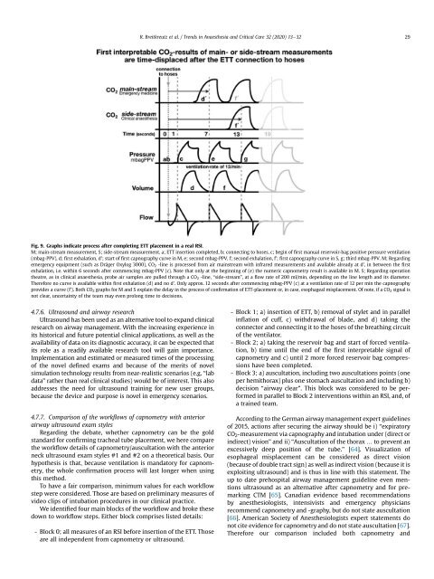
Fig. 9. Graphs indicate process after completing ETT placement in a real RSI.<br />
M; main-stream measurement, S; side-stream measurement. a; ETT insertion completed, b; connecting to hoses, c; begin of first manual reservoir-bag positive pressure ventilation<br />
(mbag-PPV), d; first exhalation, d'; start of first capnography curve in M, e; second mbag-PPV, f; second exhalation, f'; first capnography curve in S, g; third mbag-PPV. M; Regarding<br />
emergency equipment (such as Dr€ager Oxylog 3000), CO 2 -line is processed from air mainstream with infrared measurements <strong>and</strong> available already at d', in between the first<br />
exhalation, i.e. within 6 seconds after commencing mbag-PPV (c). Note that only at the beginning of (e) the numeric capnometry result is available in M. S; Regarding operation<br />
theatre, as in clinical anaesthesia, probe air samples are pulled through a CO 2 -line, “side-stream”, ataflow rate of 200 ml/min, depending on the line length <strong>and</strong> its diameter.<br />
Therefore no curve is available within first exhalation (d) <strong>and</strong> no d'. Only approx. 12 seconds after commencing mbag-PPV (c) at a ventilation rate of 12 per min the capnography<br />
provides a curve (f'). Both CO 2 graphs for M <strong>and</strong> S explain the delay in the process of confirmation of ETT-placement or, in case, esophageal misplacement. Of note, if a CO 2 signal is<br />
not clear, uncertainty of the team may even prolong time to decisions.<br />
4.7.6. <strong>Ultrasound</strong> <strong>and</strong> airway research<br />
<strong>Ultrasound</strong> has been used as an alternative tool to exp<strong>and</strong> clinical<br />
research on airway management. With the increasing experience in<br />
its historical <strong>and</strong> future potential clinical applications, as well as the<br />
availability of data on its diagnostic accuracy, it can be expected that<br />
its role as a readily available research tool will gain importance.<br />
Implementation <strong>and</strong> estimated or measured times of the processing<br />
of the novel defined exams <strong>and</strong> because of the merits of novel<br />
simulation technology results from near-realistic scenarios (e.g. “lab<br />
data” rather than real clinical studies) would be of interest. This also<br />
addresses the need for ultrasound training for new user groups,<br />
because the device <strong>and</strong> purpose is novel in emergency scenarios.<br />
4.7.7. Comparison of the workflows of capnometry with anterior<br />
airway ultrasound exam styles<br />
Regarding the debate, whether capnometry can be the gold<br />
st<strong>and</strong>ard for confirming tracheal tube placement, we here compare<br />
the workflow details of capnometry/auscultation with the anterior<br />
neck ultrasound exam styles #1 <strong>and</strong> #2 on a theoretical basis. Our<br />
hypothesis is that, because ventilation is m<strong>and</strong>atory for capnometry,<br />
the whole confirmation process will last longer when using<br />
this method.<br />
To have a fair comparison, minimum values for each workflow<br />
step were considered. Those are based on preliminary measures of<br />
video clips of intubation procedures in our clinical practice.<br />
We identified four main blocks of the workflow <strong>and</strong> broke these<br />
down to workflow steps. Either block comprises listed details:<br />
- Block 0; all measures of an RSI before insertion of the ETT. Those<br />
are all independent from capnometry or ultrasound.<br />
- Block 1; a) insertion of ETT, b) removal of stylet <strong>and</strong> in parallel<br />
inflation of cuff, c) withdrawal of blade, <strong>and</strong> d) taking the<br />
connector <strong>and</strong> connecting it to the hoses of the breathing circuit<br />
of the ventilator.<br />
- Block 2; a) taking the reservoir bag <strong>and</strong> start of forced ventilation,<br />
b) time until the end of the first interpretable signal of<br />
capnometry <strong>and</strong> c) until 2 more forced reservoir bag compressions<br />
have been completed.<br />
- Block 3; a) auscultation, including two auscultations points (one<br />
per hemithorax) plus one stomach auscultation <strong>and</strong> including b)<br />
decision “airway clear”. This block was considered to be performed<br />
in parallel to Block 2 interventions within an RSI, <strong>and</strong>, of<br />
a trained team.<br />
According to the German airway management expert guidelines<br />
of 2015, actions after securing the airway should be i) “expiratory<br />
CO 2 -measurement via capnography <strong>and</strong> intubation under (direct or<br />
indirect) vision” <strong>and</strong> ii) “Auscultation of the thorax … to prevent an<br />
excessively deep position of the tube.” [64]. Visualization of<br />
esophageal misplacement can be considered as direct vision<br />
(because of double tract sign) as well as indirect vision (because it is<br />
exploiting ultrasound) <strong>and</strong> is thus in line with this statement. The<br />
up to date prehospital airway management guideline even mentions<br />
ultrasound as an alternative after capnometry <strong>and</strong> for premarking<br />
CTM [65]. Canadian evidence based recommendations<br />
by anesthesiologists, intensivists <strong>and</strong> emergency physicians<br />
recommend capnometry <strong>and</strong> -graphy, but do not state auscultation<br />
[66]. American Society of Anesthesiologists expert statements do<br />
not cite evidence for capnometry <strong>and</strong> do not state auscultation [67].<br />
Therefore our comparison included both capnometry <strong>and</strong>