Anterior Neck and Airway Ultrasound - a practical overview.
A narrative review. Video clips on our site as well. It is available online at publisher´s site: https://authors.elsevier.com/a/1b66X7si0yR9sx as editor´s pick free of charges.
A narrative review. Video clips on our site as well. It is available online at publisher´s site: https://authors.elsevier.com/a/1b66X7si0yR9sx as editor´s pick free of charges.

Create successful ePaper yourself
Turn your PDF publications into a flip-book with our unique Google optimized e-Paper software.
R. Breitkreutz et al. / Trends in Anaesthesia <strong>and</strong> Critical Care 32 (2020) 13e32 19<br />
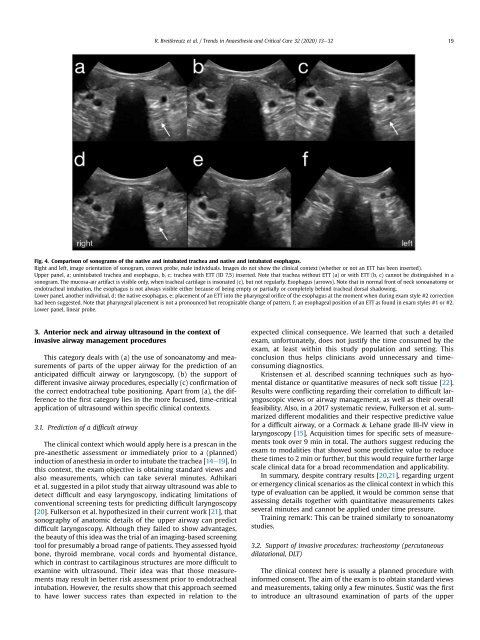
Fig. 4. Comparison of sonograms of the native <strong>and</strong> intubated trachea <strong>and</strong> native <strong>and</strong> intubated esophagus.<br />
Right <strong>and</strong> left, image orientation of sonogram, convex probe, male individuals. Images do not show the clinical context (whether or not an ETT has been inserted).<br />
Upper panel, a; unintubated trachea <strong>and</strong> esophagus, b, c; trachea with ETT (ID 7,5) inserted. Note that trachea without ETT (a) or with ETT (b, c) cannot be distinguished in a<br />
sonogram. The mucosa-air artifact is visible only, when tracheal cartilage is insonated (c), but not regularly. Esophagus (arrows). Note that in normal front of neck sonoanatomy or<br />
endotracheal intubation, the esophagus is not always visible either because of being empty or partially or completely behind tracheal dorsal shadowing.<br />
Lower panel, another individual, d; the native esophagus, e; placement of an ETT into the pharyngeal orifice of the esophagus at the moment when during exam style #2 correction<br />
had been suggested. Note that pharyngeal placement is not a pronounced but recognizable change of pattern, f; an esophageal position of an ETT as found in exam styles #1 or #2.<br />
Lower panel, linear probe.<br />
3. <strong>Anterior</strong> neck <strong>and</strong> airway ultrasound in the context of<br />
invasive airway management procedures<br />
This category deals with (a) the use of sonoanatomy <strong>and</strong> measurements<br />
of parts of the upper airway for the prediction of an<br />
anticipated difficult airway or laryngoscopy, (b) the support of<br />
different invasive airway procedures, especially (c) confirmation of<br />
the correct endotracheal tube positioning. Apart from (a), the difference<br />
to the first category lies in the more focused, time-critical<br />
application of ultrasound within specific clinical contexts.<br />
3.1. Prediction of a difficult airway<br />
The clinical context which would apply here is a prescan in the<br />
pre-anesthetic assessment or immediately prior to a (planned)<br />
induction of anesthesia in order to intubate the trachea [14e19]. In<br />
this context, the exam objective is obtaining st<strong>and</strong>ard views <strong>and</strong><br />
also measurements, which can take several minutes. Adhikari<br />
et al. suggested in a pilot study that airway ultrasound was able to<br />
detect difficult <strong>and</strong> easy laryngoscopy, indicating limitations of<br />
conventional screening tests for predicting difficult laryngoscopy<br />
[20]. Fulkerson et al. hypothesized in their current work [21], that<br />
sonography of anatomic details of the upper airway can predict<br />
difficult laryngoscopy. Although they failed to show advantages,<br />
the beauty of this idea was the trial of an imaging-based screening<br />
tool for presumably a broad range of patients. They assessed hyoid<br />
bone, thyroid membrane, vocal cords <strong>and</strong> hyomental distance,<br />
which in contrast to cartilaginous structures are more difficult to<br />
examine with ultrasound. Their idea was that those measurements<br />
may result in better risk assessment prior to endotracheal<br />
intubation. However, the results show that this approach seemed<br />
to have lower success rates than expected in relation to the<br />
expected clinical consequence. We learned that such a detailed<br />
exam, unfortunately, does not justify the time consumed by the<br />
exam, at least within this study population <strong>and</strong> setting. This<br />
conclusion thus helps clinicians avoid unnecessary <strong>and</strong> timeconsuming<br />
diagnostics.<br />
Kristensen et al. described scanning techniques such as hyomental<br />
distance or quantitative measures of neck soft tissue [22].<br />
Results were conflicting regarding their correlation to difficult laryngoscopic<br />
views or airway management, as well as their overall<br />
feasibility. Also, in a 2017 systematic review, Fulkerson et al. summarized<br />
different modalities <strong>and</strong> their respective predictive value<br />
for a difficult airway, or a Cormack & Lehane grade III-IV view in<br />
laryngoscopy [15]. Acquisition times for specific sets of measurements<br />
took over 9 min in total. The authors suggest reducing the<br />
exam to modalities that showed some predictive value to reduce<br />
these times to 2 min or further, but this would require further large<br />
scale clinical data for a broad recommendation <strong>and</strong> applicability.<br />
In summary, despite contrary results [20,21], regarding urgent<br />
or emergency clinical scenarios as the clinical context in which this<br />
type of evaluation can be applied, it would be common sense that<br />
assessing details together with quantitative measurements takes<br />
several minutes <strong>and</strong> cannot be applied under time pressure.<br />
Training remark: This can be trained similarly to sonoanatomy<br />
studies.<br />
3.2. Support of invasive procedures: tracheostomy (percutaneous<br />
dilatational, DLT)<br />
The clinical context here is usually a planned procedure with<br />
informed consent. The aim of the exam is to obtain st<strong>and</strong>ard views<br />
<strong>and</strong> measurements, taking only a few minutes. Sustic was the first<br />
to introduce an ultrasound examination of parts of the upper