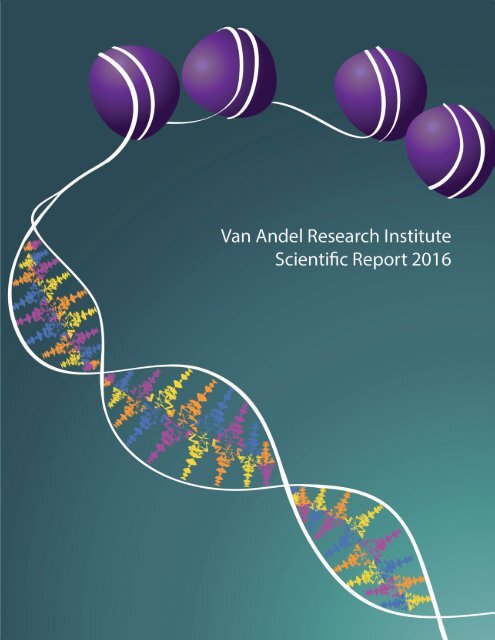
2016 Scientific Report

You also want an ePaper? Increase the reach of your titles
YUMPU automatically turns print PDFs into web optimized ePapers that Google loves.
Published June <strong>2016</strong>.<br />
Cover design by Nicole Ethen.<br />
Copyright <strong>2016</strong> by Van Andel Institute; all rights reserved.<br />
Van Andel Institute, 333 Bostwick Avenue, N.E.<br />
Grand Rapids, Michigan 49503, U.S.A.
VAN ANDEL RESEARCH INSTITUTE is proud to announce that its<br />
Chief <strong>Scientific</strong> Officer, Peter Jones, Ph.D., D.Sc., was elected a member of the National<br />
Academy of Sciences in May <strong>2016</strong>. He joins VARI’s founding Director, George Vande<br />
Woude, Ph.D., who has been a member since 1993.<br />
Dr. Jones has been a long-standing leader in the field of epigenomics. His accomplishments include<br />
• publication of the first study to prove how epigenetics regulates cellular differentiation<br />
• development of DNA methylation inhibitors (DNMTi’s) as drugs<br />
• discovery that epigenetics plays a fundamental role in aging<br />
• elucidation of the biological processes for cellular self-control<br />
• identification of ways to manipulate endogenous retroviruses at the root of some cancers<br />
• co-founding the Stand Up To Cancer (SU2C) Epigenetics Dream Team and the Van Andel Research<br />
Institute–Stand Up To Cancer Epigenetics Dream Team with Stephen Baylin, M.D.<br />
We congratulate Peter on this well-deserved recognition.
ii Van Andel Research Institute | <strong>Scientific</strong> <strong>Report</strong>
Director's Introduction 1<br />
Laboratory <strong>Report</strong>s<br />
Center for Cancer and Cell Biology<br />
Arthur S. Alberts, Ph.D. 6<br />
Patrick J. Grohar, M.D., Ph.D. 7<br />
Brian B. Haab, Ph.D. 9<br />
Yuanzheng (Ajian) He, Ph.D. 11<br />
Xiaohong Li, Ph.D. 12<br />
Jeffrey P. MacKeigan, Ph.D. 14<br />
Karsten Melcher, Ph.D. 16<br />
Lorenzo F. Sempere, Ph.D. 18<br />
Matthew Steensma, M.D. 21<br />
George F. Vande Woude, Ph.D. 22<br />
TABLE OF CONTENTS<br />
Bart O. Williams, Ph.D. 24<br />
Ning Wu, Ph.D. 26<br />
H. Eric Xu, Ph.D. 27<br />
Tao Yang, Ph.D. 29<br />
Center for Epigenetics<br />
Stephen B. Baylin, M.D. 32<br />
Peter A. Jones, Ph.D., D.Sc. 33<br />
Stefan Jovinge, M.D., Ph.D. 34<br />
Peter W. Laird, Ph.D. 36<br />
Gerd Pfeifer, Ph.D 38<br />
Scott Rothbart, Ph.D. 40<br />
Hui Shen, Ph.D. 43<br />
Piroska E. Szabó, Ph.D. 44<br />
Steven J. Triezenberg, Ph.D. 46<br />
iii
Laboratory <strong>Report</strong>s continued<br />
Center for Neurodegenerative Science<br />
Lena Brundin, M.D., Ph.D. 50<br />
Patrik Brundin, M.D., Ph.D. 52<br />
Gerhard A. Coetzee, Ph.D. 54<br />
Viviane Labrie, Ph.D. 55<br />
Jiyan Ma, Ph.D. 57<br />
Darren Moore, Ph.D. 58<br />
Jeremy M. Van Raamsdonk, Ph.D. 60<br />
Core Technologies and Services<br />
Bryn Eagleson, B.S., RLATG<br />
Vivarium and Transgenics Core 64<br />
Scott D. Jewell, Ph.D.<br />
Pathology and Biorepository Core 65<br />
Heather Schumacher, B.S., MT(ASCP)<br />
Flow Cytometry Core 67<br />
Mary E. Winn, Ph.D.<br />
Bioinformatics and Biostatistics Core 68<br />
Confocal Microscopy and Quantitative Imaging Core 69<br />
Small-Animal Imaging Facility 70<br />
IV<br />
Van Andel Research Institute | <strong>Scientific</strong> <strong>Report</strong>
Awards for <strong>Scientific</strong> Achievement<br />
Jay Van Andel Award for Outstanding Achievement 72<br />
in Parkinson’s Disease Research<br />
Han-Mo Koo Memorial Award 73<br />
Educational and Training Programs<br />
Van Andel Institute Graduate School 75<br />
Postdoctoral Fellowship Program 76<br />
Internship Programs 77<br />
VARI and Jay Van Andel Seminar Series 79<br />
Organization<br />
Boards 82<br />
Office of the Chief <strong>Scientific</strong> Officer 83<br />
Administrative Organization 84<br />
VAI Organizational Structure 86<br />
V
PETER A. JONES, Ph.D., D.SC.<br />
CHIEF SCIENTIFIC OFFICER<br />
VAN ANDEL RESEARCH INSTITUTE<br />
Van Andel Research Institute | <strong>Scientific</strong> <strong>Report</strong>
VAN ANDEL RESEARCH INSTITUTE had strong growth and progress in the past year.<br />
Most recently, in February <strong>2016</strong>, Eric Xu was selected by The Protein Society for its<br />
prestigious Hans Neurath Award, which is presented to "individuals who have made a recent<br />
contribution of exceptional merit to basic protein research". The basis for this award was the<br />
July 2015 article in Nature titled “Crystal structure of rhodopsin bound to arrestin determined<br />
by femtosecond X-ray laser”. This project involved many international collaborators and an<br />
intense effort by VARI's Xu and Melcher labs, and it produced a major advance in the field of<br />
G protein–coupled receptors. We congratulate Eric on this well-deserved honor. We at VARI<br />
are pleased at this recognition and proud to be his colleagues and collaborators.<br />
Beyond that award-winning paper, our faculty had excellent publications success in 2015.<br />
Three articles were selected as Notable Advances of 2015 by Nature Medicine: one on<br />
heart cell regeneration coauthored by Stefan Jovinge published in Cell, and two others in<br />
Cell on the DNA methyltransferase inhibitor 5-azacitidine, which stimulates an immune-like<br />
inflammatory response in hindering tumor growth, coauthored by Peter Jones and by<br />
Stephen Baylin.<br />
Peter Laird and Hui Shen were coauthors on a series of papers published in Cell and the<br />
New England Journal of Medicine coming out of work by the Cancer Genome Atlas Network.<br />
Scott Jewell coauthored several papers in Science resulting from his deep involvement in<br />
the Genotype-Tissue Expression (GTEx) project. And, Karsten Melcher and Eric Xu were<br />
coauthors of a second Nature paper on signaling by the plant hormone jasmonate.<br />
We look forward to continuing this strong record of publication in the best scientific journals.<br />
FACULTY<br />
In 2015 the Center for Epigenetics welcomed Scott<br />
Rothbart, who will focus on understanding how histone<br />
post-translational modifications and DNA methylation work<br />
together to orchestrate the dynamic functions associated<br />
with chromatin. The Center was also joined part-time<br />
by Stephen Baylin, who is co-leader of the VARI-SU2C<br />
Epigenetics Dream Team. He will continue his primary<br />
appointment at Johns Hopkins and the Sidney Kimmel<br />
Comprehensive Cancer Center.<br />
Joining the Center for Neurodegenerative Science in 2015<br />
was Gerhard Coetzee. Dr. Coetzee will use his expertise<br />
with GWAS to uncover the roles of genetic risk variants in<br />
Parkinson’s disease. Early in <strong>2016</strong>, Dr. Jeffrey Kordower,<br />
of Rush University Medical Center, began a part-time<br />
appointment at VARI and will continue his collaboration<br />
with Patrik Brundin. Also in early <strong>2016</strong>, Viviane Labrie<br />
arrived at VARI, and she will pursue her studies of the role of<br />
epigenetics in Parkinson’s disease and Alzheimer’s disease.<br />
Patrick Grohar joined the Center for Cancer and Cell<br />
Biology in July 2015. His research and clinical work is on<br />
Ewing sarcoma, a type of tumor that can occur in bone or<br />
soft tissue.<br />
1
EVENTS AND AWARDS<br />
FUNDING<br />
Research!America, the nation’s largest nonprofit public<br />
education and advocacy alliance, which works to make<br />
health research a higher national priority, named David Van<br />
Andel and George Vande Woude its 2015 Advocacy Award<br />
winners. The annual Research!America Advocacy Awards<br />
program was established in 1996 to honor outstanding<br />
advocates for medical, health, and scientific research.<br />
Congratulations to both for a well-deserved recognition of<br />
their years-long efforts.<br />
Dr. Matt Steensma was one of two recipients of<br />
the inaugural Francis S. Collins Scholars Award in<br />
Neurofibromatosis Clinical and Translational Research.<br />
The award was presented by Dr. Collins at VARI's NF1<br />
Mini-Symposium in April 2015.<br />
In May, Eric S. Lander, founding director of the Broad<br />
Institute of MIT and Harvard University, was honored with<br />
the Han-Mo Koo Award. He delivered both a scientific<br />
seminar and a lay lecture in accepting the award for his<br />
outstanding scientific achievements in genomics and the<br />
Human Genome Project.<br />
The Jay Van Andel Award for Outstanding Achievement<br />
in Parkinson’s Disease Research was presented at the<br />
September Grand Challenges in Parkinson's Disease<br />
symposium held at VARI. The awardees were Maria Grazia<br />
Spillantini, FMedSci, FRS, of the University of Cambridge,<br />
and Robert Nussbaum, M.D., of the University of California,<br />
San Francisco. In 1997, the two made related discoveries<br />
that linked Parkinson's disease to the α-synuclein gene<br />
and its protein, which have since been the focus of major<br />
research efforts.<br />
Also in 2015, VARI hosted the Michigan C. elegans<br />
meeting (April); a joint USA/Netherlands biomedical<br />
symposium, followed by a visit to VARI by His Majesty<br />
King Willem-Alexander and Her Majesty Queen Máxima of<br />
the Netherlands (June); the Origins of Cancer Symposium<br />
"Beyond the Genome: The Role of Posttranslational<br />
Modifications in Cancer" (July); and the International<br />
Society for Tryptophan Research Conference (September).<br />
Scott Jewell received a major multiyear grant from the<br />
NIH's National Cancer Institute to support operations of<br />
the VARI biorepository in serving as the Biospecimen Core<br />
Resource for the NCI's Clinical Proteomic Tumor Analysis<br />
Consortium. VARI also received part of a collaborative<br />
NSF grant that will provide us with advanced networking<br />
hardware to improve data storage and sharing.<br />
Other 2015 funding awards to our researchers included<br />
the following:<br />
• An NCI R01 award to Jeffrey MacKeigan for<br />
"Computational Model of Autophagy-Mediated<br />
Survival in Chemoresistant Lung Cancer".<br />
• An R01 award to Darren Moore for "Novel Mechanisms<br />
of LRRK2-Dependent Neurodegeneration in<br />
Parkinson's Disease". He also signed a new<br />
agreement with a pharmaceutical firm.<br />
• A Michigan Economic Development Corporation award<br />
to Peter Jones to support two new epigenetics faculty<br />
members and their research.<br />
• An NCI K99/R00 grant to Scott Rothbart for<br />
"Mechanisms Regulating DNA Methylation<br />
Maintenance in Chromatin".<br />
• An R21 award to Bart Williams for "Generation and<br />
Initial Characterization of Osteocalcin-Deficient Rats".<br />
• A Cure Parkinson's Trust award to Patrik Brundin<br />
for "Preclinical Evaluation of Deuterium-Reinforced<br />
Polyunsaturated Fatty Acids as a Therapeutic<br />
Intervention for Parkinson's Disease".<br />
We continue working in all areas toward even more success<br />
in future years.<br />
2<br />
Van Andel Research Institute | <strong>Scientific</strong> <strong>Report</strong>
3
4
CENTER FOR<br />
CANCER AND CELL BIOLOGY<br />
Bart O. Williams, Ph.D.<br />
Director<br />
The Center’s scientists study the basic<br />
mechanisms and molecular biology of cancer and<br />
other diseases, with the goal of developing better<br />
diagnostics and therapies.<br />
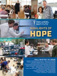
A depiction of arrestin binding by a phosphorylated and active rhodopsin.<br />
The cell membrane lipids are shown as off-white, rhodopsin is blue, arrestin is red, and<br />
phosphorus molecules are orange. The phosphorylated C-terminal tail of rhodopsin<br />
binds to the N-domain (left) of the arrestin molecule. In the main contact region between<br />
the two molecules (central), arrestin accommodates the ICL2 helix of rhodopsin. In this<br />
fully activated state, the tip of arrestin’s C-domain contacts the membrane (right).<br />
(Model by Parker de Waal of the Xu lab)<br />
5
ARTHUR S. ALBERTS, PH.D.<br />
Dr. Alberts earned his degrees in biochemistry and cell biology<br />
(B.A., 1987) and in physiology and pharmacology (Ph.D., 1993)<br />
from the University of California, San Diego. He joined VARI in<br />
January 2000, and he was promoted to Professor in 2009.<br />
STAFF<br />
SARAH VANOEVEREN, B.S., B.S.<br />
STUDENT<br />
ANDREW HOWARD, B.A.<br />
VISITING SCIENTIST<br />
JULIE TURNER, PH.D.<br />
RESEARCH INTERESTS<br />
Our lab seeks to gain a full understanding of how cells spatially and temporally organize<br />
the biochemical circuits that govern responses to injury, infection, and age. Our goal<br />
is to use this information to guide the development of pharmacological agents that<br />
block the acquisition of cancer traits. In 2015, we focused our translational research<br />
on targeted therapies that reinforce and/or repair blood cell structure and function and<br />
otherwise impair the ability of cancer cells to metastasize.<br />
RECENT PUBLICATIONS<br />
Vargas, Pablo, Paolo Maiuri, Marine Bretou, Pablo J. Sáez, Paolo Pierobon, Mathieu Maurin, Mélanie Chabaud, Danielle Lanakar,<br />
Dorian Obino, et al. <strong>2016</strong>. Innate control of actin nucleation determines two distinct migration behaviours in dendritic cells.<br />
Nature Cell Biology 18(1): 43–53.<br />
Arden, Jessica D., Kari I. Lavik, Kaitlin A. Rubinic, Nicolas Chiaia, Sadik A. Khuder, Marthe J. Howard, Andrea L. Nestor-Kalinoski,<br />
Arthur S. Alberts, and Kathryn M. Eisenmann. 2015. Small molecule agonists of mammalian Diaphanous-related (mDia) formins<br />
reveal an effective glioblastoma anti-invasion strategy. Molecular Biology of the Cell 26(21): 3704–3718.<br />
Ercan-Sencicek, A. Gulhan, Samira Jambi, Daniel Franjic, Sayoko Nishimura, Mingfeng Li, Paul El-Fishawy, Thomas M. Morgan,<br />
Stephan J. Sanders, Kaya Bilguvar, Mohnish Suri, et al. 2015. Homozygous loss of DIAPH1 is a novel cause of microcephaly in<br />
humans. European Journal of Human Genetics 23(2): 165–172.<br />
6<br />
Van Andel Research Institute | <strong>Scientific</strong> <strong>Report</strong>
PATRICK J. GROHAR, M.D., PH.D.<br />
Dr. Grohar earned his Ph.D. in chemistry and his M.D. from Wayne<br />
State University. He joined VARI in 2015 as an Associate Professor,<br />
and he has clinical and research responsibilities at Spectrum Health<br />
and Michigan State University, respectively.<br />
STAFF<br />
MATT EASTON<br />
SUSAN GOOSEN, B.S., M.B.A.<br />
MATT HARLOW, M.S.<br />
DIANA LEWIS, A.S.<br />
RESEARCH INTERESTS<br />
Our laboratory studies pediatric sarcomas, and our goal is to develop novel, molecularly<br />
targeted therapies and to translate those therapies into the clinic. Most pediatric<br />
sarcomas are characterized by oncogenic transcription factors formed by chromosomal<br />
translocations. In many cases, the tumors depend on the continued expression and<br />
activity of those transcription factors for cell survival, but few therapies that directly<br />
target specific factors have achieved clinical efficacy. Therefore, we are developing new<br />
approaches to target those transcription factors.<br />
To date, we have focused on targeting the EWS-FLI1 transcription factor in Ewing<br />
sarcoma. EWS-FLI1 is an oncogenic transcription factor formed by the t(11;22)(q24;12)<br />
chromosomal translocation that leads to the fusion of the EWSR1 and FLI1 genes. The<br />
result is a dysregulated transcription factor that alters the expression of over 500 genes<br />
and drives tumorigenesis and progression. Several independent studies have shown<br />
that silencing of EWS-FLI1 is incompatible with Ewing sarcoma cell survival. By directly<br />
targeting EWS-FLI1, we hope to eliminate its activity as the dominant oncogene in this<br />
tumor and thus improve patient survival.<br />
Trabectedin (ET-743; ecteinascidin 743; Yondelis) is a natural product originally isolated<br />
from the sea squirt, Ectenascidia turbinata. We became interested in this compound<br />
because early clinical studies suggested that translocation-positive sarcomas were<br />
sensitive to it. We subsequently demonstrated that trabectedin blocks EWS-FLI1 activity<br />
at the promoter, mRNA, and protein levels of expression. In addition, we demonstrated<br />
on a genome-wide scale that it reverses the expression of the gene signature of EWS-<br />
FLI1. However, the compound failed in a phase II study on Ewing sarcoma.<br />
CENTER FOR CANCER AND CELL BIOLOGY<br />
7
Subsequently, our work has focused on characterizing the<br />
mechanism of EWS-FLI1 suppression with the goals of<br />
understanding the failure in the phase II study, identifying<br />
second-generation trabectedin analogs, and developing<br />
new mechanism-based combination therapies. We have<br />
developed a novel combination therapy of trabectedin<br />
plus irinotecan that is synergistic. We have shown that<br />
this combination markedly improves the suppression of<br />
EWS-FLI1 and substantially increases the DNA damage<br />
in Ewing sarcoma cells. We translated this therapy into<br />
the clinic in Europe and found it was active in a patient in<br />
Italy and in a series of patients in Germany (manuscript<br />
in preparation). Since the drug is now approved in the<br />
United States, we are writing a phase II protocol for this<br />
combination therapy for the Children’s Oncology Group,<br />
which will open nationwide for patients with relapsed<br />
Ewing sarcoma.<br />
Over the past year, we have characterized the mechanism<br />
of EWS-FLI1 suppression by trabectedin, and we have<br />
shown that mechanism is not effective at the serum<br />
concentrations achieved in the failed phase II study,<br />
explaining the lack of activity. More importantly, we have<br />
identified a second-generation compound with an improved<br />
pharmacokinetic profile that will make successful EWS-FLI1<br />
suppression more likely, and we are working to translate<br />
this compound to the clinic.<br />
We have also extensively studied mithramycin, which<br />
reverses EWS-FLI1 activity and blocks the expression of<br />
key downstream targets. In a phase I/II trial at the National<br />
Cancer Institute, we found that mithramycin did not achieve<br />
serum levels high enough to block EWS-FLI1 activity. Over<br />
the past year, our work has identified two compounds with<br />
an improved clinical profile, one that is more potent and<br />
another that is less toxic than the parent compound. Both<br />
compounds reverse EWS-FLI1 activity and are extremely<br />
active in xenograft models of Ewing sarcoma. Work<br />
continues to understand the mechanism of EWS-FLI1<br />
suppression for this class of compounds.<br />
We are also taking a broader look at transcription as a<br />
Ewing sarcoma drug target, using an siRNA screening<br />
platform. We have identified a therapeutic vulnerability<br />
based on alternative mRNA splicing, and we are developing<br />
companion biomarkers that will accompany our trials and<br />
aid in the clinical translation of our EWS-FLI1-directed<br />
therapies. We have also identified a commonly employed<br />
positron emission tomography (PET) radiotracer that<br />
reflects EWS-FLI1 activity in Ewing sarcoma cells, which will<br />
allow more precise dosing of our therapies and the direct<br />
correlation of EWS-FLI1 activity to PET activity. Finally,<br />
we are beginning to expand our studies to other pediatric<br />
tumors characterized by oncogenic fusion transcription<br />
factors.<br />
RECENT PUBLICATIONS<br />
Caropreso, Vittorio, Emad Darvishi, Thomas J. Turbyville, Ranjala Ratnayake, Patrick J. Grohar, James B. MacMahon, and Girma<br />
Woldenmichael. In press. Englerin A inhibits EWS-FLI1 DNA binding in Ewing’s sarcoma cells. Journal of Biological Chemistry.<br />
Osgood, Christy L., Nichole Maloney, Christopher G. Kidd, Susan Kitchen-Goosen, Laura Segars, Meti Gebregiorgis, Girma M.<br />
Woldemichael, Min He, Savita Sankar, et al. In press. Identification of mithramycin analogs with improved targeting of the EWS-<br />
FLI1 transcription factor. Clinical Cancer Research.<br />
Kovar, Heinrich, James Amatruda, Erika Brunet, Stefan Burdach, Florencia Cidre-Aranaz, Enrique de Alava, Uta Dirksen, Wietske<br />
van der Ent, Patrick Grohar, et al. <strong>2016</strong>. The second European interdisciplinary Ewing sarcoma research summit — a joint effort<br />
to deconstructing the multiple layers of a complex disease. Oncotarget 7(8): 8613–8624.<br />
8<br />
Van Andel Research Institute | <strong>Scientific</strong> <strong>Report</strong>
BRIAN B. HAAB, PH.D.<br />
Dr. Haab obtained his Ph.D. in chemistry from the University of<br />
California at Berkeley in 1998. He joined VARI as a Special Program<br />
Investigator in 2000, became a <strong>Scientific</strong> Investigator in 2004, and is<br />
now a Professor.<br />
STAFF<br />
STEPHANIE GRANT, M.P.A.<br />
KATIE PARTYKA, B.S.<br />
BRYAN REATINI, B.S.<br />
SUDHIR SINGH, PH.D.<br />
JESSICA SINHA, M.S.<br />
HUIYUAN TANG, PH.D.<br />
RESEARCH INTERESTS<br />
The promise of molecular biomarkers: improving patient outcomes through better<br />
detection and subtyping.<br />
Tests to detect and diagnose pancreatic cancer<br />
STUDENTS<br />
DANIEL BARNETT, B.A., B.S.<br />
LELAND DUNWOODIE<br />
ELLIOT ENSINK<br />
PETER HSUEH, B.S.<br />
JOEY KRETOWICZ<br />
GARIMA VORHA, B.SC., M.B.A.<br />
The successful treatment of pancreatic cancer critically depends on achieving an<br />
accurate and early diagnosis, but this can be frustratingly difficult. Conventional<br />
methods of evaluating patients—assessing scans, visual inspection of cells from a<br />
biopsy, and weighing behavioral, health, and demographic data—do not have the detail<br />
necessary to distinguish between benign and malignant disease or between cancers<br />
with vastly different behaviors. Sometimes a physician can see a mass or other unusual<br />
feature in the pancreas but is unsure what it is. Is it benign or cancerous? And if it is<br />
cancer, what is the best course of treatment?<br />
Our research builds on the concept that molecular-level information will provide<br />
details about a condition that are not observable by conventional methods. Molecular<br />
biomarkers could provide such information and enable physicians to make accurate<br />
diagnoses and develop optimal treatment plans. We are making progress toward this<br />
goal for pancreatic cancer. For example, in recent publications in Molecular and Cellular<br />
Proteomics and the Journal of Proteome Research, we disclosed carbohydrate-based<br />
biomarkers in the blood serum that improve upon the widely used blood test called<br />
CA19-9. By using a panel of three or more independent biomarkers, we detected a<br />
greater percentage of cancers than we could with any individual biomarker. We are<br />
seeking to substantiate those findings and to evaluate their clinical value using serum<br />
samples from several clinical sites.<br />
CENTER FOR CANCER AND CELL BIOLOGY<br />
9
Other research is aimed at further improving the biomarker<br />
tests. The results so far suggest that each individual<br />
biomarker arises from a distinct subpopulation of cancer<br />
patients and from a characteristic cell type. This finding is<br />
important because the biomarkers may reveal differences<br />
between subgroups of tumors—a possibility we are<br />
exploring in the research described below. For the purpose<br />
of improving our blood tests, determining the characteristics<br />
of the cells that produce each biomarker, as well as of the<br />
cells that do not produce any of our biomarkers, will help to<br />
optimize a blood test to accurately identify cancers across<br />
the entire spectrum of patients.<br />
The ultimate goal is to get the new tests established in<br />
clinical laboratories in order to benefit patients. To that<br />
end, we are working with industry partners to transfer our<br />
biomarker assays to the clinical laboratory setting and to<br />
begin analyzing patient samples received consecutively<br />
from clinical sites. If we have good results, we hope to<br />
initiate clinical trials for the diagnosis of pancreatic cancer<br />
and, eventually, for evaluations of surveillance among<br />
people at elevated risk for pancreatic cancer.<br />
Better treatment through subtyping<br />
Pancreatic cancer characteristics, such as the cell types<br />
within the tumor, the amount of metastasis, the responses<br />
to treatments, and overall outcomes, vary greatly among<br />
patients. So far, identifying the underlying causes of such<br />
differences and predicting the behavior of individual tumors<br />
have not been possible. If we could determine what drives<br />
the differences between the tumors or identify molecules<br />
that help predict the behavior of each tumor, we could<br />
establish better treatment plans for each patient or<br />
determine the drugs that work best against each subtype.<br />
Our research is revealing major groupings of tumors<br />
based on the carbohydrates on the surface of, and in<br />
the secretions from, cancer cells. The carbohydrates are<br />
related to the CA19-9 antigen and have distinct biological<br />
functions. In current research we want to determine the<br />
molecular nature of the subgroups of cells and whether<br />
the subgroups have different levels of aggressiveness or<br />
different responses to particular drugs. We are using new<br />
approaches for measuring carbohydrates and proteins<br />
in tumor tissue, and we are employing powerful new<br />
software—introduced in our recent publication in Analytical<br />
Chemistry—to examine the cell types that produce<br />
each carbohydrate-based biomarker. We are using that<br />
information to evaluate whether certain types of cells<br />
predict clinical behavior. As advances and new options<br />
in treatments become available, this type of research is<br />
increasingly important for guiding clinical decisions. We<br />
are working closely with our physician collaborators to<br />
evaluate on a case-by-case basis the value of the molecular<br />
information and to guide our research toward improving the<br />
tests. Ultimately, physicians could use the molecular tests<br />
on material from biopsies, surgical resections, or blood<br />
samples.<br />
RECENT PUBLICATIONS<br />
Ensink, Elliot, Jessica Sinha, Arkadeep Sinha, Huiyuan Tang, Heather M. Calderone, Galen Hostetter, Jordan Winter, David<br />
Cherba, Randall E. Brand, et al. 2015. Segment and fit thresholding: a new method for image analysis applied to microarray and<br />
immunofluorescence data. Analytical Chemistry 87(19): 9715–9721.<br />
Singh, Sudhir, Kuntal Pal, Jessica Yadav, Huiyuan Tang, Katie Partyka, Doron Kletter, Peter Hsueh, Elliot Ensink, Birendra KC, et<br />
al. 2015. Upregulation of glycans containing 3' fucose in a subset of pancreatic cancers uncovered using fusion-tagged lectins.<br />
Journal of Proteome Research 14(6): 2594–2605.<br />
Tang, Huiyuan, Sudhir Singh, Katie Partyka, Doron Kletter, Peter Hsueh, Jessica Yadav, Elliot Ensink, Marshall Bern, Galen<br />
Hostetter, et al. 2015. Glycan motif profiling reveals plasma sialyl-Lewis X elevations in pancreatic cancers that are negative for<br />
CA19-9. Molecular & Cellular Proteomics 14(5): 1323–1333.<br />
10<br />
Van Andel Research Institute | <strong>Scientific</strong> <strong>Report</strong>
YUANZHENG (AJIAN) HE, PH.D.<br />
Dr. He earned his Ph.D. from the Chinese Academy of Sciences’<br />
Shanghai Institute of Biochemistry in 2000. In 2008, he was<br />
recruited to Van Andel Research Institute, where he is currently<br />
a Research Assistant Professor.<br />
RESEARCH INTERESTS<br />
Ligand binding is the key event that triggers intracellular signal transduction cascades,<br />
and it is also a major focus of drug discovery. My research involves the structural basis<br />
of ligand/receptor interactions and related drug discovery, focusing on steroid hormone<br />
receptors, specifically, the glucocorticoid receptor and the G protein–coupled receptors<br />
(GPCRs). My overall goal is to explore structural insights into receptor signaling and<br />
use them to design precision drugs that specifically deliver the desired treatment effect,<br />
but not unwanted side effects, to patients. Over the past year, we have made the<br />
following progress.<br />
• We have developed “dissociated glucocorticoid” molecules based on our<br />
finding that the dissociation of transrepression from transactivation can be<br />
achieved by interfering with the dimerization interface of the glucocorticoid<br />
receptor.<br />
• We have developed an exceptionally potent glucocorticoid for asthma<br />
treatment based on our uncovering of the structural key to glucocorticoid<br />
potency. Our primary compound outperforms the current leading drug in a<br />
mouse asthma model and promises a better side-effects profile.<br />
• We have determined the structure of arrestin-bound rhodopsin, which provides<br />
a basis for understanding GPCR-mediated arrestin-biased signaling.<br />
RECENT PUBLICATIONS<br />
Kang, Yanyong, Xiang Gao, X. Edward Zhou, Yuanzheng He, Karsten Melcher, and H. Eric Xu. <strong>2016</strong>. A structural snapshot of the<br />
rhodopsin–arrestin complex. FEBS Journal 283(5): 816–821.<br />
He, Yuanzheng, Jingjing Shi, Wei Yi, Xin Ren, Xiang Gao, Jianshuang Li, Nanyan Wu, Kevin Weaver, Qian Xie, et al. 2015.<br />
Discovery of a highly potent glucocorticoid for asthma treatment. Cell Discovery 1: 15035.<br />
Zhi, Xiaoyong, X. Edward Zhou, Yuanzheng He, Kelvin Searose-Xu, Chun-Li Zhang, Chih-Cheng Tasi, Karsten Melcher, and H.<br />
Eric Xu. 2015. Structural basis for corepressor assembly by the orphan nuclear receptor TLX. Genes and Development 29(4):<br />
440–450.<br />
CENTER FOR CANCER AND CELL BIOLOGY<br />
11
XIAOHONG LI, PH.D.<br />
Dr. Li received her Ph.D. from the Institute of Zoology, Chinese<br />
Academy of Sciences, in Beijing in 2001. She joined VARI as an<br />
Assistant Professor in September 2012.<br />
STAFF<br />
PAUL G. DAFT, PH.D.<br />
SOURIK GANGULY, PH.D.<br />
DIANA LEWIS, A.S.<br />
NEIL (XIANGQI) MENG, PH.D.<br />
ALEXANDRA VANDER ARK, M.S.<br />
JIE WANG, M.D.<br />
QI ZENG, M.D.<br />
RESEARCH INTERESTS<br />
Our laboratory is committed to understanding tumor dormancy and cancer bone<br />
metastases, specifically of breast, lung, and prostate cancers. Our long-term goal is to<br />
create a dormancy-permissive bone microenvironment so that cancer cells can be kept<br />
dormant or be killed while they are in that state.<br />
Project 1. Cell-specific roles of transforming growth factor (TGF)-β in bone metastases.<br />
STUDENTS<br />
AUSTIN M. MEADOWS<br />
GHADA Y.T. MOHSEN<br />
ERICA WOODFORD<br />
Most people who die of cancer have metastases somewhere in their body, but<br />
metastases of certain cancers, particularly of the breast, lung, or prostate, are more<br />
likely to be found in bone. Cancer cells in bone induce either osteolytic (bone<br />
resorption) or osteoblastic (abnormal bone formation) lesions, which can cause<br />
fractures, spinal cord compression, hypercalcemia, and extreme bone pain. Current<br />
treatments for bone-metastasis patients can reduce symptoms such as pain but do not<br />
increase survival. Better understanding of the mechanism of bone metastasis is needed<br />
in order to develop early diagnostic tests and targeted therapeutic strategies. The local<br />
events of bone lesion development are determined by the interactions of cancer cells<br />
with bone cells such as osteoblasts (mesenchymal lineage) and osteoclasts (myeloid<br />
lineage), and such events are regulated by growth factors and cytokines of the bone<br />
matrix. The cytokine TGF-β plays crucial roles in both cancerous and healthy bone, and<br />
its effects are highly context-dependent, spatially and temporally. We aim to delineate<br />
the cell-specific role of TGF-β in bone metastasis and identify downstream mediators<br />
that can be targeted by new therapies.<br />
12 Van Andel Research Institute | <strong>Scientific</strong> <strong>Report</strong>
Our studies have produced the following results.<br />
• Basic fibroblast growth factor (bFGF), mediated<br />
by TGF-β signaling in cells of the myeloid lineage,<br />
promotes breast cancer bone metastasis. By<br />
blocking bFGF, we can reduce such bone lesion<br />
development. In bone metastatic tissues from<br />
breast cancer patients, TGF-β and bFGF signaling<br />
are likely to be activated in osteoclasts and cancer<br />
cells but inactivated in osteoblasts.<br />
• TGF-β signaling in myeloid lineage cells promotes<br />
bone metastasis, but in cells of the mesenchymal<br />
lineage, the same signaling inhibits bone<br />
metastasis. We have found that bFGF is the<br />
functional mediator for TGF-β signaling effects only<br />
in cells of the myeloid lineage.<br />
Project 2. TGF-β signaling in the bone microenvironment<br />
affects tumor dormancy.<br />
Up to 70% of cancer patients have tumor cells in the<br />
bone marrow at the time of initial diagnosis. It is not<br />
known how cancer cells in bone remain dormant and later<br />
reactivate. Understanding tumor dormancy is important<br />
in trying to prevent the metastatic recurrences that kill<br />
patients. Studies have shown that external cues from<br />
the bone microenvironment can determine tumor cell<br />
dormancy. We aim to create a dormancy-permissive bone<br />
microenvironment and determine the mechanism by which<br />
it supports cancer cell dormancy. We have established a<br />
system in which loss of TGF-β signaling in myeloid lineage<br />
cells may promote the dormancy of prostate cancer or<br />
NSCLC in the bone marrow. We are now studying the<br />
underlying mechanism.<br />
• The cell-specific roles of TGF-β signaling are more<br />
complex for bone metastasis of non-small-cell<br />
lung cancer (NSCLC). The effects are dependent<br />
on the types of bone lesions that are produced by<br />
different NSCLC tumors.<br />
RECENT PUBLICATIONS<br />
Meng, X., A. Vander Ark, P. Lee, G. Hostetter, N.A. Bhowmick, L.M. Matrisian, B.O. Williams, C.K. Miranti, and X. Li. <strong>2016</strong>.<br />
Myeloid-specific TGF-β signaling in bone promotes basic-FGF and breast cancer bone metastasis. Oncogene 35(18): 2370-2378.<br />
CENTER FOR CANCER AND CELL BIOLOGY<br />
13
JEFFREY P. MACKEIGAN, PH.D.<br />
Dr. MacKeigan received his Ph.D. in microbiology and immunology at<br />
the University of North Carolina Lineberger Comprehensive Cancer<br />
Center in 2002. Dr. MacKeigan joined VARI in 2006 as an Assistant<br />
Professor and was promoted to Associate Professor in 2010.<br />
STAFF<br />
STEPHANIE CELANO, M.S.<br />
LUCUS CHAN, PH.D.<br />
KRISTIN DITTENHAFER-REED, PH.D.<br />
NICOLE DOPPEL, B.S.<br />
MATT KORTUS, M.S.<br />
KATIE MARTIN, PH.D.<br />
JOSH SCHIPPER, PH.D.<br />
KELLIE SISSON, B.S.<br />
STUDENTS<br />
ADITI BAGCHI, M.D.<br />
ANNALISE BOWEN<br />
DANIELLE BURGENSKE, PH.D.<br />
LELAND DUNWOODIE<br />
NATE MERRILL, B.S.<br />
NANDA KUMAR SASI, B.S.<br />
ABIGAIL SOLITRO, B.S.<br />
MEGAN VANBAREN<br />
RESEARCH INTERESTS<br />
The MacKeigan lab focuses on two hallmarks of cancer: the deregulation of cellular<br />
energetics and resistance to cell death. These hallmarks are regulated by mTOR<br />
signaling and contribute significantly to drug resistance in cancer. We seek a<br />
systems-level understanding of the network that encompasses the cell metabolism and<br />
autophagy signaling pathways. While our research focuses on human cancers, we also<br />
apply our tumor biology expertise and pathway knowledge to study tuberous sclerosis<br />
complex. Our laboratory uses cutting-edge tools and collaborates with multidisciplinary<br />
experts for robust experimental design and comprehensive data analysis. All of our<br />
research projects have one common goal: to identify novel therapeutic targets.<br />
Autophagy and resistance to cell death<br />
The process of autophagy functions to generate energy, clear damaged organelles,<br />
and delay or prevent cell death during times of cellular stress. Chemotherapeutic<br />
agents trigger autophagy, which allows cancer cells to adapt and withstand treatment.<br />
Therefore, a better understanding of autophagy is crucial for developing new and<br />
improved treatment strategies against cancer.<br />
ADJUNCT FACULTY<br />
BRIAN LANE, M.D., PH.D.<br />
In partnership with Los Alamos National Laboratory, our lab has used predictive<br />
computational modeling and cell-based measurements to accurately model the<br />
autophagic process. We are pleased to report that we have received a collaborative<br />
National Cancer Institute R01 award to validate and extend this model. The current<br />
efforts to enhance our model will help us predict the therapeutic benefit of inhibiting<br />
autophagy in cancer. We are also working with industry partners to determine the<br />
effects of candidate drugs on autophagic flux, and we have identified novel genes<br />
that are required for drug-induced autophagy. Lastly, our group conducts optimized<br />
kinase and phosphatase assays for in vitro evaluation of compounds identified in silico.<br />
Our research suggests that kinase inhibitors modulate autophagy and may be more<br />
selective and effective than current lysosomotropic agents.<br />
14 Van Andel Research Institute | <strong>Scientific</strong> <strong>Report</strong>
Cancer metabolism and dysregulated cellular<br />
energetics<br />
Aggressive cancers are well known for their altered<br />
metabolic profiles and ability to withstand cytotoxic<br />
therapies. Thus, defining the relationship between<br />
dysregulated metabolism and evasion of apoptosis<br />
represents a critical need in the cancer field.<br />
Our research has shown that increased glycolysis in cancer<br />
cells leads to significant enrichment of the mitochondrial<br />
lipid cardiolipin, which serves many important functions in<br />
maintaining mitochondrial health. Most intriguing is its role<br />
in preventing the release of cytochrome c, a key event in the<br />
initiation of apoptosis. Our results suggest that the altered<br />
metabolic program of cancer cells may inherently support<br />
the evasion of apoptosis through cardiolipin production.<br />
We are investigating whether increased cardiolipin allows<br />
cancer cells to avoid death and resist chemotherapy. We<br />
have partnered with experts in glioblastoma multiforme and<br />
lipid mass spectrometry to uncover the mechanisms that<br />
may underlie cardiolipin’s ability to promote cell survival. A<br />
more complete understanding of the synthesis of cardiolipin<br />
and how changes in its concentration regulate cytochrome<br />
c release will contribute toward new mitochondria-targeted<br />
therapeutics for chemoresistant cancers.<br />
Pathway of Hope<br />
Tuberous sclerosis complex (TSC) is a genetic disease<br />
resulting from mutations in the TSC1 and TSC2 genes.<br />
These mutations inactivate the genes’ tumor-suppressive<br />
function, driving tumor cell growth and causing<br />
noncancerous tumors in vital organs such as the brain, skin,<br />
eyes, lung, and heart. These tumors can cause a host of<br />
health issues, including epilepsy and autism.<br />
Using chemical screening techniques, we are identifying<br />
approved, targeted compounds as possible therapies for<br />
TSC. Our lab is also characterizing the genomic landscape<br />
of TSC tumors using next-generation sequencing. We<br />
have gained a comprehensive understanding of TSC tumor<br />
biology, and we are seeking other cellular changes that<br />
can be targeted by therapies. TSC tumors are not always<br />
associated with second-hit somatic mutations to TSC1<br />
or TSC2, suggesting that their pathogenesis may involve<br />
other genetic events, which we are working to uncover. We<br />
are also developing preclinical models of TSC for future<br />
validation studies of our drug candidates and genomic<br />
findings. Lastly, we have partnered with physician-scientists<br />
expert in TSC to determine whether precision medicine<br />
approaches can inform treatment strategies for TSC and<br />
predict patient outcomes.<br />
RECENT PUBLICATIONS<br />
Solitro, Abigail R., and Jeffrey P. MacKeigan. <strong>2016</strong>. Leaving the lysosome behind: novel developments in autophagy inhibition.<br />
Future Medicinal Chemistry 8(1): 73–86.<br />
MacKeigan, Jeffrey P., and Darcy A. Krueger. 2015. Differentiating the mTOR inhibitors everolimus and sirolimus in the treatment<br />
of tuberous sclerosis complex. Neuro-Oncology 17(12): 1550–1559.<br />
Szymańska, Paulina, Katie R. Martin, Jeffrey P. MacKeigan, William S. Hlavacek, and Tomasz Lipniacki. 2015. Computational<br />
analysis of an autophagy/translation switch based on mutual inhibition of MTORC1 and ULK1. PLoS One 10(3): e0116550.<br />
Wang, Tong, Megan L. Goodall, Paul Gonzales, Mario Sepulveda, Katie R. Martin, Stephen Gately, and Jeffrey P. MacKeigan.<br />
2015. Synthesis of improved lysomotropic autophagy inhibitors. Journal of Medicinal Chemistry 58(7): 3025–3035.<br />
CENTER FOR CANCER AND CELL BIOLOGY 15
KARSTEN MELCHER, PH.D.<br />
Dr. Melcher earned his Master’s degree in biology and his Ph.D. degree<br />
in biochemistry from the Eberhard Karls Universität in Tübingen,<br />
Germany. He was recruited to VARI in 2007, and in 2013 he was<br />
promoted to Associate Professor.<br />
STAFF<br />
STEPHANIE GRANT, M.P.A.<br />
XIN GU, M.S.<br />
JIYUAN KE, PH.D.<br />
AMANDA KOVACH, B.S.<br />
EDWARD ZHOU, PH.D.<br />
RESEARCH INTERESTS<br />
Our laboratory studies the structure and function of proteins that have central roles<br />
in cellular signaling. To do so, we employ X-ray crystallography in combination with<br />
biochemical and cellular methods to identify structural mechanisms of signaling at high<br />
resolution.<br />
STUDENT<br />
CHRISTIAN CAVACECE<br />
In addition to their fundamental physiological roles, most signaling proteins are also<br />
important targets of therapeutic drugs. Determination of the three-dimensional<br />
structures of protein–drug complexes at atomic resolution allows a detailed<br />
understanding of how a drug binds its target and modifies its activity. This knowledge<br />
allows the rational design of new and better drugs against diseases such as cancer,<br />
diabetes, and neurological disorders.<br />
Three areas of focus in the lab are the adenosine monophosphate (AMP)–activated<br />
protein kinase (AMPK); the receptors and key signaling proteins for a plant hormone,<br />
abscisic acid (ABA); and the folate receptors.<br />
AMP-activated protein kinase (AMPK)<br />
Cells use ATP to drive cellular processes such as muscle contraction, cell growth, and<br />
neuronal excitation. AMPK is a three-subunit protein kinase that functions as an energy<br />
sensor and regulator of homeostasis in human cells. Its kinase activity, triggered by<br />
energy stress (i.e., a drop in the ratio of ATP to AMP/ADP), activates ATP-generating<br />
pathways and reduces energy-consuming metabolic pathways and cell proliferation.<br />
To adjust energy balance, AMPK regulates<br />
• almost all cellular metabolic processes (activation of ATP-generating pathways<br />
such as glucose and fatty acid uptake and catabolism, and inhibition of<br />
energy-consuming pathways such as the synthesis of glycogen, fatty acids,<br />
cholesterol, proteins, and ribosomal RNA);<br />
16 Van Andel Research Institute | <strong>Scientific</strong> <strong>Report</strong>
• whole-body energy balance (appetite regulation in<br />
the hypothalamus via leptin, adiponectin, ghrelin,<br />
and cannabinoids); and<br />
• many nonmetabolic processes (cell growth and<br />
proliferation, mitochondrial homeostasis, autophagy,<br />
aging, neuronal activity, and cell polarity).<br />
Because of its central roles in the uptake and metabolism<br />
of glucose and fatty acids, AMPK is an important<br />
pharmacological target for treating diabetes and obesity.<br />
Moreover, AMPK activation restrains the growth and<br />
metabolism of tumor cells and has thus become an exciting<br />
new target for cancer therapy. In this project we strive to<br />
determine the structural mechanisms of AMPK regulation<br />
by direct binding of AMP, ADP, ATP, drugs, and glycogen,<br />
in order to provide a structural framework for the rational<br />
design of new therapeutic AMPK modulators.<br />
Abscisic acid<br />
Abscisic acid (ABA) is an ancient signaling molecule found<br />
in plants, fungi, and metazoans ranging from sponges to<br />
humans. In plants, ABA is an essential hormone and is<br />
also the central regulator protecting plants against abiotic<br />
stresses such as drought, cold, and high salinity. These<br />
stresses—most prominently, the scarcity of fresh water—are<br />
major limiting factors in crop production and therefore<br />
major contributors to malnutrition. Malnutrition affects an<br />
estimated one billion people and contributes to more than<br />
50% of human disease worldwide, including cancer and<br />
infectious diseases.<br />
We have determined the structure of ABA receptors in their<br />
free state and while bound to ABA. Using computational<br />
receptor-docking experiments, we have identified and<br />
verified synthetic small-molecule receptor activators as<br />
new chemical scaffolds toward the development of new,<br />
environmentally friendly, and affordable compounds that<br />
will protect plants against abiotic stresses. We have<br />
also identified the structural mechanism of the core ABA<br />
signaling pathway, which will allow modulation of this<br />
pathway through genetic engineering of crop plants.<br />
Folate receptors<br />
Folic acid and its derivatives are one-carbon donors<br />
required for the synthesis of DNA. Rapidly dividing cells<br />
such as cancer cells require rapid DNA synthesis and<br />
are therefore selectively dependent on high folate levels.<br />
This vulnerability has been therapeutically exploited since<br />
the 1940s, when toxic folate analogs (antifolates) were<br />
used as the first chemotherapeutic agents. However,<br />
current antifolates have severe side effects such as<br />
immunosuppression, nausea, and hair loss, because they<br />
also kill nonmalignant proliferative cells.<br />
Cells can take up folates in two main ways: by a ubiquitous,<br />
high-capacity, low-affinity uptake system known as RFC<br />
(reduced folate carrier) and by folate receptors. The latter<br />
are cysteine-rich cell surface glycoproteins that allow<br />
high-affinity uptake of folates by endocytosis but do not<br />
take up the current antifolate drugs. While folate receptors<br />
are expressed at very low levels in most tissues, they<br />
are “hijacked” and expressed at high levels in numerous<br />
cancers. This selective expression has been therapeutically<br />
and diagnostically exploited by administering antibodies<br />
against folate receptor α, folate-based imaging agents,<br />
and folate-conjugated drugs and toxins. We expect that<br />
antifolates that can be taken up by folate receptors but not<br />
by the RFC would have greatly reduced side effects.<br />
We have determined the structure of folate receptor α<br />
in complex with folic acid. The structure, validated by<br />
systematic mutations of pocket residues and quantitative<br />
folic acid binding assays, has provided a detailed map of<br />
the extensive interactions between folic acid and FRα. It<br />
provides a structural framework for the design of new<br />
antifolates that are selectively taken up by folate receptors.<br />
Our short-term goal is to determine the structures of novel,<br />
preclinical chemotherapeutic antifolates, bound to folate<br />
receptors and bound to the folate-metabolizing enzymes<br />
they inhibit, as a step toward designing antifolates that<br />
selectively target cancer cells.<br />
RECENT PUBLICATIONS<br />
Kang, Yanyong, X. Edward Zhou, Xiang Gao, Yuanzheng He, Wei Liu, Andrii Ishchenko, Anton Barty, Thomas A. White, Oleksandr<br />
Yefanov, et al. 2015. Crystal structure of rhodopsin bound to arrestin by femtosecond X-ray laser. Nature 523(7562): 561–567.<br />
Ke, Jiyuan, Honglei Ma, Xin Gu, Adam Thelen, Joseph S. Brunzelle, Jiayang Li, H. Eric Xu, and Karsten Melcher. 2015. Structural<br />
basis for recognition of diverse transcriptional repressors by the TOPLESS family of corepressors. Science Advances 1: 21500107.<br />
CENTER FOR CANCER AND CELL BIOLOGY 17
LORENZO F. SEMPERE, PH.D.<br />
Dr. Sempere obtained his B.S. in biochemistry at Universidad Miguel<br />
Hernández, Elche, Spain, and earned his Ph.D. at Dartmouth under<br />
Victor Ambros. He joined VARI in January 2014 as an Assistant Professor.<br />
STAFF<br />
HEATHER CALDERONE, PH.D.<br />
STEPHANIE GRANT, M.P.A.<br />
JENNI WESTERHUIS, M.S.ED., M.S.<br />
STUDENTS<br />
SHAYNA DONOGHUE<br />
DANIELA GOMEZ, B.S.<br />
ALYSSA SHEPARD<br />
RESEARCH INTERESTS<br />
Our laboratory pursues complementary lines of translational research to explain<br />
the etiological role of microRNAs and to unravel microRNA regulatory networks<br />
during carcinogenesis. We mainly investigate these questions in clinical samples<br />
and preclinical models of breast cancer and pancreatic cancer. MicroRNAs can<br />
regulate and modulate the expression of hundreds of target genes, some of which are<br />
components of the same signaling pathways or biological processes. Thus, functional<br />
modulation of a single microRNA can affect multiple target mRNAs (i.e., one drug,<br />
multiple hits), unlike therapies based on small interfering RNAs, antibodies, or smallmolecule<br />
inhibitors.<br />
The laboratory has active projects in the areas of cancer biology and tumor<br />
microenvironment, with a translational focus on molecular and cellular heterogeneity<br />
and its clinical implications for improving diagnostic applications and therapeutic<br />
strategies. Our knowledge of microRNAs is integrated into collaborative efforts with<br />
VARI researchers and cores, as well as into new technologies being developed for<br />
microRNA studies. Recent work includes the following.<br />
We use innovative multiplexed immunohistochemical/in situ hybridization assays to<br />
implement diagnostic applications of microRNA biomarkers. Because tissue samples<br />
are the direct connection between cancer research and cancer medicine, detailed<br />
molecular/cellular characterization of tumors provides the opportunity to translate<br />
scientific knowledge into useful clinical information.<br />
• Clinically validate tumor compartment–specific expression of the microRNA<br />
miR-21 as a prognostic marker for breast cancer. There is a focused interest<br />
in stromal expression of miR-21 in triple-negative breast cancer, for which<br />
prognostic markers and effective targeted therapies are lacking.<br />
18 Van Andel Research Institute | <strong>Scientific</strong> <strong>Report</strong>
• Develop integrative diagnostics for pancreatic<br />
cancer and precursor lesions using information<br />
from studies of cancer-associated microRNAs<br />
and protein glycosylation. Integrating the data<br />
from both microRNAs and protein markers should<br />
enhance diagnostic power and interpretation.<br />
• Implement new technological platforms for<br />
high-content, tissue-based marker analysis.<br />
Our goal is a fully automated pipeline from<br />
tissue stain to image analysis that we can use to<br />
characterize tumor features and to study tumor<br />
compartment–specific events, such as molecular<br />
changes in cancer cells, paracrine signaling by<br />
tumor-associated fibroblasts, and anti-tumor<br />
immune cell responses.<br />
Molecular biology and cellular biology studies help to<br />
identify microRNA targets and regulatory networks.<br />
• Develop methods for isolating microRNA/target<br />
mRNA interactions in in vitro and in vivo systems.<br />
• In preclinical models and clinical specimens,<br />
identify tumor compartment–specific target<br />
networks that are regulated by microRNAs.<br />
• Evaluate tumor compartment–specific delivery<br />
of synthetic modulators of microRNA activity in<br />
preclinical cancer models and patient-derived<br />
cells.<br />
Genetic engineering of models lets us assess the role of<br />
microRNAs within tumor microenvironment compartments.<br />
• In animal models of breast and pancreatic<br />
cancers, evaluate the miR-21 activity required in<br />
cancer cell and tumor stroma compartments to<br />
support aggressive and metastatic features.<br />
• In preclinical models of pancreatic cancer,<br />
replenish miR-155 immunostimulatory activity in<br />
combination with immune checkpoint regulators to<br />
boost anti-tumor immunity.<br />
RECENT PUBLICATIONS<br />
Andrew, Angeline S., Carmen J. Marsit, Alan R. Schned, John D. Seigne, Karl T. Kelsey, Jason H. Moore, Laurent Perreard,<br />
Margaret R. Karagas, and Lorenzo F. Sempere. 2015. Expression of tumor suppressive microRNA-34a is associated with a<br />
reduced risk of bladder cancer recurrence. International Journal of Cancer 137(5): 1158–1166.<br />
Ensink, Elliot, Jessica Sinha, Arkadeep Sinha, Huiyuan Tang, Heather M. Calderone, Galen Hostetter, Jordan Winter, David<br />
Cherba, Randall E. Brand, et al. 2015. Segment and fit thresholding: a new method for image analysis applied to microarray and<br />
immunofluorescence data. Analytical Chemistry 87(19): 9715–9721.<br />
Graveel, Carrie R., Heather M. Calderone, Jennifer J. Westerhuis, Mary E. Winn, and Lorenzo F. Sempere. 2015. Critical analysis<br />
of the potential for microRNA biomarkers in breast cancer management. Breast Cancer: Targets and Therapy 7: 59–79.<br />
Machiela, Emily, Anthony Popkie, and Lorenzo F. Sempere. 2015. Individual noncoding RNA variations: their role in shaping and<br />
maintaining the epigenetic landscape. In Personalized Epigenetics, Trygve Tollefsbol, ed. Waltham, Massachusetts: Academic<br />
Press, pp. 84–122.<br />
CENTER FOR CANCER AND CELL BIOLOGY 19
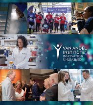
Prostate epithelial cells expressing a Pten mutant (C124S) were differentiated for 18 days under suboptimal conditions.<br />
Androgen receptor (red) in the luminal cells and integrin α6 (green) in the basal cells were visualized by immunostaining and<br />
fluorescence microscopy. Image by Mclane Watson.<br />
20 Van Andel Research Institute | <strong>Scientific</strong> <strong>Report</strong>
MATTHEW STEENSMA, M.D.<br />
Dr. Steensma received his B.A. from Hope College and his M.D. from<br />
Wayne State University School of Medicine in Detroit. Dr. Steensma<br />
is a practicing surgeon in the Spectrum Health Medical Group, and<br />
he joined VARI as an Assistant Professor in 2010.<br />
STAFF<br />
CURT ESSENBURG, B.S., LATG<br />
PATRICK DISCHINGER, B.S.<br />
DIANA LEWIS, A.S.<br />
MARIE MOONEY, M.S.<br />
MATT PRIDGEON, M.D.<br />
RESEARCH INTERESTS<br />
Our laboratory conducts research into new treatment strategies for sarcomas.<br />
Specifically, we are interested in determining the mechanisms underlying tumor<br />
formation in sporadic bone and soft tissue sarcomas and in neurofibromatosis type<br />
1, a hereditary disorder caused by mutations in the neurofibromin 1 (NF1) gene.<br />
Neurofibromin is considered a tumor suppressor that suppresses Ras activity by<br />
promoting Ras GTP hydrolysis to GDP. People with mutations in the neurofibromin<br />
1 gene develop benign tumors called neurofibromas and have an elevated risk of<br />
malignancies ranging from solid tumors to leukemia, including sarcomas. The disease<br />
affects 1 in 3000 people in the United States, of whom 8–13% will ultimately develop a<br />
neurofibromatosis-related sarcoma in their lifetime. These aggressive tumors typically<br />
arise from benign neurofibromas, but the process of benign-to-malignant transformation<br />
is not well understood, and treatment options are limited, leading to poor five-year<br />
survival rates.<br />
Our current sarcoma-related research efforts include the development of genetically<br />
engineered mouse models of neurofibromatosis type 1 tumor progression; the<br />
identification of targetable patterns of intratumoral and intertumoral heterogeneity<br />
through next-generation sequencing; genotype–phenotype correlations in<br />
neurofibromatosis type 1 and related diseases; and mechanisms of chemotherapy<br />
resistance in bone and soft-tissue sarcomas.<br />
RECENT PUBLICATIONS<br />
Foley, Jessica M., Donald J. Scholten, Noel R. Monks, David Cherba, David J. Monsma, Paula Davidson, Dawna Dylewski, Karl<br />
Dykema, Mary E. Winn, and Matthew R. Steensma. 2015. Anoikis-resistant subpopulations of human osteosarcoma display<br />
significant chemoresistance and are sensitive to targeted epigenetic therapies predicted by expression profiling. Journal of<br />
Translational Medicine 13: 110.<br />
Lane, Brian R., Jeffrey Bissonnette, Tracy Waldherr, Deborah Ritz-Holland, Dave Chesla, Sandra L. Cottingham, Sheryl Alberta,<br />
Cong Liu, Amanda Bartenbaker Thompson, et al. 2015. Development of a center for personalized cancer care at a regional<br />
cancer center. Journal of Molecular Diagnostics 17(6): 695–704.<br />
Peacock, Jacqueline D., Karl J. Dykema, Helga V. Toriello, Marie R. Mooney, Donald J. Scholten II, Mary E. Winn, Andrew<br />
Borgman, Nicholas S. Duesbery, Judith A. Hiemenga, et al. 2015. Oculoectodermal syndrome is a mosaic RASopathy<br />
associated with KRAS alterations. American Journal of Medical Genetics A 167(7): 1429–1435.<br />
CENTER FOR CANCER AND CELL BIOLOGY 21
GEORGE F. VANDE WOUDE, PH.D.<br />
STAFF<br />
CHONGFENG GAO, PH.D.<br />
LIANG KANG, B.S.<br />
KAY KOO<br />
DAFNA KAUFMAN, M.S.<br />
BEN STAAL, M.S.<br />
Dr. Vande Woude received his M.S. and Ph.D. degrees from<br />
Rutgers University. He joined the National Cancer Institute in 1972,<br />
becoming the director of the ABL–Basic Research Program in 1983,<br />
and then director of the Division of Basic Sciences in 1998. In 1999,<br />
he became the founding Director of VARI. In 2009, he stepped<br />
down as Director while retaining his laboratory as a Distinguished<br />
<strong>Scientific</strong> Fellow and Professor. He is a member of the National<br />
Academy of Sciences (1993) and a Fellow of the American<br />
Association for the Advancement of Science (2013).<br />
RESEARCH INTERESTS<br />
ADJUNCT FACULTY<br />
BRIAN CAO, M.D.<br />
HENRY B. SKINNER, PH.D.<br />
MET is overexpressed in many types of human cancer, and its expression correlates with<br />
aggressive disease and poor prognosis (visit http://www.vai.org/met/). Since discovering<br />
the MET receptor tyrosine kinase and its ligand, hepatocyte growth factor (HGF/SF), in<br />
the mid 1980s, our lab has focused on investigating the paramount role these molecules<br />
play in malignant progression and metastasis. As part of our ongoing effort, we focus on<br />
the mechanisms responsible for tumor progression under the hypothesis that phenotypic<br />
switching and chromosome instability can drive tumor progression. In addition, we<br />
continue to develop and characterize novel research models to be used in preclinical<br />
evaluation of new inhibitors that target MET in a variety of human cancers.<br />
Tumor phenotypic switching: mechanism and therapeutic implications<br />
In human carcinomas, the acquisition by cells of an invasive phenotype, a process<br />
termed the epithelial-to-mesenchymal transition (E-MT), requires a breakdown of<br />
intercellular junctions with neighboring cells. Upon arriving at secondary sites, a few of<br />
the mesenchymal cells revert to an epithelial phenotype via a mesenchymal-to-epithelial<br />
transition (M-ET). We have implicated genetic instability in cell type determination<br />
and we have developed methods to isolate phenotypic variants from epithelial or<br />
mesenchymal subclones of carcinoma cell lines. We have explored the signal pathway<br />
underlying E-MT/M-ET phenotypic switching by gene expression analysis, spectral<br />
karyotyping (SKY), and fluorescent in situ hybridization (FISH). We found that changes in<br />
chromosome content are associated with phenotypic switching. We have further shown<br />
that these changes dictate the expression of specific genes, which in E-MT events are<br />
mesenchymal related and in M-ET events are epithelial related. Our results suggest that<br />
chromosome instability can provide the diversity of gene expression needed for tumor<br />
cells to switch phenotype.<br />
22 Van Andel Research Institute | <strong>Scientific</strong> <strong>Report</strong>
In vivo research models: model development<br />
and preclinical treatment evaluation<br />
Anti-cancer therapy based on blocking the HGF–Met<br />
signaling pathway has emerged as an important goal of<br />
pharmaceutical research. One of the limitations of studying<br />
the altered Met–HGF/SF signaling of human cancers grafted<br />
in mouse models has been that the murine HGF/SF protein<br />
has a low affinity for human MET. To overcome this, our lab<br />
developed a transgenic human HGF-SCID mouse model<br />
(hHGFtg-SCID), which generates a human-compatible<br />
HGF/SF protein and thus allows for the propagation of<br />
human tumors. This model has proven to be a valuable tool<br />
for in vivo testing of MET-dependent cancers and is used to<br />
evaluate treatment strategies aimed at targeting<br />
this pathway.<br />
RECENT PUBLICATIONS<br />
Johnson, Jennifer, Maria Libera Ascierto, Sandeep Mittal, David Newsome, Liang Kang, Michael Briggs, Kirk Tanner, Francesco<br />
M. Marincola, Michael E. Berens, George F. Vande Woude, et al. 2015. Genomic profiling of a hepatocyte growth factor–<br />
dependent signature for MET-targeted therapy in glioblastoma. Journal of Translational Medicine 13: 306.<br />
CENTER FOR CANCER AND CELL BIOLOGY 23
BART O. WILLIAMS, PH.D.<br />
Dr. Williams received his Ph.D. in biology from Massachusetts Institute<br />
of Technology in 1996, where he trained with Tyler Jacks. Following<br />
his postdoctoral study with Harold Varmus, Dr. Williams joined VARI as<br />
a <strong>Scientific</strong> Investigator in July 1999. He is now a Professor and the<br />
Director of the Center for Cancer and Cell Biology.<br />
STAFF<br />
CASSIE DIEGEL, B.S.<br />
NICOLE ETHEN, B.S.<br />
DIANA LEWIS, A.S.<br />
MITCH MCDONALD, B.S.<br />
ALEX ZHONG, PH.D.<br />
STUDENTS<br />
CHERYL CHRISTIE, B.S.<br />
CASEY DROSCHA, B.S.<br />
JOHAN LEE<br />
JON LENSING<br />
KEVIN MAUPIN, B.A., B.S.<br />
AGNI NAIDU, B.S.<br />
RESEARCH INTERESTS<br />
Our laboratory is interested in understanding how alterations in the Wnt signaling<br />
pathway cause human disease. Wnt signaling is a process, conserved throughout<br />
evolution, that functions in the differentiation of most tissues. Given its central role<br />
in growth and differentiation, it is not surprising that alterations in the Wnt pathway<br />
are among the most common events associated with human cancer. In addition,<br />
other human diseases including osteoporosis, cardiovascular disease, and diabetes<br />
have been linked to altered regulation of this pathway. A specific focus of our work is<br />
characterizing the role of Wnt signaling in bone formation. Our interest is not only in<br />
normal bone development but also in understanding whether aberrant Wnt signaling<br />
plays a role in the metastasis of some common cancers (for example, prostate, breast,<br />
lung, and renal tumors) to the bone. The long-term goal of this work is to provide<br />
insights useful in developing strategies to lessen the morbidity and mortality associated<br />
with skeletal metastasis.<br />
Wnt signaling in normal bone development<br />
Mutations in Lrp5, a Wnt receptor, have been causally linked to alterations in human<br />
bone development. We have characterized a mouse strain deficient in Lrp5 and have<br />
shown that it recapitulates the low-bone-density phenotype seen in human patients<br />
who have that deficiency. We have further shown that mice carrying mutations in both<br />
Lrp5 and the related Lrp6 protein have even more-severe defects in bone density. To<br />
test whether Lrp5 deficiency causes changes in bone density due to aberrant signaling<br />
through β-catenin, we created OC-Cre;β-catenin flox/flox mice, which carry an osteoblastspecific<br />
deletion of β-catenin. We are addressing how other genetic alterations linked<br />
to Wnt/β-catenin signaling affect bone development and osteoblast function. We have<br />
generated mice with conditional alleles of Lrp6 and Lrp5 that can be inactivated via<br />
Cre-mediated recombination and have used them to show that both Lrp5 and Lrp6<br />
function within osteoblasts to regulate normal bone development and homeostasis. We<br />
have also created mice lacking the ability to secrete Wnts from osteoblasts and shown<br />
that these mice have extremely low bone mass, establishing that the mature osteoblast<br />
is an important source of Wnts.<br />
24 Van Andel Research Institute | <strong>Scientific</strong> <strong>Report</strong>
We are also examining the effects on normal bone<br />
development and homeostasis of chemical inhibitors of the<br />
enzyme porcupine, which is required for the secretion and<br />
activity of all Wnts. Given that such inhibitors are currently<br />
in human clinical trials for treatment of several tumor types,<br />
their side effects related to the lowering of bone mass must<br />
be evaluated.<br />
Wnt signaling in mammary development<br />
and cancer<br />
We are addressing the relative roles of Lrp5 and Lrp6 in<br />
Wnt1-induced mammary carcinogenesis. We have focused<br />
our initial efforts on Lrp5-deficient mice, because they are<br />
viable and fertile. A deficiency in Lrp5 dramatically inhibits<br />
the development of mammary tumors, and a germline<br />
deficiency in Lrp5 or Lrp6 results in delayed mammary<br />
development. We are also focusing on the mechanisms that<br />
underlie the role that Lrp6 plays in mammary development.<br />
We are particularly interested in the pathways that may<br />
regulate the proliferation of normal mammary progenitor<br />
cells, as well as of tumor-initiating cells.<br />
Wnt signaling in prostate development<br />
and cancer<br />
Two hallmarks of advanced prostate cancer are the<br />
development of skeletal osteoblastic metastasis and<br />
the ability of the tumor cells to become independent<br />
of androgen for survival. We have created mice with a<br />
prostate-specific deletion of the Apc gene as a disease<br />
model. These mice develop fully penetrant prostate<br />
hyperplasia by four months of age, and these tumors<br />
progress to frank carcinomas by seven months. We have<br />
found that these tumors initially regress under androgen<br />
ablation but show signs of androgen-independent growth<br />
some months later.<br />
Genetically engineered mouse models<br />
of bone disease<br />
We have also focused on developing mouse models of<br />
osteoarthritis and of fracture repair. In addition, we are<br />
interested in identifying novel genes that play key roles in<br />
skeletal development and maintenance of bone mass. For<br />
example, current work is focused on the role of galectin-3,<br />
a member of the lectin family, in this context.<br />
RECENT PUBLICATIONS<br />
Schumacher, Cassie A., Danese M. Joiner, Kennen D. Less, Melissa Oosterhouse Drewry, and Bart O. Williams. <strong>2016</strong>.<br />
Characterization of genetically engineered mouse models carrying Col2a1-cre-induced deletions of Lrp5 and/or Lrp6.<br />
Bone Research 4: 15042.<br />
Williams, Bart O. <strong>2016</strong>. Genetically engineered mouse models to evaluate the role of Wnt secretion in bone development in<br />
homeostasis. American Journal of Medical Genetics C 172(1): 24–26.<br />
Valkenburg, Kenneth C., Galen Hostetter, and Bart O. Williams. 2015. Concurrent hepsin overexpression and adenomatous<br />
polyposis coli deletion causes invasive prostate carcinoma in mice. The Prostate 75(14): 1579–1585.<br />
Zhong, Zhendong, A., Juraj Zahatnansky, John Snider, Emily Van Wieren, Cassandra R. Diegel, and Bart O. Williams. 2015.<br />
Wntless spatially regulates bone development through β-catenin-dependent and independent mechanisms. Developmental<br />
Dynamics 244(10): 1347–1355.<br />
Zhong, Zhendong A., Anderson Peck, Shihong Li, Jeff VanOss, John Snider, Casey J. Droscha, TingTung A. Chang, and Bart O.<br />
Williams. 2015. 99m Tc-Methylene diphosphonate uptake at injury site correlates with osteoblast differentiation and mineralization<br />
during bone healing in mice. Bone Research 3: 15013.<br />
CENTER FOR CANCER AND CELL BIOLOGY 25
NING WU, PH.D.<br />
Dr. Wu received her Ph.D. from the Department of Biochemistry<br />
of the University of Toronto in 2002. She joined VARI in 2013 as<br />
an Assistant Professor.<br />
STAFF<br />
HOLLY DYKSTRA, B.S.<br />
ALTHEA WALDHART, B.S.<br />
STUDENT<br />
MATT HOLLOWELL<br />
RESEARCH INTERESTS<br />
Our laboratory studies the interface between cellular metabolism and signal<br />
transduction. The generation of two daughter cells depends on the proper uptake<br />
and use of nutrients that are often limited in the tumor environment. The distribution<br />
of these nutrients is controlled not only by the intrinsic catalytic rate and allosteric<br />
regulation of the enzymes, but also by post-translational modifications of these<br />
enzymes by signaling molecules. At the same time, signaling molecules must respond<br />
to cellular nutrient status and other cues such as environmental stresses and growth<br />
factors. Our laboratory focuses on key metabolic steps in glucose and lipid catabolism<br />
and aims to understand the mutual interactions between metabolites and signaling<br />
during cell replication.<br />
Fundamentally, cancer is a disease of uncontrolled cell growth. Relative to normal cells,<br />
tumor cells have aberrant metabolic addictions that differ depending on the cell’s tissue<br />
of origin and genetic mutations. By understanding the energy requirements and<br />
regulatory pathways of tumor cells, more-effective treatments can be developed. Our<br />
projects include unraveling the molecular mechanisms that regulate glucose uptake in<br />
cancers, investigating the effect of glucose on mitochondrial activity, and exploring the<br />
role of glucose as the link between metabolic syndrome and cancer incidence.<br />
26 Van Andel Research Institute | <strong>Scientific</strong> <strong>Report</strong>
H. ERIC XU, PH.D.<br />
Dr. Xu went to Duke University and the University of Texas<br />
Southwestern Medical Center, earning his Ph.D. in molecular biology<br />
and biochemistry. He joined VARI in July 2002 and is now a Professor.<br />
Dr. Xu is also the Primary Investigator and Distinguished Director of<br />
the VARI–SIMM Research Center in Shanghai, China.<br />
STAFF<br />
XIANG GAO, PH.D.<br />
STEPHANIE GRANT, M.P.A.<br />
YUANZHENG (AJIAN) HE, PH.D.<br />
YANYONG KANG, PH.D.<br />
KUNTAL PAL, PH.D.<br />
KELLY POWELL, B.S.<br />
XIAOYIN (EDWARD) ZHOU, PH.D.<br />
STUDENTS<br />
ERIC LI<br />
HONGLEI MA, B.S.<br />
PARKER DE WAAL, B.S.<br />
TINGHAI XU, B.S.<br />
YAN YAN, B.S.<br />
YANTING YIN, B.S.<br />
FENG ZHANG, B.S.<br />
VISITING SCIENTISTS<br />
DAVID BENSON, PH.D.<br />
SOK KEAN KHOO, PH.D.<br />
ROSS REYNOLDS, PH.D.<br />
RESEARCH INTERESTS<br />
Hormone signaling is essential to eukaryotic life. Our research focuses on the signaling<br />
mechanisms of physiologically important hormones, striving to answer fundamental<br />
questions that have a broad impact on human health and disease. We are studying<br />
two families of proteins, the nuclear hormone receptors and the G protein–coupled<br />
receptors, because these proteins have fundamental roles in biology and are important<br />
drug targets for treating major human diseases.<br />
Nuclear hormone receptors<br />
The nuclear hormone receptors form a large family comprising ligand-regulated and<br />
DNA-binding transcription factors, which include receptors for the classic steroid<br />
hormones such as estrogen, androgens, and glucocorticoids, as well as receptors for<br />
peroxisome proliferator activators, vitamin D, vitamin A, and thyroid hormones. These<br />
receptors are among the most successful targets in the history of drug discovery: every<br />
receptor has one or more synthetic ligands being used as medicines. In the last five<br />
years, we have developed the following projects centering on the structural biology of<br />
nuclear receptors.<br />
Peroxisome proliferator–activated receptors<br />
The peroxisome proliferator–activated receptors (PPARα, β, and γ) are the key regulators<br />
of glucose and fatty acid homeostasis and, as such, are important therapeutic targets<br />
for treating cardiovascular disease, diabetes, and cancer. Millions of patients with type<br />
II diabetes have benefited from treatment with the novel PPARγ ligands rosiglitazone<br />
and pioglitazone. To understand the molecular basis of ligand-mediated signaling by<br />
PPARs, we have determined crystal structures of each PPAR’s ligand-binding domain<br />
(LBD) bound to many diverse ligands, including fatty acids, the lipid-lowering drugs<br />
called fibrates, and the new generation of anti-diabetic drugs, the glitazones. These<br />
structures have provided a framework for understanding the mechanisms of agonists<br />
CENTER FOR CANCER AND CELL BIOLOGY 27
and antagonists and the recruitment of co-activators and<br />
co-repressors in gene activation and repression. They<br />
also increase our understanding of the potency, selectivity,<br />
and binding mode of ligands and provide crucial insights<br />
for designing the next generation of PPAR medicines. We<br />
have discovered several natural ligands of PPARγ. Our<br />
plan is to test their physiological roles in glucose and insulin<br />
regulation and to develop them into therapeutics<br />
for diabetes and dislipidemia.<br />
The human glucocorticoid receptor<br />
The human glucocorticoid receptor (GR), the prototypical<br />
steroid hormone receptor, affects a wide spectrum<br />
of human physiology including immune/inflammatory<br />
responses, metabolic homeostasis, and control of blood<br />
pressure. GR is a well-established target for drugs, and<br />
those drugs have an annual market of over $10 billion.<br />
However, the clinical use of GR ligands is limited by<br />
undesirable side effects partly resulting from receptor<br />
cross-reactivity or low potency. The discovery of potent,<br />
more-selective GR ligands— “dissociated glucocorticoids”<br />
that have the potential to separate the good effects from the<br />
bad—remains a major goal of pharmaceutical research.<br />
We have determined a number of GR crystal structures<br />
bound to unique ligands and have found an unexpected<br />
regulatory mechanism: degradation by lysosomes. We also<br />
are studying the molecular and structural mechanisms of<br />
the dissociated glucocorticoids identified by our research.<br />
Structural genomics of receptor LBDs<br />
The ligand-binding domain of a nuclear receptor contains<br />
key structural elements that mediate ligand-dependent<br />
regulation of the receptors and, as such, it has been the<br />
focus of intense structural studies. Crystal structures<br />
for most of the 48 human nuclear receptors have been<br />
determined and have illustrated the details of ligand<br />
binding, the conformational changes induced by agonists<br />
and antagonists, the basis of dimerization, and the<br />
mechanism of co-activator and co-repressor binding.<br />
The structures also have provided many surprises about<br />
the identity of ligands and their implications for receptor<br />
signaling pathways. There are only a few “orphan” nuclear<br />
receptors for which the LBD structure remains unsolved.<br />
In the past few years, we have determined the crystal<br />
structures of the LBDs of CAR, SHP, SF-1, COUP-TFII,<br />
and LRH-1, and our structures have helped to identify<br />
new ligands and signaling mechanisms for these orphan<br />
receptors.<br />
G protein–coupled receptors (GPCRs)<br />
The GPCRs form the largest family of receptors in the<br />
human genome and account for over 40% of drug targets,<br />
but their structures remain a challenge because they<br />
are seven-transmembrane receptors. There are only<br />
a few crystal structures for class A GPCRs, and many<br />
important questions regarding GPCR ligand binding and<br />
activation remain unanswered. From our standpoint,<br />
GPCRs are similar to nuclear hormone receptors with<br />
respect to regulation by protein-ligand and protein–protein<br />
interactions. We focus on class B GPCRs, which includes<br />
receptors for parathyroid hormone (PTH), corticotropinreleasing<br />
factor (CRF), glucagon, and glucagon-like<br />
peptide-1. We have determined crystal structures of the<br />
ligand-binding domain of the PTH receptor and the CRF<br />
receptor, and we are developing hormone analogs for<br />
treating osteoporosis, depression, and diabetes. We are<br />
developing a mammalian overexpression system and plan<br />
to use it to express full-length GPCRs for crystallization and<br />
structural studies.<br />
RECENT PUBLICATIONS<br />
He, Yuanzheng, Jingjing Shi, Wei Yi, Xin Ren, Xiang Gao, Jianshuang Li, Nanyan Wu, Kevin Weaver, Qian Xie, et al. 2015.<br />
Discovery of a highly potent glucocorticoid for asthma treatment. Cell Discovery 1: 15035.<br />
Kang, Yanyong, X. Edward Zhou, Xiang Gao, Yuanzheng He, Wei Liu, Andrii Ishchenko, Anton Barty, Thomas A. White, Oleksandr<br />
Yefanov, et al. 2015. Crystal structure of rhodopsin bound to arrestin by femtosecond X-ray laser. Nature 523(7562): 561–567.<br />
28 Van Andel Research Institute | <strong>Scientific</strong> <strong>Report</strong>
TAO YANG, PH.D.<br />
Dr. Yang received his Ph.D. in biochemistry at the Shanghai Institute<br />
of Biochemistry and Cell Biology, Chinese Academy of Sciences, in<br />
2001. He joined VARI as an Assistant Professor in February 2013.<br />
STAFF<br />
DIANA LEWIS, A.S.<br />
JIANSHUANG LI, B.S.<br />
JIE LI, PH.D.<br />
HUADIE LIU, M.S.<br />
DI LU, M.S.<br />
KEVIN WEAVER, B.S.<br />
RESEARCH INTERESTS<br />
The skeletal system develops from mesenchymal cells and is the major reservoir<br />
of mesenchymal stem cells (MSCs) in adult life. MSCs play pivotal roles in skeletal<br />
tissue growth, homeostasis, and repair, while dysregulations in MSC renewal, linage<br />
specification, and pool maintenance are common causes of skeletal disorders. Our<br />
long-term interest is to investigate the signals and cellular processes orchestrating the<br />
activities of MSCs and MSC-derived cells during skeletal development and homeostasis<br />
and how those processes are involved in skeletal aging and disorders. Our current<br />
projects in skeletal development and disease include a study of the sumoylation<br />
pathway and a study of LRP1 signaling. As part of these projects, we have established<br />
in vivo and in vitro genetic models to study the molecular mechanisms underlying<br />
osteoarthritis and osteoporosis.<br />
RECENT PUBLICATIONS<br />
Chen, Shan, Monica Grover, Tarek Sibai, Jennifer Black, Nahid Rianon, Abbhirami Rajagopal, Elda Munivez, Terry Bertin,<br />
Brian Dawson, et al. 2015. Losartan increases bone mass and accelerates chondrocyte hypertrophy in developing skeleton.<br />
Molecular Genetics and Metabolism 115(1): 53–60.<br />
He, Yuanzheng, Jingjing Shi, Wei Yi, Xin Ren, Xiang Gao, Jianshuang Li, Nanyan Wu, Kevin Weaver, Qian Xie, et al. 2015.<br />
Discovery of a highly potent glucocorticoid for asthma treatment. Cell Discovery 1: 15035.<br />
Lu, Linchao, Karine Harutyunyan, Weidong Jin, Jianhong Wu, Tao Yang, Yuqing Chen, Kyu Sang Jeoeng, Yangjin Bae, Jianning<br />
Tao, et al. 2015. RECQL4 regulates p53 function in vivo during skeletogenesis. Journal of Bone and Mineral Research 30(6):<br />
1077–1089.<br />
CENTER FOR CANCER AND CELL BIOLOGY 29
30 Van Andel Research Institute | <strong>Scientific</strong> <strong>Report</strong>
CENTER FOR<br />
EPIGENETICS<br />
Peter A. Jones, Ph.D., D.Sc.<br />
Director<br />
The Center’s researchers study epigenetics and<br />
epigenomics in health and disease, with the ultimate<br />
goal of developing novel therapies to treat cancer and<br />
neurodegenerative diseases. The Center collaborates<br />
extensively with other VARI research groups and with<br />
external partners to maximize its efforts to develop<br />
therapies that target epigenetic mechanisms.<br />
Methyl (red) and acetyl (light blue) groups as epigenetic marks on nucleosomes and DNA.<br />
Illustration by Nicole Ethen.<br />
31
STEPHEN B. BAYLIN, M.D.<br />
Dr. Baylin joined VARI as a Professor in the Center for Epigenetics<br />
in January 2015 and is co-leader of the VARI-SU2C Epigenetics<br />
Dream Team. He devotes a portion of his time to VARI. His primary<br />
appointment is with Johns Hopkins University as the Virginia and D.K.<br />
Ludwig Professor of Oncology and Medicine and co-head of Cancer<br />
Biology at the Sidney Kimmel Comprehensive Cancer Center.<br />
RESEARCH INTERESTS<br />
The Van Andel Research Institute–Stand Up To Cancer (VARI-SU2C) Epigenetics Dream<br />
Team is a multi-institutional effort to develop new epigenetic therapies against cancer<br />
and to move promising therapies to clinical trials. As co-leader, Dr. Baylin oversees the<br />
team’s research, which leverages the combined expertise of its members.<br />
Epigenetics is the study of how the packaging and modification of DNA influences the<br />
genes that are active or kept silent in a particular cell, and it holds untold potential for<br />
treating cancer and other diseases. Through a detailed understanding of how normal<br />
epigenetic processes work, scientists can identify erroneous epigenetic modifications<br />
that may contribute to the development and progression of cancer. Epigenetic<br />
therapies, which work by correcting these errors, have the potential to directly treat<br />
cancer and to sensitize patients to traditional treatments such as chemotherapy and<br />
radiation and to promising new immunotherapy approaches.<br />
The VARI-SU2C Epigenetics Dream Team is headquartered at VARI in Grand Rapids,<br />
Michigan, and it includes members from Johns Hopkins University, Memorial Sloan<br />
Kettering Cancer Center, Fox Chase Cancer Center/Temple University, University of<br />
Southern California, and Rigshospitalet/University of Copenhagen. The American<br />
Association for Cancer Research (AACR), as SU2C’s scientific partner, reviews<br />
projects and provides objective scientific oversight.<br />
RECENT PUBLICATIONS<br />
Chaiappinelli, Katherine B., Pamela L. Strissel, Alexis Desrichard, Huili Li, Christine Henke, Benjamin Akman, Alexander Hein, Neal<br />
S. Rote, Leslie M. Cope, et al. 2015. Inhibiting DNA methylation causes an interferon response in cancer via dsRNA including<br />
endogenous retroviruses. Cell 162(5): 961–973.<br />
32 Van Andel Research Institute | <strong>Scientific</strong> <strong>Report</strong>
PETER A. JONES, PH.D., D.SC.<br />
Dr. Jones received his Ph.D. from the University of London. He<br />
joined the University of Southern California in 1977 and served as<br />
director of the USC Norris Comprehensive Cancer Center between<br />
1993 and 2011. Dr. Jones joined VARI in 2014 as its Chief <strong>Scientific</strong><br />
Officer and Director of the Center for Epigenetics.<br />
STAFF<br />
MINMIN LIU, PH.D.<br />
HITOSHI OTANI, PH.D.<br />
ROCHELE TIEDEMANN, PH.D.<br />
WANDING ZHOU, PH.D.<br />
ADJUNCT FACULTY<br />
RONALD CHANDLER, JR., PH.D.<br />
FEYRUZ RASSOOL, PH.D.<br />
RESEARCH INTERESTS<br />
Epigenetics may be defined as mitotically heritable changes in gene expression that<br />
are not caused by changes in the DNA sequence itself. Epigenetic processes establish<br />
the differentiated state of cells and govern how genes are used to allow organs and<br />
cells to function correctly and inherit their properties through cell division. In the case<br />
of diseases such as cancer, these processes can go wrong, changing the behavior of<br />
cells to adverse effect. However, many of these changes are potentially reversible by<br />
treatment with drugs. Because epigenetic processes are at the root of biology, they<br />
have implications for all of human development and disease.<br />
Our laboratory studies the mechanisms by which epigenetic processes become<br />
misregulated in cancer and contribute to the disease phenotype. We focus on the role<br />
of DNA methylation in controlling the expression of genes during normal development<br />
and in cancer. Our work has shifted to a holistic approach in which we are interested in<br />
the interactions between processes such as DNA methylation, histone modification, and<br />
nucleosomal positioning in the epigenome, and we want to determine how mutations<br />
in the genes which modify the epigenome contribute to the cancer phenotype. We<br />
have had a long-term interest in the mechanism of action of DNA methylation inhibitors,<br />
both in the lab and in the clinic. We are working with several major institutions to bring<br />
epigenetic therapies to the forefront of cancer medicine.<br />
RECENT PUBLICATIONS<br />
Lay, Fides D., Yaping Liu, Theresa K. Kelly, Heather Witt, Peggy J. Farnham, Peter A. Jones, and Benjamin P. Berman. 2015.<br />
The role of DNA methylation in directing the functional organization of the cancer epigenome. Genome Research 25(4): 467–477.<br />
Roulois, David, Helen Loo Yau, Rajat Singhania, Yadong Wang, Amavaz Danesh, Shu Yi Shen, Han Han, Gangning Liang, Peter<br />
A. Jones, et al. 2015. DNA-demethylating agents target colorectal cancer cells by inducing viral mimicry by endogenous<br />
transcripts. Cell 162(5): 961–973.<br />
Statham, Aaron L., Phillippa C. Taberlay, Theresa K. Kelly, Peter A. Jones, and Susan J. Clark. 2015. Genome-wide nucleosome<br />
occupancy and DNA methylation profiling of four human cell lines. Genomics Data 3: 94–96.<br />
CENTER FOR EPIGENETICS 33
STEFAN JOVINGE, M.D., PH.D.<br />
Dr. Jovinge received his M.D. (1991) and his Ph.D. (1997) at<br />
Karolinska Institute in Stockholm. Since December 2013 he has<br />
been the Medical Director of Research at the Frederik Meijer Heart<br />
and Vascular Institute and a Professor at VARI. He also directs<br />
the DeVos Cardiovascular Research Program, is a Professor at the<br />
MSU College of Human Medicine, and is a Consulting Professor at<br />
Stanford University.<br />
STAFF<br />
PAULA DAVIDSON, M.S.<br />
DAWNA DYLEWSKI, B.S.<br />
ELLEN ELLIS<br />
EMILY EUGSTER, M.A.<br />
JENS FORSBERG, PH.D.<br />
LISA KEFENE, M.A., MB(ASCP), RLAT<br />
ERIC KORT, M.D.<br />
BRITTANY MERRIFIELD, B.S.<br />
HSIAO-YUN YEH (CHRISTY) MILLIRON, PH.D.<br />
JORDAN PRAHL, B.S.<br />
LAURA TARNAWSKI, M.S.<br />
MATTHEW WEILAND, M.S.<br />
LAURA WINKLER, PH.D.<br />
RESEARCH INTERESTS<br />
The DeVos Cardiovascular Research Program is a joint effort between VARI and<br />
Spectrum Health. The basic science lab is the Jovinge laboratory at VARI, and a<br />
corresponding clinical research unit resides within the Fred Meijer Heart and Vascular<br />
Institute.<br />
Cardiovascular diseases are among the major causes of death and disability worldwide.<br />
While the incidence of ischemic heart diseases has started to decline, congestive heart<br />
failure is still rising. Medical treatment for the latter is supportive, and the only available<br />
therapy is heart transplantation.<br />
To regenerate myocardium after disease or damage is one of the major challenges in<br />
medicine. Our group is working on true heart muscle regeneration along two axes:<br />
external and internal (cardiac) cell sources. The most robust external source for<br />
generating heart muscle cells has been stem cells, either from an embryonic stem<br />
cell (ESC) system or from reprogrammed pluripotent stem cells (iPSCs). The main<br />
drawback to the stem cell approach is that their differentiation will generate a multitude<br />
of different cell types at different stages of development. A mixed cell population of<br />
undifferentiated cells always has the potential to create tumors. Also, the use of ESCs<br />
creates a need for lifelong immunosuppressive treatment. iPSCs, however, could be<br />
generated from the patient’s own peripheral blood cells, a technique established by our<br />
group in Grand Rapids. To be able to use these sources, we have developed strategies<br />
based on establishing surface marker expression—similar to those for bone-marrow<br />
cells—to help select homogenous, safe populations to transplant.<br />
34 Van Andel Research Institute | <strong>Scientific</strong> <strong>Report</strong>
The second axis we focus on is endogenous generation<br />
within the heart. Although adult human heart muscle cells<br />
are to a small extent generated after birth, the internal<br />
source of such cells and their cell cycle regulation are<br />
unknown. Some data indicate that cardiac progenitors<br />
could be involved, and other data suggest that differentiated<br />
heart muscle cells might be the source. We and our<br />
collaborators have rejected the view that adult heart muscle<br />
cells are not capable of undergoing a complete cell division.<br />
With the use of 14 C dating, the adult heart has been shown<br />
to have a regenerative capacity. This has opened a<br />
completely new field of induced local generation of heart<br />
muscle cells, which is now being explored.<br />
The final phase of patient studies will involve the<br />
administration of cells or compounds to stimulate<br />
endogenous regeneration. To prepare cells for<br />
transplantation into humans, an accredited Good<br />
Manufacturing Practice facility will be established in<br />
collaboration with Stanford University, and the first safety<br />
studies (Phase II) will be followed by studies evaluating the<br />
best route for delivering the treatment and the best timing.<br />
In the final stage, randomized prospective clinical trials will<br />
be launched.<br />
Our program’s eventual aims are clinical concept studies<br />
of heart muscle cell regeneration in patients, either by cell<br />
transplantation or stimulation of endogenous sources. The<br />
program’s clinical side involves a multistep process to<br />
prepare for these studies. Patients with the most severe<br />
heart disease, i.e., those needing mechanical support, are<br />
being studied to optimize treatments that will be used in<br />
later safety studies. We have already derived mortality<br />
prediction algorithms for patients on bedside heart-lung<br />
machines.<br />
RECENT PUBLICATIONS<br />
Bergmann, Olaf, Sofia Zdunek, Anastasia Felker, Mehran Salehpour, Kanar Alkass, Samuel Bernard, Staffan L. Sjostrom,<br />
Mirosława Szewcykowska, Teresa Jackowska, et al. 2015. Dynamics of cell generation and turnover in the human heart.<br />
Cell 161(7): 1566–1575.<br />
Raulf, Alexandra, Hannes Horder, Laura Tarnawski, Caroline Geisen, Annika Ottersbach, Wilhelm Röll, Stefan Jovinge, Bernd<br />
K. Fleischmann, and Michael Hesse. 2015. Transgenic systems for unequivocal identification of cardiac myocyte nuclei and<br />
analysis of cardiomyocyte cell cycle status. Basic Research in Cardiology 110(3): 33.<br />
Tarnawski, Laura, Xiaojie Xian, Gustavo Monnerat, Iain C. Macauley, Daniela Malan, Andrew Borgman, Sean M. Wu, Bernd<br />
K. Fleischmann, and Stefan Jovinge. 2015. Integrin based isolation enables purification of murine lineage committed<br />
cardiomyocytes. PLoS One 10(8): e0135880.<br />
CENTER FOR EPIGENETICS 35
PETER W. LAIRD, PH.D.<br />
Dr. Laird earned his Ph.D. in 1988 from the University of Amsterdam<br />
with Piet Borst. Dr. Laird was a faculty member at the University of<br />
Southern California from 1996 to 2014, where he was Skirball-Kenis<br />
Professor of Cancer Research and directed the USC Epigenome<br />
Center. He joined VARI as a Professor in September 2014.<br />
STAFF<br />
KELLY FOY, B.S.<br />
TOSHINORI HINOUE, PH.D.<br />
KWANGHO LEE, PH.D.<br />
ZHOUWEI ZHANG, M.S.<br />
WANDING ZHOU, PH.D.<br />
STUDENT<br />
NICOLE VANDER SCHAAF, B.S.<br />
RESEARCH INTERESTS<br />
Our goal is to develop a detailed understanding of the molecular basis of human<br />
disease, with a particular emphasis on the role of epigenetics in cancer. Cancer is often<br />
considered to have a primarily genetic basis, with contributions from germline variations<br />
in risk and somatically acquired mutations, rearrangements, and copy number<br />
alterations. However, it is clear that nongenetic mechanisms can exert a powerful<br />
influence on cellular phenotype, as evidenced by the marked diversity of cell types<br />
within our bodies, which virtually all contain an identical genetic code. This differential<br />
gene expression is controlled by tissue-specific transcription factors and variations in<br />
chromatin packaging and modification, which can provide stable phenotypic states<br />
governed by epigenetic, not genetic, mechanisms. It seems intrinsically likely that an<br />
opportunistic disease such as cancer would take advantage of such a potent mediator<br />
of cellular phenotype. Our laboratory is dedicated to understanding how epigenetic<br />
mechanisms contribute to the origins of cancer and how to translate this knowledge<br />
into more-effective cancer prevention, detection, treatment, and monitoring.<br />
We use a multidisciplinary approach in our research, relying on mechanistic studies<br />
in model organisms and cell cultures, clinical and translational collaborations,<br />
genome-scale and bioinformatic analyses, and epidemiological studies to advance our<br />
understanding of cancer epigenetics. In recent years, we participated in the generation<br />
and analysis of high-dimensional epigenetic data sets, including the production of<br />
all epigenomic data for The Cancer Genome Atlas (TCGA) and the application of<br />
next-generation sequencing technology to single-base-pair-resolution, whole-genome<br />
DNA methylation analysis. We are leveraging this epigenomic data for translational<br />
applications and hypothesis testing in animal models. A major focus of our laboratory<br />
is to develop mouse models for investigating epigenetic mechanisms and drivers of<br />
cancer and to develop novel strategies for single-cell epigenomic analysis.<br />
36 Van Andel Research Institute | <strong>Scientific</strong> <strong>Report</strong>
RECENT PUBLICATIONS<br />
Cancer Genome Atlas Research Network. <strong>2016</strong>. Comprehensive molecular characterization of papillary renal-cell carcinoma.<br />
New England Journal of Medicine 374(2): 135–145.<br />
Ceccarelli, Michele, Floris P. Barthel, Tathiane M. Malta, Thais S. Sabedot, Sofie R. Salama, Bradley A. Murray, Olena Morozova,<br />
Yulia Newton, Arnie Radenbaugh, et al. <strong>2016</strong>. Molecular profiling reveals biologically discrete subsets and pathways of<br />
progression in diffuse glioma. Cell 164(3): 550–563.<br />
Levine, A. Joan, Amanda I. Phipps, John A. Baron, Daniel D. Buchanan, Dennis J. Ahnen, Stacey Cohen, Noralane M. Lindor,<br />
Polly A. Newcomb, Christophe Rosty, et al. <strong>2016</strong>. Clinicopathological risk factor distributions for MLH1 promoter region<br />
methylation in CIMP positive tumors. Cancer Epidemiology, Biomarkers and Prevention 25(1): 68–75.<br />
Ryland, Katherine E., Allegra G. Hawkins, Daniel J. Weisenberger, Vasu Punj, Scott C. Borinstein, Peter W. Laird, Jeffrey R.<br />
Martens, and Elizabeth R. Lawlor. <strong>2016</strong>. Promoter methylation analysis reveals that SCNA5 ion channel silencing supports Ewing<br />
sarcoma cell proliferation. Molecular Cancer Research 14(1): 26–34.<br />
Cancer Genome Atlas Research Network. 2015. The molecular taxonomy of primary prostate cancer. Cell 163(4): 1011–1025.<br />
Ciriello, Giovanni, Michael L. Gatza, Andrew H. Beck, Matthew D. Wilkerson, Suhn K Rhie, Alessandro Pastore, Hailei Zhang,<br />
Michael McLellan, Christina Yau, et al. 2015. Comprehensive molecular portraits of invasive lobular breast cancer. Cell 163(2):<br />
506–519.<br />
CENTER FOR EPIGENETICS<br />
37
GERD PFEIFER, PH.D.<br />
Dr. Pfeifer earned his M.S. in pharmacology in 1981 and his Ph.D. in<br />
biochemistry in 1984 from Goethe University in Frankfurt, Germany.<br />
He most recently held the Lester M. and Irene C. Finkelstein Chair in<br />
Biology at the City of Hope in Duarte, California, before joining VARI<br />
in 2014 as a Professor.<br />
STAFF<br />
ZHIJUN HUANG, PH.D.<br />
SEUNG-GI JIN, PH.D.<br />
JENNIFER JOHNSON, M.S.<br />
JIYOUNG YU, PH.D.<br />
RESEARCH OVERVIEW<br />
The laboratory studies epigenetic mechanisms of disease, with a focus on DNA<br />
methylation and the role of 5-hydroxymethylcytosine in cancer and other diseases.<br />
Specifically, the lab studies hypermethylation in cancer genes with the intent of<br />
determining the mechanisms and significance of CpG island methylation. The work<br />
centers on the hypothesis that CpG island hypermethylation in tumors is driven by one<br />
or a combination of the following: carcinogenic agents, inflammation, imbalances in<br />
methylation and demethylation pathways, oncogene activation leading to epigenetic<br />
changes, and dysfunction of the Polycomb repression complex.<br />
The removal of methyl groups from DNA has recently been recognized as an important<br />
pathway in cancer and possibly in other diseases. Our lab studies mechanisms of<br />
5-methylcytosine oxidation.<br />
DNA methylation in cancer<br />
To effectively study genome-wide DNA methylation patterns, we previously developed<br />
the methylated CpG island recovery assay (MIRA), which is used in combination with<br />
sequencing to identify commonly methylated genes in human cancers and normal<br />
tissues. We investigate mechanisms of cancer-associated DNA hypermethylation using<br />
DNA-methylation and chromatin-component mapping in normal and malignant cells, as<br />
well as bioinformatics approaches and functional studies.<br />
38 Van Andel Research Institute | <strong>Scientific</strong> <strong>Report</strong>
Tet3 and related proteins<br />
We have identified three different isoforms of the Tet3<br />
5-methylcytosine oxidase and characterized them using<br />
biochemical, functional, and genetic approaches. We<br />
observed that one isoform of Tet3 specifically binds<br />
to 5-carboxylcytosine, thus establishing an anchoring<br />
mechanism of Tet3 to its reaction product, which may aid in<br />
localized 5-methylcytosine oxidation and removal. We also<br />
study several Tet-associated proteins, trying to understand<br />
their biological roles.<br />
5-methylcytosine oxidation and<br />
neurodegeneration<br />
Using ChIP sequencing, we mapped one of the isoforms of<br />
Tet3 in neuronal cell populations. Tet3 has a rather limited<br />
genomic distribution and is targeted to the transcription<br />
start sites of defined sets of genes, many of which function<br />
within the lysosome and autophagy pathways. We know<br />
these pathways are defective in neurodegenerative<br />
diseases. We are exploring the mechanistic consequences<br />
of 5-methylcytosine oxidation in this disease group, with the<br />
long-term goal of determining whether neurodegeneration<br />
has an epigenetic origin.<br />
RECENT PUBLICATIONS<br />
Jin, Seung-Gi, Zhi-Min Zhang, Thomas L. Dunwell, Matthew R. Harter, Xiwei Wu, Jennifer Johnson, Zheng Li, Jiancheng Liu,<br />
Piroska E. Szabó, et al. <strong>2016</strong>. Tet3 reads 5-carboxylcytosine through its CXXC domain and is a potential guardian against<br />
neurodegeneration. Cell <strong>Report</strong>s 14(3): 493–505.<br />
Jung, Marc, Seung-Gi Jin, Xiaoying Zhang, Wenying Xiong, Grigoriy Gogoshin, Andrei S. Rodin, and Gerd P. Pfeifer. 2015.<br />
Longitudinal epigentic and gene expression profiles analyzed by three-component analysis reveal down-regulation of genes<br />
involved in protein translation in human aging. Nucleic Acids Research 43(15): e100.<br />
Jung, Marc, Swati Kadam, Wenying Xiong, Tibor A. Rauch, Seung-Gi Jin, and Gerd P. Pfeifer. 2015. MIRA-seq for DNA<br />
methylation analysis of CpG islands. Epigenomics 7(5): 695–706.<br />
CENTER FOR EPIGENETICS<br />
39
SCOTT ROTHBART, PH.D.<br />
Dr. Rothbart earned a Ph.D. in pharmacology and toxicology from<br />
Virginia Commonwealth University in 2010. He joined VARI in April<br />
2015 as an Assistant Professor.<br />
STAFF<br />
EVAN CORNETT, PH.D.<br />
BRADLEY DICKSON, PH.D.<br />
ROCHELLE TIEDEMANN, PH.D.<br />
STUDENT<br />
ROBERT VAUGHAN, B.S.<br />
RESEARCH INTERESTS<br />
Two major epigenetic marks regulating the structure and function of eukaryotic<br />
chromatin are the methylation of DNA and post-translational modifications (PTMs) of<br />
histone proteins. Breakthroughs in our understanding of chromatin function have been<br />
made through the identification of protein machineries that incorporate (write), remove<br />
(erase), and bind (read) these epigenetic marks. Chromatin modification and remodeling<br />
shape cellular identity, and it is becoming increasingly apparent that deregulation of<br />
epigenetic signaling contributes to, and may cause, the initiation and progression<br />
of cancer and other human diseases. Unlike genetic abnormalities, chromatin<br />
modifications are reversible, making the writers, erasers, and readers of these marks<br />
attractive therapeutic targets. The goal of our research is to define the molecular details<br />
of chromatin accessibility, interaction, and function. We are particularly interested in<br />
understanding how DNA and histone modifications work together as a language or<br />
code that is read and interpreted by specialized proteins to orchestrate the dynamic<br />
functions of chromatin. We hope our studies will lead to a better understanding of<br />
the etiology of disease and will contribute to the discovery of effective therapeutic<br />
approaches that target the epigenetic machinery.<br />
Mechanics of chromatin interaction<br />
It appears that many chromatin-associated factors have multiple known (or<br />
predicted) chromatin regulatory domains, both within a single protein and within the<br />
subunits of complexes. There is a diverse and exciting potential here, a previously<br />
underappreciated layer of complexity and specificity to chromatin recognition and<br />
regulation. Our studies are using expertise in biochemistry, computational and<br />
molecular biophysics, and cell biology to define the molecular underpinnings of<br />
multivalency and allostery in chromatin interaction and function.<br />
40 Van Andel Research Institute | <strong>Scientific</strong> <strong>Report</strong>
Mechanisms regulating the inheritance of DNA<br />
methylation<br />
Application of microarray technology to the<br />
study of histone PTMs.<br />
The faithful inheritance of DNA methylation patterns is<br />
essential for normal mammalian development and long-term<br />
transcriptional silencing. We recently discovered that<br />
the E3 ubiquitin ligase UHRF1 is a key regulator of this<br />
process through its interaction with a histone signature of<br />
transcriptionally silent heterochromatin. Current studies are<br />
focused on defining the molecular interconnections between<br />
UHRF1, DNMTs, and chromatin, and on elucidating the role of<br />
UHRF1 deregulation in tumor initiation and progression.<br />
We recently developed a histone peptide microarray platform<br />
that has greatly improved our understanding of histone<br />
PTM function in development and disease, as well as during<br />
the fundamental processes of transcription, chromatin<br />
organization, and DNA repair. We are developing several<br />
new microarray-based platforms to enable high-throughput<br />
discovery of histone PTM function. Two areas of focus are<br />
to expand the utility of our current histone peptide array in<br />
defining the influence of the “histone code” on writers and<br />
erasers of these marks and to develop a multiplex array assay<br />
for comparative profiling of histone PTM patterns in stages of<br />
differentiation and disease.<br />
RECENT PUBLICATIONS<br />
Rothbart, Scott B., Bradley M. Dickson, Jesse R. Raab, Adrian T. Grzybowski, Krzysztof Krajewski, Angela H. Guo, Erin K. Shanle,<br />
Steven Z. Josefowicz, Stephen M. Fuchs, et al. 2015. An interactive database for the assessment of histone antibody specificity.<br />
Molecular Cell 59(3): 502–511.<br />
Simon, Jeremy M., Joel S. Parker, Feng Liu, Scott B. Rothbart, Slimane Ait-Si-Ali, Brian D. Strahl, Jian Jin, Ian J. Davis,<br />
Amber L. Moseley, and Samantha G. Pattenden. 2015. A role for widely interspaced zinc finger (WIZ) in retention of the G9a<br />
methyltransferase on chromatin. Journal of Biological Chemistry 290(43): 26088–26102.<br />
Zhang, Zhi-Min, Scott B. Rothbart, David F. Allison, Qian Cai, Joseph S. Harrison, Lin Li, Yinsheng Wang, Brian D. Strahl, Gang<br />
Greg Wang, and Jikui Song. 2015. An allosteric interaction links USP7 to deubiquitination and chromatin targeting of UHRF1.<br />
Cell <strong>Report</strong>s 12(9): 1400–1406.<br />
CENTER FOR EPIGENETICS<br />
41
Mapping of the mammalian 5-methylcytosine oxidase Tet3.<br />
Top left: Ribbon representation of the mTet3 CXXC domain (light blue) bound to CcaCG DNA (tan). The zinc ions in the CXXC domain<br />
and the carboxylates in DNA are shown as green and red spheres, respectively. Top right: Electrostatic surface representation, with<br />
positive charge shown as blue and negative charge as red.<br />
Bottom: ChiP-seq data shows that Tet3FL peaks center on transcription start sites (horizontal arrows) for four lysosome-related genes.<br />
The peaks from neuronal progenitors (NPC) and mouse brain cells correspond to the locations of low 5-methylcytosine content in the<br />
DNA sequence of NPCs and mouse embryonic stem cells (Stadler et al., 2011). Figure from the Pfeifer laboratory.<br />
42<br />
Van Andel Research Institute | <strong>Scientific</strong> <strong>Report</strong>
HUI SHEN, PH.D.<br />
Dr. Shen earned her Ph.D. at the University of Southern California<br />
in genetic, molecular, and cellular biology. She joined VARI in<br />
September 2014 as an Assistant Professor.<br />
STAFF<br />
HUIHUI FAN, PH.D.<br />
WANDING ZHOU, PH.D.<br />
RESEARCH INTERESTS<br />
The laboratory focuses on the epigenome and its interaction with the genome in various<br />
diseases, with a specific emphasis on female cancers and cross-cancer comparisons.<br />
We use bioinformatics as a tool to understand the etiology, cell of origin, and epigenetic<br />
mechanisms of various diseases and to devise better approaches for cancer prevention,<br />
detection, therapy, and monitoring. We have extensive experience with genome-scale<br />
DNA methylation profiles in primary human samples, and we have made major<br />
contributions to epigenetic analysis within The Cancer Genome Atlas (TCGA).<br />
DNA methylation is ideally suited for deconstructing heterogeneity among cell types<br />
within a tissue sample. In cancer research, this approach can be used for cancercell<br />
clonal evolution studies or for quantifying normal cell infiltration and stromal<br />
composition. The latter can provide insights into the tumor microenvironment, and<br />
in noncancer studies it can be a useful tool for accurately estimating cell populations<br />
and providing insights into lineage structures and population shifts in disease. In<br />
addition, we are interested in translational applications of epigenomic technology. To<br />
this end, we bring markers emerging from our bioinformatics analysis into clinical assay<br />
development, marker panel assembly, and optimization, with the ultimate goal of clinical<br />
testing and validation.<br />
RECENT PUBLICATIONS<br />
Cancer Genome Atlas Research Network. <strong>2016</strong>. Comprehensive molecular characterization of papillary renal-cell carcinoma.<br />
New England Journal of Medicine 374(2): 135–145.<br />
Ciriello, Giovanni, Michael L. Gatza, Andrew H. Beck, Matthew D. Wilkerson, Suhn K Rhie, Alessandro Pastore, Hailei Zhang,<br />
Michael McLellan, Christina Yau, et al. 2015. Comprehensive molecular portraits of invasive lobular breast cancer. Cell 163(2):<br />
506–519.<br />
Yao, Lijing, Hui Shen, Peter W. Laird, Peggy J. Farnham, and Benjamin P. Berman. 2015. Inferring regulatory element landscapes<br />
and transcription factor networks from cancer methylomes. Genome Biology 16: 105.<br />
CENTER FOR EPIGENETICS<br />
43
PIROSKA E. SZABÓ, PH.D.<br />
Dr. Szabó earned an M.Sc. in biology and a Ph.D. in molecular<br />
biology from József Attila University, Szeged, Hungary. She joined<br />
VARI in 2014 as an Associate Professor.<br />
STAFF<br />
FUJUNG CHANG, M.S.<br />
JI LIAO, PH.D.<br />
TIE-BO ZENG, PH.D.<br />
STUDENT<br />
NICK PIERCE<br />
RESEARCH INTERESTS<br />
Our laboratory studies the molecular mechanisms responsible for resetting the<br />
mammalian epigenome between generations, globally and specifically in the context of<br />
genomic imprinting. We focus on how genomic imprints are established at differentially<br />
methylated regions (DMRs) in germ cells and how they are maintained in the zygote and<br />
in the soma. Our main hypothesis is that cytosine 5-hydroxymethylation, chromatin<br />
composition, and noncoding RNAs are essential components of the imprint cycle, being<br />
involved at the DNA methylation imprint-maintenance phase in the zygote and in the<br />
soma, as well as at the imprint-erasure and -establishment phases in the germline.<br />
Epigenetic mechanisms that maintain imprinting in the zygote<br />
and in the soma<br />
In somatic cells, the parental DMR alleles are differentially marked by covalent histone<br />
modifications, including H3K79 methylation. To test whether these methylation marks<br />
maintain imprinting in the soma, we will measure allele-specific gene expression,<br />
chromatin composition, and DNA methylation in embryos and placentas having targeted<br />
inactivation of histone methyltransferase genes (for example, of Dot1L). We and others<br />
have shown that oxidation of 5-methylcytosine (5mC) to 5-hydroxymethylcytosine<br />
(5hmC) plays a role in global demethylation in the paternal pronucleus of the zygote.<br />
H3K9 dimethylation protects some paternal methylation imprints from TET3-mediated<br />
oxidation in the zygote, but it is not known whether maternal imprints are similarly<br />
protected. In addition, 5hmC may be important in the maintenance of hypomethylation<br />
of one DMR allele after fertilization and in the soma, because it is not recognized by the<br />
maintenance methyltransferase. In agreement with this hypothesis, we have detected<br />
allele-specific 5hmC marks at some imprinted DMRs in somatic cells. We will follow up<br />
with genetic experiments to determine the exact role of 5hmC and H3K9 methylation at<br />
the phase of imprint maintenance in the zygote and in the soma.<br />
44 Van Andel Research Institute | <strong>Scientific</strong> <strong>Report</strong>
The role of transcription and chromatin in<br />
imprint establishment in the germline<br />
To understand how imprint establishment at specific loci is<br />
related to global epigenetic remodeling events, we recently<br />
mapped dynamic changes in DNA CpG methylation,<br />
transcription, and chromatin in fetal male germ cells. We<br />
found broad, low-level transcription across paternal DMRs<br />
prior to DNA de novo methylation in prospermatogonia.<br />
We are testing genetically whether such transcription<br />
is required for paternal imprint establishment. Active<br />
chromatin marks such as H3K4 methylation diminish<br />
at paternal DMRs prior to establishment of the DNA<br />
methylation imprint. We are generating conditional mutant<br />
mice and prospermatogonia-specific inducible Cre-deletor<br />
mice to test whether removing H3K4 methylation is<br />
essential for paternal imprint establishment. Maternal<br />
DMRs are occupied by H3K4 methylation peaks in<br />
prospermatogonia. We are testing genetically whether this<br />
mark is sufficient to protect maternal DMRs from de novo<br />
DNA methylation.<br />
RECENT PUBLICATIONS<br />
Jin, Seung-Gi, Zhi-Min Zhang, Thomas L. Dunwell, Matthew R. Harter, Xiwei Wu, Jennifer Johnson, Zheng Li, Jiancheng Liu,<br />
Piroska E. Szabó, et al. <strong>2016</strong>. Tet3 reads 5-carboxylcytosine through its CXXC domain and is a potential guardian against<br />
neurodegeneration. Cell <strong>Report</strong>s 14(3): 493–505.<br />
Iqbal, Khursheed, Diana A. Tran, Arthur X. Li, Charles Warden, Angela Y. Bai, Purnima Singh, Xiwei Wu, Gerd P. Pfeifer, and<br />
Piroska E. Szabó. 2015. Deleterious effects of endocrine disruptors are corrected in the mammalian germline by epigenome<br />
reprogramming. Genome Biology 16: 59.<br />
CENTER FOR EPIGENETICS 45
STEVEN J. TRIEZENBERG, PH.D.<br />
Dr. Triezenberg earned his Ph.D. at the University of Michigan. He<br />
was a faculty member at Michigan State University for more than 18<br />
years before joining VAI in 2006 as the founding Dean of Van Andel<br />
Institute Graduate School and as a VARI Professor.<br />
STAFF<br />
GLEN ALBERTS, B.S.<br />
CAROLYN BOTTING, M.S.<br />
KRISTIE VANDERHOOF, B.S.<br />
STUDENT<br />
NIKKI THELLMAN, D.V.M.<br />
RESEARCH INTERESTS<br />
Our research explores the mechanisms that control how genes are expressed inside<br />
cells. Some genes must be expressed more or less constantly throughout the life<br />
of any eukaryotic cell; others must be turned on (or off) in particular cells at specific<br />
times or in response to specific signals or events. Regulation of gene expression helps<br />
determine how a given cell will function. Our laboratory explores the mechanisms<br />
that regulate the first step in that flow, the process of transcription. We use infection<br />
by herpes simplex virus as an experimental context for exploring the mechanisms of<br />
transcriptional activation in human cells.<br />
Transcriptional activation during herpes simplex virus infection<br />
Herpes simplex virus type 1 (HSV-1) causes the common cold sore or fever blister. The<br />
initial lytic or productive infection by HSV-1 results in the obvious symptoms in the<br />
skin and mucosa, typically in or around the mouth. Like all viruses, HSV-1 relies on<br />
the molecular machinery of the infected cell to express viral genes so that the infection<br />
can proceed and new copies of the virus can be made. This process is triggered by a<br />
viral protein known as VP16, which stimulates the initial expression of viral genes in the<br />
infected cell. Much of our work over the years has explored how VP16 activates these<br />
genes during lytic infection.<br />
After the initial infection resolves, HSV-1 finds its way into nerve cells, where the virus<br />
can remain in a latent mode for long periods of time—essentially for the entire life of the<br />
host. Occasionally, some triggering event (such as emotional stress or damage to the<br />
nerve from a sunburn or a root canal operation) will cause the latent virus to reactivate,<br />
producing new viruses in the nerve cell and sending them back to the skin to cause a<br />
recurrence of the cold sore. We are investigating the role that VP16 might play during<br />
such reactivation.<br />
46 Van Andel Research Institute | <strong>Scientific</strong> <strong>Report</strong>
Chromatin-modifying coactivators in<br />
reactivating latent HSV<br />
The strands of DNA in which the human genome is encoded<br />
are much longer than the diameter of a typical human cell.<br />
To help fit the DNA into the cell, cellular DNA is typically<br />
packaged as chromatin, in which the DNA is wrapped<br />
around “spools” of histone proteins and then further<br />
arranged into higher-order structures. When genes need to<br />
be expressed, they are partially unpackaged by the action<br />
of chromatin-modifying coactivator proteins, which either<br />
chemically change the histone proteins or physically slide<br />
the histones along the DNA.<br />
Transcriptional activator proteins such as VP16 can<br />
recruit these chromatin-modifying coactivator proteins to<br />
specific genes. We have shown that this process is not<br />
very important during lytic infection, because viral DNA in<br />
either the viral particle or the infected cell is not effectively<br />
packaged into chromatin. However, in the latent state,<br />
few viral genes are expressed because the viral DNA is<br />
packaged much like the silent genes of the host cell. Our<br />
present hypothesis is that the coactivators recruited by<br />
VP16 are required to open up chromatin as an early step in<br />
reactivating the viral genes from latency. We are currently<br />
testing this hypothesis in quiescent infections of cultured<br />
human nerve cells.<br />
Regulating the regulatory proteins: posttranslational<br />
modification of VP16<br />
The activity of a given protein is not only dependent on<br />
being expressed at the right time, but also on chemical<br />
modifications of that protein. Proteins can be posttranslationally<br />
modified by adding chemical groups,<br />
including phosphates, sugars, methyl or acetyl groups,<br />
lipids, or small proteins such as ubiquitin. Each of these<br />
modifications can affect how the protein folds, how it<br />
interacts with other proteins, and how stable it remains in<br />
the cell.<br />
We know that VP16 can be phosphorylated, and we have<br />
already identified several sites within the VP16 protein<br />
where this happens. We are now testing whether these or<br />
other modifications affect how VP16 functions, either as a<br />
transcriptional activator protein or as a structural protein of<br />
the HSV-1 virion. In some experiments, we make mutations<br />
that either prevent phosphorylation or that introduce an<br />
amino acid that mimics phosphorylation, and then we test<br />
the effects of these mutations on VP16 functions. In other<br />
experiments, we inhibit the enzymes, such as kinases,<br />
that apply the modifications. We expect that this work<br />
will lead to new ideas about ways to selectively inhibit<br />
the modification of VP16 using small-molecule drugs and<br />
thereby prevent or shorten infection.<br />
Other cellular regulators of HSV infections<br />
When HSV makes use of cellular proteins to promote its<br />
infection, infected cells take defensive measures to inhibit<br />
the virus. We would like to find ways to block the cellular<br />
proteins that support the virus or boost the cellular proteins<br />
that inhibit it. Because some of the cellular proteins that<br />
normally repair damaged DNA in the host cell become active<br />
upon HSV infection, we predicted that the DNA damage<br />
response might be important for the growth of the virus,<br />
but our experimental results don’t support that hypothesis.<br />
We have also found that a number of protein kinases from<br />
the host cell help with early steps in the infection process.<br />
Some of those seem to be involved in the entry of the virus<br />
into the cell; we are now testing whether chemical inhibitors<br />
of those kinases might be useful treatments for cold sores.<br />
Other kinases seem to affect viral infection at later stages,<br />
but we don’t yet know why. We are studying each of these<br />
potential participants to find out what roles they play in virus<br />
infection and whether drugs that block these kinases might<br />
be useful in treating viral infection in humans.<br />
RECENT PUBLICATIONS<br />
Botting, Carolyn, Xu Lu, and Steven J. Triezenberg. <strong>2016</strong>. H2AX phosphorylation and DNA damage kinase activity are<br />
dispensable for herpes simplex virus replication. Virology Journal 13: 15.<br />
CENTER FOR EPIGENETICS<br />
47
48 Van Andel Research Institute | <strong>Scientific</strong> <strong>Report</strong>
CENTER FOR<br />
NEURODENERATIVE SCIENCE<br />
Patrik Brundin, M.D., Ph.D.<br />
Director<br />
The Center’s laboratories focus on the development<br />
of novel treatments that slow or stop the progression<br />
of neurodegenerative disease, in particular<br />
Parkinson’s disease. The work revolves around<br />
three main goals: disease modification, biomarker<br />
discovery, and brain repair.<br />
Neurons from the brain of a mouse model of Parkinson's disease.<br />
The neurons are stained green, cell nuclei are stained blue with DAPI, and pathological<br />
inclusions of α-synuclein are stained red. (Image by Nolwen Rey of the Patrik Brundin lab.)<br />
49
LENA BRUNDIN, M.D., PH.D.<br />
Dr. Brundin earned her Ph.D. in neurobiology and her M.D. from Lund<br />
University, Sweden. In 2012, she arrived at VARI as an Associate<br />
Professor and she now holds a full-time appointment.<br />
STAFF<br />
AUDREY ANDERSON, B.S.<br />
ELENA BRYLEVA, PH.D.<br />
STAN KRZYZANOWSKI, B.A.<br />
KEERTHI RAJAMANI, PH.D.<br />
ANALISE SAURO, B.S.<br />
DAN TUINSTRA, B.A.<br />
STUDENTS<br />
JAMIE GRIT, B.S.<br />
SARAH KEATON, M.S.<br />
RESEARCH INTERESTS<br />
Our laboratory works with the hypothesis that inflammation in the brain causes<br />
psychiatric symptoms such as depression and thoughts of suicide. This hypothesis<br />
stems from the fact that people with infections such as the flu often develop behavioral<br />
symptoms known as sickness behavior. We have shown that individuals who attempt<br />
suicide have high levels of inflammation and toxic products of inflammation in both the<br />
blood and the cerebrospinal fluid. The higher the degree of inflammation, the more<br />
depressed and suicidal is the affected patient. Therefore, we think that the biological<br />
mechanisms of sickness behavior and the disease traditionally known as psychiatric<br />
depression are similar, involving activation of the inflammatory response in the brain<br />
and subsequent effects on nerve cells. In a recent clinical study, we showed that<br />
when depression is successfully treated, it is associated with a significant decrease of<br />
inflammation products in the blood.<br />
The laboratory is conducting clinical studies on patients in the Grand Rapids<br />
area and translational experiments in the laboratory at VARI, trying to detail what<br />
inflammatory mechanisms are responsible for the effects on emotion and behavior.<br />
Such mechanisms could be the foundation of novel treatments directed at depression<br />
and suicidal behavior. The medications used today are based on principles identified<br />
about 50 years ago in the monoamine hypothesis of depression. Unfortunately, these<br />
medications help only about 50% of affected patients. If anti-inflammatory agents<br />
could be used to treat depressive and suicidal symptoms, it would be a huge step<br />
toward helping patients suffering from so-called treatment-resistant depression.<br />
50 Van Andel Research Institute | <strong>Scientific</strong> <strong>Report</strong>
In recent years, we have identified some infections and<br />
genetic variants associated with a higher risk for suicidal<br />
behavior and depression. Intriguingly, we found that<br />
infection with the parasite Toxoplasma gondii is associated<br />
with a sevenfold risk of attempted suicide. Some 10-20%<br />
of all Americans are infected with this parasite, which<br />
was previously considered harmless to everyone except<br />
pregnant women and immunocompromised individuals.<br />
After initial infection by ingesting undercooked meat or<br />
contaminated soil, the parasite enters the brain and resides<br />
in nerve cells. The parasite may be the cause of subtle<br />
behavioral changes in the infected hosts, perhaps due to<br />
low-grade chronic brain inflammation. Toxoplasma infection<br />
may be treatable using current medications, but it still<br />
needs to be proved in clinical trials that such treatment has<br />
a beneficial effect on depressive and suicidal behavior.<br />
Our laboratory is currently conducting two clinical<br />
studies in Grand Rapids. The first is a collaborative<br />
study of perinatal depression (depression during and<br />
after pregnancy) together with Pine Rest Christian Mental<br />
Health, Spectrum Health, and Michigan State University.<br />
This multi-institutional NIH-sponsored effort, led by Dr.<br />
Brundin, investigates the possible role of inflammation of<br />
the placenta in the development of depression in pregnant<br />
women. The goals of the study are to understand the cause<br />
of depression during pregnancy, something that is currently<br />
unknown, and to find biomarkers in the blood to identify<br />
women who are at risk for depression during and after<br />
pregnancy. If we know which women are at risk, they can<br />
be closely monitored during pregnancy for symptoms and<br />
receive prompt support and help. Finally, if we uncover<br />
the trigger of depression in pregnancy, we will be optimally<br />
positioned for developing novel therapies to target the<br />
cause of the disease.<br />
The second clinical study is called the Heart Failure and<br />
Inflammation in Depression (HFIND) study. With Spectrum<br />
Health, we will look at the co-morbidity of cardiovascular<br />
disease and depression. We predict that patients suffering<br />
from heart failure who have a high level of inflammatory<br />
products in their blood will also suffer from depression. Our<br />
hypothesis is that if we treat the inflammation, the patient’s<br />
mood and cardiovascular status will both improve, giving a<br />
doubly beneficial effect.<br />
RECENT PUBLICATIONS<br />
Ventorp, Filip, Cecillie Bay-Richter, Analise Sauro, Janelidze Shorena, Viktor Sjödahl Matsson, Jack Lipton, Ulrika Nordström, Åsa<br />
Westrin, and Lena Brundin. <strong>2016</strong>. The CD44 ligand hyaluronic acid is elevated in the cerebrospinal fluid of suicide attempters<br />
and is associated with increased blood–brain barrier permeability. Journal of Affective Disorders 193: 349–354.<br />
Bay-Richter, Cecillie, Shorena Janelidze, Analise Sauro, Richard Bucala, Jack Lipton, Tomas Deierborg, and Lena Brundin. 2015.<br />
Behavioural and neurobiological consequences of macrophage migration inhibitory factor gene deletion in mice. Journal of<br />
Neuroinflammation 12: 163.<br />
Bay-Richter, Cecillie, Klas R. Linderholm, Chai K. Lim, Martin Samuelsson, Lil Träskman-Bendz, Gilles J. Guillemin, Sophie<br />
Erhardt, and Lena Brundin. 2015. A role for inflammatory metabolites as modulators of the glutamate N-methyl-D-aspartate<br />
receptor in depression and suicidality. Brain, Behavior, and Immunity 43: 110–117.<br />
CENTER FOR NEURODEGENERATIVE SCIENCE<br />
51
PATRIK BRUNDIN, M.D., PH.D.<br />
Dr. Brundin earned both his M.D. and Ph.D. at Lund University<br />
in Sweden. He was a professor of neuroscience at Lund before<br />
becoming a Professor and Associate Research Director of VARI<br />
in 2012.<br />
STAFF<br />
KIM COUSINEAU, MPA<br />
SONIA GEORGE, PH.D.<br />
NOLAN REY, PH.D.<br />
EMILY SCHULTZ, B.S.<br />
JENNIFER STEINER, PH.D.<br />
TREVOR TYSON, PH.D.<br />
ADJUNCT FACULTY<br />
WILLIAM BAER, M.D., PHARM.D.<br />
RESEARCH INTERESTS<br />
The mission of the laboratory is to understand why Parkinson’s disease (PD) develops<br />
and to use cellular and animal PD models to discover new treatments that slow or<br />
stop disease progression. To achieve this goal, the laboratory has several ongoing,<br />
externally funded projects that study the pathogenic processes of PD.<br />
Misfolded variants of the protein α-synuclein (α-syn) are the main constituent<br />
of the protein aggregates that make up intraneuronal Lewy bodies, the major<br />
neuropathological hallmark of PD. Mutations in the gene encoding α-syn underlie<br />
rare forms of inherited PD, and these mutations trigger α-syn aggregation in neurons.<br />
Furthermore, genetic changes that increase the amount of α-syn in neurons also<br />
result in α-syn aggregation and cause neurodegenerative disease. The molecular<br />
mechanisms that cause cell death when α-syn aggregates are poorly understood. Our<br />
team was one of the first to propose and demonstrate that intercellular propagation<br />
of abnormal α-syn protein might drive the progression of symptoms by involving more<br />
brain regions. Several of our projects aim to identify the mechanisms underlying α-syn<br />
transmission and to clarify the role of this process in PD development.<br />
One project uses C. elegans to examine α-syn transfer and assembly into small<br />
aggregates. We have created a genetically modified worm in which α-syn coupled<br />
to a truncated fluorescent reporter protein is expressed in one set of neurons, while<br />
α-syn coupled to the remaining part of the fluorescent reporter is expressed in different<br />
neurons that are anatomically connected to the first set. When α-syn transfers from<br />
one neuron to a neighboring one, it can assemble with the α-syn protein already<br />
present, allowing the reporter protein to reconstitute and fluoresce. We are continuing<br />
to modify these worms to study the genetic pathways that control intercellular transfer<br />
and assembly of α-syn.<br />
52 Van Andel Research Institute | <strong>Scientific</strong> <strong>Report</strong>
We also use mouse models to evaluate α-syn transmission<br />
between brain regions. In one model, mice are first<br />
engineered to express large amounts of human α-syn<br />
protein in the nigrostriatal pathway. Immature neurons<br />
lacking human α-syn (graft) are transplanted into the<br />
striatum. After several weeks, human α-syn can be found<br />
in the transplanted neurons, which could only occur by<br />
transmission from the host brain to the grafted neurons.<br />
We are currently defining how inflammation contributes to<br />
α-syn spread in this model. We hypothesize that microglia<br />
remove extracellular α-syn that is available for transfer from<br />
cell to cell, and that the activation of microglia (as occurs in<br />
PD) will influence the efficacy of clearance of α-syn from the<br />
extracellular space.<br />
We have developed another mouse model based on<br />
injections of misfolded α-syn into the olfactory bulb. The<br />
loss of olfaction is an early change in PD, and the olfactory<br />
bulb has been proposed to be a starting point of Lewy body<br />
pathology, possibly due to an environmental insult. In our<br />
model, α-syn aggregate pathology gradually spreads along<br />
olfactory pathways, causing progressive olfactory deficits.<br />
Given that α-syn transmission between cells is thought to<br />
drive PD progression, interfering with this process might<br />
slow the worsening of symptoms. We have partnered with<br />
GISMO Therapeutics Inc. and obtained funding from the<br />
Michael J. Fox Foundation to evaluate the ability of heparan<br />
sulfate proteoglycan (HSPG) inhibitors to prevent transfer of<br />
α-syn between cells in cell culture and in mouse models.<br />
Genetic factors other than α-syn also influence PD<br />
risk. Recent genetic studies have identified the enzyme<br />
aminocarboxy-muconate semialdehyde decarboxylase<br />
(ACMSD) as a modifier of PD risk. This enzyme is a key<br />
regulator of the kynurenine pathway, which regulates<br />
neuroinflammation. The Michael J. Fox Foundation funds a<br />
joint project with Dr. Lena Brundin in which we are exploring<br />
whether overexpression of ACMSD in a rat model of PD can<br />
reduce neuroinflammation and be neuroprotective.<br />
We are also exploring whether modulation of the<br />
mitochondrial pyruvate carrier (MPC) can protect neurons<br />
from death. We use the compound MSDC-0160, which is<br />
an MPC modulator originally developed as an anti-diabetic<br />
agent. Thanks to funding from the Cure Parkinson’s Trust<br />
UK, the Campbell Foundation, and the Spica family, we<br />
have shown that MSDC-0160 is a powerful protectant<br />
against neurodegeneration in several toxin and genetic<br />
models of PD. The compound influences the capacity<br />
of the neurons to carry out the autophagy process (a cell<br />
stress response that is altered in PD), promoting their<br />
survival, and it also inhibits neuroinflammation. Given the<br />
favorable safety profile of MSDC-0160, the drug is already<br />
under consideration for PD clinical trials, demonstrating its<br />
high potential for clinical translation.<br />
RECENT PUBLICATIONS<br />
Brundin, Patrik, Graham Atkin, and Jennifer T. Lamberts. 2015. Basic science breaks through: new therapeutic advances in<br />
Parkinson’s disease. Movement Disorders 30(11): 1521–1527.<br />
Nordström, Ulrika, Geneviève Beauvais, Anamitra Ghosh, Baby Chakrapani Pulikkaparambil Sasidharan, Martin Lundblad,<br />
Julia Fuchs, Rajiv L. Joshi, Jack W. Lipton, Andrew Roholt, et al. 2015. Progressive nigrostriatal terminal dysfunction and<br />
degeneration in the engrailed1 heterozygous mouse model of Parkinson’s disease. Neurobiology of Disease 73: 70–82.<br />
Reyes, Juan F., Tomas T. Olsson, Jennifer T. Lamberts, Michael J. Devine, Tilo Kunath, and Patrik Brundin. 2015. A cell culture<br />
model for monitoring α-synuclein cell-to-cell transfer. Neurobiology of Disease 77: 266–275.<br />
CENTER FOR NEURODEGENERATIVE SCIENCE<br />
53
GERHARD A. COETZEE, PH.D.<br />
Dr. Coetzee earned his Ph.D. in medical biochemistry from the<br />
University of Stellenbosch, South Africa, in 1977. He was a<br />
professor in the Departments of Urology, Microbiology, and<br />
Preventive Medicine at the Keck School of Medicine at USC before<br />
joining VARI as a Professor in November 2015.<br />
STAFF<br />
ALIX BOOMS, B.S.<br />
KIM COUSINEAU, MPA<br />
STEVE PIERCE, PH.D.<br />
TREVOR TYSON, PH.D.<br />
J.C. VANDERSCHANS, B.S.<br />
RESEARCH INTERESTS<br />
Our laboratory focuses on applying genome-wide association studies (GWAS)<br />
to uncovering the roles of genetic risk variants in Parkinson’s disease. GWAS of<br />
complex phenotypes have become more powerful as the sample sizes of cases and<br />
controls have increased and meta-analyses have been employed. Additionally, as<br />
next-generation sequencing techniques have become more feasible and increasingly<br />
affordable, more single nucleotide polymorphisms (SNPs) with lower minor allele<br />
frequencies have been identified. Thus, association signals at any given locus have<br />
become increasingly complex, in large part due to the many candidate risk SNPs<br />
correlated with each other due to linkage disequilibrium (LD). Consequently, it is<br />
virtually impossible to assign functionality, let alone causality, to any given SNP at a<br />
risk locus. This dispiriting situation is only made more daunting by the unexpected<br />
finding that for many complex diseases, more than 80% of the risk SNPs are located<br />
in noncoding DNA. To address these issues, we and others have used chromatin<br />
biofeatures to inform potential functionality on the original discovery SNPs (known to<br />
the field as “index SNPs”) and their many surrogate SNPs—the former revealed by<br />
GWAS and the latter defined by the r 2 of the population-specific LD.<br />
54 Van Andel Research Institute | <strong>Scientific</strong> <strong>Report</strong>
VIVIANE LABRIE, PH.D.<br />
Dr. Labrie received her Ph.D. in genetics and neuroscience from the<br />
University of Toronto. She was an assistant professor at University<br />
of Toronto before joining VARI in early <strong>2016</strong>.<br />
STAFF<br />
AUDREY ANDERSON, B.S.<br />
JENNIFER JAKUBOWSKI, M.S.<br />
RESEARCH INTERESTS<br />
Our goal is to gain an in-depth understanding of the primary molecular causes of<br />
Alzheimer’s disease and Parkinson’s disease in order to help develop new treatments.<br />
Specifically, we study epigenetic involvement in these neurodegenerative illnesses.<br />
Epigenetic marks act like a layer over the top of the DNA sequence code, controlling gene<br />
activities without changing the DNA sequence. Epigenetic marks are partially stable:<br />
i.e., they have the capacity to change in response to environmental signals and over<br />
time. This dynamic aspect is highly relevant, because advanced age is the best-known<br />
risk factor for both Alzheimer’s and Parkinson’s disease. It takes years before symptoms<br />
arise in patients, and after disease onset, the pathological features and symptoms worsen<br />
with time. We propose that aberrant epigenetic changes, accumulating with age at key<br />
genomic regions, contribute to the etiology of these diseases.<br />
We perform genome-wide searches for epigenetic abnormalities in genomic regulatory<br />
elements such as enhancers, which affect the complex spatial and temporal expression<br />
of genes. Under the influence of regulatory elements, genes can be highly expressed<br />
in certain tissues or cell types and weakly or not at all in others. By activating or<br />
repressing regulatory elements, epigenetic marks can modify the abundance, timing,<br />
and cell-specific patterns of gene expression, which is central to healthy brain function.<br />
By applying epigenomic and next generation sequencing–based techniques in human<br />
samples, we aim to identify epigenetically misregulated regulatory elements in Alzheimer’s<br />
and Parkinson’s disease. We also examine the interaction between DNA sequence<br />
factors (SNPs) and epigenetic marks to determine whether certain disease risk variants<br />
help coordinate epigenetic misregulation at regulatory elements.<br />
Once the regulatory elements that bear epigenetic disturbances are identified, functional<br />
studies help to understand how these elements contribute to disease susceptibility. We<br />
look for changes in 3D chromatin conformation and in gene transcripts to identify the<br />
genes and pathways affected. We also use genome editing techniques (CRISPR-Cas9)<br />
in cell lines and mice to determine the extent to which epigenetically disrupted regulatory<br />
elements contribute to disease pathology and symptoms. Through this research we can<br />
uncover new genomic regions causally involved in Alzheimer’s and Parkinson’s disease.<br />
CENTER FOR NEURODEGENERATIVE SCIENCE<br />
55
Double calcein-labeled bone cross section from a six-month-old female Hrpt2 cKO mouse.<br />
Bones were labeled ten days apart to measure the bone formation rate. The large cortical pits<br />
outlined in green stain are due to mature osteoblasts and osteocytes lacking the Hrpt2 gene, which<br />
is required for proper regulation of transcription. Image by Casey Droscha of the Williams lab.<br />
56 Van Andel Research Institute | <strong>Scientific</strong> <strong>Report</strong>
JIYAN MA, PH.D.<br />
Dr. Ma earned his Ph.D. in biochemistry and molecular biology from<br />
the University of Illinois at Chicago. He was at Ohio State University<br />
from 2002 until he joined VARI in November 2013 as a Professor.<br />
STAFF<br />
ROMANY ABSKHARON, PH.D.<br />
AUDREY ANDERSON, B.S.<br />
KATELYN BECKER, M.S.<br />
AMANDINE ROUX, PH.D.<br />
JUXIN RUAN, PH.D.<br />
FEI WANG, PH.D.<br />
XINHE WANG, PH.D.<br />
RESEARCH INTERESTS<br />
Protein aggregation is a key pathological feature of a large group of late-onset<br />
neurodegenerative disorders, including Alzheimer’s and Parkinson’s diseases. Our<br />
overall goals are to elucidate the molecular events leading to protein misfolding in<br />
the aging central nervous system; to understand the relationship between misfolded<br />
protein aggregates and neurodegeneration; and, to develop approaches to prevent,<br />
stop, or reverse protein aggregation and neurodegeneration in these devastating<br />
diseases.<br />
We study protein aggregates in prion diseases (transmissible spongiform<br />
encephalopathies). These are true infectious diseases that can spread from<br />
individual to individual and cause outbreaks. We have established an in vitro system<br />
to reconstitute prion infectivity with bacterially expressed prion protein plus defined<br />
cofactors. We use this system to dissect the essential components and the structural<br />
features of an infectious prion and to uncover the molecular mechanisms responsible<br />
for the prion strain and species barrier.<br />
Recently, the concept of prions has expanded to Parkinson’s and Alzheimer’s<br />
diseases. α-Synuclein has been suggested to spread the disease pathology in a prionlike<br />
manner from a sick cell to healthy ones. We want to understand the similarities<br />
and differences between prions and amyloidogenic proteins. We are investigating<br />
cellular factors that affect α-synuclein aggregation and the connections between<br />
various α-synuclein aggregated forms, their prion-like spread, and dopaminergic<br />
neuron degeneration.<br />
RECENT PUBLICATIONS<br />
Yu, Guohua, Ajun Deng, Wanbin Tang, Junzhi Ma, Chonggang Yuan, and Jiyan Ma. In press. Hydroxytyrosol induces phase II<br />
detoxifying enzyme expression and effectively protects dopaminergic cells against dopamine- and 6-hydroxydopamine induced<br />
cytotoxicity. Neurochemistry International.<br />
Yu, Guohua, Huiyan Liu, Wei Zhou, Xuewei Zhu, Chao Yu, Na Wang, Yi Zhang, Ji Ma, Yulan Zhao, et al. 2015. In vivo protein<br />
targets for increased quinoprotein adduct formation in aged substantia nigra. Experimental Neurology 271: 13–24.<br />
CENTER FOR NEURODEGENERATIVE SCIENCE 57
DARREN J. MOORE, PH.D.<br />
Dr. Moore earned a Ph.D. in molecular neuroscience from the<br />
University of Cambridge, U.K., in 2001 in the laboratory of Piers<br />
Emson. He was at Johns Hopkins (2002–2008) and at the Swiss<br />
Federal Institute of Technology (EPFL) in Lausanne (2008–2014) before<br />
joining the VARI faculty as an Associate Professor in early 2014.<br />
STAFF<br />
AUDREY ANDERSON, B.S.<br />
XI CHEN, PH.D.<br />
LINDSEY CUNNINGHAM, B.S.<br />
SHARIFUL ISLAM, PH.D.<br />
JEN KORDICH, M.S.<br />
NATE LEVINE, B.S.<br />
AN PHU TRAN NGUYEN, PH.D.<br />
ERIN WESTON, B.A.<br />
LESLIE WYMAN, B.S.<br />
RESEARCH INTERESTS<br />
Our laboratory studies the molecular pathogenesis of Parkinson’s disease, with<br />
the long-term goal of developing novel, targeted, disease-modifying therapies and<br />
neuroprotective strategies. Although most cases of PD are sporadic, 5–10% of cases<br />
are inherited, with causative mutations identified in at least 12 genes. We focus on the<br />
cell biology and pathophysiology of several proteins that cause inherited PD, including<br />
the dominantly inherited leucine-rich repeat kinase 2 (LRRK2; a multi-domain protein<br />
with GTPase and kinase activity) and vacuolar protein sorting 35 ortholog (VPS35; a<br />
component of the retromer complex), as well as the recessive proteins parkin (a RINGtype<br />
E3 ubiquitin ligase) and ATP13A2 (a lysosomal P 5B<br />
-type ATPase). We seek to<br />
explain the normal biological function of these proteins in the mammalian brain and the<br />
molecular mechanism(s) through which disease-associated variants produce neuronal<br />
dysfunction and eventual neurodegeneration in inherited forms of Parkinson’s.<br />
We employ a multidisciplinary approach that combines molecular, cellular, and<br />
biochemical techniques in experimental model systems such as human cell lines,<br />
primary neuronal cultures, Saccharomyces cerevisiae, and human brain tissue. We also<br />
have developed several unique rodent-based models (transgenic, knock-out, knock-in)<br />
for mechanistic studies of these proteins.<br />
58 Van Andel Research Institute | <strong>Scientific</strong> <strong>Report</strong>
Some of our current projects focus on<br />
• the contribution of enzymatic activity and protein aggregation to neurodegeneration in novel, adenoviral-based, LRRK2<br />
rodent models of PD;<br />
• neuroprotective effects of pharmacological kinase inhibition in LRRK2 rodent models;<br />
• genome-wide identification of genetic modifiers of LRRK2 toxicity in S. cerevisiae;<br />
• identification of novel GTPase effector proteins and kinase substrates for LRRK2;<br />
• the role of ArfGAP1 in mediating LRRK2-induced neurotoxic pathways; and<br />
• the development of novel rodent models of VPS35-linked PD and the pathological interactions of VPS35 with<br />
α-synuclein and LRRK2.<br />
RECENT PUBLICATIONS<br />
Daniel, Guillaume, and Darren J. Moore. 2015. Modeling LRRK2 pathobiology in Parkinson’s disease: from yeast to rodents.<br />
In Behavioral Neurobiology of Huntington’s Disease and Parkinson’s Disease, Hoa Huu Phuc Nguyen and M. Angela Cenci, eds.<br />
Current Topics in Behavioral Neurosciences series, Vol. 22. Berlin: Springer Verlag, pp. 331–368.<br />
Daniel, Guillaume, Alessandra Musso, Elpida Tsika, Aris Fiser, Liliane Glauser, Olga Pletnikova, Bernard L. Schneider, and Darren<br />
J. Moore. 2015. α-Synuclein-induced dopaminergic neurodegeneration in a rat model of Parkinson’s disease occurs independent<br />
of ATP13A2 (PARK9). Neurobiology of Disease 73: 229–243.<br />
Tsika, Elpida, An Phu Tran Nguyen, Julien Dusonchet, Philippe Colin, Bernard L. Schneider, and Darren J. Moore. 2015.<br />
Adenoviral-mediated expression of G2019S LRRK2 induces striatal pathology in a kinase-dependent manner in a rat model of<br />
Parkinson’s disease. Neurobiology of Disease 77: 49–61.<br />
CENTER FOR NEURODEGENERATIVE SCIENCE 59
JEREMY VAN RAAMSDONK, PH.D.<br />
Dr. Van Raamsdonk completed a Ph.D. in medical genetics at<br />
the University of British Columbia in 2005. He joined VARI as an<br />
Assistant Professor in 2012.<br />
STAFF<br />
AUDREY ANDERSON, B.S.<br />
DYLAN DUES, B.S.<br />
MEGAN SENCHUK, PH.D.<br />
STUDENTS<br />
JASON COOPER, B.S.<br />
EMILY MACHIELA, B.S.<br />
RESEARCH INTERESTS<br />
As the average human life span continues to increase, the likelihood of an individual<br />
developing a neurodegenerative disease also increases. Thus, there is a need to<br />
understand the aging process and its role in the development of age-onset disorders<br />
such as Alzheimer’s disease, Parkinson’s disease, and Huntington’s disease. Our<br />
research is focused on gaining insight into the aging process and the pathogenesis<br />
of such diseases. Beyond benefit to the individual, this work has potential benefits<br />
for society by decreasing health care costs and helping to maintain productivity and<br />
independence to a later age.<br />
The free radical theory of aging (FRTA) proposes that aging results from the<br />
accumulation of oxidative damage caused by reactive oxygen species (ROS) generated<br />
during normal metabolism. However, recent work in the worm Caenorhabditis elegans<br />
has indicated that the relationship between ROS and life span is more complex.<br />
Superoxide dismutase (SOD) is an enzyme that decreases the levels of ROS, but the<br />
deletion of SOD genes (individually or in combination) does not decrease life span. In<br />
fact, quintuple-mutant worms lacking all five sod genes live as long as wild-type worms<br />
despite a markedly increased sensitivity to oxidative stress. Thus, it appears that while<br />
oxidative damage increases with age, it does not cause aging, and the result with the<br />
quintuple mutants suggests a balance between the pro-survival signaling and the toxic<br />
effects of superoxide.<br />
Recent evidence suggests that increased levels of superoxide can act as a pro-survival<br />
signal that leads to increased longevity. This is demonstrated by life-span increases<br />
following the deletion of the mitochondrial gene sod-2 and the treatment of wild-type<br />
worms with the superoxide generator paraquat. Thus, one of the main goals of<br />
this work is to uncover the mechanism by which superoxide-mediated pro-survival<br />
signaling leads to increased longevity. Using a combination of genetic mutants and<br />
RNA interference, we explore how increases in superoxide trigger the signal, how the<br />
signal is transmitted, and which of the changes the signal introduces lead to increased<br />
life span.<br />
60 Van Andel Research Institute | <strong>Scientific</strong> <strong>Report</strong>
The role of aging in Parkinson’s disease<br />
The greatest risk factor for developing Parkinson’s disease<br />
(PD) is advanced age. Even individuals with the inherited<br />
forms of PD live decades without exhibiting symptoms or<br />
neuronal loss, despite the fact that the disease-causing<br />
mutation is already present at birth. This suggests that<br />
changes taking place during normal aging make cells<br />
more susceptible to the mutations implicated in PD. This<br />
conclusion is supported by the fact that the onset of the<br />
disease in animal models is proportional to the life span<br />
of the organism and is not related to chronological time.<br />
Moreover, several changes known to take place during the<br />
aging process have been shown to affect functions involved<br />
in the pathogenesis of PD.<br />
crosses to generate double mutants and will use RNA<br />
interference via feeding to specifically knock down genes<br />
of interest. The health of the resulting worms will be<br />
compared with that of control worms to determine whether<br />
the aging gene affects the disease-like abnormalities. By<br />
examining the role of aging in PD, this project will provide<br />
new insight into the mechanism underlying the disease.<br />
This knowledge will provide novel therapeutic targets that<br />
may lead to an effective treatment.<br />
This work will be conducted using C. elegans PD models,<br />
because these worms have orthologs to almost all of the<br />
genes implicated in PD, including PARK2 (pdr-1), PINK-1<br />
(pink-1), LRRK-2 (lrk-1), DJ-1 (djr-1.1,-1.2), UCHL-1 (ubh-1),<br />
ATP13A2 (catp-6), VPS-35 (vps-35), and GBA (gba-1-4).<br />
Several worm models of PD have been developed,<br />
including chemical models such as 6-OHDA and MPTP<br />
transgenic worms expressing α-synuclein; transgenic<br />
worms expressing mutant LRRK2; and deletion mutants of<br />
pdr-1, pink-1, djr-1.1, and catp-6. These models exhibit a<br />
number of PD-related phenotypes, including aggregation<br />
of α-synuclein, decreased mobility, decreased adaption to<br />
food (a response mediated by dopamine neurons), and,<br />
importantly, degeneration of dopaminergic neurons. The<br />
main goals of this work will be 1) to determine whether<br />
genes that extend life span are beneficial in the treatment of<br />
worm models of PD and 2) to determine whether processes<br />
that show decreased function with age specifically<br />
exacerbate PD-like features. We will use genetic<br />
RECENT PUBLICATIONS<br />
Cooper, Jason F., Dylan J. Dues, Katie K. Spielbauer, Emily Machiela, Megan M. Senchuk, and Jeremy M. Van Raamsdonk. 2015.<br />
Delaying aging is neuroprotective in Parkinson’s disease: a genetic analysis in C. elegans models. npj Parkinson’s Disease 1: 15022.<br />
Schaar, Claire E., Dylan J. Dues, Katie K. Spielbauer, Emily Machiela, Jason F. Cooper, Megan Senchuk, Siegfried Hekimi, and<br />
Jeremy M. Van Raamsdonk. 2015. Mitochondrial and cytoplasmic ROS have opposing effects on life span. PLoS Genetics 11(2):<br />
e1004972.<br />
CENTER FOR NEURODEGENERATIVE SCIENCE<br />
61
62
CORE TECHNOLOGIES<br />
AND SERVICES<br />
Scott D. Jewell, Ph.D.<br />
Director<br />
Staining of mouse bone to visualize bone marrow<br />
(red cells), solid bone with embedded osteocytes<br />
(brown areas) and region of actively growing new<br />
bone (blue-green). Image by Alexis Bergsma.<br />
63
VIVARIUM AND TRANSGENICS CORE<br />
BRYN EAGLESON, M.S., LATG<br />
Ms. Eagleson earned an M.S. degree in laboratory animal science<br />
from Drexel University’s College of Medicine. She worked for many<br />
years at the National Cancer Institute’s Frederick Cancer Research<br />
and Development Center in Maryland before joining VARI as the<br />
Director of Vivarium and Transgenics in 1999.<br />
STAFF<br />
MEGAN BRIGGS, B.S.<br />
THOMAS DINGMAN<br />
NICHOLAS GETZ, B.S.<br />
NAOMI GRABER<br />
AUDRA GUIKEMA, B.S.<br />
TRISTAN KEMPSTON, B.S.<br />
MICHAEL KUBIK<br />
TINA MERINGA, A.A.<br />
DAVID MONSMA, PH.D.<br />
JANELLE POST, B.ED.<br />
MALISTA POWERS<br />
MATHEW RACKHAM<br />
LISA RAMSEY, A.S., LVT<br />
ADAM RAPP, B.S.<br />
APRIL STAFFORD, B.S.<br />
YANLI SU, A.M.A.T.<br />
AURORA THOMS<br />
WILLIAM WEAVER, B.S.<br />
SERVICES<br />
The goal of the VARI Vivarium and Transgenics core is to develop, provide, and maintain<br />
high-quality mouse modeling services. The vivarium is a state-of-the-art facility that<br />
includes a high-level containment barrier. Van Andel Research Institute is an AAALACaccredited<br />
institution, most recently reaccredited in September 2013. All procedures<br />
are conducted according to the Guide for the Care and Use of Laboratory Animals.<br />
The staff provides rederivation, surgery, dissection, necropsy, breeding, weaning,<br />
tail biopsies, sperm and embryo cryopreservation, animal data management, project<br />
management, and health-status monitoring. Transgenic mouse models are produced<br />
on request for project-specific needs. The creation of gene-targeted mice using the<br />
CRISPR/Cas9 systems has recently been implemented. We also provide therapeutic<br />
testing and preclinical model development services. Projects include pharmacological<br />
testing, target validation testing, patient-derived xenograft (PDX) development,<br />
orthotopic engraftment model development, and subcutaneous xenograft/allograft<br />
model development.<br />
64 Van Andel Research Institute | <strong>Scientific</strong> <strong>Report</strong>
PATHOLOGY AND BIOREPOSITORY CORE<br />
SCOTT D. JEWELL, PH.D.<br />
Dr. Jewell earned his Master’s and Ph.D. degrees in experimental<br />
pathology and immunology from The Ohio State University. He<br />
served there as director of the Human Tissue Resource Network in<br />
the Department of Pathology. He joined VARI in 2010 as a Professor,<br />
as well as Director of the Program for Technologies and Cores and of<br />
the Program for Biospecimen Science.<br />
STAFF<br />
BREE BERGHUIS, B.S.<br />
ALEX BLANSKI, B.S.<br />
ERIC COLLINS, B.S.<br />
MELISSA DEHOLLANDER, M.B.A., B.S.<br />
BRIANNE DOCTER, M.S.<br />
KRISTIN FEENSTRA, B.S.<br />
PHIL HARBACH, M.S.<br />
MEGHAN HODGES, B.S.<br />
GALEN HOSTETTER, M.D.<br />
ERIC HUDSON, B.S.<br />
CARRIE JOYNT, B.S., HT<br />
JULIE KOEMAN, B.S., CG(ASCP) CM<br />
ROB MONTROY, B.S.<br />
LORI MOON, M.B.A.<br />
CHELSEA PETERSON, B.S.<br />
DANIEL ROHRER, M.B.A., B.S.<br />
LISA TURNER, B.S., HT, QIHC(ASCP)<br />
DANA VALLEY, B.A., CPIA<br />
ANTHONY WATKINS, A.S.<br />
SERVICES<br />
The Pathology and Biorepository Core integrates anatomic pathology expertise with<br />
biorepository and biospecimen science in order to assist in VARI’s research. We build<br />
upon historical strengths in standard histology, microscopy, and biobanking, and we<br />
use novel technologies to test and apply best practices in biospecimen science. The<br />
pathology discipline provides complementary emphasis on high-quality biospecimens<br />
and interpretable results with which to validate experimental models and extend them<br />
to clinical samples, thereby advancing our common translational mission.<br />
Dr. Jewell, with his expertise in experimental pathology, immunology, and biobanking,<br />
and Dr. Hostetter, who is board-certified in anatomic pathology, together provide a wide<br />
range of expertise to the VARI laboratories. Currently, they are studying the effects of<br />
preanalytical variables in tissue collection and transport on the integrity of downstream<br />
analytes. The assessment of tumor suppressors and immunomodulators in tumor<br />
tissues and the application of genomic and epigenomic assays for biospecimens<br />
are among the services provided by the core. The VARI biorepository is nationally<br />
and internationally recognized, serving as the NCI Comprehensive Biospecimen<br />
Resource for the Cancer Human Biobank (caHUB). In 2015, it was designated as<br />
the Biorepository Core Resource for the NCI Clinical Proteomic and Tumor Analysis<br />
Consortium (CPTAC) and as the biorepository for the Tuberous Sclerosis Alliance.<br />
In addition, we are moving into our sixth year of providing biorepository services for<br />
the Multiple Myeloma Research Foundation’s CoMMpass Study. The biorepository<br />
is serving the VARI/SU2C consortium for epigenetics clinical trials biobanking,<br />
collaborating with Drs. Jones and Baylin. Dr. Jewell serves as a committee member for<br />
the College of American Pathologist (CAP) Biorepository Accreditation Program (BAP).<br />
The VARI biorepository has been a CAP BAP-accredited biorepository since 2012 and<br />
was reaccredited in 2015.<br />
CORE TECHNOLOGIES AND SERVICES<br />
65
Pathology Core services<br />
Biorepository Core services<br />
• Histology and diagnostic tissue services,<br />
including morphology, immunohistochemistry, in<br />
situ hybridization, and multiplex fluorescent IHC<br />
assays<br />
• Pathology review and annotation of clinical<br />
samples from VARI’s prospective and retrospective<br />
tissue collections<br />
• Design and construction of tissue microarrays<br />
• Digital imaging and spectral microscopy coupled<br />
with image analysis tools<br />
• Biobanking services for VARI investigators, the<br />
National Cancer Institute, the Multiple Myeloma<br />
Research Foundation, and the Tuberous Sclerosis<br />
Alliance<br />
• Biospecimen kit construction, shipping, and<br />
tracking<br />
• Clinical trials biobanking coordination<br />
• Quality management program<br />
• Cell fractionation and biospecimen processing<br />
• Laser capture microdissection<br />
• Cytogenetics<br />
• Ion Torrent genomic technology<br />
RECENT PUBLICATIONS<br />
The GTEx Consortium. 2015. The Genotype-Tissue Expression (GTEx) analysis: multitissue gene regulation in humans. Science<br />
348(6235): 648–660.<br />
Melé, Marta, Pedro G. Ferreira, Ferran Reverter, David S. DeLuca, Jean Monlong, Michael Sammeth, Taylor R. Young, Jakob M<br />
Goldmann, et al. 2015. The human transcriptome across tissues and individuals. Science 348(6235): 660–665.<br />
Rivas, Manuel A,. Matti Pirinen, Donald F. Conrad, Monkol Lek, Emily K. Tsang, Konrad J. Karczewski, Julian B. Maller, Kimberly<br />
R. Kukurba, et al. 2015. Effect of predicted protein-truncating genetic variants on the human transcriptome. Science 348(6235):<br />
666-669.<br />
66 Van Andel Research Institute | <strong>Scientific</strong> <strong>Report</strong>
FLOW CYTOMETRY CORE<br />
HEATHER SCHUMACHER, B.S., MT<br />
(ASCP)<br />
Heather Schumacher has a B.S. in medical technology from<br />
Ferris State University and is certified by the American Society of<br />
Clinical Pathologists as a generalist (MT). She has over 12 years of<br />
experience in hematology/flow cytometry and is proficient on three<br />
major vendor platforms, including eight different flow cytometers.<br />
She joined VARI as the Flow Cytometry Core manager in 2012.<br />
SERVICES<br />
The core provides comprehensive flow cytometry analysis and sorting services<br />
in support of VARI research. Additional services include assistance with protocol<br />
development and training in data analysis. Flow cytometry services are provided using<br />
a Beckman Coulter MoFlo Astrios and Beckman Coulter CytoFLEX S. Other equipment<br />
for blood analysis includes a VetScan instrument, a VetScan HMII, and a Shandon<br />
Cytospin 3.<br />
CORE TECHNOLOGIES AND SERVICES<br />
67
BIOINFORMATICS AND BIOSTATISTICS CORE<br />
MARY E. WINN, PH.D.<br />
Dr. Winn earned her Ph.D. from the University of California, San Diego.<br />
She became VARI’s Bioinformatics and Biostatistics Core manager in 2013.<br />
STAFF<br />
MEGAN BOWMAN, PH.D.<br />
BENJAMIN JOHNSON, PH.D.<br />
ZACHARY MADAJ, M.S.<br />
STUDENTS<br />
MICHELE GORT<br />
MARGARET KLEIN<br />
ZACHARY WEBER<br />
NICOLE ZOLMAN<br />
SERVICES<br />
Established in April 2013, the Bioinformatics and Biostatistics Core serves the analytical<br />
needs of VARI by providing efficient, high-quality computational and statistical<br />
support for VARI research labs wrestling with the analysis and interpretation of data.<br />
The broader mission of the BBC is to strengthen and maintain bioinformatics and<br />
biostatistics techniques across all VARI laboratories. The BBC maintains sequencing<br />
pipelines for processing and analyzing genomic data sets; provides access to a variety<br />
of proprietary and open-source resources; supports the design, planning, conduct,<br />
analysis, and reporting of research; and more.<br />
We provide statistical consulting; experimental design (including research proposal<br />
development, sample size determination, and randomization procedures); analysis,<br />
interpretation, and presentation of small and large data sets; manuscript preparation<br />
and data deposition; genomic variant detection and annotation; transcript/isoform<br />
differential expression; and DNA copy number determination. We also perform<br />
systems-level analysis such as gene-set or network-based analysis. We support the<br />
greater educational mission of the Institute, helping students and staff develop an<br />
analytic approach and skills in experimental design through seminars, lectures, and<br />
workshops.<br />
The BBC maintains external collaborations with various academic and industrial<br />
partners, including Michigan State University and Henry Ford Health Systems.<br />
RECENT PUBLICATIONS<br />
Osgood, Christy L., Nichole Maloney, Christopher G. Kidd, Susan Kitchen-Goosen, Laura Segars, Meti Gebregiorgis, Girma M.<br />
Woldemichael, Min He, Savita Sankar, et al. In press. Identification of mithramycin analogs with improved targeting of the EWS-<br />
FLI1 transcription factor. Clinical Cancer Research.<br />
Sameni, Mansoureh, Elizabeth A. Tovar, Curt Essenburg, Anita Chalasanik, Erik S. Linklater, Andrew Borgman, David M. Cherba,<br />
Arulselvi Anbalagan, Mary E. Winn, et al. <strong>2016</strong>. Cabozantinib (XL184) inhibits growth and invasion of preclinical TNBC models.<br />
Clinical Cancer Research 22(4): 923–934.<br />
68<br />
Van Andel Research Institute | <strong>Scientific</strong> <strong>Report</strong>
CONFOCAL MICROSCOPY AND<br />
QUANTITATIVE IMAGING CORE<br />
STAFF<br />
KRISTIN FEENSTRA, B.S.<br />
ERIC HUDSON, B.S.<br />
JEFFREY KORDOWER, PH.D.<br />
ANDERSON PECK, M.S.E.<br />
SERVICES<br />
Established in October 2013, the core provides optical imaging services for Van Andel<br />
Research Institute and collaborating institutions, as well as the expertise and analytical tools<br />
to use them effectively. Our services include live-cell imaging and biosensor studies of cell<br />
signaling in cancer and brain tissue; the measurement of gene expression, protein transport,<br />
and protein-protein interactions; and 3D reconstruction of large fluorescent structures in<br />
tissue blocks. Training in these techniques and in the management of the data obtained is<br />
provided. We have two scanning confocal microscopes, a Nikon A1-RSi with coded stage<br />
and a Zeiss 510 META-MP instrument. The core has collaborations with academic partners<br />
at nearby universities, including Michigan State University and Western Michigan University.<br />
The core allows users of all experience levels to perform quantitative research at or<br />
exceeding the professional standards of their field. We have implemented a comprehensive<br />
solution for the collection, management, and processing of all imaging data for researchers<br />
at VARI.<br />
A suite of commercial and open-source image analysis programs on a powerful Z620<br />
workstation is available. Options include deconvolution and complex 3D visualization<br />
(Huygens Professional), neuron tracing (IMARIS Suite), high-throughput phenotype<br />
quantitation, machine learning (CellProfiler and CPAnalyst), sophisticated mathematical<br />
analysis options (MATLAB), and image manipulation or figure preparation software such<br />
as Fiji/Image J, Photoshop, and GIMP.<br />
The core has also added a PerkinElmer Vectra, a multi-modal, automated imaging system<br />
for scanning tissue sections and acquiring multispectral images. IT supports workflows<br />
including whole-slide scanning, annotation, and review through a simple, intuitive interface.<br />
It includes an operator-centric system for performing whole-slide scans and acquiring<br />
multispectral images in regions of interest. Regions for image acquisition can be selected<br />
using inForm Tissue Finder software for fully automated operation.<br />
CORE TECHNOLOGIES AND SERVICES<br />
69
SMALL-ANIMAL IMAGING FACILITY<br />
STAFF<br />
ANDERSON PECK, M.S.E.<br />
SERVICES<br />
The Small-Animal Imaging Facility focuses on the development of preclinical imaging<br />
technologies that offer anatomic and functional information to biomedical investigators. We<br />
also aim to develop imaging technologies capable of monitoring organ/tissue activity at the<br />
molecular level in order to advance clinical applications such as early detection and staging<br />
of cancer. By combining new tracers, imaging analysis, and genomic information, we are<br />
assisting investigators in non-invasive imaging technologies for translational research. Our<br />
technologies include digital X-ray, high-resolution microCT, microSPECT/CT, microPET/CT,<br />
micro-ultrasound, optical imaging, radiochemistry, and custom tracers. Our comprehensive<br />
facility management system was designed to provide real-time analysis capabilities for<br />
imaging studies. This system allows researchers to group mice based on results from<br />
previous time points, enhancing the study’s overall quality and making effective use of<br />
resources.<br />
We have developed 1) an automated system to quantify tail residual activity for correction of<br />
standard uptake value–related calculations in PET and SPECT imaging; 2) a QA/QC protocol<br />
to evaluate whether an optical imager with certain characteristics is adequate for Cherenkov<br />
luminescence imaging acquisition; 3) an in vivo, non-invasive, high-resolution imaging<br />
method for Kupffer cell migration in response to early liver metastasis; and 4) a method<br />
to reduce respiratory artifacts in microCT imaging by using a high-frequency oscillatory<br />
ventilation system.<br />
70 Van Andel Research Institute | <strong>Scientific</strong> <strong>Report</strong>
AWARDS FOR<br />
SCIENTIFIC ACHIEVEMENT<br />
71
JAY VAN ANDEL AWARD FOR OUTSTANDING<br />
ACHIEVEMENT IN PARKINSON’S DISEASE RESEARCH<br />
This award was established to honor distinguished researchers in the field of Parkinson’s disease and is named after Van Andel<br />
Institute founder Jay Van Andel, who passed away in 2004 after a long struggle with the disease. Awardees are selected on the<br />
basis of their scientific achievements and renown as a leader in Parkinson’s research or in research on closely related neurodegenerative<br />
disorders.<br />
2015 Award Recipients<br />
Robert Nussbaum, M.D., FACP, FACMG<br />
Maria Grazia Spillantini, Ph.D., FMedSci, FRS<br />
Dr. Nussbaum was the senior author on a 1997 Science paper<br />
that first linked a mutation in the gene that codes for α-synuclein<br />
to an inherited form of Parkinson’s disease. Later that year, Dr.<br />
Spillantini and her colleagues published a paper in Nature that<br />
identified α-synuclein as the main component of Lewy bodies<br />
in all forms of Parkinson’s, not just inherited cases. These<br />
discoveries were groundbreaking, opening a new, crucial area of<br />
research into the role of this protein in Parkinson’s disease. Dr.<br />
Nussbaum holds the Holly Smith Distinguished Professorship in<br />
Science and Medicine and is Chief of the Division of Genomic<br />
Medicine at the University of California, San Francisco. Dr.<br />
Spillantini is a Professor of molecular neurology at the University<br />
of Cambridge’s Department of Clinical Neuroscience.<br />
Drs. Spillanti and Nussbaum at the award ceremony.<br />
Prior Recipients<br />
Andrew John Lees, M.D., FRCP, FMedSci 2014<br />
Alim-Louis Benabid, M.D., Ph.D. 2013<br />
Andrew Singleton, Ph.D. 2012<br />
72 Van Andel Research Institute | <strong>Scientific</strong> <strong>Report</strong>
HAN-MO KOO MEMORIAL AWARD<br />
Dr. Han-Mo Koo joined the Van Andel Research Institute in 1999 as one of its founding investigators. He established important<br />
projects to identify genetic targets for anticancer drugs against melanoma and pancreatic cancer, and he worked tirelessly to<br />
contribute to the Institute's mission to improve health and enhance lives. In May 2004, Dr. Koo passed away following a six-month<br />
battle with cancer. To honor his memory and scientific contributions, the Han-Mo Koo Memorial Award and Lecture was established<br />
in 2010. Awardees are selected based upon their scientific achievements and their contributions to human health and research that<br />
align with the scientific legacy of Han-Mo Koo.<br />
2015 Award Recipient<br />
Eric S. Lander, Ph.D.<br />
Dr. Eric Lander is founding director of the Broad Institute,<br />
Professor of biology at Massachusetts Institute of Technology,<br />
and Professor of systems biology at Harvard Medical School.<br />
As one of the principal leaders of the Human Genome Project,<br />
Lander and his colleagues created many of the key tools for<br />
the study of human genomics and have applied these tools<br />
in pioneering new ways to understand cancer, diabetes, and<br />
inflammatory diseases. In 2009, President Obama appointed<br />
him to co-chair the President’s Council of Advisors on Science<br />
and Technology. He is a member of the U.S. National Academy<br />
of Sciences, among many other honors. Dr. Lander earned his<br />
B.A. in mathematics from Princeton University and his Ph.D. in<br />
mathematics from Oxford University as a Rhodes Scholar.<br />
Dr. Lander delivering his Dr. Han-Mo Koo Award address.<br />
Prior Recipients<br />
Frank P. McCormick, Ph.D., F.R.S 2013<br />
Phillip A. Sharp, Ph.D. 2012<br />
73
EDUCATIONAL AND<br />
TRAINING PROGRAMS<br />
74 Van Andel Research Institute | <strong>Scientific</strong> <strong>Report</strong>
VAN ANDEL INSTITUTE<br />
GRADUATE SCHOOL<br />
Steven J. Triezenberg, Ph.D.<br />
President and Dean<br />
Van Andel Institute Graduate School develops future leaders in biomedical research<br />
through an intense, problem-focused Ph.D. degree in cellular, molecular, and genetic<br />
biology. VAIGS has created an innovative curriculum that guides doctoral students<br />
to think and act like research leaders through problem-based learning. In doing so,<br />
students develop key skills of finding and evaluating scientific knowledge and of<br />
designing experimental approaches to newly arising questions. We also foster the<br />
development of leadership skills and professional behavior, and we seek to integrate<br />
graduate students into the professional networks and culture of science. VAIGS<br />
currently has 23 students and seeks to admit five to six students each year. VAIGS<br />
alumni have gone on to postdoctoral positions at leading biomedical research<br />
institutions throughout the United States. VAIGS is accredited by the Higher Learning<br />
Commission (www.hlcommission.org; 1-800-621-7440).<br />
Julie Davis Turner, Ph.D., Associate Dean<br />
Kathy Bentley, B.S.<br />
Patty Farrell-Cole, Ph.D.<br />
Michelle Love, M.A.<br />
Christy Mayo, M.A.<br />
Nancy Schaperkotter, A.M., LCSW, CEAP<br />
Kristie Vanderhoof, B.A.<br />
75
POSTDOCTORAL FELLOWSHIP PROGRAM<br />
Van Andel Research Institute provides postdoctoral training opportunities to advance the knowledge and<br />
research experience of new Ph.D.s while at the same time supporting our research endeavors. Each fellow is<br />
assigned to a scientific investigator who oversees the progress and direction of research. Fellows who worked<br />
in VARI laboratories in late 2015 and in <strong>2016</strong> are listed below.<br />
Romany Abskharon<br />
Virje University, Egypt<br />
VARI mentor: Jiyan Ma<br />
Xiangqi (Neil) Meng<br />
Sun Yat-sen University, China<br />
VARI mentor: Xiaohong Li<br />
Laura Tarnawski<br />
Lund University, Sweden<br />
VARI mentor: Stefan Jovinge<br />
Xi Chen<br />
University of Liverpool, UK<br />
VARI mentor: Darren Moore<br />
An Phu Tran Nguyen<br />
Universität Tübingen, Germany<br />
VARI mentor: Darren Moore<br />
Rochelle Tiedemann<br />
Georgia Reagents University, Augusta<br />
VARI mentor: Peter Jones/Scott Rothbart<br />
Evan Cornett<br />
University of Central Florida, Orlando<br />
VARI mentor: Scott Rothbart<br />
Hitoshi Otani<br />
Tokyo Medical and Dental University<br />
VARI mentor: Peter Jones<br />
Elizabeth Tovar<br />
Wayne State University, Detroit, Michigan<br />
VARI mentor: Carrie Graveel<br />
Paul Daft<br />
University of Alabama, Tuscaloosa<br />
VARI mentor: Xiaohong Li<br />
Kuntal Pal<br />
National University of Singapore<br />
VARI mentor: Eric Xu<br />
Laura Winkler<br />
University of Wisconsin, Madison<br />
VARI mentor: Stefan Jovinge<br />
Kristin Dittenhafer-Reed<br />
University of Wisconsin, Madison<br />
VARI mentor: Jeff MacKeigan<br />
Keerthi Thirtamara Rajamani<br />
The Ohio State University, Columbus<br />
VARI mentor: Lena Brundin<br />
Jiyoung Yu<br />
Seoul National University, South Korea<br />
VARI mentor: Gerd Pfeifer<br />
Sourik Ganguly<br />
University of Kentucky, Lexington<br />
VARI mentor: Cindy Miranti/Xiaohong Li<br />
Nolwen Rey<br />
University of Lyon, France<br />
VARI mentor: Patrik Brundin<br />
Tie-Bo Zeng<br />
Harbin Institute of Technology, China<br />
VARI mentor: Prioska Szabó<br />
Shariful Islam<br />
Max Plank Institute for Heart<br />
and Lung Research, Germany<br />
VARI mentor: Darren Moore<br />
Amandine Roux<br />
University of Pierre and Marie Currie,<br />
France<br />
VARI mentor: Jiyan Ma<br />
Wanding Zhou<br />
Rice University, Houston, Texas<br />
VARI mentor: Peter Jones/Peter Laird/<br />
Hui Shen<br />
Yanyong Kang<br />
Institute of Biophysics,<br />
Chinese Academy of Sciences<br />
VARI mentor: Eric Xu<br />
Juxin Ruan<br />
Shanghai Institute for Biological Sciences,<br />
Chinese Academy of Sciences<br />
VARI mentor: Jiyan Ma<br />
76 Van Andel Research Institute | <strong>Scientific</strong> <strong>Report</strong>
INTERNSHIP PROGRAMS<br />
The Summer Internship Programs are designed<br />
to provide undergraduate college students<br />
opportunities to be mentored by professionals in<br />
their chosen research field, to become familiar with<br />
the use of state-of-the-art scientific equipment and<br />
technology, and to learn valuable interpersonal and<br />
presentation skills. The goal of these programs is<br />
to expose aspiring researchers and clinicians to<br />
exciting advances in biomedical sciences that will<br />
help define their career paths. Internships last 10<br />
weeks, with two cohorts per summer.<br />
Since 2001, hundreds of VARI internships have<br />
been generously supported through the Frederik<br />
and Lena Meijer Summer Internship Program.<br />
Meijer interns are noted in the listing below by<br />
an asterisk (*).<br />
Van Andel Education Institute also partners with<br />
United Negro College Fund (UNCF) to match<br />
students interested in biomedical research careers<br />
with summer research internships at VARI.<br />
2015 UNDERGRADUATE INTERNS<br />
Amherst College,<br />
Amherst, Massachusetts<br />
Michael Bessey (Williams)<br />
Aquinas College,<br />
Grand Rapids, Michigan<br />
Hannah Jablonski (D, C, and M)<br />
Caitlin Rietsema (D, C, and M)<br />
Calvin College,<br />
Grand Rapids, Michigan<br />
Amy Bohner (Graduate School)<br />
Rachel Buikema (Willliams)<br />
Michael DeMeester (Laird)<br />
Matthew Hollowell (Wu)<br />
John Lensing (Williams)<br />
Megan VanBaren (MacKeigan)<br />
Central Michigan University,<br />
Mount Pleasant<br />
Alyssa Shepard (Sempere)<br />
Clemson University,<br />
Clemson, South Carolina<br />
Leland Dunwoodie (Haab)<br />
Dillard University,<br />
New Orleans, Louisiana<br />
Latisha Franklin, (Duesbery)<br />
Ferris State University,<br />
Big Rapids, Michigan<br />
Shayna Donoghue (Sempere)<br />
Luke Gillespie (Facilities)<br />
Grand Valley State University,<br />
Allendale, Michigan<br />
Daniela Gomez (Sempere)<br />
Margaret Klein (Winn)<br />
Austin Meadows (Li)<br />
Madison Schmidtmann<br />
(Glassware/media)<br />
Megan Thompson (Duesbery)<br />
Hope College,<br />
Holland, Michigan<br />
Zachary DeBruine (Melcher)<br />
Claire Schaar (Van Raamsdonk)<br />
Philip Versluis (Rothbart)<br />
Kalamazoo College,<br />
Kalamazoo, Michigan<br />
Reid Blanchett (Triezenberg)<br />
Michigan State University,<br />
East Lansing<br />
William Hanrahan (MacKeigan)<br />
Joseph Kretowicz (Haab)<br />
Jack Pfeiffer (Yang)<br />
Purdue University,<br />
Lafayette, Indiana<br />
Eric Li (Xu)<br />
University of Michigan,<br />
Ann Arbor<br />
Christian Cavacece (Melcher)<br />
John Cooper (Ma)<br />
Kellie Spahr (Miranti)<br />
University of Pennsylvania,<br />
Phildelphia<br />
Elizabeth Goodspeed (P. Brundin)<br />
Washington University in St. Louis,<br />
Missouri<br />
Saranya Sundaram (Moore)<br />
Wayne State University,<br />
Detroit, Michigan<br />
Ethan Cutler (Library)<br />
Western Michigan University,<br />
Kalamazoo<br />
Nathan Morgan (Logistics)<br />
Wheaton College,<br />
Wheaton, Illinois<br />
Devon Jeltema (Jewell)<br />
77
2015 Summer Interns<br />
Kneeling, left to right: Cavacece, Versluis, Gillespie, Morgan, Meadows, Dunwoodie, Shepard, Cutler. Standing, left to right: Lensing,<br />
DeBruin, Hanrahan, DeMeester, Bohner, Kretowicz, Goodspeed, Li, Sundaram, Klein, Pfeiffer, Rietsema, Schmidtmann, Blanchett,<br />
Spahr, Cooper, Buikema, Schaar, VanBaren, Jeltema, Bessey, Donoghue, Thompson, Hollowell, Franklin<br />
Academy of Modern Engineering<br />
The Academy of Modern Engineering (AME) is one<br />
of four specialized programs within Innovation<br />
Central High School administered by Grand<br />
Rapids Public Schools. It provides selected high<br />
school students who plan to major in science or<br />
engineering the opportunity to work in a research<br />
laboratory. Since 2000, VARI has mentored 55<br />
students in this program and its predecessor,<br />
GRAPCEP.<br />
The 2015 AME interns, Vanessa Baraza and Yesenia Barnel.<br />
78 Van Andel Research Institute | <strong>Scientific</strong> <strong>Report</strong>
VARI AND JAY VAN ANDEL SEMINAR SERIES<br />
January 2015<br />
Tiago Fleming Outeiro,<br />
University Medical Center,<br />
Göttingen, Germany<br />
“From the baker to the bedside:<br />
unraveling the molecular basis<br />
of neurodegeneration”<br />
February<br />
Steven Finkbeiner, Gladstone<br />
Institute of Neurological<br />
Disease, University of California,<br />
San Francisco<br />
“Unraveling mechanisms<br />
of neurodegeneration with<br />
genomics, single-cell analysis,<br />
and human iPSCs models”<br />
March<br />
Nora M. Navone, M.D. Anderson<br />
Cancer Center, Houston, Texas<br />
“Prostate cancer cell–stromal<br />
cell crosstalk via FGFR1<br />
mediates antitumor activity of<br />
dovitinib in bone metastases”<br />
Jie Shen, Harvard Medical<br />
School, Boston, Massachusetts<br />
“Insights into Alzheimer's and<br />
Parkinson's diseases from<br />
genetic approaches”<br />
April<br />
Robert Stroud, University of<br />
California, San Francisco<br />
“Wiggle wiggle, not a trickle:<br />
how do transmembrane<br />
transporters work”<br />
Gerry Coetzee, USC Norris<br />
Comprehensive Cancer Center,<br />
Los Angeles<br />
“Unlocking the secrets of<br />
enhancer biology with GWAS”<br />
May<br />
Michael F. Clarke, Stanford<br />
Institute, Stanford, California<br />
“Degenerative diseases and<br />
cancer: the yin and yang of<br />
stem cells”<br />
Matthew J. Farrer, University of<br />
British Columbia, Vancouver<br />
“Parkinson's disease: pathology,<br />
ontology, and etiology”<br />
June<br />
Douglas R. Spitz, University of<br />
Iowa, Iowa City<br />
“Metabolic oxidative stress in<br />
cancer biology and therapy:<br />
from the bench to the bedside”<br />
August<br />
Amy Manning-Bog, Sangamo<br />
BioSciences, Inc., Richmond,<br />
California<br />
“Unsuspected pathogenetic<br />
interactions in parkinsonism”<br />
September<br />
Chuan He, University of<br />
Chicago<br />
“Reversible RNA and DNA<br />
methylation in gene expression<br />
regulation”<br />
October<br />
Yifan Cheng, Howard Hughes<br />
Medical Institute, University of<br />
California, San Francisco<br />
“Structures of TRP ion channels<br />
by single particle cryo-EM: from<br />
blob-ology to atomic structures”<br />
Anne C. Ferguson-Smith,<br />
University of Cambridge,<br />
England<br />
“Parental origin effects and the<br />
epigenetic control of genome<br />
function”<br />
November<br />
Li-Huei Tsai, Picower Institute<br />
for Learning and Memory,<br />
Cambridge, Massachusetts<br />
“Epigenetic mechanisms of<br />
neuronal gene expression and<br />
memory”<br />
December<br />
Yang Shi, Harvard Medical<br />
School, Boston Children’s<br />
Hospital<br />
“Histone methylation regulation,<br />
recognition, and link to human<br />
disease”<br />
Ali Shilatifard, Northwestern<br />
University, Evanston, Illinois<br />
“Enhancer malfunction in<br />
cancer”<br />
79
80 Van Andel Research Institute | <strong>Scientific</strong> <strong>Report</strong>
ORGANIZATION<br />
81
David L. Van Andel<br />
Chairman and CEO, Van Andel Institute<br />
VARI Board of Trustees<br />
David L. Van Andel, Chairman and CEO<br />
Tom R. DeMeester, M.D.<br />
James B. Fahner, M.D.<br />
Michelle Le Beau, Ph.D.<br />
George F. Vande Woude, Ph.D.<br />
Ralph Weichselbaum, M.D.<br />
Max Wicha, M.D.<br />
Board of <strong>Scientific</strong> Advisors<br />
The Board of <strong>Scientific</strong> Advisors advises the CEO and<br />
the Board of Trustees, providing recommendations and<br />
suggestions regarding the overall goals and scientific<br />
direction of VARI. The members are<br />
Michael S. Brown, M.D., Chairman<br />
Richard Axel, M.D.<br />
Joseph L. Goldstein, M.D.<br />
Tony Hunter, Ph.D.<br />
Phillip A. Sharp, Ph.D.<br />
82 Van Andel Research Institute | <strong>Scientific</strong> <strong>Report</strong>
Office of the Chief <strong>Scientific</strong> Officer<br />
Van Andel Research Institute<br />
Peter A. Jones, Ph.D., D.Sc.<br />
Chief <strong>Scientific</strong> Officer<br />
Patrick Brundin, M.D., Ph.D.<br />
Associate Director<br />
Staff<br />
External <strong>Scientific</strong> Advisory Board<br />
Aubrie Bruinsma, B.A., Events and Meetings Coordinator<br />
David Cabrera, M.S., Chief of Staff<br />
Kayla Habermehl, B.A., B.S., Science Communications<br />
Speciaist<br />
Jennifer Holtrop, B.S., <strong>Scientific</strong> Administrator<br />
Chelsea John, B.S., Research Department Administrator<br />
David Nadziejka, M.S., Science Editor<br />
Aaron Patrick, B.S., Research Operations Supervisor<br />
Bonnie Petersen, Executive Assistant<br />
Beth Resau, B.A., M.B.A., Events and Meetings Supervisor-<br />
Daniel Rogers, B.S., CCRC, CIP, Clinical Research<br />
Administrator<br />
Ann Schoen, Senior Executive Assistant<br />
Tony Hunter, Ph.D.<br />
Marie-Francoise Chesselet, M.D., Ph.D.<br />
Howard J. Federoff, M.D., Ph.D.<br />
Theresa Guise, M.D.<br />
Kristian Helin, Ph.D.<br />
Rudolf Jaenisch, Ph.D.<br />
Max S. Wicha, M.D.<br />
83
ADMINISTRATIVE ORGANIZATION<br />
The departments listed below provide administrative support to both the Van Andel Research Institute and the Van Andel<br />
Education Institute.<br />
Executive<br />
David Van Andel, Chairman and CEO<br />
Christy Goss, Senior Executive Assistant<br />
Operations<br />
Jana Hall, Ph.D., M.B.A., Chief Operations Officer<br />
Ann Schoen, Senior Executive Assistant<br />
Legal<br />
David Whitescarver, Vice President and Chief Legal Officer<br />
Business Development and Extramural Administration<br />
Thomas DeKoning<br />
Robert Garces, Ph.D.<br />
Compliance<br />
Gwenn Oki, Director<br />
Jessica Austin<br />
Ryan Burgos<br />
Angie Jason<br />
Emily Koster<br />
Andrea Poma, M.P.A.<br />
Laura Kersjies<br />
Dave Lutkenhoff<br />
Communications and Marketing<br />
Beth Hinshaw Hall, Director<br />
Frank Brenner<br />
David Jackiewicz<br />
Rachel Harden<br />
Development<br />
Patrick Placzkowski, Director<br />
Hannah Acosta<br />
Kim Bosko<br />
Aubrie Bruinsma<br />
Sarah Murphy Lamb<br />
Teresa Marchetti<br />
Ashley Owens<br />
Megan Schroeder<br />
Angie Stumpo<br />
Facilities<br />
Samuel Pinto, Director<br />
Tim Bachinski<br />
Amber Baldwin<br />
Maria Bercerra-Mota<br />
Schuyler Black<br />
Rob Cairns<br />
Marilouise Carlson<br />
Jeff Cooling<br />
Deb Dale<br />
Jason Dawes<br />
Katherine Delacruz<br />
Lupe Delgado<br />
Ken DeYoung<br />
Art Dorsey<br />
Michelle Fraizer<br />
Kristi Gentry<br />
Hodilia Jimenez<br />
Matthew Jump<br />
Hannah Kaiserlian<br />
Todd Katerburg<br />
Tracy Lewis<br />
Lewis Lipsey<br />
Merriebelle Martinez<br />
Dave Marvin<br />
Samanthat Meekie<br />
Joan Morrison<br />
Anjayala Newland<br />
Jamison Pate<br />
Karen Pittman<br />
Amber Ritsema<br />
Tyler Rosel-Pieper<br />
Jose Santos<br />
Amber Smith<br />
Ebony Taylor<br />
Amber TenBrink<br />
Rich Ulrich<br />
Jeff Vadeboncouer<br />
Pete Van Conant<br />
Erik Varga<br />
Jeff Wilbourn<br />
LeeAnn Winger<br />
Finance<br />
Timothy Myers, Vice President and Chief Financial Officer<br />
Katie Helder, VAI/VAEI Finance Director<br />
Rich Herrick, VARI Finance Director<br />
Kathryn Bishop<br />
Mark Denhof<br />
Sandi Dulmes<br />
Nate Gras<br />
Tess Kittridge<br />
Angie Lawrence<br />
Jessica Parker<br />
Leah Postema<br />
Susan Raymond<br />
Cindy Turner<br />
84 Van Andel Research Institute | <strong>Scientific</strong> <strong>Report</strong>
Human Resources<br />
Security<br />
Linda Zarzecki, Vice President<br />
Ryan DeCaire<br />
Deirdre Griffin<br />
Eric Miller<br />
Pamela Murray<br />
John Shereda<br />
Kevin Denhof, CPP, Director<br />
Jonathan Fey<br />
Adam Garvey<br />
Katee McCarthy<br />
Brian Nix<br />
Andriana Vincent<br />
Information Technology<br />
Bryon Campbell, Ph.D., Chief Information Officer<br />
David Drolett, Manager Jason Kotecki<br />
Candy Wilkerson, Manager Ben Lewitt<br />
Bill Baillod<br />
Deb Marshall<br />
Terry Ballard<br />
Randy Mathieu<br />
Tom Barney<br />
Matt McFarlane<br />
Phil Bott<br />
Bruce Racalla<br />
James Clinthorne<br />
Thad Roelofs<br />
Dan DeVries<br />
Michael Stolsky<br />
Sean Haak<br />
Lisa VanDyk<br />
Kenneth Hoekman<br />
Sponsored Research<br />
David Ross, Director<br />
Marilyn Becker<br />
Kathy Koehler<br />
Sara O’Neal<br />
Contract Support<br />
Caralee Lane, Librarian<br />
(Grand Valley State University)<br />
Michele Quick<br />
Heather Wells<br />
Barbara Wygant<br />
Innovation and Collaboration<br />
Jerry Callahan, Ph.D., M.B.A., I&C Officer<br />
Norma Torres<br />
Investments Office<br />
Kathy Vogelsang, Chief Investment Officer<br />
Ted Heilman<br />
Turner Novak<br />
Karla Mysels<br />
Austin Way<br />
Materials Management<br />
Richard M. Disbrow, C.P.M., Director<br />
Matt Donahue<br />
Cheryl Poole<br />
Tracey Farney<br />
Bob Sadowski<br />
Heather Frazee<br />
Kyle Sloan<br />
Chris Kutschinski<br />
Kim Stringham<br />
Emily McPherson<br />
John Waldon<br />
Shannon Moore<br />
85
VAN ANDEL INSTITUTE<br />
Van Andel Institute Board of Trustees<br />
David Van Andel, Chairman<br />
John C. Kennedy<br />
Mark Meijer<br />
Board of <strong>Scientific</strong> Advisors<br />
Michael S. Brown, M.D., Chairman<br />
Richard Axel, M.D.<br />
Joseph L. Goldstein, M.D.<br />
Tony Hunter, Ph.D.<br />
Phillip A. Sharp, Ph.D.<br />
Van Andel Research Institute<br />
Board of Trustees<br />
David Van Andel, Chairman<br />
Tom R. DeMeester, M.D.<br />
James B. Fahner, M.D.<br />
Michelle Le Beau, Ph.D.<br />
George F. Vande Woude, Ph.D.<br />
Ralph Weichselbaum, M.D.<br />
Max Wicha, M.D.<br />
Chief Executive Officer<br />
David Van Andel<br />
Van Andel Education Institute<br />
Board of Trustees<br />
David Van Andel, Chairman<br />
James E. Bultman, Ed.D.<br />
Donald W. Maine<br />
Juan R. Olivarez, Ph.D.<br />
Gordon L. Van Harn, Ph.D.<br />
Van Andel Research Institute<br />
Chief <strong>Scientific</strong> Officer<br />
Peter A. Jones, Ph.D., D.Sc.<br />
Chief Operations Officer<br />
Jana Hall, Ph.D., M.B.A.<br />
VP Business Development<br />
Jerry Callahan, Ph.D.<br />
VP and Chief Financial Officer<br />
Timothy Myers<br />
VP Human Resources<br />
Linda Zarzecki<br />
Vice President and<br />
Chief Legal Officer<br />
David Whitescarver<br />
Communications<br />
and Marketing<br />
Beth Hinshaw Hall<br />
Development<br />
Patrick Placzkowski<br />
Facilities<br />
Samuel Pinto<br />
Security<br />
Kevin Denhof<br />
86 Van Andel Research Institute | <strong>Scientific</strong> <strong>Report</strong>
The Van Andel Institute and its affiliated organizations (collectively the “Institute”) support and comply with applicable laws prohibiting<br />
discrimination based on race, color, national origin, religion, gender, age, disability, pregnancy, height, weight, marital status, U.S. military<br />
veteran status, genetic information, or other personal characteristics covered by applicable law. The Institute also makes reasonable<br />
accommodations required by law. The Institute’s policy in this regard covers all aspects of the employment relationship, including recruiting,<br />
hiring, training, and promotion, and, if applicable, the student relationship.
333 Bostwick Avenue, N.E., Grand Rapids, Michigan 49503<br />
Phone 616.234.5000 Fax 616.234.5001 www.vai.org