Dental Asia May/June 2018
For more than two decades, Dental Asia is the premium journal in linking dental innovators and manufacturers to its rightful audience. We devote ourselves in showcasing the latest dental technology and share evidence-based clinical philosophies to serve as an educational platform to dental professionals. Our combined portfolio of print and digital media also allows us to reach a wider market and secure our position as the leading dental media in the Asia Pacific region while facilitating global interactions among our readers.
For more than two decades, Dental Asia is the premium journal in linking dental innovators
and manufacturers to its rightful audience. We devote ourselves in showcasing the latest dental technology and share evidence-based clinical philosophies to serve as an educational platform to dental professionals. Our combined portfolio of print and digital media also allows us to reach a wider market and secure our position as the leading dental media in the Asia Pacific region while facilitating global interactions among our readers.
- No tags were found...
You also want an ePaper? Increase the reach of your titles
YUMPU automatically turns print PDFs into web optimized ePapers that Google loves.
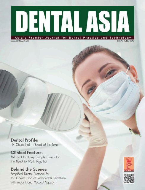
<strong>Dental</strong> Profile:<br />
Mr. Chuck Hall - Ahead of His Time<br />
Clinical Feature:<br />
ENT and Dentistry: Sample Cases for<br />
the Need to Work Together<br />
Behind the Scenes:<br />
Simplifi ed <strong>Dental</strong> Protocol for<br />
the Construction of Removable Prosthesis<br />
with Implant and Mucosal Support
Contents<br />
www.dentalasia.net<br />
Under the Spotlight<br />
18 Dr. Lawrence Lau: Rockstar in Dentistry<br />
24<br />
<strong>Dental</strong> Profile<br />
28<br />
32<br />
Clinical Feature<br />
36<br />
41<br />
46<br />
Dr. Grant Duncan: Shaping the Future<br />
of Orthodontics<br />
Mr. Chuck Hall: Ahead of His Time<br />
Dr. Angelo Mariotti: Personalising<br />
Dentistry<br />
Small, Powerful and Sterile: The Mighty<br />
Mini 90 Disposable Prophy Angle<br />
ENT and Dentistry: Sample Cases for<br />
the Need to Work Together<br />
Factors of Poor Breastfeeding<br />
Efficiency and Appropriate Management<br />
for Tongue-tie and Lip-tie<br />
Behind the Scenes<br />
54<br />
58<br />
Indepth With<br />
64<br />
65<br />
Elastic, Aesthetic Ceramics<br />
Simplified <strong>Dental</strong> Protocol for the<br />
Construction of Removable Prosthesis<br />
with Implant and Mucosal Support<br />
AUTO spin by Renfert: Universal<br />
System for Model Production with<br />
Acrylic Plates<br />
Nobel Biocare: Ease, Speed and<br />
Hygiene, All-in-one with PureSet <br />
Show Review<br />
70<br />
71<br />
IDEM <strong>2018</strong> Takes Clinical Excellence<br />
to the Next Level<br />
40 th <strong>Asia</strong> Pacific <strong>Dental</strong> Congress <strong>2018</strong>:<br />
“Intensifying Professionalism in<br />
Synergy with <strong>Dental</strong> Science and<br />
Technology”<br />
Show Preview<br />
72<br />
Regulars<br />
04<br />
06<br />
10<br />
66<br />
73<br />
74<br />
76<br />
DAMA <strong>2018</strong>: Inaugural <strong>Dental</strong><br />
Aesthetics Meeting in <strong>Asia</strong> Releases<br />
Conference Programme<br />
First Words<br />
Giving Back to Society<br />
<strong>Dental</strong> Updates<br />
Product Highlights<br />
Events Calendar<br />
Books for Sale<br />
Advertiser’s Index<br />
2 DENTAL ASIA<br />
MAY / JUNE <strong>2018</strong>
Rainer Wöran, AUS<br />
Amann Girrbach <strong>Asia</strong><br />
Fon +65 6592 5190<br />
singapore@amanngirrbach.com<br />
www.amanngirrbach.com
First Words<br />
Education is essential and<br />
necessary in any profession. But I<br />
always believed Aristotle’s quote<br />
saying ‘Educating the mind without<br />
educating the heart is no education<br />
at all.’ Being educated is not solely<br />
based on awards and recognitions<br />
a person receives but how you make<br />
use of all the knowledge gained in<br />
helping and giving back to others.<br />
Shaping the Future<br />
Through Education<br />
I have so much respect<br />
and appreciation to those who<br />
are willing to go out of their way<br />
just to help out those in need.<br />
<strong>Dental</strong> <strong>Asia</strong> wants to recognise<br />
these outstanding individuals,<br />
organisations and foundations<br />
contributing humanitarian efforts<br />
such as Dr. “T. Bob” Davis, who not<br />
only helps underserved people in<br />
Latin America but is also moulding<br />
the next generation of dentists by<br />
giving them purpose through sharing<br />
(page 6).<br />
There are so many ways in<br />
educating and spreading knowledge<br />
– conventional lectures/workshops<br />
and modern online platforms.<br />
A very good example is how<br />
Dr. Lawrence Lau maximises all these<br />
approaches through his lectures and<br />
the Cerec-files, a Facebook group<br />
that eventually became a community<br />
managed by clinicians for dentists<br />
solely to help in everything about<br />
CEREC (pages 18-22).<br />
Nowadays, modern platforms and<br />
innovations are<br />
adapted in the<br />
industry. Mobile<br />
applications<br />
now are used<br />
in dentistry<br />
specifically to<br />
monitor patients<br />
undergoing orthodontics known as<br />
<strong>Dental</strong> Monitoring (DM). Thanks to<br />
Dr. Grant Duncan, the pioneer DM<br />
user for Invisalign, patients are more<br />
compliant, see better results and are<br />
guided by their dentists with fewer<br />
dental visits (pages 24-27).<br />
Another dental innovation in<br />
dentistry that keeps on expanding<br />
is 3D printing. We are so grateful for<br />
the imagination and persistence of<br />
Mr. Charles “Chuck” Hull, the<br />
inventor of stereolithography<br />
(commonly known as 3D printing)<br />
for his remarkable breakthrough<br />
that changed the design and<br />
manufacturing world (pages 28-31).<br />
Aside from being persistent, I<br />
believe that loving one’s profession is<br />
imperative in order to be successful<br />
and of help to others. Through my<br />
interview with Dr. Angelo Mariotti,<br />
a periodontal specialist and an<br />
IDEM speaker, he shared that he<br />
loves his work and most especially<br />
the interactions he has with his<br />
patients. He continues promoting<br />
awareness of periodontal disease<br />
in hoping to help prevent its spread<br />
(pages 32-34).<br />
It was also a very educational<br />
few months for me attending<br />
IDEM <strong>2018</strong> and APEC <strong>2018</strong>, two<br />
of the biggest dental events held<br />
in <strong>Asia</strong>. It was a privilege and an<br />
honour to be present among the<br />
international speakers, exhibitors,<br />
manufacturers and colleagues in<br />
the industry (pages 70-71). I am<br />
excited and looking forward to<br />
further expanding my knowledge in<br />
the industry and in return pass it on<br />
to all <strong>Dental</strong> <strong>Asia</strong> readers. DA<br />
<br />
<br />
ADVISORY BOARD<br />
Dr William Cheung<br />
Dr Choo Teck Chuan<br />
Dr Chung Kong Mun<br />
Dr George Freedman<br />
Dr Fay Goldstep<br />
Prof Urban Hägg<br />
Prof Nigel M. King<br />
Dr Ramonito Rafols Lee<br />
Dr Kevin Ng<br />
Dr William O’Reilly<br />
Dr Ryan Seto<br />
Dr Adrian U J Yap<br />
Dr Christopher Ho<br />
Dr How Kim Chuan<br />
Dr Derek Mahony<br />
Prof Alex Mersel
Giving back to Society<br />
<strong>Dental</strong> Students Provide <strong>Dental</strong> Humanitarian Missions in Latin America<br />
by David Burger<br />
“Do what you can while you still can.”<br />
– Dr. T. Bob Davis<br />
It is a philosophy that has driven, General dentist Dr. Davis – more<br />
commonly known as “T. Bob”- for over 36 years to bring more than<br />
1,500 dental students and about 500 dentists to Latin America to<br />
provide dental humanitarian missions.<br />
The tradition continued this year, as Dr. Davis returned from a<br />
late March trip to Guatemala accompanied by 50 dental students<br />
—21 from Texas A&M University College of Dentistry and 29 from<br />
The University of Texas School of Dentistry at Houston. Along with<br />
16 dentists recruited by Dr. Davis, the large contingent treated 820<br />
people in the impoverished region of San Raymundo, Guatemala<br />
delivering more than US $329,000 worth of dental care.<br />
Dr. Davis, president of Academy of Dentistry International,<br />
said the trip during the<br />
students’ spring break<br />
was “probably the best<br />
trip ever in most every<br />
respect.” This is his sixth<br />
annual trip to Guatemala<br />
after years in Mexico<br />
and Nicaragua, and he<br />
said sustainability in his<br />
beloved San Raymundo is<br />
what inspired him to come<br />
back year after year. While<br />
the need for dentistry is<br />
still deep in this southern<br />
area, it brings Dr. Davis joy<br />
to see returning patients<br />
who now require simple<br />
cleanings rather than root<br />
canals.<br />
“It was unbelievable,”<br />
said Brady Atkins, a fourthy<br />
e a r d e n t a l s t u d e n t a t<br />
UT-Houston who went<br />
Vital assistance: First-year dental student<br />
Dorothy Hino and Dr. Rachel Sells treat a<br />
young patient in a Guatemala operatory<br />
d u r i n g t h e i r h u m a n i t a r i a n t r i p t o t h e<br />
Central American country in March.<br />
with Dr. Davis on the spring break trip for the fourth time.<br />
“Wanting to serve people and making a tangible dierence is what<br />
immediately drew me to dentistry.”<br />
During Mr. Atkins’ trips, he saw many people helping others and<br />
knew ‘that it was what he wanted to do’.<br />
John Ratli, a second-year dental student at Texas A&M, also<br />
went to Guatemala in March, his second time with Dr. Davis. “My<br />
learning can make a dierence in changing people’s lives. You see<br />
everything you see in school, in practice. The trips are a testament<br />
to people believing in T. Bob’s message: ‘Take a leap of faith and<br />
be of service to others’,” Mr. Ratli said.<br />
Dr. Davis’ trips have been annual rites of passage among many<br />
Texas A&M and UT-Houston dental students, who gravitate<br />
toward the trips in part because of Dr. Davis’ long time ties to the<br />
Christian <strong>Dental</strong> Society.<br />
Initially, Mr. Ratli was sceptical until he saw people in pain<br />
transformed after attending the Guatemala trips. “I was a little<br />
nervous but Dr. Davis is so organised and the dentists are all<br />
inspirational with their commitment to impacting the youth,”<br />
shared Mr. Ratli.<br />
Though volunteer dental students are busy accommodating<br />
patients and, at times, skipping their meals, students love helping<br />
during dental missions. Dr. Davis makes time to schedule worship<br />
services and fun team-building exercises, such as soccer games,<br />
giving the locals and dental students time to bond and enjoy.<br />
Dr. Davis went on his rst dental mission four decades ago, after<br />
his wife signed him up for it, and he immediately became hooked.<br />
It only took him ve years before he started taking dental students<br />
with him.<br />
“They get a sense of who they are becoming. They share their<br />
heart and love for people. They come here with a purpose in mind,”<br />
Dr. Davis said of the students.<br />
For more information about the Academy of Dentistry International,<br />
visit www.adint.org<br />
To learn more about international volunteerism and nd an<br />
opportunity, visit the ADA Foundation’s international volunteer<br />
website: www.internationalvolunteer.ADA.org.<br />
If anyone is interested in helping or funding products and<br />
equipment for the dental missions in <strong>Asia</strong> Pacic, please contact<br />
Ms. Jamie Tan at jamietan@pabloasia.com for more information. DA<br />
Hugging line: <strong>Dental</strong> students, from left, Chris Shin, Megan Luna,<br />
Abigail Livesay, Blake Hamblin and Raven Grant receive thanks from local<br />
students in San Raymundo, Guatemala, for treating them with dental<br />
care on a humanitarian trip in March.<br />
6 DENTAL ASIA<br />
MAY / JUNE <strong>2018</strong>
Get hooked!<br />
Hartzell Instruments Makes Gutta-Percha Removal Easy!<br />
Get hooked today!<br />
©2016 Den-Mat Holdings, LLC . All rights reserved. 805000129 08/16SN<br />
Make your next retreatment stress-free and<br />
order your Gutta-Percha removal instruments!<br />
Email: international@denmat.com<br />
To find a local distributor, visit www.denmat.com
Giving back to Society<br />
<strong>Dental</strong> Units Donated to Pobba Thiri <strong>Dental</strong> Hospital in Myanmar<br />
The Pobba Thiri <strong>Dental</strong> Hospital<br />
was established in 2016 as<br />
the first national hospital<br />
in Nay Pyi Taw, the capital<br />
of Myanmar, which has<br />
developed remarkably in recent years. This<br />
hospital is expected to further develop<br />
a s a b a se o f o ra l h ea lth c a re i n M y a n m a r.<br />
GC Corporation, one of the largest and most<br />
successful manufacturer for dental products<br />
today, contributed three <strong>Dental</strong> Units<br />
(Sovereign) to the Pobba Thiri <strong>Dental</strong> Hospital.<br />
A contribution ceremony was held at<br />
the site on 19 th February <strong>2018</strong>. Myanmar<br />
administration ocial, President Nakao and<br />
Mr. Watanabe, Chairman of Japan Myanmar<br />
Association attended the ceremony.<br />
After the ribbon cutting opening<br />
ceremony, a site inspection and a lunch<br />
gathering was held with many hospital<br />
staff. In the afternoon, GC Corporation<br />
visited Dr. Myint Htwe, Union Minister of<br />
Myanmar (Ministry of Health and Sports).<br />
GC has promised to continually cooperate<br />
and support Myanmar government, not<br />
only materials but also in education and<br />
training of dental health care staff for<br />
improvement of oral health in Myanmar.<br />
After the discussion, President Nakao<br />
received a certicate of appreciation from<br />
the minister.<br />
Four GC associates namely Mr. Saruya<br />
P r e s i d e n t o f G C I C o m m u n i c a t i o n s ,<br />
M r . K u r a s h i m a , M r . M i z u t a n i a n d<br />
Mr. Koshiba worked diligently towards the<br />
success of the event. They were responsible<br />
for the communication and negotiation<br />
w i t h M y a n m a r G o v e r n m e n t , H o s p i t a l a n d<br />
Japan Myanmar Association and the<br />
installation of dental units in the hospital. DA<br />
<strong>Dental</strong> Missions by the Rise Above Foundation Cebu<br />
Originally connected with a<br />
foundation in Manila from<br />
2000-2008, Rise Above<br />
Foundation Cebu Inc.,<br />
became an independent<br />
group in Cebu in July 2008. Behind the<br />
foundation is a group of dedicated<br />
individuals, local and from around the<br />
world, whose main concern is bringing<br />
humanitarian help to everyone in need.<br />
The rst <strong>Dental</strong> Mission took place in<br />
July 2002 and since then <strong>Dental</strong> Missions<br />
have been held two to three times a year.<br />
Dentists, dental technicians, hygienists<br />
and other volunteers from Denmark,<br />
Sweden, Norway, England, Germany,<br />
Switzerland, Australia, New Zealand, Japan,<br />
Singapore, Iran and USA have travelled to<br />
Cebu, Philippines to help treat the poor<br />
in depressed areas in the cities of Cebu,<br />
Mandaue, Talisay and Lapulapu, as well as<br />
several small islands near Cebu and other<br />
bigger islands in the Visayas region.<br />
Through the years, over 35,000 people<br />
have received free treatment, sponsored<br />
by companies and individuals from all over<br />
the world.<br />
THE NEED:<br />
1. <strong>Dental</strong> Missions welcome dentists,<br />
dental hygienists, dental technicians,<br />
dental assistants and anyone else who<br />
wants to give their services to the<br />
underserved population in the Visayas<br />
region of the Philippines. It has all the<br />
equipment and instruments needed,<br />
but needs volunteers to come and help.<br />
2. Financial aid. The average cost per<br />
patient is 300 Pesos. Often patients have<br />
more than one tooth that needs to be<br />
treated. The Mission gives treatment like<br />
restoration, extraction and prophylaxis.<br />
The patients receive the needed<br />
medication free of charge. Any help will<br />
be greatly appreciated.<br />
For more information on how<br />
t o b e p a r t o f t h i s , p l e a s e w r i t e t o :<br />
riseabove@riseabove-Cebu.org DA<br />
8 DENTAL ASIA<br />
MAY / JUNE <strong>2018</strong>
<strong>Dental</strong> Updates<br />
New 3Shape and Oventus Integration Gets Patients Into Sleep Therapy Faster<br />
New digital integration between<br />
Oventus and the 3Shape TRIOS intraoral<br />
scanner creates a faster devicemanufacturing<br />
turnaround time to get<br />
patients into sleep therapy faster.<br />
The just-announced integration enables<br />
dental professionals using the awardwinning<br />
3Shape TRIOS intraoral scanner to<br />
send digital impressions direct to Oventus<br />
for the ordering and designing of its sleep<br />
devices for snoring and sleep apnea.<br />
3Shape TRIOS users can simply choose<br />
Oventus from the more than 45 treatment<br />
providers in the intraoral scanner’s menu to<br />
instantly cloud-send the digital impressions<br />
to Oventus.<br />
According to Dr. Chris Hart, Clinical<br />
Director and inventor of Oventus,<br />
“ T h e d i g i t a l w o r k f l o w w i t h<br />
3Shape TRIOS increases overall<br />
efficiency in its production<br />
workow, particularly quicker<br />
turnaround times from start<br />
to finish. This enables<br />
patients with sleep apnea<br />
to be treated more quickly,<br />
which is appreciated by patients<br />
and referring providers alike.”<br />
The digital workow with 3Shape TRIOS<br />
also increases accuracy, which in turn<br />
means fewer remakes and less chair time for<br />
patients, according to Oventus. Adding that<br />
the digital workow eliminates analoguerelated<br />
challenges such as impressions<br />
being distorted because of gagging, tongue<br />
thrusts, saliva contamination, shipping<br />
temperature, or imprecise lab pour-ups.<br />
“We are excited to bring<br />
another terrific treatment<br />
opportunity like Oventus to<br />
doctors and their patients.<br />
The digital workflow,<br />
because of its accuracy, speed<br />
and connectivity, makes it so<br />
much easier for professionals<br />
to provide quality care to their patients. As<br />
more and more solutions choose to connect<br />
with TRIOS, its accuracy, ease of use, and<br />
open and expanding ecosystem, make it<br />
the right choice for dental professionals,”<br />
M r . A l l a n H y l d a l , V i c e P r e s i d e n t o f<br />
3Shape Orthodontics. <br />
<strong>2018</strong> ADIA Research Grants Tackles Periodontitis<br />
Pioneering research to address chronic<br />
periodontitis has been awarded to the<br />
Australian <strong>Dental</strong> Industry Association<br />
Research Grant bestowed by the<br />
Australian <strong>Dental</strong> Research Foundation (ADRF),<br />
the nation’s leading funding body for earlycareer<br />
researchers into dental and oral<br />
health issues. The grant is funded by the<br />
Australian <strong>Dental</strong> Industry Association (ADIA),<br />
the peak business organisation representing<br />
innovative dental product manufacturers<br />
and suppliers.<br />
The most recent award was provided to<br />
Associate Professor Neil O’Brien-Simpson<br />
from the University of Melbourne<br />
<strong>Dental</strong> School into research associated<br />
with antimicrobial peptides that target<br />
periodontal pathogens. He’s a project<br />
manager for the Oral Health Cooperative<br />
Research Centre. The research project<br />
will be supported by Dr. Jason Lenzo<br />
a n d D r . J a m e s H o l d e n , b o t h f r o m t h e<br />
Melbourne <strong>Dental</strong> School.<br />
The research seeks to address chronic<br />
periodontitis, an inflammatory disease<br />
associated with a pathogenic subgingival<br />
bacteria leading to the destruction of the<br />
tooth’s supporting tissues and ultimately<br />
tooth loss. It is a major public health<br />
problem in all societies and is estimated to<br />
aect at least 30% of the adult population,<br />
including 5% to 6% who experience severe<br />
forms of the disease. The prevention of<br />
chronic periodontitis relies on removing<br />
the bacterial burden by effective tooth<br />
cleaning, sometimes in combination with<br />
antibiotics and/or antiseptics; however,<br />
long-term use of antibiotics is undesirable<br />
due to the emergence of resistant bacteria.<br />
Indeed, several reports have highlighted<br />
an increasing trend towards antibiotic<br />
resistance in oral bacteria.<br />
The increasing reports of multi-drug and<br />
antibiotic resistant bacteria and the limits on<br />
the use of antiseptics, such as chlorhexidine,<br />
have resulted in extensive research to<br />
discover and develop new and novel classes<br />
of antimicrobials that are eective and safe<br />
for human use.<br />
“The objectives of the research<br />
a l i g n w i t h t h e c o n d i t i o n s o f t h e<br />
Australian <strong>Dental</strong> Industry Association<br />
Research Grant that provides<br />
funding for projects that may,<br />
following commercialisation, support<br />
the development of new and/or<br />
innovative dental and oral healthcare<br />
treatment pathways or products,” said<br />
Mr. Troy Williams, ADIA Chief Executive<br />
Ocer.<br />
The Australian <strong>Dental</strong> Industry<br />
Association Research Grant provides<br />
funding to explore the potential of a<br />
n o v e l c l a s s o f a n t i b i o t i c s c a l l e d<br />
antimicrobial peptides (AMP). The research<br />
team have already identied several AMPs<br />
by altering their sequence and adding<br />
chemical moieties that reduce toxicity<br />
to mammalian cells while targeting and<br />
enhancing antibacterial activity towards<br />
periodontal pathogens.<br />
“ADIA has a long track-record of<br />
supporting the development of innovative<br />
patient diagnostic and treatment<br />
options and it’s in this context that we’re<br />
c o mm i tted to th e lo n g -term fu n di n g o f th e<br />
Australian <strong>Dental</strong> Industry Association<br />
Research Grant is funded,” Mr. Williams<br />
said. <br />
10 DENTAL ASIA<br />
MAY / JUNE <strong>2018</strong>
<strong>Dental</strong> Updates<br />
Convergent <strong>Dental</strong> Expands Executive Leadership Team with Two New Additions<br />
Convergent <strong>Dental</strong>, Inc., developers<br />
of Solea ® , the industry-leading CO 2<br />
all-tissue dental laser system, is pleased<br />
to announce two new appointments<br />
t o i t s e x e c u t i v e l e a d e r s h i p t e a m .<br />
Mr. Daryl Donatelli recently joined the<br />
Company as Vice President of Marketing,<br />
and Mr. Charles Kerbage now<br />
holds the title of Vice President of<br />
Research and Development.<br />
In his new role as Vice President<br />
of Marketing, Mr. Daryl Donatelli<br />
w i l l d r i v e a l l m a r k e t i n g e f f o r t s f o r<br />
Convergent <strong>Dental</strong>’s agship product Solea,<br />
the No.1 selling dental laser for hard, soft,<br />
and osseous tissue procedures. Mr. Donatelli<br />
will lead Convergent <strong>Dental</strong>’s marketing<br />
team eorts including product strategy,<br />
marketing communications, business<br />
partnerships, and sales enablement.<br />
“I’m very excited to be part of creating<br />
what will denitely be a new standard of<br />
care for dentistry,” remarked Mr. Donatelli.<br />
“I am even more excited about enabling<br />
an experience for patients that will forever<br />
change what it means to go to the dentist.”<br />
Mr. Donatelli brings over fifteen<br />
years of executive marketing experience<br />
to Convergent <strong>Dental</strong>, including the<br />
commercialisation<br />
and growth of major<br />
medical devices<br />
a n d c o n s u m e r h e a l t h p o r t f o l i o s a c r o s s<br />
North America, <strong>Asia</strong> and Europe.<br />
Mr. Donatelli holds a BA in Economics<br />
from the University of Rochester, and an<br />
MBA from Simon Business School.<br />
In addition, Mr. Charles Kerbage<br />
h a s j o i n e d C o n v e r g e n t D e n t a l a s<br />
Vice President of Research and Development.<br />
Mr. Kerbage will lead the Company’s R&D<br />
team in identifying opportunities for<br />
innovation that will both improve existing<br />
product performance and bring new<br />
capabilities into the company’s product<br />
portfolio.<br />
“Having come from the laser industry, I<br />
was attracted to the innovative culture and<br />
commitment to creating excellent products<br />
that exists at Convergent <strong>Dental</strong>,” remarked<br />
Mr. Kerbage. “The folks at Convergent are<br />
true pioneers in dental laser technology,<br />
and their reputation for a superior customer<br />
experience is second to none. I am thrilled<br />
to be working with such a high-powered and<br />
motivated team of professionals.”<br />
As a technical leader with strong business<br />
acumen, Mr. Kerbage has over 15 years of<br />
experience in scientic and technological<br />
innovation and has transitioned numerous<br />
technical developments into gamechanging<br />
solutions in the medical devices,<br />
life sciences, and photonics industries.<br />
M r . K e r b a g e h o l d s a P h D i n<br />
Applied Physics from Columbia University,<br />
and an MBA from Boston University. <br />
Complete Make-over: University of Pretoria Installs Dentsply Sirona’s Technologies<br />
I n J a n u a r y 2 0 1 8 , t h e<br />
University of Pretoria and Dentsply Sirona<br />
completed an ambitious renovation project.<br />
In just four weeks, Dentsply Sirona equipped<br />
two levels of the Faculty of Health Sciences,<br />
Prinshof Campus, School of Dentistry with<br />
its technologies – in particular 22 Intego<br />
treatment centres.<br />
The University of Pretoria (South Africa)<br />
has chosen Dentsply Sirona’s<br />
technologies for its ambitious renovation<br />
p r o j e c t . T h e S c h o o l o f D e n t i s t r y o f t h e<br />
F a c u l t y o f H e a l t h S c i e n c e s ,<br />
P r i n s h o f C a m p u s , h a s a l r e a d y u s e d<br />
Dentsply Sirona’s CAD/CAM technologies<br />
and imaging systems. Now, Dentsply Sirona<br />
installed 22 Intego treatment centres and<br />
additional equipment on two levels: On<br />
level 02 in the Department of Maxillo-Facial<br />
and Oral Surgery as well as on level 03 in the<br />
Department of Prosthodontics.<br />
Renovation while running operations<br />
Requirements for the comprehensive<br />
r e n o v a t i o n p r o j e c t w e r e a m b i t i o u s .<br />
“Our hospital could<br />
not stop operations.<br />
So, we had to t all<br />
patients in on the<br />
available oors. Any<br />
delay would not only<br />
have affected the<br />
students who started<br />
in February, but<br />
also the patients”,<br />
explained Professor AJ Ligthelm, CEO and<br />
Chairperson of the School of Dentistry.<br />
Besides the treatment centres<br />
Dentsply Sirona equipped also the sterilising<br />
rooms and the joinery on the two levels.<br />
In the Department of Prosthodontics,<br />
Dentsply Sirona also installed third party<br />
equipment for the clinical lab.<br />
Ready to use in just four weeks<br />
Dentsply Sirona completed the renovation<br />
i n v e r y s h o r t t i m e . M r . A n d y C y p r i a n o s ,<br />
C o m m e r c i a l D i r e c t o r D e n t s p l y S i r o n a<br />
South Africa, describes the challenge: “We<br />
were dependent on other contractors to be<br />
nished before we could<br />
start. Therefore, we had<br />
to cope with a very short<br />
time frame to complete<br />
our installations by the<br />
end of January <strong>2018</strong>”.<br />
Intego and more –<br />
new equipment for<br />
students<br />
In the Department of Maxillo-Facial and<br />
Oral Surgery Dentsply Sirona set up six<br />
operatories and in the Department of<br />
Prosthodontics in total 16 cubicles.<br />
The renovation works in the<br />
Department of Prosthodontics were even<br />
more extensive. Dentsply Sirona equipped<br />
cubicles for the sta and for the students.<br />
I n t h e d e p a r t m e n t ’ s c l i n i c a l l a b ,<br />
Dentsply Sirona put in third party<br />
equipment such as polishing lathes,<br />
micromotors and extraction units. In the<br />
Department of Prosthodontics, third party<br />
suppliers contributed also cleaning devices<br />
such as instrument washers. <br />
12 DENTAL ASIA<br />
MAY / JUNE <strong>2018</strong>
Service Engineers from<br />
Roland D G Australia and Aarque in<br />
New Zealand Win Big in<br />
Global SE Awards<br />
Mr. Mark Johnson<br />
→ THE EVOLUTION<br />
OF PROPHYLAXIS<br />
NEW COMBI touch<br />
→ AIR-POLISHING AND ULTRA-GENTLE<br />
ULTRASOUND IN ONE UNIT<br />
→ www.mectron.com<br />
Mr. Peter Holbrook<br />
Roland DG Australia, a leading manufacturer of wide<br />
format inkjet printers and 3D devices, are excited to announce<br />
that Roland DG Australia Service Agent, Mr. Peter Holbrook,<br />
and Mr. Mark Johnson of Aarque in New Zealand both won<br />
awards as a part of the Roland DG Global SE Awards.<br />
The Global SE Awards <strong>2018</strong> is the third global contest to<br />
be held, following the most recent event in 2015. From a total<br />
of 778 Roland certied Service Engineers (SEs) around the<br />
world, 28 nalists from the local competitions were brought<br />
together for the nal contest, held from 23 rd to 25 th Ap ri l a t<br />
Roland DG Corporation in Hamamatsu, Japan.<br />
T h e t h r e e - d a y c o m p e t i t i o n i n c l u d e d w r i t t e n a n d<br />
hands-on exams to test the participants’ skills and knowledge<br />
of maintenance services. Representing the Oceania region<br />
(Australia and New Zealand), Mr. Mark Johnson of Aarque in<br />
New Zealand, took out 1 st place in the inkjet printer category,<br />
whilst 2 nd place went to Mr. Matthew Wrench from Roland DG UK<br />
and 3 rd place went to Mr. Vincent Votruba from Slovakia.<br />
As a part of the competition, Mr. Peter Holbrook,<br />
o f R e d P e r i l , R o l a n d D G ’ s S e r v i c e A g e n t f o r<br />
Western Australia, was awarded the Engineer whose<br />
customer satisfaction score was highest for the <strong>Asia</strong> Pacic<br />
region (Korea, ASEAN, NZ, Australia, China and Japan), based<br />
on the results from satisfaction surveys sent to customers.<br />
Roland DG Australia would like to congratulate both<br />
Mark and Peter on their well-deserved awards, as well<br />
finalists Mr. Allan Cooke, SE for the QLD region, and<br />
Mr. Darren McInnes, SE for the VIC region, for their continued<br />
hard work and for making it to the top 28 SEs in the world. <br />
120° 90° PERIO<br />
→ easy switch from supra to subgingival<br />
air-polishing by a simple click<br />
→ the best access thanks to 3 available nozzles<br />
→ soft mode: maximum comfort for sensitive<br />
patients<br />
→ more than 40 inserts for the widest range<br />
of ultrasound indications<br />
DENTAL ASIA<br />
MAY / JUNE <strong>2018</strong>
<strong>Dental</strong> Updates<br />
CS 8100SC 3D Wins <strong>2018</strong> Edison Award for its Innovative Design<br />
The CS 8100SC 3D<br />
e x t r a o r a l i m a g i n g s y s t e m b y<br />
Carestream <strong>Dental</strong> has been awarded a<br />
bronze <strong>2018</strong> Edison Award in the category of<br />
<strong>Dental</strong>/Medical Digital Imaging by the<br />
internationally renowned Edison Awards.<br />
T h e d i s t i n g u i s h e d a w a r d , i n s p i r e d b y<br />
Thomas Edison’s persistence and<br />
inventiveness, recognises innovation,<br />
creativity and ingenuity in the global<br />
economy.<br />
“It’s exciting to see companies<br />
l i k e C a r e s t r e a m D e n t a l c o n t i n u i n g<br />
Thomas Edison’s legacy of challenging<br />
c o n v e n t i o n a l t h i n k i n g , ” s a i d<br />
Mr. Frank Bonafilia, executive director,<br />
Edison Awards. “Edison Awards recognises<br />
the game-changing products and services,<br />
and the teams that brought them to<br />
consumers.”<br />
The award was announced on 11 th April<br />
<strong>2018</strong>, at the Edison Awards Annual Gala,<br />
held in the historic ballroom of The Capitale<br />
in New York City, N.Y. Mr. Matthew Greer,<br />
regional product manager for extraoral<br />
imaging, was in attendance to accept the<br />
award on Carestream <strong>Dental</strong>’s behalf.<br />
“Carestream <strong>Dental</strong> always strives<br />
to deliver innovative technology that<br />
s u p p o r t s d o c t o r s i n t h e i r d i a g n o s e s , ”<br />
Ed Shellard, D.M.D., chief dental ocer,<br />
Carestream <strong>Dental</strong> said. “Winning a bronze<br />
<strong>2018</strong> Edison Award reinforces that the<br />
company is meeting the needs of doctors<br />
and their patients around the world.”<br />
To earn a bronze <strong>2018</strong> Edison Award,<br />
the CS 8100SC 3D was judged on its concept<br />
and development, value and impact on the<br />
industry.<br />
The CS 8100SC 3D oers two-dimensional<br />
panoramic imaging, cephalometric imaging,<br />
cone beam computed tomography (CBCT)<br />
imaging and model/impression scanning<br />
all in one compact system. Its minimal<br />
f o o t p r i n t a n d a f f o r d a b i l i t y m a k e s<br />
3D imaging available to smaller practices<br />
that either couldn’t aord or didn’t have<br />
space for an extraoral imaging system in<br />
the past. That means doctors can go from<br />
diagnosis to treatment faster, without<br />
having to send patients to an imaging<br />
centre. Not only is the system more<br />
convenient, but it’s also faster—featuring<br />
the fastest scanning cephalometric<br />
m o d u l e o n t h e m a r k e t — a n d s a f e r . T h e<br />
CS 8100SC 3D’s low dose program can<br />
deliver 3D imaging at a dose equal to or<br />
lower than panoramic imaging.<br />
“The success of the CS 8100SC 3D<br />
began more than a decade ago with<br />
the initial development of the awardwinning<br />
CS 8100,” Dr. Shellard said. “That<br />
one system would eventually grow a<br />
‘family’ of units, all with dierent imaging<br />
capabilities. The CS 8100SC 3D represents<br />
Carestream <strong>Dental</strong>’s ultimate goal<br />
by combining the best features of<br />
its predecessor systems into one<br />
multifunctional solution making it a costeective,<br />
powerful imaging available for<br />
more doctors.”<br />
Edison Award nominees are judged<br />
by more than 3,000 senior business<br />
executives and academics from across<br />
th e n a ti o n , wh o se vo tes a c kn o wledg e th e<br />
CS 8100SC 3D’s success in meeting the<br />
organisation’s stringent criteria of quality.<br />
The voting panel includes members of:<br />
Chief Marketing Officer Council (CMO);<br />
Design Management Institute (DMI);<br />
American Productivity & Quality Center<br />
(APQC); American Society of Mechanical<br />
Engineers (ASME); Georgia State Marketing<br />
Roundtable (GSU); Product Development<br />
and Management Association (PDMA);<br />
Association of Technology Management<br />
a n d A p p l i e d E n g i n e e r i n g ( AT M A E ) ; p a s t<br />
Edison Award winners; marketing<br />
professionals; scientists; designers;<br />
engineers; and academics. <br />
Henry Schein Buys 50% of Ortho2<br />
Henry Schein Inc, Melville, NY, and<br />
Ortho2, Ames, Iowa, have signed a<br />
den i t i v e a g r e e m e n t a i m e d a t i n c r e a s i n g<br />
Henry Schein’s position in the orthodontic<br />
practice management software market. As<br />
a result of the denitive agreement signed<br />
by the two companies, Henry Schein will<br />
own a 50% interest in Ortho2.<br />
“We welcome our new Ortho2<br />
colleagues to Henry Schein, and look forward<br />
t o c o n t i n u e d s u c c e s s t o g e t h e r , ” s a i d<br />
Mr. Stanley M. Bergman, chairman of<br />
the board and chief executive oce r o f<br />
Henry Schein. “With this investment in<br />
Ortho2, we round out our dental software<br />
portfolio and gain a leadership position<br />
in orthodontist practice management<br />
software. This transaction supports our<br />
to advanced technology, and to<br />
category leadership. We look forward to<br />
oering advanced software solutions to<br />
orthodontists, just as we currently do to<br />
general practice dentists.”<br />
Founded in 1982 by Mr. Dan Sargent and<br />
Dr. William Iversen, Ortho2 currently has a<br />
sta of over 70 and oers the Edge Cloud<br />
practice management solution, as well<br />
as ViewPoint, a Windows-based software<br />
option. According to a press release,<br />
Ortho2 serves nearly 1,700 orthodontists<br />
in approximately 2,400 locations, and had<br />
recorded revenues of $14 million.<br />
“We are delighted to be joining forces<br />
with Henry Schein,” said Ms. Amy Schmidt,<br />
president, Ortho2. “We’re proud of what<br />
we’ve accomplished in our 35 years as<br />
The ongoing technology explosion<br />
and evolving orthodontic market now<br />
m a kes j o i n i n g fo rc es wi th Hen ry Sc h ei n a<br />
win-win for us and, most signicantly, for<br />
o u r c u s t o m e r s . T h e a b i l i t y t o l e v e r a g e<br />
Henry Schein’s comprehensive ecosystem<br />
will allow us to offer a turnkey solution<br />
including software, digital imaging,<br />
equipment, supplies, and many other<br />
benefits to our current and future<br />
customers.”<br />
“Practice management software is<br />
the backbone of an efficient, profitable<br />
practice,” Mr. Bergman added. “Henry Schein<br />
embraces an open architecture approach<br />
to all software and advanced technology,<br />
which allows for the purchase of products<br />
and services that best meet the needs of<br />
each individual practice.” <br />
commitment to dental specialty markets, a c o m p l e t e l y i n d e p e n d e n t c o m p a n y .<br />
14 DENTAL ASIA<br />
MAY / JUNE <strong>2018</strong>
<strong>Dental</strong> Updates<br />
Glidewell <strong>Dental</strong> to Present 2 nd Annual Educational Symposium<br />
Glidewell <strong>Dental</strong>, a leading technological<br />
innovator in restorative dentistry for<br />
o v e r 4 8 y e a r s , h a s a n n o u n c e d t h e<br />
2 nd Annual Glidewell <strong>Dental</strong> Symposium.<br />
Scheduled for 19 th -20 th October,<br />
<strong>2018</strong>, at the Gaylord National<br />
Reso rt & Co n ven ti o n Cen tre i n<br />
National Harbor, Maryland, near<br />
Washington, D.C., this event<br />
will include more than a dozen<br />
prominent educators providing two days<br />
of intensive instruction, with a rst day of<br />
fast-paced presentations and a second day<br />
of in-depth clinical and business training.<br />
“Dentists have more options for<br />
treating patients today than ever before,”<br />
said Dr. Neil Park, scientific chair of the<br />
Glidewell Symposium and vice president<br />
of clinical affairs at Glidewell <strong>Dental</strong>.<br />
“This period of rapid innovation in dental<br />
technology presents many opportunities<br />
for clinicians to expand their knowledge<br />
and practice offerings, and to explore<br />
more eective and ecient solutions for<br />
elevating the standard of care within our<br />
profession.”<br />
The symposium brings together<br />
notable experts from many different<br />
backgrounds and specialties,<br />
including aesthetic dentistry, digital<br />
dentistry, dental implants, tissue<br />
regeneration, sleep-related dentistry,<br />
practice management, and more. The event<br />
kicks o Friday, 19 th October, with<br />
a presentation on the current<br />
and future state of dentistry<br />
from Mr. Jim Glidewell,<br />
C D T , p r e s i d e n t a n d C E O o f<br />
Glidewell <strong>Dental</strong>, followed by<br />
an energising lineup of informative<br />
presentations and a very special keynote<br />
address delivered by Dr. Jack Hahn, a<br />
legendary pioneer in dental implantology.<br />
On Saturday, 20 th October, in-depth,<br />
hands-on workshops will be led by some<br />
of the most distinguished leaders in<br />
clinical dentistry, representing faculties<br />
of such esteemed organisations as the<br />
M i sc h In tern a ti o n a l Im p la n t In sti tu te, th e<br />
International Congress of Oral Implantologists,<br />
and the American Academy of Implant Dentistry.<br />
The workshops will provide practical clinical<br />
information on the latest innovations,<br />
protocols, products and procedures in the<br />
field, so that clinicians can immediately<br />
implement focused techniques into their<br />
practice when they return to the oce on<br />
Monday.<br />
“Following the success of last year’s<br />
sold-out symposium, we received an<br />
abundance of feedback from attendees<br />
who expressed their desire for refining<br />
their clinical skills and adopting the latest<br />
technology to grow their practice,” said<br />
Dr. Park. “They inspired us to expand the<br />
event to two days, to cover even more<br />
clinical and business topics, with nearly a<br />
dozen hands-on Saturday workshops to<br />
choose from. And we’ve moved to a more<br />
spacious venue to accommodate a vast<br />
array of interactive exhibits and technology<br />
demonstrations — to create an even bigger<br />
and better experience than last year.”<br />
During both days of the event, the<br />
exhibition floor will be packed wall to<br />
wall with displays of 3D printing, intraoral<br />
scanning, in-office milling, the CAD/CAM<br />
workow, dental implants, and regenerative<br />
and restorative materials, as well as live<br />
demonstrations and a virtual reality tour of<br />
Glidewell <strong>Dental</strong>’s expansive R&D campus,<br />
technology centre and laboratory.<br />
The <strong>2018</strong> Glidewell <strong>Dental</strong> Symposium is<br />
designed for dentists, hygienists, and chairside<br />
and front-office auxiliaries, and attendees<br />
enrolled for both days will earn up to 12 hours<br />
of continuing education (CE) credits. <br />
Renfert Education – Opening Event<br />
On 10 th April <strong>2018</strong>, Renfert opened<br />
their new training rooms at the Renfert<br />
headquarters in Hilzingen with guests<br />
from the press, cooperation partners and<br />
opinion leaders.<br />
Renfert took the opening ceremony<br />
as an opportunity to present their new<br />
training rooms and show guests the Renfert<br />
philosophy, the courses on oer and the<br />
unique transmission technique using the<br />
video microscope EASY view 3D.<br />
Renfert Course Themes<br />
Renfert training venue offers space for<br />
12 participants to work at state-of-theart<br />
workstations. These workstations<br />
are equipped with the most modern<br />
devices, including a video microscope<br />
with 3D monitor. The video microscope<br />
transmits the magnification via the<br />
m o n i t o r i n t h e o p t i o n a l 2 D o r 3 D m o d e s .<br />
The “Demonstrator” workstation is<br />
additionally equipped with a PC for live<br />
transmission to the monitors. There is<br />
also a at screen that can be used for live<br />
transmission and presentations.<br />
Course program<br />
Every restoration follows a process that has<br />
to be mastered. Teaching individual modules<br />
as well as complete work processes makes<br />
it possible to create restorations that are<br />
even better in technical terms and that are<br />
close to nature. Renfert will in future oer<br />
courses for dental technicians, dentist and<br />
dental assistants. Courses are developed by<br />
Renfert specically or in collaboration with<br />
cooperation partners.<br />
• Histoanatomic topography of the<br />
teeth<br />
• Monolithic, aesthetic and hyper<br />
realistic near to nature wax-up<br />
• Biomorphology of the teeth<br />
• Implementation using layering<br />
ceramics, pressed ceramics, CAM<br />
technology<br />
• Functional gauge, occlusal compass,<br />
NAT- natural wax-up technology<br />
• Model fabrication (systems based on:<br />
plaster and plastic base-plates)<br />
• <strong>Dental</strong> photography (studio and<br />
patient photography)<br />
• <strong>Dental</strong> communications training<br />
(coaching, marketing) <br />
16 DENTAL ASIA<br />
MAY / JUNE <strong>2018</strong>
Under the Spotlight<br />
Rockstar in<br />
Dentistry<br />
Interviewed by Ms. Jamie Tan<br />
Written by Dr. Chala R. Platon<br />
THROUGH upholding the philosophy<br />
of pursuing excellence in personalised<br />
dental care, with comprehensive treatment<br />
planning using both restorative and<br />
cosmetic dentistry to achieve optimal<br />
health, Dr. Lawrence Lau together with his<br />
wife (Dr. Gloria Shih) are the principal dentists<br />
a n d o w n e r s o f S y d n e y L a s e r D e n t a l C a r e .<br />
It is located at Sylvania Waters which later<br />
on successfully expanded to two more<br />
branches in Pyrmont and Illawong.<br />
Looking back<br />
Prior from entering dentistry,<br />
D r . L a u a t t e n d e d b o a r d i n g s c h o o l a t<br />
St. Joseph’s College. In 1997, he graduated<br />
with honours from the University of Sydney<br />
with a Bachelor of <strong>Dental</strong> Surgery.<br />
Since graduation, he has not stopped and<br />
spent extensive time on continuing education,<br />
to<br />
keep his dental practice at the<br />
forefront of dental technology.<br />
This includes the use of CEREC<br />
technology to optimise the<br />
outcomes of his cosmetic and<br />
restorative procedures, laser<br />
dentistry, guided implant surgery<br />
and<br />
a range of orthodontic<br />
treatments. His enthusiasm in<br />
expanding knowledge comes from<br />
understanding that dentistry is a<br />
relentlessly evolving practice.<br />
“So when we graduated, we were<br />
warned not to take on anything too<br />
challenging. Our professors/ mentors<br />
guided us to keep within our boundaries. As<br />
a result, I feel we did come out with better<br />
skills sets,” reminisced Dr. Lau.<br />
He shared that graduates nowadays<br />
come out of dental universities more<br />
condent but still lack clinical experience.<br />
During his time, graduates tended to be<br />
very conservative and had to search for<br />
answers on their own.<br />
Provision of excellent care<br />
Dr. Lau’s practice is a general practice<br />
rst and foremost. He specialises in the<br />
provision of excellent care for his patients<br />
whether it be in orthodontics, implantology<br />
or endodontics.<br />
“Basically, we don’t specialise in any one<br />
thing but we specialise in trying to give our<br />
patients the best experience possible,” he said.<br />
Presently, his practice strongly<br />
considers digitalisation as a contributing<br />
factor in their success. He believes that<br />
laser gives the perception to the general<br />
public that a practice is incorporating a<br />
very niche, unique and advance technology.<br />
18<br />
DENTAL ASIA<br />
MAY / JUNE <strong>2018</strong>
The universe at your fingertips.<br />
The highest technology dental laser system<br />
Supreme clinical results:<br />
TwinLight ® Perio Treatments<br />
TwinLight ® Endo Treatments<br />
No-sutures soft-tissue surgery<br />
Patient-friendly conservative dentistry<br />
Pre-sets for over 40 applications<br />
Unmatched simplicity of use:<br />
Balanced and weightless OPTOflex ® arm<br />
Nd:YAG handpiece detection system<br />
Quantum Square Pulse technology for fast<br />
minimally invasive treatments<br />
X-Runner TM - the first digitally controlled<br />
Er:YAG dental laser handpiece<br />
Visit www.fotona.com to find out more!<br />
Fotona App<br />
www.fotona.com<br />
88897/25
Under the Spotlight<br />
Patients often come into the clinic inquiring<br />
about laser dentistry. Dr. Lau sees this as a great<br />
conversation opener wherein he can talk about<br />
their advance practice giving them a favourable<br />
market position.<br />
“A lot of patients do come in and ask ‘what<br />
is laser dentistry?’. It has made our practice<br />
successful and it has denitely made us stand out<br />
from the rest,” he stated.<br />
Dr. Lau constantly upgrades his materials<br />
and equipment in his practice. Moreover, his<br />
practice is very aesthetically-driven which means<br />
he surrounds himself with pleasing and functional<br />
equipment. It is no surprise why Dentsply Sirona<br />
has been part of his practice as he appreciates how<br />
useful and appealing the company’s products and<br />
equipment are.<br />
In fact, during IDEM <strong>2018</strong>, Dr. Lau purchased<br />
two additional chairs to be installed in his main<br />
practice – making it a five chair practice. He<br />
will be incorporating the Teneo, the top of the<br />
line dental chair from Dentsply Sirona, which<br />
includes numerous functions (massage, implant,<br />
endodontic functions with intraoral cameras<br />
and screens). He is looking forward to this new<br />
enhancement to his practice and believes that it<br />
will be benecial for the patient and for him. In<br />
addition, he recently invested in a 3D printer which<br />
will further contribute to the ecient and modern<br />
workow of his practice.<br />
Fully embracing continuing education<br />
Aside from frequent upgrades to his materials<br />
and equipment, Dr. Lau certainly considers that<br />
continuing education is an integral part for all<br />
professionals. With the fast pace of technology and<br />
change in the eld of medicine and dentistry, he<br />
recognises that it is a duty to keep oneself updated.<br />
“It is not just sitting down and listening to<br />
lectures but it is also speaking to peers, to the<br />
industry and learning as much as you can. In my<br />
opinion, I nd that the younger generations fully<br />
embrace continuing education. Unfortunately, and<br />
sadly, some of my older colleagues may not share<br />
the same view,” he shared.<br />
Though this might be the case, Dr. Lau believes<br />
that it is slowly changing. He believes that through<br />
the help of the government body, organisations,<br />
associations and societies they can provide better<br />
continuing education and help everybody to<br />
participate even more.<br />
He mentioned that it is good to see colleagues<br />
wanting to learn further on new advancements<br />
20 DENTAL ASIA<br />
MAY / JUNE <strong>2018</strong>
Under the Spotlight<br />
and technologies. He encourages<br />
everyone, young and experienced, to<br />
embrace continuing education as part of<br />
their commitment and responsibility as<br />
professionals.<br />
“Having said this, even though I’m very<br />
much embracing the best in advances in<br />
dentistry, a wise person once told me,<br />
‘I don’t care what you do as<br />
long as you do it well’<br />
If some of my colleagues still cling to<br />
traditional methods, it doesn’t matter. If<br />
it works for them and they are providing<br />
the best for their patients, it is absolutely<br />
alright,” explained Dr. Lau.<br />
According to him, traditional doesn’t<br />
mean bad or wrong. He’d rather have<br />
someone do good traditional dentistry<br />
than poor up-to-date dentistry. No matter<br />
what method a dentist prefers, the utmost<br />
priority is providing excellent care and<br />
quality service for patients.<br />
Cerec-files community<br />
Dr. Lau has been an avid CEREC user since<br />
2004 and also runs the only independent<br />
CEREC training course in Australia. With<br />
his experience and wanting to help<br />
others, he founded Cerec-files. It is a<br />
Facebook group that turned into a genuine<br />
community wherein like-minded dentists,<br />
more forward-thinking and open-minded<br />
practitioners communicate to each other<br />
about CEREC cases.<br />
“We have a good community. Everybody<br />
helps and looks out for each other. I admit<br />
we do have a little bit of fun. We don’t take<br />
DENTAL ASIA<br />
MAY / JUNE <strong>2018</strong><br />
21
Under the Spotlight<br />
ourselves too seriously but we take CEREC<br />
dentistry very seriously,” he revealed.<br />
Cerec-files is based in Australia and<br />
founded around ve years ago. The group<br />
have accumulated a number of members<br />
not only in Australia or New Zealand but<br />
all over <strong>Asia</strong>, Europe and North America.<br />
It is a community managed by clinicians<br />
and not by companies. Dr. Lau admits<br />
that they even have members that are<br />
utilising other CAD/CAM systems that have<br />
joined their community. He explained that<br />
other systems don’t have such a strong<br />
community to support users like CEREC<br />
presently does. He shared that he feels<br />
fullled and accomplished when he sees<br />
fellow dentists helping each other in the<br />
Cerec-les group. He said this is the main<br />
purpose why he set-up the group in the<br />
rst place.<br />
Digital Smile Design (DSD) workflow<br />
In addition to founding a support group<br />
for CEREC users, Dr. Lau has been applying<br />
DSD into his practice. He considers this as<br />
an innovation and development in dentistry<br />
workflow wherein scientifically based<br />
algorithms and dened parameters are used<br />
to help in treatment planning and providing<br />
digital smile simulations (shape and size of<br />
teeth according to the patient’s prole).<br />
“It is mathematically-driven. It is based<br />
in science. It is a revolution to us (dentists).<br />
All of a sudden, we can give patients their<br />
predicted smile that suits them. Now with<br />
CEREC, we can reproduce them (crowns,<br />
veneers, onlays and inlays) accurately and<br />
faster,” he added.<br />
Looking ahead<br />
Dr. Lau keeps an open-mind for the future.<br />
He thinks that 3D printing will be huge in<br />
the next few years. It will not solve all the<br />
problems in dentistry but it will propel the<br />
industry to the next level. He sees CAD/CAM<br />
continuously growing and eventually being<br />
present in all the practices in years to come.<br />
“Other trends will be minimal<br />
preparations and minimally invasive<br />
dentistry. There is also fascinating work<br />
with stem cells and regeneration/growing<br />
teeth. I believe it will eventually happen<br />
and it would be a major disruption into the<br />
profession,” he shared.<br />
Giving back<br />
Dr. Lau emphasised that all he wants is<br />
to give back to the profession he knows<br />
and loves. He is not trying to seek fame<br />
and considers himself a conservative and<br />
introvert person. Some of my younger<br />
colleagues see me as approachable, he<br />
believes it is because he is not a fulltime<br />
lecturer or in academia but rather he is<br />
a fulltime clinician. He tries to make his<br />
lectures lively and engaging which makes<br />
him popular and well-liked among his peers,<br />
some even call him a “Rockstar.”<br />
“I love talking to people. I love sharing<br />
what we do on a daily basis. We do get a<br />
lot of people visiting our practice. To all the<br />
readers of <strong>Dental</strong> <strong>Asia</strong> and whoever wants<br />
to visit me in our practice, please spend a<br />
day with us. We’d love to have you!”<br />
Dr. Lau not only gives back to the<br />
industry but makes sure that he applies this<br />
principle in his life. He also makes sure to<br />
give back by providing excellent care and<br />
services to all his patients. Likewise, with<br />
his family, he wants to provide the best for<br />
his family and imparts values to his children.<br />
“We try to adhere to being a good<br />
person and just try to do the best we can,”<br />
emphasised Dr. Lau. DA<br />
22<br />
DENTAL ASIA<br />
MAY / JUNE <strong>2018</strong>
SHAPING THE FUTURE OF<br />
ORTHODONTICS<br />
Interviewed by Ms. Jamie Tan<br />
Advancement in technology<br />
is incessant in the industry.<br />
Digital dentistry is<br />
continuously expanding its<br />
reach and function in all the<br />
dierent elds. Recent examples include<br />
digital impressions in prosthodontics,<br />
CBCT scans for implant guided implants,<br />
3D printing of prosthesis (onlays,<br />
i n l a y s , v e n e e r s a n d d e n t u r e s ) a n d n o w<br />
<strong>Dental</strong> Monitoring for orthodontics. To<br />
find out about <strong>Dental</strong> Monitoring, what<br />
it can bring to practices and patients<br />
a n d h o w i t w o r k s , D e n t a l A s i a s p o k e t o<br />
D r . G r a n t D u n c a n , t h e p i o n e e r<br />
<strong>Dental</strong> Monitoring user for Invisalign in the<br />
<strong>Asia</strong> Pacic region.<br />
Dr. Grant Duncan is a specialist<br />
orthodontist in Adelaide, Australia.<br />
H e g r a d u a t e d i n D e n t i s t r y f r o m t h e<br />
University of Adelaide, and subsequently<br />
went to the University of Manitoba<br />
to achieve his Master of Science in<br />
Orthodontics.<br />
H e i s a c e r t i f i e d t r a i n e r f o r<br />
Align Technology (Invisalign), a clinical<br />
tutor in the University of Adelaide,<br />
past co-director of the Adelaide<br />
Seattle Study Group, a founding<br />
president of the Australian<br />
Orthodontist Study Group,<br />
member of the prestigious<br />
Pierre Fauchard Academy of<br />
D e n t i s t r y a n d c o - f o u n d e r o f<br />
The Invisible Orthodontist.<br />
Orthodontic community<br />
Dr. Duncan’s dedication<br />
to his work is shown<br />
through his passion for<br />
education. This also led<br />
h i m t o b e a c o - f o u n d e r o f<br />
The Invisible Orthodontist (TIO),<br />
an international network of<br />
orthodontists throughout<br />
Australia, USA and UK that are committed to<br />
treating patients using invisible techniques<br />
like Invisalign – orthodontic treatment<br />
which uses clear plastic aligners that are<br />
nearly invisible – to move teeth into the<br />
desired position.<br />
24 DENTAL ASIA<br />
MAY / JUNE <strong>2018</strong>
Under the Spotlight<br />
Some may say that the orthodontic<br />
community has been slow in embracing<br />
Invisalign as a legitimate treatment option<br />
as many orthodontists are still reluctant<br />
to treat complex cases with Invisalign.<br />
TIO group actually believes that not only<br />
is Invisalign a possible treatment option in<br />
the majority of cases but in many instances,<br />
it is preferable to traditional metal braces.<br />
“The TIO community has turned<br />
itself, through community involvement<br />
and engagement, into a leading voice<br />
for specialist orthodontist and dental<br />
innovation. Now with close to 100 members<br />
worldwide and still rapidly growing, we<br />
seem to attract people who not only<br />
want to expand their practices but also<br />
want to be part of shaping the future of<br />
orthodontics,” stated Dr. Duncan.<br />
Years of experience using Invisalign<br />
Dr. Duncan has been a practicing<br />
orthodontist for over 25 years with a special<br />
interest in aligner treatment and completes<br />
about 500 Invisalign cases a year. Hence,<br />
it is no surprise that he is one of the rst<br />
Invisalign providers in Australia to be honoured<br />
with a Black Diamond Provider Certication<br />
(more than 400 patients) from Invisalign<br />
in 2015. This only shows his commitment<br />
and clinical experience with Invisalign<br />
treatment.<br />
He has preferred Invisalign since<br />
2000, when he started using the system,<br />
because of the company’s long history and<br />
commitment in research and development<br />
of aligner treatments.<br />
“As professionals, we have a lot to be<br />
grateful for with Align Technology. There<br />
are some major companies moving into<br />
the space and I expect <strong>2018</strong> will be a big<br />
year for orthodontists interested in aligner<br />
treatments. I am excited for new and<br />
dierent products with new price points.<br />
There will be fun times ahead.”<br />
D r . D u n c a n i s a l s o p a r t o f t h e<br />
National Invisalign Accreditation team,<br />
lectures nationally and internationally<br />
on the system, and teaches Invisalign to<br />
g r a d u a t e o r t h o d o n t i c s t u d e n t s a t t h e<br />
University of Adelaide, School of Dentistry.<br />
Integrating technology into<br />
the practice<br />
A s i d e f r o m h i s i n t e r e s t i n e d u c a t i o n ,<br />
Dr. Duncan believes that digitalisation<br />
is the way of the future in dentistry. His<br />
practice has been in the digital space for<br />
close to 20 years by utilising digital photos,<br />
digital radiographs, digital communication,<br />
digital appliances (like Invisalign and<br />
Insignia), 3D printing, digital orthodontic<br />
s e t - u p s , i n - h o u s e a l i g n e r f a b r i c a t i o n ,<br />
<strong>Dental</strong> Monitoring for treatment<br />
monitoring, marketing through social media<br />
and digital advertising.<br />
“TIO is almost entirely digital, through<br />
educational portals, community forums and<br />
in fact our expertise is in the entire digital<br />
marketing space,” he added.<br />
What is <strong>Dental</strong> Monitoring?<br />
As previously mentioned, Dr. Duncan<br />
has been using <strong>Dental</strong> Monitoring (DM)<br />
as a tool in orthodontics to monitor<br />
patient’s treatment after he came<br />
DENTAL ASIA<br />
MAY / JUNE <strong>2018</strong><br />
25
Under the Spotlight<br />
across it in an orthodontic conference a<br />
c o u p l e y e a r s a g o . D M i s t h e b r a i n c h i l d o f<br />
Mr. Philippe Salah, a French innovator and<br />
entrepreneur. Dr. Duncan believes that<br />
everyone should be thankful to Mr. Salah<br />
for coming up with DM.<br />
DM uses articial intelligence (AI) and<br />
deep learning to monitor orthodontic<br />
tooth movement, archwire passivity, tooth<br />
movement velocity, and aligner t, amongst<br />
a myriad of other intra oral observations. DM<br />
is divided into three integrated platforms: a<br />
mobile application for patients, a patented<br />
movement tracking algorithm, and a webbased<br />
Doctor Dashboard where practitioners<br />
receive updates of patient’s progress.<br />
The DM application for patients is a<br />
revolutionary tool designed to help patients<br />
capture dental<br />
pictures easily<br />
and efficiently.<br />
DM is developed<br />
for Android and<br />
iPhones, making<br />
it accessible to<br />
everyone. The app<br />
guides the patient<br />
through the<br />
process of taking<br />
the pictures on a<br />
regular schedule<br />
suggested by<br />
the orthodontist<br />
according to the<br />
treatment plan.<br />
From a patient’s<br />
perspective, they<br />
use a smartphone app and video camera<br />
to record necessary data for analysis. As a<br />
result, patients only need to be seen by their<br />
orthodontist when a clinical action (broken<br />
archwire, dislodgment of bracket, or aligner<br />
t issues) is required.<br />
<strong>Dental</strong> Monitoring in the practice<br />
Dr. Duncan has been using DM in his<br />
practice since July 2017. Subsequently, all<br />
his aligner cases are managed using DM on<br />
a weekly basis.<br />
“It has been the most radical change in<br />
my practice since introducing Invisalign in<br />
2000. It is a game change and will continue<br />
to evolve rapidly.” he remarked.<br />
In addition, Dr. Duncan has been<br />
running a trial on doing an entire aligner<br />
treatment with as few appointments<br />
as possible. Currently, this trial (with<br />
20 patients) is tracking about three to<br />
five appointments in total including<br />
i n s e r t i o n , p l a c e m e n t a t t a c h m e n t s ,<br />
interproximal reduction (IPR) and nishing.<br />
He believes that this is a ground-breaking<br />
success in Invisalign treatment and will<br />
make orthodontics more accessible to the<br />
public.<br />
“In the last few months, I am starting<br />
to move selected patients into this format.<br />
We oer patients the opportunity to only<br />
come as needed when using DM. Hence, our<br />
appointment schedules, sta time and my<br />
time are all rapidly changing,” he continued.<br />
The basis of DM is improving quality<br />
treatment outcomes. Dr. Duncan shared<br />
that with the help of DM, he could see<br />
clinical problems and issues within a few<br />
days of it occurring, and at a pixel level,<br />
which is clearly far higher than the human<br />
eye can detect. As a result, his patients are<br />
more compliant, treatments track better,<br />
and fewer and less complicated treatment<br />
renements are needed.<br />
“Anything the human eye can see, DM<br />
can do it better and it is denitely capable<br />
of learning from any recorded data,” he<br />
added.<br />
In addition, patients get a quality<br />
outcome and they also appreciate the<br />
reduction of appointments with improved<br />
digital communication. Dr. Duncan considers<br />
time to be one of a person’s most valuable<br />
commodities and DM respects this to<br />
eectively manage patient appointments.<br />
26 DENTAL ASIA<br />
MAY / JUNE <strong>2018</strong>
He sees DM being integrated into other elds of dentistry.<br />
According to him, it will be useful for oral hygiene and<br />
periodontal concerns. DM will also be helpful in diagnostic<br />
dentistry.<br />
Words of advice<br />
Dr. Duncan strongly regards education as a valuable part<br />
in being successful in this industry. He shared that going<br />
to meetings, talking to people, visiting other practices and<br />
businesses, engaging with Key Opinion Leaders (KOL),<br />
keeping an open mind, being innovative and entrepreneurial<br />
and saying “yes” will truly be helpful to one’s growth.<br />
“You have to decide if you are a follower or a leader.<br />
During one of my presentations, I was asked: ‘Will AI<br />
replace orthodontists?’. I answered no. It would not replace<br />
orthodontists but it will replace those orthodontists who<br />
don’t use AI. The same will hold in any part of dentistry –<br />
examination, diagnostic or treatment planning,” he stressed.<br />
He mentioned that being true to oneself, helping others,<br />
giving back and being prepared to fail are all part of learning<br />
and building oneself.<br />
Moreover, Dr. Duncan wished that he had come across<br />
Schopenhauer’s three stages of truth (ridicule, violent<br />
opposition and self evidence) earlier in his career. It would<br />
have given him some inner peace and guided him in helping<br />
others to reach the self-evident stage of truth earlier.<br />
“Time and time again, I have seen and started something<br />
new that tests the status quo. I teach my children to welcome<br />
failure. If you are not failing you are not learning and will not<br />
try new things. I believe in helping others and giving back in<br />
everything and at every opportunity. It is far more rewarding<br />
and one of life’s greatest joy. Start today,” Dr. Duncan rmly<br />
concluded. DA<br />
Extreme flexibility,<br />
precise results.<br />
Reliable model production thanks<br />
to the ingenious AUTO spin system.<br />
AUTO spin<br />
Model System<br />
Precise working models<br />
made easy: The AUTO spin<br />
model system ensures<br />
highly precise acrylic plate<br />
drilling holes with optimal<br />
working comfort.<br />
High flexibility through<br />
compatible components<br />
with other model systems<br />
such as Giroform ® or<br />
Zeiser ® .<br />
+<br />
For more information<br />
renfert.com/autospin<br />
DENTAL ASIA<br />
MAY / JUNE <strong>2018</strong>
Mr. Hull is inducted into the<br />
National Inventors Hall of Fame<br />
at the U.S. Patent oce in 2014.<br />
Ahead of His Time<br />
Interviewed by Ms. Jamie Tan<br />
For over 30 years, 3D Systems<br />
has bridged the gap between<br />
inspiration and innovation<br />
by connecting customers<br />
with the expertise and digital<br />
manufacturing workflow required to<br />
solve their business, design or engineering<br />
problems. Through the inspiration of<br />
Mr. Charles “Chuck” W. Hall, co-founder and<br />
inventor of stereolithography (3D Printing),<br />
3D Systems has grown into one of the global<br />
leaders in 3D solutions.<br />
Read on to get a clearer picture of<br />
t h e i n s p i r a t i o n b e h i n d t h e i n n o v a t i o n ,<br />
3D printing’s widespread (and still<br />
expanding) application, and to know more<br />
about Mr. Chuck Hull, the pioneer behind<br />
one of the most inuential technological<br />
advancements of the last three decades.<br />
Inspiration behind the innovation<br />
As a design engineer in the early 1980s,<br />
Mr. Hull realised that the production<br />
process to design plastic parts with injection<br />
moulding was very time consuming and<br />
tedious. Working at a company that made<br />
equipment for UV curable inks and coatings,<br />
he wondered whether the UV curable<br />
plastic could be formed into thin sheets and<br />
fused together to form three-dimensional<br />
objects. This is where his idea to make<br />
prototypes for plastic parts came from as<br />
he believed this would greatly speed up<br />
plastic part design.<br />
Mr. Hull described that the previous<br />
process involved: designing the part,<br />
making blueprints, discussing the designs<br />
with the toolmaker, producing the mould<br />
for the plastic part, and then injecting the<br />
mould with the material. A waiting period<br />
of six to eight weeks passes before the part<br />
could be produced.<br />
The process, needless to say, eats up a<br />
lot of time. In addition, sometimes parts<br />
would come in with aws or would require<br />
adjustments, which entails going through<br />
the same process of redesigning and<br />
executing the changes. He admits that often<br />
it could take months to just develop a rst<br />
part/article before they could actually test it.<br />
“That was the way to design plastic<br />
parts and everybody struggled with it.<br />
My goal was to see if I could come up<br />
with a way to get that rst article faster,<br />
do iterations quickly, test the design and<br />
then n a lly h a vi n g a to o l fo r p ro du c ti o n ,”<br />
Mr. Hull recalls.<br />
He admits that he tried a couple of<br />
ideas that didn’t work before he nally got<br />
on to what was ultimately known now as<br />
stereolithography. Finally, in 1983 he made<br />
the rst 3D printed part.<br />
28 DENTAL ASIA<br />
MAY / JUNE <strong>2018</strong>
<strong>Dental</strong> Profile<br />
Video monitor shroud 3D printed in 3D Systems’ DuraForm ® ProX ® FR1200<br />
nylon material – signicantly lighter weight as compared to traditional<br />
manufacturing processes.<br />
3D Systems’ 3D printing technology enables production of the largest<br />
number of orthodontic aligners in the world.<br />
An eye wash cup, the rst 3D printed part ever produced, was created by<br />
Mr. Chuck Hull in 1983.<br />
Unlimited opportunities and application of 3D printing<br />
Right from the start, Mr. Hull imagined that 3D printing would<br />
signicantly change design and manufacturing as the world knew<br />
it. But he confessed that he didn’t anticipate the profound impact<br />
3D technology would have in people’s lives. He is both humbled<br />
and exhilarated to be a part of this incredible transformation<br />
and innovation.<br />
“When we rst started 3D printing all those years ago, I didn’t<br />
expect it to become mainstream for a long time. I still can’t<br />
believe that it is still in motion today. We’ve seen an increase<br />
in adaptation of the technology for prototyping but now the<br />
industry is heading into the realm of production,” explained<br />
Mr. Hull.<br />
Nowadays, manufacturers are incorporating 3D printing<br />
into their workows, producing tremendous benets through<br />
improvement of productivity, accuracy, and repeatability – all<br />
while lowering the cost of operation. As such, industries from<br />
healthcare to automotive – even aerospace, have all made<br />
advancements by using 3D printing and it appears they will<br />
continue to do so.<br />
During the Lab Day at the Midwinter’s <strong>Dental</strong> Meeting<br />
i n C h i c a g o i n F e b r u a r y , D e n t a l A s i a ’ s P u b l i c a t i o n D i r e c t o r<br />
Ms. Jamie Tan had the opportunity to meet with Mr. Vyomesh Joshi<br />
(President & CEO) and Mr. Rik Jacobs (VP, General Manager <strong>Dental</strong>)<br />
of 3D Systems and talk about the benets the dental market is<br />
seeing from adopting 3D printing and launching new products<br />
from the company.<br />
Overcoming obstacles<br />
Mr. Hull shared that at rst no one knew what 3D printing was or<br />
why anyone would need it. He said it was dicult to get started,<br />
to raise capital and to begin a new enterprise in an entirely<br />
new eld.<br />
“The early work was pioneering with the hope and expectation<br />
it would then have value. Fortunately, the U.S. automotive<br />
industry embraced the technology to help speed up and to help<br />
reduce the cost of designing new automobiles,” he narrated.<br />
Eventually, the aerospace industry took on the innovation,<br />
followed by companies in general manufacturing. Mr. Hull<br />
admits being tremendously grateful to these early adopters of<br />
3D printing which drove the technology forward.<br />
After overcoming the hurdles and finally establishing<br />
3D printing in the manufacturing industry, Mr. Hull was<br />
i n d u c t e d i n t o t h e N a t i o n a l I n v e n t o r s H a l l o f F a m e a t t h e<br />
U.S. Patent Oce in 2014. He also received the European Inventor<br />
Award for Non-European countries in the same year.<br />
3D printing in the dental industry<br />
3D Systems, founded in 1986, eventually evolved into a global<br />
leader in the 3D printing industry. Looking back, perhaps<br />
the rst major success of 3D printing in the dental industry<br />
resulted from a collaboration between 3D Systems and a major<br />
orthodontic aligner company to develop and implement the<br />
30<br />
DENTAL ASIA<br />
MAY / JUNE <strong>2018</strong>
stereolithography-based technology that could be used<br />
to manufacture aligners. Mr. Hull revealed that presently,<br />
3D Systems is still a major supplier to that company, now<br />
also a huge success in its field.<br />
“Most of our historical dental lab offerings have been<br />
based on using inkjet-based 3D printers and this will<br />
continuously remain. We are adding Figure 4 TM based<br />
technology because it can address more dental lab<br />
applications resulting in total cost of operation being lower.<br />
Moreover, with the introduction of NextDent TM 5100, we<br />
are redefining digital industry and also enabling enhanced<br />
patient care which is very powerful,” he added.<br />
Words of wisdom<br />
Mr. Hull has spoken with many renowned inventors and he<br />
revealed that their stories, though diverse, are remarkably<br />
the same: All of them have breakthrough ideas while facing<br />
discouragement and criticism. When asked what he could<br />
advise new generations of would-be innovators, Mr. Hull<br />
was impactful and concise: “Persist”, he decisively replied.<br />
Mr. Hull is truly an innovator well ahead of his time. DA<br />
DENTAL ASIA<br />
MAY / JUNE <strong>2018</strong><br />
31
<strong>Dental</strong> Prole<br />
Personalising<br />
Dentistry<br />
Interviewed and written by Dr. Chala R. Platon<br />
Despite advances in dentistry<br />
and dental hygiene, many<br />
people still do not have good<br />
periodontal health. Most<br />
patients are not aware that<br />
they have gum problems and some even<br />
think bleeding gums during brushing and<br />
flossing are normal. On the contrary, it<br />
is actually an early sign of gum problems<br />
which can later on lead to periodontitis<br />
(gum diseases/periodontal disease). Read<br />
on to find out more about periodontal<br />
h e a l t h i n D e n t a l A s i a ’ s i n t e r v i e w w i t h<br />
Dr. Angelo Mariotti, a periodontal specialist<br />
and speaker at IDEM <strong>2018</strong>.<br />
Dr. Angelo Mariotti received his<br />
Ph.D. in Pharmacology and Toxicology<br />
including his Doctor of <strong>Dental</strong> Surgery from<br />
West Virginia University. He later on obtained<br />
his speciality training in Periodontology<br />
from Virginia Commonwealth University.<br />
He is currently Professor and Chair of<br />
t h e d i v i s i o n o f P e r i o d o n t o l o g y a t<br />
Ohio State University. Dr. Mariotti is also a<br />
member of the Council on Scientic Aairs<br />
for the American <strong>Dental</strong> Association, on<br />
the Advisory Board for the Journal of<br />
Periodontology and Clinical Advances in<br />
Periodontics and an editor for Pharmacology<br />
and Therapeutics for Dentistry.<br />
Enjoying his craft<br />
In addition to his research and teaching,<br />
Dr. Mariotti still finds time to maintain<br />
his private practice. This clearly shows his<br />
passion and dedication to his work. He nds<br />
Periodontology (periodontics) fascinating<br />
- the disease and research process, the<br />
therapeutic components (surgical and<br />
non-surgical) and developing relationships<br />
with patients throughout the course of<br />
treatment.<br />
Dr. Mariotti treasures these connections,<br />
as due to the regular check-ups needed<br />
in treating periodontal disease, he sees<br />
patients regularly for a number of years in<br />
order to monitor and stabilise his patient’s<br />
condition.<br />
“The thing that I really enjoy about my<br />
specialty is having a research component<br />
to explore the disease progression and<br />
treatment options related to the disease. I<br />
like the fact that in patient treatment I’ve<br />
had patients in my practice for decades<br />
that become like family to me. It’s pleasant<br />
to see people and develop friendships and<br />
this is the positive aspect of it all,” said<br />
Dr. Mariotti.<br />
Dr. Mariotti appreciates and enjoys what<br />
he does and couldn’t imagine doing any<br />
other specialisation besides periodontics,<br />
which he describes as both unique and<br />
delightful.<br />
Making use of his expertise<br />
A background in pharmacology has<br />
proven useful in treating his patients.<br />
Originally trained as a male reproductive<br />
pharmacologist, Dr. Mariotti has extensive<br />
knowledge on the signicant and related<br />
effects of steroid hormones on the<br />
periodontium and gingiva for both men and<br />
women. In fact, Dr. Mariotti was an editor<br />
for an issue of Periodontology 2000 in 2013<br />
that dealt with reproductive hormones and<br />
their eects in the periodontium.<br />
Dr. Mariotti mentioned that there are<br />
bone-sparing drugs (like Fosamax and<br />
Zometa) that may cause lesions in the<br />
oral cavity. Some patients might be taking<br />
these medications for osteoporosis or<br />
for some type of bone cancer in dierent<br />
concentrations.<br />
“Steroid hormones definitely have a<br />
signicant eect in the oral cavity. Having<br />
this knowledge and a pharmacology<br />
background helps me bring to light both<br />
the benets and risks of medications and<br />
treatments whenever I give patient care<br />
instructions or advice because I have a very<br />
rm understanding of how it aects their<br />
dental treatment,” explained Dr. Mariotti.<br />
Promoting awareness<br />
In the U.S. where Dr. Mariotti is based,<br />
he admits that it is very difficult to get<br />
patients to understand periodontal disease.<br />
Generally speaking, periodontal disease<br />
causes no pain at times. Often patients<br />
discover that they have periodontal<br />
disease already in its aggressive or<br />
latter stages. There are certain types of<br />
periodontal disease that cause pain like<br />
Acute Necrotising Ulcerative Gingivitis (ANUG)<br />
and periodontal abscess but these are not<br />
necessarily the most common forms of<br />
the disease. Moreover, people who have<br />
gingivitis and chronic periodontitis even<br />
the aggressive forms sometimes feel little<br />
to no discomfort.<br />
“Since some patients don’t visit<br />
dentists and have regular check-ups, giving<br />
32 DENTAL ASIA<br />
MAY / JUNE <strong>2018</strong>
information and education to the patient is<br />
not always easy or possible. This is where<br />
the local dental societies and national dental<br />
organisations play an important role in<br />
promoting awareness to the patients,” he said.<br />
Dr. Mariotti is the Vice-Chair for the<br />
A m e r i c a n D e n t a l A s s o c i a t i o n ( A D A )<br />
Council on Scientific Affairs. He shared<br />
that they are very interested in providing<br />
evidence-based information for the<br />
diagnosis and effect of treatment of<br />
periodontal disease.<br />
“When you think about it, what are the<br />
major dental diseases that aect us? There<br />
are two major ones and these are: dental<br />
caries and periodontal disease. Being able<br />
to get information out to patients and<br />
dentists, I believe is necessary,” he added.<br />
Contributing factors of the disease<br />
At present, periodontal disease is prevalent<br />
i n A s i a a n d i n t h e U . S . A c c o r d i n g t o<br />
Dr. Mariotti’s expert opinion on why this is<br />
the case, he mentioned that the following<br />
contribute to the present situation:<br />
susceptibility, environmental factors, and<br />
practice of correct (or lack of) home care<br />
regimen.<br />
“Some people are more susceptible<br />
than others because of a genetic<br />
component. Periodontal disease is a<br />
complex genetic disease which means that<br />
there are multiple genes interacting with<br />
one another to produce it. On the other<br />
hand, environmental factors like smoking<br />
and diabetes aect the progression and<br />
initiation of the disease,” he explained.<br />
Dr. Mariotti recommends daily routines<br />
like practicing correct home care regimen<br />
to keep plaque off teeth and therefore<br />
reduce the incidence of periodontal disease<br />
particularly in the portion of the population<br />
that is extremely susceptible to the disease.<br />
Prevalence of the disease in men<br />
and women<br />
It is a common misconception that men are<br />
more susceptible to periodontal disease.<br />
Dr. Mariotti discussed that it depends on the<br />
type of the disease as chronic or aggressive<br />
periodontitis seems to be equally distributed<br />
between men and women. But there is a<br />
theory that men are more susceptible to<br />
chronic forms just because most men don’t<br />
take care of themselves as much as women<br />
do. In fact, Dr. Mariotti mentioned that an<br />
informative article in Periodontology 2000<br />
by Professor Haytac about periodontal<br />
disease in men explains that the dierence<br />
is basically the hygiene. For gingival disease,<br />
it is somewhat prevalent in women because<br />
women undergo dierent stages in life like<br />
pregnancy and later on menopause which<br />
make them susceptible to the disease at<br />
those particular stages of their lives.<br />
Prevalence of the disease in senior<br />
active patients<br />
Age used to be a widely accepted risk<br />
factor for periodontal disease as senior<br />
active patients (55 years and up) lose more<br />
attachment and bone around their teeth<br />
Dr. Mariotti (left) with our assistant editor, Chala Platon<br />
compared to younger patients. But recent<br />
studies have shown that attachment loss in<br />
senior patients versus younger patients are<br />
remarkably the same. Everyone consumes<br />
food and drinks every single day. The type<br />
of food and drink, whether hot or cold,<br />
acidic or basic, soft or coarse, together<br />
with plaque build-up will eventually have<br />
an eect on wearing down one’s teeth and<br />
tissue and bone attachment as one ages.<br />
“So being mature does not make you<br />
more susceptible to periodontal disease.<br />
Oral hygiene plays an important role but<br />
having continuous constant stress on your<br />
teeth for over 50 years will eventually cause<br />
loss of attachment and bone. This only show<br />
that you’ve been around longer, that’s all,”<br />
said Dr. Mariotti.<br />
Eect of the disease on implants<br />
Nowadays, more and more patients<br />
are opting to have implants. One<br />
must not forget though that even<br />
i m p l a n t s g e t a f f e c t e d b y p e r i o d o n t i t i s ,<br />
(called peri-implantitis) and bone loss can<br />
occur in approximately three quarters of<br />
patients who actually receive implants.<br />
“When implants rst came out, everybody<br />
thought it was the answer to everything.<br />
Today, I am not so sure. We’ve found out<br />
that even patients who have periodontal<br />
disease can keep their teeth as long, if<br />
not longer, with less complication, even<br />
with restorations, than implants,” added<br />
Dr. Mariotti.<br />
DENTAL ASIA<br />
MAY / JUNE <strong>2018</strong><br />
33
<strong>Dental</strong> Prole<br />
According to Dr. Mariotti, over the<br />
lifetime of an implant, it is cheaper to keep<br />
a tooth versus an implant. Aesthetically,<br />
with the patient’s preference, there is not<br />
a whole lot of dierence between implants<br />
and natural teeth. For Dr. Mariotti, keeping<br />
the actual natural tooth is an advantage for<br />
the patient and for the dentist as managing<br />
it is easier, as implants, once aected by<br />
peri-implantitis, are very dicult to treat<br />
and maintain.<br />
Susceptibility to the disease<br />
One of the risk factors for peri-implantis are<br />
patients who have concurrent periodontal<br />
disease. Part of the reason why people lose<br />
their teeth is because they had uncontrolled<br />
periodontitis and a patient who lost some<br />
or most of his teeth from periodontal<br />
disease is most likely to be susceptible to<br />
the disease again if implants are placed.<br />
The dentist should nd out the patient’s<br />
history and determine the root cause of<br />
the disease before suggesting implants.<br />
Dr. Mariotti believes in the importance<br />
of the conversation between patient and<br />
dentist wherein the dentist thoroughly<br />
explains the risk as well as the benefits<br />
of having implants. The patient must be<br />
fully informed and understand the whole<br />
situation before moving forward. Having<br />
that in mind though, Dr. Mariotti highlighted<br />
that there is a quality of life issue as well.<br />
If the patient understands the risks and<br />
is willing to make a dierence in the way<br />
they treat their implant, then providing<br />
an implant for the patient can benet and<br />
improve the quality of their life.<br />
Maintenance of teeth and implants<br />
Meanwhile, when asked about the<br />
differences in maintaining natural teeth<br />
compared to implants, Dr. Mariotti<br />
answered that they’re basically the same.<br />
He believes that there are a number of<br />
things patients, with or without implants,<br />
should do. The most important of which is<br />
to develop a personalised treatment plan<br />
with the help of their dentist.<br />
For example, most patients are not fond<br />
of removing interproximal plaque with<br />
flossing. In this situation, dentists have<br />
to gure out how they are going to help<br />
motivate their patients with interproximal<br />
plaque removal in a different manner.<br />
For example, dentists could suggest<br />
interproximal brushes or waterpiks.<br />
Patient needs vary and depend on what<br />
they are dealing with. Dr. Mariotti suggests<br />
that if a patient is young and not suering<br />
from periodontitis, they need to brush for<br />
at least two minutes with a toothpaste that<br />
contains uoride (caries prevention). But if<br />
a patient has periodontal disease they need<br />
a toothpaste that will reduce inammation<br />
as well as plaque.<br />
“Since patients don’t usually brush for<br />
the full two minutes, I think patients need<br />
something with substantivity that will<br />
endure in the oral cavity. So if patients suer<br />
from periondontitis, I recommend products<br />
like Colgate Total which has Triclosan and<br />
reduces inammation,” stated Dr. Mariotti.<br />
Dr. Mariotti adds that mouthrinses should<br />
only be used if the dentist advises it. The use<br />
of mouthrinses add cost and demand more<br />
time from patients. There have been studies<br />
that demonstrate patients will primarily use<br />
o n ly two devi c es to c lea n th ei r teeth . So ,<br />
Dr. Mariotti believes that less products/<br />
equipment make the patient more compliant<br />
and focused on their oral regimen.<br />
With regard to manual and electric<br />
toothbrushes, Dr. Mariotti prefers the<br />
latter. Based on systematic reviews, they<br />
say electric brushes with oscillating heads<br />
are statistically better. But if patients do<br />
prefer manual toothbrush he suggests<br />
that soft bristles are the best. To each his<br />
own. Regardless of which toothbrush a<br />
patient prefers, it will always depend on the<br />
quality and proper technique of brushing<br />
that matters.<br />
Dr. Mariotti emphasised brushing,<br />
interproximal cleaning, the use of certain<br />
suggested products, and regular visits to<br />
dentists to have the best achievable oral<br />
health.<br />
“Everyone is dierent and that’s what<br />
makes humanity wonderful. Everyone has<br />
a slightly dierent response to dierent<br />
stimuli. And I think dentistry is going in<br />
the direction where they are starting to<br />
personalise dentistry to the person. I think<br />
“precision dentistry” is going to be the<br />
next realm where dentists start to, due<br />
to a patient’s genetic make-up and things<br />
they do, prescribe a specic regimen for<br />
patients to do.”<br />
Looking ahead<br />
Dr. Mariotti looks forward to what dentistry<br />
can bring in the near future. He believes<br />
that there will be more innovations on<br />
diagnosis and making home care simpler<br />
and less laborious. Also, he anticipates<br />
the development of more dental devices<br />
and more innovation in toothpastes and<br />
toothbrushes to reduce the incidence of<br />
periodontal disease.<br />
Dr. Mariotti expects the industry and<br />
governments to be better in promoting<br />
prevention of the disease. He started a<br />
safety net clinic where he works with a<br />
physician group for patients who could<br />
not aord dental care. Dr. Mariotti shared<br />
how he emphasised to the physicians the<br />
importance of preventive care.<br />
“For example, if someone has lung<br />
cancer you just don’t ignore the fact that the<br />
person is smoking. Same thing in dentistry,<br />
if someone lost their teeth due to dental<br />
caries you have to talk to them about what<br />
they consume, how they consume sugar<br />
and how they clean their teeth. So, I think<br />
awareness is another thing that has to come<br />
up in the future,” he added.<br />
Dr. Mariotti believes that dentistry will<br />
be moving forward with regenerative<br />
therapies, not only bone regeneration<br />
b u t e v e n t u a l l y r e g e n e r a t i n g t o o t h<br />
(enamel, dentin and pulp). He said it is<br />
unlikely to be any time in the near future<br />
but he would be expecting it and that it<br />
would be a game-changer in dentistry when<br />
it happens. DA<br />
34 DENTAL ASIA<br />
MAY / JUNE <strong>2018</strong>
Clinical Feature<br />
Most progressive<br />
dental practices<br />
today are recallbased.<br />
This<br />
means that<br />
they depend on their hygiene<br />
department for a significant<br />
portion of their incomes. It stands<br />
to reason that a more ecient and<br />
more eective hygiene program<br />
will have a major positive impact<br />
on practices. Furthermore, in a mature practice, patients typically<br />
spend much more time with the hygienist than the dentist, receiving<br />
routine re-care and maintenance treatment. It is essential that the<br />
patient be completely comfortable at all stages of the hygiene<br />
procedure. This means as little time as possible in the dental chair,<br />
rapid and eective cleansing and polishing of the teeth, and as little<br />
vibration and noise as possible.<br />
The hygienist’s most time-consuming tasks include scaling and<br />
prophylaxis. The most common operator complaints about tooth<br />
polishing include limited clinical access, visibility, hand fatigue due<br />
to vibration, poor ergonomics and prophylaxis paste spatter.<br />
The problem is further complicated by the fact that purchase<br />
decisions for hygiene products are often made by the dentist or<br />
a designated sta member, neither of whom actually uses these<br />
products, or has great familiarity with them. Oftentimes, purchase<br />
decisions are often made on price alone, without thinking about<br />
eciency, eectiveness and comfort. “Purchasing momentum”,<br />
the re-ordering of products simply because they were previously<br />
stocked, without any consideration for upgraded or improved<br />
alternatives, is also a major limitation on practice growth and<br />
development.<br />
Easier, faster and better polishing<br />
The Mighty Mini 90 Disposable Prophy Angle is considered to be an<br />
interchangeable consumable. Each hygienist uses at least one for<br />
every patient. The time of use is several minutes or until all tooth<br />
surfaces in the mouth are thoroughly cleaned. However, recent<br />
innovations and developments have created a clear distinction<br />
between “typical” prophy angles and those that have been<br />
specically designed to make tooth polishing better, faster, and<br />
easier.<br />
T h e M i g h t y M i n i 9 0 D i s p o s a b l e P r o p h y A n g l e<br />
(PacDent International Inc.) is the smallest and most powerful<br />
compact prophy angle available to the dental profession in the<br />
market. PacDent International Inc. canvassed dental professionals<br />
to better address problems and drawbacks with existing products.<br />
The result is an ecient and user-friendly second generation prophy<br />
angle that denes a new standard.<br />
Innovative patented bevelled gear design<br />
The Mighty Mini 90 Disposable Angle’s patented bevelled gear<br />
design provides steady cup rotation that allows the instrument<br />
Small, Powerful and<br />
Sterile The Mighty<br />
Mini 90 Disposable<br />
Prophy Angle<br />
By Dr. George Freedman BSc, DDS, FAACD, FIADFE, FACD<br />
to operate smoothly and quietly,<br />
with the necessary torque for<br />
optimal polishing (Fig. 1). It runs<br />
smoothly without over-heating.<br />
The direction-changing gear does<br />
not slip which provides predictable<br />
but limited rotational power to<br />
the cup. Assumingly, the hygienist<br />
inadvertently apply too much<br />
pressure to the tooth, the Mighty<br />
Mini 90 Disposable Prophy Angle<br />
simply stalls without damaging the dental surfaces or causing<br />
discomfort for the patient. The quiet, bevelled gear precision design<br />
not only reduces<br />
the operational<br />
noise levels, but<br />
also diminishes the<br />
vibration felt by both<br />
the operator and the<br />
patient, making the<br />
entire prophylaxis<br />
procedure less<br />
intrusive.<br />
Fig. 1<br />
Ergonomic and slimmest profile for better vision, easier<br />
handling and functional reach<br />
In order to improve visibility inside the oral cavity, the Mighty Mini 90<br />
oers the slimmest head prole available (Fig. 2). Hygienists do not<br />
need to reposition their heads or assume non-ergonomic postures<br />
because the prophy angle is small enough to see around clearly. The<br />
ergonomic prole of the prophy angle makes handling comfortable<br />
and less tiring which is truly appreciated at the end of a long day. In<br />
addition to preventing fatigue, the innovative design reduces hand<br />
and arm micro-traumas (Fig. 3).<br />
Fig. 2 Fig. 3<br />
The business head of the Mighty Mini 90 is 20% smaller and<br />
shorter than previous generation prophy angles for better access<br />
to tight posterior areas such as the buccal areas of maxillary<br />
molars and the lingual areas of mandibular molars (Fig. 4). The<br />
25% slimmer neck of the cup further enhances direct visibility.<br />
The smaller head size and increased visibility are essential for<br />
polishing children and patients with a limited opening of the jaw.<br />
36<br />
DENTAL ASIA<br />
MAY / JUNE <strong>2018</strong>
Performance.<br />
It runs in the family!<br />
From the more affordable E scanners to the award-winning D scanners, 3Shape<br />
delivers documented-accuracy, speed, and ease of use. An open ecosystem,<br />
backed by a wide selection of integrated partners and CAD/CAM solutions,<br />
3Shape powers you past the competition. Learn more at: go.3shape.com/lab<br />
#changingdentistrytogether
Clinical Feature<br />
Signicantly, the reduction in prophy angle<br />
size of Mighty Mini 90 has been accomplished<br />
without compromising power.<br />
The advanced design of the second<br />
generation prophy cup also boosts clinical<br />
eciency. It has been designed with an<br />
edge are that makes subgingival, lingual<br />
and interproximal access much easier and<br />
more thorough. As<br />
the prophylaxis is<br />
Fig. 4<br />
extended slightly<br />
beyond the gingival margin, the patient feels<br />
more thoroughly cleansed. Signicantly,<br />
from a clinical perspective, the marginal<br />
area which is considered to be at greatest<br />
risk of re-colonisation is eectively polished<br />
and more resistant to plaque deposition.<br />
The cup is thin and exible for improved<br />
access to interproximal embrasures, areas<br />
of crowding or misalignment.<br />
Conveniently reduces “splatter” and<br />
enhances stain removal<br />
The innovative external ribbing on the Mighty Mini 90 cup enhances<br />
cleaning and polishing. The internal design reduces splattering<br />
signicantly. The “splatter” problem is signicant for hygienists;<br />
even a small amount of inadvertent spray on a uniform makes it<br />
unt when seeing additional patients. The inconvenience and cost<br />
of storing multiple uniforms at the practice can be prohibited. Thus,<br />
it is highly benecial to minimise or altogether eliminate prophy cup<br />
splattering. The shape of the Mighty Mini 90 is designed to contain<br />
the prophylaxis paste instead of allowing it to escape at the cup<br />
edges, even when the teeth are moist with saliva. The counterclockwise<br />
rotation of the cup also helps to eliminate splatter.<br />
The overall eect of the innovations listed above is that the<br />
process of stain removal is easier and more efficient for the<br />
hygienist hence less time spent on the procedure and makes it<br />
less demanding. Stain removal is enhanced by the internal ridges<br />
on the Mighty Mini 90 Prophy Cup such that no additional force or<br />
pressure on the handpiece is required.<br />
Safer to use<br />
The Mighty Mini 90 Disposable Prophy Angle is 100% latex free as<br />
is the new cup material. This is benecial for many dental team<br />
members and patients who are sensitive or allergic to latex,<br />
sometimes unknowingly. It is easier and safer for practices to simply<br />
eliminate latex from all clinical procedures rather than risking an<br />
adverse reaction.<br />
Most prophy angles are disposable; this is a good idea when<br />
they are virtually impossible to disinfect externally and internally.<br />
Some are individually pre-packaged to prevent cross-contamination<br />
when one is removed from the box but only Mighty Mini 90 is<br />
sterile (Fig. 5). It is interesting that dentists and hygienists who<br />
would never break the chain of infection control with any of their<br />
other equipment or materials do not think twice about polishing<br />
teeth with a non-sterile disposable prophy angle. Gamma radiation<br />
sterilisation removes the risks associated with the utilisation of a<br />
non-sterile prophy angle, and eliminates this potential hazard from<br />
the practice.<br />
Since every practice uses large numbers of disposable prophy<br />
angles (50 or more per week for an average hygienist), cost<br />
is, and should be, a concern. And it<br />
would be normal for all the advantages<br />
listed above to command a premium<br />
price tag. Fortunately, this is not a<br />
dicult decision for the dentist (or the<br />
individual responsible for ordering) to<br />
make; the Mighty Mini 90 Disposable<br />
Prophy Angles are significantly less<br />
expensive than their competitors. The<br />
manufacturer is also the distributor,<br />
eliminating the middleman. The<br />
beneciary is the dental practice.<br />
The PacDent’s Mighty Mini 90<br />
Fig. 5 Disposable Prophy Angle is disposable,<br />
pre-packaged and sterile. It embodies<br />
the many advances and innovations that make the Mighty Mini 90<br />
a 2 nd generation prophy angle. More comfortable for the patient,<br />
more ecient and eective for the hygienist, and more economical<br />
for the dentists, the Mighty Mini 90 Disposable Prophy Angle is a<br />
win-win-win concept. DA<br />
<br />
<br />
<br />
<br />
<br />
<br />
<br />
<br />
<br />
<br />
<br />
<br />
<br />
<br />
<br />
<br />
<br />
<br />
<br />
<br />
<br />
<br />
<br />
38 DENTAL ASIA<br />
MAY / JUNE <strong>2018</strong>
Trefoil – the next full-arch revolution<br />
<br />
<br />
<br />
nobelbiocare.com/trefoil<br />
*Depending on clinician preference and close cooperation with the laboratory.<br />
GMT 53206. Disclaimer of liability: This product is part of an overall concept and may only be used in conjunction with the associated original products according to the instructions and recommendation of Nobel<br />
Biocare. Non-recommended use of products made by third parties in conjunction with Nobel Biocare products will void any warranty or other obligation, express or implied, of Nobel Biocare. The user of Nobel Biocare<br />
products has the duty to determine whether or not any product is suitable for the particular patient and circumstances. Nobel Biocare disclaims any liability, express or implied, and shall have no responsibility<br />
for any direct, indirect, punitive or other damages, arising out of or in connection with any errors in professional judgment or practice in the use of Nobel Biocare products. The user is also obliged to study the latest<br />
developments in regard to this Nobel Biocare product and its applications regularly. In cases of doubt, the user has to contact Nobel Biocare. Since the utilization of this product is under the control of the user, it is<br />
his/her responsibility. Nobel Biocare does not assume any liability whatsoever for damage arising thereof. Please note that some products detailed in this Instructions for Use may not be regulatory cleared, released
Clinical Feature<br />
ENT and Dentistry:<br />
Sample Cases for the Need to Work Together<br />
by Dr. Derek Mahony (Orthodontics) and Dr. David McIntosh (ENT)<br />
Ear, Nose and Throat specialists (ENT) and dentists alike,<br />
dedicate their professional careers to the management<br />
of the upper aerodigestive tract and its appendages. It<br />
is very simple to provide clinical conditions where there<br />
is an overlap between the two professions, with the<br />
following immediately coming to mind: obstructive sleep apnoea,<br />
reux disease, bruxism and interceptive orthodontics as it relates<br />
to facial growth and development, and upper aerodigestive tract<br />
malignancies.<br />
As to be expected, the paradigm by which one group of<br />
specialists look at one thing is moulded by their education and<br />
learning. The old analogy of three blindfolded people feeling a<br />
dierent part of an elephant, and coming to dierent conclusions<br />
as to what is before them, comes to mind. For the dental profession,<br />
the focus of assessment is the teeth, and for ENT it is everything<br />
above the palate.<br />
The truth is, however, that we are all dealing with the same<br />
anatomical landscape, and by understanding each other’s<br />
perspective, a greater appreciation of the whole picture can emerge<br />
especially with respect to clinical management.<br />
The following summarises supportive literature as to why<br />
dentists that do more than “drill and ll” should include an ENT<br />
surgeon as part of their clinical team. Likewise, it behoves an ENT<br />
who has an interest in working with dentists, to appreciate their<br />
perspectives on clinical conditions.<br />
Clinical Assessment<br />
One of the biggest areas of dental medicine is the eld of upper<br />
airway obstruction. There is a signicant number of untreated<br />
obstructive sleep apnoea, in the community, and this applies to both<br />
children and adults, alike. Of all the medical professions, dentists<br />
are by far the group that pays more attention to examining the oral<br />
cavity than any other. Such examinations, however, may only be<br />
conned to the teeth and adjacent mucous membranes, which is a<br />
shame as it presents a signicant opportunity to detect additional<br />
pathology and provide advice on subsequent management that<br />
can make a medical dierence to patients.<br />
Deviated septum<br />
DENTAL ASIA<br />
MAY / JUNE <strong>2018</strong><br />
41
Clinical Feature<br />
On this premise, the best place to start is how to assess the<br />
upper airway. Medical and <strong>Dental</strong> history, as always, is the initial<br />
clue provider, and in this regard questions about snoring, mouth,<br />
breathing, and teeth grinding are crucial. Issues with choking on<br />
food, troubles with chewing and breathing at the same time are<br />
also useful clues. The relevance of all of these symptoms will be<br />
claried in the subsequent discussion.<br />
to dismiss such clinical presentations. With respect to bruxism,<br />
more and more studies are showing an association with sleep<br />
disordered breathing, and in children there is literature indicating<br />
an 80% resolution of bruxism, after their airway obstruction has<br />
been addressed.<br />
Clinical Examination<br />
The next stage in clinical assessment is the examination. The rst<br />
impression is to simply look at the patient before you. Do they<br />
have an open mouth posture for their breathing? Can you hear<br />
them breathing (the “Darth Vader” noise, that is clinically known as<br />
stertor)? As you then look inside the mouth, apart from the dental<br />
health, are there malocclusions, altered facial bone development,<br />
a high arched palate, evidence of grinding or suggestion of reux<br />
damage to the teeth. The next step is to look beyond the teeth: Is<br />
there a tongue tie? How big is the tongue relative to the space of<br />
the oral cavity? And then nally, what is the size and position of the<br />
tonsils? With respect to the tonsils, the important issue is the space<br />
between them, as this is the functional area of the transitional space<br />
from mouth to oropharynx.<br />
Sleep Apnoea and Bruxism<br />
Armed with a wealth of this information, dentists nd themselves in<br />
the privileged position of being able to alter the course of a patient’s<br />
life, for the better. Let’s start with children. To be absolutely clear<br />
on this matter, a starting point that is important to realise is children<br />
are not supposed to snore, mouth breathe, or grind their teeth.<br />
Habitual snoring, which is dened as snoring four nights of the week<br />
or more, is now classied as being part of the spectrum of paediatric<br />
sleep disordered breathing, with the extreme of this spectrum being<br />
sleep apnoea. With respect to mouth breathing, this is brought<br />
about as a response to obstructed nasal breathing in more than 90%<br />
of cases and should not be written o as a habit. The main cause<br />
of obstructed nasal breathing is enlarged adenoids, something<br />
that can be assessed through taking x-rays – Orthopantomogram<br />
(OPG/ Panoramic), Lateral Cephalometric (Lat. Ceph.) and cone<br />
beam computed tomography<br />
(CBCT)- with the next most<br />
common cause being enlarged<br />
turbinates related to allergy<br />
(again, this is evident on OPG<br />
and more so on cone beam CT).<br />
Children with nasal obstruction<br />
have breathing diculties, and<br />
this can impact their cardiorespiratory<br />
physiology to the<br />
point that they can develop<br />
a serious medical condition<br />
called pulmonary hypertension.<br />
Hence, it is important not<br />
Large adenoids<br />
Enlarged nasal turbinates<br />
Facial Growth and Developmental<br />
Factors<br />
There is a debate in literature regarding<br />
the interplay between nasal obstruction,<br />
resultant mouth breathing, and<br />
subsequent aberrations in facial growth<br />
and development. In order to help<br />
address this question, we need a greater<br />
understanding into what inuences facial<br />
growth and development. In its simplistic<br />
form, it is a combination of genetic and<br />
epigenetic programming on how things<br />
develop, susceptibility to abnormal<br />
42 DENTAL ASIA<br />
MAY / JUNE <strong>2018</strong>
A small step for dentists,<br />
a great step for dentistry!<br />
PRESIDENT ®<br />
The Original<br />
The introduction of PRESIDENT in 1975 – the first A-Silicone in the market – is considered a milestone in the<br />
development of high-precision dental impression materials. After 40 years of innovation and know-how the<br />
well-known A-Silicone brand has now been renewed.<br />
PRESIDENT appears in a new look with fresh colours for a better contrast and readability as well as new viscosities<br />
in the portfolio to satisfy dentist’s needs.<br />
002525<br />
info.ch@coltene.com | president.coltene.com
Clinical Feature<br />
development in adverse conditions, and resilience against abnormal<br />
development in adverse conditions. In essence, it’s a bit like: “Why<br />
do some people get cancer and others don’t?”. There are certainly<br />
plenty of literature that suggests nasal obstruction is implicated<br />
as a cause of abnormal growth and development of the face, jaw,<br />
and dentition. The mechanism by which this transpires is that the<br />
resultant open mouth posture interferes with proper mandibular<br />
and maxillary arch development by disrupting muscle forces and<br />
soft tissue positioning. The consequences of these are an increased<br />
anterior vertical facial height, an open bite and incompetent lip<br />
posture, so called “adenoid facies”, narrow external nares, and a<br />
V-shaped, high arched palate.<br />
Correlation between <strong>Dental</strong> Occlusion and Site of Airway<br />
Obstruction in Children<br />
There are a multitude of studies that can be quoted to conrm and<br />
refute these suppositions. In an interesting study by Nunes Jr., a<br />
correlation was found between the type of dental occlusion in<br />
children and the site of airway obstruction. In this study, children<br />
who were presented to an ENT clinic for assessment of their airway<br />
obstruction were also oered the opportunity to be assessed from<br />
an orthodontic point of view. From the 114 children, aged between<br />
three to 12 years (who exhibited mouth breathing and snoring), the<br />
ndings were as follows:<br />
1. All types of obstruction were related to a high prevalence of<br />
posterior crossbite (37%)<br />
2. There was a statistically signicant association found between<br />
dental occlusion and the site of lymphoid tissue obstruction<br />
(P < .05).<br />
3. There was a higher rate of Class II relationship (43%) in the group<br />
with combined adenoid and tonsil enlargement.<br />
4. Isolated tonsil obstruction showed a higher rate of Class III<br />
relationship (38%).<br />
Large tonsils, as are evident in this photo, may contribute to<br />
airway obstruction and have been reported to be associated with<br />
Class III malocclusion.<br />
More relevant studies were also done on airway obstruction,<br />
which can be seen in the following:<br />
Nunes Jr RW, Di Francesco, RC. Variation of Patterns of<br />
Malocclusion by Site of Pharyngeal Obstruction in Children.<br />
Arch Otolaryngol Head Neck Surg. 2010; 136(11):1116-1120.<br />
This is a helpful study also for ENT doctors, as it highlights the<br />
signicant amount of dental pathology present that a general ENT,<br />
would possibly not normally assess or consider. This is probably<br />
a detriment to patient care, as shown by a study of 32 children,<br />
all of which were assessed independently by both an ENT and<br />
orthodontist because of airway compromise and were thought to<br />
need both surgery and orthodontics for airway problems. This group<br />
of 32 were split into two groups, where one modality of treatment<br />
was utilised, time allowed to pass, and then the children were<br />
reassessed to see if the additional treatment was still indicated.<br />
Basically, half had surgery, and the other half had orthodontics;<br />
subsequently only two of the original 32 did not need the other<br />
treatment modality for their airway issues. Incidentally, the two<br />
children of the original 32 that did not need further treatment both<br />
had orthodontic intervention rst, and hence did not need surgery.<br />
Guilerminault et al. 2008. Sleep. Orthodontic expansion and<br />
adenotonsillectomy in the treatment of obstructive sleep<br />
apnoea in prepubertal children.<br />
Lest dental colleagues get excited about oering “surgery free”<br />
treatment based on this study, 30 still needed their operation.<br />
Furthermore, the literature shows that ENT intervention can<br />
obviate the need for orthodontic interventionIn a similar study<br />
design with respect to assessment, children who had orthodontic<br />
treatment and adenotonsillectomy (operation for the removal of<br />
adenoids and tonsils) for obstructive sleep apnoea (OSA) had preand<br />
post-surgical (two years post-op) dental casts taken. A control<br />
group of children who had no airway obstruction was used for<br />
comparison. The children with airway issues had a higher proportion<br />
of malocclusion, especially open bite and crossbite, before surgery.<br />
Two years after surgery, 77% of the open bites and 50–65% of the<br />
buccal and anterior crossbites were normalised, without the need<br />
for orthodontic intervention of any sort. The best results were seen<br />
in children operated on before the age of 6.<br />
E. Hultcrantz, et al. International Journal of Pediatric<br />
Otorhinolaryngology. 22 (2): 125-34. 1991. The influence of<br />
tonsillar obstruction and tonsillectomy on facial growth and<br />
dental arch morphology<br />
Moving along in age, we come to adult sleep apnoea. Now the<br />
stereotypical man who is obese is not inaccurate, but also not<br />
the full story. It is important to be mindful of nose and throat<br />
problems, as per children (particularly if they have a blocked nose,<br />
and intend to use a dental oral appliance, and hence are essentially<br />
putting a plug in their alternate airway!). Associated reux also<br />
needs to be assessed and managed appropriately. With respect to<br />
decient facial growth and development, the types of malocclusion<br />
seen in kids that snore is what we are seeing in adults with OSA.<br />
Furthermore, the facial phenotype is closely linked to upper airway<br />
44 DENTAL ASIA<br />
MAY / JUNE <strong>2018</strong>
Clinical Feature<br />
anatomy in adults. In a study utilising magnetic resonance imaging<br />
(MRI) to assess the airway, and comparing this to facial dimensions,<br />
and neck circumference, there were signicant positive correlations<br />
between facial width, facial height, nose width, interocular and<br />
intercanthal width and upper airway structures. Specically, the<br />
strongest associations were between tongue volume and midface<br />
width (r = 0.70, P < 0.001), and lower-face width (r = 0.60, P
Clinical Feature<br />
Tongue-tie (ankyloglossia) can cause distress for a<br />
mother attempting to breastfeed and lead to early<br />
cessation and reliance on bottle feeding. If quickly<br />
diagnosed, anatomic anomalies can be corrected using<br />
appropriate surgical intervention. The mother can then<br />
feed more eciently, and the child will benet and continue to thrive.<br />
palate, and spends little time in contact with the infant’s teeth. 4,5<br />
However, if an infant is only latched to the nipple, rather than the<br />
surrounding breast tissue, the nipple is situated more anteriorly in<br />
the mouth so milk comes into contact with the teeth more frequently.<br />
Structural defects such as lip-tie and tongue-tie can restrict<br />
t h e m o v e m e n t o f t h e u p p e r l i p a n d t o n g u e , r e s p e c t i v e l y ,<br />
Factors of Poor Breastfeeding<br />
Effciency and Appropriate Management<br />
for Tongue-tie and Lip-tie<br />
By Dr. Lisa M Bowden, Assoc. Prof. Robert P Anthonappa, Prof. Nigel M King<br />
Introduction<br />
Although maternal breastfeeding is known to be a natural activity<br />
that provides the infant with sustenance and immunological factors<br />
that promote health and normal development, it is not practiced<br />
in all communities. For example, in some developing countries,<br />
breastfeeding has been shunned in favour of bottle feeding with<br />
formula milk; the latter being considered to be more sophisticated.<br />
Furthermore, “customs have an important inuence when it comes<br />
to mothers rearing their children. This inuence is often negative” 1 .<br />
However, due to concerns regarding the purity and hence the safety<br />
of some brands of formula milk, bottle feeding has started to decline<br />
in popularity. Although there have been advancements in the<br />
equipment available, the risk of contamination from poorly sterilised<br />
bottles and teats remains a potential hazard. An association of<br />
bottle feeding with formula milk with childhood obesity has also<br />
been identied. 2 These issues have led to at least one country to<br />
ban the advertising of infant formula, in favour of breastfeeding. 3<br />
Nevertheless, there may be both maternal and infant factors<br />
that prevent successful breastfeeding. Thus, it is important<br />
to identify and correct these problems because ineffective<br />
breastfeeding can lead to maternal distress and complications for<br />
the infant, including early childhood caries (ECC).<br />
Two separate meta-analyses that were conducted in 2015<br />
concluded that breastfeeding was associated with a decreased risk<br />
of ECC. 4-5 However, these meta-analyses only assessed whether or<br />
not an infant was breastfed, and did not specically investigate<br />
infants with poor feeding techniques. Unfortunately, very little<br />
research has been conducted into the relationship between poor<br />
nursing ecacy and the risk of ECC. Epidemiological studies have<br />
identied an association with increased frequency and duration<br />
of breastfeeding with increased rates of dental caries. 6-9 The<br />
association between dental caries and increased breastfeeding<br />
frequency may be due to poor nursing ecacy because the infant<br />
needs to feed more frequently in an attempt to increase the intake<br />
of nutrients. The main argument against breastfeeding contributing<br />
to ECC is that the breastmilk is ejected into the mouth near the soft<br />
which in turn may cause milk to stagnate in the buccal sulcus<br />
around the teeth and prevent self-cleaning by the tongue. 10 Other<br />
physiological factors can aect an infant’s ability to accomplish<br />
successful breastfeeding. For example, it has been claimed that<br />
the presence of natal or neonatal teeth can make breastfeeding<br />
uncomfortable or there may even be laceration of the nipple,<br />
while the infant may suer ulceration of the ventral surface of the<br />
tongue. 11 Case studies have reported mothers ceasing breastfeeding<br />
early due to complications from natal teeth. 12-13<br />
Recently, the roles of tongue-tie and lip-tie in breastfeeding<br />
ecacy have been a controversial topic. Often it is recommended<br />
that these physical abnormalities be surgically corrected to allow<br />
normal breastfeeding to commence. However, the oral physiology<br />
of breastfeeding is a complex and debatable topic. Nevertheless,<br />
it is important to consider all possible factors that may relate<br />
to poor feeding ecacy and their appropriate management. If<br />
it is determined that a frenal attachment is the cause of poor<br />
breastfeeding technique, dierent surgical methods are available<br />
to manage this condition. Therefore, the aim of this article is to<br />
focus on tongue-tie and lip-tie in the infant as factors that can<br />
cause unsuccessful breastfeeding. To fully understand the nature<br />
of these problems, the physiological mechanism of breastfeeding<br />
will be reviewed prior to explaining the management of these two<br />
conditions.<br />
Oral physiology of breastfeeding<br />
The precise nature of the mechanism by which an infant obtains<br />
milk from a breast is still somewhat controversial. Nevertheless, it<br />
is proposed to review the current knowledge on the mechanism<br />
of breastfeeding.<br />
Infant reexes are important for successful breastfeeding;<br />
the reexes involved in breastfeeding are rooting, suckling and<br />
swallowing. 14-15 The rooting reex occurs when something touches<br />
an infant’s lips or cheek. The infant’s head will turn to locate<br />
the stimulus, the mouth will open and the infant will lower and<br />
protrude the tongue. When something touches an infant’s palate,<br />
46<br />
DENTAL ASIA<br />
MAY / JUNE <strong>2018</strong>
Clinical Feature<br />
the suckling reex causes the child to suck on it. Finally, when an<br />
infant’s mouth lls with milk, the swallowing reex is initiated and<br />
the infant swallows the uid.<br />
After rooting, the infant will take the nipple and surrounding<br />
breast tissue into the mouth and evert the upper and lower lips<br />
to create a seal around the nipple. 16 A vacuum is created by the<br />
posterior of the tongue being lowering. This vacuum draws the<br />
nipple out into a teat that stretches up to three times its original<br />
length so that it reaches as far back as the junction of the hard and<br />
soft palates. 16 The base of the teat rests between the maxillary<br />
alveolar ridge, and the tongue which lies over the mandibular<br />
alveolar ridge. 16 Finally, the lateral margins of the tongue roll<br />
upwards to create a central trough in which the nipple then lies.<br />
The sucking cycle is initiated by the anterior rim of the tongue<br />
curving upwards, followed by elevation of the mandible, which<br />
creates compression on the ventral surface of the nipple. 15 The<br />
wave of compression propagates posteriorly, gradually occluding<br />
the central trough in which the nipple lies. The tongue elevation<br />
continues posteriorly to move the milk bolus into the pharynx. As<br />
swallowing is triggered, the soft palate rises and closes o the<br />
nasal cavity. 17 The larynx moves upwards and anteriorly to close<br />
the trachea, allowing the milk bolus (ball of food or substance)<br />
into the oesophagus. Meanwhile, a fresh peristaltic wave will have<br />
commenced at the tip of the tongue.<br />
There has been controversy regarding the role of the<br />
peristaltic tongue movements versus intra-oral negative pressure<br />
in feeding. Previously, it was believed that peristaltic movements<br />
of the tongue create a “stripping” action on the nipple, causing<br />
milk to be ejected from the teat via compression. 18 This<br />
hypothesis is based on the process of milking<br />
cows that involves applying pressure to the teat Type<br />
in a “stripping” motion to release milk that has Class I<br />
lled the teat’s lactiferous sinuses. 15 In humans,<br />
Class II<br />
negative pressure was thought to act primarily<br />
to retain the nipple and breast position within Class III<br />
the mouth. 18 A cineradiographic (radiographs of Class IV<br />
moving objects) film produced by Ardran and<br />
colleagues, depicted an infant at the moment of detaching from<br />
the breast. 16 This release was preceded by an air pocket appearing<br />
in the oropharyngeal space accompanied by sudden and marked<br />
retraction of the nipple in the mouth and general relaxation of the<br />
oral tissues. This led to the assumption that human nipples have<br />
lactiferous sinuses like other mammals. However, recent studies<br />
have demonstrated that human nipples do not contain lactiferous<br />
ducts. 19 It is therefore more likely that the ow of milk occurs as a<br />
combination of peristaltic movements and the intraoral negative<br />
pressure within the mouth. Contemporary ultrasound studies<br />
have demonstrated that milk ow from the nipple into the infant’s<br />
mouth coincides with both the lowering of the tongue and peak<br />
vacuum, indicating that the intra-oral vacuum plays a much greater<br />
role than simply maintaining the position of breast tissue in the<br />
mouth. 20 Nevertheless, the exact mechanism of this process still<br />
remains unclear.<br />
Factors that may impact feeding ecacy: Maternal factors<br />
A number of maternal factors can aect the ow rate of milk<br />
and the infant’s ability to latch onto the breast. For example,<br />
the size and shape of the maternal nipple, including whether it<br />
is at or inverted. Most at nipples can be stretched out when<br />
pulled with the ngers and so considered to be protractile. Infants<br />
should have no diculties feeding from a protractile nipple as the<br />
vacuum applied intraorally stretches out the nipple. 21 However,<br />
non-protractile nipples do not stretch out when pulled, instead<br />
the tip goes in an inward direction, thus making it dicult for<br />
an infant to attach and feed. 21-22 Mothers with protractile nipples<br />
can be encouraged to try various feeding positions allowing the<br />
nipple to drop downwards toward the infant’s mouth. 21 As the<br />
infant grows, the mouth becomes larger thus able to hold more<br />
of the breast, enabling them to feed more easily. If mothers with<br />
non-protractile nipples wish to breastfeed, they may require either<br />
surgical intervention or the use of a device that applies suction to<br />
the nipple to encourage it to evert. 23<br />
Tongue-tie and Lip-tie<br />
For an infant to achieve an adequate supply of milk, the nipple and<br />
a signicant amount of the breast tissue must be taken into the<br />
infant’s mouth and a surrounding seal created with the lips. An<br />
infant who is poorly attached is likely to suckle ineectively. If the<br />
lips are unable to ange due to a high frenal attachment (lip-tie),<br />
this may impact on the infant’s ability to create a seal.<br />
Lip-tie is diagnosed when the maxillary anterior labial frenum<br />
aec ts th e mo vemen t o f th e u p p er lip du e to its a n a to mic a l fo rm.<br />
Table 1. Kotlow’s classication of maxillary lip-ties. 25<br />
Characteristics<br />
Minimal visible frenum attachment<br />
Attachment of the frenum where the free and attached gingiva meet<br />
Frenum inserts just anterior to the incisive papilla<br />
Attachment of the frenum into the incisive papilla or hard palate area<br />
DENTAL ASIA<br />
MAY / JUNE <strong>2018</strong><br />
47
Clinical Feature<br />
In severe instances, the frenum may be attached high on the<br />
alveolar ridge, even into the region of the incisive papillae. When a<br />
lip-tie restricts the movement of the upper lip, it may be dicult for<br />
an infant to evert its upper lip and latch properly onto the breast.<br />
If the infant can only take the mother’s nipple into its mouth,<br />
rather than the surrounding areola, traumatic breakdown of the<br />
nipple may occur, causing pain and discomfort for the mother and<br />
an inadequate transfer of milk for the infant. 24 In 2004, Kotlow<br />
proposed a classication for lip-ties, this is displayed in Table 1. 25<br />
Tongue-tie, also known as ankyloglossia, is an abnormal lingual<br />
frenum which limits the normal range of movement and function<br />
of the tongue (Fig. 2). There is currently no consensus on a precise<br />
denition of ankyloglossia; however, several attempts have been<br />
made to categorise various types of tongue-tie based on both<br />
anatomy and functional limitations. Kotlow developed one of<br />
the most commonly used classications of tongue-tie (Table 2),<br />
based on the measurements of 322 children ranging from ages 18<br />
months to 14 years. 26 Class I and II tongue-tie is considered to be an<br />
anterior tongue-tie, while Class III and IV are referred to as posterior<br />
tongue-tie. 27 This dierentiation between anterior and posterior<br />
t o n g u e - t i e i s a r e l a t i v e l y a n e w d i s t i n c t i o n . U n l i k e a n t e r i o r<br />
tongue-tie, posterior tongue-tie is not obvious at rst glance as<br />
the frenum is hidden beneath the mucosal layer. There may not<br />
be a frenal membrane present, instead the diagnosis may be made<br />
based on the mid-tongue’s inability to elevate.<br />
Type Characteristics Range of tongue<br />
movement<br />
Clinically<br />
acceptable<br />
Table 2. Kotlow’s classication of the<br />
dierent types of ankyloglossia. 26<br />
Normal range of free<br />
tongue movements<br />
Greater than 16mm<br />
Class I Mild tongue-tie 12-16mm<br />
Class II Moderate tongue-tie 8-11mm<br />
Class III Severe tongue-tie 3-7mm<br />
Class IV Complete tongue-tie Less than 3mm<br />
Fig. 2 (a)<br />
Fig. 2 (b)<br />
Fig. 2: (a) shows a tongue tie that has not been released and is attached<br />
close to the tip and (b) is preventing the tongue from being raised and is<br />
distorting the tip into a “W” shape.<br />
Tongue-tie has been linked to difficulties in breastfeeding<br />
due to the infant’s inability to move the tip of the tongue during<br />
suckling. While many studies have suggested that the presence of<br />
ankyloglossia adversely aects an infant’s ability to breastfeed,<br />
the same studies indicate that not every child with ankyloglossia<br />
is adversely aected. 28-30 The quality of research-based evidence<br />
in the literature is limited, with retrospective audits or case series<br />
making up the bulk of the published literature.<br />
Consequences of poor feeding ecacy<br />
Poor feeding can have physical and psychological implications on<br />
both the infant and mother (Table 3). In particular, the psychological<br />
impact of poor breastfeeding practices is often overlooked. The<br />
worry of an infant failing to thrive, combined with a lack of sleep,<br />
complicated by frequent night feeds and persistent crying, can<br />
lead to maternal stress and post-natal depression. This stress can<br />
result to decreased milk production which perpetuates the poor<br />
feeding cycle.<br />
Table 3. The maternal and infant consequences of<br />
poor breastfeeding ecacy21,31-34<br />
Maternal Consequences<br />
Mastitis<br />
Breast abscess<br />
Fissured and cracked nipples<br />
Stress<br />
Post-natal depression<br />
Rejection of breastfeeding<br />
Reduced milk production<br />
Infant Consequences<br />
Poor weight gain or weight loss<br />
Failure to thrive<br />
Death<br />
Frequent or prolonged feeding<br />
due to hunger<br />
Frustrated crying<br />
Management of Lip-tie<br />
Removal of an offending frenum can be achieved either by a<br />
frenotomy which is the incision and subsequent relocation of the<br />
frenal attachment, or by a frenectomy which involves the complete<br />
excision of the frenum along with the attachment to the underlying<br />
bone.<br />
Traditionally, a high buccal frenal attachment in the maxilla that<br />
prevents eversion of the upper lip has been managed either by a<br />
medical, dental or nursing practitioner by snipping the oending<br />
attachment with scissors or a scalpel. 35 However, this frenotomy<br />
procedure does not remove the dense collagenous tissue and elastic<br />
bres that insert into the bone between the central incisors. 36 This<br />
may result in the maintenance of bres that may prevent the upper<br />
lip from everting, as well as maintaining the diastema between the<br />
central incisors.<br />
Management of Ankyloglossia<br />
Similarly, if a tongue-tie is simply snipped, posterior lingual frenal<br />
bres may not be severed, consequently the movement of the<br />
tongue may still be limited. A complete frenectomy ensures that<br />
adequate tongue and lip movement is achieved without the need<br />
for further frenum revision.<br />
48 DENTAL ASIA<br />
MAY / JUNE <strong>2018</strong>
6th Biennial Meeting of ADSS | 15th Biennial Meeting of AAAD<br />
Pre-Congress Day: 27 September <strong>2018</strong><br />
Main Congress: 28-29 September <strong>2018</strong><br />
Post-Congress Day: 30 September <strong>2018</strong><br />
Level 4, Marina Bay Sands, Singapore<br />
LEARNING | COLLABORATION | INSPIRATION<br />
REGISTRATION NOW AVAILABLE ONLINE!<br />
<strong>2018</strong> SPEAKERS<br />
LUCKY DRAW<br />
Frank Milnar<br />
USA<br />
Stefen Koubi<br />
France<br />
Hilal Kuday<br />
Turkey<br />
Shamsul Alam<br />
Bangladesh<br />
REGISTER AS A<br />
DELEGATE FOR<br />
DAMA <strong>2018</strong> AND<br />
STAND A CHANCE<br />
TO WIN!<br />
Cheung Lai Chun<br />
Philippines<br />
Ratna Dissanayake<br />
Sri Lanka<br />
Onny Eryanto<br />
Indonesia<br />
Ho Feng Chuan<br />
Taiwan<br />
Kim Ki-Seong<br />
South Korea<br />
Jerry Lim<br />
Singapore<br />
Joan Lim Zhi Yin<br />
Malaysia<br />
Paulo Monteiro<br />
Portugal<br />
NEWLY LAUNCHED<br />
EYESPECIAL C-III<br />
worth SGD 3,500.<br />
The EYESPECIAL C-III<br />
is sponsored by Shofu<br />
<strong>Dental</strong> <strong>Asia</strong>-Pacific<br />
Pte Ltd<br />
Park Chul-Wan<br />
South Korea<br />
Giulio Pavolucci<br />
Italy<br />
Rita Singh<br />
Nepal<br />
Kazunobu Yamada<br />
Japan<br />
www.dama.sg<br />
Organiser Co-host Event Manager
31.5.<strong>2018</strong><br />
<br />
15.5.<strong>2018</strong><br />
IDEAS<br />
<br />
<br />
<br />
www.hkideas.org<br />
<br />
Professor Tomas Albrektsson <br />
Dr. David Craig <br />
Dr. Michel Dard <br />
Dr. James Foster <br />
Dr. Christopher Ho <br />
Dr. Jerry Hu <br />
Dr. Sabrina Huang <br />
Dr. Terence Jee <br />
Dr. Alfred Lau <br />
Dr. Donald Li <br />
Dr. Jingping Li <br />
Professor Xiaobing Li <br />
Dr. Edmond Pow <br />
Dr. Alan Reid <br />
Dr. Mario Roccuzzo <br />
Dr. Frankie So <br />
Dr. Chong-meng Tay <br />
Professor Wim Teughels <br />
Professor Maurizio Tonetti <br />
Dr. Victoria Yu
Clinical Feature<br />
Frenectomy / Frenotomy procedure<br />
A frenectomy can be performed under topical or local anaesthesia<br />
using a scalpel, electrosurgery, or soft tissue laser. Procedures<br />
completed with electrosurgery probes have been advocated due to<br />
their safety, only mild bleeding and the absence of post-operative<br />
complications in comparison to conventional surgical methods.<br />
However, electrosurgery can be associated with complications<br />
such as burns and the production of surgical smoke from the burnt<br />
tissue. The use of soft tissue lasers such as laser diodes, Er:YAG lasers<br />
and CO 2<br />
lasers have the advantage of shorter procedure duration,<br />
absence of post-operative infections, and minimal pain, swelling and<br />
scarring. 10 There may be slightly delayed healing in comparison to<br />
conventional scalpel techniques; however, sutures are not required.<br />
After excision of the frenum, the mother is encouraged to move<br />
the lip and/or tongue by physically lifting the lip to separate it from<br />
gingival tissues, and encouraging the infant to move their mouth<br />
by touching their cheeks and lips during play. 10<br />
Three randomised control trials have evaluated the eects of<br />
frenotomy on mothers and infants when tongue-tie was considered<br />
to be responsible for feeding problems. Hogan and colleagues<br />
showed that 96% of infants in the frenotomy group had reduced<br />
symptoms soon after the procedure, compared to 3% of infants in the<br />
control group. 37 Dollberg, who conducted a double-blind crossover<br />
randomised control trial involving 25 patients, demonstrated<br />
decreased nipple pain and improved latching immediately following<br />
frenotomy as compared to a sham procedure. 38 In the third study<br />
of 58 neonates with breastfeeding difficulties, Buryk showed<br />
signicantly greater reduction in nipple pain immediately following<br />
frenotomy. 39 However, the main limitation of these randomised<br />
control trials are based on subjective scoring from the mothers,<br />
and potential bias due to the diculties of blinding an obvious<br />
surgical procedure. Furthermore, there are no randomised studies<br />
comparing the eectiveness of frenectomies vs frenotomies, or<br />
the dierent success rates for the dierent treatment techniques<br />
have been conducted. Therefore, further research is necessary<br />
before specic techniques can be recommended for frenotomies<br />
or frenectomies to release the lip and tongue in an infant.<br />
Conclusions<br />
Despite epidemiological studies pointing to an association with poor<br />
breastfeeding ecacy and ECC, the specic link to either maternal or<br />
infant factors has not been adequately investigated. If a relationship<br />
could be demonstrated between poor feeding ecacy and ECC, this<br />
would enable practitioners to better identify infants with a higher<br />
caries risk and implement early preventative strategies.<br />
Breastfeeding is a natural process that enables optimum<br />
growth and health of the infant. If tongue-tie or lip-tie is identied<br />
early as being a factor that impedes breastfeeding, then surgical<br />
intervention can facilitate successful nursing for the mother and<br />
infant, while avoiding stress for the mother and ensuring that the<br />
infant thrives. DA<br />
References:<br />
1. Larsson E. Breast-feeding, suckling and the sucking urge: Their<br />
development and their influence on the developing dentition. Mariestad,<br />
Sweden: E. Larsson; 1999.<br />
2. Armstrong J, Reilly JJ. Breastfeeding and lowering the risk of childhood<br />
obesity. Lancet. 2002;359(9322):2003-4.<br />
3. The Infant Formula and Follow-on Formula Regulations 1995. SI 1995/77.<br />
4. Avila WM, Pordeus IA, Paiva SM, Martins CC. Breast and bottle feeding<br />
as risk factors for dental caries: a systematic review and meta-analysis.<br />
PLoS ONE. 2015;10(11):e0142922.<br />
5. Tham R, Bowatte G, Dharmage S, et al. Breastfeeding and the risk of<br />
dental caries: a systematic review and meta-analysis. Acta Paediatr.<br />
2015;104(467):62-84.<br />
6. Matee M, Mikx F, Maselle S, Van Palenstein Helderman W. Rampant<br />
caries and linear hypoplasia. Caries Res. 1992;26(3):205-8.<br />
7. Abbey LM. Is breast feeding a likely cause of dental caries in young<br />
children? J Am Dent Assoc. 1979;98(1):21-3.<br />
8. Duperon DF. Early childhood caries: a continuing dilemma. J Calif Dent<br />
Assoc. 1995;23(2):15-16, 18, 20-2.<br />
9. Tanaka K, Miyake Y. Association between breastfeeding and dental<br />
caries in Japanese children. J Epidemiol. 2012;22(1):72-7.<br />
10. Kotlow LA. The influence of the maxillary frenum on the development<br />
and pattern of dental caries on anterior teeth in breastfeeding infants:<br />
prevention, diagnosis, and treatment. J Hum Lact. 2010;26(3):304-8.<br />
11. Leung AK, Robson WL. Natal teeth: a review. J Natl Med Assoc.<br />
2006;98(2):226-8.<br />
12. Primo LG, Alves AC, Pomarico I, Gleiser R. Interruption of breast feeding<br />
caused by the presence of neonatal teeth. Braz Dent J. 1995;6(2):137-42.<br />
13. Kates GA, Needleman HL, Holmes LB. Natal and neonatal teeth: a<br />
clinical study. J Am Dent Assoc. 1984;109(3):441-3.<br />
14. World Health Organization. The physiological basis of breastfeeding.<br />
Infant and Young Child Feeding: Model Chapter for Textbooks for<br />
Medical Students and Allied Health Professionals. Geneva: World Health<br />
Organization; 2009.<br />
15. Woolridge MW. The ‘anatomy’ of infant sucking. Midwifery.<br />
1986;2(4):164-71.<br />
16. Ardran G, Kemp F, Lind J. A cineradiographic study of breast feeding.<br />
Br J Radiol. 1958;31(363):156-62.<br />
17. Logan WJ, Bosma JF. Oral and pharyngeal dysphagia in infancy. Pediatr<br />
Clin North Am. 1967;14(1):47-61.<br />
18. Gwynne-Evans E. The organisation of the oro-facial muscles in relation<br />
to breathing and feeding. Br Dent J. 1951;91(6):135-42.<br />
19. Ramsay D, Kent J, Hartmann R, Hartmann P. Anatomy of the<br />
lactating human breast redefined with ultrasound imaging. J Anat.<br />
2005;206(6):525-34.<br />
20. Geddes DT, Kent JC, Mitoulas LR, Hartmann PE. Tongue movement<br />
DENTAL ASIA<br />
MAY / JUNE <strong>2018</strong><br />
51
Clinical Feature<br />
and intra-oral vacuum in breastfeeding infants. Early Hum Dev.<br />
2008;84(7):471-7.<br />
21. World Health Organization. Management of breast conditions and<br />
other breastfeeding difficulties. Infant and Young Child Feeding:<br />
Model Chapter for Textbooks for Medical Students and Allied Health<br />
Professionals. Geneva: World Health Organization; 2009.<br />
22. Dewey KG, Nommsen-Rivers LA, Heinig MJ, Cohen RJ. Risk factors for<br />
suboptimal infant breastfeeding behavior, delayed onset of lactation,<br />
and excess neonatal weight loss. Pediatrics. 2003;112(3):607-19.<br />
23. Hernandez Yenty QM, Jurgens WJFM, van Zuijlen PPM, de Vet<br />
HCW, Verhaegen PDHM. Treatment of the benign inverted nipple: a<br />
systematic review and recommendations for future therapy. Breast.<br />
2016;29:82-9.<br />
24. Kotlow LA. Diagnosing and understanding the maxillary lip-tie (superior<br />
labial, the maxillary labial frenum) as it relates to breastfeeding. J Hum<br />
Lact. 2013;29(4):458-64.<br />
25. Kotlow L. Oral diagnosis of abnormal frenum attachments in neonates<br />
and infants. J Pediatr Dent Care. 2004;10(3):26-8.<br />
26. Kotlow LA. Ankyloglossia (tongue-tie): a diagnostic and treatment<br />
quandary. Quintessence Int. 1999;30(4):259-62.<br />
27. Pransky SM, Lago D, Hong P. Breastfeeding difficulties and oral cavity<br />
anomalies: The influence of posterior ankyloglossia and upper-lip ties.<br />
In J Pdiatr Otorhinolaryngol. 2015;79(10):1714-7.<br />
28. Messner AH, Lalakea M, Aby J, Macmahon J, Bair E. Ankyloglossia:<br />
incidence and associated feeding difficulties. Arch Otolaryngol Head<br />
Neck Surg. 2000;126(1):36-9.<br />
29. Ballard JL, Auer CE, Khoury JC. Ankyloglossia: assessment, incidence,<br />
and effect of frenuloplasty on the breastfeeding dyad. Pediatrics.<br />
2002;110(5):e63.<br />
30. Pransky SM, Lago D, Hong P. Breastfeeding difficulties and oral cavity<br />
anomalies: The influence of posterior ankyloglossia and upper-lip ties.<br />
Int J Pediatr Otorhinolaryngol. 2015;79(10):1714-7.<br />
31. Centuori S, Burmaz T, Ronfani L, et al. Nipple care, sore nipples, and<br />
breastfeeding: a randomized trial. J Hum Lact. 1999;15(2):125-30.<br />
32. Inch S, Von Xylander S. Mastitis: causes and management. Geneva,<br />
Switzerland: World Health Organization (WHO) Department of child<br />
and adolescent health and development. 2000.<br />
33. Taveras EM, Capra AM, Braveman PA, Jensvold NG, Escobar GJ, Lieu<br />
TA. Clinician support and psychosocial risk factors associated with<br />
breastfeeding discontinuation. Pediatrics. 2003;112(1):108-15.<br />
34. Douglas P, Hill P. Managing infants who cry excessively in the first few<br />
months of life. BMJ. 2011;343:d7772.<br />
35. Benoiton L, Morgan M, Baguley K. Management of posterior<br />
ankyloglossia and upper lip ties in a tertiary otolaryngology outpatient<br />
clinic. Int J Pediatr Otorhinolaryngol. 2016;88:13-6.<br />
36. Henry SW, Levin MP, Tsaknis PJ. Histologic features of the superior<br />
labial frenum. J Periodontol. 1976;47(1):25-8.<br />
37. Hogan M, Westcott C, Griffiths M. Randomized, controlled trial of<br />
division of tongue-tie in infants with feeding problems. J Paediatr Child<br />
Health. 2005;41(5-6):246-50.<br />
38. Dollberg S, Botzer E, Grunis E, Mimouni FB. Immediate nipple pain relief<br />
after frenotomy in breast-fed infants with ankyloglossia: a randomized,<br />
prospective study. J Pediatr Surg. 2006;41(9):1598-600.<br />
39. Buryk M, Bloom D, Shope T. Efficacy of neonatal release of<br />
ankyloglossia: a randomized trial. Pediatrics. 2011;128(2):280-8.<br />
<br />
<br />
<br />
<br />
<br />
<br />
<br />
<br />
<br />
<br />
<br />
<br />
<br />
<br />
<br />
<br />
<br />
<br />
<br />
<br />
<br />
<br />
<br />
<br />
<br />
<br />
<br />
<br />
<br />
<br />
<br />
<br />
<br />
<br />
<br />
52 DENTAL ASIA<br />
MAY / JUNE <strong>2018</strong>
Behind the Scenes<br />
As a pilot<br />
u s e r ,<br />
I have<br />
already<br />
b e e n<br />
using the Vita’s<br />
Enamic hybrid ceramic<br />
(Panadent), launched<br />
i n 2 0 1 3 , s i n c e 2 0 1 1<br />
(Fig. 1). Although I tend<br />
to be sceptical on principles regarding<br />
anything new, the special properties of this<br />
innovative material attracted my interest. In<br />
the meantime, the product has successfully<br />
proven its worth in chairside treatments in<br />
my practice. In this respect, the particularly<br />
easy and fast processing that it oers has<br />
turned out to be of enormous benefit.<br />
However, in order to achieve perfect<br />
shade results, some experience is certainly<br />
required initially.<br />
Fig. 1: Vita Enamic is a hybrid ceramic that<br />
represents a new generation of materials.<br />
Initial situation<br />
During a routine check-up of a 21-yearold<br />
male patient, who regularly visits<br />
our dental practice for check-ups and<br />
undergoes dental cleaning once every year,<br />
caries was noted in his UL6 tooth (Fig. 2).<br />
Following a comprehensive consultation,<br />
including presentation of the Vita Enamic<br />
h y b r i d c e r a m i c , t h e p a t i e n t c h o s e a<br />
CAD/CAM chairside restoration, with an<br />
inlay comprised of this innovative material.<br />
Fig. 2: Initial situation – caries in tooth UL6<br />
ELASTIC,<br />
AESTHETIC CERAMICS<br />
In this article, Dr. Otmar Rauscher uses a patient’s case as<br />
an example to explain the entire treatment workflow<br />
utilising Vita Enamic for inlays.<br />
by Dr. Otmar Rauscher<br />
Material properties<br />
P r o f . R u s s e l l A . G i o r d a n o f r o m<br />
Boston University (USA) had already begun<br />
working on the idea of elastic aesthetic<br />
ceramics in 1996. In his collaboration<br />
with Vita, it was finally possible for the<br />
new generation of materials to reach<br />
m a rket m a tu ri ty . Th ey a re b a sed o n a du a l<br />
ceramic-polymer network structure, in<br />
which the silicate ceramic content (86%)<br />
forms a three-dimensional framework<br />
similar to a grid. The polymer content (14%)<br />
is added to this open structure as part of<br />
a special industrial production process,<br />
creating an interpenetrating adhesive bond<br />
with the ceramic.<br />
In itself, the polymer also forms a<br />
cohesive 3D grid structure, which in<br />
turn penetrates the ceramic in a fully<br />
homogenous and isotropic fashion. The<br />
material bond results in a hybrid ceramic<br />
with an elastic modulus of 30 GPa, which is<br />
in the range of the elastic values of human<br />
dentine.<br />
At the same time, the exural strength<br />
of Vita Enamic is 150-160 MPa. Load tests<br />
c a r r i e d o u t a t t h e U n i v e r s i t y o f F r e i b u r g<br />
(Dr. Petra Güß, associate professor)<br />
(Bilkhair and Güß), demonstrate that<br />
adhesively cemented crowns fabricated<br />
using Vita Enamic have significant loadbearing<br />
capacity, even in cases of reduced<br />
wall thickness. Regarding processing<br />
with the Cerec MC XL unit (Sirona <strong>Dental</strong>,<br />
Salzburg, Austria), the special material<br />
properties favour a chipping-free milling<br />
of restorations with a wall thickness that<br />
can, in the case of veneers, be reduced<br />
to 0.2 mm in the marginal area. Further<br />
investigations by Vita Zahnfabrik conrm<br />
m y o w n e x p e r i e n c e r e g a r d i n g f a s t e r<br />
CAD/CAM processing using Cerec MC XL<br />
in comparison with conventional ceramics<br />
both in fast and normal<br />
mode.<br />
Commercially<br />
speaking, it is also relevant<br />
that compared with other<br />
ceramics, Vita Enamic<br />
achieves the longest<br />
service life with the<br />
diamond milling tools used<br />
with the Cerec MC XL unit.<br />
Moreover, material tests carried out<br />
by the University of Zurich (Switzerland)<br />
u n d e r P r o f . W e r n e r H . M ö r m a n n<br />
(Mörmann W et al, 2013) have also<br />
demonstrated that the hybrid ceramic<br />
results in less abrasion of the tooth enamel<br />
of natural antagonists than conventional<br />
silicate ceramics, and that its own occlusal<br />
wear is within the range of natural enamel.<br />
Accordingly, with regards to attrition<br />
of the occlusal surfaces, differences in<br />
height between the tooth substance and<br />
restoration generally do not occur in my<br />
experience. Hybrid ceramics otherwise<br />
require regular intraoral adaptive measures<br />
in the case of materials that oer greater<br />
abrasion resistance.<br />
Fig. 3: Results in two translucency levels<br />
Fig. 4: Vita Toothguide 3D-Master shade scale<br />
Preparation and finishing<br />
The planned treatment began as<br />
usual with shade-taking. Vita Enamic is<br />
provided in two levels of translucency,<br />
H T ( H T = h i g h l y t r a n s l u c e n t ) a n d<br />
T (T = translucent), each of which is available<br />
54<br />
DENTAL ASIA<br />
MAY / JUNE <strong>2018</strong>
Behind the Scenes<br />
in the ve Vita system 3D-master shades<br />
0M1, 1M1, 1M2, 2M2 and 3M2 (Fig. 3). Shade<br />
determination in accordance with this<br />
scientically-substantiated shade system<br />
is recommended, not just for this reason,<br />
but in general.<br />
I n o u r p r a c t i c e , w e u s e t h e<br />
Vita Toothguide 3D-master (Fig. 4) for this<br />
purpose; alternatively, the Vita Linearguide<br />
3D-master can be used as well.<br />
To facilitate digital verification, our<br />
practice uses the Vita Easyshade Advance 4.0<br />
shade measurement device.<br />
Fig. 8: Image of the<br />
buccal view with<br />
teeth clenched<br />
Fig. 12: Final virtual model following calculation<br />
Fig. 9: The three<br />
images...<br />
Fig. 5: Prepared tooth following removal of caries<br />
In this example, the relevant shade for<br />
the patient was 2M2. It has been shown that<br />
in case of doubt, a level brighter rather than<br />
a level darker is always preferable. It must<br />
be taken into consideration that the greater<br />
the wall thickness of the restoration, the<br />
more intensive or darker the shade eect<br />
will be. In regards to inlay restorations, the<br />
HT variant should generally be used.<br />
In the next step, the caries in tooth<br />
UR6 is removed and prepared (Fig. 5).<br />
Fig. 6: Image of<br />
the prepared<br />
quadrant<br />
Fig. 10: ... are correlated with one another<br />
Preparation is performed in accordance with<br />
the established guidelines for all-ceramic<br />
restorations. Impression-taking is carried<br />
out digitally using Cerec Omnicam (Sirona).<br />
Figures 6 to 10 show images of the<br />
preparation, antagonist and nal bite as<br />
well as the correlation of the three model<br />
elements. Following determination of the<br />
model axis, the fully-calculated virtual<br />
model was ready in V 4.24 of the Cerec<br />
software (Fig. 11-13) for the design phase.<br />
Fig. 13: Dened preparation margin and setting<br />
the insertion axis<br />
The suggested design was conrmed<br />
and, as a nal step, veried in the milling<br />
preview (Fig. 14-15). For fabrication using<br />
the Cerec MC XL milling machine, the EM-10<br />
block geometry measuring 8 x 10 x 15 mm<br />
was selected, which is only available in HT.<br />
EM-14 measures 12 x 14 x 18 mm.<br />
Fig. 14: Suggested design, showing the occlusal<br />
contacts<br />
Fig. 7: Image of<br />
the antagonist<br />
Fig. 11: Determining the model axis<br />
Fig. 15: Inlay construction in the milling preview<br />
DENTAL ASIA<br />
MAY / JUNE <strong>2018</strong><br />
55
Behind the Scenes<br />
Cementation<br />
Drainage is achieved using a rubber dam and<br />
clamps without wings (Fig. 16). The milled<br />
inlay was separated from the block and the<br />
cone piece is milled manually while occlusal<br />
contacts only required minimal adjustment.<br />
Restorations fabricated using the hybrid<br />
ceramic may not be reworked using carbide<br />
instruments; instead, only diamond-tipped<br />
milling tools or special polishing instruments<br />
such as the Vita Enamic polishing set clinical/<br />
technical may be used.<br />
Fig. 16: Complete drainage using a rubber dam<br />
S h a d e c h a r a c t e r i s a t i o n u s i n g t h e<br />
Vita Enamic stains kit was not performed<br />
in this case. Manual reworking was<br />
followed by a try-in. Whether the correct<br />
block shade has been selected cannot be<br />
determined at this point. As only air rather<br />
than cementation material is present as<br />
a medium between the tooth substance<br />
and the restoration, as there is no shade<br />
correlation between the dentine and the<br />
inlay, the shade result at this stage is not<br />
yet convincing.<br />
Cementation was performed<br />
u s i n g t h e a d h e s i v e t e c h n i q u e , w i t h<br />
Syntac Classic (Ivoclar Vivadent) for dentine<br />
bonding and Vita Duo Cement in shade A3 for<br />
cementation. In preparation, the inlay was<br />
etched for 60 seconds using hydrouoric<br />
acid, then silanised for 60 seconds using<br />
Monobond Plus, and then finally coated<br />
with Heliobond (Ivoclar Vivadent). After<br />
using a medium shade for cementation,<br />
which aects the depth shade from within<br />
the remaining tooth structure, a change in<br />
shade was immediately visible (Fig. 17). By<br />
the time the restoration has been fully cured<br />
and cleared of all excess adhesive, after nal<br />
polishing is done and the rubber dam has<br />
been removed, the tooth has dried to such<br />
an extent that the shade result at this point<br />
is not yet convincing (Fig. 18). However,<br />
the photographic image from the checkup<br />
a week after placement demonstrates<br />
the excellent shade result (Fig. 19). At this<br />
point, the occlusal contacts were once again<br />
successfully veried using articulating paper<br />
(Fig. 20).<br />
Fig. 18: Incorrect shade impression following<br />
removal of the rubber dam due to dehydration<br />
of the tooth<br />
Fig. 20: Successful verication of the occlusal contacts<br />
Conclusion<br />
The positive results achieved in tests using<br />
Vita Enamic have been confirmed in my<br />
day to day clinical experience. Thanks<br />
to excellent processing properties, it is<br />
particularly easy to quickly achieve superior<br />
quality results that are also impressive in<br />
terms of aesthetics. As usual, a certain<br />
level of experience and the trained eye of<br />
a seasoned practitioner are benecial in<br />
this aspect. DA<br />
References<br />
1. Bilkhair A, Güß P. Fatigue behavior and<br />
damage modes of a monolithic CAD/CAM<br />
hybrid ceramic (Vita Enamic) material<br />
compared to CAD/CAM all-ceramic<br />
posterior crown restorations. Publication<br />
forthcoming<br />
2. Mörmann W, Stawarczyk B, Ender A,<br />
Sener B, Attin T, Mehl A (2013). Wear<br />
characteristics of current aesthetic dental<br />
restorative CAD/CAM materials: Two-body<br />
wear, gloss retention, roughness and<br />
Martens hardness. In: J Mech Behav Biomed<br />
Mater. Apr 20(4): S. 113-125<br />
<br />
Fig. 17: After using a medium shade for<br />
cementation material, there is an immediate<br />
visible change in shade.<br />
Fig. 19: Perfect shade result one week after<br />
placement<br />
<br />
<br />
<br />
<br />
<br />
<br />
<br />
<br />
<br />
56 DENTAL ASIA<br />
MAY / JUNE <strong>2018</strong>
The 3rd<br />
“MYANMAR’S LEADING MEDICAL, DENTAL,<br />
LAB AND PLASTIC SURGERY EVENT!”<br />
Behind the Scenes<br />
6 th <strong>2018</strong><br />
Co-Located With<br />
The 3rd<br />
Edition of<br />
<strong>2018</strong><br />
Edition of<br />
PHARMA 6th<br />
The 4th Edition of<br />
MYANMAR <strong>2018</strong><br />
10-12<br />
OCTOBER <strong>2018</strong><br />
Novotel Yangon Max<br />
ConventionCentre,<br />
Yangon, Myanmar<br />
Tel : (+95) 9 250 175 661<br />
E-mail : myanmar@asiafireworks.com<br />
Endorsed d and<br />
dSupported dby :<br />
In nAss<br />
Association so With : Brought To You By :<br />
DENTAL ASIA<br />
MAY / JUNE <strong>2018</strong><br />
Myanmar Pharmaceutical<br />
& Medical Equipment<br />
Entrepreneurs Association<br />
Myanmar <strong>Dental</strong> Association SAVOR International Limited Fireworks Myanmar<br />
Part of Fireworks Trade Media Pte Ltd<br />
57
Behind the Scenes<br />
Simplified <strong>Dental</strong> Protocol for the Construction of Removable Prosthesis<br />
WITH IMPLANT AND MUCOSAL SUPPORT<br />
By Dr. Maurizio Sedda and Mr. Simone Fedi<br />
The removable prosthesis with implant - mucosal support combines the aesthetic advantages of a removable<br />
prosthesis with the stability given by the implants. The protocol for this type of prostheses may be intricate, both<br />
for the clinician and for the dental technician. This work illustrates a simplified dental protocol with, at its starting<br />
point, the recognition of a diagnostic prosthesis. This is used as a guide for the placement of implants and, once<br />
duplicated, for impression registration of occlusal relationships and for obtaining an aesthetic prototype. Also, the<br />
management of milled bar becomes simplified, thanks to the use of attachments with reduced dimensions, and the<br />
superstructure, made of PEEK (Polyether ether ketone - thermoplastic composite polymer). By following what is<br />
illustrated in the article, it is possible to provide the patient with an aesthetic prosthesis which is stable and easily<br />
maintainable from a hygiene point of view.<br />
Introduction<br />
Gradually, more edentulous patients are<br />
specifically asking clinicians for a fixed<br />
prosthesis. Mostly in the lower jaw, the<br />
implant prosthetic treatment presents<br />
no high technical difficulties. However,<br />
in the upper arch, there are a number<br />
of variables that are essential to keep in<br />
mind during the commencement of the<br />
treatment plan. If these elements are<br />
undervalued, potential failure may easily<br />
occur. The shape of the jawbone and soft<br />
tissues can hold the patient back during<br />
cleansing of the prosthesis that, too often, is<br />
fabricated trying to meet only the aesthetic<br />
requirements, ignoring the importance<br />
of hygiene and basic requirement for the<br />
long-term success of the treatment 1,2 . For<br />
these reasons, prosthesis with implants<br />
should be designed prior to their placement<br />
where insertion must be “guided” by<br />
the prosthesis itself 3 . Additionally, in the<br />
upper arch, more than in the inferior, the<br />
shape and the position of the lip strongly<br />
contribute to the aesthetics; indeed the<br />
perioral tissues must be correctly supported<br />
in order to restore facial harmony and the<br />
phonetics 4 .<br />
As mentioned above, the removable<br />
prosthesis with implant and mucosal<br />
support nds its application in dentistry.<br />
This type of prosthesis is called “hybrid”: on<br />
one hand the implants perform a supporting<br />
function (like a Toronto Bridge), and not<br />
only as retention (for overdenture), while<br />
the prosthesis remains removable from<br />
the patient. The Anglo-Saxon definition<br />
i s o f t e n “ m i l l e d b a r o v e r d e n t u r e ” o r<br />
“fixed - removable overdenture”. In<br />
particular, according to the classication<br />
of Misch, we speak of removable prosthesis<br />
“RP-4” when the prosthesis is completely<br />
implant supported and removable<br />
prosthesis “RP-5” when the prosthesis<br />
is implant and mucosal supported 5 . The<br />
latter type and the subject of this work,<br />
is indicated when simultaneously 1) the<br />
patient requests for an implant-supported<br />
and 2) there is a need for a good support for<br />
the lips with a ange. In fact, a vestibular<br />
xed ange will impede the patient’s ability<br />
to maintain a proper hygiene, causing the<br />
loss of the implants. Combining the use of<br />
implants and of a removable prosthesis, the<br />
clinician is able to oer the patient a stable,<br />
aesthetic and easily hygienic prosthesis<br />
without the palate ange.<br />
The purpose of this article is to provide<br />
a simplied technical protocol, compared<br />
to the classical one, with the intention to<br />
58 DENTAL ASIA<br />
MAY / JUNE <strong>2018</strong>
Behind the Scenes<br />
increase the accuracy of the restoration and<br />
to decrease the diculty of implementation<br />
and the necessary time of processing.<br />
Clinical case<br />
A 54-year-old, male patient, non-smoker<br />
with no major clinical diseases, came to<br />
the clinician with an upper edentulous<br />
arch with some lower teeth present (from<br />
34 to 45). The upper total prosthesis was<br />
incongruous, and caused some diculties<br />
in chewing and phonation. The patient<br />
manifested a psychological discomfort<br />
due the prosthesis condition in relation<br />
to his age, which hindered his speech<br />
when socialising with his co-workers. He<br />
also expressed the wish to replace the<br />
removable upper dentures with implants,<br />
quoting his own words, “something that<br />
will stay xed in the mouth and without<br />
palate.”<br />
The clinician started constructing the<br />
upper jaw with a new provisional but still<br />
entirely removable prosthesis for diagnostic<br />
purposes, and with the periodontal<br />
treatment of the lower arch, with a<br />
provisional and removable partial denture.<br />
Once the aesthetics and correct occlusal<br />
plane was restored, it is fundamental to<br />
maintain the buccal flange to support<br />
the upper lip. Some diagnostic tests were<br />
performed to study the placement of<br />
implants with a panoramic radiography and<br />
a computed tomography. In accordance<br />
to the patient’s approval, the clinician<br />
proceeds with the following treatment plan:<br />
insertion of four implants, in 14, 12, 22 and 24<br />
areas and the construction of an implant -<br />
mucosal supported prosthesis with a milled<br />
bar on the upper arch; and the lower jaw<br />
was maintained healthy and restored with<br />
a removable partial denture.<br />
Once the implants were placed, the<br />
position was established according to the<br />
availability of the bone and the prosthetic<br />
requirements, the patient waited for the<br />
successful osseointegration with the total<br />
temporary prosthesis, suitably modied.<br />
In this period, the patient was subjected<br />
to periodontal maintenance therapy. The<br />
same prosthesis was used as a base for the<br />
nal restoration.<br />
Implementation of the transparent<br />
acrylic resin replica<br />
The provisional prosthesis was positioned<br />
with a precision silicone with a hardness of<br />
70 Shore-A.<br />
The prosthesis and model silicone<br />
obtained were placed in a ask and then<br />
an insulator (insulating silicone spray,<br />
Transformer) was applied. Another silicone<br />
was placed between the replica and the<br />
cover of the ask, which was closed and<br />
held in place until the full curing of the<br />
silicone. The prosthesis was removed<br />
from the ask and two holes through the<br />
upper silicon (a 0.5 cm diameter for the<br />
input channel along with a 0.3 cm for the<br />
output channel) to allow the injection of<br />
the transparent acrylic resin. The resin was<br />
mixed and injected inside the ask, which<br />
was maintained at 50°C for 25 minutes at a<br />
pressure of 2.5 bars. Once cured, the ask<br />
was opened and the replica was nished<br />
with rotary instruments mounted on a<br />
laboratory handpiece and delivered to the<br />
clinician.<br />
Fig. 1: Impression obtained with<br />
the prosthesis replica<br />
Fig. 2: Placing of the laboratory analogues and<br />
the articial gingiva<br />
Impression with the prosthesis replica<br />
The transparent resin replica was used in a<br />
single chairside appointment, as customised<br />
tray, as a reference of the teeth set-up<br />
(control of the vertical dimension, the<br />
masticatory plane and the relationship<br />
with the antagonist), and as a rst test of<br />
the aesthetics (smile line, midline, etc). The<br />
clinician proceeded with the insertion of<br />
the replica in the oral cavity, checking the<br />
occlusion and removing the wrong occlusal<br />
contacts. The precise occlusion was then<br />
recorded using an additional fast-curing<br />
silicone. The replica had been perforated<br />
in correspondence with the emergence of<br />
the implants and daubed with adhesive.<br />
For the impression an addition silicone has<br />
been used and the replica was maintained<br />
in position by the patient with his bite until<br />
the complete polymerisation occurs; before<br />
its removal, a face bow was recorded. After<br />
removing the replica from the oral cavity,<br />
the silicone inside the holes was removed<br />
with a scalpel to allow the insertion of the<br />
pick-up transfers. The replica was placed<br />
back into the oral cavity and the<br />
transfers were screwed on the<br />
implants. Keeping the prosthesis in<br />
place, the transfers were blocked<br />
on the replica using light curing<br />
resin with low shrinkage. The xing<br />
screws were removed from the<br />
transfers and the impression was<br />
delivered to the laboratory after the<br />
disinfection protocol (Fig. 1).<br />
Fabrication of the master model<br />
and aesthetic try-in<br />
A silicone reproducing the soft<br />
tissues was placed at the area<br />
around the transfers then a proper<br />
insulation was daubed (Fig. 2).<br />
The master model was poured<br />
by developing the impression<br />
obtained with the replica with a<br />
class IV plaster; according to the<br />
manufacturer’s instructions (Fig. 3-4).<br />
Once hardened, the transfers were<br />
removed and the master model was<br />
positioned on the articulator using<br />
the replica and the face bow. The<br />
antagonist model was placed on<br />
the articulator with the silicone bite.<br />
DENTAL ASIA<br />
MAY / JUNE <strong>2018</strong><br />
59
Behind the Scenes<br />
With a light curing resin, the rims’ produced<br />
the basis for an aesthetic set-up. Since the<br />
treatment plan involved the construction of<br />
a milled bar and a superstructure, in order to<br />
reduce the encumbrance of the prosthesis,<br />
a set of preformed composite veneers<br />
were used. With the models positioned on<br />
the articulator, the veneers were placed<br />
on the resin basis, following the aesthetic<br />
indications of the transparent replica, using<br />
a hard wax (Fig. 5). In agreement with the<br />
p a t i e n t , t h e f o l l o w i n g s e t o f t e e t h : t h e<br />
I 4 7 s e t f o r t h e a n t e r i o r t e e t h a n d t h e<br />
L3 form for the posterior teeth was<br />
selected. The purpose of this rst assembly<br />
is to obtain an aesthetic prototype to be<br />
delivered to the clinician.<br />
Fig. 3: Master model<br />
Fig. 4: Master model with articial gingiva and<br />
analogues in position<br />
Aesthetic try-in<br />
The patient was given the opportunity<br />
to evaluate the aesthetic result of the<br />
restoration prior to the finalisation. The<br />
clinician with the set-up inside the patient’s<br />
mouth evaluated the aesthetics, phonetics,<br />
overall dimensions of the buccal anges and<br />
the resulting support for the upper lip and<br />
colour of the dental elements. The occlusal<br />
relationships were also controlled, together<br />
with the protrusive and lateral movements<br />
(Fig. 6). The necessary adjustments were<br />
made directly to the chair, being the<br />
aesthetic facets mounted on wax. With<br />
the patient’s agreement, the prototype<br />
was delivered to the laboratory after the<br />
disinfection protocol.<br />
Fig. 5: Aesthetic prototype<br />
Fig. 6: Try-in of the aesthetic prototype<br />
Fig. 7: Reference mask on the articulator<br />
Realisation of the bar<br />
In order to preserve the changes made<br />
by the clinician, a silicone key was<br />
created (Universal, Transformer) using<br />
an articulator (Fig. 7). Subsequently, the<br />
master model and the aesthetic prototype<br />
were positioned inside a ask, using the<br />
plexiglass cover, suitable for the light<br />
curing of the composite. Two wax pins for<br />
spruing were connected to the prototype<br />
to create the injection channels, then once<br />
the flask was closed it is injected with<br />
a t r a n s p a r e n t s i l i c o n e 2 2 s h o r e ( F i g . 8 ) .<br />
After curing, the prototype and the master<br />
model were removed from the ask and<br />
digitised through a laboratory<br />
scanner and then the les were<br />
loaded to a modelling software,<br />
exocad (Fig. 9). The design of<br />
the primary bar has been relative<br />
according to the teeth set- up in<br />
order to put the attachments<br />
perpendicular to the occlusal<br />
plane. The bar surface facing<br />
the gingiva was drawn convex<br />
to minimise the accumulation of<br />
plaque/food and to facilitate the<br />
manoeuvres of hygiene. Using<br />
the CAD/CAM technique, the<br />
bar has been obtained by milling<br />
from a solid titanium alloy block<br />
(Fig. 10-11).<br />
Four attachments, with the<br />
respective housings, were put on<br />
the bar. The attachment chosen<br />
(OT Equator) has a reduced<br />
vertical dimension compared<br />
to the spherical attachments,<br />
which allows saving space with<br />
an even stronger retention.<br />
Such attachments were screwed<br />
into the thread inside the bar<br />
created directly by the milling<br />
centre - this avoided the use of<br />
adhesive materials or to perform<br />
a weld (Fig. 12). The threaded<br />
attachments allow for quick<br />
and easy replacement without<br />
removing the bar. The bar with<br />
the attachments screwed was<br />
delivered to the clinician.<br />
Verification of the liabilities of the bar<br />
The clinician proceeded to screw the bar<br />
on the abutments, verifying its passive<br />
seating (Fig. 13). The distance between<br />
the gingiva and the bar, plays a crucial role<br />
in daily hygiene, was checked, and a test<br />
60 DENTAL ASIA<br />
MAY / JUNE <strong>2018</strong>
Behind the Scenes<br />
was performed to verify that the patient<br />
was able to use floss and brushes. The<br />
compressive points on the gingiva were<br />
reduced. The bar was returned to the<br />
laboratory after the disinfection protocol.<br />
Fig. 8: Transparent ask for future nalisation<br />
the modelling of the superstructure. The<br />
thermoplastic mask was placed on the bar<br />
screwed into the master model; all this<br />
was repositioned on the articulator and<br />
the veneers were xed in place within the<br />
mould silicone (Fig. 16).<br />
Fig. 11: Bar seated and screwed without<br />
contacts with the soft tissues<br />
The anatomic supports for veneers<br />
were modelled in wax (Fig. 14) and then<br />
inserted inside the casting cylinder along<br />
with the investment. The cylinder was<br />
preheated to 630°C for 90 minutes, and<br />
then cooled down to 400°C and maintained<br />
at this temperature for 60 minutes A<br />
predetermined amount of polymer<br />
based on PEEK was introduced inside<br />
the cylinder and left in position<br />
for 20 minutes The cylinder was<br />
placed inside the unit for the vacuum<br />
injection system and the pressing<br />
was carried out at 4.5 bar. After<br />
cooling, the coating was removed<br />
and the superstructure obtained<br />
w a s s u b j e c t e d t o s a n d b l a s t i n g<br />
Fig. 9: Design of the bar with the attachments<br />
Fig. 12: Detail of the attachments used<br />
Fig. 15: PEEK superstructure<br />
Fig. 10: Master Model with bar and attachments<br />
Finalising the restoration<br />
The bar was placed on the model and<br />
undercuts were eliminated with wax.<br />
The housings with retentive caps were<br />
placed into position and the master model<br />
duplicated with silicone. The duplicate<br />
model was developed with class IV<br />
plaster; according to the manufacturer’s<br />
instructions. A rigid thermoplastic mask<br />
with a thickness of 1 mm was adapted<br />
over the duplicated model as the basis for<br />
Fig. 13: Try-in of bar in the patient’s mouth<br />
Fig. 14: Modelling of the superstructure<br />
Fig. 16: Positioning of the facets inside the ask<br />
(aluminium oxide, 110 pm, 2.5 bar)<br />
(Fig. 15). A primer was dabbed into<br />
the spaces of the metal housing and<br />
cured for 90 seconds then the housings<br />
were blocked with dual adhesive. The<br />
bar and superstructure were placed on<br />
the master model, inside the mue.<br />
The gap between the two structures<br />
was filled with silicone. The inner<br />
part of the veneers was subjected<br />
t o s a n d b l a s t i n g ( a l u m i n i u m o x i d e ,<br />
110 pm, 2.5 bar) and a primer was<br />
dabbed and then polymerised for<br />
90 seconds The veneers were blocked<br />
DENTAL ASIA<br />
MAY / JUNE <strong>2018</strong><br />
61
Behind the Scenes<br />
by inserting transparent silicone inside the<br />
cover ask and an absence of interference<br />
between superstructure and veneer were<br />
veried (Fig. 16).<br />
Fig. 17: Master model, bar, superstructure and<br />
veneers before nalisation<br />
Fig. 20: Denture completed, frontal view<br />
of the literature. Clin Oral Implants Res<br />
2002; 13:1-9.<br />
2. Mombelli A, Van Oosten MAC, Schurch<br />
E, et al. The microbial associated with<br />
successful or failing osseo-integrated<br />
titanium implants. Oral Microbiol Immunol<br />
1987:2:145-51.<br />
3. Misch CE. Considerations of biomechanical<br />
stress in treatment with dental implants.<br />
Dent Today 2006;25:80-5.<br />
4. Zarb GA, Bolender CL. Prosthodontic<br />
Treatment fo Edentulous Patients:<br />
Complete Dentures and Implant-Supported<br />
Prostheses. Twelfth Edition. St. Louis,<br />
Mosby, 2004.<br />
5. Misch CE. Prosthetic options in implant<br />
dentistry. Int J Oral Implantol 1991:7:17-21.<br />
Fig. 18: Denture completed, internal view<br />
On the outer surface of the<br />
superstructure, a primer was rst applied<br />
then a pink opaque material (Fig. 17) over<br />
the ange areas (GUM colour) followed<br />
by a dental shade in correspondence with<br />
the facets and then cured for 90 seconds.<br />
The anges have been characterised with<br />
composite resin with the technique of<br />
reverse stratication; on the inside of the<br />
facets, a specic composite was used. Once<br />
the flask is closed, the resin was mixed<br />
Fig. 19: Denture<br />
completed with<br />
an aesthetic<br />
vestibular<br />
ange<br />
Fig. 21: Denture completed, inside the patient’s mouth<br />
and injected in the intake channel until the<br />
system was kept at 50°C for 25 minutes at a<br />
pressure of 2.5 bar. The prosthesis obtained<br />
was finished with rotary instruments<br />
mounted on a laboratory handpiece and<br />
delivered to the clinician (Fig. 18-21).<br />
Conclusion<br />
The removable prosthesis with implant<br />
and mucosal support is a solution that<br />
combines aesthetics and the support for<br />
the lip of a removable prosthesis with the<br />
stability of a xed prosthesis over implants.<br />
Furthermore, a greater ease in cleaning and<br />
the absence of encumbrance of the palatal<br />
ange gives comfort to the patient.<br />
The execution of the simplied protocol<br />
presented allows for a reduction of the<br />
processing time without any loss of quality<br />
and accuracy.<br />
The use of low profile attachments<br />
screwed directly into the bar without gluing<br />
or welding, allows for an easy installation<br />
and possible replacement, ensuring at the<br />
same time an excellent retention within<br />
small spaces. DA<br />
References<br />
1. Quirynen M, deSote M, Steeberghe D.<br />
Infectious risks for oral implants: a review<br />
<br />
<br />
<br />
<br />
<br />
<br />
<br />
<br />
<br />
<br />
<br />
<br />
<br />
<br />
<br />
<br />
<br />
<br />
<br />
<br />
<br />
<br />
<br />
<br />
<br />
<br />
<br />
<br />
<br />
<br />
62 DENTAL ASIA<br />
MAY / JUNE <strong>2018</strong>
In depth with<br />
- Renfert<br />
AUTO spin by Renfert: Universal<br />
System for Model Production<br />
with Acrylic Plates<br />
Precise working models at low cost and time<br />
investment along with a free choice of system!<br />
Renfert’s new AUTO spin model production system not<br />
only makes model production easier and more reliable,<br />
but also faster and at a lower cost. The perfectly<br />
coordinated pin drilling system makes the production<br />
of highly precise working models with an acrylic<br />
base plate easier. The pin drilling system features a dual-xture,<br />
universal plate holder and Renfert‘s new, innovative<br />
“Level Controller” – for more precision when<br />
positioning challenging impressions and greater<br />
control over conical pins. The extensive AUTO spin<br />
accessory, which is compatible with other model<br />
system manufacturers, guarantees a reliable and<br />
reproducible working process.<br />
Following the motto “making work easy”,<br />
Renfert’s AUTO spin is not only just the latest<br />
generation of automatic pin drilling devices but for<br />
the rst time, the dental technician is provided<br />
with an open model system with acrylic base<br />
plates. Also, it is a flexible concept with<br />
individual components which can easily be<br />
integrated into other model systems such as<br />
Zeiser ® and Giroform ® .<br />
Open pin drilling system with precision<br />
guarantee<br />
The new AUTO spin is a precision device<br />
which provides unique drilling accuracy due<br />
to its perfectly coordinated components and<br />
automated drilling process – for ecient<br />
model production and pin friction.<br />
For the first time, the new integrated<br />
“Level Controller” allows impressions to be<br />
positioned and aligned safely, even in challenging situations. Less<br />
time, work, cost investment and material consumption thanks<br />
to the prefabricated AUTO spin base plate, which comes in a<br />
standard and comfort version (with metal disc). As a result, the<br />
dental technician saves the working step of creating the plaster<br />
base. In addition, the high-precision base plate eliminates the need<br />
for a splitcast, whilst at the same time allowing precise and clean<br />
repositioning of the working models in the articulator.<br />
Better pin drilling: intuitive and integrative in the system<br />
For exible use, a universal base plate holder with secure dual-<br />
xation is integrated in the system which is compatible with almost<br />
every plate size available on the market. The AUTO spin impression<br />
holder made of stainless steel and the innovative cone pins with<br />
a shaft length of 14mm (0.55’’) can also be used universally.<br />
T h e r e f o r e , t h e p r e c i s i o n p i n s c a n b e u s e d w i t h t h e<br />
AUTO spin system as well as with other systems such<br />
as Giroform ® and Zeiser ® .<br />
Other advantages of the model pins: Due to its<br />
ne surface structure and minimal manufacturing<br />
tolerances it provides controlled friction behavior,<br />
which allows a conical retention eect and a secure<br />
and uniform t in the base plate. The system is<br />
rounded o with the silicone putty, AUTO spin<br />
putty, for fixing, aligning and blocking out the<br />
impression.<br />
All in all, the AUTO spin provides the dental<br />
technician with a modern, perfectly coordinated<br />
system, wherein highly accurate acrylic-based<br />
working models can be produced in a time and cost<br />
ecient manner which optimally complements<br />
model systems from other manufacturers. DA<br />
64<br />
DENTAL ASIA<br />
MAY / JUNE <strong>2018</strong>
PureSet Tray layouts are specially designed<br />
to reflect the surgical workflow and make<br />
components easy to identify, as shown here<br />
in the NobelReplace CC PureSet Tray.<br />
With success built on more than 60 years of<br />
experience developed from ground-breaking<br />
work with osseointegration, Nobel Biocare<br />
has been the pioneer of implant science. The<br />
company has introduced a number of solutions<br />
that have become leading standards in the industry. Believing<br />
hygiene must never be compromised, the company continues to<br />
push boundaries striving to help dental professionals treat more<br />
patients better.<br />
Small practices and large health care facilities alike are on a<br />
mission to meet the strict standards required, with eciency as<br />
an important consideration in achieving this. Nobel Biocare has<br />
developed a surgical kit that can help save time from surgery<br />
to sterilisation. Designed for automated cleaning and intuitively<br />
organised to follow the workow, PureSet has been created to<br />
combine ease, speed and eciency.<br />
In depth with<br />
- Nobel Biocare<br />
<br />
Supporting a dedication to hygiene<br />
The main asset of the new surgical kit is the PureSet Tray, available<br />
for Nobel Biocare’s conical connection implant systems<br />
and the Trefoil system. Made from<br />
high-grade stainless steel*, the tray<br />
is designed to enable automated<br />
cleaning with few manual steps and<br />
for re-use in the clinical environment.<br />
Eciency and ease, step-by-step, have<br />
b e e n k e y d r i v e r s b e h i n d t h e d e s i g n o f t h e<br />
PureSet Trays. At the pre-cleaning stage, manual<br />
cleaning is only needed for dismountable parts of<br />
the kit, which are suitable for ultra-sonic<br />
cleaning (excluding PureSet Plate). Time<br />
can also be saved in the cleaning and<br />
disinfecting processes, as the tray and<br />
instruments can remain together, and with no need<br />
for manual drying. At sterilisation, the PureSet Tray is<br />
compatible with standard sterilisation containers for<br />
safe and reliable storage.<br />
At each stage in the workow, from surgery through<br />
to sterilisation, the instruments can stay securely<br />
contained in the tray thanks to their retention by<br />
special metal springs.<br />
Organised to save time<br />
It is not just the ease and eciency of the cleaning<br />
workflow that can save time through PureSet.<br />
To help meet practitioners’ needs for simplicity<br />
during surgery, the straightforward and intuitive<br />
layouts of PureSet Trays have been specially<br />
designed to reect surgical workows and make<br />
the components easy to identify. Furthermore,<br />
to avoid the potential risks of distraction and<br />
Surgical instruments<br />
are contained and<br />
secured using special<br />
metal springs.<br />
inconvenience caused by light reection on a metallic surface, the<br />
PureSet Plate is purposefully coloured black in order to absorb the<br />
bright light of the surgery.<br />
Both the Trefoil and NobelReplace CC<br />
implant systems each have a PureSet Tray<br />
dedicated to reecting their surgical workow.<br />
For even greater eciency, two of Nobel Biocare’s<br />
leading implant systems – NobelActive and<br />
NobelParallel CC – can both be stored in just one single<br />
tray, requiring fewer instruments, thanks to their similar<br />
drilling protocols.<br />
In essence, PureSet Trays oer ease, eciency<br />
and hygiene – all-in-one.<br />
*PureSet Trays are made of stainless steel except<br />
for the PureSet Plates and grommets. DA<br />
DENTAL ASIA<br />
MAY / JUNE <strong>2018</strong><br />
The high-grade stainless steel* PureSet Trays, with secure<br />
retention of components, are specially designed to enable<br />
automated cleaning.<br />
65
Product Highlight<br />
Mectron<br />
New Perio Anatomic Inserts for Full Mouth Periodontal Treatment<br />
Since 1979, Mectron has actively and successfully developed and produced top-quality devices. In fact, the<br />
company was responsible for some of the most important innovations in dentistry, especially the first ultrasonic<br />
titanium handpieces, the first LED curing lamps and the first ultrasonic surgical unit for piezoelectric surgery.<br />
Recently, Mectron launched its three<br />
new ultrasonic inserts – P15, P16R and P16L<br />
– which are specically designed to perform<br />
gentle and safe periodontal debridement.<br />
Why use Mectron’s new ultrasonic inserts?<br />
With its unique shape, the inserts allow for<br />
optimal access into difficult-to-reach areas<br />
and deep periodontal pockets – furcations,<br />
root surfaces and concavities. The inserts are<br />
gentle on soft tissue, which helps in tissue<br />
preservation and tissue attachment formation.<br />
Thanks to its oxygen delivery system, the<br />
cavitation eect provides mechanical biolm<br />
disruption, bacteria dispersion and periodontal<br />
pocket detoxication.<br />
Taking a closer look<br />
P15: Universal curette for supra and<br />
subgingival treatment. It is recommended<br />
for deep periodontal pocket debridement<br />
and provides easy access to canine and<br />
anterior teeth, while replacing manual<br />
curettes n° 1-2, 3-4, 5-6, 7-8.<br />
P16R – P16L: Right (P16R) and left<br />
(P16L) angled periodontal curettes for<br />
subgingival concrements and biofilm<br />
removal from furcations and deep pockets.<br />
It is recommended for supra and subgingival<br />
interproximal spaces and for ecient root<br />
planing on molars and premolars. These<br />
replaces manual curettes n° 11-12, 13-14,<br />
15-16, 17-18. <br />
GC ASIA<br />
Combining Precision, Ergonomics and Economy<br />
The new Aadva IOS 100 intraoral scanner from GC has evolved to develop a faster, less obtrusive, more accurate<br />
and predictable replacement for conventional impressions.<br />
In the current digital age, everyday work<br />
in the dental oce should ow as smoothly<br />
as possible, with high accuracy, eciency and<br />
fast communication channels. Especially when<br />
it comes to laboratory-made restorations, all<br />
parties concerned should benefit from the<br />
technological process – the dentist, dental<br />
team, dental technician and of course, the<br />
patient.<br />
With the new Aadva IOS 100 from GC, it<br />
can create a digital impression in less than ve<br />
minutes, which can then be directly forwarded<br />
to the laboratory via the cloud.<br />
The Aadva IOS 100 modern, robust cart<br />
design features a built-in computer and<br />
adjustable touch screen to meet all ergonomic<br />
needs for any treatment in any position.<br />
Its handpiece is one of the smallest<br />
a n d l i g h t e s t a v a i l a b l e i n t h e m a r k e t ,<br />
providing real time scanning<br />
with no ongoing<br />
calibration<br />
requirements.<br />
It does not<br />
require any powder<br />
spray prior to scanning,<br />
which increases patient comfort.<br />
C o m b i n e d w i t h a u s e r - f r i e n d l y<br />
Graphics User Interface, the Aadva IOS 100<br />
makes for a quick and easy scanning process.<br />
Designed as an open system with two<br />
USB ports, the Aadva IOS 100 allows dentists<br />
to save STL les from the unit to a USB stick<br />
to transmit them to the dental technician in<br />
their preferred way – providing full control<br />
a t t h e d e n t i s t ’ s h a n d s . T h e m u l t i t a s k ,<br />
cloud-based Digital Services Platform allows<br />
all les to be stored virtually and facilitates<br />
speed of communication<br />
between the practice and the<br />
laboratory with the option<br />
to discuss the digital patient<br />
model and available prosthetic<br />
options online. In addition, the<br />
Digital Services Platform<br />
oers training programmes<br />
and technical services<br />
online. With appropriate<br />
training, the scanning process<br />
can be performed by other members of the<br />
dental team to further simplify workow.<br />
The digital scanner will play an<br />
increasingly important role as an interface in<br />
the computer-assisted restorative dentistry.<br />
GC’s Aadva IOS 100 is an extremely costeffective<br />
high-tech system that is an<br />
attractive solution for any dentist. <br />
66 DENTAL ASIA<br />
MAY / JUNE <strong>2018</strong>
Product Highlight<br />
Beautiful, Ecient and Now with Even Greater Flexibility!<br />
Blank portfolio expansion of the SHT zirconium oxide Zolid FX Multilayer<br />
Amann Girrbach<br />
Amann Girrbach now supplements the<br />
portfolio of Zolid FX Multilayer blanks with<br />
zirconium blocks, which are available not<br />
only with an Amann Girrbach mandrel, but<br />
also with a universal mandrel. This allows<br />
the material to be eciently processed with<br />
all Amann Girrbach dry milling machines as<br />
well as with third-party systems.<br />
The polychrome, ultra-highly translucent<br />
zirconium oxide with integrated shade<br />
and translucency gradients simulates<br />
enamel, dentine and cervical shades with<br />
soft smoothly owing transitions without<br />
any disturbing colour breaks. Aesthetic<br />
restorations with a natural appearance can<br />
therefore be achieved completely without<br />
reworking.<br />
Users can choose between the two block<br />
sizes C (crown) for single-tooth restorations<br />
and B (bridge) for bridges with a<br />
maximum of three pontics. This<br />
can considerably reduce costs<br />
for stocks, especially for shades<br />
that are relatively rare. Owing to<br />
an intelligent nesting concept,<br />
all 16 Vita shades can be covered<br />
wi th j u st ten b lo c ks, a s wi th th e<br />
Zolid FX ML blanks. The<br />
combination with the<br />
sophisticated holder concept<br />
of the Ceramill Motion 2<br />
milling machine enables milling<br />
up to nine different shades<br />
simultaneously. This further<br />
optimises the exibility and eciency of<br />
fabrication in the laboratory. <br />
DENTATUS<br />
Aesthetics Have Never Been Easier with the New Pron ® Reciprocating System<br />
The Pron ® Reciprocating System from<br />
Dentatus is the instrument of choice for<br />
shaping, contouring and polishing natural<br />
dentition and cosmetic restorations with<br />
easy access and exibility. The Lamineer tips<br />
can be set to rotate freely and follow tooth<br />
contours naturally, or they can be locked<br />
radially for direct and specic modications.<br />
Moreover, the tungsten Lamineer tip<br />
quickly cleans up residual ortho cement<br />
without scratching healthy enamel.<br />
Since Dentatus’ launch of Pron IPR ,<br />
Lamineer tips for interproximal reduction,<br />
interest in invisible orthodontics and<br />
aesthetic orthodontics has increased.<br />
Profin’s reciprocating motion allows for<br />
easy interproximal reduction with greater<br />
control and eciency.<br />
Dentatus has developed IPR tips with<br />
diamond coating (on both sides or on one side)<br />
which ensures safe interproximal stripping<br />
and eliminating the risk of iatrogenic<br />
damage to adjacent teeth.<br />
Advantages of the Pron IPR <br />
• Minimal risk of adjacent tooth<br />
damage<br />
• Increased control during<br />
interproximal reduction<br />
• Increased patient comfort<br />
• Simple and ecient<br />
The IPR tips allows for safe interproximal<br />
reduction with optimal control. Simply<br />
choose the tip with the desired thickness<br />
(0.25, 0.3, 0.4 or 0.5mm) for the prescribed<br />
amount of reduction.<br />
Like all Pron tips, they are harmless on<br />
soft (gingival) tissue.<br />
L a s t l y , a l l I P R t i p s h a v e a g r i t o f<br />
50 microns and colour-coded, corresponding<br />
to the thickness of the blade. <br />
DENTAL ASIA<br />
MAY / JUNE <strong>2018</strong><br />
67
PRODUCT HIGHLIGHT<br />
Ivoclar Vivadent<br />
Ivoclar Vivadent Launches New Product Portal to<br />
Round O its Online Services<br />
Ivoclar Vivadent has<br />
announced the go-live of<br />
a new portal. Under the<br />
heading of “Highlights”,<br />
the portal offers dental<br />
professionals and laboratory<br />
professionals the latest<br />
news about the company’s<br />
products.<br />
The new online portal<br />
is now operational. It<br />
complements the wellestablished<br />
and successful<br />
Dentist and <strong>Dental</strong> Technician<br />
blogs, which focus on topics and issues<br />
concerning everyday work in the<br />
dental practice and the dental lab.<br />
Update on new developments<br />
In the new Highlights portal, not only new<br />
product innovations will be presented,<br />
The new Highlights portal from Ivoclar Vivadent<br />
but readers will also be provided with<br />
the most recent findings from Research<br />
& Development to keep them updated.<br />
Dentists and laboratory technicians who<br />
subscribe to the newsletter of this gateway<br />
will automatically be informed about new<br />
publications once a month.<br />
Available in ve languages<br />
Similarly to the existing blogs, the new<br />
product portal will be available in<br />
the following five languages:<br />
English, German, Italian,<br />
French and Spanish. The<br />
portal features two sections,<br />
which are in line with the<br />
company’s relevant target<br />
groups: A section is dedicated<br />
to dentists and the other to<br />
laboratory technicians. Thus, both<br />
target groups will be supplied with<br />
relevant information for their profession. <br />
F o r m o r e i n f o r m a t i o n g o t o :<br />
https://highlights.ivoclarvivadent.com<br />
DenMat<br />
NV ® PRO3 Microlaser: The Best Just Got Better<br />
For nearly a decade, the NV ® Microlaser<br />
from DenMat has set the bar for convenience,<br />
portability and ease of use among all dental<br />
soft-tissue diode lasers. The next-generation<br />
NV ® PRO3 Microlaser continues that awardwinning<br />
tradition. Optimised for all of your<br />
periodontal, restorative and orthodontic<br />
procedural needs, the latest evolution in<br />
cordless soft-tissue lasers enables you to<br />
deliver the benets of laser dentistry to each<br />
patient, while increasing practice production<br />
across all departments.<br />
NV ® PRO3 Microlaser is truly portable<br />
weighing only 1.9 ounces which comes<br />
with a wireless foot pedal, for the ultimate<br />
convenience between<br />
operatories. Its base<br />
charger stores the laser<br />
unit and charges up to<br />
three batteries at the same<br />
time. It also comes with<br />
lithium-ion batteries that<br />
indicate overcharge and<br />
undercharge for added<br />
protection.<br />
NV ® PRO3 Microlaser<br />
provides 12 preset procedures for all<br />
periodontics, restorative and orthodontic<br />
needs. Additionally, surgical and periodontics<br />
disposable bre tips are available to maintain<br />
sterile and hygienic<br />
purposes.<br />
Dedicated support from<br />
DenMat representatives<br />
are available to cater<br />
to all user’s needs and<br />
demands. Furthermore, a<br />
one-of-a-kind customisable<br />
online training program is<br />
included for users together<br />
w i t h t h e a v a i l a b i l i t y o f<br />
hands-on training options. DenMat continues<br />
its award-winning tradition through providing<br />
high-quality products and services to dental<br />
professionals and the dental industry. <br />
68 DENTAL ASIA<br />
MAY / JUNE <strong>2018</strong>
Buenos Aires<br />
Argentina<br />
5-8 September <strong>2018</strong><br />
BUENOS AIRES <strong>2018</strong><br />
World <strong>Dental</strong> Congress<br />
A PASSION FOR MANY, A COMMITMENT FOR ALL<br />
EARLY-BIRD REGISTRATION DEADLINE<br />
1 <strong>June</strong> <strong>2018</strong><br />
www.world-dental-congress.org
SHOW REVIEW<br />
IDEM <strong>2018</strong> Takes Clinical<br />
Excellence to the Next Level<br />
The 10 th edition of the<br />
International <strong>Dental</strong><br />
Exhibition and Meeting (IDEM)<br />
successfully concluded after<br />
three days of exhibition,<br />
meetings, conference and insightful<br />
discussions on the latest breakthroughs in<br />
dental innovation and practices.<br />
Co-organised by Koelnmesse and the<br />
Singapore <strong>Dental</strong> Association, <strong>Asia</strong> Pacic’s<br />
cornerstone event in dentistry drew 8,571<br />
attendees from 71 countries. The exhibition<br />
oor hosted 13 national pavilions and a total<br />
of 506 exhibitors from 41 countries.<br />
“We are proud of how IDEM has<br />
developed since its inauguration 18 years<br />
ago. The 10 th edition of IDEM once again<br />
focused on integrating breakthrough<br />
technologies into dental and biomaterial<br />
sciences, as well as better care for patients.<br />
Looking ahead, we will continue to provide<br />
our audience with a world-class event<br />
that is cutting-edge and contemporary.<br />
This includes the further blending of<br />
education, trade, and networking, and<br />
full use of digital technologies to facilitate<br />
maximum interactions between exhibitors<br />
and visitors,” said Mr. Mathias Kuepper,<br />
Managing Director, Koelnmesse Pte Ltd.<br />
The exhibition oor featured the latest<br />
innovations, including 3D printing, an<br />
integrated blade that minimises the risk<br />
of injuries during surgeries as well as<br />
the latest range of intraoral scanners<br />
that are ergonomic, portable and able<br />
to provide real-time scanning with no<br />
ongoing calibration requirements, which<br />
are designed for patient comfort.<br />
“The IDEM exhibition is the perfect<br />
event to meet all the <strong>Asia</strong>n customers at a<br />
c e n t r a l l o c a t i o n , ” s a i d M r . S t e f a n L e b e n ,<br />
A. Schweickhardt GmbH & Co. KG, an<br />
exhibitor who participated at IDEM <strong>2018</strong>.<br />
Highlights from the conference floor<br />
included key sessions by Dr. Christopher Ho,<br />
Dr. Sergio Piano and Dr. Anthony Mak.<br />
“IDEM provides dental professionals<br />
with the best opportunities to further<br />
their professional education and<br />
development as well as strengthen their<br />
p r a c t i c e s a n d i n d u s t r y k n o w l e d g e , ” s a i d<br />
D r . K u a n C h e e K e o n g , C h a i r p e r s o n ,<br />
IDEM <strong>2018</strong> Committee. “We are working<br />
closely with the Koelnmesse team to<br />
deliver an even more attractive programme<br />
for dental professionals and will look for<br />
opportunities to showcase<br />
the latest technological<br />
advances as well as increase<br />
the engagement between<br />
speakers and the audience<br />
at the next edition of IDEM.”<br />
Most of the exhibitor’s<br />
booths were busy especially<br />
those with their own<br />
workshop sessions like<br />
Shofu <strong>Dental</strong>. Dr. Anand Narvekar held<br />
interactive sessions on ‘Mastering the Art<br />
of Life-like Restorations’ wherein dentists<br />
were able to use all Shofu products to feel<br />
and use rst-hand the latest products from<br />
the company.<br />
“Basically, I use the Build-up System<br />
by Shofu in making ceramics. Utilising<br />
the four-side structure technique with<br />
ceramics gives a very natural eect because<br />
of its translucency. Additionally, Shofu<br />
materials are easy to use and handle - like<br />
the Beautil composites by just using two<br />
shades making it simple to use. Moreover,<br />
S h o f u r e c e n t l y i n t r o d u c e d t h e n e w<br />
Beautil LS (Low Shrinkage) which has high<br />
durability and polishing ability. They have<br />
a wide range of products especially when<br />
it comes to translucency – high value and<br />
low value. And I believe that IDEM <strong>2018</strong> is a<br />
great event to present the latest products<br />
to the market, interact with my colleagues<br />
a n d l e a r n f r o m t h e m a s w e l l , ” s a i d<br />
Dr. Anand Narvekar, aesthetic dentist from<br />
India and workshop speaker for Shofu.<br />
IDEM <strong>2018</strong> exceeded expectations by<br />
providing informative lectures, engaging<br />
workshops and being well organised event.<br />
The next edition of IDEM will<br />
take place on 24 th – 26 th A p r i l 2 0 2 0 a t<br />
Suntec Singapore Convention and<br />
Exhibition Centre. DA<br />
70 DENTAL ASIA<br />
MAY / JUNE <strong>2018</strong>
SHOW REVIEW<br />
The 40 th <strong>Asia</strong> Pacific <strong>Dental</strong><br />
Congress <strong>2018</strong> (APDC) was<br />
held concurrently with the 109 th<br />
Philippine <strong>Dental</strong> Association<br />
(PDA) Annual Convention and<br />
Scientic Sessions at the SMX Convention<br />
Centre in Manila on 7 th - 11 th <strong>May</strong> <strong>2018</strong>.<br />
With the theme, “Intensifying<br />
Professionalism in Synergy with <strong>Dental</strong><br />
Science and Technology”, APDC<br />
was a highly anticipated event that<br />
included 32 lecturers streaming from<br />
<strong>Asia</strong>, Europe and the U.S. together<br />
with 37 lecturers from the Philippines.<br />
APDC did not disappoint the<br />
attendees, who were approximately<br />
14,000 delegates, with the<br />
40 th <strong>Asia</strong> Pacific <strong>Dental</strong> Congress <strong>2018</strong>:<br />
“Intensifying Professionalism in Synergy<br />
with <strong>Dental</strong> Science and Technology”<br />
Written by Dr. Chala R. Platon<br />
jam-packed daily lectures and handson<br />
workshops that were held in five<br />
function rooms and ten meeting rooms.<br />
Additionally, local and international<br />
manufacturers and suppliers were present<br />
in the event to display the latest innovations<br />
a n d t e c h n o l o g i e s – s u c h a s C A D / C A M ,<br />
3D printing, laser and implants.<br />
<strong>Dental</strong> <strong>Asia</strong> was able to see familiar faces<br />
like Mr. Milan Rajek, Managing Director of<br />
Bausch Articulating Papers at<br />
the OrDent booth, Philippines<br />
exclusive distributor who prereleased<br />
OccluSense, latest<br />
product for digital occlusion. As<br />
well as Mr. Martin Rolfe, Area<br />
Manager- <strong>Asia</strong> Pacific of W&H<br />
at the <strong>Dental</strong> Domain, Philippine<br />
exclusive distributor present at<br />
the busy halls of the event.<br />
From the participants<br />
“We promote Dentsply Sirona<br />
in the Philippines by introducing<br />
four to ve new products every<br />
six to 12 months. Moreover,<br />
Dentsply Sirona advocates<br />
education with lectures, trainings<br />
and workshops throughout<br />
the year as well as in dental<br />
schools to provide education on the latest<br />
products, innovations and technologies<br />
offered by our company. Through our<br />
efforts, we have seen improvement and<br />
development wherein patients now consider<br />
conservative alternatives aside from sole<br />
e x t r a c t i o n c o m p a r e d t o p r e v i o u s y e a r s .<br />
We hope to continue helping and delivering<br />
the best possible dental care to the<br />
Philippines,” said Mr. Victor T. Mallillin,<br />
Country Manager of Dentsply Sirona.<br />
“Overall, compared to previous years,<br />
APDC <strong>2018</strong> was organised and a well<br />
thought event having around 14,000<br />
delegates in attendance. It is good to see<br />
regional players in the dental industry<br />
that are not usually represented here in<br />
the Philippines but they are here because<br />
of APDC. There are a lot of excellent<br />
international speakers and the Philippine<br />
speakers are well represented as well,”<br />
shared Dr. Kim Fajardo of <strong>Dental</strong> Center by<br />
Dr. Kim Fajardo.<br />
APDC <strong>2018</strong> was denitely a success and<br />
participants are looking forward to what<br />
next year will bring. DA<br />
DENTAL ASIA<br />
MAY / JUNE <strong>2018</strong><br />
71
SHOW PREVIEW<br />
INAUGURAL DENTAL AESTHETICS<br />
MEETING IN ASIA RELEASES<br />
CONFERENCE PROGRAMME<br />
The <strong>Dental</strong> Aesthetics Meeting in <strong>Asia</strong> (DAMA)<br />
releases its conference programme with keynote<br />
s p e a k e r s D r . F r a n k M i l n a r , D r . S t e f e n K o u b i a n d<br />
M r . H i l a l K u d a y h e a d l i n i n g t h e e v e n t f r o m<br />
28 th -29 th September <strong>2018</strong> in Singapore.<br />
Organised by the Aesthetic Dentistry Society,<br />
Singapore (ADSS), the rst edition of DAMA <strong>2018</strong><br />
is themed ‘Learning | Collaboration | Inspiration’<br />
and will provide dental professionals with the<br />
latest updates in dental science and practice<br />
to assist dentists in achieving more aesthetic<br />
outcomes for their patients. To encourage dentists<br />
Dr. Frank Milnar<br />
in the <strong>Asia</strong> Pacic region to update their skills and knowledge in<br />
aesthetic dentistry, this edition of DAMA will also host the biennial<br />
meeting of the <strong>Asia</strong>n Academy of Aesthetic Dentistry (AAAD),<br />
where the next president of the AAAD, Dr. Kim Myung Jin from<br />
South Korea will be inaugurated.<br />
“Today’s patients are increasingly more demanding with the<br />
aesthetic outcomes of their dental treatment. DAMA intends<br />
to assist dental practitioners to meet this demand by inspiring<br />
attendees with the latest developments in aesthetic dentistry,”<br />
explained Dr. Jerey Seow, Chairperson, <strong>Dental</strong> Aesthetics Meeting<br />
in <strong>Asia</strong> <strong>2018</strong>.<br />
The Main Scientic Conference will cover topics such as:<br />
• ‘Teamwork in the Everyday Practice: Cosmetic/Worn Dentition/<br />
Implant, a Guided & Predictable Approach Between the <strong>Dental</strong><br />
C l i n i c i a n & t h e D e n t a l Te c h n i c i a n ’ b y D r . S t e f e n K o u b i a n d<br />
Mr. Hilal Kuday<br />
• ‘The Power of Pink: Techniques for<br />
Pink Composite Restorations with Bioactive<br />
Giomer Composites’ by Dr. Frank Milnar<br />
• ‘Simplified Restorative Procedures<br />
in the Daily Practice’ by Dr. Giulio Pavolucci<br />
And much more. In addition, the<br />
conference will feature an ‘<strong>Asia</strong>n Showcase of<br />
the AAAD’, where speakers from <strong>Asia</strong> will<br />
highlight the interactions between<br />
the various dental specialties and<br />
Mr. Hilal Kuday<br />
the area of practice of aesthetic dentistry with topics such<br />
as ‘Aesthetic Consideration of Endodontically Treated<br />
Anterior Teeth’, ‘Orthodontics in Dento-Facial Aesthetics’,<br />
‘Incorporating Periodontal Surgery to Optimise the<br />
Treatment Result’ and more.<br />
“With the development of materials,<br />
digital technologies and imaging hardware<br />
and software, it is now easier to incorporate<br />
aesthetic treatment into every dental<br />
professional’s daily workflow. Working<br />
together with ADSS for DAMA, the AAAD<br />
aims to help every dentist achieve higher<br />
standards of aesthetic dentistry with an informed and well-rounded<br />
conference programme,” explained Dr. Ronnie Yap, President,<br />
<strong>Asia</strong>n Academy of Aesthetic Dentistry.<br />
A total of seven Hands-on Workshops on<br />
both Pre- and Post-congress Days have been<br />
planned for dentists, who wish to supplement<br />
the theoretical knowledge being shared at the<br />
main conference. Workshops will be limited<br />
attendance to ensure an optimal ratio of<br />
presenter-to-attendee.<br />
The organisers have also curated an<br />
exhibition of leading dental companies<br />
in aesthetic dentistry to enrich the<br />
learnings at the conference by having<br />
the latest products, services and<br />
solutions on display alongside the<br />
Dr. Stefen Koubi<br />
main conference. Participants to DAMA will be able to attend the<br />
Exhibition Networking Reception on the rst event day together<br />
with exhibitors to help them expand their professional network.<br />
Guests to the reception will have time to meet and discuss the<br />
latest learnings over light refreshments within the exhibition hall.<br />
T h e i n a u g u r a l D e n t a l A e s t h e t i c s M e e t i n g i n A s i a i s a l s o t h e<br />
6 th Biennial Meeting of the ADSS and the 15 th Biennial Meeting of<br />
the AAAD.<br />
O n l i n e R e g i s t r a t i o n f o r t h e D A M A 2 0 1 8 i s n o w o p e n .<br />
Visit www.dama.sg to nd out more. DA<br />
72 DENTAL ASIA<br />
MAY / JUNE <strong>2018</strong>
<strong>2018</strong> Events Calendar<br />
MAY<br />
<br />
<br />
<br />
<br />
<br />
<br />
JUNE<br />
<br />
<br />
<br />
JULY<br />
<br />
AUGUST<br />
<br />
<br />
<br />
<br />
<br />
<br />
<br />
<br />
<br />
<br />
<br />
<br />
<br />
<br />
<br />
<br />
<br />
<br />
<br />
<br />
<br />
<br />
<br />
<br />
<br />
<br />
<br />
<br />
<br />
<br />
<br />
<br />
<br />
<br />
<br />
<br />
<br />
<br />
<br />
<br />
<br />
<br />
<br />
<br />
<br />
<br />
<br />
<br />
<br />
SEPTEMBER<br />
<br />
<br />
OCTOBER<br />
<br />
<br />
<br />
<br />
<br />
<br />
NOVEMBER<br />
<br />
<br />
<br />
<br />
DECEMBER<br />
<br />
<br />
<br />
<br />
<br />
<br />
<br />
<br />
<br />
<br />
<br />
<br />
<br />
<br />
<br />
<br />
<br />
<br />
<br />
<br />
<br />
<br />
<br />
<br />
<br />
<br />
<br />
<br />
<br />
<br />
<br />
<br />
<br />
<br />
<br />
<br />
<br />
<br />
<br />
<br />
<br />
<br />
<br />
<br />
<br />
DENTAL ASIA<br />
MAY / JUNE <strong>2018</strong><br />
73
BOOKS for SALE<br />
AFG (Anatomic Functional<br />
Geometry) Modelling<br />
by Alberto Battistelli<br />
Dario Severino<br />
Oto La Manna<br />
314 pages • 865 photos<br />
<br />
Aesthetics and Techniques<br />
of New Material<br />
by MDT Paolo Smaniotto<br />
Dr. Alexander Beikircher<br />
216 pages • 1,000 photos<br />
<br />
Available in 12 languages:<br />
Italian, English, German,<br />
Portuguese, Spanish, French,<br />
Russian, Bulgarian, Arabic,<br />
Czech, Greek, Korean<br />
Though written as an operational<br />
handbook for those who are just<br />
starting out or want to begin again<br />
without the typical “scientific”<br />
books, this text has sunk its<br />
roots into the hard sciences<br />
(mathematics and geometry)<br />
and the most ancient of artistic<br />
techniques, entrusting a system<br />
of innovative images not just with<br />
the technique but also the magic<br />
and excitement of expressiveness.<br />
This book is crucial for every<br />
dental technician and dentist.<br />
Users of this technique and<br />
geometric parameters will find<br />
plenty of improvements in their<br />
daily reconstructive practice. The<br />
AFG supplements all the known<br />
gnathological techniques.<br />
Available in 3 languages:<br />
Italian, English, German<br />
The authors show the way to a<br />
beautiful, aesthetic smile through<br />
a detailed description of their<br />
working methods. Their excellent<br />
works presented show how to<br />
solve every clinical case in a very<br />
aesthetic way, choosing the most<br />
suitable techniques.<br />
Why are these two dental<br />
professionals able to obtain<br />
such exemplary results? On one<br />
hand, it is due to their 30 years of<br />
experience, while on the other,<br />
it is because they are able to find<br />
a balance between theoretical<br />
concepts and practical applications<br />
that can lead to success.<br />
The Terminal Dentition<br />
by Dr. Leonello Biscaro<br />
404 pages • 1,862 photos<br />
<br />
Aesthetic & Function:<br />
Methodologies<br />
for <strong>Dental</strong> Clinics<br />
and Laboratories<br />
by Claudio Nannini<br />
336 pages • 827 photos<br />
<br />
Available in 3 languages:<br />
English, Italian and Spanish<br />
The real reason for the choice of<br />
the topic is make it understood<br />
how dentistry, which seems to<br />
be ever increasingly oriented<br />
towards cosmetics, remains a<br />
deeply medical discipline, making<br />
“ill” patients (a paradoxical<br />
tautology imposed by this<br />
historic time in which we often<br />
see “healthy” patients) the main<br />
players who can reacquire good<br />
health and joie de vivre thanks<br />
to our treatment. This book was<br />
written is merely quantitative<br />
but with significant professional<br />
repercussions. In contrast to<br />
patients with purely cosmetic<br />
needs, those with terminal or<br />
very much compromised dentition<br />
are much more numerous when<br />
one considers the rate at which<br />
the western world is ageing or if<br />
the horizons are widened to the<br />
millions of people in the world<br />
who are coming out of poverty and<br />
want to look after their oral health.<br />
Available in 6 languages:<br />
Italian, English, German,<br />
Russian, Spanish, Portuguese<br />
Chapter 1: The principles of<br />
occlusion<br />
Chapter 2: Face bows • Articulators<br />
• Accesories<br />
Chapter 3: Recording the<br />
relationships between the arches<br />
Methods for avoiding bite-raising<br />
Chapter 4: Aesthetic analysis<br />
Fundamental principles of<br />
prosthetic reconstructions<br />
Chapter 5: Diagnostic aesthetic<br />
and functional wax-ups<br />
with preview in the oral cavity<br />
Chapter 6: Temporary restorations:<br />
Immediate provisionals,<br />
on prepared teeth, on implants,<br />
orthotics<br />
Chapter 7: Techniques for<br />
transferring temporary<br />
restorations into the oral cavity<br />
Chapter 8: Clinical cases<br />
74 DENTAL ASIA<br />
MAY / JUNE <strong>2018</strong>
Kindly contact Jamie Tan, jamietan@pabloasia.com<br />
with your personal data for a quotation (including shipping cost).<br />
Zirconia: The Power<br />
of Light<br />
by Aldo Zilio<br />
272 pages • 1,200 photos<br />
<br />
Veneers: Mini-Invasive<br />
Reconstructions<br />
by MDT Attilio Sommella<br />
Dr. Guerino Paolantoni<br />
240 pages • 1,336 photos<br />
<br />
Available in 4 languages:<br />
Italian, English, German, Spanish<br />
This highly-requested international<br />
speaker and author offers the<br />
reader multiple solutions for<br />
achieving success in working<br />
with Zirconia. Thanks to his vast<br />
experience with zirconia, the<br />
author describes his step-by-step<br />
approach to creating zirconia<br />
solutions for several types of cases.<br />
Available in 5 languages: English,<br />
Italian, German, Spanish,<br />
Portuguese.<br />
Thanks to beautifully depicted<br />
techno-clinical cases, this book<br />
introduces restorations of a<br />
single tooth or more extensive<br />
rehabilitation cases. These<br />
show the placement of one or<br />
more veneers with a high level<br />
of predictability that give the<br />
ambitious dental technician the<br />
ability to provide the aesthetic<br />
and functional needs that patients<br />
request. To achieve success<br />
when working with veneers, the<br />
technoclinical team requires an<br />
extremely accurate execution<br />
that can be obtained through a<br />
standardised system described in<br />
the book. By carefully following the<br />
procedural algorithm outlined, the<br />
dental team will be able to perform<br />
minimally invasive restorations<br />
easily and free from risk.<br />
Available in 8 languages:<br />
Italian, English, German,<br />
Portuguese, Spanish,<br />
Russian, Greek, Korean<br />
Fascinating All-Ceramics<br />
by Oliver Brix<br />
The author describes in detail<br />
how to use lithium disilicate by<br />
presenting clinical cases that he<br />
has worked on in cooperation with<br />
other world-renowned clinicians.<br />
Chapter 1: Introduction<br />
Chapter 2: The correct procedures<br />
for aesthetics restorations –<br />
treatment goals<br />
Chapter 3: e.max: The material<br />
from A to Z<br />
292 pages • 1,233 photos<br />
<br />
Chapter 4: The layering technique<br />
with e.max Ceram<br />
Chapter 5: Colour examples,<br />
enamel blends and chroma control<br />
Chapter 6: Preparation instructions<br />
Chapter 7: Veneers<br />
Chapter 8: Crown restorations with<br />
the e.max system from A to Z<br />
Chapter 9: Crowns and veneers<br />
Chapter 10: Zirconium and bridges<br />
made of zirconium<br />
Chapter 11: Implants<br />
Chapter 12: Cross-mounting<br />
Chapter 13: Minimally invasive and<br />
no-prep method<br />
Chapter 14: <strong>Dental</strong> miracles<br />
Chapter 15: Instruments<br />
Available in 3 languages:<br />
English, Italian, Spanish<br />
Today, the combined prosthesis<br />
offers the edentulous/partially<br />
edentulous patient comfort that<br />
was unthinkable a few decades<br />
ago. But combined prosthesis only<br />
represent an excellent solution<br />
if carried out by dental experts<br />
and dental technicians with<br />
cultural backgrounds and relevant<br />
experience in every branch of<br />
the sector – clinical, anatomical,<br />
occlusion, gnathology, material<br />
Techno-Clinical Aspects<br />
of Fixed Removable<br />
Prosthesis<br />
by Various authors<br />
616 pages • 1,550 photos<br />
<br />
science, physics and chemistry.<br />
In this book, 27 experts in<br />
combined prosthesis present<br />
clinical cases resolved using<br />
currently available materials and<br />
techniques. Due to the complexity<br />
of prosthetic manufacturing, strict<br />
cooperation between the dental<br />
practice and dental laboratory is<br />
necessary. Thanks to a team of<br />
experienced dentists and dental<br />
technicians, this book presents<br />
various polyhydric techniques and<br />
solutions rarely presented in one<br />
single book.<br />
**Prices are before tax and shipping cost.<br />
DENTAL ASIA<br />
MAY / JUNE <strong>2018</strong><br />
75
N-Cement Collection<br />
Luting materials from Ivoclar Vivadent<br />
A strong bond provides<br />
confidence and support<br />
Variolink ® N | Multilink ® N | Multilink ® Speed<br />
• Powerful<br />
luting materials<br />
• Tried-and-tested product combinations<br />
• A wide collection for different demands:<br />
ESTHETICS | UNIVERSALITY | SIMPLICITY<br />
www.ivoclarvivadent.com<br />
Ivoclar Vivadent AG<br />
Bendererstr. 2 | 9494 Schaan | Liechtenstein | Tel. +423 235 35 35 | Fax +423 235 33 60<br />
Ivoclar Vivadent Pte. Ltd.<br />
171 Chin Swee Road | #02-01 San Centre | Singapore 169877 | Tel. +65 6535 6775 | Fax +65 6535 4991


