Chemicon Internation, Inc. - Product Data Sheet - Millipore
Chemicon Internation, Inc. - Product Data Sheet - Millipore
Chemicon Internation, Inc. - Product Data Sheet - Millipore

You also want an ePaper? Increase the reach of your titles
YUMPU automatically turns print PDFs into web optimized ePapers that Google loves.
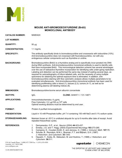
CATALOG NUMBER: MAB3424<br />
LOT NUMBER:<br />
QUANTITY: 50 μg<br />
CONCENTRATION: 1.1 mg/mL<br />
USA & Canada Phone: +1(800) 437-7500 Fax: +1 (951) 676-9209<br />
Australia +61 3 9839 2000<br />
www.millipore.com<br />
12-09-2008/MAB4072/SO<br />
MOUSE ANTI-BROMODEOXYURIDINE (BrdU)<br />
MONOCLONAL ANTIBODY<br />
SPECIFICITY: The antibody specifically binds to bromodeoxyuridine and crossreacts with iodouridine (10%).<br />
Anti-bromodeoxyuridine does not crossreact with fluorodeoxyuridine, nor with any<br />
endogenous cellular components such as thymidine or uridine.<br />
BACKGROUND: Bromodeoxyuridine (BrdU) is a thymidine analog and is specifically incor-porated into DNA<br />
during DNA synthesis. Anti-bromodeoxyuridine monoclonal antibody is used to identify cells<br />
that have incorporated BrdU. This immunological detection scheme has several advantages<br />
over the use of radioactive thymidine incorporation for identifying cells under-going replication.<br />
Labeling and detection can be performed the same day instead of waiting several days, as<br />
required for autoradiography of tritium-labeled cells, and the necessity of using multiple<br />
specimens for obtaining the optimal exposure time is eliminated. In addition, antibromodeoxyuridine<br />
staining with flow cytometric analysis allows multiple parameters to be<br />
evaluated simultaneously. Anti-bromodeoxyuridine monoclonal antibody has been used for<br />
identifying proliferating cells in blood (1), tissues (2,3), tumors (4–7), as well as for<br />
determining plasma cell labeling indices (8).<br />
IMMUNOGEN: Bromodeoxyuridine-bovine serum albumin concentrate<br />
ISOTYPE: IgG1<br />
CLONE: AH4H7-1 / 131-14871<br />
APPLICATIONS: Immunohistochemistry: 6 μg/mL<br />
Flow Cytometry: 0.2 μg/100 μL/10 6 cells<br />
Optimal working dilutions must be determined by end user.<br />
FORMAT: Protein A purified immunoglobulin.<br />
PRESENTATION: Liquid in 10 mM Phosphate buffer, pH 7.4 containing 150 mM NaCl and 0.1% sodium azide.<br />
STORAGE/HANDLING: Maintain frozen at -20°C in undiluted aliquots for up to 6 months after date of receipt. Avoid<br />
repeated freeze/thaw cycles.<br />
REFERENCES: 1. Demyanenko, G.P., et al., Neuron (2004) 44:423-437.<br />
2. Emsley, J.G. and T. Hagg, (2003) Experimental Neurology 183:273-285.<br />
3. Campana, D., Coustan-Smith, E. and Janossy, G. (1988) J. Immunol. Meth. 107:79.<br />
4. Schutte, B., Reynders, M.M.J., Bosman, F.T. and Blijham, G.H. (1987)<br />
J. Histochem. and Cytochem. 35:1343.<br />
5. Hayashi, Y., Koike, M., Matsutani, M. and Hoshino, T. (1988) J. Histochem. and<br />
Cytochem. 36:511.
USA & Canada Phone: +1(800) 437-7500 Fax: +1 (951) 676-9209<br />
Australia +61 3 9839 2000<br />
www.millipore.com<br />
12-09-2008/MAB4072/SO<br />
6. Hoshino, T., Nagashima, T., Cho, K., Murovic, J., Hodes, J., Wilson, C., Edwards, M. and<br />
Pitts, L. (1986) Int. J. Cancer 38:369.<br />
7. Nagashima, T., Murovic, J., Hoshino, T., Wilson, C., and Dearmond, S. (1986) J.<br />
Neurosurg. 64:588.<br />
8. Nagashima, T., DeArmond, S., Murovic, J. and Hoshino, T. (1985) Acta Neuropathol.<br />
67:155.<br />
9. Morstyn, G., Hsu, S.-M., Kinsella, T., Gratzner, H., Russo, A. and Mitchell, J. (1983) J.<br />
Clinical Invest. 72:1844.<br />
10. Greipp, P.R., Witzig, T.E. and Gonchoroff, N.J. (1985) Amer. J. Hematol. 20: 289.<br />
11. Vanderlaan, M., Watkins, B., Thomas, C., Dolbeare, F., and Stanker, L. (1986)<br />
Cytometry 7:499.<br />
Flow cytometry:<br />
APPLICATIONS<br />
The method below is based on that of M. Vanderlaan et al. (9). Variations of this method exist<br />
in the literature, one consideration being the effect various fixation procedures have on the<br />
light-scattering properties of different cell populations.<br />
Procedure:<br />
1. To label cells, pulse with 10 μM bromodeoxyuridine for 30 minutes. Harvest cells from<br />
culture.<br />
2. Fix cells in 70% ethanol at +2-8°C for at least 30 min. Extract histones by resuspending<br />
cells in 1 mL chilled 0.1 M HCI containing 0.5% Triton X-100; incubate the suspension on<br />
ice for 10 minutes. Dilute acid with 5 mL distilled water and centrifuge at 200 x g for 10<br />
min. Resuspend cells in 2 mL distilled water.<br />
3. Denature cellular DNA by submerging the cell suspension into a boiling water bath for 10<br />
min. Afterwards, quickly cool by placing the cell suspension in an ice slurry for several<br />
minutes. Wash cells in PBS that contains 0.5% Triton X-100.<br />
4. Resuspend the cells (1-2 x 10 6 cells) in 100 μL of solution containing approximately 2<br />
μg/mL anti-bromodeoxyuridine antibody diluted in PBS containing 0.1% BSA (0.2 μg/test).<br />
<strong>Inc</strong>ubate for 30 min at room temperature. Wash cells with PBS.<br />
5. Resuspend cells in 100 μL of diluted goat anti-mouse IgG-FlTC Wash cells with PBS.
USA & Canada Phone: +1(800) 437-7500 Fax: +1 (951) 676-9209<br />
Australia +61 3 9839 2000<br />
www.millipore.com<br />
12-09-2008/MAB4072/SO<br />
APPLICATIONS (Cont.)<br />
Immunohistochemistry: Below is a procedure for staining cells that have been labeled with BrdU in vivo or in vitro. The<br />
procedure is based on the methods of B. Schutte et al. (2) and D. Campana et al.<br />
Preparation of tissue: Inject animal with 50 mg BrdU/kg body weight. Sacrifice animal one hour later and remove organ or<br />
tissue under study.<br />
Embed tissue in OCT medium and snap-freeze by immersion into liquid nitrogen.<br />
Cut 4 mm frozen sections with a cryostat. Place sections on either albumin- or gelatin-coated slides.<br />
Preparation of cells: Pulse cells with 10 mM BrdU for 60 min. Cells grown on coverslips, or cytocentrifuge preparations<br />
made from cells grown in suspension, can be used for anti-bromodeoxyuridine staining according to the procedure below.<br />
Procedure<br />
1. Fix tissue sections or cells (on slide or coverglass) by immersing in absolute methanol for 10 minutes at +2-8°C. Air dry<br />
after removing from fixative. The slides can be stored at -20°C in a sealed box, or rehydrated to prepare for the assay<br />
procedure. To rehydrate, immerse in PBS for 3 min.<br />
2. Denature DNA by incubating the slides in 2 N HCI for 60 min at +37°C.<br />
3. Neutralize the acid by immersing the slides in 0.1 M borate buffer, pH 8.5. Change the buffer twice over a 10 min<br />
period.<br />
4. Wash slides with PBS, changing the solution three times over a 10 min period.<br />
5. Place slides in a humidified chamber (e.g., a sealed plastic box layered with wet paper towels) and cover cells with<br />
150–300 μL of solution containing approximately 6 μg/mL anti-bromodeoxyuridine antibody diluted in PBS with 0.1%<br />
BSA. <strong>Inc</strong>ubate for 60 min at room temperature. (Optimal concentration should be determined by user.)<br />
6. Wash slides with PBS, changing the solution three times over a 10 min period.<br />
7. Apply optimal dilution of a second antibody conjugate (e.g., anti-mouse IgG-peroxidase), incubate, wash, and perform<br />
detection with a substrate that produces an insoluble product.<br />
After detection, counterstain with Harris-modified hematoxylin if desired. Slides can then be dehydrated and mounted.<br />
For research use only; not for use in diagnostic procedures.<br />
Important Note: During shipment, small volumes of product will occasionally become entrapped in the seal of the<br />
product vial. For products with volumes of 200 μL or less, we recommend gently tapping the vial on a<br />
hard surface or briefly centrifuging the vial in a tabletop centrifuge to dislodge any liquid in the<br />
container’s cap.<br />
©2002 - 2009: <strong>Millipore</strong> Corporation. All rights reserved. No part of these works may be reproduced in any form without permission in writing.