2021 Head & Neck Cancer Conference

You also want an ePaper? Increase the reach of your titles
YUMPU automatically turns print PDFs into web optimized ePapers that Google loves.
Hidden Figures: The role of a Pathologist<br />
in a head and neck cancer patient journey<br />
By Dr Ali Khurram- Consultant Pathologist<br />
& Senior Lecturer, University of Sheffield<br />
What is pathology?<br />
Pathology is a branch of medical science that involves study of<br />
diseases. There are numerous branches of the specialty involving<br />
examination of micro-organisms and viruses, autopsy and bodily<br />
fluids. Histopathology is the branch of pathology which deals with<br />
examination of biopsy/tissue specimens under a microscope and<br />
these are the people involved in your diagnosis and treatment.<br />
You may not be aware of the fact that that Pathologists not only<br />
play a play role in improving understanding of diseases and<br />
research but are also at the forefront of developing and using<br />
cutting edge technologies to identify patterns in the tissue to aid<br />
patient treatment.<br />
What role does a Pathologist play in<br />
a patient’s journey?<br />
The role of a pathologist in a patient’s journey is<br />
multifold with involvement every step of the way.<br />
a. Prior to treatment<br />
When you are sent to a hospital to have an initial<br />
biopsy, that is the first stage when a pathologist<br />
gets involved. The piece of tissue that is removed,<br />
undergoes a series of steps to preserve it in a<br />
life-like state and thin slices are cut and coloured<br />
followed by placement on a glass slide which<br />
can then be viewed under the microscope by a<br />
pathologist.<br />
The light shining under the slide on the<br />
microscope allows the pathologist (with the help<br />
of magnifying lenses) to meticulously analyse<br />
features within the tissue including presence of<br />
abnormal, pre-cancerous or cancerous cells.<br />
These microscopic findings are then shared<br />
with the surgeons and oncologists and form the<br />
foundation of your future treatment.<br />
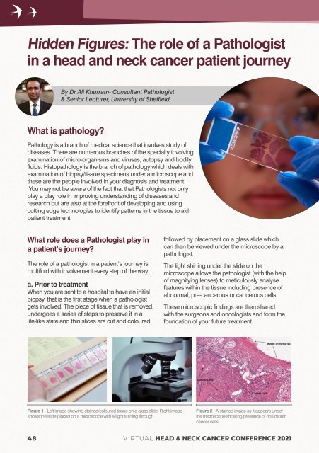
Figure 1 - Left image showing stained/coloured tissue on a glass slide. Right image<br />
shows the slide placed on a microscope with a light shining through.<br />
Figure 2 - A stained image as it appears under<br />
the microscope showing presence of oral/mouth<br />
cancer cells.<br />
48<br />
VIRTUAL HEAD & NECK CANCER CONFERENCE <strong>2021</strong>