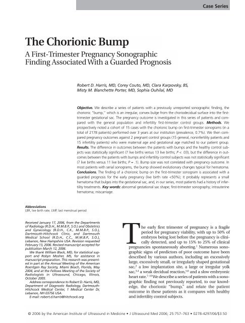
The Chorionic Bump - Journal of Ultrasound in Medicine
The Chorionic Bump - Journal of Ultrasound in Medicine
The Chorionic Bump - Journal of Ultrasound in Medicine

You also want an ePaper? Increase the reach of your titles
YUMPU automatically turns print PDFs into web optimized ePapers that Google loves.
<strong>The</strong> <strong>Chorionic</strong> <strong>Bump</strong><br />
A First-Trimester Pregnancy Sonographic<br />
F<strong>in</strong>d<strong>in</strong>g Associated With a Guarded Prognosis<br />
Abbreviations<br />
LBR, live birth rate; LMP, last menstrual period<br />
Received January 17, 2006, from the Departments<br />
<strong>of</strong> Radiology (R.D.H., M.M.B.P., S.O.) and Obstetrics<br />
and Gynecology (R.D.H., C.K., M.M.B.P., S.O.),<br />
Dartmouth-Hitchcock Cl<strong>in</strong>ic, and Dartmouth<br />
Medical School (R.D.H., C.C., M.M.B.P., S.O.),<br />
Lebanon, New Hampshire USA. Revision requested<br />
February 13, 2006. Revised manuscript accepted for<br />
publication March 12, 2006.<br />
We thank William Black, MD, for statistical support<br />
and Robyn Mosher, MS, for assistance <strong>in</strong><br />
manuscript preparation. This research was presented<br />
<strong>in</strong> part at the Annual Meet<strong>in</strong>g <strong>of</strong> the American<br />
Roentgen Ray Society, Miami Beach, Florida, May<br />
2004, and at the Fellows Meet<strong>in</strong>g <strong>of</strong> the Society <strong>of</strong><br />
Radiologists <strong>in</strong> <strong>Ultrasound</strong>, Chicago, Ill<strong>in</strong>ois,<br />
October 2005.<br />
Address correspondence to Robert D. Harris, MD,<br />
Department <strong>of</strong> Diagnostic Radiology, Dartmouth-<br />
Hitchcock Medical Center, 1 Medical Center Dr,<br />
Lebanon, NH 03756 USA.<br />
E-mail: robert.d.harris@hitchcock.org<br />
Robert D. Harris, MD, Corey Couto, MD, Clara Karpovsky, BS,<br />
Misty M. Blanchette Porter, MD, Sophia Ouhilal, MD<br />
Case Series<br />
Objective. We describe a series <strong>of</strong> patients with a previously unreported sonographic f<strong>in</strong>d<strong>in</strong>g, the<br />
chorionic “bump,” which is an irregular, convex bulge from the choriodecidual surface <strong>in</strong>to the firsttrimester<br />
gestational sac. <strong>The</strong> pregnancy outcome is <strong>in</strong>vestigated <strong>in</strong> this series <strong>of</strong> patients and compared<br />
with the general population and <strong>in</strong>fertility first-trimester control groups. Methods. We<br />
prospectively noted a cohort <strong>of</strong> 15 cases with the chorionic bump on first-trimester sonograms (<strong>in</strong> a<br />
total <strong>of</strong> 2178 patients) performed over 3 years at our <strong>in</strong>stitution (prevalence, 0.7%). We then compared<br />
pregnancy outcomes aga<strong>in</strong>st 2 pregnant control groups (15 general, non<strong>in</strong>fertility patients and<br />
15 <strong>in</strong>fertility patients) who were maternal age and gestational age matched to our patient group.<br />
Results. <strong>The</strong> difference <strong>in</strong> outcomes between the patients with bumps and the healthy control subjects<br />
was statistically significant (7 live births versus 13 live births; P < .03), but the difference <strong>in</strong> outcomes<br />
between the patients with bumps and <strong>in</strong>fertility control subjects was not statistically significant<br />
(7 live births versus 11 live births; P = .1). <strong>Bump</strong> size was not correlated with pregnancy outcome. In<br />
most patients with serial sonograms, the bump showed evolutionary changes typical for hematoma.<br />
Conclusions. <strong>The</strong> f<strong>in</strong>d<strong>in</strong>g <strong>of</strong> a chorionic bump on the first-trimester sonogram is associated with a<br />
guarded prognosis for the early pregnancy (live birth rate
<strong>The</strong> <strong>Chorionic</strong> <strong>Bump</strong><br />
Materials and Methods<br />
We prospectively analyzed all first-trimester<br />
sonograms obta<strong>in</strong>ed between November 2001<br />
and November 2004 (2178 sonograms) at our<br />
<strong>in</strong>stitution, and found 15 cases that showed the<br />
f<strong>in</strong>d<strong>in</strong>g that we have called the chorionic bump,<br />
def<strong>in</strong>ed as a focal, irregular convexity or bulge <strong>in</strong><br />
the surround<strong>in</strong>g choriodecidual reaction <strong>in</strong>to the<br />
early gestational sac (Figure 1). We also reviewed 2<br />
control groups for this study, obta<strong>in</strong>ed through<br />
our patient logbook manually, to f<strong>in</strong>d control<br />
patients with<strong>in</strong> 1 year <strong>of</strong> maternal age and 0.5<br />
weeks’ gestational age matched to our experimental<br />
group: 15 randomly selected patients<br />
from the non<strong>in</strong>fertility, general pregnant population<br />
seen <strong>in</strong> our ultrasound section and 15<br />
patients from our <strong>in</strong>fertility cl<strong>in</strong>ic referred for<br />
sonography dur<strong>in</strong>g the same period. None <strong>of</strong><br />
these patients had any vag<strong>in</strong>al bleed<strong>in</strong>g or other<br />
complications. Institutional Review Board<br />
approval was obta<strong>in</strong>ed for the study.<br />
Sonography was performed with HDI 5000<br />
mach<strong>in</strong>es (Philips Medical Systems, Bothell, WA)<br />
with a C8-4 endovag<strong>in</strong>al probe. <strong>The</strong> <strong>in</strong>dication<br />
for the sonogram was recorded. Gestational age<br />
was determ<strong>in</strong>ed by last menstrual period (LMP),<br />
if accurate, or by mean sac diameter or crownrump<br />
length if there were uncerta<strong>in</strong> menstrual<br />
dates or discordance between LMP and sonographic<br />
dates.<br />
Figure 1. Color Doppler transvag<strong>in</strong>al image <strong>of</strong> a moderately sized chorionic bump<br />
(arrows), a slightly irregular convexity bulg<strong>in</strong>g <strong>in</strong>to the gestational sac, which resulted<br />
<strong>in</strong> a normal pregnancy outcome <strong>in</strong> a 34-year-old woman (patient 7).<br />
758<br />
We measured the chorionic bump <strong>in</strong> 3 dimensions<br />
with electronic calipers (either prospectively<br />
or on a picture archiv<strong>in</strong>g and communications system;<br />
IDX-Stentor, Burl<strong>in</strong>gton, VT) to obta<strong>in</strong> its volume<br />
(length × width × height/2, formula for<br />
approximated prolate ellipse), and recorded its<br />
appearance on serial sonograms where available.<br />
Color and power Doppler sonography was applied<br />
<strong>in</strong> some cases by the sonographer/sonologist but<br />
was m<strong>in</strong>imized because <strong>of</strong> first-trimester energy<br />
deposition concerns.<br />
We also recorded by review <strong>of</strong> the medical<br />
charts any history <strong>of</strong> <strong>in</strong>fertility therapy (past<br />
pregnancy or present), vag<strong>in</strong>al bleed<strong>in</strong>g, and<br />
thrombophilia, maternal smok<strong>in</strong>g history, other<br />
f<strong>in</strong>d<strong>in</strong>gs <strong>in</strong> the gestational sac (embryo [with or<br />
without heartbeat], yolk sac, mean sac diameter,<br />
and <strong>in</strong>ternal debris/echoes), pregnancy outcome<br />
(by chart review or contact<strong>in</strong>g referr<strong>in</strong>g physicians),<br />
and pathologic report (when available).<br />
<strong>The</strong> Fisher exact test <strong>of</strong> proportions was used to<br />
compare the outcomes between the patients<br />
with chorionic bumps and the <strong>in</strong>fertility and<br />
healthy control groups, as well as the bump size<br />
versus outcomes.<br />
Results<br />
<strong>The</strong> prevalence <strong>of</strong> the chorionic bump was 0.7%.<br />
<strong>The</strong> mean age <strong>of</strong> patients with the chorionic<br />
bump was 33.1 years (range, 21–40 years), and<br />
the mean gestational age was 6.7 weeks (range,<br />
5.8–9.3 weeks) at the time <strong>of</strong> sonography (Table<br />
1). <strong>The</strong> <strong>in</strong>dications for the sonograms were as follows:<br />
to evaluate viability <strong>in</strong> 9 patients; to evaluate<br />
gestational dat<strong>in</strong>g <strong>in</strong> 4 patients; to evaluate<br />
right lower quadrant pa<strong>in</strong> <strong>in</strong> 1 patient; and a<br />
referral for an abnormal gestational sac mass <strong>in</strong> 1<br />
patient seen on an outside sonogram. Six<br />
patients had vag<strong>in</strong>al bleed<strong>in</strong>g before sonography,<br />
whereas 4 did not. N<strong>in</strong>e patients had a history <strong>of</strong><br />
<strong>in</strong>fertility treatment <strong>in</strong> this or prior pregnancy<br />
attempts, and 6 had been treated with <strong>in</strong>fertility<br />
drugs or procedures dur<strong>in</strong>g the gestation <strong>of</strong><br />
<strong>in</strong>terest. None <strong>of</strong> our patients had positive screen<br />
results for thrombophilia. One patient was a<br />
smoker.<br />
<strong>The</strong> bumps ranged <strong>in</strong> size from 0.7 × 0.4 × 0.4 to<br />
1.9 × 1.9 × 1.4 cm, with volumes from 0.1 to 2.5<br />
mL. Two patients with bumps had empty gestational<br />
sacs (both result<strong>in</strong>g <strong>in</strong> miscarriages); 5<br />
patients had only yolk sacs but no embryos <strong>in</strong> the<br />
gestational sacs (result<strong>in</strong>g <strong>in</strong> 3 live births and 2<br />
J <strong>Ultrasound</strong> Med 2006; 25:757–763
Table 1. Patient Characteristics<br />
miscarriages); 2 patients had small embryos<br />
without a heartbeat (crown-rump lengths <strong>of</strong> 2<br />
and 6 mm, 1 result<strong>in</strong>g <strong>in</strong> miscarriage and 1 <strong>in</strong> a<br />
live birth); and 5 had embryos with a heartbeat <strong>of</strong><br />
100 beats per m<strong>in</strong>ute or greater (result<strong>in</strong>g <strong>in</strong> 4<br />
live births and 1 miscarriage). In the miscarriage<br />
after visualization <strong>of</strong> an embryo with a positive<br />
heartbeat, the patient had a 9-mm embryo with<br />
a heart rate <strong>of</strong> 124 beats per m<strong>in</strong>ute.<br />
Of the 15 pregnancies with chorionic bump,<br />
7 (47%) had live births, whereas 8 (53%) had nonviable<br />
outcomes. One live birth result was monochorionic,<br />
diamniotic tw<strong>in</strong>s born before term<br />
at 33 weeks’ gestation) <strong>in</strong> a mother with preeclampsia.<br />
Of the 8 nonviable gestations, 6 were<br />
(or became) nonviable <strong>in</strong> the first trimester, and 2<br />
had fetal demise <strong>in</strong> the second trimester (at 17<br />
and 18 weeks’ gestation).<br />
<strong>The</strong> <strong>in</strong>fertility control group had a mean age <strong>of</strong><br />
32.7 years (range, 21–40 years) and a mean gestational<br />
age <strong>of</strong> 6.3 weeks (range, 5.3–8.6 weeks) at<br />
the time <strong>of</strong> their sonograms. Of this group, 11<br />
(73%) <strong>of</strong> 15 had live births; 3 had miscarriages<br />
(20% nonviable rate); and 1 had an elective term<strong>in</strong>ation.<br />
<strong>The</strong> healthy control group had a mean age <strong>of</strong><br />
32.3 years (range, 21–39 years) and a mean gestational<br />
age <strong>of</strong> 6.4 weeks (range, 4.7–9.3 weeks) at<br />
sonography. <strong>The</strong> pregnancy outcome <strong>in</strong> the<br />
healthy control group was live birth <strong>in</strong> 13<br />
patients (87%) and miscarriage <strong>in</strong> 2 patients<br />
(13%). <strong>The</strong> outcome <strong>of</strong> the bump group (live<br />
birth rate [LBR], 47%) was significantly different<br />
from that <strong>of</strong> the healthy control group (LBR, 87%;<br />
P < .03) but not statistically different from that <strong>of</strong><br />
the <strong>in</strong>fertility control group (LBR, 73%; P = .1)<br />
<strong>The</strong> bump was avascular on color or power<br />
Doppler imag<strong>in</strong>g <strong>in</strong> all cases <strong>in</strong> which color or<br />
power Doppler imag<strong>in</strong>g was used (5 cases)<br />
(Figure 1) and hypoechoic centrally <strong>in</strong> 3 <strong>of</strong> 15<br />
cases (Figure 2). Swirl<strong>in</strong>g <strong>of</strong> low-level echoes <strong>in</strong><br />
the bump dur<strong>in</strong>g real-time sonography was seen<br />
Harris et al<br />
Age, Sonographic No. <strong>of</strong> MSD, Yolk Sac <strong>Bump</strong> Vag<strong>in</strong>al Infertility<br />
Patient y F<strong>in</strong>d<strong>in</strong>gs, wk Gestations cm or Embryo Vol, mL Bleed? Outcome History<br />
1 37 5.8 MC DA tw<strong>in</strong>s 1.1 Yolk sac 0.2 No Tw<strong>in</strong>s, 33 wk Yes<br />
2 29 6.1 S<strong>in</strong>gleton 1.3 Yolk sac 0.5 Yes Normal term No<br />
3 38 6.7 S<strong>in</strong>gleton 1.7 Yolk sac 0.3 No Normal term No<br />
4 40 6.4 S<strong>in</strong>gleton 1.5 6 mm no HB 0.8 No 1st-trimester demise No<br />
5 21 7.0 S<strong>in</strong>gleton 1.9 No 1.3 Yes 1st-trimester demise No<br />
6 31 6.9 S<strong>in</strong>gleton 1.8 6 mm + HB 1.2 No Normal term No<br />
7 34 6.5 S<strong>in</strong>gleton 1.8 2 mm no HB 0.3 No Normal term Yes<br />
8 37 6.7 S<strong>in</strong>gleton 1.7 Yolk sac 0.3 Yes Fetal demise 17 wk Yes<br />
9 30 6.6 S<strong>in</strong>gleton 1.6 3 mm + HB 0.4 No Normal term Yes<br />
10 30 5.8 S<strong>in</strong>gleton 0.9 9 mm + HB 0.7 No Fetal demise 18 wk Yes<br />
11 32 6.7 MC DA tw<strong>in</strong>s 1.7 Yolk sac 0.1 Yes 1st-trimester demise Yes<br />
12 24 7.6 S<strong>in</strong>gleton 2.3 11 mm + HB 0.5 No Normal term No<br />
13 40 9.3 Tw<strong>in</strong> sacs, 3.4 25 mm + HB 0.7 No Normal term with chorionic No<br />
1 embryo bump <strong>in</strong> vanish<strong>in</strong>g tw<strong>in</strong><br />
14 37 6.6 S<strong>in</strong>gleton 1.6 No 0.1 Yes 1st-trimester demise Yes<br />
15 36 6.2 S<strong>in</strong>gleton 1.8 5 mm no HB 2.5 Yes 1st-trimester demise Yes<br />
HB <strong>in</strong>dicates heartbeat; MC DA, monochorionic diamniotic; MSD, mean sac diameter; and Vol, volume.<br />
Figure 2. Normal outcome <strong>in</strong> a woman (patient 6) with a large hypoechoic bump<br />
(arrow). <strong>The</strong> bump measured 2 × 1.6 × 1.4 cm and was centrally hypoechoic, and<br />
the pregnancy resulted <strong>in</strong> a healthy term fetus; no further sonograms were available<br />
for review. Note the normal yolk sac (asterisk).<br />
J <strong>Ultrasound</strong> Med 2006; 25:757–763 759
<strong>The</strong> <strong>Chorionic</strong> <strong>Bump</strong><br />
760<br />
<strong>in</strong> 4 cases. <strong>The</strong> bump decreased <strong>in</strong> size over 1 to 4<br />
weeks when serial sonographic exam<strong>in</strong>ations<br />
were performed (Figure 3) <strong>in</strong> 3 patients and<br />
<strong>in</strong>creased <strong>in</strong> size <strong>in</strong> 2 cases, both <strong>of</strong> which ended<br />
<strong>in</strong> nonviable pregnancy. No fibr<strong>in</strong> strands were<br />
noted dur<strong>in</strong>g serial scans <strong>of</strong> any patients. <strong>The</strong>re<br />
was no correlation between bump size and outcomes;<br />
the mean volume <strong>in</strong> the good outcomes<br />
group was 0.49 mL, versus 0.59 mL <strong>in</strong> the poor<br />
outcomes group (P > .1, Fisher exact t test).<br />
Discussion<br />
Choriodecidual irregularity <strong>in</strong> the first-trimester<br />
gestational sac, or an unusual or bizarre shape <strong>of</strong><br />
the early gestational sac, has been well described<br />
as a sign <strong>of</strong> threatened abortion by Nyberg et al 5<br />
among others. Nyberg et al 5 reported a distorted<br />
sac shape to have 100% specificity for nonviability.<br />
We have, <strong>in</strong> contradist<strong>in</strong>ction, encountered a<br />
focal bulge or bump <strong>in</strong> the choriodecidual reaction<br />
on occasion <strong>in</strong> our first-trimester population,<br />
which, on the basis <strong>of</strong> our review, has a<br />
2-fold <strong>in</strong>crease <strong>in</strong> miscarriage rate compared<br />
with <strong>in</strong>fertility control patients and a 4-fold<br />
<strong>in</strong>crease <strong>in</strong> miscarriage rate compared with the<br />
general population, based on our small series.<br />
Nyberg et al 5 did not specifically def<strong>in</strong>e their<br />
criteria for distorted sac shape, other than stat<strong>in</strong>g<br />
that there was a “grossly aberrant shape compared<br />
to the more uniformly round or oval shape<br />
<strong>of</strong> normal gestational sacs.” <strong>The</strong> chorionic bump<br />
seems to constitute a less prom<strong>in</strong>ent irregularity,<br />
be<strong>in</strong>g focal, with unclear etiology, and may represent<br />
a small hematoma. <strong>The</strong> few cases <strong>in</strong> which<br />
the pregnancy cont<strong>in</strong>ued and serial sonograms<br />
were available showed that most <strong>of</strong> the bumps<br />
became hypoechoic and smaller over time.<br />
However, this theory <strong>of</strong> orig<strong>in</strong> is without pro<strong>of</strong><br />
<strong>in</strong> our series because <strong>of</strong> the difficulty <strong>in</strong> obta<strong>in</strong><strong>in</strong>g<br />
histologic confirmation <strong>in</strong> most cases and also<br />
because <strong>of</strong> the fact that most first-trimester<br />
patients do not undergo histologic evaluation or<br />
other imag<strong>in</strong>g apart from serial sonography. One<br />
<strong>of</strong> our <strong>in</strong>fertility patients <strong>in</strong> the study group, however,<br />
had pathologic tissue available for analysis,<br />
which showed hypovascular chorionic villi with<br />
hydropic changes and fibrous material, as well as<br />
some material that was characterized as foreign<br />
body material (cellulose) on histologic exam<strong>in</strong>ation<br />
and at crystallography. It seems unlikely that<br />
this material, <strong>in</strong> fact, represents the chorionic<br />
bump. We had no other cases with similar material,<br />
and we know <strong>of</strong> no foreign or plant material<br />
that was placed <strong>in</strong>side the uterus for treatment<br />
Figure 3. A, Transvag<strong>in</strong>al image <strong>of</strong> a bump (arrows) <strong>in</strong> a 38-year-old woman (patient 3) reportedly at 6.7 weeks by LMP who had a<br />
yolk sac present but no embryo. This patient had a normal term pregnancy. B, Transvag<strong>in</strong>al image 2 weeks later show<strong>in</strong>g that the<br />
bump was less prom<strong>in</strong>ent; only a slight bulge (arrows) <strong>in</strong>to the gestational sac rema<strong>in</strong>ed. A portion <strong>of</strong> the embryo (asterisk) is present,<br />
as well as a small <strong>in</strong>tramural fibroid marked by electronic calipers.<br />
A B<br />
J <strong>Ultrasound</strong> Med 2006; 25:757–763
dur<strong>in</strong>g fertility therapy. Conversely, we have<br />
recently encountered a similar case <strong>of</strong> a chorionic<br />
bump (T. Dub<strong>in</strong>sky, MD, oral communication,<br />
2005) at another <strong>in</strong>stitution <strong>in</strong> which magnetic<br />
resonance imag<strong>in</strong>g revealed the bump to have a<br />
shortened T1, compatible with a hematoma<br />
(Figure 4).<br />
<strong>The</strong> chorionic bump temporal relationship to<br />
miscarriage is also unclear. In 10 <strong>of</strong> our cases,<br />
there was never a liv<strong>in</strong>g embryo seen (Figure 5)<br />
to document that the normal early gestational<br />
development was occurr<strong>in</strong>g, and <strong>in</strong> the 5 cases<br />
with an embryo and a normal early heart rate, 4<br />
patients had normal outcomes, which confirms<br />
the good prognostic f<strong>in</strong>d<strong>in</strong>g <strong>of</strong> a normal early<br />
heartbeat for viability. Certa<strong>in</strong>ly, it could represent<br />
an associated sign <strong>of</strong> miscarriage (similar to<br />
a calcified yolk sac) <strong>of</strong> a nonembryonic gestation<br />
or a demised embryo <strong>in</strong> some cases.<br />
Figure 4. A, Longitud<strong>in</strong>al first-trimester image (6 weeks by<br />
LMP) <strong>of</strong> a chorionic bump <strong>in</strong> an 18-year-old woman with elevated<br />
human chorionic gonadotrop<strong>in</strong> level <strong>of</strong> greater than<br />
200,000 IU. No embryo was seen. B, Fast T2-weighted magnetic<br />
resonance image obta<strong>in</strong>ed at 7 weeks (because <strong>of</strong> concern<br />
about a possible molar pregnancy) show<strong>in</strong>g a small fibroid anteriorly<br />
(F) and a slight irregularity to gestational sac represent<strong>in</strong>g<br />
a portion <strong>of</strong> the chorionic bump (arrow). This pregnancy resulted<br />
<strong>in</strong> a first-trimester miscarriage (anembryonic pregnancy). C,<br />
T1-weighted fat-suppressed magnetic resonance image show<strong>in</strong>g<br />
an <strong>in</strong>creased signal <strong>in</strong> the chorionic bump (arrow) consistent<br />
with hemorrhage. Images courtesy <strong>of</strong> Ted Dub<strong>in</strong>sky, MD<br />
(University <strong>of</strong> Wash<strong>in</strong>gton, Seattle, WA). R <strong>in</strong>dicates rectum.<br />
B<br />
Serial scann<strong>in</strong>g, when available, showed that<br />
the irregularity generally decreased dur<strong>in</strong>g the<br />
short <strong>in</strong>terval, except <strong>in</strong> 2 cases <strong>in</strong> which the<br />
bump cont<strong>in</strong>ued to <strong>in</strong>crease <strong>in</strong> size, both <strong>of</strong><br />
which ended <strong>in</strong> nonviable outcomes. Although<br />
the etiology <strong>of</strong> the chorionic bump is unknown,<br />
it is reasonable to assume that it represents a<br />
hematoma or small area <strong>of</strong> hemorrhage. This<br />
theory is supported by 4 sonographic po<strong>in</strong>ts: the<br />
swirl<strong>in</strong>g appearance <strong>of</strong> echoes <strong>in</strong> the bump (similar<br />
to that <strong>of</strong> a venous lake <strong>in</strong> the second or third<br />
trimester); the lack <strong>of</strong> vascularity on color<br />
Doppler imag<strong>in</strong>g; the serial hypoechoic echo<br />
A<br />
C<br />
Harris et al<br />
J <strong>Ultrasound</strong> Med 2006; 25:757–763 761
<strong>The</strong> <strong>Chorionic</strong> <strong>Bump</strong><br />
762<br />
texture <strong>in</strong> some patients’ sonograms; and the<br />
tendency <strong>of</strong> the bump, <strong>in</strong> most patients, to<br />
become smaller over time. However, there did<br />
not seem to be a statistically significant relationship<br />
between the presence <strong>of</strong> a bump and a history<br />
<strong>of</strong> vag<strong>in</strong>al bleed<strong>in</strong>g.<br />
Most women who have vag<strong>in</strong>al bleed<strong>in</strong>g dur<strong>in</strong>g<br />
the first trimester and who have sonographic<br />
f<strong>in</strong>d<strong>in</strong>gs <strong>of</strong> hemorrhage have a subchorionic or<br />
an <strong>in</strong>trauter<strong>in</strong>e hematoma that resides outside<br />
<strong>of</strong>, but closely parallel to, the gestational sac.<br />
Previous studies have clearly shown an <strong>in</strong>verse<br />
relationship between hematoma size and pregnancy<br />
outcome. 11 Most early pregnancy bleed<strong>in</strong>g<br />
appears to be venous and thus occurs at low pressure<br />
and tends to conform to the gestational sac<br />
shape. Whether the chorionic bump hematoma is<br />
more arterial <strong>in</strong> orig<strong>in</strong> than the common subchorionic<br />
hemorrhage is purely conjectural, but<br />
this could account for the convexity <strong>of</strong> shape and<br />
the rather poor prognostic implications. Another<br />
possible theory is that the develop<strong>in</strong>g placenta<br />
has a tendency for bleed<strong>in</strong>g to occur <strong>in</strong> the decidua<br />
or cytotrophoblastic shell; hence, any bleed<strong>in</strong>g<br />
is crescentic or oval on the outer side <strong>of</strong> the choriodecidual<br />
reaction and mirrors the gestational<br />
sac. In the case <strong>of</strong> the bump, it would seem plausible<br />
that the hematoma arises from the develop<strong>in</strong>g<br />
<strong>in</strong>tervillous space or chorionic plate and<br />
bulges <strong>in</strong>to the gestational sac.<br />
Most patients <strong>in</strong> our small series had a history<br />
<strong>of</strong> <strong>in</strong>fertility treatment, but we cannot assess the<br />
relative frequency <strong>of</strong> <strong>in</strong>fertility treatment <strong>in</strong> the<br />
general population because there is no computerized<br />
method <strong>of</strong> determ<strong>in</strong><strong>in</strong>g the number <strong>of</strong><br />
sonographic exam<strong>in</strong>ation per <strong>in</strong>fertility patient<br />
because our radiology and hospital <strong>in</strong>formation<br />
systems are not <strong>in</strong>tegrated fully.<br />
Limitations <strong>of</strong> our study <strong>in</strong>clude lack <strong>of</strong> histologic<br />
correlation, absence <strong>of</strong> correlative imag<strong>in</strong>g,<br />
the small number <strong>of</strong> cases, and the <strong>in</strong>herently<br />
confound<strong>in</strong>g data from us<strong>in</strong>g an <strong>in</strong>fertility control<br />
group, albeit a useful comparison group. <strong>The</strong><br />
small number <strong>of</strong> cases limits the statistical significance<br />
somewhat, although there was a significant<br />
difference <strong>in</strong> the pregnancy outcomes<br />
between the bump group and healthy control<br />
subjects by the Fisher exact t test. <strong>The</strong> prevalence<br />
<strong>of</strong> the bump may be overestimated <strong>in</strong> the <strong>in</strong>fertility<br />
population because this group tends to be<br />
scanned more frequently than the general population<br />
dur<strong>in</strong>g early pregnancy; therefore, the<br />
bump may be encountered more <strong>of</strong>ten <strong>in</strong> this<br />
group <strong>of</strong> patients.<br />
In conclusion, the f<strong>in</strong>d<strong>in</strong>g <strong>of</strong> a chorionic bump<br />
is a newly described but uncommon sign (0.7%)<br />
that is associated with a guarded prognosis for<br />
early pregnancy. It may represent a small<br />
hematoma that bulges <strong>in</strong>to the gestational sac but<br />
could also represent a blighted second pregnancy<br />
be<strong>in</strong>g resorbed, and it is also seen <strong>in</strong> some patients<br />
undergo<strong>in</strong>g <strong>in</strong>fertility treatment, although this latter<br />
group has more frequent sonograms, which<br />
may spuriously <strong>in</strong>crease its <strong>in</strong>cidence overall <strong>in</strong><br />
this group. Whether it is causal to the poor outcome<br />
<strong>of</strong> pregnancy or is a f<strong>in</strong>d<strong>in</strong>g occurr<strong>in</strong>g after<br />
Figure 5. Transverse (A) and longitud<strong>in</strong>al (B) endovag<strong>in</strong>al images <strong>of</strong> a poor outcome <strong>in</strong> an <strong>in</strong>fertility patient at 6.2 weeks (patient 15) who had undergone<br />
vitro fertilization and who had a large (1.9 × 1.9 × 1.4-cm) bump and a 4.6-mm embryo without a heartbeat. This pregnancy resulted <strong>in</strong> a nonviable<br />
gestation.<br />
B<br />
A<br />
J <strong>Ultrasound</strong> Med 2006; 25:757–763
nonviability rema<strong>in</strong>s to be determ<strong>in</strong>ed, and the<br />
f<strong>in</strong>d<strong>in</strong>g <strong>of</strong> an embryonic heartbeat <strong>in</strong> the presence<br />
<strong>of</strong> this sign is confirmed as a good prognostic<br />
sign for the pregnancy (80% [4/5] <strong>in</strong> our small<br />
series).<br />
References<br />
1. Wilcox AJ, We<strong>in</strong>berg CR, O’Connor JF, et al. Incidence <strong>of</strong><br />
early pregnancy loss. N Engl J Med 1988; 319:189–194.<br />
2. Elson J, Salim R, Tailor A, Banerjee S, Zosmer N, Jukovic D.<br />
Prediction <strong>of</strong> early pregnancy viability <strong>in</strong> the absence <strong>of</strong> an<br />
ultrasonically detectable embryo. <strong>Ultrasound</strong> Obstet<br />
Gynecol 2003; 21:57–61.<br />
3. Nyberg DA, Mack LA, Harvey D, Wang K. Value <strong>of</strong> the yolk<br />
sac <strong>in</strong> evaluat<strong>in</strong>g early pregnancies. J <strong>Ultrasound</strong> Med<br />
1988; 7:129–135.<br />
4. L<strong>in</strong>dsay DJ, Lovett IS, Lyons EA, et al. Yolk sac diameter and<br />
shape at endovag<strong>in</strong>al US: predictors <strong>of</strong> pregnancy outcome<br />
<strong>in</strong> the first trimester. Radiology 1992; 183:115–118.<br />
5. Nyberg DA, La<strong>in</strong>g FC, Filly RA. Threatened abortion: sonographic<br />
dist<strong>in</strong>ction <strong>of</strong> normal and abnormal gestation sacs.<br />
Radiology 1986; 158:397–400.<br />
6. Ball RH. <strong>The</strong> sonography <strong>of</strong> pregnancy loss. Sem<strong>in</strong> Reprod<br />
Med 2000; 18:351–355.<br />
7. May DA, Sturtevant NV. Embryonal heart rate as a predictor<br />
<strong>of</strong> pregnancy outcome: a prospective analysis.<br />
J <strong>Ultrasound</strong> Med 1991; 10:591–593.<br />
8. Doubilet PM, Benson CB. Embryonic heart rate <strong>in</strong> the early<br />
first trimester: what rate is normal? J <strong>Ultrasound</strong> Med<br />
1995; 14:431–434.<br />
9. Choong S, Rombauts L, Ugoni A, Meagher S. <strong>Ultrasound</strong><br />
prediction <strong>of</strong> risk <strong>of</strong> spontaneous miscarriage <strong>in</strong> live<br />
embryos from assisted conceptions. <strong>Ultrasound</strong> Obstet<br />
Gynecol 2003; 22:571–577.<br />
10. Falco P, Milano V, Pilu G, et al. Sonography <strong>of</strong> pregnancies<br />
with first-trimester bleed<strong>in</strong>g and a viable embryo: a study<br />
<strong>of</strong> prognostic <strong>in</strong>dicators by logistic regression analysis.<br />
<strong>Ultrasound</strong> Obstet Gynecol 1996; 7:165–169.<br />
11. Benson CB, Doubilet PM, Cooney MJ, Frates MC, David V,<br />
Hornste<strong>in</strong> MD. Early s<strong>in</strong>gleton pregnancy outcome: effects<br />
<strong>of</strong> maternal age and mode <strong>of</strong> conception. Radiology 1997;<br />
203:399–403.<br />
Harris et al<br />
J <strong>Ultrasound</strong> Med 2006; 25:757–763 763