Injection techniques for drug administration and methods of restraint
Injection techniques for drug administration and methods of restraint
Injection techniques for drug administration and methods of restraint

You also want an ePaper? Increase the reach of your titles
YUMPU automatically turns print PDFs into web optimized ePapers that Google loves.
loose-needle technique<br />
over-the-needle catheter<br />
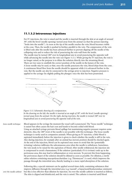
11.1.3.2 Intravenous injections<br />
Jos Ensink <strong>and</strong> Joris Robben 211<br />
For IV injections, the vein is raised <strong>and</strong> the needle is inserted through the skin at an angle <strong>of</strong> around<br />
30°, with the bevel (needle opening) turned away from the animal (so that the person injecting<br />
“looks into the needle”). As soon as the tip <strong>of</strong> the needle reaches the vein, blood becomes visible<br />
at the cone. Then, the needle is pushed in further, parallel to the vein. The compression <strong>of</strong> the vein<br />
is lifted only after the needle has been advanced further to prevent slipping <strong>of</strong>f the needle <strong>of</strong> the<br />
collapsing vein <strong>and</strong> to reduce the risk <strong>of</strong> puncturing the vein wall from the inside.<br />
The needle may be turned 180° over its longitudinal axis to avoid puncturing the opposite wall<br />
while advancing the needle into the vein (see Figure 11.5.). While giving the IV injection, the vein is<br />
no longer raised, as the purpose is to dilute the solution directly into the streaming blood.<br />
There are two ways to establish the correct position <strong>of</strong> the needle in the lumen <strong>of</strong> the vein.<br />
A loose needle may be used, so that, once the needle punctures the vein, blood drips from the cone.<br />
A continuous blood flow from the needle should be apparent while it is advanced further in the<br />
vein. But the needle can also be connected to the syringe prior to injection. Negative pressure is<br />
applied to the syringe (by slightly pulling the plunger) once the skin has been penetrated.<br />
Figure 11.5. Schematic drawing <strong>of</strong> a venipuncture.<br />
In the drawing on the left, the needle is inserted at an angle <strong>of</strong> 30°, with the bevel (needle opening)<br />
turned away from the animal. On the right, during insertion, the needle is twisted 180° over its<br />
longitudinal axis to avoid puncturing the opposite wall <strong>of</strong> the vein.<br />
Blood appears in the syringe the moment the vessel wall is punctured. The “loose-needle” technique<br />
is easiest but <strong>of</strong>ten causes the hair coat <strong>and</strong> h<strong>and</strong>s to become soiled with blood.<br />
Using an attached syringe prevents blood spillage but maintaining negative pressure requires some<br />
dexterity. Also, the 180°-twist <strong>of</strong> the needle is not possible with this technique. The loose-needle<br />
technique is rarely used in companion animals. When giving an IV injection, a little blood is<br />
aspirated immediately be<strong>for</strong>e the injection is given to check whether the needle is still in the vein.<br />
Also during injection, <strong>for</strong> example after an animal has moved, position should be checked again.<br />
At the end <strong>of</strong> the injection, blood can be aspirated once more to avoid that the (potentially<br />
irritating) solution infiltrates the subcutaneous area when the needle is withdrawn. Sometimes,<br />
the vein needs to be raised <strong>for</strong> the aspiration <strong>of</strong> blood. After needle withdrawal, the injection site<br />
is compressed to avoid a haematoma. If the solution (particularly if it is irritant) ends up in the<br />
perivenous area, this may lead to inflammation or even necrosis. To prevent this from happening,<br />
the perivenous depot <strong>of</strong> the solution should be diluted as soon as possible. For dilution, a sterile<br />
saline solution containing mucopolysaccharidase (e.g. Thiomucase ® ) is used, which improves the<br />
passage through the interstitial area, thereby leading to a more rapid absorption <strong>of</strong> the solution.<br />
Following this, a special ointment can be applied several times daily, containing substances that<br />
improve diffusion through the interstitial area (e.g. Lasonil ® , a heparinoid/hyaluronidase ointment)<br />
but this is less effective than dilution by injection. An over-the-needle catheter is inserted as follows.<br />
The skin is clipped <strong>and</strong> disinfected, <strong>and</strong> the vein is raised. The catheter is inserted until blood