Injection techniques for drug administration and methods of restraint
Injection techniques for drug administration and methods of restraint
Injection techniques for drug administration and methods of restraint

Create successful ePaper yourself
Turn your PDF publications into a flip-book with our unique Google optimized e-Paper software.
<strong>Injection</strong> <strong>techniques</strong> <strong>for</strong> <strong>drug</strong><br />
<strong>administration</strong> <strong>and</strong> <strong>methods</strong><br />
<strong>of</strong> <strong>restraint</strong><br />
11.1 <strong>Injection</strong> <strong>techniques</strong> <strong>for</strong> <strong>drug</strong> <strong>administration</strong><br />
11.1.1 Routes <strong>of</strong> <strong>administration</strong><br />
11.1.1.1 Introduction<br />
11.1.1.2 Oral <strong>administration</strong><br />
11.1.1.3 Intravenous injections<br />
11.1.1.4 Intramuscular injections<br />
11.1.1.5 Subcutaneous injections<br />
11.1.1.6 Intraperitoneal injections<br />
11.1.1.7 Intramedullary injections
Chapter 11<br />
11.1.2 Materials<br />
11.1.2.1 Syringes<br />
11.1.2.2 Needles<br />
11.1.2.3 Injectable solutions<br />
11.1.2.4 Butterfly needles with extension tubing<br />
11.1.2.5 Catheters<br />
11.1.2.6 Drip fluid <strong>administration</strong> sets<br />
11.1.3 Practical application<br />
11.1.3.1 Preparing the injection site<br />
11.1.3.2 Intravenous injections<br />
11.1.3.3 Intramuscular injections<br />
11.1.3.4 Subcutaneous injections<br />
11.2 Restraint <strong>methods</strong><br />
11.2.1 Introduction<br />
11.2.2 Horses<br />
11.2.3 Cattle<br />
11.2.4 Pigs<br />
11.2.5 Companion animals
204<br />
Chapter 11 <strong>Injection</strong> <strong>techniques</strong> <strong>for</strong> <strong>drug</strong> <strong>administration</strong> <strong>and</strong> <strong>methods</strong> <strong>of</strong> <strong>restraint</strong><br />
11.1 <strong>Injection</strong> <strong>techniques</strong> <strong>for</strong> <strong>drug</strong><br />
<strong>administration</strong><br />
11.1.1 Routes <strong>of</strong> <strong>administration</strong><br />
11.1.1.1 Introduction<br />
Various routes are available <strong>for</strong> the <strong>administration</strong> <strong>of</strong> <strong>drug</strong>s. A distinction can be<br />
made between <strong>drug</strong> <strong>administration</strong> <strong>for</strong> a systemic effect or <strong>for</strong> a local effect.<br />
The local (or topical) <strong>administration</strong> may reduce the overall dose needed or may<br />
reduce systemic absorption (thereby limiting any systemic toxic effects), while<br />
effective concentrations <strong>of</strong> the substance can be obtained locally. If a substance is<br />
irritating; however, local <strong>administration</strong> may cause problems.<br />
Examples <strong>of</strong> systemic <strong>and</strong> local routes <strong>of</strong> <strong>administration</strong> are:<br />
• systemic:<br />
• enteral;<br />
• oral (per os or PO);<br />
• rectal;<br />
• parenteral;<br />
• intravenous (IV);<br />
• intramuscular (IM);<br />
• subcutaneous (SC);<br />
• transcutaneous;<br />
• intraperitoneal (IP);<br />
• intramedullary;<br />
• intracardial (mainly during cardiopulmonary resuscitation <strong>and</strong> euthanasia);<br />
• intratracheal (during cardiopulmonary resuscitation);<br />
• intrabronchial (inhalation anaesthesia);<br />
• in the conjunctival pouch (substances that will enter the systemic circulation);<br />
• intranasal (substances will enter the circulation, mainly in man).<br />
• local (topical):<br />
• enteral (substances that are not absorbed by the intestines);<br />
• intra-articular (IART);<br />
• in the conjunctival pouch (substances with a low level <strong>of</strong> systemic absorption);<br />
• subconjunctival;<br />
• intra-uterine (IU);<br />
• epidural;<br />
• intramammary (IMM);<br />
• intrabronchial (nebulisation);<br />
• intra-auricular;<br />
• intranasal (substances with low level <strong>of</strong> systemic absorption).<br />
This chapter will only discuss routes <strong>of</strong> injection <strong>for</strong> systemic <strong>administration</strong>. To<br />
choose between routes <strong>of</strong> <strong>administration</strong>, two factors should be considered. First <strong>of</strong><br />
all, the route has a major influence on the degree <strong>of</strong> absorption <strong>of</strong> the substance in<br />
the general circulation <strong>and</strong>, thereby, on its overall effect. Secondly, every route has<br />
its potential side effects, which also depend on the substance administered (e.g.<br />
venous thrombosis, injection site reaction).
oral <strong>administration</strong><br />
intravenous injections<br />
shock<br />
thrombosis<br />
intramuscular injection<br />
slow-release<br />
11.1.1.2 Oral <strong>administration</strong><br />
Even though this chapter primarily deals with parenteral routes, oral <strong>administration</strong> will be<br />
discussed briefly so that it can be weighed as an alternative to the various routes <strong>of</strong> <strong>administration</strong><br />
by injection. Oral <strong>administration</strong> is simple to carry out (unless the animal resists). This means<br />
that the owner can do it by himself at home, which enables, <strong>for</strong> example, the <strong>administration</strong> <strong>of</strong><br />
a substance twice daily (BID) or even more frequently without interference <strong>of</strong> the veterinarian.<br />
Furthermore, oral <strong>administration</strong> is painless, carries no risk <strong>of</strong> infection (except with aspiration<br />
while administering the <strong>drug</strong>) <strong>and</strong> causes no s<strong>of</strong>t tissue damage. If a substance is administered<br />
PO, it has to be absorbed by the digestive tract in order to enter the circulation. As a result, it can<br />
take several hours be<strong>for</strong>e maximum plasma concentrations are reached. Furthermore, the effect <strong>of</strong><br />
oral medications is less predictable as the rate <strong>of</strong> absorption may vary widely between substances,<br />
between species <strong>and</strong> even between individuals. Digestive disorders (anorexia, vomiting, diarrhoea)<br />
may have an unpredictable influence on the absorption; in those cases, it is better to refrain from<br />
oral <strong>administration</strong>. Oral antibiotics may cause diarrhoea, if the antibiotic is still active in the large<br />
intestine <strong>and</strong> disturbs the intestinal flora. This causes diarrhoea by the enhanced multiplication <strong>of</strong><br />
certain pathogens. Injectable antibiotics can also cause diarrhoea if they lead to high concentrations<br />
in the intestines (e.g. via the gall bladder). In ruminants, the possibilities <strong>of</strong> oral <strong>administration</strong><br />
are further limited, since the intestinal flora may break down the substance while, vice versa, some<br />
substances disturb the flora.<br />
11.1.1.3 Intravenous injections<br />
High plasma concentrations can be rapidly reached with intravenous injections. The entire dose<br />
is almost immediately biologically available. This may be important <strong>for</strong> example with anaesthetic<br />
induction or the pre-operative <strong>administration</strong> <strong>of</strong> antibiotics. The shorter dosage interval in-between<br />
IV <strong>administration</strong>s compared to IM or SC <strong>administration</strong>s can be a practical disadvantage (<strong>of</strong>ten 6<br />
to 12 hours). Solutions that are irritating <strong>for</strong> tissues when given IM can <strong>of</strong>ten be administered IV: by<br />
injecting slowly, the solution is distributed over a large volume <strong>of</strong> blood.<br />
Also large quantities <strong>of</strong> solutions are better given IV rather than IM or SC. A dreaded complication<br />
<strong>of</strong> IV injections is shock (“falling <strong>of</strong>f the needle”). The risk <strong>of</strong> shock is mainly determined by the<br />
speed <strong>of</strong> injection <strong>and</strong> the substance used. However, IV injections carried out correctly bear a low<br />
risk <strong>of</strong> shock. In the horse, it may happen that a substance, meant <strong>for</strong> the external jugular vein,<br />
is accidentally injected in the carotid artery. In such a situation the risk <strong>of</strong> shock is high, as the<br />
solution reaches the brain as a bolus. Another complication <strong>of</strong> IV injections is venous thrombosis,<br />
which, especially in horses, can lead to serious complications. Thrombosis is caused by damage to<br />
the intima (the inner coat <strong>of</strong> vessels), e.g. following repeated punctures <strong>of</strong> the vessel wall with the<br />
tip <strong>of</strong> the needle, or when using highly irritating substances. To avoid these problems, IV injections<br />
should be given slowly <strong>and</strong> strictly IV. Highly irritating substances should be diluted if possible.<br />
As the animal <strong>of</strong>ten moves during injection, it is possible that, using a sharp needle, the venous<br />
wall is punctured <strong>and</strong> part <strong>of</strong> the substance ends up in the perivenous area. By using a catheter or a<br />
butterfly needle with extension tubing, this risk can be reduced. Both shock <strong>and</strong> venous thrombosis<br />
may lead to liability lawsuits.<br />
11.1.1.4 Intramuscular injections<br />
Jos Ensink <strong>and</strong> Joris Robben 205<br />
Following intramuscular injections, the injected substance is gradually absorbed from the injection<br />
site into the circulation. This means that it may take several hours up to a day until the desired<br />
plasma concentrations are reached. As a result <strong>of</strong> the slow uptake, however, once or twice daily<br />
(SID/BID) <strong>administration</strong>s may suffice. So-called ”slow-release” or “long-acting” substances may<br />
even maintain constant plasma concentrations <strong>for</strong> more than a day, but the plasma concentrations<br />
obtained are <strong>of</strong>ten too low <strong>for</strong> an optimal effect.
206<br />
side effect<br />
injection site reaction<br />
subcutaneous injection<br />
injection-site sarcoma<br />
intraperitoneal injection<br />
intramedullary injection<br />
Chapter 11 <strong>Injection</strong> <strong>techniques</strong> <strong>for</strong> <strong>drug</strong> <strong>administration</strong> <strong>and</strong> <strong>methods</strong> <strong>of</strong> <strong>restraint</strong><br />
The main side effect <strong>of</strong> IM injections is the possibility <strong>of</strong> an inflammatory reaction at the injection<br />
site: a local painful swelling can occur <strong>and</strong> will disappear over time, but can leave scar tissue in<br />
the muscle. There<strong>for</strong>e, considering IM <strong>administration</strong>, the risk <strong>of</strong> local adverse effects should be<br />
considered seriously. The local reaction depends on the volume injected, the viscosity <strong>and</strong> chemical<br />
nature <strong>of</strong> the substance <strong>and</strong> the number <strong>of</strong> injections needed. By giving subsequent injections in<br />
different muscles, severe injection site reactions may be avoided. ”Slow-release” substances are<br />
usually more irritating to tissues than others. If IM injections are given to farm animals, local<br />
changes in the muscle may reduce their slaughter value, in particular if they occur in the more<br />
expensive muscles <strong>of</strong> the hindquarters. Owners <strong>of</strong> horses <strong>and</strong> companion animals <strong>of</strong>ten find<br />
injection site reactions unacceptable as they cause discom<strong>for</strong>t to the animal.<br />
Apart from the a<strong>for</strong>ementioned reaction, occasionally injection site abscesses (sterile or not) may<br />
develop following an IM injection. These abscesses are <strong>of</strong>ten easy to treat in theory, but may cause<br />
major problems if located in an area that is difficult to drain (e.g. neck muscles, back muscles,<br />
gluteal muscles). Particularly in horses, IM injections have lead to liability lawsuits.<br />
11.1.1.5 Subcutaneous injections<br />
Following subcutaneous injections, absorption is <strong>of</strong>ten slower <strong>and</strong> more variable than after IM<br />
injections, since the subcutis is less vascularised than muscle. In case <strong>of</strong> hypovolaemia <strong>and</strong> shock,<br />
absorption is even further reduced due to a diminished perfusion <strong>of</strong> the subcutis. In cats, injectionsite<br />
sarcomas have been described that developed after SC injection(s) <strong>of</strong> chronic irritating<br />
substances such as certain vaccines <strong>and</strong> ”slow-release” antibiotics.<br />
11.1.1.6 Intraperitoneal injections<br />
Substances that are administered via intraperitoneal injection usually enter the circulation quickly<br />
due to the highly-vascularised peritoneum that constitutes a large absorption surface. However,<br />
IP <strong>administration</strong> has two major disadvantages: the peritoneum is highly sensitive to irritating<br />
solutions <strong>and</strong> the accidental puncture <strong>of</strong> an intestine may lead to peritonitis. Here, too, absorption<br />
is strongly reduced by hypovolaemia <strong>and</strong> shock.<br />
11.1.1.7 Intramedullary injections<br />
When administering resuscitation fluids by continuous infusion to (especially small) dehydrated<br />
animals, intravenous injections may prove difficult due to collapsed veins. In these cases, infusion<br />
into the bone marrow may be an alternative. Within the rigid bone structure, the capillary bed <strong>of</strong><br />
the marrow does not collapse. From the bone marrow the administered solution rapidly reaches<br />
the main circulation. The bones most <strong>of</strong>ten used <strong>for</strong> intramedullary injections are the ilium <strong>and</strong><br />
the tibia (calf <strong>and</strong> foal) <strong>and</strong> the femur (companion animals). The technique is not further discussed<br />
here.
disposable<br />
glass<br />
cone<br />
barrel<br />
plunger<br />
bevel<br />
Gauge<br />
11.1.2 Materials<br />
11.1.2.1 Syringes<br />
<strong>Injection</strong> syringes should be clean <strong>and</strong> sterile at all times. This is why disposable syringes are <strong>of</strong>ten<br />
used. Re-usable (glass) syringes should be cleaned <strong>and</strong> sterilised after use. Re-usable syringes are<br />
only cheaper in use if they are not broken too <strong>of</strong>ten! Disposable syringes exist in many sizes, from<br />
1 to 50 ml. For larger volumes (250 ml), glass syringes may be used,<br />
but <strong>of</strong>ten infusion with the aid <strong>of</strong> a fluid <strong>administration</strong> set is a better<br />
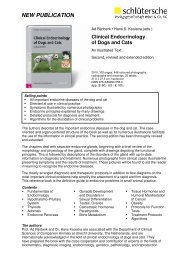
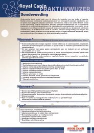
solution. In syringes with a small volume, the outflow opening (cone)<br />
is positioned centrally, while larger syringes (usually) have an eccentric<br />
cone (Figure 11.1.). The cone is eccentric to allow the syringe (<strong>and</strong><br />
needle) to be held as parallel as possible to the blood vessel during<br />
IV injections. The calibration marks on these syringes are printed on<br />
the barrel opposite to position <strong>of</strong> the cone. When unwrapping the<br />
disposable syringe, the cone should remain sterile. This is why the<br />
wrapping should be opened at the side <strong>of</strong> the plunger - <strong>and</strong> not by<br />
pushing the cone end through the wrapping!<br />
11.1.2.2 Needles<br />
Jos Ensink <strong>and</strong> Joris Robben 207<br />
Figure 11.1. Disposable<br />
syringes: a 2 ml syringe with a<br />
central cone (left) <strong>and</strong> a 10 ml<br />
syringe with an eccentric cone<br />
(right)<br />
<strong>Injection</strong> needles, too, should be clean <strong>and</strong> sterile <strong>and</strong> have a sharp <strong>and</strong> properly-shaped bevel<br />
(needle tip). When whetting re-usable needles, the bevel should not be tapered in such a manner<br />
that it will punch a piece <strong>of</strong> skin, as this will block the needle upon penetrating the skin. When<br />
using disposable needles, one can be assured <strong>of</strong> a correct bevel shape. The size <strong>of</strong> the needle is<br />
determined by its use. The thinner (<strong>and</strong> sharper) the needle, the less painful it will be. But the<br />
smaller diameter will increase the injection time unacceptably if a large quantity or a viscous<br />
solution is to be injected. For an IM injection, the needle should not be longer than the desired<br />
injection depth to avoid too deep an injection. For SC injections too, a short needle will prevent<br />
a too deep (IM) injection. The size <strong>of</strong> a needle is described by its diameter in mm (inches) or in<br />
“Gauge” (G) <strong>and</strong> the length in mm (inches). Each size has its own colour code <strong>for</strong> easy recognition.<br />
Un<strong>for</strong>tunately, this colour code is <strong>of</strong>ten different depending on the manufacturer. Below, some<br />
common sizes are listed. For each needle size, the corresponding target species <strong>and</strong> injection<br />
technique is given:<br />
• 0.45 mm (26 G) x 12 mm;<br />
• 0.6 mm (23 G) x 25 mm;<br />
• 0.7 mm (22 G) x 30 mm;<br />
• 0.8 mm (21 G) x 40 mm;<br />
• 0.9 mm (20 G) x 30 mm;<br />
• 1.2 mm (18 G) x 40 mm;<br />
• 1.8 mm (16 G) x 40 mm;<br />
• 2.1 mm (14 G) x 80 mm;<br />
• 2.7 mm (12 G) x 80 mm.<br />
In dogs <strong>and</strong> cats, 22-23 G needles are most commonly used. For injections <strong>of</strong> viscous solutions or<br />
blood samples <strong>of</strong> large volume (e.g. 20 ml), 20 G needles are <strong>of</strong>ten used. For small quantities (1 ml,<br />
e.g. vaccines), 26 G needles are used. Their short length is practical <strong>for</strong> SC injections. In horses,<br />
18 G needles are commonly used <strong>for</strong> IM <strong>and</strong> IV injections. In cattle, 18 G needles may be used too,<br />
but they can bend if the animal moves or resists during IM injections. This is why the thicker, 16 G<br />
needles are more <strong>of</strong>ten used in cattle. For SC injections in the horse, 23 G needles usually suffice.<br />
If more than one SC depot is required (<strong>for</strong> example <strong>for</strong> diagnostic anaesthesia <strong>of</strong> the lower limb),<br />
the longer, 21 G needles may also be used. For IV infusions <strong>of</strong> horse <strong>and</strong> cattle, large-bore needles
208<br />
sharps bin<br />
vial<br />
bottle<br />
breached bottles<br />
Chapter 11 <strong>Injection</strong> <strong>techniques</strong> <strong>for</strong> <strong>drug</strong> <strong>administration</strong> <strong>and</strong> <strong>methods</strong> <strong>of</strong> <strong>restraint</strong><br />
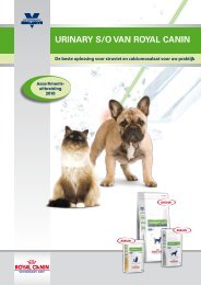
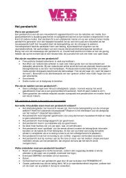
(14 G or 12 G) or catheters are used (Figure 11.2). The needle is<br />
unwrapped at the side <strong>of</strong> the cone. For the aspiration <strong>of</strong> solutions,<br />
first the needle is attached to the syringe. The cone <strong>of</strong> the needle<br />
should be pressed down on the syringe <strong>for</strong> a good fit. If, in doing so,<br />
too much pressure is put on the protective needle guard, this may<br />
cause the needle guard to get stuck on the needle (check be<strong>for</strong>e use<br />
if the needle guard can be easily removed). Only after the needle has<br />
been connected to the syringe, the needle <strong>and</strong> its guard are extracted<br />
from the wrapping.<br />
Needles should be disposed <strong>of</strong> in such a manner that nobody can<br />
be injured. To this end, the needle is best discarded straight after use<br />
in a special container <strong>for</strong> sharp objects (e.g. sharps bin). Another<br />
possibility is to replace the needle in the needle guard, although there<br />
is a risk <strong>of</strong> injuring oneself when doing so.<br />
11.1.2.3 Injectable solutions<br />
Injectable solutions are available in vials or bottles (Figure 11.3). Often, some <strong>of</strong> the fluid is located<br />
in the “head” <strong>of</strong> the vial. Emptying the head by flicking a finger against it may create air bubbles.<br />
It is better to shake it down like a mercury thermometer. If the vial has a coloured ring around the<br />
“neck” (the insertion between the head <strong>and</strong> the actual fluid container), this means the head can be<br />
snapped <strong>of</strong>f easily. If it also has a coloured dot on the head, the neck should be broken by putting<br />
pressure on the head at the position <strong>of</strong> the dot. If the vial has neither ring nor dot (rare nowadays),<br />
a small groove should be made with a little saw which should come with the vials. It may be<br />
prudent to protect the h<strong>and</strong> that is used to break the vial, e.g. with a gauze swab. After opening the<br />
vial, the solution can be aspirated with a syringe <strong>and</strong> needle. This can be done from a st<strong>and</strong>ing vial,<br />
but the vial may also be held upside-down as the meniscus <strong>of</strong> the fluid prevents it from pouring<br />
out. Vials are intended <strong>for</strong> single use only.<br />
Medication bottles have a rubber stopper that can be pierced by an injection needle, usually held<br />
in place by a metal seal. A protective lid has to be removed prior to first use. The absence <strong>of</strong> a<br />
lid indicates the bottle has been used be<strong>for</strong>e. With an absent lid, the rubber surface should first<br />
be disinfected (preferably with alcohol, which does not leave a residue after evaporation <strong>and</strong> is<br />
there<strong>for</strong>e unlikely to have an impact on the content <strong>of</strong> the bottle).<br />
Negative pressure in the bottle may impede aspiration <strong>of</strong> the solution. To avoid this, prior to<br />
aspiration the syringe is filled with an amount <strong>of</strong> air equal to the volume <strong>of</strong> fluid to be collected.<br />
The bottle top is pierced be<strong>for</strong>e turning it upside-down. The air in the syringe is then “exchanged”<br />
<strong>for</strong> the solution. In case <strong>of</strong> large volumes (e.g. 20 ml from a 100 ml bottle), it is recommended to<br />
per<strong>for</strong>m this “exchange” in a stepwise manner as infusion <strong>of</strong> 20 ml <strong>of</strong> air into a 100-ml bottle may<br />
cause leakage due to high positive pressure.<br />
Breached bottles may be stored if certain guidelines are respected. First <strong>of</strong> all, the bottle needs to be<br />
stored according to the manufacturer’s recommendations (<strong>for</strong> example in a refrigerator). A “useby”<br />
period after first use applies to each solution. This is mentioned<br />
in the medication guide <strong>and</strong> usually is one day <strong>for</strong> solutions without<br />
preservatives <strong>and</strong> one month <strong>for</strong> those with preservatives. The day<br />
<strong>of</strong> first use should be written on the label so that the use-by period<br />
can be checked. The bottle may be stored only if the solution was<br />
extracted in an aseptic manner. For extraction <strong>of</strong> the solution from<br />
the bottle or vial, a different needle than the one <strong>for</strong> injection can<br />
be used. This keeps the injection needle clean (especially important<br />
<strong>for</strong> irritating substances) <strong>and</strong> sharp, while a different (larger) needle<br />
size can be chosen <strong>for</strong> aspiration than <strong>for</strong> injection. A little more<br />
solution is aspirated than necessary to allow <strong>for</strong> the removal <strong>of</strong> air<br />
bubbles. This is done by holding the syringe upright <strong>and</strong> by pushing<br />
Figure 11.2. <strong>Injection</strong> needles:<br />
the small one is 0.45<br />
mm (26G) x 12 mm, the<br />
big one is 2.1mm (14G) x<br />
80 mm <strong>and</strong> is positioned so<br />
that one can “look inside the<br />
needle”<br />
Figure 11.3. A vial (left)<br />
<strong>and</strong> a bottle (right) with an<br />
injectable solution
utterfly needle<br />
catheter<br />
over-the-needle<br />
the plunger until all air has been removed. Often, the syringe should be flicked first to ensure that<br />
air bubbles attached to the inner wall <strong>of</strong> the syringe are released. Following this, the needle guard is<br />
placed over the needle. In this way, the cone <strong>of</strong> the syringe is protected against contamination <strong>and</strong><br />
needle injury is prevented so that the syringe can be safely transported to the patient or put aside<br />
(label <strong>for</strong> safety) <strong>for</strong> the time being.<br />
11.1.2.4 Butterfly needles with extension tubing<br />
A butterfly needle is a needle with “wings” at both sides <strong>of</strong> its base, by which the needle can be held.<br />
The needle <strong>of</strong>ten has an extension tube with a cone at the end to which a syringe can be connected.<br />
This extension tube allows the syringe to be moved independently <strong>of</strong> the needle, giving both<br />
operator <strong>and</strong> patient more freedom <strong>of</strong> movement during the injection. This increases the chance<br />
that the needle remains in position (inside the blood vessel). Butterfly needles are there<strong>for</strong>e used in<br />
particular if:<br />
• the animal is difficult to restrain;<br />
• the injection takes longer, which increases the risk <strong>of</strong> movement <strong>and</strong> resistance;<br />
• irritating substances are to be administered.<br />
11.1.2.5 Catheters<br />
Jos Ensink <strong>and</strong> Joris Robben 209<br />
Catheters are <strong>of</strong>ten used instead <strong>of</strong> needles <strong>for</strong> IV (fluid) <strong>administration</strong>. A catheter can remain<br />
in place <strong>for</strong> several days (“long-term” catheter), but a catheter may also be applied <strong>for</strong> a single<br />
injection or <strong>for</strong> medications given IV over several hours (e.g. during surgery). Both long-term <strong>and</strong><br />
short-term catheters should be well secured (adhesive b<strong>and</strong>age <strong>and</strong>/or stay sutures) to keep them<br />
in place (<strong>and</strong> intravenously). A catheter has the advantage that, once correctly in place, one can be<br />
sure that all injectables arrive strictly IV, even if the patient moves. A synthetic catheter causes less<br />
damage to the vessel wall than a metal needle, especially after a longer period <strong>of</strong> time in place.<br />
Easiest to insert are the so-called “over-the-needle” catheters. These consist <strong>of</strong> a synthetic catheter<br />
with an internal metal needle. The metal needle is hollow <strong>and</strong> has a sharp bevel that slightly exceeds<br />
the non-sharp tip <strong>of</strong> the catheter. At the other end, the needle has a transparent chamber in which<br />
blood can become visible once the needle has punctured a vein. The catheter can be fitted with<br />
small wings to secure it to the skin. Once the catheter is positioned in the vein with help <strong>of</strong> the<br />
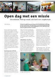
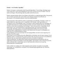
internal needle, the needle is removed (Figure 11.4).<br />
Figure 11.4. An over-the-needle catheter, from the bottom<br />
upward: 1. the catheter as it comes out <strong>of</strong> the wrapping,<br />
with protective needle guard; 2.the catheter after removal<br />
<strong>of</strong> the needle guard; 3. the catheter alone;<br />
4. the needle guard with transparent chamber alone.
210<br />
<strong>administration</strong> set<br />
drip chamber<br />
Luer lock<br />
flow regulator<br />
air vent<br />
disinfection<br />
clipping<br />
Chapter 11 <strong>Injection</strong> <strong>techniques</strong> <strong>for</strong> <strong>drug</strong> <strong>administration</strong> <strong>and</strong> <strong>methods</strong> <strong>of</strong> <strong>restraint</strong><br />
11.1.2.6 Fluid <strong>administration</strong> sets<br />
Fluids <strong>for</strong> infusion are available in glass or synthetic bottles or plastic bags with penetrable stoppers,<br />
sealed with a protective cover. The solutions are usually administered through a fluid <strong>administration</strong><br />
set. The set consists <strong>of</strong> a length <strong>of</strong> tubing with on one end a needle or spike that can be used<br />
to pierce through the stopper. The tubing is equipped with a drip chamber <strong>and</strong> an in-line flow<br />
regulator. The other end <strong>of</strong> the tubing ends in a special connector (<strong>for</strong> example a so-called “Luer<br />
lock” connection) to which a needle or catheter can be attached. The drip chamber is partially filled<br />
with fluid to function as a “buffer” when the bottle <strong>of</strong> bag is empty <strong>and</strong> air tends to fill the tubing.<br />
Furthermore, the speed <strong>of</strong> flow can be adjusted by counting the number <strong>of</strong> drops per minute/<br />
second in the drip chamber. The flow regulator (usually an adjustable clamp or wheel that squeezes<br />
the tubing to a more or lesser extent) allows control <strong>of</strong> the flow speed. When glass or stiff plastic<br />
bottles are used negative pressure is built up during infusion, preventing the flow <strong>of</strong> solution. This is<br />
why an air vent has to be used with these types <strong>of</strong> bottles. Administration sets can have an air vent<br />
built in the spike. For non-vented sets separate tubing <strong>for</strong> air ventilation has to be used. Such an<br />
extension has a needle at one end that is pierced through the stopper, <strong>and</strong> a filter at the other end to<br />
prevent contamination <strong>of</strong> the solution.<br />
When fluids need to be given, the <strong>administration</strong> set should be set up ready prior to placement<br />
<strong>of</strong> the catheter or needle. The bottle or bag is hung upside-down above the level <strong>of</strong> the patient’s<br />
heart. This ensures the solution to flow into the animal, rather than the blood into the tubing after<br />
hooking up the set to the catheter or needle. When bottles requiring air ventilation are used in<br />
combination with a non-vented fluid <strong>administration</strong> set, the needle <strong>of</strong> a separate air vent system<br />
is pierced through the stopper, while the other end <strong>of</strong> tubing is positioned above the fluid level in<br />
the bottle. To spike the <strong>administration</strong> set with fluids, it is taken out <strong>of</strong> its wrapping <strong>and</strong> the flow<br />
regulator is closed (when the flow regulator is open after attaching the set to a fluid bottle or bag,<br />
both fluid <strong>and</strong> air bubbles will fill the system, making it difficult to free the tubing <strong>of</strong> air bubbles).<br />
Then, the needle or spike is pierced through the stopper (bottle) or connector (bag). The drip<br />
chamber is gradually filled by gently pinching it a few times, or by holding it horizontal if made <strong>of</strong><br />
rigid plastic. Then the flow regulator is opened until all air has been expelled from the tubing.<br />
The end <strong>of</strong> the tubing has a protective cap that protects it against contamination. Sometimes, this<br />
cap needs to be removed in order to completely fill the drip tubing.<br />
11.1.3 Practical application<br />
11.1.3.1 Preparing the injection site<br />
Much debate surrounds the question whether it is necessary to disinfect the skin prior to IM or<br />
SC injections. The condition <strong>of</strong> the skin or coat plays an important role: injections through soiled<br />
skin (e.g. in case <strong>of</strong> diarrhoea) or dermal lesions carry an increased risk <strong>of</strong> infection, even after<br />
disinfection. In horses, it has been demonstrated that a rapid disinfection with alcohol without<br />
clipping leads to a significant reduction (± 70%) in the number <strong>of</strong> bacteria on the skin. Electrical<br />
clipping followed by disinfection leads to a reduction <strong>of</strong> over 90%. In horses, it is there<strong>for</strong>e<br />
considered good practice to disinfect the skin prior to IM or SC injections (clipping is only<br />
per<strong>for</strong>med with a thick hair coat).<br />
Also in cattle, the injection site is best disinfected, unless this is impractical (injection <strong>of</strong> a group <strong>of</strong><br />
animals; a poorly restrained or unrestrained animal). In pigs, such situations nearly always apply<br />
<strong>and</strong> injection sites are not disinfected. Prior to IM or SC injections in companion animals with a<br />
well-kept coat, clipping or disinfection is considered not necessary. Often, alcohol is used but this<br />
has merely a function <strong>of</strong> improving the view on the injection site by parting or smoothing the hairs.<br />
However, <strong>for</strong> IV injections, the injection site is always disinfected, <strong>and</strong> <strong>of</strong>ten clipped, in all species.<br />
Sometimes, clipping is necessary to make the vein visible <strong>for</strong> IV injection (e.g. dog, cat, thick-coated<br />
horse). If the risk <strong>of</strong> infection is to be as low as possible (e.g. intra-articular injections, long-term<br />
catheter) the skin should be clipped, cleaned <strong>and</strong> disinfected (like preparing a surgical field).
loose-needle technique<br />
over-the-needle catheter<br />
11.1.3.2 Intravenous injections<br />
Jos Ensink <strong>and</strong> Joris Robben 211<br />
For IV injections, the vein is raised <strong>and</strong> the needle is inserted through the skin at an angle <strong>of</strong> around<br />
30°, with the bevel (needle opening) turned away from the animal (so that the person injecting<br />
“looks into the needle”). As soon as the tip <strong>of</strong> the needle reaches the vein, blood becomes visible<br />
at the cone. Then, the needle is pushed in further, parallel to the vein. The compression <strong>of</strong> the vein<br />
is lifted only after the needle has been advanced further to prevent slipping <strong>of</strong>f the needle <strong>of</strong> the<br />
collapsing vein <strong>and</strong> to reduce the risk <strong>of</strong> puncturing the vein wall from the inside.<br />
The needle may be turned 180° over its longitudinal axis to avoid puncturing the opposite wall<br />
while advancing the needle into the vein (see Figure 11.5.). While giving the IV injection, the vein is<br />
no longer raised, as the purpose is to dilute the solution directly into the streaming blood.<br />
There are two ways to establish the correct position <strong>of</strong> the needle in the lumen <strong>of</strong> the vein.<br />
A loose needle may be used, so that, once the needle punctures the vein, blood drips from the cone.<br />
A continuous blood flow from the needle should be apparent while it is advanced further in the<br />
vein. But the needle can also be connected to the syringe prior to injection. Negative pressure is<br />
applied to the syringe (by slightly pulling the plunger) once the skin has been penetrated.<br />
Figure 11.5. Schematic drawing <strong>of</strong> a venipuncture.<br />
In the drawing on the left, the needle is inserted at an angle <strong>of</strong> 30°, with the bevel (needle opening)<br />
turned away from the animal. On the right, during insertion, the needle is twisted 180° over its<br />
longitudinal axis to avoid puncturing the opposite wall <strong>of</strong> the vein.<br />
Blood appears in the syringe the moment the vessel wall is punctured. The “loose-needle” technique<br />
is easiest but <strong>of</strong>ten causes the hair coat <strong>and</strong> h<strong>and</strong>s to become soiled with blood.<br />
Using an attached syringe prevents blood spillage but maintaining negative pressure requires some<br />
dexterity. Also, the 180°-twist <strong>of</strong> the needle is not possible with this technique. The loose-needle<br />
technique is rarely used in companion animals. When giving an IV injection, a little blood is<br />
aspirated immediately be<strong>for</strong>e the injection is given to check whether the needle is still in the vein.<br />
Also during injection, <strong>for</strong> example after an animal has moved, position should be checked again.<br />
At the end <strong>of</strong> the injection, blood can be aspirated once more to avoid that the (potentially<br />
irritating) solution infiltrates the subcutaneous area when the needle is withdrawn. Sometimes,<br />
the vein needs to be raised <strong>for</strong> the aspiration <strong>of</strong> blood. After needle withdrawal, the injection site<br />
is compressed to avoid a haematoma. If the solution (particularly if it is irritant) ends up in the<br />
perivenous area, this may lead to inflammation or even necrosis. To prevent this from happening,<br />
the perivenous depot <strong>of</strong> the solution should be diluted as soon as possible. For dilution, a sterile<br />
saline solution containing mucopolysaccharidase (e.g. Thiomucase ® ) is used, which improves the<br />
passage through the interstitial area, thereby leading to a more rapid absorption <strong>of</strong> the solution.<br />
Following this, a special ointment can be applied several times daily, containing substances that<br />
improve diffusion through the interstitial area (e.g. Lasonil ® , a heparinoid/hyaluronidase ointment)<br />
but this is less effective than dilution by injection. An over-the-needle catheter is inserted as follows.<br />
The skin is clipped <strong>and</strong> disinfected, <strong>and</strong> the vein is raised. The catheter is inserted until blood
212<br />
horse<br />
cattle<br />
pig<br />
Chapter 11 <strong>Injection</strong> <strong>techniques</strong> <strong>for</strong> <strong>drug</strong> <strong>administration</strong> <strong>and</strong> <strong>methods</strong> <strong>of</strong> <strong>restraint</strong><br />
appears in the transparent chamber, <strong>and</strong> is moved a little further into the vessel to make sure that<br />
the tip <strong>of</strong> the catheter, which is slightly shorter than the needle, is also inside the vein. The needle<br />
is now held in this position (<strong>and</strong> certainly not withdrawn), while the catheter is advanced further<br />
into the vein. Then the needle is fully withdrawn <strong>and</strong> the catheter is flushed <strong>and</strong> capped if not<br />
immediately connected to an <strong>administration</strong> or extension set. The catheter is secured to the skin<br />
<strong>and</strong> protected with a b<strong>and</strong>age if possible. Catheters are placed towards the heart, firstly to avoid<br />
turbulence <strong>of</strong> the inflowing fluid, <strong>and</strong> secondly because the catheter can only pass the venous valves<br />
in the direction <strong>of</strong> the heart. Sometimes, resistance is encountered while the catheter is inserted.<br />
This could indicate that the catheter is no longer inside the vessel lumen <strong>and</strong> could crinkle if<br />
advanced any further. In this situation, the needle should never be re-inserted into the catheter, as<br />
the needle might puncture the catheter. It even may cut <strong>of</strong>f part <strong>of</strong> the catheter that will remain<br />
inside the patient after catheter removal. When encountering problems with catheter advancement,<br />
the compression <strong>of</strong> the vein should be lifted <strong>and</strong> both needle <strong>and</strong> catheter should be removed.<br />
To prepare <strong>for</strong> a second attempt, the needle can be carefully re-inserted into the catheter (if still<br />
completely intact).<br />
If the bottle or bag is left to be emptied completely, depending on the venous pressure, blood may<br />
flow into the catheter, causing it to block (e.g. animal in lateral recumbency), or air may flow into<br />
the vein, with a risk <strong>of</strong> air embolism (e.g. st<strong>and</strong>ing horse with an IV drip in the external jugular<br />
vein). There<strong>for</strong>e, someone needs to be present when the bottle or bag is nearly empty, in order to<br />
disconnect the <strong>administration</strong> set or to replace the bottle or bag.<br />
In horses, the external jugular vein is most commonly used <strong>for</strong> IV injections. At about one third<br />
between head <strong>and</strong> shoulders (measuring from the head), the risk <strong>of</strong> puncturing the carotid artery is<br />
minimal, as it runs a little deeper here <strong>and</strong> is covered by the omohyoid muscle. An assistant slightly<br />
lifts the horse’s head up <strong>and</strong> away from the person injecting.<br />
Restraint <strong>methods</strong> are rarely necessary <strong>and</strong> will cause the horse to tense up its neck muscles, causing<br />
the vein to become less visible. With a h<strong>and</strong> placed around the front base <strong>of</strong> the neck, the vein<br />
can easily be compressed using a thumb. The vein raises slowly, however, <strong>and</strong> is not always clearly<br />
visible. By sudden release <strong>of</strong> the compression, the vein can be seen to deflate fast.<br />
This action is valuable in establishing the location <strong>of</strong> the vein. Horses rarely resist venipuncture,<br />
especially if the needle is inserted through the skin calmly. If the external jugular veins cannot be<br />
used (e.g. damage due to previous injections), the superficial thoracic, cephalic or lateral saphenous<br />
veins may be used. Beware <strong>of</strong> being kicked if using these veins.<br />
In cattle, IV injections can also be given into the external jugular vein. Cattle tend to resist more<br />
than horses to venipuncture. It is there<strong>for</strong>e wise to control the head with the use <strong>of</strong> a rope <strong>and</strong>/or<br />
halter, so that the neck is stretched. A jugular vein clamp or neck tourniquet is recommended.<br />
Both skin <strong>and</strong> venous wall are thick <strong>and</strong> sturdy in cattle. This means that the needle sometimes<br />
needs to be “thrust in” to attain the vein. In cattle, also the subcutaneous abdominal (“milk”) vein<br />
can be used <strong>for</strong> IV injections. This vein is always raised. A disadvantage is that leakage <strong>of</strong> irritating<br />
solutions (e.g. calcium-containing solutions) may lead to extensive inflammatory lesions. When<br />
using the subcutaneous abdominal vein, attention should be paid to possible <strong>for</strong>ward kicking <strong>of</strong> the<br />
cow. For blood sampling, the median caudal vein <strong>of</strong> the tail is <strong>of</strong>ten used, as the animal hardly needs<br />
to be restrained. This site is less suitable <strong>for</strong> IV injections, as the vein can only be punctured at an<br />
angle <strong>of</strong> 90° <strong>and</strong> the needle cannot be advanced very far into the vein. This means that part <strong>of</strong> the<br />
liquid is likely to arrive in the perivenous area by extravasation <strong>of</strong> the tip <strong>of</strong> the needle.<br />
In pigs, the cranial vena cava at the thoracic inlet or the external jugular vein just cranial <strong>of</strong> this<br />
inlet are suitable <strong>for</strong> blood sampling but less so <strong>for</strong> IV injections, as the veins are reached at an angle<br />
<strong>of</strong> around 90° <strong>and</strong> the needle cannot be advanced very far into the vein.<br />
For blood sampling, pigs are restrained as follows: in a st<strong>and</strong>ing position large animals are<br />
restrained with a noose over the upper jaw; smaller individuals are placed in lateral or dorsal<br />
recumbency, or upside-down with the <strong>for</strong>elimbs held back. An auricular (“ear”) vein is usually<br />
chosen <strong>for</strong> IV injections. The animal is restrained <strong>and</strong> the base <strong>of</strong> the ear is clamped by h<strong>and</strong> or<br />
with a tourniquet. For venipuncture, a thin needle or catheter is used, which is secured to the ear<br />
with adhesive tape or a clamp to prevent it from dislodging if the animal moves its head.
small ruminants<br />
dog <strong>and</strong> cat<br />
sphinx position<br />
Jos Ensink <strong>and</strong> Joris Robben 213<br />
In small ruminants, IV injections can be given in the external jugular vein. In fat animals, this vein<br />
is poorly visualised <strong>and</strong> in sheep this area needs to be clipped or shorn. For this reason, the cephalic<br />
vein is <strong>of</strong>ten used (<strong>for</strong> the technique, see below).<br />
In dogs <strong>and</strong> cats, the antebrachial part <strong>of</strong> the cephalic vein is preferred <strong>for</strong> IV injections as the<br />
animal accepts this with minimal <strong>restraint</strong>, <strong>and</strong> the vein is easy to locate. The animal is preferably<br />
placed on a table in the so-called “sphinx position” (a sitting or st<strong>and</strong>ing position is preferred by<br />
some animals <strong>and</strong> can there<strong>for</strong>e be more successful, but unexpected movements <strong>of</strong> the animal are<br />
more difficult to control).<br />
With one h<strong>and</strong>, the assistant restrains the head <strong>of</strong> the animal, turning it away to prevent the animal<br />
from biting the face <strong>of</strong> the person injecting. With the arm over the back <strong>of</strong> the animal, the other<br />
h<strong>and</strong> <strong>of</strong> the assistant grasps the opposite <strong>for</strong>elimb <strong>of</strong> the animal, at elbow level. In large dogs, the<br />
assistant thereby leans on the back <strong>of</strong> the animal, preventing it from sitting or st<strong>and</strong>ing up.<br />
The assistant compresses the vein with the thumb. In doing so, the entire h<strong>and</strong> is rotated outward:<br />
this causes the slightly medially running cephalic vein to be pulled “on top” <strong>of</strong> the <strong>for</strong>elimb, <strong>for</strong> a<br />
better presentation to the person injecting. To prevent the animal from pulling back its limb, the<br />
h<strong>and</strong> <strong>and</strong> lower arm is kept in contact with the tabletop. In cats, a second assistant may be necessary<br />
to control the hind limbs.<br />
The <strong>for</strong>elimb is grasped at the carpus (wrist) by the person injecting (right limb in left h<strong>and</strong><br />
<strong>and</strong> vice versa), whereby the thumb is positioned parallel to the cephalic vein to prevent lateral<br />
movement <strong>of</strong> the blood vessel. The person injecting should not grasp the limb too tightly in<br />
an ef<strong>for</strong>t to extend the limb (this should be per<strong>for</strong>med by the assistant), as this compresses the<br />
peripheral part <strong>of</strong> the cephalic vein, impeding circulation in the vessel. In preparation <strong>of</strong> the actual<br />
injection, the connection <strong>of</strong> the needle to the syringe should be checked, the calibration markings<br />
visible, the eccentric cone placed against the skin <strong>and</strong> the inside <strong>of</strong> the bevel visible. The needle is<br />
then advanced through the skin, into the blood vessel at an angle <strong>of</strong> 20-35°. The blood vessel can be<br />
punctured directly or the skin next to the blood vessel can be penetrated first, followed by puncture<br />
<strong>of</strong> the vein. The latter method prevents flattening <strong>of</strong> the vein at the time <strong>of</strong><br />
skin penetration, which leads to an increased risk <strong>of</strong> puncturing the opposite venous wall. Once the<br />
needle has penetrated the skin, the plunger <strong>of</strong> the syringe is pulled slightly backward to create slight<br />
negative pressure in the barrel, so that blood is aspirated the moment the needle punctures the vein.<br />
At this moment, the needle is advanced slightly further into the vessel to ensure that the needle<br />
does not slip out when the venous compression is released. Securing the syringe against the ball<br />
<strong>of</strong> the thumb after venipuncture allows the absorbtion <strong>of</strong> any unexpected limb movements. After<br />
releasing the compression (but not the limb!), the solution is slowly injected. After the injection,<br />
the skin is pulled taut over the injection site by the assistant after the needle is withdrawn, <strong>and</strong> kept<br />
in this position <strong>for</strong> a minute. The injection site may also be compressed with a b<strong>and</strong>age <strong>for</strong> 5 to 15<br />
minutes. An alternative location <strong>for</strong> IV injections is the saphenous vein in the hind limb.<br />
In dogs, the lateral, in the cat the median saphenous vein is routinely used. The dog is placed<br />
in lateral recumbency <strong>and</strong> the assistant restrains the upper hind limb at the stifle. The thumb is<br />
positioned over the patellar tendon, with the fingers around the popliteal area. Increased pressure<br />
<strong>of</strong> the h<strong>and</strong> will raise the lateral saphenous vein. The vein is better visualised than the cephalic vein,<br />
but is more difficult to puncture as proper fixation to reduce lateral movement is harder. In cats,<br />
the uppermost limb is held away by the assistant <strong>and</strong> the femoral vein is compressed in the inguinal<br />
area <strong>of</strong> the opposite (lower) limb. The median saphenous vein is clearly visible through the thin,<br />
nearly hairless skin. The external jugular vein is mainly used in small animals if (a large volume<br />
<strong>of</strong>) blood needs to be sampled, or if the injectable substance is too irritant <strong>for</strong> peripheral injection.<br />
For an IV injection in the external jugular vein, the animal is placed in the sphinx or in a sitting<br />
position. The assistant stretches the neck by lifting the head, turning it slightly to the contralateral<br />
side <strong>of</strong> which the jugular vein will be used. If this is done slowly <strong>and</strong> the neck is not overstretched,<br />
this generally leads to little resistance. In cats <strong>and</strong> small dogs it may be necessary to restrain the<br />
<strong>for</strong>elimbs, bringing them in a straight line with the head <strong>and</strong> neck, so that the person injecting has<br />
sufficient room to place the needle <strong>and</strong> syringe more parallel to the vein.<br />
The <strong>for</strong>elimbs may be restrained by holding them from the back in one h<strong>and</strong>, with a finger between<br />
the limbs. For this, the arm is placed over the back <strong>of</strong> the animal to the other side <strong>and</strong> brought
214<br />
horse<br />
cattle<br />
pig<br />
small ruminant<br />
dog <strong>and</strong> cat<br />
Chapter 11 <strong>Injection</strong> <strong>techniques</strong> <strong>for</strong> <strong>drug</strong> <strong>administration</strong> <strong>and</strong> <strong>methods</strong> <strong>of</strong> <strong>restraint</strong><br />
behind the <strong>for</strong>elimbs. In particular in fractious cats <strong>and</strong> dogs, the “stretching” <strong>of</strong> the head <strong>and</strong><br />
<strong>for</strong>elimbs improves the grip, as it is more difficult <strong>for</strong> the animal to use its muscles in defence when<br />
stretched. It is advisable to restrain the hind limbs separately or to restrain the hindquarters by<br />
pressing them with the arm holding the <strong>for</strong>elimb to the assistant’s upper body.The external jugular<br />
vein runs from the base <strong>of</strong> the ear to the thoracic inlet. By pressing a thumb flatly across the base<br />
<strong>of</strong> the neck, the vein can be raised over the whole length <strong>of</strong> the neck. There are three <strong>methods</strong> <strong>for</strong><br />
locating the vein: releasing compression <strong>of</strong> the vein will show a deflating vein; compressing <strong>and</strong><br />
releasing the compression while feeling <strong>for</strong> the deflating vein with a finger; flicking a finger against<br />
the neck while the vein is raised: if the vein is hit, a pulsation can be felt by the thumb compressing<br />
the vein. Often, a combination <strong>of</strong> <strong>methods</strong> is used. Once the vein has been located, the thumb is<br />
positioned just below the middle <strong>of</strong> the lower neck, so that the thumb can act as support <strong>for</strong> the<br />
needle (after prior disinfection <strong>of</strong> the thumb with alcohol) <strong>and</strong> to better prevent the vein from<br />
moving sideways. The needle is then inserted just above the thumb at an angle <strong>of</strong> around 30°.<br />
For the rest <strong>of</strong> the technique, see under cephalic vein. During anaesthesia, the sublingual vein may<br />
be used to administer substances or to draw blood.<br />
11.1.3.3 Intramuscular injections<br />
In IM injections, the injectable solution should not enter the blood stream as certain substances<br />
(e.g. procaine-penicillin suspension in the horse) may cause shock. Prior to injection, the syringe<br />
should be briefly aspirated to check whether the needle has not accidentally penetrated a vein.<br />
In horses, the pectoral muscle (pectoralis descendens) <strong>and</strong> the hamstring (biceps, semitendinous<br />
<strong>and</strong> semimembranosus) may be used. An injection in the pectoral muscle will <strong>of</strong>ten cause a<br />
visible (but benign) swelling; the hamstring is covered by a fascia, making the swelling invisible.<br />
When injecting in the hamstring, the operator should st<strong>and</strong> as close as possible to the shoulder<br />
<strong>of</strong> the horse to avoid injury if the horse kicks. When injecting in the hamstring, <strong>restraint</strong> may be<br />
necessary. The semimembranosus muscle is difficult to reach safely because <strong>of</strong> its medial position<br />
<strong>and</strong> there<strong>for</strong>e rarely used. Many horses will tolerate IM injections without too much resistance,<br />
especially if the muscle <strong>of</strong> the injection site is relaxed. This may be obtained by “pounding” a few<br />
times on the muscle with the side <strong>of</strong> the fist, needle in h<strong>and</strong>, be<strong>for</strong>e turning the h<strong>and</strong> <strong>and</strong> inserting<br />
the needle in the muscle. The injection is carried out as close as possible to the muscle belly, not too<br />
close to the insertion on the bone (e.g. ischial tuber <strong>for</strong> the semimembranosus). After injecting 20<br />
ml, the needle is usually repositioned 10 cm apart <strong>for</strong> the next 20 ml. In horses, the neck muscles<br />
are avoided, as a possible abscess in this area is very difficult to treat: due to the vertical fascia, an<br />
abscess is more likely to extend vertically than to burst <strong>and</strong> drain sideways. If the horse owner insists<br />
that the injection is given in the neck (<strong>for</strong> fear <strong>of</strong> reduced per<strong>for</strong>mance due to pain after injection in<br />
the pectoral or hamstring muscles), he should be well in<strong>for</strong>med <strong>of</strong> this risk.<br />
In cattle, the neck muscles are the preferred injection site as these muscles are economically not very<br />
important. The injection should be placed in the muscles cranial to the shoulder blade, well above<br />
the vertebral column. In animals that need to be given a large number <strong>of</strong> injections, the hamstring<br />
muscles may be used, as in the horse. Cattle will always resist being injected. “Pounding” <strong>of</strong> the<br />
muscles in preparation <strong>of</strong> needle insertion is <strong>of</strong> little use <strong>and</strong> <strong>restraint</strong> is usually necessary.<br />
In pigs, the neck muscles are also used. The needle is inserted in a medio-vertical direction about<br />
three fingers’ width behind the ear. Group injections (using revolver syringes) are best carried out<br />
at the feeding trough. Young pigs may also be injected in the (more expensive!) hamstring muscles.<br />
Care should be taken, not to insert the needle too deeply to avoid damage to the ischiatic nerve.<br />
In small ruminants, the triceps muscles may be used <strong>for</strong> IM injections (accessible in sheep through<br />
the hairless skin on the inside <strong>of</strong> the elbow) or the hamstring muscles.<br />
In IM injections <strong>of</strong> dogs <strong>and</strong> cats, quantities <strong>of</strong> 2 to 6 ml are considered acceptable depending on<br />
the size <strong>of</strong> the animal. In dogs <strong>and</strong> cats, a number <strong>of</strong> muscles can be considered <strong>for</strong> IM injections:<br />
behind the femur in the hamstring muscles (dog), in front <strong>of</strong> the femur in the quadriceps muscles<br />
(cat), the triceps muscles <strong>and</strong> the back muscles in the lumbar area (dog <strong>and</strong> cat). Traditionally,<br />
injections have been given in the hamstring muscles. But in this area, two major complications may<br />
arise, so that this site is no longer considered acceptable. First <strong>of</strong> all, there is a risk <strong>of</strong> severe damage
to the ischiatic nerve (in particular with irritating substances). Secondly, contrast studies have<br />
shown that, in the majority <strong>of</strong> cases, the injected solution does not arrive in the muscle but in the<br />
intermuscular fascia, with an increased risk <strong>of</strong> diffusion to <strong>and</strong> damage <strong>of</strong> the ischiatic nerve, as well<br />
as a highly unpredictable absorption rate.<br />
<strong>Injection</strong>s in the quadriceps muscles will end up intramuscularly <strong>and</strong> rarely intermuscularly.<br />
The injection is made from lateral to medial at right angles to the muscle to avoid the needle being<br />
inserted deeper, reaching the femur if the animal makes a sudden <strong>for</strong>ward kick with the hind limb.<br />
<strong>Injection</strong>s in the lumbar back muscles (next to the midline) rarely cause complications, will always<br />
end up intramuscularly <strong>and</strong> are <strong>of</strong>ten used when giving anaesthetics as IM injections in this area<br />
lead to rapid absorption.<br />
11.1.3.4 Subcutaneous injections<br />
Subcutaneous injections are given in areas where the skin lies loosely over the underlying tissue.<br />
To give the injection, a skin fold is held between thumb <strong>and</strong> index finger, creating a skin triangle<br />
with the body wall as a base. The centre <strong>of</strong> this triangle is slightly dented, into which the needle<br />
is inserted, below thumb <strong>and</strong> index finger <strong>and</strong> in parallel to the body wall. The skin fold is then<br />
released <strong>and</strong> with a small movement it is checked that the needle is still in the subcutaneous area.<br />
As with IM injections, it should be checked if the needle has not accidentally punctured a vein<br />
with slight drawback <strong>of</strong> the plunger. The solution is then injected (Figure 11.6). At the end <strong>of</strong> the<br />
injection, the needle is withdrawn <strong>and</strong> the injection site is compressed. The pressure only aims<br />
at preventing leakage from the skin. The injection site is not massaged, as this only increases the<br />
tissue trauma <strong>and</strong> does not increase the speed <strong>of</strong> absorption. In horses, SC injections are given in<br />
the pectoral area, in cattle in the dewlap <strong>and</strong> in small ruminants on the ribcage behind the elbow.<br />
In pigs, SC injections are given just behind the ear. Since in pigs it is not possible to check whether<br />
the needle is indeed SC (the animals are not or poorly restrained), it is possible that part <strong>of</strong> the<br />
injections intended <strong>for</strong> SC <strong>administration</strong> are in fact given IM. By using a short needle <strong>for</strong> SC<br />
injections, this can be prevented as much as possible. In dogs <strong>and</strong> cats, subcutaneous injections are<br />
given dorsally between the shoulder blades (Fig. 11.6.) or ventrally on the side <strong>of</strong> the thorax behind<br />
the elbow. Advantages <strong>of</strong> the first location are the loose skin, the accessibility <strong>and</strong> the difficulty<br />
<strong>for</strong> the animal to bite the person injecting. Disadvantage are the more complicated treatment <strong>of</strong><br />
a possible injection site abscess, as well as aesthetical aspects as some injectables may cause tissue<br />
reactions with swellings (imidocarb) or a change in coat colour (progestogens). The second site<br />
does not have these disadvantages, but it is less accessible <strong>and</strong> although the skin is thinner, it also<br />
appears more sensitive, while it is easier <strong>for</strong> the animal to reach around <strong>and</strong> bite the operator.<br />
In cats, it is no longer recommended to give certain vaccinations between the shoulder blades or on<br />
the side <strong>of</strong> the thorax, but rather in the SC <strong>of</strong> the ventral abdomen or distally on one <strong>of</strong> the limbs.<br />
This precautionary method facilitates the treatment <strong>of</strong> possible complications such as injection site<br />
sarcomas. The earlier mentioned areas (behind the elbow <strong>and</strong> between the shoulder blades) do not<br />
allow a wide resection <strong>of</strong> this aggressive tumour.<br />
Figure 11.6. A subcutaneous injection is given<br />
in a Labrador retriever in the loose subcutaneous<br />
tissue between the shoulder blades.<br />
Jos Ensink <strong>and</strong> Joris Robben 215
216<br />
twitch<br />
Chapter 11 <strong>Injection</strong> <strong>techniques</strong> <strong>for</strong> <strong>drug</strong> <strong>administration</strong> <strong>and</strong> <strong>methods</strong> <strong>of</strong> <strong>restraint</strong><br />
11.2 Restraint <strong>methods</strong><br />
11.2.1 Introduction<br />
11.2.2 Horses<br />
Veterinary surgeons will <strong>of</strong>ten need to carry out diagnostic acts (blood sampling,<br />
taking radiographs) or treatments (giving injections, applying b<strong>and</strong>ages) on<br />
animals when it is important that the animal does not move (whether lying,<br />
sitting or st<strong>and</strong>ing) <strong>and</strong> does not resist (kicking, biting). Two major requirements<br />
<strong>for</strong> treatment without resistance from the patient are a calm environment <strong>and</strong><br />
personnel with experience with the species concerned. Un<strong>for</strong>tunately, these are<br />
not always available <strong>and</strong> sedation or <strong>restraint</strong> may be necessary. For non-painful<br />
interventions, the desired result can be obtained using sedation or <strong>restraint</strong>.<br />
In some cases, sedation is not an option; it is <strong>for</strong> example impossible to examine<br />
lameness in a horse that has been sedated. For painful interventions, local<br />
anaesthesia or sedation in combination with sufficient (local) analgesia may be used<br />
to prevent resistance.<br />
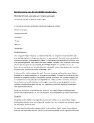
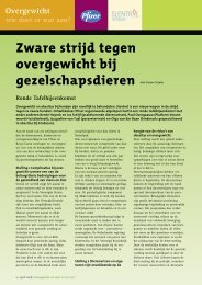
A commonly-used <strong>restraint</strong> method in the horse is the twitch. A twitch is a stick with a loop <strong>of</strong><br />
rope (8 mm thick, circumference <strong>of</strong> the loop 50 cm) on one end. The loop is passed over the upper<br />
lip <strong>of</strong> the horse <strong>and</strong> is tightened by twisting the stick (Figure 11.7). The pressure on the upper lip<br />
causes endorphin release, giving the horse a sedated aspect after about 30 seconds <strong>and</strong> at which<br />
time it no longer resists treatment. Normally, firm pressure on the upper lip is sufficient, although<br />
in some cases the twitch needs to be tightened more to control the horse. In the majority <strong>of</strong> horses,<br />
the twitch works well, but in some cases, a horse resists <strong>and</strong> may suddenly “explode”. This resistance<br />
is most commonly observed in horses where the twitch needs to be tightened very firmly <strong>and</strong> in<br />
(Arab) thoroughbreds. If the twitch is used correctly, the risk <strong>of</strong> permanent damage to the upper lip<br />
is small, although repeated use <strong>of</strong> the twitch may cause skin lesions. A twitch may also be applied<br />
to the ear. This may become necessary in horses that do not (or no longer) accept grasping <strong>of</strong> the<br />
upper lip. The person holding the twitch should twist the ear with the loop against the horse’s neck,<br />
but should refrain from further pulling at the ear. It has not been demonstrated that the use <strong>of</strong> the<br />
twitch on the ear causes endorphin release. Furthermore, the ear twitch may lead to damage <strong>of</strong> the<br />
ear muscles if pulled at. This is why the ear twitch should be used sparingly.<br />
An easier <strong>restraint</strong> method in the horse is grasping a thick fold <strong>of</strong> skin at the side <strong>of</strong> the neck or<br />
Figure 11.7. The twitch is usually placed<br />
around the horse’s upper lip.
grasping the nose<br />
nose ring<br />
pushing up the tail<br />
kick stops<br />
noose<br />
dog<br />
muzzle<br />
cat<br />
11.2.3 Cattle<br />
11.2.4 Pigs<br />
behind the elbow. The skin fold is then pulled tightly with the full h<strong>and</strong>. In foals, the tail base can<br />
be grasped in the full h<strong>and</strong>, pulling it upward <strong>and</strong> <strong>for</strong>ward. This upward pulling <strong>of</strong> the tail reduces<br />
the movement <strong>of</strong> the hind limbs. In horses that keep lifting the limb that needs attention, another<br />
limb can be lifted to counter this. For example by lifting the left <strong>for</strong>elimb, the horse will leave the<br />
right <strong>for</strong>e- <strong>and</strong> left hind limb on the ground. Keeping the <strong>for</strong>elimb lifted is hard work <strong>for</strong> the helper,<br />
especially if the horse keeps on attempting to lift the limb that is to be examined or treated (even<br />
more so if a hind limb were to be lifted).<br />
In cattle, “grasping the nose” is <strong>of</strong>ten used as a <strong>restraint</strong> method. The aim is to exert traction to the<br />
muzzle. The ring can be pulled in animals with a nose ring. Without a nose ring, the thumb can be<br />
placed in one nostril, one or two fingers in the other, pulling the muzzle <strong>for</strong>ward.<br />
This quickly becomes tiring, <strong>and</strong> there<strong>for</strong>e nose tongs are available. With such tongs the muzzle can<br />
be pulled if necessary while the h<strong>and</strong> <strong>of</strong> the operator can relax at moments when the tong needs to<br />
be slackened. Pushing up the tail as described <strong>for</strong> foals is also a useful technique in cattle to prevent<br />
lifting <strong>of</strong> the hind limbs. The tail should be grasped near its base to avoid the risk <strong>of</strong> fractures.<br />
To prevent <strong>for</strong>ward kicking <strong>of</strong> the hind limb, a h<strong>and</strong> can grasp the inguinal fold <strong>and</strong> pull it up<br />
firmly. With so-called “kick stops”, <strong>of</strong>ten used to stop cows kicking <strong>of</strong>f the milking clusters, the same<br />
effect is obtained. One end <strong>of</strong> the adjustable hook is placed under the inguinal fold, while the other<br />
end is hooked behind the spinal processes <strong>of</strong> the sacrum. For treatment <strong>of</strong> the lower limb, the limb<br />
concerned is best lifted <strong>and</strong> attached in a ho<strong>of</strong>-trimming crush.<br />
Small pigs can simply be restrained in lateral recumbency or be lifted up <strong>for</strong> various treatments.<br />
Larger animals can be restrained by securing a noose over the upper jaw. The noose should be<br />
placed behind the canines so that it does not slip <strong>of</strong>f. The pig will resist <strong>and</strong> pull backwards, after<br />
which interventions such as blood sampling can be carried out with ease.<br />
11.2.5 Companion animals<br />
Jos Ensink <strong>and</strong> Joris Robben 217<br />
Restraint <strong>methods</strong> in dogs <strong>and</strong> cats are numerous. A few examples will be given here to give an<br />
impression <strong>of</strong> the range <strong>of</strong> possibilities. A calm <strong>and</strong> confident approach to the patient is a first<br />
requirement. Depending on the degree <strong>of</strong> co-operation <strong>of</strong> the animal <strong>and</strong> the <strong>restraint</strong> required<br />
<strong>for</strong> the treatment concerned, more or less firm immobilising grips are available. See also under the<br />
descriptions <strong>of</strong> the various injection <strong>techniques</strong>. In dogs, the main aim is to avoid being bitten. The<br />
easiest way is to restrain the head by grasping the loose skin at both sides <strong>of</strong> the neck (Figure 11.8.).<br />
In certain brachycephalic breeds (Pekinese), this grip is not recommended in view <strong>of</strong> the risk <strong>of</strong><br />
eyeball prolapse. If this does not give sufficient immobilisation, a muzzle (e.g. cotton b<strong>and</strong>) can be<br />
tied around the dog’s snout. Different types <strong>of</strong> commercial muzzles are also available.<br />
If the dog tries to wriggle or escape, it can be placed in lateral recumbency. Good <strong>restraint</strong> <strong>of</strong> the<br />
lying dog can be obtained by holding the lower limbs while st<strong>and</strong>ing at the side <strong>of</strong> the dog’s back:<br />
one limb in each h<strong>and</strong>, just above the elbow <strong>and</strong> hock. This makes it virtually impossible <strong>for</strong> the dog<br />
to get up. The dog’s head <strong>and</strong> neck are restrained by placing the elbow (<strong>of</strong> the same arm that holds<br />
the <strong>for</strong>elimb) on the table, securing the neck between upper arm <strong>and</strong> breast. The elbow should not<br />
press the neck down on the table as this may cause damage to the neck (See also Chapter 14). Cats<br />
<strong>of</strong>ten resist <strong>restraint</strong> more than dogs. However, a calm approach with a “loose” <strong>restraint</strong> <strong>of</strong>ten gives<br />
excellent results. If immobilisation is nevertheless necessary, care should be taken to avoid both<br />
biting <strong>and</strong> scratching, <strong>and</strong> <strong>restraint</strong> should be firm from the start. There is <strong>of</strong>ten no in-between with<br />
cats. Here, too, many options are available. If the cat is placed in lateral recumbency, both <strong>for</strong>e- <strong>and</strong>
218<br />
Chapter 11 <strong>Injection</strong> <strong>techniques</strong> <strong>for</strong> <strong>drug</strong> <strong>administration</strong> <strong>and</strong> <strong>methods</strong> <strong>of</strong> <strong>restraint</strong><br />
hind limbs need to be grasped with a finger between the limbs. Stretching the body (see above)<br />
leads to a reduced <strong>for</strong>ce <strong>and</strong> causes a less effective resistance <strong>of</strong> the cat. As in the dog, the head can<br />
be fixed with the lower arm <strong>and</strong>/or a muzzle. Muzzles that cover the eyes are especially effective in<br />
cats. Apparently, this causes a certain degree <strong>of</strong> calm <strong>and</strong> acceptance. Often, a second assistant can<br />
be useful <strong>for</strong> proper <strong>restraint</strong>. In this case it is important <strong>for</strong> all concerned that, if someone loses<br />
control, or at the end <strong>of</strong> the procedure, this is notified in time so that everyone can let go at the<br />
same time. Usually, cats calm down once they are released. To avoid being scratched, cats may be<br />
wrapped in a towel. For IV injections, a single limb may be extracted.<br />
Figure 11.8. The easiest way to restrain the<br />
head in dogs is to grasp the loose skin at both<br />
sides <strong>of</strong> the neck
Jos Ensink <strong>and</strong> Joris Robben 219