Epiphytic coralline crusts (Corallinales, Rhodophyta) from South ...
Epiphytic coralline crusts (Corallinales, Rhodophyta) from South ...
Epiphytic coralline crusts (Corallinales, Rhodophyta) from South ...
You also want an ePaper? Increase the reach of your titles
YUMPU automatically turns print PDFs into web optimized ePapers that Google loves.
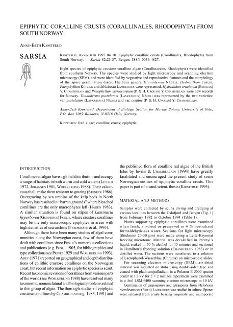
EPIPHYTIC CORALLINE CRUSTS (CORALLINALES, RHODOPHYTA) FROM<br />
SOUTH NORWAY<br />
ANNE-BETH KJØSTERUD<br />
SARSIA<br />
INTRODUCTION<br />
KJØSTERUD, ANNE-BETH 1997 04 10. <strong>Epiphytic</strong> <strong>coralline</strong> <strong>crusts</strong> (<strong>Corallinales</strong>, <strong>Rhodophyta</strong>) <strong>from</strong><br />
<strong>South</strong> Norway. — Sarsia 82:23-37. Bergen. ISSN 0036-4827.<br />
Eight species of epiphytic crustose <strong>coralline</strong> algae (Corallinaceae, <strong>Rhodophyta</strong>) were identified<br />
<strong>from</strong> southern Norway. The species were studied by light microscopy and scanning electron<br />
microscopy (SEM), and were identified by vegetative and reproductive features and the morphology<br />
of the spore germination discs. The four genera Titanoderma NÄGELI, Hydrolithon FOSLIE,<br />
Pneophyllum KÜTZING and Melobesia LAMOUROUX were represented. Hydrolithon cruciatum (BRESSAN)<br />
Y. CHAMBERLAIN and Pneophyllum myriocarpum (P. & H. CROUAN) Y. CHAMBERLAIN were new records<br />
for Norway. Titanoderma pustulatum (LAMOUROUX) NÄGELI was represented by the two varieties:<br />
var. pustulatum (LAMOUROUX) NÄGELI and var. confine (P. & H. CROUAN) Y. CHAMBERLAIN.<br />
Anne-Beth Kjøsterud, Department of Biology, Section for Marine Botany, University of Oslo,<br />
P.O. Box 1069 Blindern, N-0316 Oslo, Norway.<br />
KEYWORDS: Red algae; <strong>coralline</strong> <strong>crusts</strong>; epiphytic.<br />
Coralline red algae have a global distribution and occupy<br />
a range of habitats in both warm and cold waters (LITTLER<br />
1972, JOHANSEN 1981, WOELKERLING 1988). Their calcareous<br />
thalli make them resistant to grazing (STENECK 1986).<br />
Overgrazing by sea urchins of the kelp beds in North<br />
Norway has resulted in “barren grounds” where bleached<br />
<strong>coralline</strong>s are the only macrophytes left (HAGEN 1983).<br />
A similar situation is found on stipes of Laminaria<br />
hyperborea (GUNNERUS) FOSLIE, where crustose <strong>coralline</strong>s<br />
may be the only macroscopic epiphytes in areas with<br />
high densities of sea urchins (FREDRIKSEN & al. 1995).<br />
Although there have been many studies of algal communities<br />
along the Norwegian coast, few of them have<br />
dealt with <strong>coralline</strong>s since FOSLIE’s numerous collections<br />
and publications (e.g. FOSLIE 1905, for bibliographies and<br />
type collections see PRINTZ 1929 and WOELKERLING 1993).<br />
ADEY (1971) reported on geographical and depth distributions<br />
of epilithic crustose <strong>coralline</strong>s on the Norwegian<br />
coast, but recent information on epiphytic species is scant.<br />
Recent taxonomic revisions of <strong>coralline</strong>s <strong>from</strong> various parts<br />
of the world (see WOELKERLING 1988) have resolved many<br />
taxonomic, nomenclatural and biological problems related<br />
to this group of algae. The thorough studies of epiphytic<br />
crustose <strong>coralline</strong>s by CHAMBERLAIN e.g. 1983, 1991) and<br />
the published flora of <strong>coralline</strong> red algae of the British<br />
Isles by IRVINE & CHAMBERLAIN (1994) have greatly<br />
facilitated and encouraged the present study of some<br />
Norwegian entities of epiphytic <strong>coralline</strong> <strong>crusts</strong>. This<br />
paper is part of a cand.scient. thesis (KJØSTERUD 1995).<br />
MATERIAL AND METHODS<br />
Samples were collected by scuba diving and dredging at<br />
various localities between the Oslofjord and Bergen (Fig. 1)<br />
<strong>from</strong> February 1992 to October 1994 (Table 1).<br />
Plants supporting epiphytic <strong>coralline</strong>s were examined<br />
when fresh, air-dried or preserved in 4 % neutralized<br />
formaldehyde-sea water. Sections for light microscopy<br />
(thickness 20-30 µm) were made using a Leitz-Kryomat<br />
freezing microtome. Material was decalcified in Perenyi’s<br />
liquid, soaked in 70 % alcohol for 15 minutes and sectioned<br />
in Hamilton’s freezing solution (CHAMBERLAIN 1983) or in<br />
distilled water. The sections were transferred to a solution<br />
of Lactophenol-Wasserblau (Chroma) on microscopic slides.<br />
For scanning electron microscopy (SEM), air-dried<br />
material was mounted on stubs using double-sided tape and<br />
coated with platinum/palladium in a Polaron E 5000 sputter<br />
coater at 1.2 kV for 2 × 2 minutes. Specimens were examined<br />
in a Jeol LSM-6400 scanning electron microscope at 10 kV.<br />
Germination of carpospores and tetraspores <strong>from</strong> Melobesia<br />
membranacea (ESPER) LAMOUROUX was studied in culture. Spores<br />
were released <strong>from</strong> <strong>crusts</strong> bearing uniporate and multiporate
24 Sarsia 82:23-37 – 1997<br />
conceptacles and isolated into petri dishes, containing IMR 1/<br />
2 culture medium (EPPLEY & al. 1967) at a salinity of 30 ‰ and<br />
maintained at 12° C and 17° C.<br />
Measurements of diagnostic features and descriptive terminology<br />
follow IRVINE & CHAMBERLAIN (1994).<br />
Voucher specimens of collected algae <strong>from</strong> this study have been<br />
deposited at the Section for Marine Botany, University of Oslo.<br />
OBSERVATIONS<br />
Eight species of epiphytic crustose <strong>coralline</strong> algae were<br />
identified (Table 2) in this study. A key to the species is<br />
given in Table 3. In the following account details of morphological<br />
and anatomical features are presented along<br />
with field data, substrata relations and information on<br />
earlier Norwegian records. Nomenclature and delimitation<br />
of <strong>coralline</strong> taxa follow IRVINE & CHAMBERLAIN (1994).<br />
Because taxonomic concepts have changed so much in<br />
recent years, older collections <strong>from</strong> Norway kept in herbaria<br />
are in need of critical re-examination. It has not been<br />
possible to confirm species identification for earlier<br />
records. Data on epiphytic crustose <strong>coralline</strong>s <strong>from</strong><br />
Norway in the literature should therefore be treated with<br />
caution.<br />
Table 1. Sample sites, dates and depths.<br />
Station<br />
number<br />
Area County Date Depth<br />
1 Tisler Østfold June 1992 0-5 m<br />
2 Akerøya Østfold August 1992 0-5 m<br />
3 Vesleøy Østfold August 1993 8-12 m<br />
4 Kalkgrunnen Østfold August 1992 2-7 m<br />
5 Seikrakk Østfold August 1993 5-20 m<br />
6 Rauer Østfold June 1992,<br />
July 1993 3-20 m<br />
7 Engelsviken Østfold July 1993 3-20 m<br />
8 Jeløya Østfold July 1993 0-4 m<br />
9 Storskjær Østfold April 1992 5-35 m<br />
10 Hallangspollen Østfold August 1993 0-4 m<br />
11 Sandspollen Østfold April 1992 0-4 m<br />
12 Vollen Akershus September 1992,<br />
July 1993 0-4 m<br />
13 Gåsøya Akershus September 1993 0-2 m<br />
14 Komersøya Vestfold August 1992 0-4 m<br />
15 Fjærholmen Vestfold May 1992 0-2 m<br />
16 Ørastranda Vestfold October 1992, ———<br />
August 1993<br />
17 Hvasser Vestfold October 1992, ———<br />
August 1993<br />
18 Verdens ende Vestfold October 1992, 0-3 m<br />
August 1993<br />
19 Lyngholmen Vestfold June 1992 2-14 m<br />
20 Rakke Vestfold May 1992 0-4 m<br />
21 Oddaneskjær Vestfold June 1992 2-27 m<br />
22 Vestre Rauane Telemark September 1993 2-25 m<br />
23 Portør Telemark October 1992 5-10 m<br />
24 Risør Aust-Agder October 1994 6 m<br />
25 Lyngør Aust-Agder February 1992 2-15 m<br />
26 Tromøy Nord Aust-Agder June 1992 2-20 m<br />
27 Præstholmen Aust-Agder June 1992 2-21 m<br />
28 Kverven Aust-Agder August 1992 0-3 m<br />
29 Humleøy Aust-Agder June 1992 1-10 m<br />
30 Stolen Vest-Agder June 1992 2-20 m<br />
31 Rossøya Rogaland June 1992 1 m, 24 m<br />
32 Orresanden Rogaland August 1993 5 m<br />
33 Ylvesøy Hordaland June 1992 2-12 m<br />
34 Risøy Hordaland September 1992 10-15 m<br />
35 Eggholmen Hordaland September 1992 0-3 m
Kjøsterud – <strong>Epiphytic</strong> <strong>coralline</strong> <strong>crusts</strong> <strong>from</strong> <strong>South</strong> Norway 25<br />
Fig. 1. Map of the southern part of Norway and the Oslofjord, showing the numbered<br />
sampling sites.<br />
Table 2. <strong>Epiphytic</strong> calcareous algae <strong>from</strong> Norway, found in this study.<br />
Subfamily Lithophylloideae<br />
Titanoderma coralinae<br />
(P. & H. CROUAN) WOELKERLING, CHAMBERLAIN, SILVA<br />
Titanoderma laminariae<br />
(P. & H. CROUAN) Y. CHAMBERLAIN<br />
Titanoderma pustulatum var. confine<br />
(P. & H. CROUAN) Y. CHAMBERLAIN<br />
Titanoderma pustulatum var. pustulatum<br />
(LAMOUROUX) Y. CHAMBERLAIN<br />
Subfamily Mastophoroideae<br />
Hydrolithon cruciatum (BRESSAN) Y. CHAMBERLAIN<br />
Pneophyllum fragile KÜTZING<br />
Pneophyllum limitatum (FOSLIE) Y. CHAMBERLAIN<br />
Pneophyllum myriocarpum<br />
(P. & H. CROUAN) Y. CHAMBERLAIN<br />
Subfamily Melobesioideae<br />
Melobesia membranacea (ESPER) LAMOUROUX
26 Sarsia 82:23-37 – 1997<br />
Subfamily Lithophylloideae SETCHELL<br />
Titanoderma NÄGELI<br />
Titanoderma corallinae (P. & H. CROUAN) WOELKERLING,<br />
CHAMBERLAIN & SILVA (1985 p. 333)<br />
This species formed pink <strong>crusts</strong> that reached at least 4<br />
mm in diameter, and grew epiphytically on Corallina<br />
officinalis LINNAEUS. Uniporate conceptacles were<br />
immersed to slightly raised (Figs 2, 3). Surface view in<br />
SEM showed a smooth thallus surface with rounded<br />
epithallial concavities.<br />
Material examined: Sites 19, 22, 25. <strong>Epiphytic</strong><br />
on Corallina officinalis.<br />
Earlier records <strong>from</strong> Norway: <strong>South</strong>ern<br />
Norway (FOSLIE 1905, LEVRING 1937, ÅSEN 1978).<br />
Remarks: Distinguishing features are the conceptacle<br />
floor that are situated at least 6 cell layers below the<br />
thallus surface (WOELKERLING & CAMPBELL 1992), and<br />
the internal diameter of the bisporangial conceptacle<br />
chamber (< 250 µm) (CHAMBERLAIN 1991). Since no<br />
vertical sections through conceptacles were successful<br />
in this investigation, these characters could not be<br />
ascertained. However, sinuate palisade cells, which<br />
are typical for the genus Titanoderma (CHAMBERLAIN<br />
1991, CHAMBERLAIN et al. 1991), were seen. The<br />
immersed and slightly raised conceptacles exclude<br />
Titanoderma pustulatum, while the host Corallina<br />
officinalis is the most common basiphyte for<br />
Titanoderma corallinae (CHAMBERLAIN 1991). Rounded<br />
epithallial concavities are found to be typical of the<br />
species (GARBARY 1978).<br />
Titanoderma laminariae (P. & H. CROUAN) Y. CHAMBER-<br />
LAIN (1991 p. 69)<br />
Crusts were up to 3 mm in diameter. Conceptacles were<br />
flat and immersed (Figs 4, 5) with the conceptacle floor<br />
5-7 cells below the thallus surface. Old conceptacles<br />
were often buried in thallus. No gametangial conceptacles<br />
were seen. Tetrasporangial conceptacle (Fig. 6) was<br />
observed once. The conceptacle chamber was elliptical,<br />
291 µm in diameter × 135 µm high and the roof was 103<br />
µm thick. Bisporangial conceptacle chambers (Fig. 7) were<br />
elliptical, 168-344 µm in diameter × 31-100 µm high, roof<br />
31-188 µm thick. Bisporangia were 34-70 µm in length ×<br />
16-40 µm in diameter.<br />
Material examined: Site 34. <strong>Epiphytic</strong> on the<br />
stipes of Laminaria hyperborea.<br />
Earlier records <strong>from</strong> Norway: The northern coast<br />
(FOSLIE 1905) and the west coast near Bergen (HYGEN<br />
& JORDE 1935, LEVRING 1937). Due to possible<br />
confusion with other species, verification of earlier<br />
finds is required.<br />
Remarks: As in Titanoderma corallinae the<br />
conceptacle floor is situated at least 6 cells below the<br />
thallus surface, but Titanoderma laminariae has larger<br />
conceptacles (tetrasporangial conceptacles > 300 µm)<br />
(IRVINE & CHAMBERLAIN 1994). Laminaria LAMOUROUX is<br />
the most common basiphyte for Titanoderma laminariae<br />
(CHAMBERLAIN 1991).<br />
The species is rare, and bisporangial<br />
conceptacles have not been reported in this<br />
species before (IRVINE & CHAMBERLAIN 1994).<br />
FOSLIE (1905) did however notice bisporangial<br />
conceptacles in the species (as Lithophyllum<br />
pustulatum f. laminaria (CROUAN) FOSLIE).<br />
Titanoderma pustulatum (LAMOUROUX) NÄGELI (1858 p.<br />
532)<br />
This is a species aggregate separated into four varieties<br />
by CHAMBERLAIN (1991). Two of these were recorded in<br />
the present survey. Some specimens could not be referred<br />
to any of the infraspecific entities. WOELKERLING &<br />
CAMPBELL (1992) also found a similar variation in southern<br />
Australian populations of the species, but could not<br />
identify the same four varieties. This is probably because<br />
the British populations of Titanoderma pustulatum are<br />
bisporangial, while Australian ones are mostly<br />
tetrasporangial- and gametangial (WOELKERLING &<br />
CAMPBELL 1992, Chamberlain pers. commn).<br />
Titanoderma pustulatum var. confine (P. & H. CROUAN) Y.<br />
CHAMBERLAIN (1991 p. 50)<br />
Crusts at least 2 mm in diameter. Conceptacles were flat<br />
to slightly raised. Conceptacle floor was situated 2-3<br />
cells below the thallus surface. The conceptacle roof was<br />
thicker near the pore, to four cells thick, and roof cells<br />
irregularly sized. There were small papillae around the<br />
pore opening. Bisporangial conceptacle (Fig. 8) chambers<br />
were hemispherical to elliptical, 130-188 µm in diameter<br />
× 25-63 µm high, roof 33-53 µm thick. Bisporangia were<br />
23-65 µm in length × 13-30 µm in diameter. Some of the<br />
vertical sections apparently showed trichocyte like structures<br />
in the conceptacle chamber (Fig. 8).
Kjøsterud – <strong>Epiphytic</strong> <strong>coralline</strong> <strong>crusts</strong> <strong>from</strong> <strong>South</strong> Norway 27<br />
Fig. 2. Titanoderma corallinae growing on Corallina officinalis, immersed conceptacles, bar =<br />
200 µm. Fig. 3. Titanoderma corallinae, SEM showing slightly raised conceptacles. Rounded epithallial<br />
concavities, bar = 100 µm. Fig. 4. Titanoderma laminariae, slightly immersed conceptacles, bar = 200<br />
µm. Fig. 5. Titanoderma laminariae, SEM showing flat conceptacles, bar = 100 µm. Fig. 6. Titanoderma<br />
laminariae, cross section of tetrasporangial conceptacle. Basal palisade layer, bar = 20 µm. Fig. 7.<br />
Titanoderma laminariae, cross section of bisporangial conceptacle. Old conceptacles (arrow) buried in<br />
thallus, bar = 80 µm.<br />
Material examined: Site 32. <strong>Epiphytic</strong> on<br />
Furcellaria lumbricalis (HUDSON) LAMOUROUX, overgrowing<br />
Melobesia membranacea.<br />
Earlier records <strong>from</strong> Norway: Only reported<br />
by SUNDENE (1953) <strong>from</strong> the outer Oslofjord (as<br />
Lithophyllum litorale SUNESON).
28 Sarsia 82:23-37 – 1997<br />
Fig. 8. Titanoderma pustulatum var. confine, cross section of bisporangial conceptacle.<br />
Trichocyte-like structure (arrow), bar = 40 µm. Fig. 9. Titanoderma pustulatum var.<br />
pustulatum, SEM showing two trichocyte hairs (arrow), bar = 10 µm. Fig. 10. Titanoderma<br />
pustulatum var. pustulatum, cross section of bisporangial conceptacle. Conceptacle floor<br />
is situated three cell layers below the thallus surface. Conceptacle roof is three cells thick,<br />
with tall central cells (arrow). Small papillae at the pore opening (P), bar = 20 µm. Fig.<br />
11. Titanoderma pustulatum var. pustulatum, cross section showing an uninucleate<br />
bisporangium, bar = 40 µm. Fig. 12. Titanoderma pustulatum var. pustulatum, raised and<br />
domed conceptacles, bar = 400 µm.<br />
Remarks: It seems rather unusual that trichocytes are produced<br />
in conceptacle chambers. In earlier observations of<br />
this variety, hair cells have been seen in palisade and<br />
perithallial filaments (CHAMBERLAIN 1991). SUNESON (1943,<br />
fig. 20 A-C) recognized trichocytes quite frequently in var.<br />
confine (as Lithophyllum litorale SUNESON).<br />
Titanoderma pustulatum (LAMOUROUX) NÄGELI (1858 p.<br />
532) var. pustulatum<br />
Crusts were up to 6 mm in diameter. Trichocytes were<br />
seen in surface view in SEM (Fig. 9). Conceptacles were<br />
raised and domed (Fig. 12). The conceptacle floor was<br />
situated 2-3 cells below the thallus surface. Only<br />
bisporangial conceptacles were seen, with elliptical<br />
chambers 161-248 µm in diameter × 59-140 µm high.<br />
Roof was 3 cells thick (28-70 µm), with small epithallial<br />
and inner cells and tall, thin middle cell (Fig. 10). There<br />
were small papillae around the pore opening. Bispores<br />
were uninucleate (Fig. 11), and sporangia 22-81 µm in<br />
length × 9-68 µm in diameter.<br />
Material examined: Site 18, growing on<br />
Furcellaria lumbricalis; site 22, on Chondrus crispus
Kjøsterud – <strong>Epiphytic</strong> <strong>coralline</strong> <strong>crusts</strong> <strong>from</strong> <strong>South</strong> Norway 29<br />
Fig. 13. Hydrolithon cruciatum, SEM showing domed conceptacles and trichocyte fields (arrow), bar = 100<br />
µm.<br />
Fig. 14. Hydrolithon cruciatum, cross section of spermatangial conceptacle with a spout (arrow), bar = 20<br />
µm.<br />
Fig. 15. Hydrolithon cruciatum, cross section of a carposporangial conceptacle. Fusion cell (arrow) with<br />
three-celled gonimoblast filaments, bar = 20 µm.<br />
Fig. 16. Hydrolithon cruciatum, cross section of a tetrasporangial conceptacle, bar = 20 µm.<br />
STACKHOUSE and lamina of Laminaria hyperborea; site<br />
23, on lamina of Laminaria sp.; site 29, on holdfasts of<br />
Laminaria hyperborea; site 32, on Furcellaria<br />
lumbricalis. The species was often found overgrowing<br />
other <strong>coralline</strong>s such as Melobesia membranacea,<br />
Pneophyllum fragile, Pneophyllum limitatum and<br />
Pneophyllum myriocarpum.<br />
Earlier records <strong>from</strong> Norway: Geographical<br />
distribution is uncertain because of confusion with other<br />
species. Some finds <strong>from</strong> the west coast of Norway have<br />
been published by FREDRIKSEN & al. (1995).<br />
Subfamily Mastophoroideae SETCHELL<br />
Hydrolithon FOSLIE<br />
Hydrolithon cruciatum (BRESSAN) Y. CHAMBERLAIN in IRVINE<br />
& CHAMBERLAIN (1994 p. 120)<br />
Crusts were up to 2 mm in diameter, and orbital rings<br />
were seen on the surface. Spore germination discs<br />
consisted of a four-celled central element and eight<br />
surrounding cells (Fig. 17). A field of terminal trichocytes<br />
was evident in surface view (Figs 13, 18). Conceptacles<br />
were raised and domed. Sporangial conceptacles with<br />
pore cells oriented vertically to the conceptacle roof.<br />
Bisporangial plants were not seen.<br />
Spermatangial conceptacles (Fig. 14) were slightly raised,<br />
34-50 µm in diameter × 30-32 µm high, spout up to 40 µm<br />
long and simple spermatangial system. Carposporangial<br />
conceptacle chambers (Fig. 15) were domed, 60-96 µm in<br />
diameter × 30-70 µm high, roof 4-28 µm thick. Carpospores<br />
were 4-40 µm in length × 4-22 µm in diameter. One fusion<br />
cell with gonimoblast filaments born <strong>from</strong> the periphery.<br />
Tetrasporangial conceptacle chambers (Fig. 16) were<br />
domed, 80-110 µm in diameter × 68-124 µm high.<br />
Tetrasporangia were 32-82 µm in length × 8-52 µm in
30 Sarsia 82:23-37 – 1997<br />
Fig. 17. Hydrolithon cruciatum, spore germination disc with four central cells ( ) and eight surrounding cells, bar<br />
= 20 µm. Fig. 18. Hydrolithon cruciatum, trichocyte field consisting of four terminal trichocytes (arrow), bar = 20 µm.<br />
Fig. 19. Pneophyllum fragile, spore germination disc with eight central cells ( ), bar = 20 µm. Fig. 20. Pneophyllum<br />
fragile, intercalary trichocytes (arrow) and cell fusions (arrow head), bar = 20 µm. Fig. 21. Pneophyllum fragile, cross<br />
section showing tetrasporangial conceptacle, bar = 20 µm. Fig. 22. Pneophyllum limitatum, cross section of spermatangial<br />
conceptacle with spout (arrow) and simple spermatangial system, bar = 20 µm. Fig. 23. Pneophyllum limitatum, cross<br />
section of carposporangial conceptacle with multicellular free pore filaments (arrow), bar = 20 µm.<br />
diameter. All conceptacle types had one layer of triangular<br />
cells in the conceptacle roof.<br />
Material examined: Site 28, growing on Zostera<br />
marina LINNAEUS, as the only crustose calcareous algae.<br />
The material was collected by Professor J. Rueness, and<br />
the locality is situated in a warm water bay where summer<br />
temperatures are high.<br />
Remarks: This is the first find of Hydrolithon<br />
cruciatum in Norway, and there are only a few records of<br />
this species <strong>from</strong> northern Europe. Kylin recorded it
Kjøsterud – <strong>Epiphytic</strong> <strong>coralline</strong> <strong>crusts</strong> <strong>from</strong> <strong>South</strong> Norway 31<br />
<strong>from</strong> Sweden, where he observed it epiphytically on<br />
Zostera (as Melobesia lejolisii ROSANOFF) in 1905 and<br />
1933 (Chamberlain pers. commn). From the British Isles<br />
there are only three records of Hydrolithon cruciatum<br />
(IRVINE & CHAMBERLAIN 1994). In the Adriatic Sea the<br />
species has been noted as rather common, growing on<br />
seagrasses and algae (BRESSAN & al. 1977).<br />
Pneophyllum KÜTZING<br />
Pneophyllum fragile KÜTZING (1843 p. 385)<br />
Crusts up to 700 µm in diameter. Epithallial concavities<br />
were broader than long seen in SEM (Fig. 24). The spore<br />
germination disc had eight-celled central element (Fig. 19).<br />
Intercalary trichocytes occurred (Fig. 20). Uniporate<br />
sporangial conceptacles were flat or slightly raised. Vertical<br />
sections showed the typical tall, thin erect filament<br />
beside the conceptacles. The conceptacle roof is thin, with<br />
one cell layer plus epithallial cells. Small pore cells around<br />
the pore canal were not specialized. Carposporangial and<br />
tetrasporangial conceptacles were observed.<br />
Carposporangial conceptacle chambers were elliptical,<br />
50-105 µm in diameter × 18-33 µm high, roof 8-13 µm<br />
thick. Carpospores were 20-40 µm in length × 3-28 µm<br />
in diameter. Tetrasporangial conceptacle chambers (Fig.<br />
21) were elliptical, 58-138 µm in diameter × 18-55 µm<br />
high, roof 10-18 µm thick. Tetrasporangia were 22-58<br />
µm in length × 8-46 µm in diameter.<br />
Material examined:Site 2, growing on Furcellaria<br />
lumbricalis; sites 13, 16, on Zostera marina; sites 17,<br />
18, 23, 24, on laminae of Laminaria and Zostera marina.<br />
Earlier records <strong>from</strong> Norway: The Oslofjord and<br />
Tønsbergfjord (GRAN 1893, 1897), southwestern coast of<br />
Norway (FOSLIE 1905, HYGEN & JORDE 1935, ARWIDSSON<br />
1936) and Trondheimsfjorden (PRINTZ 1926).<br />
Remarks: Pneophyllum fragile was observed <strong>from</strong><br />
the inner Oslofjord to the coast of the Skagerrak. The<br />
species has not been registered in this area since GRAN’s<br />
(1897) record <strong>from</strong> the outer Oslofjord.<br />
Pneophyllum limitatum (FOSLIE) Y. CHAMBERLAIN (1983<br />
p. 376)<br />
Crusts at least 4 mm in diameter. Intercalary trichocytes<br />
were observed (Fig. 27). Conceptacles were slightly<br />
raised to conical with pore filaments appearing as a<br />
pale central ring. The pore filaments were united below<br />
and free above, forming a corona of long, multicellular<br />
fused filaments in an outer ring and an inner ring of<br />
shorter filaments (Figs 25,26). Tetrasporangial<br />
conceptacles were uniporate, bisporangial conceptacles<br />
not seen.<br />
Spermatangial conceptacle chambers (Fig. 22) were<br />
domed, 53-78 µm in diameter × 38-55 µm high, spout up<br />
to 58 µm long, with a simple spermatangial system.<br />
Carposporangial conceptacle chambers (Fig. 23) were<br />
elliptical, 85-188 µm in diameter × 40-98 µm high, and<br />
the roof was 20-33 µm thick. Carposporangia were 8-40<br />
µm in length × 10-30 µm in diameter. Tetrasporangial<br />
conceptacle chambers were elliptical, 60-180 µm in inner<br />
diameter × 45-65 µm high, roof 12-25 µm thick.<br />
Tetrasporangia were 20-50 µm in length × 10-38 µm in<br />
diameter.<br />
Material examined: Sites 22, 24, growing on<br />
laminae of Laminaria hyperborea; site 23, on laminae of<br />
Laminaria and Chondrus crispus.<br />
Earlier records <strong>from</strong> Norway: Outer<br />
Oslofjord (SUNDENE 1953), Sandefjordsfjord (IVERSEN<br />
1981) and southwestern coast of Norway (FOSLIE 1905,<br />
LEVRING 1937).<br />
Pneophyllum myriocarpum (P. & H. CROUAN) Y. CHAM-<br />
BERLAIN (1983 p. 410)<br />
Crusts up to 3 mm in diameter. Intercalary trichocytes<br />
were seen. Conceptacles were prominent and domed (Fig.<br />
28). Pore filaments fused into a hyaline collar surrounding<br />
the ostiole (Figs 29, 30). Tetrasporangial conceptacles<br />
were uniporate, bisporangial conceptacles not seen.<br />
Spermatangial conceptacle (Fig. 29) observed once. This<br />
was 51 µm in diameter × 37 µm high, spout 31 µm long,<br />
and was situated next to a tetrasporangial conceptacle.<br />
Carposporangial conceptacle chambers were domed (Fig.<br />
30), 116-150 µm in diameter × 42-66 µm high.<br />
Carposporangia were 12-28 µm in length × 6-22 µm in<br />
diameter. Tetrasporangial conceptacle chambers were<br />
domed (Fig. 29), 104-204 µm in inner diameter × 66-133<br />
µm high. Tetrasporangia were 24-64 µm in length × 10-<br />
31 µm in diameter.<br />
Material examined: Site 21, growing on<br />
Chondrus crispus, Phyllophora truncata (PALLAS)<br />
NEWROTH et A.R.A. TAYLOR and Phyllophora<br />
pseudoceranoides (GMELIN) NEWROTH et A. TAYLOR; site<br />
22, on Chondrus crispus; sites 27, 29, on holdfasts of<br />
Laminaria hyperborea.<br />
Remarks: This is the first find of Pneophyllum<br />
myriocarpum in Norway. There were only a few obser-
32 Sarsia 82:23-37 – 1997<br />
Fig. 24. Pneophyllum fragile, SEM showing flat conceptacles. Epithallial concavities are broader than long, bar<br />
= 100 µm. Fig. 25. Pneophyllum limitatum, SEM showing a corona of fused pore filaments, bar = 10 µm. Fig. 26.<br />
Pneophyllum limitatum, SEM showing an inner and outer ring of pore filaments (arrows), bar = 10 µm. Fig. 27.<br />
Pneophyllum limitatum, SEM showing intercalary trichocyte (arrow head), bar = 100 µm.
Kjøsterud – <strong>Epiphytic</strong> <strong>coralline</strong> <strong>crusts</strong> <strong>from</strong> <strong>South</strong> Norway 33<br />
Fig. 31. Melobesia membranacea, SEM showing multiporate tetrasporangial conceptacles. Arrows denote pores, bar =<br />
100 µm. Fig. 32. Melobesia membranacea, cross section of spermatangial conceptacle. Simple spermatangial<br />
system, bar = 20 µm. Fig. 33. Melobesia membranacea, cross section of newly developed carpogonial conceptacle with<br />
one layer of uplifted cells (arrow). Carpogonial branches (arrow head), bar = 20 µm. Fig. 34. Melobesia membranacea,<br />
cross section of carposporangial conceptacle. Many small fusion cells (arrow) and one cell in the gonimoblast filament<br />
(arrow head), bar = 20 µm. Fig. 35. Melobesia membranacea, <strong>from</strong> culture studies. Many-celled germination disc (arrow),<br />
bar = 100 µm. Fig. 36. Melobesia membranacea, cross section of multiporate tetrasporangial conceptacle. Arrow denotes<br />
pore plug, bar = 20 µm.<br />
� Fig. 28. Pneophyllum myriocarpum, SEM showing domed conceptacles, bar = 100 µm. Fig. 29. Pneophyllum myriocarpum,<br />
cross section of spermatangial conceptacle with a spout (arrow) situated next to a tetrasporangial conceptacle. Pore<br />
filaments fused into a hyaline collar surrounding the pore canal (arrow head), bar = 20 µm. Fig. 30. Pneophyllum<br />
myriocarpum, cross section of carposporangial conceptacle. Note collar of pore filaments, bar = 20 µm.
34 Sarsia 82:23-37 – 1997<br />
vations of this species in the present survey, but <strong>crusts</strong><br />
registered as Pneophyllum sp. might belong to Pneophyllum<br />
myriocarpum because of the conceptacles appearance.<br />
Macroscopically these <strong>crusts</strong> differ <strong>from</strong> those of the<br />
other two Pneophyllum species recorded, in their<br />
prominent, domed conceptacles. This indicates that the<br />
species is rather common in the outer Oslofjord and<br />
Skagerrak, where Pneophyllum <strong>crusts</strong> often were observed<br />
together with Melobesia membranacea. It is reported to<br />
be a common species in the British Isles, France and<br />
Italy where it grows both epiphytically and epilithically<br />
(IRVINE & CHAMBERLAIN 1994).<br />
Table 3. Key to epiphytic <strong>coralline</strong> <strong>crusts</strong> <strong>from</strong> south Norway.<br />
Subfamily Melobesioideae BIZZOZERO<br />
Melobesia LAMOUROUX<br />
Melobesia membranacea (ESPER) LAMOUROUX (1812 p.<br />
186)<br />
Crusts at least up to 2 mm in diameter. When dry they<br />
became wrinkled and remained attached to the substratum.<br />
Cell fusions were seen between adjacent filaments.<br />
1. Uniporate sporangial conceptacles, secondary pit connection (subfamily Lithophylloideae) .................................... 2<br />
Uniporate sporangial conceptacles, cellfusions (subfamily Mastophoroideae) .............................................................. 5<br />
Multiporate sporangial conceptacles, cellfusions (subfamily Melobesioideae) ............................................................... 9<br />
2. Thallus with basal palisade layer. Conceptacles immersed in thallus, conceptacle floor at least 6 cell layers below thallus surface<br />
................................................................................................................................................................................................. 3<br />
Thallus with basal palisade layer. Conceptacles raised, conceptacle floor 2-3 cell layers below thallus surface<br />
(Titanoder pustulatum agg.) ................................................................................................................................................ 4<br />
Plant usually growing on Corallina officinalis. Bisporangial conceptacle chambers < 250 µm internal diameter .......<br />
........................................................................................................................................................... Titanoderma corallinae<br />
Plant usually growing on Laminaria. Tetrasporangial conceptacle chambers > 300 µm internal diamete ...................<br />
......................................................................................................................................................... Titanoderma laminariae<br />
4. Conceptacle roof thicker near pore, up to 4 cells thick. Roof cells irregulary sized .......................................................<br />
.................................................................................................................................. Titanoderma pustulatum var. confine<br />
Bisporangial conceptacle roof 3 cells thick, with small epithallial and inner cells and tall,thin middle cell ..........................<br />
...............................................................................................................................Titanoderma pustulatum var. pustulatum<br />
5. Sporangial conceptacles with enlarged, vertically oriented porecells. Germination disc with 4-celled centre, terminal trichocytes<br />
(Hydrolithon) ........................................................................................................................................................................... 6<br />
Sporangial conceptacles with porecells oriented horizontally at least initally. Germination disc with 8-celled centre, intercalary<br />
trichocytes (Pneophyllum) ...................................................................................................................................................... 7<br />
6. Germination disc centre surrounded by 8 cells .............................................................................. Hydrolithon cruciatum<br />
7. Sporangial conceptacle flat to slightly raised. Porecells not specialized ....................................... Pneophyllum fragile<br />
Sporangial conceptacle prominent ...................................................................................................................................... 8<br />
8. Conceptacles conical. Pore canal surrounded by corona of long, multicellular fused filaments in an outer ring and an<br />
inner ring of shorter filaments ...................................................................................................... Pneophyllum limitatum<br />
Conceptacles domed. Pore canal surrounded by hyalin collar of fused pore filaments ....Pneophyllum myriocarpum<br />
9. Conceptacles with dark coloured pore plate. Roof cells squarish, subepithallial and upper perithallial cells triangular.<br />
Many-celled germination disc ...................................................................................................... Melobesia membranacea
Kjøsterud – <strong>Epiphytic</strong> <strong>coralline</strong> <strong>crusts</strong> <strong>from</strong> <strong>South</strong> Norway 35<br />
Conceptacles were hemispherical, having a typical dark<br />
coloured pore plate and multiporate tetrasporangial<br />
conceptacles (Fig. 31) with an apical pore plug.<br />
Spermatangial, carposporangial and tetrasporangial<br />
conceptacles were observed, and both monoecious and<br />
dioecious <strong>crusts</strong> were seen.<br />
Spermatangial conceptacle chambers (Fig. 32) were<br />
domed, 70-146 µm in diameter × 54-72 µm high, roof<br />
22-34 µm thick. Simple spermatangial system with<br />
spermatangia scattered all around the conceptacle chamber<br />
surface. Carposporangial conceptacle chambers were<br />
domed, 80-150 µm in diameter × 48-76 µm high, roof<br />
28-38 µm thick. Carpospores were 22-44 µm in length ×<br />
16-30 µm in diameter. Carpogonial branches developed<br />
under one layer of uplifted cells (epithallial cell layer)<br />
(Fig. 33). There were many small fusion cells and one cell<br />
in the gonimoblast filament (Fig. 34). Tetrasporangial<br />
conceptacle chambers (Fig. 36) were domed, 42-160 µm<br />
in diameter × 34-84 µm high, roof 14-52 µm thick.<br />
Tetrasporangia were 26-93 µm in length × 6-56 µm in<br />
diameter. All conceptacle types had squarish roof cells.<br />
Subepithallial and upper perithallial cells were often triangular.<br />
In culture, Melobesia membranacea developed into a<br />
germination disc with up to 34 cells (Fig. 35). New <strong>crusts</strong><br />
were observed 1-2 weeks after spore release. One crust<br />
developed uniporate conceptacles after three months,<br />
but no reproductive structures were seen.<br />
Material examined: Sites 1, 2, 3, 4, 5, 6, 7, 8, 17, 18, 19,<br />
20, 21, 22, 23, 25, 26, 27, 29, 30, 31, 32, 33. <strong>Epiphytic</strong> on<br />
various host species: Furcellaria lumbricalis, Chondrus<br />
crispus, Phyllophora truncata, Phyllophora<br />
pseudoceranoides, Odonthalia dentata (LINNAEUS)<br />
LYNGBYE, Polysiphonia elongata (HUDSON) SPRENGEL,<br />
Palmaria palmata (LINNAEUS) O. KUNTZE, Phycodrys<br />
rubens (LINNAEUS) BATTERS, Cladophora rupestris<br />
(LINNAEUS) KÜTZING, Chaetomorpha melagonium (WEBER<br />
et MOHR) KÜTZING, laminae and holdfasts of Laminaria<br />
hyperborea. The species was often overgrown by other<br />
<strong>coralline</strong>s, but never overgrew other species itself.<br />
Earlier records <strong>from</strong> Norway: The species<br />
is registered <strong>from</strong> the inner Oslofjord (GRAN 1897,<br />
KLAVESTAD 1978) to Vega, Nordland (SØRLIE, 1994).<br />
Remarks: Melobesia membranacea had squarish cells<br />
in the conceptacle roofs, and when new carposporangial<br />
conceptacles develop there was only one uplifted cell<br />
layer. WILKS & WOELKERLING (1991) were the first to use<br />
these characters for species delimitation within the genus.<br />
They also used the thin conceptacle roof in Melobesia<br />
Table 4. Earlier records of species of epiphytic calcareous algae <strong>from</strong> Norway not found in this study.<br />
Species Recorded as Recorded by<br />
Lithophyllum crouanii Lithophyllum crouanii FOSLIE 1905:115<br />
FOSLIE FOSLIE SØRLIE 1994:36<br />
FREDIKSEN et al. 1995<br />
Titanoderma pustulatum Melobesia macrocarpa KLEEN 1874:11<br />
var. macrocarpum ROSANOFF<br />
(ROSANOFF) Y. CHAMBERLAIN<br />
Lithophyllum macrocarpum PRINTZ 1926:134<br />
(ROSANOFF) FOSLIE<br />
Pneophyllum caulerpae Fosliella tenuis ADEY & ADEY 1973:398<br />
(FOSLIE) P. JONES & WOELKERLING ADEY & ADEY<br />
Pneophyllum confervicola Melobesia fosliei LEVRING 1937:99<br />
(KÜTZING) Y. CHAMBERLAIN ROSENVINGE SUNDENE 1953:193<br />
Melobesia minutula LEVRING 1937:99<br />
FOSLIE JORDE 1966:50<br />
SUNDENE 1953:193<br />
Melobesia minutula SIVERTSEN 1981:78<br />
(FOSLIE) GANESA
36 Sarsia 82:23-37 – 1997<br />
membranacea as a species character (male conceptacle<br />
roofs < 20 µm thick and carposporangial conceptacle<br />
roofs < 25 µm thick). In this study the conceptacle roofs<br />
were thicker (male conceptacle roofs 22-34 µm thick and<br />
female conceptacle roofs 28-38 µm thick). IRVINE & CHAM-<br />
BERLAIN (1994) also measured thicker conceptacle roofs<br />
than observed in south Australian material of Melobesia<br />
membranacea. Culture studies and field observations of<br />
other <strong>coralline</strong> <strong>crusts</strong> indicate that the dimensions depend<br />
on the environment (CHAMBERLAIN 1983), which may explain<br />
geographical differences.<br />
Melobesia membranacea was the commonest species<br />
in this study, observed at several localities. It was also<br />
the most frequently recorded species in earlier studies,<br />
probably because Melobesia membranacea is readily recognized<br />
by the darker conceptacle surface (FOSLIE 1905,<br />
CHAMBERLAIN 1983).<br />
DISCUSSION<br />
A total of eleven different epiphytic calcareous <strong>crusts</strong><br />
have now been recorded <strong>from</strong> Norwegian waters. The<br />
previously recorded Pneophyllum confervicola,<br />
Pneophyllum caulerpae, Lithophyllum crouanii and<br />
Titanoderma pustulatum var. macrocarpum (Table 4) were<br />
not observed in this study. In the outer Oslofjord,<br />
Pneophyllum confervicola was noted as a common species<br />
(SUNDENE 1953), whereas Pneophyllum caulerpae has been<br />
recorded only once (ADEY & ADEY 1973). Lithophyllum<br />
crouanii and Titanoderma pustulatum var. macrocarpum<br />
have been observed <strong>from</strong> the western and northern coast<br />
of Norway (PRINTZ 1926, FREDRIKSEN & al. 1995).<br />
<strong>Epiphytic</strong> calcareous algae were found growing on<br />
many different host species, but some <strong>coralline</strong> algae<br />
were more common on specific host species than others.<br />
Substrata can therefore give useful information in species<br />
delimitation (CHAMBERLAIN 1978, CHAMBERLAIN 1983).<br />
Melobesia membranacea was usually observed on<br />
Furcellaria lumbricalis, growing on the lower, older parts<br />
of the host. It was also growing together with<br />
Pneophyllum species on Chondrus crispus, Phyllophora<br />
and holdfasts of Laminaria. Pneophyllum fragile was<br />
common on Zostera marina, but occasionally grew on<br />
lamina of Laminaria hyperborea and once on Furcellaria<br />
lumbricalis.<br />
Most of the host species in this study were perennial.<br />
However, crustose calcareous algae were also observed<br />
on Zostera leaves, and laminae of Laminaria hyperborea.<br />
Newly developed parts of Furcellaria lumbricalis were<br />
often covered with young, vegetative <strong>crusts</strong> of Melobesia<br />
membranacea.<br />
ACKNOWLEDGEMENTS<br />
This paper is part of a cand.scient. thesis. The work was done at<br />
the University of Oslo, Section for Marine Botany, with Professor<br />
J. Rueness as supervisor. I am grateful to Dr. Y. Chamberlain for<br />
valuable comments and help in identifying some of the species,<br />
and to Professor J. Rueness for comments on the manuscript. For<br />
help in collecting material, I would like to thank the following:<br />
Jan Rueness, Are Pedersen, Frithjof Moy, Jon Larsen, Anne<br />
Cathrine Sørlie, Megumi Otha and Fredrik Langfeldt.<br />
REFERENCES<br />
Åsen, P.A. 1978. Marine benthosalger i Vest-Agder. – Cand.real.<br />
thesis. Universitetet i Bergen. 190 pp.<br />
Adey, W.H. & P.J. Adey 1973. Studies on the biosystematics<br />
and ecology of the epilithic crustose corallinaceae<br />
of the British Isles. – British Phycological Journal<br />
8(4):343-407.<br />
Adey, W.H. 1971. The sublittoral distribution of crustose<br />
<strong>coralline</strong>s on the Norwegian coast. – Sarsia 46:41-58.<br />
Arwidsson, T. 1936. Meeresalgen aus Vestagder und<br />
Rogaland. – Nytt Magasin for Naturvidenskapene<br />
Bind 76, Oslo 1936:81-150.<br />
Bressan, G., D. Miniati-Radin & L. Smundin 1977. Ricerche sul<br />
genere Fosliella (Corallinaceae - <strong>Rhodophyta</strong>): Fosliella<br />
cruciata sp. nov. – Giornale Botanico Italiano 111:27-<br />
44.<br />
Chamberlain, Y.M. 1978. Investigation of taxonomic relationships<br />
amongst epiphytic crustose Corallinaceae. – Pp.<br />
223-246 in: Modern approaches to the taxonomy of red<br />
and brown algae. (ed.by. D.E.G. Irvine & J.H. Price)<br />
Academic Press, London.<br />
— 1983. Studies in the Corallinaceae with special<br />
reference to Fosliella and Pneophyllum in the British<br />
Isles. – Bulletin of the British Museum (Natural<br />
History), Botany Series 11:291-463.<br />
— 1991. Historical and taxonomic studies in the genus<br />
Titanoderma (<strong>Rhodophyta</strong>, <strong>Corallinales</strong>) in the<br />
British Isles. – Bulletin of the British Museum (Natural<br />
History), Botany Series 21(1):1-80.<br />
Chamberlain, Y.M., L.M. Irvine & R. Walker 1991. A<br />
rediscription of Lithophyllum orbiculatum (<strong>Rhodophyta</strong>,<br />
<strong>Corallinales</strong>) in the British Isles, and a reassessment of<br />
generic delimitation in the Lithophylloideae. – British<br />
Phycological Journal 26:149-167.<br />
Eppley, R.W., R.W. Holmes & J.D.H. Strickland 1967.<br />
Sinking rates of marine phytoplankton measured with<br />
a fluorometer. – Journal of Experimental Marine<br />
Biology and Ecology 1:191-208.<br />
Foslie, M. 1905. Remarks on northern Lithothamnia. –<br />
Det Kongelige Norske Videnskabers Selskabs Skrifter<br />
1905 (3):1-138.<br />
Fredriksen, S., A.C. Sørlie & A.B. Kjøsterud 1995. Titanoderma<br />
pustulatum (Lamouroux) Nägeli and Lithophyllum<br />
crouanii Foslie (<strong>Corallinales</strong>, <strong>Rhodophyta</strong>): two common<br />
epiphytes on Laminaria hyperborea (Gunnerus)<br />
Foslie stipes in Norway. – Sarsia 80(1):41-46.
Kjøsterud – <strong>Epiphytic</strong> <strong>coralline</strong> <strong>crusts</strong> <strong>from</strong> <strong>South</strong> Norway 37<br />
Garbary, D.J. 1978. An introduction to the scanning electron<br />
microscopy of red algae. – Pp. 205-222 in: Modern<br />
Approaches to the Taxonomy of Red and Brown<br />
Algae. (ed.by. D.E.G. Irvine &. J.H. Price) Academic<br />
Press, London.<br />
Gran, H.H. 1893. Algevegetationen i Tønsbergfjorden. –<br />
Christiania Videnskabs Selskabs Forhandlinger for<br />
1893. No. 7. Kristiania: 1-38.<br />
— 1897. Kristianiafjordens algeflora. 1. Rhodophyceæ<br />
og Phæophyceæ. – Vitenskabsselskabets Skrifter. I.<br />
Mathematisk- Naturvidenskabelige Klasse. 1986. No.<br />
2. Kristiania: 1-56.<br />
Hagen, N.T. 1983. Destructive grazing of kelp beds by sea<br />
urchins in Vestfjorden, Northern Norway. – Sarsia<br />
69(3):177-190.<br />
Hygen, G. & I. Jorde 1935. Beitrag zur Kenntnis der Algenflora<br />
der norwegishen Westküste. – Bergens Museums Årbok<br />
1934 Naturvidenskapelig rekke 9:1-60.<br />
Irvine, L.M. & Y.M. Chamberlain 1994. Seaweeds of the<br />
British Isles. Volum 1, <strong>Rhodophyta</strong> Part 2B<br />
<strong>Corallinales</strong>, Hildenbrandiales, – HMSO & Natural<br />
History Museum, London. 276 pp.<br />
Iversen, P.E. 1981. Benthosalgevegetasjonen i<br />
Sandefjordsfjorden og Mefjorden, søndre Vestfold. –<br />
Cand. real. thesis. Universitet i Oslo, Del II. 173 pp.<br />
Johansen, H.W. 1981. Coralline algae, a first synthesis. –<br />
CRC Press, Boca Raton, Florida. 239 pp.<br />
Jorde, I. 1966. Algal associations of a costal area south of<br />
Bergen, Norway. – Sarsia 23: 1-52.<br />
Kjøsterud, A.B. 1995. Epifyttiske kalkalger, hovedsakelig<br />
fra Oslofjorden og Skagerrak. – Cand.scient. thesis.<br />
Universitetet i Oslo. 87 pp.<br />
Klavestad, N. 1978. The marine Algae of the Polluted Inner<br />
Part of the Oslofjord. A survey carried out 1962-<br />
1965. – Botanica Marina XXI: 71-97.<br />
Kleen, E.A.G. 1874. Om nordlandens högre hafsalger. –<br />
Öfversigt af Kongliga Vetenskaps Akademiens<br />
Fšrhandlingar 1874 (9):3-46.<br />
Kützing, F.T. 1843. Phycologia generalis. – Leipzig. xxxii+<br />
458 pp.<br />
Lamouroux, J.V.F. 1812. Extrait d’un mémoire sur la classification<br />
des polypiers coralligènes non entièrement<br />
pierreux. – Nouveau Bulletin des Sciences, par la<br />
Société Philomatique de Paris 3:181-188.<br />
Levring, T. 1937. Zur Kenntnis der Algenflora der<br />
Norwegischen Westküste. – Lunds Universitets<br />
Årsskrift, N. F. Avd. 2. Bd 33. Nr. 8.: 1-137.<br />
Littler, M.M. 1972. The crustose Corallinaceae. – Oceanography<br />
and Marine Biology an Annual Review 10: 311-347.<br />
Nägeli, C. 1858. Die Stärkekörner. – In Planzenphysiologische<br />
untersuchungen. (Ed. NŠgeli, C. & Kramer, C.) Fredrich<br />
Schulthess, Zürich. 2:1-624.<br />
Penrose, D. & W.J. Woelkerling 1991. Pneophyllum fragile<br />
in southern Australia: implications for generic<br />
concepts in the Mastophoroidea (Corallinaceae,<br />
<strong>Rhodophyta</strong>). – Phycologia 30(6):495-506.<br />
Printz, H. 1926. Die Algenvegetation des Trondheimsfjordes. –<br />
Skrifter utgitt av Det Norske Vitenskaps-Akademi i Oslo.<br />
I. Matematisk-naturvitenskapelig klasse, 5:273 pp.<br />
— 1929. M. Foslie - Contributions to a monograph of<br />
the Lithothamnia. – Det Kongelig Norske Videnskabers<br />
Selskab Museet, Trondhjem. 60 pp.<br />
Rosenvinge, L. K. 1909. The marine algae of Denmark.<br />
contributions to their natural history. Part I.<br />
Introduction. Rhodophyceæ I. (Bangiales and<br />
Nemalionales). – Kongelige Danske Videnskabernes<br />
Selskab Skrifter, 7. Række, Naturvidenskapelig og<br />
Mathematisk Afdeling, 7:284 pp.<br />
Sivertsen, K. 1981. Algevegetasjonen i Frøyfjorden, Sør-<br />
Trøndelag. – Cand.real. thesis. Universitet i Oslo. 303 pp.<br />
Steneck, R.S. 1986. The ecology of <strong>coralline</strong> algal <strong>crusts</strong>:<br />
Convergent patterns and adaptive strategies. – Annual<br />
Review of Ecology and Systematics 17:273-303.<br />
Sundene, O. 1953. The Algal Vegetation of Oslofjord. – Skrifter<br />
utgitt av Det Norske Videnskaps-Akademi i Oslo. I.<br />
Matematisk-Naturvitenskapelig Klasse. No. 2:1-245.<br />
Suneson, S. 1943. The structure, life-history and taxonomy<br />
of the Swedish Corallinaceae. – Lunds Universitets<br />
Årsskrift, N. F., Avd. 2, Bd 39 9(39):1-66.<br />
Sørlie, A.C. 1994. Epifyttiske alger på hapterer og stipes av<br />
Laminaria hyperborea (Gunn.) Foslie fra Vega i<br />
Nordland fylke. – Cand.scient thesis. Universitetet i<br />
Oslo. 110 pp.<br />
Wilks, K.M. & W.J. Woelkerling 1991. <strong>South</strong>ern Australian<br />
species of Melobesia (Corallinaceae, <strong>Rhodophyta</strong>).<br />
– Phycologia 30(6):507-533.<br />
Woelkerling, W.J. 1988. The Coralline Red Algae: An<br />
Analysis of the Genera and Subfamilies of<br />
Nongeniculate Corallinaceae. – British museum<br />
(Natural History), London and Oxford University<br />
Press, Oxford, London. 268 pp.<br />
— 1993. Type collections of <strong>Corallinales</strong> (<strong>Rhodophyta</strong>)<br />
in the Foslie herbarium (TRH). – Gunneria 67:1-289.<br />
Woelkerling, W.J., Y.M. Chamberlain & P.C. Silva 1985. A<br />
taxonomic and nomenclatural reassessment of Tenarea,<br />
Titanoderma and Dermatolithon (Corallinaceae,<br />
<strong>Rhodophyta</strong>) based on studies of type and other critical<br />
specimens. – Phycologia 24(3):317-337.<br />
Woelkerling, W.J. & S.J. Campbell 1992. An account of<br />
southern Australian species of Lithophyllum<br />
(Corallinaceae, <strong>Rhodophyta</strong>). – Australian Systematic<br />
Botany. 6:277-293.<br />
Accepted 26 November 1996


