Neurokinin Bs and neurokinin B receptors in zebrafish- potential role ...
Neurokinin Bs and neurokinin B receptors in zebrafish- potential role ...
Neurokinin Bs and neurokinin B receptors in zebrafish- potential role ...

Create successful ePaper yourself
Turn your PDF publications into a flip-book with our unique Google optimized e-Paper software.
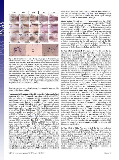
Fig. 3. (A–O) Localization of tac3a dur<strong>in</strong>g early stages of development, as<br />
detected by whole-mount ISH. tac3a is dom<strong>in</strong>antly expressed <strong>in</strong> the right<br />
habenular nuclei, midbra<strong>in</strong>, <strong>and</strong> h<strong>in</strong>dbra<strong>in</strong>. Dorsal view of larva heads, anterior<br />
is to the left (A–E). High magnification of boxed area <strong>in</strong> Upper panel. Note the<br />
unilateral expression of tac3a to right of the midl<strong>in</strong>e (dotted l<strong>in</strong>e) <strong>in</strong> the<br />
habenula (F–J), lateral view of larva heads (K–O). rHbn, right habenula; MB,<br />
midbra<strong>in</strong>; HB, h<strong>in</strong>dbra<strong>in</strong>. (P–R) Localization of tac3a <strong>in</strong> adult <strong>zebrafish</strong> bra<strong>in</strong> as<br />
<strong>in</strong>dicated by ISH (nomenclature accord<strong>in</strong>g to ref. 40). Tac3a mRNA-express<strong>in</strong>g<br />
cells were observed <strong>in</strong> the ventral (Hav) <strong>and</strong> medial (Ham) habenula (P) tac3a<br />
mRNA-express<strong>in</strong>g cells detected <strong>in</strong> the periventricular nucleus of posterior<br />
tuberculum (TPp), dorsal (Hd), <strong>and</strong> ventral zone (Hv) of periventricular hypothalamus<br />
(Q) tac3a mRNA-express<strong>in</strong>g cells observed <strong>in</strong> the posterior tuberal<br />
nucleus (PTN) <strong>and</strong> central zone (Hc) of periventricular hypothalamus (R).<br />
(Magnification: A–E <strong>and</strong> K–O, 40×; F–J, 120×).<br />
these two systems, as previously shown <strong>in</strong> mammals; however, this<br />
needs further <strong>in</strong>vestigation.<br />
Pharmacological Analysis <strong>and</strong> Signal Transduction Pathways of zfTac3<br />
Receptors. We used functional expression analysis with COS-7<br />
cells to evaluate the response, b<strong>in</strong>d<strong>in</strong>g selectivity, <strong>and</strong> signal<br />
transduction pathways of the unique TK <strong>receptors</strong> to their agonists.<br />
We previously showed the specificity of the reporter serum<br />
responsive element (SRE)-Luc <strong>and</strong> cAMP responsive element<br />
(CRE)-Luc, to activation of PKC/Ca 2+ <strong>and</strong> PKA/cAMP signal<br />
transduction pathways, respectively (15). Graded concentrations<br />
of the zfTKs (NKBa, NKBb, <strong>and</strong> NKF), <strong>and</strong> of hNKB <strong>and</strong> its<br />
agonist senktide were applied to COS-7 cells that expressed<br />
hNKBR, zfTac3ra, or zfTac3rb. The EC 50 values of TKs for each<br />
receptor are summarized (Table S4). Both human <strong>and</strong> pisc<strong>in</strong>e<br />
TKs <strong>in</strong>duced concentration-dependent <strong>in</strong>creases <strong>in</strong> both SRE-<br />
Luc <strong>and</strong> CRE-Luc activity (Fig. 4 A–F). For hNKBR, <strong>in</strong> both<br />
signal transduction systems, zfNKBa <strong>and</strong> NKF showed high potency,<br />
very similar to human NKB, but zfNKBb peptide exhibited<br />
relatively low potency (Fig. 4 A–D). For both zfTac3 <strong>receptors</strong>,<br />
zfNKBa <strong>and</strong> NKF (both derived from tac3a) were similarly<br />
highly potent <strong>in</strong> both signal transduction pathways (Fig. 4).<br />
These results confirmed that both zfNKBa <strong>and</strong> NKF were endogenous<br />
lig<strong>and</strong>s of Tac3 <strong>receptors</strong>, <strong>and</strong> it is noteworthy that this<br />
report of activation of TK <strong>receptors</strong> by a second peptide derived<br />
from the NKB gene (zfNKF) is unique. However, NKBb was less<br />
effective than the other forms <strong>in</strong> elicit<strong>in</strong>g luciferase activity by<br />
both signal transduction pathways. It was previously shown unequivocally<br />
that hNKBR <strong>potential</strong>ly can couple directly to both<br />
phospholipase C <strong>and</strong> adenylate cyclase, <strong>and</strong> stimulate both<br />
phospho<strong>in</strong>ositides hydrolysis <strong>and</strong> cAMP formation (27). Indeed<br />
both zftac3 <strong>receptors</strong>, as well as the hNKBR posses both PKC<br />
<strong>and</strong> PKA phosphorylation sites (Fig. S3). These f<strong>in</strong>d<strong>in</strong>gs confirm<br />
that the unique <strong>zebrafish</strong> <strong>receptors</strong> relay their signal through<br />
both PKC <strong>and</strong> PKA transduction pathways.<br />
Lig<strong>and</strong> Models. Fig. 4G is a ribbon representation of the zfNKB<br />
structural model prediction, compared with the hNKB (PDB ID<br />
1p9f). Intrigu<strong>in</strong>gly, although the three zfNK<strong>Bs</strong> vary <strong>in</strong> size—10,<br />
24, <strong>and</strong> 13 aa for NKBa, NKBb, <strong>and</strong> NKFa, respectively—all of<br />
the predicted peptides yielded high-resolution, high-quality<br />
structures with typical globular fold<strong>in</strong>g. These structures comprised<br />
α-helix-loop motifs (highlighted <strong>in</strong> red <strong>in</strong> Fig. 4G). All<br />
three zfNK<strong>Bs</strong> model structures approximated a b<strong>in</strong>d<strong>in</strong>g-competent<br />
conformation similar to the human NKB. Our results corroborate<br />
previous ones found for mammals that the formation of<br />
a helical conformation <strong>in</strong> the mid region of each of the TKs<br />
appears to be crucial for TK-receptor activation (28). Moreover,<br />
mammalian NKB were found to form a helical structure <strong>in</strong> the<br />
presence of dodecylphosphochol<strong>in</strong>e micelles (29).<br />
In Vivo Effect of Estradiol. Our next aim was to test the <strong>in</strong>volvement<br />
of the NKB system <strong>in</strong> reproduction. In fish, clear evidence<br />
exists regard<strong>in</strong>g the important <strong>role</strong> estradiol plays dur<strong>in</strong>g<br />
the period of reproduction (1). Moreover, ISH was recently used<br />
to show that estradiol <strong>in</strong>creased kisspept<strong>in</strong> cell number <strong>in</strong> the<br />
ventral hypothalamus, where the zfkiss2 neurons are homologous<br />
to the estrogen-sensitive kiss1 hypothalamic neurons <strong>in</strong> medaka<br />
(17, 22, 30). Similarly to mammals, <strong>zebrafish</strong> have two forms of<br />
GnRH: GnRH2 is localized to the midbra<strong>in</strong> tegmentum, <strong>and</strong><br />
GnRH3 (considered to be the hypophysiotropic form) is located<br />
at both the olfactory bulb term<strong>in</strong>al nerve <strong>and</strong> the preoptic area<br />
(31). Because <strong>in</strong> mammals kisspept<strong>in</strong> <strong>and</strong> NKB are expressed <strong>in</strong><br />
the same neurons <strong>in</strong> the hypothalamic ARC, <strong>and</strong> play a key <strong>role</strong><br />
<strong>in</strong> physiological regulation of GnRH neurons (25), we tested the<br />
effect of estradiol on the expression of genes along the GnRHkisspept<strong>in</strong><br />
system. Estradiol treatment of prepubertal <strong>zebrafish</strong><br />
enhanced expression of key genes <strong>in</strong>volved <strong>in</strong> reproduction<br />
(gnrh3, kiss2, <strong>and</strong> kiss1) concomitantly with significantly <strong>in</strong>creased<br />
tac3a (Fig. 5A). In parallel, we also found significantly <strong>in</strong>creased<br />
expression of tac3ra, tac3rb, <strong>and</strong> kiss1ra (Fig. 5B). Both Tac3<br />
<strong>receptors</strong> bound the zfNK<strong>Bs</strong> (Fig. 4 A–F), <strong>and</strong> Kiss1ra was shown<br />
previously to b<strong>in</strong>d Kiss2, considered to be the more important<br />
form, with higher aff<strong>in</strong>ity than Kiss1 (32). In mammals there is<br />
strong evidence of sexual dimorphism of NKB neurons: larger<br />
numbers of NKB neurons have been identified <strong>in</strong> ARC of ewes<br />
than of rams (33). The transcription of NKB could be directly<br />
altered by estrogen <strong>receptors</strong>, as sequences correspond<strong>in</strong>g to the<br />
estrogen responsive element <strong>and</strong> imperfect pal<strong>in</strong>dromic estrogen<br />
responsive element have been reported upstream of the TAC3<br />
gene transcriptional start site (8). In fish estradiol is <strong>in</strong>volved <strong>in</strong><br />
both early oogenesis <strong>and</strong> the beg<strong>in</strong>n<strong>in</strong>g of the first wave of vitellogenesis<br />
that precede puberty (34), collaborat<strong>in</strong>g our f<strong>in</strong>d<strong>in</strong>g<br />
that tac3a expression peaked <strong>in</strong> prepubertal fish (Fig. 2B).<br />
Moreover, <strong>in</strong>creased levels of estradiol are a required characteristic<br />
of both follicular growth <strong>and</strong> f<strong>in</strong>al oocyte maturation <strong>in</strong><br />
fish (3, 35), po<strong>in</strong>t<strong>in</strong>g toward the <strong>in</strong>volvement of the pisc<strong>in</strong>e NKB<br />
system <strong>in</strong> control of reproduction, probably <strong>in</strong> concert with<br />
kisspept<strong>in</strong> <strong>and</strong> GnRH.<br />
In Vivo Effect of NK<strong>Bs</strong>. We next tested the <strong>in</strong> vivo biological<br />
function of <strong>zebrafish</strong> NKB peptides. S<strong>in</strong>gle <strong>in</strong>traperitoneal <strong>in</strong>jection<br />
of zfNKBa or zfNKF elicited significant LH secretion <strong>in</strong><br />
sexually mature female <strong>zebrafish</strong> (Fig. 5C). The magnitude of the<br />
<strong>in</strong>duced LH discharge was comparable with that observed <strong>in</strong><br />
response to GnRH. LH response to the hNKBR agonist, senktide,<br />
or zfNKBb was less pronounced (Fig. 5C), <strong>in</strong> a similar way<br />
to the order of potency obta<strong>in</strong>ed <strong>in</strong> the transactivation assay (Fig.<br />
4). Our f<strong>in</strong>d<strong>in</strong>gs corroborate previous f<strong>in</strong>d<strong>in</strong>gs that activation of<br />
NKB <strong>receptors</strong> evoked potent LH-secretory responses <strong>in</strong><br />
rodents, sheep, <strong>and</strong> monkey (4, 36–38).<br />
10272 | www.pnas.org/cgi/doi/10.1073/pnas.1119165109 Biran et al.