HUMAN RANKL SINGLE PLEX KIT 96 Well Plate - Millipore
HUMAN RANKL SINGLE PLEX KIT 96 Well Plate - Millipore
HUMAN RANKL SINGLE PLEX KIT 96 Well Plate - Millipore
Create successful ePaper yourself
Turn your PDF publications into a flip-book with our unique Google optimized e-Paper software.
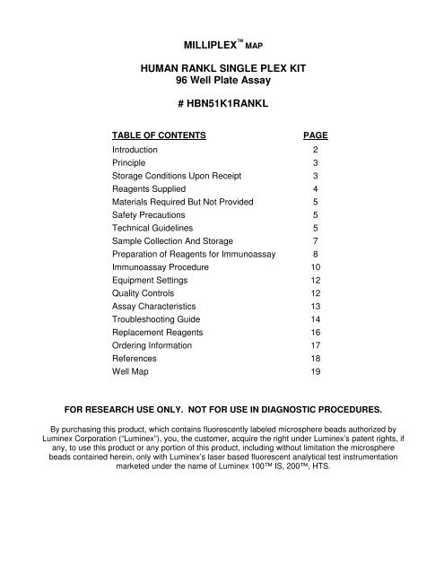
MILLI<strong>PLEX</strong> MAP<br />
<strong>HUMAN</strong> <strong>RANKL</strong> <strong>SINGLE</strong> <strong>PLEX</strong> <strong>KIT</strong><br />
<strong>96</strong> <strong>Well</strong> <strong>Plate</strong> Assay<br />
# HBN51K1<strong>RANKL</strong><br />
TABLE OF CONTENTS PAGE<br />
Introduction 2<br />
Principle 3<br />
Storage Conditions Upon Receipt 3<br />
Reagents Supplied 4<br />
Materials Required But Not Provided 5<br />
Safety Precautions 5<br />
Technical Guidelines 5<br />
Sample Collection And Storage 7<br />
Preparation of Reagents for Immunoassay 8<br />
Immunoassay Procedure 10<br />
Equipment Settings 12<br />
Quality Controls 12<br />
Assay Characteristics 13<br />
Troubleshooting Guide 14<br />
Replacement Reagents 16<br />
Ordering Information 17<br />
References 18<br />
<strong>Well</strong> Map 19<br />
FOR RESEARCH USE ONLY. NOT FOR USE IN DIAGNOSTIC PROCEDURES.<br />
By purchasing this product, which contains fluorescently labeled microsphere beads authorized by<br />
Luminex Corporation (“Luminex”), you, the customer, acquire the right under Luminex’s patent rights, if<br />
any, to use this product or any portion of this product, including without limitation the microsphere<br />
beads contained herein, only with Luminex’s laser based fluorescent analytical test instrumentation<br />
marketed under the name of Luminex 100 IS, 200, HTS.
INTRODUCTION<br />
Bone metabolism is the dynamic process of ongoing bone deposition and resorption,<br />
controlled by osteoblasts, osteocytes, and osteoclasts. While osteoblasts and osteocytes<br />
(osteoblasts surrounded by matrix) are responsible for bone deposition, osteoclasts are<br />
responsible for bone resorption. Both are required to maintain bone structure, as well as an<br />
adequate supply of calcium. To maintain this metabolic balance these cells rely on complex<br />
signaling pathways involving hormones and cytokines to achieve the appropriate rates of<br />
growth and differentiation. One of these proteins, Receptor Activator for Nuclear Factor κB<br />
Ligand (<strong>RANKL</strong>) activates osteoclasts and has been implicated in degenerative bone<br />
diseases such as rheumatoid arthritis and osteomyelitis. The balance between OPG and<br />
<strong>RANKL</strong> regulates osteoclast differentiation, activation and survival.<br />
<strong>Millipore</strong> recognizes the need to better understand the role that bone metabolism biomarkers<br />
like <strong>RANKL</strong> play both in preserving normal bone structure and in the development of disease.<br />
Therefore, we are proud to announce that the former LINCOplex Human Bone Metabolism<br />
<strong>RANKL</strong> Single Plex now has the MILLI<strong>PLEX</strong> MAP optimized format. While you will<br />
immediately recognize the quality and reproducibility that you have always trusted, you will<br />
also enjoy the enhancements that we have built into MILLI<strong>PLEX</strong> MAP.<br />
<strong>Millipore</strong>’s MILLI<strong>PLEX</strong> MAP Human Bone Metabolism <strong>RANKL</strong> Single Plex is to be used for the<br />
quantification of <strong>RANKL</strong> in human tissue, plasma and cell / tissue culture supernatant<br />
samples. The single plex provides biomedical researchers with quality tools for the study of<br />
bone metabolism related diseases.<br />
This kit is for research purposes only.<br />
Please read entire protocol before use.<br />
It is important to use same assay incubation conditions throughout your study.<br />
HBN51K1<strong>RANKL</strong> 02/09/2009 PAGE 2 MILLIPORE
PRINCIPLE<br />
MILLI<strong>PLEX</strong> MAP is based on the Luminex® xMAP® technology — one of the fastest growing<br />
and most respected multiplex technologies offering applications throughout the life sciences<br />
and capable of performing a variety of bioassays including immunoassays on the surface of<br />
fluorescent-coded beads known as microspheres.<br />
• Luminex® uses proprietary techniques to internally color-code microspheres with two<br />
fluorescent dyes. Through precise concentrations of these dyes, 100 distinctly colored<br />
bead sets can be created, each of which is coated with a specific capture antibody.<br />
• After an analyte from a test sample is captured by the bead, a biotinylated detection<br />
antibody is introduced.<br />
• The reaction mixture is then incubated with Streptavidin-PE conjugate, the reporter<br />
molecule, to complete the reaction on the surface of each microsphere.<br />
• The microspheres are allowed to pass rapidly through a laser which excites the internal<br />
dyes marking the microsphere set. A second laser excites PE, the fluorescent dye on<br />
the reporter molecule.<br />
• Finally, high-speed digital-signal processors identify each individual microsphere and<br />
quantify the result of its bioassay based on fluorescent reporter signals.<br />
The capability of adding multiple conjugated beads to each sample results in the ability to<br />
obtain multiple results from each sample. Open-architecture xMAP® technology enables<br />
multiplexing of many types of bioassays reducing time, labor and costs over traditional<br />
methods.<br />
STORAGE CONDITIONS UPON RECEIPT<br />
• Recommended storage for kit components is 2 - 8°C.<br />
• Once the standards and controls have been reconstituted, immediately transfer<br />
contents into polypropylene vials. DO NOT STORE RECONSTITUTED STANDARDS<br />
OR CONTROLS IN GLASS VIALS. For long-term storage, freeze reconstituted<br />
standards and controls at ≤ -20°C. Avoid multiple (>2) freeze/thaw cycles.<br />
• DO NOT FREEZE Antibody-Immobilized Beads, Detection Antibodies, and<br />
Streptavidin-Phycoerythrin.<br />
HBN51K1<strong>RANKL</strong> 02/09/2009 PAGE 3 MILLIPORE
REAGENTS SUPPLIED<br />
Note: Store all reagents at 2 – 8°C<br />
REAGENTS SUPPLIED CATALOG NUMBER VOLUME QUANTITY<br />
Human <strong>RANKL</strong> Standard LHBN-8051-<strong>RANKL</strong> lyophilized 1 vial<br />
Human <strong>RANKL</strong> Quality Controls 1 and 2 LHBN-6051-<strong>RANKL</strong> lyophilized 2 vials<br />
Serum Matrix<br />
Note: Contains 0.08% Sodium Azide<br />
LHED-SD lyophilized<br />
HBN51K1<strong>RANKL</strong> 02/09/2009 PAGE 4 MILLIPORE<br />
1 vial<br />
(required for serum and<br />
plasma samples only)<br />
Bead Diluent LPX-BD 3.5 mL 1 bottle<br />
Set of one <strong>96</strong>-<strong>Well</strong> Filter <strong>Plate</strong> with 2<br />
Sealers<br />
MX-PLATE -----------<br />
1 plate<br />
2 sealers<br />
Assay Buffer L-AB1 30 mL 2 bottles<br />
10X Wash Buffer<br />
Note: Contains 0.05% Proclin<br />
L-WB 30 mL 1 bottle<br />
Human <strong>RANKL</strong> Detection Antibody LHBN1051<strong>RANKL</strong> 5.5 mL 1 bottle<br />
Streptavidin-Phycoerythrin L-SAPE 5.5 mL 1 bottle<br />
Mixing Bottle ------------ -------------- 1 bottle<br />
Bead/Analyte Name<br />
Luminex<br />
Bead<br />
Region<br />
Anti-Human <strong>RANKL</strong> Bead 14 ✔<br />
(20X concentration, 200µL)<br />
Available Cat. #<br />
H<strong>RANKL</strong>
MATERIALS REQUIRED BUT NOT PROVIDED<br />
Reagents<br />
1. Luminex Sheath Fluid (Luminex Catalogue #40-50000)<br />
Instrumentation / Materials<br />
1. Adjustable Pipettes with Tips capable of delivering 5 µL to 1000 µL<br />
2. Multichannel Pipettes capable of delivering 5 µL to 50 µL or 25 µL to 200 µL<br />
3. Reagent Reservoirs<br />
4. Polypropylene Microfuge Tubes<br />
5. Rubber Bands<br />
6. Absorbent Pads<br />
7. Laboratory Vortex Mixer<br />
8. Sonicator (Branson Ultrasonic Cleaner Model #B200 or equivalent)<br />
9. Titer <strong>Plate</strong> Shaker (Lab-Line Instruments Model #4625 or equivalent)<br />
10. Vacuum Filtration Unit (<strong>Millipore</strong> Vacuum Manifold Catalog #MSVMHTS00 or<br />
equivalent with <strong>Millipore</strong> Vacuum Pump Catalog #WP6111560 or equivalent)<br />
11. Luminex 100 IS, 200, or HTS by Luminex Corporation<br />
12. <strong>Plate</strong> Stand (<strong>Millipore</strong> Catalog # MX-STAND)<br />
SAFETY PRECAUTIONS<br />
• All blood components and biological materials should be handled as potentially<br />
hazardous. Follow universal precautions as established by the Centers for Disease<br />
Control and Prevention and by the Occupational Safety and Health Administration<br />
when handling and disposing of infectious agents.<br />
• Sodium Azide or Proclin has been added to some reagents as a preservative.<br />
Although the concentrations are low, Sodium Azide and Proclin may react with lead<br />
and copper plumbing to form highly explosive metal azides. On disposal, flush with a<br />
large volume of water to prevent azide build up.<br />
TECHNICAL GUIDELINES<br />
To obtain reliable and reproducible results, the operator should carefully read this entire<br />
manual and fully understand all aspects of each assay step before running the assay. The<br />
following notes should be reviewed and understood before the assay is set up.<br />
• FOR RESEARCH USE ONLY. NOT FOR USE IN DIAGNOSTIC PROCEDURES.<br />
• Do not use beyond the expiration date on the label.<br />
• Do not mix or substitute reagents with those from other lots or sources.<br />
• The Antibody-Immobilized Beads are light sensitive and must be protected from light<br />
at all times. Cover the assay plate containing beads with opaque plate lid or<br />
aluminum foil during all incubation steps.<br />
• It is important to allow all reagents to warm to room temperature (20-25°C) before use<br />
in the assay.<br />
HBN51K1<strong>RANKL</strong> 02/09/2009 PAGE 5 MILLIPORE
• The bottom of the Microtiter Filter <strong>Plate</strong> should not be in direct contact with any<br />
surface during assay set-up or incubation times. The plate can be set on a plate<br />
stand or on the non-flat side of the plate cover or any other plate holder to raise the<br />
plate from the surface. A plate stand can be purchased separately from <strong>Millipore</strong><br />
(<strong>Millipore</strong> Catalog #MX-STAND).<br />
• Incomplete washing can adversely affect the assay outcome. All washing must be<br />
performed with the Wash Buffer provided.<br />
• After the wash steps, keep the bottom of the Microtiter Filter <strong>Plate</strong> clean by blotting on<br />
paper towels or absorbent pads to prevent any leakage due to capillary action.<br />
• Keep the vacuum suction on the plate as low as possible. It is recommended to have<br />
a vacuum setting that will remove 200 µL of buffer in ≥ 5 seconds (equivalent to < 100<br />
mmHg).<br />
• After hydration, all Standards and Controls must be transferred to polypropylene<br />
tubes.<br />
• The Standards prepared by serial dilution must be used within 1 hour of preparation.<br />
Discard any unused standards except the standard stock which may be stored at<br />
≤ -20°C for 1 month and at ≤ -80°C for greater than one month.<br />
• If samples fall outside the dynamic range of the assay, further dilute the samples with<br />
the appropriate diluent and repeat the assay.<br />
• Any unused mixed Antibody-Immobilized Beads may be stored in the Mixing Bottle at<br />
2-8°C for up to one month.<br />
• During the preparation of the standard curve, make certain to mix the higher<br />
concentration well before making the next dilution. Use a new tip with each dilution.<br />
• The plate should be read immediately after the assay is finished. If, however, the<br />
plate cannot be read immediately, seal the plate, cover with aluminum foil or an<br />
opaque lid, and store the plate at 2-8°C for up to 24 hours. Prior to reading, agitate<br />
the plate on the plate shaker at room temperature for 10 minutes. Delay in reading a<br />
plate may result in decreased sensitivity for some analytes.<br />
• The titer plate shaker should be set at a speed to provide maximum orbital mixing<br />
without splashing of liquid outside the wells. For the recommended plate shaker, this<br />
would be a setting of 5-7 which is approximately 500-800 rpm.<br />
• Ensure that the needle probe is clean. This may be achieved by sonication and/or<br />
alcohol flushes. Adjust probe height according to the protocols recommended by<br />
Luminex to the kit filter plate using 3 alignment discs prior to reading an assay.<br />
• For cell culture supernatants or tissue extraction, use the culture or extraction<br />
medium as the matrix solution in background, standard curve and control wells. If<br />
samples are diluted in Assay Buffer, use the Assay Buffer as matrix.<br />
• For serum/plasma samples, use the Serum Matrix provided in the kit.<br />
• For cell/tissue homogenate, the final cell or tissue homogenate should be prepared in<br />
a buffer that has a neutral pH, contains minimal detergents or strong denaturing<br />
detergents, and has an ionic strength close to physiological concentration. Avoid<br />
debris, lipids, and cell/tissue aggregates. Centrifuge samples before use.<br />
• Vortex all reagents well before adding to plate.<br />
HBN51K1<strong>RANKL</strong> 02/09/2009 PAGE 6 MILLIPORE
SAMPLE COLLECTION AND STORAGE<br />
A. Preparation of Serum Samples:<br />
• Allow the blood to clot for at least 30 minutes before centrifugation for 10<br />
minutes at 1000xg. Remove serum and assay immediately or aliquot and<br />
store samples at ≤ -20°C.<br />
• Avoid multiple (>2) freeze/thaw cycles.<br />
• When using frozen samples, it is recommended to thaw the samples<br />
completely, mix well by vortexing and centrifuge prior to use in the assay to<br />
remove particulates.<br />
• Samples should not be diluted until immediately prior to setting up the<br />
assay.<br />
• Serum samples should be diluted 1:2 in the Assay Buffer provided in the kit.<br />
B. Preparation of Plasma Samples:<br />
• Plasma collection using EDTA as an anticoagulant is recommended.<br />
Centrifuge for 10 minutes at 1000xg within 30 minutes of blood collection.<br />
Remove plasma and assay immediately or aliquot and store samples at<br />
≤ -20°C.<br />
• Avoid multiple (>2) freeze/thaw cycles.<br />
• When using frozen samples, it is recommended to thaw the samples<br />
completely, mix well by vortexing and centrifuge prior to use in the assay to<br />
remove particulates.<br />
• Samples should not be diluted until immediately prior to setting up the<br />
assay.<br />
• Plasma samples should be diluted 1:2 in the Assay Buffer provided in the kit.<br />
C. Preparation of Tissue / Cell Culture Supernatant:<br />
NOTE:<br />
• Centrifuge the sample for 10 minutes at 1000xg to remove debris and assay<br />
immediately or aliquot and store samples at ≤ -20°C.<br />
• Avoid multiple (>2) freeze/thaw cycles.<br />
• Customers may need to determine the optimal dilution factors for their tissue<br />
culture supernatant samples depending on the expected biological range. (If<br />
dilution is required, samples should not be diluted until immediately prior<br />
to setting up the assay.)<br />
• A maximum of 25 µL per well of cell / tissue culture supernatant samples or neat<br />
serum, plasma samples can be used.<br />
• All samples must be stored in polypropylene tubes. DO NOT STORE SAMPLES IN<br />
GLASS.<br />
• Avoid using samples with gross hemolysis or lipemia.<br />
• Care must be taken when using heparin as an anticoagulant since an excess of<br />
heparin will provide falsely high values. Use no more than 10 IU heparin per mL of<br />
blood collected.<br />
HBN51K1<strong>RANKL</strong> 02/09/2009 PAGE 7 MILLIPORE
PREPARATION OF REAGENTS FOR IMMUNOASSAY<br />
A. Preparation of Antibody-Immobilized Beads<br />
Pre-warm the Bead Diluent to room temperature. Sonicate the antibody-bead tube<br />
for 30 seconds, and then vigorously vortex the tube for 1 minute. Add 150 µL of<br />
<strong>RANKL</strong> beads to the Mixing Bottle and add 2.85 mL of Bead Diluent to obtain 3.0 mL<br />
of diluted beads. Mix the diluted beads well immediately prior to use.<br />
B. Preparation of Quality Controls<br />
Before use, reconstitute Quality Control 1 and Quality Control 2 with 250 µL<br />
deionized water. Invert the vial several times to mix and vortex. Allow the vial to sit<br />
for 5-10 minutes and then transfer the controls to appropriately labeled polypropylene<br />
microfuge tubes. Unused portion may be stored at ≤ -20°C for up to one month.<br />
C. Preparation of Wash Buffer<br />
Bring the 10X Wash Buffer to room temperature and mix to bring all salts into<br />
solution. Dilute 30 mL of 10X Wash Buffer with 270 mL deionized water. Store<br />
unused portion at 2-8°C for up to one month.<br />
D. Preparation of Serum Matrix<br />
This step is required for serum or plasma samples only.<br />
Before the assay, add 1 mL of deionized water and 4 mL of L-AB1 Assay Buffer to<br />
the bottle containing the lyophilized Serum Matrix, gently swirl the bottle and then<br />
place the bottle on bench for 10 minutes. Transfer the reconstituted Serum Matrix<br />
solution to an appropriately-labeled polypropylene tube. Unused portion may be<br />
stored at ≤ -20°C for up to one month.<br />
E. Preparation of Human <strong>RANKL</strong> Standard<br />
1.) Prior to use, reconstitute the lyophilized Human <strong>RANKL</strong> Standard with 250 µL<br />
Deionized Water to give a concentration of 20,000 pg/mL for <strong>RANKL</strong>. Invert the<br />
vial several times to mix. Allow the vial to set for 5-10 minutes to make sure that<br />
the standard is completely reconstituted, and then transfer the standard to an<br />
appropriately-labeled polypropylene microfuge tube. This will be used as the<br />
20,000 pg/mL Standard. The unused portions of this stock may be stored at<br />
≤ -20°C for up to one month.<br />
2.) Preparation of Working Standards<br />
Label six polypropylene microfuge tubes as 5,000, 1,250, 312.5, 78.13, 19.53,<br />
and 4.88 pg/mL. Add 150 µL of Assay Buffer to each of the six tubes. Prepare<br />
serial dilutions by adding 50 µL of the reconstituted 20,000 pg/mL standard to the<br />
5,000 pg/mL tube, mix well and transfer 50 µL of the 5,000 pg/mL standard to the<br />
1,250 pg/mL tube, mix well and transfer 50 µL of the 1,250 pg/mL standard to the<br />
312.5 pg/mL tube, mix well and transfer 50 µL of the 312.5 pg/mL standard to the<br />
78.13 pg/mL tube, mix well and transfer 50 µL of the 78.13 pg/mL standard to the<br />
19.53 pg/mL tube, mix well and transfer 50 µL of the 19.53 pg/mL standard to the<br />
4.88 pg/mL tube and mix well. The 0 pg/mL standard (Background) will be Assay<br />
Buffer.<br />
HBN51K1<strong>RANKL</strong> 02/09/2009 PAGE 8 MILLIPORE
Standard<br />
50 µL<br />
Preparation of Working Standards<br />
Standard<br />
Concentration<br />
Original<br />
20,000 pg/mL<br />
Standard<br />
Concentration<br />
(pg/mL)<br />
150 µL<br />
Volume of<br />
Deionized Water<br />
to Add<br />
250 µL<br />
Volume of Assay<br />
Buffer to Add<br />
Volume of<br />
Standard<br />
to Add<br />
HBN51K1<strong>RANKL</strong> 02/09/2009 PAGE 9 MILLIPORE<br />
0<br />
Volume of Standard<br />
to Add<br />
5,000 150 µL 50 µL of 20,000 pg/mL<br />
1,250 150 µL 50 µL of 5,000 pg/mL<br />
312.5 150 µL 50 µL of 1,250 pg/mL<br />
78.13 150 µL 50 µL of 312.5 pg/mL<br />
19.53 150 µL 50 µL of 78.13 pg/mL<br />
4.88 150 µL 50 µL of 19.53 pg/mL<br />
50 µL<br />
150 µL<br />
50 µL<br />
150 µL<br />
20,000 pg/mL 5,000 pg/mL 1,250 pg/mL 312.5 pg/mL<br />
50 µL<br />
50 µL<br />
50 µL<br />
150 µL 150 µL<br />
150 µL<br />
78.13 pg/mL<br />
19.53 pg/mL 4.88 pg/mL
IMMUNOASSAY PROCEDURE<br />
• Prior to beginning this assay, it is imperative to read this protocol completely and to<br />
thoroughly understand the Technical Guidelines.<br />
• Allow all reagents to warm to room temperature (20-25°C) before use in the assay.<br />
• Diagram the placement of Standards [0 (Background), 4.88, 19.53. 78.13, 312.5, 1250,<br />
5,000 and 20,000 pg/mL], Controls 1 and 2, and Samples on <strong>Well</strong> Map Worksheet in a<br />
vertical configuration. (Note: Most instruments will only read the <strong>96</strong>-well plate vertically<br />
by default.) It is recommended to run the assay in duplicate.<br />
• Set the filter plate on a plate holder at all times during reagent dispensing and incubation<br />
steps so that the bottom of the plate does not touch any surface.<br />
1. Prewet the filter plate by pipetting 200 µL of<br />
Assay Buffer into each well of the Microtiter<br />
Filter <strong>Plate</strong>. Seal and mix on a plate shaker for<br />
10 minutes at room temperature (20-25°C).<br />
2. Remove Assay Buffer by vacuum. (NOTE: DO<br />
NOT INVERT PLATE.) Blot excess Assay<br />
Buffer from the bottom of the plate with an<br />
absorbent pad or paper towels.<br />
3. Add 25 µL Assay Buffer to all wells.<br />
4. Add 25 µL of each Standard or Control into the<br />
appropriate wells. Assay Buffer should be used<br />
for the 0 pg/mL standard (Background).<br />
5. Add 25 µL of Assay Buffer to the sample wells.<br />
6. Add 25 µL of an appropriate matrix to the<br />
Background, Standards, and Control wells.<br />
When assaying 1:2 diluted Serum or Plasma<br />
samples, use the diluted Serum Matrix<br />
provided. When assaying Tissue Culture or<br />
other supernatant, use control culture medium<br />
as the matrix solution.<br />
7. Add 25 µL of Sample into the appropriate wells.<br />
(Serum and plasma samples should be diluted<br />
1:2 in Assay Buffer immediately prior to setting<br />
up assay.)<br />
8. Vortex Mixing Bottle and add 25 µL of the<br />
Mixed Beads to each well. (Note: During<br />
addition of Beads, shake bead bottle<br />
intermittently to avoid settling.)<br />
9. Seal the plate with a plate sealer, cover it with<br />
the lid. Wrap a rubber band around the plate<br />
holder, plate and lid and incubate with agitation<br />
on a plate shaker overnight (16-20 hours) at<br />
4°C.<br />
Add 200 µL Assay Buffer per<br />
well<br />
• Add 25 µL Assay Buffer to<br />
all wells<br />
• Add 25 µL Standard or<br />
Control to appropriate wells<br />
• Add 25 µL Assay Buffer to<br />
background and sample<br />
wells<br />
• Add 25 µL Matrix to<br />
background, standards and<br />
control wells<br />
• Add 25 µL Samples to<br />
sample wells<br />
• Add 25 µL Beads to each<br />
well<br />
HBN51K1<strong>RANKL</strong> 02/09/2009 PAGE 10 MILLIPORE<br />
Shake 10 min, RT<br />
Vacuum<br />
Incubate overnight<br />
at 4°C with shaking
10. Gently remove fluid by vacuum. (NOTE: DO<br />
NOT INVERT PLATE.)<br />
11. Wash plate 3 times with 200 µL/well of Wash<br />
Buffer, removing Wash Buffer by vacuum<br />
filtration between each wash. Blot excess<br />
Wash Buffer from the bottom of the plate with<br />
an absorbent pad or paper towels.<br />
12. Add 50 µL of Detection Antibody into each well.<br />
(Note: Allow the Detection Antibody to warm to<br />
room temperature prior to addition.)<br />
13. Seal, cover with lid, and incubate with agitation<br />
on a plate shaker for 1 hour at room<br />
temperature (20-25°C). DO NOT VACUUM<br />
AFTER INCUBATION.<br />
14. Add 50 µL Streptavidin-Phycoerythrin to each<br />
well containing the 50 µL of Detection<br />
Antibodies.<br />
15. Seal, cover with lid and incubate with agitation<br />
on a plate shaker for 30 minutes at room<br />
temperature (20-25°C).<br />
16. Gently remove all contents by vacuum. (NOTE:<br />
DO NOT INVERT PLATE.)<br />
17. Wash plate 3 times with 200 µL/well Wash<br />
Buffer, removing Wash Buffer by vacuum<br />
filtration between each wash. Wipe any excess<br />
buffer on the bottom of the plate with a tissue.<br />
18. Add 100 µL of Sheath Fluid to all wells.<br />
Resuspend the beads on a plate shaker for 5<br />
minutes.<br />
19. Run plate on Luminex 100 IS, 200, or HTS.<br />
20. Save and analyze the Median Fluorescent<br />
Intensity (MFI) data using a weighted 5parameter<br />
logistic or spline curve-fitting method<br />
for calculating <strong>RANKL</strong> concentrations in<br />
samples.<br />
Vacuum and wash<br />
3X with 200 µL<br />
Wash Buffer<br />
Add 50 µL Detection Antibody<br />
per well<br />
Incubate 1 hour<br />
at RT<br />
Do Not Vacuum<br />
Add 50 µL Streptavidin-<br />
Phycoerythrin per well<br />
Incubate for 30<br />
minutes at RT<br />
Vacuum and wash<br />
3X with 200 µL<br />
Wash Buffer<br />
Add 100 µL Sheath Fluid per<br />
well<br />
Read on Luminex (50 µL,<br />
50 beads per bead set)<br />
HBN51K1<strong>RANKL</strong> 02/09/2009 PAGE 11 MILLIPORE
EQUIPMENT SETTINGS<br />
These specifications are for the Luminex 100 IS v.1.7 or Luminex 100 IS<br />
v2.1/2.2, Luminex 200 v2.3, xPONENT, and Luminex HTS. Luminex instruments<br />
with other software (e.g. MasterPlex, StarStation, LiquiChip, Bio-Plex, LABScan100)<br />
would need to follow instrument instructions for gate settings and additional<br />
specifications from the vendors.<br />
QUALITY CONTROLS<br />
Events: 50, per bead<br />
Sample Size: 50 µL<br />
Gate Settings: 8,000 to 15,000<br />
Reporter Gain: Default (low PMT)<br />
Time Out: 60 seconds<br />
Bead Set: <strong>RANKL</strong> 14<br />
The ranges for each analyte in Quality Control 1 and 2 are provided on the card<br />
insert or can be located at the MILLIPORE website<br />
www.millipore.com/techlibrary/index.do using the catalog number as the keyword.<br />
HBN51K1<strong>RANKL</strong> 02/09/2009 PAGE 12 MILLIPORE
ASSAY CHARACTERISTICS<br />
Assay Sensitivities (minimum detectable concentrations, pg/mL)<br />
MinDC: Minimum Detectable Concentration is calculated by the StatLIA® Immunoassay<br />
Analysis Software from Brendan Technologies. It measures the true limits of detection<br />
for an assay by mathematically determining what the empirical MinDC would be if an<br />
infinite number of standard concentrations were run for the assay under the same<br />
conditions.<br />
Precision<br />
Analyte MinDC (pg/mL)<br />
<strong>RANKL</strong> 4.8<br />
Intra-assay precision is generated from the mean of the %CV’s from sixteen reportable<br />
results across two different concentrations of analyte in one experiment. Inter-assay<br />
precision is generated from the mean of the %CV’s from two reportable results each for<br />
two different concentrations of analyte across eight different experiments.<br />
Accuracy<br />
Intra-Assay (%CV)
TROUBLESHOOTING GUIDE<br />
Problem Probable Cause Solution<br />
Filter plate will not<br />
vacuum<br />
Insufficient bead<br />
count<br />
Vacuum pressure is<br />
insufficient<br />
Samples have insoluble<br />
particles<br />
Increase vacuum pressure such that 0.2mL<br />
buffer can be suctioned in 3-5 seconds.<br />
Centrifuge samples just prior to assay set-up<br />
and use supernatant.<br />
If high lipid concentration, after<br />
centrifugation, remove lipid layer and use<br />
supernatant.<br />
Sample too viscous May need to dilute sample.<br />
Vacuum pressure too high Adjust vacuum pressure such that 0.2mL<br />
buffer can be suctioned in 3-5 seconds.<br />
Bead mix prepared<br />
incorrectly<br />
Samples cause<br />
interference due to<br />
particulate matter or<br />
viscosity<br />
Sonicate bead vials and vortex just prior to<br />
adding to bead mix bottle according to<br />
protocol. Agitate bead mix intermittently in<br />
reservoir while pipetting into the plate.<br />
See above. Also sample probe may need to<br />
be cleaned with alcohol flush, backflush and<br />
washes; or, if needed, probe should be<br />
removed and sonicated.<br />
Probe height not adjusted<br />
correctly<br />
Adjust probe to 3 alignment discs in well H6.<br />
<strong>Plate</strong> leaked Vacuum pressure too high Adjust vacuum pressure such that 0.2mL<br />
buffer can be suctioned in 3-5 seconds. May<br />
need to transfer contents to a new<br />
(prewetted) plate and continue.<br />
Background is too<br />
high<br />
<strong>Plate</strong> set directly on table<br />
or absorbent towels during<br />
incubations or reagent<br />
additions<br />
Insufficient blotting of filter<br />
plate bottom causing<br />
wicking<br />
Pipette touching plate filter<br />
during additions<br />
Probe height not adjusted<br />
correctly<br />
Background wells were<br />
contaminated<br />
Matrix used has<br />
endogenous analyte or<br />
interference<br />
Set plate on plate stand or raised edge so<br />
bottom of filter is not touching any surface.<br />
Blot the bottom of the filter plate well with<br />
absorbent towels after each wash step.<br />
Pipette to the side of well.<br />
Adjust probe to 3 alignment discs in well H6.<br />
Avoid cross-well contamination by using<br />
sealer appropriately and by pipeting with<br />
multichannel pipets without touching reagent<br />
in plate.<br />
Check matrix ingredients for crossreacting<br />
components (e.g. interleukin modified tissue<br />
culture medium).<br />
Insufficient washes Increase number of washes.<br />
HBN51K1<strong>RANKL</strong> 02/09/2009 PAGE 14 MILLIPORE
Beads not in region<br />
or gate<br />
Signal for whole<br />
plate is same as<br />
background<br />
Low signal for<br />
standard curve<br />
Signals too high,<br />
standard curves are<br />
saturated<br />
Sample readings<br />
are out of range<br />
Luminex not calibrated<br />
correctly or recently<br />
Gate settings not adjusted<br />
correctly<br />
Wrong bead regions in<br />
protocol template<br />
Incorrect sample type<br />
used<br />
Instrument not washed or<br />
primed<br />
Beads were exposed to<br />
light<br />
Incorrect or no Detection<br />
Antibody was added<br />
Streptavidin-Phycoerythrin<br />
was not added<br />
Detection Antibody may<br />
have been vacuumed out<br />
prior to adding Streptavidin<br />
Phycoerythrin<br />
Incubations done at<br />
incorrect temperatures,<br />
timings or agitation<br />
Calibration target value set<br />
too high<br />
<strong>Plate</strong> incubation was too<br />
long with standard curve<br />
and samples<br />
Samples contain no or<br />
below detectable levels of<br />
analyte<br />
Samples contain analyte<br />
concentrations higher than<br />
highest standard point<br />
Standard curve was<br />
saturated at higher end of<br />
curve<br />
Calibrate Luminex based on instrument<br />
manufacturer’s instructions at least once a<br />
week or if temperature has changed by >3 o C.<br />
Some Luminex instruments (e.g. Bio-Plex)<br />
require different gate settings than those<br />
described in the kit protocol. Use instrument<br />
default settings.<br />
Check kit protocol for correct bead regions or<br />
analyte selection.<br />
Samples containing organic solvents or if<br />
highly viscous should be diluted or dialyzed<br />
as required.<br />
Prime the Luminex 4 times to eliminate air<br />
bubbles. Wash 4 times with sheath fluid or<br />
water if there is any remnant alcohol or<br />
sanitizing liquid.<br />
Keep plate and bead mix covered with dark<br />
lid or aluminum foil during all incubation<br />
steps.<br />
Add appropriate Detection Antibody and<br />
continue.<br />
Add Streptavidin-Phycoerythrin according to<br />
protocol. If Detection Antibody has already<br />
been vacuumed out, sensitivity may be low.<br />
May need to repeat assay if desired<br />
sensitivity not achieved.<br />
Assay conditions need to be checked.<br />
With some Luminex instruments (e.g. Bio-<br />
Plex) default target setting for RP1 calibrator<br />
is set at High PMT. Use low target value for<br />
calibration and reanalyze plate.<br />
Use shorter incubation time.<br />
If below detectable levels, it may be possible<br />
to use higher sample volume. Check with<br />
tech support for appropriate protocol<br />
modifications.<br />
Samples may require dilution and reanalysis<br />
for that particular analyte.<br />
See above.<br />
HBN51K1<strong>RANKL</strong> 02/09/2009 PAGE 15 MILLIPORE
High variation in<br />
samples and/or<br />
standards<br />
Multichannel pipet may not<br />
be calibrated<br />
<strong>Plate</strong> washing was not<br />
uniform<br />
Samples may have high<br />
particulate matter or other<br />
interfering substances<br />
<strong>Plate</strong> agitation was<br />
insufficient<br />
Calibrate pipets.<br />
Confirm all reagents are vacuumed out<br />
completely in all wash steps.<br />
See above.<br />
<strong>Plate</strong> should be agitated during all incubation<br />
steps using a vertical plate shaker at a speed<br />
where beads are in constant motion without<br />
splashing.<br />
Cross-well contamination Check when reusing plate sealer that no<br />
reagent has touched sealer.<br />
Care should be taken when using same pipet<br />
tips that are used for reagent additions and<br />
that pipet tip does not touch reagent in plate.<br />
REPLACEMENT REAGENTS Catalog #<br />
Human <strong>RANKL</strong> Standard LHBN-8051-<strong>RANKL</strong><br />
Human <strong>RANKL</strong> Quality Controls LHBN-6051-<strong>RANKL</strong><br />
Human <strong>RANKL</strong> Detection Antibody LHBN1051<strong>RANKL</strong><br />
Streptavidin-Phycoerythrin L-SAPE<br />
Bead Diluent LPX-BD<br />
Assay Buffer L-AB1<br />
Serum Matrix LHED-SD<br />
10X Wash Buffer L-WB<br />
Set of two <strong>96</strong>-<strong>Well</strong> Filter <strong>Plate</strong>s with Sealers MX-PLATE<br />
Antibody-Immobilized Bead<br />
Analyte Bead # Cat. #<br />
Human <strong>RANKL</strong> Beads 14 H<strong>RANKL</strong><br />
HBN51K1<strong>RANKL</strong> 02/09/2009 PAGE 16 MILLIPORE
ORDERING INFORMATION<br />
To place an order:<br />
To assure the clarity of your custom Human <strong>RANKL</strong> kit order, please FAX the<br />
following information to our customer service department:<br />
• Your name, telephone and/or fax number<br />
• Customer account number<br />
• Shipping and billing address<br />
• Purchase order number<br />
• Catalog number and description of product<br />
• Quantity of kits<br />
FAX: (636) 441-8050<br />
Toll-Free US: (866) 441-8400<br />
(636) 441-8400<br />
Mail Orders: <strong>Millipore</strong> Corp.<br />
6 Research Park Drive<br />
St. Charles, Missouri 63304 U.S.A.<br />
For International Customers:<br />
To best serve our international customers in placing an order or obtaining<br />
additional information about MILLI<strong>PLEX</strong> MAP products, please contact your<br />
multiplex specialist or sales representative or email our European Customer<br />
Service at customerserviceEU@<strong>Millipore</strong>.com.<br />
Conditions of Sale<br />
All products are for research use only. They are not intended for use in clinical<br />
diagnosis or for administration to humans or animals. All products are<br />
intended for in vitro use only.<br />
Material Safety Data Sheets (MSDS)<br />
Material Safety Data Sheets for <strong>Millipore</strong> products may be ordered by fax or<br />
phone or through our website at www.millipore.com/techlibrary/index.do.<br />
HBN51K1<strong>RANKL</strong> 02/09/2009 PAGE 17 MILLIPORE
REFERENCES<br />
Buxton EC, Yao W, Lane NE. Changes in serum receptor activator of nuclear<br />
factor-kappaB ligand, osteoprotegerin, and interleukin-6 levels in patients with<br />
glucocorticoid-induced osteoporosis treated with human parathyroid hormone (1-<br />
34). J Clin Endocrinol Metab. 89:3332-2336 (2004)<br />
Collin-Osdoby P. Review: Regulation of vascular calcification by osteoclast<br />
regulatory factors <strong>RANKL</strong> and osteoprotegerin. Circ Res. 95:1046-1057 (2004)<br />
Eghbali-Fatourechi G, Khosla S, Sanyal A, Boyle WJ, Lacey DL, Riggs BL. Role<br />
of RANK ligand in mediating increased bone resorption in early postmenopausal<br />
women. J Clin Invest. 2003 111:1221-1230 (2003)<br />
Franchimont N, Reenaers C, Lambert C, Belaiche J, Bours V, Malaise M,<br />
Delvenne P, Louis E. Increased expression of receptor activator of NF-kappaB<br />
ligand (<strong>RANKL</strong>), its receptor RANK and its decoy receptor osteoprotegerin in the<br />
colon of Crohn's disease patients. Clin Exp Immunol. 138:491-498 (2004)<br />
Fuller K, Wong B, Fox S, Choi Y, Chambers TJ. TRANCE is necessary and<br />
sufficient for osteoblast-mediated activation of bone resorption in osteoclasts. J<br />
Exp Med 188:997-1001 (1998)<br />
Hofbauer, LC and Schoppet M. Clinical implications of the<br />
osteoprotegerin/<strong>RANKL</strong>/RANK system for bone and vascular diseases. JAMA<br />
292:490-495 (2004)<br />
Kong YY, Feiger, Sarosi I, Bolon B, Tafuri A, Morony S, Capparelli, Elliot R,<br />
McCabe S, Wong T, Campagnuolo G, Moran E, Bogoch ER, Van G, Nguen LT,<br />
Ohashi PS, Lacey, DL, Fish E, Boyle WJ, and Penninger JM. Activated T cells<br />
regulate bone loss and joint destruction in adjuvant arthritis through<br />
osteoprotegerin ligand. Nature 402:304-309 (2000)<br />
Rogers A and Eastell R. Review: Circulating osteoprotegerin and receptor<br />
activator for nuclear factor kappaB ligand: clinical utility in metabolic bone disease<br />
assessment. J Clin Endocrinol Metab. 90:6323-6331 (2005)<br />
Terpos E, Szydlo R, Apperley JF, Hatjiharissi E, Politou M, Meletis J, Viniou N,<br />
Yataganas X, Goldman JM, Rahemtulla A. Soluble receptor activator of nuclear<br />
factor kappaB ligand-osteoprotegerin ratio predicts survival in multiple myeloma:<br />
proposal for a novel prognostic index. Blood. 102:1064-1069 (2003)<br />
Yasuda H, Shima N, Nakagawa N, Yamaguchi K, Kinosaki M, Mochizuki S,<br />
Tomoyasu A, Yano K, Goto M, Murakami A, Tsuda E, Morinaga T, Higashio K,<br />
Udagawa N, Takahashi N, Suda T. Osteoclast differentiation factor is a ligand for<br />
osteoprotegerin/osteoclastogenesis-inhibitory factor and is identical to<br />
TRANCE/<strong>RANKL</strong>. Proc Natl Acad Sci U S A. 95:3597-3602 (1998)<br />
HBN51K1<strong>RANKL</strong> 02/09/2009 PAGE 18 MILLIPORE
A<br />
B<br />
C<br />
D<br />
E<br />
F<br />
G<br />
H<br />
WELL MAP<br />
1 2 3 4 5 6 7 8 9 10 11 12<br />
0<br />
Standard<br />
(Background)<br />
0<br />
Standard<br />
(Background)<br />
4.88<br />
pg/mL<br />
Standard<br />
4.88<br />
pg/mL<br />
Standard<br />
19.53<br />
pg/mL<br />
Standard<br />
19.53<br />
pg/mL<br />
Standard<br />
78.13<br />
pg/mL<br />
Standard<br />
78.13<br />
pg/mL<br />
Standard<br />
312.5<br />
pg/mL<br />
Standard<br />
312.5<br />
pg/mL<br />
Standard<br />
1,250<br />
pg/mL<br />
Standard<br />
1,250<br />
pg/mL<br />
Standard<br />
5,000<br />
pg/mL<br />
Standard<br />
5,000<br />
pg/mL<br />
Standard<br />
20,000<br />
pg/mL<br />
Standard<br />
20,000<br />
pg/mL<br />
Standard<br />
QC 1 Etc.<br />
QC 1<br />
QC 2<br />
QC 2<br />
Sample<br />
1<br />
Sample<br />
1<br />
Sample<br />
2<br />
Sample<br />
2<br />
HBN51K1<strong>RANKL</strong> 02/09/2009 PAGE 19 MILLIPORE


