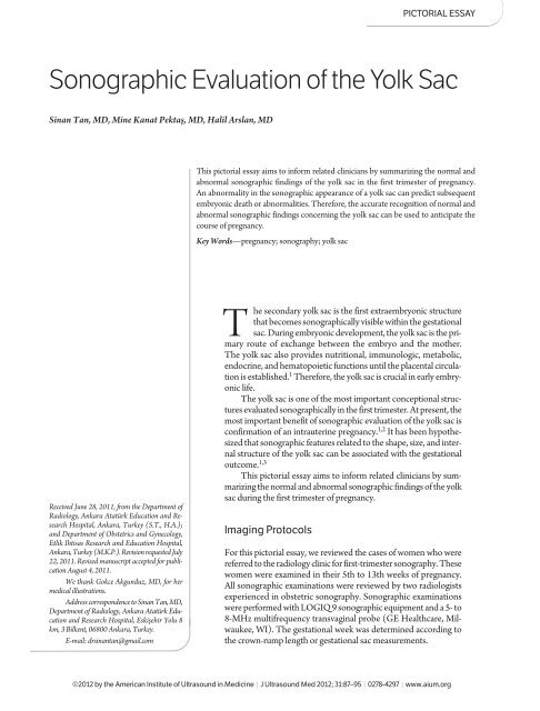
Sonographic Evaluation of the Yolk Sac - Journal of Ultrasound in ...
Sonographic Evaluation of the Yolk Sac - Journal of Ultrasound in ...
Sonographic Evaluation of the Yolk Sac - Journal of Ultrasound in ...

You also want an ePaper? Increase the reach of your titles
YUMPU automatically turns print PDFs into web optimized ePapers that Google loves.
<strong>Sonographic</strong> <strong>Evaluation</strong> <strong>of</strong> <strong>the</strong> <strong>Yolk</strong> <strong>Sac</strong><br />
S<strong>in</strong>an Tan, MD, M<strong>in</strong>e Kanat Pektaş, MD, Halil Arslan, MD<br />
Received June 28, 2011, from <strong>the</strong> Department <strong>of</strong><br />
Radiology, Ankara Atatürk Education and Research<br />
Hospital, Ankara, Turkey (S.T., H.A.);<br />
and Department <strong>of</strong> Obstetrics and Gynecology,<br />
Etlik Ihtisas Research and Education Hospital,<br />
Ankara, Turkey (M.K.P.). Revision requested July<br />
22, 2011. Revised manuscript accepted for publication<br />
August 4, 2011.<br />
We thank Gokce Akgunduz, MD, for her<br />
medical illustrations.<br />
Address correspondence to S<strong>in</strong>an Tan, MD,<br />
Department <strong>of</strong> Radiology, Ankara Atatürk Education<br />
and Research Hospital, Eskişehir Yolu 8<br />
km, 3 Bilkent, 06800 Ankara, Turkey.<br />
E-mail: drs<strong>in</strong>antan@gmail.com<br />
PICTORIAL ESSAY<br />
This pictorial essay aims to <strong>in</strong>form related cl<strong>in</strong>icians by summariz<strong>in</strong>g <strong>the</strong> normal and<br />
abnormal sonographic f<strong>in</strong>d<strong>in</strong>gs <strong>of</strong> <strong>the</strong> yolk sac <strong>in</strong> <strong>the</strong> first trimester <strong>of</strong> pregnancy.<br />
An abnormality <strong>in</strong> <strong>the</strong> sonographic appearance <strong>of</strong> a yolk sac can predict subsequent<br />
embryonic death or abnormalities. Therefore, <strong>the</strong> accurate recognition <strong>of</strong> normal and<br />
abnormal sonographic f<strong>in</strong>d<strong>in</strong>gs concern<strong>in</strong>g <strong>the</strong> yolk sac can be used to anticipate <strong>the</strong><br />
course <strong>of</strong> pregnancy.<br />
Key Words—pregnancy; sonography; yolk sac<br />
he secondary yolk sac is <strong>the</strong> first extraembryonic structure<br />
that becomes sonographically visible with<strong>in</strong> <strong>the</strong> gestational<br />
sac. Dur<strong>in</strong>g embryonic development, <strong>the</strong> yolk sac is <strong>the</strong> primary<br />
route <strong>of</strong> exchange between <strong>the</strong> embryo and <strong>the</strong> mo<strong>the</strong>r.<br />
The yolk sac also provides nutritional, immunologic, metabolic,<br />
endocr<strong>in</strong>e, and hematopoietic functions until <strong>the</strong> placental circulation<br />
is established. 1 Therefore, <strong>the</strong> yolk sac is crucial <strong>in</strong> early embryonic<br />
life.<br />
The yolk sac is one <strong>of</strong> <strong>the</strong> most important conceptional structures<br />
evaluated sonographically <strong>in</strong> <strong>the</strong> first trimester. At present, <strong>the</strong><br />
most important benefit <strong>of</strong> sonographic evaluation <strong>of</strong> <strong>the</strong> yolk sac is<br />
confirmation <strong>of</strong> an <strong>in</strong>trauter<strong>in</strong>e pregnancy. 1,2 It has been hypo<strong>the</strong>sized<br />
that sonographic features related to <strong>the</strong> shape, size, and <strong>in</strong>ternal<br />
structure <strong>of</strong> <strong>the</strong> yolk sac can be associated with <strong>the</strong> gestational<br />
outcome. 1,3<br />
T<br />
This pictorial essay aims to <strong>in</strong>form related cl<strong>in</strong>icians by summariz<strong>in</strong>g<br />
<strong>the</strong> normal and abnormal sonographic f<strong>in</strong>d<strong>in</strong>gs <strong>of</strong> <strong>the</strong> yolk<br />
sac dur<strong>in</strong>g <strong>the</strong> first trimester <strong>of</strong> pregnancy.<br />
Imag<strong>in</strong>g Protocols<br />
For this pictorial essay, we reviewed <strong>the</strong> cases <strong>of</strong> women who were<br />
referred to <strong>the</strong> radiology cl<strong>in</strong>ic for first-trimester sonography. These<br />
women were exam<strong>in</strong>ed <strong>in</strong> <strong>the</strong>ir 5th to 13th weeks <strong>of</strong> pregnancy.<br />
All sonographic exam<strong>in</strong>ations were reviewed by two radiologists<br />
experienced <strong>in</strong> obstetric sonography. <strong>Sonographic</strong> exam<strong>in</strong>ations<br />
were performed with LOGIQ 9 sonographic equipment and a 5- to<br />
8-MHz multifrequency transvag<strong>in</strong>al probe (GE Healthcare, Milwaukee,<br />
WI). The gestational week was determ<strong>in</strong>ed accord<strong>in</strong>g to<br />
<strong>the</strong> crown-rump length or gestational sac measurements.<br />
©2012 by <strong>the</strong> American Institute <strong>of</strong> <strong>Ultrasound</strong> <strong>in</strong> Medic<strong>in</strong>e | J <strong>Ultrasound</strong> Med 2012; 31:87–95 | 0278-4297 | www.aium.org
Tan et al—<strong>Sonographic</strong> <strong>Evaluation</strong> <strong>of</strong> <strong>the</strong> <strong>Yolk</strong> <strong>Sac</strong><br />
Structure <strong>of</strong> <strong>the</strong> <strong>Yolk</strong> <strong>Sac</strong><br />
At <strong>the</strong> fourth week <strong>of</strong> embryologic development, <strong>the</strong> wall<br />
<strong>of</strong> <strong>the</strong> yolk sac consists <strong>of</strong> 3 th<strong>in</strong> cellular layers (Figure 1).<br />
The outermost layer is <strong>the</strong> ectoderm, which faces <strong>the</strong> exocoelomic<br />
cavity. The ectodermal layer is <strong>in</strong> fact a dist<strong>in</strong>ct<br />
layer <strong>of</strong> flattened cells. However, <strong>the</strong> <strong>in</strong>nermost layer fac<strong>in</strong>g<br />
<strong>the</strong> yolk sac cavity is <strong>the</strong> endodermal epi<strong>the</strong>lium, which is<br />
composed <strong>of</strong> a s<strong>in</strong>gle layer <strong>of</strong> cuboidal epi<strong>the</strong>lial cells.<br />
Located between <strong>the</strong>se two layers is <strong>the</strong> mesodermal layer,<br />
which is a very narrow tissue. This mesodermal layer consists<br />
<strong>of</strong> blood island formations <strong>in</strong> which hematopoietic<br />
stem cells can be identified throughout a primitive capillary<br />
network. At this embryonic age, hematopoietic cells<br />
are also seen for <strong>the</strong> first time <strong>in</strong>side <strong>the</strong> embryonic body.<br />
These densely packed hematopoietic cells adhere to <strong>the</strong><br />
endo<strong>the</strong>lium <strong>of</strong> embryonic vessels that are surrounded by<br />
endo<strong>the</strong>lial cells. Initially, <strong>the</strong> clusters <strong>of</strong> hematopoietic<br />
cells are located at <strong>the</strong> cephalic pole <strong>of</strong> <strong>the</strong> embryo, near<br />
<strong>the</strong> develop<strong>in</strong>g heart. By <strong>the</strong> end <strong>of</strong> <strong>the</strong> fourth gestational<br />
week, primitive blood cells are widely scattered <strong>in</strong> embryonic<br />
blood vessels located <strong>in</strong> <strong>the</strong> primordium <strong>of</strong> <strong>the</strong> heart,<br />
mesonephros, and o<strong>the</strong>r embryonic organs. 4,5<br />
From <strong>the</strong> fifth gestational week onward, two compartments<br />
are clearly dist<strong>in</strong>guished <strong>in</strong> <strong>the</strong> wall <strong>of</strong> <strong>the</strong> yolk<br />
sac. The mesodermal compartment is formed by blood<br />
vessels and mesenchymal tissue, whereas <strong>the</strong> endodermal<br />
compartment is made up <strong>of</strong> <strong>the</strong> endodermal epi<strong>the</strong>lium<br />
and endodermal vesicles or tubules. Both <strong>in</strong>traembryonic<br />
and extraembryonic blood vessels consist <strong>of</strong> primitive nu-<br />
Figure 1. Diagram shows stages <strong>of</strong> <strong>the</strong> yolk sac and embryonic development.<br />
88<br />
cleated erythroblasts and basophilic erythroblasts. In addition,<br />
<strong>the</strong> extracellular matrix component surround<strong>in</strong>g <strong>the</strong><br />
vessels with<strong>in</strong> <strong>the</strong> yolk sac wall becomes markedly reduced.<br />
After <strong>the</strong> seventh week <strong>of</strong> embryologic development, <strong>the</strong><br />
signs <strong>of</strong> regression beg<strong>in</strong> on <strong>the</strong> wall <strong>of</strong> <strong>the</strong> yolk sac. 5,6<br />
Normal <strong>Yolk</strong> <strong>Sac</strong><br />
A yolk sac can be detected easily by transvag<strong>in</strong>al sonography<br />
when <strong>the</strong> mean gestational sac diameter is 5 to 6 mm.<br />
It is generally accepted that <strong>the</strong> yolk sac should be observed<br />
when a gestational sac measures greater than 8 mm (Figure<br />
2). 7 The yolk sac is connected to <strong>the</strong> embryo by <strong>the</strong><br />
vitell<strong>in</strong>e duct (Figure 3). Normally, <strong>the</strong> yolk sac appears as<br />
a circular structure with an anechoic center surrounded by<br />
a uniform well-def<strong>in</strong>ed echogenic wall (Figure 4). 8 Usually<br />
<strong>the</strong> <strong>in</strong>ner diameter <strong>of</strong> a yolk sac measures 3 to 5 mm. In<br />
fact, <strong>the</strong> yolk sac size progressively <strong>in</strong>creases from <strong>the</strong> beg<strong>in</strong>n<strong>in</strong>g<br />
<strong>of</strong> <strong>the</strong> 5th gestational week to <strong>the</strong> end <strong>of</strong> <strong>the</strong> 10th<br />
gestational week. Afterward, <strong>the</strong> yolk sac size decreases<br />
gradually. 9<br />
The number <strong>of</strong> yolk sacs present <strong>in</strong> a gestational sac<br />
can aid <strong>in</strong> determ<strong>in</strong><strong>in</strong>g <strong>the</strong> amnionicity <strong>of</strong> <strong>the</strong> pregnancy.<br />
As a general rule, <strong>the</strong> number <strong>of</strong> yolk sacs and <strong>the</strong> number<br />
<strong>of</strong> amniotic sacs match if <strong>the</strong> embryos are alive. Thus, <strong>the</strong>re<br />
will be 2 embryos, 1 chorionic sac, 1 amniotic sac, and 1<br />
yolk sac <strong>in</strong> a monochorionic monoamniotic pregnancy. 7<br />
Figure 2. Gestational yolk sac. Transvag<strong>in</strong>al sonography at 5 weeks<br />
shows a yolk sac (arrow) clearly with<strong>in</strong> <strong>the</strong> gestational sac. No embryo is<br />
shown.<br />
J <strong>Ultrasound</strong> Med 2012; 31:87–95
Figure 3. Vitell<strong>in</strong>e duct. Transvag<strong>in</strong>al sonography at 6 weeks 5 days<br />
shows a live embryo (black arrow), vitell<strong>in</strong>e duct (arrowhead), and yolk<br />
sac (white arrow).<br />
Actually, it is common to detect a dead embryo due<br />
to <strong>the</strong> absence <strong>of</strong> a yolk sac. In parallel, it is uncommon to<br />
observe a yolk sac and an empty amniotic sac without an<br />
embryo. The sequela <strong>of</strong> embryonic death is probably reabsorption<br />
<strong>of</strong> <strong>the</strong> very early embryo, <strong>the</strong> amnion, and <strong>the</strong><br />
yolk sac. However, <strong>the</strong>se statements are largely anecdotal<br />
because, to our knowledge, <strong>the</strong>re are no published studies<br />
that report <strong>the</strong> exact order <strong>of</strong> <strong>the</strong> reabsorption process. 7<br />
In a case series by Levi et al, 10 4 monochorionic<br />
monoamniotic pregnancies with a s<strong>in</strong>gle yolk sac were<br />
evaluated. One <strong>of</strong> <strong>the</strong>se pregnancies was a conjo<strong>in</strong>ed tw<strong>in</strong>,<br />
whereas ano<strong>the</strong>r pregnancy <strong>in</strong>cluded an ectopic tw<strong>in</strong>. Both<br />
<strong>of</strong> <strong>the</strong>se pregnancies were term<strong>in</strong>ated. The rema<strong>in</strong><strong>in</strong>g 2<br />
pregnancies delivered normally at <strong>the</strong> 34th week <strong>of</strong> gestation.<br />
Of <strong>the</strong> 4 cases, 2 had a larger-than-normal yolk sac<br />
(>5.6 mm), whereas <strong>the</strong> yolk sac was normal <strong>in</strong> <strong>the</strong> o<strong>the</strong>r<br />
pregnancies. Embryonic cardiac activity was noted <strong>in</strong> all <strong>of</strong><br />
<strong>the</strong> <strong>in</strong>vestigated pregnancies. 10<br />
Abnormal <strong>Yolk</strong> <strong>Sac</strong><br />
Tan et al—<strong>Sonographic</strong> <strong>Evaluation</strong> <strong>of</strong> <strong>the</strong> <strong>Yolk</strong> <strong>Sac</strong><br />
Absence <strong>of</strong> <strong>the</strong> <strong>Yolk</strong> <strong>Sac</strong><br />
The yolk sac performs important functions for embryonic<br />
development dur<strong>in</strong>g organogenesis. On transvag<strong>in</strong>al<br />
sonography, absence <strong>of</strong> <strong>the</strong> yolk sac <strong>in</strong> <strong>the</strong> presence <strong>of</strong> an<br />
embryo is always abnormal and <strong>in</strong> general is associated<br />
with subsequent embryonic death (Figure 5). 1,7,11<br />
Large <strong>Yolk</strong> <strong>Sac</strong><br />
Although <strong>the</strong>re is no clearly identified consensus, most authors<br />
accept ei<strong>the</strong>r 5 or 6 mm as <strong>the</strong> upper limit for <strong>the</strong> size<br />
<strong>of</strong> a normal yolk sac <strong>in</strong> pregnancies with a gestational age<br />
from <strong>the</strong> 5th to <strong>the</strong> 10th weeks. 1 A recent study has shown<br />
that a yolk sac diameter <strong>of</strong> greater than 5 mm is associated<br />
with an <strong>in</strong>creased risk <strong>of</strong> spontaneous abortion. 12 How-<br />
Figure 4. Normal yolk sac. A, Diagram shows a normal yolk sac (white arrow) with<strong>in</strong> <strong>the</strong> gestational sac. The embryo (black arrow), amniotic membrane<br />
(arrowheads), amniotic cavity, chorionic cavity, and vitell<strong>in</strong>e duct (curved arrow) are also shown. B, Transvag<strong>in</strong>al sonography shows a liv<strong>in</strong>g<br />
embryo (black arrow) at 9 weeks 0 days and a yolk sac with a uniformly thick and echogenic wall (white arrow). The calipers are placed at <strong>the</strong> <strong>in</strong>ner<br />
edges <strong>of</strong> <strong>the</strong> yolk sac wall. The amniotic membrane (arrowheads) is also shown.<br />
A<br />
J <strong>Ultrasound</strong> Med 2012; 31:87–95 89<br />
B
Tan et al—<strong>Sonographic</strong> <strong>Evaluation</strong> <strong>of</strong> <strong>the</strong> <strong>Yolk</strong> <strong>Sac</strong><br />
ever, a few authors have mentioned <strong>the</strong> existence <strong>of</strong> a very<br />
large yolk sac (eg, 8.1 mm) <strong>in</strong> a normal live pregnancy. 13<br />
Generally, it has been suggested that an abnormally large<br />
yolk sac may <strong>in</strong>dicate a poor obstetric outcome; <strong>the</strong>refore,<br />
close follow-up with sonography is recommended for <strong>the</strong>se<br />
pregnancies (Figure 6).<br />
Small <strong>Yolk</strong> <strong>Sac</strong><br />
The literature presents scant knowledge about <strong>the</strong> cl<strong>in</strong>ical<br />
importance <strong>of</strong> small yolk sacs. It has been claimed that a<br />
very small yolk sac may be a normal f<strong>in</strong>d<strong>in</strong>g dur<strong>in</strong>g early<br />
periods <strong>of</strong> normal embryologic development. On <strong>the</strong> o<strong>the</strong>r<br />
hand, a much earlier published study, with certa<strong>in</strong> limitations,<br />
suggests that a yolk sac diameter <strong>of</strong> 2 mm or less may<br />
be associated with an adverse outcome <strong>in</strong> pregnancies with<br />
a gestational age <strong>of</strong> 8 to 12 weeks (Figure 7). 14<br />
It is well known that <strong>the</strong> yolk sac size beg<strong>in</strong>s to decrease<br />
dur<strong>in</strong>g <strong>the</strong> late weeks <strong>of</strong> <strong>the</strong> first trimester. 1,12 This<br />
process is why gestational age should be taken <strong>in</strong>to account<br />
when <strong>the</strong> size <strong>of</strong> <strong>the</strong> yolk sac is assessed. However, it would<br />
be prudent to perform serial sonographic exam<strong>in</strong>ations<br />
with<strong>in</strong> a short period whenever a smaller-than-expected<br />
yolk sac has been visualized.<br />
Calcified <strong>Yolk</strong> <strong>Sac</strong><br />
A calcified yolk sac may appear as an echogenic r<strong>in</strong>g on<br />
sonography. It has not been reported to be associated with<br />
a live embryo before <strong>the</strong> 12th week <strong>of</strong> gestation. 7 Indeed,<br />
a calcified yolk sac would be observed only with a dead embryo<br />
because <strong>the</strong> yolk sac would undergo calcification<br />
with<strong>in</strong> a few days after embryonic death has occurred (Figures<br />
8 and 9). 15<br />
Echogenic <strong>Yolk</strong> <strong>Sac</strong><br />
An echogenic (ra<strong>the</strong>r than anechoic) yolk sac is different<br />
from a calcified yolk sac <strong>in</strong> that echogenicity may represent<br />
various types <strong>of</strong> materials o<strong>the</strong>r than calcium (Figure 10).<br />
However, tissue harmonic imag<strong>in</strong>g may be more useful<br />
for confirmation <strong>of</strong> this f<strong>in</strong>d<strong>in</strong>g. To our knowledge, only 1<br />
study reported that an echogenic yolk sac can be associated<br />
with fetal death or abnormalities. 16 As for our experience,<br />
however, an echogenic yolk sac is not associated with<br />
anomalies or a poor pregnancy outcome. In <strong>the</strong> related literature,<br />
it has also been anecdotally emphasized that an<br />
echogenic yolk sac does not predict an embryonic anomaly<br />
or death. 7 In fact, large-scale prospective studies are anticipated<br />
to clarify <strong>the</strong> prognostic importance <strong>of</strong> echogenic<br />
yolk sacs.<br />
Irregular <strong>Yolk</strong> <strong>Sac</strong> Shape<br />
<strong>Yolk</strong> sacs with ma<strong>in</strong>ly wr<strong>in</strong>kled marg<strong>in</strong>s, <strong>in</strong>dented walls, or<br />
both are usually identified as hav<strong>in</strong>g an irregular shape<br />
(Figures 11 and 12). 3 The cl<strong>in</strong>ical importance <strong>of</strong> an abnormal<br />
yolk sac shape is controversial and thus still under<br />
debate. There are a number <strong>of</strong> cl<strong>in</strong>ical studies that have declared<br />
that <strong>the</strong> persistence <strong>of</strong> an irregular yolk sac shape<br />
Figure 5. Absence <strong>of</strong> a yolk sac. A, Diagram shows absence <strong>of</strong> a yolk sac with<strong>in</strong> <strong>the</strong> gestational sac. B, Transvag<strong>in</strong>al sonography at 6 weeks 6 days<br />
shows embryonic death (arrow) with absence <strong>of</strong> a yolk sac <strong>in</strong> <strong>the</strong> extraembryonic area.<br />
A<br />
90<br />
B<br />
J <strong>Ultrasound</strong> Med 2012; 31:87–95
may be used to <strong>in</strong>dicate an adverse gestational outcome. 1,17<br />
Just <strong>the</strong> same, a newly published study suggests that an irregular<br />
yolk sac shape is unrelated to an <strong>in</strong>creased risk <strong>of</strong><br />
spontaneous abortion. 3<br />
Persistent <strong>Yolk</strong> <strong>Sac</strong><br />
When <strong>the</strong> 10th or 11th week <strong>of</strong> gestation is completed, <strong>the</strong><br />
yolk sac beg<strong>in</strong>s to shr<strong>in</strong>k rapidly and eventually disappears.<br />
12,17 However, sometimes <strong>the</strong> yolk sac can persist between<br />
<strong>the</strong> amnion and <strong>the</strong> chorion even after 12 weeks’<br />
gestation (Figure 13). The persistent yolk sac usually<br />
stands close to <strong>the</strong> site where <strong>the</strong> umbilical cord enters <strong>the</strong><br />
placenta. The cl<strong>in</strong>ical importance <strong>of</strong> a persistent yolk sac<br />
is unknown.<br />
A<br />
B C<br />
Conclusions<br />
Tan et al—<strong>Sonographic</strong> <strong>Evaluation</strong> <strong>of</strong> <strong>the</strong> <strong>Yolk</strong> <strong>Sac</strong><br />
Assessment <strong>of</strong> <strong>the</strong> yolk sac should be part <strong>of</strong> a complete<br />
first-trimester sonographic exam<strong>in</strong>ation. An abnormality<br />
<strong>in</strong> <strong>the</strong> sonographic appearance <strong>of</strong> a yolk sac can predict<br />
subsequent embryonic death or abnormalities. Therefore,<br />
accurate recognition <strong>of</strong> <strong>the</strong> normal and abnormal sonographic<br />
f<strong>in</strong>d<strong>in</strong>gs related to <strong>the</strong> yolk sac can be used to anticipate<br />
<strong>the</strong> course <strong>of</strong> pregnancy.<br />
Figure 6. Large yolk sac. A, Diagram shows a large yolk sac (arrow)<br />
with<strong>in</strong> <strong>the</strong> gestational sac. B. Transvag<strong>in</strong>al sonography at 7 weeks 1 day<br />
shows an embryo with cardiac activity (black arrow) and a large yolk sac<br />
(white arrows) with a mean diameter <strong>of</strong> 7.5 mm. C, On a follow-up exam<strong>in</strong>ation<br />
7 days later, no cardiac activity was identified, <strong>in</strong>dicat<strong>in</strong>g embryonic<br />
death (black arrow), and <strong>the</strong> yolk sac (white arrow) had become<br />
larger (9.5 mm).<br />
J <strong>Ultrasound</strong> Med 2012; 31:87–95 91
Tan et al—<strong>Sonographic</strong> <strong>Evaluation</strong> <strong>of</strong> <strong>the</strong> <strong>Yolk</strong> <strong>Sac</strong><br />
Figure 7. Small yolk sac. A, Diagram shows a small yolk sac (arrow) with<strong>in</strong> <strong>the</strong> gestational sac. B, Transvag<strong>in</strong>al sonography at 7 weeks shows embryonic<br />
death (black arrow) and a small yolk sac (white arrow) with mean diameter <strong>of</strong> 1.9 mm.<br />
A<br />
Figure 8. Calcified yolk sac. A, Diagram shows a calcified yolk sac (arrow) with<strong>in</strong> <strong>the</strong> gestational sac. B, Transvag<strong>in</strong>al sonography at 6 weeks 5 days<br />
shows embryonic death (black arrow) and a calcified yolk sac (white arrow).<br />
A<br />
92<br />
B<br />
B<br />
J <strong>Ultrasound</strong> Med 2012; 31:87–95
Figure 9. Calcified yolk sac. Transvag<strong>in</strong>al sonography at 8 weeks 2 days<br />
shows an embryo without cardiac activity (black arrow). The yolk sac is<br />
calcified (white arrow).<br />
Tan et al—<strong>Sonographic</strong> <strong>Evaluation</strong> <strong>of</strong> <strong>the</strong> <strong>Yolk</strong> <strong>Sac</strong><br />
Figure 10. Echogenic yolk sac. A, Diagram shows an echogenic yolk sac (arrow) with<strong>in</strong> <strong>the</strong> gestational sac. B, Transvag<strong>in</strong>al sonography at 6 weeks<br />
6 days shows a live embryo (black arrow) and an echogenic yolk sac (white arrow).<br />
A<br />
J <strong>Ultrasound</strong> Med 2012; 31:87–95 93<br />
B
Tan et al—<strong>Sonographic</strong> <strong>Evaluation</strong> <strong>of</strong> <strong>the</strong> <strong>Yolk</strong> <strong>Sac</strong><br />
Figure 11. Irregular yolk sac. A, Diagram shows an irregular yolk sac (arrow) with<strong>in</strong> <strong>the</strong> gestational sac. B, Transvag<strong>in</strong>al sonography <strong>of</strong> a live embryo<br />
at 6 weeks 6 days shows a yolk sac with an irregular shape (arrow).<br />
A<br />
Figure 12. Irregular yolk sac. Transvag<strong>in</strong>al sonography at 7 weeks 1 day<br />
shows a live embryo (black arrow) and an irregular yolk sac (white<br />
arrow). The vitell<strong>in</strong>e duct (arrowhead) is also shown.<br />
94<br />
B<br />
J <strong>Ultrasound</strong> Med 2012; 31:87–95
Tan et al—<strong>Sonographic</strong> <strong>Evaluation</strong> <strong>of</strong> <strong>the</strong> <strong>Yolk</strong> <strong>Sac</strong><br />
Figure 13. Persistent yolk sac. A, Diagram shows a persistent yolk sac (arrow) with<strong>in</strong> <strong>the</strong> gestational sac. B, Transvag<strong>in</strong>al sonography at 12 weeks 3<br />
days shows a live fetus with a persistent yolk sac (white arrow). The fetal head (black arrow), choroid plexus (star), and amniotic membrane (arrowhead)<br />
are <strong>in</strong>dicated.<br />
A<br />
References<br />
1. L<strong>in</strong>dsay DJ, Lovett IS, Lyons EA, et al. <strong>Yolk</strong> sac diameter and shape at endovag<strong>in</strong>al<br />
US: predictors <strong>of</strong> pregnancy outcome <strong>in</strong> <strong>the</strong> first trimester. Radiology<br />
1992; 183:115–118.<br />
2. Makikallio K, Tekay A, Jouppila P. <strong>Yolk</strong> sac and umbilicoplacental hemodynamics<br />
dur<strong>in</strong>g early human embryonic development. <strong>Ultrasound</strong><br />
Obstet Gynecol 1999; 14:175–179.<br />
3. Tan S, Ipek A, Pektas MK, Arifoğlu M, Teber MA, Karaoğlanoğlu M. Irregular<br />
yolk sac shape: is it really associated with an <strong>in</strong>creased risk <strong>of</strong> spontaneous<br />
abortion? J <strong>Ultrasound</strong> Med 2011; 30:31–36.<br />
4. Pereda TJ, Motta PM. New advances <strong>in</strong> human embryology: morph<strong>of</strong>unctional<br />
relationship between <strong>the</strong> embryo and <strong>the</strong> yolk sac. Med Electron<br />
Microsc 1999; 32:67–78.<br />
5. Pereda J, Niimi G. Embryonic erythropoiesis <strong>in</strong> human yolk sac: two different<br />
compartments for two different processes. Microsc Res Tech 2008;<br />
71:856–862.<br />
6. Burton GJ, Hempstock J, Jauniaux E. Nutrition <strong>of</strong> <strong>the</strong> human fetus dur<strong>in</strong>g<br />
<strong>the</strong> first trimester: a review. Placenta 2001; 22:70–76.<br />
7. Lyons EA, Levi CS. The first trimester. In: Rumack CM, Wilson SR, Charboneau<br />
JW (eds). Diagnostic <strong>Ultrasound</strong>. 3rd ed. St Louis, MO: CV Mosby<br />
Co; 2005:1070–1100.<br />
8. Levi CS, Dashefsky SM, Lyons EA, Holt CS, L<strong>in</strong>dsay DJ. First-trimester<br />
ultrasound. In: McGahan JP, Goldberg BB (eds). Diagnostic <strong>Ultrasound</strong>:<br />
A Logical Approach. 1st ed. Philadelphia, PA: Lipp<strong>in</strong>cott-Raven;<br />
1997:134–148.<br />
9. Nyberg DA, Mack LA, Harvey D, Wang K. Value <strong>of</strong> <strong>the</strong> yolk sac <strong>in</strong> evaluat<strong>in</strong>g<br />
early pregnancies. J <strong>Ultrasound</strong> Med 1988; 7:129–135.<br />
10. Levi CS, Lyons EA, Dashefsky SM, L<strong>in</strong>dsay DJ, Holt SC. <strong>Yolk</strong> sac number,<br />
size and morphologic features <strong>in</strong> monochorionic monoamniotic tw<strong>in</strong><br />
pregnancy. Can Assoc Radiol J 1996; 47:98–100.<br />
11. Varelas FK, Prapas NM, Liang RI, Prapas IM, Makedos GA. <strong>Yolk</strong> sac size<br />
and embryonic heart rate as prognostic factors <strong>of</strong> first trimester pregnancy<br />
outcome. Eur J Obstet Gynecol Reprod Biol 2008; 138:10–13.<br />
12. Berdahl DM, Bla<strong>in</strong>e J, Van Voorhis B, Dokras A. Detection <strong>of</strong> enlarged<br />
yolk sac on early ultrasound is associated with adverse pregnancy outcomes.<br />
Fertil Steril 2010; 94:1535–1537.<br />
13. Cho FN, Chen SN, Tai MH, Yang TL. The quality and size <strong>of</strong> yolk sac <strong>in</strong><br />
early pregnancy loss. Aust NZ J Obstet Gynaecol 2006; 46:413–418.<br />
14. Green JJ, Hobb<strong>in</strong>s JC. Abdom<strong>in</strong>al ultrasound exam<strong>in</strong>ation <strong>of</strong> <strong>the</strong> firsttrimester<br />
fetus. Am J Obstet Gynecol 1988; 159:165–167.<br />
15. Harris RD, V<strong>in</strong>cent LM, Ask<strong>in</strong> FB. <strong>Yolk</strong> sac calcification: a sonographic<br />
f<strong>in</strong>d<strong>in</strong>g associated with <strong>in</strong>trauter<strong>in</strong>e embryonic demise <strong>in</strong> <strong>the</strong> first<br />
trimester. Radiology 1988; 166:109–110.<br />
16. Szabo J, Gellén J, Szemere G, Faragó M. Significance <strong>of</strong> hyper-echogenic<br />
yolk sac <strong>in</strong> first-trimester screen<strong>in</strong>g for chromosome aneuploidy [<strong>in</strong> Hungarian].<br />
Orv Hetil 1996; 137: 2313–2315.<br />
17. Chao S, McGahan JP. <strong>Yolk</strong> sac. In: Goldberg BB, McGahan JP (eds).<br />
Atlas <strong>of</strong> <strong>Ultrasound</strong> Measurements. 1st ed. Philadelphia, PA: CV Mosby Co;<br />
2006:15–17.<br />
J <strong>Ultrasound</strong> Med 2012; 31:87–95 95<br />
B