152 153 Intestinal Disease Meeting Berlin 2006 - Dr. Falk Pharma ...
152 153 Intestinal Disease Meeting Berlin 2006 - Dr. Falk Pharma ...
152 153 Intestinal Disease Meeting Berlin 2006 - Dr. Falk Pharma ...

You also want an ePaper? Increase the reach of your titles
YUMPU automatically turns print PDFs into web optimized ePapers that Google loves.
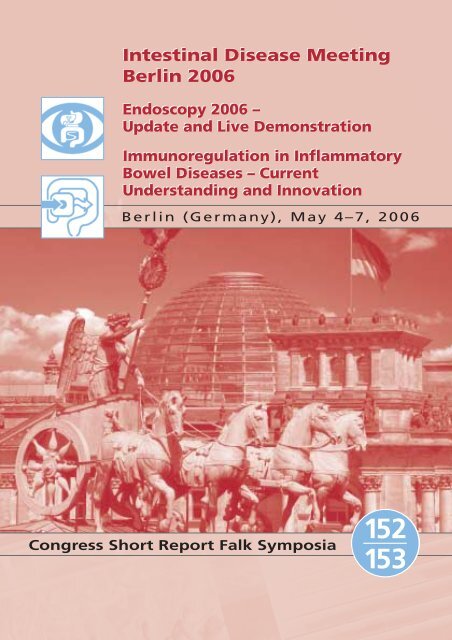
<strong>Intestinal</strong> <strong>Disease</strong> <strong>Meeting</strong><br />
<strong>Berlin</strong> <strong>2006</strong><br />
Endoscopy <strong>2006</strong> –<br />
Update and Live Demonstration<br />
Immunoregulation in Inflammatory<br />
Bowel <strong>Disease</strong>s – Current<br />
Understanding and Innovation<br />
<strong>Berlin</strong> (Germany), May 4–7, <strong>2006</strong><br />
Congress Short Report <strong>Falk</strong> Symposia<br />
<strong>152</strong><br />
<strong>153</strong>
Publisher<br />
FALK FOUNDATION e.V.<br />
Leinenweberstr. 5<br />
Postfach 6529<br />
79041 Freiburg<br />
Germany<br />
© <strong>2006</strong> <strong>Falk</strong> Foundation e.V.<br />
All rights reserved.<br />
Text:<br />
Christine Vetter<br />
Medical Journalist<br />
Cologne (Germany)<br />
© Photos: Kai-Uwe Wudtke<br />
1st edition <strong>2006</strong> ISBN 3-933186-78-1
<strong>Falk</strong> Symposium <strong>152</strong><br />
Endoscopy <strong>2006</strong> –<br />
Update and Live Demonstration<br />
<strong>Falk</strong> Symposium <strong>153</strong><br />
Immunoregulation in Inflammatory<br />
Bowel <strong>Disease</strong>s – Current<br />
Understanding and Innovation<br />
<strong>Berlin</strong> (Germany), May 4–7, <strong>2006</strong><br />
Congress Short Report <strong>Falk</strong> Symposia<br />
<strong>152</strong><br />
<strong>153</strong>
Introduction<br />
Scientific Organizers<br />
<strong>Falk</strong> Symposium <strong>152</strong> <strong>Falk</strong> Symposium <strong>153</strong><br />
P. Fockens,<br />
Amsterdam<br />
(The Netherlands)<br />
Foreword<br />
H.-J. Schulz,<br />
<strong>Berlin</strong><br />
(Germany<br />
T. Rösch,<br />
<strong>Berlin</strong><br />
(Germany)<br />
J. Špičák,<br />
Prague<br />
(Czech Republic)<br />
The <strong>Falk</strong> Symposia “Endoscopy <strong>2006</strong>” and<br />
“Immunoregulation in Inflammatory Bowel<br />
<strong>Disease</strong>s” occupy a prominent place among<br />
the topics discussed at the <strong>Intestinal</strong> <strong>Disease</strong><br />
<strong>Meeting</strong> held in <strong>Berlin</strong>, May 4–7, <strong>2006</strong>. In all,<br />
more than 1,400 participants from 48 nations<br />
traveled to the German capital in order to update<br />
their knowledge and exchange experiences<br />
with professional colleagues. “Endoscopy is of<br />
central importance in gastroenterology. As a<br />
method, it is dependent on new developments<br />
in gastroenterology, basic research and technology.”<br />
This was the message of the scientific organizers<br />
of the <strong>Falk</strong> Symposium <strong>152</strong>, P. Fockens<br />
A. Dignass,<br />
Frankfurt<br />
(Germany)<br />
E.F. Stange,<br />
Stuttgart<br />
(Germany)<br />
D. Rachmilewitz,<br />
Jerusalem<br />
(Israel)<br />
J.V. Weinstock,<br />
Boston<br />
(USA)<br />
(Amsterdam), T. Rösch (<strong>Berlin</strong>), H.-J. Schulz<br />
(<strong>Berlin</strong>) and J. Špičák (Prague). Live demonstrations<br />
were included among the attractions of<br />
this symposium.<br />
The objective of <strong>Falk</strong> Symposium <strong>153</strong> was to<br />
present current standards and potential new<br />
options in the diagnosis and therapy of inflammatory<br />
bowel diseases. It was organized by<br />
A. Dignass (Frankfurt), D. Rachmilewitz<br />
(Jerusalem), E.F. Stange (Stuttgart) und<br />
J.V. Weinstock (Boston). Among other controversial<br />
topics, participants discussed whether<br />
the treatment of inflammatory bowel diseases<br />
should follow a “Top Down” or “Step Up”<br />
approach.
<strong>Falk</strong> Symposium <strong>152</strong><br />
Endoscopy <strong>2006</strong> – Update and Live Demonstration<br />
<strong>Berlin</strong>, May 4–5, <strong>2006</strong><br />
•<br />
•<br />
•<br />
•<br />
•<br />
•<br />
•<br />
•<br />
Congress Short Report <strong>Falk</strong> Symposia<br />
Page<br />
The Most Relevant New Technologies . . . . . . . . . . . . . . . . . . . . . . . . . . . . . . . . . . . . . . . 4<br />
Role of Endoscopy . . . . . . . . . . . . . . . . . . . . . . . . . . . . . . . . . . . . . . . . . . . . . . . . . . . . . 13<br />
Bile and Pancreas . . . . . . . . . . . . . . . . . . . . . . . . . . . . . . . . . . . . . . . . . . . . . . . . . . . . . . 16<br />
State-of-the-Art Lecture . . . . . . . . . . . . . . . . . . . . . . . . . . . . . . . . . . . . . . . . . . . . . . . . . 18<br />
Gastrointestinal Oncology . . . . . . . . . . . . . . . . . . . . . . . . . . . . . . . . . . . . . . . . . . . . . . . 19<br />
Prevention and Management of Complications . . . . . . . . . . . . . . . . . . . . . . . . . . . . . . . 20<br />
Endoscopy <strong>2006</strong> – Live Demonstration . . . . . . . . . . . . . . . . . . . . . . . . . . . . . . . . . . . . . . 23<br />
Interview with Professor <strong>Dr</strong>. H.-J. Schulz (<strong>Berlin</strong>) . . . . . . . . . . . . . . . . . . . . . . . . . . . . . . . 25<br />
<strong>Falk</strong> Symposium <strong>153</strong><br />
Immunoregulation in Inflammatory Bowel <strong>Disease</strong>s –<br />
Current Understanding and Innovation<br />
<strong>Berlin</strong>, May 6–7, <strong>2006</strong><br />
•<br />
•<br />
•<br />
•<br />
•<br />
•<br />
•<br />
•<br />
•<br />
•<br />
Advances in the Pathogenesis of IBD . . . . . . . . . . . . . . . . . . . . . . . . . . . . . . . . . . . . . . . 27<br />
Optimization in Therapy Through Improved <strong>Disease</strong> Classification? . . . . . . . . . . . . . . . . . 31<br />
Conventional Immunomodulator Therapy . . . . . . . . . . . . . . . . . . . . . . . . . . . . . . . . . . . 34<br />
Relevance of Biologics . . . . . . . . . . . . . . . . . . . . . . . . . . . . . . . . . . . . . . . . . . . . . . . . . . 38<br />
State-of-the-Art Lecture . . . . . . . . . . . . . . . . . . . . . . . . . . . . . . . . . . . . . . . . . . . . . . . . . 41<br />
New Therapeutic Approaches (Part I) . . . . . . . . . . . . . . . . . . . . . . . . . . . . . . . . . . . . . . . 42<br />
New Therapeutic Approaches (Part II) . . . . . . . . . . . . . . . . . . . . . . . . . . . . . . . . . . . . . . . 47<br />
Presentation of Difficult Cases – Panel Discussion . . . . . . . . . . . . . . . . . . . . . . . . . . . . . . 50<br />
Chairpersons, Speakers and Scientific Organizers . . . . . . . . . . . . . . . . . . . . . . . . . . . . . . 51<br />
Further Congress Short Reports of <strong>Falk</strong> Symposia . . . . . . . . . . . . . . . . . . . . . . . . . . . . . . 56<br />
<strong>152</strong><br />
<strong>153</strong><br />
3
Session 1<br />
<strong>Falk</strong> Symposium <strong>152</strong><br />
Endoscopy <strong>2006</strong> – Update and Live Demonstration<br />
<strong>Berlin</strong>, May 4–5, <strong>2006</strong><br />
4<br />
The Most Relevant New Technologies<br />
Chair:<br />
P. Fockens, Amsterdam<br />
F. Hagenmüller, Hamburg<br />
J.F. Riemann, Ludwigshafen<br />
Transnasal endoscopy – not yet<br />
part of the clinical routine in<br />
Germany<br />
Although capsule endoscopy has received much<br />
recent attention and has been celebrated as an<br />
example of progress in this field, conventional<br />
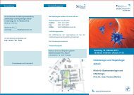
Fig. 1<br />
I Ultrathin gastroscopes (H. Neuhaus, Düsseldorf)<br />
endoscopy has not lost any of its importance.<br />
In fact, there has been a series of very promising<br />
advances that, on the one hand, improve the<br />
method’s diagnostic potential and, on the other,<br />
make the endoscopic examination less unpleasant<br />
for the patient. As an example, H. Neuhaus
(Düsseldorf) cited the development of ultrathin<br />
gastroscopes that now permit transnasal<br />
gastroscopy (figure 1). “Unlike in France,”<br />
H. Neuhaus said, “this method, which enjoys<br />
high patient acceptance, has yet to become<br />
established in Germany.”<br />
An objection to transnasal endoscopy has been<br />
that the resolution is inferior to that provided<br />
by conventional endoscopes. According to<br />
H. Neuhaus, however, the resolution is no longer<br />
a problem with the newer units. Because of<br />
the small navigation mechanism, the ultrathin<br />
endoscopes have the advantage of being able<br />
to inspect directly behind mucosal folds – an<br />
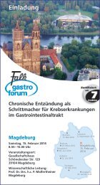
Fig. 2<br />
H. Neuhaus<br />
I Chromoendoscopy: Methylene blue staining of Barrett’s metaplasia<br />
(H. Neuhaus, Düsseldorf)<br />
Congress Short Report <strong>Falk</strong> Symposium <strong>152</strong><br />
important capability, such as in the search for<br />
esophageal carcinomas. Potential indications for<br />
the new method, H. Neuhaus said, would include<br />
diagnostic esophagogastroduodenoscopy<br />
(EGD) without sedation, screening for Barrett’s<br />
esophagus, dysphagia work-up, transnasal tube<br />
placement and passage of high-grade stenoses<br />
in the proximal gastrointestinal tract.<br />
Chromoendoscopy: better<br />
evaluation of lesions<br />
Another example of progress is the method of<br />
chromoendoscopy (figure 2), which, because of<br />
the color technology, enables a better evaluation<br />
of lesions. “In particular, lesions are much better<br />
demarcated,” H. Neuhaus explained. One method<br />
that has not gained broad acceptance is the<br />
technique of zoom endoscopy, which, despite<br />
its capabilities for magnification, does not offer<br />
any advantages over conventional methods. In<br />
the general, the reliability of findings returned<br />
by endoscopic examinations depends to a great<br />
degree on the quality of patient preparation and<br />
the examination technique. The exact clinical<br />
utility of further increases in resolution is difficult<br />
to assess, but would seem helpful.<br />
5
Session 1<br />
6<br />
Capsule endoscopy – a new<br />
dimension for examination<br />
A new dimension in the study of changes of<br />
the gastrointestinal tract is offered by capsule<br />
endoscopy, M.M. Delvaux (Vandoeuvre) said.<br />
Although not yet a routine examination, the<br />
method offers untold capabilities for visualization<br />
of the small bowel. “We must, however,”<br />
M.M. Delvaux warned, “get used to a completely<br />
different view of the bowel.” For example,<br />
with conventional endoscopy, there is a variable<br />
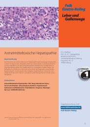
Fig. 3<br />
M.M. Delvaux<br />
Malabsorption syndromes<br />
Diffuse intestinal diseases<br />
Amyloidosis<br />
exudative enteropathy<br />
distance between the light source and the bowel,<br />
while, with capsule endoscopy, the light source<br />
impinges directly on the bowel wall, providing a<br />
completely different impression of the mucosa.<br />
“Lesions may, in some cases, have a completely<br />
different appearance under these conditions,”<br />
M.M. Delvaux cautioned.<br />
Indications for capsule endoscopy, M.M. Delvaux<br />
said, include work-up of obscure gastrointestinal<br />
bleeding in patients with either open bleeding<br />
or chronic anemia, as well as in the diagnosis of<br />
Crohn’s disease, celiac disease and NSAID-associated<br />
enteropathy. Polyposis and the work-up of<br />
tumors, together with amyloidosis, Whipple’s<br />
disease, exudative enteropathy and eosinophilic<br />
gastroenteritis can also be important indications<br />
for capsule endoscopy (figure 3). “In such disorders,<br />
capsule endoscopy does not replace conventional<br />
endoscopy but can supplement it,”<br />
M.M. Delvaux said.<br />
Whipple’s disease<br />
eosinophilic gastroenteritis<br />
I Some further indications for capsule endoscopy (M.M. Delvaux, Vandoeuvre)
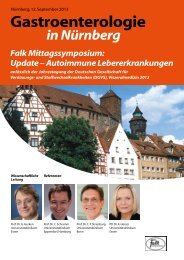
New techniques are in development<br />
“We need continued new developments in endoscopy.”<br />
This was the opinion of P. Fockens<br />
(Amsterdam). He referred especially to techniques<br />
with which flat or depressed lesions could be<br />
more reliably identified. One very useful technique<br />
that has yet to experience widespread clinical<br />
use, P. Fockens said, is chromoendoscopy. In<br />
addition, the method of “narrow band imaging”<br />
(figure 4), which has recently become commercially<br />
available, facilitates evaluation of lesions.<br />
This can be especially useful in patients with<br />
ulcerative colitis in order to limit the number of<br />
required biopsies. P. Fockens also anticipates<br />
Fig. 4<br />
I Narrow band imaging – colon (P. Fockens, Amsterdam)<br />
Congress Short Report <strong>Falk</strong> Symposium <strong>152</strong><br />
P. Fockens<br />
progress in the area of autofluorescence imaging<br />
(figure 5) and endomicroscopy (figure 6). “All<br />
these methods still require careful scientific<br />
evaluation,” P. Fockens said.<br />
7
Session 1<br />
8<br />
Fig. 5<br />
I Autofluorescence (P. Fockens, Amsterdam)<br />
Fig. 6<br />
I Endomicroscopy of normal rectal mucosa (P. Fockens, Amsterdam)
Endoscopy – It also has therapeutic<br />
value<br />
Modern endoscopy’s role is not limited to diagnosing<br />
disorders. As N. Soehendra (Hamburg)<br />
noted, it also has therapeutic value. Examples<br />
include endoscopic mucosal resection and<br />
endoscopic submucosal dissection (figure 7),<br />
as well as treatment of pancreatic abscesses and<br />
clipping.<br />
Especially in the therapy of early carcinomas of<br />
the esophagus and stomach, endoscopic mucosal<br />
resection can represent an attractive alterna-<br />
Fig. 7<br />
N. Soehendra<br />
I Endoscopic submucosal dissection (N. Soehendra, Hamburg)<br />
Congress Short Report <strong>Falk</strong> Symposium <strong>152</strong><br />
tive to conventional surgery. The endoscopic excision<br />
of circumscript malignant lesions is associated<br />
with long-term results comparable to those<br />
of conventional surgery. “There is,” N. Soehendra<br />
observed, “a significantly lower morbidity and<br />
this means increased quality of life for the patients.”<br />
Based on this positive experience the range of<br />
indications for endoscopic methods has become<br />
broader. Limitations include the depth of infiltration<br />
and the size of the tumor. Tumors that are restricted<br />
to the topmost submucosal layer and do<br />
not exceed 30 mm in diameter can be removed<br />
completely using the technique of submucosal<br />
dissection. If in certain cases it is necessary to<br />
remove long mucosal segments, N. Soehendra<br />
noted that the operator may use a “suck-andcut”<br />
technique or, where more suitable, ligatures<br />
or the so-called “piecemeal” technique can be<br />
used in order to successively remove neighboring<br />
areas of mucosa (figure 8).<br />
9
Session 1<br />
10<br />
Fig. 8<br />
I ”Piecemeal” technique (N. Soehendra, Hamburg)<br />
Another indication is the therapy of pancreatic<br />
abscesses in severely ill patients in whom conventional<br />
open surgery is not possible. In about<br />
70% of cases successful endoscopic treatment<br />
is feasible. In many other patients, the situation<br />
can at least be stabilized to the extent that surgery<br />
can be performed at a later time. In addition,<br />
endoscopy has therapeutic importance in the<br />
case of hemorrhage, which can be controlled by<br />
clipping. The same technique has also been successfully<br />
used for closure of perforations.
Pancreatobiliary endoscopy in<br />
therapy and palliation<br />
According to G. Costamagna (Rome), pancreatobiliary<br />
endoscopy has also developed in the past<br />
two decades from a purely diagnostic to a therapeutic<br />
tool. It has become the method of choice<br />
in the treatment and palliation of benign and<br />
malignant diseases. The most important methods,<br />
G. Costamagna said, include papillotomy<br />
(figure 9) and the placement of stents in the<br />
biliary tract. However, as G. Costamagna noted,<br />
the drainage of pseudocysts and even the evacuation<br />
of pancreatic necroses are possible, though<br />
this application is currently restricted to specialized<br />
centers.<br />
Fig. 9<br />
duodenal<br />
musculature<br />
Sphincter<br />
choledochus<br />
Congress Short Report <strong>Falk</strong> Symposium <strong>152</strong><br />
G. Costamagna G. Kähler<br />
Sphincter<br />
papillae<br />
Sphincter<br />
pancreaticus<br />
I Papilla of Vater (G. Costamagna, Rome)<br />
Endoscopy or minimally invasive<br />
surgery?<br />
A further new development, described by<br />
G. Kähler (Mannheim), involves the combination<br />
of endoscopy with equipment originally developed<br />
for use with laparoscopy. For example, staplers<br />
with a flexible, unfolding shaft and remote<br />
controlled triggering have been developed for<br />
use in laparoscopic procedures. Such equipment<br />
can also be used endoluminally and facilitate endoluminal<br />
applications. According to G. Kähler,<br />
such equipment can be used, for example, in the<br />
treatment of rectal anastomotic stenoses, as well<br />
as in cases of full-wall gastric resections and for<br />
the endoscopic therapy of Zenker’s diverticula.<br />
Combined procedures consisting of laparoscopy<br />
and intraluminal flexible endoscopy have been<br />
recommended for treatment of extensive adenomatous<br />
lesions of the colon and gastrointestinal<br />
tumors of the stomach (GIST). “Their clinical<br />
relevance cannot be definitively evaluated at<br />
this time,” G. Kähler noted.<br />
11
Session 1<br />
12<br />
Important new developments in<br />
ultrasound<br />
According to M. Gebel (Hannover), endoscopy<br />
has not been the only area in which new developments<br />
can be reported: Ultrasound has experi-<br />
Fig. 10<br />
M. Gebel<br />
I Contrast-enhanced echography of hepatocellular carcinoma (HCC)<br />
(M. Gebel, Hannover)<br />
enced clear progress in the diagnosis of gastrointestinal<br />
disorders. As an example, he cited contrast<br />
enhanced ultrasound. “A number of different<br />
contrast media have been introduced that can<br />
be used in ultrasound,” M. Gebel noted. Contrast-enhanced<br />
echography has, in his opinion,<br />
a role in the diagnosis of liver tumors (figure 10),<br />
which can be clearly differentiated in most cases.<br />
Further examples of progress in ultrasound examinations,<br />
M. Gebel explained, include elastography,<br />
in which the compressibility of tissue is<br />
studied, and 3D ultrasonography, which to date,<br />
however, has only achieved a narrow clinical<br />
importance.
Session 2<br />
Role of Endoscopy<br />
Chair:<br />
J. Devière, Brussels<br />
H. Lochs, <strong>Berlin</strong><br />
Gastroesophageal reflux disease –<br />
Will tomorrow’s therapy be<br />
endoscopic?<br />
According to J. Mössner (Leipzig), endoscopy<br />
plays a central role in the diagnosis of gastroesophageal<br />
reflux disease (GERD), although only<br />
about 50% of GERD patients actually exhibit<br />
endoscopically recognizable lesions in the esophagus.<br />
Obtaining a baseline examination early in<br />
the disease is important and provides a basis for<br />
assessing the subsequent disease course, as well<br />
as for work-up of a Barrett’s esophagus. In addition,<br />
an emergent endoscopic examination is indicated<br />
in any patient reporting alarm systems<br />
Fig. 11<br />
I Barrett’s esophagus (J. Mössner, Leipzig)<br />
Congress Short Report <strong>Falk</strong> Symposium <strong>152</strong><br />
J. Mössner<br />
such as dysphagia, weight loss or hemorrhage.<br />
Finally, patients with reflux symptoms who do<br />
not respond promptly to standard therapy with<br />
a proton pump inhibitor (PPI) represent a further<br />
indication for endoscopy.<br />
According to J. Mössner, the role of the newer<br />
techniques, such as chromoendoscopy, zoom<br />
endoscopy and narrow band imaging for the diagnosis<br />
of GERD cannot be definitively assessed<br />
at this time. However, he does expect significant<br />
progress from the application of high-resolution<br />
endoscopy in the identification of high-grade<br />
dysplasias and early carcinomas in patients with<br />
Barrett’s esophagus (figure 11). In this area, he<br />
13
Session 2<br />
14<br />
does not feel that chromoendoscopy or narrow<br />
band imaging offer any further advantages over<br />
high-resolution techniques.<br />
The increasing importance of<br />
endoscopy in antireflux therapy<br />
J. Mössner also sees a significant watershed<br />
event in another area. In his opinion, endoscopy<br />
is on the threshold of assuming an increasing<br />
therapeutic role in the management of GERD.<br />
This is not limited to patients who do not respond<br />
adequately to PPIs. In fact, endoscopic<br />
antireflux therapy may represent an alternative<br />
to drug therapy in patients who cannot or will<br />
not take oral medication regularly as well as in<br />
those who may experience long-term enhanced<br />
quality of life as a result of endoscopic treatment.<br />
These new techniques, however, must not be<br />
used indiscriminately. “At the present time,”<br />
J. Mössner reminded his audience, “we have<br />
only limited experience and long-term data is<br />
completely missing.”<br />
Fig. 12<br />
Diagnosis: Ulcerative colitis<br />
• Characteristic pattern of inflammation<br />
(Sub)total colitis 15–20 %<br />
Left-sided colitis 30–50 %<br />
Proctosigmoiditis 30–50 %<br />
Backwash ileitis –10 %<br />
I Endoscopy in IBD – ulcerative colitis (F. Hartmann, Frankfurt)<br />
F. Hartmann<br />
Essential in inflammatory bowel<br />
diseases<br />
Endoscopy also plays a decisive role in the workup<br />
of inflammatory bowel diseases (IBD), said<br />
F. Hartmann (Frankfurt). The examination services<br />
to establish the diagnosis, exclude other possible<br />
etiologies and to assess the pattern, extent and<br />
activity of the patient’s disease.<br />
In general, endoscopy in patients with ulcerative<br />
colitis reveals subtotal colitis in 15–20% of cases,<br />
while 30–50% show left-sided colitis and 30–50%<br />
suffer from proctosigmoiditis (figure 12). In patients<br />
with Crohn’s disease, endoscopy reveals<br />
involvement of the esophagus, stomach and<br />
duodenum in 3–5% of patients, while in 25–30%<br />
of cases only the small bowel is affected. Both<br />
the small bowel and colon are affected in
Fig. 13<br />
Diagnosis: Crohn’s disease<br />
• Characteristic pattern of inflammation<br />
Esophagus, stomach,<br />
duodenum 3–5 %<br />
Small bowel 25–30 %<br />
Small and large bowel 40–55 %<br />
Rectum 11–26 %<br />
I Endoscopy in IBD – Crohn’s disease (F. Hartmann, Frankfurt)<br />
40–55% of patients (figure 13), while rectal<br />
involvement is seen in 11–26% of patients.<br />
Thanks to endoscopy, a reliable differential diagnosis<br />
is possible in most cases and IBD can be<br />
distinguished from infectious enteritis, drug-induced<br />
enterocolitis, mesenteric ischemia, diverticulitis<br />
and malignant disease.<br />
This does not, however, represent the limits of<br />
endoscopy. “We cannot only inspect the mucosa,<br />
but also obtain biopsies,” F. Hartmann said. In<br />
his opinion, the strength of endoscopy lies in its<br />
capacity to monitor the disease course, especially<br />
with regard to the possible development of dysplasias.<br />
He was more guarded in his assessment<br />
of the new developments in endoscopy, noting<br />
that “I do not believe that we need capsule endoscopy<br />
to reliably diagnose Crohn’s disease.”<br />
Congress Short Report <strong>Falk</strong> Symposium <strong>152</strong><br />
P.N. Meier<br />
Special care is required in children<br />
Special conditions apply to pediatrics, P.N. Meier<br />
(Hannover) explained. One must always consider<br />
the special situation of children who, while they<br />
may not directly fear the endoscopic procedure,<br />
are more prone to anxiety at being separated<br />
from their parents. There are also some practical<br />
issues involved, such as the fact that children are<br />
not able to remain abstinent from food for long<br />
periods. “They should always be examined early<br />
in the morning,” P.N. Meier noted. Wherever<br />
possible, the procedure should be conducted<br />
under general anesthesia. With the exception of<br />
newborns and young children, P.N. Meier said,<br />
special endoscopy units are not required. Good<br />
cooperation between the pediatrician and the<br />
consulting gastroenterologist is essential.<br />
15
Session 3<br />
16<br />
Bile and Pancreas<br />
Chair:<br />
G. Costamagna, Rome<br />
S. Jonas, <strong>Berlin</strong><br />
Preoperative classification of<br />
tumors<br />
As an example of the importance of endoscopy<br />
in diseases of the biliary ducts and pancreas,<br />
T. Rösch (<strong>Berlin</strong>) cited the so-called Klatskin<br />
tumors. ERCP is very useful in the pre-operative<br />
work-up of these tumors, facilitating an exact<br />
classification of the tumor and providing crucial<br />
data for planning patients’ therapeutic management.<br />
Stage I and II tumors can even be treated<br />
endoscopically and, when palliative treatment<br />
is contemplated, stent placement for further<br />
drainage can be performed.<br />
Endoscopy is also useful in cases of chronic pancreatitis,<br />
when over the course of the disease<br />
patients develop complications such as obstructions<br />
and stenoses. Objective of the method<br />
is to reduce pain and increase both exo- and<br />
endocrine pancreatic function. Endoscopic<br />
treatment is necessary in about one-half of all<br />
patients at some point during their disease<br />
course, said J.F. Riemann (Ludwigshafen).<br />
T. Rösch J.F. Riemann<br />
Stents can alleviate stenoses<br />
In some cases, placement of a stent may be<br />
helpful in alleviating stenosis. If the pancreatic<br />
duct is occluded by stones, it may be possible<br />
using endoscopic methods to fragment the<br />
stone and re-open the duct. This is frequently<br />
done in combination with extracorporeal shockwave<br />
lithotripsy and several published studies<br />
have confirmed the use of this combination.<br />
Pancreatic pseudocysts represent another complication<br />
that is effectively treated with endoscopic<br />
techniques, the gastroenterologist said. “Success<br />
rates range as high as 96%,” J.F. Riemann said.<br />
Surgery is indicated only when conservative and<br />
endoscopic therapeutic methods have been exhausted.
Accurate work-up of pseudocysts<br />
According to J. Devière (Brussels), pancreatic<br />
pseudocysts are fairly common. One must, however,<br />
assess patient’s findings with a certain degree<br />
of caution, J. Devière warned, “since not<br />
everything that looks like a pseudocyst actually<br />
is one.” In fact, in about 15% of cases, patients<br />
suffer from a cystic tumor (figure 14): Hence, in<br />
no case should drainage be attempted prior to<br />
completion of a full work-up. In addition, pseudo-<br />
Fig. 14<br />
Pseudocysts<br />
85 %<br />
J. Devière<br />
I Cystic lesions of the pancreas – classification (J. Devière, Brussels)<br />
Congress Short Report <strong>Falk</strong> Symposium <strong>152</strong><br />
Cystic tumors<br />
≈15 %<br />
cysts very frequently resolve spontaneously. Intervention<br />
is therefore required only in patients<br />
with persisting symptoms. This is of even greater<br />
importance due to the fact that the procedure<br />
itself is not without complications. For example,<br />
bleeding occurs in 2–4% of cases. Retroperitoneal<br />
perforation occurs in about an equal number of<br />
cases. J. Devière also reminded his audience of<br />
the danger of infection.<br />
• mucinous 49 %<br />
• serous 32 %<br />
• IPMT 11 %<br />
• rare 8%<br />
17
State-of-the-Art-Lecture<br />
18<br />
State-of-the-Art-Lecture<br />
Chair:<br />
S. Bar-Meir, Tel Hashomer<br />
C. Ell, Wiesbaden<br />
Capsule endoscopy in the work-up<br />
of obscure bleeding<br />
Endoscopy plays a special role in the identification<br />
and therapy of bleeding of the gastrointestinal<br />
tract. Here, one distinguishes between open<br />
and obscure bleeding, the source of which is unknown,<br />
said F. Hagenmüller (Hamburg) in a<br />
state-of-the-art lecture.<br />
Occult bleeding is normally discovered by Hemoccult<br />
or similar examination of the stool or is<br />
suspected due to chronic, but otherwise unexplained<br />
anemia. In many cases, however, neither<br />
radiological nor conventional endoscopic evaluations<br />
identify the source. Capsule endoscopy and<br />
double-balloon endoscopy have, however, decisively<br />
improved our capacity to discover obscure<br />
bleeding in the upper intestinal tract. “Metaanalyses<br />
show that capsule endoscopy is clearly<br />
F. Hagenmüller<br />
superior to push endoscopy in detecting bleeding<br />
sources,” F. Hagenmüller said, noting that<br />
the most common cause of occult bleeding is<br />
angiectasis, accounting for 50% of cases. However,<br />
other causes, such as ulcers, diverticula and<br />
tumors may be responsible. Even with these new<br />
methods, the cause of occult bleeding is not<br />
identified in about one-fourth of cases. In some<br />
cases, whether the bleeding source is found or<br />
not depends on the time of the examination. If<br />
the examination is conducted within the first<br />
two weeks, the bleeding source is found in more<br />
than 90% of patients, but this rate falls to about<br />
34% if the examination is postponed past this<br />
time.
Session 4<br />
Gastrointestinal Oncology<br />
Chair:<br />
N. Soehendra, Hamburg<br />
B. Wiedenmann, <strong>Berlin</strong><br />
Overcoming stenoses by placement<br />
of stents in the colon<br />
Similar to the upper gastrointestinal tract, stenoses<br />
and obstructions in the more distal segments of<br />
the bowel may be amenable to endoscopic<br />
treatment. Usually, metal stents are placed, said<br />
T. Ponchon (Lyon). Stents should be flexible and<br />
easy to deploy. The situation in the colon, due to<br />
the possible presence of residual stool, is somewhat<br />
more difficult than in the upper gastrointestinal<br />
tract. In addition, stents must be sufficiently<br />
long and should have soft ends in order<br />
to conform to intestinal motility (figure 15).<br />
T. Ponchon told his audience in <strong>Berlin</strong> about his<br />
own experiences with the placement of stents in<br />
575 patients. In 64% of cases, the procedure<br />
was done with a palliative indication, while in<br />
the remaining 36% of patients a pre-operative<br />
Fig. 15<br />
I Types of metal stents (T. Ponchon, Lyon)<br />
Congress Short Report <strong>Falk</strong> Symposium <strong>152</strong><br />
T. Ponchon<br />
decompression was intended. The treatment<br />
was successful in 88% of cases. Of the 12% of<br />
procedures that were considered unsuccessful,<br />
medical issues were responsible in 4.5% and<br />
technical problems in 7.5% of cases.<br />
Perforation occurred in 3.8% of patients while,<br />
in 8.5%, migration of the stent was observed.<br />
Bleeding occurred in 4.7% of patients and was<br />
severe in 0.5%. According to T. Ponchon, these<br />
complications usually occur soon after the procedure.<br />
Long-term morbidity after stent placement<br />
must also be considered. For example, about one<br />
in ten patients experiences renewed obstruction<br />
despite the indwelling metal stent. This risk is<br />
higher in cases of stents placed with palliative<br />
intention, since these normal remain in place<br />
longer than those placed temporarily for<br />
drainage.<br />
19
Session 5<br />
Prevention and Management<br />
of Complications<br />
Chair:<br />
F. Schreiber, Graz<br />
P.A. Testoni, Milan<br />
20<br />
Endoscopic procedures are associated<br />
with low overall mortality<br />
The overall mortality associated with diagnostic<br />
endoscopic procedures is reported as 0.3–0.5<br />
per 1000 procedures, of which about 50% of<br />
deaths are due to cardiopulmonary complications,<br />
the majority of which develop as a result<br />
of pre-medication. “The risks of endoscopic<br />
procedures are overall very low,” said M. Jung<br />
(Mainz).<br />
In his opinion, it is important to consider the<br />
dangers of premedication. It is common practice<br />
before a procedure to administer a short-acting<br />
benzodiazepine either alone or in combination<br />
with an opiate. Most commonly used agents are<br />
midazolam or propofol. Both agents are characterized<br />
by a short elimination half-life and corresponding<br />
short duration of action. Propofol has<br />
advantages over benzodiazepines, especially<br />
because patients recover very quickly from the<br />
medication.<br />
M. Jung W. Schmitt<br />
Propofol: Does an anesthesiologist<br />
need to be present?<br />
On the other hand, this hypnotic agent induces<br />
a certain amount of cardiac and respiratory depression<br />
and there is no antagonist that can be<br />
administered if the situation becomes critical.<br />
Nevertheless, M. Jung does not consider justified<br />
those recommendations that propofol be used<br />
only in the presence of an anesthesiologist. Experience<br />
to date suggests that, with the proper<br />
training, this agent can be safely used in patients<br />
undergoing endoscopic interventions.<br />
Low rate of complications<br />
associated with polypectomy<br />
The rate of complications is also comparatively<br />
low with polypectomy, explained W. Schmitt<br />
(Munich). Overall morbidity stands at 0.36–4.8%,<br />
of which bleeding makes up 0.6–3.4% of cases.<br />
In second place are perforations at 0.08–0.83%.<br />
Mortality associated with the procedure is reported<br />
as 0.006–0.03%.<br />
Under discussion is the question of prophylactic<br />
clipping as a means of reducing the risk of<br />
bleeding. “Convincing data supporting its use<br />
have yet to be published,” said W. Schmitt. If<br />
bleeding does occur, it can be treated with sclerotherapy,<br />
clipping or by means of coagulation.
Congress Short Report <strong>Falk</strong> Symposium <strong>152</strong><br />
H.-J. Schulz J. Špičák<br />
The risks are higher with ERCP<br />
The risk of complications is significantly higher<br />
with ERCP than with polypectomy, said<br />
H.-J. Schulz (<strong>Berlin</strong>), noting that the most<br />
common and the most serious complication<br />
is pancreatitis. Pancreatitis takes a severe course<br />
in 10–15% of cases and pancreatitis is the most<br />
common cause of death following ERCP. Risk<br />
factors include female gender, sphincter of<br />
Oddi dysfunction and pre-existing pancreatitis<br />
(figure 16). Further complications include bleeding,<br />
cholangitis and perforation. Added to this<br />
are the complications secondary to sedation.<br />
The risk of complications is different for diagnostic<br />
and therapeutic ERCP; the rate of complications<br />
is necessarily higher with the latter. Complications<br />
for the most part relate to the sphincterotomy<br />
and in general depend on the extent<br />
of the procedure. Hence, one should avoid diagnostic<br />
ERCP in cases in which less invasive procedures<br />
can provide the required data.<br />
Perforation is the main risk in<br />
patients undergoing dilatation<br />
Unlike polypectomy and ERCP, the most common<br />
complication of endoscopic dilatation is<br />
perforation. In general, however, endoscopic<br />
dilatation, said J. Špičák (Prague), is considered<br />
an established procedure that can be safely used<br />
in practically all forms of strictures and stenoses<br />
of the gastrointestinal tract (figure 17), whether<br />
due to benign or malignant processes.<br />
Highly amenable to endoscopic treatment are<br />
stenoses associated with benign disorders of the<br />
esophagus and colon. Methods include balloon<br />
dilatation, use of sounds, electrocoagulation and<br />
the placement of temporary stents, or even a<br />
combination of these different techniques,<br />
J. Špičák said.<br />
21
Session 5<br />
22<br />
Fig. 16<br />
Cumulative risk factors of post-ERCP pancreatitis<br />
45<br />
40<br />
35<br />
30<br />
25<br />
20<br />
15<br />
10<br />
5<br />
0<br />
2.5<br />
female<br />
I Complication rate of ERCP (H.-J. Schulz, <strong>Berlin</strong>)<br />
4.8<br />
female<br />
+ bili < 1<br />
SOD = sphincter of Oddi dysfunction<br />
Fig. 17<br />
Schatzki ring<br />
peptic stenosis<br />
Adjusted risk of pancreatitis [%]<br />
12.4<br />
female<br />
+ bili < 1<br />
+ SOD<br />
Crohn’s disease<br />
16.2<br />
female<br />
+ bili < 1<br />
+ difficult<br />
cannulation<br />
I Stenoses of the gastrointestinal tract (J. Špičák, Prague)<br />
42.1<br />
female<br />
+ bili < 1<br />
+ difficult<br />
cannulation<br />
+ SOD
Live Demonstration<br />
Endoscopy <strong>2006</strong> – Live Demonstration<br />
G. Costamagna, J. Devière, P. Fockens, K. Gellert,<br />
F. Hagenmüller, R. Kiesslich, P.N. Meier, H. Neuhaus,<br />
G. Niemann, T. Ponchon, T. Rösch, N. Soehendra,<br />
C.B. Williams, and the OZK-Endoscopy-Team<br />
Moderators: R. <strong>Dr</strong>ossel, <strong>Berlin</strong>; S. Faiss,<br />
Hamburg; L. Familiari, Messina; F. Hagenmüller,<br />
Hamburg; M.-L. Hermans, Euskirchen; M. Jung,<br />
Mainz; H. Martin-Nowacki, <strong>Berlin</strong>; H. Neuhaus,<br />
Düsseldorf; U. Pfeifer, Düsseldorf; J.F. Riemann,<br />
Ludwigshafen; T. Rösch, <strong>Berlin</strong>; G. Schachschal,<br />
<strong>Berlin</strong>; W. Schiffelholz, Augsburg; R. Schöfl, Linz;<br />
H.-J. Schulz, <strong>Berlin</strong>; J. Špičák, Prague; K. Wietfeld,<br />
Marl; E. Zehnter, Dortmund; C. Ziegeler, <strong>Berlin</strong><br />
The live demonstrations, transmitted directly to<br />
the congress hall from the Sana Klinikum in<br />
<strong>Berlin</strong>-Lichtenberg, attracted broad interest.<br />
About 1,000 physicians and nurses were able to<br />
watch 30 patients with disorders of all segments<br />
of the gastrointestinal tract were examined and<br />
treated by German and international experts.<br />
Viewers were able to see first-hand that the<br />
limits of endoscopic therapy have broadened<br />
significantly past large common bile duct stones<br />
or large polyps in the colon.<br />
Endoscopy – soon a procedure for<br />
nurses?<br />
It is also a matter for discussion to what extent<br />
endoscopic examinations and procedures can<br />
be performed by health care professionals other<br />
than physicians. In other countries, for example,<br />
in Great Britain, it has become an established<br />
Congress Short Report <strong>Falk</strong> Symposium <strong>152</strong><br />
C.S. Neumann<br />
fact that simple examinations are entrusted<br />
to specially trained endoscopy nurses, said<br />
C.S. Neumann (Birmingham). “The doctor shortage<br />
and ever longer waiting lists have forced<br />
the issue.” Prerequisite for nurses to perform<br />
these procedures is that they complete a formal<br />
course and regular training, as is also required<br />
for physicians, C.S. Neumann said. In such<br />
cases, as experience in England has shown,<br />
endoscopic procedures performed by nurses<br />
are acceptable.<br />
English endoscopy nurses started with simple<br />
examination techniques, such as esophagogastroduodenoscopy<br />
(EGD) without sedation of<br />
the patient. The spectrum, however, has since<br />
expanded, C.S. Neumann said. Procedures requiring<br />
sedation have also become the domain<br />
of endoscopy nurses in many English clinics.<br />
Similarly, sigmoidoscopy without polypectomy<br />
has been entrusted to trained nurses, while even<br />
removal of polyps is done by nurses in certain<br />
hospitals. “It has even been discussed whether<br />
we may even perform emergency procedures,”<br />
the endoscopy nurse noted.<br />
She warned that such considerations should best<br />
be decided ahead of time. The trend of allowing<br />
endoscopy to pass partially from the hands of<br />
physicians into those of allied health professionals<br />
can hardly be stopped, once it has started.<br />
23
Live Demonstration<br />
24<br />
P. Rogalla<br />
Virtual endoscopy: Is this the next<br />
step?<br />
Congress participants in <strong>Berlin</strong> were also presented<br />
with information on a potential future development<br />
– virtual endoscopy. The technique is<br />
based on data derived from either computed tomography<br />
(CT) or magnetic resonance imaging<br />
(MRI). According to P. Rogalla (<strong>Berlin</strong>), it is possible<br />
to obtain 3D images from the acquired data<br />
sets. <strong>Intestinal</strong> polyps are very amenable to visualization<br />
using the new technology. CT-derived<br />
data sets currently provide slightly superior<br />
images. Meta-analyses report a sensitivity of<br />
70% and a specificity of 86% for the method.<br />
According to P. Rogalla, progress can also be<br />
expected in the work-up of rectal carcinoma and<br />
in the diagnosis of flat or even depressed lesions<br />
of the bowel. “These lesions are frequently overlooked<br />
during the conventional examination,”<br />
P. Rogalla said. Virtual endoscopy, however, is<br />
still in an early stage of development: Whether<br />
it will ever represent a serious competitor to<br />
conventional endoscopy remains questionable.
Interview<br />
Interview with<br />
Professor <strong>Dr</strong>. H.-J. Schulz<br />
(<strong>Berlin</strong>)<br />
The potential of modern endoscopy:<br />
exciting and live on the bog screen<br />
The <strong>Falk</strong> Symposium <strong>152</strong> “Endoscopy <strong>2006</strong>”<br />
was a great success with its presentations and<br />
especially the live demonstrations. Participants<br />
filling the congress hall to overflowing were<br />
able to watch live transmissions of endoscopic<br />
procedures conducted by experts at the Sana<br />
Klinikum in <strong>Berlin</strong>-Lichtenberg. What are the<br />
new perspectives? What’s new in terms of<br />
equipment? What do you do when unexpected<br />
complications arise? Which procedures are safe<br />
and reliable? What are the limits of endoscopy?<br />
These were questions that participants had answered<br />
– close up and in real time. In a brief<br />
interview, Professor <strong>Dr</strong>. Hans-Joachim Schulz<br />
(<strong>Berlin</strong>) explains the special attraction of the live<br />
demonstrations.<br />
? Professor Schulz, you chaired the <strong>Falk</strong> Symposium<br />
“Endoscopy <strong>2006</strong>”. What gave you the<br />
idea to include live demonstrations in this symposium?<br />
! Schulz: Live demonstrations are able to convey<br />
the potential of modern endoscopy much more<br />
effectively than presentations or video films. The<br />
therapeutic importance of endoscopic procedures<br />
continues to grow, but at the same time these<br />
procedures have become increasingly complex.<br />
Words can go only so far in conveying this. One<br />
gets a total different impression of the capabilities<br />
of modern endoscopy when experiencing<br />
the advance of the endoscope, seeing what the<br />
endoscopist does and how he reacts to different<br />
Congress Short Report <strong>Falk</strong> Symposium <strong>152</strong><br />
H.-J. Schulz<br />
situations. The demonstration thus has a very<br />
distinct educational value. Indirectly, it contributes<br />
to quality.<br />
? This does not require live transmission in the<br />
congress hall.<br />
! Schulz: That is correct. One can certainly conduct<br />
live demonstrations in the clinic and that is<br />
something we regularly do. Unfortunately, only a<br />
few persons can participate at any one session.<br />
Including the number of participants as we have<br />
here in <strong>Berlin</strong> would be impossible. In addition,<br />
over the course of the day, we have been able to<br />
present many different patients exhibiting a wide<br />
range of pathology that has been examined and<br />
treated by many different endoscopists. This,<br />
too, is exciting. One can see how many different<br />
colleagues, all of them experts in their respective<br />
fields, deal with problems as they arise.<br />
? How do you explain the unusually high<br />
degree of interest among the audience?<br />
! Schulz: There are probably a number of<br />
reasons that so many viewers have assembled.<br />
First, extensive and comprehensive live demonstrations<br />
of this kind are rare and it appears that<br />
many of our colleagues have welcomed the opportunity<br />
this event has provided for continuing<br />
medical education. Among the viewers were<br />
many colleagues who themselves perform endoscopic<br />
procedures and are interested in seeing<br />
internationally known experts use new equipment.<br />
Not only have they been able to see new<br />
25
Interview<br />
26<br />
concepts but have also learned new tips and<br />
tricks for dealing with difficult situations. There<br />
are other viewers who, although they themselves<br />
do not perform endoscopy, have taken advantage<br />
of this event to update their knowledge on<br />
the current perspectives of this method. Hence,<br />
practically every viewer, regardless of his own<br />
expertise, can find information that will help him<br />
in his own practice. Another advantage of a liver<br />
demonstration is that the situations are authentic<br />
and the cases are real. One can follow along<br />
directly and see what happens during the procedure.<br />
? What has been the development of endoscopy<br />
and what is its current role?<br />
! Schulz: The development of endoscopy over<br />
the past years and decades has been unbelievably<br />
exciting. Again and again, we have had the<br />
impression that we have been working on the<br />
cutting edge and that further improvements<br />
were practically impossible. Again and again,<br />
however, there were spectacular leaps that significantly<br />
advanced both the diagnostic and,<br />
more recently, also the therapeutic potential of<br />
endoscopic procedures. Starting with the normal<br />
optic endoscopes, we soon advanced to the<br />
flexible optical fiber units. Then came the leap<br />
to videoendoscopy that continued to improve<br />
and add new features, particularly in terms of<br />
magnification and color techniques. Currently,<br />
we are experiencing another technological leap.<br />
Image resolution has become significantly better<br />
and we have benefited from new technologies,<br />
such as narrow band imaging, with which we<br />
can visualize vasculature and thus better assess<br />
areas suspicious for malignancy. Endoscopy continues<br />
to develop into a more therapeutically<br />
relevant method.<br />
In addition, pathology of the biliary tract and<br />
the pancreas is now increasingly amenable to<br />
endoscopic treatment, sparing patients the risks<br />
of conventional open surgery. Using the doubleballoon<br />
technique, we can now reach all regions<br />
of the gastrointestinal tract without surgery.<br />
Biopsies can be obtained, pre-cancerous and<br />
early-stage tumors can be removed and other<br />
disorders can be therapeutically addressed. Besides<br />
this, further advances are occurring, including<br />
capsule endoscopy, which is already being<br />
used and developed, as well as other endoscopic<br />
technologies. The relevance of virtual endoscopy<br />
cannot, however, be definitively assessed at this<br />
time.<br />
Professor Schulz, thank you for the interview.
Session 1<br />
Congress Short Report <strong>Falk</strong> Symposium <strong>153</strong><br />
<strong>Falk</strong> Symposium <strong>153</strong><br />
Immunoregulation in Inflammatory Bowel <strong>Disease</strong>s –<br />
Current Understanding and Innovation<br />
<strong>Berlin</strong>, May 6–7, <strong>2006</strong><br />
Advances in the Pathogenesis<br />
of IBD<br />
Chair:<br />
A. Dignass, Frankfurt<br />
D. Rachmilewitz, Jerusalem<br />
Inflammatory bowel diseases:<br />
strong genetic components<br />
Inflammatory bowel diseases (IBD) such as<br />
Crohn’s disease (CD) and ulcerative colitis (UC)<br />
usually manifest themselves in adolescents or<br />
young adults. Their incidence has been increasing<br />
over the past decades and, according to<br />
S. Schreiber (Kiel), it occurs more frequently in<br />
some families. This, together with the high concordance<br />
in monozygotic twins (over 55%),<br />
which is not observed in fraternal twins (under<br />
5%), supports the hypothesized strong genetic<br />
component of these diseases.<br />
IBD are polygenetic diseases, although recent research<br />
has identified the NOD2 (CARD15) gene<br />
that potentially acts as a trigger in the pathogenesis<br />
of Crohn’s disease (figure 18). According to<br />
S. Schreiber, NOD2 belongs to a large family of<br />
S. Schreiber<br />
proteins involved in defending the organism from<br />
bacterial invasion. “NOD2, however, is not by<br />
far the only component of the pathomechanism<br />
of inflammatory bowel diseases,” S. Schreiber<br />
said. Corresponding mutations are involved in<br />
the disease process in less than half of all patients<br />
(figure 19), which means that the search must<br />
continue for further potential pathogenetic<br />
factors.<br />
Like NOD2, some other factors may involve chromosome<br />
16, on which several areas have been<br />
identified that may be involved in the development<br />
of IBD. Here, S. Schreiber said, gene splicing<br />
may play a role. This process, in which RNA<br />
fragments of varying length and corresponding<br />
protein fragments are produced, lends high<br />
plasticity to the human genome.<br />
27
Session 1<br />
28<br />
Fig. 18<br />
NOD2 (CARD15, IBD1)<br />
G908R (CD)<br />
LP1007P (CD) truncation<br />
I 3-Dimensional protein structure of NOD2 (S. Schreiber, Kiel)<br />
Fig. 19<br />
NOD2<br />
contributes<br />
No contribution of NOD2<br />
I Crohn’s disease and NOD2 (S. Schreiber, Kiel)<br />
NOD2 major factor<br />
R702W<br />
(CD)
Congress Short Report <strong>Falk</strong> Symposium<br />
D.K. Podolsky E. Cario I. Fuss<br />
Set point changes in the Toll-like<br />
receptors<br />
A human being’s genetic material serves as the<br />
basis for his inborn immunity, which is distinguished<br />
from his acquired immunity, D.K. Podolsky<br />
(Boston) said. Both systems control a person’s<br />
immune reactions by means of the formation of<br />
cytokines and chemokines, which, in turn, trigger<br />
signal cascades in the involved cells and thus<br />
mediate both immune and inflammatory reactions.<br />
An important role is played by the so-called<br />
Toll-like receptors (TLR), which are closely associated<br />
with the NOD2 system. D.K. Podolsky believes<br />
that changes in the “set points” in the TLRs<br />
are responsible for the interaction of epithelial<br />
and mucosal cells with the intestinal flora, which<br />
can also serve as a trigger in the pathogenesis<br />
of IBD. “We still understand far too little about<br />
how the different signal pathways coincide,”<br />
D.K. Podolsky conceded.<br />
According to E. Cario (Essen), however, the TLRs<br />
do not operate independently. They are, in fact,<br />
dependent on cooperation with the MD-2 receptor<br />
(myeloid differentiation protein 2) in order to<br />
adequately recognize lipopolysaccharides (LPS).<br />
Although MD-2 has a high affinity for LPS, the<br />
exact role of this co-receptor in the pathogenesis<br />
remains unclear, E. Cario said.<br />
Differences between ulcerative colitis and Crohn’s<br />
disease may, according to I. Fuss (Bethesda), lie<br />
in the activated cell systems. In his opinion, there<br />
is strong evidence that it is especially the Th1<br />
cells that control the inflammatory process in<br />
Crohn’s disease through formation of inflammatory<br />
cytokines such as interleukin-12 (IL-12), interferon-γ<br />
(IFN-γ) and tumor necrosis factor-α<br />
(TNF-α). Ulcerative colitis, on the other hand, is<br />
believed to be a disease that develops due to<br />
changes in the Th2 cells, with an additional association<br />
with the natural killer cells (NK cells).<br />
<strong>153</strong><br />
29
Session 1<br />
30<br />
Fig. 20<br />
A B<br />
C<br />
P. Marteau<br />
Does the intestinal flora itself<br />
trigger Crohn’s disease?<br />
Beside the immune system, the bacteria of the<br />
gastrointestinal tract play a role in the pathogenesis<br />
of Crohn’s disease. In the presence of a genetically<br />
determined susceptibility the intestinal<br />
A: Listeria monocytogenes; B: Measles<br />
C. Mycobacterium paratuberculosis; D: Saccharomyces<br />
I Crohn’s disease and pathogens? Conflicting results (P. Marteau, Paris)<br />
flora can actually directly promote inflammation.<br />
According to P. Marteau (Paris), bacteria exert<br />
physiological effects on the mucosal structure,<br />
epithelial turnover, the development of immune<br />
reactions and even on intestinal function. These<br />
reactions are mediated through the TLRs and<br />
the NOD2 system. This explains why changes in<br />
the intestinal flora can result in changes on the<br />
immunological level. According to P. Marteau,<br />
discussion has centered on the involvement of<br />
numerous bacterial species (figure 20), and there<br />
is evidence that there may be differences between<br />
Crohn’s disease and ulcerative colitis. While involvement<br />
by intestinal flora seems most pronounced<br />
in Crohn’s disease, it may be considerably<br />
less so in ulcerative colitis.<br />
D
Session 2<br />
Optimization in Therapy<br />
Through Improved <strong>Disease</strong><br />
Classification?<br />
Chair:<br />
M. Gasull, Badalona<br />
H. Goebell, Essen<br />
Classification of the clinical<br />
phenotype – pros and cons<br />
Just as there are controversies regarding the<br />
exact pathogenesis of IBD, there is also disagreement<br />
on the best classification of these two disease<br />
entities. W. Reinisch (Vienna) argued for a<br />
classification by clinical phenotype: “The clinical<br />
phenotype is the fastest and most objective<br />
means of assessing the disease.” However, the<br />
claim of different classification schemas to describe<br />
the disease according to objective and<br />
reproducible criteria is, W. Reinisch said, hardly<br />
more than a “wish list” that “is out of touch<br />
with reality”. Similarly, there is no standard<br />
Congress Short Report <strong>Falk</strong> Symposium <strong>153</strong><br />
W. Reinisch<br />
method for the diagnosis of IBD. “We are actually<br />
confronted more with a diagnostic puzzle,”<br />
W. Reinisch said. Important components include<br />
the clinical evaluation in combination with the<br />
endoscopic, histological and radiological findings,<br />
as well as the results of laboratory studies.<br />
W. Reinisch also criticized the so-called Vienna<br />
Classification, which, among other factors, also<br />
takes into account the localization and clinical<br />
course of the disease. Both can be variable and<br />
are not, in his opinion, suitable for describing<br />
the disease.<br />
31
Session 2<br />
32<br />
Fig. 21<br />
inflammatory<br />
G. van Assche<br />
Include genetic and serological<br />
parameters<br />
In the opinion of G. van Assche (Leuven), a clear<br />
classification of IBD, based not only on clinical<br />
phenotype but also on serological and genetic<br />
parameters, is essential. “It would give us important<br />
clues on the future course of the disease<br />
I Long-term evolution of disease behavior in Crohn’s disease<br />
(G. van Assche, Leuven)<br />
and for assessing the patient’s prognosis,”<br />
G. van Assche said (figure 21). For example, there<br />
is a close correlation between NOD2 variants<br />
and the clinical phenotype in Crohn’s disease.<br />
In addition, G. van Assche argued for further<br />
“fine tuning” of both the Vienna Classification<br />
and also of the Montreal Classification, derived<br />
from it. For example, he recommended including<br />
serological markers such as antibodies to Saccharomyces<br />
cerevisiae (ASCA), which is associated<br />
with Crohn’s disease, as well as perinuclear antineutrophilic<br />
cytoplasmic antibody (p-ANCA),<br />
which appears to be associated with ulcerative<br />
colitis. The serological markers will probably also<br />
be important in predicting the reaction of individual<br />
patients to biological therapy options,<br />
which are currently under development.<br />
stricturing
Fig. 22<br />
CDP571<br />
34<br />
21<br />
Placebo<br />
I CRP and response to biologicals (S. Vermeire, Leuven)<br />
49<br />
15<br />
Congress Short Report <strong>Falk</strong> Symposium<br />
S. Vermeire K. Herrlinger<br />
CRP helps assess patients’ reaction<br />
to biologics<br />
In addition to these serological markers, said<br />
S. Vermeire (Leuven), there are also biomarkers<br />
that could be used to better classify the disease<br />
manifestation. <strong>Disease</strong> activity is most commonly<br />
assessed using C-reactive protein (CRP), which<br />
also predicts to a certain degree patients’ reaction<br />
to biologics (figure 22). A further possible<br />
marker is calprotectin, which is determined from<br />
stool. Both CRP and calprotectin, S. Vermeire<br />
said, are very sensitive markers for inflammation.<br />
They are, however, not specific and can be<br />
elevated in cases of infection or malignant<br />
processes.<br />
Response [%]<br />
60<br />
50<br />
40<br />
30<br />
20<br />
10<br />
0<br />
60<br />
50<br />
40<br />
30<br />
20<br />
10<br />
In the opinion of K. Herrlinger (Stuttgart), pharmacogenetic<br />
studies could be used to predict<br />
the reaction to a given therapeutic agent. He<br />
cited azathioprine as an example. This immunosuppressant<br />
helps maintain remission in many<br />
patients and use of this agent helps spare corticosteroids.<br />
Thiopurine S-methyltransferase<br />
(TPMT), the enzyme responsible for the breakdown<br />
of azathioprine, however, exhibits a genetic<br />
polymorphism, with the result that there<br />
are rapid, intermediate and slow metabolizers of<br />
azathioprine. Patients’ status can be determined<br />
prior to treatment, K. Herrlinger said, thus allowing<br />
the physician to estimate to some degree the<br />
risk of side effects and to adjust the medication<br />
dose accordingly.<br />
0<br />
Week 2 all Week 2 CRP Week 12 all Week 12 CRP<br />
> 10 mg/l<br />
> 10 mg/l<br />
44<br />
37<br />
CDP571<br />
53<br />
Placebo<br />
18<br />
<strong>153</strong><br />
33
Session 3<br />
Conventional Immunomodulator<br />
Therapy<br />
Chair:<br />
W.E. Fleig, Leipzig<br />
M. Lémann, Paris<br />
34<br />
Increasing importance of<br />
immunomodulators<br />
Alongside the standard medications, immunomodulators,<br />
such as azathioprine, have been<br />
gaining increased importance in the treatment<br />
of IBD, said S.P.L. Travis (Oxford). Azathioprine is<br />
indicated when, after a severe disease flare, patients<br />
require treatment with steroids multiple<br />
times within one year, if the disease recurs while<br />
patients are on steroids or within three months<br />
of their discontinuance, and also for postoperative<br />
prophylaxis of Crohn’s disease.<br />
Fig. 23<br />
Remission induced by prednisolone; tapered over 12 weeks<br />
Patients not failing trial [%]<br />
100<br />
80<br />
60<br />
40<br />
20<br />
Steroids +<br />
azathioprine<br />
Azathioprine<br />
S.P.L. Travis<br />
Studies show that azathioprine is significantly<br />
more effective than placebo for maintenance of<br />
remission (figure 23). Usually, the agent is administered<br />
when patients are either refractory to,<br />
or dependent on, steroids. Azathioprine is also<br />
the agent in this class with the most clinical experience.<br />
Only after it has been shown to be<br />
ineffective does the focus shift to the other immunomodulators,<br />
such as methotrexate.<br />
Placebo (n = 30)<br />
Azathioprine 2.5 mg/kg<br />
bw/day (n = 33)<br />
0<br />
0 15<br />
Duration of trial [Months]<br />
Candy et al. Gut 1995, 37: 674<br />
I Azathioprine for maintaining clinical remission in Crohn’s disease after steroid<br />
induction (S.P.L. Travis, Oxford)
Optimize clinical efficacy, minimize<br />
risks<br />
It is important to remember, said J.-F. Colombel<br />
(Lille), that therapy with immunosuppressants<br />
has specific side effects, such as a significantly<br />
increased risk of infection (figure 24). This is less<br />
pronounced with the clinically established immunosuppressants,<br />
such as azathioprine, whose<br />
side effects are well known, than with the biologics<br />
now flooding the market. “The risk of side<br />
Fig. 24<br />
Herpes simplex<br />
CMV<br />
Varicella zoster<br />
EBV<br />
HPV<br />
Congress Short Report <strong>Falk</strong> Symposium<br />
J.-F. Colombel M. Lémann<br />
M. tuberculosis<br />
M. avium sp.<br />
M. xenopi<br />
Listeria monocytogenes<br />
Staphylococcus sp.<br />
Nocardia<br />
E. coli<br />
Salmonella sp.<br />
I Infections associated with immunomodulator therapy in IBD<br />
(J.-F. Colombel, Lille)<br />
effects associated with these substances must<br />
not be underestimated. We must do all we can<br />
to optimize clinical efficacy and reduce the risks<br />
of these substances secondary to their toxicity,”<br />
J.-F. Colombel advised. Because of their clinical<br />
efficacy, immunosuppressants such as azathioprine<br />
and also methotrexate are being used<br />
earlier in the clinical course of IBD. They are indicated<br />
in chronic active disease and also for postoperative<br />
prophylaxis of disease recurrence, said<br />
Histoplasmosis<br />
Aspergillus spp.<br />
Cryptococcus spp.<br />
Candida spp.<br />
Coccidioides immitis<br />
Blastomyces<br />
Pneumocystis jiroveci<br />
Toxoplasma gondii<br />
<strong>153</strong><br />
35
Session 3<br />
36<br />
M. Lémann (Paris). M. Lémann also reported current<br />
discussion of a combination of azathioprine<br />
and anti-TNF antibodies (figure 25).<br />
The recurrence rate increases after<br />
azathioprine is discontinued<br />
At this point it must be asked, how long patients<br />
need to be kept on immunosuppressants. In general,<br />
once maintenance of remission with azathioprine<br />
has been started, it should be continued<br />
long-term. Current data show that the recurrence<br />
rate rapidly increases once the immunosuppressant<br />
is discontinued (figure 26). For example,<br />
a study in 157 patients with Crohn’s disease<br />
in whom azathioprine was discontinued<br />
after they had been treated for six months with<br />
the agent, showed a massive increase in disease<br />
recurrence compared to those who continued<br />
to take the medication. In general, a four-year<br />
follow-up shows an increased risk of disease<br />
recurrence is observed after discontinuing the<br />
immunosuppressant.<br />
Fig. 25<br />
Chronically active IBD Emerging indications<br />
Early<br />
I Azathioprine/6-mercaptopurine – when to start? (M. Lémann, Paris)<br />
This was recently confirmed by data reported<br />
from the GETAID (Groupe d'Etudes Therapeutiques<br />
des Affections Inflammatoires Digestives)<br />
study, which found an increase in disease recurrence<br />
after discontinuing azathioprine. In the<br />
study, 83% of patients receiving azathioprine<br />
reported no acute symptoms. Patients in whom<br />
a placebo was substituted for azathioprine,<br />
however, suffered a disease relapse within<br />
18 months in 21% of cases, compared to only<br />
8% of those who continued to receive azathioprine.<br />
According to M. Lémann, the risk of recurrence<br />
is significantly increased in patients taking<br />
azathioprine for periods less than 3.5–4 years.<br />
“Azathioprine should be taken for at least this<br />
period of time,” M. Lémann advised.<br />
In addition, the risk of recurrence can be assessed<br />
using biological markers, such as the CRP level.<br />
“High CRP levels combined with low hemoglobin<br />
suggests an increased risk of recurrence,”<br />
M. Lémann said.<br />
Postoperative prevention<br />
Combined with anti-TNF antibodies
Fig. 26<br />
% of relapse<br />
70<br />
60<br />
50<br />
40<br />
30<br />
20<br />
10<br />
0<br />
7<br />
13<br />
Years<br />
I Recurrence rate after azathioprine withdrawal (M. Lémann, Paris)<br />
Infliximab and azathioprine can be<br />
combined<br />
An initial combination of immunosuppressants<br />
appears to improve overall efficacy. This is also<br />
true for the combination of the immunosuppressant<br />
azathioprine and corticosteroids. This kind of<br />
induction therapy, said W.J. Sandborn (Rochester),<br />
also appears to increase the success rate of a<br />
maintenance therapy with azathioprine. A similar<br />
situation appears to apply to a combination<br />
7<br />
Congress Short Report <strong>Falk</strong> Symposium<br />
stopped<br />
4<br />
150<br />
99<br />
57<br />
35<br />
24<br />
n.s.<br />
12<br />
5<br />
0–1 1–2 2–3 3–4 4–5 > 5<br />
6<br />
n.s.<br />
maintained<br />
W.J. Sandborn<br />
of azathioprine and infliximab prior to monotherapy<br />
with azathioprine. The addition of azathioprine<br />
to infliximab appears to reduce the risk of<br />
antibodies being produced against the anti-TNF<br />
antibodies.<br />
<strong>153</strong><br />
37
Session 4<br />
38<br />
Relevance of Biologics<br />
Chair:<br />
M.A. Kamm, Harrow<br />
H. Lochs, <strong>Berlin</strong><br />
Therapy planning – Step Up or<br />
Top Down?<br />
The classification of IBD was not the only area<br />
that sparked controversy in <strong>Berlin</strong>. Questions of<br />
therapy planning were also a matter of discussion,<br />
focusing to a great extent on whether the<br />
conventional “Step Up” approach should continue<br />
to be applied or whether, by integrating the<br />
use of biologics, a “Top Down” approach might<br />
have better chances of success.<br />
Speaking for this concept, in which biologics, such<br />
as infliximab, are used initially, was S.B. Hanauer<br />
(Chicago). The early, aggressive therapy with<br />
biologics can, he said, result in rapid disease<br />
stabilization and mucosal healing, with possible<br />
Fig. 27<br />
Crohn’s disease<br />
Surgery<br />
Infliximab<br />
MTX<br />
AZA/6-MP<br />
Systemic steroids<br />
Budesonide<br />
Antibiotics<br />
5-ASA<br />
Severe<br />
Moderate<br />
Mild<br />
I Current “therapeutic pyramids” (S.B. Hanauer, Chicago)<br />
S.B. Hanauer<br />
favorable effects on the further clinical course<br />
(figure 27). Evidence for this concept has been<br />
reported from a first comparison study in which<br />
130 patients with active Crohn’s disease were<br />
treated initially with either infliximab plus azathioprine<br />
or with budesonide or prednisolone.<br />
Patients not responding to infliximab/azathioprine<br />
could receive prednisolone, while those<br />
not responding to steroids could receive azathioprine<br />
with or without infliximab. According to<br />
S.B. Hanauer, another piece of evidence underscoring<br />
the benefit of the “Top Down” approach<br />
was that patients in this group more frequently<br />
exhibited mucosal healing than did those in the<br />
“Step Up” group.<br />
Ulcerative colitis<br />
Surgery<br />
Cyclosporine<br />
Infliximab<br />
Systemic steroids<br />
5-ASA
J. Schölmerich<br />
Only a minority of patients profits<br />
from the “Top Down” concept<br />
Not in favor of this concept was J. Schölmerich<br />
(Regensburg). In his opinion, the proponents of<br />
the “Top Down” concept overlook the fact that<br />
only a minority of patients do not benefit from<br />
the currently standard “Step Up” paradigm. The<br />
more aggressive “Top Down” approach would<br />
represent an unnecessary risk for these patients<br />
and also burden the medical insurance system<br />
with unjustifiable increased costs. According to<br />
J. Schölmerich, the goal of treatment of IBD consists<br />
of reducing patients’ symptoms, inducing<br />
and maintaining disease remission and thus preventing<br />
renewed acute disease flares and structural<br />
changes. “These objectives,” he said, “are<br />
realized in the majority of patients using the<br />
standard therapy.”<br />
Start with medications of proven<br />
efficacy<br />
The “Step Up” model begins with medications<br />
of proven efficacy, such as mesalazine in ulcerative<br />
colitis and, in Crohn’s disease, of this medication<br />
together with the locally acting steroid<br />
budesonide and, where indicated, antibiotics.<br />
Only when these medications do not prove effective,<br />
said J. Schölmerich, should patients be<br />
started on medications, such as systemic corticosteroids,<br />
azathioprine or even methotrexate,<br />
Congress Short Report <strong>Falk</strong> Symposium<br />
that are stronger but also are associated with<br />
greater risk of side effects. If patients fail to respond<br />
to these medications with remission or if<br />
they relapse the next step can be administration<br />
of the TNF-α inhibitor infliximab or cyclosporine,<br />
which is indicated in ulcerative colitis.<br />
Very few patients require<br />
accelerated therapy<br />
This type of accelerated therapy is required in<br />
only a small percentage of patients, the gastroenterologist<br />
said. A majority of patients, in<br />
fact, can be brought to remission during the first<br />
therapy step and can be maintained free of disease<br />
flares for extended periods. J. Schölmerich<br />
cited data according to which 35% of Crohn’s<br />
patients and 48% of those with ulcerative colitis<br />
remain symptom-free and feel well at four years.<br />
The fact that patients with inflammatory bowel<br />
diseases exhibit a practically normal life expectancy<br />
also confirms the success of the current standard<br />
treatment in the majority of cases.<br />
In addition, J. Schölmerich said, the exact meaning<br />
for an individual patient of endoscopically<br />
documented mucosal healing and whether this<br />
observation is associated with a better long-term<br />
outcome remains completely unclear. “Unlike in<br />
rheumatology, where joint destruction can be<br />
directly visualized, there are no corresponding<br />
endpoints in inflammatory bowel diseases,”<br />
J. Schölmerich said. At present, mucosal healing<br />
can be considered a surrogate parameter, the<br />
long-term meaning of which in achieving a favorable<br />
disease course remains unclear. According<br />
to J. Schölmerich, the current situation does<br />
not justify treating patients with newly diagnosed<br />
Crohn’s disease or ulcerative colitis according to<br />
the “Top Down” model. This would, in his opinion,<br />
represent “overtreatment”.<br />
<strong>153</strong><br />
39
Session 4<br />
40<br />
G. D´Haens<br />
Set mucosal healing as a goal<br />
Nevertheless, said G. D’Haens (Bonheiden), mucosal<br />
healing should be a goal. This is the only<br />
effective way of preventing further loss of iron,<br />
blood and proteins. Mucosal healing can also<br />
help prevent development of complications,<br />
such as fistulation and the formation of stenoses,<br />
and it also counters the risk of developing dysplasias.<br />
Mucosal healing is of particular importance<br />
in children, G. D’Haens said, because of<br />
its role of preventing growth retardation.
State-of-the-Art-Lecture<br />
State-of-the-Art-Lecture<br />
Chair:<br />
J. Schölmerich, Regensburg<br />
What is the relevance of defensins?<br />
Bacteria play a central role in the induction of inflammatory<br />
processes in the bowel. Hence, the<br />
body’s own immune system, especially the socalled<br />
defensins, should be a center of attention,<br />
said J. Wehkamp (Stuttgart). The defensins can<br />
be thought of as a kind of “antibiotic” produced<br />
by the body to prevent bacterial invasion of the<br />
intestinal wall. “Nearly all species possess a broad<br />
spectrum of these substances, which have antimicrobial<br />
effects,” J. Wehkamp said.<br />
In patients with Crohn’s disease, however, this<br />
system appears to be abnormal, which would<br />
Fig. 28<br />
Contact?<br />
Mucous layer between bacteria and inflammatory cells<br />
I Normal gut epithelium is sterile! (J. Wehkamp, Stuttgart)<br />
Congress Short Report <strong>Falk</strong> Symposium <strong>153</strong><br />
J. Wehkamp<br />
explain why patients exhibit bacteria in the normally<br />
sterile intestinal epithelium (figure 28).<br />
J. Wehkamp’s own studies have shown that<br />
Crohn’s patients exhibit reduced levels of defensins,<br />
especially in the so-called Paneth cells.<br />
The current data point to a primary defect. This<br />
fuels optimism that, based on these findings,<br />
wholly new therapy concept can be established.<br />
“These could have the goal of re-establishing<br />
the disturbed balance of the defensins,”<br />
J. Wehkamp concluded.<br />
41
Session 5<br />
New Therapeutic Approaches<br />
(Part I)<br />
Chair:<br />
W.J. Sandborn, Rochester<br />
E.F. Stange, Stuttgart<br />
42<br />
The search for new therapy concepts<br />
Because the currently available therapeutic options<br />
do not help all IBD patients, the search for new<br />
therapeutic concepts continues with heightened<br />
intensity. Researchers are focusing on promising<br />
concepts such as immunostimulating DNA sequences<br />
(ISS) and on synthetic oligonucleotides<br />
which, said D. Rachmilewitz (Jerusalem) inhibit<br />
apoptosis in the epithelial cells of the colon<br />
(figure 29). In experimental models of colitis, ISS<br />
possess pronounced anti-inflammatory properties,<br />
making these DNA sequences attractive<br />
Fig. 29<br />
naive<br />
D. Rachmilewitz A. Sturm<br />
target structures for the development of new<br />
therapeutic agents for IBD. Another promising<br />
therapy concept, said A. Sturm (<strong>Berlin</strong>) is represented<br />
by the probiotics, which appear to inhibit<br />
the release of proinflammatory cytokines. “Probiotics<br />
reduce disease activity,” A. Sturm said.<br />
As evidence, he cited three double-blind placebo-controlled<br />
studies in patients with ulcerative<br />
colitis, in which the active agent successfully<br />
maintained remission. Probiotics could therefore<br />
be a possible alternative to mesalazine in patients<br />
who do not tolerate this medication.<br />
DSS DSS & M-ODN DSS & ISS-ODN<br />
ISS-ODN = immunostimulatory sequence oligodinucleotides<br />
M-ODN = methylated oligodinucleotides<br />
DSS = dextran sulfate sodium<br />
I ISS-ODN inhibits DSS-induced apoptosis of colonic epithelium (D. Rachmilewitz, Jerusalem)
Promising results with helminths<br />
The observation that IBD occurs almost exclusively<br />
in industrial nations and much less frequently<br />
in the developing world may also contribute to<br />
the development of new therapy concepts. “The<br />
pathogenesis of these diseases also appears to<br />
have something to do with the standard of hygiene,”<br />
said J.V. Weinstock (Boston). Observations<br />
in mice that animals exposed to certain worms<br />
of the helminth group appeared to be protected<br />
against inflammation in the colon led to the<br />
effort to establish this principle in humans. According<br />
to J.V. Weinstock, the helminths may<br />
Fig. 30<br />
J.V. Weinstock R.W. Summers<br />
2. Larvae<br />
released<br />
1. Ingestion<br />
Infective ova<br />
Ingested<br />
3. Mucosal attachment<br />
Penetrate<br />
and develop<br />
in mucosa<br />
Internal<br />
Embryonated egg<br />
(infective stage)<br />
External<br />
I Trichuris suis life cycle (R.W. Summers, Iowa City)<br />
Congress Short Report <strong>Falk</strong> Symposium<br />
trigger immune reactions, such as the release of<br />
IL-12 and IFN-γ.<br />
Meanwhile the first clinical studies on humans<br />
treated with helminth (Trichuris suis) ova (figure<br />
30) report a reduction in disease symptoms. Very<br />
promising were the results of a pilot study of<br />
54 patients with ulcerative colitis presented in<br />
<strong>Berlin</strong> by R.W. Summers (Iowa City). Patients were<br />
treated with Trichuris suis ova, with 45% of patients<br />
responding to therapy. The difference with<br />
respect to placebo was not initially significant<br />
but became so after six weeks. Over the twelveweek<br />
treatment a total of 60% of patients responded<br />
positively to treatment with helminth<br />
ova compared to 12% showing improvement<br />
on placebo.<br />
There are also first reports of therapeutic efficacy<br />
in patients with Crohn’s disease. In one study,<br />
29 patients were treated, of whom 65% achieved<br />
remission within twelve weeks, with a total of<br />
76% of patients showing a clear clinical response.<br />
Side effects and serious complications were not<br />
observed.<br />
Cell<br />
Eggs in feces<br />
(diagnostic stage<br />
4. Maturation<br />
5. Egg excretion(noninfective)<br />
6. Embryonation<br />
in soil<br />
<strong>153</strong><br />
43
Session 5<br />
44<br />
Phosphatidyl choline as a<br />
protective layer in the bowel<br />
Also promising is a completely different therapy<br />
concept, namely the administration of phosphatidylcholine<br />
(PC), about 90% of which accumulates<br />
in the phospholipid fraction of the intestinal<br />
mucus (figure 31). PC is arranged in the<br />
form of laminary corpuscles and apparently<br />
provides a type of surface protection, reported<br />
W. Stremmel (Heidelberg). According to findings<br />
in animal experiments, PC is synthesized in the<br />
ileum, but not in the colon. A more careful study<br />
Fig. 31<br />
PC<br />
Laminary<br />
corpuscles<br />
W. Stremmel<br />
showed that the concentration of laminary corpuscles<br />
is reduced by more than 70% in patients<br />
with ulcerative colitis, independent of whether<br />
acute inflammation is present. “This would appear<br />
to be a fundamental pathogenetic factor,”<br />
W. Stremmel said. The loss of this protective<br />
layer appears to promote bacterial invasion with<br />
the corresponding consequences, such as inflammation.<br />
Scientists are now confronted with<br />
the development of a topical therapy with PC,<br />
which would substitute for the missing protective<br />
layer and provide direct anti-inflammatory<br />
efficacy. A first “proof of concept” study, in<br />
which 60 patients with chronic active steroiddependent<br />
ulcerative colitis participated, appears<br />
to confirm that this is possible. Remission was<br />
documented in 53% of patients receiving phosphatidyl<br />
choline but in only 10% of patients in<br />
the placebo group.<br />
Because of the significant side effects of systemic<br />
steroids attention has been paid to establishing a<br />
method for local treatment with steroids, said<br />
Mucous cell<br />
layer<br />
Mucous layer<br />
<strong>Intestinal</strong> lumen<br />
I Phosphatidylcholine (PC) is enriched in the phospholipid fraction of intestinal<br />
mucus – active secretory mechanism (W. Stremmel, Heidelberg)
T. Andus (Bad Cannstatt). The principle foresees<br />
taking full advantage of the steroids’ anti-inflammatory<br />
action (figure 32) while minimizing side<br />
effects. Ulcerative colitis appears to be most<br />
amenable to such local therapy, because the dis-<br />
Fig. 32<br />
T. Andus<br />
Anti-inflammatory<br />
actions<br />
Inflammatory<br />
cells<br />
I Corticosteroids – pleiotropic actions (T. Andus, Bad Cannstatt)<br />
Congress Short Report <strong>Falk</strong> Symposium<br />
ease manifests as proctosigmoiditis in more than<br />
70% of cases (figure 33). According to T. Andus,<br />
the greatest experience has been had with<br />
budesonide, which is also the best studied drug<br />
in clinical investigations.<br />
It has been, for example, shown that doses of<br />
both 2 mg/day and 8 mg/day, applied as an<br />
enema, are clinically effective, resulting in complete<br />
remission in 19% and 27% of patients,<br />
respectively. More than 90% of study subjects<br />
exhibited normal ACTH levels. “In the meantime,<br />
meta-analyses have shown that budesonide<br />
possesses good clinical efficacy but is associated<br />
with significantly lower rates of side effects than<br />
is the case with systemic corticosteroid therapy.”<br />
Metabolic<br />
actions<br />
90% bound to transcortin<br />
2,000–70,000 receptors/cell<br />
Affinity: 30 nmol/l (~ conc. in blood)<br />
Negative feedback receptor-regulation<br />
<strong>153</strong><br />
45
Session 5<br />
46<br />
Fig. 33<br />
Cushing<br />
Osteoporosis<br />
Acne<br />
Weight gain<br />
Water<br />
retention<br />
Psychological<br />
disturbances<br />
Diabetes mellitus<br />
Increased<br />
appetite<br />
Others<br />
2.0<br />
4.6<br />
4.6<br />
4.6<br />
7.3<br />
12.6<br />
16.6<br />
24.5<br />
23.2<br />
0 10% 20% 30%<br />
I Corticosteroids – side effects from the doctor’s view (T. Andus, Bad Cannstatt)
Session 6<br />
New Therapeutic Approaches<br />
(Part II)<br />
Chair:<br />
S.B. Hanauer, Chicago<br />
M. Zeitz, <strong>Berlin</strong><br />
Apheresis in cases otherwise<br />
refractory to therapy<br />
Leukocyte apheresis can also be used in the<br />
treatment of ulcerative colitis, but the method<br />
involves significant investment for the gastroenterologist.<br />
According to T. Hibi (Tokyo), the<br />
method resembles hemodialysis treatment and<br />
can in many cases contribute to inducing<br />
remission in patients refractory to mesalazine<br />
or steroids. Pilot studies suggest that the method<br />
Fig. 34<br />
CDAI<br />
400<br />
300<br />
200<br />
100<br />
0<br />
0<br />
0<br />
0<br />
Box plot showing 75th and 25th percentile<br />
I GM-CSF (sargramostim) – pilot study (J.R. Korzenik, Boston)<br />
Congress Short Report <strong>Falk</strong> Symposium <strong>153</strong><br />
*<br />
Treatment [Weeks]<br />
T. Hibi J.R. Korzenik<br />
may also be useful as a therapeutic improvement<br />
in patients with Crohn’s disease.<br />
A further innovative concept, treatment with<br />
growth factor GM-CSF (sargramostim), was presented<br />
by J.R. Korzenik (Boston), who reported<br />
on a pilot study in which patients responded<br />
with reduced disease activity (figure 34).<br />
K. Fellermann (Stuttgart) presented data according<br />
to which inhibition of calcineurin reduces the<br />
risk of colectomy by 85%. Calcineurin is inhibit-<br />
*<br />
*<br />
p < 0.01<br />
* *<br />
0 1 2 3 4 6 8<br />
Mean CDAI<br />
pre: 346<br />
post: 156<br />
47
Session 6<br />
48<br />
K. Fellermann D.W. Hommes B.G. Feagan<br />
ed by cyclosporine A but there are many different<br />
agents that have the same effect. Examples include<br />
tacrolimus and TOR inhibitors (target of<br />
rapamycin), such as everolimus and sirolimus.<br />
In this area, too, there is a future potential for<br />
development of new therapy options.<br />
This holds also for T-cell modulating antibodies,<br />
a class to which infliximab belongs. According to<br />
D.C. Baumgart (<strong>Berlin</strong>), further substances with<br />
a similar mechanism of action are appearing on<br />
the market, such as adalimumab, already used<br />
in rheumatology, and the humanized CD3-antibody,<br />
visilizumab.<br />
Modulation of cytokines may also open new<br />
therapeutic vistas, said D.W. Hommes (Amster-<br />
dam). “Cytokines are linked to one another in a<br />
complex network and control a wide variety of<br />
processes in the organism,” D.W. Hommes said<br />
(figure 35). The problem is finding a way to put<br />
this complex feedback system to therapeutic<br />
use. “We have already seen so many cytokines<br />
come and go on which we had placed serious<br />
therapeutic hopes,” D.W. Hommes said.<br />
A completely different concept could involve<br />
prevention of leukocyte migration into inflamed<br />
tissue. This would, however, said B.G. Feagan<br />
(London/Ontario), require blocking cell adhesion.<br />
Current research is also focusing on such strategies,<br />
such as inhibition of the adhesion molecule<br />
VCAM-1.
Fig. 35<br />
Wound<br />
healing<br />
Infections<br />
Tissue<br />
remodeling<br />
I Which cytokine to select? (D.W. Hommes, Amsterdam)<br />
Congress Short Report <strong>Falk</strong> Symposium<br />
Cell<br />
proliferation Cell<br />
differentiation<br />
Cytokine<br />
functions<br />
Cell<br />
metabolism<br />
Immune<br />
response<br />
Hematopoesis<br />
<strong>153</strong><br />
49
Presentation of Difficult Cases – Panel Discussion<br />
Presentation of Difficult Cases -<br />
Panel Discussion<br />
Chair:<br />
M.A. Kamm, Harrow<br />
50<br />
I Panel: (from left to right): A Dignass, Frankfurt; H.J. Buhr, <strong>Berlin</strong>; W. Kruis, Cologne; S.P.L. Travis,<br />
Oxford, D. Rachmilewitz, Jerusalem; W.J. Sandborn, Rochester; J. Schölmerich, Regensburg,<br />
E.F. Stange, Stuttgart<br />
I Case Presentations: M. Böhmig, Frankfurt; C. Büning, <strong>Berlin</strong>; A. Grabig, <strong>Berlin</strong>; B. Wittig, <strong>Berlin</strong>
Index<br />
Chairpersons, Speakers and<br />
Scientific Organizers<br />
Prof. <strong>Dr</strong>. T. Andus<br />
Innere Medizin<br />
Klinikum Stuttgart<br />
Krankenhaus Bad Cannstatt<br />
Prießnitzweg 24<br />
D-70374 Stuttgart<br />
Germany<br />
Prof. <strong>Dr</strong>. G. van Assche<br />
Univ. Ziekenhuis Gasthuisberg<br />
Department of Gastroenterology<br />
Herestraat 49<br />
B-3000 Leuven<br />
Belgium<br />
Prof. <strong>Dr</strong>. S. Bar-Meir<br />
Chaim Sheba Medical Center<br />
Department of Gastroenterology<br />
2 Sheba Road<br />
IL-52 621 Tel Hashomer<br />
Israel<br />
PD <strong>Dr</strong>. D.C. Baumgart<br />
Hepatologie/Gastroenterologie<br />
Charité Universitätsmedizin<br />
Campus Virchow Klinikum (CVK)<br />
Augustenburger Platz 1<br />
D-13353 <strong>Berlin</strong><br />
Germany<br />
<strong>Dr</strong>. M. Bertullies<br />
Innere Medizin<br />
Sana-Klinikum Lichtenberg<br />
Oskar-Ziethen-Krankenhaus<br />
Fanningerstr. 32<br />
D-10365 <strong>Berlin</strong><br />
Germany<br />
<strong>Dr</strong>. M. Böhmig<br />
Markus-Krankenhaus<br />
Wilhelm-Epstein-Str. 2<br />
D-60431 Frankfurt<br />
Germany<br />
Congress Short Report <strong>Falk</strong> Symposia<br />
Prof. <strong>Dr</strong>. H.J. Buhr<br />
Allgemein und Gefäßchirurgie<br />
Charité Universitätsmedizin<br />
Campus Benjamin Franklin (CBF)<br />
Hindenburgdamm 30<br />
D-12203 <strong>Berlin</strong><br />
Germany<br />
<strong>Dr</strong>. C. Büning<br />
Gastroenterologie/Hepatologie<br />
Charité Universitätsmedizin<br />
Campus Charité Mitte<br />
Schumannstr. 20–21<br />
D-10117 <strong>Berlin</strong><br />
Germany<br />
PD <strong>Dr</strong>. E. Cario<br />
Gastroenterologie/Hepatologie<br />
Universitätsklinikum Essen<br />
Hufelandstr. 55<br />
D-45147 Essen<br />
Germany<br />
Prof. <strong>Dr</strong>. J.-F. Colombel<br />
Hôpital Claude Huriez<br />
CHRU Lille<br />
Gastroenterology & Hepatology<br />
1, place de Verdun<br />
F-59037 Lille<br />
France<br />
Prof. <strong>Dr</strong>. G. Costamagna<br />
Policlinico Gemelli<br />
Unitá Operativa di<br />
Endoscopia Digestiva<br />
Largo Gemelli, 8<br />
I-00168 Rome<br />
Italy<br />
Prof. <strong>Dr</strong>. G. D’Haens<br />
Imelda Ziekenhuis<br />
Gastroenterologie<br />
Imeldalaan 9<br />
B-2820 Bonheiden<br />
Belgium<br />
<strong>Dr</strong>. M.M. Delvaux<br />
Hôpitaux de Brabois Adultes<br />
C.H.U. de Nancy<br />
Department of Internal Medicine &<br />
Digestive Pathology<br />
Rue du Morvan<br />
F-54511 Vandoeuvre-Nancy<br />
France<br />
Prof. <strong>Dr</strong>. J. Devière<br />
Université Libre de Bruxelles<br />
Hôpital Erasme<br />
Route de Lennik 808<br />
B-1070 Brussels<br />
Belgium<br />
Prof. <strong>Dr</strong>. A. Dignass<br />
Innere Medizin I<br />
Markus-Krankenhaus<br />
Wilhelm Epstein Str. 2<br />
D-60431 Frankfurt<br />
Germany<br />
<strong>152</strong><br />
<strong>153</strong><br />
51
Index<br />
52<br />
<strong>Dr</strong>. R. <strong>Dr</strong>ossel<br />
Internist<br />
Gastroenterologie<br />
Etkar-André-Str. 8<br />
D-12619 <strong>Berlin</strong><br />
Germany<br />
Prof. <strong>Dr</strong>. C. Ell<br />
Innere Medizin II<br />
HSK <strong>Dr</strong>. Horst Schmidt Klinik<br />
Ludwig-Erhard-Str. 100<br />
D-65199 Wiesbaden<br />
Germany<br />
PD <strong>Dr</strong>. S. Faiss<br />
Innere Medizin III<br />
Asklepios Klinik Barmbek<br />
Rübenkamp 220<br />
D-22307 Hamburg<br />
Germany<br />
Prof. <strong>Dr</strong>. L. Familiari<br />
Policlinico Universitario<br />
Clinica Medica I<br />
I-98100 Messina<br />
Italy<br />
<strong>Dr</strong>. B.G. Feagan<br />
University of Western Ontario<br />
Robarts Research Institute<br />
LCTRG<br />
100 Perth <strong>Dr</strong>ive<br />
London, ON N6A 5K8<br />
Canada<br />
<strong>Dr</strong>. K. Fellermann<br />
Robert Bosch Krankenhaus<br />
Innere Medizin I<br />
Auerbachstr. 110<br />
D-70376 Stuttgart<br />
Germany<br />
Prof. <strong>Dr</strong>. W.E. Fleig<br />
Medizinischer Vorstand<br />
Universitätsklinikum Leipzig<br />
Philipp-Rosenthal-Str. 27<br />
D-04103 Leipzig<br />
Germany<br />
Prof. <strong>Dr</strong>. P. Fockens<br />
Universiteit van Amsterdam<br />
Academisch Medisch Centrum<br />
Department of Endoscopy<br />
Meibergdreef 9<br />
NL-1105 AZ Amsterdam<br />
The Netherlands<br />
I. Fuss, M.D.<br />
National Institutes of Health<br />
NIAID, Bldg. 10, Room 11N242<br />
Mucosal Immunity Section<br />
Bethesda, MD 20892<br />
USA<br />
Prof. <strong>Dr</strong>. M. Gassull<br />
Hospital Universitari Germans<br />
Trias i Pujol<br />
Servicio de Gastroenterologia<br />
Carretera del Canyet s/n<br />
E-08916 Badalona<br />
Spain<br />
Prof. <strong>Dr</strong>. M. Gebel<br />
Gastroenterologie/Hepatologie<br />
Medizinische Hochschule Hannover<br />
Carl-Neuberg-Str. 1<br />
D-30625 Hannover<br />
Germany<br />
Prof. <strong>Dr</strong>. K. Gellert<br />
Chirurgie<br />
Sana-Klinikum Lichtenberg<br />
Oskar-Ziethen-Krankenhaus<br />
Fanningerstr. 32<br />
D-10365 <strong>Berlin</strong><br />
Germany<br />
Prof. <strong>Dr</strong>. H. Goebell<br />
Schinkelstr. 34<br />
D-45138 Essen<br />
Germany<br />
A. Grabig<br />
Hepatologie/Gastroenterologie<br />
Charité Universitätsmedizin<br />
Campus Virchow Klinikum (CVK)<br />
Augustenburger Platz 1<br />
D-13353 <strong>Berlin</strong><br />
Germany<br />
Prof. <strong>Dr</strong>. F. Hagenmüller<br />
Innere Medizin I<br />
Asklepios Klinik Altona<br />
Paul-Ehrlich-Str. 1<br />
D-22763 Hamburg<br />
Germany<br />
S.B. Hanauer, M.D.<br />
Professor of Medicine<br />
University of Chicago<br />
Department of Medicine<br />
Gastroenterology Section<br />
5841 S. Maryland Ave.<br />
Chicago, IL 60637 1463<br />
USA<br />
Prof. <strong>Dr</strong>. F. Hartmann<br />
Katharina-Kasper-Kliniken<br />
St. Marienkrankenhaus<br />
Innere Medizin<br />
Richard-Wagner-Str. 14<br />
D-60318 Frankfurt<br />
Germany<br />
<strong>Dr</strong>. M.-L. Hermans<br />
Internistin<br />
Gastroenterologische<br />
Schwerpunktpraxis<br />
Viktoriastr. 5<br />
D-53879 Euskirchen<br />
Germany<br />
<strong>Dr</strong>. K. Herrlinger<br />
Innere Medizin I<br />
Robert Bosch Krankenhaus<br />
Auerbachstr. 110<br />
D-70376 Stuttgart<br />
Germany<br />
Prof. <strong>Dr</strong>. T. Hibi<br />
Keio University<br />
School of Medicine<br />
35, Shinano machi<br />
Shinjuku ku, Tokyo 160 8582<br />
Japan<br />
<strong>Dr</strong>. D.W. Hommes<br />
Universiteit van Amsterdam<br />
Academisch Medisch Centrum<br />
Academisch Ziekenhuis<br />
Meibergdreef 9<br />
NL-1105 AZ Amsterdam<br />
The Netherlands
Prof. <strong>Dr</strong>. S. Jonas<br />
Allgemein- und Viszeralchirurgie<br />
Charité Universitätsmedizin<br />
Campus Virchow-Klinikum (CVK)<br />
Augustenburger Platz 1<br />
D-13353 <strong>Berlin</strong><br />
Germany<br />
Prof. <strong>Dr</strong>. M. Jung<br />
Innere Medizin<br />
Katholisches Klinikum Mainz<br />
St. Hildegardis-Krankenhaus<br />
Hildegardstr. 2<br />
D-55131 Mainz<br />
Germany<br />
PD <strong>Dr</strong>. G. Kähler<br />
Chirurgische Endoskopie<br />
Klinikum Mannheim<br />
Theodor-Kutzer-Ufer 1–3<br />
D-68167 Mannheim<br />
Germany<br />
Prof. <strong>Dr</strong>. M.A. Kamm<br />
St. Mark’s Hospital<br />
Gastroenterology Unit<br />
Colorectal Disorders<br />
Watford Road<br />
Harrow HA1 3UJ<br />
Great Britain<br />
PD <strong>Dr</strong>. R. Kiesslich<br />
Innere Medizin I<br />
Klinikum der Universität<br />
D-55131 Mainz<br />
Germany<br />
J. Korzenik, M.D.<br />
Assistant Professor of Medicine<br />
Harvard Medical School<br />
MGH Digestive Health Center<br />
100 Charles River Plaza<br />
Boston, MA 02114<br />
USA<br />
Prof. <strong>Dr</strong>. W. Kruis<br />
Innere Medizin<br />
Evang. Krankenhaus Kalk<br />
Buchforststr. 2<br />
D-51103 Köln<br />
Germany<br />
Prof. <strong>Dr</strong>. M. Lémann<br />
Hôpital Saint Louis<br />
Service d’Hépato Gastro<br />
Entérologie<br />
1, ave. C. Vellefaux<br />
F-75010 Paris<br />
France<br />
Congress Short Report <strong>Falk</strong> Symposia<br />
Prof. <strong>Dr</strong>. H. Lochs<br />
Gastroenterologie/Hepatologie<br />
Charité Universitätsmedizin<br />
Campus Charité Mitte<br />
Schumannstr. 20 21<br />
D-10117 <strong>Berlin</strong><br />
Germany<br />
Prof. <strong>Dr</strong>. P. Marteau<br />
Hôpital Européen<br />
Georges Pompidou<br />
Service d’Hépato Gastroentérologie<br />
20, rue Leblanc<br />
F-75908 Paris<br />
France<br />
H. Martin-Nowacki<br />
Ltd. Endoskopiefachschwester<br />
Elisabeth-Klinik<br />
Lützowstr. 24–26<br />
D-10785 <strong>Berlin</strong><br />
Germany<br />
<strong>Dr</strong>. P.N. Meier<br />
Gastroenterologie/Hepatologie<br />
Medizinische Hochschule Hannover<br />
Carl-Neuberg-Str. 1<br />
D-30625 Hannover<br />
Germany<br />
Prof. <strong>Dr</strong>. J. Mössner<br />
Universitätsklinikum Leipzig<br />
Innere Medizin II<br />
Philipp-Rosenthal-Str. 27<br />
D-04103 Leipzig<br />
Germany<br />
Prof. <strong>Dr</strong>. H. Neuhaus<br />
Innere Medizin<br />
Evangelisches Krankenhaus<br />
Kirchfeldstr. 40<br />
D-40217 Düsseldorf<br />
Germany<br />
C.S. Neumann<br />
University of Birmingham<br />
City Hospital Birmingham<br />
c/o Clinical Investigation Unit<br />
Dudley Road<br />
Birmingham, B18 7QH<br />
Great Britain<br />
<strong>Dr</strong>. G. Niemann<br />
Innere Medizin II<br />
Helios Klinikum Emil von Behring<br />
Walterhöferstr. 11<br />
D-14165 <strong>Berlin</strong><br />
Germany<br />
U. Pfeifer<br />
Endoskopieschwester<br />
Evangelisches Krankenhaus<br />
Kirchfeldstr. 40<br />
D-40217 Düsseldorf<br />
Germany<br />
D.K. Podolsky, M.D.<br />
Professor of Medicine<br />
Massachusetts General Hospital<br />
School of Medicine<br />
GI Unit<br />
55 Fruit Street<br />
Boston, MA 02114 2696<br />
USA<br />
Prof. <strong>Dr</strong>. T. Ponchon<br />
Hôpital Eduard Herriot<br />
Specialités Digestives<br />
Pavillon H<br />
Place d’Arsonval<br />
F-69437 Lyon<br />
France<br />
Prof. <strong>Dr</strong>. D. Rachmilewitz<br />
Shaare Zedek Medical Center4<br />
Department of Medicine<br />
P.O. Box 3235<br />
IL-91 031 Jerusalem<br />
Israel<br />
Prof. <strong>Dr</strong>. W. Reinisch<br />
Universitätskliniken Wien<br />
Gastroenterologie/Hepatologie<br />
Währinger Gürtel 18–20<br />
A-1090 Wien<br />
Austria<br />
<strong>152</strong><br />
<strong>153</strong><br />
53
Index<br />
54<br />
Prof. <strong>Dr</strong>. J.F. Riemann<br />
Innere Medizin C<br />
Klinikum der Stadt Ludwigshafen<br />
Bremserstr. 79<br />
D-67063 Ludwigshafen<br />
Germany<br />
PD <strong>Dr</strong>. P. Rogalla<br />
Röntgendiagnostik<br />
Charité Universitätsmedizin<br />
Campus Charité Mitte<br />
Schumannstr. 20–21<br />
D-10117 <strong>Berlin</strong><br />
Germany<br />
Prof. <strong>Dr</strong>. T. Rösch<br />
Interdisziplinäre Endoskopie<br />
Charité Universitätsmedizin<br />
Campus Virchow-Klinikum (CVK)<br />
Augustenburger Platz 1<br />
D-13353 <strong>Berlin</strong><br />
Germany<br />
W.J. Sandborn, M.D.<br />
Professor of Medicine<br />
Mayo Clinic<br />
Division of Gastroenterology<br />
West 19A<br />
200 First Street SW<br />
Rochester, MN 55905<br />
USA<br />
<strong>Dr</strong>. G. Schachschal<br />
Gastroenterologie/Hepatologie<br />
Charité Universitätsmedizin<br />
Campus Charité Mitte<br />
Schumannstr. 20–21<br />
D-10117 <strong>Berlin</strong><br />
Germany<br />
<strong>Dr</strong>. W. Schiffelholz<br />
Internist<br />
Gastroenterol. Schwerpunktpraxis<br />
Halderstr. 29<br />
D-86150 Augsburg<br />
Germany<br />
Prof. <strong>Dr</strong>. W. Schmitt<br />
Innere Medizin I<br />
Städtisches Klinikum München<br />
Krankenhaus München Neuperlach<br />
Oskar-Maria-Graf-Ring 51<br />
D-81737 München<br />
Germany<br />
Prof. <strong>Dr</strong>. R. Schöfl<br />
Krankenhaus der Elisabethinen<br />
Gastroenterologie/Hepatologie<br />
Fadingerstr. 1<br />
A-4020 Linz<br />
Austria<br />
Prof. <strong>Dr</strong>. J. Schölmerich<br />
Klinik für Innere Medizin I<br />
Klinikum der Universität<br />
Regensburg<br />
D-93042 Regensburg<br />
Germany<br />
Prof. <strong>Dr</strong>. F. Schreiber<br />
Medizinische Universität Graz<br />
Universitätsklinik für Chirurgie<br />
Innere Medizin I und III<br />
Auenbruggerplatz 29<br />
A-8036 Graz<br />
Austria<br />
Prof. <strong>Dr</strong>. S. Schreiber<br />
Klinische Molekularbiologie<br />
Universitätsklinikum<br />
Schleswig Holstein,<br />
Campus Kiel<br />
Schittenhelmstr. 12<br />
D-24105 Kiel<br />
Germany<br />
Prof. <strong>Dr</strong>. H.-J. Schulz<br />
Innere Medizin<br />
Sana-Klinikum Lichtenberg<br />
Oskar-Ziethen-Krankenhaus<br />
Fanningerstr. 32<br />
D-10365 <strong>Berlin</strong><br />
Germany<br />
Prof. <strong>Dr</strong>. N. Soehendra<br />
Interdisziplinäre Endoskopie<br />
Universitätsklinikum Eppendorf<br />
Martinistr. 52<br />
D-20251 Hamburg<br />
Germany<br />
Prof. <strong>Dr</strong>. J. Špičák<br />
Institute for Clinical and<br />
Experimental Medicine<br />
Videnska 1958/9<br />
CZ-140 21 Prague 4<br />
Czech Republic<br />
Prof. <strong>Dr</strong>. E.F. Stange<br />
Robert Bosch Krankenhaus<br />
Innere Medizin I<br />
Auerbachstr. 110<br />
D-70376 Stuttgart<br />
Germany<br />
Prof. <strong>Dr</strong>. W. Stremmel<br />
Innere Medizin IV<br />
Klinikum der Universität<br />
Im Neuenheimer Feld 410<br />
D-69120 Heidelberg<br />
Germany<br />
PD <strong>Dr</strong>. A. Sturm<br />
Hepatologie/Gastroenterologie<br />
Charité Universitätsmedizin<br />
Campus Virchow Klinikum (CVK)<br />
Augustenburger Platz 1<br />
D-13353 <strong>Berlin</strong><br />
Germany<br />
R.W. Summers, M.D.<br />
Professor of Medicine<br />
University of Iowa<br />
School of Medicine<br />
Department of Internal Medicine<br />
200 Hawkins <strong>Dr</strong>ive<br />
Iowa City, IA 52242<br />
USA<br />
Prof. <strong>Dr</strong>. P.A. Testoni<br />
Ospedale S. Raffaele<br />
Divisione di Gastroenterologia<br />
e Endoscopia<br />
Via Olgettina 60<br />
I-20132 Milan<br />
Italy<br />
<strong>Dr</strong>. S.P.L. Travis<br />
John Radcliffe Hospital<br />
NHS Trust<br />
Department of Gastroenterology<br />
Headley Way Headington<br />
Oxford OX3 9DU<br />
Great Britain
<strong>Dr</strong>. S. Vermeire<br />
Univ. Ziekenhuis Gasthuisberg<br />
Gastroenterology Unit<br />
Herestraat 49<br />
B-3000 Leuven<br />
Belgium<br />
<strong>Dr</strong>. J. Wehkamp<br />
<strong>Dr</strong>. M. Fischer Bosch Institut<br />
für Klinische <strong>Pharma</strong>kologie<br />
Auerbachstr. 112<br />
D-70376 Stuttgart<br />
Germany<br />
J.V. Weinstock, M.D.<br />
Professor of Medicine<br />
Tufts New England<br />
Medical Center<br />
Box 233<br />
750 Washington Street<br />
Boston, MA 02111<br />
USA<br />
Prof. <strong>Dr</strong>. B. Wiedenmann<br />
Hepatologie/Gastroenterologie<br />
Charité Universitätsmedizin<br />
Campus Virchow-Klinikum (CVK)<br />
Augustenburger Platz 1<br />
D-13353 <strong>Berlin</strong><br />
Germany<br />
Congress Short Report <strong>Falk</strong> Symposia<br />
K. Wietfeld<br />
Ltd. Endoskopiefachschwester<br />
Paracelsus-Klinik der Stadt Marl<br />
Lipper Weg 11<br />
D-45770 Marl<br />
Germany<br />
Prof. <strong>Dr</strong>. C.B. Williams<br />
St. Mark’s Hospital<br />
Wolfson Unit for Endoscopy<br />
Watford Road<br />
Harrow, HA1 3UJ<br />
Great Britain<br />
PD <strong>Dr</strong>. B. Wittig<br />
Medizinische Klinik I<br />
Charité Universitätsmedizin<br />
Campus Benjamin Franklin (CBF)<br />
Hindenburgdamm 30<br />
D-12203 <strong>Berlin</strong><br />
Germany<br />
<strong>Dr</strong>. E. Zehnter<br />
Gastroenterologische Fachpraxis<br />
Am Oelpfad 12<br />
D-44263 Dortmund<br />
Germany<br />
Prof. <strong>Dr</strong>. M. Zeitz<br />
Medizinische Klinik I<br />
Charité Universitätsmedizin<br />
Campus Benjamin Franklin (CBF)<br />
Hindenburgdamm 30<br />
D-12203 <strong>Berlin</strong><br />
Germany<br />
C. Ziegeler<br />
Ltd. Endoskopieschwester<br />
Vivantes Klinikum Neukölln<br />
Standort Rudower Straße<br />
Rudower Str. 48<br />
D-12351 <strong>Berlin</strong><br />
Germany<br />
<strong>152</strong><br />
<strong>153</strong><br />
55
Further Congress Short Reports<br />
of <strong>Falk</strong> Symposia<br />
56<br />
FSK142-144e<br />
“Autoimmune Liver <strong>Disease</strong>s”<br />
“Pancreatitis: Advances in Pathobiology,<br />
Diagnosis and Treatment”<br />
“Gastroenterology Yesterday – Today – Tomorrow:<br />
A Review and Preview”<br />
Compiled by B. Fessler and C. Vetter<br />
(2005) 60 pages<br />
FSK148e<br />
“Diverticular <strong>Disease</strong>: Emerging Evidence<br />
in a Common Condition”<br />
Compiled by C. Vetter<br />
(2005) 38 pages<br />
FSK149/150e<br />
“Highlights in Gastrointestinal Oncology”<br />
“<strong>Disease</strong> Progression and <strong>Disease</strong> Prevention in<br />
Hepatology and Gastroenterology”<br />
Compiled by B. Fessler<br />
(<strong>2006</strong>) 52 pages<br />
These reports are free of charge and can be ordered from <strong>Falk</strong> Foundation e.V. or the local <strong>Falk</strong> partner.<br />
Congress Report:<br />
The official congress reports of <strong>Falk</strong> Symposium <strong>152</strong> “Endoscopy <strong>2006</strong> – Update and Live Demonstration”<br />
and <strong>Falk</strong> Symposium <strong>153</strong> “Immunoregulation in Inflammatory Bowel <strong>Disease</strong>s – Current Understanding<br />
and Innovation” will be published by Springer, Dordrecht, The Netherlands by the end of <strong>2006</strong> and can<br />
be ordered at a reduced price of 3 35,– each from <strong>Falk</strong> Foundation e.V. or the local <strong>Falk</strong> partner.<br />
New: Both books can be obtained on CD-ROM at the same price.
FALK FOUNDATION e.V.<br />
Leinenweberstr. 5<br />
Postfach 65 29<br />
FOUNDATION e.V.<br />
79041 Freiburg<br />
Germany<br />
ISBN 3-933186-78-1<br />
FSK<strong>152</strong>/<strong>153</strong>e 1-11/<strong>2006</strong>/5.000 Konk