Minimally invasive surgery - Robotics and Embedded Systems - TUM
Minimally invasive surgery - Robotics and Embedded Systems - TUM
Minimally invasive surgery - Robotics and Embedded Systems - TUM

You also want an ePaper? Increase the reach of your titles
YUMPU automatically turns print PDFs into web optimized ePapers that Google loves.
T U M<br />
I N S T I T U T F Ü R I N F O R M A T I K<br />
The Endo[PA]R System for <strong>Minimally</strong> Invasive<br />
Robotic Surgery<br />
Istvan Nagy, Hermann Mayer, Alois Knoll<br />
<strong>TUM</strong>-I0320<br />
Dezember 03<br />
T E C H N I S C H E U N I V E R S I T Ä T M Ü N C H E N
<strong>TUM</strong>-INFO-12-I0320-0/1.-FI<br />
Alle Rechte vorbehalten<br />
Nachdruck auch auszugsweise verboten<br />
c<br />
2003<br />
Druck: Institut für Informatik der<br />
Technischen Universität München
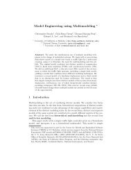
The Endo[PA]R System for <strong>Minimally</strong> Invasive<br />
Robotic Surgery<br />
István Nagy, Hermann Mayer, Alois Knoll<br />
Technische Universität München, 85747 Garching, Germany,<br />
knoll@in.tum.de,<br />
mayerh@in.tum.de,<br />
nagy@in.tum.de,<br />
WWW home page: http://www6.in.tum.de<br />
Abstract. During the last decade minimally <strong>invasive</strong> <strong>surgery</strong> has become<br />
the leading method for many surgical interventions. Unlike open<br />
<strong>surgery</strong>, minimally <strong>invasive</strong> <strong>surgery</strong> only needs small incisions in the patient’s<br />
body. This leads to a drastic reduction of tissue trauma <strong>and</strong> therefore<br />
to shorter recovery times. In the beginning, this technique was performed<br />
manually with specialized instruments. Surgeons had to cope with<br />
restricted manipulability of the end-effector <strong>and</strong> poor visual feedback.<br />
These drawbacks were overcome by employment of dedicated robotic<br />
systems. We present an exhaustive overview on similar systems, both<br />
in research <strong>and</strong> for commercial use. Despite the advantages the systems<br />
offer, there are also needs of surgeons that have not been met. The most<br />
crucial issue is the lack of sensitive force feedback. This often leads to<br />
unpleasant side effects like damaging thread material or even lacerating<br />
healthy tissue. It is in particular this shortcoming that results in fatigue<br />
of the operator, due to visual compensation of the missing haptic feedback.<br />
Incorporation of force feedback in systems for robotic <strong>surgery</strong> is<br />
therefore a crucial factor in improving reaction to tissue contact. Our aim<br />
is to provide the surgeon with an operation environment very similar to<br />
manual instrumental <strong>surgery</strong> (i.e. the surgeon can always feel forces exerted<br />
on the instruments). Therefore we have developed the Endo[PA]R<br />
system, which we describe below in detail. Several experiments demonstrated<br />
the usefulness of this setup as an evaluation platform.<br />
1 Introduction<br />
Advanced <strong>surgery</strong> techniques, mainly developed in the last decades, dramatically<br />
increased life expectancy <strong>and</strong> the quality of life after surgical procedures. Today<br />
no one worry about diagnoses like appendicitis or hernia, since their (often<br />
ambulatory) treatment became routine in modern medical centers.<br />
But the beginnings were often accompanied by excessive loss of blood <strong>and</strong><br />
infections, due to the large incisions made. Many patients deceased as a consequence<br />
of the surgical procedure <strong>and</strong> not the disease itself. A huge amelioration,<br />
apart from technical improvements in the operating room, was brought by the<br />
introduction of the minimally <strong>invasive</strong> <strong>surgery</strong> (MIS) in the 1980s. In contrast to
2<br />
conventional open <strong>surgery</strong> the operation area is accessed through small incisions:<br />
usually at least two for the instruments, one for the endoscope <strong>and</strong> sometimes<br />
one for CO2 insuflating. Figure 1a shows a schematic overview of a minimally<br />
<strong>invasive</strong> procedure, figure 1b is a snapshot shortly before a real MI procedure.<br />
Note the difference to a conventional open procedure as shown in figure 1c.<br />
There are obvious advantages compared to open <strong>surgery</strong>: reduced trauma<br />
<strong>and</strong> pain due to the smaller incisions, shorter rehabilitation time (which results<br />
in shorter hospital stays), <strong>and</strong> last but not least cosmetical considerations. But<br />
despite the advantages this new technique did not produce the response from<br />
the public as hoped. The reason therefor is that advantages at the patient’s<br />
side are almost countervailed against disadvantages at the surgeon’s side. The<br />
surgeon has to deal with orientation problems due to reduced sight, finding<br />
anatomical structures often becomes a challenge. The instruments have to be<br />
h<strong>and</strong>led around so called trocar points on the patient’s abdomen, restricting the<br />
degrees of freedom inside the body to four <strong>and</strong> resulting in a reverse h<strong>and</strong> motion.<br />
Furthermore the surgeon’s h<strong>and</strong> tremor gets amplified by the long instruments,<br />
<strong>and</strong> there is no haptic feedback, compensating it’s absence visually has been<br />
found quite fatiguing.<br />
(a) Schematic MIS (b) <strong>Minimally</strong> Invasive (c) Open Surgery<br />
Fig. 1. Open vs. <strong>Minimally</strong> Invasive Surgery<br />
To circumvent some of the problems of conventional minimally <strong>invasive</strong> <strong>surgery</strong><br />
robotic technologies were integrated. There are three different main areas in<br />
which robotic <strong>surgery</strong> systems became commercially available <strong>and</strong> entered medical<br />
centers for daily use. All have to cope with different requirements, so that<br />
a ”general purpose” <strong>surgery</strong> robot doesn’t make sense <strong>and</strong> consequently doesn’t<br />
exist.<br />
1. Bone <strong>surgery</strong> The most typical actions in this area are high precision<br />
drilling <strong>and</strong> milling, which leads to high forces <strong>and</strong> vibrations. Therefore<br />
adequately modified industrial robots are often used.<br />
2. Neuro<strong>surgery</strong> In this field the most important requierement is precision<br />
in a very limited workspace, but there are no mentionable forces to apply.<br />
The biggest challange is navigation, which can be planned only preoperative<br />
using medical imaging techniques.
3. Abdominal/Thorax <strong>surgery</strong> We have the biggest workspace in this area<br />
leading to special requierements to the robot arms <strong>and</strong> endeffectors. Dealing<br />
with highly deformable organs in an online teleoperative manner (in the<br />
other two cases above mainly preoperatively planned actions are executed)<br />
asks for high fidelity force feedback.<br />
During the 1990s several robotic systems for <strong>surgery</strong> leaved research institutes<br />
<strong>and</strong> entered dedicated medical centers for evaluation purposes or even daily practice.<br />
The first application area is represented by the systems Caspar TM from<br />
Universal Robotic <strong>Systems</strong> Ortho GmbH [5] <strong>and</strong> Robodoc TM from Integrated<br />
Surgical <strong>Systems</strong> [4] (Figure 2). Integrated Surgical <strong>Systems</strong> provides also the<br />
system NeuroMate TM (Figure 3a) which together with PathFinder from Armstrong<br />
Healthcare Ltd. [6] (Figure 3b) represent robotic neuro<strong>surgery</strong>.<br />
The two most technically mature systems are daV inci TM (Figure 4) from<br />
Intuitive Surgical Inc. [2] <strong>and</strong> Zeus TM (Figure 5) from Computer Motion Inc.<br />
[3] which we want to describe in more detail. Both are general purpose teleoperation<br />
systems for abdomen <strong>and</strong> thorax <strong>surgery</strong>, but mainly evaluated in the<br />
field of heart <strong>surgery</strong>. There is on both systems only position control possible,<br />
<strong>and</strong> therefore no autonomy can be achieved. None of them provides instrumental<br />
side force/torque sensory, nor (the possibility of) haptic feedback at the master<br />
console. Motion scaling, tremor filtering, optical magnification <strong>and</strong> stereo vision<br />
is available with both systems. The instruments differ in the number of degrees<br />
of freedom, the Intuitive system has 6, while the Zeus setup has only 5.<br />
(a) Robodoc (b) Caspar<br />
Fig. 2. Bone <strong>surgery</strong> robots<br />
The advantages of robotic <strong>surgery</strong> are obvious: very high precision, the possibility<br />
of integration of preoperative planning data using medical imaging techniques.<br />
Unfortunately there are also a few disadvantages: high costs resulting<br />
from hardware costs on the one h<strong>and</strong>, <strong>and</strong> from increased personnel training time<br />
on the other h<strong>and</strong>. Nonetheless we think that robot assisted <strong>surgery</strong> will rev-<br />
3
4<br />
(a) PathFinder (b) NeuroMate<br />
Fig. 3. Neuro<strong>surgery</strong> robots<br />
(a) daVinci Console (b) daVinci Arms<br />
Fig. 4. The daVinci system from Intuitive Surgical<br />
(a) Zeus Console (b) Zeus Arms<br />
Fig. 5. The Zeus system from Computer Motion
(a) daVinci Master (b) Zeus Master<br />
Fig. 6. Masters of the two systems daVinci <strong>and</strong> Zeus<br />
olutionise todays operations comparably to imaging techniques like Computed<br />
Tomography, Magnetic Resonance Imaging <strong>and</strong> Positron Emission Tomography.<br />
2 <strong>Minimally</strong> Invasive Robotic Surgery<br />
In this section we give a brief technological overview of robotic <strong>surgery</strong> systems<br />
as they should be from both the system architecture <strong>and</strong> configuration point<br />
of view. We confine ourself to real telepresence (online human-machine interaction)<br />
systems like the daV inci TM <strong>and</strong> Zeus TM setups. We identify three main<br />
components:<br />
1. Master This subsystem (also called user interface, see figures 4a <strong>and</strong> 5a) is<br />
the surgeon’s workplace <strong>and</strong> has to feedback modalities of the visual, kinesthetic<br />
<strong>and</strong> tactile senses generated using appropriate actuator hardware (see<br />
the point below). The surgeon’s actions at the input devices (misleadingly<br />
also called masters) are immediately transformed to the adequate actuator<br />
movements, the possibility of scaling <strong>and</strong> tremor suppression increase usability<br />
<strong>and</strong> safety. A high quality stereo vision system is indispensable, the<br />
lack of depth information has been found very hard to compensate. Unfortunately<br />
there is no commercially available system, which provides kinesthetic<br />
or tactile feedback. Many research projects, also including ours, deal with<br />
this very important issue.<br />
2. Slave Located at the patient’s side this subsystem (also called actuator,<br />
see figures 4b <strong>and</strong> 5b) consists of two main components: the robot arms <strong>and</strong><br />
the minimally <strong>invasive</strong> surgical instruments (see upper part of figures 6a <strong>and</strong><br />
6b). The distinction between arms <strong>and</strong> instruments is due to the fact, that<br />
the possibility of changing the instruments during a surgical procedure is<br />
5
6<br />
one of the most important requirements. Actuated arms <strong>and</strong> instruments<br />
are expected to give full manipulability inside the body, providing the same<br />
degrees of freedom as the human h<strong>and</strong>. The robot arm kinematics must<br />
be able to h<strong>and</strong>le the trocar point limitations without affecting the overall<br />
functionality.<br />
3. Communication channel Several high b<strong>and</strong>width connections are necessary<br />
to h<strong>and</strong>le the data transfer between master <strong>and</strong> slave. Requirements<br />
like guaranteed b<strong>and</strong>with, no (or very low) delays have to be fulfilled, otherwise<br />
severe safety problems can occur. The communication subsystem has to<br />
be flexible enough allowing the connection of multiple masters to the same<br />
slave, or even dynamic on the fly master-slave mapping.<br />
Fulfilling the requirements of each subsystem described above will lead to<br />
faster <strong>and</strong> safer robotic <strong>surgery</strong>. Faster <strong>surgery</strong> brings considerable cost reduction<br />
one the one h<strong>and</strong>, <strong>and</strong> less postoperative complications for the patient on the<br />
other h<strong>and</strong>. In addition there are a few advanced techniques evaluated at research<br />
institutes which potentially could enter clinical practice in the near future:<br />
– Automatic Camera Guidance Currently available robotic <strong>surgery</strong> systems<br />
leave the camera control to the surgeon. Whenever the camera has to be<br />
repositioned the surgeon switches control from the input devices driving the<br />
instruments to the camera control mechanism, which is both time consuming<br />
<strong>and</strong> potentially dangerous. Knowing the exact positions of the instruments,<br />
consequently also the working area, a robotic system could provide optimal<br />
camera positioning to overview that area.<br />
– Partial Autonomy Asisstance is in traditional surgical procedures a st<strong>and</strong>ard<br />
practice. Possible (partially) autonomous tasks in a minimally <strong>invasive</strong><br />
robotic scenario are for example: temporarily holding the needle or the suturing<br />
material, grasping of tissue for stretching purposes, automatic suturing<br />
<strong>and</strong> cutting.<br />
– Organ Motion Compensation Mainly in the area of thoracoscopic <strong>surgery</strong><br />
a noticeable amount of motion is due to the patient’s heart beat <strong>and</strong> respiration.<br />
The motion of the lung is rather slow (low frequent) <strong>and</strong> quite easy<br />
to track <strong>and</strong> eventually compensate. Quite to the contrary tracking <strong>and</strong><br />
compensating heart movements is a challenging task, but it is absolutely<br />
necessary for <strong>surgery</strong> on the beating heart.<br />
The last two items are sophisticated <strong>and</strong> therefore currently only at research<br />
institutes in evaluational use. Several groups at both research institutes <strong>and</strong><br />
companies are working on minimally <strong>invasive</strong> robotic <strong>surgery</strong> systems. The next<br />
section gives an overview of such systems, needless to say, this list is far from<br />
being complete.
2.1 Experimental Research Setups<br />
Research in this area concentrates mainly to the developement of micro-instruments<br />
(often equipped with force/torque sensory), robotic arms fulfilling special requirements<br />
<strong>and</strong> force/torque reflective input devices.<br />
(a) Overview (b) Arms (c) Instruments<br />
Fig. 7. The ”Robotic Telesurgical Workstation for Laparoscopy” at Berkley<br />
The Berkley system In a joint project between the <strong>Robotics</strong> <strong>and</strong> Intelligent<br />
Machines Laboratory of the University of California, Berkeley (UCB) <strong>and</strong> the<br />
Department of Surgery of the University of California San Francisco (UCSF), a<br />
robotic telesurgical workstation (see fig. 7a) for laparoscopy was developed. The<br />
current design is a bimanual system with two 6 DOF manipulators instrumented<br />
with grippers, controlled by a pair of 6 DOF master manipulators. The slave is<br />
based on a modified Millirobot, the masters are the well known PHANToM<br />
devices, <strong>and</strong> as a characteristic the arms are driven by hydraulic actuators. The<br />
system provides no force feedback nor stereo vision. The design of the millirobot<br />
is dexterous enough to perform suturing <strong>and</strong> knot-tying tasks. Refer to [9], [10]<br />
<strong>and</strong> [11] for further details.<br />
The KAIST system At the Korea Advanced Institute of Science <strong>and</strong> Technology<br />
(KAIST) a microsurgical telerobot system has been developed. It is composed<br />
of a 6 DOF parallel micromanipulator (based on a Stewart plattform,<br />
see fig. 8c) attached to a macro-motion industrial robot (fig. 8b), <strong>and</strong> a 6 DOF<br />
force/torque-reflective haptic master device (fig. 8a). The master device is using<br />
a five-bar parallel mechanism driven by harmonic DC servomotors. According<br />
to [12] <strong>and</strong> [13] this setup doesn’t seem to have a (stereo) vision system, but<br />
the haptical feedback works quite well. The communication between master <strong>and</strong><br />
slave is via Ethernet.<br />
The ARTEMIS system Developed at the Forschungszentrum Karlsruhe (FZK)<br />
the ARTEMIS system (Advanced Robotic <strong>and</strong> Telemanipulator System for Minimal<br />
Invasive Surgery) was the first German setup for robotic <strong>surgery</strong> <strong>and</strong> one of<br />
7
8<br />
(a) Master (b) Industrial Robot (c) Instrument<br />
Fig. 8. The ”Telerobotic System for Micro<strong>surgery</strong>” at KAIST<br />
the first worldwide. Even though not further developed it is a technically quite<br />
mature system. The ARTEMIS system consists of the following components:<br />
Man Machine Interface, Work System <strong>and</strong> Control System. The Man Machine<br />
Interface (fig. 9a) is composed of several devices: two haptic manipulators, graphical<br />
user interface, 3D video imaging of the operating environment, speech input<br />
(for controlling the laparoscope), foot pedals <strong>and</strong> a trackball. The Work System<br />
(fig. 9b) has two different telemanipulation units: a TISKA based computer controlled<br />
carrier system with surgical effectors <strong>and</strong> a ROBOX computer controlled<br />
endoscope guidance system. Knowing the relative position between the TISKA<br />
<strong>and</strong> ROBOX robots allows automatic camera guidance. The Control System<br />
provides the cooperation between the other two components of ARTEMIS, the<br />
user interface <strong>and</strong> the work system. Each master on the user interface side can<br />
be connected with each slave on the work system side. The kinematics of master<br />
<strong>and</strong> slave do not need to be identical (universal master principal). Different<br />
control modes (eg. world coordinates, screen coordinates) as well as different<br />
functions (eg. scaling, indexing) can be selected. The communication is via LAN<br />
ethernet, <strong>and</strong> it can even be over larger distances by means of ATM connection.<br />
The MONSUN concept is implemented (Manipulator Control System Utilizing<br />
Network Technology). Besides the communication, the control system incorporates<br />
track control <strong>and</strong> the safety system. The KISMET 3D-simulation software<br />
is also part of the system, the only drawback is the lack of force feedback.<br />
The DLR system At the Deutsches Zentrum für Luft- und Raumfahrt (DLR,<br />
Oberpfaffenhofen) a tele<strong>surgery</strong> scenario has been developed based on modified<br />
AESOP 3000 arms (fig. 10a) from Computer Motion, PHANToM input<br />
devices from Sensable Technologies <strong>and</strong> a sensorized scalpell (4 DOF, 3 forces +
(a) Master (b) Slave<br />
Fig. 9. The ARTEMIS system at the FZK<br />
1 torque) developed at the DLR. A prototype of a sensorized 6 DOF forceps is<br />
also available, but not yet integrated (fig. 10b). Stereo vision <strong>and</strong> vision based<br />
automatic camera guidance are also available. Cartesian control of the (initially<br />
only position controlled) arms allows the validation of more advanced techniques<br />
like motion estimation <strong>and</strong> compensation in beating heart <strong>surgery</strong> <strong>and</strong> special<br />
control laws (velocity <strong>and</strong> position/force). The communication between master<br />
<strong>and</strong> slave is CORBA-TCP/IP based.<br />
(a) AESOP 3000 Arms (b) Forceps <strong>and</strong> Scalpell<br />
Fig. 10. The system at DLR<br />
9
10<br />
The experimental system of the University of Tokio At the University<br />
of Tokyo, Department of Engineering Synthesis Faculty of Engineering a<br />
tele-endoscopic surgical system with force-feedback capability was developed.<br />
According to [19] the system consists of a multi-media cockpit, surgical site <strong>and</strong><br />
a communication link. The multi-media cockpit (fig. 11a) is equipped with force<br />
feedback type master manipulators, visual <strong>and</strong> auditory information presentation<br />
apparatures <strong>and</strong> foot switches. A slave manipulator (fig. 11b) with three<br />
arms is located at the surgical site. Two arms hold forceps or a radio knife <strong>and</strong><br />
one arm holds an endoscope. Force sensing capability is equipped on the active<br />
forceps to implement force feedback. The (SCARA type) slave manipulator is<br />
designed to maintain the insert position at a fixed point for safety. The system<br />
was evaluated in an experiment, where the gallbladder of a pig was successfuly<br />
removed.<br />
(a) (b)<br />
Fig. 11. The experimental system of the University of Tokio<br />
The ”Hyper Finger” system A new robotic system named ”Hyper Finger”<br />
for minimally <strong>invasive</strong> <strong>surgery</strong> in deep organs has been developed at the Nagoya<br />
University, Department of Micro System Engineering. This is one of the smallest<br />
master-slave robots in medicine, each finger has nine degrees of freedom <strong>and</strong> is<br />
driven by wires. A prototypical detachable gripper mechanism was also developed.<br />
Note that the master (fig. 12a) is not exoskeletal but hold by the surgeon<br />
like a real instrument. This construction dosn’t require special robotic arms, the<br />
slave can be simply mounted on a camera tripod (fig. 12b). The system provides<br />
no force feedback nor stereo vision, but according to [21] the effectiveness of the<br />
system was verified by in-vivo experiments. The main field of application seems<br />
to be surgical procedures in hardly accessible narrow areas.
(a) Master (b) Slave<br />
Fig. 12. The ”Hyper Finger” system at the Nagoya University<br />
The Remote Micro<strong>surgery</strong> System A proposal of a new method of micro<strong>surgery</strong><br />
were made at the Nagoya University, Department of Micro System<br />
Engineering. The target of the work is micro<strong>surgery</strong> in deep, narrow sites of the<br />
human body, which are currently the most difficult areas to perform minimally<br />
<strong>invasive</strong> <strong>surgery</strong>. The proposal contains both a new method of micro<strong>surgery</strong> <strong>and</strong><br />
surgical tools. H<strong>and</strong>ling the master (fig. 13a) is similar to a classical endoscopic<br />
instrument, the implementation of force feedback could be quite complicated, if<br />
intended. The slave (fig. 13b top) doesn’t require a robotic arm, it is designed<br />
to be mounted on any stable plattform in the near of the patient. Then the<br />
catheter like guide tube (fig. 13b bottom) can be inserted to the desired operation<br />
area. Typical fields of application are neoro<strong>surgery</strong>, head <strong>and</strong> neck <strong>surgery</strong><br />
in otolaryngology <strong>and</strong> micro<strong>surgery</strong> on esophageal diseases. According to [20]<br />
the system was successfully tested on animals.<br />
2.2 The Experimantal Tele<strong>surgery</strong> System Endo[PA]R<br />
Developed at the Technische Universität München, Chair for <strong>Robotics</strong> <strong>and</strong> <strong>Embedded</strong><br />
<strong>Systems</strong> [7], the Endo[PA]R (Endoscopic Partially Autonomous Robot)<br />
system is an experimental setup which claims applicability in at least animal<br />
experiments. A more detailed description is presented in the next sections.<br />
3 Methodology<br />
Similar to other systems, our setup comprises an operator-side master console for<br />
in-output <strong>and</strong> a patient-side robotic manipulator that directly interacts with the<br />
operating environment. As shown in Fig. 14, our system has two manipulators,<br />
which are controlled by two input devices. Each of the two arms of our surgical<br />
robot is composed of the following subsystems. A low-payload robot, which bears<br />
a surgical instrument that is deployed with the surgical workstation daVinci<br />
11
12<br />
(a) Master (b) Slave<br />
Fig. 13. The Remote Micro<strong>surgery</strong> System<br />
(TM). We have developed a special adapter that interconnects the robot’s flange<br />
with the instrument. The surgical instruments have three degrees of freedom. A<br />
micro-gripper at the distal end of the shaft can be rotated <strong>and</strong> adaptation of<br />
pitch <strong>and</strong> yaw angles is possible. Since the yaw angle of each of the two fingers<br />
of the gripper can be controlled separately, it is possible to open <strong>and</strong> close the<br />
gripper. All movable parts of the gripper are driven by steel wires. Their motion<br />
is controlled by four driving wheels at the proximal end of the instrument, one<br />
for each degree of freedom (two for yaw of the fingers). In order to control<br />
the instrument, we have flanged servos to each driving wheel by means of an<br />
Oldham coupling. This guarantees instrument movement free of jerk. The servo<br />
controllers are connected via serial lines to a multi-port interface card. Since the<br />
rotation of the robot’s flange <strong>and</strong> the rotation of the instrument share one axis,<br />
the combination of robot <strong>and</strong> instrument results in a manipulator with eight<br />
degrees of freedom. That means our system is a redundant manipulator. This<br />
can be exploited to evaluate different kinematical behaviors. The most important<br />
one is trocar kinematics. This allows 6 dof control of the end effector, while the<br />
shaft of the instrument has to be moved about a fixed fulcrum (keyhole <strong>surgery</strong>).<br />
Position <strong>and</strong> orientation of the manipulators are controlled by two PHANToM<br />
devices (Fig. 14). This device is available in different versions with different<br />
capabilities. Our version provides a full 6 dof input, while force feedback is<br />
restricted to three translational directions. The user controls a stylus pen that is<br />
equipped with a switch that can be used to open <strong>and</strong> close the micro-grippers.<br />
A third robot is carrying an endoscopic stereo camera system. The steroscopic<br />
view is presented via a head mounted display.
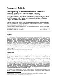
3.1 Force Feedback<br />
Fig. 14. System Setup<br />
The most interesting feature of the PHANToM devices we used, is their capability<br />
of providing the user with haptic feedback. Forces are feeded back by<br />
small servo motors incorporated in the device. They are used to steer the stylus<br />
pen in a certain direction. This creates the impression of occurring forces,<br />
while the user is holding the pen at a certain posture. The force sensors were<br />
applied directly on the shaft of the instrument. Since the shaft of the surgical<br />
instrument is made of carbon fibre, force sensors have to be very sensitive <strong>and</strong><br />
reliable. Therefore we decided to apply strain gauge sensors, which are employed<br />
for industrial force registration. As shown in Fig. 15, the sensor gauges are applied<br />
at the distal end of the instrument’s shaft, i.e. near the gripper. At the top<br />
of Fig. 15, one can see the perpendicular arrangement of strain gauges as full<br />
bridges. One full bridge of sensors is used for each direction. The signals from<br />
the sensors are amplified <strong>and</strong> transmitted via CAN-bus to a PC system. Sensor<br />
readings are blurred with noise, hence we have applied digital filters to stabilize<br />
the results. Since we know the position <strong>and</strong> orientation of the instruments, we<br />
can transform occurring forces back to the coordinate system of the PHANToM<br />
devices. Therefore the user has the impression of direct haptic immersion.<br />
13
14<br />
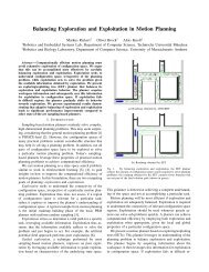
3.2 Trocar Kinematics<br />
Fig. 15. Application of Strain Gauges to an instrument<br />
The basic idea of minimally <strong>invasive</strong> <strong>surgery</strong> is, that only small openings have<br />
to be made into the surface of the patient’s thorax (so-called keyholes, Fig.<br />
16). That means the translational movements of the instruments are essentially<br />
restricted by shifts <strong>and</strong> rotations about these holes. In order to provide the<br />
surgeon with a comfortable environment, it is desirable to map the movements<br />
of the stylus at the input device directly to instrument motions. Therefore we<br />
have to consider the inverse kinematics of our system. That means we have to<br />
find a mapping of an arbitrary posture of the instrument’s tip to a position of<br />
the motors that control the eight degrees of freedom.<br />
The desired position of the instrument is given by the position of the input<br />
stylus. It is represented by a homogenous transform matrix. Since the position of<br />
the instrument’s shaft is restricted by the port (the position of the keyhole), there<br />
is only one possibility for aligning the instrument. The angle of the corresponding<br />
joints of the instrument can be found by geometric considerations. For result, we<br />
get the position of the instrument’s shaft. As this axis is identical to the flange<br />
axis of the robot, we have got the position of the flange. Given this information,<br />
we now can determine the backwards kinematics of the robot. This is a st<strong>and</strong>ard<br />
procedure, whose detailed calculation will be neglected here. As a final result we<br />
can implement a mapping from the position of the input stylus to the position of<br />
the instrument. That means the surgeon is provided with a direct remote control<br />
of the surgical instruments.
3.3 System Features<br />
Fig. 16. Trocar Point Kinematics<br />
We give only a short list of system features we think to be important. The use of<br />
commercially available subsystems (robots, instruments, amplifiers) guarantees<br />
reliability <strong>and</strong> simplifies mass-production at comparable low prices. Particular<br />
advantages of this setup with multi-purpose robots are high precision <strong>and</strong> stiffness,<br />
moderate costs <strong>and</strong> an advanced dynamic behavior. The latter could be<br />
exploited to perform advanced tasks in motion compensation (e.g. support for<br />
beating heart <strong>surgery</strong> as it was proposed in [31], or compensation for respiratory<br />
motion of the ribs). The modular character of this setup simplifies the adaptation<br />
of the system to technical improvements (e.g. new surgical instruments). Another<br />
advantage is the fact that our manipulator is a robot under Cartesian control<br />
whose position can be controlled precisely. Finally, the most important feature<br />
is the possibility for evaluation of force feedback in combination with endoscopic<br />
vision in robotic <strong>surgery</strong>. In order to make navigation easier, we additionally<br />
equipped the system with an endoscopic stereo camera system to observe the<br />
operation environment.<br />
4 Experimental Results<br />
With the help of this setup we have performed different tasks known from surgical<br />
practice <strong>and</strong> evaluated the impact of force measurement. Our hope is, that haptic<br />
feedback contributes to a better performance of systems for robotic <strong>surgery</strong> by<br />
preventing force-induced damages. Examples for such harms are breaking of<br />
thread material, ripping tissue <strong>and</strong> strangulate sutures.<br />
15
16<br />
Force[N]<br />
2<br />
1.5<br />
1<br />
0.5<br />
0<br />
-0.5<br />
-1<br />
-1.5<br />
-2<br />
Force Progression<br />
-2.5<br />
-3<br />
Left Fx<br />
Left Fy<br />
Right Fx<br />
-3.5<br />
Right Fy<br />
5000 10000 15000 20000 25000 30000 35000 40000<br />
Time[ms]<br />
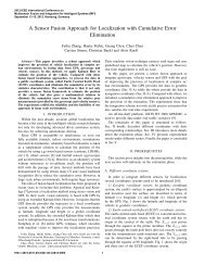
4.1 Winding<br />
Fig. 17. Winding a thread to make loops<br />
Force[N]<br />
2.5<br />
2<br />
1.5<br />
1<br />
0.5<br />
0<br />
-0.5<br />
-1<br />
Force Progression<br />
Left Fx<br />
Left Fy<br />
Right Fx<br />
Right Fy<br />
-1.5<br />
21800 21900 22000 22100<br />
Time[ms]<br />
22200 22300 22400<br />
The first operation sequence we evaluated was winding thread during knot tying.<br />
Forces are acquired only in the XY –Plane perpendicular to the instrument<br />
shaft, as our current setup does not yet allow the measurement of forces along<br />
the shaft. Winding thread to form loops is a subtask in instrumental knot tying<br />
(cf. [32]), <strong>and</strong> if executed by a surgeon only very low forces arise, since a<br />
human operator easily copes with this task using only visual feedback. However<br />
in robot assisted <strong>surgery</strong> scenarios high fidelity force sensory is indispensable,<br />
as the visual modality is very difficult to interpret. Accordingly, robotic winding<br />
can be accomplished only in a force-controlled manner. On the one h<strong>and</strong><br />
forces are preferably to be kept constant, on the other h<strong>and</strong> suture break must<br />
be avoided. Fig. 17 (left) shows the force progression during a winding process.<br />
The frequency of force peeks in a certain direction grows, as the suture material<br />
gets shorter. Nevertheless the forces are quite constant during the whole manipulation.<br />
Figure 17 (right) shows a magnified view of an accidental break of the<br />
thread during a further winding process. Due to the high time resolution (1 ms)<br />
the instant recognition of such suture breaks is possible, preventing the robotic<br />
system from unexpected behavior.<br />
4.2 Preventing Suture Material Damage<br />
The tensile strength of absorbable <strong>and</strong> non-absorbable sutures is critical both<br />
during <strong>and</strong> after surgical procedures. Breaking strength can be measured using<br />
either a ”straight pull” test or a ”knot pull” test. Having the breaking strengths<br />
of all used sutures enables us to prevent suture material damage by limiting the<br />
applicable forces to adequate maximal values. Fig. 18 (left) shows the progression<br />
of forces while trying to break original surgical suture material, in this case<br />
Ethicon PROLENE (7/0, Polypropylen, not absorbable). Fig. 18 (right) shows<br />
breaking the thread (PROLENE 7/0) while tying a knot. As expected, the thread<br />
was broken at the knot position by significantly less force impact.
Force[N]<br />
1<br />
0.5<br />
0<br />
-0.5<br />
-1<br />
-1.5<br />
-2<br />
Force Progression<br />
-2.5 Left Fx<br />
Left Fy<br />
Right Fx<br />
-3<br />
Right Fy<br />
2500 3000 3500 4000 4500<br />
Time[ms]<br />
5000 5500 6000 6500<br />
Force[N]<br />
0.5<br />
0<br />
-0.5<br />
-1<br />
-1.5<br />
Force Progression<br />
17<br />
-2<br />
Left Fx<br />
Left Fy<br />
Right Fx<br />
Right Fy<br />
-2.5<br />
4000 4500 5000 5500 6000 6500 7000 7500<br />
Time[ms]<br />
Fig. 18. Breaking Ethicon 7/0 by normal pulling (left) <strong>and</strong> knot tying (right)<br />
4.3 Collision Detection<br />
Avoiding the collision of the instruments in robot assisted minimally <strong>invasive</strong><br />
<strong>surgery</strong> is not an easy task. Therefore a symbolic representation of the whole<br />
robotic system, including both the instruments <strong>and</strong> the arms, were necessary.<br />
Furthermore exact position control <strong>and</strong> a collision detection software subsystem<br />
are indispensable. Most setups however do not provide the above mentioned infrastructure.<br />
A human operator will easily avoid instrument collisions, but in an<br />
autonomous mode other solutions are necessary. A force controlled setup will not<br />
prevent collisions, but an early detection can avoid from damaging the instruments.<br />
Figure 19 shows the forces recorded while an instrument collision, the<br />
instrument velocities were within ranges typical to this scenario. We observe,<br />
that the highest peak (Y -force component of the left instrument) arises in approximately<br />
35ms. With a robot arm interpolation of 12ms there are nearly 3<br />
interpolation periods to react when such a situation appears, providing a satisfactory<br />
collision interception.<br />
5 Simulation<br />
In order to check certain operation sequences (e.g. the complicated procedure<br />
of knot-tying) before applying them to the real world, we have developed a<br />
realistic simulation of our system. Since the model has the same geometry as<br />
the real system, all joint angles obtained from the inverse kinematics can be<br />
directly applied to it. The model is displayed in an Open Inventor-GUI. Input<br />
data can be recorded to a data base for subsequent use with the simulation or<br />
the real system. This simulation was especially useful to detect some unusual<br />
motion sequences that could lead to failures of the real system. For example, the<br />
robot tends to move too fast if the instrument tips approach come too close to<br />
the port. The simulation can also be used in parallel with real manipulations.<br />
This can be very helpful if the remote user has no full sight of the operation<br />
environment (e.g. if instruments are occluded by other objects).
18<br />
Force[N]<br />
4<br />
3<br />
2<br />
1<br />
0<br />
-1<br />
Force Progression<br />
-2 Left Fx<br />
Left Fy<br />
Right Fx<br />
-3<br />
Right Fy<br />
2800 2900 3000 3100<br />
Time[s]<br />
3200 3300 3400<br />
Fig. 19. Colliding instruments<br />
Fig. 20 shows the simulation environment including a CT-scan of the thorax<br />
<strong>and</strong> heart phantom. A detailed closeup view of the operation situs is depicted<br />
in the lower right corner of the simulation window. The exact model allows for<br />
an appropriate instantiation of previously acquired tasks, since transformation<br />
parameters (translation, rotation, scaling) can be extracted from simulation.<br />
A possible scenario is automatically completing a knot: as an occurrence of<br />
an already recorded manipulation sequence is recognized, a context-sensitive<br />
instance of that sequence is replayed. Before the task is actually completed by<br />
the robotic system, a virtual execution is displayed to the surgeon, who can<br />
choose between either discarding or performing the task.<br />
6 Partial Autonomy<br />
We have performed several knot-tying tasks with our system <strong>and</strong> recorded both,<br />
force progression <strong>and</strong> the corresponding trajectories (described by position <strong>and</strong><br />
orientation of the instruments). Due to inevitable physiological tremor of the human<br />
operator, the acquired trajectories exhibit some noise. Therefore two-stage<br />
preprocessing was applied to the raw data. The first stage comprises sliding window<br />
averaging, the second stage approximates the smoothed data with natural<br />
cubic splines.<br />
Our first experiment was replay of an original sample with no smoothing <strong>and</strong><br />
approximation applied. Since our system features a high repeat accuracy, this<br />
procedure was performed very reliable. The only prerequisite is positioning the<br />
needle at a known place. Since we leave the needle placement to the surgeon <strong>and</strong><br />
we know the geometry of our system, we can always exactly locate the corresponding<br />
position. Due to exact kinematics, execution of up to double speed has<br />
raised no difficulties. As our objective is not restricted to acceleration, we also<br />
want to generate optimized trajectories with respect to smoothness <strong>and</strong> path
Fig. 20. Screenshot of the Simulation Environment<br />
planning. Therefore we have applied spline approximation to the raw data (see<br />
fig. 21 right) . This results in a symbolic representation of the trajectory in the<br />
form of a parametric space-curve. Before applying the generated curve to the<br />
real system, collision avoidance has to be guaranteed, since overmodified paths<br />
can contingently result in instrument collision.<br />
7 Conclusions <strong>and</strong> Outlook<br />
We have presented a novel approach of a robotic system for minimally <strong>invasive</strong><br />
<strong>surgery</strong>. It is mainly composed of commercially available subsystems. This has<br />
several advantages like precision, reliability <strong>and</strong> a good dynamic behavior. The<br />
main purposes of the system are evaluation of force feedback <strong>and</strong> machine learning.<br />
We found out that performance of certain surgical tasks like knot tying will<br />
profit from this feature. Experiments have shown that haptic feedback can be<br />
employed to prevent the surgeon from potentially harmful mistakes. Tension of<br />
thread material <strong>and</strong> tissue parts can be measured <strong>and</strong> displayed in order to restrict<br />
force application to a tolerable amplitude. Collision of instruments can be<br />
detected <strong>and</strong> intercepted by real-time force evaluation. Forces are measured at<br />
the surgical instruments <strong>and</strong> feeded back into the surgeon’s h<strong>and</strong>s using multidimensional<br />
haptic styluses. For future evaluation we are planning long-term<br />
19
20<br />
Fig. 21. Raw <strong>and</strong> Spline-Approximated Trajectory (Knot-Tying)<br />
tests to find out if force feedback can prevent surgeon’s fatigue. The current<br />
arrangement of input devices, however, is not very comfortable. Therefore we<br />
are planning to test different rearrangements of this setup <strong>and</strong> to develop own<br />
input instruments to replace the stylus pens. Additionally we are planning to<br />
include measurement of torques <strong>and</strong> their incorporation in the control loop of<br />
the system. Currently we are also working on a simulation environment that can<br />
be used to model haptic interaction with a tissue model. This can be applied for<br />
off-line evaluation of critical tasks.<br />
Integration of force feedback with stereo vision, as offered by the system, can<br />
improve accuracy, drastically reduce the time needed for operations <strong>and</strong> tissue<br />
trauma, along with a reduction of stress on the surgeon. This could lead to a<br />
wider acceptance of robotic <strong>surgery</strong> by both, patients <strong>and</strong> surgeons. The system’s<br />
software interface <strong>and</strong> mechanical set-up descriptions are freely available<br />
to enable other research groups to participate in the development.<br />
References<br />
1. http://www.kuka.de<br />
2. http://www.intuitivesurgical.com<br />
3. http://www.computermotion.com<br />
4. http://www.robodoc.com<br />
5. http://www.urs-ortho.de<br />
6. http://www.armstrong-healthcare.com<br />
7. http://www6.in.tum.de<br />
8. http://www.hbm.de<br />
9. Murat Cenk Cavusoglu, Frank Tendick, Michael Cohn <strong>and</strong> S. Shankar Sastry: A<br />
Laparoscopic Telesurgical Workstation. IEEE TRA, Vol. 15, No. 4, August 1999.<br />
10. M. Cenk Cavusoglu, Winthrop Williams, Frank Tendick, S. Shankar Sastry:<br />
<strong>Robotics</strong> for Tele<strong>surgery</strong>: Second Generation Berkley/UCSF Laparoscopic Telesurgical<br />
Workstation <strong>and</strong> Looking towards the Future Applications. Industrial Robot,<br />
Special Issue on Medical <strong>Robotics</strong>, Vol. 30, No. 1, January 2003.
11. Murat Cenk Cavusoglu: Tele<strong>surgery</strong> <strong>and</strong> Surgical Simulation: Design, Modeling,<br />
<strong>and</strong> Evaluation of Haptic Interfaces to Real <strong>and</strong> Virtual Surgical Environments.<br />
PhD Thesis, UC Berkeley, August 2000.<br />
12. Dong-Soo Kwon, Ki Young Woo, Se Kyong Song, Wan Soo Kim, Hyung Suck Cho:<br />
Microsurgical Telerobot System. IEEE/RSJ International Conference on Intelligent<br />
Robots <strong>and</strong> <strong>Systems</strong>, pp. 945-950, 1998.<br />
13. Dong-Soo Kwon, Ki Young Woo, Hyung Suck Cho: Haptic Control of the Master<br />
H<strong>and</strong> Controller for a Microsurgical Telerobot System. IEEE ICRA, Detroit,<br />
Michigan, May 1999.<br />
14. U. Voges, E. Holler, B. Neisius, M. Schurr, T. Vollmer: Evaluation of ARTEMIS,<br />
the Advanced <strong>Robotics</strong> <strong>and</strong> Telemanipulator System for <strong>Minimally</strong> Invasive Surgery.<br />
Proceedings IARP 2nd Workshop on Medical <strong>Robotics</strong>, Forschungszentrum Karlsruhe,<br />
1997, pp. 137-148.<br />
15. Fuji Lai <strong>and</strong> Robert D. Howe: Evaluating Control Modes for Constrained Robotic<br />
Surgery. IEEE ICRA, San Francisco, CA, April 2000.<br />
16. V. F. Muñoz, C. Vara-Thorbeck, J. G. DeGabriel, J. F. Lozano, E. Sanchez-<br />
Badajoz, A. García-Cerezo, R. Toscano <strong>and</strong> A. JImenez-Garrido: A Medical Robotic<br />
Assistant for <strong>Minimally</strong> Invasive Surgery. IEEE ICRA, San Francisco, CA, April<br />
2000.<br />
17. Gary. S. Guthart, J. Kenneth Salisbury: The Intuitive TM Tele<strong>surgery</strong> System:<br />
Overview <strong>and</strong> Application. IEEE ICRA, San Francisco, CA, April 2000.<br />
18. Mamoru Mitsuishi, Shin Tomisaki, Takumi Yoshidome, Hiroyuki Hashizume <strong>and</strong><br />
Kazuo Fujiwara: Tele-micro-<strong>surgery</strong> system with intelligent user interface. IEEE<br />
ICRA, San Francisco, CA, April 2000.<br />
19. Mamoru Mitsuishi, Jumpei Arata, Katsuya Tanaka, Manabu Miyamoto, Takumi<br />
Yoshidome, Satoru Iwata, Shin’ichi Warisawa <strong>and</strong> Makoto Hashizume: Developement<br />
of a Remote <strong>Minimally</strong>-Invasive Surgical System with Operational Environment<br />
Transmission Capability. IEEE ICRA, Taipei, Taiwan, September 2003.<br />
20. Koji Ikuta, Keiichi Yamamoto <strong>and</strong> Keiji Sasaki: Developement of Remote Micro<strong>surgery</strong><br />
Robot <strong>and</strong> New Surgical Procedure for Deep <strong>and</strong> Narrow Space. IEEE ICRA,<br />
Taipei, Taiwan, September 2003.<br />
21. Koji Ikuta, Takahiko Hasegawa <strong>and</strong> Shinichi Daifu: Hyper Redundant Miniature<br />
Manipulator ”Hyper Finger” for Remote <strong>Minimally</strong> Invasive Surgery in Deep Area.<br />
IEEE ICRA, Taipei, Taiwan, September 2003.<br />
22. P. Berkelman, E. Boidard, P. Cinquin, J. Troccaz: LER: The Light Endoscope<br />
Robot. IEEE/RSJ, Las Vegas, Nevada, October 2003.<br />
23. M. Tavakoli, R.V. Patel <strong>and</strong> M. Moallem: A Force Reflective Master-Slave System<br />
for <strong>Minimally</strong> Invasive Surgery. IEEE/RSJ, Las Vegas, Nevada, October 2003.<br />
24. P. Knappe, I. Gross, S. Pieck, J.Wahrburg, S. Kuenzler, F. Kerschbaumer: Position<br />
control of a surgical robot by a navigation system. IEEE/RSJ, Las Vegas, Nevada,<br />
October 2003.<br />
25. M.D. O’Lary, C. Simone, T. Washio, K. Yoshinaka <strong>and</strong> A.M. Okamura: Robotic<br />
Needle Insertion: Effects of Friction <strong>and</strong> Needle Geometry. IEEE ICRA, Taipei,<br />
Taiwan, September 2003.<br />
26. A.M. Okamura, R.J. Webster III, J.T. Nolin, K.W. Johnson <strong>and</strong> H. Jafry: The<br />
Haptic Scissors: Cutting in Virtual Environments. IEEE ICRA, Taipei, Taiwan,<br />
September 2003.<br />
27. Wei Tech Ang, Pradeep K. Khosla <strong>and</strong> Cameron N. Riviere: Design of All-<br />
Accelerometer Inertial Measurement Unit for Tremor Sensing in H<strong>and</strong>-held Microsurgical<br />
Instrument. IEEE ICRA, Taipei, Taiwan, September 2003.<br />
21
22<br />
28. Mark Minor, Ranjan Mukherjee: A Dexterous Manipulator for <strong>Minimally</strong> Invasive<br />
Surgery. IEEE ICRA, Detroit, Michigan, May 1999.<br />
29. Roger Baumann <strong>and</strong> Reymond Clavel: Haptic Interface for Virtual Reality Based<br />
<strong>Minimally</strong> Invasive Surgery Simulation. IEEE ICRA, Leuven, Belgium, May 1998.<br />
30. Mamoru Mitsuishi, Yasuhiro Iizuka, Hiroyoshi Watanabe, Hiroyuki Hashizume <strong>and</strong><br />
Kazuo Fujiwara: Remote operation of a micro-surgical system. IEEE ICRA, Leuven,<br />
Belgium, May 1998.<br />
31. T. Ortmaier, M.Groeger <strong>and</strong> G. Hirzinger: Robust Motion Estimation in Robotic<br />
Surgery on the Beating Heart. Computer Assisted Radiology <strong>and</strong> Surgery (CARS),<br />
2002.<br />
32. C. Cao, C. MacKenzie <strong>and</strong> S. Pay<strong>and</strong>eh: Task <strong>and</strong> motion analyses in endoscopic<br />
<strong>surgery</strong>, Proceedings ASME Dynamic <strong>Systems</strong> <strong>and</strong> Control Division, pp. 583-590,<br />
Atlanta, USA, 1996