MultiScreen MIC 3um.qxd - Millipore
MultiScreen MIC 3um.qxd - Millipore
MultiScreen MIC 3um.qxd - Millipore

Create successful ePaper yourself
Turn your PDF publications into a flip-book with our unique Google optimized e-Paper software.
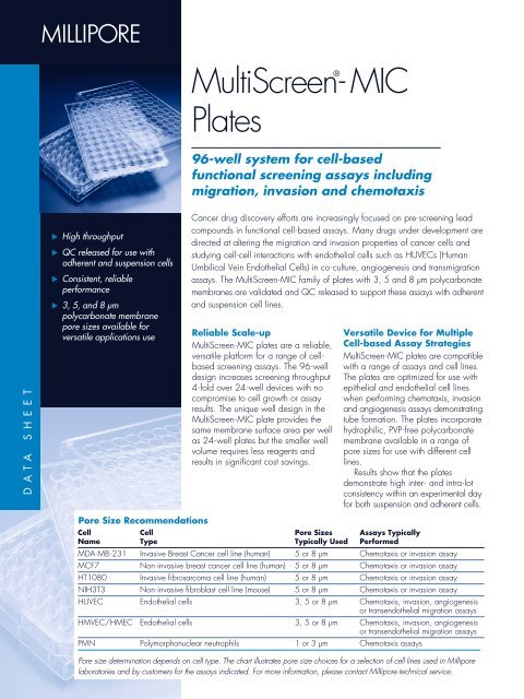
DATA SHEET<br />
Â<br />
� High throughput<br />
� QC released for use with<br />
adherent and suspension cells<br />
� Consistent, reliable<br />
performance<br />
� 3, 5, and 8 µm<br />
polycarbonate membrane<br />
pore sizes available for<br />
versatile applications use<br />
<strong>MultiScreen</strong>-<br />
®<br />
<strong>MIC</strong><br />
Plates<br />
96-well system for cell-based<br />
functional screening assays including<br />
migration, invasion and chemotaxis<br />
Cancer drug discovery efforts are increasingly focused on pre-screening lead<br />
compounds in functional cell-based assays. Many drugs under development are<br />
directed at altering the migration and invasion properties of cancer cells and<br />
studying cell-cell interactions with endothelial cells such as HUVECs (Human<br />
Umbilical Vein Endothelial Cells) in co-culture, angiogenesis and transmigration<br />
assays. The <strong>MultiScreen</strong>-<strong>MIC</strong> family of plates with 3, 5 and 8 µm polycarbonate<br />
membranes are validated and QC released to support these assays with adherent<br />
and suspension cell lines.<br />
Reliable Scale-up<br />
<strong>MultiScreen</strong>-<strong>MIC</strong> plates are a reliable,<br />
versatile platform for a range of cellbased<br />
screening assays. The 96-well<br />
design increases screening throughput<br />
4-fold over 24-well devices with no<br />
compromise to cell growth or assay<br />
results. The unique well design in the<br />
<strong>MultiScreen</strong>-<strong>MIC</strong> plate provides the<br />
same membrane surface area per well<br />
as 24-well plates but the smaller well<br />
volume requires less reagents and<br />
results in significant cost savings.<br />
Versatile Device for Multiple<br />
Cell-based Assay Strategies<br />
<strong>MultiScreen</strong>-<strong>MIC</strong> plates are compatible<br />
with a range of assays and cell lines.<br />
The plates are optimized for use with<br />
epithelial and endothelial cell lines<br />
when performing chemotaxis, invasion<br />
and angiogenesis assays demonstrating<br />
tube formation. The plates incorporate<br />
hydrophilic, PVP-free polycarbonate<br />
membrane available in a range of<br />
pore sizes for use with different cell<br />
lines.<br />
Results show that the plates<br />
demonstrate high inter- and intra-lot<br />
consistency within an experimental day<br />
for both suspension and adherent cells.<br />
Pore Size Recommendations<br />
Cell Cell Pore Sizes Assays Typically<br />
Name Type Typically Used Performed<br />
MDA-MB-231 Invasive Breast Cancer cell line (human) 5 or 8 µm Chemotaxis or invasion assay<br />
MCF7 Non-invasive breast cancer cell line (human) 5 or 8 µm Chemotaxis or invasion assay<br />
HT1080 Invasive fibrosarcoma cell line (human) 5 or 8 µm Chemotaxis or invasion assay<br />
NIH3T3 Non-invasive fibroblast cell line (mouse) 5 or 8 µm Chemotaxis or invasion assay<br />
HUVEC Endothelial cells 3, 5 or 8 µm Chemotaxis, invasion, angiogenesis<br />
or transendothelial migration assays<br />
HMVEC/HMEC Endothelial cells 3, 5 or 8 µm Chemotaxis, invasion, angiogenesis<br />
or transendothelial migration assays<br />
PMN Polymorphonuclear neutrophils 1 or 3 µm Chemotaxis assays<br />
Pore size determination depends on cell type. The chart illustrates pore size choices for a selection of cell lines used in <strong>Millipore</strong><br />
laboratories and by customers for the assays indicated. For more information, please contact <strong>Millipore</strong> technical service.
Performance<br />
Consistent Assay Results<br />
Consistent intra-lot and inter-lot values<br />
with minimal standard deviation was<br />
obtained. For assays with HB-124<br />
cells intra-lot standard deviation was<br />
± 4 – 6 % and for assays with K562<br />
cells intra-lot standard deviation was<br />
± 0.7 – 1% in response to stimulant.<br />
Percent chemotaxis with HB-124 and<br />
K562 cells was ≥ 3x over unstimulated<br />
cells across all lots.<br />
Consistent assay results were also<br />
determined for <strong>MultiScreen</strong>-<strong>MIC</strong> with<br />
5 µm and 8 µm membrane using highly<br />
migratory adherent breast cancer cell<br />
line MDA-MB-231. Intralot standard<br />
deviation was ±1 – 7% for 3 lots tested<br />
(data not shown).<br />
Chemotaxis Profiles of Two Suspension Cell Lines<br />
<strong>MultiScreen</strong>-<strong>MIC</strong> Plates with 3 µm Membrane<br />
% Chemotaxis<br />
% Chemotaxis<br />
HB-124<br />
50<br />
40<br />
30<br />
20<br />
10<br />
0<br />
K562<br />
10<br />
8<br />
6<br />
4<br />
2<br />
0<br />
Stimulated Cells Unstimulated Cells<br />
Lot 1 Lot 2 Lot 3 Overall<br />
Average<br />
Stimulated Cells Unstimulated Cells<br />
Lot 1 Lot 2 Lot 3 Overall<br />
Average<br />
Figures 1 and 2. Chemotaxis in response to 10%<br />
serum-containing medium (stimulated cells) or 0.2 %<br />
BSA-containing medium (unstimulated cells) as a<br />
chemoattractant. Percent chemotaxis (stimulated migration)<br />
is calculated relative to the number of cells seeded.<br />
Plates were seeded with 50,000 cells/well.<br />
Chemotaxis assays were carried out over a period of<br />
4 hours at 37 °C. Migrated cells were evaluated using<br />
Calcein AM fluorescent label. Percent chemotaxis was<br />
calculated using a Calcein AM standard cell reference<br />
curve for each cell line.
Proven Tube Formation for<br />
Angiogenesis Assays<br />
Figures 3a and 3b show tube formation<br />
exhibited by HUVEC (Human<br />
Umbilical Vein Endothelial Cells) on<br />
5 µm <strong>MultiScreen</strong>-<strong>MIC</strong> plates in<br />
response to EGM-2 (Endothelial<br />
Growth Medium) for 24 hours at<br />
37 ° C. Image demonstrates the<br />
ability of <strong>MultiScreen</strong>-<strong>MIC</strong> plates to<br />
support in vitro angiogenesis assays.<br />
Superior Percent Invasion<br />
<strong>MultiScreen</strong>-<strong>MIC</strong> plates perform<br />
consistently across lots. They also<br />
exhibit superior percent invasion<br />
results in invasion assays with<br />
MDA-MB-231 cells in a parallel<br />
comparison to Competitor B and<br />
Competitor C 24-well inserts.<br />
<strong>MultiScreen</strong>-<strong>MIC</strong> Plates with 5 µm Membrane<br />
Angiogenesis<br />
3a.<br />
3b.<br />
25<br />
20<br />
15<br />
10<br />
5<br />
0<br />
<strong>MIC</strong><br />
Lot 1<br />
<strong>MIC</strong><br />
Lot 2<br />
<strong>MIC</strong><br />
Lot 3<br />
<strong>MIC</strong> Interlot<br />
Average<br />
Figure 3a and 3b. Angiogenesis<br />
(tube formation) experiments were<br />
performed using HUVEC cells on<br />
5 µm <strong>MultiScreen</strong>-<strong>MIC</strong> plates precoated<br />
with extracellular matrix<br />
(400 µg/well). Plates were seeded<br />
with10,000 cells/well. Tube formation<br />
was imaged with Zeiss ®<br />
Axiovision software.<br />
Invasion Profile of Highly Invasive Adherent Breast Cancer<br />
Cell Line MDA-MB-231<br />
<strong>MultiScreen</strong>-<strong>MIC</strong> Plates vs Competitor B and Competitor C on 8 µm<br />
Membrane Plates<br />
% Invasion<br />
MDA-MB-231<br />
Stimulated Cells Unstimulated Cells<br />
Competitor<br />
B<br />
Competitor<br />
C<br />
Figure 4. Percent invasion exhibited by MDA-MB-231 cells in response to<br />
10% serum-containing medium (stimulated cells) or 0.2 % BSA-containing<br />
medium (unstimulated cells) as a chemoattractant. Plates were seeded with<br />
50,000 cells/well. Invasion assays were carried out over a period of<br />
24 hours at 37 °C. Invaded cells for <strong>MultiScreen</strong>-<strong>MIC</strong> plates were quantified<br />
using KS300 cell-counting software on a Zeiss Axioplan 2 microscope with<br />
an automated stage.
Â<br />
Ordering Information<br />
Each complete <strong>MultiScreen</strong>-<strong>MIC</strong> plate includes a 96-well filter plate, a 96-well<br />
receiver plate housed in a single-well tray, and a lid. All parts are gamma<br />
irradiated.<br />
Description* Qty/Pk Catalogue No.<br />
<strong>MultiScreen</strong>-<strong>MIC</strong> 3 µm Plates 10 MAMI C3S 10<br />
<strong>MultiScreen</strong>-<strong>MIC</strong> 5 µm Plates 10 MAMI C5S 10<br />
<strong>MultiScreen</strong>-<strong>MIC</strong> 8 µm Plates 10 MAMI C8S 10<br />
*For additional pore sizes contact your <strong>Millipore</strong> representative.<br />
Accessory products<br />
Single well trays and receiver plates are also available separately.<br />
Description Qty/Pk Catalogue No.<br />
Single-well culture tray 10 MAMC S01 10<br />
96-well receiver plate 10 MAMC S96 10<br />
Related Literature<br />
Applications Note AN1060EN00: Evaluation of Multiscreen-<strong>MIC</strong> Plates<br />
in Chemotaxis Assays<br />
Applications Note AN1675EN00: Evaluation of Multiscreen-<strong>MIC</strong> Plates<br />
in Invasion and Angiogenesis Assays<br />
To Place an Order or Receive Technical Assistance<br />
For additional information call your nearest <strong>Millipore</strong> office:<br />
In the U.S. and Canada, call toll-free 1-800-MILLIPORE (1-800-645-5476)<br />
In the U.S., Canada and Puerto Rico, fax orders to 1-800-MILLIFX<br />
(1-800-645-5439)<br />
Outside of North America contact your local office.<br />
To find the office nearest you visit www.millipore.com/offices.<br />
Internet: www.millipore.com<br />
Technical Service: www.millipore.com/techservice<br />
Now you can buy<br />
<strong>Millipore</strong> products<br />
online @<br />
www.millipore.com/purecommerce<br />
<strong>Millipore</strong> and <strong>MultiScreen</strong> are registered trademarks of <strong>Millipore</strong> Corporation.<br />
Zeiss and Axiovision are registered trademarks of Carl-Zeiss-Stiftung dba Carl Zeiss Corporation.<br />
Lit. No. PF2627EN00 Rev. - 05/03 Printed in U.S.A. 03-149<br />
Copyright 2003 <strong>Millipore</strong> Corporation, Billerica, MA 01821 U.S.A. All rights reserved.