

20 - World Journal of Gastroenterology
20 - World Journal of Gastroenterology
20 - World Journal of Gastroenterology

You also want an ePaper? Increase the reach of your titles
YUMPU automatically turns print PDFs into web optimized ePapers that Google loves.
A 1<strong>20</strong><br />
P = 0.04<br />
B 1<strong>20</strong><br />
P = 0.07<br />
C 1<strong>20</strong><br />
P = 0.24<br />
Cell viability (%)<br />
100<br />
80<br />
60<br />
40<br />
<strong>20</strong><br />
0<br />
0 25 50 100 150 <strong>20</strong>0<br />
μmol/L<br />
HCT 116-18 h-MTT<br />
tential in vivo effects <strong>of</strong> siRNA on inhibition <strong>of</strong> the proliferation<br />
<strong>of</strong> colon cancer cells. Viable HCT 116 colon<br />
cancer cells were injected subcutaneously into the right<br />
flank <strong>of</strong> the nude mice.<br />
In the first series <strong>of</strong> experiments, the inoculated mice<br />
were divided randomly into four groups <strong>of</strong> ten mice each<br />
and were treated with different doses <strong>of</strong> siRNA (control,<br />
10, <strong>20</strong> and 50 μmol/L) weekly, for 4 wk. Over a 4 wk period<br />
after siRNA injection, the tumor volumes were estimated<br />
every week. As seen in Figure 6, the rates <strong>of</strong> tumor<br />
growth significantly decreased in higher than <strong>20</strong> μmol/L<br />
transfected groups with siRNA as compared with the<br />
control group 2 wk following treatment (P = 0.03). How-<br />
WJG|www.wjgnet.com<br />
100<br />
80<br />
60<br />
40<br />
<strong>20</strong><br />
0<br />
0 25 50 100 150 <strong>20</strong>0<br />
μmol/L<br />
HCT 116-24 h-MTT<br />
100<br />
80<br />
60<br />
40<br />
<strong>20</strong><br />
0<br />
0 25 50 100 150 <strong>20</strong>0<br />
μmol/L<br />
HCT 116-30 h-MTT<br />
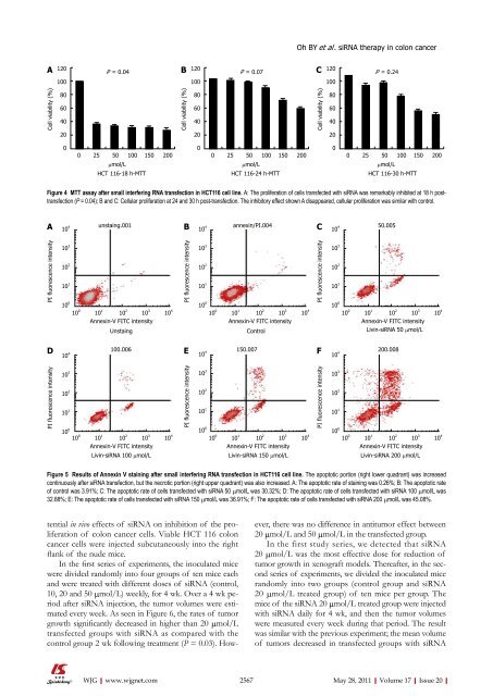
Figure 4 MTT assay after small interfering RNA transfection in HCT116 cell line. A: The proliferation <strong>of</strong> cells transfected with siRNA was remarkably inhibited at 18 h posttransfection<br />
(P = 0.04); B and C: Cellular proliferation at 24 and 30 h post-transfection. The inhibitory effect shown A disappeared, cellular proliferation was similar with control.<br />
A B C<br />
PI fluorescence intensity<br />
D<br />
PI fluorescence intensity<br />
10 4<br />
10 3<br />
10 2<br />
10 1<br />
10 0<br />
10 4<br />
10 3<br />
10 2<br />
10 1<br />
10 0<br />
unstaing.001 annexin/PI.004 50.005<br />
10 0 10 1 10 2 10 3 10 4<br />
Annexin-V FITC intensity<br />
Unstaing<br />
100.006<br />
10 0 10 1 10 2 10 3 10 4<br />
Annexin-V FITC intensity<br />
Livin-siRNA 100 μmol/L<br />
Cell viability (%)<br />
PI fluorescence intensity<br />
E<br />
PI fluorescence intensity<br />
10 4<br />
10 3<br />
10 2<br />
10 1<br />
10 0<br />
10 4<br />
10 3<br />
10 2<br />
10 1<br />
10 0<br />
10 0 10 1 10 2 10 3 10 4<br />
Annexin-V FITC intensity<br />
Control<br />
150.007<br />
10 0 10 1 10 2 10 3 10 4<br />
Annexin-V FITC intensity<br />
Livin-siRNA 150 μmol/L<br />
Oh BY et al . siRNA therapy in colon cancer<br />
PI fluorescence intensity<br />
F<br />
PI fluorescence intensity<br />
10 4<br />
10 3<br />
10 2<br />
10 1<br />
10 0<br />
10 4<br />
10 3<br />
10 2<br />
10 1<br />
10 0<br />
10 0 10 1 10 2 10 3 10 4<br />
Annexin-V FITC intensity<br />
Livin-siRNA 50 μmol/L<br />
<strong>20</strong>0.008<br />
10 0 10 1 10 2 10 3 10 4<br />
Annexin-V FITC intensity<br />
Livin-siRNA <strong>20</strong>0 μmol/L<br />
Figure 5 Results <strong>of</strong> Annexin V staining after small interfering RNA transfection in HCT116 cell line. The apoptotic portion (right lower quadrant) was increased<br />
continuously after siRNA transfection, but the necrotic portion (right upper quadrant) was also increased. A: The apoptotic rate <strong>of</strong> staining was 0.26%; B: The apoptotic rate<br />
<strong>of</strong> control was 3.91%; C: The apoptotic rate <strong>of</strong> cells transfected with siRNA 50 μmol/L was 30.32%; D: The apoptotic rate <strong>of</strong> cells transfected with siRNA 100 μmol/L was<br />
32.88%; E: The apoptotic rate <strong>of</strong> cells transfected with siRNA 150 μmol/L was 36.91%; F: The apoptotic rate <strong>of</strong> cells transfected with siRNA <strong>20</strong>0 μmol/L was 45.08%.<br />
Cell viability (%)<br />
ever, there was no difference in antitumor effect between<br />
<strong>20</strong> μmol/L and 50 μmol/L in the transfected group.<br />
In the first study series, we detected that siRNA<br />
<strong>20</strong> μmol/L was the most effective dose for reduction <strong>of</strong><br />
tumor growth in xenograft models. Thereafter, in the second<br />
series <strong>of</strong> experiments, we divided the inoculated mice<br />
randomly into two groups (control group and siRNA<br />
<strong>20</strong> μmol/L treated group) <strong>of</strong> ten mice per group. The<br />
mice <strong>of</strong> the siRNA <strong>20</strong> μmol/L treated group were injected<br />
with siRNA daily for 4 wk, and then the tumor volumes<br />
were measured every week during that period. The result<br />
was similar with the previous experiment; the mean volume<br />
<strong>of</strong> tumors decreased in transfected groups with siRNA<br />
2567 May 28, <strong>20</strong>11|Volume 17|Issue <strong>20</strong>|


