Download the entire issue - American Association for Clinical ...
Download the entire issue - American Association for Clinical ...
Download the entire issue - American Association for Clinical ...

Create successful ePaper yourself
Turn your PDF publications into a flip-book with our unique Google optimized e-Paper software.
concentrations characteristic of patients<br />
with metastatic disease. These high Tg levels<br />
overwhelm <strong>the</strong> assay’s reagent binding<br />
capacities, which yields inappropriately low<br />
values. Although adoption of a two-step<br />
assay design has reduced this problem, a<br />
hook effect still can occur when measuring<br />
saline wash-outs of <strong>the</strong> needles used to biopsy<br />
metastatic lymph nodes. In such cases,<br />
Tg levels often exceed 10,000 µg/L. Because<br />
of <strong>the</strong>se challenges, laboratories need to use<br />
linearity studies to check <strong>the</strong> high range <strong>for</strong><br />
hooking and <strong>the</strong> potential <strong>for</strong> carry-over<br />
contamination of specimens.<br />
Countering Interferences<br />
Un<strong>for</strong>tunately, <strong>the</strong> Tg IMA methodology<br />
favored by most laboratories is more prone<br />
to interferences from both human antimouse<br />
antibody (HAMA) and TgAb than<br />
is Tg RIA methodology (3,12,13). HAMA<br />
interference usually results in a falsely high<br />
serum Tg that may prompt unnecessary<br />
imaging or radioiodine treatment <strong>for</strong> presumed<br />
disease. In contrast, TgAb interference<br />
causes falsely low or undetectable<br />
serum Tg that can have more serious consequences<br />
because it can mask <strong>the</strong> presence<br />
of disease. Whereas contemporary IMA<br />
methods routinely include blocker reagents<br />
to minimize HAMA interference to around<br />
0.5%, currently <strong>the</strong>re are no effective measures<br />
to overcome TgAb interference encountered<br />
with Tg IMA methodology (8).<br />
12 CliniCal laboratory news July 2011<br />
HAMA in <strong>the</strong> specimen can interact<br />
with one of <strong>the</strong> monoclonal antibody reagents<br />
to create a false signal that simulates<br />
<strong>the</strong> presence of a high antigen (Tg) concentration<br />
(12). Rarely, HAMA can block <strong>the</strong><br />
participation of <strong>the</strong> antibody reagents and<br />
cause a falsely low Tg value (13). Physicians<br />
should suspect HAMA when <strong>the</strong> serum Tg<br />
level appears inappropriate in <strong>the</strong> context<br />
of <strong>the</strong> patient’s clinical status, or fails to<br />
respond appropriately to changes in TSH.<br />
An example would be when Tg levels rise<br />
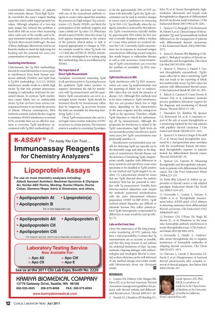
Apo AII<br />
> Apo CII<br />
> Apo CIII<br />
> Apo E<br />
see us at <strong>the</strong> 2011 clin lab expo, booth no. 2220<br />
Lipoprotein CLN 09-2010.indd 1 8/2/2010 1:11:04 PM<br />
so <strong>for</strong> <strong>the</strong> approximately 20% of DTC patients<br />
with detectable TgAb, <strong>the</strong> TgAb concentration<br />
can be used to monitor changes<br />
in tumor mass in preference to measuring<br />
Tg by IMA (14). Specifically, when <strong>the</strong> antigenic<br />
stimulus is removed by thyroidectomy,<br />
TgAb concentrations typically decline<br />
by approximately 50% within <strong>the</strong> first year<br />
and eventually disappear within a median<br />
of 3 years when patients are rendered disease-free<br />
(14). Conversely, TgAb concentrations<br />
rise in response to increased antigen<br />
concentrations following second surgeries,<br />
fine needle biopsy, or radioiodine <strong>the</strong>rapy<br />
as well as with recurrence. Serial monitoring<br />
of TgAb concentrations can overcome<br />
<strong>the</strong> problem of unreliable Tg IMA measurements.<br />
TgAb Interference in IMA<br />
TgAb interference with Tg IMA measurements<br />
can cause Tg underestimation and<br />
<strong>the</strong> reporting of falsely low or undetectable<br />
values that can mask <strong>the</strong> presence of<br />
disease (3,6). Although RIA methods tend<br />
to be more resistant to TgAb interference,<br />
<strong>the</strong>y too can produce falsely low or high<br />
values, depending on <strong>the</strong> characteristics<br />
of <strong>the</strong> assay reagents and <strong>the</strong> endogenous<br />
TgAb in <strong>the</strong> specimen. There<strong>for</strong>e, reliable<br />
TgAb detection is critical <strong>for</strong> au<strong>the</strong>nticating<br />
all Tg measurements. Although <strong>the</strong><br />
propensity <strong>for</strong> interference is related to <strong>the</strong><br />
TgAb concentration, high TgAb levels do<br />
not necessarily produce interference, and in<br />
some cases, low TgAb concentrations may<br />
profoundly interfere (3).<br />
Manufacturer-recommended assay cutoffs<br />
<strong>for</strong> detecting TgAb are typically set in<br />
<strong>the</strong> detectable range and relate to <strong>the</strong> diagnosis<br />
of autoimmune thyroid disease, not<br />
<strong>the</strong> detection of interfering TgAb. Inappropriate<br />
cutoffs, toge<strong>the</strong>r with differences in<br />
assay sensitivity and specificity, cause some<br />
specimens to be classified as TgAb-positive<br />
by one method and TgAb-negative by ano<strong>the</strong>r<br />
(5). Laboratorians should be aware<br />
that any TgAb detected above <strong>the</strong> analytic<br />
sensitivity limit has <strong>the</strong> potential to interfere<br />
with Tg measurement. Notably, <strong>the</strong>se<br />
between-method disparities exist despite<br />
<strong>the</strong> methods’ purported standardization<br />
with <strong>the</strong> same international reference<br />
preparation (WHO 1st IRP 65/93). TgAb<br />
method-related disparities are difficult to<br />
eliminate because <strong>the</strong>y reflect patient-related<br />
TgAb heterogeneity compounded by<br />
differences in assay sensitivity and specificity<br />
(5).<br />
Labs on <strong>the</strong> Front Lines<br />
Given <strong>the</strong> importance of life-long postoperative<br />
monitoring of DTC patients, labs<br />
have a vital responsibility to ensure that Tg<br />
measurements are as accurate as possible,<br />
and that <strong>the</strong>y keep abreast of and address<br />
<strong>the</strong> analytical limitations of <strong>the</strong>ir Tg assay<br />
methods. Ongoing dialogue with endocrinologists<br />
and oncologists likewise is essential,<br />
so <strong>the</strong>se clinicians can be well-in<strong>for</strong>med<br />
of any method changes and confer readily<br />
with laboratorians about any discrepant<br />
results. CLN<br />
REFERENCES<br />
1. Cooper DS, Doherty GM, Haugen BR,<br />
Kloos RT, et al. Revised <strong>American</strong> Thyroid<br />
<strong>Association</strong> management guidelines <strong>for</strong> patients<br />
with thyroid nodules and differentiated<br />
thyroid cancer. Thyroid 2009;19:1–48.<br />
2. Snozek CL, Chambers EP, Reading CC,<br />
Sebo TJ, et al. Serum thyroglobulin, highresolution<br />
ultrasound, and lymph node<br />
thyroglobulin in diagnosis of differentiated<br />
thyroid carcinoma nodal metastases. J Clin<br />
Endocrinol Metab 2007;1992:4278–4281.<br />
3. Spencer CA, Bergoglio LM, Kazarosyan<br />
M, Fatemi S, et al. <strong>Clinical</strong> impact of thyroglobulin<br />
(Tg) and Tg autoantibody method<br />
differences on <strong>the</strong> management of patients<br />
with differentiated thyroid carcinomas.<br />
J Clin Endocrinol Metab 2005;1990:5566–<br />
5575.<br />
4. Jensen E, Petersen PH, Blaabjerg O, Hegedüs<br />
L. Biological variation of thyroid autoantibodies<br />
and thyroglobulin. Clin Chem<br />
Lab Med 2007;45:1058–1064.<br />
5. Spencer C, Petrovic I, Fatemi S. Current<br />
thyroglobulin autoantibody (TgAb)<br />
assays often fail to detect interfering TgAb<br />
that can result in <strong>the</strong> reporting of falsely<br />
low/undetectable serum Tg IMA values <strong>for</strong><br />
patients with differentiated thyroid cancer.<br />
J Clin Endocrinol Metab 96:1283–91, 2011.<br />
6. Baloch Z, Carayon P, Conte-Devolx B,<br />
Demers LM, et al. Laboratory medicine<br />
practice guidelines: laboratory support <strong>for</strong><br />
<strong>the</strong> diagnosis and monitoring of thyroid<br />
disease. Thyroid 2003;13:3–126.<br />
7. Mazzaferri EL, Robbins RJ, Spencer<br />
CA, Braverman LE, et al. A consensus report<br />
of <strong>the</strong> role of serum thyroglobulin as<br />
a monitoring method <strong>for</strong> low-risk patients<br />
with papillary thyroid carcinoma. J Clin<br />
Endocrinol Metab 2003;88:1433–1441.<br />
8. Spencer CA, Fatemi S, Singer P, Nicoloff<br />
JT, et al. Serum basal thyroglobulin measured<br />
by a 2nd generation assay correlates<br />
with <strong>the</strong> recombinant human tsh-stimulated<br />
thyroglobulin response in patients<br />
treated <strong>for</strong> differentiated thyroid cancer.<br />
Thyroid 2010;20:587–95.<br />
9. Spencer CA, Lopresti JS. Measuring<br />
thyroglobulin and thyroglobulin autoantibody<br />
in patients with differentiated thyroid<br />
cancer. Nat Clin Pract Endocrinol Metab<br />
2008;4:223–233.<br />
10. Tuttle RM, Leboeuf R. Follow up approaches<br />
in thyroid cancer: a risk adapted<br />
paradigm. Endocrinol Metab Clin North<br />
Am 2008;37:419–435.<br />
11. Giovanella L, Ceriani L, Suriano S,<br />
Ghelfo A, et al. Thyroglobulin measurement<br />
be<strong>for</strong>e rhTSH-aided (131)I ablation<br />
in detecting metastases from differentiated<br />
thyroid carcinoma. Clin Endocrinol (Oxf)<br />
2008;69:659–663.<br />
12. Preissner CM, O’Kane DJ, Singh RJ,<br />
Morris JC, et al. Phantoms in <strong>the</strong> assay<br />
tube: heterophile antibody interferences in<br />
serum thyroglobulin assays. J Clin Endocrinol<br />
Metab 2003; 88:3069–3074.<br />
13. Giovanella L, Ghelfo A. Undetectable<br />
serum thyroglobulin due to negative<br />
interference of heterophile antibodies in<br />
relapsing thyroid carcinoma. Clin Chem<br />
2007;53:1871–1872.<br />
14. Chiovato L, Latrofa F, Braverman LE,<br />
Pacini F, et al. Disappearance of humoral<br />
thyroid autoimmunity after complete removal<br />
of thyroid antigens. Ann Intern Med<br />
2003;139:346–351.<br />
Carole Spencer, MT, PhD,<br />
FACB, is a professor of<br />
medicine in <strong>the</strong> Department<br />
of Medicine at <strong>the</strong> University<br />
of Sou<strong>the</strong>rn Cali<strong>for</strong>nia.<br />
Email: cspencer@usc.edu