IMAGERIE EN LOMBALGIE - Europa Ziekenhuizen
IMAGERIE EN LOMBALGIE - Europa Ziekenhuizen
IMAGERIE EN LOMBALGIE - Europa Ziekenhuizen

Create successful ePaper yourself
Turn your PDF publications into a flip-book with our unique Google optimized e-Paper software.
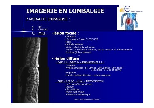
<strong>IMAGERIE</strong> <strong>EN</strong> <strong>LOMBALGIE</strong><br />
2.MODALITE D’<strong>IMAGERIE</strong> D <strong>IMAGERIE</strong> :<br />
A. RX<br />
B. CT-SCAN CT SCAN<br />
C. MRI : -lésion sion focale :<br />
-métastase tastase<br />
-hémangiome mangiome (hyper T1/T2/ STIR<br />
-Paget Paget<br />
-ost ostéoide oide ostéome ost ome<br />
-bénign nign notochordal cell tumor<br />
(hyper T2, trabécules trab cules normaux, pas de masse ni de rehaussement)<br />
-énostose nostose (îlot ( lot condensant)<br />
….<br />
- lésion sion diffuse<br />
-<br />
- hypo T1 / hyper T2 / rehaussement +++<br />
-métastase tastase<br />
-my myélome lome multiple ( nb. 28% nl / 29% diffuse / 30% focal /<br />
11% mixte / 3 % sel et poivre)<br />
-lymphome lymphome<br />
-atteinte atteinte myéloprolif my loproliférative rative – anémie an mie aplasique<br />
-- hypo hypo T1 T1 et et T2 T2 –– STIR STIR = = fibrose/sclérose<br />
fibrose/scl rose<br />
-ost ostéomy omyélofibrose lofibrose/scl /sclérose rose<br />
-Gaucher Gaucher<br />
-hémosid mosidérose rose<br />
-fibrose fibrose post-chimio<br />
post chimio<br />
-métastase tastase ostéoblastique<br />
ost oblastique<br />
Ateliers de St-Elisabeth St Elisabeth 27/11/2010