nerveux, des structures fonctionnelles de la - Bienvenue sur ...
nerveux, des structures fonctionnelles de la - Bienvenue sur ...
nerveux, des structures fonctionnelles de la - Bienvenue sur ...

Create successful ePaper yourself
Turn your PDF publications into a flip-book with our unique Google optimized e-Paper software.
UNIVERSITÉ DU QUÉBEC<br />
CONTRIBUTION À L'AMÉLIORATION DES CONNAISSANCES<br />
SUR LA PHYSIOLOGIE DE MYA ARENARIA<br />
(MOLLUSQUE BIVALVE) : DESCRIPTION DU SYSTÈME<br />
NERVEUX, DES STRUCTURES FONCTIONNELLES DE LA<br />
GONADE ET DE LEURS INTERACTIONS<br />
THÈSE PRÉSENTÉE À<br />
L'UNIVERSITÉ DU QUÉBEC À RIMOUSKI<br />
Comme exigence partielle<br />
du programme <strong>de</strong> doctorat en océanographie<br />
pour l'obtention du gra<strong>de</strong> <strong>de</strong><br />
PHILOSOPHIAE DOCTOR (OCÉANOGRAPHIE)<br />
par<br />
FLORENT GARNEROT<br />
Novembre 2007
UNIVERSITÉ DU QUÉBEC À RIMOUSKI<br />
Service <strong>de</strong> <strong>la</strong> bibliothèque<br />
Avertissement<br />
La diffusion <strong>de</strong> ce mémoire ou <strong>de</strong> cette thèse se fait dans le respect <strong><strong>de</strong>s</strong> droits <strong>de</strong> son<br />
auteur, qui a signé le formu<strong>la</strong>ire « Autorisation <strong>de</strong> reproduire et <strong>de</strong> diffuser un rapport,<br />
un mémoire ou une thèse ». En signant ce formu<strong>la</strong>ire, l’auteur concè<strong>de</strong> à l’Université du<br />
Québec à Rimouski une licence non exclusive d’utilisation et <strong>de</strong> publication <strong>de</strong> <strong>la</strong> totalité<br />
ou d’une partie importante <strong>de</strong> son travail <strong>de</strong> recherche pour <strong><strong>de</strong>s</strong> fins pédagogiques et non<br />
commerciales. Plus précisément, l’auteur autorise l’Université du Québec à Rimouski à<br />
reproduire, diffuser, prêter, distribuer ou vendre <strong><strong>de</strong>s</strong> copies <strong>de</strong> son travail <strong>de</strong> recherche à<br />
<strong><strong>de</strong>s</strong> fins non commerciales <strong>sur</strong> quelque support que ce soit, y compris l’Internet. Cette<br />
licence et cette autorisation n’entraînent pas une renonciation <strong>de</strong> <strong>la</strong> part <strong>de</strong> l’auteur à ses<br />
droits moraux ni à ses droits <strong>de</strong> propriété intellectuelle. Sauf entente contraire, l’auteur<br />
conserve <strong>la</strong> liberté <strong>de</strong> diffuser et <strong>de</strong> commercialiser ou non ce travail dont il possè<strong>de</strong> un<br />
exemp<strong>la</strong>ire.
Cette thèse est dédiée à mon père, à mon grand-père<br />
Garnerot, à ma grand-mère Cassard, à mon oncle<br />
Denis et à mon ami Guy-Juslin qui nous ont quittés<br />
bien trop tôt. Vous nous manquez !
REMERCIEMENTS<br />
Je profite <strong>de</strong> ces avant-propos pour remercier toutes les personnes qui ont participé <strong>de</strong><br />
près ou <strong>de</strong> loin à ce travail.<br />
J'exprime toute ma reconnaissance envers ma directrice <strong>de</strong> thèse, Madame Jocelyne<br />
Pellerin, qui a accepté d'encadrer cette thèse, pour son accueil chaleureux au sein <strong>de</strong> son<br />
équipe, pour ses qualités pédagogiques, humaines et pour <strong>la</strong> confiance et <strong>la</strong> compréhension<br />
dont elle a fait preuve pour gui<strong>de</strong>r mes recherches .<br />
.Je remercie mon codirecteur, le professeur Christian B<strong>la</strong>ise, pour sa confiance et pour<br />
<strong>la</strong> gran<strong>de</strong> liberté qu'il m'a accordé dans mes recherches, ainsi que pour son ai<strong>de</strong> lors <strong><strong>de</strong>s</strong><br />
cOlTections linguistiques <strong><strong>de</strong>s</strong> publications présentes à l'intérieur <strong>de</strong> ce travail.<br />
.Je remercie tout particulièrement le professeur Michel Mathieu qui , avec passion, a<br />
SUIVI mes travaux <strong>de</strong> recherche et m'a accueilli au sein <strong>de</strong> son <strong>la</strong>boratoire à plusieurs<br />
reprises. II m'a guidé et conseillé tout le long <strong>de</strong> ce travail. Je tiens à lui expri mer toute ma<br />
gratitu<strong>de</strong> .<br />
.Je remercie sincèrement le professeur Céline Au<strong>de</strong>t d'avoir accepté <strong>de</strong> juger cette<br />
thèse, pour sa confiance, ses encouragements, ses suggestions et ses corrections et le<br />
professeur Réjean Tremb<strong>la</strong>y <strong>de</strong> m'avoir permis d'utiliser, tout au long <strong>de</strong> ma thèse, son<br />
analyseur d'i mages.
Il<br />
Je tiens à remercier le professeur Jean-Luc Bouchereau <strong>de</strong> m'avoir pen11lS <strong>de</strong><br />
découvrir <strong>la</strong> recherche scientifique et <strong>de</strong> sUivre encore aujourd'hui mes travaux <strong>de</strong><br />
recherche.<br />
Merci aux autres membres <strong>de</strong> l'équipe: Simon Cartier, Nico/as Lemaire, Pascal<br />
Rioltx, Sandrine Briatte, Nico<strong>la</strong>s Pichaud, Séverine Louis, Luna Greco pour leur soutien et<br />
pour avoir entretenu une atmosphère chaleureuse au sein du groupe.<br />
J'adresse aussi une pensée particulière aux étudiants, jeunes chercheurs et professeurs<br />
qUI ont contribué, d'une manière ou d'une autre, à ce que cette thèse soit un moment<br />
agréable; Anibal et Judite Médina, Jean Mamelona, Hacene Tamdrari, Dounia Daoud,<br />
Sylvain Joly, Sophie Gauthier-Clerc, Olivier Etchian, Jean-C<strong>la</strong>u<strong>de</strong> Brêthes et Michel<br />
Fournier et tous ceux que j'ai pu malencontreusement oublier.<br />
Ma reconnaissance ne serait pas complète sans avoir adressé un remerciement à ma<br />
famille, mes grands parents, Jeannette et Laurent, ma mère Dominique, mes sœurs Carine<br />
et Anne, ma nièce Cybill, mes oncles et mes tantes Cassard, pour leurs témoignages<br />
permanents, leurs présences, et ce, malgré <strong>la</strong> distance. Je ne trouve aucun mot pour définir<br />
ce que je ressens pour vous. Merci à tous!<br />
Pour finir, ce travail est l'aboutissement <strong>de</strong> longues années d'étu<strong><strong>de</strong>s</strong>, auxquelles<br />
l'amour et le souti en <strong>de</strong> ma conjointe, Mé<strong>la</strong>nie, a <strong>la</strong>rgement contribué. Je voudrais lui<br />
rappeler mon amour et ma reconnaissance pour sa compréhension et sa présence<br />
affectueuse à mes côtés et pour le plus beau ca<strong>de</strong>au <strong>de</strong> ma vie, notre fils Pierre.
RÉSUMÉ<br />
Mya arenaria, le bivalve utilisé pour ce travail, est d'intérêt économique et fait partie<br />
<strong><strong>de</strong>s</strong> espèces sentinelles couramment employées en écotoxicologie. La mye est un organisme<br />
filtreur et sé<strong>de</strong>ntaire qui bioaccumule les contaminants au-<strong><strong>de</strong>s</strong>sus <strong><strong>de</strong>s</strong> taux retrouvés dans le<br />
milieu, et donc, constitue un excellent indicateur <strong>de</strong> <strong>la</strong> contamination ambiante. Un déficit<br />
d' information concernant <strong>la</strong> physiologie <strong>de</strong> <strong>la</strong> mye rend l'interprétation <strong><strong>de</strong>s</strong> données<br />
écotoxicologiques <strong>de</strong> plus en plus problématique. Notre thématique <strong>de</strong> recherche tend à<br />
répondre à ce besoin d'information. Considérant que <strong>la</strong> reproduction <strong><strong>de</strong>s</strong> bivalves semble<br />
être contrôlée par les neurosécrétions ganglionnaires et les stéroï<strong><strong>de</strong>s</strong>, nous nous sommes<br />
intéressés à l'étu<strong>de</strong> du système <strong>nerveux</strong>, du système reproducteur et à leurs interactions.<br />
Le premier objectif <strong>de</strong> cette recherche était <strong>de</strong> mieux connaître <strong>la</strong> physiologie et <strong>la</strong><br />
composition cellu<strong>la</strong>ire du système <strong>nerveux</strong> et <strong>de</strong> <strong>la</strong> masse viscérale <strong>de</strong> Mya arenaria. Le<br />
système <strong>nerveux</strong> présente un p<strong>la</strong>n <strong>de</strong> symétrie sagittal. Il consiste en trois paires <strong>de</strong><br />
ganglions : les cérébroï<strong><strong>de</strong>s</strong> situés au niveau <strong>de</strong> l'oesophage, les viscéraux situés <strong>sur</strong> <strong>la</strong> face<br />
ventrale du muscle adducteur postérieur et les pédieux situés à <strong>la</strong> base du pied. Les<br />
ganglions pédieux et viscéraux sont fusionnés, tandis que les ganglions cérébroï<strong><strong>de</strong>s</strong> sont<br />
réunis dorsalement au moyen <strong>de</strong> <strong>la</strong> commis<strong>sur</strong>e cérébrale. Les ganglions cérébroï<strong><strong>de</strong>s</strong> sont<br />
connectés aux pédieux et aux viscéraux, respectivement, par les connectifs cérébro-pédieux<br />
et cérébro-viscéraux. Notre étu<strong>de</strong> a aussi démontré l'existence d'un rapprochement <strong><strong>de</strong>s</strong><br />
connectifs cérébro-viscéraux du côté antérieur du muscle rétracteur postérieur, d' un tronc<br />
<strong>nerveux</strong> émanant du connectif cérébro-viscéral et innervant le muscle rétracteur postérieur<br />
et <strong>de</strong> plusieurs troncs <strong>nerveux</strong> dérivant <strong><strong>de</strong>s</strong> connectifs cérébro-viscéraux et innervant <strong>la</strong><br />
gona<strong>de</strong>. La masse viscérale présente une organisation générale semb<strong>la</strong>ble à celles <strong><strong>de</strong>s</strong> autres<br />
bivalves, et est composée <strong><strong>de</strong>s</strong> systèmes digestif, muscu<strong>la</strong>ire, <strong>nerveux</strong> et reproducteur. Les<br />
<strong>de</strong>ux systèmes principaux, digestif et reproducteur, sont étroitement entre<strong>la</strong>cés bien que<br />
tout à fait distincts l'un <strong>de</strong> l'autre, ce qui optimise le potentiel <strong>de</strong> transfert <strong><strong>de</strong>s</strong> nutriments<br />
vers les gamètes en développement. L'étu<strong>de</strong> histol ogique <strong>de</strong> <strong>la</strong> masse viscérale a pennis <strong>de</strong><br />
caractériser le développement et <strong>la</strong> composition cellu<strong>la</strong>ire <strong><strong>de</strong>s</strong> alvéoles gonadiques. Ell es se<br />
composent <strong>de</strong> cellules somatiques <strong>de</strong> réserve, strictement nutritives ("cellules follicu<strong>la</strong>ires"<br />
Coe & Turner [1938]), et <strong><strong>de</strong>s</strong> cellules <strong>de</strong> <strong>la</strong> lignée germinale. De plus, chez le mâle nous<br />
avons mis en évi<strong>de</strong>nce, par immunohistochimie, <strong>la</strong> présence <strong>de</strong> cellules somatiques <strong>de</strong><br />
soutien intratubu<strong>la</strong>ires. Ces cellules, appelées chez les vertébrés « cellules <strong>de</strong> Sertoli », sont<br />
uniformément distribuées à l'intérieur <strong><strong>de</strong>s</strong> tubules mâles en développement.
IV<br />
La sérotonine (5-hydroxytryptamine ou 5-HT) joue un rôle central dans plusieurs<br />
processus physiologiques chez les mollusques marins, particulièrement au niveau <strong>de</strong> <strong>la</strong><br />
reproduction. Nos travaux ont montré <strong>la</strong> présence <strong>de</strong> gran<strong><strong>de</strong>s</strong> quantités <strong>de</strong> cellules<br />
sérotoninergiques à l'intérieur du système <strong>nerveux</strong> <strong>de</strong> Mya arenaria (ganglions cérébraux,<br />
viscéraux et pédieux), ce qui confinne son rôle <strong>de</strong> premier p<strong>la</strong>n comme neurotransmetteur.<br />
Au sein <strong><strong>de</strong>s</strong> ganglions viscéraux, les corps cellu<strong>la</strong>ires immunoréactifs sont regroupés en<br />
nodules, appelés glomérules, circonscrits au niveau <strong><strong>de</strong>s</strong> racines <strong><strong>de</strong>s</strong> nerfs branchiaux. Notre<br />
étu<strong>de</strong> montre également l'existence <strong>de</strong> fibres sérotoninergiques dans <strong>la</strong> gona<strong>de</strong> et les<br />
branchies. La présence <strong>de</strong> cellules sérotoninergiques dans les gangl ions et <strong>de</strong> fibres<br />
sérotoninergiques au niveau <strong><strong>de</strong>s</strong> branchies et à <strong>la</strong> périphérie <strong><strong>de</strong>s</strong> alvéoles gonadiques<br />
confirme <strong>la</strong> re<strong>la</strong>tion existant entre le système <strong>nerveux</strong> et les tissus périphériques. Ces<br />
résultats p<strong>la</strong>i<strong>de</strong>nt en faveur d'une implication <strong>de</strong> <strong>la</strong> 5-HT dans le contrôle <strong>de</strong> certaines<br />
fonctions physiologiques, telles que <strong>la</strong> respiration et <strong>la</strong> reproduction.<br />
Récemment, les recherches <strong>sur</strong> les perturbateurs endocriniens et <strong>la</strong> régu<strong>la</strong>tion <strong>de</strong> <strong>la</strong><br />
reproduction se sont intéressées à l'étu<strong>de</strong> <strong><strong>de</strong>s</strong> variations <strong><strong>de</strong>s</strong> taux d ' hormones stéroïdiennes<br />
(l7~-oestradiol , testostérone et progestérone) en fonction <strong>de</strong> <strong>la</strong> maturité gonadique. Nos<br />
travaux montrent que les niveaux <strong>de</strong> progestérone dans <strong>la</strong> g<strong>la</strong>n<strong>de</strong> digestive sont trois fois<br />
supérieurs à ceux dans <strong>la</strong> gona<strong>de</strong>. Les niveaux élevés <strong>de</strong> progestérone dans <strong>la</strong> g<strong>la</strong>n<strong>de</strong><br />
digestive et <strong>la</strong> similitu<strong>de</strong> <strong><strong>de</strong>s</strong> profils entre <strong>la</strong> g<strong>la</strong>n<strong>de</strong> digestive et <strong>la</strong> gona<strong>de</strong> suggèrent une<br />
synthèse et/ou un stockage dans <strong>la</strong> g<strong>la</strong>n<strong>de</strong> digestive. Nos travaux ont aussi montré que les<br />
profils <strong><strong>de</strong>s</strong> hormones stéroïdiennes (17~-oestradiol et testostérone) me<strong>sur</strong>és dans <strong>la</strong> gona<strong>de</strong><br />
<strong>de</strong> <strong>la</strong> mye pouvaient fortement varier d'une étu<strong>de</strong> à l'autre, ce qui soulève <strong>de</strong> nombreuses<br />
questions. De telles variations ont déjà été rapportées dans <strong>la</strong> littérature et peuvent<br />
s' expliquer <strong>de</strong> <strong>de</strong>ux manières: une source exogène <strong>de</strong> stéroï<strong><strong>de</strong>s</strong> et/ou <strong><strong>de</strong>s</strong> variations<br />
interannuelles d'activité métabolique.<br />
De nouvelles perspectives <strong>de</strong> recherche, aussi bien au p<strong>la</strong>n <strong>de</strong> <strong>la</strong> <strong><strong>de</strong>s</strong>cription <strong>de</strong> <strong>la</strong><br />
gamétogénèse en microscopie électronique, qu'au p<strong>la</strong>n <strong><strong>de</strong>s</strong> techniques <strong>de</strong> traçage<br />
molécu<strong>la</strong>ire neuroanatomique <strong>de</strong> <strong>la</strong> 5-HT ou <strong>de</strong> dosage <strong><strong>de</strong>s</strong> enzymes clés intervenant dans<br />
<strong>la</strong> stéroidogenèse, peuvent être proposées pour améliorer les connaissances <strong>sur</strong> <strong>la</strong><br />
physiologie <strong>de</strong> Mya arenaria.
ABSTRACT<br />
The soft-shell c<strong>la</strong>m Mya arenaria is of economic interest and an ecologically<br />
important bivalve. The c<strong>la</strong>m is a sentinel species <strong>la</strong>rgely used in ecotoxicology. It is a<br />
se<strong>de</strong>ntary and filter-feeding bivalve which accumu<strong>la</strong>tes pollutants above levels found in the<br />
environment. This species is an excellent indicator of environmental contamination.<br />
However, information is <strong>la</strong>cking about its reproductive physiology which make difficult the<br />
analysis of ecotoxicological data. The objective of our research sought information on the<br />
re<strong>la</strong>tionship between ganglia neurosecretions and the concentration of steroids both in<br />
gonad and in the digestive g<strong>la</strong>nd.<br />
The first goal of this investigation was to <strong><strong>de</strong>s</strong>cribe the physiology and the cell<br />
composition of the nervous system and the visceral mass of Mya arenaria. The nervous<br />
system follows the typical pelecypod p<strong>la</strong>n. It is formed by three pairs of ganglia: the<br />
cerebral ganglia lying on both si<strong><strong>de</strong>s</strong> of the oesophagus, the visceral ganglion located on the<br />
ventral si<strong>de</strong> of the adductor muscle and the pedal ganglion which is located at the base of<br />
the foot. The two symmetric visceral and pedal ganglia are fused at the midline, whereas<br />
the cerebral ganglia are connected by the cerebral commis<strong>sur</strong>e. Each cerebral ganglion is<br />
connected to the pedal and visceral ganglia by connective nerves. Our study showed the<br />
presence of a link between the cerebrovisceral connectives at the anterior si<strong>de</strong> of the<br />
posterior adductor muscle, and of gonadal and posterior foot retractor muscle innervations<br />
appearing to originate from the ramification of the cerebrovisceral connectives. The<br />
visceral mass of Mya arenaria has a generaI organization simi<strong>la</strong>r to those of other bivalves<br />
and is composed of digestive, muscu<strong>la</strong>r, nervous and reproductive systems. The digestive<br />
and reproductive systems are intertwined and closely associated. Gonadal <strong>de</strong>velopment<br />
around the intestine optimizes the potential transfer of nutrients to the <strong>de</strong>veloping gametes.<br />
The histologicaI study of the visceral mass provi<strong><strong>de</strong>s</strong> general infonnation on the cell<br />
composition and the <strong>de</strong>velopment of gonadic alveoli. Alveoli consist of storage somatic<br />
("cellules follicu<strong>la</strong>ires" Coe & Turner [1938]) and germinal cells. ln males, alveoli are also<br />
filled with intratubu<strong>la</strong>r supporting somatic cell s, called "Sertoli cells," in vertebrates.<br />
A <strong>la</strong>rge number of soma tic cells was <strong>de</strong>tected by immunohistochemistry uniformly<br />
distributed in male tubules.
VI<br />
Serotonin (5-hydroxytryptamine or 5-HT) Cl OH 12N20 p<strong>la</strong>ys a central role in several<br />
physiological processes in marine molluscs, especially in reproduction. We <strong>de</strong>monstrated<br />
that the nervous system of Mya arenaria contains re<strong>la</strong>tively <strong>la</strong>rge amounts of serotonin<br />
immunoreactive cells, supporting the hypothesis that 5-HT p<strong>la</strong>ys a role as a eurotransmitter.<br />
In the visceral ganglia, serotonin-immunoreactive cell bodies appeared to be wholly<br />
restricted to tightly c1ustered popu<strong>la</strong>tions, called glomeruli . These two glomeruli were<br />
located symmetrically at the root of the branchial nerves. Our study also showed the<br />
presence of numerous 5-HT nerve fibers of various diameters in the gonad and gills of<br />
Mya arenaria. The presence of gills and gonadal 5-HT immunoreactive connectives and<br />
serotonin-immunoreactive cells in the cortex ganglia confirm the presence of a pathway<br />
between the nervous system and peripheral tissues. These results indicate a l'ole of 5-HT in<br />
the control of physiological functions such as respiration and reproduction.<br />
Recently, research on endocrine disruption and on gametogenesis regu<strong>la</strong>tion focussed<br />
on variations of sex steroid levels (l7~-oestradiol, testosterone and progesterone) in<br />
re<strong>la</strong>tion to gonadic maturity. Our study showed that the progesterone level in c<strong>la</strong>m<br />
digestive g<strong>la</strong>nd was three times higher than in gonad. The high levels of progesterone in the<br />
digestive g<strong>la</strong>nd and the simi<strong>la</strong>rity of the steroid profile between the digestive g<strong>la</strong>nd and<br />
gonad suggest that, in Mya arenaria, the digestive g<strong>la</strong>nd may synthesize and/or accumu<strong>la</strong>te<br />
this steroid. Our work showed interannual variations in the gonadal steroid profiles<br />
(17~-oestradiol and testosterone). Such steroid variations have been previously reported in<br />
the literature and can be exp<strong>la</strong>ined by interannual changes in metabolic activity and/or by<br />
the presence of an exogenous source of steroids.<br />
8ased on our fondings, additional work using electron microscopy to <strong><strong>de</strong>s</strong>cribe<br />
gametogenesis, coupled with steroid metabolism enzyme level mea<strong>sur</strong>ements and tracking<br />
5-HT via neuroanatomical techniques, would be of benefit to further improve physiological<br />
knowledge in Mya arenaria.
LISTE DES ABRÉVIATIONS<br />
17~-HSO<br />
17~-H ydroxy Stero id<br />
Dehydrogenase<br />
/ 17~-H ydroxyS t éroïd e<br />
Déshydrogénase<br />
3~-HSD<br />
3~ - H ydroxy Steroid<br />
Dehydrogenase<br />
/ 3 ~-H ydrox yStéroï<strong>de</strong><br />
Déshydrogénase<br />
5-HIAA<br />
5-1-1T<br />
5-HTP<br />
AMPc<br />
ASO<br />
CPG (GCP)<br />
DAB<br />
DG OF<br />
5-HydroxylndoleAcetic Acid (aci<strong>de</strong> 5-hydroxy-indoly<strong>la</strong>cétique)<br />
5-HydroxyTryptamine (serotonin / sérotonine)<br />
5-HydroxyTryptoPhan / 5-H ydroxyTryptoPhane<br />
Adénosine MonoPhosphate cyc lique<br />
AntiSerum Diluent<br />
Cerebro Pleural Ganglia (Ganglions Cérébro- Pleuraux)<br />
DiAminoBenzidine<br />
Digestive g<strong>la</strong>nd, Gonad, Digestive tract and Foot (g<strong>la</strong>n<strong>de</strong> digestive,<br />
gona<strong>de</strong>, tractus digestif et pied)<br />
DH EA<br />
DeHydroEpiAndrosterone / DéHydroÉpiAndrostérone<br />
Estrone<br />
Estradiol-17~ / 17~-oEstradiol<br />
ELISA<br />
GVBD<br />
Enzyme Linked ImmunoSorbent Assay<br />
Germinal Vesicle Break Down (di ssolution <strong>de</strong> <strong>la</strong> vésicul e gem1i native)
VIII<br />
GPCR<br />
HPLC<br />
G Protein-Coupled Receptors (récepteurs couplés à <strong><strong>de</strong>s</strong> protéines G)<br />
High Performance Liquid Chromatography (Chromatographie en phase<br />
Liqui<strong>de</strong> à Haute Pression)<br />
HSD<br />
IP3<br />
NADPH<br />
NSC (CNS)<br />
P<br />
PAP<br />
PBS<br />
PG (GP)<br />
PGs<br />
T<br />
Trp<br />
TX-IOO<br />
YG (GY)<br />
HydroxySteroid Dehydrogenase / HydroxyStéroï<strong>de</strong> Déshydrogénase<br />
Inositol tri-Phosphate<br />
Nicotinami<strong>de</strong> Adénine Dinucléoti<strong>de</strong> Phosphate<br />
NeuroSecretory Cells (Cell ul es NeuroSécrétrices)<br />
Progestrone / Progestérone<br />
Peroxidase Anti-Peroxidase / Peroxydase Anti-Peroxydase<br />
Phosphate Buffered Saline (tampon phopsphate sa lin)<br />
Pedal Ganglia (Ganglions Pédieux)<br />
ProstaG<strong>la</strong>ndin / ProstaG<strong>la</strong>ndines<br />
Testosterone / Testostérone<br />
Tryptophan / Tryptophane<br />
Triton X-IOO<br />
Visceral Ganglia (Ganglions Viscéraux)
TABLE DES MATIÈRES<br />
R ~EMERCI EMENTS .......... .................. ......... ........................................................ ..... .. .... .. .. i<br />
RÉSUMÉ .... .... .... ......... ...... ............. ... ....... ...................................... .. ................................... iii<br />
ABSTRACT ...................................................................... .............. ......................... ...... .. ...... v<br />
LISTE DES ABRÉVIATIONS ........................................................ ..................... .. ....... ... vii<br />
TABLE DES MATIÈRES .................................................................................................. ix<br />
LISTE DES TABLEAUX .......... .. .................. .. ....... ..... ....................................................... xv<br />
LISTE DES FIGURES ................................. .. ......... ............ ......... .... ................................ xvi<br />
INTRODUCTION GÉNÉRALE .............................................................. ...... ..... ................ 1<br />
CHAPITRE 1 : Synthèse bibliographique .......................................................................... 5<br />
1.1 Mya arenaria mollusque bivalve .......................................................................... 5<br />
1.1.1 Anatomie <strong>de</strong> Mya (lJ'enaria ..................................................................................... 6<br />
1.1.2 Biologie et physiologie <strong>de</strong> <strong>la</strong> reproduction <strong>de</strong> Mya arenaria ..................... .......... 10<br />
1.2 Contrôle <strong>de</strong> <strong>la</strong> reproduction chez les mollusques bivalves .............................. 14<br />
1.2.1 Contrôle neuroendocrini en ........................................................................ .. ......... 14<br />
1.2. 1.1 Système <strong>nerveux</strong> neuroendocrinien et cellules neurosécrétrices .......... .. ... ......... .. 14<br />
1.2. 1.2 Les alnines biogènes ............................................................................................. 16<br />
1.2.1.3 Implication <strong><strong>de</strong>s</strong> catécho<strong>la</strong>mines dans le contrôle <strong>de</strong> <strong>la</strong> reproduction ... ................. 26
x<br />
1.2.1.4 Implication <strong>de</strong> <strong>la</strong> sérotonine (5-HT) dans le contrôle <strong>de</strong> <strong>la</strong> reproduction ..... ...... .. 27<br />
1.2.1.5 Synthèse <strong>de</strong> <strong>la</strong> sérotonine et récepteurs sérotoninergiqucs ... ... .. ..... .... ...... ........... .41<br />
1.2.2 Contrôle stéroïdien ... ................. .. ..... ... ....... .. ........................................................ .44<br />
1.2.2.1 La voie <strong>de</strong> biosynthèse <strong><strong>de</strong>s</strong> hormones stéroïdiennes ......... ........ .................. ....... ..44<br />
1.2.2.2 Implication <strong><strong>de</strong>s</strong> hormones stéroïdiennes dans le contrôle <strong>de</strong> <strong>la</strong> reproduction .. ... .45<br />
1.2.3 Rôle <strong><strong>de</strong>s</strong> prostag<strong>la</strong>ndines dans <strong>la</strong> reproduction .............................. ..... .... .. ..... ...... .49<br />
1.2.4 Régu<strong>la</strong>tion croisée .......... .... .... ... ... ... ... .................................................. ... ....... .... ... 52<br />
C HAPIT RE 2 : Studies of the nervous system of Mya arenarÎa (Mollu sca: Bivalvia):<br />
Anatomical study and immunohistochemical localization of serotonin-Iike<br />
immunoreactive cells in cerebral, visceral and pedaI ganglia . ...... ................................. 54<br />
2.1 Abstract. ........................................ ... .. ..... ............................................ .... ............. 55<br />
2.2 Key ,,,ords ..... ...... ............. ... .... .. .... .. .... ....... ......... ........... ..... ....... .... .. ............. ....... 56<br />
2.3 Introduction .................................................... ... ..... .. .................... ..... ....... ... .... .... 57<br />
2.4 Materi al and methods .. .... ... ................. .... ........................ .. ................................. 60<br />
2.4.1 Chemicals and reagents .................................................. .... ....................... .. ....... .. 60<br />
2.4.2 C<strong>la</strong>n1 collection ................................. ................................. ........ ... ......... ..... ... .... ... 60<br />
2.4.3 Anatomical <strong><strong>de</strong>s</strong>cription of the nervous system ............. ... ............. .. ...... ...... .......... 61<br />
2.4.4 Light microscopy .............. .. ...... ..... ........ ... ............ .. .............................................. 61<br />
2.4.4. 1 Paraffin embedding and sectioning ............................................................... .... .... 61<br />
2.4.4.2 Serotonin (5-HT) immunohistochemistry ... ...... ........ .. ...... .......... ..... ....... ...... .... .. .. 62<br />
2.4.4.3 Mount and photography .... .. ...................................................................... .... ........ 63
X l<br />
2.5 Results ......................................................................... .......................... .. ...... ....... 63<br />
2.6 Discussion .......................... ... ...................................... ................................ .. ... ... . 69<br />
2.7 Acknowledgments ................... ................................................. ..... ...... ...... .......... 73<br />
2.8 References ................................. .. ... ... ..... ... ................................... .. ...................... 73<br />
CHAPITRE 3 : 1 m m unohistochemical localization of serotonin<br />
(5-hydroxytryptamine) in the gonad and digestive g<strong>la</strong>nd of Mya arenaria (Mollu sca:<br />
Bivalvia) .. ........ .... ..... ................. ....... .. .. .............. ........ ............................................ .. ............ 79<br />
3.1 Abstract. .. .... ....................... ...... ............................................................. ............... 80<br />
3.2 Key words .. .... ....................... .. ................. ... ... .. .... ................................................ 80<br />
3.3 Introduction .......................................... ........... ............. .. ..................................... 81<br />
3.4 Material and methods ...... .... .... .. .... ... ....... ....... .................................................... 83<br />
3.4.1 Chemicals and reagents .................. ...................................................................... 83<br />
3.4.2 C<strong>la</strong>in collection ..................................................................................................... 84<br />
3.4.3 Spawning induction .............................................................................................. 84<br />
3.4.4 Tissue preparation .......................................................................................... .. ..... 85<br />
3.4.5 Histology ...................... .......... .. ..... ..... .. .... .... ..... ... ................................................. 85<br />
3.4.6 Setotonin (5-HT) immunohistochemistry ................................................... .. ........ 86<br />
3.5 Results .................................. ... .... .... .. .... .......................................................... ..... 87<br />
3.6 Discussion ............... ... ........ ...... .. ... ... ..... ..... .... .... ... ... .... .................... ... .... ....... .. .... 92<br />
3.7 Acknowledgments ................... ............................................... ............................. 96<br />
3.8 References ............................................... ............................................................. 96
XII<br />
CHAPITRE 4: Anatomical study of the visceral mass and new knowledge of<br />
gametogenesis in the soft-shell c<strong>la</strong>m (Mya arenaria): Histochemical and<br />
immunohistochemical cell i<strong>de</strong>ntification ........... ....... ..... ... ....................................... ...... . 1 02<br />
4.1 Abstract. .. .... ...... ...... ... ..... ..... ...... ...... .................. ............ ..... ........... .. ........ .. ........ 103<br />
4.2 Key words ......................... ................... ............................... ........ ....................... 104<br />
4.3 Introduction .. .. ....... ......................... ... ... .. ..... .. ............ ..................... ....... ... ......... 105<br />
4.4 Material and methods ................................ ... ......... ................................... ..... ... 108<br />
4.4.1 Chemicals and reagents ............. .... ...... .. ........ ............................. ........................ 108<br />
4.4.2 C<strong>la</strong>nl collection ....... .............. ... ... ............ .. .............................................. .. ..... ... .. 108<br />
4.4.3 Anatomical <strong><strong>de</strong>s</strong>cription of the visceral mass ........................ ...... ........................ 109<br />
4.4.4 Light microscopy ................................. ...... .. .......................................... ........ ..... 109<br />
4.4.4.1 Paraffin embedding and sectioning ...... ...... ... .. .... .. ..... ....... ...... ............... ............. 109<br />
4.4.4.2 Histological staining ........................................................... ................................ 11 0<br />
4.4.4.3 Immunohistochemistry actin and a-tubulin ......... ... .... ... ...... ..... .. ........................ 110<br />
4.4.4.4 Mounting of sections and photography .......................... .... ... ........ .... ............ .... . 111<br />
4.5 Results .... ... .... ..... ........ ................ ................................... ...... ....... ........ ..... ..... ..... . III<br />
4.5.1 Anatomical <strong><strong>de</strong>s</strong>cription of the visceral mass ....... ....... ....... ....... .......... ..... ........ ... 1Il<br />
4.5.2 Histology and immunohistochemistry of the visceral mass .... ............. .. ....... ..... 114<br />
4.6 Discussion ...... .............. ... .... .... ... ....... ....... .. .. .... .... ............. .... ... ................. ......... 130<br />
4.7 Acknowledgments .. .. .... .. ......... .. ......... .............. ....... ..... .... ...... ................ .... .. ... .. 140<br />
4.8 References ..................... .... .................. ... ........ ...... ....... ... .... ........ ......... ...... .... .... . 140
XliI<br />
CHAPITRE 5: Re<strong>la</strong>tionship between levels of sexual steroids (estradiol-17P,<br />
progesterone and testosterone), Iipids and gametogenesis in male and female Mya<br />
arenaria (Mollusca: Bivalvia) ............. ... ... ... ......... .. ......... ..... ................... ....... ...... .... ... ..... 149<br />
5.1 Abstract. ... .. ....... ..... ... .... .... .... ...... ... ...... .. ..... ................... ........ .. ..... .... ..... ... ... .... .. 150<br />
5.2 Key words .... .............. .. .... .. .............. .... .... .. .... .. ................. .. .......................... .. .. . 151<br />
5.3 Introduction .......... .. ..... .. ....... ... .......... ................ ........ ...... ....... ........................... 152<br />
5.4 Material and methods ...... .. .. .. ........ .. .. ................ .. ...... .. .. .... .... .. .......... .. .... .. ....... 155<br />
5.4.1 C<strong>la</strong>m coll ection .. ...... .. .. .. ... .. ...... ... .... .. ....... ..... .. .... ............... ...... .. ....... ... ....... .... ... 155<br />
5.4.2 Hi stology ... ............. .. ........... ....... .. ....... .... .. ...... ... .......... .. .......... .. ... .... ... .......... .... . 155<br />
5.4.3 Steroids and lipids analysis .... .. .. ............ .. .......... .. ...... .......... .. ................ .. ...... .. ... 156<br />
5.4.3.1 Lipids ................. ........ ................... ....................... ......................... ....... ...... .. .. ... .. 156<br />
5.4.3 .2 Steroids extractions and assays .. .. .... .................. .. .... .. ...... .... .... .. .... ............... .. .. .. 156<br />
5.4.4 Stasti sti cs ... ...... .. ....... ...... ...... .. .. .. ...... .. .... .. .. .. ...... ..... .. ........................... ... ........ ... 157<br />
5.5 Results ..... ..... ..... .... .... ..... .... .... .. ........... .. .... ... ...... ........ ... .... ....... ........... .......... .. ... 158<br />
5.5 .1 Variation in lipid levels in gonad and digesti ve g<strong>la</strong>nd of Mya arenaria ............ 15 8<br />
5. 5.1.1 Vari ati on in 1 7 ~- es tra dio1 , testosterone and progesterone levels in gonads and<br />
digesti ve g<strong>la</strong>nds of Mya arenaria .. .... .... .......... .. .................. .. .. .... .. ... ...... .. .. .. .. .. .. 160<br />
5.6 Discussion ......... ......... ... ... .. ........ ... ... ........... ................ ....... ... ... ...... .... ..... ..... .... .. 164<br />
5.7 Acknowledgments ... ..... ................... .... ... ... ... .... .. ... ... ...... ...... .... ....... ...... .... .... .... 173<br />
5.8 References ..... ... ............ ... ... .. .. .... .... .. ... ..... .. ..... .. .. .... .. .. .. .... .... .... .... .... .. ..... ..... ..... 173
XIV<br />
CHAPITRE 6 : Discussion générale et perspectives ... ........ ....... ... ..... ... ..... ... ............ ... .. 179<br />
6.1 Anatomie du système <strong>nerveux</strong> <strong>de</strong> Mya arenaria .... .. ..... .......... ................. .. .... . 180<br />
6.2 Anatomie <strong>de</strong> <strong>la</strong> masse viscérale <strong>de</strong> Mya arenaria ..... .. .. .. .... ... ................. ... ..... 182<br />
6.3 Localisation <strong>de</strong> <strong>la</strong> 5-HT et implication dans <strong>la</strong> ponte ...... ..................... .... ... .. 183<br />
6.4 Variation <strong><strong>de</strong>s</strong> taux <strong>de</strong> lipi<strong>de</strong>, progestérone, testostérone et 17p-oestradiol<br />
dans <strong>la</strong> gona<strong>de</strong> et <strong>la</strong> g<strong>la</strong>n<strong>de</strong> digestive <strong>de</strong> Mya arenaria ....... ............. .... ...... ... . 186<br />
6.5 Conclusions et perspectives ..... ....... ... ..... ...... ... ......... .................................. .... .. 188<br />
LrSTE DES RÉFÉRENCES ... ............. ... .......... ......... .. ........ ................... ......... ....... ..... ... . 191<br />
ANNEXES ... .... ... .... .... ....... ..... .. ...... ......... .. .............. ... ... ........ ..... ..................... ....... ........ ... 214
LISTE DES TABLEAUX<br />
CHAPITRE 1<br />
Tableau 1.1<br />
C<strong>la</strong>ssification et <strong><strong>de</strong>s</strong>cription <strong><strong>de</strong>s</strong> six (6) sta<strong><strong>de</strong>s</strong> <strong>de</strong> maturité <strong>de</strong> <strong>la</strong> gona<strong>de</strong><br />
femelle et <strong><strong>de</strong>s</strong> cinq (5) sta<strong><strong>de</strong>s</strong> <strong>de</strong> <strong>la</strong> gona<strong>de</strong> mâle chez Mya arenaria selon<br />
les caractéristiques histologiques (Modifié <strong>de</strong> Coe & Turner, 1938;<br />
Brousseau, 1976; Potts, 1993; Gauthier-Clerc et al., 2002) . .. ... ... ... .. . 11-12<br />
Tableau 1.2<br />
Les catécho<strong>la</strong>mines chez les mollusques bivalves : dopamine (fond b<strong>la</strong>nc)<br />
et noradrénaline [norépinéphrine] (fond gris) ..................................... 17-21<br />
Tableau 1.3<br />
La 5-HT chez les mollusques bivalves ............................................... 22-25<br />
Tableau 1.4<br />
Induction <strong>de</strong> <strong>la</strong> ponte chez les mollusques bivalves par <strong>la</strong> 5-HT ........ 29-31<br />
Tableau 1.5<br />
Induction <strong>de</strong> <strong>la</strong> GVBD chez les mollusques bivalves par <strong>la</strong> 5-HT. .......... 32<br />
Tableau 1.6<br />
Caractérisation génétique et pharmacologique <strong><strong>de</strong>s</strong> récepteurs<br />
sérotoninergiques présents dans <strong>la</strong> gona<strong>de</strong> <strong><strong>de</strong>s</strong> mollusques ................ 35-39<br />
CHAPITRE 5<br />
Tableau 5.1<br />
Concentrations of estradiol-17P, testosterone and progesterone in the<br />
gonads of three marine bivalves, as reported in the literature ......... 168-169
LISTE DES FIGURES<br />
CHAPITRE 1<br />
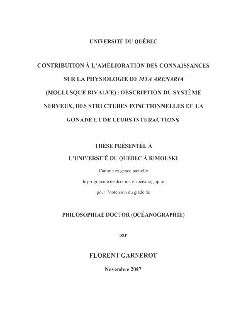
Figure 1.1: Mya arenaria L., mollusque bivalve endobenthique ......... .... ........ ... .............. 7<br />
Figure 1.2:<br />
Anatomie interne <strong>de</strong> Mya arenaria, mollusque bivalve (Modifié <strong>de</strong> Hanks,<br />
1963) .......................................................... .......... .. .... ... .................................. 8<br />
Figure 1.3:<br />
Schémas hi stologiques <strong>de</strong> <strong>la</strong> gona<strong>de</strong> <strong>de</strong> Mya arenaria (Modifié <strong>de</strong> Coe &<br />
Turner, 1938) ................ .. ................. ..... ..... ........................ ............... ..... ... ... . 13<br />
Figure 1.4: Biosynthèse <strong>de</strong> <strong>la</strong> sérotonine .. ........ ........ ...... .................. .............................. .41<br />
Figure 1.5:<br />
La stéroïdogenèse chez les mollusques bivalves: activités enzymatiques et<br />
stéroï<strong><strong>de</strong>s</strong> impl iquées ......................... ........... ............................. ........ ... .... ... ... 47
XV II<br />
CHAPITRE 2<br />
Figure 2.1 Schematic diagram of the nervous system of soft-shell c<strong>la</strong>m<br />
Mya arenaria . ..... ..... ..... .. .... ........ .. .......................................................... ... ... 64<br />
Figure 2.2<br />
Diagram of serotonin-like immunoreactivity in the nervous system of<br />
Mya arenaria . ......... ............................. ......... ...... ... ....... ..................... ...... .. ... 66<br />
Figure 2.3<br />
Serotonin (5-HT) immunohistochemical loca lization in the nervous system<br />
of Mya arenaria .............................. .............. ...... .... ........... .. ................... 67-68<br />
CHAPITRE 3<br />
Figure 3.1:<br />
Histology and serotonin (5-HT) immunohistochemistry local ization in the<br />
gonad of Mya arenaria . .. ... ... ... .... .... ........ ........... .................... ........... .. ... 88-89<br />
Figure 3.2:<br />
Histology and immunohistochemistry localization of 5-HT in the digestive<br />
g<strong>la</strong>nd and the gills ...................... ....... .. ...... .. ..... ........ .. ................... ... ..... ... ... .. 90<br />
Figure 3.3:<br />
Histology and 5-HT immunohistochemistry loca lization in the gonad<br />
parasitized by Prosorhynchus squamatus .................................. ......... .......... 91
XVlll<br />
CHAPITRE 4<br />
Figure 4.1: Schema tic diagram of visceral mass of the c<strong>la</strong>m Mya arenaria: .......... ..... 112<br />
Figure 4.2: Histology of gametogenesis in male gonad of Mya arenaria ..... .. ..... . 115-116<br />
Figure 4.3: Histology of gametogenesis in female gonad of Mya arenaria .... ..... . 117-118<br />
Figure 4.4:<br />
Actine and a-tubulin immunohistochemistry localization in the gonad of<br />
Mya arenaria . ............................................................................................. 122<br />
Figure 4.5: Muscu<strong>la</strong>r fibre localization in the DGDF of Mya arenaria ................ 124-127<br />
Figure 4.6: Nervous fibre localization in the DGDF of Mya arenaria .. .... .. .. ..... .. 128-129<br />
CHAPITRE 5<br />
Figure 5.1: Variation 111 lipid concentrations 111 gonad and digestive g<strong>la</strong>nd of<br />
Mya arenaria . ... ........... ... ............................................................................ 159<br />
Figure 5.2:<br />
Variations in estradiol-17P levels in Mya arenaria gonads and digestive<br />
g<strong>la</strong>nds based on <strong>de</strong>velopmental stages in both sexes ..... ............................. 161<br />
Figure 5.3:<br />
Variations in testosterone levels in Mya arenaria gonads and digestive<br />
g<strong>la</strong>nds based on <strong>de</strong>velopmental stages in both sexes .. ... ............ ....... ... ...... . 162<br />
Figure 5.4:<br />
Variations in progesterone levels in Mya arenaria gonads and digestive<br />
g<strong>la</strong>nds based on <strong>de</strong>velopment stages in both sexes .. ..... .................. .... .. .. .. .. 163
INTRODUCTION GÉNÉRALE<br />
Depuis le début <strong>de</strong> l'ère industrielle, <strong>de</strong> très gran<strong><strong>de</strong>s</strong> quantités <strong>de</strong> polluants ont été<br />
introdu ites dans l'écosystème marin du Saint-Laurent, soit directement à partir <strong><strong>de</strong>s</strong> effluents<br />
municipaux et industriels, soit indirectement par le rui ssell ement et les retombées<br />
atmosphériques (Gobeil & Cossa, 1993). Depuis 20 ans, une baisse significative <strong><strong>de</strong>s</strong><br />
apports directs en conta minants est observée. Malgré tout, les concentrations actuelles<br />
retrouvées dans le milieu sont touj ours supéri eures aux concentrations préindustriell es<br />
(Loring, 1975 ; Smith & Loring, 198 1; Gobeil & Cossa, 1993; A<strong>la</strong>ee, 2003).<br />
Au cours <strong><strong>de</strong>s</strong> 20 <strong>de</strong>rnières années, <strong>la</strong> communauté sci enti fi que internati onale s' est<br />
particul ièrement intéressée à l' impact <strong><strong>de</strong>s</strong> polluants d' origine anthropique <strong>sur</strong><br />
l' environnement. Une exposition à <strong>de</strong> faibles concentrations perturbe le fo nctionnement<br />
naturel <strong><strong>de</strong>s</strong> organi smes, notamment <strong>la</strong> reproducti on. Ces dérèglements s'observent, chez les<br />
poissons et les bi valves, par une diminution du nombre <strong>de</strong> spermatozoï<strong><strong>de</strong>s</strong> produits, par <strong>la</strong><br />
présence <strong>de</strong> malfo rmati ons génitales, amsl que par <strong><strong>de</strong>s</strong> changements <strong>de</strong> sexe<br />
(masculinisati on ou féminisation) et du sexe rati o au sein <strong><strong>de</strong>s</strong> popu<strong>la</strong>ti ons affectées<br />
(Depl edge & Billinghurst, 1999; B<strong>la</strong>ise et al. , 2003 ; Gagné et al., 2006) .<br />
Les mollusques bi valves, à l'exemple <strong>de</strong> Mya arenaria, sont <strong><strong>de</strong>s</strong> organi smes filtreurs<br />
et sé<strong>de</strong>ntaires, qui bioaccumulent les contaminants au-<strong><strong>de</strong>s</strong>sus <strong><strong>de</strong>s</strong> taux retrouvés dans le
2<br />
milieu, et donc, constituent d'excelIents indicateurs <strong>de</strong> <strong>la</strong> contamination <strong><strong>de</strong>s</strong> eaux marines<br />
(Rama<strong>de</strong>, 1992). Dans le domaine marin, ils sont couramment utilisés comme organismes<br />
sentinelles dans les étu<strong><strong>de</strong>s</strong> écotoxicologiques évaluant les effets <strong>de</strong> <strong>la</strong> contamination<br />
ambiante (Pellerin-Massicotte, 1997; MorcilIo et al., 1997; Gauthier-Clerc et al., 2002;<br />
Siah et al. , 2002). Les effets cumu<strong>la</strong>tifs non létaux <strong>de</strong> l'exposition chronique aux polluants<br />
conduisent à <strong><strong>de</strong>s</strong> dérèglements du système neuroendocrinien, du système immunitaire et <strong>de</strong><br />
l'appareil reproducteur. L ' insuffisance d' information <strong>sur</strong> <strong>la</strong> physiologie <strong><strong>de</strong>s</strong> bivalves, ainsi<br />
que <strong><strong>de</strong>s</strong> facteurs et processus physiques et chimiques impliqués, rend l' interprétation <strong><strong>de</strong>s</strong><br />
données écotoxicologiques souvent problématique. Il <strong>de</strong>vient donc indispensable<br />
d' approfondir les connaissances <strong>de</strong> base <strong>sur</strong> <strong>la</strong> physiologie <strong><strong>de</strong>s</strong> organismes sentinelles.<br />
Mya arenaria, l'espèce utilisée pour ce travail, fait partie <strong><strong>de</strong>s</strong> espèces sentinelIes<br />
couramment utilisées en écotoxicologie. À l'heure actuelle, peu <strong>de</strong> choses sont connues <strong>sur</strong><br />
sa physiologie. L 'anatomie et le fonctionnement internes ressemblent, à différents niveaux,<br />
à celui <strong><strong>de</strong>s</strong> autres bivalves (Vlès, 1909; Potts, 1993), mais plusieurs <strong>de</strong> ses organes n'ont<br />
jamais encore été correctement décrits. Entre autres, citons le cas du système <strong>nerveux</strong> qui<br />
fut étudié, avec les outils et les connaissances <strong>de</strong> l'époque, par Vlès (1909). La gona<strong>de</strong>, et<br />
plus précisément l' évolution <strong>de</strong> <strong>la</strong> gamétogenèse, a fait l'objet <strong>de</strong> plusieurs étu<strong><strong>de</strong>s</strong><br />
histologiques (Battle, 1932; Coe & Turner, 1938; Rogers, 1959; Shaw, 1962; Brousseau,<br />
1976; Potts, 1993; Gauthier-Clerc et al., 2002). Seule l' étu<strong>de</strong> <strong>de</strong> Coe & Turner (1938)<br />
décrit les différents types cellu<strong>la</strong>ires présents dans les alvéoles gonadiques. Récemment, les<br />
recherches <strong>sur</strong> <strong>la</strong> reproduction se sont orientées vers l' étu<strong>de</strong> <strong><strong>de</strong>s</strong> hormones stéroïdiennes<br />
(Siah et al. , 2002, 2003; Gauthier-Clerc et al. , 2006) et <strong><strong>de</strong>s</strong> enzymes impliquées dans <strong>la</strong>
3<br />
stéroïdogenèse (Hathaway, 1965; Mori et al., 1965a, 1965b; De Longcamp et al. , 1970,<br />
1974; Varaksina & Varaksin, 1988; Matsumoto et al. , 1997; Morcillo et aL. , 1999;<br />
Le Curieux-Bel fond et aL. , 2001), mais l'interprétation <strong>de</strong> ces résultats amène <strong>de</strong><br />
nombreuses questions.<br />
Chez les invertébrés, et plus précisément chez les bivalves, <strong>la</strong> reproduction serait<br />
contrôlée par les neurosécrétions ganglionnaires et les hormones stéroïdiennes (Motavkine<br />
& Varaskine, 1989). Les travaux effectués dans le cadre <strong>de</strong> cette thèse s'inscrivent dans<br />
cette thématique <strong>de</strong> recherche. L'objectif général <strong>de</strong> cette recherche est d'améliorer les<br />
connaissances <strong>sur</strong> <strong>la</strong> physiologie et les re<strong>la</strong>tions existant entre le système <strong>nerveux</strong> et le<br />
système reproducteur, chez Mya arenaria (mollusque bivalve endobenthique). L 'exposé <strong>de</strong><br />
cette étu<strong>de</strong> s'articule en six (6) chapitres:<br />
Dans le premier chapitre <strong>de</strong> cette thèse, une revue <strong><strong>de</strong>s</strong> connaissances concernant <strong>la</strong><br />
physiologie <strong>de</strong> Mya arenaria, le système <strong>nerveux</strong> neuroendocrinien <strong><strong>de</strong>s</strong> mollusques<br />
bivalves ainsi que l'implication <strong><strong>de</strong>s</strong> neurosécrétions et <strong><strong>de</strong>s</strong> hormones stéroïdiennes dans le<br />
contrôle <strong>de</strong> <strong>la</strong> reproduction est présentée.<br />
Dans le second chapitre (en ang<strong>la</strong>is et sous forme d'article), <strong>la</strong> <strong><strong>de</strong>s</strong>cription du système<br />
<strong>nerveux</strong> et <strong>la</strong> localisation <strong>de</strong> <strong>la</strong> sérotonine (5-HI) dans celui-ci ont été abordées.
4<br />
Dans le troisième chapitre (en ang<strong>la</strong>is et sous form e d'article), un test in vivo <strong>de</strong><br />
stimu<strong>la</strong>tion <strong>de</strong> <strong>la</strong> ponte par <strong>la</strong> 5-HT et sa localisation par immunohistochimie dans <strong>la</strong><br />
gona<strong>de</strong> ont été étudiés afin <strong>de</strong> mieux comprendre l'implication <strong>de</strong> <strong>la</strong> 5-HT dans le contrôle<br />
<strong>de</strong> <strong>la</strong> gamétogenèse.<br />
Dans le quatrième chapitre (en ang<strong>la</strong>is et sous forme d'article), <strong>la</strong> <strong><strong>de</strong>s</strong>cription<br />
physiologique <strong>de</strong> <strong>la</strong> masse viscérale, <strong>la</strong> composition cellu<strong>la</strong>ire <strong><strong>de</strong>s</strong> alvéoles gonadiques et le<br />
développement <strong>de</strong> <strong>la</strong> gamétogenèse ont été ré-évalués avec les techniques d'aujourd 'hui .<br />
Dans le cinquième chapitre (en ang<strong>la</strong>is et sous forme d'article), les variations <strong><strong>de</strong>s</strong> taux <strong>de</strong><br />
progestérone, testostérone et l7p-oestradiol en fonction <strong>de</strong> <strong>la</strong> maturité <strong>de</strong> <strong>la</strong> gona<strong>de</strong> ont été<br />
me<strong>sur</strong>ées afin d'approfondir nos connaissances <strong>sur</strong> l'implication <strong><strong>de</strong>s</strong> honnones<br />
stéroïdiennes dans le contrôle <strong>de</strong> <strong>la</strong> reproduction.<br />
Enfin, dans le sixième et <strong>de</strong>rnier chapitre, une discussion générale traite <strong><strong>de</strong>s</strong> principaux<br />
résultats, <strong>de</strong> <strong>la</strong> contribution <strong>de</strong> ce travail à l'acquisition <strong>de</strong> nouvelles connaissances et offre<br />
<strong><strong>de</strong>s</strong> perspectives <strong>de</strong> recherche <strong>sur</strong> le sujet traité.
CHAPITRE 1 : SYNTHÈSE BIBLIOGRAPHIQUE<br />
1.1 Mya arenaria mollusque bivalve<br />
Mya arenaria (Linnaeus 1758), mollusque bivalve appartenant à l'ordre <strong><strong>de</strong>s</strong><br />
Eu<strong>la</strong>mellibranches et au sous-ordre <strong><strong>de</strong>s</strong> Hétérodontes, fait partie <strong>de</strong> <strong>la</strong> famille <strong><strong>de</strong>s</strong> Myidae<br />
(Potts, 1993). Mya arenaria est une espèce pélécypo<strong>de</strong>, endobenthique, sé<strong>de</strong>ntaire,<br />
suspensivore et microphage comme <strong>la</strong> moule. Elle se nourrit <strong>de</strong> petites particules en<br />
suspension (p<strong>la</strong>ntes et animaux microscopiques) situées juste au-<strong><strong>de</strong>s</strong>sus du sédiment à <strong>la</strong><br />
hauteur <strong>de</strong> son siphon. Mya arenaria peut filtrer chaque jour jusqu'à 54 litres d'eau<br />
(Karsten, 1985). Ses noms vernacu<strong>la</strong>ires les plus utilisés sont: <strong>la</strong> coque, <strong>la</strong> c<strong>la</strong>nque, le Bec<br />
<strong>de</strong> jar, <strong>la</strong> pisseuse, le bedjar ou encore <strong>la</strong> mye <strong><strong>de</strong>s</strong> sables. Cette espèce d'intérêt<br />
économique, faisant l'objet <strong>de</strong> pêche à pied artisale et commerciale, est présente <strong>sur</strong> toutes<br />
les côtes <strong>de</strong> l 'hémisphère nord (Abbott et al., 1982) entre les <strong>la</strong>titu<strong><strong>de</strong>s</strong> 30° et 35°. Sa<br />
di stribution s'étend tout au long <strong>de</strong> <strong>la</strong> côte Est du continent américain (au nord-ouest <strong>de</strong><br />
l' océan At<strong>la</strong>ntique) du <strong>la</strong>brador méridional jusqu'à <strong>la</strong> Flori<strong>de</strong> (Lubinsky, 1980).<br />
Dans l'estuaire maritime du Saint-Laurent, Mya arenaria fait partie <strong>de</strong> <strong>la</strong><br />
communauté boréo-at<strong>la</strong>ntique à Macoma baltica (L.) (Desrosiers & Brêthes, 1984). Cet<br />
organisme endobenthique se retrouve dans les zones intertidale et subtidale, jusqu'à 200
6<br />
mètres <strong>de</strong> profon<strong>de</strong>ur. Il s' enfouit principalement dans les sédiments sableux, vaseux et<br />
marno-sableux riches en matière organique et quitte rarement son terrier lorsque sa taille est<br />
supéri eure à 5 cm. La forte abondance <strong>de</strong> Mya arenaria dans l'estuaire du Saint-Laurent et<br />
dans le Fjord du Saguenay démontre ainsi une tolérance élevée aux variations <strong>de</strong> salinité<br />
(Gauthier-Clerc et al., 2002).<br />
1. 1.1 Anatomie <strong>de</strong> Mya arenaria<br />
Mya arenaria possè<strong>de</strong> une coquille bivalve, allongée et elliptique, <strong>de</strong> couleur<br />
b<strong>la</strong>nchâtre et noirâtre, pouvant atteindre 12-15 centimètres <strong>de</strong> longueur pour les plus grands<br />
spécimens. Sur l'extérieur <strong>de</strong> <strong>la</strong> coquille, <strong><strong>de</strong>s</strong> lignes concentriques appelées « stri es <strong>de</strong><br />
croissance» se di stinguent. Lorsque Mya arenaria est enfouie dans le sédiment, un long<br />
siphon contractile s'étend <strong>de</strong> <strong>la</strong> partie postérieure <strong>de</strong> l'animal jusqu'à <strong>la</strong> <strong>sur</strong>face (environ<br />
20 cm et même 40 cm pour les grands spécimens). Le siphon est composé <strong>de</strong> <strong>de</strong>ux siphons<br />
soudés, un inha<strong>la</strong>nt (pompant l'eau) et un exha<strong>la</strong>nt (rejetant les particules indésirables).<br />
Le pied est petit et musculeux. Il s'étend vers l'extérieur <strong>de</strong>puis une ouverture située à<br />
l'extrémité antérieure <strong>de</strong> l'animal (Fig. 1.1).<br />
L'anatomie interne <strong>de</strong> <strong>la</strong> mye (Fig. 1.2) ressemble, à différents niveaux, à celle <strong><strong>de</strong>s</strong><br />
autres bivalves (Vlès, 1909; Potts, 1993). Le corps est logé entre <strong>de</strong>ux valves qui s'écartent<br />
et se rapprochent par l'intermédiaire <strong>de</strong> <strong>de</strong>ux muscles adducteurs (antérieur et postérieur).<br />
Le manteau sécrète <strong>la</strong> coquille et il constitue une mince couche entre les valves.<br />
Mya arenaria a un système circu<strong>la</strong>toire ouvert. Le sang est collecté dans un sinus ventral
7<br />
Eau<br />
• r •<br />
.... _ '7 \<br />
. 1<br />
Sédiment)<br />
.. , ( .<br />
'. ,. _ (" r. l ,<br />
, . )-.) ~ ,<br />
. ./ - I l<br />
./ . '<br />
Siphon<br />
=---<br />
, . i 7'- •<br />
. 'c. - .<br />
( .. ' / .;-<br />
. :,~ )<br />
\J 1 _...-)<br />
) '-, -<br />
. - 1<br />
. l -'<br />
......_ ... \ "- Coquille<br />
,<br />
./. ' .<br />
f. : J .<br />
. \ l _<br />
1 - " .<br />
, )<br />
'- .<br />
r<br />
. / .<br />
/<br />
. . -,<br />
~<br />
. ( Pied<br />
: \ .. l.,... . \ 1<br />
Figure 1.1: Mya arenaria L. , mollusque bivalve endobenthique_
8<br />
.làce 1)()Slériellre<br />
siphon exha<strong>la</strong>nt<br />
(<br />
siphon Inhal ant<br />
cavité palléale<br />
muscle adducteur postérieur<br />
allus<br />
bord fusion né du manteau (coupé)<br />
stylet cri stallin<br />
cavité ép ibranchiale<br />
FlCe dorsale rem<br />
coeuf<br />
intestin<br />
gona<strong>de</strong><br />
.lÙ(.'t: "en/raIe<br />
estomac<br />
pied<br />
pa lpes <strong>la</strong>biaux<br />
muscle adducteur an tùieur<br />
bord fusionné du manteau (cou<br />
ou verture pour le pied<br />
face Glllérielire<br />
Figure 1.2: Anatomie interne <strong>de</strong> Mya arenaria, mollusque bi va lve (Modifié <strong>de</strong> Hanks,<br />
1963).
9<br />
et gagne les reins (situés dans <strong>la</strong> cavité péricardique) où il est filtré. Par <strong>la</strong> suite, le sang<br />
pénètre dans les branchies par les veines afférentes puis en ressort par les veines efférentes<br />
qui le conduisent au cœur. Le cœur, formé <strong>de</strong> <strong>de</strong>ux oreillettes et d'un ventricule, propulse le<br />
sang dans l'aorte antérieure et postérieure pour rejoindre les diverses régions <strong>de</strong><br />
l'organisme. L ' alimentation est basée <strong>sur</strong> <strong>la</strong> filtration <strong><strong>de</strong>s</strong> particules contenues dans l'eau.<br />
Les particules sont filtrées par l'action <strong>de</strong> cils p<strong>la</strong>cés <strong>sur</strong> les branchies. Les cils acheminent<br />
<strong>la</strong> nourriture vers le sillon digestif, puis antérieurement, vers les palpes <strong>la</strong>biaux et <strong>la</strong> bouche<br />
situés au-<strong><strong>de</strong>s</strong>sus et à l'avant du pied. La nourriture est ensuite transportée dans l' œsophage,<br />
dans l'estomac puis dans l'intestin. L'estomac est entouré par <strong>la</strong> g<strong>la</strong>n<strong>de</strong> digestive et est<br />
pourvu d'un c
10<br />
au niveau <strong>de</strong> l'oesophage, les ganglions viscéraux situés <strong>sur</strong> <strong>la</strong> face ventrale du muscle<br />
adducteur postérieur et les ganglions pédieux situés à <strong>la</strong> base du pied. Les ganglions<br />
cérébroï<strong><strong>de</strong>s</strong> sont connectés aux pédieux et aux viscéraux, respectivement, par les connectifs<br />
cérébro-pédieux et cérébro-viscéraux.<br />
1.1.2 Biologie et physiologie <strong>de</strong> <strong>la</strong> reproduction <strong>de</strong> Mva arenaria<br />
Mya arenaria est une espèce gonochorique (à sexes séparés) et itéropare (plusieurs<br />
pério<strong><strong>de</strong>s</strong> <strong>de</strong> reproduction possibles <strong>la</strong> même année et <strong>sur</strong> plusieurs années) (Coe & Turner,<br />
1938). Dans l'hémisphère nord, <strong>la</strong> mye atteint sa maturité sexuelle lorsque sa taille est<br />
comprise entre 25 et 38 mm (Hanks, 1963). À l'intérieur <strong>de</strong> l'estuaire du Saint-Laurent et<br />
lorsque les conditions du milieu sont favorables, Mya arenaria présente une reproduction<br />
biannuelle (Belding, 1930; Roseberry et al., 1991; Gauthier-Clerc et al. , 2002). La première<br />
gamétogenèse s'initie durant l'hiver et se termine par une ponte printanière. La secon<strong>de</strong><br />
gamétogenèse s'amorce au début <strong>de</strong> l'été et s'achève parfois lors <strong>de</strong> <strong>la</strong> ponte automnale<br />
(Gauthier-Clerc et al., 2002). La gamétogenèse <strong>de</strong> Mya arenaria (les différentes spécificités<br />
du développement <strong>de</strong> <strong>la</strong> gona<strong>de</strong>) a fait l'objet <strong>de</strong> différentes étu<strong><strong>de</strong>s</strong> histologiques (Coe &<br />
Turner, 1938; Rogers, 1959; Shaw, 1962; Brousseau, 1976; Potts, 1993; Gauthier-Clerc et<br />
al., 2002). De ces étu<strong><strong>de</strong>s</strong>, six (6) sta<strong><strong>de</strong>s</strong> <strong>de</strong> maturation ont été déterminés chez <strong>la</strong> femelle et<br />
cinq (5) chez le mâle (Tableau l.1) (Brousseau, 1976; Potts, 1993; Gauthier-Clerc et al. ,<br />
2002). Les travaux <strong>de</strong> Coe & Turner (1938) ont montré que, chez Mya arenaria, <strong>la</strong> gona<strong>de</strong><br />
n' est constituée que <strong>de</strong> <strong>de</strong>ux types cellu<strong>la</strong>ires: les cellules follicu<strong>la</strong>ires strictement
~ -~<br />
--<br />
Tableau 1.1 C<strong>la</strong>ssification et <strong><strong>de</strong>s</strong>cription <strong><strong>de</strong>s</strong> six (6) sta<strong><strong>de</strong>s</strong> <strong>de</strong> maturité <strong>de</strong> <strong>la</strong> gona<strong>de</strong> femelle et <strong><strong>de</strong>s</strong> cinq (5) sta<strong><strong>de</strong>s</strong> <strong>de</strong> <strong>la</strong><br />
gona<strong>de</strong> mâle chez Mya arenaria selon les caractéristiques histologiques (Modifié <strong>de</strong> Coe & Turner, 1938; Brousseau, 1976;<br />
Potts, 1993; Gauthier-Clerc et al. , 2002).<br />
Sta<strong><strong>de</strong>s</strong><br />
Femelle<br />
Ordre<br />
Mâle<br />
Descriptions<br />
Les cellules follicu<strong>la</strong>ires, riches en inclusions, colonisent les alvéoles.<br />
Chez les femelles et les mâles, <strong>de</strong> petites cellules germinales (chez <strong>la</strong><br />
Indifférencié 1 er 1 er<br />
femelle <strong>de</strong> petits ovocytes à noyau rond et chez le mâle <strong>de</strong> nombreuses<br />
spermatogonies) sont visibles au niveau <strong>de</strong> <strong>la</strong> membrane basale <strong><strong>de</strong>s</strong><br />
alvéoles.<br />
Le processus <strong>de</strong> l'ovogenèse s'initie. Les ovocytes sont plus nombreux et<br />
Pré-vitellogenèse 2 ième plus gros qu'au sta<strong>de</strong> précé<strong>de</strong>nt. Le diamètre moyen <strong><strong>de</strong>s</strong> ovocytes est<br />
inférieur à 20 micromètres.<br />
Le nombre <strong>de</strong> cellules follicu<strong>la</strong>ires et d'inclusions diminue, ce qui<br />
favori se une augmentation <strong>de</strong> <strong>la</strong> lumière alvéo<strong>la</strong>ire. À ce sta<strong>de</strong>, <strong>de</strong>ux<br />
types d 'ovocytes sont différentiables (<strong><strong>de</strong>s</strong> ovocytes sphériques et<br />
Vitellogenèse 3 ième solidaires <strong>de</strong> <strong>la</strong> paroi alvéo<strong>la</strong>ire et <strong><strong>de</strong>s</strong> ovocytes plus allongés et<br />
faiblement rattachés à cette même paroi), ce qui révèle différents <strong>de</strong>grés<br />
d'avancement. Le diamètre moyen <strong><strong>de</strong>s</strong> ovocytes est compris entre 20 et<br />
40 micromètres.<br />
À ce sta<strong>de</strong>, <strong><strong>de</strong>s</strong> ovocytes sphériques et libres sont visibles dans <strong>la</strong> lumière<br />
Post-vitellogenèse 4 ième <strong>de</strong> l'alvéole. Il est possible <strong>de</strong> rencontrer <strong><strong>de</strong>s</strong> ovocytes à <strong><strong>de</strong>s</strong> sta<strong><strong>de</strong>s</strong> <strong>de</strong><br />
développement plus précoce au sein d 'une même alvéole. Le diamètre<br />
moyen <strong><strong>de</strong>s</strong> ovocytes est supérieur à 40 micromètres.<br />
- --- - -- ----- --- -<br />
11
Tableau 1.1 C<strong>la</strong>ssifi cation et <strong><strong>de</strong>s</strong>cription <strong><strong>de</strong>s</strong> six (6) sta<strong><strong>de</strong>s</strong> <strong>de</strong> maturité <strong>de</strong> <strong>la</strong> gona<strong>de</strong> femell e et <strong><strong>de</strong>s</strong> cinq (S) sta<strong><strong>de</strong>s</strong> <strong>de</strong> <strong>la</strong><br />
gona<strong>de</strong> mâle chez My a arenaria selon les caractéri stiques hi stologiques (Modifié <strong>de</strong> Coe & Turner, 1938; Brousseau, 1976;<br />
Potts, 1993; Gauthier-Clerc et al. , 2002) (suite).<br />
Sta<strong><strong>de</strong>s</strong><br />
Femelle<br />
Ordre<br />
Mâle<br />
Descriptions<br />
Le processus <strong>de</strong> spermatogenèse s'initie. Les spermatogonies se<br />
di fférencient en spennatocytes (primaires et secondaires ), pUiS en<br />
Développement 2 ième spermatozoï<strong><strong>de</strong>s</strong> <strong>de</strong> façon centripète <strong>de</strong> <strong>la</strong> membrane basale vers <strong>la</strong> lumière<br />
<strong>de</strong> l' alvéole. L'abondance <strong><strong>de</strong>s</strong> cellules foll icu<strong>la</strong>ires diminue<br />
progressi vement avec <strong>la</strong> proliférati on et <strong>la</strong> di fférenciation <strong><strong>de</strong>s</strong><br />
spermatogonies.<br />
La lumière <strong><strong>de</strong>s</strong> alvéoles est occupée par <strong><strong>de</strong>s</strong> spermatozoï<strong><strong>de</strong>s</strong> en position<br />
Mûr 3 ième radiale (l a queue dirigée vers <strong>la</strong> lumière <strong>de</strong> l' al véole). À ce sta<strong>de</strong>, il ne<br />
reste que très peu <strong>de</strong> cellules follicu<strong>la</strong>ires dans les alvéoles.<br />
Ponte<br />
Sième<br />
Lors <strong>de</strong> <strong>la</strong> ponte, les cellules germinales matures (spermatozoï<strong><strong>de</strong>s</strong> et<br />
4 ième ovocytes libres) sont libérées vers l'extérieur <strong><strong>de</strong>s</strong> alvéoles. Les cellules<br />
follicu<strong>la</strong>ires réapparaissent (au ni veau <strong>de</strong> <strong>la</strong> membrane basale) afin <strong>de</strong><br />
combler l'espace <strong>la</strong>issé libre par l'expulsion <strong><strong>de</strong>s</strong> gamètes.<br />
Quelques cellules germinales (ovocytes chez <strong>la</strong> femell e et spermatozoï<strong><strong>de</strong>s</strong><br />
chez le mâle) subsistcnt à l'intéri eur <strong><strong>de</strong>s</strong> alvéoles et seront ra pi<strong>de</strong>ment<br />
Passé 6 ième S ième lysées. Chez <strong>la</strong> femell e, les cellules fo llicu<strong>la</strong>ires, riches en inclusions<br />
nutriti ves, recouvrent <strong>la</strong> membrane basale. Chez le mâle, les cellules<br />
fo llicu<strong>la</strong>ires proli fèrent et recolonisent complètement les alvéoles.<br />
--- --- - - -<br />
12
13<br />
(D)<br />
Figure 1.3:<br />
Schémas histologiques <strong>de</strong> <strong>la</strong> gona<strong>de</strong> <strong>de</strong> Mya arenaria: (A) Aspect d'une<br />
alvéole indifférenciée chez un individu juvénile; (B) Aspect d'une alvéole femelle au sta<strong>de</strong><br />
pré-vitellogenèse; (C) Aspect d'une alvéole femelle au sta<strong>de</strong> vitellogenèse; (0) Aspect<br />
d'une alvéole mâle en développement (Modifié <strong>de</strong> Coe & Turner, 1938).<br />
Légen<strong><strong>de</strong>s</strong>: Fc : Cellules follicu<strong>la</strong>ires; Pg: cellules germinales primaires; Oc : Ovocytes;<br />
Oc' : petits ovocytes; Spg et Spg': spermatogonies primaires et secondaires; Spc et<br />
Spc' : spermatocytes primaires et secondaires; Spt: spern1ati<strong><strong>de</strong>s</strong>; Spz: spermatozoi<strong><strong>de</strong>s</strong>;<br />
Oc : inclusions cellu<strong>la</strong>ires atypiques.
14<br />
nutritives et les cellules <strong>de</strong> <strong>la</strong> lignée germinale (Fig. 1.3). Au sta<strong>de</strong> juvénile, les alvéoles <strong>de</strong><br />
<strong>la</strong> gonad sont constituées principalement <strong>de</strong> cellules follicu<strong>la</strong>ires. Cell es-ci sont composées<br />
d' un petit noyau caractéristique, d'une couche mince <strong>de</strong> cytop<strong>la</strong>sme et d'une gran<strong>de</strong><br />
vacuole centrale. Les gonies primaires, <strong><strong>de</strong>s</strong>quell es dériveront l'ensemble <strong><strong>de</strong>s</strong> futurs<br />
gamètes, sont di spersées, en faible quantité, le long <strong>de</strong> <strong>la</strong> membrane basale <strong><strong>de</strong>s</strong> alvéoles et<br />
sont i<strong>de</strong>ntifiables par leurs imposants noyaux (Figs 1.3b-d). Durant <strong>la</strong> différenciation<br />
sexuell e, les cellules germinales se multiplient et se différencient en ovogoni e chez <strong>la</strong><br />
fe mell e et en spermatogonies chez le mâle.<br />
1.2 Contrôle <strong>de</strong> <strong>la</strong> reproduction chez les mollusques bivalves<br />
1.2. 1 Contrôle neuroendocrinien<br />
1.2.1.1 Système <strong>nerveux</strong> neuroendocrinien et cellules neurosécrétrices<br />
Des travaux effectués chez les bi val ves ont montré que le système reproducteur est<br />
soumis à une régu<strong>la</strong>tion neurohormonale du système <strong>nerveux</strong> neurosécréteur (Motavkine &<br />
Varaskine, 1989). Chez les bival ves, le système <strong>nerveux</strong> neuroendocrinien est formé <strong>de</strong><br />
trois paires <strong>de</strong> ganglions: les ganglions cérébroï<strong><strong>de</strong>s</strong>, les ganglions viscéraux et les<br />
ganglions pédieux. Quatre (4) types <strong>de</strong> cellules neurosécrétrices (CNS) : al , a2, a3 et a4,<br />
ont été c<strong>la</strong>irement i<strong>de</strong>ntifiés dans les ganglions <strong>de</strong> Mytilus edulis (Il<strong>la</strong>nes-Bücher, 1979;<br />
ll<strong>la</strong>nes-Bücher & Lubet, 1980) et <strong>de</strong> Pern a pern a (Benomar et al. , 2003). Les CNS sont
15<br />
localisées dans <strong>la</strong> zone corticale antérodorsale <strong><strong>de</strong>s</strong> ganglions cérébroï<strong><strong>de</strong>s</strong>, et dans le cortex<br />
dorsal <strong><strong>de</strong>s</strong> ganglions viscéraux et pédieux. Les plus abondantes sont les cellules <strong>de</strong> type al .<br />
Elles représentent plus <strong>de</strong> 75 % <strong>de</strong> <strong>la</strong> totalité <strong>de</strong> CNS (Lubet & Mathieu, 1990;<br />
Mathieu, 1991). Chez Mytilus edulis, l'ab<strong>la</strong>tion <strong><strong>de</strong>s</strong> ganglions viscéraux et cérébraux au<br />
début <strong>de</strong> <strong>la</strong> gamétogenèse provoque un retard important <strong>de</strong> <strong>la</strong> maturité sexuelle. Au<br />
contraire, une ab<strong>la</strong>tion durant <strong>la</strong> fin <strong>de</strong> gamétogenèse semblerait hâter <strong>la</strong> maturation <strong><strong>de</strong>s</strong><br />
gamètes (Lubet, 1965). Chez <strong>la</strong> même espèce, IIIanes-Bucher & Lubet (1980) ont mis en<br />
évi<strong>de</strong>nce une corré<strong>la</strong>tion significative entre le cycle <strong>de</strong> reproduction et le nombre <strong>de</strong> CNS<br />
<strong>de</strong> type al. Le nombre <strong>de</strong> CNS a 1 augmente en automne lors <strong>de</strong> <strong>la</strong> reprise <strong>de</strong> <strong>la</strong><br />
gamétogenèse et est maximum lorsque <strong>la</strong> gona<strong>de</strong> est mûre. Les facteurs provenant <strong><strong>de</strong>s</strong><br />
ganglions cérébroï<strong><strong>de</strong>s</strong> et viscéraux provoquent <strong>la</strong> mitose goniale, <strong>la</strong> méiose chez le mâle, <strong>la</strong><br />
vitellogenèse et le maintien <strong><strong>de</strong>s</strong> tissus <strong>de</strong> réserve, mais ne semblent ni sexualisés,<br />
ni spécifiques au sexe (Lubet & Mathieu, 1978, 1982, 1990; Mathieu & Lubet, 1980;<br />
Lubet et aL. , 1986, 1987; Mathieu et aL., 1988). Les facteurs responsables <strong>de</strong> l'activité<br />
mitogénique n'ont pas encore été i<strong>de</strong>ntifiés, mais leurs poids molécu<strong>la</strong>ires sont inférieurs<br />
à 50 000 Da chez Limax maximus (Mel rose et al. , 1983) et à 5 000 Da chez Mytilus edulis<br />
(Mathieu et al. , 1988), ce qui sous-entend que ces facteurs sont <strong><strong>de</strong>s</strong> pepti<strong><strong>de</strong>s</strong>.
16<br />
1.2.1.2 Les amines biogènes<br />
Les amines biogènes, présentes dans les tissus neuronaux et non neuronaux <strong>de</strong> <strong>la</strong><br />
plupart <strong><strong>de</strong>s</strong> invertébrés, sont connues pour agir en tant que neurohormones,<br />
neuromodu<strong>la</strong>teurs et neurotransmetteurs (Roe<strong>de</strong>r, 1999) et sont <strong>la</strong>rgement étudiées.<br />
La présence <strong><strong>de</strong>s</strong> catécho<strong>la</strong>mines: dopamine (Sweeney, 1963; Stefano & Catapane, 1980;<br />
Smith, 1982; Matsutani & Nomura, 1984), noradrénaline [norépinéphrine] (Stefano &<br />
Catapane, 1977, 1980; Stefano et al. , 1978; Osada et al. , 1987), et <strong><strong>de</strong>s</strong> indo<strong>la</strong>mines, telles<br />
que <strong>la</strong> sérotonine (Stefano & Catapane, 1980; Smith, 1982; Matsutani & Nomura, 1984)<br />
ont été rapportées chez les bivalves comme le montrent les tableaux 1.2 et 1.3. Les amines<br />
biogènes sont synthétisées dès les premières étapes <strong>de</strong> <strong>la</strong> vie et sont <strong>la</strong>rgement impliquées<br />
dans le développement <strong>la</strong>rvaire (Cann-Moissan et al. , 2002). Chez les individus adultes, les<br />
bioamines comman<strong>de</strong>nt <strong>de</strong> nombreuses fonctions physiologiques, telles que l'activité<br />
ciliaire (Catapane et al., 1978; Smith, 1982), l'activité du cœur (Painter & Greenberg,<br />
1982; Croll et al. , 1995), l'activité <strong><strong>de</strong>s</strong> muscles adducteurs (Sa<strong>la</strong>nki & Hiripi, 1970;<br />
Sa<strong>la</strong>nki et al. , 1974) et du muscle rétracteur du byssus (York & Twarog, 1973).<br />
Stefano & Catapane (1977, 1980) et Osada & Nomura (1989b), respectivement chez<br />
My tilus edulis et Crassostrea gigas, ont corrélé les variations <strong>de</strong> concentration <strong><strong>de</strong>s</strong><br />
catécho<strong>la</strong>mines et <strong><strong>de</strong>s</strong> indo<strong>la</strong>mines dans les ganglions et dans <strong>la</strong> gona<strong>de</strong> avec le cycle<br />
reproducteur. Ces résultats suggèrent une régu<strong>la</strong>tion <strong>de</strong> <strong>la</strong> gamétogenèse par les bioamines.<br />
Cependant, cette implication reste hypothétique et n'a jamais encore été c<strong>la</strong>irement<br />
démontrée.
Tableau 1.2 Les catécho<strong>la</strong>mines chez les<br />
mollusques<br />
bivalves:<br />
dopamine<br />
(fond<br />
b<strong>la</strong>nc)<br />
et<br />
noradrénaline [norépinéphrine] (fond gri s).<br />
Espèces<br />
Références<br />
Tissus<br />
Dosages<br />
T<br />
Aequipecten irradians Sweeney, 1963<br />
GCP+GV+GP<br />
74 ~g / g<br />
SF<br />
Anodonta piscinalis Oahl et al. , 1966<br />
GCP<br />
GV<br />
GP<br />
11 ,6 ~g / g<br />
19,2 ~g / g<br />
47, 1 ~g / g<br />
HF<br />
Argopecten purpura tus Martinez et al., 1996<br />
gona<strong>de</strong><br />
GCP<br />
GV<br />
gona<strong>de</strong><br />
Gep<br />
GV<br />
0,2 - 2,3 ng / g*<br />
32 - 42 ng / g*<br />
58 - 82 ng / g*<br />
..........................................<br />
0,15 -1 ,20 ng / g*<br />
24 - 31 ng / g*<br />
25 - 42 ng / g*<br />
SF<br />
SF<br />
Clinocardium nuttallii Smith, 1982<br />
palpes<br />
branchies<br />
GP<br />
GV<br />
l,50 ± 0,23 nmol / mg <strong>de</strong> prot.<br />
1,40 ± 0,38 nmol / mg <strong>de</strong> prot.<br />
0,42 ± 0,07 nmol / PG<br />
0,18 ± 0,03 nmol / PG<br />
H<br />
Crassostrea gigas<br />
Osa da et al. , 1987<br />
Osada & Nomura, 1989b<br />
branchies<br />
gona<strong>de</strong><br />
.................<br />
branchies<br />
gona<strong>de</strong><br />
..................<br />
branchies<br />
gona<strong>de</strong><br />
.................<br />
branchies<br />
gona<strong>de</strong><br />
3187,37 ± 986,50 ng / g<br />
972,99 ± 638,95 ng / g<br />
. .................................................... .<br />
313,00 ± 103,95 ng / g<br />
306,67 ± 114,40 ng / g<br />
. ................................... .<br />
1 ,1 - 1,9 ~g / g*<br />
0- 150 ng / g*<br />
. ............................. .<br />
100 - 225 ~g / g*<br />
10 - 230 ng / g*<br />
H<br />
H<br />
H<br />
H 17
Tableau 1.2 Les catécho<strong>la</strong>mines chez<br />
les<br />
mollusques<br />
bivalves: dopamine (fond<br />
b<strong>la</strong>nc)<br />
et<br />
noradrénaline [norépinéphrine] (fond gris) (suite).<br />
Espèces<br />
Références<br />
Tissus<br />
Dosages<br />
T<br />
Ensis directus Sweeney, 1963<br />
GCP+GY+GP<br />
37 /lg ! g<br />
SF<br />
Macama nasuta Smith, 1982<br />
palpes<br />
branchies<br />
GP<br />
GY<br />
0,56 ± 0,09 nmol ! mg <strong>de</strong> prot.<br />
0,07 ± 0,04 nmol ! mg <strong>de</strong> prot.<br />
0,12 ± 0,01 nmol ! PG<br />
0,2 1 ± 0,04 nmol ! PG<br />
H<br />
Mercenaria mercenaria Sweeney, 1963<br />
GCP+GY+GP<br />
261 /lg ! g<br />
SF<br />
Madialus <strong>de</strong>missus Ma<strong>la</strong>nga et al., 1972<br />
branchies<br />
branchies<br />
0,417 /lg ! g<br />
............................ ,<br />
0,030 /lg ! g<br />
SF<br />
SF<br />
Madialus madialus<br />
Sweeney, 1963<br />
Ma<strong>la</strong>nga et al., 1972<br />
GCP+GY+GP<br />
branchies<br />
branchies<br />
85 /lg ! g<br />
....................... .<br />
0,307 /lg ! g<br />
............................ ,<br />
0,032 /lg ! g<br />
SF<br />
SF<br />
SF<br />
Mya arenaria Sweeney, 1963<br />
GCP+GY+GP<br />
96 /lg ! g<br />
SF<br />
Mytilus califarnianus<br />
Smith, 1982<br />
Smith, 1987<br />
palpes<br />
branchies<br />
GCP<br />
GP<br />
GY<br />
GCP<br />
GP<br />
GY<br />
0,74 ± 0,16 nmol ! mg <strong>de</strong> prot.<br />
0,06 ± 0,01 nmol ! mg <strong>de</strong> prot.<br />
0,06 ± 0,02 nmol ! PG<br />
0,60 ± 0,01 nmol ! PG<br />
0,69 ± 0,04 nmol ! PG<br />
...................................................<br />
24,7± 3,4 pmol ! PG<br />
31,7 ± 2,7 pmol ! PG<br />
83,4 ± 1,1 pmol ! PG<br />
H<br />
H<br />
IR
Tableau 1.2 Les catécho<strong>la</strong>mines chez les mollusques bi valves: dopamine (fond b<strong>la</strong>nc) et<br />
noradrénaline [norépinéphrine] (fond gris) (suite).<br />
Espèces Références Tissus Dosages T<br />
My tilus edulis<br />
Sweeney, 1963 GCP+GY+GP 35 ~g ! g<br />
...........................................................................................................................<br />
SF<br />
Ma<strong>la</strong>nga et al. , 1972<br />
branchies 0,419 ~g ! g SF<br />
branchies 0,029 ~g ! g<br />
..............................................................................<br />
SF<br />
Stefano & Aiell o, 1975<br />
GCP Loc +<br />
GY Loc +++<br />
...........................................................................................<br />
HF<br />
Stefano & Catapane,<br />
GCP+GY+GP 37,25 ± 0,60 ~g ! g SF<br />
1977 GCP+GY+GP 3,57 ± 0,52 ~g ! g<br />
........... ............ ........................... .................................................................................................................<br />
SF<br />
GCP+GY+GP 14,80 ± 1,20 - 36,30 ± 0,80 ~g ! g<br />
............................................................................................................................<br />
SF<br />
palpes<br />
ND<br />
branchies ND<br />
Stefano & Catapane,<br />
GCP+GY+GP l ,50 ± 0,10 - 3,60 ± 0,10 ~g ! g<br />
1980<br />
SF<br />
GCP<br />
0,93 ± 0,10 ~g / g<br />
GP<br />
1,79 ± 0,14 Ilg ! g<br />
GV<br />
l ,58 ± 0,16 ~g ! g<br />
..........................................................................................<br />
palpes<br />
0,53 ± 0,150 nmol ! mg <strong>de</strong> prot.<br />
Smith, 1982<br />
branchies 0, Il ± 0,030 nmol ! mg <strong>de</strong> prot.<br />
GCP<br />
0,03 ± 0,003 nmol ! PG<br />
H<br />
GP<br />
0,03 ± 0,003 nmol ! PG<br />
19
Tableau 1.2 Les catécho<strong>la</strong>mines chez<br />
les<br />
mollusques<br />
bi valves: dopamine (fond<br />
b<strong>la</strong>nc)<br />
et<br />
noradrénaline [norépinéphrine] (fond gris) (suite).<br />
Espèces<br />
Références<br />
Tissus<br />
Dosages<br />
T<br />
Mytilus edu Lis Osada et al. , 1987<br />
branchies<br />
GCP + GP<br />
GY<br />
branchies<br />
GCP + GP<br />
GY<br />
3108,58 ng / g<br />
9648,53 ng / g<br />
58591,84 ng / g<br />
.....................................<br />
23,64 ng / g<br />
1635,67 ng / g<br />
9580,55 ng / g<br />
H<br />
H<br />
Patinopecten yessoensis Osada et al. , 1987<br />
branchies<br />
gona<strong>de</strong><br />
.................<br />
branchies<br />
gona<strong>de</strong><br />
6014,74 ± 1030,68 ng / g<br />
404,86 ± 28,49 ng / g<br />
. ....................................... .<br />
755,38 ± 124,91 ng / g<br />
26,71 ± 4,29 ng / g<br />
H<br />
H<br />
Pecten maximus<br />
Paul et et al. , 1993<br />
GCP<br />
GY<br />
GCP<br />
GV<br />
150-300 ng / ganglion*<br />
30-90 ng / ganglion*<br />
...............................<br />
150-550 ng / ganglion*<br />
30-60 ng / ganglion*<br />
H<br />
H<br />
P<strong>la</strong>copecten<br />
magel<strong>la</strong>nicus<br />
Pani & Croll, 1995<br />
GCP+GY+GP<br />
coeur<br />
gona<strong>de</strong><br />
pied<br />
GCP+GY+GP<br />
cœur<br />
branchies<br />
gona<strong>de</strong><br />
1400 pg / mg<br />
850 pg / mg*<br />
400 pg / mg*<br />
550 pg / mg*<br />
.........................<br />
1000 pg / mg<br />
700 pg / mg<br />
600 pg / mg<br />
550 pg / mg*<br />
H<br />
H<br />
20
Tableau 1.2 Les catécho<strong>la</strong>m ines chez<br />
les<br />
mollusques<br />
bivalves :<br />
dopamine<br />
(fond<br />
b<strong>la</strong>nc)<br />
et<br />
noradrénaline [norépinéphrine] (fond gris) (suite).<br />
Espèces<br />
Références<br />
Tissus<br />
Dosages<br />
T<br />
Sphaerium sulcatum Sweeney, 1968<br />
animal<br />
animal<br />
5,2 ± 1,6 ng / animal<br />
3,5 ± 1,2 ng / animal<br />
SF<br />
SF<br />
Spisu<strong>la</strong> solidissima Sweeney, 1963<br />
GCP+GY+GP<br />
26 ~g / g<br />
SF<br />
Tresus capax Smith, 1982<br />
palpes<br />
branchies<br />
GCP<br />
GP<br />
GY<br />
15,20 ± 2,90 nmol / mg <strong>de</strong> prot.<br />
4,20 ± 0,27 nmol / mg <strong>de</strong> prot.<br />
0,39 ± 0,08 nmol / PG<br />
1,06 ± 0,11 nmol / PG<br />
l,40 ± 0,07 nmol / PG<br />
H<br />
Légen<strong>de</strong> : PG: paire <strong>de</strong> ganglions; GCP: ganglions cérébro-pleuraux; GY: ganglions viscéraux; GP: ganglions pédieux;<br />
T: techniques; prot.: protéines; SF: spectrofluorométrie; HF: histochimie en fluorescence; H: HPLC; *: données prises<br />
graphiquement dans les articles.<br />
21
Tableau 1.3<br />
La 5-HT chez les mollusques bivalves.<br />
Espèces<br />
Références<br />
Tissus<br />
Dosages<br />
T<br />
Anadonta cataracta Welsh & Moorhead, 1960<br />
GCP+GV+GP<br />
1,3 flg / g<br />
SF<br />
Anodonta cygnea Sa<strong>la</strong>nki et al. , 1974<br />
GCP+GV+GP<br />
33 ,7 ± 3,8 - 42,2 ± 4,8 flg / g<br />
SF<br />
Anodonta piscinalis Dahl et al., 1966<br />
GCP<br />
GV<br />
GP<br />
58,35 flg / g<br />
29,73 flg / g<br />
62,50 flg / g<br />
HF<br />
Argopecten purpuratus Martinez et al., 1996<br />
gona<strong>de</strong><br />
GCP<br />
GV<br />
0,5 - 2,1 ng / g*<br />
74 - 108 ng / g*<br />
50 - 54 ng / g*<br />
SF<br />
Artica is<strong>la</strong>ndica Welsh & Moorhead, 1960<br />
GCP+GV+GP<br />
20 flg / g<br />
SF<br />
Clinocardium nuttallii Smith, 1982<br />
palpes<br />
branchies<br />
GP<br />
GV<br />
0,26 ± 0,24 nmol / mg <strong>de</strong> prot.<br />
0,09 ± 0,03 nmol / mg <strong>de</strong> prot.<br />
0,22 ± 0,01 nmol / PG<br />
0,24 ± 0,04 nmol / PG<br />
H<br />
Dreissena poly morpha Ram et al. , 1992<br />
gona<strong>de</strong><br />
Loc +++<br />
Ensis direct us Welsh & Moorhead, 1960<br />
GCP+GV+GP<br />
21 - 39 flg / g<br />
SF<br />
Ligumia subrostrata Dietz et al. , 1981<br />
branchies<br />
2,26 ± 0, 18 flg / g<br />
SF<br />
Macoma nasuta Smith, 1982<br />
palpes<br />
branchies<br />
GP<br />
GV<br />
0,34 ± 0,04 nmol / mg <strong>de</strong> prot.<br />
0,65 ± 0,17 nmol / mg <strong>de</strong> prot.<br />
0,20 ± 0,06 nmol / PG<br />
0, 15± 0,004 nmol / PG<br />
H<br />
Mya arenaria Welsh & Moorhead, 1960<br />
GCP+GV+GP<br />
22 flg / g<br />
SF<br />
22
Tableau 1.3<br />
La 5-HT chez les mollusques bivalves (suite).<br />
Espèces<br />
Références<br />
Tissus<br />
Dosages<br />
T<br />
Mytilus californianus<br />
Smith, 1982<br />
Smith, 1987<br />
palpes<br />
branchies<br />
GCP<br />
GP<br />
GY<br />
GCP<br />
GP<br />
GY<br />
0,57± 0,47 nmol ! mg <strong>de</strong> prot.<br />
0,10 ± 0,01 nmol ! mg <strong>de</strong> prot.<br />
0,09 ± 0,02 nmol ! PG<br />
0,22 ± 0,05 nmol ! PG<br />
0,41 ± 0,07 nmol ! PG<br />
................................................<br />
42,1 ± 4,3 pmol ! PG<br />
5,9 ± 6,7 pmol ! PG<br />
1484,0 ± 22,4 pmol ! PG<br />
H<br />
H<br />
Welsh & Moorhead, 1960<br />
York & Twarog, 1973<br />
Stefano & Aiello, 1975<br />
Mytilus edulis Stefano et al. , 1976<br />
................. .................................<br />
Stephano & Catapane, 1977<br />
...........................................................<br />
Stephano & Catapane, 1980<br />
Smith, 1982<br />
GCP<br />
GP<br />
GY<br />
15 Ilg ! g<br />
15 Ilg ! g<br />
10 Ilg ! g<br />
......................................... ......... ..........................................<br />
GP<br />
21 ,10 ± 2,50 Ilg ! g<br />
MRB<br />
0,66 ± 0, 13 Ilg ! g<br />
................. ....... ..................... ................................................,<br />
GCP Loc ++<br />
GP Loc ++<br />
......... ... ............................................................ ........ ....................................<br />
GY<br />
123 ± 12 - 263 ± 85 ng ! PG<br />
..........................................................................................................................<br />
GCP+GV+GP<br />
25,1 ± 2,71 - 57,28 ± 2,49 Ilg ! g<br />
. ...................................... ................. ..... .................................... ... ..................... ......<br />
GCP+GY+GP<br />
26,3 ± 1,3 - 69,9 ± 0,8 Ilg ! g<br />
............................................................................................................... .<br />
palpes<br />
branchies<br />
GP<br />
GY<br />
0,23 ± 0,18 nmol ! mg <strong>de</strong> prot.<br />
0, II ± 0,03 nmol ! mg <strong>de</strong> prot.<br />
0,04 ± 0,01 nmol ! PG<br />
0,06 ± 0,003 nmol ! PG<br />
SF<br />
F<br />
HF<br />
SF<br />
SF<br />
SF<br />
H<br />
2:)
Tableau 1.3<br />
La 5-HT chez les mollusques bivalves (suite).<br />
Espèces<br />
Références<br />
Tissus<br />
Dosages<br />
T<br />
Mytilus galloprovincialis<br />
Vitel<strong>la</strong>ro-Zuccarello et<br />
al.,1988<br />
Vitell aro-Zuccarell o et<br />
al., 1991<br />
GV<br />
GY<br />
GCP<br />
Loc ++<br />
Loc ++<br />
Loc ++<br />
C<br />
1<br />
Mytilus sp.<br />
De Biasi et al. , 1984<br />
GP<br />
Loc ++<br />
HF<br />
Patinopecten yessoensis<br />
Matsutani & Nomura, 1986b<br />
gona<strong>de</strong><br />
GCP<br />
GV<br />
GP<br />
Loc ++<br />
Loc ++<br />
Loc ++<br />
Loc --<br />
1<br />
Pecten magel<strong>la</strong>nicus<br />
Welsh & Moorhead, 1960<br />
GP<br />
36 flg 1 g<br />
SF<br />
Pecten maximus<br />
Paulet et al., 1993<br />
GCP<br />
GY<br />
150 - 550 ng 1 ganglion*<br />
200 - 460 ng 1 ganglion*<br />
H<br />
P<strong>la</strong>copecten<br />
magel<strong>la</strong>nicus<br />
Croll et al., 1995<br />
pied<br />
MA<br />
gona<strong>de</strong><br />
GCP+GV+GP<br />
GCP + GV<br />
manteau<br />
branchies<br />
cœur<br />
palpes<br />
2509 ± 391 pg 1 mg<br />
363 ± 207 pg 1 mg et Loc - -<br />
791 ± 408 pg 1 mg et Loc ++<br />
1483 ± 828 pg lm g<br />
Loc ++<br />
280 ± 123 pg 1 mg et Loc ++<br />
202 ± 177 pg 1 mg et Loc ++<br />
183± 1 13 pg 1 mg et Loc ++<br />
63 ± 37 pg 1 mg et Loc ++<br />
et H<br />
Pani & CraU, 1995<br />
pied<br />
GCP+GV+GP<br />
gonad<br />
2700 pg 1 mg<br />
1150 pg 1 mg<br />
1000 pg 1 mg<br />
H<br />
24
Tableau 1.3<br />
La 5-HT chez les mollusques bivalves (suite).<br />
Espèces<br />
Références<br />
Tissus<br />
Dosages<br />
T<br />
Sphaerium sulcatum Sweeney, 1968<br />
animal<br />
13,4 ± 2,5 ng ! animal<br />
SF<br />
Spisu<strong>la</strong> solidissima<br />
Welsh & Moorhead, 1960<br />
Masseau et al. , 2002<br />
GCP+GY+GP<br />
gona<strong>de</strong><br />
8,0 - 14,3 Ilg ! g<br />
Loc +++<br />
200 - 900 ng ! g<br />
SF<br />
1<br />
H<br />
Tapes philippinarum Campioni et al., 1997<br />
gona<strong>de</strong><br />
GCP<br />
GY<br />
GP<br />
Loc +++<br />
Loc +++<br />
Loc +<br />
Loc +<br />
Tresus capax Smith, 1982<br />
palpes<br />
branchies<br />
GCP<br />
GP<br />
GY<br />
l ,80 ± 0,32 nmol ! mg <strong>de</strong> prot.<br />
0,93 ± 0,14 nmol ! mg <strong>de</strong> prot.<br />
0,70 ± 0, 11 nmol ! PG<br />
0,39 ± 0,06 nmol ! PG<br />
0,48 ± 0,06 nmol ! PG<br />
H<br />
Venus mercenaria Welsh & Moorhead, 1960<br />
GCP+GY+GP<br />
40 Ilg ! g<br />
SF<br />
Légen<strong>de</strong> : PG: paire <strong>de</strong> ganglions; GCP: ganglions cérébro-pleuraux; GY: ganglions viscéraux; GP: ganglions pédieux;<br />
MA: muscle adducteur; MRB: muscle rétracteur du byssus; T: techniques; prot. : protéines; SF: spectrofluorométrie;<br />
HF: histochimie en fluorescence; H: HPLC; 1: immunohistochimie; Loc: localisation; C: immunocoloration à l'or<br />
colloïdal; *: données prises graphiquement dans les articles.<br />
25
26<br />
1.2.1.3 Implication <strong><strong>de</strong>s</strong> catécho<strong>la</strong>mines dans le contrôle <strong>de</strong> <strong>la</strong> reproduction<br />
Chez les mollusques bivalves, <strong>la</strong> présence <strong>de</strong> catécho<strong>la</strong>mines (dopamine,<br />
noradrénaline [norépinéphrine]) a été établie dans les branchies, les palpes <strong>la</strong>biaux, le<br />
muscle adducteur, le système <strong>nerveux</strong> et <strong>la</strong> gona<strong>de</strong> <strong>de</strong> nombreuses espèces (Tableau 1.2).<br />
Chez My a arenaria, parmi les catécho<strong>la</strong>mines, seule <strong>la</strong> dopamine a été quantifiée. Les<br />
ganglions cérébro-pleuraux, viscéraux et pédieux contiennent, réunis, 96 J.lg <strong>de</strong> dopamine<br />
par gramme (g) <strong>de</strong> tissu (Sweeney, 1963). Chez Crassoslrea gigas, Palinopecten yessoensis<br />
et Mytilus edulis, les variations annuelles <strong><strong>de</strong>s</strong> taux <strong>de</strong> dopamine (Osada et al. , 1987;<br />
Paulet et al. , 1993) et <strong>de</strong> noradrénaline [norépinéphrine] sont corrélées au cycle<br />
reproducteur (Osada et al. , 1987; Osada & Nomura, 1989b). Les concentrations <strong>de</strong><br />
noradrénaline [norépinéphrine] et <strong>de</strong> dopamine augmentent durant le développement <strong>de</strong> <strong>la</strong><br />
gona<strong>de</strong>, diminuent durant <strong>la</strong> pério<strong>de</strong> active <strong>de</strong> ponte (pério<strong>de</strong> <strong>de</strong> développement maximum<br />
<strong><strong>de</strong>s</strong> gonies) (Osada et al., 1987; Osada & Nomura, 1989a). Ces résultats suggèrent que les<br />
catécho<strong>la</strong>mines, et plus précisément <strong>la</strong> dopamine, sont impliquées dans <strong>la</strong> reproduction et<br />
dans <strong>la</strong> ponte (Osa da et aL., 1987).<br />
L'augmentation <strong><strong>de</strong>s</strong> cellules neurosécrétrices (CNS) dopaminergiques lors <strong>de</strong> <strong>la</strong> ponte<br />
induirait <strong>la</strong> libération <strong><strong>de</strong>s</strong> ovocytes chez <strong>de</strong> nombreux mollusques, par stimu<strong>la</strong>tion <strong><strong>de</strong>s</strong><br />
mécanismes sérotoninergiques via les mécanismes dopaminergiques (Matsutani & Nomura,<br />
1986a). Expérimentalement, <strong>la</strong> dopamine ne stimule <strong>la</strong> ponte qu'à fortes concentrations<br />
(Matsutani & Nomura, 1987) ce qui démontre que sa fonction dans <strong>la</strong> gamétogenèse reste à<br />
préciser.
27<br />
1.2.1A Implication <strong>de</strong> <strong>la</strong> sérotonine (5-HT) dans le contrôle <strong>de</strong> <strong>la</strong> reproduction<br />
La sérotonine (5-hydroxytryptamine / 5-HT) CloHI2N20<br />
est présente chez <strong>de</strong><br />
nombreux invertébrés, <strong>de</strong> l 'hydrozoaire à l'échino<strong>de</strong>rme en passant par les mollusques<br />
(Welsh & Moorhead, 1960; Uemura et al. , 1987; Fujii & Takeda, 1988; Roe<strong>de</strong>r, 1999).<br />
Chez les mollusques bivalves, <strong>la</strong> présence <strong>de</strong> 5-HT a été démontrée dans les branchies, les<br />
palpes <strong>la</strong>biaux, le système <strong>nerveux</strong> et <strong>la</strong> gona<strong>de</strong> <strong>de</strong> nombreuses espèces (Tableau 1.3).<br />
Chez Mya arenaria, 22 ~g<br />
<strong>de</strong> 5-HT/g <strong>de</strong> tissu sont contenus dans les ganglions<br />
cérébro-pleuraux, viscéraux et pédieux, homogénéisés ensemble (Welsh & Moorhead,<br />
1960). Le rôle <strong>de</strong> <strong>la</strong> 5-HT (comme neurotransmetteur), dans <strong>de</strong> nombreuses fonctions<br />
physiologiques, est <strong>la</strong>rgement documenté: activité du cœur (Painter & Greenberg, 1982;<br />
Croll et al., 1995), mouvement du manteau et du siphon (Ram et al. , 1999), activité du<br />
muscle adducteur (Croll et al., 1995; Martinez et al., 1996) et dans l'activité ciliaire<br />
(Gosselin, 1961; Stefano & Aiello, 1975; Stefano et al., 1977; Ma<strong>la</strong>nga & Poli, 1979;<br />
Smith, 1982; Schei<strong>de</strong> & Dietz, 1983; Croll et al. , 1995). L'injection <strong>de</strong> 5-HT et <strong>de</strong><br />
dopamine, respectivement, augmente et diminue l'activité ciliaire chez Mytilus edulis<br />
(Stefano et al., 1977). La concentration <strong>de</strong> 5-HT dans certains tissus serait un indicateur <strong>de</strong><br />
l'activité globale <strong>de</strong> l'animal (Fujii & Takeda, 1988). Par ailleurs, <strong>la</strong> 5-HT induit <strong>la</strong><br />
métamorphose chez les <strong>la</strong>rves du gastéropo<strong>de</strong> Ilyanassa obsoleta (Couper & Leise, 1996),<br />
contrôle <strong>la</strong> sécrétion <strong>de</strong> <strong>la</strong> gonadotrophine chez certains vertébrés (Pinil<strong>la</strong> et al., 1994) et a<br />
un effet antidépresseur pour le genre humain (Gardier et al. , 2001). Le rôle <strong>de</strong> <strong>la</strong> 5-HT<br />
(comme neurohormone) dans <strong>la</strong> reproduction <strong><strong>de</strong>s</strong> bivalves est mal connu. Selon Stefano &
28<br />
Catapane (l977), <strong>la</strong> concentration <strong>de</strong> 5-HT dans les ganglions <strong>nerveux</strong> <strong>de</strong> <strong>la</strong> moule subit<br />
<strong><strong>de</strong>s</strong> variations saisonnières que l'on peut corréler avec le cycle sexuel: les valeurs les plus<br />
élevées sont me<strong>sur</strong>ées en été, pério<strong>de</strong> <strong>de</strong> repos sexuel, et diminuent progressivement en<br />
automne, hiver et début du printemps tandis que <strong>la</strong> gamétogenèse s' intensifie<br />
(Lenoir & Mathieu, 1986). Les résultats obtenus chez plusieurs espèces établissent que <strong>la</strong><br />
5-HT entraîne l'émission <strong><strong>de</strong>s</strong> gamètes (Tableau lA), <strong>la</strong> parturition (Fong & Warner, 1995;<br />
Fong et al. , 1996, 1998), stimule <strong>la</strong> mobilité <strong><strong>de</strong>s</strong> spermatozoï<strong><strong>de</strong>s</strong> (Kadam & Koi<strong>de</strong>, 1990;<br />
Kadam et al. , 1991) et le développement <strong><strong>de</strong>s</strong> oocytes bloqués en prophase-I <strong>de</strong> méiose<br />
(Tableau 1.5).<br />
L'un <strong><strong>de</strong>s</strong> rôles reconnus <strong>de</strong> <strong>la</strong> 5-HT dans <strong>la</strong> reproduction est l'induction <strong>de</strong> <strong>la</strong> ponte.<br />
In vitro, elle déclenche <strong>la</strong> libération <strong><strong>de</strong>s</strong> spermatozoï<strong><strong>de</strong>s</strong> et <strong><strong>de</strong>s</strong> ovocytes chez <strong>de</strong><br />
nombreuses espèces (Tableau lA), notamment chez Crassostrea virginica, Tridacna gigas<br />
ou encore Patinopecten yessoensis (Matsutani & Nomura, 1982; Gibbons & Castagna,<br />
1984; Braley, 1985; Tanaka & Murakoshi, 1985; Hirai et al., 1988). La présence <strong>de</strong> fibres<br />
nerveuses sérotoninergiques le long <strong>de</strong> l' épithélium germinal et autour <strong><strong>de</strong>s</strong> gonoductes<br />
(Matsutani & Nomura, 1984; Ram et al., 1992; Paulet et al. , 1993; Croll et al., 1995;<br />
Campioni et al. , 1997; Masseau et al. , 2002), respecti vement chez Patinopecten yessoensis,<br />
Dreissena polymorpha, Pecten maximus, Tapes philippinarum, Spisu<strong>la</strong> solidissima et<br />
P<strong>la</strong>copecten magel<strong>la</strong>nicus, confirme que <strong>la</strong> libération <strong>de</strong> <strong>la</strong> 5-HT, par ces fibres, serait<br />
responsable <strong>de</strong> l'induction <strong>de</strong> <strong>la</strong> ponte (effets myotropes <strong>sur</strong> les tubules). Matsutani &<br />
Nomura (l984) proposent que l'innervation sérotoninergique dans <strong>la</strong><br />
gona<strong>de</strong> <strong>de</strong>
Tableau 1.4<br />
Induction <strong>de</strong> <strong>la</strong> ponte chez les mollusques bi va lves par <strong>la</strong> 5-HT.<br />
Espèces Références CT 1 RM RF T<br />
Amusium pleuronectes 8 elda & Del Norte, 1988 2mM GO,4ml Ponte Ponte 26-28°C<br />
Arctica is<strong>la</strong>ndica 2mM M O,4ml Ponte Ponte 15-16°C<br />
------------------------------- Gibbons & Castagna, 1984 -----------------------------------------------------------------------------------<br />
Argopecten irradians 2mM G 0,4ml Ponte Ponte 20-21°C<br />
Argopecten<br />
purpuratus<br />
Bariles & Gaete, 1991<br />
0,02!lM-20mM<br />
GO,4ml<br />
Ponte<br />
Pas <strong>de</strong><br />
ponte<br />
14-1 8°C<br />
Ch<strong>la</strong>mys asperrima<br />
O'connor & Heasman,<br />
1995<br />
1!lM-I0mM<br />
.................<br />
ImM<br />
GO,5ml<br />
,.................<br />
M 0,05 ml<br />
Ponte<br />
Ponte<br />
Ponte<br />
Ponte<br />
15°C<br />
Ch<strong>la</strong>mys varia<br />
Louro et al., 2003<br />
0,2mM<br />
MO,2 ml<br />
Ponte<br />
Ponte<br />
12-14°C<br />
Crassostrea gigas<br />
Osanai, 1985<br />
ImM<br />
MY 0,5ml<br />
Ponte<br />
24-30°C<br />
Crassostrea virginica<br />
Gibbons & Castagna, 1984<br />
2mM<br />
GO,4ml<br />
Ponte<br />
Ponte<br />
25°C<br />
Dreissena polymorpha<br />
Ram et al. , 1993<br />
ImM<br />
ImM<br />
G 0,1 ml<br />
EA<br />
Ponte<br />
Ponte<br />
Ponte<br />
Ponte<br />
20-25°C<br />
Fong et al. , 1993 , 1994a<br />
O,l-lmM<br />
EA<br />
Ponte<br />
Ponte<br />
12°C<br />
Geukensia <strong>de</strong>missa<br />
Gibbons & Castagna, 1984<br />
2mM<br />
MO,4ml<br />
Ponte<br />
Ponte<br />
28°C<br />
Hippopus hippopus<br />
Braley, 1985<br />
2mM<br />
G 1,5-3 ml<br />
Ponte<br />
27,8-30,5°C<br />
Hippopus porcel<strong>la</strong>nus Alcazar et al. , 1987<br />
ImM<br />
G 2ml<br />
Ponte<br />
Ponte<br />
30°C<br />
29
Tableau 1.4<br />
Induction <strong>de</strong> <strong>la</strong> ponte chez les mollusques bivalves par <strong>la</strong> 5-HT (suite).<br />
Espèces<br />
Références<br />
CT<br />
1<br />
RM<br />
RF<br />
T<br />
Katelysia sca<strong>la</strong>J'ina<br />
Kent et al., 1998<br />
2- 15mM<br />
M 30-<br />
100 111<br />
Ponte<br />
Pas <strong>de</strong><br />
ponte<br />
25°C<br />
Mercenaria<br />
mercenaria<br />
Gibbons & Castagna, 1984<br />
.................................. .... ... ...................<br />
Gibbons & Castagna, 1985<br />
2mM<br />
. .......................... .<br />
0,02-20mM<br />
MO,4ml<br />
MO,4ml<br />
Ponte<br />
Ponte<br />
Ponte<br />
Ponte<br />
28-29°C<br />
Matsutani & Nomura, ° 021l M - 2mM G O,4ml Ponte Pas <strong>de</strong> 6,7-1O,50C<br />
1982 ' ponte<br />
......................................... ...............................................................................................................................................<br />
Patinopecten Matsutani & Nomura, 0,2mM G 0,4ml Ponte Ponte<br />
yessoensis<br />
Pecten albicans<br />
Pecten ziczac<br />
P<strong>la</strong>cuna p<strong>la</strong>centa<br />
Spisu<strong>la</strong> sachalinensis<br />
Spisu<strong>la</strong> solidissima<br />
1986a<br />
.................................<br />
Matsutani & Nomura, 0,1 Il M - I mM PG RO 10°C<br />
1987<br />
Tanaka & Murakoshi,<br />
1985<br />
V é!ez et al. , 1990<br />
Madrones-Ladja, 1997<br />
Hirai et al., 1984a, 1984b<br />
........................................<br />
Hirai et al., 1988<br />
Gibbons & Castagna, 1984<br />
........... ............. ................................<br />
Hirai et al., 1984a, 1984b<br />
....... ............................ .....<br />
Hirai et al., 1988<br />
0,025-2,5mM<br />
0,1-6mM<br />
2mM<br />
20llM et +<br />
. ................... .<br />
0,2IlM-2mM<br />
2mM<br />
. ................ .<br />
20llM et +<br />
0,2IlM-2mM<br />
G 0,5m!<br />
G + M<br />
0,4m!<br />
G 0,5m!<br />
G 0,5m!<br />
. .................. .<br />
GO,4m!<br />
G O,4m!<br />
. .................. .<br />
G 0,5m!<br />
. ............... ... .<br />
G 0,4m!<br />
Ponte<br />
Ponte<br />
Ponte<br />
Ponte<br />
Ponte<br />
Ponte<br />
Ponte<br />
Ponte<br />
Pas <strong>de</strong><br />
ponte<br />
Pas <strong>de</strong><br />
ponte<br />
Ponte<br />
Ponte<br />
Ponte<br />
Ponte<br />
Ponte<br />
Ponte<br />
20°C<br />
30 Co<br />
19°C<br />
30
Tableau lA<br />
Induction <strong>de</strong> <strong>la</strong> ponte chez les mollusques bivalves par <strong>la</strong> 5-HT (suite).<br />
Espèces<br />
Références<br />
CT<br />
1<br />
RM<br />
RF<br />
T<br />
Tridacna crocea Braley, 1985<br />
2mM<br />
G 0,5-2ml<br />
Ponte<br />
Ponte<br />
27,8-30,5°C<br />
Tridacna <strong>de</strong>rasa Braley, 1985<br />
2mM<br />
G 1,5ml<br />
Ponte<br />
Ponte<br />
27,8-30,5 Co<br />
Tridacna gigas<br />
Braley, 1985<br />
............................. .........<br />
Crawford et al. , 1986<br />
2mM<br />
ImM<br />
G lml et +<br />
. ................... .<br />
G 1-2 ml<br />
Ponte<br />
Ponte<br />
Ponte<br />
Ponte<br />
27 ,8-30,5°C<br />
. ................ .<br />
19-25°C<br />
Tridacna maxima Braley, 1985<br />
2mM<br />
G 1-5ml<br />
Ponte<br />
Ponte<br />
27,8-30,5°C<br />
Tridacna squamosa Braley, 1985<br />
2mM<br />
G 0,5 -lml<br />
Ponte<br />
27,8-30,5°C<br />
Légen<strong>de</strong> : CT: concentrations testées; RM: résultat chez le mâle; RF: résultat chez <strong>la</strong> femelle; T: température<br />
expérimentale; G: injection intragonadique; M: injection dans le muscle adducteur; MY: injection dans <strong>la</strong> masse viscérale;<br />
RO: re<strong>la</strong>rguage d'ovocytes; PG: stimu<strong>la</strong>tion <strong>sur</strong> <strong><strong>de</strong>s</strong> pièces <strong>de</strong> gona<strong>de</strong>; EA: application externe <strong>de</strong> <strong>la</strong> 5-HT.<br />
3 1
32<br />
Tableau 1.5<br />
Induction <strong>de</strong> <strong>la</strong> GVBD chez les mollusques bivalves par <strong>la</strong> 5-HT.<br />
Espèces<br />
Références<br />
Crassostrea gigas<br />
Osanai, 1985<br />
Osanai & Kuraishi, 1988<br />
Kyozuka et al. , 1997<br />
Dreissena polymorpha<br />
Hiatel<strong>la</strong> j<strong>la</strong>ccida<br />
Fong et al., 1994b<br />
Deguchi & Osanai, 1995<br />
Togo et al., 1993<br />
MytiLus edulis Osanai & Kuraishi, 1988<br />
Ruditapes <strong>de</strong>cussatus Hamida et al., 2004<br />
Ruditapes phiLippinarium<br />
Osanai & Kuraishi, 1988<br />
Guerrier et al., 1993<br />
Gobet et al. , 1994<br />
Spisu<strong>la</strong> sachalinensis<br />
Hirai et al. , 1988<br />
Hirai et al., 1984a, 1984b<br />
Varaksin et al. , 1992<br />
Hirai et al., 1988<br />
Spisu<strong>la</strong> solidissima<br />
Hirai et al. , 1984a, 1984b<br />
Toraya et al. , 1987<br />
Kadam & Koi<strong>de</strong>, 1989a<br />
Krantic et al. , 1991, 1993b
33<br />
Patinopecten yessoensis serait modulée par les ganglions viscéraux. À l'inverse, les<br />
résultats chez les espèces Tapes philippinarum (Campioni et al., 1997) et P<strong>la</strong>copecten<br />
magel<strong>la</strong>nicus (Croll et al. , 1995) établissent que les CNS <strong><strong>de</strong>s</strong> ganglions cérébro-pleuraux<br />
(GCP) ont une plus forte immunoréactivité envers <strong>la</strong> 5-HT que les CNS <strong><strong>de</strong>s</strong> ganglions<br />
viscéraux (GV), attestant ainsi d'une application possible <strong><strong>de</strong>s</strong> GCP dans le contrôle <strong>de</strong> <strong>la</strong><br />
ponte. Chez Argopecten purpuratus, une diminution du taux <strong>de</strong> 5-HT dans les GCP durant<br />
<strong>la</strong> ponte, aucunement rencontrée dans les GV (Martinez et al. , 1996), renforce l' idée d'un<br />
contrôle par les GCP <strong>de</strong> <strong>la</strong> ponte chez les bivalves. Il est important <strong>de</strong> souligner, malgré<br />
l'ensemble <strong>de</strong> ses résultats, qu ' aucune étu<strong>de</strong> n'a établi avec certitu<strong>de</strong> l'existence d' une<br />
re<strong>la</strong>tion entre l' une <strong><strong>de</strong>s</strong> paires <strong>de</strong> ganglions et l'innervation sérotoninergique <strong>de</strong> <strong>la</strong> gona<strong>de</strong>.<br />
Chez les bivalves, les ovocytes expulsés lors <strong>de</strong> <strong>la</strong> ponte (avant fécondation) sont<br />
bloqués soit au sta<strong>de</strong> <strong>de</strong> première prophase (Prophase-I) ou soit au sta<strong>de</strong> <strong>de</strong> première<br />
métaphase (Métaphase-I). La dissolution <strong>de</strong> <strong>la</strong> vésicule germinale (GVBO) <strong><strong>de</strong>s</strong> ovocytes<br />
bloqués en prophase-I ou <strong>la</strong> formation du premier globule po<strong>la</strong>ire pour ceux bloqués en<br />
métaphase-l, sont les signes <strong>de</strong> <strong>la</strong> reprise <strong>de</strong> <strong>la</strong> méiose. La GVBO permet aux ovocytes<br />
bloqués en prophase-I d'atteindre le sta<strong>de</strong> <strong>de</strong> métaphase-I nécessaire à une fécondation<br />
future (Osanai, 1985). La reprise <strong>de</strong> <strong>la</strong> méiose (<strong>la</strong> GVBD) dépend d'une augmentation <strong>de</strong> <strong>la</strong><br />
concentration intracellu<strong>la</strong>ire en Ca 2 +, résultant d'une libération <strong>de</strong> ces ions à partir <strong><strong>de</strong>s</strong><br />
stocks intracellu<strong>la</strong>ires (Ab<strong>de</strong>lmajid et al., ] 993 ; Co<strong>la</strong>s & Dubé, 1998). Chez <strong>de</strong> nombreux<br />
bivalves, <strong>la</strong> GVBO peut être provoquée par l'ajout <strong>de</strong> 5-HT dans le milieu (Tableau 1.5).<br />
Les ovocytes <strong>de</strong> Mytilus edulis y sont insensibles (Osanai & Kuraishi, 1988). Chez<br />
Crassostrea gigas et Ruditapes philippinarum, <strong>la</strong> stimu<strong>la</strong>tion <strong>de</strong> <strong>la</strong> GVBD est possible
34<br />
indépendamment du taux <strong>de</strong> calcium présent dans le milieu (Osanai & Kuraishi, 1988;<br />
Guerrier et al., 1993; Leclerc et al., 2000). La 5-HT, en se fixant <strong>sur</strong> son récepteur, serait<br />
responsable indirectement du flux <strong>de</strong> Ca 2 + intracellu<strong>la</strong>ire accompagnant <strong>la</strong> GVBD (Guerrier<br />
et al., 1993; Deguchi & Osanai, 1995). Chez Spisu<strong>la</strong> solidissima, <strong>la</strong> 5-HT stimule <strong>la</strong><br />
mobilité <strong><strong>de</strong>s</strong> spermatozoï<strong><strong>de</strong>s</strong> immobilisés par un traitement au froid (Kadam & Koi<strong>de</strong>,<br />
1990). Ces résultats sous-enten<strong>de</strong>nt que les gamètes (ovocytes et spennatozoï<strong><strong>de</strong>s</strong>) <strong>de</strong><br />
certaines espèces possè<strong>de</strong>nt, <strong>sur</strong> leurs membranes p<strong>la</strong>smiques, <strong><strong>de</strong>s</strong> récepteurs sensibles à <strong>la</strong><br />
5-HT (Matsutani & Nomura, 1987; Krantic et al., 1993a; Gobet et al., 1994).<br />
Par une caractérisation pharmacologique (utilisation d'agonistes et d'antagonistes), <strong>la</strong><br />
présence <strong>de</strong> récepteurs <strong>de</strong> types 5-HT 1 , 5-HT 2 , 5-HT 3 et 5-HTs <strong>sur</strong> les membranes<br />
p<strong>la</strong>smiques <strong><strong>de</strong>s</strong> ovocytes et <strong><strong>de</strong>s</strong> spermatozoï<strong><strong>de</strong>s</strong> est confirmée chez Crassostrea gigas,<br />
Patinopecten yessoensis,<br />
Ruditapes philippinarum et Spisu<strong>la</strong> solidissima (Tableau<br />
1.6) (Kadam & Koi<strong>de</strong>, 1989b, 1990; Bandiv<strong>de</strong>kar et al. , 1989, 1991 , 1992; Kadam et al.,<br />
1991; Krantic et al., 1991, 1993b; Gobet et al., 1994; Osada et al., 1998). Les résultats <strong>de</strong> <strong>la</strong><br />
caractérisation pharmacologique n'étant pas toujours c<strong>la</strong>irs, l'analyse du génome est et a été<br />
nécessaire afin <strong>de</strong> déterminer l 'homologie <strong><strong>de</strong>s</strong> récepteurs sérotoninergiques <strong>de</strong> mollusques<br />
avec ceux <strong>de</strong> mammifères, et ainsi définir leur <strong>de</strong>gré d'évolution. Jusqu'ici, chez les<br />
mollusques, les gènes <strong>de</strong> cinq récepteurs ont été clonés et leurs séquences d'aci<strong><strong>de</strong>s</strong> aminés<br />
en ont été déduites : <strong>de</strong>ux chez l'escargot Lymnaea stagnalis (Sugamori et al., 1993;<br />
Gerhardt et al., 1996) et trois chez Aplysia califarnica (Li et al., 1995; Angers et al., 1998).<br />
Le premier récepteur cloné et i<strong>de</strong>ntifié le fut chez Lymnaea stagnalis. Ce récepteur
Tableau 1.6<br />
mollusques.<br />
Caractérisation génétique et pharmacologique <strong><strong>de</strong>s</strong> récepteurs sérotoninergiques présents dans <strong>la</strong> gona<strong>de</strong> <strong><strong>de</strong>s</strong><br />
Espèces<br />
Références<br />
Analyses<br />
Profils pharmacologiques:<br />
Agonistes/ Antagonistes<br />
Récepteurs<br />
Aplysia<br />
californica<br />
Li et al. , 1995<br />
Angers et al., 1998<br />
Clonage<br />
Clonage<br />
Ag. : 5-CT > PA PP > 5-HT > 8-0H-OPAT<br />
Antag.: Methiothepin > Methysergi<strong>de</strong> ><br />
Clozapine > Metergoline > Yohimbine ><br />
Mesulergine > Ketanserin > NAN-190<br />
Ap5-HT B , et<br />
Ap5-HT B2<br />
5-HTap '<br />
Crassostrea<br />
gigas<br />
Kyozuka et al.,<br />
1997<br />
GVBO<br />
Ag.: 5-HT > a-methyl-5HT » 8-0H-DPAT<br />
TFMPP = mCPBG<br />
Antag. : Propranolol Cyproheptadine ><br />
Metocloprami<strong>de</strong> > Mianserin<br />
Présence<br />
d'un<br />
seul type <strong>de</strong><br />
récepteur <strong>sur</strong><br />
<strong>la</strong> MP-Ov.<br />
Osada et al., 1998<br />
[3H]5HT:<br />
MP-Ov.<br />
Ag. : 8-0H-OPAT > 5-HT > a-methyl-5HT<br />
Antag.: Metocloprami<strong>de</strong> > Retanserin ><br />
Methiothepin<br />
5-HT,<br />
Dreissena<br />
polymorpha<br />
Fong et al. , 1993<br />
Ponte<br />
Ag. : 5-HT > 8-0H-OPAT > TFMPP > 2-methyl-<br />
5HT > a-methyl-5HT<br />
Antag. : Cyproheptadine > Mianserin > NAN-190 ><br />
Propranolol = Ketanserin<br />
5-HT,/5-HT 2<br />
nouveau type<br />
<strong>de</strong> récepteur<br />
non décrit<br />
35
Tableau 1.6<br />
mollusques (suite).<br />
Caractérisation génétique et phannacologique <strong><strong>de</strong>s</strong> récepteurs sérotoninergiques présents dans <strong>la</strong> gona<strong>de</strong> <strong><strong>de</strong>s</strong><br />
Espèces<br />
Références<br />
Analyses<br />
Profils pharmacologiques:<br />
Agonistes/ Antagonistes<br />
Récepteurs<br />
Lymnaea<br />
stagna lis<br />
Sugamori et al. ,<br />
1993<br />
Gerhardt et al.,<br />
1996<br />
Clonage<br />
Clonage<br />
Ag. : 5-CT > 8-0H-DPAT > 5-HT<br />
Antag.: Methiothepin > LSD > Clozapine ><br />
Ergotamine > Methysergi<strong>de</strong> > Metergoline ><br />
Ketanserin<br />
Ag. : 5-HT<br />
Antag. : Metergoline > Ritanserin > Mianserin ><br />
Methysergi<strong>de</strong> > Clozapine > m-chlorophénylpipérazine<br />
> Yohimbine > Ketanserine > Spi perone<br />
5-HT 1ym<br />
5-HT 21ym<br />
Patinopecten<br />
yessoensis<br />
Osada et al., 1998<br />
[3H]5HT:<br />
MP-Ov.<br />
Ag. : 8-0H-DPAT > 5-HT > a-methyl-5HT<br />
Antag.: Metocloprami<strong>de</strong> > Retanserin<br />
Methiothepin<br />
><br />
5-HT 1 /5-HT 2<br />
Ruditapes<br />
philippinarum<br />
Gobet et al. , 1994<br />
Fong et al., 1997<br />
GVBD<br />
GVBD<br />
Ag. : 5-HT > 8-0H-DPA T = TFMPP<br />
Antag. : Ritanserin > Mianserin > MDL 72222<br />
Methocloprami<strong>de</strong> > Spiperone<br />
Ag.: 5-HT > a-methyl-5HT > 8-0H-DPAT ><br />
TFMPP > I-phenyl biguani<strong>de</strong><br />
Antag. : Cyproheptadine > Mianserin<br />
Metocloprami<strong>de</strong> > Propranolol<br />
Présence<br />
d'un seul <strong>de</strong><br />
récepteur <strong>sur</strong><br />
<strong>la</strong> MP-Ov.<br />
5-HT 2<br />
nouveau type<br />
<strong>de</strong> récepteur<br />
non décrit<br />
Jo
Tableau 1.6<br />
mollusques (suite).<br />
Caractérisation génétique et phannacologique <strong><strong>de</strong>s</strong> récepteurs sérotoninergiques présents dans <strong>la</strong> gona<strong>de</strong> <strong><strong>de</strong>s</strong><br />
Espèces<br />
Références<br />
Analyses<br />
Profils pharmacologiques:<br />
Agonistes/ Antagonistes<br />
Récepteurs<br />
Sphaerium<br />
transversum<br />
Fong et al., 1996<br />
Part.<br />
Ag.: a-methyl-5HT > 5-HT »> TFMPP =<br />
8-0H-DPAT<br />
Antag.: Cyproheptadine > Mianserin »><br />
Propranolo1 = 1-phenyl biguani<strong>de</strong> = 1-( 1-naphthyl)<br />
piperazine = Oxymetazoline<br />
5-HT 1 /5-HT 2<br />
Kadam & Koi<strong>de</strong>,<br />
1989b<br />
GVBD<br />
Ag. : 5-HT > 8-0H-DPAT »> 5-MT = 2-methyl-<br />
5-HT = 5-hydroxyindole-3-acetic acid = RU 24969<br />
Antag. : Mianserin »> Ketanserin = Metergoline<br />
5-HT 1A<br />
Kadam & Koi<strong>de</strong>,<br />
1990<br />
MOSp.<br />
Ag.: 5-HT > 8-0H-DPAT > 2-methyl-5-HT ><br />
5-MT > RU 24969 = 5-HIAA<br />
Antag. : Ketanserin = Mianserin = Metergoline<br />
5-HT 3<br />
Spisu<strong>la</strong><br />
solidissima<br />
Bandiv<strong>de</strong>kar et al.,<br />
1989, 1991<br />
[3H]5HT:<br />
MP-Ov.<br />
Ag.: 5-HT > 5-CT > 8-0H-DPAT > 2-methyl-<br />
5HT > a-methyl-5HT<br />
Antag. : ICS 205-930 > Mianserin > Methysergi<strong>de</strong><br />
> BMY 7378 > Ketanserin > Quizapine<br />
5-HT 1 /5-HT 3<br />
Kadam et al., 1991<br />
GVBD<br />
Ag. : 8-0H-DPAT > a-methyl-5HT »> PAPP =<br />
m-CPP-HeL = 5-CT = CGS 12066 = I-phenylbiguani<strong>de</strong><br />
= 2-Methyl-5HT<br />
Antag. : Mianserin > Ketanserin > GR 38320F ><br />
Methysergi<strong>de</strong> > Propranolol > BMY 7378 ><br />
Quizapine »> [CS 205-930 = MDL 72222<br />
5-HT 1A /<br />
5-HT 2<br />
37
Tableau 1.6<br />
mollusques (suite).<br />
Caractérisation génétique et pharmacologique <strong><strong>de</strong>s</strong> récepteurs sérotoninergiques présents dans <strong>la</strong> gona<strong>de</strong> <strong><strong>de</strong>s</strong><br />
Espèces<br />
Références<br />
Analyses<br />
Profils pharmacologiques:<br />
Agonistes/ Antagonistes<br />
Récepteurs<br />
Kadam et al. , 1991<br />
8 andiv<strong>de</strong>kar et al. ,<br />
1992<br />
MOSp.<br />
[3H]5HT:<br />
MP-Sp.<br />
Ag. : 8-0H-DPAT = a-methyl-5HT > 5-CT ><br />
2-methyl-5HT »> PAPP = m-CPP-HCL<br />
CGS 12066 = I-phenyl biguani<strong>de</strong><br />
Antag. : Mianserin > lCS 205-930 = GR 38320F ><br />
Ketanserin »» Methysergi<strong>de</strong> = Propranol ol<br />
BMY 7378 = Quizapine = MDL 72222<br />
............................................................................<br />
Ag.: 2-methyl-5HT > 8-0H-DPAT > 5-HT ><br />
5-CT > a-methyl-5HT<br />
Antag. : lCS 205-930 > BMY 7378 > Mianserin ><br />
Methysergi<strong>de</strong> »> I-phenyl bi guani<strong>de</strong> = Ketanserin<br />
5-HT 1A /<br />
5-HT 2 /5-HT 3<br />
5-HT 1 /5-HT3<br />
Spisu<strong>la</strong><br />
solidissima<br />
Krantic et al. , 1991<br />
GVBD<br />
Ag. : 5-HT > 8-0H-DPAT > 2-methyl-5HT =<br />
T FMPP = Methysergi<strong>de</strong><br />
Antag. : Ritanserin > ICS 205-930 > Ketanserin ><br />
Proprano\ol > Mianserin > Metocloprami<strong>de</strong><br />
MDL 72222 = Spiperone<br />
Présence <strong>de</strong><br />
récepteur <strong>sur</strong><br />
les ovocytes<br />
Kranti c et al. ,<br />
1993 a<br />
GVBD<br />
[3H]5HT:<br />
MP-Ov.<br />
Ag. : 5-HT > 8-0H-DPAT > TFMPP<br />
Antag. : Mianserin = Metocl oprami<strong>de</strong> = Ritanserin<br />
> Proprano\ol > MDL 72222<br />
Ag. : 8-0H-DPAT > 5-HT > T FMPP<br />
Antag. : Mianserin = Metocl oprami<strong>de</strong> = Ritanserin<br />
= MDL 72222 > Propranolol = ICS 205-930 =<br />
Imipramine<br />
Présence <strong>de</strong><br />
récepteur <strong>sur</strong><br />
<strong>la</strong> MP-Ov.<br />
l R
Tableau 1.6<br />
Caractérisation génétique et phannacologique <strong><strong>de</strong>s</strong> récepteurs sérotoninergiques présents dans <strong>la</strong> gona<strong>de</strong> <strong><strong>de</strong>s</strong><br />
mollusques (suite).<br />
Espèces<br />
Références<br />
Analyses<br />
Profils pharmacologiques:<br />
Agonistes/ Antagonistes<br />
Récepteurs<br />
Spisu<strong>la</strong><br />
solidissima<br />
Krantic et al.,<br />
1993b<br />
[3H]5HT:<br />
MP-Ov.<br />
Ag. : 8-0H-DPAT > 5-HT > TFMPP<br />
Antag. : Mianserin Metocloprami<strong>de</strong><br />
Ritanserin = MDL 72222 > Propranolol<br />
lCS 205930 = Imipramine<br />
5-HTs<br />
Légen<strong>de</strong>: Part. = partuntIOn; MO Sp. = mobilité <strong><strong>de</strong>s</strong> spennatozoï<strong><strong>de</strong>s</strong>; MP-Ov. = membrane p<strong>la</strong>smique <strong><strong>de</strong>s</strong> ovocytes;<br />
MP-Sp. = membrane p<strong>la</strong>smique <strong><strong>de</strong>s</strong> spennatozoï<strong><strong>de</strong>s</strong>; GYBD = disparition <strong><strong>de</strong>s</strong> vésicules genninales; Ag. = agoniste;<br />
Antag. = antagoniste; 5-CT = 5-carboxamidotryptamine; PAPP = p-aminophenethyl-m-trifluoromethylphenyl piperazine;<br />
5-HT = sérotonine; 8-0H-DPA T = (±)-8-hydroxy-2-(dipropy<strong>la</strong>mino)tetralin; TFMPP l-(a,a,a-trifluoro-mtolyl)piperazine;<br />
mCPBG = l-(m-chlorophenyl)biguani<strong>de</strong>; 5-HIAA 5-hydroxyindole-3-acetic acid;<br />
5-MT = 5-methoxytryptamine; m-CPP-HCL = m-chlorophénylpipérazine hydrochlori<strong>de</strong>.<br />
39
40<br />
nommé 5-HTIYI11 a une forte homologie avec <strong>la</strong> famille <strong><strong>de</strong>s</strong> récepteurs <strong>de</strong> mammifère <strong>de</strong> type<br />
5-HTI (53%) (Sugamori et al., 1993). 5-HTIYI11<br />
est exprimé spécifiquement dans certaines<br />
CNS et dans le cœur. En revanche, le second récepteur sérotoninergique cloné chez<br />
Lymnaea est structurellement et fonctionnellement semb<strong>la</strong>ble au récepteur <strong>de</strong> mammifère<br />
<strong>de</strong> type 5-HT2 (46-49%) (Gerhardt et al., 1996). Ce récepteur nommé 5-HT2IYI11<br />
est exprimé<br />
principalement dans les tissus périphériques, à savoir le cœur, l 'œsophage, <strong>la</strong> g<strong>la</strong>n<strong>de</strong><br />
salivaire et le spermoviducte. Chez Aplysia califarnica, Li et al. (1995) ont cloné <strong>de</strong>ux<br />
types <strong>de</strong> récepteurs appelés Ap5-HT BI et Ap5-HT B2. Les séquences <strong>de</strong> Ap5-HT BI et<br />
Ap5-HTB2 sont analogues à 90 %, mais leurs zones d'expression diffèrent: Ap5-HTBI est<br />
exprimé au niveau du système reproducteur et Ap5-HT B2 au niveau du système <strong>nerveux</strong>.<br />
Leur c<strong>la</strong>ssification en terme <strong>de</strong> simi<strong>la</strong>rité avec les récepteurs <strong>de</strong> vertébrés est<br />
problématique. Les mécanismes <strong>de</strong> transduction c<strong>la</strong>ssent Ap5-HT BI et Ap5-HT B2 parmi les<br />
récepteurs <strong>de</strong> type 5-HT 2 , tandis que leur structure molécu<strong>la</strong>ire ne s'y apparente pas. Le<br />
troisième récepteur sérotoninergique cloné, nommé 5-HT apl , a une forte homologie avec <strong>la</strong><br />
famille <strong><strong>de</strong>s</strong> récepteurs <strong>de</strong> mammifère <strong>de</strong> type 5-HTI (51,8% pour 5-HT <strong>la</strong> humain). 5-HT apl<br />
est exprimé dans <strong>de</strong> nombreux tissus, en particulier dans les branchies, le cœur, le<br />
spermoviducte, le rein, l'ovotestis et le CNS, démontrant ainsi l'implication <strong>de</strong> <strong>la</strong> 5-HT<br />
dans <strong>la</strong> régu<strong>la</strong>tion <strong>de</strong> nombreuses fonctions physiologiques (Angers et al., 1998).
41<br />
1.2.1 .5 Synthèse <strong>de</strong> <strong>la</strong> sérotonine et récepteurs sérotoninergiques<br />
La sérotonine est une amll1e biogène hydroxy-indolique synthétisée à partir du<br />
L-tryptophane (Trp), aci<strong>de</strong> aminé essentiel <strong>de</strong> structure indolique (Fig. lA). La synthèse a<br />
lieu dans le compartiment cérébral où <strong>la</strong> <strong>de</strong>mi-vie <strong>de</strong> <strong>la</strong> 5-HT est <strong>de</strong> quelques minutes),<br />
dans les neurones sérotoninergiques et également dans le compartiment extra cérébral<br />
(où <strong>la</strong> <strong>de</strong>mi-vie <strong>de</strong> <strong>la</strong> 5-HT est d'une<br />
dizaine d'heures): chez les vertébrés,<br />
cette <strong>de</strong>rnière a lieu essentiellement<br />
dans les cellules entérochromaffines<br />
du tractus digestif et également dans<br />
les p<strong>la</strong>quettes. La transformation du<br />
Trp en 5-HT comporte <strong>de</strong>ux étapes:<br />
(i) l'hydroxy<strong>la</strong>tion du Trp en<br />
5-hydroxytryptophane (5-HTP) sous<br />
HO<br />
l<br />
Tryptophane<br />
Trjptophane<br />
hydroxy<strong>la</strong>se<br />
:--r---,--CH2-CH- NH2<br />
Décarboxy<strong>la</strong>se l<br />
1<br />
COOH<br />
5 -HydroX1;ttyptophane<br />
l'influence <strong>de</strong> <strong>la</strong> tryptophane<br />
hydroxy<strong>la</strong>se qui est l'étape limitante<br />
<strong>de</strong> <strong>la</strong> synthèse; (ii) <strong>la</strong> décarboxy<strong>la</strong>tion<br />
HO<br />
ÙNJ<br />
1<br />
H<br />
Sérotonine<br />
ou<br />
5 -H ydroxyttyptamine<br />
du 5-HTP en 5-HT sous l'influence <strong>de</strong><br />
<strong>la</strong> décarboxy<strong>la</strong>se <strong><strong>de</strong>s</strong> aci<strong><strong>de</strong>s</strong> aminés<br />
Figure 1.4: Biosynthèse <strong>de</strong> <strong>la</strong> sérotonine.<br />
L-aromatiques. L'abondance <strong>de</strong> 5-HTP, chez P<strong>la</strong>copecten magel<strong>la</strong>nicus, suggère que <strong>la</strong><br />
voie <strong>de</strong> synthèse <strong>de</strong> <strong>la</strong> 5-HT chez les bivalves est semb<strong>la</strong>ble à celle établie chez les
42<br />
mammifères (Pani & Crol!, 1998) et d'autres mollusques (Eisenstadt et al., 1973 ;<br />
Osborne, 1973; Osborne & Neuhoff, 1974). Par <strong>la</strong> suite, <strong>la</strong> 5-HT est stockée d'une part dans<br />
les neurones sérotoninergiques, d'autre part dans les cellules entérochromaffines. Chez les<br />
mammifères, les p<strong>la</strong>quettes sanguines sont capables <strong>de</strong> capter activement et passivement <strong>la</strong><br />
5-HT. Au niveau <strong>de</strong> <strong>la</strong> synapse, <strong>la</strong> libération <strong>de</strong> 5-H, par les neurones sérotoninergiques<br />
activés, dans <strong>la</strong> fente synaptique conduit à modifier l'activité <strong><strong>de</strong>s</strong> neurones ou cellules<br />
cibles. C'est en se fixant à ces récepteurs spécifiques, que le neurotransmetteur transmet<br />
l' information <strong>de</strong> l'élément présynaptique à l'élément postsynaptique. Ces récepteurs sont<br />
<strong><strong>de</strong>s</strong> protéines ou <strong><strong>de</strong>s</strong> complexes protéiques qui déclencheront toute une casca<strong>de</strong><br />
d'évènements conduisant à <strong>la</strong> stimu<strong>la</strong>tion (ou l'inhibition) d'un système fonctionnel cible.<br />
Les récepteurs sérotoninergiques sont c<strong>la</strong>ssés en sept (7) familles: <strong>de</strong> 5HT 1 à 5HT 7 •<br />
Certaines familles renferment plusieurs sous types: 5-HT 1 (5-HT 1A , 5-HT 1B , 5-HT IO ) ,<br />
5-HT 2 , 5-HT 3 , 5-HT 4 , 5-HTs, 5-HT 6 , 5-HT 7 (Bradley et al., 1986; Peroutka, 1986, 1988;<br />
Hoyer & Neijt, 1988). Deux types <strong>de</strong> récepteurs sont mis en oeuvre dans le fonctionnement<br />
du système sérotoninergique: d'une part les récepteurs <strong>de</strong> type canaux ioniques (5-HT 3 ) qui<br />
dépo<strong>la</strong>risent rapi<strong>de</strong>ment <strong>la</strong> cellule en permettant le passage <strong><strong>de</strong>s</strong> cations; d'autre part, les<br />
récepteurs <strong>de</strong> type GPCR (récepteurs couplés à <strong><strong>de</strong>s</strong> protéines G) qui modulent les activités<br />
cellu<strong>la</strong>ires via <strong>la</strong> production d'un second messager, parmi lesquels l'adénosine<br />
monophosphate cyclique (AMPc), le phosphate d'inositol (IP3) et le diacylglycerol. Les<br />
récepteurs <strong>de</strong> type canaux ioniques sont constitués <strong>de</strong> cinq (5) unités protéiques organisées<br />
en canal. Les cinq (5) unités (<strong>de</strong>ux unités a, une unité ~, une unité 8 et une unité y)<br />
traversent complètement <strong>la</strong><br />
membrane cellu<strong>la</strong>ire. Les récepteurs sérotoninergiques
43<br />
proprement dits sont situés <strong>sur</strong> les <strong>de</strong>ux (2) unités a <strong>de</strong> ces canaux. Les récepteurs<br />
sérotoninergiques <strong>de</strong> type GPCR sont tous construits <strong>sur</strong> le modèle <strong>de</strong> <strong>la</strong><br />
bactériorhodopsine : protéine monocaténaire à sept (7) domaines transmembranaires réunis<br />
par <strong><strong>de</strong>s</strong> boucles intra et extracellu<strong>la</strong>ires; <strong>la</strong> 3 ème boucle intracellu<strong>la</strong>ire joue un rôle essentiel<br />
dans le coup<strong>la</strong>ge avec les protéines G correspondantes. Chez les mollusques, les récepteurs<br />
clonés chez Lymnaea stagnalis (5-HT1ym et 5-HT2Iym) et ceux clonés chez Aplysia<br />
califarnica (Ap5-HT BI , Ap5-HT B2 et 5-HT apl ) comptent sept (7) régions hydrophobes<br />
représentant les sept (7) domaines transmembranaires <strong><strong>de</strong>s</strong> récepteurs sérotoninergiques<br />
(Sugamori et al. , 1993; Li et al., 1995; Gerhardt et al. , 1996; Angers et al. , 1998). Chez les<br />
mollusques Lymnaea stagnalis et Aplysia califarnica, les récepteurs 5-HTlym et 5-HT apl<br />
sont i<strong>de</strong>ntifiés comme GPCR (Sugamori et al., 1993; Angers et al., 1998). Pour maintenir<br />
une certaine efficacité fonctionnelle au niveau synaptique, il existe trois (3) mécanismes<br />
d'inactivation du neurotransmetteur: <strong>la</strong> diffusion passive, <strong>la</strong> dégradation enzymatique et <strong>la</strong><br />
recapture. La dégradation enzymatique <strong>de</strong> <strong>la</strong> 5-HT se compose <strong>de</strong> <strong>de</strong>ux étapes:<br />
(i) l'oxydation <strong>de</strong> <strong>la</strong> sérotonine par une mono-ami ne-oxydase en 5-hydroxyindo<strong>la</strong>cétaldéhy<strong>de</strong>;<br />
(ii) puis une réduction en 5-HIAA (aci<strong>de</strong> 5-hydroxy-indoly<strong>la</strong>cétique)<br />
par une aldéhy<strong>de</strong>-réductase. Le mécanisme le plus efficace et le plus économique <strong>de</strong>meure<br />
sans aucun doute <strong>la</strong> recapture <strong>de</strong> <strong>la</strong> 5-HT par <strong><strong>de</strong>s</strong> transporteurs spécifiques situés <strong>sur</strong> <strong>la</strong><br />
membrane cellu<strong>la</strong>ire <strong><strong>de</strong>s</strong> terminaisons nerveuses.
44<br />
1.2.2 Contrôle stéroïdien<br />
1.2.2. 1 La voie <strong>de</strong> biosynthèse <strong><strong>de</strong>s</strong> hormones stéroïdiennes<br />
La première étape <strong>de</strong> <strong>la</strong> biosynthèse <strong><strong>de</strong>s</strong> hormones stéroïdiennes, ou stéroïdogenèse,<br />
est <strong>la</strong> conversion du cholestérol (esters <strong>de</strong> cholestérol) en prégnénolone (Andrew et al.,<br />
1998) par le cytochrome P450 scc , et ce, après sa mobilisation à partir <strong><strong>de</strong>s</strong> gouttelettes<br />
lipidiques libres (Jarzebski, 1985). Chez Mytilus edulis et Mya arenaria, le cholestérol,<br />
principal précurseur <strong><strong>de</strong>s</strong> stéroï<strong><strong>de</strong>s</strong>, est le composant le plus abondant <strong><strong>de</strong>s</strong> stérols, et donc<br />
aisément biodisponible (Jarzebski, 1985). À partir <strong>de</strong> <strong>la</strong> prégnènolone, il existe <strong>de</strong>ux voies<br />
<strong>de</strong> métabolisation <strong><strong>de</strong>s</strong> androgènes et <strong><strong>de</strong>s</strong> œstrogènes. La première voie est <strong>la</strong> conversion <strong>de</strong><br />
<strong>la</strong> prégnènolone en progestérone par <strong>la</strong> 3 ~-hydroxystéroï<strong>de</strong><br />
déshydrogénase-isomérase<br />
(3~-HSD), subséquemment transformée en 17~-estradiol (E 2 ) en trois étapes: (i) <strong>la</strong><br />
conversion <strong>de</strong> <strong>la</strong> progestérone en 17a-hydroxyprogestérone par <strong>la</strong> 17a-hydro<strong>la</strong>se; (ii) <strong>la</strong><br />
conversion <strong>de</strong> <strong>la</strong> 17a-hydroxyprogestérone en estrone (El) par <strong>la</strong> 17, 20 Iyase; (iii) pour<br />
finir, <strong>la</strong> conversion <strong>de</strong> Elen 17~-estradiol (ou E2) par <strong>la</strong> 17~-hydroxy s téroï<strong>de</strong><br />
déshydrogénase-isomérase (17~-HSD).<br />
La secon<strong>de</strong> voie est <strong>la</strong> transformation <strong>de</strong> <strong>la</strong><br />
prégnènolone en androstènedione (prégnènolone -<br />
17a-hydroxyprégnènolone par <strong>la</strong><br />
17a-hydro<strong>la</strong>se, 17a-hydroxyprégnènolone -<br />
androstènedione par <strong>la</strong> 17, 20 Iyase), suivie<br />
<strong>de</strong> <strong>la</strong> conversion en testostérone par <strong>la</strong> 17~-HSD .<br />
Pour finir, <strong>la</strong> testostérone peut être<br />
convertie en E 2 par <strong>la</strong> cytochrome P450 aromatase. Certaine <strong><strong>de</strong>s</strong> étapes <strong>de</strong> <strong>la</strong> stéroïdogenèse<br />
sont catalysées par les cytochromes P450 stéroïdogéniques (C 27scc , C 2I scc ,<br />
21-, 17a-,
45<br />
II ~-hydroxy<strong>la</strong>se et l' aromatase). Ce sont <strong><strong>de</strong>s</strong> protéines possédant un groupement hème et<br />
un protoporphyrine comme co-facteur. La réaction catalysée par le cytochrome P450 est<br />
l 'hydroxy<strong>la</strong>tion <strong><strong>de</strong>s</strong> substrats lipophiles en présence du nicotinami<strong>de</strong> adénine dinucléoti<strong>de</strong><br />
phosphate (NADPH) et d'une molécule d'oxygène. Les enzymes <strong>de</strong> <strong>la</strong> stéroïdogenèse<br />
(3~-HSD , 17~-HSD, HSD-isomérase, réductase, lyase, aromatase) ont déjà été observées<br />
chez les mollusques bivalves dans <strong>de</strong> nombreux tissus, dont <strong>la</strong> gona<strong>de</strong>, <strong>la</strong> g<strong>la</strong>n<strong>de</strong> digestive,<br />
le muscle adducteur, le manteau, les intestins et les branchies (Hathaway, 1965;<br />
Mori et al., 1965a, 1965b, 1966; De Longcamp et al. , 1970, 1974; Varaksina & Varaksin,<br />
1988; Matsumoto et al., 1997; Morcillo et al., 1999; Le Curieux-Belfond et al., 2001).<br />
Osada et al. (2004) suggère même que les oestrogènes soient synthétisés par l' aromatase<br />
P450 dans <strong><strong>de</strong>s</strong> cellules spécifiques (cellules estrogéniques).<br />
1.2.2.2 Implication <strong><strong>de</strong>s</strong> hormones stéroïdiennes dans le contrôle <strong>de</strong> <strong>la</strong> reproduction<br />
Comme le montre <strong>la</strong> figure 1.5, les hormones stéroïdiennes (17~-oestradiol,<br />
testostérone, androstènedione, oestrone et progestérone) et les enzymes clés intervenant<br />
dans <strong>la</strong> stéroidogenèse, en particulier <strong>la</strong> 3~-HSD isomérase, <strong>la</strong> 17,20 Iyase, l'aromatase et<br />
<strong>la</strong> 17~-HSD ,<br />
ont été i<strong>de</strong>ntifiés chez <strong>de</strong> nombreux bivalves, comme Crassostrea gigas<br />
(Mori, 1980), Mytilus edulis (Reis-Henriques et al. , 1990; Reis-Henriques & Coimbra,<br />
1990), Patinopecten yessoensis (Matsumoto et al., 1997) et Mya arenaria (Gauthier-Clerc<br />
et al. , 2002; Siah et al. , 2002, 2003). Les progestagènes, les androgènes et les œstrogènes<br />
comman<strong>de</strong>raient le stockage énergétique et réguleraient <strong>la</strong> maturation sexuelle (Mori, 1969,
46<br />
1980; Reis-Henriques et al. , 1990; Reis-Henriques & Coimbra, 1990; Matsumoto et al. ,<br />
1997; Gauthier-Clerc et al., 2002). Des injections d'E 2 dans <strong>la</strong> gona<strong>de</strong> stimulent <strong>la</strong><br />
glycolyse, accélèrent <strong>la</strong> maturation sexuelle (De Longcamp et al. , 1974) et augmentent <strong>la</strong><br />
respiration et <strong>la</strong> consommation en oxygène (0 2 ). Chez Crassostrea gigas, l' E 2 active le<br />
développement <strong>de</strong> <strong>la</strong> gona<strong>de</strong> femelle et le diamètre <strong><strong>de</strong>s</strong> ovocytes primaires (Li et al. , 1998)<br />
et est considérée comme responsable <strong>de</strong> <strong>la</strong> maturation sexuelle (Mori, 1969). Chez les<br />
mâles, elle déclenche <strong>la</strong><br />
mobilité <strong><strong>de</strong>s</strong> spermatozoï<strong><strong>de</strong>s</strong> lors <strong>de</strong> <strong>la</strong> spermatogenèse<br />
(Mori et al., 1969; Mori, 1969, 1980). L ' E 2 serait aussi impliquée dans l'induction et le<br />
contrôle <strong>de</strong> <strong>la</strong> vitellogenèse chez Crassostrea gigas et Patinopecten yessoensis (Matsumoto<br />
et al., 1997; Li et al., 1998). Li et al. (1998) ont établi qu'une exposition <strong><strong>de</strong>s</strong> ovocytes à<br />
l'E 2<br />
active <strong>la</strong> synthèse <strong>de</strong> protéines <strong>de</strong> réserve <strong>de</strong> type vitellines, par stimu<strong>la</strong>tion<br />
oestrogénique du récepteur spécifique (B<strong>la</strong>ise et al., 1999).<br />
Les concentrations <strong>de</strong> progestérone (Reis-Henriques et Coimbra, 1990; Siah et al.,<br />
2002), <strong>de</strong> testostérone (Gauthier-Clerc et al. , 2006) et d'E 2 (Matsumoto et al. , 1997;<br />
Gauthier-Clerc et al. , 2006) dans <strong>la</strong> gona<strong>de</strong> <strong>de</strong> certain mollusque présenteraient <strong><strong>de</strong>s</strong><br />
variations corré<strong>la</strong>ble avec le cycle sexuel, donc l'état <strong>de</strong> maturité gonadique. Les profils <strong><strong>de</strong>s</strong><br />
taux <strong>de</strong> progestérone dans les gona<strong><strong>de</strong>s</strong> mâles et femelles sont quasi i<strong>de</strong>ntiques chez<br />
Mya arenaria et <strong>de</strong> Mytilus edulis (Reis-Henriques & Coimbra, 1990; Siah et al., 2002),<br />
tandis que ceux <strong>de</strong> <strong>la</strong> 17~-oestradiol sont différents chez Crassostrea gigas et Patinopecten<br />
yessoensis (Matsumoto et al. , 1997). Les mâles ont <strong><strong>de</strong>s</strong> concentrations d'E 2 plus faibles que<br />
les femelles (Matsumoto et al., 1997). Reis-Henriques & Coimbra (1990) ont retrouvé <strong>de</strong><br />
fortes concentrations <strong>de</strong> progestérone au cours <strong>de</strong> <strong>la</strong> ponte chez les mâles et les femelles, ce
Références<br />
' Botticelli et ai., 1961<br />
bHathaway, 1965<br />
' Mori et al., 1965a<br />
dMori et ai., 1965b<br />
e Mori et al., 1966<br />
fleDer et al., 1969<br />
g Saliot & Barbier, 1971<br />
hDeLongcamp et ai., 1970 , 1974<br />
i Mori , 1980<br />
J Varaksina & Varaksin, 1988<br />
k Reis-Henriques et al., 1990<br />
1 Rei s-Henriques & Coimbra, 1990<br />
m Matsumoto et al., 1997<br />
nMoreill o et ai., 1998a, 1998b<br />
a Moreill 0 et al., 1999<br />
PMoreill o & Porte, 2000<br />
q Le Curieux-BelfOlld et al., 2001<br />
r Siah et al., 2002, 2003<br />
S Zhu et al., 2003<br />
t Osada et al., 2004<br />
"Le Curieux-BelfOlld et ai., 2005<br />
y Gauthier-Clerc et al., 2006<br />
3~ _HSDc , h,m<br />
l '.H'"<br />
Espèces c hol e~"térol 20u.-hydroxyprégntllololle . . ~ 20a.-hydrol.)']lrogedérolle<br />
l<br />
lsomerase<br />
Pecten hericJUs<br />
Crassostrea glgas<br />
Crassostrea gigas<br />
l '·HID<br />
Crassostrea gigas<br />
Crassostrea glgas<br />
3p-HSD c, h, m<br />
l<br />
20, 22 dihydroxydlOlestérol ~ pl'égnènololle progedérooe a, " le; ~ r<br />
Pecten magel<strong>la</strong>n/cus<br />
lsomerase<br />
Pecten maxlmus<br />
Mytllus edulJs<br />
Crassostrea gigas I17u._HY drO<strong>la</strong>se f<br />
CrenomytJlus grayanus<br />
Patinopecten yessoensls<br />
l<br />
3~ _HSDc , h,m<br />
Mytilus edulis<br />
17u.-hydl·oX)llrégnènololle<br />
~ 17a.-hydrol.)']lrogedérolle<br />
isomérase<br />
Mytilus edulis<br />
Crassostrea glgas<br />
Patinopecten yessoensis<br />
n , '"'''~'<br />
1"'"<br />
R1.I1iJtapes <strong>de</strong>cussata<br />
l,o.'<br />
Mytilus galioprovlnciahS"<br />
Ruditapes <strong>de</strong>cussata<br />
3~ _HSDc , h,m<br />
Aromatase JR, n. 0 , q> t<br />
Crassostrea glgas<br />
Mya arenana<br />
MyWus edulis<br />
Patinopecten yessoensis<br />
Crassostrea gigas<br />
Mya arenana<br />
d'.'ŒO~F:::, :::':0' •<br />
3p_HSDc, h,m<br />
Alldl'ostènediole --....:....-----1•• Tedostérolle 11,1
48<br />
qui indiquerait une implication <strong>de</strong> cette hormone dans <strong>la</strong> libération <strong><strong>de</strong>s</strong> gamètes matures. La<br />
présence dans <strong>la</strong> gona<strong>de</strong> <strong><strong>de</strong>s</strong> bivalves <strong>de</strong> plusieurs enzymes impliquées dans <strong>la</strong><br />
stéroidogenèse (Fig. 1.5), particulièrement <strong>la</strong> 17~-HSD ,<br />
renforcent l'hypothèse d'une<br />
implication <strong><strong>de</strong>s</strong> hormones stéroïdiennes dans <strong>la</strong> reproduction. Citons le cas <strong>de</strong> l'activité <strong>de</strong><br />
<strong>la</strong> 17~-HSD<br />
qui augmente dans <strong>la</strong> gona<strong>de</strong> <strong>de</strong> Patinopecten yessoensis, Crenomytilus<br />
grayanus (Varaksina & Varaksin, 1988; Matsumoto et al., 1997) et Crassostrea gigas<br />
(Mori et al., 1966; Matsumoto et al. , 1997) durant <strong>la</strong> gamétogenèse.<br />
Actuellement, l' origine <strong><strong>de</strong>s</strong> hormones stéroïdiennes est un sujet <strong>de</strong> discussion parmi<br />
<strong>la</strong> communauté scientifique (Swevers et al. , 1991). Chez les mollusques bivalves,<br />
considérant que les enzymes <strong>de</strong> <strong>la</strong> stéroïdogenèse ont déjà été observées et que les<br />
concentrations d'hormones stéroïdiennes dans <strong>la</strong> gona<strong>de</strong> présentent <strong><strong>de</strong>s</strong> variations<br />
corré<strong>la</strong>bles avec le cycle sexuel, un bon nombre d'étu<strong><strong>de</strong>s</strong> <strong>la</strong>isse sous-entendre une source<br />
endogène et un rôle physiologique <strong><strong>de</strong>s</strong> stéroï<strong><strong>de</strong>s</strong> dans le contrôle <strong>de</strong> <strong>la</strong> reproduction. Les<br />
stéroï<strong><strong>de</strong>s</strong>, produits naturellement par <strong><strong>de</strong>s</strong> vertébrés, et les composés stéroïdogéniques<br />
(phytoestrogène, phytoandrogène, etc.) peuvent être présents dans l'eau, le sédiment et <strong>la</strong><br />
nourriture (Langston et al., 2005). Le Curieux-Bel fond et al. (2005) ont étudié, in vivo, <strong>la</strong><br />
bioaccumu<strong>la</strong>tion et le métabolisme <strong>de</strong> <strong>la</strong> 17~-oestradiol<br />
chez l'huître Crassostrea gigas.<br />
Lorsque <strong>la</strong> 17~-oestradiol<br />
dissoute dans l'eau <strong>de</strong> mer est injectée dans le muscle adducteur<br />
<strong>de</strong> l 'huître, elle est rapi<strong>de</strong>ment acheminée, puis accumulée par <strong>la</strong> gona<strong>de</strong>, les branchies,<br />
le manteau, les palpes <strong>la</strong>biaux et <strong>la</strong> g<strong>la</strong>n<strong>de</strong> digestive (après 48h, <strong>la</strong> concentration peut<br />
augmenter jusqu'à 31 fois) (Le Curieux-Belfond et al. , 2005). Chez les bivalves, le<br />
cytochrome P450 est une enzyme clé dans le métabolisme oxydatif <strong>de</strong> divers substrats,
49<br />
xénobiotiques et endogènes. Le Curieux-Belfond et al. (2005) proposent <strong>de</strong> considérer <strong>la</strong><br />
17~-oestradiol<br />
comme un contaminant potentiel en eau <strong>de</strong> mer et, conséquemment, sa<br />
bioaccumu<strong>la</strong>tion et sa transformation en oestrone par <strong>la</strong> 17~-HSD pourraient être<br />
d' excellents biomarqueurs <strong><strong>de</strong>s</strong> perturbateurs endocriniens. Le manque d'informations <strong>sur</strong><br />
l'origine endogène <strong><strong>de</strong>s</strong> stéroï<strong><strong>de</strong>s</strong> n'exclut pas une fonction biologique <strong>de</strong> ces molécules,<br />
mais néanmoins, soulève <strong><strong>de</strong>s</strong> questions <strong>sur</strong> leur rôle endocrine, principalement dans le<br />
contrôle <strong>de</strong> <strong>la</strong> reproduction (Couch et al. , 1987; Janer et al. , 2004). Le problème n'est pas le<br />
manque <strong>de</strong> conversion <strong><strong>de</strong>s</strong> enzymes <strong>de</strong> <strong>la</strong> stéroïdogenèse, mais plutôt, <strong>la</strong> nécessité <strong>de</strong><br />
démontrer que ces enzymes sont spécifiques, soit à <strong>la</strong> stéroidogenèse (biosynthèse <strong><strong>de</strong>s</strong><br />
hormones stéroïdiennes) et/ou soit à <strong>la</strong> désintoxication (dégradation <strong><strong>de</strong>s</strong> stéroï<strong><strong>de</strong>s</strong> exogènes<br />
accumulés par l'organisme).<br />
1.2.3 Rôle <strong><strong>de</strong>s</strong> prostag<strong>la</strong>ndines dans <strong>la</strong> reproduction<br />
Les prostag<strong>la</strong>ndines (PGs: PGF2a, PGE2 et 6-keto-F 1 a) et les dérivés actifs <strong><strong>de</strong>s</strong><br />
aci<strong><strong>de</strong>s</strong> gras poly-insaturés sont présents chez <strong>de</strong> nombreux mollusques (Stanley-Samuelson,<br />
1987), dont les bivalves Patinopecten yessoensis, Mytilus edulis et Crassostrea virginica et<br />
C. gigas (Nomura & Ogata, 1976; Ogata et al., 1978; Ono et al. , 1982; Ruggeri &<br />
Thoroughgood, 1985; Osada et al., 1989). La présence <strong>de</strong> PGs a été démontrée dans le<br />
manteau, les branchies, le pied, <strong>la</strong> g<strong>la</strong>n<strong>de</strong> digestive et <strong>la</strong> gona<strong>de</strong> <strong>de</strong> Mytilus edulis, ainsi que<br />
dans les branchies et le pied <strong>de</strong> Patinopecten yessoensis (Nomura & Ogata, 1976).<br />
Plusieurs types <strong>de</strong> PGs ont été détectés chez les bivalves : <strong>de</strong> type F2a dans <strong>la</strong> gona<strong>de</strong> <strong>de</strong>
50<br />
Crassostrea gigas (Ono et al., 1982), <strong>de</strong> type F2a, PGE2 et 6-keto-F1a chez Mytilus edulis<br />
et Crassostrea virginica (Ruggeri & Thoroughgood, 1985) et <strong>de</strong> type F2a, E2, D2 et<br />
6-keto-F1 a dans le système <strong>nerveux</strong>, les branchies, <strong>la</strong> gona<strong>de</strong> et l 'hémolymphe <strong>de</strong><br />
Patinopecten yessoensis (Osada et al., 1989). À l'exception <strong>de</strong> <strong>la</strong> 6-keto-F1a, les taux <strong><strong>de</strong>s</strong><br />
trois autres PGs retrouvés chez Patinopecten yessoensis sont quatre (4) fois plus importants<br />
dans les gona<strong><strong>de</strong>s</strong> femelles durant <strong>la</strong> pério<strong>de</strong> <strong>de</strong> ponte (Osada et al. , 1989). La composition<br />
quantitative et qualitative <strong><strong>de</strong>s</strong> aci<strong><strong>de</strong>s</strong> gras, donc <strong><strong>de</strong>s</strong> PGs, change considérablement au cours<br />
du cycle reproducteur dans <strong>de</strong> nombreux tissus, incluant <strong>la</strong> gona<strong>de</strong> (Pollero et al., 1979;<br />
Zan<strong>de</strong>e et al., 1980; Piretti et al., 1987, 1988). Une augmentation <strong><strong>de</strong>s</strong> taux <strong>de</strong> PGs au cours<br />
<strong>la</strong> pério<strong>de</strong> <strong>de</strong> reproduction a été démontrée: <strong>de</strong> type F2a dans les follicules ovariens <strong>de</strong><br />
Crassostrea gigas et Patinopecten yessoensis (Ono et al., 1982; Osada & Nomura, 1990) et<br />
<strong>de</strong> type F2a et E2 dans l'hémolymphe <strong>de</strong> Patinopecten yessoensis (Osada & Nomura,<br />
1990). À l'inverse, chez l'espèce hermaphrodite Argopecten purpuratus, les taux <strong>de</strong> PGs<br />
F2a et E2 diminuent dans les <strong>de</strong>ux parties (mâle et femelle) <strong>de</strong> <strong>la</strong> gona<strong>de</strong> lors <strong>de</strong> <strong>la</strong><br />
gamétogenèse (Marti nez et al. , 1999). Chez le gastéropo<strong>de</strong> Helisoma duryi, l'injection dans<br />
<strong>la</strong> gona<strong>de</strong> <strong>de</strong> PGs <strong>de</strong> type E2 augmente le nombre et <strong>la</strong> masse <strong><strong>de</strong>s</strong> ovocytes produits<br />
(Kunigelis & Saleuddin, 1986). Ces résultats sous-enten<strong>de</strong>nt que les PGs F2a et E2 sont<br />
impliquées dans le contrôle <strong>de</strong> <strong>la</strong> gamétogenèse (Martinez et al., 1999), en étant <strong><strong>de</strong>s</strong><br />
indicateurs <strong>de</strong> <strong>la</strong> maturité gonadique (Ono et al., 1982; Osada & Nomura, 1990).<br />
Chez les mollusques, plusieurs étu<strong><strong>de</strong>s</strong> suggèrent une implication <strong><strong>de</strong>s</strong> PGs dans <strong>la</strong><br />
libération <strong><strong>de</strong>s</strong> gamètes matures (Kikuchi & Uki, 1974; Uki & Kikuchi, 1974; Morse et al.,<br />
1977; Tanaka, 1978; Matsutani & Nomura; 1986a; Madrones-Ladja, 1997). Chez
51<br />
Patinopecten yessoensis, les taux <strong>de</strong> PGs F2a, E2 et 02 diminuent dans <strong>la</strong> gona<strong>de</strong> femelle<br />
et augmentent dans <strong>la</strong> gona<strong>de</strong> mâle durant le ponte (Osada et al. , 1989). La ponte peut être<br />
déclenchée par une ilTadiation UV <strong>de</strong> l'eau <strong>de</strong> mer, chez Ha/iotis discus hannai, Ha/iotis<br />
nifescens et Nordotis gigantea (Kikuchi & Uki, 1974), P<strong>la</strong>cuna p<strong>la</strong>centa (Madrones-Ladja,<br />
1997), Patinopecten yessoensis (Uki & Kikuchi, 1974; Matsutani & Nomura, 1986a) ou par<br />
une simple addition <strong>de</strong> peroxy<strong>de</strong> d'hydrogène au milieu, chez Nordotis gigantea et Ha/iotis<br />
rufescens (Morse et al. , 1977; Tanaka, 1978). L 'oxygène actif dérivé du peroxy<strong>de</strong><br />
d'hydrogène et <strong>de</strong> l'ilTadiation UV agit <strong>de</strong> concert avec les PGs endopéroxy<strong>de</strong> synthétases.<br />
Une inhibition <strong>de</strong> <strong>la</strong> ponte par l'aspirine et l'indométacine, via l'inhibition <strong>de</strong> <strong>la</strong><br />
cyclooxygénase (PGs synthétases), confinne que les PGs sont indispensables à <strong>la</strong><br />
reproduction, mais n' ont aucun effet direct <strong>sur</strong> <strong>la</strong> ponte (Matsutani & Nomura, 1986a). Il<br />
est admis que les PGs F2a et E2 poulTaient être responsables, respectivement, <strong>de</strong><br />
l' inhibition ou <strong>de</strong> l'activation <strong>de</strong> l'action <strong>de</strong> <strong>la</strong> sérotonine (Matsutani & Nomura, 1987) et<br />
<strong>de</strong> <strong>la</strong> dopamine (Osada et al., 1987) <strong>sur</strong> <strong>la</strong> ponte. Des hauts taux <strong>de</strong> PG F2a dans l'ovaire<br />
préviendraient <strong>la</strong> ponte jusqu'à entière maturation. Par <strong>la</strong> suite, une augmentation <strong>de</strong> <strong>la</strong><br />
dopamine ou <strong>de</strong> <strong>la</strong> sérotonine induite par <strong><strong>de</strong>s</strong> stimuli extérieurs (Osada et al. , 1987;<br />
Matsutani & Nomura, 1987) inhiberait <strong>la</strong> production <strong><strong>de</strong>s</strong> PG F2a, et donc, supprimerait<br />
l'inhibition <strong>de</strong> <strong>la</strong> ponte produite par cette même prostag<strong>la</strong>ndine. Les résultats <strong>de</strong> Matsutani<br />
& Nomura (1987) confirment cette hypothèse en démontrant que <strong>la</strong> PG F2a réduit <strong>de</strong> façon<br />
significative l'action <strong>de</strong> <strong>la</strong> 5-HT <strong>sur</strong> <strong>la</strong> ponte.
52<br />
1.2A Régu<strong>la</strong>tion croisée<br />
Étant donné que le rôle <strong><strong>de</strong>s</strong> hormones stéroïdiennes, <strong><strong>de</strong>s</strong> neurosécrétions et <strong><strong>de</strong>s</strong><br />
prostag<strong>la</strong>ndines (PGs) dans le contrôle <strong>de</strong> <strong>la</strong> reproduction est progressivement élucidé, <strong>la</strong><br />
recherche se tourne à présent vers une compréhension globale <strong><strong>de</strong>s</strong> mécanismes régu<strong>la</strong>nt <strong>la</strong><br />
gamétogenèse. Chez les bivalves, plusieurs étu<strong><strong>de</strong>s</strong> présentent <strong><strong>de</strong>s</strong> résultats suggérant<br />
l'existence d' une régu<strong>la</strong>tion complexe <strong>de</strong> <strong>la</strong> gamétogenèse par les neurosécrétions<br />
ganglionnaires, les hormones stéroïdiennes et les PGs. La sérotonine endogène serait<br />
régulée par <strong>la</strong> dopamine au niveau <strong>de</strong> CNS (Stefano et al. , 1976; Stefano & Catapane,<br />
1977). Une diminution du taux <strong>de</strong> dopamine cOlTespondrait à une augmentation du taux <strong>de</strong><br />
sérotonine, tandis qu'une augmentation <strong>de</strong> <strong>la</strong> dopamine correspondrait à une diminution <strong>de</strong><br />
<strong>la</strong> sérotonine (Pani & Crol1, 1998). Les résultats <strong>de</strong> nombreuses étu<strong><strong>de</strong>s</strong> démontrent<br />
c<strong>la</strong>irement l'implication <strong>de</strong> <strong>la</strong> 5-HT dans l'émission <strong><strong>de</strong>s</strong> gamètes et dans <strong>la</strong> GYBD<br />
(Tableaux lA et 1.5). Une préincubation avec <strong>de</strong> <strong>la</strong> dopamine réduit significativement<br />
l'action <strong>de</strong> <strong>la</strong> 5HT <strong>sur</strong> <strong>la</strong> ponte, et suggère <strong><strong>de</strong>s</strong> rôles opposés <strong>de</strong> ces <strong>de</strong>ux hornlones au cours<br />
<strong>de</strong> <strong>la</strong> reproduction (Fong et al., 1993). Selon Osada & Nomura (1989b), les œstrogènes<br />
réguleraient <strong>la</strong> synthèse <strong>de</strong> certaines neurosécrétions, comme les monoamines, ces <strong>de</strong>rnières<br />
exerçant, à leurs tours, une action <strong>sur</strong> <strong>la</strong> stéroïdogenèse. Un prétraitement à <strong>la</strong><br />
17p-oestradiol (E 2 )<br />
inhibe l' augmentation <strong><strong>de</strong>s</strong> catécho<strong>la</strong>mines dans <strong>la</strong> gona<strong>de</strong> <strong>de</strong><br />
Patinopecten yessoensis (Osada & Nomura, 1989a, 1990). L' E 2 est aussi impliquée<br />
indirectement dans <strong>la</strong> GYBD et l'émission <strong><strong>de</strong>s</strong> gamètes stimulée par une injection <strong>de</strong> 5-HT.<br />
L'E 2 augmente <strong>la</strong> synthèse <strong>de</strong> l'ARN messager <strong><strong>de</strong>s</strong> récepteurs sérotoninergiques dans les
53<br />
ovocytes <strong>de</strong> Patinopecten yessoensis et <strong>de</strong> Crassostrea gigas. Une augmentation du nombre<br />
<strong>de</strong> récepteurs sérotoninergiques <strong>sur</strong> <strong>la</strong> membrane p<strong>la</strong>smique <strong><strong>de</strong>s</strong> ovocytes modifie <strong>la</strong><br />
sensibilité <strong>de</strong> <strong>la</strong> cellule vis-à-vis <strong>de</strong> <strong>la</strong> 5-HT (Osada et al. , 1998). À l'inverse, selon Osada<br />
et al. (1992), l'émission <strong><strong>de</strong>s</strong> gamètes, induite par <strong>la</strong> sérotonine, serait inhibée par <strong>la</strong><br />
l7p-oestradiol (E 2 ) via <strong>la</strong> synthèse <strong><strong>de</strong>s</strong> PGs. Un prétraitement à l'œstradiol neutralise<br />
l'augmentation <strong><strong>de</strong>s</strong> PGs F2a et E2 (Osada & Nomura, 1989a, 1990). Leur concentration<br />
dans <strong>la</strong> gona<strong>de</strong> constituerait <strong><strong>de</strong>s</strong> indicateurs <strong>de</strong> <strong>la</strong> maturité gonadique (Ono et al., 1982;<br />
Osada & Nomura, 1989a). Matsunami & Nomura (1987) ont établi, in vitro, l'existence<br />
d'une régu<strong>la</strong>tion endogène <strong><strong>de</strong>s</strong> effets <strong>de</strong> <strong>la</strong> 5-HT par les PGs (PGs F2a et E2,<br />
respectivement, inhibiteur et activateur). L'ensemble <strong>de</strong> ces résultats confirme <strong>la</strong><br />
complexité <strong><strong>de</strong>s</strong> interactions entre les hormones stéroïdiennes, les neurosécrétions et les<br />
prostag<strong>la</strong>ndines dans <strong>la</strong> régu<strong>la</strong>tion <strong>de</strong> <strong>la</strong> reproduction. Les travaux effectués dans le cadre <strong>de</strong><br />
cette thèse s'inscrivent dans cette thématique <strong>de</strong> recherche et ont pour objectif<br />
d'approfondir les connaissances <strong>sur</strong> <strong>la</strong> physiologie et les re<strong>la</strong>tions existant entre le système<br />
<strong>nerveux</strong> et le système reproducteur, chez Mya arenaria.
CHAPITRE 2 : STUDIES OF THE NERVOUS SYSTEM OF MYA ARENARIA<br />
(MOLLUSCA: BIV AL VIA): ANATOMICAL STUDY AND<br />
IMMUNOHISTOCHEMICAL LOCALIZATION OF SEROTONIN-LIKE<br />
IMMUNOREACTIVE CELLS IN CEREBRAL, VISCERAL AND PEDAL<br />
GANGLIA.<br />
F. Gamerot l , J. Pellerin 1, C. B<strong>la</strong>ise 2 and M. Mathieu 3<br />
1) Institut <strong><strong>de</strong>s</strong> Sciences <strong>de</strong> <strong>la</strong> Mer <strong>de</strong> Rimouski, Université du Québec à Rimouski, 310<br />
allée <strong><strong>de</strong>s</strong> Ursulines, Rimouski, Québec, Canada G5L 3A1<br />
2) Direction <strong>de</strong> <strong>la</strong> Recherche pour <strong>la</strong> Protection <strong><strong>de</strong>s</strong> Écosystèmes Aquatiques, Science et<br />
Technologie <strong>de</strong> l'Eau, Environment Canada, 105 McGill Street, 7th Floor, Montréal,<br />
Québec, Canada H2Y 2E7<br />
3) Biologie et Biotechnologies Marines, UMR IFREMER, Université <strong>de</strong> Caen, 14032 Caen<br />
Ce<strong>de</strong>x, France
55<br />
2.1 Abstract<br />
The nervous system in bivalves is involved in many regu<strong>la</strong>tion processes like growth<br />
and reproduction. The nervous system of the soft-shell c<strong>la</strong>m Mya arenaria follows the<br />
typical pelecypod p<strong>la</strong>n. It is formed by three pairs of ganglia (cerebral, visceral and pedal),<br />
with each cerebral ganglion connected to the pedal and visceral ganglia by connective<br />
nerves. The two symmetric visceral and pedal ganglia are fused at the midline. Serotonin<br />
(5-hydroxytryptamine or 5-HT) CIOH'2N20 p<strong>la</strong>ys a central role in severa! physiological<br />
processes in marine molluscs, especially reproduction. 5-HT acts as a neurohormone to<br />
modu<strong>la</strong>te spawning, parturition and meiosis by re-initiating prophase in arrested oocytes.<br />
In Mya arenaria, the nervous system appears to be the primary source of 5-HT for<br />
peripheral tissues like gonad. In this study, immunohistochemistry <strong>de</strong>tected <strong>la</strong>rge numbers<br />
of serotonin-immunoreactive cells in the cortices of the cerebral and pedal ganglia. In the<br />
visceral ganglia, serotonin-immunoreactive cell bodies appeared to be wholly restricted to<br />
tightly clustered popu<strong>la</strong>tions. These two glomeruli were localized symmetrically at the<br />
roots of the branchial nerves. Such gonadal innervation appears to originate from the<br />
ramification of the cerebrovisceral connectives. The presence in the cerebral ganglia cortex<br />
and the absence in the visceral ganglia cortex of serotonin-immunoreactive cells suggest<br />
that gonadal serotoninergic innervation could be modu<strong>la</strong>ted by cerebral ganglia.
56<br />
2.2 Key words<br />
Serotonin, 5-hydroxytryptamine, manne bivalves, Mya arenaria, nervous system,<br />
immunohistochemistry.
57<br />
2.3 Introduction<br />
The nervous system in molluscan bivalves is composed of three pairs of ganglia:<br />
cerebral, pedal and visceral. Ganglia have been <strong><strong>de</strong>s</strong>cribed in Au<strong>la</strong>comy a aIra aIra<br />
(Zaixso, 2003), Mytilus edulis and M. galloprovinciallis (List, 1902; Makman & Stefano,<br />
1984), in Pern a perna (Benomar et aL. , 2003), in Sphaerium sulcalum (Sweeney, 1968) and<br />
in Patinopecten yessoensis (Matsutani & Nomura, 1984). ln bivalves, neurosecretory cells<br />
(NSCs) are located in the three ganglia and are <strong><strong>de</strong>s</strong>cribed as four different types<br />
(al, a2, a3 and a4), and clearly i<strong>de</strong>ntified in the ganglia of Mytilus edulis (Il<strong>la</strong>nes-Bücher,<br />
1979; IIIanes-Bücher & Lubet, 1980) and Perna perna (Benomar et al. , 2003). A<br />
corre<strong>la</strong>tion between the reproductive cycle and the number of active neurosecretory cells<br />
has been proposed by several authors (Lubet, 1955, 1959; Lubet et aL. , 1986). The number<br />
of al active NSCs increases during the spawning period and rises again during gonadal<br />
tubule reorganization (Il<strong>la</strong>nes-Bücher & Lubet, 1980).<br />
Serotonin (5-hydroxytryptamine or 5-HT), belonging to the biogenic monoamines, is<br />
a neurotransmitter, a neuromodu<strong>la</strong>tor and a neurohormone in invertebrates (Walker, 1984;<br />
Roe<strong>de</strong>r, 1999). Serotonin is present in gills of Mytilus edulis (Stefano & Aiello, 1975;<br />
Ma<strong>la</strong>nga & Poil, 1979), in gills and <strong>la</strong>bial palps of M. californianus, Tresus capax,<br />
Clinocardium nuttalli and Macoma nasuta (Smith, 1982), and in the primitive nervous<br />
systems of Mytilus edulis, M. galloprovincialis (Stefano & Aiello, 1975; De Biasi et aL. ,<br />
1984; Vitel<strong>la</strong>ro-Zuccarello et aL., 1988, 1991), Anodon<strong>la</strong> piscinalis (Da hl et aL. , 1966),<br />
Patinopecten yessoensis (Matsutani & Nomura, 1984, 1986), Venus verrucosa
58<br />
(SiniscaJchi et al., 2004) and Tapes philippinarum (Campioni et al., 1997). In molluscs,<br />
5-HT functions as a neurotransmitter in numerous physiological functions: heart function<br />
(Painter & Greenberg, 1982; Croll et al. , 1995), muscle adductor activity (Croll et al., 1995;<br />
Martinez et al. , 1996), gill and siphon movement (Ram et al., 1999) and ciliary activity<br />
(Gosselin, 1961 ; Stefano & Aiello, 1975; Ma<strong>la</strong>nga & Poli, 1979; Croll et al. , 1995).<br />
In addition to its physiological effects, S-HT is also known to p<strong>la</strong>y a major role in<br />
controlling reproduction in bivalve molluscs. Serotonin-like immunoreactivity has been<br />
observed in the gonads of Patinopecten yessoensis (Matsutani & Nomura, 1984, 1986),<br />
Dreissena polymorpha (Ram et al., 1992), Pecten maximus (Paulet et al. , 1993),<br />
Tapes philippinarum (Campioni et al., 1997), Spisu<strong>la</strong> solidissima (Masseau et al. , 2002),<br />
Venus verrucosa (SiniscaJchi et al. , 2004) and P<strong>la</strong>copecten magel<strong>la</strong>nicus (Croll et al. ,<br />
1995). Receptor binding studies also suggest that bivalve gametes possess receptors with<br />
high affinity for 5HT (Bandiv<strong>de</strong>kar et al., 1992; Krantic et al., 1993). Furthermore, in vitro<br />
application of 5-HT to mollusc gonad modu<strong>la</strong>tes meiosis reinitiation of prophase-arrested<br />
oocytes, as evi<strong>de</strong>nced by germinal vesicle breakdown in Crassostrea gigas (Osanai, 1985;<br />
Osanai & Kuraishi, 1988; Kyozuka et al., 1997), Spisu<strong>la</strong> solidissima (Hirai et al. , 1988;<br />
Krantic et al., 1991), S. sachalinensis (Hirai et al., 1988; Varaksin et al. , 1992), and<br />
Tapes philippinarum (Osanai & Kuraishi, 1988; Guerrier et al. , 1993; Gobet et al. , 1994),<br />
parturition in Sphaerium transversum (Fong & Warner, 1995) and spawning in Argopecten<br />
irradians and A. purpuratus (Gibbons & Castagna, 1984; Bariles & Gaete, 1991),<br />
Crassostrea gigas (Gibbons & Castagna, 1984; Osanai, 1985), Patinopecten yessoensis<br />
(Matsutani & Nomura, 1982, 1986, 1987), Pecten albicans (Tanaka & Murakoshi, 1985),
59<br />
Spisu<strong>la</strong> solidissima and S. sachalinensis (Gibbons & Castagna, 1984; Hirai et al. , 1988),<br />
Tridacna sp., Hippopus hippopus (Braley, 1985), Arctica is<strong>la</strong>ndica, Geukensia <strong>de</strong>missa,<br />
Mercenaria mercenaria (Gibbons & Castagna, 1984) and Venus verrucosa (Siniscalchi et<br />
al. , 2004).<br />
In the present study, we used the soft-shell c<strong>la</strong>m Mya arenaria L. (Karsten, 1985),<br />
an endobenthic and se<strong>de</strong>ntary pelecypod, as the animal mo<strong>de</strong>!. This intertidal marine<br />
bi valve is found in coastal marine and estuarine regions of the Northern Hemisphere<br />
(Abbott, 1968; Potts, 1993) and is an economically and ecologically important component<br />
of the Macoma baltica tidal community of the St. Lawrence lower estuary ecosystem<br />
(Desrosiers & Brethes, 1984; Wal<strong>la</strong>ce, 1997; Department of Fisheries and Oceans Canada,<br />
1998). In Mya arenaria, 5-HI has been quantified in the nervous system (Welsh &<br />
Moorhead, 1960) and serotonin-like immunoreactivity has been observed in gonads and<br />
gills (Garnerot et al., 2006). Garnerot et al. (2006) also observed 5-HT-induced spawning<br />
movements in ripe c<strong>la</strong>ms and in both sexes of Mya arenaria, while only a few males<br />
rel eased sperm. Thus, a better knowledge of serotonin distribution in the nervous system is<br />
essential to un<strong>de</strong>rstand the neurophysiological mechanisms controlling gametogenesis and<br />
spawning in this bivalve. The aim of this investigation was to <strong><strong>de</strong>s</strong>cribe the nervous system<br />
and to ascertain the existence and features of serotonin (5-HI) immunoreactive neurons and<br />
nerve fibers in the cerebral, visceral and pedal ganglia of Mya arenaria.
60<br />
2.4 Material and methods<br />
2.4.1 Chemicals and reagents<br />
Xylene, Triton X-IOO (TX-IOO), 3,3'-diaminobenzidine tetrahydrochlori<strong>de</strong> (OAB)<br />
and serotonin (5-hydroxytryptamine creatinine sulfate salt), rabbit polyclonal anti-serotonin<br />
and anti-rabbit IgG (whole molecule) peroxidase antibody produced in goat were obtained<br />
from Sigma Chemical Co.<br />
2.4.2 C<strong>la</strong>m collection<br />
C<strong>la</strong>ms were collected from April to July 2006 at Metis Beach (48°40' 44"; 68°02'<br />
17") on the southem coast of the St. Lawrence Estuary (Quebec, Canada). Organisms<br />
(n = 86) were collected at low ti<strong>de</strong>, brought back to the <strong>la</strong>boratory and kept at 4°C.<br />
Ali animaIs used were superior to 65 mm in shell length. Bivalves (n = 36) were dissected<br />
on the same day and pedal, visceral and cerebral ganglia were prepared for<br />
immunohistochemistry and histochemistry. A portion of the collected bivalves (n = 50) was<br />
p<strong>la</strong>ced in a 65-L g<strong>la</strong>ss aquarium (containing 1/3 sediment and 2/3 water) with continuous<br />
seawater flow before being used in the <strong><strong>de</strong>s</strong>cription of the nervous system.
61<br />
2.4.3 Anatomical <strong><strong>de</strong>s</strong>cription of the nervous system<br />
Live specimens of Mya arenaria were dissected un<strong>de</strong>r a binocu<strong>la</strong>r microscope for the<br />
anatomical <strong><strong>de</strong>s</strong>cription of the nervous system. First, cerebral (CG), visceral (YG) and pedal<br />
(PG) ganglia were quickly located. In the second stage, inter-ganglion connective tissues<br />
and commis<strong>sur</strong>es were found and cleared from each ganglion. Finally, to establish a precise<br />
mapping of the nervous system (Iocalization of nerves along connective tissues),<br />
the nervous system was lightly stained with neutral red (1 %) dye, a biological stain used on<br />
living cells.<br />
2.4.4 Light microscopy<br />
2.4.4.1 Paraffin embedding and sectioning<br />
Cerebral, visceral and pedal ganglia were quickly removed from live specimens of<br />
Mya arenaria un<strong>de</strong>r a binocu<strong>la</strong>r microscope and p<strong>la</strong>ced in Oavidson's fixative overnight.<br />
Fixed tissue was embed<strong>de</strong>d in paraffin according to standard methods and cut to a thickness<br />
of 3 f-lm using a Zeiss microtome.
62<br />
2.4.4.2 Serotonin (5-HT) immunohistochemistry<br />
Ali operations were performed at room temperature unless otherwise stated.<br />
Paraffin-embed<strong>de</strong>d sections were <strong>de</strong>paraffinized by incubating slices twice for 10 min each<br />
time in xylene, treated with 3% hydrogen peroxi<strong>de</strong> in methanol to quench the endogenous<br />
peroxidase activity for 30 min, and then rehydrated for 5 min in 100% (v/v) ethanol, 5 min<br />
in 95% ethanol, 5 min in 70% ethanol, 5 min in ddH 2 0 and, finally, twice for 5 min in Tris<br />
buffer 1 (Tris l, 50 mM Tris, 150 mM NaCI , 0.25% w/v ge<strong>la</strong>tin, 0.5% v/v Triton X 100,<br />
pH 7.4). Sections were incubated overnight in a humidity chamber at 4°C with primary<br />
monoclonal antiserum (rabbit polyclonal anti-5HT) diluted 1 :500 in Tris 1. Sections were<br />
incubated for 2 hours with secondary antiserum (goat anti-rabbit antiserum) diluted 1: 100<br />
in Tris buffer 2 (Tris 2, 50mM Tris, 150 mM NaCl, pH 7.4) and then washed twice in<br />
Tris 1 for 5 min each time and twice for 5 min each as above. Staining was viewed by<br />
incubation with DAB/chromogen for at least 30 min in 0.05 M Tris-HCl buffer (pH 7.6) at<br />
room temperature. Final peroxidase reactions were performed by adding H 2 0 2 to the DAB<br />
reaction solution. Sli<strong><strong>de</strong>s</strong> were th en rinsed twice for 5 min each with Tris 2, <strong>de</strong>hydrated and<br />
counterstained by incubation for 5 min in 70% ethanol, 5 min in 95% ethanol, immersed<br />
quickly in light green solution (0.2%) and dipped twice, for 5 min each time, in<br />
100% ethanol.
63<br />
2.4.4.3 Mount and photography<br />
After <strong>de</strong>hydration, sections were incubated twice for 5 min in xylene, mounted with<br />
Cytoseal (VWR Scientific) and cover-slipped. Ail sli<strong><strong>de</strong>s</strong> were observed with an Olympus<br />
BX41<br />
light microscope. Images were captured by an Evolution VF camera using<br />
Image-Pro Plus 5.0.2 software.<br />
2.5 Results<br />
The nervous system of Mya arenaria follows the typical pelecypod p<strong>la</strong>n and is<br />
formed by three pairs of ganglia, each cerebral ganglion being connected to the pedal and<br />
visceral ganglia by connective nerves (Fig. 2.1). In Mya arenaria, the two symmetric<br />
visceral and pedal ganglia are fused at the midline. The cerebral ganglia (average diameter<br />
0.8 mm) lying on either si<strong>de</strong> of the oesophagus are connected by the cerebral commis<strong>sur</strong>e.<br />
The visceral ganglion (average diameter 1 mm), located on the ventral si<strong>de</strong> of the adductor<br />
muscle, is the <strong>la</strong>rgest of the central ganglia. The cerebrovisceral connective tissue crosses<br />
through the digestive g<strong>la</strong>nd and gonad and connects the cerebral and visceral ganglia.<br />
Moreover, the existence of gonadal and posterior foot retractor muscle connectives<br />
branching from the cerebrovisceral connective tissues has been <strong>de</strong>monstrated. The pedal<br />
ganglion (average diameter 0.6 mm) is located at the base of the foot. The cerebropedal<br />
connective tissue skirts along the anterior foot retractor muscle to connect the cerebral and<br />
pedal ganglia. A histological examination of the ganglia of Mya arenaria shows that they
64<br />
p.r.<br />
Figure 2.1 Schematic diagram of the nervous system of soft-shell c<strong>la</strong>m Mya arenaria.<br />
Legend : (CG) cerebral ganglion, (PG) pedal ganglion, (VG) visceral ganglion, (1) cerebral<br />
commis<strong>sur</strong>e, (2) cerebropedal connective, (3) cerebrovisceral connective, (4) buccal nerve,<br />
(5) anterior palliaI and adductor nerve, (6) gonadal nerve, (7) pedal nerve, (8) hyaline style<br />
nerve, (9) posterior foot retractor nerve, (10) branchial nerve, (1 1) posterior adductor nerve,<br />
(12) siphon and posterior palliaI nerve, (13) cerebrobranchial nerve (f.) foot, (d.g.) digestive<br />
g<strong>la</strong>nd, (g.) gonad, (a.r.m.) anterior foot retractor muscle, (a.a.m.) anterior adductor muscle,<br />
(p.r.m.) posterior foot retractor muscle, (p.a.m.) posterior adductor muscle.
65<br />
follow a general organization simi<strong>la</strong>r to that of other bivalves. These neural ganglia consist<br />
of a perineurium (a connective-tissue sheath) covering the ganglionic cortex. The cortex<br />
contains several cellu<strong>la</strong>r bodies, which send their processes into the central neuropil<br />
(Fig. 2.2).<br />
The nervous system appears to be the primary source of 5-HT for the peripheral<br />
tissues of Mya arenaria. Immunohistochemical analyses <strong>de</strong>tected <strong>la</strong>rge numbers of<br />
serotonin-immunoreactive cells in the cortices of the cerebral, visceral and pedal ganglia<br />
(Fig. 2.3), but these cells were not uniformly distributed in the bivalve's nervous system.<br />
More serotonin-immunoreactive cells were <strong>de</strong>tected in the cerebral ganglia than in the<br />
fused pedal and visceral ganglia. In the cerebral ganglia, serotonin-immunoreactive neurons<br />
are scattered throughout the cortex (Fig. 2.3c) and arranged around the nerves' starting<br />
zones (Fig. 2.2). By contrast, serotonin-immunoreactive cells were symmetrically<br />
distributed in the pedal and visceral ganglia cortex (Figs 2.2 and 2.3a). In the visceral<br />
ganglia, serotonin-immunoreactive cell bodies appeared to be wholly restricted to tightly<br />
c1ustered popu<strong>la</strong>tions, called glomeruli. These two glomeruli were located symmetrically at<br />
the roots of the branchial nerves (Figs 2.3e-t). The pedal, cerebral and visceral ganglia also<br />
contained numerous immunoreactive fibers throughout their neuropi<strong>la</strong>r regions and in the<br />
emanating trunks.
66<br />
II -..c;:~4.... 12<br />
V 12J-::.::::-_<br />
-- - - -- - - --<br />
10<br />
p. ~<br />
3 ! \ 3<br />
10<br />
Figure 2.2 Diagram of serotonin-like immunoreactivity in the nervous system of Mya<br />
arenaria. The schematic map of cerebral, pedal and visceral ganglia indicates the serotoninpositive<br />
cell s most consistently observed in each: (CG) cerebral ganglion, (PG) pedal gangli on,<br />
(YG) visceral gangli on, ( 1) cerebral commis<strong>sur</strong>e, (2) cerebropedal connective, (3)<br />
cerebrovisceral connective, (4) buccal nerve, (5) anterior palliai and adductor nerves, (8)<br />
hyaline style nerve, (10) branchial nerve, (11) posterior adductor nerve, ( 12) siphon and<br />
posterior palliai nerves, (n.) neuropil, (p.) perineurium.
67<br />
Figure 2.3 Suite ---+
68<br />
H<br />
Figure 2.3 Serotonin (5-HT) immunohistochemical localization in the nervous system of Mya<br />
arenaria. (A) 5-HT immunohistochemistry-stained sections of pedal ganglia. (B) Negative<br />
control of immunohistochemistry-stained sections of pedal ganglia. (C) 5-HT<br />
immunohistochemistry-stained sections of cerebral ganglia. (D) Negative control of<br />
immunohistochemistry-stained sections of cerebral ganglia. (E) Immunohistochemistry-stained<br />
section of accessory ganglia in visceral ganglia. (F) Immunohistochemistry-stained section of<br />
visceral ganglia (glo. = glomerulus, p. = perineurium, n. = neuropil, i.c. = 5-HT immunoreactive<br />
cells). Scale bars = 100)lm in A-F. Paraffin sections were prepared at 3-)lm thick.
69<br />
2.6 Discussion<br />
In Mya arenaria, the morphological and anatomical study of the nervous system and<br />
the histological examination of the ganglia <strong>de</strong>monstrate a general simi<strong>la</strong>rity of organization<br />
with other bivalves (List, 1902; Makman & Stefano, 1984; Matsutani & Nomura, 1984;<br />
Benomar et aL., 2003; Zaixso, 2003). The nervous system follows the typical pelecypod<br />
p<strong>la</strong>n of three pairs of ganglia: a pair of cerebral ganglia and two symmetric visceral and<br />
pedal ganglia fused at the midline. The nervous system of this species differs from that of<br />
the Mytilidae family, with the exception of Au<strong>la</strong>comya atra atra (Zaixso, 2003), by the<br />
absence of a common trunk to the cerebrovisceral and cerebropedal connectives. This<br />
variation is also observed in Sphaerium sulcatum (Sweeney, 1968).<br />
In this study, using paraffin sections and immunological techniques, we <strong>de</strong>monstrated<br />
that the nervous system of Mya arenaria contains re<strong>la</strong>tively <strong>la</strong>rge amounts of serotonin<br />
(5-HT), supporting the hypothesis that 5-HT p<strong>la</strong>ys a role as a neurotransmitter. These<br />
results are confirmed by a previous biochemical study (Welsh & Moorehead, 1960), which<br />
<strong>de</strong>monstrated very high levels of 5-HT (22 )lg/g) in the nervous system of Mya arenaria.<br />
The nervous system contains numerous immunoreactive cell bodies, particu<strong>la</strong>rly in the<br />
cerebral and pedal ganglia (Fig. 2.2), and appears to be a major source of monoamines for<br />
physiological actions in this animal. The same ten<strong>de</strong>ncy has been reported in Anodonta<br />
piscinalis (Dahl et al., 1966), Sphaerium sulcatum (Sweeney, 1968), Mytilus edulis<br />
(Stefano & Aiello, 1975), Patinopecten yessoensis (Matsutani & Nomura, 1984),
70<br />
Pecten maximus (Paulet et al. , 1993), P<strong>la</strong>copecten magel<strong>la</strong>nicus (Croll et al., 1995) and<br />
Tapes philippinarum (Campioni et al. , 1997).<br />
In Mya arenaria, the visceral ganglia contain only 5-HT positive neurons in two<br />
tightly c1ustered popu<strong>la</strong>tions (called glomeruli) that are very ri ch in immunoreactive fibers,<br />
as already reported in Venus verrucosa (Siniscalchi et al., 2004). Specific serotonergic<br />
actions in the nervous system are difficult to <strong>de</strong>termine on the basis of evi<strong>de</strong>nce presented<br />
in this study. The location of 5-HT immunoreactive cells in glomeruli of the visceral<br />
ganglia at the roots of the branchial nerves (Fig. 2.2), and the presence of 5-HT<br />
immunoreactive fibers in gills (Gamerot et al., 2006), suggest the implication of serotonin<br />
in peripheral neurotransmission, like respiration and nutrition.<br />
The neuropil of the three ganglia exhibited intense serotonin-immunoreactivity<br />
(Figs 2.3a-f). The neuropi<strong>la</strong>r regions within the visceral ganglia of Mya arenaria contain<br />
<strong>la</strong>rge numbers of immunoreactive fibers. Simi<strong>la</strong>rly, 5-HT-containing fibers are wi<strong>de</strong>ly<br />
distributed in the ganglia of Patinopecten yessoensis (Masutani & Nomura, 1984, 1986)<br />
and P<strong>la</strong>copecten magel<strong>la</strong>nicus (Croll et al. , 1995). The presence of 5-HT immunoreactive<br />
fibers in the visceral neuropil can <strong>de</strong>pend on an extensive axonal branching of visceral<br />
ganglia neurons (restricted almost entirely to the accessory ganglia) or, more probably, the<br />
visceral ganglia may receive serotonergic nerve fibers from the cerebral ganglia. This<br />
second hypothesis is supported by: (1) the presence of a <strong>la</strong>rge amount of immunoreactive<br />
fibers in the cerebrovisceral connective; (2) the absence of a common trunk to the<br />
cerebrovisceral and cerebropedal connectives (the cerebropedal connective is fixed directly
71<br />
to the cerebral ganglia); and (3) the results of Stefano & AieIlo (1975) on axonal transport,<br />
showing that 5-HT is transported from the cerebral ganglia to the visceral ganglia.<br />
In addition to this physiological effect like neurotransmitter, 5-HT is known to p<strong>la</strong>y a<br />
major reproductive role in bivalve molluscs. Serotonin-like immunoreactivity has al so been<br />
observed in the gonads of many species (Matsutani & Nomura, 1984, 1986; Ram et al. ,<br />
1992; Paulet et al. , 1993; CroIl et al. , 1995; Campioni et al. , 1997; Masseau et al., 2002;<br />
Sinisca\chi et al. , 2004). Garnerot et al. ([2006] "chapter 3") found numerous 5-HT nerve<br />
fibers of various diameters around germinal tubule in the gonad of Mya arenaria. During<br />
spawning, a quantitative change in 5-HT level was shown in cerebropedal ganglia of<br />
Argopecten purpuratus, but not in its visceral ganglia (Martinez et al., 1996). In contrast,<br />
Paulet et al. (1993) observed a small number of serotonin-immunoreactive ceIls in the<br />
accessory lobes of the visceral ganglia, which send nerves directly to the gonad, in Pecten<br />
maximus. Matsutani & Nomura (1984) also suggested that serotoninergic innervation in the<br />
gonad of Patinopecten yessoensis can be modu<strong>la</strong>ted by visceral ganglia. However, these<br />
authors observed that the gonadal nerves <strong>de</strong>rived from two sources: the cerebrovisceral<br />
connectives and the visceral ganglia, which send some nerve bundles directly into the<br />
gonad. In the present study, we have <strong>de</strong>termined that su ch gonadal innervation appears to<br />
originate from the ramification of the cerebrovisceral connectives (Fig. 2.1), as has already<br />
been reported in Mya arenaria (Stickney, 1963) and Venus verrucosa (Sinisca\chi et al.,<br />
2004). Stickney (1963) suggested that cerebrovisceral commis<strong>sur</strong>es pass directly un<strong>de</strong>r the<br />
ternlinal gonoducts and that two emanating trunks from each of these commis<strong>sur</strong>es follow<br />
the gonoduct from the vesicle <strong>de</strong>ep into the gonad. The present anatomical <strong><strong>de</strong>s</strong>cription
72<br />
showed that, in Mya arenaria, several gonadal connectives branching from the<br />
cerebrovisceral connectives go into the reproductive system (Fig. 2.1). In Argopecten<br />
purpuratus, a quantitative change in 5-HT level was <strong>de</strong>monstrated in cerebropedal ganglia,<br />
but not in visceral ganglia (Martinez et al., 1996). In contrast, Matsutani & Nomura (1984)<br />
suggested that serotoninergic innervation in the gonad can be modu<strong>la</strong>ted by visceral ganglia<br />
in Patinopecten yessoensis. The presence in the cerebral ganglia cortex (around the<br />
cerebrovisceral connective starting zones) and the absence in the visceral ganglia cortex of<br />
serotonin-immunoreactive cells in Mya arenaria suggest that gonadal serotoninergic<br />
innervation can be modu<strong>la</strong>ted by the cerebral ganglia. However, further investigation is<br />
required to establish how many serotonergic neurons direct their axons to the gonads and to<br />
<strong>de</strong>termine the location of their somata.<br />
In conclusion, the presence of 5-HT immunoreactive nerve fibers and cells in the<br />
cortex and neuropil of ganglia, in the connectives, nerves, gonads and gills ("chapter 3"<br />
Garnerot et al. [2006]) of Mya arenaria indicates that 5-HT is involved in both peripheral<br />
and central neurotransmission. To study the re<strong>la</strong>tionship between reproduction and the<br />
nervous system, it is necessary to un<strong>de</strong>rstand the precise origin of gonadal serotoninergic<br />
innervation. Further immunohistochemical studies associated with tracing neuroanatomical<br />
techniques may help in un<strong>de</strong>rstanding the synaptic circuitry of Mya arenaria ganglia in<br />
peripheral tissues.
73<br />
2.7 Acknowledgments<br />
This work was supported by an NSERC Discovery Grant awar<strong>de</strong>d to<br />
Jocelyne Pellerin and the St. Lawrence Ecotoxicology Network. We thank Dr. Réjean<br />
Tremb<strong>la</strong>y (ISMER, UQAR) for providing the light microscope and Tmage-Pro Plus<br />
software. Finally, the authors wish ta thank Environment Canada for providing resources to<br />
revise the English version ofthis paper.<br />
2.8 References<br />
Abbott, R.T., 1968. Gui<strong>de</strong> <strong><strong>de</strong>s</strong> Coquil<strong>la</strong>ges <strong>de</strong> l 'Amérique du Nord, Marcel ed. Broquet,<br />
Quebec, 288 pp.<br />
Bandiv<strong>de</strong>kar, A.H., Segal, S.J., Koi<strong>de</strong>, S.S., 1992. Binding of 5-hydroxytryptamine analogs<br />
by iso<strong>la</strong>ted Spisu<strong>la</strong> sperm membrane. Invertebr. Reprod. Dev. 21 , 43-46.<br />
Bariles, J.S., Gaete, M.U., 1991. Induccion <strong>de</strong> liberacion <strong>de</strong> espermatozoi<strong><strong>de</strong>s</strong> en el ostion<br />
Argopecten purpuratus (Bivalvia: Pectinidae) mediante el uso <strong>de</strong> serotonina<br />
(5-hidroxitriptamina). Ma<strong>la</strong>col. Rev. 24, 19-24.<br />
Benomar, S., Kellner, K. , Ouichou, A., Mathieu, M. , Moukrim, A., 2003. Contribution à<br />
l'étu<strong>de</strong> <strong><strong>de</strong>s</strong> cellules neurosécrétrices <strong>de</strong> <strong>la</strong> moule africaine Penna perma: Étu<strong><strong>de</strong>s</strong><br />
histologique et immunocytochimique. Haliotis 32, 1-20.<br />
Braley, R.D. , 1985. Serotonin-induced spawning in giant c<strong>la</strong>ms (Bivalvia: Tridacnidae).<br />
Aquaculture 47, 321 -325.<br />
Campioni, D. , Micciarelli Sbrenna, A., Bolognani Fantin, A. , Sbrenna, G., 1997.<br />
Localization of serotonin-immunoreactive neurons in the nervous system of Tapes<br />
philippinarum (Bivalvia: Veneroida). Biol. Mar. Medit. 4, 309-311.
74<br />
Croll, KP., Too, C.K.L., Pani, A.K., Nason, 1., 1995. Distribution of serotonin in the sea<br />
scallop P<strong>la</strong>copecten magel<strong>la</strong>nicus. lnvertebr. Reprod. Dev. 28, 125-135.<br />
Dahl, E. , Falck, B., Von Mecklenburg, c., Myhrberg, H. , Rosengren, E. , 1966. Neuronal<br />
localization of dopamine and 5-hydrxytryptamine 111 sorne mollusca.<br />
Z. Zellforschung 71 , 489-498.<br />
De Biasi, S. , Vitel<strong>la</strong>ro-Zuccarello, L. , Blum, !., 1984. Histochemical localization of<br />
monoam1l1es and cholinesterases in Mytilus pedal ganglion. Histochemistry 81 ,<br />
561-565.<br />
Desrosiers, G. , Brethes, 1.c., 1984. Étu<strong>de</strong> bionomique <strong>de</strong> <strong>la</strong> communauté à Macoma baltica<br />
<strong>de</strong> <strong>la</strong> batture <strong>de</strong> Rimouski. Sci. Tech. Eau. 17,25-30.<br />
Department of Fisheries and Oceans Canada, 1998. Quebec Marine Fisheries. Annual<br />
Statistical Review 1997-1998, 203 pp.<br />
Fong, P.P. , Warner, M., 1995. Serotonin-induced parturition 111<br />
the fingernail c<strong>la</strong>m<br />
Sphaerium (Musculium) transversum (Say). 1. Exp. Zool. 272, 163-166.<br />
Garnerot, F. , Pellerin, 1., B<strong>la</strong>ise, c., Mathieu, M., 2006. Immunohistochemical loca1ization<br />
of serotonin (5-hydroxytryptamine) in the gonad and digestive g<strong>la</strong>nd of<br />
Mya arenaria (Mollusca: Biva1via). Gen. Comp. Endocrinol. 149, 278-284.<br />
Gibbons, M.C., Castagna, M. , 1984. Serotonin as an inducer of spawning in six bivalve<br />
species. Aquaculture 40, 189-191.<br />
Gobet, 1., Durocher, Y , Leclerc, c., Moreau, M. , Guerrier, P. , 1994. Reception and<br />
transduction of the serotonin signal responsible for meiosis reinitiation in oocytes of<br />
the Japanese c<strong>la</strong>m Ruditapes philippinarum. Dev. Biol. 164, 540-549.<br />
Gosselin, R.E. , 1961. The cilioexcitatory activity of serotonin. 1. Cell. Comp. Physiol. 58 ,<br />
17-25.<br />
GuelTier, P., Leclerc-David, c., Moreau, M. , 1993. Evi<strong>de</strong>nce for the involvement of<br />
internaI calcium stores during serotonin-induced reinitiation of bivalve mollusc<br />
Ruditapes phillippinarum. Dev. Biol. 159,474-484.
75<br />
Hirai, S., Kishimoto, T., Kadam, A.L., Kanatani, H., Koi<strong>de</strong>, S.S., 1988. Induction of<br />
spawning and oocyte maturation by 5-hydroxytryptamine in the <strong>sur</strong>f c<strong>la</strong>m.<br />
1. Exp. Zool. 254, 318-321.<br />
Il<strong>la</strong>nes-Bücher, 1., 1979. Recherches cytologiques et expérimentales <strong>sur</strong> les neurosécrétions<br />
<strong>de</strong> <strong>la</strong> moule Mytdus edulis L. PhO thesis, Université <strong>de</strong> Caen, UFR <strong><strong>de</strong>s</strong> Sciences <strong>de</strong><br />
<strong>la</strong> Vie et du Comportement, 149 pp.<br />
Il<strong>la</strong>nes-Bücher, 1., Lubet, P. , 1980. Étu<strong>de</strong> <strong>de</strong> l' activité neurosécrétrice au cours du cycle<br />
sexuel annuel <strong>de</strong> <strong>la</strong> moule (Mytilus edulis L.) Mollusque <strong>la</strong>mellibranches.<br />
Bull. Soc. Zool. Fr. 105, 141-145.<br />
Karsten, R., 1985. Tidal fiat ecology: An experimental approach to species interactions.<br />
Ecological Studies 54. Springer-Ver<strong>la</strong>g, Berlin. 191 pp.<br />
Krantic, S., Oubé, F. , Quirion, R. , GueITier, P., 1991 . Pharmacology of the serotonininduced<br />
meiosis reinitiation in Spisu<strong>la</strong> solidissima oocytes. Oev. Biol. 146, 491-498.<br />
Krantic, S., Oubé, F., GueITier, P. , 1993. Evi<strong>de</strong>nce for a new subtype of serotonin receptor<br />
in oocytes of the <strong>sur</strong>f c<strong>la</strong>m Spisu<strong>la</strong> solidissima. Gen. Comp. Endocrinol. 90,<br />
125-13I.<br />
Kyozuka, K. , Oeguchi, R. , Yoshida, N., Yamashita, M., 1997. Change in intracellu<strong>la</strong>r Ca 2 +<br />
is not involved in serotonin-induced meiosis reinitiation from the first prophase in<br />
oocytes of the marine bivalve Crassostrea gigas. Oev. Biol. 182, 33-41 .<br />
List, T. , 1902. Die Mytili<strong>de</strong>n <strong><strong>de</strong>s</strong> Golfes von Neapel. Fauna und Flora <strong><strong>de</strong>s</strong> Golfes von<br />
Neapel. 27. Monogr., Bd. 9, S. 312.<br />
Lubet, P., 1955. Cycle neurosécrétoire chez Ch<strong>la</strong>mys varia L. et Mytdus edulis L.<br />
C.R. Acad. Sci. Paris 241, 119-12I.<br />
Lubet, P. , 1959. Recherches <strong>sur</strong> le cycle sexuel et l'émission <strong><strong>de</strong>s</strong> gamètes par les ab<strong>la</strong>tions<br />
<strong>de</strong> ganglions <strong>nerveux</strong> chez Mytdus edulis L. et Mytilus galloprovincialis Lmk.<br />
(Moli. Lamellibranches). Ann. Endrocrinol. 27, 353-365.
76<br />
Lubet, P., Albertini, L. , Robbins, 1., 1986. Recherches expérimentales au cours <strong>de</strong> cycles<br />
annuels <strong>sur</strong> l'action gonadotrope exercée par les ganglions cérébroï<strong><strong>de</strong>s</strong> <strong>sur</strong> <strong>la</strong><br />
gamétogenèse femell e chez <strong>la</strong> moule Mytilus edulis L. (Mollusque bivalve).<br />
C.R. Acad. Sci. Paris 303, 575-580.<br />
Makman, H.H., Stefano, G.B., 1984. Marine mussels and cephalopods as mo<strong>de</strong>ls for study<br />
of neuronal aging. In Invertebrate Mo<strong>de</strong>ls in Aging Research (D.H. Mitchell and<br />
T.E. Johnson, Eds.), CRC Press, Boca Raton, F<strong>la</strong>., pp. 165-189.<br />
Ma<strong>la</strong>nga, C.l, Poli, K.A. , 1979. Effects of the cilioexcitatory neurohumors dopamine and<br />
5-hydroxytryptamine on cyclic AMP levels in the gill of the mussel Mytilus edulis.<br />
Life Sci. 25, 365-374.<br />
Martinez, G. , Saleh, F., Mettifogo, L., Campos, E., Inestrosa, N., 1996. Monoamines and<br />
the release of gametes by the scallop Argopecten purpuratus. l Exp. Zool. 274,<br />
365-372.<br />
Masseau, L, Bannon, P. , Anctil, M., Dubé, F., 2002. Localization and quantification of<br />
gonad serotonin during gametogenesis of the <strong>sur</strong>f c<strong>la</strong>m Spisu<strong>la</strong> solidissima.<br />
Biol. Bull. 202,23- 33.<br />
Matsutani, T., Nomura, T., 1982. Induction of spawning by serotonin 1t1<br />
Patinopecten yessoensis (Jay). Mar. Biol. Lett. 3,353-358.<br />
the scallop<br />
Matsutani, T., Nomura, T. , 1984. Localization of monoamines 1t1 the central nervous<br />
system and gonad of the scallop Patinopecten yessoensis. Bull. Jpn. Soc. Sci. Fish.<br />
50, 425-430.<br />
Matsutani, T. , Nomura, T., 1986. Serotonin-like immunoreactvity in the central nervous<br />
system and gonad of the scallop Patinopecten yessoensis. Cell Tissue Res. 244,<br />
515-517.<br />
Matsutani T, Nomura T., 1987. In vitro effects of serotonin and prostag<strong>la</strong>ndins on release of<br />
eggs from the ovary of the scallop, Patinopecten yessoensis. Gen. Comp.<br />
Endocrinol. 67, 111-118.<br />
Osanai, K. , 1985. In vitro induction of germinal vesicle breakdown 1t1 oyster oocytes.<br />
Bull. Mar. Biol. Stn. Asamushi 18, 1-9.
77<br />
Osanai, K., Kuraishi, R., 1988. Response of oocytes to meiosis-inducing agents In<br />
pelecypods. Bull. Mar. Biol. Stn. Asamushi, Tôhoku Univ. 18, 45-56.<br />
Paulet, Y.-M. , Donval, A., Bekhadra, F. , 1993. Monoamines and reproduction in Pecten<br />
maximus, a preliminary approach. lnvertebr. Reprod. Dev. 23, 89- 94.<br />
Painter, S.D., Greenberg, M.J., 1982. A <strong>sur</strong>vey of the responses of bivalve hearts to the<br />
molluscan neuropepti<strong>de</strong> FMRfami<strong>de</strong> and to 5-hydroxytryptamine. Biol. Bull. 162,<br />
311-332.<br />
Potts, M., 1993. Effects of hematopoietic neop<strong>la</strong>sma on physio10gical processes in<br />
soft-shell c<strong>la</strong>m Mya arenaria (Linne). PhD thesis, University of New Hampshire,<br />
USA 150 pp.<br />
Ram, J.L., Croll, R.P. , Nichols, S.J., Wall, D. , 1992. The zebra mussel (Dreissena<br />
polymorpha), a new pest in North America: Reproductive mechanisms as possible<br />
targets of control strategies. Tnvertebr. Reprod. Dev. 22, 77- 86.<br />
Ram, J.L. , Moore, D., Putchakaya<strong>la</strong>, S., Pare<strong><strong>de</strong>s</strong>, A. A. , Ma, D. , Croll, R.P ., 1999.<br />
Serotonergic responses of the siphons and adjacent mantle tissue of the zebra<br />
mussel, Dreissena polymorpha. Comp. Biochem. Physiol. 124C, 211 - 220.<br />
Roe<strong>de</strong>r, T., 1999. Octopamine in invertebrates. Prog. Neurob. 59, 533-561 .<br />
Siniscalchi, A., Cavallini, S., Sonetti, D., Sbrenna, G. , Capuano, S., Barbin, L. , Turol<strong>la</strong>, E.,<br />
Rossi, R. , 2004. Serotonergic neurotransmission in the bivalve Venus verrucosa<br />
(Veneridae): A neurochemical and immunohistochemical study of the visceral<br />
ganglion and gonads. Mar. Biol. 144, 1205- 1212.<br />
Smith, J.R., 1982. A <strong>sur</strong>vey of endogenous dopamine and serotonin in ciliated and nervous<br />
tissues of five species of marine bivalves, with evi<strong>de</strong>nce for specific, high-affinity<br />
dopamine receptors in ciliated tissue of Mytdus californianus. Comp. Biochem.<br />
Physiol. 71, 57-6l.<br />
Stefano, G.B. , Aiello, E., 1975. Histofluorescent localization of serotonin and dopamine in<br />
the nervous system and gill of Mytilus edulis (Bivalvia). Biol. Bull. 148, 141-156.<br />
Stickney, A.P., 1963. The histology of the reproductive system of Mya arenaria. Biol. Bull.<br />
125, 344-351.
78<br />
Sweeney, D., 1968. The anatomical distribution of monoamines in a fresh-water bivalve<br />
mollusc, Sphaerium sulcatum. Comp. Biochem. Physiol. 2, 601-613.<br />
Tanaka, Y. , Murakoshi, M. , 1985. Spawning induction of the hermaphroditic scallop,<br />
Pecten albicans, by injection with serotonin. Bull. Natl. Res. lnst. Aquac. 7, 9-12.<br />
Varaksin, A.A., Varaksina, G.S., Reunova, O.V., Latyshev, N.A., 1992. Effect of serotonin,<br />
sorne fatty acids and their metabolites on reinitiation of meiotic maturation in<br />
oocytes of bivalve Spisu<strong>la</strong> sachalinensis (Schrenk). Comp. Biochem. Physiol. 101 C,<br />
627-630.<br />
Vitel<strong>la</strong>ro-Zuccarello, L. , De Biasi, S., Bairati, A., 1988. Subcellu<strong>la</strong>r localization of<br />
serotonin-immunoreactivity in the pedal ganglion of Mytilus galloprovincialis<br />
(Mollusca: Bivalvia). l Submicrosc. Cytol. Pathol. 20, 109-113.<br />
Vitel<strong>la</strong>ro-Zuccarello, L. , De Biasi, S., Bernardi, P., Oggioni, A. , 1991. Distribution of<br />
serotonin-, gamma- aminobutyric acid- and substance p-1ike immunoreactivity in the<br />
central and peripheral nervous system of Mytilus galloprovincialis. Tissue Cell 23 ,<br />
261-270.<br />
Walker, RJ., 1984. 5-hydroxytryptamine in invertebrates. Comp. Biochem. Physiol. 79C,<br />
231-235.<br />
Wal<strong>la</strong>ce, D.E., 1997. "The Molluscan Fisheries of Maine." In: MacKenzie, C.L., Bun"ell,<br />
V.G. , Rosenfield, A. , Hobart, W.L. (eds.), The His to ry, Present Condition, and<br />
Future of the Molluscan Fisheries of North and Central America and Europe. Vol.<br />
1, AtLantic and Gulf Coast, US Dept. Comm.,Washington, D.C., pp. 63- 86. NOAA<br />
Tech. Rep. 127.<br />
Welsh, lH., Moorhead, M., 1960. The quantitative distribution of 5-hydroxytryptamine in<br />
the invertebrates, especially in their nervous systems. J. Neurochem. 6, 146-169.<br />
Zaixso, H.E., 2003. Nervous system and receptors in the ribbed mussel Au<strong>la</strong>comya atra<br />
atra (Bivalvia: Mytilidae). Rev. Biol. Mar. Oceanogr. 38, 43 -56.
CHAPITRE 3 : IMMUNOHISTOCHEMICAL LOCALIZATION OF SEROTONIN<br />
(5-HYDROXYTRYPT AMINE) IN THE GONAD AND DIGESTIVE GLAND OF<br />
MYA ARENARIA (MOLLUSCA: BIV AL VIA)<br />
F. Gamerot', 1. Pellerin', C. B<strong>la</strong>ise 2 and M. Mathieu 3<br />
General and Comparative Endocrinology 149 (2006) 278-284<br />
1) Institut <strong><strong>de</strong>s</strong> Sciences <strong>de</strong> <strong>la</strong> Mer <strong>de</strong> Rimouski, Université du Québec à Rimouski , 310<br />
allée <strong><strong>de</strong>s</strong> Ursulines, Rimouski, Québec, Canada G5L 3A1<br />
2) Direction <strong>de</strong> <strong>la</strong> Recherche pour <strong>la</strong> Protection <strong><strong>de</strong>s</strong> Écosystèmes Aquatiques, Science et<br />
Technologie <strong>de</strong> l'Eau, Environment Canada, 105 McGill Street, 7th Floor, Montréal,<br />
Québec, Canada H2Y 2E7<br />
3) Biologie et Biotechnologies Marines, UMR IFREMER, Université <strong>de</strong> Caen, 14032 Caen<br />
ce<strong>de</strong>x, France
80<br />
3.1 Abstract<br />
Serotonin (5-hydroxytryptamine or 5-HT) C lO H I2 N 2 0 p<strong>la</strong>ys a central role in several<br />
physiological processes in marine molluscs, especially in reproduction. 5-HT acts as a<br />
neurohormone to modu<strong>la</strong>te spawning, parturition and meiosis by reinitiating prophase in<br />
arrested oocytes. Preliminary experiments using 10- 5 M 5-HT dissolved in aquarium water<br />
showed that 5-HT induced spawning movements in ripe c<strong>la</strong>ms and in both sexes of<br />
Mya arenaria while only a few males released sperm. The occurrence of serotoninergic<br />
fibers was <strong>de</strong>monstrated by PAP immunohistochemical reaction in the gonad of both sexes<br />
during gametogenesis. In an organism infected by the tremato<strong>de</strong> parasite Prosorhynchus<br />
squamatus, we <strong>de</strong>monstrated that serotoninergic innervation completely disappeared<br />
around the gonad's tubules. Although the gonad and digestive g<strong>la</strong>nd are intertwined,<br />
no serotoninergic innervations were found in the digestive g<strong>la</strong>nd. These findings suggest,<br />
for the first time to our knowledge, that serotonin might be involved in the regu<strong>la</strong>tion of<br />
gametogenesis in the soft shell c<strong>la</strong>m.<br />
3.2 Key words<br />
Serotonin, 5-hydroxytryptamine, manne bivalves, Mya arenaria, gametogenesis,<br />
gonad, digestive g<strong>la</strong>nd, immunohistochemistry, Prosorhynchus squamatus.
81<br />
3.3 Introduction<br />
Serotonin (5-hydroxytryptamine or 5-HT), a biogenic monoamme, IS a<br />
neurotransmitter, a neuromodu<strong>la</strong>tor and a neurohormone in invertebrates (Walker, 1984;<br />
Roe<strong>de</strong>r, 1999). Serotonin is present in gills of Mytilus edulis (Stefano & Aiello, 1975;<br />
Ma<strong>la</strong>nga & Poli, 1979), in gills and <strong>la</strong>bial palps of M. californianus, Tresus capax,<br />
Clinocardium nuttallii and Macoma nasuta (Smith, 1982), in the gonad of Patinopecten<br />
yessoensis (Matsutani & Nomura, 1984) and in the nervous system of Mytilus edulis,<br />
M. galloprovinvialis (Stefano & Aiello, 1975; De Biasi et al., 1984; Yitel<strong>la</strong>ro-Zuccarello et<br />
al., 1988, 1991), Patinopecten yessoensis (Matsutani & Nomura, 1984), and Tapes<br />
philippinarum (Campioni et al., 1997).<br />
In mollusc gonad, 5-HT acts as a neurohormone to modu<strong>la</strong>te spawning, parturition<br />
(Fong & Warner, 1995), and meiosis reinitiation of prophase-arrested oocytes, as evi<strong>de</strong>nced<br />
by germinal vesicle breakdown (GY8D). Intragonadal injection of 5-HT induced spawning<br />
in Pecten albicans (Tanaka & Murakoshi, 1985), Patinopecten yessoensis (Matsutani &<br />
Nomura, 1982, 1986, 1987), Argopecten irradians and A. purpuratus (Gibbons &<br />
Castagna, 1984; 8ariles & Gaete, 1991), Spisu<strong>la</strong> solidissima and S. sachalinensis (Gibbons<br />
& Castagna, 1984; Hirai et al., 1988), Crassostrea gigas (Gibbons & Castagna, 1984;<br />
Osanai, 1985), Tridacna sp., Hippopus hippopus (Braley, 1985), Arctica is<strong>la</strong>ndica,<br />
Geukensia <strong>de</strong>missa and Mercenaria mercenaria (Gibbons & Castagna, 1984). ln vitro<br />
experiments conducted with iso<strong>la</strong>ted oocytes in Spisu<strong>la</strong> solidissima (Hirai et al. , 198 8;<br />
Krantic et al., 1991), Spisu<strong>la</strong> sachalinensis (Hirai et al., 1988; Yaraksin et al. , 1992),
82<br />
Crassostrea gigas (Osanai, 1985; Osanai & Kuraishi, 1988; Kyozuka et al., 1997), and<br />
Tapes philippinarum (Osanai & Kuraishi, 1988; Guerrier et al. , 1993; Gobet et al., 1994)<br />
provi<strong>de</strong>d direct evi<strong>de</strong>nce of 5-HT induction of GVBD. In<br />
the marine shrimp<br />
Sicyonia ingentis fertilization releases the oocytes from the metaphase block to un<strong>de</strong>rgo cell<br />
division, but this process can be artificially produced by serotonin stimu<strong>la</strong>tion or by high<br />
concentrations of K+ or Mg 2 + as the ones found in sea water (Lindsay et al., 1992).<br />
In bi valves, the reproductive cycle is controlled by interactions occUlTing between<br />
ganglion neurosecretions and steroids, thus showing a direct re<strong>la</strong>tion between ganglia and<br />
gonad (Motavkine & Varaskine, 1989). In rats, changes in testicu<strong>la</strong>r steroidogenesis and<br />
spermatogenesis would be un<strong>de</strong>r the influence of brain 5-HT neurones in adult (Das et al.,<br />
1985) and 5-HT was necessary for the <strong>de</strong>velopment of normal spermatogenesis in<br />
prepubertal (Aragon et al., 2005). In bivalves, monoamine concentrations can be controlled<br />
by estrogens (Osa da & Nomura, 1989). Estradiol could therefore be indirectly involved in<br />
GVBD and spawning by stimu<strong>la</strong>ting the synthesis of 5-HT RNA messenger receptors in<br />
oocytes (Osada et al. , 1998). Wang & Croll (2004) observed for their part, that injections of<br />
estradiol, testosterone, progesterone, and <strong>de</strong>hydroepiandrosterone (DHEA) ail accelerated<br />
gonadal differentiation and shifted sex ratios toward more males in the sea scallop.<br />
Testosterone and estradiol both showed transient increases at the onset of vitellogenesis in<br />
female c<strong>la</strong>ms and during the spawning stage in both sexes (Gauthier-Clerc et al., 2006).<br />
These findings indicate that these hormones could have a role as endogenous modu<strong>la</strong>tors of<br />
gametogenesis as neurohormones to promote sexual maturation. Serotonin and dopamine<br />
are also implicated in bivalves in many physiological processes as immunocompetence
83<br />
(Kream et al., 1980). Recently, a link between immunosuppression and spawn1l1g was<br />
observed in Mytdus edulis L. (Cartier et al. , 2004). Immunological modu<strong>la</strong>tion by steroid<br />
hormones was already recognised in fish (Watanuki et al., 2002). These findings thus<br />
suggested that there could be a close link between immunocompetence, gametogenesis and<br />
neuroendocrine regu<strong>la</strong>tion.<br />
In the present study, we used the soft-shell c<strong>la</strong>m Mya arenaria L. (Linne 1758),<br />
an endobenthic and se<strong>de</strong>ntary pelecypod as animal mo<strong>de</strong>!. There is therefore a need to<br />
un<strong>de</strong>rstand the role of nervous systems and 5-HT in the reproductive cycle. Experiments<br />
were conducted to <strong>de</strong>tennine whether serotonin could be involved in gametogenesis control<br />
and whether it could induce spawning in Mya arenaria.<br />
3.4 Material and methods<br />
3.4.1 Chemicals and reagents<br />
Type XIV protease, Triton X-I00 (TX-I00), bovine serum albumin (BSA),<br />
3,3'-diaminobenzidine tetrahydrochlori<strong>de</strong> and serotonin (5-Hydroxytryptamine creatinine<br />
sulfate salt) were obtained from Sigma Chemical Co. Paraformal<strong>de</strong>hy<strong>de</strong> was obtained from<br />
Baker. Rabbit polyclonal antibody against serotonin, goat IgG (whole molecule) antibody<br />
against rabbit antibody and rabbit peroxidase anti-peroxidase (rabbit PAP) were purchased<br />
fro m Sigma Chemical Co.
84<br />
3.4.2 C<strong>la</strong>m collection<br />
C<strong>la</strong>ms were collected from April to October 2004 at Metis Beach (48°40' 44"; 68°02'<br />
17") on the southem coast of the St. Lawrence Estuary (Quebec, Canada). Environmental<br />
contamination at Metis Beach is consi<strong>de</strong>red minimal when compared to that reported for ail<br />
other sites along the St. Lawrence Lower Estuary (Lebeuf et al. , 1995). Organisms<br />
(n = 206) were collected at low ti<strong>de</strong>, brought back to the <strong>la</strong>boratory and kept at 4°C.<br />
Ali animais used were superior at 65 mm in shell length. The sa me day, bivalves (n = 176)<br />
were dissected and tissue specimens were prepared and stored at -80°C until analysis.<br />
During the breeding season, a part of bivalves collected (n = 30) were p<strong>la</strong>ced in a 65-1 g<strong>la</strong>ss<br />
aquarium (containing 1/3 of sediment and 2/3 of water) with continuous seawater flow for<br />
acclimatization. In Mya arenaria, the digestive g<strong>la</strong>nd, gonad, digestive tract and foot<br />
(DG OF) are intertwined. To maintain the integrity of the specimen, these four tissues were<br />
dissected as only one piece and named as DGDF.<br />
3.4.3 Spawning induction<br />
C<strong>la</strong>ms (n = 40) p<strong>la</strong>ced for acclimatization were used for the serotonin (5-HT)<br />
spawning induction. As early as the c<strong>la</strong>ms collected at Metis Beach reached the ripe stage,<br />
the experiment of spawning induction was initiated. After having cut off continuous<br />
seawater flow, serotonin was dissolved in the aquarium water at a concentration of 10-5 M<br />
to <strong>de</strong>termine wh ether serotonin could be involved in gametogenesis control and whether it
85<br />
could induce spawnmg m Mya arenaria. This 5-HT concentration IS<br />
simi<strong>la</strong>r to the<br />
concentration used to induce spawning in other species of bivalves (Matsutani & Nomura,<br />
1987; Hirai et al. , 1988; Bariles & Gaete, 1991). The external application of 5-HT ma<strong>de</strong> it<br />
possible to stimu<strong>la</strong>te c<strong>la</strong>ms without extracting them from the sediment. C<strong>la</strong>ms were<br />
assessed for spawning every minute over a 3-hour period.<br />
3.4.4 Tissue preparation<br />
Tissue specimens (n = 176) were treated with 0.5% type XIV protease for 10 minutes<br />
and then washed in normal saline (0.15 M NaCI). Specimens were fixed in<br />
4% paraformal<strong>de</strong>hy<strong>de</strong> at 4°C for 4 hours, rinsed twice for 30 minutes each time with<br />
cacody<strong>la</strong>te buffer (0.2 M cacodylic acid [Na salt] in 0.3 M NaCI, pH 7.5), washed again,<br />
twice, for 30 minutes each time with 4% Triton X-IOO in phosphate buffered saline<br />
(PBS, 50mM Na2HP04, 140 mM NaCI, pH 7.2), then submerged in 30% sucrose<br />
overnight. After incubation in sucrose, preparations were embed<strong>de</strong>d in Cryomatrix<br />
(Thermo Shandon), frozen and stored until staining at -80°e. Sections (7 j .. un thick) were<br />
prepared at -18°C using a Shandon cryotome (Thermo Electron Corporation, Pittsburgh).<br />
3.4.5 Histology<br />
Sections were stained with Lee's methylene blue/basic fuchsin stain. DGDF<br />
organization and gametogenesis stages of both sexes were <strong>de</strong>termined usmg a light
86<br />
microscope (Olympus BX41). Five maturation stages were <strong>de</strong>termined for the males<br />
(indifferent, <strong>de</strong>velopment l, <strong>de</strong>velopment 2, spawning and spent) and six for the females<br />
(indifferent, pre-vitellogenic, vitellogenic, post-vitellogenic, spawning and spent) (Coe &<br />
Turner, 1938; Brousseau, 1976; Roseberry et al. , 1991; Potts, 1993; Gauthier-Clerc et al. ,<br />
2002). For immunohistochemistry, three sampi es were selected at each gametogenic stage<br />
(n = 33). One organism parasitized by a castrator tremato<strong>de</strong> Prosorhynchus squamatus was<br />
also studied (n = 1).<br />
3.4.6 Serotonin C5-HT) immunohistochemistry<br />
Tissue sections were washed, rinsed and incubated at 4°C. The tissue sections were<br />
th en washed in PBS (twice, 30 minutes), and rinsed in PBS containing 4% Triton X-IOO<br />
(twice, 30 minutes). To eliminate endogenous peroxidase activity, the samples were treated<br />
with 3% hydrogen peroxi<strong>de</strong> for 10 min. Finally the tissue sections were immersed in an<br />
antiserum diluent's (ASD consisting of 0.5% Triton X-IOO, 1 % bovine serum, in PBS)<br />
for 1 hour. Sections were incubated for 72 hours in primary antiserum (rabbit polyc1onal<br />
anti-5HT) diluted 1: 10,000 in ASD. Negative controls were inc1u<strong>de</strong>d using the same<br />
procedure, but omitting the primary antiserum. After rinsing in PBS-X (0.5 % TX-IOO<br />
in PBS) for 6 hours, the sections were exposed for 72 hours to goat anti-rabbit antiserum<br />
diluted 1 :400 in ASD. After rinsing in PBS-X (0.5% TX-l 00 in PBS) for 6 hours, sections<br />
were incubated with rabbit PAP for 24 hours at a final dilution of 1/200. Sections were th en<br />
incubated at least 2 hours in 0.05 M Tris-HC1 buffer (pH 7.6) with diaminobenzidine
87<br />
at room temperature. Final peroxidase reactions were performed by adding H 2 0 2 to DAB<br />
reaction solution, followed by rinsing with <strong>de</strong>ionised water. After this step, ail sli<strong><strong>de</strong>s</strong> were<br />
mounted and cover-slipped using Geltol mounting media (Thermo Electron Corporation,<br />
Pittsburgh). Ali sli<strong><strong>de</strong>s</strong> were observed with an Olympus BX41 light microscope at 400X<br />
magnification. Images were captured by an Evolution VF camera using Image-Pro Plus<br />
5.0.2 software.<br />
3.5 Results<br />
Preliminary experiments showed that serotonin (10- 5<br />
M) induced spawnmg<br />
movements in ripe c<strong>la</strong>ms and in both sexes of Mya arenaria. Spawning movements began<br />
after 128 ± 43 min of expo<strong>sur</strong>e for males and 132 ± 25 min for females. During this<br />
external application of 5-HT, only 6 of 18 males released sperm. Lee' s methylene<br />
blue-basic fuchsine-stained section showed the histological aspect in the stage 2 male<br />
gonad (Fig. 3.1a), in the post-vitellogenic female gonad (Fig. 3.1b), in the digestive g<strong>la</strong>nd<br />
(Fig. 3.2a) and in the gonad parasitized by Prosorhynchus squamatus (Fig. 3.3a).<br />
Immunohistochemistry showed the presence of 5-HT in the gonad (stage 2 male gonad:<br />
Fig. 3.1c; post-vitellogenic female gonad: Fig. 3.1d) and the gills (Fig. 3.2b) for ail<br />
gametogenic stages of Mya arenaria in both sexes when compared to the negative control<br />
(Figs 3.1 e, 3.1 f). Serotonin-immunoreactive fibers were clearly visible around germinal<br />
tubules for both sexes and ail gametogenic stages. Numerous 5-HT nerve fibers of various<br />
diameters were found. 5-HT innervation was also found in the muscu<strong>la</strong>r fibers of gonadal
88<br />
Figure 3.1 Suite -
89<br />
"<br />
Ov<br />
,/<br />
E<br />
F<br />
.. f','!&<br />
Figure 3.1: Histology and serotonin (5-HT) immunohistochemistry localization ln the<br />
gonad of Mya arenaria. (A) Histological aspect of the male gonad, Lee's methylene<br />
blue-basic fuchsin stained section of stage 2 of <strong>de</strong>velopment (Spg: spermatogonia; Fe:<br />
follicu<strong>la</strong>r ceIl), (B) Histological aspect of the female gonad, Lee's methylene blue-basic<br />
fuchsin-stained section of post-vitellogenic stage (Ov: Ovocyte). (C) 5-HT<br />
irnrnunohistochemistry stained section of male, stage 2 of <strong>de</strong>velopment (S! : serotonin<br />
fibers). (0) 5-HT immunohistochemistry stained section of female, post-vitellogenic stage<br />
(Ov: Ovocyte; SI serotonin fibers) . Negative controls of 5-HT immunohistochemistry<br />
stained section (E) of male gonad in stage 2 male of <strong>de</strong>velopment and (F) post-vitellogenic<br />
female gonad stage (Ov: Ovocyte). Scale bars =100 !lm in A-F. Cryotome sections were<br />
prepared at 7!lm thick.
90<br />
Figure 3.2: Histology and immunohistochemistry localization of 5-HT III the digestive<br />
g<strong>la</strong>nd and the gills. (A) Histological aspect of digestive g<strong>la</strong>nd, Lee's methylene blue-basic<br />
fuchsin stained section through the digestive g<strong>la</strong>nd (Dt: digestive tubule). (B) 5-HT<br />
immunohistochemistry stained section of gills, fiber innervations (Sf : serotoninergic fibers).<br />
(C) 5-HT immunohistochemistry stained section of digestive g<strong>la</strong>nd (Dt: digestive tubule).<br />
(D) Negative control of 5-HT immunohistochemistry stained section of digestive g<strong>la</strong>nd<br />
(Dt: digestive tubule). Scale bars = 100 !-lm in A-D. Cryotome sections were prepared at<br />
7 !-lm thick.
91<br />
Figure 3.3: Histology and 5-HT immunohistochemistry localization in the gonad parasitized<br />
by Prosorhynchus squamatus. (A) Histological aspect of the female gonad parasitized with<br />
Prosorhynchus S., blue-basic fuchsin-stained section (Pa: Parasite). (B-C) 5-HT<br />
immunohistochemistry stained section of parasitized female (Pa: Parasite; Ns: nervous<br />
system). (D) Negative control of 5-HT immunohistochemistry stained section of parasitized<br />
female (Pa : Parasite; Ns: nervous system). Scale bars =100 !lm in A-F. Cryotome sections<br />
were prepared at 7!lm thick.
92<br />
external epithelium. In the digestive g<strong>la</strong>nd, no serotoninergic fibers were found around the<br />
digestive tubules (Fig. 3.2c) when compared to the negative control (Fig. 3.2d). Only a few<br />
5-HT nerve fibers were located in the muscles around the digestive system. In the gonad<br />
parasitized by Prosorhynchus squamatus, serotonin-immunoreactive fibers around the<br />
tubules and host genital tubules disappeared. 5-HT staining was clearly visible insi<strong>de</strong> the<br />
parasite (Figs 3.3b- c) wh en compared to the negative control (Fig. 3.3d).<br />
3.6 Discussion<br />
Recent studies have suggested that bivalve's reproduction IS<br />
regu<strong>la</strong>ted by the<br />
neuroendocrine system, but little information is avai<strong>la</strong>ble on the re<strong>la</strong>tionship between<br />
ganglia and gonads (Motavkine & Varaskine, 1989). Many studies in bivalve have showed<br />
that serotonin is present and has physiological effects on muscles (York & Twarog, 1973),<br />
tonic re<strong>la</strong>xation of smooth muscles (Gies, 1986), siphon activity (Ram et al. , 1993) and<br />
ciliated tissues (Stefano et al., 1977; Smith, 1982).<br />
Experiments companng injected versus externally applied 5-HT showed no<br />
significant difference in the frequency of spawning in Dreissena polymorpha (Ram et al. ,<br />
1993). In the present study, experiments with external application of 5-HT were initiated to<br />
<strong>de</strong>terrnine if serotonin could be involved in regu<strong>la</strong>ting gametogenesis and can induce<br />
spawning. 5-HT dissolved in aquarium seawater stimu<strong>la</strong>ted spawning movements in ripe<br />
Mya arenaria ofboth sexes, but few males released sperrn. These results suggested that two<br />
different mechanisms might be involved for controlling the release of sperm and oocytes in
93<br />
the gonad. In the scallop Argopecten purpuratus, the serotoninergic route would conduct<br />
information for male spawning, while catecho<strong>la</strong>mines would be involved in the release of<br />
oocytes (Marti nez et al. , 1996). Simi<strong>la</strong>r observations were ma<strong>de</strong> in zebra mussels where<br />
treatment with serotonin uptake-inhibitors reduced spawning in males and blocked the<br />
fertilization of oocytes (Har<strong>de</strong>ge et al. , 1997; Gagné & B<strong>la</strong>ise, 2003).<br />
Another aim of this study was to investigate serotoninergic innervation in the gonad<br />
of Mya arenaria. Fixation and staining immunohistochemical PAP protocols preserved and<br />
<strong>de</strong>fined immunoreactive 5-HT in the DGDF with a high <strong>de</strong>gree of resolution. The only<br />
problem encountered was a partial loss of gametes for five maturation stages<br />
(male spawning and spent; female post-vitellogenic, spawning and spent). During<br />
gametogenesis, the rapid increase of germ cells numbers and, in the same time, the <strong>de</strong>crease<br />
of germ cell connections with germinal tubule, support the partial loss of less fixed gametes<br />
during tissue sections immersions. The present immunohistochemical PAP method enabled<br />
us to show the presence of serotoninergic fibers around germinal tubules and in the<br />
muscu<strong>la</strong>r fibers of gonad external epithelium. Serotoninergic fibers were present<br />
throughout the gametogenic stages. An earlier investigation using a histochemical<br />
fluorescence technique indicated the presence of serotonin in serotoninergic nerve, in the<br />
muscle un<strong>de</strong>r the epithelium around the gonad, insi<strong>de</strong> the gonad and along the gonoduct of<br />
Patinopecten yessoensis (Matsutani & Nomura, 1984). These results are in agreement with<br />
our findings and confirmed that ganglia and gonad are connected by nerve fibers.<br />
Therefore, 5-HT released from these nerves may be responsible for the induction of<br />
spawning and GVBD in many marine bivalves (Matsutani & Nomura, 1987; Hirai et a!. ,
94<br />
1988; Krantic et al., 1991). Spawning induction and GVBD by serotonin could indicate that<br />
5-HT receptors are present in the gonad and on the oocyte <strong>sur</strong>face of these marine bivalves,<br />
but this remains to be <strong>de</strong>monstrated. 5HT 5 receptors were shown on oocytes of<br />
Spisu<strong>la</strong> solidissima, (Krantic et al., 1993), mixed 5HT 1 /5HT 2 receptors in Patinopecten<br />
yessoensis as weil as 5-HT 1 receptors in Crassostrea gigas (Osada et al., 1998). In the<br />
present study, numerous 5-HT nerve fibers of various diameters were found, but no<br />
re<strong>la</strong>tionship could be ma<strong>de</strong> with cerebral, pedal or visceral ganglia. In M. arenaria, the<br />
pedal ganglia located at the junction between the gonad and the foot and/or the cerebral<br />
ganglia located between the digestive g<strong>la</strong>nd and the mantle could be responsible for<br />
controlling gametogenesis. In Argopecten purpuratus, a quantitative change in 5-HT level<br />
was shown in cerebropedal ganglia, but not in visceral ganglia (Martinez et al., 1996). In<br />
contrast, Matsutani & Nomura (1984) suggested that serotoninergic innervation in the<br />
gonad could be modu<strong>la</strong>ted by the visceral ganglia in Patinopecten yessoensis. Differences<br />
between these two results may be exp<strong>la</strong>ined by physiological differences between these<br />
species, like the reproductive biology: A. purpuratus is gonochoristic like Mya arenaria<br />
and P. yessoensis is hermaphroditic. In M. arenaria digestive g<strong>la</strong>nd, no serotoninergic<br />
fibers were found around the digestive tubules. These results suggested that serotoninergic<br />
innervation does not control c<strong>la</strong>m energy storage or utilization of energy reserves in the<br />
digestive g<strong>la</strong>nd. 5-HT would thus only control muscu<strong>la</strong>r activity by axons located in the<br />
muscle around the digestive system.
95<br />
When the mussels Mytifus edu/is and M. gaffoprovincia/is are infected by the<br />
tremato<strong>de</strong> parasite Prosorhynchus squamatus, the organism is invariably castrated<br />
(Coustau et al., 1990) and the action of the parasite appears at a very early stage of<br />
reproduction (Matthews, 1973). In the present study and in Mytifus edu/is (Coustau et al.,<br />
1990), ho st genital follicles were not observed in the parasitized organisms, but were<br />
rep<strong>la</strong>ced by sporocysts at different stages of <strong>de</strong>velopment. The exact molecu<strong>la</strong>r mechanisms<br />
involved in the castration by P. squamatus are yet unclear. Coustau et al. (1993) suggested<br />
that the parasite has an endogenous mechanism blocking gametogenesis, like the inhibition<br />
of gonad mitotic activity. In the present study, we showed that serotoninergic innervations<br />
around the tubules completely disappeared in the parasitized organism. These results<br />
suggested that P. squamatus could modify the gonadic structure while blocking the<br />
proliferation of nerve fibers and, consequently, the neuroendocrine stimu<strong>la</strong>tion of gonadal<br />
mitosis initiating gametogenesis (Mathieu et al., 1988). In the present study, we also<br />
showed that the nervous system of P. squamatus is localized in the anterior region of the<br />
parasite, as also shown by Matthews (1973). This study is, to our knowledge, the first<br />
report on the role of serotonin in Mya arenaria spawning regu<strong>la</strong>tion and on the presence of<br />
serotoninergic innervations around gonad tubules in healthy and parasitized gonads.<br />
ln conclusion, ganglion fibers, through serotonin, could p<strong>la</strong>y a significant role in<br />
gametogenesis regu<strong>la</strong>tion in Mya arenaria. Further work is un<strong>de</strong>r way to study the role of<br />
5-HT in gametogenesis and <strong>de</strong>termine which ganglion pairs control gametogenesis as a way<br />
of un<strong>de</strong>rstanding the action of the castrator tremato<strong>de</strong> Prosorhynchus squamatus on nerve<br />
fibers.
96<br />
3.7 Acknowledgments<br />
This work was supported by the CRSNG through the Discovery grant attributed to<br />
Jocelyne Pellerin and the St. Lawrence Ecotoxicology Network. We thank Dr. Réjean<br />
Tremb<strong>la</strong>y (ISMER, UQAR) for providing the light microscope and Image-Pro Plus<br />
software. We are greatly in<strong>de</strong>bted to Dr. Celine Au<strong>de</strong>t (ISMER, UQAR) for her helpful<br />
criticisms and suggestions during the writing of the article. Finally, the authors wish to<br />
thank Environment Canada for providing resources to revise the English version of this<br />
paper.<br />
3.8 References<br />
Aragon, M.A., Aya<strong>la</strong>, M.E., Marin, M., Aviles, A. , Damian-Matsumura, P. , Dominguez,<br />
R. , 2005. Serotoninergic system blockage in the prepubertal rat inhibits<br />
spermatogenesis <strong>de</strong>velopment. Reprod. 129, 717-727.<br />
Bariles, J.S., Gaete, M.U., 1991. Induccion <strong>de</strong> liberacion <strong>de</strong> espermatozoi<strong><strong>de</strong>s</strong> en el ostion<br />
Argopecten purpuratus (Bivalvia: Pectinidae) mediante el uso <strong>de</strong> serotonina<br />
(5-hidroxitriptamina). Ma<strong>la</strong>col. Rev. 24, 19-24.<br />
Braley, R.D., 1985. Serotonin-induced spawning in giant c<strong>la</strong>ms (Bivalvia: Tridacnidae).<br />
Aquaculture 47,321-325.<br />
Brousseau, D.J., 1976. Life history parameters of Mya arenaria (Pelecypoda: Mollusca)<br />
and the popu<strong>la</strong>tion consequences. PhD thesis, University of Massachusetts. 151 pp.<br />
Campioni, D., Micciarelli Sbrenna, A., Bolognani Fantin, A. , Sbrenna, G. , 1997.<br />
Localization of serotonin-immunoreactive neurons in the nervous system of<br />
Tapes philippinarum (Bivalvia: Veneroida). Biol. Mar. Medit. 4, 309-311.
97<br />
Cartier, S. , Pellerin, 1., Fournier, M., Tamigneaux, E., Girault, L., Lemaire, N ., 2004. Use<br />
of an in<strong>de</strong>x based on the blue mussel (Mytilus edulis and Mytilus trossulus) digestive<br />
g<strong>la</strong>nd weight to assess the nutritional quality of mussel farm sites. Aquaculture 241,<br />
633-654.<br />
Coe, W.R., Turner, H.J., 1938. Development of the gonads and gametes in the soft-shell<br />
c<strong>la</strong>m (Mya arenaria). 1. Morphol. 62, 91-111.<br />
Coustau, c., Combes, c., Mail<strong>la</strong>rd, c., Renaud, F., De<strong>la</strong>y, B., 1990. Prosorhynchus<br />
squamatus (Trematoda) parasitosis in the Mytilus edulis-Mytilus galloprovincialis<br />
complex: Specificity and host-parasite re<strong>la</strong>tionships. ln: F.O. Perkins and T.c.<br />
Cheng (eds.), Pathology in Marine Science. Aca<strong>de</strong>mic Press Inc. , New York,<br />
p. 291-298.<br />
Coustau, c., Robbins, L, De<strong>la</strong>y, 8., Renaud, F. , Mathieu, M., 1993. The parasitic castration<br />
of the mussel Mytilus edulis by the tremato<strong>de</strong> parasite Prosorhynchus squamatus:<br />
specificity and partial characterization of endogenous and parasite-induced antimitotic<br />
activities. Comp. Biochem. Physiol. 104A, 229-233.<br />
Das, T.K., Mazum<strong>de</strong>r, R. , Biswas, N.M., 1985. Effect of intraventricu<strong>la</strong>r injection of<br />
5,6-dihydroxytryptamine on spennatogenesis and p<strong>la</strong>sma testosterone levels in the<br />
rat. 1. Endocrinol. 106, 395-400.<br />
De Biasi, S., Vitel<strong>la</strong>ro-Zuccarello, L. , Blum, L, 1984. Histochemical localization of<br />
monoammes and cholinesterases in Mytilus pedal ganglion. Histochemistry 81 ,<br />
561-565.<br />
Fong, P.P., Warner, M ., 1995. Serotonin-induced parturition m the fingernail c<strong>la</strong>m<br />
Sphaerium (Musculium) transversum (Say). 1. Exp. Zool. 272, 163-166.<br />
Gagné, F., B<strong>la</strong>ise, c., 2003. Effects of municipal effluents on serotonin and dopamine<br />
levels in the freshwater mussel Elliptio comp<strong>la</strong>nata. Comp. Biochem. Physiol.<br />
136C, 117-125.<br />
Gauthier-Clerc, S., Pellerin, 1., B<strong>la</strong>ise, c., Gagné, F. , 2002. De<strong>la</strong>yed gametogenesis of<br />
Mya arenaria in the Saguenay Fjord (Canada): A consequence of endocrine<br />
disruptors? Comp. Biochem. Physiol. 131 C, 457-467.
98<br />
Gauthier-Clerc, S., Pellerin, l , Amiard, lC., 2006. Es tradiol-17~ and testosterone<br />
concentrations in male and female Mya arenaria (Mollusca: Bivalvia) during the<br />
reproductive cycle. Gen. Comp. Endocrinol. 145, 133-139.<br />
Gibbons, M.C., Castagna, M. , 1984. Serotonin as an inducer of spawning in six bivalve<br />
species. Aquaculture 40, 189-191 .<br />
Gies, A., 1986. Serotonin and dopamine as regu<strong>la</strong>tors of a<strong>de</strong>ny<strong>la</strong>te cyc<strong>la</strong>se and re<strong>la</strong>xation in<br />
a smooth muscle of the mus sel Mytilus edulis. Comp. Biochem. Physiol. 84C,<br />
61 - 66.<br />
Gobet, 1., Ourocher, Y., Leclerc, c., Moreau, M. , Guerrier, P. , 1994. Reception and<br />
transduction of the serotonin signal responsible for meiosis reinitiation in oocytes of<br />
the Japanese c<strong>la</strong>m Ruditapes philippinarum. Oev. Biol. 164, 540-549.<br />
Guerrier, P., Leclerc-David, c., Moreau, M., 1993. Evi<strong>de</strong>nce for the involvement of<br />
internai calcium stores during serotonin-induced meiosis rei nitiation in oocytes of<br />
bivalve mollusc Ruditapes phillippinarum. Dev. Biol. 159,474-484.<br />
Har<strong>de</strong>ge, lD., Duncan, l , Ram, lL., 1997. Tricyclic anti<strong>de</strong>pressants suppress spawning<br />
and fertilization in the zebra mussel, Dreissena polymorpha. Comp. Biochem.<br />
Physiol. 11 8C, 59-64.<br />
Hirai, S., Kishimoto, T., Kadam, A.L., Kanatani, H., Koi<strong>de</strong>, S.S., 1988. Induction of<br />
spawning and oocyte maturation by 5-hydroxytryptamine in the <strong>sur</strong>f c<strong>la</strong>m.<br />
l Exp. Zool. 254, 318-321.<br />
Krantic, S. , Dubé, F., Quirion, R. , Guerrier, P. , 1991 . Pharmacology of the serotonininduced<br />
meiosis reinitiation in Spisu<strong>la</strong> solidissima oocytes. Oev. Biol. 146, 491-498.<br />
Krantic, S., Guerrier, P. , Dubé, F., 1993. Meiosis reinitiation in <strong>sur</strong>f c<strong>la</strong>m oocytes is<br />
mediated via a 5-hydroxytryptamines serotonin membrane receptor and a vitelline<br />
envelope-associated high affinity binding site. l Biol. Chem. 268, 7983-7989.<br />
Kream, R.M. , Zukin, R.S., Stefano, G.B., 1980. Demonstration of two c<strong>la</strong>sses of opiate<br />
binding sites in the nervous tissue of the marine mollusc, Mytilus edulis.<br />
l Biol. Chem. 255, 9218- 9224.
99<br />
Kyozuka, K. , Deguchi, R. , Yoshida, N. , Yamashita, M., 1997. Change in intracellu<strong>la</strong>r Ca 2 +<br />
is not involved in serotonin-induced meiosis reinitiation from the first prophase in<br />
oocytes of the marine bivalve Crassostrea gigas. Dev. Biol. 182, 33-41.<br />
Lebeuf, M., Gobeil, C, Clermont, Y., Brochu, C , Moore, S.,<br />
1995. Non-ortho<br />
chlorobiphenyls in fish and sediments of the Estuary and Gulf of St. Lawrence.<br />
Organohalogen Comp. 26,421-426.<br />
Lindsay, L.L., Hertzler, P.L., C<strong>la</strong>rk, W.H.Jr., 1992. Extracellu<strong>la</strong>r Mg 2 + induces an<br />
intracellu<strong>la</strong>r Ca 2 + wave during oocyte activation in the marine shrimp<br />
Sicyonia ingentis. Dev. Biol. 152, 94-102.<br />
Ma<strong>la</strong>nga, Cl., Poli, K.A., 1979. Effects of the cilioexcitatory neurohumors dopamine and<br />
5-hydroxytryptamine on cyclic AMP levels in the gill of the musse1 Mytilus edulis.<br />
Life Sci. 25, 365-374.<br />
Martinez, G., Saleh, F. , Mettifogo, L., Campos, E., Inestrosa, N ., 1996. Monoamines and<br />
the release of gametes by the scallop Argopecten purpuratus. l. Exp. Zool. 274,<br />
365-372.<br />
Mathieu, M., Lenoir, M., Robbins, L, 1988. A gonial mitosis-stimu<strong>la</strong>ting factor in cerebral<br />
ganglia and hemolymph of the marine mussel Mytilus edulis L. Gen. Comp.<br />
Endocrinol. 72, 257-263.<br />
Matsutani, T , Nomura, T , 1982. Induction of spawning by serotonin 111<br />
Patinopecten yessoensis (Jay). Mar. Biol. Lett. 3, 353-358.<br />
the scallop<br />
Matsutani, T , Nomura, T, 1984. Localization of monoamines 111<br />
the central nervous<br />
system and gonad of the scallop Patinopecten yessoensis. Bull. Jap. Soc. Sci. Fish.<br />
50, 425-430.<br />
Matsutani, T , Nomura, T , 1986. Pharmacological observations on the mechanism of<br />
spawnll1g 111 the scallop Patinopecten yessoensis. Bull. Jap. Soc. Sci. Fish. 52,<br />
1589-1594.<br />
Matsutani, T. , Nomura, T. , 1987. ln vitro effects of serotonin and prostag<strong>la</strong>ndins on release<br />
of eggs from the ovary of the scallop, Patinopecten yessoensis. Gen. Comp.<br />
Endocrinol. 67,111-118.
100<br />
Matthews, R., 1973. The life-cycle of Prosorhynchus crucibulum (Rudolphi, 1819) Odhner,<br />
1905, and a comparison of its cercaria with that of Prosorhynchus squamatus<br />
Odhner, 1905. Parasitol. 66, 133-164.<br />
Motavkine, P.A., Varaskine, A.A., 1989. La reproduction chez les mollusques bivalves.<br />
Rôle du système <strong>nerveux</strong> et régu<strong>la</strong>tion. Trans<strong>la</strong>ted from the Russian by Chantal<br />
Bellon-Hubert. Rapports scientifiques et Techniques <strong>de</strong> l'IFREMER, N° lO, 250 pp.<br />
Osada, M., Nomura, T., 1989. Estrogen effect on the seasonal levels of catacho<strong>la</strong>mines in<br />
the scallop Patinotecten yessoensis. Comp. Biochem. Physiol. 93C, 349-353.<br />
Osada, M. , Nakata, A., Matsumoto, T., Mori, K., 1998. Pharmacological characterization of<br />
serotonin receptor in the oocyte membrane of bivalve molluscs and its formation<br />
during oogenesis. l Exp. Zool. 281, 124-131.<br />
Osanai, K. , 1985. In vitro induction of germinal vesicle breakdown ln oyster oocytes.<br />
Bull. Mar. Biol. Stn. Asamushi, Tôhoku Univ. 18, 1-9.<br />
Osanai, K. , Kuraishi, R. , 1988. Response of oocytes to meiosis-inducing agents ln<br />
pelecypods. Bull. Mar. Biol. Stn. Asamushi, Tôhoku Univ. 18, 45-56.<br />
Potts, M., 1993. Effects of hematopoietic neop<strong>la</strong>sma on physiological processes ln<br />
soft-shell c<strong>la</strong>m Mya arenaria (Linne). PhO thesis, University of New Hampshire,<br />
USA 150 pp.<br />
Ram, lL., Crawford, G.W., Walker, lU., Mojares, J.1. , Patel, N., Fong, P.P. , Kyozura, K.,<br />
1993. Spawning in the zebra mussel (Dreissena polymorpha): Activation by internai<br />
or external application of serotonin. l Exp. Zool. 265, 587-598.<br />
Roe<strong>de</strong>r, T. , 1999. Octopamine in invertebrates. Prog. Neurobiol. 59, 533-561.<br />
Roseberry, L., Vincent, B. , Lemaire, c., 1991. Croissance et reproduction <strong>de</strong> Mya arenaria<br />
dans <strong>la</strong> zone intertidale <strong>de</strong> l'estuaire du Saint-Laurent. Cano l Zool. 69, 724- 732.<br />
Smith, lR., 1982. A <strong>sur</strong>vey of endogenous dopamine and serotonin in ciliated and nervous<br />
tissues of five species of marine bivalves, with evi<strong>de</strong>nce for specific, high-affinity<br />
dopamine receptors in ciliated tissue of Mytilus californianus. Comp. Biochem.<br />
Physiol. 71C, 57-61.
lOI<br />
Stefano, G.B. , Aiello, E., 1975. Histofluorescent localization of serotonin and dopamine in<br />
the nervous system and gill of My/Uus edulis (Bivalvia). Biol. Bull. 148, 141-156.<br />
Stefano, G.B., Ca tapa ne, E.J., Stefano, lM., 1977. Temperature <strong>de</strong>pen<strong>de</strong>nt ciliary<br />
rhythmicity in My/Uus edulis and effects of monoaminergic agents on its<br />
manifestation. Biol. Bull. 153, 618-629.<br />
Tanaka, Y., Murakoshi, M., 1985. Spawning induction of the hermaphroditic scallop,<br />
Pee/en albicans, by injection with serotonin. Bull. Natl. Res. Inst. Aquacult. 7, 9-12.<br />
Varaksin, A.A., Varaksina, G.S., Reunova, O.V., Latyshev, N.A, 1992. Effect ofserotonin,<br />
some fatty acids and their metabolites on reinitiation of meiotic maturation in<br />
oocytes of bivalve Spisu<strong>la</strong> sachalinensis (Schrenk). Comp. Biochem. Physiol. 101 C,<br />
627-630.<br />
Vitel<strong>la</strong>ro-Zuccarello, L., De Biasi, S. , Bairati, A , 1988. Subcellu<strong>la</strong>r localization of<br />
serotonin-immunoreactivity in the pedal ganglion of Mytilus galloprovincialis<br />
(Mollusca: Bivalvia). l Submicrosc. Cytol. Pathol. 20, 109-113.<br />
Vitel<strong>la</strong>ro-Zuccarello, L., De Biasi, S., Bernardi, P. , Oggioni, A, 1991. Distribution of<br />
serotonin-, gamma- aminobutyric acid- and substance p-like immunoreactivity in the<br />
central and peripheral nervous system of My/Uus galloprovincialis. Tissue Cell 23 ,<br />
261-270.<br />
Walker, R.J. , 1984. 5-hydroxytryptamine in invertebrates. Comp. Biochem. Physiol. 79C,<br />
231-235.<br />
Wang, c., Croll, R.P., 2004. Effects of sex steroids on gonadal <strong>de</strong>velopment and gen<strong>de</strong>r<br />
<strong>de</strong>termination in the sea scallop P<strong>la</strong>copec/en magel<strong>la</strong>nicus. Aquaculture 238,<br />
483-498.<br />
Watanuki, H., Yamaguchi, T., Sakai, M., 2002. Suppression in function of phagocytic cells<br />
in common carp Cyprinus carpio L. injected with estradiol, progesterone or<br />
II-ketotestosterone. Comp. Biochem. Physiol. 132C, 407-413.<br />
York, 8., Twarog, 8.M., 1973. Evi<strong>de</strong>nce for release of serotonin by re<strong>la</strong>xing nerves in<br />
Mollusca muscle. Comp. Biochem. Physiol. 44A, 423-430.
CHAPITRE 4 : ANATOMICAL STUDY OF THE VISCERAL MASS AND NEW<br />
KNOWLEDGE OF GAMETOGENESIS IN THE SOFT -SHELL CLAM (MYA<br />
ARENARIA): HISTOCHEMICAL AND IMMUNOHISTOCHEMICAL CELL<br />
IDENTIFICATION<br />
F. Gamerot I , J. Pellerin I , C. B<strong>la</strong>ise 2 , M. Mathieu 3 , K. Kellner-Cousin 3 and C. Heu<strong>de</strong> 3<br />
1) Institut <strong><strong>de</strong>s</strong> Sciences <strong>de</strong> <strong>la</strong> Mer <strong>de</strong> Rimouski, Université du Québec à Rimouski, 310<br />
Allée <strong><strong>de</strong>s</strong> Ursulines, Rimouski, Québec, Canada G5L 3Al<br />
2) Direction <strong>de</strong> <strong>la</strong> Recherche pour <strong>la</strong> Protection <strong><strong>de</strong>s</strong> Écosystèmes Aquatiques, Science et<br />
Technologie <strong>de</strong> l'Eau, Environment Canada, 105 McGill Street, 7th Floor, Montréal,<br />
Québec, Canada H2Y 2E7<br />
3) Biologie et Biotechnologies Marines, UMR IFREMER, Université <strong>de</strong> Caen, 14032 Caen<br />
Ce<strong>de</strong>x, France
103<br />
4.1 Abstract<br />
The soft shell c<strong>la</strong>m Mya arenaria (Linnaeus, 1758) an endobenthic and se<strong>de</strong>ntary<br />
pelecypod, is an economical and ecologically important bivalve. The present study aimed to<br />
<strong><strong>de</strong>s</strong>cribe the anatomical structure of the visceral mass. Histology confirmed the presence of<br />
the four systems intertwined and closely associated within the visceral mass.<br />
(l) The digestive system occupies a <strong>la</strong>rge portion of the visceral mass and is composed of<br />
two pairs of <strong>la</strong>bial palps, a mouth, a short eosophagus, a stomach, a hyaline style,<br />
a digestive tract, a digestive g<strong>la</strong>nd and an anus.<br />
(2) Three principal components of the muscle system were i<strong>de</strong>ntified: a wedge-shaped foot,<br />
two pairs (anterior and posterior) of foot retractor muscle and muscu<strong>la</strong>r fibers passing<br />
through the reproductive system. The totality of the muscu<strong>la</strong>r fibers, which envelop and<br />
cross over the visceral mass, main tains the integrity of the visceral mass and enable foot<br />
movements. Using staining and actin immunohistochemistry, <strong>la</strong>rge numbers of muscle<br />
fibers were <strong>de</strong>tected into the walls of the digestive tract/stomach and blood vesse!.<br />
(3) Several components of the nervous system were i<strong>de</strong>ntified in the visceral mass: the<br />
pedal ganglia, the cerebropedal connectives connect the cerebral and pedal ganglia, the<br />
cerebrovisceral connectives connect the cerebral and visceral ganglia. Moreover, gonadal<br />
and posterior foot retractor muscle connectives branching from the cerebro-visceral<br />
connectives have been clearly <strong>de</strong>monstrated. Using immunohistochemistry, the<br />
anti-a-tubulin stained the presence of numerous nerve fibers insi<strong>de</strong> the visceral mass: in the
104<br />
neuropil of the pedal ganglia, around germinal tubules of both sexes and insi<strong>de</strong> gonadal<br />
muscle fibers.<br />
(4) The gonad (the reproductive system) consisting of highly ramified tubules bearing<br />
numerous alveoli. Tubules merge into gonoducts and one fused genital vesicle. The genital<br />
aperture is located on either si<strong>de</strong> of the posterior dorsal apex of the visceral mass. Alveoli<br />
consist of a basement membrane <strong>sur</strong>rounding two cellu<strong>la</strong>r types: storage cells (Coe &<br />
Turner' s "follicle cells" [1938]) and germinal cells. In males, a-tubulin-immunoreactive<br />
cells, called "supporting somatic cells", were scattered throughout the alveoli between the<br />
storage and germinal cells and are arranged in radial columns oriented toward the tubule<br />
centre. In females, using immunohistochemistry, a-tubulin-immunoreactive zones were<br />
focused in the foot of <strong>de</strong>veloping ova. In the present study, we have also c<strong>la</strong>rified the<br />
pattern of gametogenic <strong>de</strong>velopment and each gametogenic stage in both sexes of<br />
Mya arenaria : indifferent, <strong>de</strong>velopment, ripe (post-vitellogenic for females), spawning and<br />
spent. These standardized stages will facilitate the comparison of results in future studies.<br />
4.2 Key words<br />
Actin, a-tubulin, marine bivalves, Mya arenaria, gametogenesis, gonad, digestive<br />
g<strong>la</strong>nd, foot, histology, immunohistochemistry.
105<br />
4.3 Introduction<br />
The soft-shell c<strong>la</strong>m Mya arenaria (Linnaeus, 1758) an endobenthic and se<strong>de</strong>ntary<br />
pelecypod, is of economic interest and an ecologically important bivalve (Wal<strong>la</strong>ce, 1997;<br />
Department of Fisheries and Oceans Canada, 1998). Due to poor <strong>de</strong>toxification processes<br />
and high uptake of chemicals, this species has the potential to accumu<strong>la</strong>te contaminants<br />
from both the water and sediment compartments and is consi<strong>de</strong>red, in ecotoxicology,<br />
as a good bioindicator of environmental quality (Pellerin-Massicotte et al. , 1993; B<strong>la</strong>ise et<br />
al., 1999; Gagné et al., 2002). This intertidal marine bivalve is found in coastal marine and<br />
estuarine regions in the Northern Hemisphere (Abbott, 1968; Potts, 1993) and is a<br />
component of the Macoma baltica tidal community of the St. Lawrence lower estuary<br />
ecosystem (Desrosiers & Brethes, 1984).<br />
ln the Bivalvia, the digestive and reproductive systems (digestive g<strong>la</strong>nd, gonad,<br />
digestive tract) are intertwined and closely associated, either within the visceral mass<br />
(as in Mya arenaria [vlès, 1909]), or more distinctly separated from the visceral mass, as in<br />
Crassostrea virginica and Pecten maximus (Gaitsoff, 1964; Beninger & Le Pennec, 1991).<br />
Though VI ès (1909) was the first to <strong><strong>de</strong>s</strong>cribe in sorne <strong>de</strong>tail the anatomy of<br />
Mya arenaria; this author <strong>de</strong>voted very little time to the reproductive organs. Coe & Turner<br />
(1938) <strong><strong>de</strong>s</strong>cribed the reproductive systems more precisely from a <strong>de</strong>velopmental and<br />
gametogenic viewpoint. Because Mya arenaria differs from many marine bivalves in this<br />
responsiveness to artificial spawning, Stickney (1963) <strong><strong>de</strong>s</strong>cribed and compared with other
106<br />
species the general morphology of the reproductive system and ciliated and non-ciliated<br />
epithelial tissues in the terminal gonoduct. He found that the morphology of the<br />
reproductive system is simi<strong>la</strong>r in both sexes and consists of paired alveo<strong>la</strong>r gonads<br />
(consisting of highly ramified tubules bearing numerous terminal and <strong>la</strong>teral alveoli)<br />
connected by paired gonoducts to a common vesicle. From the vesicle, a pair of short<br />
terminal gonoducts leads to the genital apertures, opening into the dorsal palliai cavity<br />
(VI ès , 1909; Stickney, 1963). The histological structure of the alveo<strong>la</strong>r gonads has been<br />
<strong><strong>de</strong>s</strong>cribed by Coe & Turner (1938). Alveoli consist of a basement membrane <strong>sur</strong>rounding<br />
two cellu<strong>la</strong>r types: storage cells (Coe & Turner's "follicle cells" [193 8]) and germinal cells.<br />
The undifferentiated gonads consist of cylindrical, vacuo<strong>la</strong>ted and transparent masses of<br />
storage cells, with scattered germinal cells along the basement membrane of the gonoducts<br />
between the storage cells. From these few germinal cells ail the future gametes will be<br />
<strong>de</strong>rived. In marine bivalves, the histological and ultrastructural characteristics of<br />
gametogenesis have recently begun to be elucidated and other cellu<strong>la</strong>r types were<br />
characterized both insi<strong>de</strong> (intra-acinal) and outsi<strong>de</strong> (extra-acinal) the acinus (Pipe, 1987a,<br />
1987b; Dorange & Le Pennec, 1989; Eckelbarger & Davis, 1996; Osada et al., 2004).<br />
In Mytilus edulis, Pipe (l987a) showed the presence in the male alveoli of Sertoli cells.<br />
It would appear that nutrients are channelled principally through the Sertoli cells directly<br />
toward the <strong>de</strong>veloping gametes.ln Pecten maximus, Dorange & Le Pennec (1989) observed<br />
the ultrastructure of auxiliary cells, closely associated with <strong>de</strong>veloping oocytes.<br />
In Patinopecten yessoensis, Osada et al. (2004) suggested that estrogen may be synthesized
107<br />
in the estrogenic cells, distributed along the insi<strong>de</strong> of the acinar wall of the testis and along<br />
the outsi<strong>de</strong> of the acinar wall in the ovary.<br />
Studies on sexual maturation of bivalves have shown that the cycle and frequency of<br />
reproductive activity was influenced by geographical distribution, and thus by<br />
environmental factors !ike food avai<strong>la</strong>bility, ambient temperature (Bayne, 1974; Lubet,<br />
1976; Ruiz et al. , 1992; Mathieu, 1994), diseases like gonadal neop<strong>la</strong>sm (Barber, 1996;<br />
Van Bene<strong>de</strong>n et al., 1998) and pollutants (Morcillo & Porte, 2000; Gauthier-Clerc et al.,<br />
2002). Coe & Turner (1938) and Rogers (1959) <strong><strong>de</strong>s</strong>cribed, respectively, changes that occur<br />
in gonadal <strong>de</strong>velopment, but !ittle was known about the complete yearly cycle.<br />
Shaw (1962) was the first to <strong><strong>de</strong>s</strong>cribe the seasonal cycle of gonadal <strong>de</strong>velopment in female<br />
Mya arenaria. [n<br />
the St. Lawrence lower estuary, data on sexual maturation in<br />
Mya arenaria showed a bimodal reproductive pattern: the first spawning period normally<br />
occurs at the beginning of the summer (between May and June) with a second<br />
gametogenesis in autumn (Roseberry et al., 1991; Tremb<strong>la</strong>y, 1992; Gauthier-Clerc et al. ,<br />
2002). Simi<strong>la</strong>r observations have been reported on the North At<strong>la</strong>ntic coast from<br />
Chesapeake Bay to the Gulf of Maine (Battle, 1932; Shaw, 1962; Brousseau, 1978). This<br />
bimodal reproductive pattern is closely linked to the avai<strong>la</strong>bi!ity of nutrients and may<br />
<strong>de</strong>pend on the location of the popu<strong>la</strong>tion (Potts, 1993) and on environmental events<br />
(Gauthier-Clerc et al. , 2002).<br />
From an economic and ecotoxicological point of view, there is a need to better<br />
un<strong>de</strong>rstand bivalve reproductive systems and their re<strong>la</strong>tionship to the visceral mass and
108<br />
other tissues or organs that are intertwined or closely associated. A <strong>de</strong>tailed study of the<br />
morphology and histology of the reproductive system in the soft-shell c<strong>la</strong>m Mya arenaria is<br />
required. The present study sought to <strong><strong>de</strong>s</strong>cribe: (1) the anatomy of the visceral mass, (2) the<br />
cellu<strong>la</strong>r composition of the reproductive system, and (3) the cellu<strong>la</strong>r changes that occur in<br />
the gonad during gametogenesis.<br />
4.4 Material and methods<br />
4.4.1 Chemicals and reagents<br />
Xylene, Triton X-IOO (TX-IOO), 3,3'-diaminobenzidine tetrahydrochlori<strong>de</strong> (DAB),<br />
rabbit polyclonal anti-actin, mousse monoclonal anti-tubulin, anti-mouse IgG (whole<br />
molecule) peroxidase antibody produced in goat and anti-rabbit IgG (whole molecule)<br />
peroxidase antibody produced in goat were obtained from Sigma Chemical Co.<br />
4.4.2 C<strong>la</strong>m collection<br />
C<strong>la</strong>ms were collected from April to July 2006 at Metis Beach (48°40' 44"; 68°02'<br />
17") on the southern coast of the St. Lawrence Estuary (Quebec, Canada). Organisms<br />
(n = 86) were collected at low ti<strong>de</strong>, brought back to the <strong>la</strong>boratory and kept at 4°C.<br />
Ali animais used were superior to 65 mm in shell length. Bivalves (n = 36) were dissected<br />
the same day and tissue specimens were prepared for immunohistochemical and
109<br />
histochemical analyses. In Mya arenaria, the digestive g<strong>la</strong>nd, gonad, digestive tract and<br />
foot (DGDF) are intertwined. To maintain the integrity of the specimen, these four tissues<br />
were dissected as a single unit and <strong>la</strong>belled DGDF.<br />
4.4.3 Anatomical <strong><strong>de</strong>s</strong>cription of the visceral mass<br />
Live specimens of Mya arenaria were dissected for the anatomical <strong><strong>de</strong>s</strong>cription of the<br />
visceral mass un<strong>de</strong>r a binocu<strong>la</strong>r microscope. The digestive system, muscle, nervous system<br />
and reproductive system were first quickly located. Then, to establish a precise <strong><strong>de</strong>s</strong>cription<br />
of the various organs, the visceral mass was lightly stained with neutral red (1 %) dye,<br />
a biological stain used on living cells.<br />
4.4.4 Light microscopy<br />
4.4.4.1 Paraffin embedding and sectioning<br />
DGDF were rapidly removed from live specimens of Mya arenaria and p<strong>la</strong>ced in<br />
Davidson's fixative ovemight. Fixed tissue was embed<strong>de</strong>d in paraffin according to standard<br />
methods and cut to a thickness of 3 ~m using a Zeiss microtome.
110<br />
4.4.4.2 Histological staining<br />
Paraffin-embed<strong>de</strong>d sections were <strong>de</strong>paraffinized according to standard methods.<br />
Sections were stained with Prenant-Gabe's trichrome (Gabe, 1968).<br />
4.4.4.3 Immunohistochemistry actin and a-tubulin<br />
Ali operations were performed at room temperature unless otherwise stated. Paraffinembed<strong>de</strong>d<br />
sections were <strong>de</strong>paraffinized by incubating twice for 10 min each time in xylene.<br />
To eliminate endogenous peroxidase activity, the samples were treated with 3% hydrogen<br />
peroxi<strong>de</strong> in methanol for 30 min. After rinsing in 100% (v/v) ethanol for 5 min, the sections<br />
were rehydrated with <strong>de</strong>creasing ethanol concentrations (95%, 70% for 5 min each) and<br />
p<strong>la</strong>ced in distilled water for 5 min. Finally the tissue sections were immersed twice for<br />
5 min each time in Tris buffer 1 (Tris 1, 50 mM Tris, ISO mM NaCl, 0.25% w/v ge<strong>la</strong>tin,<br />
0.5% v/v TXI 00, pH 7.4). Sections were incubated overnight in a humidity chamber at 4°C<br />
with primary monoclonal antiserum (rabbit polyc\onal anti-actin and mouse monoclonal<br />
anti-tubulin) diluted 1 :500 in Tris 1. Sections were incubated for 2 hours with secondary<br />
antiserum (goat anti-rabbit anti-serum or goat anti-mouse anti-serum) diluted 1: 100 in Tris<br />
buffer 2 (Tris 2, 50mM Tris, 150 mM NaCl, pH 7.4) and then washed in Tris 1 twice for<br />
5 min each time and twice for 5 min as above. Staining was viewed by incubation with<br />
3,3-diaminobenzidine/chromogen at least 30 min in 0.05 M Tris-HCl buffer (pH 7.6) at<br />
room temperature. Final peroxidase reactions were performed by adding H 2 0 2 to DAB
1 Il<br />
reaction solution. Sli<strong><strong>de</strong>s</strong> were then rinsed weil twice for 5 min with Tris 2, <strong>de</strong>hydrated and<br />
counterstained by incubation for 5 min in 70% ethanol, 5 min in 95% ethanol, quickly<br />
immersed in Iight green solution (0.2%), and twice in 100% ethanol for 5 min each time.<br />
4.4.4.4 Mounting of sections and photography<br />
After <strong>de</strong>hydration, sections were incubated twice for 5 min in xylene, mounted with<br />
Cytoseal (VWR Scientific) and cover-slipped. Ali sli<strong><strong>de</strong>s</strong> were observed with an Olympus<br />
BX41 Iight microscope at 400X magnification. Images were captured by an Evolution VF<br />
camera using Image-Pro Plus 5.0.2 software.<br />
4.5 Results<br />
4.5.1 Anatomical <strong><strong>de</strong>s</strong>cription of the visceral mass<br />
The visceral mass of Mya arenaria is simi<strong>la</strong>r in both sexes and is composed of<br />
digestive system, muscu<strong>la</strong>r system, nervous system and reproductive system (Fig. 4.1).<br />
The digestive system occupies a <strong>la</strong>rge portion of the visceral mass and is composed of<br />
two pairs of <strong>la</strong>bial palps, a mou th, a short eosophagus, a stomach, a crystalline<br />
caecum/hyaline style, a digestive tract, a digestive g<strong>la</strong>nd and an anus (Fig. 4.1). After the<br />
mou th, an opening is found between two pairs of long and triangu<strong>la</strong>r <strong>la</strong>bial palps then the<br />
short oesophagus leads into the <strong>la</strong>rge stomach. Oesophagus and stomach are <strong>sur</strong>roun<strong>de</strong>d by
112<br />
Figure 4.1: Schematic diagram of visceral mass of the c<strong>la</strong>m Mya arenaria. Legend :<br />
(h.) heart, (h.s.) caecum/hyaline style, (f.) foot, (d.t.) digestive tract, (d.g.) digestive g<strong>la</strong>nd,<br />
(an.) anus, (l.p.) <strong>la</strong>bial palps, (g.) gonad, (a.r.m.) anterior foot retractor muscle, (a.a.m.)<br />
anterior adductor muscle, (p.r.m.) posterior foot retractor muscle, (p.a.m.) posterior<br />
adductor muscle, (CG) cerebral ganglion, (PG) pedal ganglion, (VG) visceral ganglion, (l)<br />
siphon and posterior palliai nerve, (2) branchial nerve, (3) cerebrovisceral connective, (4)<br />
cerebropedal connective, (5) cerebral commis<strong>sur</strong>e, (6) anterior palliai and adductor nerve,<br />
(7) gonadal nerve, (8) posterior foot retractor nerve.
113<br />
the digestive g<strong>la</strong>nd. The digestive g<strong>la</strong>nd is located insi<strong>de</strong> of the posterior dorsal visceral<br />
mass and occupies a good part of the visceral mass (Fig. 4.1). Two ducts, the hyaline<br />
caecum and the digestive tract, enter in the ventral posterior wall of the stomach. In<br />
Mya arenaria, there is no communication between hyaline caecum and the digestive tract.<br />
The crystalline caecum extends dorso-ventrally in the visceral mass while forming a <strong>la</strong>rge<br />
U handle and fixes/finishes at the foot base (Fig. 4.1). The digestive tract leaves the tloor of<br />
the ventral posterior region of the stomach and extends dorso-ventrally into the visceral<br />
mass. When the digestive tract starts, the intestine is re<strong>la</strong>tively broad. Thence, this tract<br />
<strong>de</strong>creases gradually in diameter, makes loops, and then at the foot level, it extends<br />
posteriorly in writing a <strong>la</strong>rge U handle, passing slightly on the right from the stylet. Here, it<br />
goes posteriorly through the anterior wall of the peri cardial cavity, penetrates the ventricle,<br />
and terminates in the anus. The anus is located between the posterior adductor muscle and<br />
the dorsal siphon (Fig. 4.1).<br />
In the visceral mass of Mya arenaria, three principal components of the muscle<br />
system were i<strong>de</strong>ntified: a foot and two pairs (anterior and posterior) of foot retractor<br />
muscles (Fig. 4.1). The foot is small, wedge-shaped, int<strong>la</strong>table and thin and has no byssal<br />
g<strong>la</strong>nd. The extrinsic pedal muscu<strong>la</strong>ture consists of bi<strong>la</strong>teral pairs of thin anterior and<br />
posterior pedal retractor muscles (Fig. 4.1). Each retractor muscle is attached to the shell<br />
valves by a dorsal round insertion slightly separated from the adductor muscle scar. Both<br />
anterior and posterior retractor muscles, the right and left muscles fuse rapidly into a single<br />
bundle then converge on the sagittal typical pelecypod p<strong>la</strong>ne towards the foot. Each
114<br />
posterior retractor muscle extends further fanwise fibres anteriorly, which is enveloping the<br />
visceral mass ventrally before entering into the foot.<br />
Several components of the nervous system were also i<strong>de</strong>ntified in the visceral mass of<br />
Mya arenaria (Fig. 4.1). The pedal ganglia, fused at the midline, are located at the base of<br />
the foot. The cerebropedal connective skirts along the anterior foot retractor muscle to<br />
connect the cerebral and pedal ganglia. The cerebrovisceral connective crosses through the<br />
digestive g<strong>la</strong>nd and gonad and connects the cerebral and visceral ganglia. Gonadal and<br />
posterior foot retractor muscle connectives branching from the cerebrovisceral connectives<br />
have been clearly <strong>de</strong>monstrated.<br />
The gonad occupies a <strong>la</strong>rge portion of the visceral mass and remains iso<strong>la</strong>ted from<br />
other systems (Fig. 4.l). The gonad consists of highly ramified tubules bearing numerous<br />
alveoli. Tubules merge into gonoducts and one fused genital vesicle. The genital aperture is<br />
located on either si<strong>de</strong> of the posterior dorsal apex of the visceral mass, ventral to the<br />
cerebrovisceral commis<strong>sur</strong>e and anterior to the foot posterior retractor muscle.<br />
4.5.2 Histology and immunohistochemistry of the visceral mass<br />
Using Prenant-Gabe's trichrome, histology confirmed the presence of a single fused<br />
genital vesicle and the high branching tubules and alveoli. Five morphological criteria were<br />
used to characterize each gametogenic stage in male (Figs 4.2a- h) and female (Figs 4.3a- h)<br />
c<strong>la</strong>ms: indifferent, <strong>de</strong>velopment, ripe, spawning and spent.
115<br />
Figure 4.2 Suite ~
116<br />
Figure 4.2: Histology of gametogenesis ln male gonad of Mya arenaria. Prenant-Gabe's<br />
trichrome-stained section of (A) indifferent, (B)-(C) <strong>de</strong>velopment 1, (D) <strong>de</strong>velopment 2,<br />
(E) ripe, (F)-(H) spent stages. (b.m. = basal membrane, 1. = lumen, r.s.c. = storage somatic<br />
cells, p.g. = undifferentiated gonia, spg. = spermatogonia, spc. = spermatocytes, spt. =<br />
spermatids, spz. = spermatozoa, r.c.i. = storage somatic cells inclusions). Scale bars = 100 )..Lm<br />
in A, C and E-G and scale bars = 50 )..Lm in B, 0 and H. Paraffin sections were prepared at<br />
3-~tm thick.
117<br />
Figure 4.3 Suite ~
118<br />
Figure 4.3: Histology of gametogenesis in female gonad of Mya arenaria. Prenant-Gabe's<br />
trichrome stained section of (A) indifferent, (B) pre-vitellogenic, (C)-(D) vitellogenic,<br />
(E) post-vitellogenic, (F) spawning, (G)-(H) spent stages. (b.m. = basa! membrane, 1. = lumen,<br />
r.s.c. = storage somatic cells, p.g. = undifferentiated gonia, r.c.i. = storage somatic cells<br />
inclusions, ov. = ovocytes, ov.' = smaller ovocytes, c.ov. = cytolisis ovocytes). Scale bars =<br />
100 /lm in A- C and E- H and scale bars = 50 /lm in D. Paraffin sections were prepared at<br />
3-/lm thick.
119<br />
The indifferent stage corresponds to sexual repose and a period of energy<br />
build-up. Examination of the indifferent stage in both sexes showed that each alveolus is<br />
constituted by a wall and vacuo<strong>la</strong>ted storage cells (storage cells ca lied follicu<strong>la</strong>r cells by<br />
Coe & Turner, 1938) (Figs 4.2a, 4.3a). The walls consist of undifferentiated germinal<br />
epithelium having a sometimes shrunken appearance. The tubules present are filled with<br />
storage cells having few inclusions. Even at these early stages during which inclusions are<br />
present, it is possible to differentiate male from female c<strong>la</strong>ms: in females the granules are<br />
much smaller, uniform in size and much more numerous (Figs 4.2a, 4.3a).<br />
The <strong>de</strong>velopment stages are <strong><strong>de</strong>s</strong>cribed by a specific gametogenic activity, a gonia<br />
multiplication and an increase in gonadal mass. In males, during stage 1 <strong>de</strong>velopment<br />
(Fig. 4.2b), the primary spermatocytes proliferate starting from the spermatogonies existing<br />
at the basal membrane and force their way between storage cells to the centre of the alveoli<br />
(centripetal differentiation). At this stage, spermatocytes predominate in the tubules while<br />
storage cells and cell inclusions progressively disappear. During stage 2 <strong>de</strong>velopment<br />
(Fig. 4.2c), the spermatids differentiated starting from the spermatocytes nearing the centre<br />
of the alveoli. At this stage, spermatocytes and spermatids predominate in the tubules and<br />
few storage cells aggregate at the basal membrane. In females, the pre-vitellogenic stage is<br />
characterized by the appearance of oogonia and small <strong>de</strong>veloping oocytes (primary<br />
ovocytes) attached to the basal membrane and growing between the storage cells nearing<br />
the centres of the alveoli (Fig. 4.3b). The female vitellogenic stage is <strong>de</strong>fined by the<br />
occurrence of the central lumen, an increase in oocyte numbers and a <strong>de</strong>crease in storage<br />
cell abundance (Figs 4.3c- d) . Storage cell numbers drop because they un<strong>de</strong>rgo cytolysis.
120<br />
Primary ovocytes are attached to basal membranes and grow between the storage cells,<br />
half-grown oocytes are still attached to alveo<strong>la</strong>r walls by slen<strong>de</strong>r stalks and a few ova have<br />
reached maturity, broken loose and appeared in the lumen (Figs 4.3c- d). Female gonads<br />
begin to fill in.<br />
Ripe stages are <strong><strong>de</strong>s</strong>cribed by a well-<strong>de</strong>veloped gonad, an increase in mature gametes<br />
111 the tubules, and the elongated appearance of the wall. In males, the ripe stage is<br />
characterized by radial columns and the appearance of spermatozoa that are only visible at<br />
the centre of the tubule (Figs 4.2d- e). During the post-vitellogenic stage (female ripe<br />
stage), each alveolus contains a <strong>la</strong>rge number of post-vitellogenic, spherical, mature ova<br />
that appear to be loose within the ovarian central lumen or attached to the alveo<strong>la</strong>r walls by<br />
slen<strong>de</strong>r stalks (Fig. 4.3e).<br />
Spawning stages are characterized by the release of gametes and by the loss of size<br />
and colour in the gonad. In males, spermatozoa still occupy a substantial portion of the<br />
central tubules. Spermatozoa are arranged in more or less radial columns with their tails<br />
oriented towards the centre. Female aveoli are packed with free oocytes in the central<br />
lumen and sorne stalked oocytes appear on alveo<strong>la</strong>r walls.<br />
The spent stage corresponds to an empty gonad due to sexual repose and/or to the<br />
final period of gamete release. Spawning is finished, tubules have a shrunken appearance<br />
and contain numerous phagocytes or even pycnotic gametes. Reabsorption of the gametes<br />
and reinvasion of the tubules by storage cells follow. ln males, the tubules are col<strong>la</strong>psed<br />
and the storage cells increase in number (Figs 4.2g- h). Degenerating spermatozoa and
121<br />
spermatids occupy the tubule less and less and numerous granu<strong>la</strong>r inclusions appear on the<br />
storage cells (Figs 4.2f- h). In females, each tubule now consists of a single row of cells<br />
(Fig. 4.3g) and occasionally contains residual gametes in the central lumen (Fig. 4.3f).<br />
Unspent ova are at their <strong>la</strong>rgest at this stage (Fig. 4.3f) and will <strong>de</strong>generate (oocyte atresia)<br />
and un<strong>de</strong>rgo cytolysis (Figs 4.3f, 4.3h). Reabsorption products are collected within the<br />
storage cells as spherical granu<strong>la</strong>r inclusions (Fig. 4.3h). Even at these <strong>la</strong>ter stages, during<br />
which inclusions are present, it is possible to differentiate male from female c<strong>la</strong>ms. The<br />
granule forms are simi<strong>la</strong>r to those found in the indifferent stages in males. Contrary, the<br />
granules are <strong>la</strong>rge and round in females (Figs 4.2h, 4.3h).<br />
Large numbers of a-tubulin-immunoreactive cells were <strong>de</strong>tected by<br />
immunohistochemical staining in both sexes of Mya arenaria. In females, a-tubulinimmunoreactive<br />
zones were focused in the foot (before slen<strong>de</strong>r stalks) of <strong>de</strong>veloping ova<br />
(Fig. 4.4a). In males, a-tubulin-immunoreactive cells were scattered throughout the tubule<br />
between the storage and germinal cells. These supporting somatic cells are uniformly<br />
distributed in male alveoli and are arranged in radial columns oriented toward the tubule<br />
centre (Fig. 4.4b). Actin-immunoreactive cells were c1early visible in the acinal walls at aIl<br />
gametogenic stages for both sexes (Figs 4.4c- d) compared to the negative controls.
122<br />
Figure 4.4: Actine and a-tubulin immunohistochemistry localization 10 the gonad of<br />
Mya arenaria: a-tubulin immunohistochemistry localization of female gonad at vitellogenic<br />
stage (A) and in male gonad at ripe stage (B); actin immunohistochemistry localization in male<br />
gonad at indifferent stage (C) and at spawning stage (D); (a. = alveoli, r.s.c = storage somatic<br />
cells, s.s.c. = supporting somatic cells, ov. = Ovocyte, n.f. = nervous fibers, c.e.c. = contractile<br />
epithelial cells). Scale bars = 100 Ilm in A-B and scale bars = 50 Ilm in C- D. Paraffin sections<br />
were prepared at 3-llm thick.
123<br />
Using Prenant-Gabe's trichrome and actin immunohistochemistry, <strong>la</strong>rge numbers of<br />
muscle fibers were <strong>de</strong>tected insi<strong>de</strong> the foot (Figs 4.5a- c), passing through the reproductive<br />
system, (sometimes through the digestive g<strong>la</strong>nd) (Figs 4.5d-e), into the walls of the<br />
digestive tract (Figs 4.5g-h) and stomach (Figs 4.5i-k), and into the walls of the blood<br />
vessel (Figs 4.51-n). The foot is composed of inter<strong>la</strong>ced/interwoven muscle fibers. The<br />
walls of the digestive tract consist of a circu<strong>la</strong>r inner muscu<strong>la</strong>r <strong>la</strong>yer and an outer<br />
lengthwise muscu<strong>la</strong>r <strong>la</strong>yer.<br />
Using Prenant-Gabe's trichrome, a histological analysis corroborated the presence of<br />
several components of the nervous system: pedal ganglia, cerebropedal and cerebrovisceral<br />
connectives. The organization of the pedal ganglion of Mya arenaria is generally simi<strong>la</strong>r to<br />
that of other bivalves. Pedal ganglia are composed of a perineurium (a connective-tissue<br />
sheath) covering the ganglionic cortex. The cortex contains several cellu<strong>la</strong>r bodies that send<br />
their processes into the central neuropil (Figs 4.6a- b), wh en compared to the negative<br />
control (Fig. 4.6c). Anti-a-tubulin and anti-actin immunohistochemistry coloured the<br />
nervous system <strong>structures</strong> differently (Figs 4.6a- b). First, the anti-a-tubulin stained the<br />
presence of numerous nerve fibers when compared to the negative control (Fig. 4.6c).<br />
a-tubulin-immunoreactive fibers were clearly visible in the pedal ganglia, mainly in the<br />
central neuropil (Fig. 4.6a) and around germinal tubules for ail gametogenic stages in both<br />
sexes (Figs 4.6e and 4.6g). By contrast, the anti-actin stained the perineurium of the pedal<br />
ganglia (Fig. 4.6b) and connectives (Fig. 4.6d) wh en compared to the negative control<br />
(Fig.4.6c).
124<br />
Figure 4.5 Suite ~
125<br />
Figure 4.5 Suite ~
126<br />
Figure 4.5 Suite ~
127<br />
Figure 4.5: Muscu<strong>la</strong>r fibre localization in the DGDF of Mya arenaria. (A) Prenant-Gabe's<br />
trichrome, (B) actin immunohistochemistry stained sections of foot. (C) Negative control of<br />
immunohistochemistry stained sections of foot. (D)-(E) Histologica1 aspect of the male gonad,<br />
Prenant-Gabe's trichrome-stained section of stage 1 <strong>de</strong>velopment. (F) Prenant-Gabe's trichrome<br />
and (G) actin immunohistochemistry stained sections of digestive tract. (H) Negative control<br />
of immunohistochemistry stained sections of digestive tract. (1) Prenant-Gabe's trichrome,<br />
(1) actin immunohistochemistry stained sections of digestive g<strong>la</strong>nd. (K) Negative controle of<br />
immunohistochemistry stained sections of digestive g<strong>la</strong>nd. (L) Prenant-Gabe's trichrome, (M)<br />
actin immunohistochemistry stained sections of gonad and blood vessel. (N) Negative control<br />
of immunohistochemistry stained sections of gonad and blood vessel. (m.f. = muscu<strong>la</strong>r fibers, f.<br />
= foot, a. = alvoli, d.t. = digestive tubule, b.v. = blood vessel). Scale bars = 100 )lm in A-C, F-L<br />
and N, and scale bars = 50 )lm in D-E and M. Paraffin sections were prepared at 3-)lm thick.
128<br />
Figure 4.6 Suite ~
129<br />
Figure 4.6: Nervous fibre localization in the DGDF of Mya arenaria. (A) a-tubulin and (B)<br />
actin immunohistochemistry-stained section of pedal ganglia. (C) Negative control of<br />
immunohistochemistry-stained section of peda! ganglia. (D) actin and (E) a-tubulin<br />
immunohistochemistry-stained section of female gonad at vitellogenic stage. (F) Negative<br />
control of immunohistochemistry-stained section of female gonad at vitellogenic stage. (G)<br />
a-tubulin immunohistochemistry-stained section of male gonad at stage 2 <strong>de</strong>velopment. (H)<br />
Histological aspect in the foot, Prenant-Gabe's trichrome-stained section. (p. = perineurium,<br />
n. = neuropil, n.f. = nervous fibers, m.f. = muscu<strong>la</strong>r fibers, go. = gonad, f. = foot, ov. = ovocyte,<br />
s.s.c. = supporting somatic cells). Scale bars =1 00 ~m . Paraffin sections were prepared at 3-~m<br />
thick.
130<br />
4.6 Discussion<br />
This study of the functional and histological morphology of visceral mass broa<strong>de</strong>ns<br />
our current un<strong>de</strong>rstanding of the lifestyle of the Myidae and <strong>de</strong>fines new anatomical<br />
features that may be useful for any future discussion.<br />
The morphological and anatomical study showed that the general organization of the<br />
visceral mass of Mya arenaria is simi<strong>la</strong>r to that of other bivalves. The digestive system,<br />
muscu<strong>la</strong>r system, nervous system and reproductive system are intertwined and c10sely<br />
associated within the visceral mass.<br />
The primary characteristic of the visceral mass is that the two main systems, digestive<br />
and reproductive, though physically close, are quite distinct. According to Vlès (1909) and<br />
Stickney (1963), the gonads always remain iso<strong>la</strong>ted from other systems. The reproductive<br />
system of Mya arenaria follows the typical pelecypod p<strong>la</strong>n and is formed by a pair of<br />
organs. This system is composed of a pair of genital apertures, one genital vesicle, a pair of<br />
gonoducts and highly ramified tubules bearing numerous alveoli. In most specimens, the<br />
gonoducts are joined together in a common vesicle (VI ès, 1909; Stickney, 1963), as<br />
observed in the present study.<br />
Gametogenesis <strong>de</strong>pends on the transfer of nutrients and energy reserves accumu<strong>la</strong>ted<br />
by storage cells, which are acquired almost exclusively by other tissues or organs and<br />
transferred to the gonad. ln Mya arenaria, the digestive systems were <strong><strong>de</strong>s</strong>cribed for the first<br />
time by Vlès (1909). lnsi<strong>de</strong> the digestive system, the mouth, eosophagus, stomach and
131<br />
digestive g<strong>la</strong>nd never intermingle with the highly ramified gonadal tubules and alveoli.<br />
Only the crystalline caecum and digestive tract twist through the gonad. Bivalves present a<br />
seasonal cycle of storage and mobilization of energy reserves that corre<strong>la</strong>te with the annual<br />
reproductive cycle and with food avai<strong>la</strong>bility (Gabbott, 1975; Bayne et al., 1982; Ruiz et<br />
al., 1992; Mathieu & Lubet, 1993). The digestive g<strong>la</strong>nd stores nutrients that are then<br />
dispatched throughout the organism, either to provi<strong>de</strong> energy for active metabolism or to<br />
build up reserves for further energy requirements like gametogenesis (Berthelin et al.,<br />
2000). Gonadal <strong>de</strong>velopment around the intestine optimizes the potential transfer of<br />
nutrients to <strong>de</strong>veloping gametes. The cytological and enzymatic equipment of the intestinal<br />
epithelium (Le Pennee et al., 1991) suggests that the intestine is well adapted for both a<br />
digestive and a transfer function. Beninger et al. (2003) showed a pathway of nutrient<br />
transfer from the intestines and, more generally, the digestive system, to <strong>de</strong>veloping<br />
oocytes in Bivalvia. The scallop's intestinal cells may themselves move nutrients from the<br />
lumen to the basal <strong>la</strong>mina, with hemocytes subsequently acting as transport vectors to the<br />
<strong>sur</strong>rounding gonadal tissue. Gonadic tubules would then support the transport of energy<br />
directly amidst the gametogenic cells. Both si<strong><strong>de</strong>s</strong> of the crystalline caecum are filled with a<br />
long hyaline style. In the stomach, the crystalline style has a central role in abrading food<br />
particles, a consequence of style rotation, and in secreting a variety of extracellu<strong>la</strong>r<br />
enzymes (Nelson, 1918; Purchon, 1971; Morton, 1970, 1973).<br />
In the present study, we showed that the muscu<strong>la</strong>r system of the visceral mass is<br />
constituted of a wedge-shaped foot and extrinsic pedal muscu<strong>la</strong>ture consisting of two<br />
anterior and posterior bi<strong>la</strong>teral pairs of pedal retractor muscles. lt has two functions in
132<br />
Mya arenaria, an endobenthic pelecypod. The first is to enable foot <strong>de</strong>ployment for<br />
burrowing (Moore, 1969). Bivalves move downward into the substrate by extending the<br />
foot into the sediment, anchoring the foot by expanding its tip, and pulling the shell<br />
downward toward the anchor by muscu<strong>la</strong>r action (Pojeta, 1987). In c<strong>la</strong>ms each retractor<br />
muscle is attached to the shell valves by a dorsal round insertion and fuses rapidly into a<br />
single bundle that then converges toward the foot. This configuration of foot and extrinsic<br />
muscle enables the foot to spread out, stimu<strong>la</strong>ted by the nervous system and the pedal<br />
ganglia. The second function is to maintain the integrity of the visceral mass and to prevent<br />
compaction and spoi<strong>la</strong>ge of tissues. The morphological study showed that each posterior<br />
retractor muscle extends more fanwise fibres anteriorly, enveloping the visceral mass<br />
ventrally. Moreover, using Prenant-Gabe's trichrome and actin immunohistochemistry,<br />
several muscle fibers of all sizes connecting walls transversely were <strong>de</strong>tected in the<br />
reproductive system, and sometimes the digestive g<strong>la</strong>nd, in the interacinal spaces. These<br />
observations about numerous bands of muscle fibers passing through the gonad are simi<strong>la</strong>r<br />
to the findings of Stickney (1963). These results suggest that the totality of the muscu<strong>la</strong>r<br />
fibers, which envelop and cross over the visceral mass, maintain the integrity of the visceral<br />
mass, during the foot movements.<br />
Histological techniques and an assessment of gamete <strong>de</strong>velopment provi<strong>de</strong> general<br />
information on gonadal evolution. For example, seasonal variations in the gametogenic<br />
stages have been tracked by histological examination, which also allows for the<br />
i<strong>de</strong>ntification of any phenomenon liable to affect reproductive activity in bivalves (Bayne,<br />
1974; Brousseau, 1978; Roseberry et al., 1991; Tremb<strong>la</strong>y, 1992; Paulet et al. , 1997;
133<br />
Gauthier-Clerc et al., 2002). Several researchers have used recognized histological features<br />
generally recovered in all bivalves to <strong>de</strong>fine gametogenic stages of Mya arenaria.<br />
Unfortunately, stage <strong>de</strong>termination is often subjective; standardized stage numbers would<br />
facilitate the comparison of results. The present study completes those from Siah et al.<br />
(2002, 2003) and Gauthier-Clerc et al. (2006) who have used a simi<strong>la</strong>r scale, respectively,<br />
five and six maturation stages were <strong>de</strong>telmined for male and female gonads during<br />
microscopic examinations. In the present study, we have <strong>de</strong>limited a clearly <strong>de</strong>fined pattern<br />
of gametogenic <strong>de</strong>velopment and each gametogenic stage in both sexes of Mya arenaria :<br />
indifferent, <strong>de</strong>velopment, ripe (post-vitellogenic for females), spawning and spent. The<br />
<strong>de</strong>velopment stage can be further divi<strong>de</strong>d into <strong>de</strong>velopment 1 and 2 for males and<br />
pre-vitellogenic and vitellogenic for females .<br />
In My a arenaria, the acini is a simple structure containing only germinal cells with<br />
associated storage cells within a thin germinal epithelium. Actin-immunoreactive cells were<br />
clearly visible in the acinal wall at ail gametogenic stages in both sexes. During the<br />
indifferent stage, the wall has a shrunken appearance due to the presence of contractile<br />
epithelial cells insi<strong>de</strong> the germinal epithelium. The spent stage corresponds to an empty<br />
gonad, actin in the myoepithellial cells promoting shrinkage of the tubule. The<br />
myoepithelial cells that partially <strong>sur</strong>round the acini might contract during spawning to force<br />
mature eggs through the gonoducts, but they do not present an effective barrier to the<br />
movement of molecules (Eckerbarger & Davis, 1996).
134<br />
Storage cells are intragonadal and may concentrate nutrient reserves in Mya arenaria<br />
(Coe & Turner, 1938), Paphia staminea (Quayle, 1943) Macoma baltica (Caddy, 1967) and<br />
Crassostrea gigas (Eckerbarger & Davis, 1996), or in the connective tissue of the gonad of<br />
Crassostrea gigas (Loosanoff, 1965, 8erthelin et al. , 2000) and Ostrea edulis (Loosanoff,<br />
1963). Recent studies have also pointed to the involvement of such cells in oogenesis<br />
(<strong>la</strong>belled "trophocytes" or "auxiliary cells" [Motavkine & Varaksine, 1983; De Gaulejac<br />
et al., 1995]), in Patinopecten yessoensis, Crenomytilus grayana and Pinna nobilis, and in<br />
My tilus edulis and Crassostrea gigas (Eckerbarger & Davis, 1996), where they are called<br />
"follicu<strong>la</strong>r cells" (Pipe, 1987a). In the Pectinidae, adductor muscle cells that are<br />
unspecialized storage cells are implicated in the storage of protein reserves (Lubet et al. ,<br />
1986; Epp et al. , 1988). In the Veneridae, both specialized and non-specialized cells are<br />
used in the storage and release of nutrients (Mathieu & Lubet, 1993). In some families like<br />
the Mytilidae, reserves are retained only in specialized storage cells. In Mytilus edulis, two<br />
types of storage cells have been <strong><strong>de</strong>s</strong>cribed: vesicu<strong>la</strong>r connective tissue cells (VCT) and<br />
adipogranu<strong>la</strong>r cells (ADG) (Pipe, 1987a; Lenoir, 1989). The ADG cells accumu<strong>la</strong>te lipids<br />
and proteins and VCT cells or glycogen cells are filled with a single <strong>la</strong>rge-membraned<br />
vesicle containing mainly glycogen.<br />
In Mya arenaria, gonadal histology showed that storage cells are of variable volume<br />
and dimension. These cells were called follicu<strong>la</strong>r cells by Coe & Turner (1938). During<br />
gametogenesis, a <strong>de</strong>crease in storage cell numbers from the onset of vitellogenesis to the<br />
ripe stages was observed. Throughout the indifferent stage, a period of energy accumu<strong>la</strong>tion<br />
and sexual rest, each alveolus is filled by a wall of storage cells. Then storage cells are
135<br />
rep<strong>la</strong>ced by a proliferation of germinal cells. Previous studies have suggested that, in<br />
molluscs, energy reserves contained in storage cells could be mobilized during periods of<br />
reproductive activity, to fuel the <strong>de</strong>velopment of gonads and production of gametes (Coe &<br />
Turner, 1938; Pipe, 1987a, 1987b; Dorange & Le Pennec, 1989; Eckelbarger, 1994;<br />
De Gaulejac et al., 1995). In females, several storage cells are associated with each oocyte,<br />
at least during early oogenesis. In Pecten maximus, however, only a single cell is associated<br />
wich each oocyte (Dorange & Le Pennec, 1989). In the present study, the presence of <strong>de</strong>nse<br />
inclusions in the storage cells and strong storage cellu<strong>la</strong>r variations during the early<br />
<strong>de</strong>velopmental stages suggests that these cells nourish the rapidly growing ova and intense<br />
proliferation of spermatogonia. Coe & Turner (1938) stated that inclusions were reserve<br />
nutritive substances <strong>de</strong>rived in part from the cytolysis of <strong>de</strong>generating ovocytes and in part<br />
from cytop<strong>la</strong>smic activities. These inclusions consisted of small globules of a lipoidal<br />
nature and <strong>la</strong>rger globules of albuminous composition (Coe & Turner, 1938).<br />
Using immunohistochemistry, a-tubulin-immunoreactive cells were <strong>de</strong>tected between<br />
storage and germinal cells in the gonad of male Mya arenaria. a-tubulin has been shown to<br />
be a good marker for supporting-cell localization (Mathieu, Kellner-Cousin & Heu<strong>de</strong>, pers.<br />
comm.). These supporting cells, called "Sertoli cells," are uniformly distributed in male<br />
tubules and arranged in radial columns oriented toward the tubule centre. We called these<br />
new cells supporting cells because Sertoli function was not yet <strong><strong>de</strong>s</strong>cribed in marine<br />
bivalves. In the blue mussel M. edulis, storage cells in the female and supporting cells in<br />
the male are the somatic support for gamete <strong>de</strong>velopment (Pipe, 1987a). Their principal<br />
functions appear to be the regu<strong>la</strong>tion of material passing into the <strong>de</strong>veloping germ cells.
136<br />
During early gonadal <strong>de</strong>velopment, storage cells and supporting cells are characterized by<br />
the presence of numerous lipid inclusions; however, during gametogenesis, the lipids are<br />
<strong>de</strong>pleted (Pipe, 1987b). FOllowing spawning, the well-<strong>de</strong>veloped Iysosomal system<br />
is evi<strong>de</strong>nt in the Sertoli cells and appears to be engaged in intracellu<strong>la</strong>r digestion<br />
of phagocytosed waste sperm and residual cytop<strong>la</strong>sm (Pipe, 1987a). In Mytilus edulis,<br />
storage cells appear to endocytose the resorption of the products of gamete <strong>de</strong>generation<br />
(Pipe, 1987a).<br />
Oocyte evolution has already been <strong><strong>de</strong>s</strong>cribed for specles such as Patinopecten<br />
yessoensis, Crenomytilus grayana and Pecten maximus (Motavkine & Varaksine, 1983),<br />
Mytilus edulis (Pipe, 1987b; Lubet et al., 1987) and Crassostrea virginica (Eckerbarger &<br />
Davis, 1996). In Mya arenaria, the apparition of oogonia and young oocytes along internai<br />
wall of the acini characterize the beginning of gametogenesis activity. These ovocytes are<br />
attached to basal membrane and grow between the storage cells. Thereafter, vitellogenic<br />
oocytes gradually fill the acini, whereas the storage cells <strong>de</strong>crease and <strong>de</strong>tach from the<br />
apical zone of the oocytes when they become peduncu<strong>la</strong>ted. The germinal vesicle of the<br />
oocytes migrates apically and a prominent nucleolus persists. Using mmunohistochemistry,<br />
a-tubulin-immunoreactive localization was <strong>de</strong>tected in the slen<strong>de</strong>r stalks of the oocyte<br />
peduncule; a-tubulin is responsible for oocyte peduncule formation in bivalves. Contact<br />
between the "auxiliary cell" and the <strong>de</strong>veloping oocyte would be maintained by a<br />
<strong><strong>de</strong>s</strong>mosome-like junction in Pecten maximus (Dorange & Le Pennec, 1989). Upon reaching<br />
maturity, oocytes separate from the acinus wall and migrate into the lumen, where they<br />
remain until spawning.
137<br />
In spent females, every tubule consists of a single row of cells and occasionally<br />
contains residual gametes in the central lumen. Unspent ova are <strong>la</strong>rgest at this stage. These<br />
observations are simi<strong>la</strong>r to the findings of Lango-Reynoso et al. (2000) in Crassostrea<br />
gigas. After spawning, resting ovocytes continue to grow before <strong>de</strong>generating. The process<br />
of oocytic <strong>de</strong>generation is a commonly observed phenomenon in bivalves (Motavkine &<br />
Varaksine, 1983; Lubet et al., 1987; Pipe, 1987b; DOl"ange & Le Pennec, 1989). In sorne<br />
species, the cells implicated in the oocytic Iysis seem to be macrophagic haemocytes<br />
(Auffret, 1985; Dorange & Le Pennec, 1989), and Iikely p<strong>la</strong>y a role in the resorption of<br />
lysitic material. Lubet et al. (1987) and Pipe (1987b) suggested that epithelial cells of<br />
gonoducts could resorb this material. In Pinna nobilis, De Gaulejac et al. (1995) conclu<strong>de</strong>d<br />
that the ovarian "auxiliary cells" are responsible for phagocytosis and intracellu<strong>la</strong>r digestion<br />
of product originating from the <strong>de</strong>generating oocyte. During the spent stage in<br />
Mya arenaria, the presence in the storage cells of specific sex inclusions, granu<strong>la</strong>r in males<br />
and spherical in females, suggests that storage cells are responsible for phagocytosis of<br />
products originating from the <strong>de</strong>generating gonia. Coe & Turner (1938) stated that<br />
inclusions were reserve nutritive substances <strong>de</strong>rived in part from the cytolysis of<br />
<strong>de</strong>generating gonia. The graduai disappearance of inclusions during sexual repose,<br />
corresponding to the indifferent stage, also suggests that storage cells could be responsible<br />
for resorption of this material.<br />
The anatomical study of the visceral mass in Mya arenaria showed the presence of<br />
several components of the nervous system: pedal ganglia at the base of the foot,<br />
cerebropedal and cerebrovisceral connective tissues cross through the digestive g<strong>la</strong>nd and
138<br />
gonad to connect the cerebral and visceral or pedal ganglia. Moreover, the presence of the<br />
posterior foot retractor muscle connectives branching from the cerebrovisceral connectives<br />
has been clearly <strong>de</strong>monstrated. The pedal ganglia and the posterior foot retractor muscle<br />
connectives control the muscle contractions, respectively, of the foot and the posterior foot<br />
retractor muscle. In Mya arenaria, Garnerot et al. (2006) found numerous 5-HT nerve<br />
fibers of various diameters around germinal tubules, but no re<strong>la</strong>tionship could be<br />
established with cerebral, pedal or visceral ganglia. We have <strong>de</strong>termined that such gonadal<br />
innervation appears to originate from the ramification of the cerebrovisceral connectives, as<br />
already reported in Mya arenaria (Stickney, 1963) and Venus verrucosa (Siniscalchi et al.,<br />
2004). The present anatomical <strong><strong>de</strong>s</strong>cription suggests that seroninergic innervation could be<br />
modu<strong>la</strong>ted by cerebral and/or visceral ganglia. However, further investigation of this issue<br />
is required to establish the origin of gonadal seroninergic innervation. For neuroanatomical<br />
studies in invertebrates, antibodies directed against 5-HT and a-tubulin are an accepted<br />
standard. Nerve cell axons are rich in acety<strong>la</strong>ted a-tubulin and a-tubulin antibodies have<br />
been used to stain the nervous system of, for example, nemato<strong><strong>de</strong>s</strong> (Siddiqui et al., 1989),<br />
molluscs (Jackson et al., 1995) and echino<strong>de</strong>nns (Garcia-Arras & Viruet, 1993). Using<br />
immunohistochemistry, anti-a-tubulin stain nervous fibers insi<strong>de</strong> the visceral mass: in<br />
neuropile of the pedal ganglia, around germinal tubules of both sexes and insi<strong>de</strong> gonadal<br />
muscule fibers. This confirms the 5-HT results ("chapter 3" Garnerot et al. [2006]), which<br />
show that a-tubulin acts as a marker for nerve fibers and would be sufficient to establish the<br />
origin of gonadal seroninergic innervation or to localize nerve fibers insi<strong>de</strong> tissues.
139<br />
Serotonin (5-hydroxytryptamine or 5-HT) p<strong>la</strong>ys a central role in several physiological<br />
processes, especially reproduction, in marine molluscs. Many studies on bivalves have<br />
confirmed the presence of serotonin and its physiological effects on muscles (York &<br />
Twarog, 1973), siphon activity (Ram et al., 1993), tonic re<strong>la</strong>xation of smooth muscles<br />
(Gies, 1986), and ciliated tissues (Stefano et al., 1977; Smith, 1982). 5-HT also acts as a<br />
neurohormone to modu<strong>la</strong>te spawning (Matsutani & Nomura, 1982; Gibbons & Castagna,<br />
1984; Braley, 1985; Bariles & Gaete, 1991; Garnerot et al., 2006), parturition (Fong &<br />
Warner, 1995), and meiosis by reinitiating prophase in arrested oocytes (Hirai et al., 1988;<br />
Krantic et al., 1991 ; Varaksin et al. , 1992). ]n Mya arenaria, the externa1 application of<br />
5-HT induced spawning movements, like siphon activity, in ripe c<strong>la</strong>ms of both sexes, while<br />
only a few males re1eased sperm (Garnerot et al. , 2006). Garnerot et al. (2006) also<br />
suggested that two different mechanisms might be involved in controlling the release of<br />
sperm and oocytes in the gonad. In the scallop Argopecten purpuratus, the serotoninergic<br />
route would conduct information for male spawning, while catecho<strong>la</strong>mines would be<br />
involved in the release of oocytes (Martinez et al., 1996). An overall know1edge of c<strong>la</strong>m<br />
physiology enables us to un<strong>de</strong>rstand the discrepancy in the responses of both sexes. Sperm<br />
release is probably an effect of 5-HT action on the tonic re<strong>la</strong>xation of visceral mass muscle<br />
system, the tonic re<strong>la</strong>xation of myoepithelial cells, and spermatozoan mobility. The<br />
addition of 5-HT to cold-immobilized Spisu<strong>la</strong> solidissima sperm instantaneous1y reinitiated<br />
spermatozoan mobility (Kadam & Koi<strong>de</strong>, 1990; Kadam et al., 1991). Thereafter, when<br />
5-HT action is sufficiently reduced, the visceral muscu<strong>la</strong>r system can return to its initial
140<br />
state while contracting. The muscu<strong>la</strong>r tensing of visceral mass and the increased mner<br />
pres<strong>sur</strong>e supports partial sperm release into the palleal cavity before release in water.<br />
The observations presented here show that further work using electron microscopy<br />
would be useful to fully <strong><strong>de</strong>s</strong>cribe the ultrastructural features un<strong>de</strong>rlying spermatogenesis<br />
and oogenesis in Mya arenaria, to compare and distinguish differences with other species.<br />
4.7 Acknowledgments<br />
This work was supported by an NSERC Discovery Grant awar<strong>de</strong>d to Jocelyne<br />
Pellerin and the St. Lawrence Ecotoxicology Network. We thank Dr. Réjean Tremb<strong>la</strong>y<br />
(TSMER, UQAR) for providing the light microscope and Image-Pro Plus software. Finally,<br />
the authors wish to thank Environment Canada for providing resources to revise the English<br />
version of this paper.<br />
4.8 References<br />
Abbott, R.T. , 1968. Gui<strong>de</strong> <strong><strong>de</strong>s</strong> Coquil<strong>la</strong>ges <strong>de</strong> l 'Amérique du Nord, Marcel ed. Broquet,<br />
Quebec, 288 pp.<br />
Auffret, M ., 1985. Morphologie comparative <strong><strong>de</strong>s</strong> types hémocytaires chez quelques<br />
mollusques bivalves d'intérêt commercial. PhD thesis, Université <strong>de</strong> Brest.<br />
Barber, B.1. , 1996. Effects of gonadal neop<strong>la</strong>sms on oogenesis in softshell c<strong>la</strong>ms<br />
Mya arenaria. 1. Invertebr. Pathol. 67, 161-168.
141<br />
Bariles, J.S., Gaete, M.U., 1991. Induccion <strong>de</strong> liberacion <strong>de</strong> espermatozoi<strong><strong>de</strong>s</strong> en el ostion<br />
Argopecten purpuratus (Bivalvia: Peetinidae) mediante el uso <strong>de</strong> serotonina<br />
(5-hidroxitriptamina). Ma<strong>la</strong>col. Rev. 24, 19-24.<br />
Baule, H.I. , 1932. Rhythmic sexual maturity and spawning of certain bivalve mollusks.<br />
Contrib. Cano Biol. Fish. 7,255-276.<br />
Bayne, B.L., 1974. Reproduction in bivalve molluscs un<strong>de</strong>r environmental stress. Physiol.<br />
Mar. Invertebr. 259-277.<br />
Bayne, B.L., Bubel, A., Gabbott, P.A., Livingstone, D.R., Lowe, D.M., Moore, M.N., 1982.<br />
G1yeogen utilization and gametogenesis in Mytilus edulis (L.). Mar. Biol. Lett. 3,<br />
89- 105.<br />
Beninger, P. G. , Le Pennec, M., 1991. "Funetional Anatomy of Seallops." Pp. 133- 223 in<br />
Scallops: Biology, Ecology and Aquaculture, S.E. Shumway, ed. Elsevier Science<br />
Publishers B.V., Amsterdam.<br />
Beninger, P.G., Le Pennec, G.L., Le Pennee, M. , 2003. Demonstration of nutrient pathway<br />
from the digestive system to oocytes in the gonad intestinal loop of the scallop<br />
Pecten maximus L. Biol. Bull. 205, 83- 92.<br />
Berthelin, c., Kellner, K. , Mathieu, M., 2000. Histological characterization and glucose<br />
incorporation into glycogen of the Pacific oyster Crassostrea gigas storage cells.<br />
Mar. Biotechnol. 2, 136- 145.<br />
B<strong>la</strong>ise, c., Gagné, F. , Pellerin, F., Hansen, P.O., 1999. Determination of vitellogenin-like<br />
properties in Mya arenaria hemolymph (Saguenay Fjord, Canada): A potential<br />
biomarker for endocrine disruption. Environ. Res. 14, 455-465.<br />
Braley, R.D. , 1985. Serotonin-indueed spawning in giant c<strong>la</strong>ms (Bivalvia: Tridacnidae).<br />
Aquaculture 47, 321-325.<br />
Brousseau, 0.1., 1978. Spawning cycle, fecundity, and recruitment 111 a popu<strong>la</strong>tion of<br />
soft-shell c<strong>la</strong>m, Mya arenaria, from Cape Ann, Massachusetts. Fish Bull. 76,<br />
155-166.<br />
Caddy, J.F., 1967. Maturation of gametes and spawning in Macoma balthica (L). Cano J.<br />
Zool. 45, 955-965.
142<br />
Coe, W.R., Turner, H.J., 1938. Development of the gonads and gametes in the soft-shell<br />
c<strong>la</strong>m (Mya arenaria). J. Morphol. 62, 91-111.<br />
De Gaulejac, B. , Henry, M., Vicente, N., 1995. An ultrastructural study of gametogenesis<br />
of the marine bivalve Pinna nobilis (Linnaeus, 1758) Il. Ooogenesis. l Mollusc<br />
Stud. 61, 375-392.<br />
Desrosiers, G., Brêthes, lC., 1984. Étu<strong>de</strong> bionomique <strong>de</strong> <strong>la</strong> communauté à Macoma baltica<br />
<strong>de</strong> <strong>la</strong> batture <strong>de</strong> Rimouski. Sci. Tech. Eau. 17,25-30.<br />
Dorange, G. , Le Pennec, M., 1989. Ultrastructure study of oogenesls and oocytic<br />
<strong>de</strong>generation in Pecten maximus from Bay of St. Brieuc. Mar. Biol. 103, 339-348.<br />
Eckelbarger, K.J., 1994. Diversity of metazoan ovaries and vitellogenine mechanisms:<br />
Implications for life history theory. Proc. Biol. Soc. Wash. 107, 193-218.<br />
Eckelbarger, K.J. , Davis, C.V., 1996. Ultrastructure of the gonad and gametogenesis in the<br />
eastern oyster, Crassostrea virginica. 1. Ovary and oogenesis. Mar. Biol. 127, 79-87.<br />
Epp, l , Bricelj, V.M., Malouf, R.E., 1988. Seasonal partitioning and utilization of energy<br />
reserves in two age c<strong>la</strong>sses of the bay scallop Argopecten irradians irradians<br />
(Lamarck). l Exp. Mar. Biol. Ecol. 121 , 113- 136.<br />
Department of Fisheries and Oceans Canada, 1998. Quebec Marine Fisheries. Annual<br />
Statistical Review 1997-1998, 203 pp.<br />
Fong, P.P., Warner, M., 1995. Serotonin-induced parturition 111 the fingernail c<strong>la</strong>m<br />
Sphaerium (Musculium) transversum (Say). l Exp. Zool. 272, 163-166.<br />
Gabe, M. , 1968. Techniques Histologiques. Masson and Cie, Paris.<br />
Gabbott, P.A. , 1975. Storage cycles in marine bivalve molluscs: A hypothesis concerning<br />
the re<strong>la</strong>tionship between glycogen metabolism and gametogenesis. In: Proc. 9th<br />
Europ. Mar. Biol. Symp. H. Barnes (ed.), Aber<strong>de</strong>en University Press: pp. 191-211.<br />
Gagné, F. , B<strong>la</strong>ise, c., Pellerin, l, Gauthier-Clerc, S. , 2002. Alteration of the biochemical<br />
properties of female gonads and vitellins in the c<strong>la</strong>m Mya arenaria at contaminated<br />
sites in the Saguenay Fjord. Mar. Environ. Res. 53 , 295-310.<br />
GaItsoff, P.S. 1964. The American oyster Crassostrea virginica Gmelin. Fish. Bull. 64,<br />
1-480.
143<br />
Garcia-Arras, lE., Viruet, E. , 1993. Enteric nerve fibers of h010thurians are recognized by<br />
an antibody to acety<strong>la</strong>ted a-tubulin. Neurosci. Left. 157, 153-156.<br />
Garnerot, F. , Pellerin, l , B<strong>la</strong>ise, e., Mathieu, M. , 2006. lmmunohistochemical localization<br />
of serotonin (5-hydroxytryptamine) in the gonad and digestive g<strong>la</strong>nd of<br />
Mya arenaria (Mollusca: Bivalvia). Gen. Comp. Endocrinol. 149, 278-284.<br />
Gauthier-Clerc, S. , Pellerin, l , B<strong>la</strong>ise, e., Gagné, F. , 2002. De<strong>la</strong>yed gametogenesis of Mya<br />
arenaria in the Saguenay Fjord (Canada): A consequence of endocrine disruptors?<br />
Comp. Biochem. Physio!. 131 C, 457-467.<br />
Gauthier-Clerc, S. , Pellerin, l, Amiard, le., 2006. E stradio1-17~ and testosterone<br />
concentrations in male and fema1e Mya arenaria (Mollusca: Bivalvia) during the<br />
reproductive cycle. Gen. Comp. Endocrinol. 145, 133-139.<br />
Gibbons, M.e., Castagna, M. , 1984. Serotonin as an inducer of spawning in six bivalve<br />
species. Aquaculture 40, 189-191 .<br />
Gies, A. , 1986. Serotonin and dopamine as regu1ators of a<strong>de</strong>ny<strong>la</strong>te cyc<strong>la</strong>se and re<strong>la</strong>xation in<br />
a smooth muscle of the mussel Mytdus edulis. Comp. Biochem. Physiol. 84C,<br />
61 - 66.<br />
Hirai, S. , Kishimoto, T., Kadam, AL., Kanatani, H., Koi<strong>de</strong>, S.S. , 1988. Induction of<br />
spawning and oocyte maturation by 5-hydroxytryptamine in the <strong>sur</strong>f c<strong>la</strong>m. l Exp.<br />
Zoo!. 254, 318-321.<br />
Jackson, A.R., Macrae, T.H., Croll, R.P., 1995. Unusual distribution of tubu1in isoforms in<br />
the snail Lymnaea stagnalis. Cell Tissue Res. 281 , 507-515.<br />
Kadam, AL., Koi<strong>de</strong>, S.S., 1990. Stimu<strong>la</strong>tion of spisu<strong>la</strong> sperm mobility by<br />
5-hydroxytryptamine analogs. Invert. Reprod. Dev. 17, 33-37.<br />
Kadam, P.A, Kadam, AL., Segal, S.J., Koi<strong>de</strong>, S.S., 1991 . Functional serotonin receptor<br />
sites on At<strong>la</strong>ntic <strong>sur</strong>fc<strong>la</strong>m Spisu<strong>la</strong> solidissima (Dillwyn, 1817) oocyte and sperm. l<br />
Shellfish Res. 10, 215-219.<br />
Krantic, S., Dubé, F. , Quirion, R. , Guerrier, P., 1991. Pharrnacology of the serotonininduced<br />
meiosis reinitiation in Spisu<strong>la</strong> solidissima oocytes. Dev. Biol. 146,<br />
491-498.
144<br />
Lango-Reynoso, F. , Chavez-Vil<strong>la</strong>lba, 1., Cochard, l-C., Le Pennec, M., 2000. Oocyte size,<br />
a means to evaluate the gametogenic <strong>de</strong>velopment of the Pacific oyster, Crassostrea<br />
gigas (Thunberg). Aquaculture 190, 183-199.<br />
Lenoir, F., 1989. Mise au point <strong>de</strong> techniques <strong>de</strong> dissociation, <strong>de</strong> purification et <strong>de</strong> culture<br />
cellu<strong>la</strong>ires chez <strong>la</strong> moule Mytilus edulis L. Application à l'étu<strong>de</strong> <strong><strong>de</strong>s</strong> régu<strong>la</strong>tions du<br />
métabolisme du glucose et du glycogène dans les cellules à glycogène (= cellules<br />
vésiculeuses). PhD thesis, Université <strong>de</strong> Caen, UFR <strong><strong>de</strong>s</strong> Sciences <strong>de</strong> <strong>la</strong> Vie et du<br />
Comportement, 169 pp.<br />
Le Pennec, M. , Dorange, G., Beninger, P.G., Donval, A., Widowati, 1., 1991. Les re<strong>la</strong>tions<br />
trophiques anse intestinale-gona<strong>de</strong> chez Pecten maximus (Mollusque, Bivalve).<br />
Haliotis 21 , 57- 69.<br />
Loosanoff, V.L., 1963. Gametogenesis and spawning of the European oyster 0. edulis in<br />
waters of Maine. Biol. Bull. Mar. Biol. Lab. Woods Hole 122, 86-94.<br />
Loosanoff, V.L. , 1965. Gonad <strong>de</strong>velopment and discharge of spawn in oysters of Long<br />
Is<strong>la</strong>nd Sound. Biol. Bull. Mar. Biol. Lab. Woods Hole 129, 546-561.<br />
Lubet, P. , 1976. Écophysiologie <strong>de</strong> <strong>la</strong> reproduction chez les mollusques <strong>la</strong>mellibranches.<br />
Haliotis, Paris 7, 49-55.<br />
Lubet, P. , Albertini, L. , Robbins, 1., 1986. Recherches expérimentales au cours <strong>de</strong> cycles<br />
annuels <strong>sur</strong> l' action gonadrotrope exercée par les ganglions cérébroi<strong><strong>de</strong>s</strong> <strong>sur</strong> <strong>la</strong><br />
gamétogenèse femelle chez <strong>la</strong> moule Mytilus edulis L. (mollusque, bivalve). C.R.<br />
Hebd. Séanc. Acad. Sci., Paris 303, 575-580.<br />
Lubet, P., Besnard, 1.y., Faveris, R. , Robbins, L, 1987. Physiologie <strong>de</strong> <strong>la</strong> reproduction <strong>de</strong> <strong>la</strong><br />
coquille Saint-Jacques Pecten maximus L. Oceanis 13, 265- 290.<br />
Martinez, G. , Saleh, F., Mettifogo, L., Campos, E. , Inestrosa, N. , 1996. Monoamines and<br />
the release of gametes by the scallop Argopecten purpuratus. J. Exp. Zool. 274,<br />
365-372.<br />
Mathieu, M. , Lubet, P., 1993. Storage tissue metabolism and reproduction 111<br />
bivalves: A briefreview. Invert. Reprod. Dev. 23 , 123- 129.<br />
manne
145<br />
Mathieu, M., 1994. "Endocrine Control of Carbohydrate Metabolism in Molluscs." In:<br />
Davey, K.G., Peter, R.E. , Tobe, S.S. (eds.), Perspectives in Comparative<br />
Endocrinology. National Research Council of Canada, Ottawa, pp. 471- 474.<br />
Matsutani, T. , Nomura, T., 1982. Induction of spawning by serotonin in the scallop<br />
Patinopecten yessoensis (Jay). Mar. Biol. Lett. 3, 353-358.<br />
Moore, R.C., (ed.) 1969. Treatise on invertebrate paleontology. Part N, Mollusca 6, Vols. 2<br />
and 3, Bivalvia. Geological Society of America and University of Kansas Press,<br />
Lawrence, 952 pp.<br />
Morcillo, Y. , Porte, c., 2000. Evi<strong>de</strong>nce of endocrine disruption in c<strong>la</strong>ms - Ruditapes<br />
<strong>de</strong>cussata - transp<strong>la</strong>nted to a tributyltin-polluted environment. Environ. Pollut. 107,<br />
47-52.<br />
Morton, B.S.,1970. The tidal rhythm of feeding and digestion in Cardium edule. J. Mar.<br />
Biol. Ass. u.K. 50, 499-512.<br />
Morton, B.S., 1973. A new theory of feeding and digestion 111 the filter-feeding<br />
Lamellibranchia. Ma<strong>la</strong>cologia 14, 63-79.<br />
Motavkine, P.A., Varaksine, A.A., 1983. Histologie du système <strong>nerveux</strong> et régu<strong>la</strong>tion <strong>de</strong> <strong>la</strong><br />
reproduction chez les mollusques bivalves Nauka Moscow.<br />
Nelson, T.C., 1918. On the origin, nature and function of the crystalline style of<br />
<strong>la</strong>mellibranchs. 1. Morph. 31, 53-III.<br />
Osada, M., Tawarayama, H. , Mori, K., 2004. Estrogen synthesis in re<strong>la</strong>tion to gonadal<br />
<strong>de</strong>velopment of Japanese scallop, Patinopecten yessoensis: Gonadal profile and<br />
immunolocalization of P450 aromatase and estrogen. Comp. Biochem. Physiol.<br />
139B, 123-128.<br />
Paulet, Y.M., Bekhadra, F., Devauchelle, N. , Donval, A., Dorange, G., 1997. Cycles<br />
saisonniers, reproduction et qualité <strong><strong>de</strong>s</strong> ovocytes chez Pecten maximus en ra<strong>de</strong> <strong>de</strong><br />
Brest. Ann. Inst. Oceanogr. 73, 101-112.<br />
Pellerin-Massicotte, 1., Vincent, B. , Pelletier, E. , 1993. Evaluation ecotoxicologique <strong>de</strong> <strong>la</strong><br />
Baie <strong><strong>de</strong>s</strong> Ang<strong>la</strong>is à Baie-Comeau (Québec). Water Pollut. Res. 1. Cano 28 , 665- 686.
146<br />
Pipe, R.K., 1987a. Ultrastructural and cytochemical study on interactions between nutrient<br />
storage cells and gametogenesis in the mussel Mytilus edulis. Mar. Biol. 96,<br />
519-528.<br />
Pipe, R.K., 1987b. Oogenesis in the marine mussel Mytilus edulis: An ultrastructural study.<br />
Mar. Biol. 95,405-414.<br />
Pojeta, 1., 1987. "C<strong>la</strong>ss pelecypoda in fossil invertebrates". In Boardman, R.S ., Cheetham,<br />
A.H. and Rowell, AJ. (eds.). Fossi/ invertebrates. B<strong>la</strong>ckwell Scientific Publications,<br />
Palo Alto, Oxford, 713 pp.<br />
Potts, M., 1993. Effects of hematopoietic neop<strong>la</strong>sma on physiological processes in softshell<br />
c<strong>la</strong>m Mya arenaria (Linne). PhD thesis, University of New Hampshire,<br />
USA 150 pp.<br />
Purchon, R.D. , 1971. Digestion in filter feeding bivalves: A new concept. Proc. Ma<strong>la</strong>c. Soc.<br />
Lond. 39, 253-262.<br />
Quayle, D.B. , 1943. Sex, gonad <strong>de</strong>velopment and seasonal gonad changes ln Paphia<br />
staminea Conrad. 1. Fish. Res. Bd. Cano 61, 140-151.<br />
Ram, 1.L., Crawford, G.W., Walker, 1.u., Mojares, 1.1., Patel, N., Fong, P.P. , Kyozura, K.,<br />
1993. Spawning in the zebra mussel (Dreissena polymorpha): Activation by internai<br />
or external application of serotonin. 1. Exp. Zool. 265, 587-598.<br />
Rogers, W.E., 1959. Gonad <strong>de</strong>velopment and spawning of the soft-shell c<strong>la</strong>m. Mary<strong>la</strong>nd<br />
Ti<strong>de</strong>water News 15, 9-10.<br />
Roseberry, L., Vincent, B., Lemaire, c., 1991. Croissance et reproduction <strong>de</strong> Mya arenaria<br />
dans <strong>la</strong> zone intertidale <strong>de</strong> l'estuaire du St Laurent. Cano 1. Zool. 69, 724-732.<br />
Ruiz, c., Martinez, D., Mosquera, G., Abad, M. , Sanchez, 1.L., 1992. Seasonal variations in<br />
condition, reproduction activity and biochemical composition of the f<strong>la</strong>t oyster,<br />
Ostrea edulis from San Cibran (Galicia, Spain). Mar. Biol. 112, 67-74.<br />
Shaw, W.N., 1962. Seasonal gonadal changes in female soft-shell c<strong>la</strong>ms, Mya arenaria, in<br />
the Tred Avon River, Mary<strong>la</strong>nd. Pro. Nat. Shellfisheries Assoc. 53, 121-132.
147<br />
Siah, A , Pellerin, 1., Benosman, A., Gagné, 1.-P., Amiard, 1.-c., 2002. Seasonal gonad<br />
progesterone pattern in the soft-shell c<strong>la</strong>m Mya arenaria. Comp. Biochem. Physiol.<br />
132A, 499-51l.<br />
Siah, A , Pellerin, 1., Amiard. 1.-c., Pelletier, E. , Viglino, L., 2003. De<strong>la</strong>yed gametogenesis<br />
and progesterone levels in soft-shell c<strong>la</strong>ms (Mya arenaria) in re<strong>la</strong>tion to in situ<br />
contamination to organotins and heavy metals in the St. Lawrence River (Canada).<br />
Comp. Biochem. Physiol. 135C, 145-156.<br />
Siddiqui, S.S., Aamodt, E., Rastinejad, F., Culotti, 1., 1989. Anti-tubulin monoclonal<br />
antibodies that bind to specific neurons in Caenorhabditis elegans. 1. Neurosci. 9,<br />
2963-2972.<br />
Siniscalchi, A , Cavallini, S. , Sonetti, O., Sbrenna, G. , Capuano, S. , Barbin, L. , Turol<strong>la</strong>, E. ,<br />
Rossi, R., 2004. Serotonergic neurotransmission in the bivalve Venus verrucosa<br />
(Veneridae): A neurochemical and immunohistochemical study of the visceral<br />
ganglion and gonads. Mar. Biol. 144, 1205- 1212.<br />
Smith, 1.R., 1982. A <strong>sur</strong>vey of endogenous dopamine and serotonin in ci1iated and nervous<br />
tissues of five species of marine bivalves, with evi<strong>de</strong>nce for specific, high-affinity<br />
dopamine receptors in ciliated tissue of Mytilus californianus. Comp. Biochem.<br />
Physiol. 71, 57-61.<br />
Stefano, G.B., Catapane, EJ., Stefano, lM., 1977. Temperature <strong>de</strong>pen<strong>de</strong>nt ciliary<br />
rhythmicity in Mytilus edulis and effects of monoaminergic agents on its<br />
manifestation. Biol. Bull. 153, 618-629.<br />
Stickney, AP., 1963. The histology ofthe reproductive system of Mya arenaria. Biol. Bull.<br />
125,344-351.<br />
Tremb<strong>la</strong>y, R. , 1992. Caractérisation <strong>de</strong> certains processus nutritionnels à différentes<br />
échelles temporelles chez <strong>de</strong>ux bivalves vivant en zone intertidale dans l'estuaire du<br />
St Laurent. Master's thesis, University of Quebec at Rimouski (UQAR). 94 pp.<br />
Van Bene<strong>de</strong>n, R.J., Rho<strong><strong>de</strong>s</strong>, L.D., Gardner, G.R., 1998. Studies of the molecu<strong>la</strong>r basis of<br />
gonadal tumors in the marine bivalve, Mya arenaria. Mar. Environ. Res. 46,<br />
209-213.
148<br />
Varaksin, A.A. , Varaksina, G.S., Reunova, O.V. , Latyshev, N.A., 1992. Effect ofserotonin,<br />
sorne fatty acids and their metabolites on reinitiation of meiotic maturation in<br />
oocytes of bivalve Spisu<strong>la</strong> sachalinensis (Schrenk). Comp. Biochem. Physiol. 101 C,<br />
627-630.<br />
Vlès, F., 1909. Monographie sommaire <strong>de</strong> <strong>la</strong> Mye (Mya arenaria, Linné, 1767). Mem. Soc.<br />
Zool. France 22, 90-142.<br />
Wal<strong>la</strong>ce, D.E., 1997. "The Molluscan Fisheries of Maine." In: MacKenzie, CL., Burrell,<br />
V.G., Rosenfield, A. , Hobart, W.L. (eds.), Th e His to ry, Present Condition, and<br />
Future of the Molluscan Fisheries of North and Central America and Europe. Vol.<br />
1, At<strong>la</strong>ntic and Gulf Coast, US Dept. Comm.,Washington, D.C, pp. 63- 86. NOAA<br />
Tech. Rep. 127.<br />
York, B. , Twarog, B.M. , 1973. Evi<strong>de</strong>nce for release of serotonin by re<strong>la</strong>xing nerves in<br />
Mollusca muscle. Comp. Biochem. Physiol. 44A, 423-430.
CHAPITRE 5 : RELATIONSHIP BETWEEN LEVELS OF SEXUAL STEROIDS<br />
(ESTRADIOL-17B, PROGESTERONE AND TESTOSTERONE), LIPIDS AND<br />
GAMETOGENESIS IN MALE AND FEMALE MYA ARENARIA (MOLLUSCA:<br />
BIVALVIA)<br />
F. Gamerot l , 1. Pellerin l and C. B<strong>la</strong>ise 2<br />
1) Institut <strong><strong>de</strong>s</strong> Sciences <strong>de</strong> <strong>la</strong> Mer <strong>de</strong> Rimouski, Université du Québec à Rimouski, 310<br />
Allée <strong><strong>de</strong>s</strong> Ursulines, Rimouski, Québec, Canada G5L 3Al<br />
2) Direction <strong>de</strong> <strong>la</strong> Recherche pour <strong>la</strong> Protection <strong><strong>de</strong>s</strong> Écosystèmes Aquatiques, Science et<br />
Technologie <strong>de</strong> l'Eau, Environment Canada, 105 McGill Street, 7th Floor, Montréal,<br />
Québec, Canada H2Y 2E7
150<br />
5.1 Abstract<br />
In marine bivalves, the fluctuations in sex steroids that corre<strong>la</strong>te with the cycle of<br />
sexual maturation suggest their possible implication in regu<strong>la</strong>ting gametogenesis.<br />
Progesterone, estradiol-17P and testosterone were recently characterized in Mya arenaria,<br />
concentrations of these steroids being reported during a complete gametogenic cycle. In<br />
this study, we <strong>de</strong>termined levels of lipids, estradiol-17p, testosterone and progesterone in<br />
female and male M. arenaria during the reproductive cycle. Both steroids were mea<strong>sur</strong>ed<br />
by ELISA in gonads and digestive g<strong>la</strong>nds. In females, no significant variations were<br />
observed in gonadal lipid levels. During gametogenesis, lipid levels in the digestive g<strong>la</strong>nd<br />
fell significantly between the indifferent and vitellogenic stages and increased between the<br />
vitellogenic and spent stages. Lipids stored in the digestive g<strong>la</strong>nd can be mobilized to the<br />
go nad to be used as a source of energy and a substrate for oocyte growth. In males, by<br />
contrast, lipid concentrations in both tissues remained stable during gametogenesis. These<br />
results suggest that energy support of the digestive g<strong>la</strong>nd might not be the same in both<br />
sexes. Progesterone levels in the digestive g<strong>la</strong>nds of c<strong>la</strong>ms were three times higher than in<br />
their gonads. This finding, and the simi<strong>la</strong>r profile of progesterone in digestive g<strong>la</strong>nds and<br />
gonads, suggests that, in Mya arenaria, the digestive g<strong>la</strong>nd could p<strong>la</strong>y a key role in<br />
steroidal metabolism. Estradiol-17P and testosterone profiles in gonad showed interannuel<br />
variations in hormone levels. These variations in sex steroid levels can be exp<strong>la</strong>ined by<br />
interannual trophic conditions and/or by the still unknown role p<strong>la</strong>yed by steroids in<br />
physiological processes other than gametogenesis. Further studies are therefore nee<strong>de</strong>d to
151<br />
portray the pathway of steroidal synthesis in c<strong>la</strong>m gonad and other tissues, as a means of<br />
un<strong>de</strong>rstanding the involvement of steroids in gametogenesis and, ultimately, in other<br />
physiological processes.<br />
5.2 Key words<br />
Estradiol-17P, progesterone, testosterone, manne bivalves, Mya arenaria,<br />
gametogenesis, gonad, digestive g<strong>la</strong>nd, ELISA.
152<br />
5.3 Introduction<br />
Steroidogenesis and sex steroids p<strong>la</strong>y a major role in the reproductive cycle of<br />
vertebrates. In invertebrates and particu<strong>la</strong>rly in bivalves, a direct re<strong>la</strong>tionship has been<br />
established between ganglia and gonad (Motavkine & Varaskine, 1989), thus indicating<br />
that the reproductive cycle can be controlled by ganglion neurosecretions. The presence of<br />
sex steroids (in particu<strong>la</strong>r, estradiol-17P, testosterone, androstenedione, estrone and<br />
progesterone) and many enzymes involved 111 steroidogenesis (Desmo<strong>la</strong>se,<br />
3p-hydroxysteroid <strong>de</strong>hydrogenase lsomerase, 17p-hydroxysteroid <strong>de</strong>hydrogenase,<br />
C l7 , 20<br />
Iyase, 5a-reductase, P450 aromatase) has been reported in bivalves such as<br />
Crassostrea gigas (Hathaway, 1965; Mori et al., 1965a, 1965b; Mori, 1980; Matsumoto et<br />
al., 1997; Le Curieux-Bel fond et al. , 2001), Crenomytilus grayanus (Varaksina &<br />
Varaksin,1988), Mya arenaria (Siah et al., 2002, 2003; Gauthier-Clerc et al., 2006),<br />
My tilus edulis and M. galloprovincialis (De Longcamp et al. , 1970, 1974; Reis-Henriques<br />
et al. , 1990; Reis-Henriques & Coimbra, 1990; Morcillo et al. , 1999), Patinopecten<br />
yessoensis (Varaksina & Varaksin, 1988; Matsumoto et al., 1997; Osada et al. , 2004),<br />
Pecten hericius, P. magel<strong>la</strong>nicus and P. maximus (Botticelli et al., 1961 ; Idler et al., 1969;<br />
Saliot & Barbier, 1971) and Ruditapes <strong>de</strong>cussata (Morcillo et al. , 1998a, 1998b; Morcillo<br />
& Porte, 2000). The presence of sex steroids and steroidogenic enzymes in the reproductive<br />
organs of marine bivalves opens up a new field of research into the role of sex steroids in<br />
the bivalve reproductive cycle and supports a possible parallel with the well-known<br />
hormone-regu<strong>la</strong>ting system of vertebrates.
153<br />
Recent studies on marine bivalves found variations in sex steroid 1eve1s that were<br />
weil corre<strong>la</strong>ted with gametogenesis (Mori, 1969, 1980; Reis-Henriques et al., 1990 ;<br />
Reis-Henriques & Coimbra, 1990; Matsumoto et al. , 1997; Gauthier-Clerc et al. , 2002,<br />
2006; Siah et al. , 2002; Osada et al., 2004). When injected into bivalves, estradiol (E2) ,<br />
testosterone (T), progesterone (P) and <strong>de</strong>hydroepiandrosterone (DHEA) accelerated<br />
gonadal differentiation (Wang & Croll, 2004). Varaksina & Varaksin (1991) and Varaksina<br />
et al. (1992) a1so reported that injections of E2 , T and P stimu<strong>la</strong>ted both spermatogenesis<br />
and oogenesis in adult Mizuhopecten yessoensis scallops and led to increased gonad weight<br />
and oocyte diameter. Earlier studies have also hinted at the role p1ayed by sex steroids in<br />
molluscan gen<strong>de</strong>r <strong>de</strong>termination. In the c<strong>la</strong>m Mu/inia <strong>la</strong>teralis, Moss (1989) reported that<br />
administration of methyltestosterone accelerates sexual maturation, increases spawning<br />
frequency and changes the sex ratio from 0.8 to 1.6 (MlF). E2, T, P and DHEA injections<br />
into the adductor muscle also tipped the sex ratios in favour of males in P. magel<strong>la</strong>nicus<br />
(Wang & Croll, 2004). In contrast, E2 injections during the early stages of sexual<br />
maturation appeared to induce sexual reversai (from male to female) in the oyster<br />
Crassostrea gigas (Mori et al. , 1969). Osada et al. (2004) suggested that estrogen may be<br />
synthesized in the estrogenic cells through aromatization by P450 aromatase and that<br />
estrogen may p<strong>la</strong>y a physiological role in spermatogenesis. Estradiol-17~-induced<br />
vitellogenin and vitellogenin-like protein synthesis have been <strong>de</strong>monstrated in the c<strong>la</strong>m<br />
Mya arenaria (B<strong>la</strong>ise et al., 1999), in the Japanese scallop Patinopecten yessoensis (Osada<br />
et al. , 2003) and the Pacific oyster Crassostrea gigas (Li et al., 1998). In the scallop<br />
p. yessoensis, estradiol-17~ (E2) may regu<strong>la</strong>te leve1s of catecho<strong>la</strong>mines (Osa da & Nomura,
154<br />
1989) and prostag<strong>la</strong>ndins (Osada & Nomura, 1990). E2 might be involved in spawning and<br />
increase the number of egg release induced by serotonin (Osada et a!. , 1992). E2 is<br />
indirectly involved in spawning by stimu<strong>la</strong>ting the synthesis of 5-HT RNA messenger<br />
receptors in oocytes (Osa da et a!., 1998). Together, these results suggest that sex steroids<br />
may p<strong>la</strong>y an important role in marine bivalve gametogenesis (oogenesis and<br />
spermatogenesis).<br />
The present study used the soft-shell c<strong>la</strong>m Mya arenaria L. (Karsten, 1985), an<br />
endobenthic and se<strong>de</strong>ntary pelecypod. Progesterone, estradiol-17~<br />
and testosterone were<br />
recently characterized in Mya arenaria (Siah et al., 2002, 2003; Pelletier, 2006, pers.<br />
comm.). During a complete gametogenic cycle, progesterone (P), estradiol-17~<br />
(E 2 ) and<br />
testosterone (T) concentrations were reported in the mussel Mytilus edulis for P<br />
(Reis-Henriques & Coimbra, 1990), in the Japanese scallop Patinopecten yessoensis for E 2<br />
(Osada et a!., 2004) and in the c<strong>la</strong>m Mya arenaria for P (Siah et al., 2002), E2 and T<br />
(Gauthier-Clerc et a!., 2006). Gauthier-Clerc et al. (2006) were the first to <strong><strong>de</strong>s</strong>cribe a<br />
steroid profile in digestive g<strong>la</strong>nd and gonad in re<strong>la</strong>tion to each stage of gametogenic<br />
<strong>de</strong>velopment. In Mya arenaria, the digestive system (oesophagus, stomach, crystalline<br />
cecum, digestive tract and digestive g<strong>la</strong>nd) and gonad are intertwined insi<strong>de</strong> the visceral<br />
mass. In this paper, we report on the concentrations of lipids and three sex steroids<br />
(estradiol-17~ ,<br />
testosterone and progesterone) in the gonad and digestive g<strong>la</strong>nd re<strong>la</strong>tive to<br />
each stage of gonadal <strong>de</strong>velopment. The purpose of this study was to discuss the corre<strong>la</strong>tion<br />
between the results obtained for these two tissues. Lipid and steroid profiles are necessary
155<br />
to better interpret their presence and involvement ln the reproductive cycle of<br />
Mya arenaria.<br />
5.4 Material and methods<br />
5.4.1 C<strong>la</strong>m collection<br />
C<strong>la</strong>ms were collected From April to October 2004 at Metis Beach (48 0 40' 44" ; 68 0 02'<br />
17") on the southem coast of the St. Lawrence lower estuary (Quebec, Canada).<br />
Environmental contamination at Metis Beach is consi<strong>de</strong>red minimal when compared to that<br />
reported for a1l other sites along the St. Lawrence lower estuary (Lebeuf et al. , 1995).<br />
C<strong>la</strong>ms (n = 176) were collected at low ti<strong>de</strong>, brought back to the <strong>la</strong>boratory and kept at 4°C.<br />
Bivalves were dissected on the same day and tissue specimens were prepared and stored at<br />
-80°C until analysis.<br />
5.4.2 Histology<br />
Tissue specimens were washed in normal saline (0.15 M NaCI) and fixed in 4%<br />
paraformal<strong>de</strong>hy<strong>de</strong> at 4°C for 4 hours, rinsed twice for 30 minutes with phosphate-buffered<br />
saline (PBS, 50mM Na2HP04, 140 mM NaCl, pH 7.2), then submerged in 30% sucrose<br />
ovemight at 4°C. After incubation in sucrose, preparations were embed<strong>de</strong>d in Cryomatrix<br />
(Thermo Shandon), frozen and stored until staining at -80°C. Sections (7-llm thick) were
156<br />
prepared at 18°C using a Shandon cryotome (Thermo Electron Corp., PA). Sections were<br />
stained with Lee's methylene blue-basic fuchsin stain. Gonadal organization and<br />
gametogenic stages of both sexes were <strong>de</strong>termined using a light microscope (Olympus<br />
BX41). Five maturation stages were <strong>de</strong>termined for males (indifferent, <strong>de</strong>velopment 1,<br />
<strong>de</strong>velopment 2, spawning and spent) and six for females (indifferent, pre-vitellogenic,<br />
vitellogenic, post-vitellogenic, spawning and spent) (Coe & Turner, 1938; Brousseau, 1976;<br />
Roseberry et al., 1991; Potts, 1993; Gauthier-Clerc et al., 2002; Siah et al., 2003).<br />
5.4.3 Steroids and lipids analysis<br />
5.4.3.1 Lipids<br />
The lipid concentration 111<br />
the gonad and digestive g<strong>la</strong>nd of Mya arenaria<br />
(n = 4- 11 at each stage for both sexes) was mea<strong>sur</strong>ed using the sulfo-phospho-vanillin<br />
reaction method <strong><strong>de</strong>s</strong>cribed by Frings et al. (1972) using olive oil as the standard. Tissues<br />
were homogenized in phosphate buffer (100 mM Na2HP04, pH 7). Lipid concentrations<br />
were expressed as the means ± standard error to the mean (SEM).<br />
5.4.3.2 Steroids extractions and assays<br />
Steroid extractions and assays were performed as <strong><strong>de</strong>s</strong>cribed in Siah et al. (2003) and<br />
Gauthier-Clerc et al. (2006). Briefly, frozen gonad and digestive g<strong>la</strong>nd tissues (n = 3-5 at
157<br />
each stage for both sexes) were separately thawed and homogenized in nanopure water<br />
(l :5 v:v) using 15 to 20 up-and-down strokes of a g<strong>la</strong>ss-teflon homogenizer and then<br />
sonicated for 30 sec. 400 )...lL of a pre-warmed HCl buffer (25 mM) were ad<strong>de</strong>d to 500 )...lL<br />
of the homogenate and incubated at 40°C for 15 min. After incubation, 1.25 mL of<br />
phosphate buffer (70 mM Na2HP04, pH 7.4) was ad<strong>de</strong>d. Thereafter, the organic phase<br />
containing steroids was extracted four times with 7 mL dichloromethane, evaporated un<strong>de</strong>r<br />
nitrogen gas at room temperature and dissolved in 400 )...lL enzyme immunoassay buffer<br />
(ElA) before quantification with the ELISA kit (Cayman Chemical Co. Ann Arbor, MI).<br />
AlI samples and standards were prepared and analyzed in duplicate. Ali steroid<br />
concentrations presented in the text and in the figures are expressed as the means ±<br />
standard error to the mean (SEM).<br />
5.4.4 Stastistics<br />
A one-way analysis of variance was performed to examine differences between the<br />
means of lipids and of each steroid hormone level (testosterone, progesterone and estradiol-<br />
17~). Ail data were statistically analyzed with SigmaStat 3.0 (<strong>la</strong>n<strong>de</strong>l Corp.). The two<br />
groups were compared by a Stu<strong>de</strong>nt 1-test for single comparisons, in the presence of a<br />
normal distribution of data, and by a Kruskal-Wallis one-way analysis wh en the<br />
distribution of data was not normal. To assess multiple comparisons, a parametric<br />
one-way analysis of variance (ANOY A) was performed on the data or a Kruskal-Wallis test<br />
was used wh en the normality of distribution was not respected. Normal distribution and
158<br />
homogeneity of variances were tested earlier on the data. For ail statistical tests, individual<br />
c<strong>la</strong>ms were used as replicates and the results were consi<strong>de</strong>red significant with a probability<br />
Cp) value of< 0.05.<br />
5.5 Results<br />
5.5.1 Variation in lipid levels in gonad and digestive g<strong>la</strong>nd of Mya arenaria<br />
Changes in Iipid levels in both male and female c<strong>la</strong>ms re<strong>la</strong>tive to each stage of<br />
gonadal <strong>de</strong>velopment are shown in figure 5.1.<br />
In females, lipid concentrations varied from 16.56 to 26.38 mg/g wet weight Cww) for<br />
digestive g<strong>la</strong>nd. The concentration of lipids <strong>de</strong>creased significantly (t = 2.87, p = 0.035) by<br />
37% between the indifferent and the vitellogenic stage, and increased (t = 4.42, p = 0.03)<br />
by 30% from the vitellogenic to the post-vitellogenic/spent stages.<br />
In males, lipid concentrations varied from 18.41 to 24.21 mgl g ww for digestive<br />
g<strong>la</strong>nd. The concentration oflipids increased significantly (t = 2.33, p = 0.042) between the<br />
indifferent and the spawning/spent stages.<br />
In both sexes, no significant variations were observed in gonadal lipid levels.
159<br />
40<br />
* *<br />
_ Gonad<br />
c:::=J Digestive g<strong>la</strong>nd<br />
:E 30 Cl<br />
ëii<br />
~<br />
-QI<br />
~<br />
.....<br />
~<br />
Cl<br />
E<br />
III<br />
"C<br />
'0.<br />
:.:i<br />
20<br />
10<br />
o<br />
Females <strong>de</strong>velopment stages<br />
40<br />
_<br />
Gonad<br />
c:::=J Digestive g<strong>la</strong>nd<br />
.....<br />
èn 20<br />
dl<br />
E<br />
III<br />
"C<br />
'0. 10<br />
:.:i<br />
0 + --...,.-<br />
n<br />
Indiff.<br />
D1 D2 Spawning Spent<br />
Males <strong>de</strong>velopment stages<br />
Figure 5.1: Variation in lipid concentrations 10 gonad and digestive g<strong>la</strong>nd of Mya<br />
arenaria. Each value of lipid concentration indicates the mean ± SEM, "a" (p < 0.05) is<br />
significantly different from "b" (p < 0.05); "*,, indicates a significant difference (p < 0.05)<br />
between gonad and digestive g<strong>la</strong>nd according to the corresponding <strong>de</strong>velopmental stage.
160<br />
5.5.1.1 Variation in 1 7~-estradiol, testosterone and progesterone levels In gonads and<br />
digestive g<strong>la</strong>nds of Mva arenaria<br />
Changes in estradiol-17~ (E2), testosterone (T) and progesterone (P) levels in gonads<br />
and digestive g<strong>la</strong>nds of both male and female Mya arenaria are shown in figures 5.2, 5.3<br />
and 5.4, respectively.<br />
In femal es, E2 and T levels in gonad varied, respectively, from 3.49 to 7.46 ng/g ww<br />
and from 1.20 to 2.41 ng/g ww. In digestive g<strong>la</strong>nds, E2 and T levels varied, respectively,<br />
from 1.06 to 3.50 ng/g ww and from 0.32 to 0.74 ng/g ww. A significant reduction (gonad:<br />
t = 4.70, p = 0.005, digestive g<strong>la</strong>nd: t = 8. 09, p = 0.015) in E2 levels was observed from<br />
the onset of vitellogenesis to the spawning stage in both tissues (53% in gonad and 69% in<br />
digestive g<strong>la</strong>nd) (Fig. 5.2). Simultaneously, significant <strong>de</strong>creases were observed in T titers<br />
in gonad between the indifferent and the post-vitellogenic stage (50%: t = 6.69, p = 0.001)<br />
(Fig. 5.3). In the digestive g<strong>la</strong>nd, T concentrations were quite stable throughout sexual<br />
maturation, with a significant <strong>de</strong>crease between the post-vitellogenic and the<br />
spawning/spent stage (50% : t = 3.306, p = 0,046) (Fig. 5.3). P concentrations in gonads<br />
varied from 1.60 to 5.58 ng/g ww, with a significant <strong>de</strong>crease observed between the<br />
indifferent and the pre-vitellogenic stages (31 %: t = 3.80, p = 0.013) (Fig. 5.4). ln digestive<br />
g<strong>la</strong>nd, no significant variations in P levels were observed during female gametogenesis<br />
(Fig. 5.4).
161<br />
10<br />
__ gonad<br />
-oC<br />
Cl<br />
ëii<br />
~<br />
êii<br />
~<br />
'Cl<br />
Cl<br />
c:<br />
8<br />
6<br />
a<br />
ab<br />
b<br />
~ digestive g<strong>la</strong>nd<br />
ab<br />
~ 4 ab<br />
a<br />
b<br />
b<br />
o . ~--,-----,------.------.------.------.-----~<br />
n<br />
Indiff. PreV. Vitello. PostV. Spawning Spent<br />
Females <strong>de</strong>velopment stages<br />
__ gonad<br />
~ digestive g<strong>la</strong>nd<br />
-oC<br />
Cl<br />
'Qi<br />
~<br />
8<br />
ab<br />
1<br />
a<br />
ab<br />
o . ~--,-------,-------,-------,-------,-------,<br />
n<br />
Indiff. 01 D2 Spawning Spent<br />
Males <strong>de</strong>velopment stages<br />
Figure 5.2: Variati ons in es tra di o l-1 7 ~ levels in Mya arenaria gonads and digesti ve g<strong>la</strong>nds<br />
based on <strong>de</strong>velopmental stages in both sexes. Each va lue represents the mean ± SEM,<br />
"a" (p < 0.05) is significa ntly diffe rent from "b" (p < 0.05).
162<br />
3<br />
a ab<br />
-:E<br />
Cl<br />
'Qi<br />
~<br />
Qi 2<br />
~<br />
'7<br />
Cl<br />
b<br />
___ go nad<br />
-0- digestive g<strong>la</strong>nd<br />
Cl<br />
c::<br />
CI)<br />
c::<br />
0<br />
"-<br />
CI)<br />
ab<br />
-III<br />
a<br />
0<br />
îii<br />
CI)<br />
1- b b<br />
o .<br />
n<br />
Indiff. PreY. Vitello. PostV. Spawning Spent<br />
Females <strong>de</strong>velopment stages<br />
3<br />
-.J:.<br />
Cl<br />
'Qi<br />
~<br />
Qi 2<br />
~<br />
'7<br />
Cl<br />
___ go nad<br />
-0- digestive g<strong>la</strong>nd<br />
Cl<br />
c::<br />
CI)<br />
c::<br />
0<br />
"-<br />
CI)<br />
-III<br />
0<br />
-III<br />
CI)<br />
1-<br />
a<br />
ab<br />
b<br />
ab<br />
o .<br />
n<br />
Indiff. 01 02 Spawning Spent<br />
Males <strong>de</strong>velopment stages<br />
Figure 5.3: Variations in testosterone levels in Mya arenaria gonads and digestive g<strong>la</strong>nds<br />
based on <strong>de</strong>velopmental stages in both sexes. Each va lue represents the mean ± SEM,<br />
"a" (p < 0.05) is significantly different from "b" (p < 0.05).
163<br />
14<br />
_ 12<br />
~<br />
Cl<br />
'Qj<br />
~ 10<br />
-CIl<br />
~<br />
'Cl 8<br />
cil<br />
c:<br />
CIl 6<br />
c:<br />
e<br />
CIl<br />
êij 4<br />
CIl<br />
Cl<br />
e<br />
a.. 2<br />
b<br />
_____ gonad<br />
-0- digestive g<strong>la</strong>nd<br />
o - ~--r-----~----~------~----~------~----~<br />
n<br />
Indiff. PreY. Vitello. PostV. Spawning Spent<br />
Females <strong>de</strong>velopment stages<br />
14<br />
----- gonad<br />
-0- digestive g<strong>la</strong>nd<br />
_ 12<br />
~<br />
Cl<br />
'Qj<br />
~ 10<br />
-CIl<br />
~<br />
";0) 8<br />
Cl<br />
c:<br />
CIl 6<br />
c:<br />
e<br />
CIl<br />
C;; 4<br />
CIl<br />
Cl<br />
e<br />
a.. 2<br />
o '~--r-------r-------~------'-------~----~<br />
n<br />
Indiff. 01 02 Spawning Spent<br />
Males <strong>de</strong>velopment stages<br />
F igure 5.4: Variations in progesterone levels in Mya arenaria gonads and digestive g<strong>la</strong>nds<br />
based on <strong>de</strong>velopment stages in both sexes, Each value represents the mean ± SEM,<br />
"a" (p < 0,05) is significantly different from "b" (p < 0,05),
164<br />
In males, E2 levels in gonad varied from 2.61 to 5.97 ng/g wet weight. A significant<br />
<strong>de</strong>crease of 55% in E2 levels was observed in the gonad at the onset of gametogenesis<br />
(from the indifferent to the second <strong>de</strong>velopmental stage: F = 0.268, P = 0.632) (Fig. 5.2).<br />
During the spawning stage, in comparison with less advanced stages, a significant increase<br />
(F = 1.10, p = 0.353) was observed in E2 levels and the highest values were reached<br />
(5.97 g/g ww) (Fig. 5.2). After the spawning stage, the mean of the E2 levels in both tissues<br />
remained lowest at the <strong>de</strong>velopmental stages. No significant variations were observed in E2<br />
levels in digestive g<strong>la</strong>nd or in P levels in either tissue. T concentrations in digestive g<strong>la</strong>nds<br />
varied from 0.28 to 0.59 g/g ww. No significant variations were observed in T levels in<br />
gonad during male gametogenesis (Fig. 5.4). In digestive g<strong>la</strong>nd, T levels <strong>de</strong>creased<br />
significantly between the indifferent and the second <strong>de</strong>velopmental stage (30%: F = 12.22,<br />
p = 0.025), then increased rapidly during the spawning and spent stages (44%: F = 15.28,<br />
P = 0.011) (Fig. 5.3).<br />
Throughout gametogensis in both sexes, gonadal estradiol-17~ levels were 2.6- 2.8<br />
times higher and testosterone concentrations were 2.7- 3.2 times higher than in digestive<br />
g<strong>la</strong>nds (Figs 5.2 and 5.3). By contrast, gonadal progesterone levels were 2.2 to 2.7 times<br />
lower than P levels in digestive g<strong>la</strong>nd (Fig. 5.4).<br />
5.6 Discussion<br />
In the St. Lawrence lower estuary, sexual maturation in Mya arenaria showed a<br />
bimodal reproductive pattern, with a first reproductive period early in spring and a second
165<br />
in autumn (Roseberry et al., 1991; Tremb<strong>la</strong>y, 1992; Gauthier-Clerc et al., 2002). Simi<strong>la</strong>r<br />
observations have been reported on the North At<strong>la</strong>ntic coast from Chesapeake Bay to the<br />
Gulf of Maine (Battle, 1932; Shaw, 1962; Brousseau, 1978). Throughout the reproductive<br />
period, sexual maturation and energy metabolism are linked to the avai<strong>la</strong>bility of nutrients<br />
such as algae and auspicious water temperatures (Brousseau, 1978; Roseberry et al., 1991;<br />
Gauthier-Clerc et al., 2002; Assoi Etchian et al., 2004). Previous studies have reported<br />
monthly variations in lipid levels during a complete gametogenic cycle in the c<strong>la</strong>m<br />
Mya arenaria from May to October (Gauthier-Clerc et al. , 2002; Siah et al. , 2002). In this<br />
study, lipid profiles of gonads and digestive g<strong>la</strong>nds of molluscs agree with other studies<br />
showing that energy reserves are mobilized during the reproductive period for gametes<br />
production and gonad <strong>de</strong>velopment (Pipe, 1987). Since gonads and digestive g<strong>la</strong>nds are<br />
si<strong>de</strong> by si<strong>de</strong> and intestines twist through the gonad, lipid transfers between the digestive<br />
g<strong>la</strong>nd, the intestine and the gonad cannot be exclu<strong>de</strong>d. C<strong>la</strong>y ton (1996) suggested that<br />
macromolecu<strong>la</strong>r lipoprotein complexes convey lipids toward tissues for storage in<br />
Mya arenaria. At the beginning of vitellogenesis (from the indifferent to vitellogenic<br />
stages), we observed a <strong>de</strong>pletion in lipid levels in the digestive g<strong>la</strong>nd. Lipids stored in the<br />
digestive g<strong>la</strong>nd are probably mobilized to the gonad to serve in ovocyte growth. In contrast,<br />
male lipid profiles in both tissues remained simi<strong>la</strong>r du ring gametogenesis. These results<br />
therefore <strong>de</strong>monstrate that energy support of the digestive g<strong>la</strong>nd might be different in male<br />
and female Mya arenaria.
166<br />
Previous studies have reported the presence in bivalves of estradiol-17~ ,<br />
testosterone,<br />
androstenedione, estrone and progesterone in gonads (Botticelli et al. , 1961 ; Hathaway,<br />
1965; Saliot & Barbier, 1971; Reis-Henriques et al. , 1990; Reis-Henriques & Coimbra,<br />
1990; Matsumoto et al., 1997; Morcillo et al., 1999; Morcillo & Porte, 2000; Siah et al.,<br />
2002, 2003; Zhu et al. , 2003; Gauthier-Clerc et a!. , 2006), but in many cases their<br />
endogenous origin is questioned (Swevers et al., 1991; Le Curieux-Belfond et al. , 2005).<br />
Progesterone, estradiol-17~<br />
and testosterone were characterized and mea<strong>sur</strong>ed during a<br />
complete gametogenic cycle in Mya arenaria (Siah et a!. , 2002, 2003; Gauthier-Clerc et a!. ,<br />
2006; Pelletier, 2006 pers. comm.). The present study is the first to <strong><strong>de</strong>s</strong>cribe the<br />
progesterone profile in the gonad and digestive g<strong>la</strong>nd re<strong>la</strong>tive to each gonadal <strong>de</strong>velopment<br />
stage. Gonadal progesterone levels mea<strong>sur</strong>ed in Mytilus edulis (5- 10 ng/g of gonad) by<br />
Reis-Henriques & Coimbra (1990) and in Mya arenaria (1 - 5 ng/g of gonad) by Siah et a!.<br />
(2002) approximate our own data (1.5- 8 ng/g of gonad). However, in the present study, the<br />
progesterone level in c<strong>la</strong>m digestive g<strong>la</strong>nd was three times higher than in gonad. The high<br />
levels of progesterone in the digestive g<strong>la</strong>nd and the simi<strong>la</strong>rity of the steroid profile<br />
between the digestive g<strong>la</strong>nd and gonad suggest that, in Mya arenaria, the digestive g<strong>la</strong>nd<br />
could synthesize and/or accumu<strong>la</strong>te this steroid. If one accepts the hypothesis that, in<br />
bivalves, as in vertebrates, progesterone is the natural precursor to the other sex steroids,<br />
the digestive g<strong>la</strong>nd p<strong>la</strong>ys a key role in steroidogenesis and/or in steroid metabolism. Since<br />
gonad and digestive g<strong>la</strong>nd are si<strong>de</strong> by si <strong>de</strong> and the intestines twist through the gonad,<br />
steroidal transfer cannot be ruled out. Progesterone seasonal patterns being simi<strong>la</strong>r in<br />
Mya arenaria and Mytilus edulis of both sexes, Reis-Henriques & Coimbra (1990) and
167<br />
Siah et al. (2002) suggest that progesterone could p<strong>la</strong>y the same role for males and females.<br />
In the present study, progesterone levels in females were different from males. In females, a<br />
drop in progesterone levels was observed at the onset of vitellogenesis in both tissues (only<br />
gonad results were significant), whereas no significant variations were observed in either<br />
tissue in males (Fig. 5.3). In males, a tendancy towards increased progesterone levels was<br />
observed during the spawning stage in both tissues, reinforcing the point that progesterone<br />
pattern are likely different between sexes.<br />
The presence and levels of estradiol-17~ and testosterone have been <strong>de</strong>monstrated in<br />
many bivalves, inc1uding Mya arenaria (Gauthier-Clerc et al., 2006), Mytilus edulis<br />
(Reis-Henriques et al., 1990; Reis-Henriques & Coimbra, 1990; Zhu et al. , 2003) and<br />
Patinopecten yessoensis (Matsumoto et al., 1997; Osada et al., 2004). In Mya arenaria,<br />
steroid concentrations reported earlier by Gauthier-Clerc et al. (2006) show evi<strong>de</strong>nce of<br />
interannual variations: gonadal estradiol-17~<br />
and testosterone levels reported in this study<br />
were, respectively, 17 and 45 times more concentrated (Table 5.1). Inter-species and<br />
interannual variations were also reported in Mytilus edulis and Patinopecten yessoensis<br />
(Table 5.1). In M. edulis, gonadal estradiol-l7P concentrations mea<strong>sur</strong>ed by Zhu et al.<br />
(2003) were approximately 180 times higher than those mea<strong>sur</strong>ed by De Longcamp et al.<br />
(1974). ln Patinopecten yessoensis, those mea<strong>sur</strong>ed by Osada et al. (2004) were 5 to 10<br />
times higher th an levels mea<strong>sur</strong>ed by Matsumoto et al. (1997). These sex steroids<br />
variations can be exp<strong>la</strong>ined by changes in metabolic activity and/or sex-<strong>de</strong>pen<strong>de</strong>nt and<br />
tissue-specifie physiological parameters. The presence of many enzymes involved in<br />
steroidal metabolism have been reported in various tissues other than the gonad:
Tableau 5.1 Concentrations of estradio l -17~ , testosterone and progesterone in the gonads of three marine bivalves, as<br />
reported in the literature.<br />
Species<br />
Authors<br />
Bivalves<br />
collection<br />
Methods Progesterone* Testosterone* Estradiol-17f3*<br />
Anse à ELISA F : ~ 2,9 to 5,0<br />
l' Orignal M : ~ 3,0 to 4,8<br />
........................... ............. .............................<br />
Trois- F : ~ 0,9 to 4,8<br />
Mya Siah et al. , 2002,<br />
Pistoles M : ~ 1,4 to 3,5<br />
arenaria 2003 F : ~ 0,07 to 0,4 1<br />
Rimouski<br />
M: ~ 0,22 to<br />
(St.<br />
harbor<br />
0,40<br />
Lawrence<br />
maritime<br />
F : ~ 2,3 to 4,0<br />
Les Capucins<br />
estuary) M : ~ 1,8 to 4,8<br />
Gauthier-Clerc et<br />
al., 2006<br />
F : ~ 0,03 to 0,05 F : ~ 0,20 to 0,37<br />
Metis Beach ELISA M : ~ 0,02 to M : ~ 0, 14 to<br />
0,03 0,4 1<br />
F: 1,6 to 5,6 F: 1,2 to 2,4 F : 3,5 to 7,5<br />
Present study Metis Beach ELISA<br />
M: 1,5 to 8,2 M: 1,0 to 1,4 M : 2,5 to 6,0<br />
-----------------------------------------------------------------------------------------------------------------------------------------------------<br />
F : 2,1(11) and F : 4,2 (III)<br />
De Longcamp et<br />
5,4 (III) M : 4,9 (III)<br />
Luc <strong>sur</strong> Mer RlA<br />
al., 1974<br />
M : 1,4 (II) and<br />
Mytilus<br />
43 (III)<br />
edulis<br />
Reis-Henriques Lagoon of<br />
F: ~ 4 to 30<br />
RIA<br />
& Coimbra, 1990 Aveiro M: ~ 4,0 to 4,3<br />
........................................................................................... ......................................................................... ....................................................................... ,<br />
!flR
Tableau 5.1 Concentrations of estradio l-1 7~, testosterone and progesterone in the gonads of three marine bivalves, as<br />
reported in the literature (suite).<br />
Species<br />
Authors<br />
Bivalves<br />
collection<br />
Methods<br />
Progesterone* Testosterone* Estradiol-17f3*<br />
Mytilus<br />
edulis<br />
Zhu et al.,<br />
2003<br />
Long Is<strong>la</strong>nd<br />
Sound<br />
RIA<br />
HPLC<br />
854<br />
165 ± 54<br />
Patinopecten<br />
yessoensis<br />
Matsumoto et Onagawa<br />
al. , 1997 Bay<br />
........................................... (Miyagi<br />
~~~~a et al. , prefecture)<br />
HPLC<br />
HPLC<br />
F: ~ 0,6 to 1,1<br />
M : ~ 0,4 to 0,55<br />
F: ~ 1,7 to 4,8<br />
M : ~ 1,7 to 4,7<br />
Legend : "*,, ng/g wet weigt; "F" female; "M" male; "II" stage II; "III" stage III; ,,~" indicates values estimated visually on<br />
figures of cited papers.<br />
1()9
170<br />
nephridium epithelia, digestive diverticulum intestine (Mori et a!., 1966), adductor muscle,<br />
elongated epitheloid tissues adjacent to the visceral ganglion (Mori, 1965a, 1965b),<br />
digestive g<strong>la</strong>nds and gills (Le Curieux-Belfond et al., 2001). Janer et al. (2005)<br />
<strong>de</strong>monstrated tissue-specifie pathways in androgen metabolism and the ubiquity of sorne<br />
androgen biotransformation processes in invertebrates. Steroids can be involved in<br />
controlling other physiological functions such as digestion or respiration, and steroid levels<br />
could be an indication of specifie physiological activities. Sex steroid variations can also be<br />
exp<strong>la</strong>ined by an exogenous source (Swevers et al., 1991). Steroids produced naturally by<br />
vertebrates and steroid-like (phytoestrogen, phytoandrogen, etc.) compounds may be<br />
present in water, sediment and food (Langston et al., 2005). Le Curieux-Bel fond et al.<br />
(2005) have investigated, in vivo, the bioaccumu<strong>la</strong>tion and metabolism of estradiol-17P in<br />
the oyster Crassostrea gigas. Estradiol-17P dissolved in seawater and injected into the<br />
adductor muscle was rapidly transported to and accumu<strong>la</strong>ted by gonad, gills, mantle, <strong>la</strong>bial<br />
palps, digestive g<strong>la</strong>nd (concentrated up to 31 times after 48 h) (Le Curieux-Belfond et a!.,<br />
2005). Sediment-bound estrogens, however, were likely consi<strong>de</strong>red to be a major<br />
contributor to this accumu<strong>la</strong>tion in benthic invertebrates, particu<strong>la</strong>rly where environmental<br />
concentrations are elevated (Langston et al., 2005). Upstream of our sampling zone are the<br />
"Reford Gar<strong>de</strong>ns," a local tourist attraction that receives over 40 000 visitors every<br />
summer. Until 2006, untreated domestic wastewater was discharged directly into the<br />
St. Lawrence River. Consi<strong>de</strong>ring the current, effluents from the Reford Gar<strong>de</strong>ns could have<br />
contaminated our sampling site with exogenous vertebrate steroids. ln bivalves,<br />
cytochrome P450 is a key enzyme in the oxidative metabolism of a diverse array of
171<br />
xenobiotic and endogenous substrate. Le Curieux-Bel fond et al.<br />
(2005) proposed<br />
estradiol-17P as a potential contaminant in seawater; its bioaccumu<strong>la</strong>tion and<br />
transformation into estrone by l7p-HSD-like activity could therefore be a potential<br />
biomarker of endocrine disruption. Sorne physiological perturbations in marine bivalves<br />
could be linked to steroids and steroid-like contaminants as estrogenic or anti-estrogenic<br />
compounds (Gauthier-Clerc et al., 2002).<br />
In the present study, simi<strong>la</strong>r fluctuations between estradiol-17P and testosterone in<br />
females and between estradiol-17P and progesterone in males were also reported.<br />
Simultaneously, a peak in progesterone and estradiol-17P in both tissues was observed in<br />
males at the spawning stage. In females, by contrast, estradiol-17P and testosterone levels<br />
in gonads drop at the onset of vitellogenesis and remain quite stable throughout sexual<br />
maturation, with a slight <strong>de</strong>crease in the digestive g<strong>la</strong>nd after spawning. These results<br />
suggest that the enzymatic activity involved in steroid metabolism (metabollization and/or<br />
<strong>de</strong>toxification of lipophilic substances) is different in males and females. In males, the<br />
principal enzymatic activity is the conversion of progesterone into estradiol-17P by<br />
l7a-hydro<strong>la</strong>se (conversion of progesterone into 17a-hydroxyprogesterone), 17, 20-lyase<br />
(conversion of l7a-hydroxyprogesterone into estrone) and l7p-HSD (conversion of estrone<br />
into estradiol-17P). The main enzymatic activity in females is aromatization. In vertebrates,<br />
aromatase-like activity con verts androstenedione into estrone and testosterone into<br />
estradiol. Osada et al. (2004) reported good aromatase activity in Patinopecten yessoensis<br />
gonads, with maximum activity <strong>de</strong>tected before spawning in both sexes. In Mya arenaria,<br />
Gauthier-Clerc et al. (2006) suggested that the aromatization pathway to convert
172<br />
testosterone into estradiol could be marginal in the gonad. In addition, and consistent with<br />
the findings for vertebrates, Janer et al. (2005) observed sexual dimorphism in androgen<br />
metabolism in invertebrates. In the context of steroidogenesis, l7a-hydro<strong>la</strong>se, 17, 20-lyase<br />
and perhaps l7~-HSD could regu<strong>la</strong>te aromatase activity by controlling levels of substrates<br />
and products of these enzymes. In a <strong>de</strong>toxification context, they may prepare compounds<br />
for <strong>la</strong>ter conjugation and elimination.<br />
In conclusion, we have <strong>de</strong>monstrated that energy support of the digestive g<strong>la</strong>nd for<br />
gametogenesis might not be i<strong>de</strong>ntical in male and female Mya arenaria. The mechanism of<br />
nutrient transfer from the digestive system to the gonad acini and <strong>de</strong>veloping oocytes<br />
remains to be investigated. A comparison of our results and those of other studies show<br />
<strong>la</strong>rge interannual variations in estradiol -17~<br />
and testosterone profiles in Mya arenaria.<br />
Inter-species and interannual variations were also reported in other bivalves, raising many<br />
questions about the role of endocrines and the endogenous origin of steroids. It now<br />
appears necessary to check for and locate the c<strong>la</strong>m enzymes involved in steroid metabolism<br />
and to show that the enzyme in question is specifie to steroidogenesis.
173<br />
5.7 Acknowledgments<br />
This work was supported by an NSERC Discovery Grant awar<strong>de</strong>d to Jocelyne<br />
Pellerin. We are greatly in<strong>de</strong>bted to Dr. Celine Au<strong>de</strong>t (lSMER, UQAR) and Michel<br />
Mathieu (Université <strong>de</strong> Caen, France) for their helpful criticisms and suggestions during the<br />
writing of the article. Finally, the authors wish to thank Environment Canada for providing<br />
resources to revise the English version of this paper.<br />
5.8 References<br />
Assoi Etchian, O. , Pellerin, J., Au<strong>de</strong>t, c., Mathieu, M. , 2004. Sexual maturation and re<strong>la</strong>ted<br />
changes in aspartate transcarbamy<strong>la</strong>se activity of gonad tissues in the soft shell c<strong>la</strong>m<br />
(Mya arenaria). Comp. Biochem. Physiol. 139, 287-297.<br />
Battle, H.!., 1932. Rhythmic sexual maturity and spawning of certain bivalve mollusks.<br />
Contrib. Cano Biol. Fish. 7,255-276.<br />
B<strong>la</strong>ise, C. , Gagné, F. , Pellerin, F., Hansen, P.D., 1999. Determination of vitellogenin-like<br />
properties in Mya arenaria hemolymph (Saguenay Fjord, Canada): A potential<br />
biomarker for endocrine disruption. Environ. Res. 14, 455-465.<br />
Botticelli, C.R., Hisaw, F.L., Wotiz, H.H., 1961. Estrogens and progesterone in the sea<br />
urchin (Strongylocentrotus franciscanus) and pecten (Pecten hericius). ProC. Soc.<br />
Exp. Biol. Med. 106,887-889.<br />
Brousseau, DJ., 1976. Life history parameters of Mya arenaria (Pelecypoda: Mollusca)<br />
and the popu<strong>la</strong>tion consequences. PhD thesis, University of Massachusetts. 151 pp.<br />
Brousseau, DJ., 1978. Spawning cycle, fecundity, and recruitment in a popu<strong>la</strong>tion of softshell<br />
c<strong>la</strong>m, Mya arenaria, from Cape Ann, Massachusetts. Fish Bull. 76, 155-166.
174<br />
C<strong>la</strong>y ton, M.E., 1996. Lipoproteins and heat shock proteins as mea<strong>sur</strong>es of reproductive<br />
physiology in the soft-shell c<strong>la</strong>m Mya arenaria. PhD thesis, MIT/WHO!.<br />
Coe, W.R., Turner, H.J., 1938. Development of the gonads and gametes in the soft-shell<br />
c<strong>la</strong>m (Mya arenaria). l Morpho\. 62, 91-11l.<br />
De Longcamp, P.D., Drosdowsky, M., Lubet, P. , 1970. Endocrinologie <strong><strong>de</strong>s</strong> invertébrés -<br />
Biosynthèse <strong><strong>de</strong>s</strong> stéroï<strong><strong>de</strong>s</strong> chez les mollusques. Mise en évi<strong>de</strong>nce d'une<br />
17-p-hydroxysteroi<strong>de</strong> déshydrogénase dans les gona<strong><strong>de</strong>s</strong> <strong>de</strong> My /dus edulis L.<br />
(Mollusques bivalves). e. R. Acad. Sei. Paris D 271 , 1564-1566.<br />
De Longcamp, D., Lubet, P., Drosdowsky, M., 1974. The in vitro biosynthesis of steroid by<br />
the gonad of the mussel (Mytilus edulis). Gen. Comp. Endocrino\. 22, 116-127.<br />
Frings, e.S., Fendley, T.W., Dunn, RT., Queen, e.A., 1972. Improved <strong>de</strong>termination of<br />
totallipids by the sulfo-phospho-vanillin reaction. Clin. Chem. 18,673-674.<br />
Gauthier-Clerc, S., Pellerin, l , B<strong>la</strong>ise, e., Gagné, F., 2002. De<strong>la</strong>yed gametogenesis of Mya<br />
arenaria in the Saguenay Fjord (Canada): A consequence of endocrine disruptors?<br />
Comp. Biochem. Physiol. 131 C, 457-467.<br />
Gauthier-Clerc, S., Pellerin, l, Amiard, le., 2006. Estradiol-17P and testosterone<br />
concentrations in male and female Mya arenaria (Mollusca: Bivalvia) during the<br />
reproductive cycle. Gen. Comp. Endocrino\. 145, 133-139.<br />
Hathaway, R.R. , 1965. Conversion of estradiol-17P by sperm preparations of sea urchins<br />
and oysters. Gen. Comp. Endocrino\. 5, 504-508.<br />
Idler, D.R., Sanga<strong>la</strong>ng, G.B., Kanazawa, A., 1969. Steroid <strong><strong>de</strong>s</strong>mo<strong>la</strong>se in gonads of a marine<br />
invertebrate P<strong>la</strong>copec/en magel<strong>la</strong>nicus Gmelin. Gen. Comp. Endocrinol. 12,<br />
222-230.<br />
Janer, G., Leb<strong>la</strong>nc, G.A., Porte, e., 2005. A comparative study on androgen metabolism in<br />
three invertebrate species. Gen. Comp. Endocrino\. 143, 211-221.<br />
Karsten, R. , 1985. Tidal fiat ecology: An experimental approach to species interactions.<br />
Ecological Studies 54. Springer-Ver<strong>la</strong>g, Berlin. 191 pp.
175<br />
Langston, W.J., Burt, G.R., Chesman, B.S., Vane, CH., 2005. Partitioning, bioavai<strong>la</strong>bility<br />
and effects of oestrogens and xeno-oestrogens in the aquatic environment. 1. Mar.<br />
Biol. Ass. U.K. 85,1-31.<br />
Lebeuf, M., Gobeil, C, Clermont, Y. , Brochu, C , Moore, S., 1995. Non-ortho<br />
chlorobiphenyls in fish and sediments of the Estuary and Gulf of St. Lawrence.<br />
Organohalogen Comp. 26, 421-426.<br />
Le Curieux-Bel fond , O. , Moslemi, S. , Mathieu, M., Séralini, G.E., 2001 . Androgen<br />
metabolism in oyster Crassostrea gigas: Evi<strong>de</strong>nce for 17p-HSD activities and<br />
characterization of an aromatase-like activity inhibed by phannacological compound<br />
and a marine pollutant. 1. Steroid Biochem. Mol. Biol. 78 , 359-366.<br />
Le Curieux-Bel fond, O., Fievet, B. , Séralini, G.E. , Mathieu, M ., 2005. Short-tenn<br />
bioaccumu<strong>la</strong>tion, circu<strong>la</strong>tion and metabolism of estradiol-17P 111 the oyster<br />
Crassostrea gigas. 1. Exp. Mar. Biol. Ecol. 325, 125-133.<br />
Li, Q., Osada, M., Suzuki, T., Mori, K. , 1998. Changes in vitellin during oogenesis and<br />
effect of oestradiol-17P on vitellogenesis in the Pacific oyester Crassostrea gigas.<br />
Int. 1. lnvertebr. Reprod. Dev. 33, 87-93.<br />
Matsumoto, T. , Osada, M. , Osawa, Y. , Mori, K., 1997. Gonadal estrogen profile and<br />
immunohistochemical localization of steroidogenic enzymes in the oyster and<br />
scallop during sexual maturation. Comp. Biochem. Physiol. 118, 811-817.<br />
Morcillo, Y., Ronis, M.J.J., Porte, C, 1998a. Effects of tributyltin on the phase 1<br />
testosterone metabolism and steroid titres of the c<strong>la</strong>m Ruditapes <strong>de</strong>cussata. Aquat.<br />
Toxicol. 42, 1-13.<br />
Morcillo, Y. , Ronis, M.J.1., Solé, M. , Porte, C , 1998b. Effects of tributyltin on the<br />
cytochrome P450 monooxygenase system and sex steroid metabolism in the c<strong>la</strong>m<br />
Ruditapes <strong>de</strong>cussata. Mar. Environ. Res. 46, 583-586.<br />
Morcillo, Y. , Albatat, A. , Porte, C , 1999. Mussels as sentinels of organotin pollution:<br />
Bioaccumu<strong>la</strong>tion and effect on P450-mediated aromatase activity. Environ. Toxicol.<br />
Chem. 18, 1203-1208.
176<br />
Morcillo, Y. , Porte, c., 2000. Evi<strong>de</strong>nce of endocrine disruption in c<strong>la</strong>ms -<br />
Ruditapes<br />
<strong>de</strong>cussata - transp<strong>la</strong>nted to a tributyltin-polluted environment. Environ. Pollut. 107,<br />
47-52.<br />
Mori, K., Tamate, H. , Imai, T., 1965a. Presence of ~5 -3p-hydroxysteroid <strong><strong>de</strong>s</strong>hydrogenase<br />
activity in the tissues of maturing oysters. Tohoku 1. Agric. Res. 15, 269-277.<br />
Mori, K., Tamate, H., Imai, T., 1965b. Presence of 17p-hydroxysteroid <strong><strong>de</strong>s</strong>hydrogenase<br />
activity in tissues ofmaturing oysters. Tohoku J. Agric. Res. 16, 147-153.<br />
Mori, K. , Tamate, H., Imai, T.,<br />
1966. Histochemical study on the change of<br />
17p-hydroxysteroid <strong>de</strong>hydrogenase activity in the oyster during the stages of sexual<br />
maturation and spawning. Tohoku J. Agric. Res. 17, 179-191.<br />
Mori, K., 1969. Effect of steroid in oyster-IV. Acceleration of sexual maturation in female<br />
Crassostrea gigas by estradiol-17p. Bull. Jpn. Soc. Sci. Fish. 35, 1077-1079.<br />
Mori, K. , Muramatsu, T. , Nakamura, Y. , 1969. Effect of steroid on oyster-III. Sex reversaI<br />
from male Crassostrea gigas by estradiol-17b. Bull. Jpn. Soc. Sci. Fish. 35, 1073-<br />
1076.<br />
Mori, K. , 1980. Physiological effects of estradiol-17P on the Japanese oyster Crassostrea<br />
gigas. Proc. No. Pac. Aquaculture Symp., Aug.<br />
1980. Anchorage, A<strong>la</strong>ska,<br />
University of A<strong>la</strong>ska, 305-317.<br />
Moss, S.M. , 1989. Effects of exogenous androgens on growth, biochemical composition,<br />
and reproduction of the coot c<strong>la</strong>m, Mulinia <strong>la</strong>teralis. Pac. Sci. 43, 200.<br />
Motavkine, P.A., Varaskine, A.A., 1989. La reproduction chez les mollusques bivalves.<br />
Rôle du système <strong>nerveux</strong> et régu<strong>la</strong>tion. Trans<strong>la</strong>ted from the Russian by Chantal<br />
Bellon-Hubert. Rapports scientifiques et Techniques <strong>de</strong> l 'IFREMER, N° 10,250 pp.<br />
Osada, M., Nomura, T. , 1989. Estrogen effect on the seasonal levels of catacho<strong>la</strong>mines in<br />
the scallop Patinotecten yessoensis. Comp. Biochem. Physiol. 93C, 349-353.<br />
Osada, M., Nomura, T. , 1990. The levels of prostag<strong>la</strong>ndins associated with the reproductive<br />
cycle ofthe scallop, Patinopecten yessoensis. Prostag<strong>la</strong>ndins 40,229-239.<br />
Osada, M. , Mori, K., Nomura, T., 1992. In vitro effects of estrogen and serotonin on release<br />
of eggs from the ovary of the scallop. Nippon Suisan Gakkaishi 58,223-227.
177<br />
Osada, M., Nakata, A., Matsumoto, T., Mori, K. , 1998. Pharmacological characterization of<br />
serotonin receptor in the oocyte membrane of bivalve molluscs and its formation<br />
during oogenesis. 1. Exp. Zoo\. 281, 124-13l.<br />
Osada, M., Takamura, T., Sato, H. , Mori, K., 2003. Vitellogenin synthesis in the ovary of<br />
scallop, Patinopecten yessoensis: Control by est radiol-17~<br />
and the central nervous<br />
system. 1. Exp. Zoo\. 299 A, 172-179.<br />
Osada, M ., Tawarayama, H. , Mori, K. , 2004. Estrogen synthesis in re<strong>la</strong>tion to gonadal<br />
<strong>de</strong>velopment of Japanese scallop, Patinopecten yessoensis: Gonadal profile and<br />
immunolocalization of P450 aromatase and estrogen. Comp. Biochem. Physio\.<br />
139B, 123-128.<br />
Pipe, R.K., 1987. Oogenesis in the marine mussel My/Uus edulis: An ultrastructural study.<br />
Mar. Bio\. 95, 405-414.<br />
Potts, M., 1993. Effects of hematopoietic neop<strong>la</strong>sma on physiological processes in<br />
soft-shell c<strong>la</strong>m Mya arenaria (Linne). PhD thesis, University of New Hampshire,<br />
USA 150 pp.<br />
Reis Henriques, M.A., Coimbra, 1., 1990. Variations in the levels of progesterone in<br />
MytUus edulis during the annual reproductive cycle. Comp. Biochem. Physio\. 95A,<br />
343-348.<br />
Reis Henriques, M.A., Le Guellec, D. , Remy-Martin, 1.P., A<strong><strong>de</strong>s</strong>si, G.L., 1990. Studies of<br />
endogenous steroids from the marine mollusc MytUus edulis L. by gas<br />
chromatography and mass spectrometry. Comp. Biochem. Physio\. 95B, 303-309.<br />
Roseberry, L. , Vincent, B. , Lemaire, c., 1991. Croissance et reproduction <strong>de</strong> Mya arenaria<br />
dans <strong>la</strong> zone intertidale <strong>de</strong> l'estuaire du Saint-Laurent. Cano 1. Zoo\. 69, 724-732.<br />
Saliot, A., Barbier, M., 1971. Sur l' isolement <strong>de</strong> <strong>la</strong> progestérone et <strong>de</strong> quelques<br />
cétostéroï<strong><strong>de</strong>s</strong> <strong>de</strong> <strong>la</strong> partie femelle <strong><strong>de</strong>s</strong> gona<strong><strong>de</strong>s</strong> <strong>de</strong> <strong>la</strong> coquille Saint-Jacques<br />
Pecten maximus. Biochimie 53 , 265-266.<br />
Shaw, W.N., 1962. Seasonal gonadal changes in female soft-shell c<strong>la</strong>ms, Mya arenaria, in<br />
the Tred Avon River, Mary<strong>la</strong>nd. Pro. Nat. Shellfisheries Assoc. 53,121-132.
178<br />
Siah, A., Pellerin, 1., Benosman, A., Gagné, 1.-P., Amiard, J.-C., 2002. Seasonal gonad<br />
progesterone pattern in the soft-shell c<strong>la</strong>m Mya arenaria. Comp. Biochem. Physiol.<br />
132, 499-511.<br />
Siah, A., Pellerin, 1., Amiard, J.-C. , Pelletier, E., Viglino, L., 2003. De<strong>la</strong>yed gametogenesis<br />
and progesterone levels in soft-shell c<strong>la</strong>ms (Mya arenaria) in re<strong>la</strong>tion to in situ<br />
contamination to organotins and heavy metals in the St. Lawrence River (Canada).<br />
Comp. Biochem. Physiol. 135, 145-156.<br />
Swevers, L., Lambert, J.G.D., De Loof, A., 1991. Synthesis and metabolism of vertebratetype<br />
steroids by tissues of insects: A critical evaluation. Experientia 47, 687-698.<br />
Tremb<strong>la</strong>y, R. , 1992. Caractérisation <strong>de</strong> certains processus nutritionnels à différentes<br />
échelles temporelles chez <strong>de</strong>ux bivalves vivant en zone intertidale dans l'estuaire du<br />
St Laurent. Master's thesis, University of Quebec at Rimouski (UQAR). 94 pp.<br />
Varaksina, G.S., Varaksin, A.A., 1988. Localisation of 17~-hydroxysteroid <strong><strong>de</strong>s</strong>hydrogenase<br />
in gonads of common mussel Patinopecten yessoensis (Jay) and Crenomyilus<br />
grayanus (Dunker). Archivi Anatomii Gistoloii l Embriologi 95, 79-82.<br />
Varaksina, G.S., Varaksin, A.A., 1991. Effects of estradiol, progesterone, and testosterone<br />
on oogenesis of yezo scallop. Biol. Morya 3, 61-68.<br />
Varaksina, G.S., Varaksin, A. A. , Maslennikova, L.A. , 1992. The role of gonadal steroid<br />
hormones in the spermatogenesis of the scallop Mizuhopecten yessoensis. Russ. J.<br />
Mar. Biol. 1-2, 77-83.<br />
Wang, c., Croll, R.P. , 2004. Effects of sex steroids on gonadal <strong>de</strong>velopment and gen<strong>de</strong>r<br />
<strong>de</strong>termination in the sea scallop P<strong>la</strong>copecten magel<strong>la</strong>nicus. Aquaculture 238,<br />
483-498.<br />
Zhu, W., Mantione, K. , Jones, D. , Sa<strong>la</strong>mon, E., Cho, J.1. , Ca<strong>de</strong>t, P., Stefano, G.B., 2003.<br />
The presence of 17~-estradiol in Mytilus edulis gonadal tissues: Evi<strong>de</strong>nce for<br />
estradiol isoforms. Neuroendocrinol. Lett. 24, 136-140.
CHAPITRE 6 : DISCUSSION GÉNÉRALE ET PERSPECTIVES<br />
Mya arenaria (Linnaeus 1758) est une espèce d'intérêt économique et<br />
écotoxicologique. Elle fait l'objet <strong>de</strong> pêche à pied, artisale et commerciale (Wal<strong>la</strong>ce, 1997;<br />
Department of Fisheries and Oceans Canada, 1998), et est utilisée à titre d' espèce sentinelle<br />
(Pellerin-Massicotte, 1997; Gauthier-Clerc et al. , 2002; Siah et al., 2002; B<strong>la</strong>ise et al.,<br />
1999). Une exposition chronique à certains polluants conduit au dérèglement du système<br />
neuroendocrinien, du système immunitaire et <strong>de</strong> l'appareil reproducteur (B<strong>la</strong>ise et al. , 1999,<br />
2002; Gauthier-Clerc et al. , 2002; Siah et al. , 2003). Un déficit d'information concernant <strong>la</strong><br />
physiologie <strong><strong>de</strong>s</strong> organismes sentinelles rend l'interprétation <strong><strong>de</strong>s</strong> résultats écotoxicologiques<br />
contraignante. Notre thématique <strong>de</strong> recherche tend à répondre à ce besoin d'information.<br />
Considérant que <strong>la</strong> reproduction <strong><strong>de</strong>s</strong> bivalves semble être contrôlée par les neurosécrétions<br />
gangl ionnaires et les stéroï<strong><strong>de</strong>s</strong> (Motavkine & Varaskine, 1989), nous nous somme intéressé<br />
à l'étu<strong>de</strong> du système <strong>nerveux</strong>, du système reproducteur et à leurs interactions chez<br />
Mya arenaria (Mollusque bivalve). Plus précisément, nous avons étudié :<br />
1- <strong>la</strong> physiologie et <strong>la</strong> composition cellu<strong>la</strong>ire du système <strong>nerveux</strong> et <strong>de</strong> <strong>la</strong> masse viscérale<br />
chez Mya arenaria;<br />
2- les re<strong>la</strong>tions sérotoninergiques qUi existent entre le système <strong>nerveux</strong> et le système<br />
reproducteur chez Mya arenaria;
180<br />
3- les variations du nIveau <strong><strong>de</strong>s</strong> hormones stéroïdiennes (progestérone, testostérone et<br />
17~-oestradiol) au cours du cycle reproducteur chez Mya arenaria.<br />
6.1 Anatomie du système <strong>nerveux</strong> <strong>de</strong> Mya arenaria<br />
Le système <strong>nerveux</strong> <strong>de</strong> Mya arenaria fut étudié pour <strong>la</strong> première fois en 1909 par<br />
Vlès. Pour ses <strong><strong>de</strong>s</strong>criptions, Vlès (1909) s'appuya <strong>sur</strong> le mémoire <strong>de</strong> Duvernoy (1847)<br />
décrivant le système <strong>nerveux</strong> <strong><strong>de</strong>s</strong> mollusques Acéphales. Nos travaux confinnent que<br />
l'analyse morpho-anatomique du système <strong>nerveux</strong> et l'examen histologique <strong><strong>de</strong>s</strong> ganglions<br />
chez Mya arenaria sont analogues avec ceux <strong><strong>de</strong>s</strong> autres bivalves (List, 1902; Makman &<br />
Stefano, 1984; Matsutani & Nomura, 1984; Benomar et al. , 2003 ; Zaixso, 2003). Le<br />
système <strong>nerveux</strong> présente un p<strong>la</strong>n <strong>de</strong> symétrie sagittal. Il est fonné <strong>de</strong> trois (3) paires <strong>de</strong><br />
ganglions : les ganglions cérébroï<strong><strong>de</strong>s</strong> situés au niveau <strong>de</strong> l'œsophage; les ganglions<br />
viscéraux localisés contre <strong>la</strong> face ventrale du muscle adducteur postérieur; les ganglions<br />
pédieux p<strong>la</strong>cés à <strong>la</strong> base du pied. Les ganglions pédieux et viscéraux sont fusionnés, tandis<br />
que les ganglions cérébroï<strong><strong>de</strong>s</strong> sont réunis dorsalement au moyen <strong>de</strong> <strong>la</strong> commis<strong>sur</strong>e<br />
cérébrale. Les ganglions pédieux et viscéraux sont connectés aux ganglions cérébroï<strong><strong>de</strong>s</strong> à<br />
l'ai<strong>de</strong> <strong><strong>de</strong>s</strong> connectifs cérébro-pédieux et cérébro-viscéraux. L'architecture interne <strong><strong>de</strong>s</strong><br />
ganglions se compose, <strong>de</strong> l'extérieur vers l'intérieur, du périneurium, du cortex<br />
ganglionnaire et du neuropile médian.
181<br />
En comparant nos résultats avec ceux <strong>de</strong> Vlès (1909), plusieurs différences ont été<br />
rencontrées.<br />
-Notre étu<strong>de</strong> montre l'existence d'un rapprochement <strong><strong>de</strong>s</strong> connectifs cérébro-viscéraux du<br />
côté antérieur du muscle rétracteur postérieur <strong>sur</strong> une courte distance d'environ 1-2mm.<br />
Au niveau <strong>de</strong> l'accolement, les <strong>de</strong>ux connectifs ne sont aucunement fusionnés.<br />
-Vlès (1909) suggère <strong>la</strong> présence, chez Mya arenaria, <strong>de</strong> <strong>de</strong>ux paires <strong>de</strong> ganglions<br />
secondaires : les ganglions médians situés le long <strong><strong>de</strong>s</strong> connectifs cérébro-viscéraux et les<br />
ganglions siphonaux résidant à <strong>la</strong> base du siphon. Nous n'avons jamais retrouvé ces <strong>de</strong>ux<br />
paires <strong>de</strong> ganglions lors <strong>de</strong> nos dissections, ce qui nous a permis <strong>de</strong> rejeter ces <strong>de</strong>ux<br />
affirmations.<br />
- Vlès (1909) sous-entend l'existence d'une jonction entre les nerfs palléaux postérieurs et<br />
antérieurs, formant <strong>de</strong> <strong>la</strong> sorte le cercle palléal. L'analyse morpho-anatomique ne nous a<br />
pas permis <strong>de</strong> confirmer cette jonction.<br />
- En revanche, notre étu<strong>de</strong> montre <strong>la</strong> présence: (l) d'un tronc <strong>nerveux</strong> émanant du<br />
connectif cérébro-viscéral et innervant le muscle rétracteur postérieur; (2) <strong>de</strong> plusieurs<br />
troncs <strong>nerveux</strong> dérivant <strong><strong>de</strong>s</strong> connectifs cérébro-viscéraux et innervant <strong>la</strong> gona<strong>de</strong>; (3) <strong>de</strong><br />
nerfs innervant le muscle adducteur postérieur; (4) <strong>de</strong> nerfs cérébro-branchiaux; et (5) <strong>de</strong><br />
nerfs buccaux;<br />
- Enfin, nous avons aussi redécrit <strong>la</strong> structure du cordon <strong>nerveux</strong> palléal postérieur<br />
innervant le siphon et le manteau.
182<br />
6.2 Anatomie <strong>de</strong> <strong>la</strong> masse viscérale <strong>de</strong> Mya arenaria<br />
Chez Mya arenaria, <strong>la</strong> masse viscérale présente une organisation générale semb<strong>la</strong>ble<br />
à celles <strong><strong>de</strong>s</strong> autres bivalves, et est composée du système digestif, du système muscu<strong>la</strong>ire, du<br />
système <strong>nerveux</strong> et du système reproducteur. Les <strong>de</strong>ux systèmes principaux, digestif et<br />
reproducteur, sont étroitement entre<strong>la</strong>cés bien que tout à fait distincts l'un <strong>de</strong> l'autre. Le<br />
développement <strong>de</strong> <strong>la</strong> gona<strong>de</strong> autour <strong>de</strong> l'intestin optimise le potentiel <strong>de</strong> transfert <strong><strong>de</strong>s</strong><br />
nutriments vers les gamètes en développement. Le système muscu<strong>la</strong>ire est constitué du<br />
pied, <strong><strong>de</strong>s</strong> muscles rétracteurs et <strong>de</strong> muscles transversaux. L'ensemble <strong>de</strong> ce système<br />
maintient l'intégrité <strong>de</strong> <strong>la</strong> masse viscérale et le déploiement du pied. La gamétogenèse <strong>de</strong><br />
Mya arenaria a fait l'objet, par le passé, <strong>de</strong> différentes étu<strong><strong>de</strong>s</strong> histologiques (Coe & Turner,<br />
1938; Rogers, 1959; Shaw, 1962; Brousseau, 1976; Potts, 1993; Gauthier-Clerc et al. ,<br />
2002). Dans <strong>la</strong> présente étu<strong>de</strong>, <strong>la</strong> <strong><strong>de</strong>s</strong>cription <strong>de</strong> l'évolution <strong>de</strong> <strong>la</strong> gamétogenèse, chez les<br />
mâles et les femelles, s'effectue au moyen <strong>de</strong> cinq (5) sta<strong><strong>de</strong>s</strong> bien définis : indifférencié,<br />
développement, mûr, ponte et passé. Les travaux <strong>de</strong> Coe & Turner (1938) effectués chez<br />
Mya arenaria ont montré que les alvéoles gonadiques sont constituées <strong>de</strong> <strong>de</strong>ux types<br />
cellu<strong>la</strong>ires: les cellules <strong>de</strong> réserve intra-tubu<strong>la</strong>ires ("cellules follicu<strong>la</strong>ires") et les cellules <strong>de</strong><br />
<strong>la</strong> lignée germinale. La composition cellu<strong>la</strong>ire <strong><strong>de</strong>s</strong> alvéoles gonadiques change <strong>de</strong> manière<br />
importante tout au long <strong>de</strong> <strong>la</strong> gamétogenèse : au début <strong>de</strong> <strong>la</strong> gamétogenèse, pério<strong>de</strong> <strong>de</strong> repos<br />
sexuel, les alvéoles sont principalement composées <strong><strong>de</strong>s</strong> cellules <strong>de</strong> réserves. Par <strong>la</strong> suite,<br />
lors <strong>de</strong> <strong>la</strong> gamétogenèse, les cellules <strong>de</strong> réserves sont substituées par les cellules <strong>de</strong> <strong>la</strong> lignée<br />
genninale en développement. Nos travaux ont montré qu ' il existe, dans les alvéoles mâles,
183<br />
un autre type cellu<strong>la</strong>ire: les cellules somatiques <strong>de</strong> soutien intratubu<strong>la</strong>ires. Ces cellules,<br />
appelées chez les vertébrés « cellules <strong>de</strong> Sertoli », sont uniformément distribuées dans les<br />
tubules.<br />
6.3 Localisation <strong>de</strong> <strong>la</strong> 5-HT et implication dans <strong>la</strong> ponte<br />
La sérotonine (5-hydroxytryptamine ou 5-HT) est présente chez <strong>de</strong> nombreux<br />
bivalves (Dahl et al., 1966; York & Twarog, 1973; Sa<strong>la</strong>nki et al., 1974; Stefano et al. ,<br />
1976; Smith, 1982; Martinez et al., 1996), dont Mya arenaria (Welsh & Moorhead, 1960).<br />
Elle est impliquée dans <strong>de</strong> nombreux processus physiologiques, à savoir l'activité <strong><strong>de</strong>s</strong><br />
muscles (York & Twarog, 1973), du cœur (Pain ter & Greenberg, 1982; Croll et al., 1995)<br />
et du siphon (Ram et al. , 1999), <strong>la</strong> re<strong>la</strong>xation tonique <strong><strong>de</strong>s</strong> muscles lisses (Gies, 1986) ainsi<br />
que l'activité ciliaire (Gosselin, 1961 ; Stefano & Aiello, 1975; Stefano et al., 1977;<br />
Ma<strong>la</strong>nga & PolI, 1979; Smith, 1982; Schei<strong>de</strong> & Dietz, 1983; Croll et al., 1995).<br />
Nos travaux ont montré <strong>la</strong> présence <strong>de</strong> gran<strong><strong>de</strong>s</strong> quantités <strong>de</strong> cellules sérotoninergiques à<br />
l'intérieur du système <strong>nerveux</strong> <strong>de</strong> Mya Q/'enaria (ganglions cérébraux, viscéraux et<br />
pédieux), ce qui confirme son rôle <strong>de</strong> premier p<strong>la</strong>n comme neurotransmetteur. Les cellules<br />
sérotoninergiques, dont <strong>la</strong> majorité sont localisées dans les ganglions cérébraux et pédieux,<br />
semblent être <strong>la</strong> principale source <strong>de</strong> monoamine chez Mya arenaria. Cette tendance est<br />
mentionnée chez d'autres bivalves (Dahl et al. , 1966; Sweeney, 1968; Stefano & Aiello,<br />
1975; Matsutani & Nomura, 1984; Paulet et al., 1993; Croll et al., 1995; Campioni et al. ,<br />
1997). Au sein <strong><strong>de</strong>s</strong> ganglions viscéraux, les corps cellu<strong>la</strong>ires immunoréactifs sont regroupés
184<br />
en nodules, appelés glomérules, circonscrits au niveau <strong><strong>de</strong>s</strong> racines <strong><strong>de</strong>s</strong> nerfs branchiaux.<br />
Cette disposition est simi<strong>la</strong>ire à celle retrouvée chez Venus verrucosa (Siniscalchi et al.,<br />
2004). Chez plusieurs espèces <strong>de</strong> bivalves, <strong><strong>de</strong>s</strong> fibres sérotoninergiques ont été observées<br />
dans <strong>la</strong> gona<strong>de</strong> (Matsutani & Nomura, 1984, 1986b; Ram et al., 1992; Paulet et al., 1993 ;<br />
Croll et al., 1995; Campioni et al., 1997; Masseau et al., 2002; Siniscalchi et al., 2004),<br />
tout comme chez Mya arenaria (présente étu<strong>de</strong>: Garnerot et al., 2006). Notre étu<strong>de</strong> montre<br />
également l'existence <strong>de</strong> fibres sérotoninergiques dans les muscles et les branchies. Les<br />
présences <strong>de</strong> cellules sérotoninergiques à <strong>la</strong> base <strong><strong>de</strong>s</strong> nerfs branchiaux (dans les ganglions<br />
viscéraux), <strong>de</strong> fibres sérotoninergiques au niveau <strong><strong>de</strong>s</strong> branchies et à <strong>la</strong> périphérie <strong><strong>de</strong>s</strong><br />
alvéoles gonadiques confirment <strong>la</strong> re<strong>la</strong>tion existant entre le système <strong>nerveux</strong> et les tissus<br />
périphériques. Ces résultats suggèrent une implication <strong>de</strong> <strong>la</strong> sérotonine dans le contrôle <strong>de</strong><br />
certaines fonctions physiologiques, telles que <strong>la</strong> respiration, <strong>la</strong> nutrition et <strong>la</strong> reproduction.<br />
Chez les bivalves, les rôles i<strong>de</strong>ntifiés <strong>de</strong> <strong>la</strong> 5-HT dans <strong>la</strong> reproduction sont l'induction<br />
<strong>de</strong> <strong>la</strong> ponte et l'émission <strong><strong>de</strong>s</strong> gamètes, <strong>la</strong> parturition (Fong & Warner, 1995; Fong et al. ,<br />
1996, 1998), <strong>la</strong> stimu<strong>la</strong>tion <strong>de</strong> <strong>la</strong> mobilité <strong><strong>de</strong>s</strong> spermatozoï<strong><strong>de</strong>s</strong> (Kadam & Koi<strong>de</strong>, 1990;<br />
Kadam et al., 1991) et le déclenchement <strong>de</strong> <strong>la</strong> reprise <strong>de</strong> <strong>la</strong> méiose chez les oocytes bloqués<br />
en prophase-I <strong>de</strong> méiose. Chez Mya arenaria, l'application externe <strong>de</strong> 5-HT provoque les<br />
mouvements <strong>de</strong> ponte. Par contre, seuls quelques mâles matures émettent <strong><strong>de</strong>s</strong><br />
spennatozoï<strong><strong>de</strong>s</strong> (présente étu<strong>de</strong>: Garnerot et al., 2006). Les connaissances acquises <strong>sur</strong> <strong>la</strong><br />
physiologie <strong>de</strong> <strong>la</strong> masse viscérale nous permettent maintenant d'expliquer les différences<br />
mâle/femelle obtenues. L'application <strong>de</strong> 5-HT stimulerait les mouvements muscu<strong>la</strong>ires<br />
spécifiques à <strong>la</strong> ponte, mais n'induirait pas <strong>la</strong> libération <strong><strong>de</strong>s</strong> gamètes matures. L'émission
185<br />
<strong><strong>de</strong>s</strong> spermatozoï<strong><strong>de</strong>s</strong> est, sans doute, un effet indirect <strong>de</strong> <strong>la</strong> 5-HT <strong>sur</strong> <strong>la</strong> re<strong>la</strong>xation tonique du<br />
système muscu<strong>la</strong>ire <strong>de</strong> <strong>la</strong> masse viscérale (muscles transversaux, muscles rétracteurs du<br />
pied et cellules myoépithéliales) et <strong>sur</strong> <strong>la</strong> mobilité <strong><strong>de</strong>s</strong> spermatozoï<strong><strong>de</strong>s</strong>. Chez Spisu<strong>la</strong><br />
solidissima, <strong>la</strong> 5-HT stimule <strong>la</strong> mobilité <strong><strong>de</strong>s</strong> spermatozoï<strong><strong>de</strong>s</strong> immobilisés par un traitement<br />
au froid (Kadam & Koi<strong>de</strong>, 1990; Kadam et al., 1991). Lorsque l'effet <strong>de</strong> <strong>la</strong> 5-HT s'estompe,<br />
le système muscu<strong>la</strong>ire <strong>de</strong> <strong>la</strong> masse viscérale, regagnant son état initial, se contracte.<br />
Ce rétrécissement occasionne une augmentation <strong>de</strong> <strong>la</strong> pression dans les tubules gonadiques,<br />
supportant <strong>de</strong> <strong>la</strong> sorte <strong>la</strong> libération partielle du sperme dans <strong>la</strong> cavité palléale, puis dans le<br />
milieu.<br />
Les résultats chez les espèces Tapes philippinarum (Campioni et al., 1997) et<br />
P<strong>la</strong>copecten mageffanicus (Croll et al., 1995) établissent que les cellules neurosécrétrices<br />
<strong><strong>de</strong>s</strong> ganglions cérébro-pleuraux (GCP) ont une plus forte immunoréactivité envers <strong>la</strong><br />
sérotonine (5-HT) que celles <strong><strong>de</strong>s</strong> ganglions viscéraux (GY), attestant ainsi d' une application<br />
possible <strong><strong>de</strong>s</strong> GCP dans l'innervation sérotoninergique <strong>de</strong> <strong>la</strong> gona<strong>de</strong>. Chez Argopecten<br />
purpuratus, une diminution du taux <strong>de</strong> 5-HT dans les GCP durant <strong>la</strong> ponte, aucunement<br />
rencontrée dans les GY (Marti nez et al., 1996), renforce cette idée. À l'inverse, Matsutani<br />
& Nomura (1984) suggèrent un contrôle <strong>de</strong> l'innervation sérotoninergique <strong>de</strong> <strong>la</strong> gona<strong>de</strong> par<br />
les ganglions viscéraux, chez Patinopecten yessoensis. Chez Pecten maximus, Paulet et al.<br />
(1993) ont montré <strong>la</strong> présence <strong>de</strong> cellules sérotoninergiques dans les lobes accessoires <strong><strong>de</strong>s</strong><br />
ganglions viscéraux, d'ou provient l'innervation gonadique. Cependant chez ces <strong>de</strong>ux<br />
espèces, l'innervation sérotoninergique <strong>de</strong> <strong>la</strong> gona<strong>de</strong> dérive <strong><strong>de</strong>s</strong> ganglions viscéraux et <strong><strong>de</strong>s</strong><br />
connectifs cérébro-viscéraux (Matsutani & Nomura, 1984; Paulet et al., 1993). Dans <strong>la</strong>
186<br />
présente étu<strong>de</strong>, nous avons déterminé que l'innervation sérotoninergique <strong>de</strong> <strong>la</strong> gona<strong>de</strong> <strong>de</strong><br />
Mya arenaria provient <strong><strong>de</strong>s</strong> connectifs cérébro-viscéraux, comme déjà rapporté chez<br />
Venus verrucosa (Siniscalchi et al. , 2004). La présence dans le cortex <strong><strong>de</strong>s</strong> ganglions<br />
cérébraux et l'absence dans le cortex dans les ganglions viscéraux <strong>de</strong> cellules<br />
sérotoninergiques suggèrent que l'innervation gonadique chez Mya arenaria pourrait être<br />
modulée par les ganglions cérébraux.<br />
6.4 Variation <strong><strong>de</strong>s</strong> taux <strong>de</strong> lipi<strong>de</strong>, progestérone, testostérone et 17p-oestradiol dans<br />
<strong>la</strong> gona<strong>de</strong> et <strong>la</strong> g<strong>la</strong>n<strong>de</strong> digestive <strong>de</strong> Mya arenaria<br />
La première étape <strong>de</strong> <strong>la</strong> biosynthèse <strong><strong>de</strong>s</strong> hormones stéroïdiennes est <strong>la</strong> conversion du<br />
principal précurseur <strong><strong>de</strong>s</strong> stéroï<strong><strong>de</strong>s</strong>, le cholestérol, en prégnénolone (Andrew et al. , 1998).<br />
Chez Mya arenaria, le cholestérol est le composant le plus abondant <strong><strong>de</strong>s</strong> stérols, donc<br />
aisément biodisponible, et est mobilisé à partir <strong>de</strong> gouttelettes lipidiques libres (Jarzebski,<br />
1985). Nos travaux montrent que les réserves lipidiques sont mobilisées pendant l'activité<br />
reproductrice pour le développement <strong><strong>de</strong>s</strong> gamètes et <strong>de</strong> <strong>la</strong> gona<strong>de</strong>. Chez les femelles, les<br />
lipi<strong><strong>de</strong>s</strong> stockés dans <strong>la</strong> g<strong>la</strong>n<strong>de</strong> digestive sont rapi<strong>de</strong>ment transférés vers <strong>la</strong> gona<strong>de</strong>, pour y<br />
être utilisés comme source d'énergie par les ovocytes en croissance. En revanche, chez les<br />
mâles, ce transfert n'est pas observé, ce qui suggère une utilisation <strong><strong>de</strong>s</strong> réserves<br />
énergétiques différente entre les mâles et les femelles.<br />
Chez les bivalves, les hormones stéroïdiennes seraient susceptibles <strong>de</strong> contrôler le<br />
stockage énergétique, <strong>la</strong> glycolyse, <strong>la</strong> respiration et <strong>la</strong> maturation sexuelle (Mori, 1969,
187<br />
1980; De Longcamp et al., 1974; Reis-Henriques et al., 1990; Reis-Henriques & Coimbra,<br />
1990; Matsumoto et al. , 1997; Gauthier-Clerc et al., 2002). Récemment au sein <strong>de</strong> notre<br />
<strong>la</strong>boratoire, <strong><strong>de</strong>s</strong> recherches <strong>sur</strong> le contrôle <strong>de</strong> <strong>la</strong> reproduction ont montré <strong>la</strong> présence <strong><strong>de</strong>s</strong><br />
hormones stéroïdiennes (17~-oestradiol,<br />
testostérone et progestérone) dans <strong>la</strong> gona<strong>de</strong> <strong>de</strong><br />
Mya arenaria (Siah et al., 2002, 2003; Pelletier, 2006, pers. comm.). Nos travaux montrent<br />
que les niveaux <strong>de</strong> progestérone gonadique sont analogues à ceux retrouvés par Siah et al. ,<br />
(2002). Par contre, les niveaux <strong>de</strong> progestérone dans <strong>la</strong> g<strong>la</strong>n<strong>de</strong> digestive sont trois (3) fois<br />
supérieurs à ceux dans <strong>la</strong> gona<strong>de</strong>. Les niveaux élevés <strong>de</strong> progestérone dans <strong>la</strong> g<strong>la</strong>n<strong>de</strong><br />
digestive et <strong>la</strong> similitu<strong>de</strong> <strong><strong>de</strong>s</strong> profils entre <strong>la</strong> g<strong>la</strong>n<strong>de</strong> digestive et <strong>la</strong> gona<strong>de</strong> suggèrent une<br />
synthèse et/ou un stockage dans <strong>la</strong> g<strong>la</strong>n<strong>de</strong> digestive. La progestérone étant le précurseur <strong>de</strong><br />
<strong>la</strong> synthèse <strong><strong>de</strong>s</strong> œstrogènes, ses réserves pourront par <strong>la</strong> suite être utilisées lors <strong>de</strong> <strong>la</strong><br />
conversion <strong>de</strong> <strong>la</strong> progestérone en 17~-oestradiol.<br />
En ce qui concerne les taux <strong>de</strong><br />
17~-oestradiol<br />
et <strong>de</strong> testostérone intragonadiques, nos travaux montrent qu' i Is sont,<br />
respectivement, 17 et 45 fois supérieures à ceux retrouvés par Gauthier-Clerc et al. (2006).<br />
De telles variations ont déjà été rapportées dans <strong>la</strong> littérature chez Mytilus edulis et<br />
Patinopectenyessoensis (De Longcamp et al., 1974; Matsumoto et al., 1997; Zhu et al.,<br />
2003; Osada et al., 2004) et peuvent s'expliquer <strong>de</strong> <strong>de</strong>ux manières: une source exogène <strong>de</strong><br />
stéroï<strong><strong>de</strong>s</strong> et/ou <strong><strong>de</strong>s</strong> variations interannuelles d'activités métaboliques: (1) les stéroï<strong><strong>de</strong>s</strong><br />
produits naturellement par <strong><strong>de</strong>s</strong> vertébrés et les substances proches structurellement<br />
(phytoestrogène, phytoandrogène, etc.) sont retrouvés dans l'eau, le sédiment et <strong>la</strong><br />
nourriture (Langston et al., 2005), et sont rapi<strong>de</strong>ment bioaccumulés dans l'organisme <strong><strong>de</strong>s</strong><br />
bivalves (Le Curieux-Belfond et al., 2005). Le Curieux-Belfond et al. (2005) proposent <strong>de</strong>
188<br />
considérer <strong>la</strong> 17~-oestradiol<br />
comme un contaminant potentiel en eau <strong>de</strong> mer et, par<br />
conséquent, sa bioaccumu<strong>la</strong>tion et sa transformation en oestrone par <strong>la</strong> 17~-HSD pourraient<br />
être d'excellents biomarqueurs <strong><strong>de</strong>s</strong> perturbateurs endocriniens. (2) <strong>la</strong> présence <strong><strong>de</strong>s</strong> enzymes<br />
clés <strong>de</strong> <strong>la</strong> stéroïdogenèse est rapportée dans divers tissus: gona<strong>de</strong>, muscle, branchies,<br />
g<strong>la</strong>n<strong>de</strong> digestive (Mori et al., 1965a, 1965b, 1966; Le Curieux-Bel fond et al., 2001),<br />
suggérant un contrôle par les hormones stéroïdiennes <strong>de</strong> plusieurs fonctions<br />
physiologiques. Pour finir, <strong>de</strong> fortes corré<strong>la</strong>tions entre les variations <strong>de</strong> 17p-oestradiol<br />
versus testostérone chez les femelles et <strong>de</strong> 17~-oestradiol<br />
versus progestérone chez le mâle<br />
suggèrent une activité métabolique (stéroïdogenése et/ou désintoxication) différente entre<br />
les <strong>de</strong>ux sexes. La voie enzymatique dominante serait, chez le mâle, <strong>la</strong> conversion <strong>de</strong> <strong>la</strong><br />
progestérone en 17p-oestradiol par <strong>la</strong> 17a-Hydro<strong>la</strong>se et, chez <strong>la</strong> femelle, l'aromatisation <strong>de</strong><br />
<strong>la</strong> testostérone en 17p-oestradiol.<br />
6.5 Conclusions et perspectives<br />
Chez les bivalves, <strong>la</strong><br />
reproduction serait contrôlée par les neurosécrétions<br />
ganglionnaires et les hOl1nones stéroïdiennes (Motavkine & Varaskine, 1989). Les travaux<br />
effectués dans le cadre <strong>de</strong> cette thèse s' inscrivent dans cette thématique <strong>de</strong> recherche et<br />
nous ont permis <strong>de</strong> compléter certaines connaissances <strong>sur</strong> l'anatomie et <strong>la</strong> <strong>la</strong> physiologie <strong>de</strong><br />
Mya arenaria (mollusque bivalve endobenthique). Nous avons aussi démontré l'existence<br />
d' un lien entre le système <strong>nerveux</strong> neuroendocrinien et le système reproducteur. Cependant,
189<br />
ces constats p<strong>la</strong>i<strong>de</strong>nt en faveur <strong>de</strong> futures recherches afin <strong>de</strong> mIeux comprendre les<br />
fonctions du système <strong>nerveux</strong> dans <strong>la</strong> régu<strong>la</strong>tion <strong>de</strong> <strong>la</strong> reproduction.<br />
Chez Mya arenaria, l'étu<strong>de</strong> morpho-anatomique <strong>de</strong> <strong>la</strong> masse viscérale a montré que<br />
celle-ci suit une organisation générale simi<strong>la</strong>ire à celles <strong><strong>de</strong>s</strong> autres bivalves. Le système<br />
reproducteur est étroitement lié aux autres systèmes, en particulier le système digestif.<br />
L'étu<strong>de</strong> histologique a permis <strong>de</strong> caractériser 5 sta<strong><strong>de</strong>s</strong> <strong>de</strong> développement <strong>de</strong> <strong>la</strong><br />
gamétogénèse. Coe & Turner (1938) furent les premiers à décrire <strong>la</strong> constitution cellu<strong>la</strong>ire<br />
<strong><strong>de</strong>s</strong> alvéoles gonadiques. Elles se composent <strong><strong>de</strong>s</strong> cellules somatiques <strong>de</strong> réserve, strictement<br />
nutritives, et <strong><strong>de</strong>s</strong> cellules <strong>de</strong> <strong>la</strong> lignée germinale. Durant cette recherche, nous avons mis en<br />
évi<strong>de</strong>nce <strong>la</strong> présence, chez le mâle, <strong>de</strong> cellules somatiques <strong>de</strong> soutien (ou cellules <strong>de</strong><br />
Sertoli), mais leurs fonctions restent encore à être élucidées. La <strong><strong>de</strong>s</strong>cription <strong>de</strong> <strong>la</strong><br />
gamétogénèse en microscopie électronique confirmerait <strong>la</strong> présence et <strong>la</strong> fonction <strong><strong>de</strong>s</strong><br />
cellules somatiques <strong>de</strong> soutien dans <strong>la</strong> gamétogénèse.<br />
La présence <strong>de</strong> fibres et <strong>de</strong> cellules sérotoninergiques dans les ganglions et <strong>la</strong> gona<strong>de</strong><br />
<strong>de</strong> Mya arenaria indique que <strong>la</strong> 5-HT est impliquée dans <strong>la</strong> neurotransmission périphérique<br />
et centrale. Soulignons qu'aucune étu<strong>de</strong> n'a encore établi, avec certitu<strong>de</strong>, l'existence d'une<br />
re<strong>la</strong>tion entre l'une <strong><strong>de</strong>s</strong> paires <strong>de</strong> ganglions et l'innervation sérotoninergique <strong>de</strong> <strong>la</strong> gona<strong>de</strong>.<br />
Des techniques <strong>de</strong> traçage molécu<strong>la</strong>ire neuroanatomique associées à <strong><strong>de</strong>s</strong> analyses<br />
immunohistochimiques contribueraient à connaître l'origine exacte <strong>de</strong> l'innervation<br />
peptidinegique <strong>de</strong> certains tissus périphériques.
190<br />
Chez les bivalves, <strong>la</strong> 5-HT interviendrait, au niveau <strong>de</strong> <strong>la</strong> reproduction, <strong>sur</strong><br />
l'induction <strong>de</strong> <strong>la</strong> ponte et <strong>de</strong> <strong>la</strong> parturition, <strong>la</strong> stimu<strong>la</strong>tion <strong>de</strong> <strong>la</strong> mobilité <strong><strong>de</strong>s</strong> spermatozoï<strong><strong>de</strong>s</strong><br />
et le développement <strong><strong>de</strong>s</strong> oocytes bloqués en prophase-I <strong>de</strong> méiose (GYBD). Nos résultats<br />
<strong>sur</strong> l'induction sérotoninergique <strong>de</strong> <strong>la</strong> ponte chez Mya arenaria n' étant pas sans équivoque,<br />
<strong>de</strong> futures recherches seront nécessaires. Afin <strong>de</strong> comprendre <strong>la</strong> fonction physiologique <strong>de</strong><br />
<strong>la</strong> 5-HT dans <strong>la</strong> gona<strong>de</strong>, nous suggérerons (1) <strong>de</strong> vérifier l'action <strong>de</strong> <strong>la</strong> 5-HT <strong>sur</strong> <strong>la</strong> GYBD<br />
et <strong>la</strong> mobilité <strong><strong>de</strong>s</strong> spermatozoï<strong><strong>de</strong>s</strong>, (2) <strong>de</strong> caractériser, localiser et quantifier les récepteurs<br />
sérotoninergiques dans <strong>la</strong> gona<strong>de</strong>, et plus précisément au niveau <strong><strong>de</strong>s</strong> gamètes.<br />
Récemment au sem <strong>de</strong> notre <strong>la</strong>boratoire, les recherches <strong>sur</strong> les perturbateurs<br />
endocriniens et <strong>la</strong> régu<strong>la</strong>tion <strong>de</strong> <strong>la</strong> reproduction se sont intéressées à l'étu<strong>de</strong> <strong><strong>de</strong>s</strong> variations<br />
<strong><strong>de</strong>s</strong> taux d'hormones stéroïdiennes en fonction <strong>de</strong> <strong>la</strong> maturité gonadique (Siah et al. , 2002 ,<br />
2003; Gauthier-Clerc et al. , 2006). Nos travaux ont montré que les profils d'hormones<br />
stéroïdiennes me<strong>sur</strong>és dans <strong>la</strong> gona<strong>de</strong> <strong>de</strong> <strong>la</strong> mye pouvaient fortement varier d'une étu<strong>de</strong> à<br />
l' autre, ce qui soulève encore <strong>de</strong> nombreuses questions. Des recherches concernant (1) les<br />
variations <strong>de</strong> l'activé <strong><strong>de</strong>s</strong> enzymes clés intervenant dans <strong>la</strong> stéroidogenèse, en particulier <strong>la</strong><br />
17~-HSD, (2) les variations <strong><strong>de</strong>s</strong> taux endogènes et (3) <strong>la</strong> bioaccumu<strong>la</strong>tion <strong><strong>de</strong>s</strong> stéroï<strong><strong>de</strong>s</strong> en<br />
mésocosme (en circuit fermé avec contrôle <strong>de</strong> paramètres abiotiques) <strong>de</strong>vraient pennettre<br />
d' approfondir davantage nos connaissances <strong>sur</strong> l'implication <strong><strong>de</strong>s</strong> hormones stéroïdiennes<br />
dans le contrôle <strong>de</strong> <strong>la</strong> reproduction.
LISTES DES RÉFÉRENCES<br />
Abbott, R.T., Sandstrom, G.F., Zim, H.S., 1982. Gui<strong>de</strong> <strong><strong>de</strong>s</strong> coquil<strong>la</strong>ges <strong>de</strong> l'Amérique du<br />
Nord: Gui<strong>de</strong> d'i<strong>de</strong>ntification <strong>sur</strong> le terrain.: M. Broquet (Édit.) La Prairie,<br />
Québec, 288 p.<br />
Ab<strong>de</strong>lmajid, H., Leclerc-David, c., Moreau, M., Guerrier, P. , Ryazanov, A., 1993. Release<br />
from the metaphase 1 block in invertebrate oocytes: Possible involvement of<br />
Ca2+/calmodulin-<strong>de</strong>pen<strong>de</strong>nt kinase Ill. International Journal of Developmental<br />
Biology 37, 279-290.<br />
A<strong>la</strong>ee, M., 2003. Recommendations for monitoring of polybrominated diphenyl ethers in<br />
the canadian environment. Environmental Monitoring and Assessment 88 ,<br />
327-341.<br />
Alcazar, S.N. , Solis, E.P., Alca<strong>la</strong>., A.C. , 1987. Serotonin-induced spawnll1g and <strong>la</strong>rval<br />
rearing of the china c<strong>la</strong>m, Hippopus porcel<strong>la</strong>nus Rosewater (Bivalvia: Tridacnidae).<br />
Aquaculture 66, 359-368.<br />
Andrew, A., Bogan, F., Cohen, E., Scan<strong>la</strong>n, T.S., 1998. Naturalligands of nuclear receptors<br />
have conserved volumes. Nature Structural Biology 5, 679-681.<br />
Angers, A. , Storozhuk, M.V. , Duchaîne, T. , Castellucci, V.F., DesGroseillers, L. , 1998.<br />
Cloning and functional expression of an Aplysia 5-HT receptor negatively coupled<br />
to a<strong>de</strong>ny<strong>la</strong>te cyc<strong>la</strong>se. The Journal ofNeuroscience 18, 5586-5593.<br />
Bandiv<strong>de</strong>kar, A.H., Segal, S.1., Koi<strong>de</strong>, S.S., 1989. Binding of 5-hydroxytryptamine to<br />
iso<strong>la</strong>ted p<strong>la</strong>sma membranes of Spisu<strong>la</strong> gametes. Biological Bulletin 177, 314-315.<br />
Bandiv<strong>de</strong>kar, A.H., Segal, S.1. , Koi<strong>de</strong>, S.S., 1991. Demonstration of serotonin receptors in<br />
iso<strong>la</strong>ted Spisu<strong>la</strong> oocyte membrane. lnvertebrate Reproduction and Development 19,<br />
147-150.
192<br />
Bandiv<strong>de</strong>kar, A.H., Segal, SJ., Koi<strong>de</strong>, S.S., 1992. Binding of 5-hydroxytryptamine analogs<br />
by iso<strong>la</strong>ted Spisu<strong>la</strong> sperm membrane. Invertebrate Reproduction and Development<br />
21,43-46.<br />
Bariles, J.S., Gaete, M. U., 1991. lnduccion <strong>de</strong> liberacion <strong>de</strong> espermatozoi<strong><strong>de</strong>s</strong> en el ostion<br />
Argopecten purpuratus (bivalvia: pectinidae) mediante el uso <strong>de</strong> serotonina<br />
(5-hidroxitriptamina). Ma<strong>la</strong>cological Review 24, 19-24.<br />
Battle, H.F., 1932. Rhythmic sexual maturity and spawning of certain bivalves.<br />
Contributions of Canadian Biology and Fisheries 7, 255-276.<br />
Belda, C , Del Norte, A. , 1988. Notes on the induced spawning and <strong>la</strong>rval reasing of the<br />
Asian moon scallop, Amusium pleuronectes (Linné), in the <strong>la</strong>boratory. Aquaculture<br />
72, 173-179.<br />
Belding, D.L., 1930. The soft-shelled c<strong>la</strong>m fishery of Massachusetts. Mass. Dept.<br />
Conservation, Marine Fisheries Series, Vo\. 1, 65 p.<br />
Benomar, S., Kellner, K., Ouichou, A., Mathieu, M., Moukrim, A., 2003. Contribution à<br />
l'étu<strong>de</strong> <strong><strong>de</strong>s</strong> cellules neurosécrétrices <strong>de</strong> <strong>la</strong> moule africaine Perna perna: Étu<strong><strong>de</strong>s</strong><br />
histologique et immunocytochimique. Haliotis 32, 1-20.<br />
B<strong>la</strong>ise, c., Gagné, F. , Pellerin, F. , Hansen, P.D., 1999. Determination of vitellogenin-like<br />
properties in Mya arenaria hemolymph (Saguenay Fjord, Canada): A potential<br />
biomarker for endocrine disruption. Environmental Research 14,455-465.<br />
B<strong>la</strong>ise, C, Gagné, F., Pellerin, F., Hansen, P.D. , Trottier, S., 2002. Molluscan shellfish<br />
biomarker study of the Québec, Canada, Saguenay Fjord with the soft-shell c<strong>la</strong>m,<br />
Mya arenaria. Environmental Toxicology 17, 170-186.<br />
B<strong>la</strong>ise, c., Gagné, F., Sa<strong>la</strong>zar, M., Sa<strong>la</strong>zar, S. , Trottier, S. , Hansen, P.-D., 2003.<br />
Experimentally-induced feminisation of freshwater mussels after long-tenn<br />
expo<strong>sur</strong>e to a municipal effluent. Fresenius Environmental Bulletin 12, 865-870.<br />
Botticelli, C.R., Hisaw, F.L., Wotiz, H.H., 1961. Estrogens and progesterone in the sea<br />
urchin (Strongylocentrotus franciscanus) and pecten (Pecten hericius). Proceedings<br />
of the Society for Experimental Biology and Me<strong>de</strong>cine 106, 887-889.
193<br />
Braley, R.O., 1985. Serotonin-induced spawning in giant c<strong>la</strong>ms (Bivalvia: Tridacnidae).<br />
Aquaculture 47, 321-325.<br />
Bradley, P.B., Engel, G. , Feniuk, W., Fozard, J.R. , Humphrey, P.P.A., Middlemiss, O.N.,<br />
Mylecharane, E.J., Richardson, B.P., Saxena, P.R., 1986. ProposaIs for the<br />
c<strong>la</strong>ssification and nomenc<strong>la</strong>ture of functional receptors for 5-hydroxytryptamine.<br />
Neuropharmacology 25, 563-576.<br />
Brousseau, O.J., 1976. Life history parameters of Mya arenaria (Pelecypoda : mollusca)<br />
and the popu<strong>la</strong>tion consequences. Ph. O. thesis. University of Massachusetts. 151 p.<br />
Campioni, D. , Micciarelli-Sbrenna, A., Bolognani Fantin, A., Sbrenna, G., 1997.<br />
Localization of serotonin-immunoreactive neurons in the nervous system of<br />
Tapes philippinarum (Bivalvia: Veneroida). Biological Marine Mediterranean 4,<br />
309-311.<br />
Cann-Moissan, c., Nico<strong>la</strong>s, L., Robert, R., 2002. Ontogenic changes in the contents of<br />
dopamine, norepinephrine and serotonin in <strong>la</strong>rvae and post<strong>la</strong>rvae of the bivalve<br />
Pecten maximus. Aquatic Living Resources 15, 313- 318.<br />
Catapane, E.D., Stefano, G.B., Aiello, E. , 1978. Pharmacological study of the reciprocal<br />
dual innervation of the <strong>la</strong>teral ciliated gill epithelium by the CNS of My tilus edulis<br />
(Bivalvia). Journal of Experimental Biology 74, 101-113.<br />
Coe, W.R., Turner, H.J., 1938. Oevelopment of the gonads and gametes in the soft-shell<br />
c<strong>la</strong>m (Mya arenaria). Journal of Morphology 62, 91-111.<br />
Co<strong>la</strong>s, P., Dubé, F., 1998. Meiotic maturation in mollusc oocytes. Seminars in Cell and<br />
Developmenal Biology 9, 539-548.<br />
Couper, J.M., Leise, E.M., 1996. Serotonin injections induce metamorphosis in <strong>la</strong>rvae of<br />
the gasteropod mollusc llyanassa obsolete. Biological Bulletin 191 , 178-186.<br />
Crawford, C.M., Nash, W.J., Lucas, J.S. , 1986. Spawning induction, and <strong>la</strong>rval andjuvenile<br />
rearing of the giant c<strong>la</strong>m, Tridacna gigas. Aquaculture 58, 281-295.<br />
Croll, R.P. , Too, c.K.L., Pani, A.K. , Nason, J. , 1995. Distribution of serotonin in the sea<br />
scallop P<strong>la</strong>copecten magel<strong>la</strong>nicus. Invertebrate Reproduction and Development 28 ,<br />
125-135.
194<br />
Cou ch, E.F., Hagino, N. , Lee, lW., 1987. Changes in estradiol and progesterone<br />
immunireactivity in tissues of lobster, Homm'us americanus, with <strong>de</strong>veloping and<br />
immature ovaries. Comparative Biochemistry and Physiology 87, 765-770.<br />
Dahl, E., Falck, B. , Von Mecklenburg, e., Myhrberg, H., Rosengren, E., 1966. Neuronal<br />
localization of dopamine and 5-hydroxytryptamine in some mollusca. Zeitschrift fur<br />
Zellforschung 71 , 489-498.<br />
De Biasi, S., vitel<strong>la</strong>ro-Zuccarello, L. , Blum, 1. , 1984. Histochemical localization of<br />
monoammes and cholinesterases in Mytilus pedal ganglion. Histochemistry 81,<br />
561-565.<br />
De Longcamp, D., Drosdowsky, M., Lubet, P. , 1970. Endocrinologie <strong><strong>de</strong>s</strong> invertébrés-<br />
Biosynthèse <strong><strong>de</strong>s</strong> stéroï<strong><strong>de</strong>s</strong> chez les mollusques. Mise en évi<strong>de</strong>nce d'une<br />
17~-hydrox ys teroi<strong>de</strong> déshydrogénase dans <strong>la</strong> gona<strong><strong>de</strong>s</strong> <strong>de</strong> Mytilus edulis L.<br />
(Mollusques bivalves). Compte-Rendus <strong>de</strong> l'Académie <strong><strong>de</strong>s</strong> Sciences <strong>de</strong> Paris - Série<br />
D 271 1564-1566.<br />
De Longcamp, D., Lubet, P., Drosdowsky, M. , 1974. The in vitro biosynthesis of steroid by<br />
the gonad of the mus sel (Mytilus edulis). General and Comparative Endocrinology<br />
22, 116- 127.<br />
Depledge, M.H., Billinghurst, Z. , 1999. Ecological significance of endocrine disruption in<br />
marine invertebrates. Marine Pollution Bulletin 39, 32-38.<br />
Dietz, T.H., Schei<strong>de</strong>, lI., Sainting, D.G., 1981. Monoamine transmitters and cAMP<br />
stimu<strong>la</strong>tion of Na transport in freshwater mussels. Canadian Journal of Zoology 60,<br />
1408-1411.<br />
Deguchi , R. , Osanai, K. , 1995. Serotonin-induced melOSlS reinitiation from the first<br />
prophase and from the first metaphase in oocytes of the marine bivalve Hiatel<strong>la</strong><br />
j<strong>la</strong>ccida: Respective changes in intracellu<strong>la</strong>r Ca2+ and pH. Developmental Biology<br />
171 , 483-496.<br />
Desrosiers, G., Brêthes, le., 1984. Étu<strong>de</strong> bionomique <strong>de</strong> <strong>la</strong> communauté à Macoma baltica<br />
<strong>de</strong> <strong>la</strong> batture <strong>de</strong> Rimouski. Sciences et Techniques <strong>de</strong> l'Eau 17, 25-30.<br />
Duvernoy, L.G. , 1847. Mémoires <strong>sur</strong> le système <strong>nerveux</strong> <strong><strong>de</strong>s</strong> Acéphales. Mem. Ac. France.
195<br />
Eisenstadt, M. , Goldman, lE., Kan<strong>de</strong>l, E.R., Koike, H. , Koester, l , Schwartz, lH., 1973.<br />
lntrasomatic injection of radioactive precursors for studying transmitter synthesis in<br />
i<strong>de</strong>ntified neurons of Aplysia ca lifornica. Proceedings of the National Aca<strong>de</strong>my of<br />
Sciences of the United States of America 70, 3371-3375.<br />
Department of Fisheries and Oceans Canada, 1998. Quebec Marine Fisheries. Annual<br />
Statistical Review 1997-1998, 203 pp.<br />
Fong, P.P., Noordhuis, R. , Zam, J.L. , 1993. Dopamine reduces intensity of serotonininduced<br />
spawning in the zebra mussel Dreissena polymorpha (Pal<strong>la</strong>s). The Journal<br />
of Experimental Zoology 266, 79-83 .<br />
Fong, P.P., Duncan, J. , Ram, J.L., 1994a. Inhibition and sex specific induction of spawning<br />
by serotoninergic ligands in the zebra mussel Dreissena polymorpha. Experientia<br />
50, 506-509.<br />
Fong, P.P., Kyozuka, K. , Ab<strong>de</strong>lghani, H. , Har<strong>de</strong>ge, lD., Ram, lL., 1994b. In vivo and in<br />
vitro induction of germinal vesicle breakdown in a freshwater bivalve, the zebra<br />
mussel Dreissena polymorpha (Pal<strong>la</strong>s). Journal of Experimental Zoology 269,<br />
467-474.<br />
Fong, P.P. , Warner, M. , 1995. Serotonin-induced parturition III the fingernail c<strong>la</strong>m<br />
Sphaerium (Musculium) transversum (Say). Journal of Experimental Zoology 272,<br />
163-166.<br />
Fong, P.P., Wa<strong>de</strong>, S. , Rostafin, M., 1996. Characterization of serotonin receptor mediating<br />
parturition in fingernail c<strong>la</strong>ms Sphaerium (Musculium) spp. from eastern North<br />
America. Journal of Experimental Zoology 275, 326-330.<br />
Fong, P.P., Deguchi, R. , Kyozuka, K., 1997. Characterization of serotonin receptor<br />
mediating intracellu<strong>la</strong>r calcium increase in meiosis-reinitiated oocytes of the bivalve<br />
Ruditapes philippinarum from central Japan. Journal of Experimental Zoology 279,<br />
89-10l.<br />
Fong, P.P., Huminski, P.T. , D' Urso, L.M., 1998. Induction and potentiation of parturition<br />
in fingernail c<strong>la</strong>ms (Sphaerium striatinum) by selective serotonin re-uptake<br />
inhibitors (SSRIs). The Journal of Experimental Zoology 280, 260-264.
196<br />
Fujii, K. , Takeda, N., 1988. Phylogenic <strong>de</strong>tection of serotonin immunoreactive cells in the<br />
central nervous system of invertebrates. Comparative Biochemistry and Physiology<br />
89C, 233-239.<br />
Gagné, F., B<strong>la</strong>ise, c., Pellerin, l , Pelletier, E., Strand, l , 2006. HeaIth status of<br />
Mya arenaria bivalves collected from contaminated sites in Canada (Saguenay<br />
Fjord, Québec, Canada) and Denmark (O<strong>de</strong>nse Fjord) during their reproductive<br />
period. Ecotoxicology and Environmental Safety 64,348-361.<br />
Gardier, A.M., Tril<strong>la</strong>t, A.c., Ma1agié, r., David, D., Hascoet, M., Colombel, M.-C., Jolliet,<br />
P. , Jacquot, c., Hen, R. , Bourin, M., 200l. Récepteur 5-HT'B <strong>de</strong> <strong>la</strong> sérotonine et les<br />
effets antidépresseurs <strong><strong>de</strong>s</strong> inhibiteurs <strong>de</strong> recapture sélectifs <strong>de</strong> <strong>la</strong> sérotonine.<br />
Life Science 324, 433-441.<br />
Garnerot, F., Pellerin, J. , B<strong>la</strong>ise, c., Mathieu, M., 2006. lmmunohistochemicallocalization<br />
of serotonin (5-hydroxytryptamine) in the gonad and digestive g<strong>la</strong>nd of<br />
Mya arenaria (Mollusca: Bivalvia). General and Comparative Endocrinology 149,<br />
278-284.<br />
Gauthier-Clerc, S. , Pellerin, l , B<strong>la</strong>ise, c., Gagné, F., 2002. De<strong>la</strong>yed gametogenesis of<br />
Mya arenaria in the Saguenay Fjord (Canada): A consequence of endocrine<br />
disruptors? Comparative Biochemistry and Physiology 131C, 457-467.<br />
Gauthier-Clerc, S., Pellerin, l, Amiard, lC., 2006. Estradiol-17P and testosterone<br />
concentrations in male and female Mya arenaria (Mollusca: Bivalvia) during the<br />
reproductive cycle. General and Comparative Endocrinology 145, 133-139.<br />
Gerhardt, c.c., Leysen, lE., P<strong>la</strong>nta, RJ., Vreug<strong>de</strong>nhil, E., Van Heerikhuizen, H., 1996.<br />
Functional characterization of a 5-HT 2 receptor cDNA cloned from a Lymnaea<br />
stagnalis. European Journal of Pharmacology 311,249-258.<br />
Gibbons, M.C., Castagna, M., 1984. Serotonin as an inducer of spawning in six bivalve<br />
species. Aquaculture 40, 189-191.<br />
Gibbons, M.C. , Castagna, M., 1985. Response of the hard c<strong>la</strong>m Mercenaria mercenaria<br />
(Linné) to induction of spawning by serotonin. Journal of Shellfish Research 15,<br />
65-67.
197<br />
Gies, A. , 1986. Serotonin and dopamine as regu<strong>la</strong>tors of a<strong>de</strong>ny<strong>la</strong>te cyc<strong>la</strong>se and re<strong>la</strong>xation in<br />
a smooth muscle of the mussel Mytilus edulis. Comparative Biochemistry and<br />
Physiology 84C, 61 - 66.<br />
Gobeil, C , Cossa, D. , 1993. Mercury in sediments and sediment pore water 111 the<br />
Laurentian Trough. Canadian Journal of Fisheries and Aquatic Sciences 50,<br />
1794-1800.<br />
Gobet, I. , Durocher, Y., Leclerc, C, Moreau, M., Guerrier, P., 1994. Reception and<br />
transduction of the serotonin signal responsible for meiosis reinitiation in oocytes<br />
of the Japanese c<strong>la</strong>m Ruditapes philippinarum. Developmental Biology 164,<br />
540-549.<br />
Gosselin, R.E. , 1961. The cilioexcitatory activity of serotonin. Journal of Cellu<strong>la</strong>r and<br />
Comparative Physiology 58 , 17-25.<br />
Guerrier, P. , Leclerc-David, C , Moreau, M., 1993. Evi<strong>de</strong>nce for the involvement of<br />
internaI calcium stores during serotonin-induced meiosis reinitiation in oocytes of<br />
the bivalve mollusc Ruditapes phillippinarum. Developmental Biology 159,<br />
474-484.<br />
Hamida, L. , Medhioub, M.-N., Cochard, lC, Le Pennec, M. , 2004. Evaluation of the<br />
effects of serotonin (5-HT) on oocyte competence in Ruditapes <strong>de</strong>cussatus<br />
(Bivalvia, Veneridae). Aquaculture 239, 413-420.<br />
Hanks, R.W., 1963. The soft-shell c<strong>la</strong>m. Circu<strong>la</strong>r Wildlife and Fish Service, Washington.,<br />
162, 16 p.<br />
Hathaway, R.R., 1965. Conversion of estradiol-17~ by sperrn preparations of sea urchins<br />
and oysters. General and Comparative Endocrinology 5, 504- 508.<br />
Hirai, S. , Kishimoto, T., Koi<strong>de</strong>, S.S. , Kanatani, H. , 1984a. Serotonin induction of spawning<br />
and oocyte maturation in Spisu<strong>la</strong>. Fertilization and Development 167, 518.<br />
Hirai, S. , Kishimoto, T. , Koi<strong>de</strong>, S.S., Kanatani, H. , 1984b. Induction of spawning and<br />
oocyte maturation by serotonin in <strong>sur</strong>f c<strong>la</strong>m. Development, Growth and<br />
differentiation 26, 367.
198<br />
Hirai, S., Kishimoto, T., Kadam, A.L., Kanatani, H., Koi<strong>de</strong>, S.S., 1988. Induction of<br />
spawning and oocyte maturation by 5-hydroxytryptamine in the <strong>sur</strong>f c<strong>la</strong>m.<br />
The Journal of Experimental Zoology 254, 318-321.<br />
Hoyer, D., Neijt, H.C, 1988. I<strong>de</strong>ntification of serotonin 5-HT 3 recognition sites 111<br />
membranes of NI E-115 neurob<strong>la</strong>stoma cells by radioligand binding. Molecu<strong>la</strong>r<br />
Pharmacology 33, 303-309.<br />
Idler, D.R., Sanga<strong>la</strong>ng, G.B., Kanazawa, A., 1969. Steroid <strong><strong>de</strong>s</strong>mo<strong>la</strong>se in gonads ofa marine<br />
invertebrate P<strong>la</strong>copecten magel<strong>la</strong>nicus Gmelin. General and Comparative<br />
Endocrinology 12, 222-230.<br />
IIIanes-Bücher, 1., 1979. Recherches cytologiques et expérimentales <strong>sur</strong> les neurosécrétions<br />
<strong>de</strong> <strong>la</strong> moule Mytilus edulis L. PhD thesis, Université <strong>de</strong> Caen, UFR <strong><strong>de</strong>s</strong> Sciences <strong>de</strong><br />
<strong>la</strong> Vie et du Comportement, 149 pp.<br />
IIIanes-Bücher, 1., Lubet, P., 1980. Étu<strong>de</strong> <strong>de</strong> l'activité neurosécrétrice au cours du cycle<br />
sexuel annuel <strong>de</strong> <strong>la</strong> moule (Mytilus edulis L.), Mollusque <strong>la</strong>mellibranches. Bulletin<br />
<strong>de</strong> <strong>la</strong> Société Zoologique <strong>de</strong> France 105, 141-145.<br />
Janer, G., Mesia-Ve<strong>la</strong>, S., Porte, C , Kauffman, F.C , 2004. Esterification ofvertebrate-type<br />
steroids in the Eastern oyster (Crassostrea virginica). Steroids 69, 129-136.<br />
Jarzebski, A., 1985. Major sterols of bivalve molluscs from the inner puck bay, southern<br />
baltic. Comparative Biochemistry and Physiology 81 B, 989-991.<br />
Kadam, A.L. , Koi<strong>de</strong>, S.S., 1989a. Characterization of a factor with oocyte maturation<br />
inducing activity in Spisu<strong>la</strong>. Biological Bulletin 176, 8-13.<br />
Kadam, A.L., Koi<strong>de</strong>, S.S., 1989b. Serotonin analogs and Spisu<strong>la</strong> oocyte maturation.<br />
Invertebrate Reproduction and Development 15, 225-227.<br />
Kadam, A.L., Koi<strong>de</strong>, S.S., 1990. Stimu<strong>la</strong>tion of Spisu<strong>la</strong> sperm mobility by<br />
5-hydroxytryptamine analogs. Invertebrate Reproduction and Development 17,<br />
33-37.<br />
Kadam, P.A., Kadam, A.L., Segal, S.J., Koi<strong>de</strong>, S.S., 1991. Functional serotonin receptor<br />
sites on At<strong>la</strong>ntic <strong>sur</strong>fc<strong>la</strong>m Spisu<strong>la</strong> solidissima (Dillwyn, 1817) oocyte and sperm.<br />
Journal ofShellfish Research 10,215-219.
199<br />
Karsten, R, 1985. Tidal f<strong>la</strong>t ecology: An experimental approach to species interactions.<br />
Ecological Studies 54. Springer-Ver<strong>la</strong>g, Berlin. 191 pp.<br />
Kent, G.N. , Maguire, G.B., John, M., Cropp, M., Frankish, K., 1998. Broodstock<br />
conditioning, spawning induction and <strong>la</strong>rval reanng of the stepped venerid,<br />
Katelysia sca<strong>la</strong>rina (Lamarck, 1818). Journal of Shellfish Research 17,<br />
1065-1070.<br />
Kikuchi, S., Uki, N. , 1974. Technical study on artificial spawnmg of abalone, Gemis<br />
Haliotis Il. Effect of irradiated sea water with ultraviolet rays on inducing to spawn.<br />
Bulletin ofTohoku Regional Fisheries Research Laboratory 33 , 79-86.<br />
Krantic, S., Dubé, F., Quirion, R , Guerrier, P., 1991. Pharmacology of the serotonininduced<br />
meiosis reinitiation in Spisu<strong>la</strong> solidissima oocytes. Developmental Biology<br />
146,491-498.<br />
Krantic, S. , Dubé, F., Guerrier, P. , 1993a. Evi<strong>de</strong>nce for a new subtype of serotonin receptor<br />
in oocytes of the <strong>sur</strong>f c<strong>la</strong>m Spisu<strong>la</strong> solidissima. General and Comparative<br />
Endocrinology 90, 125-131.<br />
Krantic, S., Guerrier, P. , Dubé, F. , 1993b. Meiosis reinitiation m suf c<strong>la</strong>m oocytes is<br />
mediated via a 5-hydroxytryptamines serotonin membrane receptor and a vitelline<br />
envelope-associated high affinity binding site. The Journal of 8iological Chemistry<br />
268, 7983-7989.<br />
Kunigelis, S.c., Saleuddin, A.S.M., 1986. Reproduction in the freshwater gastropod,<br />
Helisoma: Involvement of prostag<strong>la</strong>ndin in egg production. International Journal of<br />
Invertebrate Reproduction and <strong>de</strong>velopment 10, 159-167.<br />
Kyozuka, K. , Deguchi, R., Yoshida, N. , Yamashita, M. , 1997. Change in intracellu<strong>la</strong>r Ca++<br />
is not involved in serotonin-induced meiosis reinitiation from the first prophase in<br />
oocytes of the marine bivalve Crassostrea gigas. Developmental Biology 182,<br />
33-41.<br />
Langston, W.J. , Burt, G.R., Chesman, B.S., Va ne, C.H., 2005. Partitioning, bioavai<strong>la</strong>bility<br />
and effects of oestrogens and xeno-oestrogens in the aquatic environment. Journal of<br />
the Marine Biological Association of the United Kingdom 85 , 1-31.
200<br />
Leclerc, c., Guerrier, P., Moreau, M., 2000. Role of dihydropyrimidine-sensitive calcium<br />
channels in meiosis and fertilization in the molluscs Ruditapes philippinarum and<br />
Crassostrea gigas. Biology of the Cell 92, 285-299.<br />
Le Curieux-Belfond, O., Moslemi, S., Mathieu, M., Séralini, G.E., 2001. Androgen<br />
metabolism in oyster Crassostrea gigas: Evi<strong>de</strong>nce for 17~-HSD activities and<br />
characterization of an aromatase-like activity inhibed by phannacological compound<br />
and a marine pollutant. Journal of Steroid Biochemistry and Molecu<strong>la</strong>r Biology 78 ,<br />
359-366.<br />
Le Curieux-Belfond, O., Fievet, B., Séralini, G.E., Mathieu, M. , 2005. Short-tenn<br />
bioaccumu<strong>la</strong>tion, circu<strong>la</strong>tion and metabolism of estradiol-17~ in the oyster<br />
Crassostrea gigas. Journal of Experimental Marine Biology and Ecology 325,<br />
125-133.<br />
Lenoir, F. , Mathieu, M., 1986. Utilisation <strong>de</strong> cultures <strong>de</strong> cellules dissociées dans l'étu<strong>de</strong> <strong><strong>de</strong>s</strong><br />
contrôle exercés <strong>sur</strong> <strong>la</strong> gamétogenèse chez <strong>la</strong> moule My tilus edulis . Compte Rendu<br />
<strong>de</strong> l'académie <strong><strong>de</strong>s</strong> sciences (Paris) 303, 523-528.<br />
Li, x.-c., Giot, l-F., Kuhl, D. , Hen, R., Kan<strong>de</strong>l, E.R. , 1995. Cloning and characterization<br />
of two re<strong>la</strong>ted serotonergic receptors from the brain and the reproductive system of<br />
Aplysia that activate phospholipase C. The Journal of Neuroscience 15,<br />
7585-7591.<br />
Li, Q. , Osada, M. , Suzuki, T., Mori, K. , 1998. Changes in vitellin during oogenesis and<br />
effect of oestradiol-17~ on vitellogenesis in the Pacific oyster Crassostrea gigas.<br />
Journal of Invertebrate Reproduction and Development 33, 87-93.<br />
List, T., 1902. Die Mytili<strong>de</strong>n <strong><strong>de</strong>s</strong> Golfes von Neapel. Fauna und Flora <strong><strong>de</strong>s</strong> Golfes von<br />
Neapel. 27. Monogr., Bd. 9, S. 312.<br />
Loring, D.H., 1975. Mercury in the sediments of the Gulf of St. Laurent. Canadian Journal<br />
of Earth Sciences 12, 1219-1237.<br />
Louro, A. , De La Roche, lP., Campos, M.J., Roman, G. , 2003. Hatchery rearing of the<br />
b<strong>la</strong>ck scallop, Ch<strong>la</strong>mys varia. Journal of Shellfish Research 22, 95-99.
201<br />
Lubet, P., 1965. Inci<strong>de</strong>nce <strong>de</strong> l'ab<strong>la</strong>tion bi<strong>la</strong>térale <strong><strong>de</strong>s</strong> ganglions cérébroi<strong><strong>de</strong>s</strong> <strong>sur</strong> <strong>la</strong><br />
gamétogénèse et le développement du tissu conjonctif chez les moules Mytilus<br />
galloprovincialis Lmk. (Mol!. Lamel!.). Comptes Rendus <strong>de</strong> <strong>la</strong> Société <strong>de</strong> Biologie<br />
<strong>de</strong> Lyon 159,397-399.<br />
Lubet, P. , Mathieu, M., 1978. Experimental studies of the control of annual reproductive<br />
cycle in pelecypod molluscs (Mytilus edulis L. and Crassostrea gigas Th.). General<br />
and Comparative endocrinology 34, 109.<br />
Lubet, P., Mathieu, M., 1982. The action of internai factors on gametogenesis in pelecypod<br />
molluscs. Ma<strong>la</strong>cologia 22, 131-136.<br />
Lubet, P. , Albertini, L., Robbins, 1., 1986. Recherches expérimentales au cours <strong>de</strong> cycles<br />
annuels <strong>sur</strong> l'action gonadotrope exercée par les ganglions cérébroï<strong><strong>de</strong>s</strong> <strong>sur</strong> <strong>la</strong><br />
gamétogenèse femelle chez <strong>la</strong> moule Mytilus edulis L. (Mollusque bivalve). Compte<br />
Rendu <strong>de</strong> l'Académie <strong><strong>de</strong>s</strong> Sciences (Paris) 303 Série III, 575-580.<br />
Lubet, P. , Mathieu, M., Lenoir, F. , 1987. Contrôle endocrinien <strong>de</strong> <strong>la</strong> reproduction chez les<br />
mollusques bivalves. Océanis 13,291-304.<br />
Lubet, P., Mathieu, M., 1990. Les régu<strong>la</strong>tions endocriniennes chez les mollusques bivalves.<br />
Année Biologique 29,235- 252.<br />
Lubinsky, L, 1980. Marine bivalve molluscs of the canadian central and eastern Arctic :<br />
Faunal composition and zoogeography. Canadian Bulletin of Fisheries and Aquatic<br />
Sciences Bulletin 207, 111 p.<br />
Madrones-Ladja, lA., 1997. Notes on the induced spawnll1g, embryonic and <strong>la</strong>rval<br />
<strong>de</strong>velopment of the window-pane shell, P<strong>la</strong>cuna p<strong>la</strong>centa (Linnaeus, 1758), in the<br />
<strong>la</strong>boratory. Aquaculture 157, 137-146.<br />
Makman, H.H., Stefano, G.B., 1984. "Marine mussels and cephalopods as mo<strong>de</strong>ls for study<br />
of neuronal aging". In : Mitchell, D.H., and Johnson, T.E. (eds.), Invertebrate<br />
Mo<strong>de</strong>ls in Aging Research, CRC Press, Boca Raton, F<strong>la</strong>., pp. 165-189.<br />
Ma<strong>la</strong>nga, C.l, Wenger, G.R., Aiello, E.L., 1972. Endogenous dopamine in bivalve gills.<br />
Comparative Biochemistry and Physiology 43A, 825-830.
202<br />
Ma<strong>la</strong>nga, C.J., Poli, K.A., 1979. Effects of the cilioexcitatory neurohumors dopamine and<br />
5-hydroxytryptamine on cyclic AMP levels in the gill of the mussel Mytilus edulis.<br />
Life Science 25,365-374.<br />
Martinez, G. , Saleh, F. , Mettifogo, L., Campos, E., Inestrosa, N., 1996. Monoamines and<br />
the release of gametes by the scallop Argopecten purpuratus. The Journal of<br />
Experimental Zoology 274, 365-372.<br />
Martinez, G., Mettifogo, L., Lenoir, R., Campos, E., 1999. Prostag<strong>la</strong>ndins and reproduction<br />
of the scallop Argopecten purpuratus: 1. re<strong>la</strong>tionship with gamete <strong>de</strong>velopment. The<br />
Journal of Experimental Zoology 284, 225- 231.<br />
Masseau, 1., Bannon, P. , Anctil, M., Dubé, F. , 2002. Localization and quantification of<br />
gonad serotonin during gametogenesis of the <strong>sur</strong>f c<strong>la</strong>m Spisu<strong>la</strong> solidissima.<br />
Biological Bulletin 202, 23- 33.<br />
Mathieu, M., Lubet, P., 1980. Analyse expérimentale en cultures d'organes, <strong>de</strong> l'action <strong><strong>de</strong>s</strong><br />
ganglions <strong>nerveux</strong> <strong>sur</strong> <strong>la</strong> gona<strong>de</strong> adulte <strong>de</strong> <strong>la</strong> moule, My tilus edulis Linné.<br />
(Mollusque <strong>la</strong>mellibranche). Bulletin <strong>de</strong> <strong>la</strong> Société Zoologique <strong>de</strong> France 105,<br />
149-153.<br />
Mathieu, M., Lenoir, M., Robbins, 1., 1988. A gonial mitosis-stimu<strong>la</strong>ting factor in cerebral<br />
ganglia and hemolymph of the marine mussel Mytilus edulis L. General and<br />
Comparative Endocrinology 72, 257-263.<br />
Mathieu, M., 1991. Contrôle endocrinien <strong>de</strong> <strong>la</strong> reproduction chez les bivalves. Océanis 17,<br />
32l.<br />
Matsumoto, T., Osada, M., Osawa, Y. , Mori, K. , 1997. Gonadal estrogen profile and<br />
immunohistochemical localization of steroidogenic enzymes in the oyster and<br />
scallop during sexual maturation. Comparative Biochemistry and Physiology 118,<br />
811-817.<br />
Matsutani, T., Nomura, T., 1982. Induction of spawning by serotonin 111<br />
the scallop<br />
Patinopecten yessoensis (Jay). Marine Biology Letters 3, 353-358.
203<br />
Matsutani, T., Nomura, T. , 1984. Localization of monoamines in the central nervous<br />
system and gonad of the scallop Patinopecten yessoensis. Bulletin of the Japanese<br />
Society of Scientific Fisheries 50, 425-430.<br />
Matsutani, T. , Nomura, T., 1986a. Pharmacological observations on the mechanism of<br />
spwaning in the scallop Patinopecten yessoensis. Bulletin of the Japanese Society of<br />
Scientific Fisheries 52, 1589-1594.<br />
Matsutani, T. , Nomura, T. , 1986b. Serotonin-like immunoreactvity in the central nervous<br />
system and gonad of the scallop, Patinopecten yessoensis. Cell and Tissue Research<br />
244, 515-517.<br />
Matsutani, T. , Nomura, T. , 1987. In vitro effects of serotonin and prostag<strong>la</strong>ndins on release<br />
of eggs from the ovary of the scallop, Patinopecten yessoensis. General and<br />
Comparative Endocrinology 67, 111-118.<br />
Melrose, G.R., O'Neill, M.C., Sokolove, P.G., 1983. Male gonadotrophic factor in brain<br />
and blood of photoperiodically stimu<strong>la</strong>ted slugs. General and Comparative<br />
Endocrinology 52, 319-328.<br />
Morcillo, Y , Borghi, V., Porte, C. , 1997. Survey of organotin compounds in the western<br />
mediterranean using molluscs and fish as sentinel organisms. Archives of<br />
Environmental Contamination and Toxicology 32, 198-203.<br />
Morcillo, Y , Ronis, M.J.J. , Porte, c., 1998a. Effects of tributyltin on the phase 1<br />
testosterone metabolism and steroid titres of the c<strong>la</strong>m Ruditapes <strong>de</strong>cussata. Aquatic<br />
Toxicology 42, 1-13.<br />
Morcillo, Y , Ronis, M.J.J., Solé, M. , Porte, c., 1998b. Effects of tributyltin on the<br />
cytochrome P450 monooxygenase system and sex steroid metabolism in the c<strong>la</strong>m<br />
Ruditapes <strong>de</strong>cussata. Marine Environmental Research 46, 583-586.<br />
Morcillo, Y , Albatat, A. , Porte, c., 1999. Mussels as sentinels of organotin pollution:<br />
Bioaccumu<strong>la</strong>tion and effect on P450-mediated aromatase activity. Environmental<br />
Toxicology and Chemistry 18, 1203-1208.
204<br />
Morcillo, Y, Porte, c., 2000. Evi<strong>de</strong>nce of endocrine disruption in c<strong>la</strong>ms - Ruditapes<br />
<strong>de</strong>cussata - transp<strong>la</strong>nted to a tributyltin-polluted environment. Environmental<br />
Pollution 107,47-52.<br />
Mori, K. , Tamate, H. , lmai, T , 1965a. Presence of Ll5-3~-hydroxysteroid <strong><strong>de</strong>s</strong>hydrogenase<br />
activity in the tissues of maturing oysters. Tohoku Journal of Agricultural Research<br />
15,269-277.<br />
Mori, K. , Tamate, H., Imai, T , 1965b. Presence of 17~-hydroxysteroid <strong><strong>de</strong>s</strong>hydrogenase<br />
activity in tissues of maturing oysters. Tohoku Journal of Agricultural Research 16,<br />
147-153.<br />
Mori, K. , Tamate, H., Imai, T , 1966. Histochemical study on the change of<br />
17~-hydroxysteroid <strong>de</strong>hydrogenase activity in the oyster during the stages of sexual<br />
maturation and spawning. Tohoku Journal of Agricultural Research 17, 179-191 .<br />
Mori, K. , 1969. Effect of steroid in oyster-IV. Acceleration of sexual maturation in female<br />
Crassostrea gigas by estradiol-17~. Bulletin of the Japanese Society Scientific<br />
Fisheries 35, 1077-1079.<br />
Mori, K., Muramatsu, T , Nakamura, Y , 1969. Effect of steroid on oyster-III. Sex Reversai<br />
from male Crassostrea gigas by estradiol-17~. Bulletin of the Japanese Society<br />
Scientific Fisheries 35, 1073-1076.<br />
Mori, K. , 1980. Physiological effects of estradiol-17~ on the Japanese oyster Crassostrea<br />
gigas. Proc. No. Pac. Aquaculture Symp., Aug. 1980. Anchorage, A<strong>la</strong>ska,<br />
University of A<strong>la</strong>ska, 305-317.<br />
Morse, D.E, Duncan, H. , Hooker, N., Morse, A. , 1977. Hydrogen peroxi<strong>de</strong> induces<br />
spawning in mollusks, with activation of prostag<strong>la</strong>ndin endoperoxi<strong>de</strong> synthetase.<br />
Science 196, 298-300.<br />
Motavkine, P.A. , Varaskine, A. A. , 1989. La reproduction chez les mollusques bivalves.<br />
Rôle du système <strong>nerveux</strong> et régu<strong>la</strong>tion. Trans<strong>la</strong>ted from the Russian by Chantal<br />
Bellon-Hubert. Rapports scientifiques et Techniques <strong>de</strong> l 'IFREMER, N° 10, 250 pp.<br />
Nomura, T, Ogata, H. , 1976. Distribution of prostag<strong>la</strong>ndins in the animal kingdom.<br />
Biochimica et Biophysica Acta 431, 127-131.
205<br />
O'Connor, W.A., Heasman, M.P., 1995. Spawning induction and fertilisation in the<br />
doughboy scallop Ch<strong>la</strong>mys (Mimach<strong>la</strong>mys) asperrima. Aquaculture 136, 117-129.<br />
Ogata, H. , Nomura, T., Hata, M., 1978. Prostag<strong>la</strong>ndin biosynthesis in the tissue<br />
homogenates of marine animais. Bulletin of the Japanese Society of Scientific<br />
Fisheries 44, 1367-1370.<br />
Ono, K., Osada, M., Matsutani, T., Mori, K., Nomura, T., 1982. Gonadal prostag<strong>la</strong>ndin F2a<br />
profile du ring the sexual maturation in the oyster, Crassostrea gigas Thunberg.<br />
Marine Biology Letters 3, 223-230.<br />
Osada, M., Matsutani, T., Nomura, T., 1987. Implication of catecho<strong>la</strong>mines during<br />
spawning in marine bivalve molluscs. International Journal of Invertebrate<br />
Reproduction and Development 12, 241-252.<br />
Osada, M., Nomura, T., 1989a. Estrogen effect on the seasonal levels of catacho<strong>la</strong>mines in<br />
the scallop Patinotecten yessoensis. Comparative Biochemistry and Physiology 93C,<br />
349-353.<br />
Osada, M., Nomura, T. , 1989b. Seasonal variations of catecho<strong>la</strong>mine levels in the tissues of<br />
the Japanese oyster, Crassostrea gigas. Comparative Biochemistry and Physiology<br />
93C, 171-173.<br />
Osada, M. , Nishikawa, M., Nomura, T., 1989. Involvement of prostag<strong>la</strong>ndins in the<br />
spawning of the scallop Patinopecten yessoensis. Comparative Biochemistry and<br />
Physiology 94C, 595-601.<br />
Osada, M. , Nomura, T., 1990. The levels of prostag<strong>la</strong>ndins associated with the reproductive<br />
cycle of the scallop, Patinopecten yessoensis. Prostag<strong>la</strong>ndins 40,229-239.<br />
Osada, M., Mori, K., Nomura, T. , 1992. In vitro effects of estrogen and serotonin on release<br />
of eggs from the ovary of the scallop. Nippon Suisan Gakkaishi 58, 223-227.<br />
Osada, M., Nakata, A., Matsumoto, T., Mori, K., 1998. Pharmacological characterization of<br />
serotonin receptor in the oocyte membrane of bivalve molluscs and its formation<br />
during oogenesis. The Journal of Experimental Zoology 281, 124-131.
206<br />
Osada, M., Tawarayama, H. , Mori, K., 2004. Estrogen synthesis in re<strong>la</strong>tion to gonadal<br />
<strong>de</strong>velopment of Japanese scallop, Patinopecten yessoensis: Gonadal profile and<br />
immunolocalization of P450 aromatase and estrogen. Comparative Biochemistry<br />
and Physiology 139, 123- 128.<br />
Osanai, K. , 1985. In vitro induction of germinal vesicle breakdown in oyster oocytes.<br />
Bulletin of the Marine Biological Station of Asamushi, Tôhoku University 18, 1-9.<br />
Osanai, K., Kuraishi, R., 1988. Response of oocytes to meiosis-induction agents in<br />
pelecypods. Bulletin of the Marine Biological Station of Asamushi, Tôhoku<br />
University 18, 45-56.<br />
Osborne, N.N., 1973. Tryptophan metabolism in characterised neurones of Helix. British<br />
Journal ofPharmacology 48, 546-549.<br />
Osborne, N.N., Neuhoff, V., 1974. In vitro experiments on the metabolism, accumu<strong>la</strong>tion<br />
and release of 5-HT in the nervous system of the snail Helix pomatia. Journal of<br />
Neurochemistry 22, 363-371.<br />
Painter, S.D., Greenberg, M.J., 1982. A <strong>sur</strong>vey of the responses of bivalve hearts to the<br />
molluscan neuropepti<strong>de</strong> FMRfami<strong>de</strong> and to 5-hydroxytryptamine. Biological<br />
Bulletin 162, 311-332.<br />
Pani, A.K., Croll, R.P., 1995. Distribution of catecho<strong>la</strong>mines, indo<strong>la</strong>mines, and their<br />
precursors and metabolites within the scallop P<strong>la</strong>copecten megel<strong>la</strong>nicus (Bivalvia,<br />
Pectinidae). Cellu<strong>la</strong>r and Molecu<strong>la</strong>r Neurobiology 15,371- 386.<br />
Pani, A.K., Croll, R.P., 1998. Pharmacological analysis of monoamine synthesis and<br />
catabolism in the scallop, P<strong>la</strong>copecten magel<strong>la</strong>nicus. General Pharmacology 31 ,<br />
67-73.<br />
Paulet, Y-M., Donval, A. , Bekhadra, F., 1993. Monoamines and reproduction in Pecten<br />
maximus, a preliminary approach. Invertebrate Reproduction and Development 23 ,<br />
89-94.<br />
Pellerin-Massicotte, J. , 1997. Influence of elevated temperature and air-expo<strong>sur</strong>e on MDA<br />
levels and cata<strong>la</strong>se activities in digestive g<strong>la</strong>nds of the blue mussel (Mytilus edulis<br />
L.). Journal <strong>de</strong> <strong>la</strong> Recherche Océanographique 22, 91-98.
207<br />
Peroutka, SJ., 1986. Pharmacological differentiation and characterisation of 5-HT 1A ,<br />
5-HT 1B and 5-HT 1c binding sites in rat frontal cortex. Journal Neurochememistry<br />
47,529-540.<br />
Peroutka, SJ., 1988. 5-hydroxytryptamine receptor subtypes. Annual Review of<br />
Neuroscience II , 45-60.<br />
Pinil<strong>la</strong>, L. , Ranchal, A. , Agui<strong>la</strong>r, R. , Agui<strong>la</strong>r, E., 1994. Role of the serotonergic system in<br />
the control of gonadotropin secretion in prepubertal male rats. European Journal of<br />
Endocrinology 130, 617-624.<br />
Piretti, M.V., Taioli, F., Pagliuca, G., 1987. Investigation of the seasonal variations of sterol<br />
and fatty acid constituents in the bivalve molluscs Venus gallina and Scapharca<br />
inaequivalvis (Bruguiére). Comparative Biochemistry and Physiology 88B,<br />
1201-1208.<br />
Piretti, M.V., Zuppa, F., Pagliuca, G., Taioli, F. , 1988. Investigation of the seasonal<br />
variations of fatty acid constituents in selected tissues of the bivalve mollusc<br />
Scapharca inaequivalvis (Bruguiére). Comparative Biochemistry and Physiology<br />
89B, 183-187.<br />
Pollero, R.1. , Ré, M.E., Brenner, R. , 1979. Seasonal changes of the lipids of the mollusc<br />
Ch<strong>la</strong>mys tehuelcha. Comparative Biochemistry and Physiology 64A, 257-263.<br />
Potts, M.S., 1993. Effects of hematopoietic neop<strong>la</strong>sia on physiological processes in the<br />
soft-shell c<strong>la</strong>m Mya arenaria (Linne). Ph. D. thesis, University of New Hampshire,<br />
150 p.<br />
Ram, J.L., Croll, R.P., Nichols, S.1. , Wall, D., 1992. The zebra mussel (Dreissena<br />
polymorpha), a new pestin North America: Reproductive mechanisms as possible<br />
targets of control strategies. Invertebrate Reproduction and Development 22,<br />
77-86.<br />
Ram, J.L., Crawford, G .W., Walker, lU., Mojares, J.1. , Patel, N., Fong, P.P. , Kyozura, K. ,<br />
1993. Spawning in the zebra mussel (Dreissena polymorpha): Activation by internai<br />
or external application of serotonin. The Journal of Experimental Zoology 265,<br />
587-598.
208<br />
Ram, 1.L., Moore, D., Putchakaya<strong>la</strong>, S., Pare<strong><strong>de</strong>s</strong>, A.A., Ma, D. , Croll, R.P. , 1999.<br />
Serotonergic responses of the siphons and adjacent mantle tissue of the zebra<br />
mussel, Dreissena polymorpha. Comparative Biochemistry and Physiology l24C,<br />
211 - 220.<br />
Rama<strong>de</strong>, F. , 1992. Précis d'écologie. Masson, Paris, 300 p.<br />
Reis-Henriques, M.A., Coimbra, 1., 1990. Variations in the levels of progesterone in<br />
Mytilus edulis during the annual reproductive cycle. Comparative Biochemistry and<br />
Physiology 95A, 343-348.<br />
Reis-Henriques, M.A., Le Guellec, D., Remy-Martin, 1.P., A<strong><strong>de</strong>s</strong>si, G.L., 1990. Studies of<br />
endogenous steroids from the marine mollusc Mytilus edulis L. by gas<br />
chromatography and mass spectrometry. Comparative Biochemistry and Physiology<br />
95B, 303-309.<br />
Roe<strong>de</strong>r, T., 1999. Octopamine in invertebrates. Progress in Neurobiology 59, 533-561.<br />
Rogers, W.E. , 1959. Gonad <strong>de</strong>velopment and spawning of the soft-shell c<strong>la</strong>m. Mary<strong>la</strong>nd<br />
Ti<strong>de</strong>water News 15, 9-10.<br />
Roseberry, L. , Vincent, 8., Lemaire, c., 1991. Croissance et reproduction <strong>de</strong> Mya arenaria<br />
dans <strong>la</strong> zone intertidale <strong>de</strong> l'estuaire du Saint-Laurent. Canadian Journal of Zoology<br />
69,724- 732.<br />
Ruggeri, B.A., Thoroughgood, c.A., 1985. The i<strong>de</strong>ntification of several prostag<strong>la</strong>ndin<br />
moieties in Crassostrea virginica and Mytilus edulis by radioimmunoassay and high<br />
performance liquid chromatography. Prostag<strong>la</strong>ndins Leukotrienes and Medicine 20,<br />
69-77.<br />
Sa<strong>la</strong>nki, 1., Hiripi, L. , 1970. Increase of serotonin in the adductors of Anodonta cygneal L.<br />
(Pelecypoda) re<strong>la</strong>xed by nerve stimu<strong>la</strong>tion and in re<strong>la</strong>tion to the periodic activity.<br />
Comparative Biochemistry and Physiology 32, 629-636.<br />
Sa<strong>la</strong>nki, 1., Hiripi, L. , Nemcsok, 1., 1974. Seasonal variations of activity and serotonin level<br />
in fresh-water mussel, Anodonta cygnea L. Zoologische <strong>la</strong>hrbucher. Abteilung fur<br />
Allgemeine Zoologie Und Physiologie Des Tiere 78,369-377.
209<br />
Saliot, A. , Barbier, M. , 1971. Sur l'isolement <strong>de</strong> <strong>la</strong> progestérone et <strong>de</strong> quelques<br />
cétostéroï<strong><strong>de</strong>s</strong> <strong>de</strong> <strong>la</strong> partie femelle <strong><strong>de</strong>s</strong> gona<strong><strong>de</strong>s</strong> <strong>de</strong> <strong>la</strong> coquille Saint-Jacques<br />
Pecten maximus. Biochimie 53, 265-266.<br />
Schei<strong>de</strong>, J.I. , Dietz, T.H., 1983. Serotonin-stimu<strong>la</strong>ted a<strong>de</strong>ny<strong>la</strong>te cyc<strong>la</strong>se in the gill of a<br />
freshwater mussel and its re<strong>la</strong>tionship to sodium transport. Physiological Zoology<br />
56, 585-596.<br />
Shaw, W.N. , 1962. Seasonal gonadal changes in female soft-shell c<strong>la</strong>ms, Mya arenaria, in<br />
the Tred A von River, Mary<strong>la</strong>nd. Proceedings of the National Shellfisheries<br />
Association 53,121-132.<br />
Siah, A. , Pellerin, J. , Benosman, A. , Gagné, J.-P., Amiard, J.-c., 2002. Seasonal gonad<br />
progesterone pattern in the soft-shell c<strong>la</strong>m Mya arenaria. Comparative Biochemistry<br />
and Physiology 132A, 499-511.<br />
Siah, A. , Pellerin, J., Amiard. J.-c., Pelletier, E. , Viglino, L. , 2003. De<strong>la</strong>yed gametogenesis<br />
and progesterone levels in soft-shell c<strong>la</strong>ms (Mya arenaria) in re<strong>la</strong>tion to in situ<br />
contamination to organotins and heavy metals in the St. Lawrence River (Canada).<br />
Comparative Biochemistry and Physiology 135C, 145-156.<br />
Sinisca1chi, A., Cavallini, S. , Sonetti, D. , Sbrenna, G. , Capuano, S., Barbin, L. , Turol<strong>la</strong>, E. ,<br />
Rossi, R., 2004. Serotonergic neurotransmission in the bivalve Venus verrucosa<br />
(Veneridae): A neurochemical and immunohistochemical study of the visceral<br />
ganglion and gonads. Mar. Biol. 144, 1205- 1212.<br />
Smith, J.N., Loring, D.H., 1981. Geochronology for mercury pollution in the sediments of<br />
the Saguenay Fjord, Quebec. Environmental Science and Technology 15,<br />
944-951 .<br />
Smith, J.R., 1982. A <strong>sur</strong>vey of endogenous dopamine and serotonin in ciliated and nervous<br />
tissues of five species of marine bivalves, with evi<strong>de</strong>nce for specific, high-affinity<br />
dopamine receptors in ciliated tissue of Mytilus californianus. Comparative<br />
Biochemistry and Physiology 71,57-61.<br />
Smith, J.R., 1987. The role of the nervous system 111 algae-induced gamete release by<br />
Mytilus californianus. Comparative Biochemistry and Physiology 86C, 215-218.
210<br />
Stanley-Samuelson, D.W., 1987. Physiological roles of prostag<strong>la</strong>ndins and other<br />
eicosanoids in invertebrates. Biological Bulletin 173, 92-109.<br />
Stefano, G.B. , Aiello, E. , 1975. Histofluorescent localization of serotonin and dopamine in<br />
the nervous system and gill of Mytilus edulis (Bivalvia). Biological Bulletin 148,<br />
141-156.<br />
Stefano, G.B., Captane, E.J. , Aiello, E., 1976. Dopaminergic agents : Influence on<br />
serotonin in the molluscan nervous system. Science 194, 539-541.<br />
Stefano, G.B., Catapane, E.J., 1977. Seasonal monoamine changes in the central nervous<br />
system of Mytlus edulis (Bivalvia). Experientia 33, 1341-1342.<br />
Stefano, G.B., Catapane, E.l, Stefano, lM., 1977. Temperature <strong>de</strong>pen<strong>de</strong>nt ciliary<br />
rhythmicity in Mytilus edulis and effects of monoaminergic agents on its<br />
manifestation. The Biological Bulletin 153 , 618-629.<br />
Stefano, G.B. , Hiripi, L. , Catapane, E.J., 1978. The effects of short an long term<br />
temperature stress on serotonin, dopamine and norepinephrine concentrations in<br />
molluscan ganglia. Journal of Thermal Biology 3, 79-83.<br />
Stefano, G.B., Catapane, E.J. , 1980. Norepinephrine: lts presence in the central nervous<br />
system of the bivalve mollusc, Mytilus edulis. The Journal of Experimental Zoology<br />
214, 209-213.<br />
Stickney, A.P. , 1963. The histology of the reproductive system of Mya arenaria. The<br />
Biological Bulletin 125,344-351.<br />
Sugamori, K.S. , Sunahara, R.K., Guan, H.-c., Bulloch, A.G.M., Tensen, c.P., Seeman, P. ,<br />
Niznik, H.B., Van ToI, H.H.M., 1993. Serotonin receptor cDNA cJoned from<br />
Lymnaea stagna lis. Proceedings of the National Aca<strong>de</strong>my of Sciences of the United<br />
States of America 90, 11-15.<br />
Sweeney, D., 1963, Dopamine : Its occun·ence in moluscan ganglia. Science 139, 1051.<br />
Sweeney, D., 1968. The anatomical distribution of monoamines in a fresh-water bivalve<br />
mollusc, Sphaerium sulcatum. Comparative Biochemistry and Physiology 2,<br />
601-613.
211<br />
Swevers, L., Lambert, lG.D., De Loof, A., 1991. Synthesis and metabolism of vertebratetype<br />
steroids by tissues of insects: A critical evaluation. Experientia 47, 687-698.<br />
Tanaka, Y., 1978. Spawning induction of the abalone, Nordotis gigantea, by chemical<br />
control with hydrogen peroxi<strong>de</strong>. Bulletin Tokai Regional Fisheries Research<br />
Laboratory 96, 93-101.<br />
Tanaka, Y., Murakoshi, M ., 1985. Spawning induction of the hermaphroditic scallop,<br />
Pecten albicans, by injection with serotonin. Bulletin of National Research Institute<br />
of Aquaculture 7, 9-12.<br />
Togo, T., Deguchi, R. , Osanai, K. , 1993. Meiotic maturation and early <strong>de</strong>velopment in the<br />
marine bivalve Hiatel<strong>la</strong> f<strong>la</strong>ccida. Bulletin of the Marine Biological Station of<br />
Asamushi, Tôhoku University,19, 41-47.<br />
Toraya, T., Nagahama, H., Kanatani, H. , Koi<strong>de</strong>, S.S., 1987. A factor potentiating serotonin<br />
in the induction of germinal vesicle breakdown in the <strong>sur</strong>f c<strong>la</strong>m oocytes. Experientia<br />
43 , 885-886.<br />
Uemura, T., Yamashita, T., Haga, C., Miyazaki, N., Kondo, H. , Matsushita, M., 1987.<br />
Localization of serotonin-immunoreactivity in the central nervous system of<br />
Octopus vulgaris by immunohistochemistry. Brain Research 406, 73-86.<br />
Uki, N. , Kikuchi, S., 1974. On the effect of irradiated sea water with ultraviolet rays on<br />
inducing spawning of the scallop, Patinopecten yessoensis (Jay). Bulletin of Tohoku<br />
Regional Fisheries Research Laboratory 34, 87-92.<br />
Varaksina, G.S., Varaksin, A.A., 1988. Localisation of 17~-hydroxysteroid<br />
<strong><strong>de</strong>s</strong>hydrogenase in gonads of common musse] Patinopecten yessoensis (Jay) and<br />
Crenomyilus grayanus (Dunker). Archivi Anatomii Gistoloii 1 Embriologi 95,<br />
79-82.<br />
Varaksin, A.A., Varaksina, G.S., Reunova, O.V. , Latyshev, A., 1992. Effect of serotonin,<br />
some fatty acids and their metabolites on reinitiation of meiotic maturation in<br />
oocytes of bivalve Spisu<strong>la</strong> sachalinensis (Schrenk). Comparative Biochemistry and<br />
Physiology 101 C, 627-630.
212<br />
Vélez, A., Alifa, E., Azuaje, O., 1990. Induction of spawning by temperature and serotonin<br />
in the hermaphroditic tropical scallop, Pecten ziczac. Aquaculture 84, 307-313.<br />
Vitel<strong>la</strong>ro-Zuccarello, L., De Biasi, S., Bairati, A., 1988. Subcellu<strong>la</strong>r localization of<br />
serotonin-immunoreactivity in the pedal ganglion of Mytilus galloprovincialis<br />
(Mollusca: Bivalvia). Journal of Submicroscopic Cytology and Pathology 20,<br />
109-113.<br />
Vitel<strong>la</strong>ro-Zuccarello, L., De Biasi, S., Bernardi, P, Oggioni, A., 1991. Distribution of<br />
serotonin-, gamma- aminobutyric acid- and substance p-like immunoreactivity in the<br />
central and peripheral nervous system of Mytilus galloprovincialis. Tissue and Cell<br />
23,261-270.<br />
Vlès, F., 1909. Monographie sommaire <strong>de</strong> <strong>la</strong> mye (Mya arenaria Linné 1767). Mémoires<br />
<strong>de</strong> <strong>la</strong> Société Zoologique <strong>de</strong> France 22, 90-142.<br />
Wal<strong>la</strong>ce, D.E., 1997. "The Molluscan Fisheries of Maine." In: MacKenzie, c.L., Burrell,<br />
V.G., Rosenfield, A., Hobart, W.L. (eds.), The His to ry, Present Condition, and<br />
Future of the Molluscan Fisheries of North and Central America and Europe. Vol.<br />
1, At<strong>la</strong>ntic and Gulf Coast, US Dept. Comm.,Washington, D.C., pp. 63- 86. NOAA<br />
Tech. Rep. 127.<br />
Welsh, J.H. , Moorhead, M., 1960. The quantitative distribution of 5-hydroxytryptamine in<br />
the invertebrates, especially in their nervous systems. Journal of Neurochemistry 6,<br />
146-169.<br />
York, B., Twarog, B.M., 1973. Evi<strong>de</strong>nce for release of serotonin by re<strong>la</strong>xing nerves In<br />
molluscan muscle. Comparative Biochemistry and Physiology 44, 423-430.<br />
Zaixso, H.E., 2003. Nervous system and receptors in the ribbed mussel Au<strong>la</strong>comya atra<br />
atra (Bivalvia: Mytilidae). Revista <strong>de</strong> Biologia Marian y Oceanografia 38 , 43-56.<br />
Zan<strong>de</strong>e, 0.1., Kluytmans, J.H., Zurburg, W. , 1980. Seasonal variations in biochemical<br />
composition of Mytilus edulis with reference to energy metabolism and<br />
gametogenesis. Nether<strong>la</strong>nds Journal ofSea Research 14, 1-29.
213<br />
Zhu, W. , Mantione, K. , Jones, D. , Sa<strong>la</strong>mon, E. , Cho, J.1. , Ca<strong>de</strong>t, P., Stefano, G.B., 2003 .<br />
The presence of 17~-estradiol in Mytilus edulis gonadal tissues: Evi<strong>de</strong>nce for<br />
estradiol isoforms. Neuroendocrinology Letters 24, 136-140.
ANNEXES
215<br />
Annexe 1 : Protocole <strong>de</strong> localisation <strong>de</strong> <strong>la</strong> sérotonine<br />
par immunohistochimie<br />
(les étapes pour d'autres molécules [actine, a-tubuline, etc, ... ] sont les<br />
mêmes)
216<br />
1 - Technique <strong>de</strong> congé<strong>la</strong>tion <strong><strong>de</strong>s</strong> tissus<br />
a - Inclusion <strong><strong>de</strong>s</strong> tissus et coupes histologiques<br />
Étapes:<br />
Incubation <strong><strong>de</strong>s</strong> morceaux <strong>de</strong> gona<strong>de</strong> dans une solution <strong>de</strong> protéase type IV à<br />
0,5 % [Protéase diluée dans une solution <strong>de</strong> 0,45 M NaCI + 20 mM tampon<br />
phosphate; pH=7,2]<br />
---75 - 10 min<br />
Rinçage du tissu dans du PBS (tampon phosphate salin) [40 mM <strong>de</strong> Na2<br />
2<br />
HP0 4 + 140 mM <strong>de</strong> NaCl; pH=7,2]<br />
3 Fixation du tissu paraformaldéhy<strong>de</strong> 4 %<br />
---7 4 heures à 4 oC<br />
2 rinçages avec du tampon cacody<strong>la</strong>te 0,2 M [0,2 M Cacodylic aci<strong>de</strong> +<br />
4<br />
0,3 M NaCI ; Ph=7,5]<br />
---7 30 minutes à 4 oC<br />
5 Rinçage dans une solution <strong>de</strong> triton X-100 4% dilué dans du PBS<br />
---7 30 minutes à 4 oC<br />
6 Incubation dans une solution <strong>de</strong> suc rose 30 %<br />
---7 1 nuit à 4 oC<br />
7 Incorporation <strong><strong>de</strong>s</strong> tissus dans <strong>de</strong> <strong>la</strong> Cryomatrix<br />
Congé<strong>la</strong>tion <strong><strong>de</strong>s</strong> tissus <strong>sur</strong> <strong>la</strong> Cryobare du Cryotome (possibilité <strong>de</strong> stockage<br />
8<br />
<strong><strong>de</strong>s</strong> tissus congelés à - 80 oC ou - 20 oC)<br />
9 Réaliser <strong><strong>de</strong>s</strong> coupes histologiques avec un cryotome<br />
- Coupe entre 16 à 20 !lm pour les ganglions<br />
- Coupe entre 5 à 7 !lm pour <strong>la</strong> gona<strong>de</strong><br />
10 Déposer les coupes <strong>sur</strong> <strong><strong>de</strong>s</strong> <strong>la</strong>mes histologiques
217<br />
b - Traitement <strong><strong>de</strong>s</strong> tissus<br />
Étapes:<br />
Séchage <strong><strong>de</strong>s</strong> <strong>la</strong>mes à l'air libre (augmente <strong>la</strong> fixation <strong><strong>de</strong>s</strong> tissus <strong>sur</strong> <strong>la</strong> <strong>la</strong>me)<br />
~ 30 minutes et +<br />
2 2 rinçages <strong><strong>de</strong>s</strong> <strong>la</strong>mes dans du PBS<br />
~ 30 minutes<br />
3 2 rinçages dans une solution <strong>de</strong> triton X-l 004 % dilué dans du PBS<br />
~ 30 minutes<br />
Incubation dans une solution d'ASD (anti Sérum diluant) [0,5% <strong>de</strong> triton<br />
4<br />
X-100 + 1-2 % <strong>de</strong> sérum bovin dilué dans du PBS]<br />
Témoins positifs* : Incubation dans une solution d'ASD dans <strong>la</strong>quelle a été<br />
préa<strong>la</strong>blement dilué le neuropepti<strong>de</strong> [10. 4 ]<br />
~ 1 heure à 4 oC<br />
Il est indispensable <strong>de</strong> s'as<strong>sur</strong>er, par <strong><strong>de</strong>s</strong> témoins positifs et négatifs, <strong>de</strong> <strong>la</strong> fiabilité <strong><strong>de</strong>s</strong><br />
réactions anticorps / antigènes.<br />
* Les témoins positifs permettent <strong>de</strong> vérifier <strong>la</strong> spécificité <strong>de</strong> l'anticorps avec son<br />
antigène<br />
5<br />
Destruction <strong>de</strong> l'activité peroxydasique endogène par une solution <strong>de</strong> H 2 0 2<br />
[30 volumes] à 3%<br />
~ <strong>la</strong> minutes à 1 heure à 4 oC<br />
Incubation dans une solution d' ASD dans <strong>la</strong>quelle l'anticorps<br />
6 antisérotonine a été dilué (rabbit polyclonal anti-5HT) [dilution 1/10.000]<br />
Témoins négatifs** : Incubation dans une solution d'ASD sans anticorps<br />
~ 72 heures à 4 oC
218<br />
** Les témoins négatifs permettent <strong>de</strong> mettre en évi<strong>de</strong>nce les marquages non<br />
spécifiques. Pour faire disparaître une partie du bruit <strong>de</strong> fond, il est possible <strong>de</strong> faire<br />
une contre coloration (voir« Technique d'inclusion dans <strong>de</strong> <strong>la</strong> paraffine »).<br />
7<br />
Rinçage <strong><strong>de</strong>s</strong> <strong>la</strong>mes dans une solution <strong>de</strong> triton X-100 0,5 % [0,5 % <strong>de</strong> triton<br />
X-100 dans du PBS]<br />
---t 6 heures à 4 oC<br />
8<br />
Incubation dans une solution d'ASD dans <strong>la</strong>quelle l'anticorps anti-anticorps<br />
<strong>de</strong> <strong>la</strong>pin, couplé à une peroxydase, a été dilué (Anti-Rabbit IgG (whole<br />
molecule) - peroxidase antibody produced in goat) [dilution 1/400]<br />
---t 24 heures à 4 oC<br />
9<br />
Rinçage <strong><strong>de</strong>s</strong> <strong>la</strong>mes dans une solution <strong>de</strong> triton X-100 0,5 % [0,5 % <strong>de</strong> triton<br />
X-100 dans du PBS]<br />
---t 6 heures à 4 oC<br />
Révé<strong>la</strong>tion <strong>de</strong> <strong>la</strong> peroxydase (<strong>la</strong> peroxydase est couplée à l' anti-anticorps <strong>de</strong><br />
10<br />
<strong>la</strong>pin)<br />
- Incubation dans une solution <strong>de</strong> révé<strong>la</strong>tion [3mg <strong>de</strong> diaminobenzidine<br />
dans 10 ml <strong>de</strong> tampon Tris-HCl 0,05 M, pH 7.6]<br />
---t 1 à 2 heures<br />
1 1<br />
Ajout <strong>de</strong> H 2 0 2 [30 volumes] dans <strong>la</strong> solution <strong>de</strong> révé<strong>la</strong>tion<br />
---t quelques minutes jusqu'à coloration <strong><strong>de</strong>s</strong> <strong>la</strong>mes<br />
12<br />
13<br />
14<br />
Rinçage <strong><strong>de</strong>s</strong> <strong>la</strong>mes à l'eau distillée<br />
Montage <strong><strong>de</strong>s</strong> <strong>la</strong>melles avec un milieu <strong>de</strong> montage aqueux (GEL TOL<br />
[Thermo Electron Corporation, Pittsburgh])<br />
Observation <strong><strong>de</strong>s</strong> <strong>la</strong>mes au microscope
219<br />
2 - Technique d'inclusion dans <strong>de</strong> <strong>la</strong> paraffine<br />
a - Inclusion <strong><strong>de</strong>s</strong> tissus et coupes histologiques<br />
Étapes:<br />
Fixation <strong><strong>de</strong>s</strong> tissus dans une solution <strong>de</strong> Davidson (solution fille)<br />
~ 24 - 48 heures à 4 oC (chambre froi<strong>de</strong>)<br />
Solution <strong>de</strong> Davidson (solution mère) :<br />
- Solution A : mé<strong>la</strong>nger dan l'ordre 400 ml <strong>de</strong> glycérol + 800 ml <strong>de</strong> formol 40 % + 1200 ml<br />
d'éthanol 95 % + 1200 ml d'eau <strong>de</strong> mer filtrée [stocker à 4 oC]<br />
- Solution B : aci<strong>de</strong> acétique<br />
Solution <strong>de</strong> Davidson (solution fille) :<br />
~ Mé<strong>la</strong>nger 9 volumes <strong>de</strong> A avec 1 volume <strong>de</strong> B<br />
2 Déshydratation <strong><strong>de</strong>s</strong> tissus<br />
a- Éthanol 80 %<br />
~ 48 heures<br />
b- Éthanol 95 %<br />
~ 48 heures<br />
c- Éthanol 100 %<br />
~ 48 heures<br />
d- Buthanol 100 %<br />
~ 1 semaine
220<br />
3 Inclusion <strong><strong>de</strong>s</strong> tissus dans <strong>la</strong> paraffine<br />
a- Mettre les tissus dans un premier bain <strong>de</strong> paraffine liqui<strong>de</strong> [60 oC]<br />
-724 heures<br />
b- Mettre les tissus dans un second bain <strong>de</strong> paraffine liqui<strong>de</strong> [60 oC]<br />
-724 heures<br />
c- Inclure les tissus dans <strong><strong>de</strong>s</strong> blocs <strong>de</strong> paraffine liqui<strong>de</strong> [60 oC]<br />
d- Laisser refroidir à température ambiante puis au réfrigérateur<br />
4<br />
5<br />
6<br />
Réaliser <strong><strong>de</strong>s</strong> coupes histologiques avec un microtome<br />
- Coupe entre 3 à 5 /lm pour les ganglions et les gona<strong><strong>de</strong>s</strong><br />
Déposer les coupes <strong>sur</strong> <strong><strong>de</strong>s</strong> <strong>la</strong>mes histologiques<br />
P<strong>la</strong>cer les <strong>la</strong>mes 1 nuit à l' étuve [37 oC] (augmente <strong>la</strong> fixation <strong><strong>de</strong>s</strong> tissus <strong>sur</strong><br />
<strong>la</strong> <strong>la</strong>me)<br />
b - Traitement <strong><strong>de</strong>s</strong> tissus<br />
Étapes:<br />
Protocole <strong>de</strong> déparaffinage <strong><strong>de</strong>s</strong> <strong>la</strong>mes<br />
a- Xylène 100 % (dissoudre <strong>la</strong> paraffine)<br />
-75 minutes<br />
b- Xylène 100 %<br />
-75 minutes<br />
c- Éthanol 100 % (éliminer le xylène <strong><strong>de</strong>s</strong> <strong>la</strong>mes)<br />
-75 minutes<br />
d- Destruction <strong>de</strong> l'activité peroxydasique endogène grâce à une<br />
solution <strong>de</strong> méthanol 100 % contenant 3 % <strong>de</strong> H 2 0 2 [30 volumes]<br />
-7 30 minutes
221<br />
e- Éthanol 100 %<br />
-75 minutes<br />
2 Protocole <strong>de</strong> réhydratation <strong><strong>de</strong>s</strong> <strong>la</strong>mes<br />
a- Éthanol 95 %<br />
-75 minutes<br />
b- Éthanol 70 %<br />
-75 minutes<br />
c- H 2 0 distillée<br />
-75 minutes<br />
2 rinçages <strong><strong>de</strong>s</strong> <strong>la</strong>mes dans du tampon Tris 1 [50 mM Tris, 150 mM NaCI,<br />
3<br />
0,25 % w/v gé<strong>la</strong>tine, 0,5 % v/v Triton X-IOO, pH 7.4]<br />
Témoins positifs* : Incubation dans <strong>de</strong>ux solutions <strong>de</strong> tampon Tris 1 dans<br />
<strong>la</strong>quelle a été dilué du neuropepti<strong>de</strong> [10. 4 ]<br />
-75 minutes<br />
Il est indispensable <strong>de</strong> s'as<strong>sur</strong>er, par <strong><strong>de</strong>s</strong> témoins positifs et négatifs, <strong>de</strong> <strong>la</strong> fiabilité <strong><strong>de</strong>s</strong><br />
réactions anticorps / antigènes.<br />
* Les témoins positifs permettent <strong>de</strong> vérifier <strong>la</strong> spécificité <strong>de</strong> l'anticorps avec son<br />
antigène<br />
Incubation dans une solution <strong>de</strong> Tris 1 dans <strong>la</strong>quelle l'anticorps<br />
4 antisérotonine a été dilué (rabbit polyclonal anti-5HT) [dilution 1/500]<br />
Témoins négatifs** : Incubation dans une solution <strong>de</strong> Tris 1 sans anticorps<br />
-7 toute <strong>la</strong> nuit à 4 oC<br />
** Les témoins négatifs permettent <strong>de</strong> mettre en évi<strong>de</strong>nce les marquages non<br />
spécifiques.<br />
5 2 rinçages <strong><strong>de</strong>s</strong> <strong>la</strong>mes dans du tampon Tris 1<br />
-75 minutes
222<br />
Incubation dans une solution <strong>de</strong> Tris 2 [50 mM Tris, 150 mM NaCI, pH 7.4]<br />
dans <strong>la</strong>quelle l'anticorps anti-anticorps <strong>de</strong> <strong>la</strong>pin, couplé à une peroxydase, a<br />
6<br />
été dilué (Anti-Rabbit IgG (whole molecule) - peroxidase antibody<br />
produced in goat) [dilution 11100]<br />
~ 2 heures à température ambiante<br />
7 2 rinçages <strong><strong>de</strong>s</strong> <strong>la</strong>mes dans du tampon Tris 1<br />
~ 5 minutes<br />
Révé<strong>la</strong>tion <strong>de</strong> <strong>la</strong> peroxydase (<strong>la</strong> peroxydase est couplée à l'anti-anticorps <strong>de</strong><br />
8<br />
<strong>la</strong>pin)<br />
- Incubation dans <strong>la</strong> solution <strong>de</strong> révé<strong>la</strong>tion [3mg <strong>de</strong> diaminobenzidine<br />
(chromogène) dans 10 ml <strong>de</strong> tampon Tris-HCI 0,05 M, pH 7.6]<br />
~ 30 minutes<br />
9 - Ajout <strong>de</strong> H 2 0 2 [30 volumes] dans <strong>la</strong> solution <strong>de</strong> révé<strong>la</strong>tion<br />
~ quelques minutes jusqu'à coloration <strong><strong>de</strong>s</strong> <strong>la</strong>mes<br />
10 2 rinçages <strong><strong>de</strong>s</strong> <strong>la</strong>mes dans du tampon Tris 2<br />
~ 5 minutes<br />
Il Déshydratation et contre coloration <strong><strong>de</strong>s</strong> <strong>la</strong>mes<br />
a- Éthanol 95 %<br />
~ 5 minutes<br />
B- Éthanol 70 % contenant 0,2 % <strong>de</strong> vert lumière (colorant)<br />
~ quelques secon<strong><strong>de</strong>s</strong><br />
c- Éthanol 100 %<br />
~ 5 minutes<br />
d- Éthanol 100 %<br />
~ 5 minutes
223<br />
12 Montage <strong><strong>de</strong>s</strong> <strong>la</strong>mes<br />
a- Xylène 100 %<br />
~ 5 minutes<br />
b- Xylène 100 %<br />
~ 5 minutes<br />
d- Montage <strong><strong>de</strong>s</strong> <strong>la</strong>melles <strong>sur</strong> les <strong>la</strong>mes avec du Cytoseal [VWR<br />
Scientific]<br />
13 Observation <strong><strong>de</strong>s</strong> <strong>la</strong>mes au microscope