N° 3 - Salute per tutti
N° 3 - Salute per tutti
N° 3 - Salute per tutti
You also want an ePaper? Increase the reach of your titles
YUMPU automatically turns print PDFs into web optimized ePapers that Google loves.
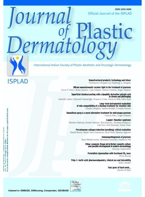
Vol. 4, n. 3, September-December 2008<br />
Indexed in: EMBASE, EMNursing, Compendex, GEOBASE<br />
ISSN 2035-0686<br />
Nanostructured products: technology and future<br />
Pierfrancesco Morganti, Lee Yuanhong, G. Morganti<br />
308 nm monochromatic excimer light in the treatment of psoriasis<br />
Steven P. Nisticò, Rosita Saraceno, Catia Schipani, Antonio Costanzo, Sergio Chimenti<br />
Su<strong>per</strong>ficial chemical peeling with a lipophilic derivative salicylic acid<br />
in chrono and photoaging<br />
Gabriella Calabrò, Emanuela Fiammenghi, Viviana Lo Conte, Serena La Bella, Fabio Ayala<br />
Long-term instrumental evaluation<br />
of skin compatibility of a shaving treatment for sensitive skin<br />
Claudia Cartigliani, Adriana Bonfigli, Fernanda Distante<br />
Nanosilicon spray is a novel alternative treatment for mild plaque psoriasis<br />
Catia de Felice, Sergio Chimenti<br />
Laugier-Hunziker syndrome<br />
Miroslava Kadurina, Borislav Dimitrov, Slava Bojinova, Antoaneta Jordanova,<br />
Ivan Litov, Vesel Kantarjiev, Stoyan Tonev<br />
Percutaneous collagen induction (needling): critical evaluation<br />
Daniela Beretta, Matteo Tretti Clementoni, Silvia Pinelli, Massimo Signorini<br />
Immunopathogenesis of psoriasis<br />
Maria Michela Lavieri, Alessandra Frezzolini, Ornella De Pità<br />
Ethno-cosmesis: Argan oil in Berber cosmetic culture<br />
and possible development in modern dermatology<br />
Giuseppe Imprezzabile<br />
Periorbital rejuvenation with fractional CO 2 laser<br />
Dvora Ancona<br />
Poly-L-lactic acid: pharmacodynamics, clinical use and tolerability<br />
Delia Colombo<br />
Four years of hard work...<br />
Antonio Di Maio
Nel 2009 l’ISPLAD compie 10 anni di vita. Sono stati dieci anni di grandi successi, di traguardi raggiunti, di pro g e t t i<br />
importanti, di crescita <strong>per</strong> la dermatologia. Da otto Soci fondatori siamo arrivati ad oggi a circa 2000 iscritti.<br />
Nessun’altra Associazione dermatologica è riuscita in questa impresa. L’età media dei soci è di circa 40 anni e <strong>per</strong> questo<br />
vuol dire che si tratta di una Società che raccoglie soprattutto i giovani, la forza del futuro. È una Associazione trasversale,<br />
infatti è composta da dermatologi libero - p rofessionisti, ambulatoriali, ospedalieri, universitari, questo è stato un<br />
a l t ro elemento di successo. È stata la prima Associazione a valorizzare la figura del dermatologo come regista e portavoce<br />
di tutte le problematiche legate all’aging cutaneo. In questi dieci anni è stata svolta una intensa attività didattica-formativa<br />
con oltre 200 corsi di aggiornamento e due Congressi Internazionali. Molti giovani dermatologi hanno appre s o<br />
i primi insegnamenti sull’uso dei laser, dei peeling, dei filler, ecc…; hanno compreso l’importanza della cosmetologia e<br />
degli integratori. Ma il merito più grande che sarà riconosciuto all’ISPLAD sarà quello di aver dato la possibilità ad un<br />
medico di non farsi<br />
e t i c h e t t a re come “estetista laure a t o”, di aver<br />
fatto di tutto <strong>per</strong> sostituire il brutto e<br />
declassante termine di “D e rm a t o l o g i a<br />
E s t e t i c a” con “D e rmatologia Plastica” .<br />
L’ISPLAD avrà sempre il merito di aver<br />
fatto capire che il medico non deve “r i e mp<br />
i re” la ruga ma la deve “c u r a re”; mai parl<br />
a re di una terapia antiaging ma di una<br />
“s t r a t e g i a” antiaging. L’ISPLAD ha sempre<br />
sostenuto che la prima cosa da considerare<br />
nella scelta di una terapia antiaging sono<br />
gli effetti collaterali e non i risultati che si<br />
possono ottenere, <strong>per</strong>ché non ci si trova a<br />
c o m b a t t e re una malattia ma ad aiutare un<br />
soggetto sano che tale deve re s t a re .<br />
Adesso bisogna pensare ai prossimi dieci<br />
anni dell’ISPLAD e credo che una buona<br />
strada da <strong>per</strong>c o r re re sia quella che porterà<br />
alla nascita e allo sviluppo della “D e rm a -<br />
tologia Rigenerativa” cioè di <strong>tutti</strong> quei mezzi<br />
e terapie capaci di stimolare le cellule cutanee<br />
e rigenerarsi a sostituire i tessuti invecchiati.<br />
Per questo da adesso la “D e rm a t o -<br />
logia Rigenerativa” diventerà la nuova missione<br />
della Dermatologia Plastica.<br />
L’ISPLAD sarà la Società Scientifica che si<br />
occu<strong>per</strong>à a 360° di “D e rmatologia Rigenerat<br />
i v a ” e di <strong>tutti</strong> quei mezzi che potranno re alizzarla:<br />
dall’alta tecnologia strumentale alla<br />
cosmetologia, alla dermonutrologia, ai fill<br />
e r, all’uso delle cellule staminali, ecc..<br />
Sono sicuro che ancora una volta l’ISPLAD<br />
indicherà una strada che molti seguiranno<br />
e altri imiteranno.<br />
In 2009, ISPLAD will celebrate its 10th birthday. It has been 10 years of great success, of<br />
achieved goals, of important projects, and of immense progress in the field of dermatology.<br />
Starting with only 8 founding members, today, we have evolved into an ever-growing association<br />
of approximately 2,000 registered members. No other dermatological association has ever<br />
been so successful in such a huge undertaking.<br />
The average age of our members is estimated to be about 40. This clearly shows that ISPLAD<br />
attracts and appeals to young professionals, the true driving force of our future. We are a<br />
dynamic association comprised of dermatologists who work as liberal professionals, in outpatient<br />
clinics, in hospitals, and at universities. And we are extremely proud of this diversity.<br />
We were the first association to emphasize the role of the dermatologist as a director of and<br />
spokes<strong>per</strong>son for issues related to cutaneous aging. In these 10 years, an intensive educational<br />
program with over 200 refresher courses and 2 international congresses has been developed.<br />
Many young dermatologists have mastered the use of lasers, peeling, fillers, e.t.c; they have<br />
understood the importance of cosmetology and supplements. But <strong>per</strong>haps ISPLAD’s biggest<br />
achievement is to have given doctors a way to avoid the label “graduated aesthesists”, and to<br />
have done everything possible to replace the degrading term “Aesthetic Dermatology” with the<br />
term “Plastic Dermatology”.<br />
ISPLAD will always be known for having clearly demonstrated that a doctor’s main job is not<br />
to fill the wrinkles, but to cure; that it is better to concentrate on an anti-aging strategy than<br />
an anti-aging therapy. ISPLAD’s has always maintained that the very first things to consider<br />
when choosing an anti-aging therapy are the possible side-effects and not the results that can<br />
be achieved. We are not fighting a disease, but rather, helping healthy subjects that must<br />
remain so.<br />
It is now time to plan the next 10 years of ISPLAD and I think that a good path to explore<br />
would be the one leading to the birth and development of “Regenerative Dermatology”. That<br />
is, all the methods and therapies that are capable of stimulating cutaneous cells to regenerate<br />
themselves to substitute old tissues. Therefore, “Regenerative Dermatology” shall become the<br />
new mission of Plastic Derm a t o l o g y. ISPLAD will dedicate itself to “Regenerative<br />
Dermatology” and pursue all of the measures necessary to <strong>per</strong>fect our understanding of it.<br />
These measures include instrumental technology, cosmetology, dermonutrology, fillers, and the<br />
use of stem cells e.t.c.<br />
I am convinced that ISPLAD will once again pave an innovate path that many will follow and<br />
others will adopt as a benchmark of standard.<br />
Antonino Di Pietro<br />
Journal of Plastic Dermatology 2008; 4, 3 249
Journal of Plastic Dermatology<br />
Editor<br />
Antonino Di Pietro (Italy)<br />
Editor in Chief<br />
Francesco Bruno (Italy)<br />
Co-Editors<br />
Bernd Rüdiger Balda (Austria)<br />
Salvador Gonzalez (USA)<br />
Pedro Jaen (Spain)<br />
Associate Editors<br />
Francesco Antonaccio (Italy)<br />
Mariuccia Bucci (Italy)<br />
Franco Buttafarro (Italy)<br />
Ornella De Pità (Italy)<br />
Giulio Ferranti (Italy)<br />
Andrea Giacomelli (Italy)<br />
Alda Malasoma (Italy)<br />
Steven Nisticò (Italy)<br />
Elisabetta Perosino (Italy)<br />
Andrea Romani (Italy)<br />
Nerys Roberts (UK)<br />
Editorial Board<br />
Lucio Andreassi (Italy)<br />
Kenneth Arndt (USA)<br />
H.S. Black (USA)<br />
Lucia Brambilla (Italy)<br />
Günter Burg (Switzerland)<br />
Michele Carruba (Italy)<br />
Vincenzo De Sanctis (Italy)<br />
Aldo Di Carlo (Italy)<br />
Robin Eady AJ (UK)<br />
Paolo Fabbri (Italy)<br />
Ferdinando Ippolito (Italy)<br />
Giuseppe Micali (Italy)<br />
Martin Charles Jr Mihm (USA)<br />
Joe Pace (Malta)<br />
Lucio Pastore (Italy)<br />
Gerd Plewig (Germany)<br />
Riccarda Serri (Italy)<br />
Adele Sparavigna (Italy)<br />
Abel Torres (USA)<br />
Stefano Veraldi (Italy)<br />
Umberto Veronesi (Italy)<br />
Managing Editor<br />
Antonio Di Maio<br />
English editing<br />
Rewadee Anujapad<br />
Direttore Responsabile Pietro Cazzola<br />
Direttore Generale Armando Mazzù<br />
Direttore Marketing Antonio Di Maio<br />
Consulenza grafica Piero Merlini<br />
Impaginazione Clementina Pasina<br />
R e g i s t r. Tribunale di Milano n. 102 del 14/02/2005<br />
Scripta Manent s.n.c. Via Bassini, 41 - 20133 Milano<br />
Tel. 0270608091/0270608060 - Fax 0270606917<br />
E-mail: scriman@tin.it<br />
Abbonamento annuale (3 numeri) Euro 39,00<br />
Pagamento: conto corrente postale n. 20350682<br />
intestato a: Edizioni Scripta Manent s.n.c.,<br />
via Bassini 41- 20133 Milano<br />
Stampa: Arti Grafiche Bazzi, Milano<br />
Sommario<br />
pag. 253 Nanostructured products: technology and future<br />
Pierfrancesco Morganti, Lee Yuanhong, G. Morganti<br />
pag. 263 308 nm monochromatic excimer light in the treatment of psoriasis<br />
Steven P. Nisticò, Rosita Saraceno, Catia Schipani, Antonio Costanzo, Sergio Chimenti<br />
pag. 271 Su<strong>per</strong>ficial chemical peeling with a lipophilic derivative salicylic acid<br />
in chrono and photoaging<br />
Gabriella Calabrò, Emanuela Fiammenghi, Viviana Lo Conte, Serena La Bella, Fabio Ayala<br />
pag. 277 Long-term instrumental evaluation of skin compatibility<br />
of a shaving treatment for sensitive skin<br />
Claudia Cartigliani, Adriana Bonfigli, Fernanda Distante<br />
pag. 281 Nanosilicon spray is a novel alternative treatment for mild plaque psoriasis<br />
Catia de Felice, Sergio Chimenti<br />
pag. 285 Laugier-Hunziker syndrome<br />
Miroslava Kadurina, Borislav Dimitrov, Slava Bojinova, Antoaneta Jordanova,<br />
Ivan Litov, Vesel Kantarjiev, Stoyan Tonev<br />
pag. 291 Induzione <strong>per</strong>cutanea del collagene (needling): valutazione critica<br />
Daniela Beretta , Matteo Tretti Clementoni, Silvia Pinelli, Massimo Signorini<br />
pag. 305 Immunopatogenesi della psoriasi<br />
Maria Michela Lavieri, Alessandra Frezzolini, Ornella De Pità<br />
pag. 309 Etnocosmesi: l’olio di Argan (Argania spinosa (L.) Skeels) nella cultura<br />
cosmetica berbera e possibile sviluppo nella dermatologia moderna<br />
Giuseppe Imprezzabile<br />
pag. 315 Ringiovanimento contorno occhi con laser frazionato<br />
Dvora Ancona<br />
pag. 321 Acido L-polilattico: farmacodinamica, clinica e tollerabilità<br />
Delia Colombo<br />
pag. 341 Quattro anni di duro lavoro...<br />
Antonio Di Maio<br />
È vietata la riproduzione totale o parziale,<br />
con qualsiasi mezzo, di articoli, illustrazioni<br />
e fotografie senza l’autorizzazione scritta dell’Editore.<br />
L’Editore non risponde dell’opinione espressa dagli<br />
Autori degli articoli.<br />
Ai sensi della legge 675/96 è possibile in qualsiasi<br />
momento opporsi all’invio della rivista<br />
comunicando <strong>per</strong> iscritto la propria decisione a:<br />
Edizioni Scripta Manent s.n.c.<br />
Via Bassini, 41 - 20133 Milano<br />
Journal of Plastic Dermatology 2008; 4, 3 251
Pierfrancesco Morganti 1<br />
Lee Yuanhong 2<br />
G. Morganti 3<br />
1 Professor of Applied Cosmetic Dermatology,<br />
II Università di Napoli<br />
Head of R&D, Mavi Sud s.r.l., Aprilia (LT)<br />
Visiting Professor of China Medical<br />
University Shenyang<br />
Secretary general of I.S.C.D.<br />
2 No.1 Hospital of China Medical<br />
University Shenyang (PRC)<br />
3 Technical Director Mavi Sud s.r.l., Aprilia (LT)<br />
Nanostructured products:<br />
technology and future<br />
hat is<br />
W nanotechnology<br />
SU M M A R Y<br />
Nanotechnology is understood<br />
as the characterization,<br />
type, production and use<br />
of structures and systems the<br />
exact size and shape of which<br />
must be measured on a nanometric<br />
scale. 1<br />
Nanotechnologies are the set of<br />
methods and techniques for<br />
processing matter on an atomic<br />
and molecular scale to create<br />
p roducts presenting special<br />
and improved chemical-physical<br />
features as compared to<br />
conventional ones.<br />
What size is a nanometer (nm)?<br />
A nanometer corresponds to one billionth of a<br />
meter (Figure 1).<br />
Considering that a bacterium measures 1000<br />
nm and that the distance between two carbon<br />
atoms of an organic molecule is 0.15 nm, one<br />
can easily comprehend how difficult it must be<br />
Figure 1. The nanometric scale.<br />
Nanostructured products:<br />
technology and future<br />
Nanotechnologies are the set of methods and techniques for processing matter on an<br />
atomic and molecular scale to create products presenting special and improved chemical-physical<br />
features as compared to conventional ones. With the current technological<br />
k n o w - h o w, it is already possible to build diff e rent types of nanostructures (DNA, proteins,<br />
cells or viruses, etc.) on special chips that can help to better understand the function<br />
<strong>per</strong>formed by proteins in cells. Thanks to nanotechnology, it is now possible to modify<br />
the chemistry and the topography of the substratum of cell cultures so as to enable<br />
them to mime the extracellular matrix, in such a way that the same signals used by cells<br />
in vivo are released. With the use of other technological platforms, it is possible to obtain<br />
thin nanostructured films organized as nets, capable of providing a huge surface that is<br />
available for interaction with the skin tissue and the external environment. An example<br />
of this is the production of 240 nm chitin nanofibrils capable of accelerating in a physiological<br />
manner the reparation of damaged skin. Chitin nanofibrils can also be used as<br />
c a rriers for pharmacological or cosmetic use.<br />
KE Y W O R D S: Nanotechnologies, Chitin nanofibrils<br />
for the industry to work with such scales!<br />
Nevertheless, it is common knowledge that<br />
n a n o m a t e r i a l s p resent mechanical, optical,<br />
chemical, magnetic or electric pro<strong>per</strong>ties that<br />
are completely different from the raw material<br />
from which they are generated. 2<br />
Journal of Plastic Dermatology 2008; 4, 3 253
254<br />
P. Morganti, L. Yuanhong, G. Morganti<br />
ndustry, nanotechnology<br />
I and market<br />
For these reasons, the ability to handle<br />
selectively materials of nanometric size has led<br />
the industry to develop raw materials with new<br />
p ro<strong>per</strong>ties and significant advantages as comp<br />
a red to the m a c roscopic world.<br />
New products are being designed, or have been<br />
designed, which present such innovative feat<br />
u res, also in terms of their applications, as to<br />
have already influenced our current lifestyle.<br />
A case in point is the wireless telephone!<br />
Other examples of innovative n a n o - p roducts p resent<br />
on the market are: sunscreens, some plastic<br />
materials with higher eco-eff i c i e n c y, coating<br />
materials that are more resistant to corro s i o n ,<br />
and, naturally, microchips for cell phones, etc.<br />
How large is this market?<br />
A c c o rdi ng to the US National Science<br />
Foundation, the global n a n o - p ro d u c t s m a r k e t<br />
will register a turnover of over 1,000 billion dollars<br />
a year in the next 10-15 years! (Figure 2).<br />
he technological platforms<br />
T<br />
Thanks to current technological<br />
know-how, it is already possible to build different<br />
types of nanostructures (DNA, proteins,<br />
cells or viruses, etc.) on special chips that can<br />
help to better understand the function <strong>per</strong>formed<br />
by proteins in cells.<br />
One need only bear in mind that histones are<br />
proteins which the DNA ribbon contained in<br />
every cell winds around (Figure 3), and that<br />
Journal of Plastic Dermatology 2008; 4, 3<br />
Figure 2. Nanotechnology investment forecasts.<br />
chromosomes are huge DNA molecules wound<br />
up tidily around themselves like balls of yarn<br />
(Figure 4). Since a mistake in winding can prevent,<br />
for instance, cell reproduction, the simple<br />
de-regulation of protein activity represents an<br />
essential etiological factor in the pathogenesis of<br />
many diseases.<br />
Thanks to nanotechnology, it is now possible to<br />
modify the chemistry and the topography of<br />
the substratum of cell cultures so as to enable<br />
them to mime the extracellular matrix, in such a<br />
way that the same signals used by cells in vivo<br />
are released (Figure 5). With the use of other<br />
technological platforms, it is possible to obtain<br />
thin nanostructured films organized as nets,<br />
capable of providing a huge surface that is available<br />
for interaction with the skin tissue and the<br />
external environment (Figure 6).<br />
Figure 3. Graphic representation of DNA. Figure 4. DNA ribbon wrapped around histones.
Figure 5. The cell with its antennae that send out signals indispensable to its daily life.<br />
Figure 7. Skin repair through the use of a particular gel containing chitin nanofibrils.<br />
This was achieved by MAVI with the production<br />
of 240 nm chitin nanofibrils capable of<br />
accelerating in a physiological manner the reparation<br />
of damaged skin (Figure 7).<br />
Figure 8. Chitin nanofibril, a natural product obtained from the chelae of shellfish.<br />
Nanostructured products: technology and future<br />
Figure 6. Nanostructure of a chitosan film.<br />
hitin nanofibrils<br />
C<br />
Chitin nanofibrils (Figure 8), of an<br />
average size of 240 nm, can also be used as carriers,<br />
since they can release in a controlled manner<br />
active principles for pharmacological or<br />
cosmetic use, such as lutein, for instance.<br />
Chitin is a known natural polyglucoside that is<br />
easily recognized and hydrolized by the skin’s<br />
cutaneous enzymes, while lutein, a natural oxicarotenoid,<br />
is an antioxidant capable of enriching<br />
the skin’s antioxidant system. If these two<br />
molecules are pro<strong>per</strong>ly treated, the complex<br />
resulting from their bonds can surely <strong>per</strong>form<br />
an interesting protective role on the skin and<br />
mucosae. In fact, it is capable of penetrating<br />
very easily through the skin’s layers, if well<br />
dosed and vehicled, without causing toxic side<br />
effects and serving, on the contrary, as an energy<br />
deposit (Figure 9).<br />
It is interesting to underscore the ease with<br />
which these chitin nanofibrils can be included<br />
both in the natural and in the artificial fibers to<br />
generate entirely innovative tissues (Figure 10).<br />
anotechnology and development<br />
N<br />
This growth will be determined by the<br />
development and type of technology capable of<br />
generating products which, being unique for<br />
their features and pro<strong>per</strong>ties, will be able to<br />
Journal of Plastic Dermatology 2008; 4, 3<br />
255
256<br />
P. Morganti, L. Yuanhong, G. Morganti<br />
Figure 9. Transcutaneous penetration of chitin nanofibrils. Figure 10. Chitin nanofibrils for innovative tissues.<br />
reach the global market after being developed<br />
in a laboratory.<br />
Thus, the diff e rent technologies, from electro n i c s<br />
to optics, from information technology to biological<br />
sciences, which are all geared towards the<br />
c reation of products, when nanostructured, have<br />
generated products so innovative as to be distributed<br />
at world level in very little time.<br />
Nanotechnology will therefore be able to spearhead<br />
innovation, giving new impetus to a globalized<br />
and… ever faster trade!<br />
While some metals, such as Ti, Cu, Ni and Sn,<br />
have proven to be more pliable and stronger<br />
when nanostructured, many nano-assembled<br />
products have revealed exceptional efficiency<br />
features. These include many substances used<br />
in catalysis, various su<strong>per</strong>conductors, batteries<br />
with a higher energy charge and longer duration,<br />
membranes with higher <strong>per</strong>meation features,<br />
as well as some biomedical structures or<br />
particular microcircuits to be used in the field<br />
of security and for brand protection. 3<br />
nnovative nanotechnologies<br />
I<br />
Some examples of such innovative<br />
technologies are electronic nano-tubes, structures<br />
used in information technology and in<br />
many medical fields like molecular diagnostics<br />
based on nanostructured biosensors (Figure<br />
11), or nanostructures capable of transporting<br />
drugs or active principles for cosmetic use 4 , or<br />
polymers used for diagnostic or monitoring<br />
purposes, etc. 5<br />
Nanotechnology has there f o re inspired the<br />
development of exceptionally small and low-<br />
Journal of Plastic Dermatology 2008; 4, 3<br />
energy sensors that have made it possible to<br />
create wireless sensors, which are useful in various<br />
applications, from CBRNE (the Chemical,<br />
Biological, Radiological, Nuclear and Explosives<br />
industry) to medical diagnostics.<br />
Furthermore, in the area of security, particular<br />
nano-sensors have been introduced which,<br />
inserted in particular systems of fluids, are<br />
starting to be used to protect and alert infrastructures<br />
in case of natural disasters or terrorist-related<br />
events. 6<br />
Also crucial for our growth is the ability to capture,<br />
file and analyze a great deal of information.<br />
Thus, the development of these new systems of<br />
sensors associated with the use of state-of-the-<br />
Figure 11. EAP sensors designed with Electroactive Polymers.
Figure 12.<br />
Finland<br />
Figure 13.<br />
United States<br />
Sweden<br />
Denmark<br />
Taiwan<br />
Singapore<br />
Iceland<br />
Switzerland<br />
Norway<br />
Australia<br />
Netherlands<br />
Japan<br />
Great Britain<br />
Competitiveness classification<br />
2004 2005 2004 2005<br />
1<br />
2<br />
3<br />
5<br />
4<br />
7<br />
10<br />
8<br />
6<br />
14<br />
12<br />
9<br />
11<br />
1<br />
2<br />
3<br />
4<br />
5<br />
6<br />
7<br />
8<br />
9<br />
10<br />
11<br />
12<br />
13<br />
Canada<br />
Germany<br />
Portugal<br />
Ireland<br />
Spain<br />
France<br />
Jordan<br />
Greece<br />
Botswana<br />
China<br />
India<br />
15 14<br />
13 15<br />
24 22<br />
30 26<br />
23 29<br />
27 30<br />
35 45<br />
37 46<br />
Italy 47 47<br />
45 48<br />
46 49<br />
55 50<br />
art information technology will step up our<br />
ability to quickly <strong>per</strong>ceive, understand and<br />
process complex messages that are hard to<br />
interpret.<br />
arketing nano-structured<br />
M products<br />
The ability to market nano-structured<br />
products will depend on the ability of companies<br />
to produce and control this new class of<br />
products, meeting the needs of both man and<br />
the environment, on the ability of governments<br />
to regulate their production and use quickly<br />
Nanostructured products: technology and future<br />
and effectively, and on the ability of the products<br />
themselves to meet the needs and expectations<br />
of consumers.<br />
That is why it is necessary for all these nanoproducts<br />
to be designed and sold in a way that<br />
fully respects the health of consumers and the<br />
environment; in other words, they must be bio<br />
and ecocompatible.<br />
In order to reach these objectives, industries<br />
must create new plants, investing the necessary<br />
capital, while governments must support adequately<br />
and with rapid decisions these industrial<br />
efforts also by introducing new services.<br />
On the other hand, both industries and the<br />
Italian government should significantly increase<br />
investments in research and development to<br />
keep pace with more virtuous European countries<br />
and with the US (Figure 12). In fact, Italy<br />
must be more competitive at the international<br />
level to maintain the level of wellbeing reached<br />
by its citizens (Figure 13).<br />
isks/benefits of nanoproducts<br />
R<br />
Any production process that generates<br />
profit inevitably entails risks and benefits.<br />
Naturally, this also applies to all nanostructured<br />
processes. When assessing the risks/benefits of<br />
these new chemical structures, the products<br />
must be distinguished according to two main<br />
categories: nanoderivatives present in nature<br />
and manmade ones.<br />
It is therefore necessary to determine whether<br />
they can be absorbed through the skin or<br />
mucosae, controlling their possible topical<br />
and/or systemic toxicity; needless to say, this<br />
should be done after studying their physicalchemical<br />
pro<strong>per</strong>ties, such as for instance: (a)<br />
the state of distribution of the single nanoparticles;<br />
(b) the state of agglomeration and size of<br />
their crystal structure; (c) the composition and<br />
chemical features of the developed surface; (d)<br />
the electrical charges present in their structure;<br />
and (e) their possible porosity.<br />
Synthetic nanostructures include, for instance,<br />
fullerene which, depending on whether or not<br />
it includes OH groups in its structure, displays<br />
completely different chemical/physical pro<strong>per</strong>ties<br />
and behaviours, also developing a different<br />
tendency for transcutaneous penetration.<br />
Another example of a product “created” in the<br />
lab is the carbon nanotube which is also used in<br />
the biomedical field.<br />
Journal of Plastic Dermatology 2008; 4, 3<br />
257
258<br />
P. Morganti, L. Yuanhong, G. Morganti<br />
These, like other synthetic nanostructures, penetrate<br />
inter- or intracutaneously in the conventional<br />
way (Figure 14). Trials showed that nanotubes<br />
can be inserted in the keratinocytes at<br />
different levels, remaining intact within biological<br />
structures (Figures 15 and 16).<br />
A completely different behaviour was observed<br />
in natural nanostructures like chitin nanofibril<br />
7,8 (Figure 17), which, like polyglucoside, is<br />
rapidly catabolized and reduced to glucose and<br />
glucosamine by the enzymes of the skin or of<br />
the mucosae following the normal catabolic<br />
process 9 (Figure 18).<br />
In fact, these nanofibrils, on which chemical-<br />
Figure 15. Penetration of nanotubes through the skin layers. Detail.<br />
physical as well as biological studies have<br />
already been conducted, appear to be useful as<br />
active carriers to be employed in cosmetics as<br />
well as in the area of smart bio tissues. 10<br />
Naturally, in order to be applied to the skin for<br />
pharmaceutical or cosmetic purposes, all these<br />
nanostructures must be introduced in suitable<br />
vehicles capable of transporting them through<br />
the cell layers by means of penetration.<br />
It is therefore possible to create macro, micro or<br />
nanoemulsions made up of different sized particles,<br />
the shape and size of which must be<br />
known (Figure 19).<br />
Figure 17. Chitin nanofribril.<br />
Journal of Plastic Dermatology 2008; 4, 3<br />
Figure 14. Skin penetration as a target for studying the possible toxicity of active principles.<br />
Figure 16. Penetration of nanotubes through the skin layers. Detail.<br />
Chitina Sol. Fisiologica<br />
Figure 18. The cell activity carried out by chitin nano fibtrils corresponds <strong>per</strong>fectly<br />
with the activity carried out by a normal vehicle.
Figure 19.<br />
Naturally, it is necessary to prove by means of<br />
trials the types and features of the emulsion<br />
considered and the size of the particles obtained<br />
(Figures 20 and 21) following the model, for<br />
instance, of NANOCREAM ® by Sinerga. 11<br />
Figure 20. Characterization of the micelles of a SINERGA nanocream (Nanocream).<br />
Figure 21. Characterization of the micelles of a SINERGA nanocream (Nanocream).<br />
In any event, it is important to underscore that<br />
nanotechnologies can surely offer new benefits<br />
to society, be a source of new progress and create<br />
new jobs, thus improving also the quality of<br />
our life.<br />
However, it is necessary for research to dedicate<br />
more resources to assess their safety and determine<br />
the impact that nanomaterials will have<br />
on the environment and on health.<br />
Therefore, it is important that EU Member<br />
States focus their resources on developing the<br />
methodologies to be followed, while industries<br />
monitor their products and pro d u c t i o n<br />
methodologies, verifying the impact they have<br />
on human health and the environment.<br />
To this end, European chemical industries have<br />
actively participated in and supported specific<br />
national and international projects involving<br />
n a n o p roducts (N a n o c a re: www.nanopartikel. info<br />
and NanoSafe2: www. n a n o s a f e . o rg) to verify<br />
their potential effects on man and the enviro n-<br />
m e n t.<br />
1 2<br />
Nanostructured products: technology and future<br />
There are many challenges to be overcome in<br />
the initiatives against and in the ones in favour<br />
of the more or less rapid distribution of<br />
nanoproducts (Figure 22).<br />
C onclusions<br />
In order to market nanostructure d<br />
p roducts and develop the related pro d u c t i o n<br />
p rocesses, in the short term both the industry<br />
and governments shall have to invest on building<br />
new infrastructure and utilize venture capital.<br />
On the other hand, the implementation<br />
and long-term success of nanotechnologies<br />
shall depend on a rational, informed and transp<br />
a rent dialogue among all the parties involved,<br />
which shall have to try to understand both the<br />
potential for developing a green and sustainable<br />
chemistry and the potential negative<br />
e ffects on human health and the enviro n m e n t<br />
that may arise.<br />
Side effects must be reduced and aspects that<br />
may help to improve the effectiveness of the<br />
p roducts must be enhanced as much as possible.<br />
The constant and factual collaboration of governments,<br />
universities and industries will lead<br />
to organizing new technological platforms and<br />
new products capable of enhancing the quality<br />
of life of individuals and of society as a whole.<br />
This project is part of the 7t h E u ro p e a n<br />
Framework Programme (FP7) in which platform<br />
Journal of Plastic Dermatology 2008; 4, 3<br />
259
260<br />
P. Morganti, L. Yuanhong, G. Morganti<br />
4 is devoted entirely to nanotechnologies<br />
(Theme 4: Nanoscience, Nanotechnologies, Materials<br />
and New Production Technologies) with<br />
special projects targeted especially to European<br />
SMEs (Small and Medium-sized companies). 13<br />
This important opportunity should be seized by<br />
presenting research projects financed by the EU<br />
with some 10 billion Euros in the next 6 years.<br />
Another opportunity has been the 8 th Conference<br />
of the International Society of Cosmetic<br />
Dermatology (I.S.C.D.) held this year in Beijing<br />
from 20-23 October. This important meeting<br />
organized by the Associations of dermatologists<br />
and Chinese Doctors has been attended by the<br />
w o r l d ’s leading ex<strong>per</strong>ts in Dermatology,<br />
Cosmetology and Wellbeing in general (Figure<br />
23). Indeed, many sessions have been dedicated<br />
to Food Supplements and special foods,<br />
health tissues and, naturally, Cosmeceuticals<br />
and Natural Cosmetics.<br />
In a completely globalized world, it is necessary<br />
now to compete also with the Chinese people,<br />
not only in the area of nanotechnologies but in<br />
all rapidly expanding sectors.<br />
R eferences<br />
1. Morganti P. Nanoscenza ed efficacia dei<br />
prodotti cosmetici Natura e Benessere 2002; 2:258-260<br />
2. Kenny JM. Nanotechnology and intelligent textiles: actual<br />
situation and future <strong>per</strong>spective. In: Atti Congresso<br />
NanoItaltex 2006: le nanotacnologie <strong>per</strong> il tessile italiano<br />
(www.unipg.it/material). Milano 15, 16 Nov. 2006<br />
3. Gallucci S. Anticontraffazione e controllo dei mercati<br />
grigi: un approccio nanotecnologico. In: Atti Congresso<br />
NanoItaltex 2006: le nanotacnologie <strong>per</strong> il tessile italiano.<br />
(www.singular-id.com) Milano 15, 16 Nov. 2006<br />
4. Morganti P. Proprietà e prospettive d’uso delle nanofibrille<br />
di chitina. In: Workshop CNR dalle Micro alle<br />
NanoTecnologie 30-31 Gennaio 2007 (morganti@mavicosmetics.it)<br />
5. De Rossi D. Tessuti elettronici basati su polimeri elettroattivi:<br />
materiali, dispositivi ed applicazioni. In: Atti<br />
Congresso NanoItaltex 2006: le nanotacnologie <strong>per</strong> il tessile<br />
italiano 2006 (www.nanotec.it)<br />
6. Zangani D. Polyfunctional Technical textiles against natural<br />
hazards. In : Atti Congresso NanoItaltex 2006: le nanotecnologie<br />
<strong>per</strong> il tessile italiano 2006 (donato.zangani@<br />
dappolonia.it)<br />
7. Morganti P. Muzzarelli R.A.A.A, Muzzarelli C. and<br />
Morganti G. Le nanofibrille di chitina: una realtà tutta italiana.<br />
Cosmetic Technology 2006; 9:21-25<br />
Journal of Plastic Dermatology 2008; 4, 3<br />
Figure 22. The commercial challenges and initiatives typical of nanotechnologies.<br />
Figure 23. Beijing.<br />
8. Morganti P. Muzzarelli R.A.A.A, Muzzarelli C. and<br />
Morganti G. Chitin nanofibrils: a natural compound for<br />
innovative cosmeceuticals 2007. In print on: C&T<br />
9. Morganti P, Mattioli-Belmonte M, Del Ciotto P, Zizzi A,<br />
Lucarini G, Giantomassi F. e Biagini G. Chitosan-linked<br />
chitin nanofibril matrix to enhance wound repair enhancement.<br />
In print on: J Appl Cosmetol 2007<br />
10. Morganti P. Muzzarelli R.A.A.A, Muzzarelli C. Multifunctional<br />
use of innovative chitin derivatives for skin care.<br />
J.Appl. Cosmetol 2006; 24:105-114<br />
11. CEFIC. Position pa<strong>per</strong> on nanomaterials, 2006<br />
(www.cefic.org) aoj@cefic.be<br />
12. Guglielmini G. Evaluating droplet size in nanoemulsions<br />
from a novel emulsifier system. C&T 2006; 121: 67-<br />
74<br />
13. CEFIC. Position pa<strong>per</strong> on nanomaterials, 2006<br />
(www.cefic.org) aoj@cefic.be<br />
14. Draft Working Document Theme 4. Nanosciences, nanotechnologies,<br />
material and new production, 2006
Steven P. Nisticò<br />
Rosita Saraceno<br />
Catia Schipani<br />
Antonio Costanzo<br />
Sergio Chimenti<br />
Department of Internal Medicine<br />
Chair of Dermatology<br />
Policlinico Tor Vergata<br />
University of Rome, “Tor Vergata”, Italy<br />
308 nm monochromatic excimer light<br />
in the treatment of psoriasis<br />
SU M M A R Y<br />
I ntroduction<br />
Psoriasis is a distressing, chronic skin<br />
disease characterised by keratinocyte hy<strong>per</strong>proliferation<br />
with the presence of acute and chronic<br />
inflammatory cells. Treatment for psoriasis<br />
include topical corticosteroids, tar, anthralin,<br />
vitamin D analogues, tazarotene and salicylic<br />
acid. 1 UVB phototherapy and PUVA photochemotherapy<br />
are also effective and well documented<br />
therapeutic options for psoriasis. In particular<br />
Narrow Band phototherapy involves irradiation<br />
on the UVB spectrum at a wavelength<br />
between 300 and 313 nm. In this spectrum, the<br />
UVB activity is effective and safe, and offers<br />
long term remission.<br />
Use of 308 nm excimer laser for psoriasis has<br />
been reported since 1997. 2 Furthermore the<br />
efficacy of the light produced by xenon-cloride<br />
excimers at 308 nm has been reported by several<br />
authors 3-10 in the treatment of stable forms of<br />
localized plaque psoriasis with a satisfactory<br />
benefit/risk profile.<br />
308 nm monochromatic excimer<br />
light in the treatment of psoriasis<br />
B a c k g round: Various reports showed the efficacy of Narrow Band UVB (311-313<br />
nm) and excimer laser (308 nm) in the treatment of psoriasis.<br />
Objective: To prove the efficacy of light produced by xenon-cloride excimer at 308<br />
nm (Monochromatic Excimer Light, MEL) in the treatment of stable forms of<br />
localized plaque psoriasis.<br />
Patients and methods: This study was an open trial with 152 patients affected with<br />
stable mild to moderate plaque psoriasis (PASI score between 4 and 12) were<br />
t reated with a weekly session of MEL A total number of 6-16 sessions was<br />
p e rformed with a dose increase according to patient phototype and re s p o n s e .<br />
Results: 152 patients were enrolled in the study and 149 completed the pro t o c o l .<br />
Patients were followed up every two weeks, 57 patients for one-year and 92 patients<br />
for 6 months. After 4 months there was complete remission in 87 patients, partial<br />
remission in 37 and moderate improvement in 25 patients.<br />
Conclusions: These pre l i m i n a ry results suggest that MEL can be considered as a<br />
valid option for treatment of selected forms of localized plaque psoriasis.<br />
KE Y W O R D S: Psoriasis, MEL, 308 nm<br />
Some investigators showed that repeated treatments<br />
may achieve a prolonged and stable relapse<br />
free disease, 7 through a modulation of the<br />
local immune response 6 , T cell depletion and<br />
alterations in apoptosis related molecules. 11<br />
To confirm a previous study we <strong>per</strong>formed an<br />
open trial in 152 patients affected by localized<br />
psoriasis.<br />
aterials and methods<br />
M<br />
The “excimer” is an excited dimer, a<br />
molecule formed by the combination of two<br />
atoms, a noble gas Xenon and Cloride. The<br />
excitation of the molecule emits an ultraviolet<br />
photon at 308 nm.<br />
The excimer light (MEL, Excilite TM Deka<br />
Medical Lasers, Florence, Italy) is a monochromatic<br />
non coherent laser; it releases a power<br />
density of 48 Mw/cm2 at the distance of 15 cm<br />
Journal of Plastic Dermatology 2008; 4, 3 263
264<br />
S.P. Nisticò, R. Saraceno, C. Schipani, A. Costanzo, S. Chimenti<br />
from the source and it has an irradiation area of<br />
512 cm 2 .<br />
In this study, conducted in the Dermatology<br />
Department of University of Rome “Tor Vergata”,<br />
were recruited 152 patients of Fitzpatrick skin<br />
type 2-3 (80 men and 72 women) with stable<br />
mild to moderate plaque psoriasis, age range<br />
25-71 (mean age of 48) years. All patients<br />
signed informed consent to therapy, and were<br />
instructed to avoid any topical or systemis<br />
medication for psoriasis during the treatment<br />
<strong>per</strong>iod. Stable plaques were defined as those<br />
that had been present and unchanged for a<br />
minimum of 2 months. Disease duration was 1-<br />
30 years. Patients were affected from localized<br />
plaque psoriasis (Table 1) with an involvement<br />
of < 15% of the body surface, with a PASI score<br />
from 4 to 12. All patients gave informed consent,<br />
and were instructed to avoid session of<br />
any topical medication during MEL treatment.<br />
Patients with a history of skin cancers or photosensitivity<br />
related disorders were excluded.<br />
Patients who had been on systemic medication<br />
for less than 8 weeks, phototherapy for 4 weeks<br />
or had used topical treatments within the past 2<br />
weeks were also excluded. Photographs were<br />
taken at baseline, on clearing if clearing occurred,<br />
and after 4 months of treatment.<br />
Minimal Erythemal Dose (MED) was determined<br />
before treatment on healthy and unexposed<br />
skin on the dorsal area of the arms. MED was<br />
evaluated at increasing light dosage. The following<br />
doses were obtained (Tables 1, 2).<br />
The initial dose of MEL was calculated according<br />
to patient phototype, thickness of the<br />
squamous component of the psoriatic plaque<br />
and the anatomical area.<br />
A double MED dose was used for the lesions on<br />
the dorsal and posterior arms and a threefold to<br />
fourfold MED dose for more infiltrated lesions<br />
with a thicker squamous component localized<br />
in more photoresistant cutaneous regions such<br />
as elbows, knees, dorsal hands and palmo-plantar<br />
areas.<br />
Single plaques were irradiated protecting the<br />
surrounding non affected skin and petrolatum<br />
ointment was applied on the scaly patches to<br />
minimize light reflection prior to irradiation.<br />
Treatment was reported every 7 days with a<br />
dose increase of 250-500 mJ/cm 2 at each session<br />
according to patient phototype and<br />
response to previous session as observed with<br />
the flattening of the plaques and their reduction<br />
in size, erythema, scaling and pustules.<br />
Journal of Plastic Dermatology 2008; 4, 3<br />
Clinical examination was evaluated before<br />
every session and the PASI score 7 calculated<br />
every 2 weeks. A maximum of 16 sessions was<br />
<strong>per</strong>formed and usually phototherapy was<br />
discontinued when all lesions had completely<br />
cleared.<br />
Response rate was defined as complete (an<br />
improvement in the PASI score between 75 to<br />
100%), partial (50 to 75%) and slight (25 to<br />
50%).<br />
R esults<br />
In our study 152 patients were enrolled<br />
and 149 patients completed treatment with<br />
6-16 (mean 11) weekly sessions; 57 patients<br />
had one year and 92 patients 6 months of follow<br />
up. After 4 sessions of MEL all patients<br />
showed a PASI improvement. In those patients<br />
who had a complete remission we carried out a<br />
maintenance session of 2 J/cm2 every 14 days<br />
until we reached a 120day follow-up.<br />
Table 1.<br />
Phototype MED<br />
I 4 sec 200 m J/ cm 2<br />
II 5 sec 250 m J/ cm 2<br />
III 4 sec 300-350 m J/ cm 2<br />
IV 7-8 sec 350-400 m J/ cm 2<br />
MED: Minimal Erythematous Dose<br />
Table 2. MED at 308 nm.<br />
% patients<br />
Complete remission 58%<br />
Partial remission 25%<br />
Moderate improvement 17%<br />
Table 3. Clinical results.<br />
Site Number Number Mean Cumulative<br />
of patients of session UV dose<br />
(%) (mean) (J/cm 2 )<br />
Knees 55% 10 14<br />
Elbows 58% 9 13.5<br />
Legs 17% 13 9.25<br />
Palmoplantar areas 45% 12 15.5
Figure 1a and b.<br />
Localized psoriasis before<br />
and after MEL<br />
Figure 2a and b.<br />
Palmar psoriasis before<br />
and after MEL<br />
Figure 3a and b.<br />
Plantar psoriasis before<br />
and after MEL<br />
BEFORE<br />
1a 1b<br />
2a 2b<br />
3a 3b<br />
After 4 months 87 patients (58%) showed a<br />
complete remission, 37 patients (25%) had partial<br />
remission and 25 patients (17%) only<br />
moderate improvement.<br />
(Table 3).<br />
A ≥ 75% improvement in the PASI score (PASI<br />
75) was considered as the primary efficacy endpoint.<br />
This result was achieved in 98 patients<br />
after the fourth session, at week 4 and in 130 at<br />
week 8.<br />
This benefit was mantained at a 16-week follow-up<br />
in 125 patients, whereas all the patients<br />
that completed treatment maintained the achieved<br />
result.<br />
308nm monochromatic excimer light in the treatment of psoriasis<br />
AFTER<br />
A prolonged erythema (24-48h) was observed<br />
in 52 patients after the first and second session<br />
with a mild pruritic sensation in 39. These<br />
common side effects were well tolerated.<br />
Transient hy<strong>per</strong>pigmentation in the tre a t e d<br />
areas was noticed in 3 patients; this effect resolved<br />
spontaneously 2 weeks after the end of<br />
treatment. Formation of vesicles and oedema<br />
were observed in 1 patient after 3 treatments,<br />
which resolved following topical application of<br />
hydrocortisone 1% ointment for 3 days.<br />
However, the patients responded to the following<br />
sessions and completed successfully the<br />
treatment<br />
Journal of Plastic Dermatology 2008; 4, 3<br />
265
266<br />
S.P. Nisticò, R. Saraceno, C. Schipani, A. Costanzo, S. Chimenti<br />
D iscussion<br />
308-nm excimer light and laser treatments<br />
appear to offer relapse-free <strong>per</strong>iods and a<br />
prolonged stabilization of localized psoriasis .<br />
Bonis et al. 2 and other authors 3-5 showed that<br />
308 nm excimer light and laser therapy appear<br />
to be safe and effective for localized plaque psoriasis,<br />
comparable or better than that offered by<br />
standard topical therapy regimes and by 311nm<br />
UVB treatment.<br />
Feldman et al. 6 in 2002 reported results of a<br />
multicenter study in 80 patients which showed<br />
that monochromatic 308-nm excimer laser has<br />
been effective and safe for psoriasis.<br />
Cappugi et al. 7 and Campolmi et al. 8 in recent<br />
reports showed an improvement ranging from<br />
75% to 100% with MEL in patients with palmoplantar<br />
psoriasis with no relapse in an over 16week<br />
follow-up.<br />
G u p t a S A et al. 9 in 2002 and Gupta SN et al. 1 0 i n<br />
2004 showed M.E.L to be safe and effective in<br />
t reating patients with recalcitrant scalp psoriasis.<br />
Cappugi suggested that 308 nm MEL (Excilite)<br />
plays an essential role in dramatically decreasing<br />
cytokine expression in psoriatic skin<br />
accompanied by clinical remission.<br />
In addition, Bianchi et al. 11 confirmed the efficacy<br />
of MEL in 2003 showing that light therapy<br />
with monochromatic excimer laser on psoriatic<br />
skin is associated with significant T cell depletion<br />
and alterations in apoptosis-related molecules,<br />
accompanied by a decreased proliferation<br />
index and clinical remission.<br />
In our opinion, further data are re q u i red to<br />
determine how the MEL remission rate can be<br />
maximized in order to provide patients with a<br />
good long-term control of their disease. In this<br />
study we demonstrated the benefits of MEL<br />
such as the selective use of high doses with a<br />
partial or total remission in over 50% of the<br />
patients. Furthermore we showed a reduction of<br />
the number of sessions and few patient visits vs<br />
N a r row Band UVB and traditional phototherapy.<br />
We also showed that dose variation represents<br />
the most important feature in the variability of<br />
clinical responses. The results we obtained were<br />
encouraging although the costs and the small<br />
Journal of Plastic Dermatology 2008; 4, 3<br />
number of centres, which can provide this<br />
treatment modality, are a limiting factor.<br />
More studies will be necessary to evaluate different<br />
therapeutical schemes and to evaluate any<br />
long-term side effects.<br />
In conclusion, this preliminary study proposes<br />
the use of MEL as a valid choice in the treatment<br />
of selected variants of psoriasis with a<br />
good overall efficacy even in the absence of<br />
topical/systemic drugs as well as the efficacy of<br />
combined therapies.<br />
R eferences<br />
1. Lebwol M. Psoriasis. Lancet 2003;<br />
361:1197-204<br />
2. Bonis B, Kemeny L, Dobazy A, et al. 308 nm UVB<br />
excimer laser for psoriasis. Lancet 1997; 350(9090):1522<br />
3. Asawanonda P, Anderson RR et al. 308 nm excimer laser<br />
for treatment of psoriasis: a dose-response study. Arch.<br />
Dermatol 2000; 136:619<br />
4. Trehan M, Taylor CR. High-dose 308 nm excimer laser<br />
for the treatment of psoriasis. J Am Acad Dermatol 2002;<br />
46:732-737<br />
5. Feldman SR. Remissions of psoriasis with excimer laser<br />
treatment. Dermatology Online Journal 2002; 8:23<br />
6. Feldman SR, Housman TS, Fitzpatrick RE et al. Efficacy<br />
of the 308-nm excimer laser for treatment of psoriasis:<br />
results of a multicenter study. J Am Acad Dermatol. 2002;<br />
46:900-6<br />
7. Cappugi P et al. 308 nm monochromatic excimer light<br />
in psoriasis. Clinical evaluation and study of cytokine levels<br />
in the skin. Int. J. Immunopath and Pharmacol. 2002;<br />
1 3 : 1 4 - 1 9<br />
8. Campolmi P. et al. 308 nm monochromatic escimer light<br />
for the treatment of palmoplantar psoriasis. Int. J.<br />
Immunopath and Pharmacol. 2002; 13:11-13<br />
9. Gupta, SA, Taneia A; Trehan, MA; Taylor, CR. A 308 nm<br />
excimer laser for the treatment of scalp psoriasis.<br />
Photodermatology, 2002; 18:105<br />
10. Gupta SN, Taylor CR. 308-nm Excimer Laser for the<br />
t reatment of scalp psoriasis. Arch Dermatol 2004;<br />
140:518-520<br />
11. Bianchi B et al. Monochromatic axcimer light (308 nm):<br />
an immunohistochemical study of cutaneous T cells and<br />
a p o p t o s i s - related molecules in psoriasis. JEADV 2003;<br />
17:408-413
Gabriella Calabrò<br />
Emanuela Fiammenghi<br />
Viviana Lo Conte<br />
Serena La Bella<br />
Fabio Ayala<br />
Section of Clinical Dermatology<br />
Department of Systematic Pathology<br />
University Federico II of Naples<br />
Su<strong>per</strong>ficial chemical peeling<br />
with a lipophilic derivative salicylic acid<br />
in chrono and photoaging<br />
SU M M A R Y<br />
I ntroduction<br />
Human skin, like all other org a n s ,<br />
u n d e rgoes chronological aging. In addition,<br />
unlike other organs, skin is in direct contact with<br />
the environment and there f o re undergoes aging<br />
as a consequence of environmental damage. 1<br />
C h ronoaging, photoaging and mechanical<br />
stress contribute to modify deeply the structure<br />
of the skin altering its colour and texture. The<br />
mechanisms of skin ageing involve intrinsic and<br />
Su<strong>per</strong>ficial chemical peeling with a<br />
lipophilic derivative salicylic acid in<br />
chrono and photoaging<br />
Chronoaging, photoaging and mechanical stress induce profound alterations of skin<br />
structure. The intrinsic aging also known as chronological aging is due to genetic factors<br />
and metabolic processes, including hormonal alteration that induce dermal and<br />
hypodermal atrophy in elderly. Extrinsic aging or environmental aging is prevalently<br />
promoted by sun exposure. Therefore the improvement of fine lines, wrinkles and<br />
hy<strong>per</strong>pigmentation marks is one of the most important field of interest of dermocosmetology.<br />
Since several years chemical peelings are used to improve the appearance<br />
of damaged skin. In this study was evaluated the effectiveness of a salicylic acid<br />
derivative known as !-lipohydroxy acid (LHA TM , BIOMEDIC LHA-PEEL ® ) on the<br />
face of marked signs of aging.<br />
Were enrolled 20 volunteers women aged between 45 and 65 years; 5% LHATM was<br />
applied once weekly for two weeks and 10% LHATM was used once every 14 days<br />
for three steps. To evalue the efficacy of the peeling were carry out clinical examination,<br />
photographic study and cutaneous non invasive evaluations, such as colorimetry<br />
(Spectrocolorimeter X-Rite ® ) and corneometry (Corneometer CM 820 ® ).<br />
The results have showed a significant increase of skin hydration middle and an<br />
improvement in complexion clearness. The volunteers ex<strong>per</strong>ienced cosmetic benefits<br />
in the clinical parameters like reported by a self-evaluation questionnaire.<br />
Therefore, the lipophilic derivative salicylic acid has showed an elevated tolerance<br />
without minimal adverse erythematous events.<br />
KEY WORDS: Peeling, !-lipohydroxy acid (LHA), Chronoaging, Photoaging, Complexion<br />
clearness<br />
extrinsic factors. 2, 3 Intrinsic factors are hereditary,<br />
and comprise the skin phototype, which is<br />
responsible for natural photoprotection, and<br />
the bone structure, which plays a role in tissue<br />
resistance and the distribution of facial fat. 2<br />
The skin is an organ that is affected by hormones,<br />
especially oestrogens, androgens and progesterone.<br />
Modification of the hormonal balance<br />
at the menopause also plays a role in skin<br />
Journal of Plastic Dermatology 2008; 4, 3 271
272<br />
G. Calabrò, E. Fiammenghi, V. Lo Conte, S. La Bella, F. Ayala<br />
ageing like a progressive atrophy of derma of<br />
ipoderma and of the structures of support. 3<br />
Extrinsic factors are mainly UV radiation,<br />
which is a major cause of skin ageing 4 and also<br />
of actinic keratoses, skin carcinomas and melanomas.<br />
The primary environmental factor that<br />
causes human skin aging is UV irradiation from<br />
the sun. This sun-induced skin aging (photoaging),<br />
like chronological aging, is a cumulative<br />
process. However, unlike chronological aging,<br />
which depends on the passage of time <strong>per</strong> se,<br />
photoaging depends primarily on the degree of<br />
sun exposure and skin pigment. 5 Therefore, the<br />
most appropriate treatments may then be offered<br />
to the patient. This comprehensive approach<br />
to care requires knowledge of the mechanisms<br />
of skin ageing and the advantages and<br />
drawbacks of the different therapeutic approaches<br />
available, in order to arrive at the best therapeutic<br />
strategy and to meet patient expectations.<br />
The use of chemical peeling agent, laser<br />
resurfacing or topical retinoids can reverse same<br />
of the signs of photoaging. Chemical peeling<br />
continues to be the gold standard in cosmetic<br />
2, 6, 7, 8<br />
enhancement of facial skin. The word ‘p e e l s’<br />
covers several kinds of treatment that, by application<br />
of a chemical agent, causes destruction<br />
of a part of entire epidermidis, with or without<br />
the dermis, leading to exfoliation and removal<br />
of su<strong>per</strong>ficial lesions, followed by regeneration<br />
of new epidermal and dermal tissue. These are<br />
classified as su<strong>per</strong>ficial, medium-depth or deep<br />
peels. 9 The level of penetration, destruction and<br />
inflammations determines the level of peeling.<br />
All types of peels may produce mild irritation or<br />
predispose to herpes infection. The agents most<br />
often used for su<strong>per</strong>ficial peels are the alphahydroxy<br />
acids, such as glycolic, lactic, malic,<br />
tartaric, citric and salicylic acids. They are<br />
widely used of their exfoliating and rejuvenating<br />
effect on photo-aged skin. Although the<br />
literature is replete with the use of alphahydroxy<br />
acids, there is dearth of published data<br />
regarding the efficacy and safety of salicylic<br />
used. 10 Salicylic acid is an excellent “keratolitic<br />
agent” because of its exfoliating and rejuvenating<br />
effect on photoaged skin. It is though to<br />
function through solubilisation of intercellular<br />
cement, thereby reducing corneocyte adhesion.<br />
It is a beta-hydroxy acid, an hydroxyl derivative<br />
of benzoic acid and represent a carboxylic<br />
acid attached to an aromatic alcohol, phenol.<br />
Recently, a lipophilic derivative of salicylic acid<br />
known in the literature as 2-hydroxy-5-octa-<br />
Journal of Plastic Dermatology 2008; 4, 3<br />
noyl benzoic acid or !-lipohydroxy acid (!-<br />
LHA) (Figure 1), has been tested as a su<strong>per</strong>ficial<br />
peel at concentrations of 5% to 10% LHA TM . 11<br />
Developed by an advanced re s e a rch team, the<br />
L H A T M molecule (lipo-hydroxy-acid) is a lipophilic<br />
derivative of salicylic acid that incorporates a<br />
fatty chain for improved affinity with the epidermis<br />
allowing for faster skin re g e n e r a t i o n .<br />
The aim of the study has been to evaluate the<br />
cosmetic efficacy on ageing skin (chrono- and<br />
photo-damaged) of !-LHA, a new su<strong>per</strong>ficial<br />
peeling.<br />
aterial and Methods<br />
M<br />
A total of 20 volunteers women aged<br />
between 45 to 65 years who attended our dermatological<br />
clinic in Naples with marked signs<br />
of chrono and photoaging including presence,<br />
on the face, of fine lines and wrinkles, dry skin<br />
and mottled pigmentation, constituted the<br />
subjects for this study. Before starting therapy,<br />
for all patients, anamnesis was carried out and<br />
the following factors evaluated: age, sex, eventual<br />
pathologies and therapies in progress, photoageing<br />
and chronoaging status and skin<br />
typing according to Fitzpatrick’s classification.<br />
Pregnant and lactating ladies, patients having<br />
known sensitivity to acetylsalicylic acid and<br />
other salicylates (like aspirin), wounds skin and<br />
impracticable photoprotection, and those with<br />
a known keloidal tendency or having active or<br />
past herpes simplex infection, were excluded.<br />
The volunteers applied a solution containing 5<br />
and 10 % salicylic acid derivative, !-lipohydroxy<br />
acid (LHATM , BIOMEDIC LHA-PEEL ® )<br />
(Figure 1) to the face. The face is previously<br />
degreased by scrubbing with a cotton gauze<br />
piece soaked with a solution with absolute ethanol<br />
and acetone (50%/50% v/v).<br />
Figure 1.<br />
Chemical structure of<br />
!-lipohydroxy acid (!-LHA).
Figure 2.<br />
Distribution of skin types.<br />
Five peeling sessions were carried put in each<br />
patients: the product containing 5% LHA TM was<br />
applied once weekly at first and second session,<br />
whereas in the remaining sessions 10% LHA TM<br />
was used once every 14 days. Two layers of<br />
application were employed in first and second<br />
session, but three layers or four, if it was necessary,<br />
were applied during the remains weeks.<br />
No neutralization was required and also moisturizing<br />
creams were applied after the application<br />
of chemical peel. We always prescribed a<br />
high SPF level sunscreen (50 + ) product with<br />
UVA/UVB protection to be applied immediately<br />
after the procedure and on a daily basis over the<br />
following 2 weeks.<br />
To value the efficacy of the peeling were carry<br />
out clinical examination, photographic study<br />
using standard positioning, and cutaneous noninvasive<br />
evaluations, such as colorimetry<br />
(Spectrocolorimeter X-Rite ® ) and corneometry<br />
(Corneometer CM 820 ® ), at week 0 (baseline)<br />
and week 8 (completion of study). Tolerance to<br />
the procedure and any undesirable effects noted<br />
during sessions were recorded. Finally, a self-<br />
Figure 3.<br />
Values of skin hydration in our 20 patients at the beginning (T0) and at the end<br />
(T1) of the treatment and average of the increase expressed in arbitrary units.<br />
Su<strong>per</strong>ficial chemical peeling with a lipophilic derivative salicylic acid in chrono and photoaging<br />
evaluation questionnaire was administered to<br />
all patients.<br />
R esults<br />
All the 20 patients included in the trial,<br />
completed the study. Sixteen patients (80%) had<br />
skin of Fitzpatrick phototype III and the re m a ining<br />
four (20%) phototype II (Figure 2).<br />
We have obtained in all patients a significant<br />
increase of skin hydration (middle 53%) measured<br />
using Corneometer CM 820 ® (Figure 3).<br />
The clarifying power of the LHA was confirmed<br />
by an increase in average of 2 in the luminosity<br />
index (L*) with the spectrocolorimeter (X-Rite ®<br />
968) (Figure 4); this results was more evident<br />
for patients with skin phototype III.<br />
At the end of the treatment the dermatologists<br />
indicated the % ameliorations for a number of<br />
cosmetic parameters (Figure 5).<br />
The dermatologists considered that the main<br />
improvement was in decreasing order: clearness<br />
complexion (100%), wrinkles (70%), smooth-<br />
Figure 4. Values of clearness complex in our 20 patients at the beginning<br />
(T0) and at the end (T1) of the treatment and average of the luminosity index<br />
(L*) increase.<br />
Figure 5. % improvement of the skin for a number of cosmetic parameters<br />
examined before and after the treatment by the dermatologists.<br />
Journal of Plastic Dermatology 2008; 4, 3 273
274<br />
G. Calabrò, E. Fiammenghi, V. Lo Conte, S. La Bella, F. Ayala<br />
ness (58%) and firmness<br />
(20%). They didn’t observed<br />
any effect on fine lines, confirming<br />
the instrumental results.<br />
F rom data reported by the<br />
self-evaluation questionnaire ,<br />
it was valued the % ameliorations<br />
assessed by the volunteers<br />
for a number of cosmetic<br />
parameters (Figure 6).<br />
The patients found that clearness<br />
complexion was mostly<br />
i m p roved (100%), followed<br />
by smoothness (66%), healthy<br />
complexion (58,3%), softness<br />
(50%), than firmness (33%).<br />
The procedure was very tolerated<br />
by almost all the patients<br />
(Figg. 7-10).<br />
Only two patient (10%) ex<strong>per</strong>ienced<br />
mild burning and irritation<br />
immediately after application<br />
of the peeling agents<br />
that lasted for a few minutes<br />
and gradually settled down<br />
within half an hour.<br />
Journal of Plastic Dermatology 2008; 4, 3<br />
Figure 6.<br />
% improvement of the skin<br />
after the treatment assessed<br />
by the panellists.<br />
Figure 7.<br />
Fifty-one years old patient: before (a 1 , b 1 ) and after (a 2 , b 2 )<br />
LHA peeling treatment.<br />
Figure 8.<br />
Sixty-four years old patient, before (a)<br />
and after (b) LHA peeling treatment.
Figure 9.<br />
Fifty-eight years old patient, before (a 1 , b 1 ) and after (a 2 , b 2 ) LHA peeling treatment.<br />
Figure 10.<br />
Fo r t y-eight years old patient,<br />
before (a) and after (b)<br />
LHA peeling treatment.<br />
C onclusions<br />
All our patients belonged to skin type II<br />
and III and tolerated the pro c e d u re very well.<br />
They ex<strong>per</strong>ienced cosmetic benefits in the clinical<br />
parameters, in decreasing order: complexion<br />
c l e a rness, smoothness, healthiness, softness and<br />
firmness. None of the patients found it unacceptable<br />
or painful. The desquamation was usually<br />
minimal and well accepted, targeted and effective.<br />
These benefits translate into an improved quality<br />
of life, without jeopardizing the relationship<br />
life of the patients.<br />
In conclusion, on the basis of clinical and<br />
instrumental parameters, we need that the<br />
lipophilic salycilic acid derivative (LHATM ) is a<br />
safe and effective exfoliant agent for the treatment<br />
of ageing skin with minimal associated<br />
infiammation. 12 It induces exfoliation and stimulation<br />
of epidermal renewal that are consi-<br />
Su<strong>per</strong>ficial chemical peeling with a lipophilic derivative salicylic acid in chrono and photoaging<br />
stent with other members of the<br />
h y d roxyl acid family. However,<br />
with its lipophilic nature and its<br />
relatively slow penetration in the<br />
skin, its exfoliating effect appears<br />
more efficient and therefore lower<br />
concentrations remain effective. 10<br />
So this LHA peel can be considered<br />
very tolerable and easy manageable.<br />
T h e re f o re, chemical peeling<br />
continues to be an integral part of a<br />
facial rejuvenation program because<br />
of its popularity with patients and<br />
minimal costs to the physician.<br />
R eferences<br />
1) Gary J. Fisher, PhD; Sewon Kang, MD;<br />
James Varani, PhD; Zsuzsanna Bata-<br />
Csorgo, MD; Yinsheng Wan, PhD; Subhash<br />
Datta, PhD; John J. Voorhees, MD.<br />
Mechanisms of Photoaging and Chronological<br />
Skin Aging. Arch Dermatol. 2002;<br />
138:1462-1470.<br />
2) Zins JE, Moreira-Gonzalez A. Cosmetic<br />
procedures for the ageing face. Clin Geriatr<br />
Med 2006; 22: 709-28.<br />
3) Dreno B, Fischer T, Perosino E, Poli F,<br />
Sanchez Viera M. Management of skin ageing:<br />
How to combine cosmetic pro c e d u re s .<br />
Eur J Dermatol 2008; 18 (4): 444-51.<br />
4) Webster GF. Common skin disorders in the<br />
e l d e r l y. Clin Cornerstone 2001; 4: 39-44.<br />
5) Ayala F. Fotoinvecchiamento. In:<br />
Santoianni P. e Monfrecola G. Fotodermatologia. Ed. CIC<br />
Roma 2003: 55-64.<br />
6) Rabe JH, Mamelak AJ, McElgunn PJS, Morison WL,<br />
Sauder DN. Photoageing: mechanisms and repair. J Am<br />
Acad Dermatol 2006; 55: 1-19.<br />
7) Monheit GD, Chastain MA. Chemical Peels. Facial Plast<br />
Surg Clin North Am 2001; 9: 239-55.<br />
8) Monheit GD. Chemical Peels. Skin Therapy Lett 2004;<br />
9: 6-11.<br />
9) Clark E., Scerri L. Su<strong>per</strong>ficial and medium-depth chemical<br />
peels. Clin Dermatol 2008; 26(2):209-218.<br />
10) Didier Saint-Leger et all. The use of hydroxy acids on<br />
the skin : characteristics of C8-lipohydroxy acid. Journal of<br />
Cosmetic Dermatology 2007; 6,59-65.<br />
11) Oresajo C, Yatskayer M, Hansenne I, Ast E. Clinical<br />
tolerance and efficacy of capryloyl salicylic acid (C8-LHA)<br />
peel compared to a glycolic acid peel in subjects with fine<br />
lines and wrinkles. “In print”.<br />
12) Leveque JL, Corc u ff P, Rougier A, Pierard GE.<br />
Mechanism of action of a lipophilic salicylic acid derivative<br />
on normal skin. Eur J Dermatol 2002; 12: 35-8.<br />
Journal of Plastic Dermatology 2008; 4, 3<br />
275
Claudia Cartigliani<br />
Adriana Bonfigli<br />
Fernanda Distante<br />
ISPE<br />
Institute of Skin and Product Evaluation<br />
Milan<br />
Long-term instrumental evaluation<br />
of skin compatibility of a shaving<br />
treatment for sensitive skin<br />
SU M M A R Y<br />
im of the study<br />
A<br />
The aim of the study is to evaluate the<br />
skin compatibility of a shaving treatment for<br />
sensitive skin, as for its soothing effect against<br />
shaving rush and bumps, through measurements<br />
of transepidermal water loss, skin redness<br />
and blood micro-flow.<br />
aterials and Methods<br />
M<br />
Selection of the volunteers<br />
a. Criteria for recruitment and admission<br />
At the beginning of the study each<br />
volunteer signed the informed consent drawn<br />
up by the technicians. 30 men (mean age 47<br />
Long-term instrumental evaluation of<br />
skin compatibility of a shaving treatment<br />
for sensitive skin<br />
In order to evaluate the long-term skin compatibility of the treatment Vichy Homme<br />
Sensi-Baume Ca and Mousse à Raser Anti-Irritations, 30 volunteers, having sensitive<br />
skin, used it for the daily shave for 3 weeks. Instrumental evaluations of transepidermal<br />
water loss (TEWL), skin redness and blood micro-flow were <strong>per</strong>formed on the cheeks,<br />
b e f o re and 30 minutes after the shaving, at the beginning and at the end of the <strong>per</strong>iod<br />
of use. TEWL: the highly significant increase in the transepidermal water loss values<br />
detected after the shaving with the habitual treatment indicated a worsening in skin barrier<br />
health. After the shaving with the new treatment, a non significant decrease in the<br />
same parameter was detected. Furthermore, the comparison between the habitual and<br />
the new treatment was highly significant. These results indicate that the shaving with<br />
the new treatment preserved the barrier health and the integrity of the skin.<br />
The comparison between the TEWL values re c o rded before the shaving, at the basal<br />
c o n t rol and at the end of the study, showed a statistically significant decrease in the values.<br />
This indicated that the new treatment is also effective in protecting and stre n g t hening<br />
the skin after a long-term use. Cutaneous colorimetry and blood micro-flow: a statistically<br />
significant increase in the skin redness and in blood micro-flow values was<br />
detected after the shaving with the habitual treatment. No significant variation in the<br />
same parameters was instead detected after the shaving with the new treatment. The<br />
comparison between the habitual and the new treatment was statistically significant.<br />
These results showed that the new treatment is effective in preventing the onset of skin<br />
i rritations after the shaving.<br />
KE Y W O R D S: Shaving treatment, TEWL, Cutaneous colorimetry, Blood micro - f l o w<br />
years) were included in this study according to<br />
the following criteria.<br />
b. Inclusion criteria<br />
• Race: Caucasian;<br />
• Age and sex: men aged from 25 to 60;<br />
• Health state: no pathological events both for<br />
the <strong>per</strong>iod immediately before and during<br />
the test;<br />
• Subjects with Fitzpatrick skin type I-IV;<br />
• Subjects who shave themselves on a daily<br />
basis;<br />
• Subjects with sensitive and/or reactive face<br />
skin;<br />
• Subjects must have discontinued the use of<br />
Journal of Plastic Dermatology 2008; 4, 3 277
278<br />
C. Cartigliani, A. Bonfigli, F. Distante<br />
systemic dietary supplementations for 1<br />
month prior to study entry;<br />
• Subjects must have discontinued the use of<br />
topical facial medication for 15 days prior to<br />
study entry;<br />
• Subjects must accept not to apply any topical<br />
products to the face for at least 7 days<br />
prior to study evaluations, nor any cosmetics<br />
on the days of study evaluations.<br />
c. Exclusion criteria<br />
• Subjects who are undergoing concurre n t<br />
therapy or who are suffering from systemic<br />
diseases or skin disorders which may interfere<br />
with the evaluation of the test articles or<br />
increase the risks for the volunteers;<br />
• Subjects involved in another clinical investigation<br />
within a <strong>per</strong>iod of 30 days prior to<br />
admission in this study;<br />
• Subjects must be willing and able to follow<br />
all study directions and to commit to all follow<br />
up visits for the duration of the study;<br />
• Subjects must have completed the informed<br />
consent process;<br />
• Subjects must be willing to avoid direct daily<br />
sun exposure on the face and the use of tanning<br />
beds;<br />
• Subjects with a history of unusual skin reactions<br />
to skin care toiletry products, cosmetics,<br />
or sensitivity to any of the test article<br />
components.<br />
d. Drop-out<br />
The following reasons were considered<br />
sufficient cause for interrupting the subject’s<br />
participation in the study:<br />
• free choice of the subject;<br />
• medical reasons not correlated with the tre a tment<br />
(ex. onset of disease, surgical o<strong>per</strong>ation);<br />
• reasons correlated with the treatment (ex.<br />
irritant or allergic reactions).<br />
Details of any cases of drop-out are anyway<br />
included.<br />
INSTRUMENTS<br />
a. Fotofinder Dermoscope Ver. 2.0<br />
Fotofinder Dermoscope is a system<br />
that allows to carry out, memorize and pro c e s s<br />
static and/or dynamic re c o rded images of any<br />
skin surface. It consists in a high-definition<br />
colour videocamera which is able to magnify any<br />
surface on which it is placed by means of a series<br />
of magnifying lenses. The digital images are<br />
shown on the screen in their real colours. This<br />
Journal of Plastic Dermatology 2008; 4, 3<br />
allows the observer to view the smallest details.<br />
b. Tewameter TM 210, Courage & Khazaka<br />
The apparatus measures the water<br />
vapour released by the skin surface, on the base<br />
of Fick's diffusion formula. This formula is only<br />
valid on the inside of a homogeneous diff u s i o n<br />
zone, obtainable by means of a cylinder open at<br />
both ends. The evaporimeter is supplied with a<br />
probe with a cylindrical part (inside diameter:<br />
10 mm; height: 20 mm), open at both ends,<br />
which contain a pair of sensors. The humidity<br />
which evaporates from the skin surface passes<br />
through the cylindrical part of the probe.<br />
The saturation gradient that is formed is indire ctly<br />
measured by the pair of sensors (tem<strong>per</strong>ature<br />
and relative humidity) and then transformed into<br />
numeric values through a micro p ro c e s s o r.<br />
The instrument is supplied with a digital indicator,<br />
on which appear:<br />
• a curve of the evaporation amount with the<br />
time;<br />
• the single measurement value;<br />
• the average value of the measurements carried<br />
out at set times;<br />
• the standard deviation of the measure d<br />
values.<br />
TEWL values are expressed in g/h m 2 .<br />
c. Chromameter CR-300, Minolta<br />
This is a portable dual channel, reflecting<br />
colorimeter with incorporated microcomputer,<br />
liquid crystals display and Xenon light<br />
source in the measuring head. The measuring<br />
head surface is 8 mm in diameter. The colour<br />
rating system used to read is L* a* b*:<br />
• L* parameter refers to skin luminosity. L*<br />
values range from 0 to 100, where 0 corresponds<br />
to black colour and 100 to white.<br />
• a* and b* refer to two-colours axis: a* represents<br />
the red - green colour while b* the yellow<br />
- blue colour.<br />
In the present study, only the values relating to<br />
the skin redness (a* parameter) were taken into<br />
consideration.<br />
d. Flowmeter Periflux PF4001, Perimed<br />
The blood micro-flow is measured by<br />
means of a computerized laser Doppler device<br />
named Periflux PF4001.<br />
A laser light, carried by an optic fibres probe, is<br />
partially reflected and partially absorbed by the<br />
examined tissue. The light, hitting the moving<br />
haematic cells, is subjected to wavelength varia
tion (Doppler effect) while the light hitting static<br />
bodies does not change its wavelength. The<br />
power and frequency distribution of the wavelength<br />
variations is correlated to the number<br />
and the speed of the haematic cells, but not to<br />
their direction.The relative information are<br />
picked up by a return optic fibre, turned into<br />
an electronic signal and analyzed.The <strong>per</strong>fusion<br />
is expressed in Perfusion Units (P.U.), that are<br />
arbitrary units of the laser Doppler-device.<br />
METHOD<br />
a. Method of evaluation<br />
The study was carried out in a bioclimatic<br />
room (24 ± 2 °C; 50 ± 10% rh) and all the<br />
instrumental evaluations were <strong>per</strong>formed after<br />
a 30-minute acclimation <strong>per</strong>iod. Subjects were<br />
asked not to apply any product on the face the<br />
day of the basal visit. At the basal control the<br />
volunteers were asked to shave themselves in<br />
the laboratory with their habitual shaving treatment<br />
(razor, shaving foam and aftershave).<br />
The instrumental evaluations of transepidermal<br />
water loss, skin redness (a* parameter) and<br />
blood micro-flow were <strong>per</strong>formed on the<br />
cheeks, before (T0) and 30 minutes after the<br />
shaving (T30min). After the shaving digital images<br />
of the shaved area were also taken.<br />
After the basal measurements, the Vichy Homme<br />
(shaving foam and aftershave, new tre a t m e n t) was<br />
given to the volunteers who used it for the daily<br />
shave, for 3 weeks. During this <strong>per</strong>iod they shaved<br />
themselves with their habitual razor.<br />
At the end of the <strong>per</strong>iod of application, the<br />
volunteers returned to the laboratory and shaved<br />
themselves with the new treatment and their<br />
habitual razor. The final instrumental measurements<br />
were taken before (T0) and 30 minutes<br />
(T30min) after the shaving.<br />
Digital images of the shaved area were also<br />
taken 30 minutes after the shaving.<br />
b. Mathematical elaboration<br />
Mean values and standard deviations<br />
Table 1.<br />
Long-term instrumental evaluation of skin compatibility of a shaving treatment for sensitive skin<br />
are calculated for the instrumental values related<br />
to the habitual and to the tested shaving<br />
treatment, recorded before and 30 minutes after<br />
the shaving. Furthermore, it is calculated:<br />
T30min – T0 = Variation of the parameter<br />
where:<br />
T30min = mean value 30 minutes<br />
after the shaving<br />
= mean value before the shaving<br />
T0<br />
This difference is reported as <strong>per</strong>centage of<br />
variation too.<br />
The obtained values and the variations are compared<br />
by means of paired samples t-test.<br />
The differences between the groups of values<br />
are considered significant when the probability<br />
p is < 0.05. The statistical comparison is <strong>per</strong>formed<br />
between the following groups of values:<br />
• the values recorded before and 30 minutes<br />
after the shaving, in order to evaluate if the<br />
shaving modifies the skin parameters (T0 vs<br />
T30min);<br />
• the basal values re c o rded before each shave<br />
made in laboratory, at the basal control and at<br />
the end of the study (T0 Hab v s T0 New tre a tment),<br />
in order to evaluate if the new tre a tment<br />
improves the skin conditions after the 3<br />
weeks of use;<br />
• the variations occurred after the shaving<br />
with the habitual and with the new treatment<br />
(T30min Hab – T0 Hab v s T30min N e w<br />
t reatment – T0 New tre a t m e n t ) in order to<br />
compare the two treatments.<br />
R esults<br />
a. Transepidermal water loss<br />
(Table 1, Figure 1)<br />
Habitual: a highly significant increase<br />
in the mean basal values of transepidermal<br />
water loss was detected after the shaving.<br />
T 0 T 30min Variation % t-test<br />
T 30min–T 0 variation T 0 vs T 30min<br />
Habitual mean 14.1 mean 16.2 2.1 14.9% p0.05<br />
std dev. 2.9 std. dev. 3.3<br />
T 0 Habitual vs T 0 New treatment: p < 0.05<br />
T 30min – T 0 Habitual vs T 30min – T 0 New treatment: p < 0.001<br />
Journal of Plastic Dermatology 2008; 4, 3<br />
279
280<br />
C. Cartigliani, A. Bonfigli, F. Distante<br />
New treatment: a non significant<br />
decrease in TEWL values was<br />
re c o rded. The comparison between<br />
the variations obtained after the shaving<br />
with the habitual treatment and<br />
with the new treatment showed a<br />
highly significant difference.<br />
The comparison between the values<br />
re c o rded before the shaving, at the<br />
basal control and at the end of the tre a tment,<br />
showed a statistically significant<br />
d e c rease in the considered parameter.<br />
b. Cutaneous colorimetry<br />
(a* parameter: skin redness index)<br />
(Table 2, Figure 2)<br />
Habitual: a statistically significant<br />
increase in the mean basal<br />
values of skin redness was detected<br />
after the shaving.<br />
Table 2.<br />
New tre a t m e n t: a non significant<br />
increase in the same parameter was<br />
re c o rded. The comparison between the<br />
variations obtained after the shaving<br />
with the habitual treatment and with<br />
the new treatment showed a statistically<br />
significant diff e re n c e .<br />
The comparison between the values<br />
re c o rded before the shaving, at the<br />
basal control and at the end of the tre a tment,<br />
resulted non significant.<br />
c. Cutaneous blood micro-flow<br />
(Table 3, Figure 3)<br />
H a b i t u a l: a statistically sig-<br />
Journal of Plastic Dermatology 2008; 4, 3<br />
T 0 T 30min Variation % t-test<br />
T 30min–T 0 variation T 0 vs T 30min<br />
Habitual mean 13.88 mean 14.52 0.64 4.6% p0.05<br />
std dev. 2.06 std. dev. 2.25<br />
T 0 Habitual vs T 0 New treatment: p > 0.05<br />
T 30min – T 0 Habitual vs T 30min – T 0 New treatment: p < 0.05<br />
Table 3.<br />
Figure 1.<br />
Figure 2.<br />
T 0 T 30min Variation % t-test<br />
T 30min–T 0 variation T 0 vs T 30min<br />
Habitual mean 80.39 mean 87.46 7.07 8.8% p0.05<br />
std dev. 22.07 std. dev. 20.49<br />
T 0 Habitual vs T 0 New treatment: p > 0.05<br />
T 30min – T 0 Habitual vs T 30min – T 0 New treatment: p = 0.05
Figure 4.<br />
Figure 3.<br />
nificant increase in the mean basal values of<br />
cutaneous blood micro-flow was detected after<br />
the shaving.<br />
New tre a t m e n t: a non significant<br />
decrease in the same parameter was recorded.<br />
The comparison between the variations obtained<br />
after the shaving with the habitual treatment<br />
and with the new treatment showed a statistically<br />
significant difference.<br />
The comparison between the values re c o rd e d<br />
b e f o re the shaving, at the basal control and at the<br />
end of the treatment, resulted non significant.<br />
C onclusion<br />
In order to evaluate the long-term<br />
skin compatibility of the treatment Vi c h y<br />
Homme Sensi-Baume Ca and Mousse à Raser<br />
Anti-Irritations, 30 volunteers, having sensitive<br />
skin, used it for the daily shave for 3 weeks.<br />
Instrumental evaluations of transepidermal<br />
water loss, skin redness and blood micro-flow<br />
were <strong>per</strong>formed on the cheeks, before and 30<br />
minutes after the shaving, at the beginning and<br />
at the end of the <strong>per</strong>iod of use.<br />
Those evaluations showed the following re s u l t s :<br />
TEWL: the highly significant increase in the transepidermal<br />
water loss values detected after the<br />
shaving with the habitual treatment indicated a<br />
After habitual shaving After new treatment shaving<br />
Long-term instrumental evaluation of skin compatibility of a shaving treatment for sensitive skin<br />
worsening in skin barrier health. After<br />
the shaving with the new treatment, a<br />
non significant decrease in the same<br />
parameter was detected. Furthermore ,<br />
the comparison between the habitual<br />
and the new treatment was highly<br />
significant. These results indicate that<br />
the shaving with the new tre a t m e n t<br />
p re s e rved the barrier health and the<br />
integrity of the skin.<br />
The comparison between the TEWL<br />
values recorded before the shaving,<br />
at the basal control and at the end of<br />
the study, showed a statistically significant<br />
decrease in the values. This<br />
indicated that the new treatment is also effective<br />
in protecting and strengthening the skin<br />
after a long-term use.<br />
Cutaneous colorimetry and blood micro-flow: a<br />
statistically significant increase in the skin redness<br />
and in blood micro-flow values was detected<br />
after the shaving with the habitual treatment.<br />
No significant variation in the same parameters<br />
was instead detected after the shaving<br />
with the new treatment.<br />
The comparison between the habitual and the<br />
new treatment was statistically significant.<br />
These results showed that the new treatment is<br />
effective in preventing the onset of skin irritations<br />
after the shaving (Figure 4).<br />
R eferences<br />
1. Colipa “ Guidelines for the evaluation of the<br />
efficacy of cosmetic products” (May 2008)<br />
2. Colipa “Cosmetic product test guidelines for the assessment<br />
of human skin compatibility” (1997)<br />
3. Glantz SA. “Statistica <strong>per</strong> discipline bio-mediche” Mc<br />
Graw-Hill Libri Italia, seconda ed.<br />
4. Serup J, Jemec GBE. "Handbook of non-invasive methods<br />
and the skin". CRC Press, Inc., (1995)<br />
5. Rogiers V, EEMCO Group. “EEMCO Guidance for the<br />
assessment of transepidermal water loss in cosmetic sciences”.<br />
Skin Pharmacol Appl Skin Physiol 2001;<br />
14(2):117-128<br />
6. Pierard GE. “EEMCO Guidance for the assessment of skin<br />
colour”. J Eur Acad Dermatol Ve n e reol 1998; 10(1):1-11<br />
7. Berardesca E, Leveque JL, Masson P, EEMCO Group.<br />
“EEMCO Guidance for the measurement of skin microcirculation”.<br />
Skin Pharmacol Appl Skin Physiol 2002;<br />
15(6):442-456<br />
Journal of Plastic Dermatology 2008; 4, 3<br />
281
Catia de Felice 1<br />
Sergio Chimenti 2<br />
1 San Gallicano Dermatological Institute Rome, Italy<br />
2 Dermatology Department of the University<br />
of “Tor Vergata” -Rome, Italy<br />
Nanosilicon spray is a novel alternative<br />
treatment for mild plaque psoriasis<br />
SUMMARY<br />
Psoriasis is a common chronic cell-mediated<br />
inflammatory skin disease with a prevalence of<br />
2% to 3% in the general population. Although<br />
in many cases plaque psoriasis may be of mild<br />
severity, it may have a strong impact on patient’s<br />
quality of life and attitude towards daily physical<br />
and social activities; therefore, novel therapeutic<br />
agents are welcomed to treat this chronic<br />
dermatosis. Up to date an important choice of<br />
therapies, topical and systemic (e.g. new biologic<br />
agents), is currently available for the treatment<br />
of psoriasis, however they do not always<br />
provide satisfactory remissions and may have<br />
side-effect profiles over long term <strong>per</strong>iods. 1-2<br />
Silicon, next to oxygen, is the most prevalent<br />
element on earth and it occurs in nature as the<br />
oxide, silica (SiO 2) or the corresponding silicic<br />
acids formed by the hydration of the oxide.<br />
Orthosilicic acid [Si(OH) 4] is the simplest acid<br />
and the main form which is soluble in water up<br />
Nanosilicon spray is a novel<br />
alternative treatment for mild<br />
plaque psoriasis<br />
Background: Psoriasis is a common chronic cell-mediated inflammatory skin disease<br />
with a strong impact on patient’s quality of life, attitude, daily physical and social<br />
activities.<br />
Nanosilicon, the smallest form of silicon, showed good results in the treatment of irritated<br />
skin with an anti-inflammatory and antioxidant effect.<br />
Objectives: To assess the efficacy, tolerability and safety of nanosilicon spray in mild<br />
plaque psoriasis involving sensitive areas.<br />
Methods: Fifty patients with a body surface area < 35% were enrolled into a prospective<br />
open label clinical study. Patients were treated for 12 weeks with a 3-4 times a<br />
day nanosilicon topical spray application. Efficacy and safety were assessed during<br />
the therapy and during a 3-month follow up <strong>per</strong>iod.<br />
Results: The study demonstrated a high clinical remission rate which progressively<br />
increased throughout therapy (8% at week 2, 34.1% at week 6 and 56.8% at week<br />
12). No serious adverse events were reported.<br />
Conclusions: The study suggests that nanosilicon spray, applied three to four times<br />
a day, is an effective topical treatment for mild plaque psoriasia involving less than<br />
35% of the body surface and sensitive areas.<br />
KEY WORDS: Nanosilicon, Plaque Psoriasis, Sensitive Areas<br />
to about 120ppm. Silicon is a non-metallic element<br />
that belongs to group IV of the Periodic<br />
Table with a strong affinity and very stable bond<br />
to oxygen (Si-O-Si). The silicon atom is structurally<br />
rigid and could contribute to the structural<br />
framework of connective tissue by acting<br />
as an important cross-linking agent and forming<br />
links within and between individual polysaccharide<br />
chains to proteins. 1-4<br />
Daily dietary intake of silicon in the United<br />
States ranges from 20mg to 50mg approximately,<br />
but the optimum dose is higher. The riches<br />
sources of silicon are cereal products and unrefined<br />
grains of high fibre content. Most of the<br />
silicon in the body is found in connective tissues,<br />
such as bone, tendons, trachea, aorta,<br />
skin, hair and nails. The proteins in connective<br />
tissue, notably collagen, also contain bound<br />
silicate and the occurrence of bound silicate as<br />
a structural component in acid mucopolysac-<br />
Journal of Plastic Dermatology 2008; 4, 3 281
282<br />
C. de Felice, S. Chimenti<br />
charides opened up a number of possibilities in<br />
biology, pathophysiology, medical research, and<br />
therapy. 5-9<br />
Supplementing the diet with minimum of<br />
40mg of silica a day the skin shows an improvement<br />
of elasticity and texture. Recently, a new<br />
form of silicon has been developed by the production<br />
method of “nanomerization” which<br />
develops nanosilicon from quartz crystals<br />
throughout many transformation processes.<br />
Nanosilicon is the smallest form of silicon therefore<br />
it has a great bio-availability with rapid<br />
tissue absorption and cell penetration.<br />
Journal of Plastic Dermatology 2008; 4, 3<br />
2-3, 5-6<br />
The use of nanosilicon showed good results in<br />
the treatment of irritated skin with anti-inflammatory<br />
and anti-oxidant effect. 8<br />
A study conducted by de Felice et al., from<br />
March 2004 to August 2004, at the<br />
Dermatology Department of “Tor Vergata”<br />
University of Rome on 50 patients affected by<br />
mild-to-moderate plaque psoriasis indicates<br />
that the combination of nanosilicon spray<br />
(Nanosan ® spray), topically administered 3-4<br />
times a day, and nanosilicon capsules<br />
(Nanosan ® cps), 1-2 cps a day for 3 months, is<br />
an effective alternative treatment for psoriatic<br />
chronic plaques which involve also sensitive<br />
areas. 11<br />
The 50 patients (26 males and 24 females),<br />
aged 9-80 years (mean age: 44.9), suffered of<br />
plaque psoriasis for a minimum time frame of 9<br />
months to 41 years and 16/50 patients had a 1st<br />
degree familiarity with psoriasis. Some patients<br />
had previously ex<strong>per</strong>ienced other conventional<br />
therapies, including calcipotriol, tacalcitol,<br />
dithranol, tazarotene, and PUVA among others<br />
and achieved only temporary responses.<br />
Children, pregnant women and elderly patients<br />
with severe alterations in their general parameters<br />
applied the product 3-4 times/day as well<br />
as the rest of patients but introduced only one<br />
capsule/day. All patients were submitted to an<br />
objective and a subjective evaluation, respectively<br />
throughout the use of Psoriasis Area and<br />
Severity Index (PASI) and Dermatology Quality<br />
of Life Index (DLQI). PASI score at baseline ranged<br />
from 1.8 to 11.7 (mean PASI: 4.6). After<br />
approval of the local ethic committee, informed<br />
consent was obtained from all patients. PASI<br />
and DLQI assessments as well as clinical and<br />
photographic documentation were <strong>per</strong>formed<br />
before treatment and after the 2 nd and 6 th week<br />
of therapy. 11<br />
After two weeks of treatment, 40/50 patients<br />
achieved partial clinical remission with a significant<br />
reduction of the itching and scaling, a slight<br />
reduction of the erythema but no change in<br />
the thickness. Four patients showed complete<br />
remission (PASI75), 6 progression of psoriasis<br />
and therefore interrupted therapy. After 6 weeks<br />
of treatment, 23/44 patients achieved partial<br />
remission (PASI50) with a clear reduction of<br />
itching, scaling, erythema, thickness; 11/44<br />
patients were stable and did not reach PASI50;<br />
10/44 showed a complete clinical remission.<br />
After a follow up <strong>per</strong>iod of 6 weeks, during<br />
which they continued to apply nanosilicon<br />
spray 3-4 times a day, 3 of the 10 patients who<br />
achieved complete remission had recurrences<br />
while the rest of patients maintained their clinical<br />
results. No serious side effects occurred<br />
during therapy. Itching was documented in 3<br />
patients who immediately interrupted therapy.<br />
An important data was the great acceptance of<br />
the treatment that was easy to administer, well<br />
tolerated by most of patients and reduced the<br />
strong impact that psoriasis had on patients’<br />
quality of life in more than 90% of cases. 11<br />
Treatment with nanosilicon (Nanosan ® ) is a<br />
new therapeutic option worthy of consideration<br />
for the treatment of mild-to-moderate plaque<br />
psoriasis that involves sensitive sites, such as<br />
folds, genitals and face, and that affects particular<br />
patients, such as children, pregnant women<br />
and elderly patients with severe alterations in<br />
their general parameters.<br />
R eferences<br />
1. Krueger GG, Feldman SR, Camisa C, Duvic<br />
M, Elder JT, Gottlieb AB, et al. Two considerations for<br />
patients with psoriasis and their clinicians: what defines<br />
mild, moderate, and severe psoriasis? What constitutes a<br />
clinically significant improvement when treating psoriasis?<br />
J Am Acad Dermatol 2000; 43:281-285.<br />
2. Schwarz K. Recent dietary trace element research, exemplified<br />
by tin, fluorine, and silicon. Fed. Proc. 1974 June, 33<br />
(6):1748-1757.<br />
3. Carlisle EM. Silicon – Handbook of nutritionally essential<br />
mineral elements. Marcel Dekker Inc, New York, USA.<br />
1997. 603-618.<br />
4. Seaborn CD, Nielsen FH. Silicon deprivation decreases<br />
collagen formation in wounds and bone, and ornithine<br />
transaminase enzyme activity in liver. Biol. Trace Elem. Res<br />
2002 Dec; 89 (3): 251-261.<br />
5. Rapi Gianfranco. Chimica e Propedeutica Biochimica.<br />
Elementi del quarto gruppo. CEDAM Padova 1981; 720-<br />
721.
6. Loe<strong>per</strong> J. Study of fatty acids in atheroma induced in rabbits<br />
by an atherogenic diet with or without silicon IV treatment.<br />
Life Sciences. 1988, 42: 2105-2112.<br />
7. Carlisle EM. Silicon: a possible factor in bone calcification.<br />
Science. 1970, 167:179-280.<br />
8. Schwarz K. A bound form of silicon in glycosaminoglycans<br />
and polyuronides. Proc Nat Acad Sci. 1973; 70:1608-1612.<br />
9. Schwarz K. Significance and functions of silicon in<br />
warm-blooded animals. In Bendz, G. & Lindquist, I.eds.,<br />
Biochemistry of Silicon and Related Problems. 207-30. New<br />
York: Plenum Press, 1978.<br />
An open-label study of nanosilicon spray<br />
10. Van Dyck K. Bioavailability of silicon from food supplements.<br />
Fresenius J Anal Chem. 1999, 363; 541-544.<br />
11. Calomme M. Silicon absorption from stabilized<br />
orthosilicic acid and other supplements in healthy subjects.<br />
Trace elements in Man and Animals, ed by Roussel et al.<br />
Plenum p1111-1114.<br />
12. de Felice C, Capriotti E, Di Terlizzi G, et al. Nuova formulazione<br />
a base di nanosilicio come possibile opzione terapeutica<br />
nella psoriasi lieve-moderata. Atti 79º Congresso<br />
Nazionale SIDeMaST. Castellaneta Marina (Ta), Italy. 26-<br />
29 Maggio 2004.<br />
Journal of Plastic Dermatology 2008; 4, 3<br />
283
Miroslava Kadurina<br />
Borislav Dimitrov<br />
Slava Bojinova<br />
Antoaneta Jordanova<br />
Ivan Litov<br />
Vesel Kantarjiev<br />
Stoyan Tonev<br />
Department of Dermatovenereology<br />
and Allergology – Military Medical Academy,<br />
Sofia, Bulgaria<br />
Laugier-Hunziker syndrome<br />
SU M M A R Y<br />
I ntroduction<br />
Laugier-Hunziker syndrome (LHS) was<br />
initially described in 1970 as acquired, benign<br />
hy<strong>per</strong>pigmentated macules of the lips and buccal<br />
mucosa frequently associated with longitudinal<br />
melanonychia. 1 Extended mucocutaneous<br />
features have been observed since that<br />
original description, including macular pigmentation<br />
of the genitalia. No underlying systemic<br />
abnormalities are associated with LHS, and<br />
no malignant predisposition exists.<br />
When associated with nonclassic body locations<br />
or atypical features, the name idiopathic<br />
lenticular mucocutaneous hy<strong>per</strong>pigmentation<br />
has been used. 2<br />
Recognition of the syndrome is important<br />
because the development of new areas of pigmentation<br />
in midlife, especially on mucosal<br />
surfaces, may be worrying. The differential dia-<br />
Laugier-Hunziker syndrome<br />
Laugier-Hunziker syndrome (LHS) was initially described in 1970 as acquired,<br />
benign hy<strong>per</strong>pigmentated macules of the lips and buccal mucosa frequently associated<br />
with longitudinal melanonychia. Extended mucocutaneous features have been<br />
observed since that original description, including macular pigmentation of the genitalia.<br />
No underlying systemic abnormalities are associated with LHS, and no malignant<br />
predisposition exists. When associated with nonclassic body locations or atypical<br />
features, the name idiopathic lenticular mucocutaneous hy<strong>per</strong>pigmentation has<br />
been used. We present a 54 - year old woman with longitudinal brown nail hy<strong>per</strong>pigmentaion<br />
and pigmented macules on the lips and genitalia. Peutz-Jeghers syndrome<br />
was excluded by appropriate investigations and the diagnosis of Laugier –<br />
Hunziker syndrome was made. The etiology of melanosis in LHS is unknown. A lack<br />
of family members with LHS is characteristic in most cases. The pigmented macules<br />
of LHS are not lentigines. They demonstrate mild-to-moderate acanthosis in most<br />
cases.The predominant finding is basal cell hy<strong>per</strong>melanosis. The melanin deposition<br />
in the basal layer is dense and uniform. Rete ridges may be normal in size, or they<br />
may be elongated. Numerous melanophages are often present in the papillary dermis.<br />
The basement membrane has been found to be intact. Pigment incontinence may<br />
also be present. Laugier-Hunziker syndrome affects usually women with an average<br />
age of 42 and no spontaneous resolution has been described.We describe the first<br />
case in Bulgaria with good prognosis, not associated with systemic symptoms. The<br />
patient is left under observation.<br />
KEY WORDS: Laugier-Hunziker syndrome<br />
gnosis includes that of melanoma and<br />
underlying systemic disorders such as Peutz-<br />
Jeghers syndrome and adrenal insufficiency.<br />
ase report<br />
C<br />
We report a 54 - year old woman who<br />
was re f e r red to our clinic with a 5-month history<br />
of brown pigmentation in her mouth, on the lips<br />
and genitalia. At the same time, the patient had<br />
noticed vertical brown lines running along a<br />
number of fingernails. The patient was systemically<br />
well with no history of preceding skin<br />
lesions, gastrointestinal problems, or anaemia.<br />
She was not on medications and had never<br />
smoked. There was no family history of mucocutaneous<br />
pigmentation or intestinal polyps.<br />
Journal of Plastic Dermatology 2008; 4, 3 285
286<br />
M. Kadurina, B. Dimitrov, S. Bojinova, A. Jordanova, I. Litov, V. Kantarjiev, S. Tonev<br />
General examination revealed diffuse melanotic<br />
pigmentation of the buccal mucosae, palate,<br />
gingivae and lower lip (Figures 1, 2, 3).<br />
Longitudinal pigmented streaks were present<br />
on the thumbnails (Figure 4) and on four other<br />
fingers of both hands. Her toenails were unaffected.<br />
Pigmented macules were observed in the<br />
genital region (Figures 5, 6).<br />
Laboratory investigations, including a full<br />
blood count, hematinic levels, serum chemistry,<br />
and inflammatory markers, were all within the<br />
normal range.<br />
The patient underwent an up<strong>per</strong> gastrointestinal<br />
study, barium enema, and sigmoidoscopic<br />
study, which revealed no evidence of polyps.<br />
To rule out Addison’s disease, the serum cortisol<br />
and adrenocorticotropic hormone (ACTH)<br />
levels were measured, and the values were normal.<br />
Punch biopsy from genital macules revealed<br />
increased melanin pigmentation of basal keratinocytes,<br />
high numbers of melanophages and<br />
areas of melanin incontinence; the appearance<br />
and number of melanocytes present appeared<br />
normal (Figures 7, 8).<br />
D iscussion<br />
The spontaneous occurrence of macular<br />
hy<strong>per</strong>pigmentation on the lips and oral<br />
mucosa in the absence of associated systemic<br />
lesion was first reported by Laugier and<br />
Hunziker in 1970. 1<br />
In LHS, pigmentary changes usually begin in<br />
the third to fifth decade of life. 2, 3 Women are<br />
affected more frequently than men. The pigmentation<br />
consists of macules, which are usually<br />
round or oval and are occasionally linear and<br />
which may vary in color from brown to slate<br />
gray or black. Classically, the areas affected<br />
include the lips, especially the lower lips, and<br />
oral cavity. 4<br />
The buccal mucosa is commonly aff e c t e d ;<br />
reports also include pigmentation of the hard<br />
and soft palate, gingiva, palatoglossal arch, floor<br />
of the mouth, and tongue. 3, 5<br />
Some cases have been described in which the<br />
neck, thorax, abdomen, fingers, and soles have<br />
been involved. The fingernails are more frequently<br />
involved than the toenails. 3, 5, 6<br />
Our patient was also a woman in whom LHS<br />
developed during middle age. She had pigmentation<br />
of the lower lips, buccal mucosa, and fin-<br />
Journal of Plastic Dermatology 2008; 4, 3<br />
Figure 1<br />
Figure 2<br />
Figure 3<br />
Figure 4
Figure 5<br />
Figure 6<br />
Figure 7<br />
Figure 8<br />
Laugier-Hunziker syndrome<br />
gernails, which are common sites for LHS, but<br />
also in genital region that is less frequent.<br />
The cause of this benign condition remains<br />
undetermined. No significant systemic pathological<br />
association has been recognized and the<br />
pigmentary lesions carry no risk of malignant<br />
degeneration.<br />
Histologically, there is increased basal keratinocyte<br />
melanin without expansion of the melanocytic<br />
population and su<strong>per</strong>ficial pigmentary<br />
incontinence with dermal melanophages.<br />
Mild to moderate acanthosis may also be<br />
noted. 1,2,6<br />
Electron microscopy reveals increased numbers<br />
of mature, isolated or grouped melanosomes of<br />
variable sizes within the cytoplasm of keratinocytes<br />
and melanophages. 1,3<br />
None of the histological features of L a u -<br />
gier–Hunziker syndro m e (acanthosis, basal<br />
hy<strong>per</strong>melanosis and dermal pigmentary incontinence)<br />
is specific, and thus any one feature is<br />
insufficient to establish the diagnosis.<br />
However, when in association and accompanied<br />
by a typical clinical setting, these histological<br />
features are characteristic. 7<br />
Several conditions may induce abnormal mucocutaneous<br />
and/or nail pigmentation resembling<br />
the clinical picture of LHS. 8<br />
These include adrenal insufficiency, exposure to<br />
systemic agents, especially tetracyclines and<br />
c h e m o t h e r a p y, and Peutz–Jeghers syndro m e<br />
(PJS). Primary adrenal insufficiency was eliminated<br />
on a clinical and biological basis. Drug<br />
ingestion did not appear relevant in the absence<br />
of prior or current drug intake likely to cause<br />
such pigmentation.<br />
Peutz-Jeghers syndrome features mucocutaneous<br />
pigmentation associated with hamartomatous<br />
polyps of the bowel that show malignant potential<br />
and often occurs in patients with a family<br />
history.<br />
The pigmentation generally occurs at birth, in<br />
infancy, or in early childhood. It consists of<br />
brown to black pigmentation of the buccal<br />
mucosa, the fingers, and especially the lower<br />
lip. Cutaneous pigmentation tends to fade after<br />
adolescence, whereas that in the oral cavity <strong>per</strong>sists.<br />
However, our patient had no hamartomatous<br />
polyps of the bowel on endoscopy and no<br />
positive family history.<br />
Pigmentation first appeared when she was<br />
middle-aged and was not associated with any<br />
other abnormalities.<br />
Many patients with LHS want treatment for<br />
Journal of Plastic Dermatology 2008; 4, 3<br />
287
288<br />
M. Kadurina, B. Dimitrov, S. Bojinova, A. Jordanova, I. Litov, V. Kantarjiev, S. Tonev<br />
cosmetic reasons because lesions appear on<br />
their lips.<br />
However, it has been reported that few patients<br />
actually receive treatment for this syndrome<br />
because it is a benign condition. Treatment<br />
using the Q-switched neodymium:<br />
yttrium-aluminum-garnet (Nd:YAG) laser, 9<br />
Q-switched alexandrite laser (QSAL), 10<br />
and cryosurgery 11 has been reported in only a<br />
small number of patients.<br />
Our patient refused any treatment and was left<br />
under observation.<br />
R eferences<br />
1. Laugier P, Hunziker N. Pigmentation<br />
melanique lenticulaire, essentielle, de la muqueuse jugale et<br />
des levres. Arch Belg Dermatol Syphilol 1970; 26:391-9.<br />
2. Gerbig AW, Hunziker T. Idiopathic lenticular mucocutaneous<br />
pigmentation or Laugier-Hunziker syndrome with<br />
atypical features. Arch Dermatol 1996; 132:844-5.<br />
3. Lenane P, Sullivan DO, Keane CO, Loughlint SO. The<br />
L a u g i e r-Hunziker syndrome. J Eur Acad Derm a t o l<br />
Venereol 2001; 15:574-7.<br />
4. Mowad CM, Shrager J, Elenitsas R. Oral pigmentation<br />
re p resenting Laugier-Hunziker syndrome. Cutis 1997;<br />
60:37-9.<br />
5. Veraldi S, Cavicchini S, Benelli C, Gasparini G. Laugier-<br />
Hunziker syndrome: a clinical, histopathologic, and ultrastructural<br />
study of four cases and review of the literature. J<br />
Am Acad Dermatol 1991; 25:632-6.<br />
6. Fisher D, Field EA, Welsh S. Laugier-Hunziker syn-<br />
Journal of Plastic Dermatology 2008; 4, 3<br />
C onclusion<br />
L a u g i e r-Hunziker syndro m e is essentially<br />
a benign disorder of undetermined etiology. It<br />
should always be kept in mind when confro n t e d<br />
with a patient with such pigmentations. It is very<br />
important to consider a complete diff e rential diagnosis<br />
of mucocutaneous pigmentation and to<br />
undertake thorough investigations before attributing<br />
the diagnosis of LHS. We describe the first<br />
case in Bulgaria with good prognosis, not associated<br />
with systemic symptoms.<br />
drome. Clin Exp Dermatol 2004; 29:312-3.<br />
7. Kanwar AJ, Kaur S, Kaur C, Thami GP. Laugier-<br />
Hunziker syndrome. J Dermatol 2001; 28:54-7.<br />
8. Vega Gutierrez J, Miranda Romero A, Martinez G, et al.<br />
Hy<strong>per</strong>pigmentation mimicking Laugier syndrome, levodopa<br />
therapy and Addison’s disease. J Eur Acad Dermatol<br />
Venereol 2003; 17:324-7.<br />
9. Ferreira MJ, Ferreira AM, Soares AP, Rodrigues JC.<br />
Laugier-Hunziker syndrome: case report and treatment<br />
with the Q-switched Nd-YAG laser. J Eur Acad Dermatol<br />
Venereol 1999; 12:171-3.<br />
10. Papadavid E, Walker NP. Q-switched alexandrite laser<br />
in the treatment of pigmented macules in Laugier-Hunziker<br />
syndrome. J Eur Acad Dermatol Venereol 2001; 15:468-9.<br />
11. Sheridan AT, Dawber RP. Laugier-Hunziker syndrome:<br />
treatment with cryosurgery. J Eur Acad Dermatol Venereol<br />
1999; 13:146-8.
Daniela Beretta<br />
Matteo Tretti Clementoni<br />
Silvia Pinelli<br />
Massimo Signorini<br />
Istituto Dermatologico Europeo, Milano<br />
Induzione <strong>per</strong>cutanea del collagene<br />
(needling): valutazione critica<br />
SU M M A R Y<br />
I ntroduzione<br />
Recenti dati statistici hanno chiarito<br />
come negli ultimi 10 anni le pro c e d u re estetiche<br />
siano aumentate del 446%. 1 Una analisi più dettagliata<br />
dei dati ci <strong>per</strong>mette di osserv a re come le<br />
p ro c e d u re chirurgiche siano aumentate del 98%<br />
m e n t re quelle non chirurgiche siano aumentate<br />
del 747%. Un tale incremento nel numero di proc<br />
e d u re e soprattutto di quelle non-chirurgiche è<br />
p robabilmente da imputare ad una società nella<br />
quale “l ’ a p p a r i re” ha assunto un ruolo fondamentale.<br />
Nello stesso tempo un sempre maggior<br />
n u m e ro di pazienti richiede trattamenti con minimo<br />
o nullo d o w n t i m e. Se la chirurgia estetica mantiene<br />
un ruolo insostituibile nelle modificazioni<br />
volumetriche poco può fare nelle modificazioni<br />
dell’aspetto della cute. Storicamente il miglioramento<br />
dell’aspetto della cute è stato inizialmente<br />
ottenuto utilizzando i peelings. È dagli anni 80<br />
che ai peelings si sono aggiunti i lasers sia ablativi<br />
che non ablativi. Il razionale scientifico, se pur<br />
d i ff e rente nelle modalità, alla base di queste metodologie<br />
è quello di cre a re una lesione che dopo<br />
una iniziale fase di infiammazione determini<br />
deposizione di collagene. Entrambe queste meto-<br />
Percutaneous collagen induction<br />
(needling): critical evaluation<br />
The <strong>per</strong>cutaneous collagen induction (needling) is a simply surgical technique born in<br />
South Africa. The skin is closely punctured with a special tool that consists of a ro l l i n g<br />
b a rrel with needles at regular intervals. By rolling backward and forward with some<br />
p re s s u re in various directions one can achieve an even distribution of the holes. The<br />
skin bleeds but this initiates the complex cascade of growth factors that results in collagen<br />
production. The Authors used this technique on severe photodamaged patients<br />
and on patients with acne scars. The mean downtime was of 4.8 days. All patients<br />
w e re clinically and photographically evaluated before the treatment and 6 months<br />
after by 2 physicians. The photodamaged patients were evaluated using a modified<br />
Dover scale while the acne scars patients were evaluated using the qualitative and the<br />
quantitative Goodman scale. The data comparision in both the categories shown a<br />
statistically significant improvement of the features of the patients.<br />
KE Y W O R D S: P e rcutaneous collagen induction, Needling<br />
dologie hanno <strong>per</strong>ò, con il passare degli anni,<br />
dimostrato come un danno eccessivo del derma<br />
p a p i l l a re e dell’epitelio di su<strong>per</strong>ficie possa determ<br />
i n a re un peggioramento a lungo termine della<br />
funzionalità di barriera e dell’aspetto della cute. A<br />
ciò si aggiunga che queste pro c e d u re non sono<br />
s c e v re da rischi presentando, a volte, effetti collaterali<br />
gravi. 2 - 7 Nel 1989 Desmond Fernandes ( C a p e<br />
Town - South Africa - Africa) comincia a studiare<br />
gli effetti istologici e clinici di una nuova tecnica<br />
c h i r u rgica che, con un ridottissimo downtime e<br />
con minime lesioni epiteliali, induce, <strong>per</strong> via <strong>per</strong>cutanea,<br />
la formazione di collagene.<br />
a tecnica<br />
L<br />
L’induzione <strong>per</strong>cutanea del collagene<br />
(needling) è una semplice tecnica chirurg i c a<br />
nella quale un rullo (di diff e renti dimensioni <strong>per</strong><br />
adattarsi alle diverse regioni anatomiche) sul<br />
quale sono montati molteplici aghi, viene ripetutamente<br />
passato sulla cute con un movimento di<br />
va e vieni (Figura 1 e Figura 2). I movimenti<br />
Journal of Plastic Dermatology 2008; 4, 3 291
292<br />
D. Beretta, M.T. Clementoni, S. Pinelli, M. Signorini<br />
Figura 1. Needling full-face eseguito <strong>per</strong> esiti cicatriziali da acne.<br />
Si notino le maggiori dimensioni del rullo utilizzato rispetto alla Figura 2.<br />
Figura 3. Istologico della cute che mostra la sede della<br />
penetrazione degli aghi (frecce nere); si noti come il tragitto<br />
dei fori sia curvilineo, lo strato epiteliale sia indenne eccetto<br />
che nelle zone di passaggio degli aghi e come il passaggio<br />
degli stessi sembri aver separato le cellule tra loro.<br />
( Per gentile concessione di Desmond Fe r n a n d e s ) .<br />
devono essere ripetuti il più possibile e con angolazioni<br />
diverse onde ottenere una uniformità di<br />
trattamento. Poiché gli aghi sono montati su di<br />
un rullo la loro penetrazione nella cute sembra<br />
e s s e re curvilinea. Le indagini istologiche eseguite<br />
da F e rn a n d es 8 hanno chiarito come l’epidermide<br />
e soprattutto lo strato corneo rimangano intatti<br />
(Figura 3). Questa manovra provoca naturalmente<br />
numerosissime piccole emorragie che rapidamente<br />
lasciano il posto alla fuoriuscita di siero .<br />
Tale sierosità deve essere regolarmente eliminata<br />
lavando la regione corporea trattata. Se questa<br />
manovra chirurgica è preceduta e seguita dalla<br />
re g o l a re applicazione topica di alte dosi di vitamina<br />
A e vitamina C si assiste ad una notevole produzione<br />
e ri-arrangiamento di collagene. Al termine<br />
del trattamento la cute trattata apparirà edematosa<br />
e ricchissima di piccole petecchie (Figura<br />
4). Diverrà nei giorni successivi eritematosa ment<br />
re l’edema tenderà rapidamente a scomparire.<br />
Journal of Plastic Dermatology 2008; 4, 3<br />
Figura 2. Needling eseguito sul labbro su<strong>per</strong>iore <strong>per</strong> attenuare<br />
il codice a barre.<br />
l ruolo della vitamina A<br />
I e della vitamina C<br />
Il needling deve essere considerata<br />
una tecnica chirurgica indissolubilmente legata<br />
ad una accurata preparazione della cute con<br />
vitamina A e C. L’acido retinoico risulta care n t e<br />
nella cute con grave photoaging. 9 Un suo adeguato<br />
re i n t e g ro quindi non solo risulta necessario<br />
<strong>per</strong> combattere gli effetti dell’esposizione ai<br />
raggi UV 1 0 ma <strong>per</strong>mette di stimolare la pro l i f erazione<br />
cellulare, il rilascio dei fattori di cre s c i t a ,<br />
l ’ a n g i o g e n e si 1 1 e la produzione di nuovo collag<br />
e n e. 8 , 1 2 Al pari della vitamina A anche la vita-<br />
Figura 4. Aspetto di una paziente al termine<br />
del trattamento chirurgico.
Fase 1<br />
Figura 5. Fase 1 della guarigione delle ferite (Per gentile concessione di Desmond Fe r n a n d e s ) .<br />
Fase 2<br />
Lesione<br />
Emorragia<br />
e attivazione piastrinica<br />
Neutrofili<br />
Proliferazione<br />
tessutale<br />
Continua la liberazione<br />
dei fattori di crescita<br />
dai fibroblasti, dai<br />
cheratinociti<br />
e dai monociti<br />
Figura 7. Fase 2 della guarigione delle ferite (Per gentile concessione di Desmond Fe r n a n d e s ) .<br />
Fase 3<br />
Fibroplasia<br />
e rimodellamento<br />
tessutale<br />
maturazione dei vasi<br />
Liberazione Fattori di crescita, ecc.<br />
Rigenerazione epidermica<br />
Chemiotassi dei fibroblasti<br />
Proliferazione dei fibroblasti<br />
Sintesi della matrice extracellulare<br />
Monociti<br />
Rigenerazione epidermica<br />
Proliferazione dei fibroblasti<br />
Collagene III, IV e I<br />
Elastina<br />
Proteoglicani<br />
Glicosoaminoglicani<br />
Angiogenesi<br />
Chiusura dei lembi cutanei<br />
Collagene III Collagene I<br />
Figura 8. Fase 3 della guarigione delle ferite (Per gentile concessione di Desmond Fe r n a n d e s ) .<br />
mina C è estremamente importante nella sintesi<br />
di collagene 1 3 , 1 4 attraverso l’idrossilazione di<br />
p rolina e lisina. 1 5<br />
’induzione alla formazione<br />
L di collagene<br />
L’induzione <strong>per</strong>cutanea del collagene<br />
deve essere intesa come una stimolazione ed una<br />
esacerbazione dei normali processi riparativi della<br />
cute. Come si può evincere dal lavoro di Falabella<br />
e Falanga 1 6 la guarigione delle ferite può essere<br />
suddivisa in 3 stadi:<br />
Induzione <strong>per</strong>cutanea del collagene (needling): valutazione critica<br />
Figura 6. Immagine al microscopio confocale della fuoriuscita<br />
di globuli rossi e piastrine dalla sede di penetrazione degli aghi.<br />
Fase 1<br />
(Figura 5) o dell’infiammazione: la penetrazione<br />
degli aghi determina rottura dei vasi sanguigni e<br />
s p a rgimento di piastrine e globuli rossi nei tessuti<br />
circostanti (Figura 6). Le piastrine attivate<br />
dal contatto con collagene e trombina liberano<br />
fattori chemiotattici che attirano altre piastrine,<br />
leucociti (soprattutto neutrofili) e fibro b l a s t i .<br />
M e n t re i leucociti sono destinati ad eliminare i<br />
tessuti danneggiati ed gli eventuali batteri le piastrine<br />
oltre a liberare fattori chemiotattici determinano<br />
un aumento della <strong>per</strong>meabilità dei vasi.<br />
I fattori chemiotattici di maggior interesse sono:<br />
Fibroblast growth factor (FGF): induce proliferazione<br />
dei fibroblasti e dei cheratinociti. Stimola<br />
inoltre la produzione di nuovi vasi sanguigni.<br />
Poiché la vit. A è essenziale nella differenziazione<br />
di fibroblasti e cheratinociti si può comprendere<br />
come la sua presenza sia fondamentale in<br />
questa fase. 17-19<br />
Platelet-derived growth factor (PDGF): chemiotattico<br />
<strong>per</strong> fibroblasti. Stimola inoltre la loro proliferazione<br />
e la sintesi di collagene e elastina.<br />
Poiché la vit. C è essenziale nell'inclusione di<br />
prolina e lisina nel collagene si può comprendere<br />
come la sua presenza sia fondamentale in<br />
questa fase.<br />
20, 21<br />
Transforming growth factor !‚ (TGF-!): potente<br />
agente chemiotattico <strong>per</strong> i fibroblasti. Stimola<br />
produzione di collagene I e III, elastina, glicosamminoglicani<br />
e proteoglicani. Allo stesso<br />
tempo il TGF-! inibisce le proteasi che digeriscono<br />
la matrice extracellulare. 22,23<br />
Connective tissue activating peptide III (CTA P -<br />
I I I ): induce produzione di sostanza interc e l l ul<br />
a re. 24,25<br />
Neutrophil activating peptide-2 (NAP-2): effetto<br />
chemiotattico sui neutrofili. 25<br />
Journal of Plastic Dermatology 2008; 4, 3<br />
293
294<br />
D. Beretta, M.T. Clementoni, S. Pinelli, M. Signorini<br />
Fase 2<br />
(Figura 7) o della proliferazione tissutale: 5<br />
giorni dopo il trauma chirurgico i neutrofili<br />
vengono sostituiti dai monociti. Questi diventando<br />
macrofagi non solo rimuovono le modestissime<br />
parti necrotiche e gli eventuali batteri<br />
ma producono fattori di crescita quali il PDGF,<br />
il TGF-!, il TGF-" e il fibroblast growth factor<br />
(FGF). Questi fattori di crescita stimolano la<br />
migrazione e la proliferazione dei fibroblasti e la<br />
produzione e la modulazione della matrice<br />
extracellulare. Il diretto contatto tra i cheratinociti<br />
ed il derma dovuto alla distruzione traumatica<br />
della lamina lucida determina il cambiamento<br />
della morfologia dei cheratinociti (dissoluzione<br />
di desmosomi ed emidesmosomi) stessi<br />
che diventano mobili. 17-19 L’iniziale rottura<br />
dei vasi sanguigni da parte del trauma chirurgico<br />
determina ipossia tissutale che stimola,<br />
anch’essa, la proliferazione dei fibroblasti e la<br />
liberazione da parte di questi di TGF-!, PDGF<br />
e soprattutto endothelial growth factor (EGF) e<br />
insuline-like growth factor (IGF). Questi ultimi<br />
due fattori di crescita risultano fondamentali<br />
nella stimolazione dell’angiogenesi. 26<br />
Punteggio Rughe I<strong>per</strong>pigmentazioni Aspetto Ruvidezza Rughe<br />
globale sottili giallo-grigiastro profonde<br />
0 Cute liscia senza pigmentazioni nessuna non pigmentazioni colore roseo cute liscia nessuna<br />
e senza rughe<br />
1 1 unità estetica ruvida rare, piccola aree di lieve leggera sensazione piccole rare presenti in<br />
e/o di aspetto giallo-grigiastro molto separate i<strong>per</strong>pigmentazione di aspetto aree ruvide una unità estetica,<br />
con pigmentazioni giallo-grigiastro (es. glabella,<br />
e/o rughe sottili fronte, regione<br />
<strong>per</strong>iorbitaria, etc)<br />
2 2 unità estetiche ruvide qualcuna, piccole aree cute spenta leggera presenti in più<br />
e/o di aspetto giallo-grigiastro molto separate di moderata con modesto sensazione di una unità<br />
con pigmentazioni i<strong>per</strong>pigmentazione aspetto di ruvidezza estetica<br />
e/o rughe sottili o aree moderate di lieve giallo-grigiastro<br />
i<strong>per</strong>pigmentazione<br />
3 3 unità estetiche ruvide numerose, moderate aree cute spenta moderata ptosi dei<br />
e/o di aspetto giallo-grigiastro vicine di moderata con evidente sensazione tessuti molli<br />
con pigmentazioni i<strong>per</strong>pigmentazione aspetto di ruvidezza<br />
e/o rughe sottili o grandi aree di lieve giallo-grigiastro<br />
i<strong>per</strong>pigmentazione<br />
o piccole aree<br />
di severa<br />
i<strong>per</strong>pigmentazione<br />
4 4 unità estetiche molto numerose, grandi aree cute opaca severa<br />
ruvide e/o di aspetto molto vicine di moderata francamente sensazione<br />
giallo-grigiastro i<strong>per</strong>pigmentazione giallo-grigiastra di ruvidezza<br />
con pigmentazioni o moderate aree<br />
e/o rughe sottili di severa<br />
i<strong>per</strong>pigmentazione<br />
Tabella 1. Grading del photoaging. Scala di Dover modificata.<br />
Journal of Plastic Dermatology 2008; 4, 3<br />
Fase 3<br />
(Figura 8) o del rimodellamento: sin dal quinto<br />
g i o rno dopo il trauma iniziano i fenomeni di<br />
rimodellamento che sono prevalentemente costituiti<br />
dalla sostituzione del collagene di tipo III con<br />
quello di tipo I.<br />
Questa trasformazione è o<strong>per</strong>a della metalloprot<br />
e i n a s i. 8 , 1 7 - 1 9 , 2 6<br />
M ateriali e metodi<br />
Dal Novembre 2005 al Novembre<br />
2007 sono stati sottoposti a needling 51 pazienti.<br />
31 pazienti presentavano photoaging mentre 20<br />
sono stati trattati <strong>per</strong> esiti cicatriziali da acne. Tu t t i<br />
i pazienti sono stati trattati <strong>per</strong> tre mesi prima del<br />
trattamento chirurgico con applicazioni topiche<br />
quotidiane di vitamina A e vitamina C. La vitamina<br />
A è stata applicata sia sotto forma di re t i n o l o<br />
allo 0,18% sia sotto forma di acido retinoico passando<br />
da una concentrazione iniziale di 0,01% ad<br />
una finale di 0,05%. La vitamina C è stata applicata<br />
utilizzando una concentrazione del 10%. Al<br />
termine della fase eritemato-edematosa seguente
T0 T1 T2<br />
Punteggio globale 3,65 ± 0,52 2,45 ± 0,60* 2,07 ± 0,57**<br />
Rughe sottili 3,40 ± 0,50 2,30 ± 0,43* 1,87 ± 0,37**<br />
I<strong>per</strong>pigmentazioni 3,35 ± 0,49 1,35 ± 0,38* 1,32 ± 0,40<br />
Aspetto giallo-grigiastro 3,00 ± 0,58 2,23 ± 0,57* 1,40 ± 0,45**<br />
Ruvidezza 3,60 ± 0,66 2,22 ± 0,44* 1,74 ± 0,44**<br />
Rughe profonde 2,94 ± 0,48 2,35 ± 0,49* 2,02 ± 0,61**<br />
Tabella 2. *p
296<br />
D. Beretta, M.T. Clementoni, S. Pinelli, M. Signorini<br />
Grado Livello di malattia Caratteristiche Esempi di cicatrici<br />
1.<br />
2.<br />
3.<br />
4.<br />
Maculare<br />
Lieve<br />
Moderata<br />
Grave<br />
(Grado) Tipo Numero di lesioni Numero di lesioni Numero di lesioni<br />
1 (1-10) 2 (11-20) 3 (> 20)<br />
(A) Cicatrizzazione lieve (1 punto ciascuna) 1 punto 2 punti 3 punti<br />
Maculare eritematosa o pigmentata<br />
Lievemente atrofica concava<br />
(B) Cicatrizzazione moderata (2 punti ciascuna) 2 punti 4 punti 6 punti<br />
Moderatamente atrofica concava<br />
Piccole cicatrici (< 5 mm) incavate con basi appiattite<br />
Appiattite ma con ampie aree atrofiche<br />
(C) Cicatrizzazione grave (3 punti ciascuna) 3 punti 6 punti 9 punti<br />
Piccole cicatrici (< 5 mm) incavate,<br />
con basi profonde normali<br />
Piccole cicatrici (< 5 mm) incavate,<br />
con basi profonde anormali<br />
Cicatrizzazione dermica lineare o incavata<br />
Aree atrofiche ampie e profonde<br />
(D) Cicatrizzazione i<strong>per</strong>plastica 2 punti 4 punti 6 punti<br />
Cicatrici papulari<br />
(D) Cicatrizzazione i<strong>per</strong>plastica Area < 5 cm 2 Area 5-20 cm 2 Area > 20 cm 2<br />
6 punti 12 punti 18 punti<br />
Cheloidi/cicatrici i<strong>per</strong>trofiche<br />
Figura 10. Grading quantitativo delle cicatrici da acne.<br />
Tratto da Goodman G.J., Baron J. A. Postacne scarring - a quantitative global scarring grading system. J Cosmet Dermatol 2006; 5:48-52.<br />
Journal of Plastic Dermatology 2008; 4, 3<br />
Segni eritematosi appiattiti, i<strong>per</strong><br />
o ipopigmentati, visibili al paziente<br />
o all’osservatore indipendentemente<br />
dalla distanza.<br />
Lieve atrofia o i<strong>per</strong>trofia che può non essere<br />
visibile a una distanza “sociale” ≥ 50 cm<br />
e può essere adeguatamente co<strong>per</strong>ta<br />
dal makeup o dall’ombra della barba rasata<br />
negli uomini o dalla normale peluria<br />
se localizzata in aree extra-facciali.<br />
Moderata cicatrizzazione atrofica<br />
o i<strong>per</strong>trofica che è visibile a una distanza<br />
“sociale” ≥ 50 cm e non è facilmente<br />
copribile con il makeup o dall’ombra della<br />
barba rasata negli uomini o dalla normale<br />
peluria se localizzata in aree extra-facciali,<br />
ma è ancora passibile di appiattimento<br />
mediante stiramento manuale della cute.<br />
Grave cicatrizzazione atrofica o i<strong>per</strong>trofica<br />
che è visibile a una distanza “sociale” ≥ 50<br />
cm e non è facilmente copribile con il<br />
makeup o dall’ombra della barba rasata<br />
negli uomini o dalla normale peluria se<br />
localizzata in aree extra-facciali, e non è più<br />
passibile di appiattimento mediante<br />
stiramento manuale della cute.<br />
Segni eritematosi, appiattiti, i<strong>per</strong><br />
o ipopigmentati<br />
Piccola, soffice, papulare e lievemente<br />
arrotolabile<br />
Significativamente maggiore<br />
arrotolabilità, cicatrici da lievemente<br />
a moderatamente i<strong>per</strong>trofiche o papulari<br />
Cicatrici notevolmente atrofiche,<br />
depresse, a ponte e a tunnel,<br />
distrofiche, i<strong>per</strong>trofiche o cheloidi<br />
Figura 9. Grading qualitativo delle cicatrici da acne.<br />
Tratto da Goodman G.J., Baron J. A. Postacne scarring: a qualitative global scarring grading system. Dermatol Surg 2006; 32:1458-1466.
i s u l t a t i<br />
R<br />
I risultati inerenti i pazienti con fotoinvecchiamento<br />
sono sommarizzati nella Tabella 2.<br />
Al tempo zero (T0) il punteggio globale di fotoinvecchiamento<br />
era di 3,65 ± 0,52 ed è diminuito<br />
a 2,45 ± 0,60 al T1; a T2 il valore era di 2,07 ±<br />
0,57. A T0 il punteggio <strong>per</strong> le rughe sottili era di<br />
3,40 ± 0,50; a T1 tale valore era diventato 2,30 ±<br />
0,43 mentre a T2 era di 1,87 ± 0,37. A T0 il punteggio<br />
delle i<strong>per</strong>pigmentazioni era di 3,35 ±<br />
0,49; a T1 tale valore era diventato 1,35 ± 0,38<br />
rimanendo pressoché invariato a T2 (1,32 ±<br />
0,40). L’aspetto giallo-grigiastro della cute aveva<br />
un punteggio di 3,00 ± 0,58 a T0; a T1 tale valore<br />
era diventato 2,23 ± 0,57 mentre a T2 il valore<br />
era di 1,40 ± 0,45. La ruvidezza della cute è<br />
passata da un valore di 3,60 ± 0,66 a T0 ad un<br />
v a l o re di 2,22 ± 0,44 a T1 ad un valore di 1,74 ±<br />
0,44 a T2. Le rughe profonde avevano un punteggio<br />
di 2,94 ± 0,48 a T0; tale risultato è sceso a<br />
2,35 ± 0,49 a T1 ed a 2,02 ± 0,61 a T2. Per <strong>tutti</strong><br />
questi risultati il test di Wi l c o x o n ha dimostrato<br />
una diff e renza statisticamente significativa tra T1<br />
e T0. Simile risultato statistico si ottiene comparando<br />
T1 e T2. Non esiste diff e renza statisticamente<br />
significativa comparando T1 e T2 <strong>per</strong><br />
quanto riguarda le i<strong>per</strong>pigmentazioni (Tabella 2).<br />
Tutti i pazienti hanno riferito assenza di dolore al<br />
termine dell’anestesia tro n c u l a re o locale praticata<br />
<strong>per</strong> eseguire il trattamento chirurgico. L’ e d e m a<br />
Induzione <strong>per</strong>cutanea del collagene (needling): valutazione critica<br />
Figura 11. B.S. 57 anni. Pre (immagini agli estremi) e post (immagini al centro) needling 6 mesi dopo il trattamento eseguito<br />
<strong>per</strong> ptosi tissutale e rughe profonde in regione <strong>per</strong>i-orale.<br />
Figura 12. B . A. 47 anni. I<strong>per</strong>pigmentazione <strong>per</strong>iorale<br />
apparsa 1 mese dopo il trattamento.<br />
post-o<strong>per</strong>atorio ha avuto una durata media di<br />
4,8 giorni con un massimo di 9 giorni; l’eritema<br />
post-o<strong>per</strong>atorio ha avuto una durata media di<br />
13,6 giorni con un massimo di 28 giorni. Nessun<br />
paziente ha valutato il risultato ottenuto come<br />
i n s u fficiente (miglioramento < del 25%), 4 pazienti<br />
(12,9 %) hanno valutato il proprio risultato<br />
come discreto (miglioramento compreso tra<br />
Journal of Plastic Dermatology 2008; 4, 3<br />
297
298<br />
D. Beretta, M.T. Clementoni, S. Pinelli, M. Signorini<br />
80%<br />
70%<br />
60%<br />
50%<br />
40%<br />
30%<br />
20%<br />
10%<br />
0% 0%<br />
1 2 1 2<br />
Miglioramento<br />
insufficiente<br />
Grafico 1. Diversi gradi di miglioramento secondo la scala<br />
qualitativa di Goodman.<br />
70%<br />
60%<br />
50%<br />
40%<br />
30%<br />
20%<br />
10%<br />
0%<br />
Miglioramento<br />
discreto/buono<br />
Valutazione medica Valutazione paziente<br />
Journal of Plastic Dermatology 2008; 4, 3<br />
Miglioramento<br />
buono/ottimo<br />
Grafico 3. Diversi gradi di miglioramento secondo<br />
la valutazione medica e quella dei pazienti.<br />
25% e 50%), 22 pazienti (70,97%) hanno valutato<br />
il proprio miglioramento come buono<br />
(miglioramento compreso tra 50% e 75%) e 5<br />
pazienti (16,13%) hanno valutato il pro p r i o<br />
miglioramento come ottimo (miglioramento ><br />
del 75%) (Figura 11). Abbiamo osservato un<br />
caso di eritema da retinoidi che si è spontaneamente<br />
risolto sospendo la terapia topica con vit.<br />
A <strong>per</strong> 10 giorni. Alla ripresa della terapia topica<br />
questo effetto collaterale non si è più manifestato.<br />
Abbiamo inoltre osservato un caso di i<strong>per</strong>pigmentazione<br />
transitoria (Figura 12) che, insorta<br />
70%<br />
60%<br />
50%<br />
40%<br />
30%<br />
20%<br />
10%<br />
Miglioramento insufficiente<br />
Miglioramento buono<br />
Miglioramento discreto<br />
Miglioramento ottimo<br />
Grafico 2. Diversi gradi di miglioramento secondo la scala<br />
quantitativa di Goodman.<br />
30 giorni dopo il trattamento chirurgico, si è<br />
spontaneamente risolta dopo 45 giorn i .<br />
I risultati inerenti i pazienti con esiti cicatriziali<br />
da acne sono sommarizzati nelle Tabelle 3 e 4<br />
(vedi anche Grafici 1 e 2).<br />
La valutazione qualitativa ha portato ai seguenti<br />
risultati: miglioramento insufficiente a T1 in 5<br />
pazienti (25%) e a T2 in 1 paziente (5%); miglioramento<br />
discreto/buono a T1 in 13 pazienti<br />
(65%) e a T2 in 16 pazienti (80%); miglioramento<br />
buono/ottimo a T1 in 2 pazienti (10%) e<br />
a T2 in 3 pazienti (15%). La valutazione quantitativa<br />
ha portato ai seguenti risultati: miglioramento<br />
insufficiente a T1 e a T2 in 1 paziente<br />
(5%), miglioramento discreto a T1 in 11 pazienti<br />
(55%) e a T2 in 5 pazienti (25%), miglioramento<br />
buono a T1 in 8 pazienti (40%) e a T2 in<br />
13 pazienti (65%), miglioramento ottimo solo a<br />
T2 in 1 pazienti (5%).<br />
Anche nel caso di pazienti trattati <strong>per</strong> esiti cicatriziali<br />
da acne non è stato segnalato dolore al termine<br />
dell’efficacia dell’anestesia. L’edema posto<strong>per</strong>atorio<br />
ha avuto una durata media di 4,4<br />
g i o rni con un massimo di 8 giorni; l’eritema<br />
post-o<strong>per</strong>atorio ha avuto una durata media di<br />
14,9 giorni con un massimo di 32 giorni. 2<br />
pazienti (10%) hanno valutato il risultato come<br />
i n s u fficiente, 7 pazienti (35%) (Figura 13) hanno<br />
valutato il risultato come discreto mentre 11<br />
pazienti (55%) hanno valutato lo stesso come<br />
buono (Figura 14 e Figura 15); nessun paziente<br />
ha valutato il risultato come ottimo. Abbiamo<br />
o s s e rvato 2 casi (10%) di recrudescenza acneica
Figura 13. D.R. 26 anni. Pre (a sinistra) e post (a destra) needling 6 mesi dopo il trattamento<br />
eseguito <strong>per</strong> esiti cicatriziali da acne Goodman 3A2B.<br />
Figura 14. S.M. 28 anni. Pre (a sinistra) e post (a destra) needling 6 mesi dopo il trattamento<br />
eseguito <strong>per</strong> esiti cicatriziali da acne Goodman 3B1A.<br />
Figura 15. B . A. 36 anni. Pre (a sinistra) e post (a destra) needling 6 mesi dopo il trattamento<br />
eseguito <strong>per</strong> esiti cicatriziali da acne Goodman 3B2B.<br />
Induzione <strong>per</strong>cutanea del collagene (needling): valutazione critica<br />
risoltasi dopo trattamento orale con azitro m i c i n a<br />
ed abbiamo osservato 2 casi (10%) di milia spontaneamente<br />
risoltasi dopo modificazione del veicolo<br />
di somministrazione della vitamina A.<br />
iscussione e conclusioni<br />
D<br />
I risultati ottenuti <strong>per</strong>mettono agli<br />
Autori di esprimere un giudizio positivo sulla<br />
induzione <strong>per</strong>cutanea di collagene. Sia i risultati<br />
dei pazienti trattati <strong>per</strong> fotoinvecchiamento che<br />
quelli dei pazienti trattati <strong>per</strong> esiti cicatriziali da<br />
acne hanno dimostrato come questa tecnica<br />
possa scatenare la produzione endogena di collagene.<br />
Una analisi più attenta dei medesimi risultati<br />
(valutazione sia statisticamente che <strong>per</strong>c e ntualmente<br />
eseguita a T2 rispetto a T1) <strong>per</strong>mette<br />
i n o l t re di aff e r m a re come il processo di deposizione<br />
di collagene e il suo rimodellamento raggiungano<br />
il massimo 6 mesi dopo il trattamento.<br />
Se da un lato, <strong>per</strong>ò, l’entità di questo miglioramento<br />
è stata globalmente considerata buona sia<br />
dagli o<strong>per</strong>atori che dai pazienti bisogna altre s ì<br />
d i re come questo intervallo di tempo sia stato a<br />
volte considerato dai pazienti troppo lungo. Gli<br />
Autori segnalano inoltre come questa metodica<br />
non sia scevra da rischi. Le complicanze osserv ate<br />
sono state modeste sia come numero che come<br />
gravità, si sono risolte spontaneamente o dopo<br />
b revi <strong>per</strong>iodi di terapia appropriata ma costituiscono<br />
nella casistica oggetto di questo lavoro ben<br />
l’11,76% del totale dei pazienti trattati. L’ 8 7 , 1 %<br />
dei pazienti trattati <strong>per</strong> fotoinvecchiamento della<br />
cute ha espresso un giudizio finale buono o ottimo;<br />
gli Autori imputano questo dato non solo al<br />
successo della terapia ma anche (e a volte soprattutto)<br />
alla brevità del downtime a questa corre l ata.<br />
La valutazione finale espressa dai pazienti trattati<br />
<strong>per</strong> esiti cicatriziali da acne è stata globalmente<br />
leggermente inferiore sia della valutazione<br />
qualitativa che di quella quantitativa (Grafico 3).<br />
Gli Autori sostengono come questo fenomeno sia<br />
s e m p re presente nel trattamento di questo tipo di<br />
pazienti e di come i risultati ottenuti siano assolutamente<br />
similari a quelli ottenibili con altre tec-<br />
n i c h e.<br />
3 1 - 3 3<br />
In ultima analisi gli Autori considerano l’induzione<br />
<strong>per</strong>cutanea di collagene una tecnica valida<br />
<strong>per</strong> i risultati ottenibili, il ridotto downtime e la<br />
facilità di esecuzione. Ritengono altresì questa<br />
tecnica come non scevra da rischi e, soprattutto,<br />
come la stessa richieda una alta compliance da<br />
parte dei pazienti.<br />
Journal of Plastic Dermatology 2008; 4, 3<br />
299
300<br />
D. Beretta, M.T. Clementoni, S. Pinelli, M. Signorini<br />
i b l i o g r a f i a<br />
B 1. www.surgery.org/press/statistics-2006<br />
2. Resnik SS, Resnik BI. Complications of chemical peeling.<br />
Dermatol Clin. 1995; 13:309-12<br />
3. Brody HJ. Complications of chemical re s u rf a c i n g .<br />
Dermatol Clin. 2001; 19:427-38<br />
4. Monheit GD, Chastain MA. Chemical peels. Facial Plast<br />
Surg Clin North Am. 2001 May; 9:239-55, viii.<br />
5. Sriprachya-Anunt S., Fitzpatrick R.E., Goldman M.P.,<br />
Smith S.R. Infections complicating pulsed carbon dioxide<br />
laser resurfacing for photoaged facial skin. Dermatol Surg.<br />
1997; 23:527-536<br />
6. Berwald C, Levy JL., Magalon G. Complications of the<br />
resurfacing laser: retrospective study of 749 patients. Ann<br />
Chir Plast Esthet. 2004; 49:360-365<br />
7. Sullivan SA, Dailey RA. Complications of laser resurfacing<br />
and their management. Ophthal Plast Reconstr Surg.<br />
2000; 16:417-426<br />
8. Fernandes D. Minimally Invasive <strong>per</strong>cutaneous collagen<br />
induction. Oral Maxillofacial Surg Clin North Am. 2005;<br />
17:51-63<br />
9. Fisher GJ, Wang ZQ, Datta SC, et al. Pathophysiology of<br />
premature skin aging induced by ultraviolet light. N Engl J<br />
Med. 1997; 337:1419-28<br />
10. Fisher GJ, Datta SC, Talwar HS, et al. Molecular basis<br />
of sun-induced premature skin aging and retinoid antagonism.<br />
Nature 1996; 379:335-9<br />
11. Diaz BV, Lenoir MC, Ladoux A, et al. Regulation of<br />
vascular endothelial growth factor expression in human<br />
keratinocytes by retinoids. J Biol Chem. 2000; 275:642-50<br />
12. Varani J, Fisher GJ, Kang S, et al. Molecular mechanisms<br />
of intrinsic skin aging and retinoid induced repair and<br />
reversal. J Investig Dermatol Symp Proc. 1998; 3:57- 60<br />
13. Nusgens BV, Hum<strong>per</strong>t P, Rougier A, et al. Topically<br />
applied vitamin C enhances the mRNA level of collagen I<br />
and III, their processing enzymes and tissue inhibitor of<br />
matrix metalloproteinase 1 in the human dermis. J Invest<br />
Dermatol. 2001; 116:853-859<br />
14. Cimini M, Boughner DR, Ronald JA. Dermal fibroblasts<br />
cultured on small intestinal submucosa: conditions for the<br />
formation of a neo-tissue. J Biomed Mater Res A. 2005; 75:<br />
895-906<br />
15. Valdes F. Vitamin C. Actas Dermosifiliogr. 2006; 97:<br />
557-568<br />
16. Falabella AF, Falanga V. Wound healing. In: Freinkel<br />
RK, Woodley DT, editors. The biology of the skin. New<br />
York, Parthenon Publishing Group, 2001, p. 281-97<br />
17. Cohen IK, Diegelmann RF, Lindblad WJ, eds: Wound<br />
Healing: Biochemical and Clinical Aspects. Philadelphia,<br />
Pa, WB Saunders, 1992<br />
18. Cohen K, Diegelmann RF, Yager DR, et al. Wound Care<br />
and Wound Healing. In: Schwartz SI, Shires GT, Spencer<br />
FC, Daly JM, Fischer JE, Galloway AC, eds. Principles of<br />
Surgery. 7 th ed. New York, NY, McGraw-Hill, 1999, 263-96<br />
Journal of Plastic Dermatology 2008; 4, 3<br />
19. Koveker GB: Growth factors in clinical practice. Int J<br />
Clin Pract. 2000; 54:590-3<br />
20. Lynch SE, Nixontt JC, Colvin RB, Antoniadest H.N.<br />
Role of platelet-derived growth factor in wound healing:<br />
Synergistic effects with other growth factors .Proc. Natl.<br />
Acad. Sci. USA 1987; 84:7696-7700<br />
21. Haber M, Cao Z, Panjwani N, Bedenice D, Li WW,<br />
Provost PJ Effects of growth factors (EGF, PDGF-BB and<br />
TGF-beta 1) on cultured equine epithelial cells and keratinoocytes:<br />
implications for wound healing. Vet Ophthalmol.<br />
2003; 6:211-7<br />
22. Faler B, Robyn A, Macsata RA, Plummer D, Mishra L,<br />
Sidawy AN. Transforming Growth Factor-ß and Wound<br />
Healing. Perspectives in Vascular Surgery and<br />
Endovascular Therapy. 2006; 18:55-62<br />
23. Shah MM, Revis DD, Herrick SS, Baillie RR.<br />
Thorgeirson S., Ferguson M.M., Roberts A. Role of Elevated<br />
Plasma Transforming Growth Factor-ß1 Levels in Wound<br />
Healing. Am J Pathology. 1999; 154:1115-1124<br />
24. Castor W, Miller JW, Walz DA. Structural and<br />
Biological Characteristics of Connective Tissue Activating<br />
Peptide (CTAP-III), a Major Human Platelet-Derived<br />
Growth Factor. PNAS 1983; 80:765-769<br />
25. Gillitzer R, Goebeler M. Chemokines in cutaneous<br />
wound healing. Journal of Leukocyte Biology. 2001;<br />
69:513-521<br />
26. Bennett NT, Schultz GS. Growth factors and wound<br />
healing: biochemical pro<strong>per</strong>ties of growth factors and their<br />
receptors. Am J Surg. 1993; 165:728-737<br />
27. Dover JS, Bhatia AC, Stewart B, Arndt KA. Topical 5aminolevulinic<br />
acid combined with intense pulsed light in<br />
the treatment of photoaging. Arch Dermatol. 2005;<br />
141:1247-52<br />
28. Goodman GJ, Baron JA. Postacne scarring: a qualitative<br />
global scarring grading system. Dermatol Surg. 2006;<br />
32: 1458-1466<br />
29. Goodman GJ, Baron JA. Postacne scarring- a quantitative<br />
global scarring grading system. J Cosmet Dermatol.<br />
2006; 5:48-52<br />
30. Jacob CI, Dover JS, Kaminer MS. Acne scarring: a classification<br />
system and review of treatment options. J Am<br />
Acad Dermatol. 2001; 45:109-117<br />
31. Bhatia AC, Dover JS, Arndt KA, Stewart B, Alam M.<br />
Patient satisfaction and reported long-term therapeutic efficacy<br />
associated with 1,320 nm Nd:YAG laser treatment of<br />
acne scarring and photoaging. Dermatol Surg. 2006;<br />
32:346-52<br />
32. Jordan R, Cummins C, Burls A. Laser re s u rfacing of<br />
the skin for the improvement of facial acne scarring: a systematic<br />
review of the evidence. Br J Dermatol. 2000;<br />
1 4 2 : 4 1 3 - 2 3<br />
33. Lee JB, Chung WG, Kwahck H, Lee KH. Focal treatment<br />
of acne scars with trichloroacetic acid: chemical<br />
reconstruction of skin scars method. Dermatol Surg. 2002;<br />
28:1017-21; discussion 1021
Maria Michela Lavieri<br />
Alessandra Frezzolini<br />
Ornella De Pità<br />
Laboratorio di Immunologia e Allergologia<br />
Istituto Dermopatico dell’Immacolata IDI, IRCCS<br />
Roma<br />
Immunopatogenesi della psoriasi<br />
SU M M A R Y<br />
La psoriasi è una patologia cutanea infiammatoria<br />
cronica ad eziologia multifattoriale descritta<br />
<strong>per</strong> la prima volta circa 3000 anni fa nel Vecchio<br />
Testamento nel III libro di Mosè, ma i primi tentativi<br />
di ricerca su questa patologia risalgono al<br />
1960.<br />
Considerando che il microscopio era l’unico<br />
mezzo a disposizione, non deve sembrare riduttivo<br />
se in base all’i<strong>per</strong>plasia e all’i<strong>per</strong>cheratosi<br />
osservate all’esame istologico la psoriasi inizialmente<br />
fu definita come patologia da alterata<br />
proliferazione e ridotto differenziamento dei<br />
cheratinociti.<br />
Partendo da questo presupposto, le successive<br />
r i c e rche degli anni ’70 si concentreranno sull’analisi<br />
dei meccanismi di crescita e di diff e re n z i amento<br />
dei cheratinociti.<br />
Negli anni ’80, con l’acquisizione di nuove<br />
conoscenze sulla risposta immunitaria, l’attenzione<br />
dei ricercatori si sposta prima sull’analisi<br />
dell’infiltrato linfocitario e poi sulla tipizzazione<br />
delle citochine prodotte dai linfociti.<br />
Successivamente, considerando i risultati degli<br />
studi di genetica, la psoriasi è stata definita<br />
come una malattia geneticamente determinata,<br />
p robabilmente scatenata dall’attivazione dell’immunità<br />
innata cutanea in risposta a un fattore<br />
non ancora identificato.<br />
A questa esagerata risposta dell’immunità innata<br />
(NK, T-cell, cellule dendritiche, cellule di<br />
Langerhans, neutrofili macrofagi, cheratinociti)<br />
segue la risposta dell’immunità acquisita con il<br />
richiamo e l’attivazione delle cellule T di tipo<br />
1e il rilascio delle relative citochine.<br />
La risposta infiammatoria della psoriasi è di tipo<br />
1 , 2<br />
Immunopathogenesis of psoriasis<br />
Several pathogenetic and therapeutic aspects of<br />
psoriasis have been investigated during the last decades of 1900s. Genetics, environmental<br />
and immunological factors determine skin lesion, but the trigger factor is<br />
still unknown. Now psoriasis is believed to be an immune-mediated disease and both<br />
innate and adaptive system are key players in the pathogenesis of skin lesion.<br />
KE Y W O R D S: Psoriasis, Immunity reaction, Keratinocytes, T-cells, Cytochines<br />
cronico, ciò indica da un lato una alterazione<br />
dei meccanismi di down-regulation della risposta<br />
immune locale e dall’altro la <strong>per</strong>sistenza di<br />
uno o più fattori antigenici non noti che attivano<br />
una risposta immunitaria di cui alcuni meccanismi<br />
sono geneticamente alterati. 2<br />
A partire dal 1991, considerando i primi successi<br />
terapeutici ottenuti con farmaci biologici a<br />
base di Ac anti TNF-alpha, l’attenzione si è spostata<br />
sulle citochine e sul TNF-alpha, considerato<br />
un fattore chiave nella immunopatogenesi<br />
della psoriasi. 1 IL TNF-alpha è prodotto da cellule<br />
residenti a livello cutaneo (soprattutto da<br />
macrofagi e in minore quantità dai cheratinociti<br />
e dalle cellule di Langherans) e nella psoriasi<br />
ha come target diversi tipi cellulari (monociti,<br />
cheratinociti e cellule endoteliali). 3-5<br />
La psoriasi, già considerata una patologia<br />
infiammatoria cronica dovuta all’attivazione<br />
delle cellule T, è oggi meglio definita come una<br />
patologia con infiltrato infiammatorio cronico<br />
di tipo Th1. Nella fase di iniziazione le cellule<br />
presentanti l’antigene (APC) nell’epidermide e<br />
le cellule dendritiche nel derma catturano e<br />
processano l’antigene con successiva attivazione<br />
e maturazione delle stesse APC che esprimono<br />
recettori di membrana necessari <strong>per</strong> la stimolazione,<br />
a livello linfonodale, delle cellule T naive.<br />
La differenziazione delle cellule T naive verso<br />
una proliferazione di tipo Th1 o Th2 e la successiva<br />
migrazione nell’epidermide dipendono<br />
dalla presenza di citochine e chemochine prodotte<br />
da cellule dell’immnunità innata (macrofagi,<br />
APC, cellule dendritiche, cheratinociti,<br />
monociti, neutrofili, natural killer).<br />
Journal of Plastic Dermatology 2008; 4, 3 305
306<br />
M.M. Lavieri, A. Frezzolini, O. De Pità<br />
Il risultato della reazione infiammatoria è i<strong>per</strong>cheratosi,<br />
paracheratosi, <strong>per</strong>dita dello strato<br />
g r a n u l a re e i<strong>per</strong>proliferazione endoteliale. 6 , 7<br />
Recentemente, come <strong>per</strong> altre patologie infiammatorie<br />
prima considerate di tipo Th1, anche<br />
nell’infiltrato infiammatorio della psoriasi è<br />
stato identificato un altro subset di cellule Th:<br />
Th17 che si caratterizza <strong>per</strong> la capacità di produrre<br />
IL-17 e Il-22, citochine espresse ad alto<br />
livello nella placca psoriasica. 8<br />
Circa 10 anni fa, quindi prima dell’indentificazione<br />
delle cellule Th17, fu sco<strong>per</strong>to che solo le<br />
cellule T CD4+ e CD8+ isolate da lesioni psoriasiche<br />
producevano IL17 (che stimola la chemiotassi<br />
dei neutrofili), questo non si rilevava<br />
in cute sana suggerendo che le cellule Th17<br />
sono solo a livello dell’infiammazione. È stato,<br />
i n o l t re, dimostrato che IL23, ampiamente<br />
espressa a livello delle lesioni psoriasiche, è prodotta<br />
da cheratinociti, cellule dendritiche, cellule<br />
di Langerhans e macrofagi induce il differenziamento<br />
funzionale delle cellule T CD4+ in<br />
Th17 9,10 (Figura 1).<br />
È stato dimostrato che la somministrazione sottocute<br />
di IL-23 nel topo induce alterazioni cutanee<br />
molto simili a quelle osservate nella cute<br />
psoriasica (eritema, i<strong>per</strong>plasia, paracheratosi,<br />
accumulo di neutrofili nelle papille dermiche,<br />
infiltrato dermico di cellule immunitarie come<br />
CD4 Th, cellule dendritiche, macrofagi, neutrofili<br />
ma non di CD8 e mastociti). 11<br />
Il-17 amplifica e mantiene la risposta infiammatoria<br />
autoimmune, induce la produzione di citochine<br />
pro-infiammatorie da parte di cellule<br />
endoteliali e macrofagi e agisce in sinergia con<br />
INF-gamma nello stimolare i cheratinociti a prod<br />
u r re citochine infiammatorie. 9 È stato dimostrato<br />
che a livello della cute psoriasica i livelli di<br />
alcune citochine sono maggiori rispetto a quelli<br />
r i t rovati in cute sana degli stessi pazienti.<br />
Essendo le citochine prodotte da vari tipi cellulari<br />
si può ipotizzare che più mediatori, in<br />
diversi momenti e con differenti meccanismi<br />
agiscono sui cheratinociti determinandone una<br />
i<strong>per</strong>proliferazione o un diminuito differenziamento.<br />
L’aumentata espressione di IL23 e IL17<br />
nella chiazza psoriasica indica che l’asse<br />
IL23/IL17 è i<strong>per</strong>attivato in corso di reazione<br />
infiammatoria cutanea. 9,10<br />
Nuovi mediatori come IL-22, IL-20, INFgamma,<br />
e IL-12 IL-6, IL-19 identificati nella<br />
sede lesionale e prodotti localmente da diverse<br />
cellule e correlati con il processo di proliferazione<br />
e di infiammazione sono oggi ritenuti<br />
Journal of Plastic Dermatology 2008; 4, 3<br />
responsabili delle modificazioni cellulari<br />
riscontrate nella psoriasi.<br />
IL-22, prodotta da Th e NK, è espressa a elevati<br />
livelli nella cute psoriasica e nel sangue circolante<br />
e i livelli correlano con il grado di malattia;<br />
i cheratinociti esprimono recettori <strong>per</strong> questa<br />
citochina che ne inibisce la differenziazione,<br />
ne accelera la mobilità e la migrazione e stimola<br />
la produzione di proteine anti-microbiche di<br />
membrana (toll-like receptors). 12-14<br />
IL-20 è prodotta da monociti, cellule dendritiche<br />
e cheratinociti e agisce sui cheratinociti. 14-16<br />
IL-12, prodotta da cellule dendritiche e macrofagi,<br />
induce la differenziazione di CD4 naive in<br />
Th1 e attiva le NK che a loro volta producono<br />
INF-gamma, IL-2 e TNF-beta.<br />
INF-gamma richiama cellule T nell’epidermide<br />
e induce la proliferazione dei cheratinociti.<br />
(3)IFN-gamma, pur essendo una citochina antiproliferativa<br />
e pro-differenziamento cellulare,<br />
risulta aumentata nella placca psoriasica.<br />
Questo apparente paradosso può essere spiegato<br />
pensando che i cheratinociti psoriasici o sono<br />
resistenti all’INF-gamma o hanno una ridotta<br />
espressione dei recettori <strong>per</strong> INF-gamma. 17<br />
I cheratinociti sono una fonte di citochine e di<br />
chemochine che amplificano la risposta infiammatoria,<br />
producono IL8, MCP1, RANTES<br />
eIP10, IL 15, VEGF, IL18. Anche le cellule residenti<br />
a livello cutaneo controllano la proliferazione<br />
dei cheratinociti, <strong>per</strong> esempio i fibroblasti<br />
producono IL1 che stimola KGF. 17<br />
Alterazioni intrinseche del cheratinocita e altera-<br />
Figura 1.
te funzioni di altre cellule residenti nella cute<br />
( f i b roblasti, cellule endoteliali) contribuiscono<br />
alla patogenesi della psoriasi.<br />
Alcuni autori sostengono che la reazione Th1 sia<br />
attivata da antigeni che su base genetica il cheratinocita<br />
esprime sulla su<strong>per</strong>ficie cellulare (<strong>per</strong><br />
esempio corneodesmosina, keratina 17, keratina<br />
13, derivati virali o batterici).<br />
L’ a rchitettura dell’epidermide dipende dalla proliferazione<br />
e diff e renziazione dei cheratinociti ed<br />
è regolata da fattori alterati in caso di psoriasi:<br />
KGF induce proliferazione ma non diff e re n z i azione<br />
dei cheratinociti, TGF a induce i<strong>per</strong>plasia e<br />
i p e rc h e r a t o s i.<br />
3a<br />
1 7<br />
Alla luce di tutte le conoscenze relative alla patogenesi<br />
della psoriasi fino ad oggi acquisite, la<br />
psoriasi può essere considerata come una re a z i one<br />
immunitaria articolata in tre fasi: fase di sensibilizzazione,<br />
fase silente e fase di attività (l’unica<br />
fase clinicamente evidente con la comparsa<br />
delle lesioni cutanee).<br />
Nelle tre fasi, vengono consecutivamente attivati<br />
diversi tipi cellulari con la produzione di un<br />
ambiente citochinico che amplifica la risposta in<br />
corso e prepara alla fase successiva.<br />
Un efficace trattamento terapeutico <strong>per</strong>mette il<br />
passaggio dalla fase di attività alla fase di silente. 6<br />
o n c l u s i o n i<br />
C<br />
Al di là delle varie acquisizioni, ancora<br />
oggi la patogenesi della psoriasi è contro v e r s a .<br />
Forse è dovuta ad una anomalia intrinseca dei<br />
cheratinociti o forse è frutto di una alterazione<br />
del sistema immunitario (acquisito o innato), ma<br />
comunque <strong>tutti</strong> i tipi cellulari ritrovati nella cute<br />
lesionale, cheratinociti, cellule dell’immunità<br />
acquisita e innata con citochine e chemochine<br />
sono, a vari livelli, coinvolti nella patogenesi.<br />
B ibliografia<br />
1. Sabat R, Sterry W, Philipp S, Wolk K. Three<br />
decades of psoriasis research: where has it led us?. Clin<br />
Dermatol 2007; 25:504-509<br />
2. Bos JD , de Rie MA, Teunissen MBM, PiskinG. Psoriasis:<br />
disregulation of innate immunity. Br J Dermatol 2005;<br />
152:1098-1107<br />
3. Nickoloff BJ, Karabin GD, Barker JN, et al. Cellular<br />
localization of interleukin-8 and its inducer, tumor necrosis<br />
factor-alpha in psoriasis. Am J Pathol 1991; 138:129-40<br />
4. Stratis A, Pasparakis M, Rupec RA, et al. Pathogenic role<br />
immunopatogenesi della psoriasi<br />
for skin macrophages in a mouse model of keratinocyteinduced<br />
psoriasis-like skin inflammation. J Clin Invest<br />
2006; 116:2094-104<br />
5. Wang H, Peters T, Kess D, et al Activeted macrophages<br />
are essential in a murine model for T cell-mediated chronic<br />
psoriasiform skin inflammation. J Clin Invest 2006;<br />
116:2105-14<br />
6. Sabat R, Philipp S, Hoflich C, Kreutzer S, Wallace E,<br />
Asadullah K, Volk HD, Sterry W, Wolk K.<br />
Immunopathogenesis of psoriasis. Exp Dermatol 2007;<br />
16:779-798<br />
7. Lee MR, Coo<strong>per</strong> AJ. Immunopathogenesis of psoriasis.<br />
Australas J Dermatol 2006; 47:151-159<br />
8. Cooke A. Th17 cells in inflammatory conditions. Rev<br />
Diabetic Stud 2006; 3:72-75<br />
9. Torti DC, Feldaman SR. Interleukin-12, interleukin-23,<br />
and psoriasis: currente prospects. J Am Acad Dermatol<br />
2007; 57:1059-68<br />
10. van Beelen AJ, Teunissen MBM, Martien L,<br />
Kapsenberg, EC. de Jong. Interleukin-17 in inflammatory<br />
skin disorders. Curr Opin Allergy Clin Immunol 2007;<br />
7:374-381<br />
11. Chan1 JR, Blumenschein W, Murphy E, et al. IL-23<br />
stimulates epidermal hy<strong>per</strong>plasia via TNF and IL-20R2dependent<br />
mechanisms with implications for psoriasis<br />
pathogenesis. J Exp Med 2006; 203:2577-87<br />
12. Wolk K, Kunz S, Witte E, Friedrich M, Asadullah K,<br />
Sabat R. Il-22 increases the innate immunity of tissues.<br />
Immunity 2004; 21:241-54<br />
13. Wolk K, Witte E, Wallace E, et al. Il-22 regulates the<br />
e x p ression of genes responsible for antimicrobial defense,<br />
cellular diff e rentation, and mobility in keratinocytes: a<br />
potential role in psoriasis. Eur J Immunol 2006; 36:1309-23<br />
14. Wolk K, Kunz S, Asadullah K, Sabat R. Cutting edge:<br />
immune cells as sources and target of the IL-10 family<br />
members ? J Immunol 2002; 168:5397-402<br />
15. Kunz S, Wolk K, Witte E, et al. Interleukin (IL)-19, IL-<br />
20 and IL-24 are produced by and act on keratinocytes and<br />
are distinct from classical Ils. Exp Dermatol 2006; 15:991-<br />
1004<br />
16. Sabat R, Wallace E, Endesfelder S, Wolk K. IL-19 and<br />
IL-20: two novel cytokines with importance in inflammatory<br />
diseas Ex<strong>per</strong>t Opin Ther Targets 2007; 11:601-12<br />
17. Albanesi C, De Pità O, Girolomoni G. Resident skin<br />
cells in psoriasis: a special look at the pathogenetic functions<br />
of keratinocytes. Clin Dermatol 2007; 25:581-588<br />
Journal of Plastic Dermatology 2008; 4, 3<br />
307
Etnocosmesi: l’olio di Argan (Argania<br />
spinosa (L.) Skeels) nella cultura<br />
cosmetica berbera e possibile sviluppo<br />
nella dermatologia moderna<br />
Giuseppe Imprezzabile<br />
SU M M A R Y<br />
Ethno-cosmesis: Argan oil in Berber<br />
cosmetic culture and possible<br />
development in modern dermatology<br />
Erborista<br />
Es<strong>per</strong>to in Medicina Erboristica<br />
ed Etnobotanica del Mondo Arabo<br />
Consigliere Nazionale Federazione<br />
Erboristi Italiani - F.E.I.<br />
Argania spinosa is an endemic tree of Morocco. Its is the only region in the world<br />
where this tree is growing. The main zone extends South East from Essaouira to the<br />
Souss plain. The forest of Argans covers about 830,000 ha. The Argan is mainly<br />
known for its oil, which is used by Berber women for skin, hair and nail care and is<br />
found in hundreds of different beauty recipes. It is normally obtained manually from<br />
the fruits using traditional methods.<br />
KE Y W O R D S: Argan oil, Cosmetic active ingredient, Sustainability<br />
I ntroduzione<br />
L’albero di Argania spinosa (L.) Skeels,<br />
dal cui frutto si estrae l’olio, è una pianta endemica<br />
della regione del sud-ovest del Marocco,<br />
nella Valle del Souss (Arganier).<br />
Il piccolo frutto ovale che a maturazione diventa<br />
bruno, contiene all’interno della polpa una<br />
noce estremamente dura, dalla quale estraiamo<br />
da uno a tre mandorle, e dalle quali viene ottenuto<br />
<strong>per</strong> macinazione artigianale o pressione<br />
meccanica l’olio di Argan.<br />
Appartenente alla famiglia delle Sapotaceae, è<br />
presente nel suolo marocchino da oltre 80<br />
milioni di anni ed occupa oggi una vasta su<strong>per</strong>ficie<br />
di terreno, attualmente circa 830.000 ettari.<br />
Oggi la regressione di questa specie è causata<br />
da un disequilibrio ecologico di origine<br />
umana.<br />
La mancata politica di protezione nel passato e<br />
l’odierno avanzare della desertificazione ha portato<br />
l’Argania ad essere una tra le specie vegetali<br />
a più alto rischio d’estinzione. L’importanza<br />
dell’Argan <strong>per</strong> la popolazione meridionale del<br />
Marocco si manifesta sia sull’aspetto economi-<br />
co-culturale che umano-territoriale. Simbolo<br />
dell’emancipazione femminile nella comunità<br />
rurale, l’Argan è preparato secondo un metodo<br />
tradizionale molto antico; questo metodo<br />
necessita di molte ore di lavoro <strong>per</strong> produrre un<br />
solo litro di olio e di una tradizione tramandata<br />
da generazioni. Nel 1996 nasce la prima<br />
Coo<strong>per</strong>ativa femminile di pro d u z i o n e<br />
dell’Argan e nel 1998 l’UNESCO dichiara la<br />
Valle del Souss patrimonio dell’umanità.<br />
Lo sviluppo e l’esigenza di una maggiore stabilità<br />
del prodotto ha imposto un’evoluzione dei<br />
processi di estrazione che ne aumentino resa e<br />
stabilità chimica nel tempo, nonché un metodo<br />
di arricchimento dell’olio estratto che ne<br />
aumenti la frazione insaponificabile, processo<br />
questo che vedremo dopo e già attivato da laboratori<br />
di cosmetica di livello mondiale.<br />
Oggi lo sviluppo delle conoscenze sull’olio di<br />
Argan ha portato a dimostrare scientificamente<br />
le proprietà farmacologiche (olio alimentare) e<br />
cosmetiche attribuite all’olio dalla tradizione<br />
berbera.<br />
Journal of Plastic Dermatology 2008; 4, 3 309
310<br />
G. Imprezzabile<br />
E tnocosmesi<br />
Da sempre utilizzato dai medici tradizionali<br />
del Marocco sia nel campo alimentare che<br />
cosmetico, l’olio di Argan veniva e viene ancora<br />
adesso raccomandato dalle levatrici alle donne<br />
dopo il parto; <strong>per</strong> ammorbidire la pelle e impedendo<br />
così la comparsa delle smagliature. Vi e n e<br />
p rescritta ai bambini affetti da varicella <strong>per</strong> far<br />
s p a r i re le cicatrici lasciate da questa malattia.<br />
L’olio di Argan rinforza inoltre capelli e unghie. Le<br />
caratteristiche chimiche di quest’olio gli conferiscono<br />
un’importante proprietà di anti-disseccamento<br />
cutaneo e di azione preventiva sull’invecchiamento<br />
fisiologico della pelle, con una non<br />
trascurabile azione antirughe, <strong>per</strong> mezzo di una<br />
stimolazione del metabolismo cellulare. Indicato<br />
anche nel trattamento dell’acne giovanile e nella<br />
psoriasi.<br />
Gli ultimi studi scientifici dimostrano che l’applicazione<br />
cutanea dell’olio di Argan ridinamizza la<br />
vita cellulare dell’epidermide, restaurando la barriera<br />
cutanea e neutralizzando i radicali liberi<br />
responsabili dell’invecchiamento cutaneo.<br />
L’azione protettiva indicata dalla tradizione oggi si<br />
p resenta come ottima barriera nei confronti di<br />
raggi UV, ozono ed esposizione al fumo da sigare<br />
t t a .<br />
Il tutto <strong>per</strong> un effetto nutritivo e idratante della<br />
p e l l e .<br />
L’olio di Argan sembra contrastare la devitalizzazione<br />
della pelle conseguente al disseccamento di<br />
questa causata dalla carenza ormonale in pre -<br />
menopausa e menopausa.<br />
Un’appunto importante deve essere fatto sulla<br />
possibilità di utilizzare in cosmetica l’estratto delle<br />
foglie, aumentando così l’attenzione verso questa<br />
specie botanica. Nelle foglie sono state re c e n t emente<br />
isolate notevoli quantità di flavonoidi.<br />
L’estratto flavonoidico totale delle foglie di Arg a n<br />
possiede un’attività antimicrobica, antiradicalica,<br />
antiossidante e di protezione dai raggi UV-A e UV-<br />
B. L’estratto delle foglie potrebbe dunque essere<br />
utilizzato in cosmesi come protettivo nella pelle.<br />
appa chimica dell’olio di Argan<br />
M<br />
La composizione chimica dell’Argan è<br />
costituita <strong>per</strong> il 99% dalla frazione gliceridica,<br />
dove i trigliceridi sono presenti al 95%. Le analisi<br />
sugli acidi grassi mostrano una predominanza<br />
degli acidi oleico e linoleico <strong>per</strong> circa<br />
l’80% e un valore trascurabile di acido linoleni-<br />
Journal of Plastic Dermatology 2008; 4, 3<br />
Fasi del processo tradizionale di estrazione dell’olio di Argan.
Albero dell’argania spinosa in due diverse tipologie di ambiente, sintomo di adattabilità<br />
della pianta.<br />
co. La quantità elevata di acido linoleico caratterizza<br />
quest’olio.<br />
La frazione insaponificabile, elemento importante<br />
e caratterizzante degli oli vegetali, può<br />
variare da specie a specie, vi si trovano carotenoidi,<br />
tocoferoli, steroli, triterpeni, isoflavoni,<br />
alcoli grassi a lunga catena, idrocarburi.<br />
Tali sostanze, applicate sulla cute, esercitano<br />
un'attivita' idratante, emolliente, antinfiammatoria<br />
e fotoprotettiva.<br />
L’olio di Argan contiene idrocarburi e caroteni<br />
37,5%, tocoferoli 7,5%, alcool triterpenici 20%,<br />
metil steroli e steroli 20% e infine xantofille<br />
6,5%. Il tenore in tocoferoli dell’olio di Argan è<br />
due volte su<strong>per</strong>iore a quello dell’olio di oliva; in<br />
particolare l’olio di Argan contiene una <strong>per</strong>centuale<br />
elevata di gamma-tocoferolo, il più importante<br />
nella lotta ai radicali liberi.<br />
Etnocosmesi: l’olio di Argan (Argania spinosa L. Skeels)<br />
I tocoferoli, di cui l’olio di Argan è relativamente<br />
ricco (620 mg/kg), sono costituiti <strong>per</strong> il 69%<br />
da ·-tocoferolo (Vit.E), 16% ‚-tocoferolo, 13%<br />
Á-tocoferolo e 2% ‰-tocoferolo.<br />
dentificazione e qualità<br />
I<br />
Utilizzando la mappa chimica sono<br />
stati fatti notevoli passi avanti sull’autentificazione<br />
dell’olio di Argan, processo necessario alla<br />
certificazione della qualità. Bisogna aff ro n t a re<br />
due aspetti fondamentali.<br />
La prima questione è quella dell’identificazione<br />
dell’olio vegetale, assicurandone l’origine e la<br />
p rovenienza, con un controllo accurato sull’intera<br />
filiera. La seconda questione è quella che <strong>per</strong>metterà<br />
di contro l l a re se la produzione e la cons<br />
e rvazione vengano fatte sotto rigide re s t r i z i o n i<br />
igenico-sanitarie, che ne pre s e rvino le caratteristiche<br />
organolettiche e dermocosmetiche.<br />
Gli organi in questione hanno provato a risolvere<br />
questi problemi attuando dei markers di cont<br />
rollo all’interno della mappa chimica<br />
dell’Argan, cercando di mettere in atto tecniche<br />
di controllo veloci da attuare, affidabili e riproducibili.<br />
Come detto prima l’origine botanica è garantita<br />
da un attento controllo sulla filiera di raccolta<br />
del frutto, invece <strong>per</strong> quanto riguarda i markers<br />
utilizzeremo le <strong>per</strong>centuali di tre acidi grassi<br />
contenuti nell’olio di Argan, acido oleico, linoleico<br />
e palmitico, contenuti rispettivamente<br />
nelle seguenti <strong>per</strong>centuali: 43-45%, 28-36%,<br />
10-15%. Una quantificazione negativa di uno<br />
dei tre porterebbe a pensare ad una sofisticazione.<br />
Un ulteriore analisi approfondita viene svolta<br />
utilizzando come markers il campesterolo,<br />
sterolo contenuto raramente e in piccolissime<br />
quantità nell’olio di Argan.<br />
L’olio di Argan si conserva relativamente bene, e<br />
dimostra una resistenza maggiore all’autossidazione<br />
dell’olio di oliva, questo <strong>per</strong> la presenza<br />
dei polifenoli e tocoferoli. Tra i polifenoli identificati,<br />
l’acido caffeico e l’oleu<strong>per</strong>ina.<br />
osmetologia moderna<br />
C<br />
La composizione chimica e i risultati<br />
delle ricerche, hanno indotto molti laboratori di<br />
livello mondiale ad inserire nelle proprie formulazioni<br />
l’olio di Argan, orientando l’interesse<br />
maggiore sull’azione di idratazione della pelle e<br />
Journal of Plastic Dermatology 2008; 4, 3<br />
311
312<br />
G. Imprezzabile<br />
la lotta all’invecchiamento cutaneo fisiologico.<br />
Tra le formule più interessanti sicuramente da<br />
segnalare quelle dove viene usato un olio di<br />
Argan arricchito, aumentando la frazione insaponificabile<br />
e di conseguenza l’attività antiradicalica.<br />
Sembre rebbe invece che l’azione di<br />
restauro della barriera epidermica sia dovuta<br />
alla parte gliceridica.<br />
Molti studi oggi dimostrano che l’utilizzo dell’olio<br />
di Argan vergine e l’olio di Argan arricchito<br />
(che mantiene le caratteristiche org a n olettiche<br />
dell’olio vergine) <strong>per</strong>mettono di interv<br />
e n i re con successo sugli effetti cutanei della<br />
menopausa, restituendo il film idro l i p i d i c o ,<br />
aumentando l’apporto nutritivo alle cellule; stimolando<br />
l’ossigenazione cellulare e neutralizzando<br />
i radicali liberi e proteggendo il tessuto<br />
c o n n e t t i v a l e .<br />
C onclusioni<br />
In conclusione possiamo dire che gli<br />
aspetti fondamentali delle nuove ricerche sull’olio<br />
di Argan si debbano orientare su tre punti<br />
principali e senza dei quali sarà difficile sviluppare<br />
un mercato e un utilizzo fuori dai confini<br />
marocchini. Il primo è quello di dimostrare<br />
scientificamente le proprietà farmacologiche<br />
attribuite alla pianta dalla tradizione; e questo è<br />
un <strong>per</strong>corso che ormai avviene da circa due<br />
decenni ed seguito con cura e professionalità<br />
dalla Pr.sa Zoubida Charrouf dell’Università<br />
Mohammed V-Agdal di Rabat. Il secondo la<br />
valorizzazione dell’olio di Argan, studiando<br />
<strong>tutti</strong> i fattori influenzanti la qualità dell’olio, il<br />
miglioramento dei processi di preparazione e<br />
produzione, di conservazione, di identificazione<br />
qualitativa e delle eventuali sofisticazioni,<br />
i n o l t re l’influenza d’origine del frutto sulle<br />
caratteristiche organolettiche dell’olio. Il terzo<br />
punto è quello della salvaguardia della specie<br />
botanica e della tradizione cosmetica culturale<br />
Journal of Plastic Dermatology 2008; 4, 3<br />
del popolo berbero-marocchino, tesoro senza il<br />
quale ogni appunto moderno non sarebbe mai<br />
stato redatto.<br />
etture consigliate<br />
l Abdelhai Sijelmassi, Les plantes médicinales du<br />
Maroc, Casablanca, Ed. Le Fennec, 2005<br />
Abdelhai Sijelmassi, Recettes de beauté des femmes du<br />
Maroc, Casablanca, Ed. Le Fennec, 2007<br />
Chabha Djourder, Secret de beauté d’Orient, Parigi, Ed.<br />
Flammarion, 2006<br />
Charrouf Zoubida, El Kabouss A., Nouaim R.; Bensouda Y.<br />
Et Yaméogo R. Etude de la composition chimique de l’huile<br />
d ’ a rgane en fonction de son mode d’extraction. Rev.<br />
Mar.Pharm. (1997) tome 13, 1, 35-39<br />
Charrouf Zoubida. L’Arganier, levier du développement<br />
humain du milieu rural marocain. Colloqui international,<br />
27-28 Avril 2007, Rabat<br />
D.Guillaume, M. Hilali, A. El Aziz Soulhi, L.Hachimi, Z.<br />
Charrouf. Authenticité de l’huile d’argane et influence de<br />
l’origine sur sa composition chimique. Actes du Colloqui<br />
International sur les Ressources Vegétales, 23-24-25 Avril,<br />
1998<br />
Gilles Pauly, Florence Henry, Zoubida Charrouf. Activité<br />
cosmétologique de l’huile d’argane et des dérivés de l’arganier.<br />
Actes du Colloqui International sur les Ressources<br />
Vegétales, 23-24-25 Avril, 1998<br />
Giuseppe Imprezzabile, Argania spinosa, Salsapariglia n.2<br />
Marzo - Aprile, Roma, 2007<br />
Jamal Bellakhdar, Le Maghreb a travers ses plantes,<br />
Casablanca, Ed. Le Fennec, 2003<br />
Jamal Bellakhdar, Plantes Médicinales au Maghreb et soins<br />
de base, Casablanca, Ed. Le Fennec, 2006<br />
P. Fabre. L’interet de l’huile d’Argan vierge eenrichie en<br />
insaponifiable ainsi que les peptides exstaits de tourteaux<br />
en cosmetologia. Actes du Colloqui International sur les<br />
Ressources Vegétales, 23-24-25 Avril, 1998<br />
Stussi I., F.Henry, Ph. Moser, L.Danoux, Ch. Jeanmaire, V.<br />
Gillon, I.Benoit, Z.Charrouf, G.Pauly. Argania spinosa –<br />
How Ecological Farming, Fair Trade and Sustainability<br />
Can Drive the Reaserch for New Cosmetic Active<br />
Ingredients, SOFW-Journal – 131 – 10-2005
Ringiovanimento contorno occhi<br />
con laser frazionato<br />
Dvora Ancona Periorbital rejuvenation<br />
SU M M A R Y with fractional CO laser 2<br />
Medico Chirurgo, Milano<br />
Background and objective: Fractional resurfacing was <strong>per</strong>formed with a fractional<br />
CO 2 laser (SmartXide DOT) which spreads energy with a scanned bean. It creates<br />
small impulses or micro spots with the distance of 350 nm between them which are<br />
distributed over the cutaneous surface and, thus, causes less thermal damage.<br />
Heating dermis produces an immediate collagen contraction, collagen deposition and<br />
remodeling. The object of this study was to examine the potential for eyelid tightening<br />
and eye a<strong>per</strong>ture opening in patients treated with fractional CO 2 laser for <strong>per</strong>iorbital<br />
rejuvenation.<br />
Materials and methods: 200 subjects with su<strong>per</strong>ficial rhytides, skin laxity and photo<br />
aging problems received an average of 2 treatments at 3 to 4 week; time <strong>per</strong> session:5<br />
minutes. For all patients photographic evaluation (Canfield Omnia System was<br />
utilised); 1 year follow up; patient satisfaction; tolerance index.<br />
Results: The results are a significant reduction of eye wrinkles and an improvement<br />
of the cutaneous surface tightening and texture. This process results in a considerable<br />
rejuvenation of the <strong>per</strong>iorbital zone. Down time with Fraxel is between 2 to 3<br />
days.<br />
Conclusion: This soft treatment is very effective and repairs the <strong>per</strong>iorbital zone<br />
from aging and photo damage. After 1 to 4 sessions the benefits are long lasting.<br />
Fractional CO 2 really gives a more youthful appearance to the aging skin by tightening<br />
and increasing the eyelid a<strong>per</strong>ture.<br />
KEY WORDS: Periorbital rejuvenation, Fractional CO 2 laser<br />
Il laser CO 2 rappresenta il gold standard <strong>per</strong> lo<br />
skin resurfacing.<br />
Quando il raggio va a contatto con l’epidermide<br />
produce calore e vaporizzazione sul tessuto. Lo<br />
strato su<strong>per</strong>ficiale della pelle si rinnova ed il<br />
calore stimola la produzione di nuovo collagene<br />
nel derma.<br />
Per questa ragione trova indicazione in trattamenti<br />
di rughe, pori dilatati e macchie.<br />
I risultati sono sicuramente incredibili ma ne<br />
limitano l’uso gli svantaggi che possono derivare,<br />
quali: down time prolungato, rischio di i<strong>per</strong>pigmentazione<br />
o depigmentazione, cicatrici,<br />
cura post o<strong>per</strong>atoria alquanto delicata, lunga<br />
<strong>per</strong>manenza dell’eritema, e non da ultimo, effetti<br />
dell’anestesia <strong>per</strong> evitare il dolore.<br />
Nel 2003 è stato introdotto il concetto di foto<br />
termolisi frazionale <strong>per</strong> su<strong>per</strong>are <strong>tutti</strong> i suddetti<br />
problemi indotti dai laser CO 2 standard. Oggi<br />
sul mercato possiamo trovare una grande quantità<br />
di laser frazionati ablativi e non ablativi. La<br />
nostra scelta deve cadere <strong>per</strong>ò su di un laser che<br />
ci <strong>per</strong>metta di ottenere il massimo risultato con<br />
il minimo rischio e con il minimo tempo di<br />
recu<strong>per</strong>o.<br />
Il laser frazionato CO 2 ha <strong>tutti</strong> i vantaggi dell’ablazione:<br />
<strong>per</strong>ché determina, <strong>per</strong> il suo effetto<br />
termico, una grossa stimolazione di fibroblasti e<br />
delle fibre elastiche in assenza di effetti collaterali.<br />
Il trattamento è <strong>per</strong>sonalizzato. La scelta dei<br />
parametri quali potenza, tempo, dot spazio e<br />
pausa, dipendono dalla situazione iniziale e<br />
dallo stato della pelle dei singoli pazienti.<br />
La potenza massima è di 30 Watt e lo scanner è<br />
maneggevole, facile da usare non crea disturbi o<br />
Journal of Plastic Dermatology 2008; 4, 3 315
316<br />
D. Ancona<br />
fastidi al paziente. Lo scanner, inoltre, <strong>per</strong>mette<br />
di selezionare 3 differenti modalità di emissione<br />
degli spot: normale, interlacciato e smart track.<br />
Per la zona trattata lo scanner si è rivelato<br />
importante in quanto è possibile direttamente<br />
dallo stesso, modificare forma, dimensione e<br />
rapporto lato-lato dell’area del trattamento. Ciò<br />
significa miglior precisione e tempi molto brevi<br />
di re c u p e ro, circa 4-5 giorni <strong>per</strong> trattare<br />
entrambi gli occhi, la possibilità di selezionare<br />
piccole aree di trattamento, la distanza tra gli<br />
spot, il tempo di <strong>per</strong>manenza, la potenza impostabile<br />
ed ancora la possibilità di selezionare la<br />
modalità di emissione.<br />
Per tutte queste ragioni, trovo che il laser frazionato<br />
CO 2 , sia l’unico, ad ora, in grado di trattare<br />
una regione così piccola e delicata come la<br />
zona <strong>per</strong>ioculare.<br />
SmartXide DOT emette impulsi diversi: un<br />
picco alto di potenza <strong>per</strong> l’ablazione rapida dell’epidermide<br />
e dei livelli su<strong>per</strong>iori del derma, ed<br />
una bassa potenza <strong>per</strong> determinare un effetto<br />
termico nei tessuti in profondità <strong>per</strong> la stimolazione<br />
di nuovo collagene.<br />
Lo scanner, inoltre, mantiene il più a lungo possibile<br />
l’intervallo fra i 2 spot adiacenti <strong>per</strong>mettendo<br />
di minimizzare l’accumulo termico attorno<br />
all’area trattata. Ciò riduce enormemente il<br />
dolore durante il trattamento.<br />
ateriali e metodi<br />
M<br />
È stato condotto uno studio sulla<br />
regione <strong>per</strong>ioculare con 200 pazienti dal<br />
Dicembre 2007 ad oggi, di età compresa tra i 35<br />
e 60 anni affetti da problemi comuni quali:<br />
Presenza di rughe <strong>per</strong>ioculari<br />
Lassità del tessuto<br />
Fotoaging<br />
Moderata dermatocalasi<br />
Presenza di occhiaie<br />
Il consenso informato scritto, nonché un modulo<br />
<strong>per</strong> poter valutare dolore, down time ed eritema,<br />
è stato dato a <strong>tutti</strong> i pazienti.<br />
Journal of Plastic Dermatology 2008; 4, 3<br />
Rimanevano esclusi dallo studio <strong>tutti</strong> i pazienti:<br />
Esposti al sole poco prima del trattamento<br />
Con possibilità di vacanze in luoghi caldi<br />
subito dopo il trattamento<br />
Sottoposti a peeling, filler o laser<br />
recentemente<br />
I<strong>per</strong>sensibili alla luce<br />
Che hanno assunto anti coagulanti<br />
ed immunosopressori<br />
Sottoposti a trattamenti concomitanti<br />
Il protocollo prevedeva da 1 a 4 trattamenti<br />
nelle zona <strong>per</strong>ioculare senza anestesia (crema<br />
anestetica) ma con l’utilizzo di un cooling (raffreddatore).<br />
Ciascun paziente è stato sottoposto ad una visita<br />
iniziale di arruolamento, in cui si decideva se<br />
il soggetto fosse il candidato idoneo <strong>per</strong> questo<br />
trattamento.<br />
Infatti è importante comprendere l’anatomia<br />
<strong>per</strong>iorbitale <strong>per</strong> fare una corretta diagnosi sul<br />
problema da trattare e, specialmente, capire se<br />
il paziente trova la giusta indicazione nell’utilizzo<br />
di tale metodica con il laser SmartXide DOT.<br />
Ciascun paziente è stato fotografato con macchina<br />
digitale (Cannon Power Shot Pro 1 da 8.0<br />
Megapixel, con sistema Confield), prima del<br />
trattamento e durante ogni visita di follow up<br />
seguente.<br />
Alla documentazione fotografica si sono<br />
aggiunti il follow up ad un anno, il grado di<br />
soddisfazione e la valutazione del dolore, questi<br />
ultimi realizzati con un questionario fatto compilare<br />
al paziente.<br />
Il numero delle sedute dipende dal soggetto, e<br />
quindi da :<br />
Area da trattare<br />
Profondità della ruga<br />
Età (compresa fra 35 e 60 anni)<br />
Fototipo (I-IV)<br />
Tipo di pelle (sensibile, normale, sebacea<br />
o secca)<br />
Obiettivo del nostro studio è ringiovanire con<br />
una tecnica non chirurgica, una zona difficile<br />
come quella <strong>per</strong>ioculare.<br />
Punteggio 0 1 2 3 4 5<br />
Descrizione non avvertito scarsamente tollerabile tollerabile scarsamente insopportabile<br />
avvertito facilmente tollerabile<br />
Pazienti 15% 45% 35% 5% 0 0<br />
Tabella 1. Scala punteggio dolore.
Figura 1.<br />
Prima<br />
Numero di trattamenti<br />
Punteggio 1 2 3 4<br />
1 20 6 0 0<br />
2 15 15 7 0<br />
3 85 9 6 0<br />
4 8 18 11 0<br />
Totale pazienti 128 48 24 0<br />
1= < 25%, 2= 25%-50%, 3= 51%-75%, 4= >75%<br />
Tabella 2. Punteggio miglioramento clinico.<br />
Dopo 1 seduta<br />
In cosa consiste il trattamento?<br />
Dopo un’accurata analisi del paziente, si deterge<br />
la zona con acqua e sapone neutro, quindi si<br />
asciuga bene l’area, si proteggono gli occhi con<br />
occhiali metallici di protezione, ed a questo<br />
punto passiamo con laser SmartXide DOT 1 o 2<br />
volte nella zona <strong>per</strong>ioculare su entrambe le palpebre.<br />
Ringiovanimento contorno occhi con laser frazionato<br />
Il trattamento dura circa 5 minuti. Al termine<br />
dello stesso applichiamo un unguento antibiotico<br />
sulla zona che dovrà essere idratata.<br />
Al paziente si consiglia di non esporsi al sole<br />
<strong>per</strong> almeno un mese dal trattamento.<br />
Trattamento pazienti<br />
200 trattamenti <strong>per</strong>ioculari<br />
Range di trattamento<br />
power 10 – 14 W<br />
spacing 500 – 900 µm<br />
pulse lenght 500 – 1000 ms<br />
time <strong>per</strong> session 5 – 7 minuti<br />
R isultati<br />
I risultati sono riassunti nelle Tabelle<br />
1 e 2 e nella Figura 1. Tutti i pazienti hanno<br />
dimostrato un significativo miglioramento della<br />
textura, del colore, ed una riduzione significati-<br />
Journal of Plastic Dermatology 2008; 4, 3<br />
317
318<br />
D. Ancona<br />
Indice di soddisfazione Vuole ripetere<br />
il trattamento?<br />
Medio<br />
Molto<br />
buono<br />
Poco<br />
Buono<br />
va delle rughe nella zona <strong>per</strong>ioculare ad un<br />
effetto open eyes importante (Figura 2).<br />
Gli effetti post trattamento immediati sono rappresentati<br />
da eritema <strong>per</strong> 1/3 giorni ed edema<br />
<strong>per</strong> 1-2 giorni. Abbiamo avuto solo due casi di<br />
i<strong>per</strong>pigmentazione, dovuti al fatto che le<br />
pazienti si fossero esposte al sole non curanti<br />
delle indicazioni, nella seconda settimana successiva<br />
al trattamento.<br />
Il ro s s o re, che <strong>per</strong>mane <strong>per</strong> 2-3 giorni, può esser<br />
risolto con make up.<br />
Tutti i pazienti sono stati soddisfatti del trattamento<br />
ed hanno consigliato ai conoscenti di farlo.<br />
D iscussione<br />
I cambiamenti nella regione <strong>per</strong>ioculare<br />
dovuti al “Aging”, sono i primi <strong>per</strong>cepiti dal<br />
paziente.<br />
Vorrei assolutamente mettere in chiaro che il<br />
laser frazionato CO SmartXide DOT non vuole<br />
2<br />
sostituire la blefaroplastica quando si hanno<br />
problemi di borse (accumulo di grasso) o di<br />
altre indicazioni chirurgiche, ma con SmartXide<br />
DOT siamo in grado di offrire a <strong>tutti</strong> i pazienti<br />
un ringiovanimento della regione <strong>per</strong>ioculare<br />
con a<strong>per</strong>tura dello sguardo, miglioramento<br />
della textura, riduzione o scomparsa delle<br />
rughe, in assenza di effetti collaterali.<br />
La mia scelta è ricaduta su SmartXide DOT proprio<br />
<strong>per</strong> la maneggevolezza dello scanner che<br />
mi <strong>per</strong>mette di muovermi rapidamente, senza<br />
esser di alcun fastidio al paziente, quando devo<br />
appoggiarlo nella zona da trattare, inoltre è da<br />
rilevare la precisione necessaria data l’area, non<br />
solo piccola, ma incava.<br />
La distanza tra un singolo spot e l’altro (<strong>per</strong>cezione<br />
della fluenza) mi impediscono di com-<br />
Journal of Plastic Dermatology 2008; 4, 3<br />
Sì<br />
Può valutare l’efficacia<br />
dei trattamenti?<br />
Aspettative<br />
di risultati<br />
mettere errori di bruciatura possibili con altri<br />
laser.<br />
Non da meno è bene ricordare l’effetto di questo<br />
laser sull’epidermide :<br />
Ablazione ! Rinnovamento<br />
Derma ! Shrinkage del collagene con formazione<br />
di nuovo collagene quindi, maggior<br />
tensione ed elasticità.<br />
Profondità di ablazione controllata ed integrità<br />
dei tessuti non trattati.<br />
Potrebbe esser messo in discussione il fatto che<br />
si abbia un’a<strong>per</strong>tura millimetrica dell’occhio ma<br />
i nostri dati lo confermano!<br />
Questa a<strong>per</strong>tura è stata descritta anche da Roy<br />
Geronemus (2008).<br />
Tutto ciò <strong>per</strong>mette la duttilità di questo laser<br />
che può esser usato su altre zone come :<br />
Volto<br />
Collo<br />
Nevi<br />
Decolletè<br />
Cicatrici acneiche<br />
Rughe<br />
Pori dilatati<br />
ase report<br />
C<br />
Donna di 42 anni con segni di invecchiamento<br />
dovuti all’age nella zona del contorno<br />
occhi, che presenta:<br />
discesa del sopracciglio<br />
lassità del tessuto<br />
presenza di rughe<br />
Il laser frazionato CO 2 usato è SmartXide DOT.<br />
Sono state effettuate 2 sedute a distanza di 3<br />
settimane.<br />
La tecnica è stata quella di fare 2 passaggi in over-<br />
Figura 2.
lapping nella zona del contorno esterno mentre<br />
sulle palpebre su<strong>per</strong>iore ed inferiore è stato fatto<br />
un solo passaggio.<br />
Dopo il trattamento è stato applicato un unguento<br />
antibiotico.<br />
La durata del trattamento è stato di circa 5 minuti<br />
<strong>per</strong> entrambi gli occhi.<br />
I parametri di questo trattamento sono stati sia<br />
nella 1° che nella 2° seduta: power 14 W, µm 600,<br />
ms 600.<br />
Gli effetti estetici si sono visti fin dalla prima settimana,<br />
il paziente era già contento del risultato<br />
o t t e n u t o .<br />
Questo caso rappresenta il paziente con le necessarie<br />
caratteristiche <strong>per</strong> fare questo trattamento.<br />
Il risultato di notevole importanza, è stato convalidato<br />
anche da fotografie, follow up ad un anno.<br />
C onclusioni<br />
Sicuramente siamo di fronte ad una<br />
nuova frontiera. 200 pazienti trattati lo dimostrano:<br />
abbiamo infatti un significativo miglioramento<br />
della textura e delle rughe con un’a<strong>per</strong>tura<br />
dell’occhio non indifferente .<br />
I tempi del trattamento sono incredibili: 5-7<br />
minuti <strong>per</strong> tutta la zona, tempi di recu<strong>per</strong>o di<br />
2/3 giorni circa, con mancanza di effetti collaterali.<br />
Ancora più incoraggiante è il fatto che il<br />
paziente sopporti il trattamento anche in assenza<br />
di anestesia locale (crema).<br />
Non escludo che <strong>per</strong> i pazienti con soglia del<br />
dolore più bassa, si possa usare oltre al raffreddamento,<br />
una crema anestetica prima di procedere<br />
al trattamento stesso.<br />
Ringiovanimento contorno occhi con laser frazionato<br />
etture consigliate<br />
L Sukal SA, Chapas AM, Bernstein LJ, Hale EK,<br />
Kim KH, Geronemus RG. Eyelid tightening and improved<br />
eyelid a<strong>per</strong>ture through nonablative fractional resurfacing.<br />
Dermatol Surg. 2008 Sep 15.<br />
Hantash BM, Bedi VP, Chan KF, Zachary CB. Ex vivo histological<br />
characterization of a novel ablative fractional<br />
resurfacing device. Lasers Surg Med 2007; 39(2):87-95<br />
Hantash BM, Bedi VP, Kapadia B, Rahman Z, Jiang K,<br />
Tanner H, Chan KF, Zachary CB. In vivo histological evolution<br />
of a novel ablative fractional resurfacing device.<br />
Laser Surg Med 2007; 39(2):96-107<br />
Geronemus Rg. Fractional photothermolysis: current and<br />
future applications. Lasers Surg Med 2006; 38(3):169-176<br />
Fischer GH, Geronemus RG. Short – term side effects of<br />
fractional photothermolysis. Dermatol Surg 2005; 31(9 Pt<br />
2):1245-1249; discussion 1249<br />
Manstein D, Herron GS, Sink RK, Tanner H, Anderson RR.<br />
Fractional photothermolysis: A new concept for cutaneous<br />
remodeling using microscopic patterns of thermal injury.<br />
Lasers Surg Med 2004; 34(5):426-438<br />
G e ronemus RG, Alster TS, Brandt FS, Dover JS,<br />
Fitzpatrick RE. Tabletalk : Common questions about laser<br />
resurfacing. Dermatol Surg 24:121-130, 1999<br />
Roberts TL. Laser blepharoplasty and laser resurfacing of<br />
the <strong>per</strong>iorbital area. Clin Plast Surg 25:95-108, 1998<br />
Bernstein LJ, Kauvar AN, Grossman MC, Geronemus RG.<br />
The short- and long- term side effects of carbon dioxide<br />
laser resurfacing. Dermatol Surg 1997; 23 (7):519-525<br />
Fitzpatrick Richard E. Laser re s u rfacing of rhytides.<br />
Dermatologic Clinics 15 (3):431-447, 1997<br />
Fitzpatrick RE. Facial resurfacing with the pulsed CO 2<br />
laser. Facial Plast Surg Clin 4:231-240, 1996<br />
Fitzpatrick RE, Goldman MP. Advances in carbon dioxide<br />
laser surgery. Clin Dermatol 13:25, 1995<br />
Journal of Plastic Dermatology 2008; 4, 3<br />
319
Delia Colombo<br />
Dermatologa, Milano<br />
Acido L-polilattico: farmacodinamica,<br />
clinica e tollerabilità<br />
SU M M A R Y<br />
Poly-L-lactic acid: pharmacodynamics,<br />
clinical use and tolerability<br />
Poly-L-lactic acid (PLLA) (Sculptra) is a biodegradable polymer for the correction of aesthetic and<br />
pathologic defects of the face. The mechanism of action of PLLA allows to return volume to a whole facial<br />
area with a minimal invasive treatment and provides an effective and prolonged (18-to 24 month) facial<br />
enhancement correction with a low rate of side effects.<br />
KEY WORDS: Poly-L-Lactic-Acid, Pharmacodynamics, Face, Neck, Hands, Tolerability<br />
Acido L-polilattico (PLLA)<br />
Biomateriale sintetico sviluppato dal CNR francese<br />
negli anni ‘60<br />
È stato approvato come impianto iniettabile (classe III)<br />
a lento riassorbimento:<br />
nel 1999 in Europa <strong>per</strong> l’aumento dei volumi del viso,<br />
<strong>per</strong> cicatrici, rughe e pieghe profonde;<br />
nel 2004 dall’FDA <strong>per</strong> l’aumento dei volumi del viso<br />
in caso di lipodistrofia nei pazienti HIV<br />
Caratteristiche chimiche<br />
Membro degli alfa - idrossi - acidi<br />
Polimero dell’acido lattico<br />
Sintesi chimica<br />
Biocompatibile<br />
Bioriassorbibile<br />
Biodegradabile<br />
Immunologicamente inattivo<br />
Forma cristallina <strong>per</strong> la massima durata<br />
Peso molecolare > 170.000 D<br />
Journal of Plastic Dermatology 2008; 4, 3 321
322<br />
D. Colombo<br />
Journal of Plastic Dermatology 2008; 4, 3<br />
Metabolismo<br />
La metabolizzazione si verifica in 4 fasi:<br />
Idratazione<br />
Rottura dei legami covalenti<br />
Perdita di peso molecolare<br />
Solubilizzazione<br />
Degradazione ed eliminazione come CO 2 (29%)<br />
PLLA: totalmente bioriassorbibile<br />
Farmacodinamica - 1<br />
Quando il PLA è iniettato nel derma sottocutaneo profondo,<br />
causa un’immediata espansione del tessuto<br />
e un miglioramento della cute (meccanico).<br />
Una volta che il carrier viene riassorbito inizia un lento processo<br />
di biodegradazione delle microsfere di PLLA.<br />
I processi di idratazione, <strong>per</strong>dita di coesione e di peso molecolare,<br />
solubilizzazione e fagocitosi da parte dei macrofagi, degradano<br />
lentamente il PLLA in acido lattico eliminando CO 2<br />
(via lattato/piruvato).<br />
Farmacodinamica - 2<br />
Nel frattempo i cristalli di PLLA stimolano la sintesi di collagene<br />
grazie ad un processo infiammatorio che porta<br />
ad un riassorbimento delle fibre di collagene danneggiate<br />
e ad una ricostituzione di nuovo tessuto connettivo risultante<br />
in una fibroplasia dermica responsabile del risultato estetico.<br />
Questo processo può continuare <strong>per</strong> una media di 10/12 mesi,<br />
con un minimo di 7 ed un massimo di 2 anni.
Farmacodinamica - 3<br />
Acido L-polilattico: farmacodinamica, clinica e tollerabilità<br />
Il meccanismo d’azione è stato studiato “in vivo” nel topo.<br />
Gogolewsky ha eseguito impianti di PLLA (con viscosità media<br />
con peso molecolare di 700.000, 320.000, o 93.000)<br />
e biopsie a 1, 3 e 6 mesi.<br />
A 6 mesi gli impianti erano duri ed integri senza necrosi,<br />
infiammazione acuta o formazione di ascessi nei tessuti<br />
circostanti, in più non c’erano mobilizzazioni cellulari<br />
o reattività tissutali in regioni lontane dal sito di impianto.<br />
Farmacodinamica - 4<br />
Dopo 1 mese, l’impianto era circondato da una capsula<br />
vascolarizzata formata da fibroblasti proliferanti, linfociti,<br />
macrofagi mononucleati, mastociti e cellule da corpo estraneo.<br />
Dopo circa 3 mesi, la capsula era diminuita del 20% in spessore,<br />
con una riduzione significativa del numero di cellule<br />
(tranne i mastociti) e con un aumento delle fibre collagene.<br />
Dopo circa 6 mesi, la capsula è costituita esclusivamente da fibre<br />
collagene con pochi fibroblasti e macrofagi mononucleati<br />
e un’ulteriore riduzione delle cellule (ad eccezione dei mastociti).<br />
Preparazione del PLLA<br />
Ricostituire (preferibilmente 24 ore prima) con acqua distillata<br />
sterile <strong>per</strong> iniezioni: il volume di acqua varia da 3 a 8 o più ml<br />
a seconda delle indicazioni e del sito di intervento.<br />
Si ottiene un idrogel <strong>per</strong> sospensione iniettabile.<br />
Il liofilizzato sterile è costituito da:<br />
PLLA (0.150 g con microparticelle diametro da 40µ a 60µ)<br />
Carbossimetilcellulosa sodica (0.090 g)<br />
Mannitolo apirogeno (0.1275 g)<br />
Acqua distillata sterile (volume variabile)<br />
Totale 0.3675 g<br />
Journal of Plastic Dermatology 2008; 4, 3 323
324<br />
D. Colombo<br />
Journal of Plastic Dermatology 2008; 4, 3<br />
Clinica - 1<br />
La correzione avviene in una serie di sedute (da 2 a 5)<br />
con almeno 30 giorni fra una seduta e l’altra.<br />
Il numero di sedute - e la diluizione usata - dipendono dal tipo<br />
di derma, dal distretto trattato e dal difetto da correggere.<br />
Infiltrare nel derma profondo o nel grasso sottocutaneo;<br />
se più su<strong>per</strong>ficialmente insistere con il massaggio.<br />
Nel derma reticolare è consigliabile una diluizione maggiore.<br />
Possono essere trattati quasi <strong>tutti</strong> i difetti cutanei<br />
(reticoli di rughe, rughe, solchi, volumi e cicatrici);<br />
trattare comunque i distretti non la ruga singola.<br />
Clinica - 2<br />
Dal 1999 al 2004 Woerle ha studiato 300 pazienti<br />
(70% donne; età: 45-74 anni; 3% con lipoatrofia)<br />
trattati con PLLA <strong>per</strong> complessivi 819 trattamenti.<br />
Diluizione da 3 a 5 m l .<br />
Persistenza dei risultati dai 1 2 ai 2 4 m e s i .<br />
Tra il 1999 e il 2002 il 1 0 % dei pazienti svilupparono noduli.<br />
Dal 1999 al 2002 la diluizione era di 3 ml,<br />
ricostituita dalle 2 alle 12 ore prima, senza aggiunta di lidocaina.<br />
Tra il 2002 e il 2004 meno dell’1% dei pazienti svilupparono noduli.<br />
Dal 2002 al 2004 la diluizione era di 5 ml,<br />
ricostituita dalle 36 alle 48 ore prima, con aggiunta di lidocaina.<br />
Dal 2002 Woerle ha iniettato più profondamente:<br />
n e l l a parte su<strong>per</strong>iore del grasso sottocutaneo,<br />
piuttosto che nel derma profondo.<br />
La maggior parte dei noduli erano palpabili, ma non visibili.<br />
Senza trattamento i noduli si risolvevano nel giro di 12/24 mesi.<br />
Quelli trattati con iniezioni intralesionali di t r i a m c i n o l o n e (10 mg/ml)<br />
o con 5 - f l u o r o - u r a c i l e ogni 2-6 settimane, si risolvevano<br />
nel giro di 3 mesi in <strong>tutti</strong> i pazienti.<br />
W o e r l e conclude: la maggior parte dei pazienti era<br />
molto soddisfatta dei risultati ottenuti, anche in presenza di noduli.<br />
Il trattamento con PLLA potrebbe essere paragonato<br />
all’infiltrazione di grasso autologo, ma con risultati più duraturi<br />
nel tempo e con una procedura meno invasiva.<br />
In genere il PLLA non è adatto <strong>per</strong> pelli molto sottili.
Sicurezza - 1<br />
Acido L-polilattico: farmacodinamica, clinica e tollerabilità<br />
Noduli palpabili, più raramente visibili<br />
Sono dovuti ad accumuli localizzati di microparticelle intorno<br />
alle quali si è sviluppata una maggiore attività dei fibroblasti<br />
In genere si riassorbono nel lungo <strong>per</strong>iodo.<br />
Per accelerare l’assorbimento: cortisone pomata.<br />
Non danno luogo ad infezioni o ad estrusioni.<br />
Per evitarli: eseguire adatte diluizioni, infiltrare profondamente<br />
e in piccole quantità, eseguire accurato massaggio.<br />
Sicurezza - 2<br />
Lem<strong>per</strong>le ha descritto le modificazioni istologiche<br />
dovute ai vari filler.<br />
Per il PLLA sono le seguenti:<br />
Gli impianti sono ben palpabili fino a 3° mese,<br />
ma scompaiono in genere intorno al 4° mese.<br />
Istologicamente potrebbe essere osservata una capsula sottile<br />
intorno all’impianto.<br />
A 3 mesi, le microsfere sono circondate da macrofagi<br />
e da alcuni linfociti.<br />
A 6 mesi, sono porose sulla su<strong>per</strong>ficie e deformate, circondate<br />
da macrofagi e piccole cellule giganti.<br />
Il PLLA viene probabilmente dissolto da un processo di idrolisi<br />
e da enzimi extracellulari, quindi distrutto dai macrofagi.<br />
A 9 mesi, la degradazione del PLLA è completa<br />
e non si osservano residui di fibrosi cicatriziale.<br />
Questi dati dimostrano l’eccellente biocompatibilità del PLLA.<br />
Journal of Plastic Dermatology 2008; 4, 3 325
326<br />
D. Colombo<br />
Journal of Plastic Dermatology 2008; 4, 3<br />
Casi clinici<br />
Prima PLLA Dopo PLLA
Cicatrici da acne e varicella<br />
Acido L-polilattico: farmacodinamica, clinica e tollerabilità<br />
Beer ha condotto uno studio in a<strong>per</strong>to su 20 pazienti affetti<br />
da cicatrici da acne e varicella il cui l’obiettivo principale<br />
era la risoluzione delle cicatrici.<br />
L’obiettivo secondario era la valutazione dei risultati<br />
da parte dei medici e dei pazienti.<br />
Test di valutazione con “The Beer Global Scarring Appearance Index” .<br />
La diluizione usata era di 5 ml.<br />
Riduzione delle dimensioni delle cicatrici acneiche dopo PLLA<br />
7<br />
6<br />
5<br />
4<br />
3<br />
2<br />
1<br />
0<br />
Riduzione della gravità delle cicatrici acneiche dopo PLLA<br />
7<br />
6<br />
5<br />
4<br />
3<br />
2<br />
1<br />
0<br />
1 2 3 4 5 6 7<br />
Numero di trattamenti<br />
2 3 4 5 6 7<br />
Numero di trattamenti<br />
Journal of Plastic Dermatology 2008; 4, 3 327
328<br />
D. Colombo<br />
Journal of Plastic Dermatology 2008; 4, 3<br />
Collo<br />
Il collo va trattato solo se il sottocutaneo è ben rappresentato,<br />
se la cute è sottile con molta mimica è meglio usare altre tecniche.<br />
Trattare tutto il distretto e non solo le rughe.<br />
Le bande platismatiche molto accentuate non trovano indicazione.<br />
Nel collo la diluizione del PLLA deve essere molto alta.<br />
L’ago utilizzato è il 27 G.<br />
Ai soliti 8 cc di acqua bidistillata e lidocaina,<br />
si devono aggiungere da 4 fino a 8 cc di acqua bidistillata.<br />
La tecnica è a tunnel, il più profonda possibile.<br />
Le quantità iniettate devono essere molto piccole<br />
(0,05 ml al massimo <strong>per</strong> iniezione).<br />
È assolutamente necessario un energico massaggio.<br />
Prima PLLA Dopo PLLA<br />
Zona orbitale<br />
La zona orbitale in assoluto è la più difficile.<br />
I risultati, se si è corretti con la tecnica,<br />
sono particolarmente brillanti e i pazienti molto soddisfatti.<br />
La diluizione è maggiore di quella del collo.<br />
Di solito 8 + 8 cc di acqua bidistillata.<br />
La quantità iniettata: al massimo 0,05 ml <strong>per</strong> iniezione.<br />
Due iniezioni <strong>per</strong> orbita. L’ago 27G.<br />
Massaggio delicato.
Casi clinici<br />
Acido L-polilattico: farmacodinamica, clinica e tollerabilità<br />
Prima PLLA Dopo PLLA<br />
Journal of Plastic Dermatology 2008; 4, 3 329
330<br />
D. Colombo<br />
Journal of Plastic Dermatology 2008; 4, 3<br />
Mani<br />
Le mani sono un ottimo distretto<br />
<strong>per</strong> l’applicazione di PLLA.<br />
Può sostituire validamente il lipofilling,<br />
con meno trauma.<br />
La diluizione è di 8 cc, come <strong>per</strong> il viso,<br />
<strong>per</strong> ottenere ottimi risultati.<br />
La tecnica lineare retrograda è la migliore.<br />
Al massimo iniettare 0,05 cc<br />
Cicatrici profonde ed esiti cicatriziali<br />
Il PLLA è utile <strong>per</strong> gli esiti di interventi chirurgici plastici,<br />
post traumatici o <strong>per</strong> errori di esecuzione.<br />
La diluizione utilizzata va da 6 a 8 cc,<br />
a seconda del distretto.<br />
La quantità iniettata è variabile in relazione<br />
all’ampiezza della lesione.<br />
Si utilizza in genere l’ago 25 G.<br />
Per grosse cavità o <strong>per</strong> gli esiti ascessuali<br />
si utilizza in genere tutta la confezione.<br />
Le sedute possono essere numerose, in genere<br />
dai 5 ai 7 trattamenti.<br />
Il massaggio deve essere particolarmente vigoroso.<br />
In genere bisogna far passare più tempo tra un trattamento<br />
e il successivo, in media 45 giorni.<br />
Ralston et al. hanno trattato una paziente di 40 anni<br />
con un’ampia escissione chirurgica <strong>per</strong> melanoma<br />
con notevole <strong>per</strong>dita di massa tissutale.<br />
Già dopo un solo trattamento la zona ha mostrato<br />
un notevole aumento di volume che è <strong>per</strong>durato <strong>per</strong> 10 mesi.<br />
Gli Autori consigliano quindi l’uso di PLLA<br />
<strong>per</strong> riempire grosse <strong>per</strong>dite di volume.
Precauzioni<br />
Acido L-polilattico: farmacodinamica, clinica e tollerabilità<br />
Speciale attenzione deve essere posta<br />
nell’evitare di iniettare il PLLA nei vasi sanguigni,<br />
<strong>per</strong>ché la loro occlusione può essere responsabile<br />
di infarto o di embolia.<br />
I pazienti devono essere informati<br />
di non esporsi a lampade UV e al sole<br />
subito dopo il trattamento in caso<br />
di edema ed ecchimosi.<br />
Conclusioni<br />
Eseguire un’adeguata diluizione del PLLA<br />
a seconda del distretto trattato.<br />
Importante è la tecnica di esecuzione<br />
<strong>per</strong> non favorire la formazione di noduli visibili.<br />
Infiltrare profondamente<br />
e far massaggiare adeguatamente.<br />
Il PLLA scompare dalle zone infiltrate<br />
dopo circa 9-10 mesi senza residui.<br />
In media i noduli si riassorbono<br />
nell’arco di 1 anno.<br />
Il PLLA è una sostanza<br />
in grado di stimolare<br />
la sintesi di collagene portando<br />
a un rimodellamento del viso<br />
molto naturale e completo.<br />
La durata del rimodellamento<br />
può <strong>per</strong>sistere da 2 a 3 anni.<br />
L’esecuzione del trattamento<br />
è semplice e veloce,<br />
molto poco invasiva.<br />
Journal of Plastic Dermatology 2008; 4, 3 331
Quattro anni di duro lavoro...<br />
Carissimi <strong>tutti</strong>,<br />
un altro anno è passato <strong>per</strong> noi e il JPD, nel 2009 entreremo nel quinto anno di<br />
pubblicazione.<br />
Sono stati anni straordinari, pieni di entusiasmo, una continua sfida.<br />
Anni densi di soddisfazione: solo dopo 9 mesi dall'uscita del primo numero<br />
la rivista è entrata a far parte delle pubblicazioni scientifiche recensite e inserite<br />
nella biblioteca del CNR e diventando di fatto consultabile a livello internazionale.<br />
Successivamente, dopo 13 mesi dall'uscita ha ottenuto l'index di EMBASE; EMNursing,<br />
Compendex, GEOBASE.<br />
Nel corso del 2008 ha ottenuto ISSN, questo numero identifica la rivista in tutte le biblioteche<br />
scientifiche nel mondo, e l'importante index internazionale di SCOPUS.<br />
Nessuna pubblicazione italiana di Dermatologia ha ottenuto traguardi così ambiti in così<br />
poco tempo!<br />
Questo è stato possibile grazie allo straordinario intuito e lungimiranza del Presidente<br />
ISPLAD Antonino Di Pietro, allo straordinario impegno del Editor in Chief Francesco<br />
Bruno, al Comitato Editoriale, al Board Scientifico dell'ISPLAD, alla Segreteria ISPLAD<br />
(un nostro grazie doveroso alla signora Titti Longobardo)<br />
e, consentitemelo, anche all’impegno delle Edizioni Scripta Manent.<br />
Vorrei ringraziare <strong>tutti</strong> i Medici che hanno voluto onorarci con i loro contributi scientifici,<br />
le aziende che così puntualmente hanno sostenuto la rivista e soprattutto voi, cari lettori!<br />
Per il prossimo anno avremo grandissime novità, a cominciare dal numero speciale di<br />
Marzo, che verrà presentato in anteprima al First International Meeting "High Technology<br />
in Dermatology", che si svolgerà il 27/28/29 Marzo a Roma.<br />
Sono orgoglioso di ribadire il nostro assoluto impegno nel fornirvi<br />
uno strumento puntuale, rigoroso e di grande qualità.<br />
Il Journal of Plastic Dermatology sarà sempre più<br />
"La rivista di Dermatologia <strong>per</strong> eccellenza"!<br />
A <strong>tutti</strong> voi e alle vostre famiglie il nostro augurio<br />
di Buon Natale e di un Felice Anno Nuovo.<br />
Antonio Di Maio<br />
Managing Editor<br />
Journal of Plastic Dermatology 2008; 4, 3 341
342<br />
Obiettivo della rivista<br />
Il Journal of Plastic Dermatology, org a n o<br />
u fficiale dell’International-Italian Society of Plastic-<br />
Aesthetic Dermatology, si rivolge a <strong>tutti</strong> i dermatologi<br />
(e cultori della materia) che vogliono mantenersi<br />
aggiornati sia sugli aspetti patogenetici degli<br />
inestetismi e dell’invecchiamento della cute, sia<br />
sull’uso delle nuove tecnologie (laser, radiofrequenza,<br />
luce pulsata, ecc), delle sostanze esfolianti,<br />
dei materiali iniettivi <strong>per</strong> la supplementazione dermica,<br />
dei dermocosmetici, degli integratori, ecc.<br />
Il Journal of Plastic Dermatology pubblica, articoli<br />
originali, casi clinici, rassegne, report congressuali<br />
e monografie.<br />
Preparazione degli articoli<br />
Gli articoli devono essere dattiloscritti<br />
con doppio spazio su fogli A4 (210 x 297 mm),<br />
lasciando 20 mm <strong>per</strong> i margini su<strong>per</strong>iore, inferiore<br />
e laterali.<br />
La prima pagina deve contenere: titolo, nome e<br />
cognome degli autori, istituzione di appartenenza e<br />
relativo indirizzo. La seconda pagina deve contenere<br />
un riassunto in italiano ed in inglese e 2-5<br />
parole chiave in italiano ed in inglese.<br />
Per la bibliografia, che deve essere essenziale, attenersi<br />
agli “Uniform Requirements for Manuscript submitted<br />
to Biomedical Journals” (New Eng J Med<br />
1997; 336:309). Più precisamente, le re f e re n z e<br />
bibliografiche devono essere numerate pro g re s s i v amente<br />
nell’ordine in cui sono citate nel testo (in<br />
numeri arabi tra parentesi). I titoli delle riviste devono<br />
essere abbreviate secondo lo stile utilizzato da<br />
P u b M e d (la lista può essere eventualmente ottenuta<br />
al seguente sito web: h t t p : / / w w w. n l m . n i h . g o v) .<br />
Articoli standard di riviste<br />
Parkin MD, Clayton D, Black RJ, Masuyer<br />
E, Friedl HP, Ivanov E, et al. Childhood leukaemia in<br />
Europe after Chernobil: 5 year follow-up. Br J Cancer<br />
1996; 73:1006<br />
Articoli con organizzazioni come autore<br />
The Cardiac Society of Australia and New<br />
Zealand. Clinical exercise stress testing. Safety and<br />
<strong>per</strong>formance guidelines. Med J Aust 1996; 164:282<br />
Journal of Plastic Dermatology 2008; 4, 3<br />
Articoli in supplementi al fascicolo<br />
Payne DK, Sullivan MD, Massie MJ.<br />
Women’s psychological reactions to breast cancer.<br />
Semin Oncol 1996; 23 (Suppl 2):89<br />
Libri<br />
Ringsven MK, Bond D. Gerontology and<br />
leadership skill for nurses. 2nd ed. Albany (NY):<br />
Delmar Publisher; 1996<br />
Capitolo di un libro<br />
Phillips SJ, Whisnant JP. Hy<strong>per</strong>tension and<br />
stroke. In: Laragh JH, Brenner BM, editors. Hy<strong>per</strong>tension:<br />
pathophysiology, diagnosis, and management.<br />
2nd ed. New York: Raven Press; 1995, p.465<br />
Figure e Tabelle<br />
Per favorire la comprensione e la memorizzazione<br />
del testo è raccomandato l’impiego di<br />
figure e tabelle. Per illustrazioni tratte da altre pubblicazioni<br />
è necessario che l’Autore fornisca il <strong>per</strong>messo<br />
scritto di riproduzione.<br />
Le figure (disegni, grafici, schemi, fotografie)<br />
devono essere numerate con numeri arabi secondo<br />
l’ordine con cui vengono citate nel testo ed accompagnate<br />
da didascalie redatte su un foglio separato.<br />
Le fotografie possono essere inviate come stampe,<br />
come diapositive, o come immagini elettroniche<br />
(formato JPEG, EPS, o TIFF).<br />
Ciascuna tabella deve essere redatta su un singolo<br />
foglio, recare una didascalia ed essere numerata<br />
con numeri arabi secondo l’ordine con cui viene<br />
citata nel testo<br />
Come e dove inviare gli articoli<br />
Oltre al dattiloscritto in duplice copia, è<br />
necessario inviare anche il dischetto magnetico<br />
(formato PC o Mac) contenente il file con il testo e<br />
le tabelle.<br />
Gli articoli vanno spediti al seguente indirizzo:<br />
Antonio Di Maio<br />
Edizioni Scripta Manent<br />
Via Bassini 41<br />
20133 Milano<br />
E-mail: jpddimaio@yahoo.it<br />
scriman@tin.it


