Aprile Vol.2 N° 1 - 2006 - Salute per tutti
Aprile Vol.2 N° 1 - 2006 - Salute per tutti
Aprile Vol.2 N° 1 - 2006 - Salute per tutti
Create successful ePaper yourself
Turn your PDF publications into a flip-book with our unique Google optimized e-Paper software.
Periodico quadrimestrale - Spedizione in abbonamento postale<br />
45% - art. 2 comma 20/B legge 662/96 - Milano<br />
In caso di mancata consegna restituire al mittente che si impegna a pagare la relativa tassa.<br />
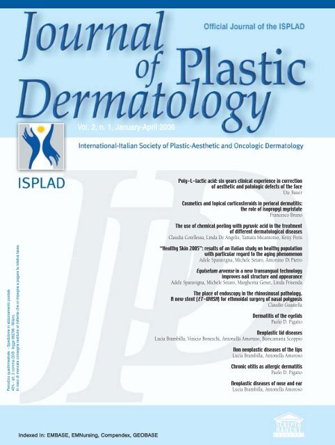
Vol. 2, n. 1, January-April <strong>2006</strong><br />
Indexed in: EMBASE, EMNursing, Compendex, GEOBASE<br />
Poly-L-lactic acid: six years clinical ex<strong>per</strong>ience in correction<br />
of aesthetic and patologic defects of the face<br />
Ute Bauer<br />
Cosmetics and topical corticosteroids in <strong>per</strong>ioral dermatitis:<br />
the role of isopropyl myristate<br />
Francesco Bruno<br />
The use of chemical peeling with pyruvic acid in the treatment<br />
of different dermatological diseases<br />
Claudia Cotellessa, Linda De Angelis, Tamara Micantonio, Ketty Peris<br />
“Healthy Skin 2005”: results of an italian study on healthy population<br />
with particular regard to the aging phenomenon<br />
Adele Sparavigna, Michele Setaro, Antonino Di Pietro<br />
Equisetum arvense in a new transungual technology<br />
improves nail structure and appearance<br />
Adele Sparavigna, Michele Setaro, Margherita Genet, Linda Frisenda<br />
The place of endoscopy in the rhinosinusal pathology.<br />
A new stent (ET-WASH) for ethmoidal surgery of nasal polyposis<br />
Claudio Guastella<br />
Dermatitis of the eyelids<br />
Paolo D. Pigatto<br />
Neoplastic lid diseases<br />
Lucia Brambilla, Vinicio Boneschi, Antonella Amoruso, Biancamaria Scoppio<br />
Non neoplastic diseases of the lips<br />
Lucia Brambilla, Antonella Amoroso<br />
Chronic otitis as allergic dermatitis<br />
Paolo D. Pigatto<br />
Neoplastic diseases of nose and ear<br />
Lucia Brambilla, Antonella Amoroso
È trascorso soltanto un anno dall’uscita del primo numero del Journal of Plastic Dermatology (JPD) ed è già tempo di bilanci!<br />
Solitamente “le somme si tirano” dopo tre, cinque anni, ma <strong>per</strong> la nostra rivista, fortunatamente, gli eventi hanno subito un corso<br />
decisamente diverso dalla norma!<br />
S<strong>per</strong>o di non cadere nella retorica se penso che, sembra ieri, quando con il Consiglio Direttivo dell’ISPLAD ed Antonio Di Maio<br />
(responsabile dei rapporti tra l’editore e la nostra società), muovevamo i primi passi <strong>per</strong> la realizzazione di questa testata.<br />
Un’idea che poteva sembrare azzardata, data la già numerosa presenza di riviste scientifiche di tradizione e ben accreditate.<br />
Ma, forse proprio <strong>per</strong> questo, il progetto diventava più interessante e sentito fermamente da noi <strong>tutti</strong>!<br />
Da subito l’impegno è stato totale e collettivo!<br />
Oggi possiamo vantare risultati straordinari:<br />
un board editoriale scientifico in continua crescita e di grande rilevanza internazionale;<br />
un continuo afflusso di articoli provenienti dall’Italia e dal’estero;<br />
un accordo, siglato a Roma, con il Consiglio Nazionale delle Ricerche (CNR) <strong>per</strong> la recensione del JPD;<br />
la continua richiesta di estratti di articoli<br />
da noi pubblicati (ben 7 nei primi 3<br />
numeri);<br />
la distribuzione capillare anche all’estero.<br />
Ma l’ultimo risultato, che ci riempie d’orgoglio<br />
e gratifica <strong>tutti</strong> gli autori, è avere<br />
ottenuto l’indicizzazione della rivista su<br />
EMBASE.<br />
Quest’ultimo traguardo <strong>per</strong>metterà al JPD,<br />
grazie alla med-line internazionale, una visibilità<br />
senza pari. (Da decine a centinaia di<br />
migliaia di consultazioni!)<br />
Nel panorama della Dermatologia il JPD<br />
non è più una nuova rivista, ma la Rivista!!!<br />
Visti i risultati così eclatanti, consentitemi di<br />
ringraziare “gli uomini che fecero l’impresa”:<br />
Francesco Bruno che, partendo da zero, ha<br />
creato un Board Internazionale di altissimo<br />
profilo scientifico e che ha onorato la sua<br />
qualifica di Editor in Chief con continuo e<br />
meticoloso impegno; i membri del<br />
Consiglio Direttivo ISPLAD <strong>per</strong> il sostegno<br />
incondizionato; e “dulcis in fundo” voi <strong>tutti</strong>,<br />
cari Soci, che avete creduto da subito nella<br />
vostra rivista fornendole il vostro supporto<br />
scientifico.<br />
La nostra gratitudine va anche all’industria<br />
farmaceutica e cosmetica, che ci ha <strong>per</strong>messo<br />
di crescere velocemente, e alle Edizioni<br />
Scripta Manent, un gruppo di veri professionisti<br />
della comunicazione.<br />
Ora “la nave” fila col vento in poppa verso il<br />
Primo Congresso Internazionale ISPLAD!!!<br />
Il nostro evento che, dal 11 al 13 Maggio a<br />
Stresa, sarà un grande momento culturale,<br />
scientifico e di confronto, con straordinarie<br />
possibilità di formazione, di aggregazione e,<br />
<strong>per</strong>ché no, di divertimento.<br />
Exactly one year has gone by since the first issue of the Journal of Plastic Dermatology was published, and it<br />
is time for a progress report. Although it is the norm that such a revision would be done after the transgression<br />
of three to five years, fortunately for our journal, the events surrounding it have rapidly developed along<br />
a different path.<br />
Without meaning to sound nostalgic, it only seems like yesterday when the Executive Committee of ISPLAD<br />
and Antonio Di Maio, in charge of the coordination between our publisher and the Society, took the first steps<br />
in creating JPD.<br />
The risky nature of such a project was taken into account during this <strong>per</strong>iod, due to the existence of a large number<br />
of well-accredited journals in the medical field. Perhaps it was this challenging predicament that made the<br />
project more interesting and led to its being firmly embraced by all of us in the first place.<br />
The constant, consequent, and collective commitment of all involved parties has enabled us to present some extraordinary<br />
results today. Some of these are as follows:<br />
The continuous growth of an internationally prominent scientific Editorial Board<br />
A steady flow of contributing articles both from Italy and abroad<br />
An agreement with National Research Committee (CNR) in Rome, for the review of JPD<br />
Continuous requests for abstracts from our publications from other journals and magazines<br />
(seven requests from the first three issues)<br />
The widespread distribution of JPD in foreign countries.<br />
In addition to the above, we are especially proud of our latest achievement: to be included in the index of<br />
EMBASE. This significant development, via MEDLINE, will allow JPD to be accessible to a limitless number of<br />
users. Within the scope of dermatology, JPD is no longer “just another new journal”, but “the” journal!<br />
The brilliant progress of JPD is a collective result of the hard work of a number of people and institutions, which<br />
I now take the opportunity to thank: Francesco Bruno, our Editor in Chief, who started from “zero” to successfully<br />
create a scientific Editorial Board of a high caliber, thanks to his continuous dedication and meticulous efforts;<br />
the members of the Executive Committee of ISPLAD for their coo<strong>per</strong>ation; and “dulcis in fundo”, all of you dear<br />
members of ISPLAD, who have put your trust and faith in our project from the very beginning, thank you for all<br />
of your scientific help and support.<br />
I also wish to thank the cosmetic and pharmaceutical industry for facilitating our advancement and Scripta<br />
Manent Publishing, whom we consider a very professional partner.<br />
Now, “our ship sails with a tailwind” towards the First International Conference of ISPLAD in Stresa, to be held<br />
on May 11-13!<br />
This event will be a grand moment for cultural, scientific, and comparative exchange and a great opportunity for<br />
formation, aggregation, and, while we're at it, a good time!<br />
Antonino Di Pietro<br />
Journal of Plastic Dermatology <strong>2006</strong>; 2, 1 1
Journal of Plastic Dermatology<br />
Editor<br />
Antonino Di Pietro (Italy)<br />
Editor in Chief<br />
Francesco Bruno (Italy)<br />
Associate Editors<br />
Francesco Antonaccio (Italy)<br />
Mariuccia Bucci (Italy)<br />
Franco Buttafarro (Italy)<br />
Ornella De Pità (Italy)<br />
Giulio Ferranti (Italy)<br />
Andrea Giacomelli (Italy)<br />
Alda Malasoma (Italy)<br />
Steven Nisticò (Italy)<br />
Elisabetta Perosino (Italy)<br />
Andrea Romani (Italy)<br />
Nerys Roberts (UK)<br />
Editorial Board<br />
Lucio Andreassi (Italy)<br />
Kenneth Arndt (USA)<br />
Bernd Rüdiger Balda (Austria)<br />
H.S. Black (USA)<br />
Günter Burg (Switzerland)<br />
Michele Carruba (Italy)<br />
Vincenzo De Sanctis (Italy)<br />
Aldo Di Carlo (Italy)<br />
Paolo Fabbri (Italy)<br />
Salvador Gonzalez (USA)<br />
Ferdinando Ippolito (Italy)<br />
Martin Charles Jr Mihm (USA)<br />
Joe Pace (Malta)<br />
Lucio Pastore (Italy)<br />
Gerd Plewig (Germany)<br />
Eady Robin AJ (UK)<br />
Abel Torres (USA)<br />
Umberto Veronesi (Italy)<br />
English editing<br />
Rewadee Anujapad<br />
Direttore Responsabile Pietro Cazzola<br />
Direzione Marketing Armando Mazzù<br />
Rapporti con ISPLAD Antonio Di Maio<br />
Consulenza grafica Piero Merlini<br />
Impaginazione Clmentina Pasina<br />
Sommario<br />
pag. 5 Acido-L-polilattico: sei anni di es<strong>per</strong>ienza clinica<br />
nella correzione dei difetti del volto<br />
Ute Bauer<br />
pag. 13 Cosmetici e corticosteroidi topici nella dermatite <strong>per</strong>iorale<br />
il ruolo dell’isopropil miristato<br />
Francesco Bruno<br />
pag. 17 The use of chemical peeling with pyruvic acid in the treatment<br />
of different dermatological diseases<br />
Claudia Cotellessa, Linda De Angelis, Tamara Micantonio, Ketty Peris<br />
pag. 23 “Healthy Skin 2005”: results of an italian study on healthy population<br />
with particular regard to the aging phenomenon<br />
Adele Sparavigna, Michele Setaro, Antonino Di Pietro<br />
pag. 31 Equisetum arvense in a new transungual technology<br />
improves nail structure and appearance<br />
Adele Sparavigna, Michele Setaro, Margherita Genet, Linda Frisenda<br />
pag. 41 La diagnosi endoscopica nella patologia rinosinusale<br />
Un nuovo stent (ET-WASH) nella chirurgia etmoidale della poliposi nasale<br />
Claudio Guastella<br />
pag. 49 Le dermatiti delle palpebre<br />
Paolo D. Pigatto<br />
pag. 55 Patologia tumorale delle palpebre<br />
Lucia Brambilla, Vinicio Boneschi, Antonella Amoruso, Biancamaria Scoppio<br />
pag. 67 Patologia infiammatoria delle labbra<br />
Lucia Brambilla, Antonella Amoroso<br />
pag. 73 Le dermatiti croniche del padiglione e del condotto auricolare esterno<br />
Paolo D. Pigatto<br />
Registr. Tribunale di Milano n. 102 del 14/02/2005<br />
Scripta Manent s.n.c. Via Bassini, 41 - 20133 Milano<br />
Tel. 0270608091/0270608060 - Fax 0270606917<br />
E-mail: scriman@tin.it<br />
Abbonamento annuale (3 numeri) Euro 39,00<br />
Pagamento: conto corrente postale n. 20350682<br />
intestato a: Edizioni Scripta Manent s.n.c.,<br />
via Bassini 41- 20133 Milano<br />
Stampa: Arti Grafiche Bazzi, Milano<br />
pag. 79 Patologia tumorale del naso e dell’orecchio<br />
Lucia Brambilla, Antonella Amoroso<br />
È vietata la riproduzione totale o parziale,<br />
con qualsiasi mezzo, di articoli, illustrazioni<br />
e fotografie senza l’autorizzazione scritta dell’Editore.<br />
L’Editore non risponde dell’opinione espressa dagli<br />
Autori degli articoli.<br />
Ai sensi della legge 675/96 è possibile in qualsiasi<br />
momento opporsi all’invio della rivista<br />
comunicando <strong>per</strong> iscritto la propria decisione a:<br />
Edizioni Scripta Manent s.n.c.<br />
Via Bassini, 41 - 20133 Milano<br />
Journal of Plastic Dermatology <strong>2006</strong>; 2, 1 3
Specialista in Chirurgia Plastica e Ricostruttiva, Padova<br />
Acido-L-polilattico:<br />
sei anni di es<strong>per</strong>ienza clinica<br />
nella correzione dei difetti del volto<br />
Ute Bauer<br />
SUMMARY<br />
Poly-L-lactic acid: six years clinical<br />
ex<strong>per</strong>ience in correction of aesthetic<br />
and patologic defects of the face<br />
I ntroduzione<br />
I primi trattamenti con acido-L-polilattico<br />
sono stati effettuati nel 1997 in Francia<br />
<strong>per</strong> la correzione della lipodistrofia del volto<br />
correlata alla terapia antiretrovirale (1).<br />
Gli ottimi risultati della ricostruzione dei tessuti<br />
molli e l’elevata tollerabilità del prodotto ha<br />
portato al suo uso<br />
anche <strong>per</strong> la correzione<br />
di dismorfismi<br />
estetici.<br />
La sua validità è stata<br />
inoltre confermata da<br />
studi condotti sulla<br />
valutazione tridimensionale<br />
del volto<br />
invecchiato (2, 3) .<br />
È oggi noto che l’invecchiamentoprogressivo<br />
causa una<br />
<strong>per</strong>dita del volume<br />
del volto dovuta alla<br />
distrofia cutanea, alla<br />
distrofia del tessuto<br />
Injectable Poly-L-lactic acid (PLLA) (Sculptra) is a synthetic biodegradable polymer<br />
for the tridimensional correction of aesthetic and pathologic defects of the face.<br />
The growing demand by patients for less-invasive nonsurgical cosmetic treatments<br />
reflects the interest in long lasting soft tissue augmentation.<br />
PLLA has been used successfully for the correction of nasolabial folds, mid and lower<br />
facial volume loss, jaw line laxity, and other signs of facial aging. The advanced tecnique<br />
and mechanism of action of the product allows to return volume to a whole<br />
facial area with a minimal invasive treatment and provides an effective and prolonged<br />
(18-to 24 month) facial enhancement correction with a low rate of side effects.<br />
KEY WORDS: Poly-L-lactic acid, Lipoatrophy, Skin aging, HIV, Face<br />
adiposo e ad un lento riassorbimento della<br />
struttura ossea.<br />
Il derma diventa più sottile <strong>per</strong> <strong>per</strong>dita di tessuto<br />
connettivo o si ispessice (foto-aging), con<br />
lo sviluppo di rughe e solchi, e l’atrofia ossea<br />
determina un aspetto senescente <strong>per</strong> l’ampia-<br />
Background<br />
Poly-L-lactic acid (PLLA)<br />
• Bioabsorbable, biodegradable and biocompatible<br />
• Over 30 years of use in medical applications<br />
Injectable PLLA<br />
• Stimulate fibroblasts to produce collagen<br />
• Gradually and progressively corrects volume deficiencies<br />
• Indicated for cosmetic applications and HIV-associated<br />
lipoatrophy of the face<br />
Clinical ex<strong>per</strong>ience with PLLA in hundreds of patients<br />
over several years is presented here<br />
Journal of Plastic Dermatology <strong>2006</strong>; 2, 1<br />
5
6<br />
U. Bauer<br />
mento dello spazio orbitale e <strong>per</strong> la riduzione<br />
dell’osso mascellare (dermatocalasi del terzo<br />
inferiore del volto) (2, 3).<br />
L’evento principale è comunque rappresentato<br />
dal riassorbimento del tessuto adiposo (lipoatrofia)<br />
e dalla ridistribuzione del grasso, soprattutto<br />
nelle regioni orbitarie, temporali e buccale,<br />
con conseguente rilassamento del tessuto<br />
cutaneo sovrastante.<br />
Ciò conferisce un aspetto incavato alle tempie e<br />
alle guance, con rughe e solchi più o meno<br />
profondi (solco naso-genieno) .<br />
Secondo alcune stime l’acido-L-polilattico è<br />
stato finora usato in più di 150.000 pazienti in<br />
Europa, Sud America ed Australia.<br />
Dal 1999 in Europa il suo uso è autorizzato<br />
<strong>per</strong> le correzioni estetiche e patologiche, e<br />
dall’agosto 2004 è stato approvato in USA<br />
dalla FDA (Food and Drug Administration) <strong>per</strong><br />
la correzione della lipoatrofia del volto in<br />
pazienti affetti da HIV (4, 5).<br />
Il lungo utilizzo del prodotto, e il particolare<br />
meccanismo d’azione, hanno dimostrato la sua<br />
validità e la sicurezza della sua composizione.<br />
Infatti gli effetti indesiderati sono riferiti in<br />
minima <strong>per</strong>centuale (meno 1%) e comunque<br />
simili a quelli provocati da altri “fillers” attualmente<br />
in commercio (6).<br />
P roprietà<br />
L’acido-L-polilattico è un polimero<br />
sintetico, biodegradabile, immunologicamente<br />
inerte e assorbibile, impiegato in medicina da<br />
oltre 30 anni sotto forma di vettori, impianti,<br />
viti, placche e fili di sutura. Essendo sintetico<br />
Journal of Plastic Dermatology <strong>2006</strong>; 2, 1<br />
Methods<br />
Patients<br />
• Between 1999 and 2004 780 patients have been treated<br />
• 90% were female patients (median age=40 years)<br />
Injection<br />
• Injectable PLLA supplied dry, as a lyophilizate<br />
• Add 5 mL of sterile water for injection (± 1mL xylocaine/lidocaine)<br />
• Allow to reconstitute for several hours<br />
• Agitate and inject<br />
• Multiple treatment sessions normally required<br />
• Treat, wait (4–6 weeks) and assess before repeating treatment<br />
Evaluating treatment efficacy<br />
• Patient and physician assessment<br />
non richiede un test di allergia. La polvere liofilizzata<br />
di microparticelle di acido polilattico<br />
è sospesa in un preparato di mannitolo apirogeno<br />
e caramellosi sodica.<br />
Il prodotto deve essere ricostituito con acqua<br />
sterile bidistillata almeno 12 ore prima dell’uso.<br />
La quantità di ricostituente varia a seconda dell’impiego:<br />
- 3-4 cc in caso di grave lipoatrofia delle zone<br />
trattate con radioterapia;<br />
- 5-6 cc <strong>per</strong> l’uso in estetica.<br />
È da ricordare che 1 cc di acqua può essere<br />
sostituito con lidocaina al 1%.<br />
Per l’utilizzo in alcune zone delicate, come la<br />
regione orbitaria, è preferibile diluire il prodotto<br />
con 8 cc di acqua bidistillata sterile.<br />
Le microparticelle, infiltrate con un ago 26<br />
gauge nello strato subdermico, si degradano<br />
lentamente, stimolando il tessuto circostante,<br />
con il risultato finale di una neocollagenogenesi<br />
(7). Questa lenta e progressiva produzione<br />
di collagene aumenta il volume nelle aree<br />
atrofiche. Biopsie della regione nasolabiale<br />
dimostrano dopo 12 e 30 mesi come le particelle<br />
di polilattico subiscano una lenta degradazione,<br />
fino ad un loro completo assorbimento,<br />
con un netto aumento delle fibre di collagene<br />
tipo I (2, 3).<br />
M etodi<br />
Sono stati trattati 780 pazienti con più<br />
di 5000 applicazioni; il follow up minimo è<br />
stato di 12 mesi, quello massimo di 48 mesi.<br />
Le applicazioni sono state distanziate almeno 4<br />
settimane una dall’altra. Nei pazienti giovani, a<br />
causa di una più<br />
rapida capacità reattiva,<br />
questo intervallo<br />
è stato ridotto.<br />
La valutazione soggettiva<br />
del risultato da<br />
parte del paziente e<br />
del medico è avvenuta<br />
comparando le fotografie<br />
scattate prima e<br />
dopo il trattamento; la<br />
valutazione oggettiva<br />
è stata ottenuta mediante<br />
ecografia o risonanza<br />
magnetica dei<br />
tessuti molli eseguite<br />
in tempi sucessivi al
trattamento. La tecnica è consistita in infiltrazioni<br />
di un’area intera a tunnel, o criss-cross, con iniezioni<br />
multiple di 0,1 cc di prodotto diluito adeguatamente,<br />
distanziate tra loro di 0.5-1 cm.<br />
L’impianto è stato rigorosamente sub-dermico.<br />
R isultati<br />
In oltre il 99% dei pazienti trattati è<br />
stato notato un evidente miglioramento e solo<br />
due sono stati classificati come “non- o slow reactors”<br />
dopo 4 trattamenti con scarso esito (8).<br />
In <strong>tutti</strong> i casi, immediatamente dopo l’infiltrazione,<br />
sono comparsi edema e rossore nella<br />
zona di inoculazione, che in alcuni casi si sono<br />
attenuati con l’applicazione locale di freddo<br />
(ghiaccio). Alcuni pazienti hanno riferito senso<br />
di calore o di tensione nell’area trattata.<br />
Le ecchimosi sono state frequenti, ma non sono<br />
mai comparsi ematomi o sieromi.<br />
Efficacy<br />
Baseline<br />
Acido-L-polilattico: sei anni di es<strong>per</strong>ienza clinica nella correzione dei difetti del volto<br />
Results<br />
12 months<br />
post treatment<br />
In un solo caso si è registrata un’infezione erpetica<br />
in un paziente senza profilassi farmacologica,<br />
mentre due sono stati i pazienti con infezioni<br />
batteriche (impetigine). In entrambi quest’ultimi<br />
casi non erano state seguite le indicazioni<br />
del medico di evitare l’utilizzo dei cosmetici<br />
nell’immediato <strong>per</strong>iodo post-trattamento.<br />
In nove pazienti sono comparsi in alcune zone<br />
trattate alcuni noduli sottocutanei palpabili,<br />
che si sono manifestati circa tre, quattro mesi<br />
dopo il trattamento. In questi casi è stato evidente<br />
che la tecnica applicata è stata errata, o il<br />
prodotto non è stato appropriatamente diluito,<br />
o è stato iniettato un eccessivo bolo.<br />
L’esame istologico in questi casi ha evidenziato<br />
un accumulo di materiale birifrangente, cristalli<br />
di acido polilattico, all’interno di un’area<br />
nodulare infiammatoria.<br />
In tre casi (0.5%) si è osservata in ogni zona trattata<br />
una reazione diffusa da corpo estraneo, con<br />
formazione di noduli duri, sclerotici, a volte<br />
Baseline 16 months<br />
post treatment<br />
40 months<br />
post treatment<br />
Journal of Plastic Dermatology <strong>2006</strong>; 2, 1<br />
7
8<br />
U. Bauer<br />
dolenti, e talora si è<br />
associata a discromia.<br />
Si tratta di una reazione<br />
immunologica ancora<br />
non ben compresa,<br />
che può essere<br />
provocata da qualsiasi<br />
dermal filler, e che<br />
spesso può essere collegata<br />
ad uno stato<br />
patologico pregresso<br />
(malattia infettiva,<br />
fluttuazioni ormonali,<br />
tiroiditi, allergie ecc...)<br />
con inizio della formazione<br />
nodulare a<br />
distanza di 8-20 mesi.<br />
Non si sono mai<br />
osservate reazioni allergiche.<br />
C onsigli<br />
Prima di tutto è necessario<br />
ricordare che<br />
ogni intervento deve<br />
essere preceduto dall’adesione<br />
del paziente<br />
che firma il modulo<br />
di consenso informato.<br />
Il paziente verrà<br />
inoltre fotografato<br />
nelle parti che devono<br />
essere trattate.<br />
Già dopo la prima<br />
infiltrazione si evidenzia<br />
una immediata<br />
correzione, dovuta<br />
all’acqua (il veicolo)<br />
che poi viene riassorbita:<br />
l’area trattata<br />
torna così allo stato<br />
pre-infiltrativo.<br />
Con il tempo si apprezzerà<br />
una correzione<br />
dei difetti trattati,<br />
in quanto l’acido<br />
L-polilattico provoca<br />
un ispessimento dello<br />
strato dermico, dovuto<br />
a neocollagenogenesi.<br />
Non devono<br />
mai essere eseguiti<br />
Journal of Plastic Dermatology <strong>2006</strong>; 2, 1<br />
Results<br />
Safety<br />
• Treatment related adverse events<br />
- Ecchymosis<br />
- Light oedema<br />
- Redness<br />
• Device related adverse events<br />
- 2 cases of bacterial infection<br />
- 9 cases of palpable subcutaneous nodules<br />
- 3 cases of late onset histologically confirmed granulomas<br />
(one of which resolved)<br />
Results<br />
• Histology<br />
• Particles of PLLA can be clearly seen in the dermis<br />
• following an injection<br />
Results<br />
• Histology<br />
• Some months after treatment, PLLA particles have been<br />
reabsorbed and collagen fibres are abundant
sovracorrezioni, che comporterebbero un effetto<br />
avverso Per raggiungere un risultato soddisfacente<br />
in pazienti con difetti minori sono<br />
necessarie 2-3 sedute, mentre in caso di gravi<br />
alterazioni occorrono 5-6 sedute.<br />
È variabile anche la quantità di acido L-polilattico<br />
da infiltrare in ogni seduta: si va da 1 cc <strong>per</strong><br />
un solco nasogenieno in una paziente giovane,<br />
a 8-10 cc <strong>per</strong> una lipodistrofia di 4° grado.<br />
Una correzione completa ha un emivita di 18<br />
mesi. L’infiltrazione di regioni dinamiche,<br />
come le labbra o come un’area intramuscolare,<br />
deve essere evitata in quanto ciò comporta<br />
<strong>per</strong> l’attività mimica dei muscoli un accumulo<br />
con ricostruzione nodulare. Solo nell’area<br />
orbitaria, dove lo strato dermico è esile, si<br />
consiglia un impianto profondo a piccoli boli,<br />
sub<strong>per</strong>iosteo (0,05 cc), intramuscolare tunnelizzante<br />
in regione temporale, essendo qui il<br />
derma sottile e il muscolo adinamico. In<br />
regioni corporee come decoltée, braccia e<br />
interno coscia il trattamento è di tunnelizzazione<br />
subdermica a rete.<br />
Dopo l’infiltrazione la zona deve essere massaggiata<br />
dal paziente 2 volte al giorno <strong>per</strong> dieci<br />
giorni: il massaggio ripetuto consente un’omogenea<br />
ed armonica distribuzione del materiale.<br />
Qualora comparissero dei noduli da accumulo,<br />
con confini netti, questi devono essere trattati<br />
con la tumescent tecnique, infiltrando acqua<br />
sterile <strong>per</strong> diluire il prodotto. Sono consigliati<br />
trattamenti settimanali. A volte si rende necessaria<br />
l’asportazione chirurgica del nodulo di<br />
accumulo. I granulomi da corpo estraneo,<br />
invece, devono essere trattati con corticosteroidi<br />
intralesionali ad alta concentrazione (una<br />
infiltrazione nel granuloma), a cui segue, a<br />
volte, anche una somministrazione <strong>per</strong> os a<br />
basso dosaggio.<br />
Acido-L-polilattico: sei anni di es<strong>per</strong>ienza clinica nella correzione dei difetti del volto<br />
Conclusions<br />
• PLLA injections can gradually restore volume to the face<br />
• in a wide spectrum of patients<br />
• The results are long lasting. 40 months<br />
• of volumetric restoration are possible<br />
• PLLA injections are also safe with a low incidence<br />
• of device related adverse events<br />
Non è consigliata l’escissione<br />
chirurgica<br />
<strong>per</strong>ché la reazione<br />
granulomatosa non<br />
ha confini netti, e si<br />
rischia una recidiva<br />
multifocale.<br />
D iscussione<br />
In caso di lipoatrofia<br />
il trattamento con<br />
acido polilattico è<br />
sicuramente il più<br />
indicato. Il materiale <strong>per</strong>mette una correzione<br />
di vaste aree con un effetto assolutamente naturale,<br />
elastico, che non influenza la mimica facciale.<br />
In un <strong>per</strong>iodo in cui i pazienti cercano<br />
trattamenti estetici di ringiovanimento,<br />
microinvasivi, e di lunga durata, l’utilizzo dell’acido<br />
polilattico rappresenta una eccellente<br />
alternativa all’intervento chirurgico.<br />
Sono essenziali una corretta tecnica d’inoculazione,<br />
e una valutazione tridimensionale armoniosa<br />
del intero volto da parte del medico.<br />
Aumenti eccessivi della regione zigomatica o<br />
della regione <strong>per</strong>i -buccale, <strong>per</strong> esempio, sono<br />
da evitare, vista la longevità del prodotto.<br />
Confrontato con altri fillers dermatologici, l’acido<br />
L-polilattico eccelle <strong>per</strong> la quantità di materiale<br />
utilizzabile, la durata della correzione e la<br />
scarsa frequenza di effetti avversi.<br />
B ibliografia<br />
1. Valantin MA, Aubron-Olivier C, Ghosn J,<br />
Laglenne E, Pauchard M, Schoen H, Bousquet R, Katz P,<br />
Costagliola D, Katlama C. Polylactic acid implants (New-<br />
Fill) to correct facial lipoatrophy in HIV-infected patients:<br />
results of the open-label study VEGA. AIDS. 2003;<br />
17(17):2471-7.<br />
2. Gilchrest BA. Skin aging 2003: recent advances and current<br />
concepts. Cutis. 2003; 72(3 Suppl):5-10.<br />
3. Gilchrest BA. A review of skin ageing and its medical<br />
therapy. Br J Dermatol. 1996; 135(6):867-7<br />
4. Vleggaar D, Bauer U. Facial enhancement and the<br />
European ex<strong>per</strong>ience with Sculptra (poly-l-lactic acid). J<br />
Drugs Dermatol. 2004; 3(5):542-7.<br />
6. Lem<strong>per</strong>le G, Romano JJ, Busso M. Soft tissue augmentation<br />
with artecoll: 10-year history, indications, techniques,<br />
and complications. Dermatol Surg. 2003; 29(6):573-87<br />
7. Hazan Gauthier N. Clinical analysis and managment of<br />
granulomas after all types of fillers. IMCAS <strong>2006</strong>.<br />
Journal of Plastic Dermatology <strong>2006</strong>; 2, 1<br />
9
10<br />
U. Bauer<br />
8. Cattelan AM, Bauer U, Trevenzoli M, Sasset L,<br />
Campostrini S, Facchin C, Pagiaro E, Gerzeli S, Cadrobbi<br />
P, Chiarelli A. Use of polylactic acid implants to correct<br />
facial lipoatrophy in human immunodeficiency virus 1-positive<br />
individuals receiving combination antiretroviral therapy.<br />
Arch Dermatol. <strong>2006</strong>; 142(3):329-34.<br />
5. Burgess CM, Quiroga RM. Assessment of the safety and<br />
efficacy of poly-L-lactic acid for the treatment of HIV-associated<br />
facial lipoatrophy. J Am Acad Dermatol. 2005;<br />
52(2):233-9.<br />
etture consigliate<br />
L 1. Dieterich DT. Long-term complications of<br />
nucleoside reverse transcriptase inhibitor therapy AIDS<br />
Read 2003; 13:176<br />
2. James J, Carruthers A, Carruthers J. HIV-associated<br />
facial lipoatrophy. Dermatol Surg 2002; 28:979<br />
3. Lem<strong>per</strong>le G, Romano JJ, Busso M. Soft tissue augmentation<br />
with artecoll: 10-year history, indications, techniques,<br />
and complications. Dermatol Surg 2003; 29:573<br />
4. Narins RS, Brandt F, Leyden J, Lorenc P, Rubin M, Smith S.<br />
A randomized, double-blind, multicenter comparison of the<br />
efficacy and tolerability of Restylane versus Zyplast for the<br />
correction of nasolabial folds. Dermatol Surg 2003; 29:588<br />
5. Amard P, Saint-Marc T, Katz P. The effects of polylactic<br />
acid as therapy for lipodystrophy of the face. Antivir Ther<br />
2000; 5:76. Abstract P94<br />
6. Valantin MA, Aubron-Olivier C, Ghosn J, Laglenne E,<br />
Pauchard M, Schoen H. Polylactic acid implants (New Fill)<br />
to correct facial lipoatrophy in HIV-infected patients: results<br />
of the open-label study VEGA. AIDS 2003; 17:2471<br />
7. Jones DH. Injectable silicone for facial lipoatrophy.<br />
Cosmet Dermatol 2002; 15:13<br />
8. Orentreich D. Liquid injectable silicone: techniques for<br />
soft tissue augmentation. Clin Plast Surg 2000; 27:595<br />
9. Conrad K, Reifen E. Gore-Tex implant as tissue filler in<br />
cheek-lip groove rejuvenation. J Otolaryngol 1992; 21:218<br />
10. Lem<strong>per</strong>le G, Morhenn V, Charrier U. Human histology<br />
and <strong>per</strong>sistence of various injectable filler substances for<br />
soft tissue augmentation. Aesth Plast Surg 2003; 27:254<br />
11. Hwang K, Han JY, Kim DJ. Dermofat graft in deep<br />
nasolabial fold and facial rhytidectomy. Aesthetic Plast<br />
Surg 2003; 27:254<br />
12. Vert M, Li SM, Garreaw H. Attempts to map the structure<br />
and degradation characteristics of aliphatic polyesters<br />
derived from lactic and glycolic acids. J Biomater Sci Pulm<br />
Ed 1994; 6:639<br />
Journal of Plastic Dermatology <strong>2006</strong>; 2, 1<br />
13. Lombardi T, Samson J, Plantier F, Husson C, Kuffer R.<br />
Orofacial granulomas after injection of cosmetic fillers.<br />
Histopathological and clinical study of 11 cases. J Oral<br />
Pathol Med 2004; 33:115<br />
14. Administration USFaD. Policy on importing unapproved<br />
AIDS drugs. Vol. 2004, 1988<br />
15. Lever WF, Schaumburg-Lever G. Non-infectious granulomas.<br />
In: Histopathology of the Skin, 8th ed. Lippincott-<br />
Raven, Philadelphia, 1997; p 317<br />
16. Collins E, Wagner C, Walmsley S. Psychosocial impact<br />
of the lipodystrophy syndrome in HIV infection. AIDS Read<br />
2000; 10:546<br />
17. Horl HW, Feller AM, Biemer E. Techique for liposuction<br />
fat reimplantation and long-term volume evaluation<br />
by magnetic resonance imaging. Ann Plast Surg 1991;<br />
26:248<br />
18. Moyle G. Lipodsytrophy: lack of agreement on definition<br />
and etiology presents a challenge to research and therapy<br />
AIDS Read 2002; 12:440-442.<br />
19. Hartmann M, Petzoldt D. Lipodystrophy syndrome in<br />
HIV infection. Hautarzt 2000; 51:159<br />
20. Lupton JR, Alster TS. Cutaneous hy<strong>per</strong>sensitivity reaction<br />
to injectable hyaluronic acid gel Dermatol Surg 2000;<br />
26:135<br />
21. Ziya Saylan MD. Facial fillers and their complications<br />
Aesth Surg 2003; 23:221<br />
22. Gruber PR, Hall ES, Kolstad JH, Iwen ML, Benson RD,<br />
Borchardt RL, inventors. Continuous process for manufacture<br />
of lactide polymers with controlled optical purity. US<br />
patent 5142023. 1992<br />
23. Micard V, Guilbert S. Int J Biol Macromolecules 2000;<br />
27:216-229.<br />
24. Hartman MH. Biopolymers from renewable resources.<br />
In. Kaplan DL (eds), Berlin: Springer-Verlag; 1998. p 367<br />
25. Cutright DE, Hunsuck EE. Tissue reaction to<br />
biodegradable polylactic acid suture. Oral Surg 1971;<br />
31:134<br />
26. Kulkarni RK, Pani KC, Neuman C, Leonard F.<br />
Polylactic acid for surgical implants. Arch Surg 1966;<br />
93:839<br />
27. Polo R, Ortiz M, Babe J, Martinez S, Madrigal P,<br />
Gonzalez-Munoz M. The effects of polylactic acid in the therapy<br />
for the lipoatrophy of the face. Program and abstracts of<br />
the 3rd International Workshop on Adverse Drug Reactions<br />
and Lipodystrophy in HIV; October 23-26, 2001; Athens,<br />
Greece Antiviral Ther 2001; 6:68. Abstract 101<br />
28. New-Fill [package insert]. Luxembourg: Biotech<br />
Industry SA; 2004
Cosmetici e corticosteroidi topici<br />
nella dermatite <strong>per</strong>iorale<br />
il ruolo dell’isopropil miristato<br />
Francesco Bruno<br />
SUMMARY<br />
Cosmetics and topical corticosteroids<br />
in <strong>per</strong>ioral dermatitis: the role of<br />
isopropyl myristate<br />
Dermatologo, Palermo<br />
Perioral dermatitis (PD) is a common dermatological disease which is not easy to<br />
classify, and whose aetiology and pathogenesis, even today, remain speculative and<br />
controversial. PD mainly affects women aged 15-50 years, and is increasing in occurrence.<br />
The feature shows discrete papules and pustules 1-2 mm in diameter, with follicular<br />
location, despite the shortage of follicles in this area. The arrangement is<br />
symmetrical around the border and corners of the mouth, often extending to the<br />
nasolabial folds, sparing a zone close to the vermilion border. There may be isolated<br />
<strong>per</strong>iocular involvement (<strong>per</strong>iocular dermatitis).<br />
Since Frumes and Lewis, described PD as a “light sensitive seborrheid” in 1957, and<br />
Steigler and Strempel as a “rosacea-like dermatitis” in 1968, several erroneous<br />
pathogenetic theories have followed. One of the most prominent of these, is the relation<br />
between acne and PD, despite the absence of comedones. There are a myriad of<br />
different pathogenetic theories: intestinal malabsorption, infection by candida or<br />
fusobacteria, sunlight - heat or cold - exposure, hormones, and oral contraceptives.<br />
Nowadays, all these different factors have merely a historical significance.<br />
Almost all the women affected by PD have a positive history of intensive use of<br />
cosmetics, fragrances, and /or topical corticosteroids. The cosmetics mostly used are<br />
skin care products such as moisturizers, detergents, abrasives, adstringents, night -<br />
antiwrinkle creams, and make-up products.<br />
All these products contain a variable concentration of isopropyl myristate (IPM).<br />
IPM is also contained in topical steroids and antimycotics. IPM is considered a very<br />
weak sensitizer. However, in patients affected by PD, patch tests are often positive<br />
because these patients are atopics in a high <strong>per</strong>centage and because they also have<br />
positive history of a cosmetics allergy.<br />
It’s very important to investigate the skin barrier function as well. In fact, in patients<br />
with PD and atopy, transepidermal water loss (TEWL), measured on the face, is<br />
significantly increased.<br />
Treatment consists of avoiding the use of all kinds of cosmetics such as soaps and moisturizers,<br />
although it is often the case that the patient doesn’t accept this advice easily.<br />
KEY WORDS: Cosmetics, Topical corticosteroids, Isopropyl myristate, Atopy<br />
La dermatite <strong>per</strong>iorale (DP), rappresenta ancora<br />
oggi una dermatosi dall’etiopatogenesi assai<br />
controversa, conseguentemente non è facile una<br />
sua “collocazione” dal punto di vista nosologico<br />
(1, 2). La sua incidenza sembra essere in aumento.<br />
È caratterizzata dalla presenza di numerose<br />
papule di pochi millimetri di diametro. Si associa<br />
un eritema finemente desquamante. Le<br />
papule hanno una localizzazione prevalentemente<br />
follicolare, nonostante la scarsità di follicoli<br />
in questa area. Si può associare una sensazione<br />
di modico prurito e bruciore (Figure 1-4).<br />
Journal of Plastic Dermatology <strong>2006</strong>; 2, 1<br />
13
14<br />
F. Bruno<br />
Da quando Frumes e Lewis nel 1957 definivano<br />
la DP “light sensitive seborrheid” e Steigler e<br />
Strempel nel 1968 “rosacea-like dermatitis”, si<br />
sono susseguite una miriade di diverse teorie<br />
patogenetiche con, a volte, fantasiosi ed erronei<br />
tentativi di forzate appartenenze.<br />
La più clamorosa è un legame con l’acne, ma la<br />
completa assenza di comedoni nella DP, ha fatto<br />
presto abbandonare questa teoria.<br />
La dermatosi colpisce prevalentemente le<br />
donne di età compresa fra i 15 e i 50 anni. La<br />
sede tipica è attorno alla bocca, la disposizione<br />
è simmetrica. A volte può essere coinvolta la<br />
zona <strong>per</strong>iorbitaria (3, 4).<br />
Le teorie etiopatogenetiche che si sono susseguite<br />
negli anni, sono innumerevoli: il malassorbimento<br />
intestinale, l’infezione da candida,<br />
l’assunzione di estroprogestinici, l’esposizione<br />
solare, l’uso e l’abuso di corticosteroidi topici.<br />
Modernamente, la causa più accettata, è da<br />
ricercare nell’uso di cosmetici. Quasi la totalità<br />
delle donne affette da DP ha una storia positiva,<br />
<strong>per</strong> un uso particolarmente intenso di creme<br />
idratanti e trucchi. Gioca un ruolo importante<br />
l’iso-propil miristato, sostanza contenuta in<br />
cosmetici (creme idratanti) e farmaci topici<br />
(corticosteroidi-antimicotici) (5-7).<br />
I corticosteroidi topici quindi giocano un triplo<br />
ruolo patogenetico: 1) nel principio attivo;<br />
2) nel veicolo; 3) nell’azione sinergica con<br />
i cosmetici. Nella pratica, sono coinvolti<br />
diversi fattori associati che agiscono spesso in<br />
modo sinergico; ad esempio l’associazione di<br />
diversi detergenti troppo aggressivi o “abrasivi”.<br />
Raccogliendo un’accurata anamnesi, si<br />
potrà notare come la maggior parte delle<br />
pazienti affette da DP, dichiara di avere una<br />
pelle molto “sensibile”. Possiamo dunque<br />
definire due tipi diversi di sinergie: una fra gli<br />
stessi cosmetici ed un’altra fra i cosmetici ed i<br />
corticosteroidi.<br />
È necessario altresì, distinguere dei fattori esogeni:<br />
cosmetici, corticosteroidi topici, e dei fattori<br />
endogeni: diatesi atopica e barriera cutanea.<br />
La chiave di lettura, dal punto di vista patogenetico,<br />
è rappresentata dall’incontro di queste<br />
due componenti.<br />
attori esogeni<br />
F<br />
L’iso-propilmiristato<br />
Al primo posto vi sono sicuramente i cosmetici:<br />
creme idratanti, scrub, detergenti, trucchi.<br />
Journal of Plastic Dermatology <strong>2006</strong>; 2, 1<br />
Figura 1. Figura 2.<br />
La dimostrazione pratica della loro importanza<br />
dal punto di vista etiopatogenetico è che la<br />
loro totale sospensione comporta, in quasi il<br />
cento <strong>per</strong> cento dei casi, una scomparsa della<br />
DP in circa 2 mesi, senza l’uso di altre terapie.<br />
“Null therapy” di Kligman (1).<br />
Di eguale peso etiologico sono gli steroidi topici<br />
(creme, lozioni, unguenti). La paziente spesso<br />
presenta una sorta di “attaccamento” a questo<br />
tipo di farmaco, <strong>per</strong> gli immediati miglioramenti<br />
sulla componente eritematosa e pruriginosa.<br />
La tendenza più comune è quella di aumentare<br />
progressivamente la frequenza di applicazione<br />
(fino a 3 volte al dì!), e di applicare creme<br />
steroidee sempre più potenti, mono e bi-fluorurate.<br />
Come se non bastasse, negli ultimi anni, si è<br />
aggiunto l’uso di antimicotici topici, <strong>per</strong> una<br />
confusione nata <strong>per</strong> la presenza del pitirosporum<br />
ovalis nella dermatite seborroica, spesso<br />
confusa con la DP.<br />
Più rara, anche <strong>per</strong> la sua recente introduzione,<br />
la DP causata dall’uso di tacrolimus topico<br />
(15).<br />
L’iso-propilmiristato (IPM)<br />
È una sostanza contenuta nella grande maggioranza<br />
nelle creme cosmetiche e nei trucchi; è<br />
impiegato come veicolo in steroidi ed antimicotici<br />
topici.<br />
Non ha di <strong>per</strong> sé forti proprietà allergeniche e<br />
sensibilizzanti ma, nelle pazienti affette da DP,<br />
la positività ai patch test a questa sostanza, è<br />
assai frequente<br />
È stimato che un’alta <strong>per</strong>centuale di pazienti<br />
affetti da DP, che fa normale uso di cosmetici e<br />
con accertata diatesi atopica, presenta una<br />
positività al patch test all’iso-propilmiristato<br />
(8-13).
Figura 3.<br />
Figura 4.<br />
attori endogeni<br />
F<br />
Atopia - Barriera cutanea<br />
Da recenti dati è stato ampiamente dimostrato<br />
che la maggior parte delle pazienti affette da<br />
Dermatite <strong>per</strong>iorale, è atopica e riferisce episodi<br />
di allergie a cosmetici e/o trucchi.<br />
È opportuno sottoporre queste pazienti a test<br />
epicutanei.<br />
La maggior parte di esse presenta, infatti, una<br />
forte positività ai test da profumi o all’iso-propilmiristato.<br />
In questi soggetti è altresì importante valutare la<br />
barriera cutanea in termini di “replezione”.<br />
Infatti la <strong>per</strong>dita di acqua transepidermica<br />
(TEWL), misurata sul viso di pazienti affetti da<br />
Dermatite <strong>per</strong>iorale ed atopia, è diminuita in<br />
modo significativo (14).<br />
T erapia<br />
Nella terapia sarà di fondamentale<br />
importanza astenersi dall’uso di cosmetici. Non<br />
si può assolutamente ottenere un buon successo<br />
terapeutico se la paziente continua ad applicare<br />
cosmetici, trucchi, e altro.<br />
In molti casi, la completa assenza di trucco,<br />
potrebbe rappresentare l’unica terapia, sebbene<br />
le pazienti accettino mal volentieri questo suggerimento.<br />
È infatti dimostrato che se una<br />
paziente atopica affetta da DP non applica alcun<br />
cosmetico, in circa due mesi, assisterà ad una<br />
completa regressione del quadro clinico (1).<br />
È necessario sospendere altresì l’uso di corticosteroidi<br />
od antimicotici topici; questo può <strong>per</strong>ò<br />
comportare un “rebound” della malattia.<br />
La terapia d’elezione resta ancora oggi la minociclina<br />
cloridrato al dosaggio di 100 mg al dì<br />
<strong>per</strong> 3-4 mesi.<br />
Cosmetici e corticosteroidi topici nella dermatite <strong>per</strong>iorale il ruolo dell’isopropl miristato<br />
Recentemente è stata s<strong>per</strong>imentata la terapia<br />
fotodinamica (PDT), in alternativa alla terapia<br />
antibiotica, ma è ancora da valutare una sua<br />
reale efficacia (16).<br />
B ibliografia<br />
1. Plewig G, Kligman AM. Acne and Rosacea.<br />
Springer-Verlag; 1993, p 479-481<br />
2. Mihan R, Ayres S. Perioral dermatitis. Arch Dermatol<br />
1964; 89:803-805<br />
3. Wilkinson DS, Kirton V, Wilkinson ID. Perioral dermatitis:<br />
a 12-year review. Br J Dermatol 1979; 101:245-247<br />
4. Hogan DJ, Epstein JD, Lane PR. Perioral dermatitis: an<br />
uncommon condition? CMAJ 1986; 134:1025-8.<br />
5. Dirschka T, Szliska C, Jackowski J, Tronnier H. Impaired<br />
skin barrier and atopic diathesis in <strong>per</strong>ioral dermatitis. J<br />
Dtsch Dermatol Ges 2003; 199-203<br />
6. Dirschka T, Tronnier H. Periocular dermatitis J Dtsch<br />
Dermatol Ges 2004; (4):274-8.<br />
7. Dirschka T, Weber K, Tronnier H. Topical cosmetics<br />
and <strong>per</strong>ioral dermatitis Dtsch Dermatol Ges 2004;<br />
2:194-9<br />
8. Uter W, Schnuch A, Geier J, Lessmann H. Isopropyl<br />
myristate recommended for aimed rather than routine<br />
patch testing. Contact Dermatitis 2004; 50:242-4<br />
9. Frosch PJ, Pilz B, Andersen KE, Burrows D, Camarasa<br />
JG, Dooms-Goossens A, Ducombs G, Fuchs T, Hannuksela<br />
M, Lachapelle JM, et al. Patch testing with fragrances:<br />
results of a multicenter study of the European<br />
Environmental and Contact Dermatitis Research Group<br />
with 48 frequently used constituents of <strong>per</strong>fumes Contact<br />
Dermatitis 1995; 33:333-42<br />
10. Parente ME, Gambaro A, Solana G Study of sensory<br />
pro<strong>per</strong>ties of emollients used in cosmetics and their correlation<br />
with physicochemical pro<strong>per</strong>ties J Cosmet Sci 2005;<br />
56:175-82.<br />
11. Malik R, Quirk CJ. Australas J Dermatol. Topical applications<br />
and <strong>per</strong>ioral dermatitis 2000; 41:34-8.<br />
12. Wells K, Brodell RT. Postgrad Med. Topical corticosteroid<br />
‘addiction’. A cause of <strong>per</strong>ioral dermatitis 1993;<br />
93:225-30.<br />
13. Daltrey DC, Cunliffe WJ Effect of betamethasone valerate<br />
on the normal human facial skin floraActa Derm<br />
Venereol 1983; 63:160-2.<br />
14. Dirschka T, Tronnier H, Folster-Holst R Epithelial barrier<br />
function and atopic diathesis in rosacea and <strong>per</strong>ioral<br />
dermatitis. Br J Dermatol 2004; 150:1136-41.<br />
15. Gerber PA, Neumann NJ, Ruzicka T, Bruch-Gerharz D.<br />
Perioral dermatitis following treatment wih tacrolimus<br />
Hautarzt 2005; 56:967-8.<br />
16. Richey DF, Hopson B. Photodynamic therapy for <strong>per</strong>ioral<br />
dermatitis J Drugs Dermatol. <strong>2006</strong>; 5(2 Suppl):12-6.<br />
Journal of Plastic Dermatology <strong>2006</strong>; 2, 1<br />
15
Claudia Cotellessa 1<br />
Linda De Angelis 2<br />
Tamara Micantonio 2<br />
Ketty Peris 3<br />
1 Dermatologo, Professore a contratto<br />
Università degli Studi de L’Aquila<br />
2 Dermatologo, Università degli Studi de L’Aquila<br />
3 Professore Associato,<br />
Direttore Clinica Dermatologica<br />
Università degli Studi de L’Aquila<br />
The use of chemical peeling<br />
with pyruvic acid in the treatment<br />
of different dermatological diseases<br />
SUMMARY<br />
I ntroduction<br />
Pyruvic acid (CH3-CO-COOH) is an<br />
alpha-keto-acid which use as peeling agent has<br />
been variously proposed in the treatment of different<br />
dermatological diseases, such as acne and<br />
acne scars (1), actinic keratosis (2) and warts (3).<br />
Several pro<strong>per</strong>ties of pyruvic acid make it particularly<br />
effective as a topical peeling agent.<br />
Pyruvic acid is characterized by the presence of<br />
only three carbon atoms; the small dimension<br />
of the molecule allows a deep penetration in<br />
skin layers (4). A distinctive characteristic of<br />
The use of chemical peeling<br />
with pyruvic acid in the treatment<br />
of different dermatological diseases<br />
Pyruvic acid (CH 3 -CO-COOH) is an alpha-keto-acid characterized by the presence<br />
of three carbon atoms. Several pro<strong>per</strong>ties make it particularly effective as a topical<br />
peeling agent. The small dimension of the molecule allows a deep penetration in skin<br />
layers. Pyruvic acid is characterized by a pKa of 2.39, so it is stronger than glycolic<br />
and salicylic acid, but less aggressive than trichloroacetic acid. Under physiological<br />
conditions, pyruvic acid converted to lactic acid, the corresponding alpha hydroxy<br />
acid. A well-equilibrated proportion of water and ethanol in the vehicle is able to<br />
balance the strength of the acid, inducing the transformation of a part of pyruvic acid<br />
into lactic acid. Furthermore, for the presence of a ketonic group, pyruvic acid is less<br />
hydrophilic as compared to alpha hydroxy acid, but it presents a more evident<br />
lipophily: this characteristic consents a good penetration into sebaceous glands,<br />
where pyruvic acid has showed a sebostatic and anti-microbial activity.<br />
At epidermal level, pyruvic acid induces keratinocyte detachment with thinning of<br />
the up<strong>per</strong> layers of the epidermis. It also penetrates down to the up<strong>per</strong> papillary dermis<br />
and stimulates an increased production of collagen, elastic fibres and glycoproteins.<br />
This action is more evident using high concentrations.<br />
Based on its peculiarities, pyruvic acid has been successfully employed in the treatment<br />
of several dermatological diseases.<br />
The Authors describe their ex<strong>per</strong>ience with pyruvic acid at different concentration in<br />
the treatment of inflammatory acne, moderate acne scars, seborrhoeic dermatitis,<br />
aging and photoaging, su<strong>per</strong>ficial hy<strong>per</strong>pigmentations and striae cutis distensae.<br />
KEY WORDS: Pyruvic acid, Chemical peeling, Dermatological diseases<br />
pyruvic acid is that, under physiological conditions,<br />
it converted to the corresponding alpha<br />
hydroxy acid, lactic acid, that is considered one<br />
of the most effective natural humectants for<br />
human skin (5). Pyruvic acid, combining the<br />
keratolitic action of an alpha-keto-acid and the<br />
humectant effects of lactic acid, can be considered<br />
a unique topical peeling agent.<br />
Pyruvic acid is characterized by a pKa of 2.39,<br />
so it is stronger than glycolic and salicylic acid,<br />
but less aggressive than trichloroacetic acid.<br />
Journal of Plastic Dermatology <strong>2006</strong>; 2, 1<br />
17
18<br />
C. Cotellessa, L. De Angelis, T. Micantonio, K. Peris<br />
However, in the execution of the chemical peeling,<br />
the strength of pyruvic acid can be modulated<br />
varying both its concentration and the<br />
composition of the solution. A well-equilibrated<br />
proportion of water and ethanol in the vehicle<br />
is able to balance the strength of the acid.<br />
The presence of more water in the vehicle has a<br />
sort of soothing effect because it induces the<br />
transformation of a part of pyruvic acid into lactic<br />
acid.<br />
The use of pyruvic acid as a chemical peeling<br />
offers the possibility to vary the potency of<br />
cutaneous effects, both modifying employed<br />
concentrations and water/ethanol proportions<br />
in solution (6).<br />
Concentrated pyruvic acid (100%) is liquid at<br />
room tem<strong>per</strong>ature and causes epidermolysis<br />
within 30-60 seconds after application to facial<br />
skin. In general, the response occurs faster in<br />
women and on younger skin (7). The clinical<br />
sign of epidermolysis following application of<br />
pyruvic acid is blanching, similar to the effect<br />
seen with high concentrations of glycolic acid.<br />
Lower concentrations of pyruvic acid, ranging<br />
between 40% and 60% in a well-balanced proportion<br />
between water and ethanol are currently<br />
used to obtain a su<strong>per</strong>ficial-medium peeling.<br />
At concentrations currently used for chemical<br />
peeling, pyruvic acid induces keratinocyte<br />
detachment with thinning of the up<strong>per</strong> layers of<br />
the epidermis. Pyruvic acid penetrates down to<br />
the up<strong>per</strong> papillary dermis and causes dermoepidermal<br />
separation and stimulates an<br />
increased production of collagen, elastic fibres<br />
and glycoproteins (8, 9). This action is more<br />
evident using high concentrations. In addition<br />
to its keratolytic and dermoplastic pro<strong>per</strong>ties,<br />
for the presence of a ketonic group, pyruvic<br />
acid is less hydrophilic as compared to alpha<br />
hydroxyl acid, but it presents a more evident<br />
lipophily: this characteristic consents a good<br />
penetration into sebaceous glands, where pyruvic<br />
acid has been showed a sebostatic and antimicrobial<br />
activity (1).<br />
Based on its peculiarity, recently pyruvic acid<br />
has been successfully employed in the treatment<br />
of several dermatologic diseases, such as<br />
inflammatory acne, moderate acne scars, wrinkles<br />
and actinically damaged skin, seborrhoeic<br />
dermatitis, striae cutis distensae (10).<br />
The Authors describe their ex<strong>per</strong>ience with<br />
pyruvic acid at different concentration and with<br />
various times of application in the treatment of<br />
several dermatological affections.<br />
Journal of Plastic Dermatology <strong>2006</strong>; 2, 1<br />
eeling procedure<br />
P<br />
The effects of pyruvic acid on the skin<br />
depend mainly on the used concentration and<br />
on the time of application before neutralization.<br />
In our clinical ex<strong>per</strong>ience we used concentrations<br />
of 40%, 50% and 60%, depending on the<br />
pathology to treat, the characteristics of patient’s<br />
skin, the expecting results. 40% pyruvic acid is<br />
available in two different solutions: 40% strong<br />
and 40% mild, that differ for the <strong>per</strong>centage of<br />
water and ethanol in the solution.<br />
Before chemical peeling, the skin is generally<br />
cleansed with cotton gauze soaked with 70%<br />
ethanol. This procedure is particularly important<br />
in patients with oily and thick skin. Then,<br />
skin must be accurately dry, because the presence<br />
of water could induce a greater transformation<br />
of pyruvic acid in the corresponding lactic<br />
acid. A solution of 40%-60% pyruvic acid is<br />
then applied on the skin. Modality of application<br />
differs according to the procedure preferred<br />
by the physician, but it is generally effected<br />
using a gauze pad or a brush. Many physicians<br />
prefer to carry out the application on successive<br />
regions, beginning from the forehead and the<br />
central area of the face and successively treating<br />
the two sides. Times of application are extremely<br />
variable and depend on patient’s reactivity<br />
and on the desired deep of peeling. Topical<br />
application of an alcoholic solution of pyruvic<br />
acid is associated with the rapid onset of a burning<br />
sensation and of an intense erythema, that<br />
slowly increases until neutralization. Pyruvic<br />
acid must be inactivated by the application of a<br />
solution of 10% sodium bicarbonate in water.<br />
Application of sodium bicarbonate solution<br />
quickly removes the burning sensation,<br />
although erythema generally <strong>per</strong>sists for a few<br />
hours after peeling. Most subjects ex<strong>per</strong>ienced<br />
no discomfort immediately after the peel.<br />
Chemical peeling with pyruvic acid is generally<br />
<strong>per</strong>fectly well-tolerated by patients, and no<br />
immediate or late side effects are observed.<br />
Application of pyruvic acid is followed by a<br />
very mild desquamation, <strong>per</strong>sisting for 3-5 days<br />
after peeling, which does not appear to interfere<br />
with the patient’s social life. Postpeel care is<br />
similar to that of other su<strong>per</strong>ficial peeling<br />
agents: patients must be instructed to avoid sun<br />
exposure and to apply facial moisturizer for a<br />
few days after treatment. Complete re-epithelialization<br />
occurs in 5-7 days. Treatments can be<br />
repeated every 2-4 weeks.
Figure 1a.<br />
Patient with mild papulopustular<br />
acne before<br />
treatment.<br />
Figure 1b.<br />
The same patient<br />
showed a complete<br />
remission of cutaneous<br />
lesions after treatment<br />
with pyruvic acid.<br />
Although pyruvic acid vapours are non-toxic,<br />
they are pungent and can irritate the lining of<br />
the up<strong>per</strong> respiratory tract and ocular mucosae.<br />
To avoid irritation, the chemical peeling procedure<br />
should be <strong>per</strong>formed in a well-ventilated<br />
area, and patients must be instructed to keep<br />
eyes closed during the whole procedure.<br />
atients and Methods<br />
P<br />
Papulo-pustular acne<br />
Peeling with pyruvic acid is very effective in the<br />
treatment of patients with mild to moderate<br />
papulo-pustular acne (Figure 1), used either<br />
alone or in association with traditional therapies.<br />
The efficacy of pyruvic acid is essentially<br />
due to its keratolytic effects, associated with a<br />
high capacity to penetrate into sebaceous<br />
glands, with a sebostatic and antimicrobic<br />
action. In a recent study, we demonstrated a<br />
good improvement in cutaneous lesions of<br />
patients with papulo-pustular acne treated with<br />
pyruvic acid. In addition, in these patients we<br />
observed an important decrease of seborrhoea<br />
after peeling treatment, without a simultaneous<br />
reduction of cutaneous hydration (1). The<br />
reduction of seborrhoea is important in the<br />
treatment of acneic patients, because it results<br />
in evident improvement in the oily skin that is<br />
characteristic of these patients. The simultaneous<br />
lack of reduction in cutaneous hydration<br />
differentiates pyruvic acid from other peeling<br />
1a. 1b.<br />
The use of chemical peeling with pyruvic acid in the treatment of different dermatological diseases<br />
agents, such as salicylic acid, that cause dryness<br />
and desquamation of the treated areas (10).<br />
Patients with acne are generally treated with<br />
medium concentrations of pyruvic acid: 40%<br />
strong or 50%, based on skin reactivity, with<br />
medium times of application (2-4 minutes).<br />
During peeling, in sites of acneic lesions it can<br />
be observed the quick onset of white frost, that<br />
indicates dermo-epidermal separation. This<br />
event should be considered <strong>per</strong>fectly normal<br />
and favours a rapid improvement of cutaneous<br />
lesions. Treatments can be repeated every two<br />
weeks and during the intervals patients can use<br />
specific local therapies.<br />
Acne scars<br />
Acne scars and other kind of su<strong>per</strong>ficial skin<br />
scars can be treated with pyruvic acid. In these<br />
patients the aim of peeling is to obtain a keratolityc<br />
effect, associated with a stimulation of an<br />
increased production of collagen, elastic fibres<br />
and glycoproteins. For this reason, acne scars<br />
are generally treated with medium-high concentrations<br />
of pyruvic acid (50-60%) and<br />
longer times of application (4-5 minutes). In<br />
fact, dermoplastic pro<strong>per</strong>ties of pyruvic acid are<br />
more evident using higher concentrations.<br />
Based on the high concentrations employed,<br />
generally peeling are repeated every 3-4 weeks.<br />
Seborrhoeic dermatitis<br />
Recently, the use of pyruvic acid has been proposed<br />
also in patients with mild to moderate seborrhoeic<br />
Journal of Plastic Dermatology <strong>2006</strong>; 2, 1<br />
19
20<br />
C. Cotellessa, L. De Angelis, T. Micantonio, K. Peris<br />
2a.<br />
dermatitis, based on its keratolityc and antinflammatory<br />
effects, associated with an elevate tolerability.<br />
In our ex<strong>per</strong>ience, better results could be<br />
obtained adjusting chemical procedure according<br />
to the great reactivity of the skin of these patients.<br />
We generally use very low concentrations (40%<br />
mild) with very short times of application (1<br />
minute), quickly followed by an accurate neutralization.<br />
Using this procedure, chemical peeling<br />
with pyruvic acid can be repeated every 7-10 days,<br />
with a good improvement of erythema, inflammation<br />
and desquamation (Figure 2).<br />
Aging and photoaging<br />
The keratolytic pro<strong>per</strong>ties of pyruvic acid<br />
induce an important thinning of the epidermis<br />
3a. 3b.<br />
Journal of Plastic Dermatology <strong>2006</strong>; 2, 1<br />
2b.<br />
and its capability of increasing the production<br />
of glycoproteins, collagen and elastic fibres can<br />
improve the texture and appearance of the skin<br />
and reduce su<strong>per</strong>ficial wrinkles (Figure 3). In a<br />
recent study it has been demonstrated that after<br />
4 peeling sessions <strong>per</strong>formed every 4 weeks,<br />
patients with photodamaged skin showed a<br />
smoother texture, less evident fine wrinkles and<br />
an important lightening of hy<strong>per</strong>pigmentations.<br />
In subjects with aging and photoaging mediumhigh<br />
concentrations are generally employed, to<br />
favour the dermoplastic effects. Based on the<br />
phototype of these patients, 50% or 60% pyruvic<br />
acid could be used, while 3-4 weeks are<br />
considered an appropriate time lapse between<br />
sessions.<br />
Figure 2a.<br />
Patient with seborrhoeic<br />
dermatitis before treatment.<br />
Figure 2b.<br />
The same patient showed<br />
a good improvement of<br />
erythema, inflammation<br />
and seborrhoea after<br />
treatment with pyruvic acid.<br />
Figure 3a.<br />
Patient before treatment.<br />
Figure 3b.<br />
The same patient showed<br />
an improvement of the<br />
texture and the<br />
appearence of the skin<br />
after treatment with<br />
pyruvic acid.
Figure 4a.<br />
Patient with striae cutis<br />
distensae before treatment.<br />
Figure 4b.<br />
The same patient<br />
showed a reduction<br />
of the size and<br />
a lightening of the striae<br />
after treatment with<br />
pyruvic acid.<br />
Hy<strong>per</strong>pigmentations<br />
Pyruvic acid is not characterized by a specific<br />
whitening action. Indeed, its keratolytic effects,<br />
associated with an improvement of the texture<br />
and the appearance of the skin, result in a lightening<br />
of discromic lesions. In patients with<br />
photodamaged skin hy<strong>per</strong>pigmentations<br />
appear lighter after the peeling sessions and in<br />
some cases fade completely (6). Peeling with<br />
pyruvic acid can be also successfully employed<br />
in the treatment of younger patients with diffuse<br />
su<strong>per</strong>ficial melasma. In these cases medium<br />
concentrations (40 strong-50%) and medium<br />
times of application (2-4 minutes) should<br />
be chosen, in order to avoid the risk of postinflammatory<br />
discromies.<br />
Sriae cutis distensae<br />
Striae cutis distensae are a very difficult affection<br />
to treat. In young patients which presente recently<br />
appeared striae distensae of the tigh, gluteus<br />
and/or breast, we have ex<strong>per</strong>ienced the use of<br />
pyruvic acid treatment, with a complexive<br />
improvement of the lesions. Based on the aim to<br />
stimulate the dermoplastic action of pyruvic acid<br />
and considering that in these cases chemical peeling<br />
is carried out on more resistant areas than<br />
facial skin, we generally use high concentrations<br />
(60%) and long times of application (5-7 minutes).<br />
After 4-6 treatments effected every 3-4<br />
weeks, an improvement of cutaneous lesions is<br />
generally observed, with a reduction of the size<br />
and a lightening of the striae (Figure 4).<br />
C onclusion<br />
Based on our ex<strong>per</strong>ience, pyruvic acid<br />
is a very effective peeling agent. The peculiar<br />
characteristics of the molecule consent to<br />
obtain at the same time a keratolytic, dermoplastic,<br />
sebostatic and antiseptic action.<br />
Furthermore, the possibility to choose among<br />
The use of chemical peeling with pyruvic acid in the treatment of different dermatological diseases<br />
4a. 4b.<br />
several different concentrations, to modulate<br />
the composition of the solution and to vary<br />
times of application, consent to adjust chemical<br />
peeling on the need of each patient. Finally,<br />
pyruvic acid treatments are generally very well<br />
accepted by patients because of the very limited<br />
discomfort of the postpeel <strong>per</strong>iod, that does not<br />
interfere with their social life.<br />
In conclusion, peeling with pyruvic acid can be<br />
considered an effective, safe and well-tolerated<br />
procedure in the treatment of different dermatological<br />
diseases.<br />
R eferences<br />
1. Cotellessa C, Manunta T, Gersetich I,<br />
Brazzini B, Peris K. The use of pyruvic acid in the treatment<br />
of acne. J Eur Acad Dermatol Venereol 2004; 18:275<br />
2. Griffin TD, Van Scott EJ. Use of pyruvic acid in the treatment<br />
of actinic keratoses: a clinical and histopathologic<br />
study. Cutis 1991; 47:325<br />
3. Halasz CL. Treatment of warts with topical pyruvic<br />
acid: with and without added 5-fluoruracil. Cutis 1998;<br />
62:283<br />
4. Griffin, TD, Van Scott EJ, Maddin S. The use of pyruvic<br />
acid as a chemical peeling agent. J Dermatol Surg Oncol<br />
1989; 15:1316<br />
5. Dahl M, Dahl A. 12% lactate loction for the treatment of<br />
xerosis: a double-blind clinical evaluation. Arch Dermatol<br />
1983; 119:27<br />
6. Ghersetich I, Brazzini B, Peris K, Cotellessa C, Manunta<br />
T, Lotti T. Pyruvic acid peels for the treatment of photoaging.<br />
Dermatol Surg 2004; 30:31<br />
7. Van Scott EJ, Yu RJ. Alpha hydroxy acids: therapeutic<br />
potentials. Canadian J Dermatol 1989; 1:108<br />
8. Van Scott EJ, Yu RJ. Control of keratinization with alpha<br />
hydroxy acids and related compounds. Arch Dermatol<br />
1974; 110:586<br />
9. Moy LS, Peace S, Moy RL. Comparison of the effects of<br />
various chemical peeling agents in a mini-pig model.<br />
Dermatol Surg 1996; 22:429<br />
10. Kligman D, Kligman AM. Salicylic acid peels for the<br />
treatment of photoaging. Dermatol Surg 1998; 24:325<br />
Journal of Plastic Dermatology <strong>2006</strong>; 2, 1 21
Adele Sparavigna 1<br />
Michele Setaro 1<br />
Antonino Di Pietro 2<br />
1 DermIng, Clinical Research<br />
and Bioengineering Institute, Monza (Milan) - Italy<br />
2 International Society of Plastic Dermatology<br />
(ISPLAD, Milan) - Italy<br />
“Healthy Skin 2005”:<br />
results of an italian study on healthy<br />
population with particular regard<br />
to the aging phenomenon<br />
SUMMARY<br />
“Healthy skin 2005”: results of an<br />
italian study on healthy population<br />
with particular regard to the aging<br />
phenomenon<br />
The study described in this pa<strong>per</strong> is the result of a campaign on healthy skin organised<br />
by the International Society of Plastic Dermatology (ISPLAD) . This campaign<br />
was at the same time an occasion to <strong>per</strong>form an epidemiological study on<br />
Italian population and was conducted during the months of May and June 2005<br />
throughout Italy. The event was previously advertised on the media and people<br />
could get a free dermatological consultation simply by visiting the pavillons prepared<br />
for the occasion and moving through nine main italian cities.<br />
A total of 2.408 subjects (1.896 females and 512 males) were evaluated. They<br />
were submitted to anamnesis, medical examination, dermatoscopic evaluation<br />
and stinging test with 10% lactic acid at the level of nasolabial fold. For each<br />
subject a “Skin Health Passport” was filled in with main habits and clinical data,<br />
in particular aging signs. On the basis of obtained clinical results, it was possible<br />
to draw a graph (Spiderming) for global evaluation of the aging of each considered<br />
subject. Women demonstrated to pay higher attention than men to engage<br />
correct life habits (55% of women versus 27%) and to use constantly and correctly<br />
skin care products (47% of women versus 7% of men).<br />
Moreover women’s skin resulted more sensitive than that of men (Stinging test:<br />
29% of women resulted sensitive versus 14% of men). Spiderming allowed to<br />
prepare model graphs based on average parameters deriving from clinical evaluation<br />
of the skin. This allowed to compare the aging phenomenon between female<br />
and male subjects, among the different considered age ranges and to evaluate<br />
the effect of life habits on aging of the skin.<br />
For both women and men skin aging signs worsened proportionally with age<br />
increase, but men obtained statistically higher clinical scores respect women in<br />
particular considering age ranges 50 years. Clinical scores of<br />
non smokers were statistically significantly lower than those of smokers and at<br />
the same time subjects who didn’t use sun protection factors presented significantly<br />
higher aging scores than subjects who applied protective creams in a correct<br />
way.<br />
This study provided an interesting overview about Italian healthy population’s<br />
skin aging. Women (80% of the examined population) resulted to be more interested<br />
in the proposed themes of the campaign and demonstrated to lead a more<br />
healthy life compared to men and to use constantly and correctly skin care products.<br />
When evaluating clinically the ageing of the face of an healthy subject,<br />
“Spiderming” graph revealed its usefulness for a global evaluation of the aging<br />
of the skin.<br />
KEY WORDS: Skin aging, Healthy skin, Skin care, Italian population<br />
Journal of Plastic Dermatology <strong>2006</strong>; 2, 1 23
24<br />
A. Sparavigna, M. Setaro, A. Di Pietro<br />
I ntroduction<br />
Dermatologist is the ideal professional<br />
for advising pro<strong>per</strong> skin treatment, even in<br />
absence of diseases. In order to stimulate public<br />
awareness on this important concept,<br />
already three years ago the International<br />
Society of Plastic Dermatology (ISPLAD) had<br />
decided to promote “Healthy Skin” campaigns,<br />
with the social aim to advice people to get<br />
pro<strong>per</strong> information on how to preserve healthy<br />
skin and to increase their knowledge on skin<br />
treatments. Encouraged by the success of previous<br />
“Healthy Skin” campaigns (1, 2) the<br />
International Society of Plastic Dermatology<br />
(ISPLAD) promoted the new study “Healthy<br />
Skin 2005” with particular regard to the study<br />
of the aging phenomenon. In this case people<br />
could get a free dermatological consultation<br />
simply by visiting the pavillons prepared for<br />
the occasion and staying for few days in the<br />
main squares of nine important italian cities*.<br />
People joining the study were submitted to an<br />
interview in order to establish their behaviour<br />
according to: life habits, skin care product use,<br />
skin sensitivity. Furthermore they were submitted<br />
to clinical and dermatoscopic examination<br />
of the face in order to evaluate the aging state<br />
of the subjects. Skin aging phenomenon is<br />
characterized by clinical signs, especially on<br />
the face (3), that can be evaluated by clinical<br />
scores on the basis of reference scales. This<br />
allows a more objective, and comparable<br />
among observers, clinical evaluation and to<br />
monitor the clinical signs before and after<br />
treatments. Nevertheless, this clinical method<br />
is based on isolated signs, that could or could<br />
not have a precise correlation with age and<br />
whose prognostic value still remains to be<br />
investigated. We thought it was better to set up<br />
a more complete method of clinical evaluation,<br />
in order to obtain full-comprehensive evaluation,<br />
able to give immediately exhaustive information<br />
about the aging of the skin. Obtained<br />
clinical scores were used to draw a graph<br />
(SpidermingTM ) for global evaluation of the<br />
aging of each considered subject. Moreover,<br />
SpidermingTM allowed to prepare model graphs<br />
based on average scores from the studied population.<br />
This allowed to compare the aging<br />
phenomenon between female and male subjects,<br />
among the different considered age<br />
ranges and to evaluate the effect of life habits<br />
on the aging state of the skin.<br />
* Bologna, Roma, Genova, Padova, Firenze, Bari, Napoli, Torino, Milano<br />
Journal of Plastic Dermatology <strong>2006</strong>; 2, 1<br />
aterials and Methods<br />
M<br />
The public was offered a free dermatological<br />
consultation by the dermatologists<br />
present in a pavillon, named “Healthy Skin<br />
Centre”, prepared in 9 main cities and moving<br />
all over of Italy from May 13 to June 18, 2005.<br />
For each subject a “Skin Health Passport” was<br />
filled in with self assessment evaluations and<br />
dermatologists’ clinical evaluations.<br />
Subjects<br />
Adult subjects, of both sexes who<br />
appeared to be healthy and had no evident dermatological<br />
disorder were included in the<br />
study, provided that they had given their<br />
informed consent. The exclusion criteria were<br />
the following: pregnancy or lactation; risk of<br />
poor co-o<strong>per</strong>ation; alterations of the facial skin,<br />
such as wounds, scars or malformations; systemic<br />
disorders; any dermatological disorders;<br />
surgery in the past year; cosmetic treatment<br />
(peeling, biomaterial implant, laser) in the last<br />
year; ongoing pharmacological treatment.<br />
Skin “health passport”<br />
Volunteers were asked to fill in a<br />
questionnaire concerning their <strong>per</strong>sonal life<br />
habits (outdoor working or spare time spent<br />
outdoor, practiced sports, sunbathing, taking<br />
drugs, diet, alcohol, recurrence to esthetic treatments<br />
and sunbaths), skin care products use<br />
(face detergents, delicate soaps, masks and<br />
scrubs, hydrating creams, nourishing creams,<br />
sun protection factors, anti-age products, makeup,<br />
skin/hair alimentary supplements) and skin<br />
sensitivity (to wind/cold, tem<strong>per</strong>ature variations,<br />
aggressive detergents, water, cosmetic<br />
products, small trauma, rubbing and emotions).<br />
For each question subjects could<br />
answer: often, sometimes or never. Moreover<br />
subjects were divided in smokers (more than 2<br />
cigarettes/day) and non smokers.<br />
Clinical examination<br />
Clinical examination was made using<br />
inspection and palpation of the skin and by the<br />
use of a special dermatoscopical device<br />
(Dermascore® by L’Oreal Recherche, France)<br />
and of special tapes (D-Squame®, CuDerm Inc,<br />
USA) for the evaluation of skin hydration. The<br />
Dermascore® device allows to evaluate a skin<br />
area of 10 cm 2 with a magnification of 10X and<br />
by polarized light, <strong>per</strong>mitting a more accurate
“Healthy Skin 2005”: results of an Italian study on healthy population with particular regard to the aging phenomenon<br />
evaluation of skin microrelief and vascular and<br />
pigmentation pattern. D-Squame® tapes were<br />
applied with a slight and constant pressure on<br />
zygomatic region previously cleaned skin with a<br />
solution of ether/acetone (1:1). Once removed,<br />
samples were submitted to a visual evaluation<br />
of desquamation grade according to a photographic<br />
reference scale.<br />
The dermatologist <strong>per</strong>formed and registered on<br />
the form the following evaluations:<br />
wrinkles: “crow’s feet” and nasolabial folds;<br />
skin surface microrelief (by means of dermatoscopical<br />
evaluation, see paragraph on<br />
dermatoscopy);<br />
vascular and pigmentary homogeneity (by<br />
means of dermatoscopical evaluation);<br />
skin hydration (by D-Squame ® visual score);<br />
resistance to traction and pinching, recovery<br />
after pinching.<br />
Visual parameters, <strong>per</strong>formed on the face, were<br />
assigned according to reference photographic<br />
atlas:<br />
Wrinkles (no wrinkles= 0, very weak= 1,<br />
weak= 2, quite evident= 3, evident= 4, very<br />
evident= 5, marked= 6, very marked= 7);<br />
Surface microrelief (very regular= 1, regular=<br />
2, irregular= 3, very irregular= 4);<br />
Vascular and pigmentary homogeneity (very<br />
homogeneous= 0, homogeneous= 1, quite not<br />
homogeneous= 2, not homogeneous= 3, very<br />
not homogeneous= 4, marked not homogeneous=<br />
5);<br />
Skin hydration (very hydrated (no desquamation)=<br />
0, hydrated (desquamation almost<br />
absent)= 1, normal (little and regular<br />
squames)= 2, slightly dry (evident desquamation,<br />
regular squames)= 3, dry (evident<br />
desquamation, large and irregular squames) =<br />
4, very dry(very evident desquamation, very<br />
large and irregular squames) = 5).<br />
Skin resistance to pinching, resistance to traction<br />
and recovery after pinching were <strong>per</strong>formed<br />
on submental zone according to the following<br />
score: 0=very important; 1=important;<br />
2= moderate; 3=weak; 4= very weak.<br />
Moreover dermatologists defined subject’s phototype<br />
according to the Fitzpatrick scale.<br />
Evaluation of skin sensitivity<br />
to stinging test<br />
The stinging test was carried out<br />
applying a 10% lactic acid solution onto the<br />
nasolabial fold and asking subjects whether<br />
they <strong>per</strong>ceived any local reaction after about 3<br />
minutes, according to the method published by<br />
Issachar et al. (7). Subjects who did not report<br />
any local reaction were classified as “non<br />
stingers”, whereas those who reported slight<br />
discomfort, burning or stinging were classified<br />
as “stingers”.<br />
Analysis of obtained data<br />
For each question a different score<br />
was assigned and according to the achieved<br />
results subjects were split up in different subgroups:<br />
correct, partially correct or not correct life<br />
habits;<br />
correct, partially correct or not correct skin<br />
care product use.<br />
sensitive or not sensitive skin<br />
Descriptive statistics (mean (SD, ES, min. and<br />
max.) related to the main features of the study<br />
population were provided. For the comparison<br />
of values reported on Spiderming TM graphs non<br />
parametric analysis were <strong>per</strong>formed (Wilcoxon<br />
test and Kruskal-Wallis test).<br />
R esults<br />
Subjects<br />
2.408 subjects received a free dermatological<br />
consultation: 1896 women aged from<br />
10 to 95 years and 512 men aged from 14 and<br />
84 years. Both the populations were splitted up<br />
in 5 subgroups:<br />
< 30 years<br />
30-40 years<br />
40-50 years<br />
50-60 years<br />
>60 years<br />
Most of the subjects were women (80%) and<br />
more than half were aged between 14 and 40<br />
years.<br />
Lifestyle<br />
Women showed higher attention than<br />
men to engage a correct habit of life; in particular<br />
55% of women (versus 27% of men) spent<br />
their spare time outdoor, practice sports, don’t<br />
exceed in drug taking, sunbaths and alcohol<br />
drinking and frequently recur to slimming diets<br />
and esthetic treatments.<br />
Skin care products<br />
Considering skin care products usage<br />
(cleansing products, delicate soaps, make-up,<br />
Journal of Plastic Dermatology <strong>2006</strong>; 2, 1 25
26<br />
A. Sparavigna, M. Setaro, A. Di Pietro<br />
Figure 1. Spiderming referred to male population.<br />
scrubs and masks, hydrating and nourishing<br />
creams, anti-age and sun protective products,<br />
food supplements) the gap between men and<br />
women rise considerably: 47% of women make<br />
a correct use of these products versus 7% of<br />
men.<br />
Skin sensitivity<br />
44% of the female subjects (against<br />
24% of men) referred their skin to be sensitive<br />
to wind/cold, changes in tem<strong>per</strong>ature, small<br />
trauma, rubbing and emotions, cosmetics use,<br />
aggressive detergents, use of water alone. This<br />
<strong>per</strong>sonal evaluation was confirmed by stinging<br />
test results; in fact 29% of women resulted sensible<br />
to the test versus 14% of men.<br />
The intensity of stinging in men was found to<br />
be inversely correlated with the cutaneous phototype<br />
(22% of sensitive subjects belong to phototype<br />
II, against 8% of sensitive subjects<br />
belonging to phototype IV). This trend wasn’t<br />
noticed in female population.<br />
Spiderming graphs<br />
Clinical parameters were reported on<br />
“Spiderming” graphs in order to <strong>per</strong>mit an<br />
immediate visualization of volunteers’ skin<br />
healthy state. Women and men were divided<br />
into 5 age-range groups and for each group<br />
Journal of Plastic Dermatology <strong>2006</strong>; 2, 1<br />
Figure 2. Spiderming referred to female population.<br />
average values of clinical evaluations were<br />
reported (Figures 1 and 2). For both women<br />
and men cutaneous aging signs worsened proportionally<br />
with age increase. In particular<br />
between the 2 populations the following statistically<br />
significant differences were noticed (all<br />
differences refer to the higher scores obtained<br />
by men respect to women):<br />
< 30 years: “crow’s feet” (p
Figure 3. Smoke influence on skin aging; females and males, age range 41-50 years.<br />
Figure 4. Influence of sun protection factors usage on cutaneous<br />
aging; females and males, age range 41-50 years.<br />
“Healthy Skin 2005”: results of an Italian study on healthy population with particular regard to the aging phenomenon<br />
Influence of using a sun protection factor<br />
on cutaneous aging<br />
Subjects who did non use sun protection<br />
products presented higher aging parameters<br />
than subjects who correctly applied protective<br />
creams. Also in this case results were statistically<br />
significant on 41-50 age range population,<br />
in particular regarding “crow’s feet” and<br />
nasolabial folds, cutaneous microrelief and pigmentary<br />
homogeneity (Figure 4).<br />
Influence of life habits, skin care products<br />
usage and skin sensitivity on the skin aging<br />
of the female population<br />
Data concerning the three considered<br />
subpopulations, correct - partially correct - not<br />
correct life habits, were reported on<br />
Spiderming graphs. Even if no statistically<br />
significant difference was found, it is evident<br />
that clinical scores of women belonging to the<br />
first group (correct life habits) were lower than<br />
scores of women who followed a less healthy<br />
life style. In particular this trend is evident for<br />
women under 50 years (Figure 5).<br />
On the contrary, observing Spiderming in<br />
Figure 5. Influence of life habits on aging; female population, age range 41-50 years.<br />
Journal of Plastic Dermatology <strong>2006</strong>; 2, 1 27
28<br />
A. Sparavigna, M. Setaro, A. Di Pietro<br />
correlation to skin care product usage it is clear<br />
that a correct or not correct use of these products<br />
did not influence clinical parameters.<br />
Considering Spiderming concerning skin sensitivity<br />
we noticed that scores obtained by very<br />
sensitive subjects were higher than scores<br />
obtained by non sensitive ones. In particular<br />
this observation was evident for vascular and<br />
pigmentary homogeneity, resistance to traction,<br />
resistance to pinching and recovery after pinching<br />
in females (Figure 6).<br />
iscussion and conclusions<br />
D<br />
Within this project a total of 2.408<br />
clinical evaluations were collected and a significative<br />
picture of skin healthy state of Italian<br />
population was obtained. Women (80% of the<br />
examined population) resulted to be more interested<br />
to the themes proposed on the campaign<br />
and demonstrated to lead a more healthy life<br />
compared to men and to use constantly and correctly<br />
skin care products. Moreover women<br />
demonstrated a higher cutaneous sensitivity (self<br />
assessment evaluation confirmed by the stinging<br />
test). Probably, healthy life style and attention<br />
demonstrated by women on skin care explain<br />
their lower grade of skin aging. Above all, considering<br />
under 30 population, statistically significative<br />
differences on skin aging degree between<br />
males and females were found and in particular<br />
the presence of wrinkles on nasolabial folds and<br />
on “crow’s feet” area in men was more evident<br />
than women. Differences between males and<br />
females reduced in 30-50 years age range, but<br />
over 50 years differences returned significant.<br />
Anyway, skin aging progress was similar for the<br />
two populations and in both cases all parameters<br />
worsened with ageing. Moreover, this study considered<br />
the influence of smoking and of incorrect<br />
usage of sun protection factors on skin<br />
aging. Similarly, both factors induced negative<br />
effects on skin aging and this was particularly<br />
evident in over 40 subjects. One of the purpose<br />
of this study was also to evaluate the parameters<br />
best representing a healthy state. In this case we<br />
decided to analyze only women, whose skin, as<br />
previously explained, demonstrated to have a<br />
better health state. Achieved results demonstrated<br />
that women under 50 years are more likely to<br />
follow a regular lifestyle, in fact in women<br />
belonging to this age range skin aging degree<br />
was more evident for subjects who did not show<br />
Journal of Plastic Dermatology <strong>2006</strong>; 2, 1<br />
Figure 6. Influence of skin sensitivity (positive stinging test) on skin aging; female population,<br />
age range 41-50 years.<br />
correct life habits. Considering creams/detergents<br />
usage, results show that women between<br />
30 and 60 years demonstrated an higher attention<br />
towards skin care respect to women over 60<br />
or under 30. Spiderming TM graph on the influence<br />
of sensitive skin on skin aging shows that<br />
“stinger” women present worse values for some<br />
clinical parameters: vascular and pigmentary<br />
homogeneity, resistance to pinching, resistance<br />
to traction and recovery after pinching. It must<br />
be underlined that the number of women with a<br />
sensitive/very sensitive skin keep constant in all<br />
age ranges. Even if in both men and women skin<br />
aging became evident after 40 years of age, it is<br />
clear that a healthy life style and a correct prevention<br />
must be adopted starting from 20 years<br />
onwards, in order to maintain a healthy and<br />
young skin. The study provided an interesting<br />
overview about Italian population’s skin aging.<br />
With the collected information it was possible to<br />
inform the participating subjects about the<br />
health state of their skin and possible cure to<br />
eventual skin problems. Assembled data will be<br />
useful to increase thr awareness of the importance<br />
of maintenance of a skin healthy state and<br />
its balance.
A cknowledgments<br />
This project was supported by a grant<br />
of Laboratories Vichy Italy. Special thanks to all<br />
the dermatologists who joined the project, they<br />
are all cited in the web: www.isplad.org.<br />
Authors declare no conflict of interest.<br />
R eferences<br />
“Healthy Skin 2005”: results of an Italian study on healthy population with particular regard to the aging phenomenon<br />
1. Sparavigna A, Di Pietro A, Setaro M.<br />
Healthy skin: significance and results of an Italian study on<br />
healthy population with particular regard to sensitive skin<br />
International Journal of Cosmetic Science 2005; 27:327-331<br />
2. Sparavigna A, Di Pietro A, Setaro M. Sensitive skin: correlation<br />
with skin surface and microrelief appearance. Skin<br />
Research and Technology <strong>2006</strong>; 12:7-10<br />
3. Lévêque J.L. Quantitative Assessment of Skin Aging.<br />
Clinics in Geriatric Medicine 2001; 17:673-689<br />
4. Corcuff P., de Rigal J., Makki S, et al. Skin Relief and<br />
Aging. J Soc Cosmet Chem 1983; 34:177- 182<br />
5. Setaro M., Abella M.L., Sparavigna A., Lévêque J.L.<br />
Ptosis of submental skin: objective measurements and effect<br />
of age. Skin Research and Technology 2004; 10:251-256<br />
6. Setaro M, Sparavigna A. Irregularity skin index (ISI): a<br />
tool to evaluate skin surface texture. Skin Research and<br />
Technology 2001; 7:159-163<br />
7. Issachar N, Gall Y, Borell MT, Poelman, MC. pH measurements<br />
during lactic acid stinging test in normal and<br />
sensitive skin. Contact Dermat 1997; 36:152-155<br />
8. Draelos ZD “Sensitive skin: <strong>per</strong>ceptions, evaluation and<br />
treatment.” Am. J. Contact Dermat. 1997; 8:67-78<br />
9. Muizzuddin N, Marenus KD, Maes DH. Factors defining<br />
sensitive skin and its treatment Am. J. Contact Dermat.<br />
1998; 9:170-175<br />
10. Placzek M, Kerkmann U, Bell S, Koepke P, Przybilla B.<br />
Tobacco smoke is phototoxic. British Journal of<br />
Dermatology 2004; 150:991-3<br />
11. Raitio A, Kontinen J, Rasi M, Bloigu R, Oikarinen A.<br />
Comparison of clinical and computerized image analyses in<br />
the assessment of skin ageing in smokers and non-smokers.<br />
Acta Dermato-Venereologica 2004; 84:422-427<br />
12. Choi EH, et al. The skin barrier state of aged hairless<br />
mice in a dry environment. British Journal of Dermatology<br />
2002; 147:244-9<br />
Journal of Plastic Dermatology <strong>2006</strong>; 2, 1<br />
29
Adele Sparavigna 1<br />
Michele Setaro 1<br />
Margherita Genet 2<br />
Linda Frisenda 2<br />
1 DermIng, Clinical Research and Bioengineering Institute,<br />
Monza, Milan, Italy<br />
2 Polichem SA, Lugano Pazzallo, Switzerland<br />
Equisetum arvense in a new transungual<br />
technology improves nail structure<br />
and appearance<br />
SUMMARY<br />
I ntroduction<br />
Changes in nail consistency may be<br />
due to impairment of one or more factors<br />
involved in healthy nail appearance and include<br />
variation in water content or in keratin constituent<br />
(1).<br />
Equisetum arvense in a new transungual<br />
technology improves nail<br />
structure and appearance<br />
Two clinical trials were carried out with a new formulation based on Equisetum<br />
arvense (a plant extract rich in organic silica) and a sulfur donor in a hydro-alcoholic<br />
solution, with the aim to evaluate the efficacy and preventive activity of this<br />
new formulation on nail alterations. In both studies, visual assessment of nail plate<br />
and optical profilometry of nail replicas were <strong>per</strong>formed comparing treated and contralateral<br />
untreated nails of each subject.<br />
For the first study, 36 women with nail plate alterations applied the test product<br />
every night on the nails of one hand, randomly assigned, for 28 days. Results demonstrated<br />
a significant reduction in longitudinal grooves as well as an 85% reduction in<br />
patients reporting lamellar splitting of treated nails, while no significant change was<br />
observed in untreated controls.<br />
In the second study, 22 women with nail plate alterations applied the test product<br />
randomly on the nails of one hand only, on alternating days, preferably in the<br />
evening, for 14 days. After drying, a common nail polish was applied on the fingernails<br />
of both hands and removed by an organic solvent every other day, before next<br />
product application.<br />
Results from this study showed a significant decrease (P< .001) of lamellar splitting<br />
compared to baseline with the test product (82% of cases).<br />
Nail fragility of treated nails decreased significantly (P < .001) compared to baseline.<br />
The product showed 90% of cases with 1 grade improvement and only 10% did not<br />
change. Control nails showed 5% with 1 grade improvement, 5% worsened and 90%<br />
did not change.<br />
Longitudinal grooves were significantly reduced by 28% after 14 days of treatment,<br />
despite the harmful effects of organic solvents.<br />
The results confirmed the efficacy and preventive activity of the test product in the<br />
improvement of nail plate alterations (splitting, fragility and longitudinal grooves).<br />
KEY WORDS: Equisetum arvense, Transungual technology, Nail<br />
Normal nails contain approximately 15% of<br />
water, this <strong>per</strong>centage increases after prolonged<br />
immersion in water and the nail becomes soft.<br />
A low lipid content may decrease the nail’s ability<br />
to retain water, consequently, if water con-<br />
Journal of Plastic Dermatology <strong>2006</strong>; 2, 1<br />
31
32<br />
A. Sparavigna, M. Setaro, M. Genet, L. Frisenda<br />
tent is reduced, the nail becomes brittle.<br />
Splitting, which results from this brittle quality,<br />
is probably due to repeated uptake and drying<br />
out of water.<br />
Moreover, the keratin content may be modified<br />
by physical insults and/or chemicals such as<br />
acrylic products used for printing colours, nail<br />
lacquers, organic solvents, formaldehyde, and<br />
hydroxylamine, these two latter widely used in<br />
chemical and pharmaceutical industry.<br />
Nail fragility may be due either to nail plate<br />
impairment or to matrix impairment: the nail<br />
may be damaged by trauma or by chemical<br />
agents such as detergents, alkaline solvents and<br />
especially by hot water. Prolonged immersion in<br />
water with alkalis or with powerful detergents<br />
render the nails softer and easily damageable.<br />
Climatic and seasonal factors may also affect the<br />
hydration of the nail plate.<br />
Fragility, due to thinning of the nail plate, may<br />
be caused by a reduction in the length of the<br />
matrix. Diminution or even complete arrest of<br />
nail formation over a variable width, may be the<br />
result of many dermatoses such as eczema,<br />
lichen planus and impairment of the <strong>per</strong>ipheral<br />
circulation. Additional causative factors for nail<br />
fragility are repeated microtrauma, infection,<br />
reduction in serum iron, vitamin A, B6, C deficiency,<br />
osteoporosis and numerous inherited<br />
defects associated with the atrophy of the nail.<br />
Dermatological or systemic diseases may also<br />
contribute to these alterations (2).<br />
Nail plate abnormalities include onychoschizia,<br />
that is horizontal lamellar splitting of the free<br />
edge of the nail into fine layers, affecting 27 to<br />
35% of normal adult women (1). This condition,<br />
caused by exogenous factors, is commonly seen<br />
in housekee<strong>per</strong>s as their nails are repeatedly<br />
soaked in water and then dried.<br />
Onycholysis refers to the detachment of the nail<br />
from its bed at its distal end and/or its lateral<br />
attachments. It may be associated with trauma,<br />
but most frequently onycholysis is due to chemical<br />
irritants or sensitizers substances, cold and<br />
hot injury and it is related to nail fragility (1).<br />
Onycholyses of the toenail is mainly associated<br />
with onychomycosis and repeated microtraumas.<br />
Other abnormalities include longitudinal<br />
grooves that may occur physiologically as shallow<br />
and delicate furrows but may become more<br />
prominent with age and in certain pathological<br />
conditions (lichen planus, rheumatoid arthritis)<br />
and longitudinal ridges that are small rectilinear<br />
Journal of Plastic Dermatology <strong>2006</strong>; 2, 1<br />
projections that extend from the proximal nail<br />
fold until the free edge of the nail, or may stop<br />
short. Transverse lines, the so called “Beau<br />
Lines”, characterized by su<strong>per</strong>ficial grooves,<br />
most frequently affecting the thumbs and big<br />
toes, are indicators of previous disease (1).<br />
These lines reflect a temporary reduction in<br />
matrix activity (1).<br />
Nail changes most frequently occur in elderly<br />
people due to the slow nail growth at that age<br />
(reduction in tissue repair), setting up of degenerative<br />
process and/or alterations of <strong>per</strong>ipheral<br />
circulation.<br />
Nail alterations cannot be neglected as they represent<br />
a suitable media for setting up fungal or<br />
bacteria infections which result in discomfort<br />
and pain. Moreover, unhealthy looking nails<br />
may cause a certain embarrassment, especially<br />
for certain professionals affecting subject’s selfesteem.<br />
There is no efficient cream able to prevent oversoftening<br />
of the nails due to water or detergents.<br />
After hydration, the nail plate should be massaged<br />
with mineral oil or a lubricating cream to<br />
prevent the nail to drying out. Products containing<br />
5% aluminium chloride in propylene glycol<br />
claim to make the nails harden, on the contrary<br />
such products make the nails stiff, brittle and less<br />
flexible. Nail varnish (water-insoluble polyvinyl<br />
resin film) acts in a similar way but the use of<br />
organic solvent for its removal damages nail<br />
structure by increasing nail splitting and fragility.<br />
Systemic treatment may be helpful. Oral iron (for<br />
6 months) even in absence of demonstrable iron<br />
deficiency, may be useful. More recently, biotin<br />
supplementation has been suggested for the<br />
treatment of brittle nails (3). Some nail alterations<br />
have been described as secondary to other<br />
therapy: docetaxel (4) or gold therapy (5).<br />
Treatment for onycholysis consists of a silicone<br />
rubber moulded toe cap or a silicone rubber<br />
orthodigital splint or a direct moulding splint,<br />
the device being produced in situ (1).<br />
A new, original topical formulation has been proposed<br />
to overcome and solve most of the above<br />
mentioned nail abnormalities. It is based on<br />
Equisetum arvense (a plant extract) and methylsulfonyl<br />
methane (a sulfur donor) in a hydroalcoholic<br />
solution into a new technology based on<br />
chitosan derivatives as film-forming agents for<br />
delivery of actives to nails.<br />
Its three main ingredients interact for the regeneration<br />
of a healthy and strong nail structure:<br />
Equisetum arvense provides organic silica, it
hardens and strengthens the nail (6). Hydroxypropyl<br />
Chitosan (HPCH) improves nail hydration<br />
and is an excellent carrier of actives (7); the<br />
sulfur donor improves quality and supports nail<br />
growth (8).<br />
Aim of the study was to assess the efficacy of this<br />
new topical formulation in improving nail splitting/fragility<br />
by means of two separate investigations<br />
in women.<br />
M ethods<br />
First study<br />
The study included women with nail<br />
alterations namely lamellar splitting caused by<br />
pathologic process (mycosis, psoriasis or bacterial<br />
infection) or exogenous causes (trauma,<br />
chemical agents or humidity) who had signed an<br />
informed consent form.<br />
Overall, 36 women were enrolled, 41% office<br />
workers, 27% housewives, 8% manual workers,<br />
6% dressmakers, 5.5% nurses, 5.5% students,<br />
7% other.<br />
Every night, the subjects applied the test<br />
productA on the surface of the nails of one randomly<br />
assigned hand for 28 days. The subjects<br />
were advised not to wash hands for the following<br />
6 hours. The nails of the other hand were<br />
evaluated as control nails.<br />
Casts (silicon replica) of nail lamina were taken<br />
on days 0, 14, 28 from the thumb of both hands<br />
(after removal of the product by washing) and<br />
subjected to computerized image analysis (9)<br />
(optical profilometry) in order to evaluate the<br />
regularity of the nail surface at baseline and any<br />
change due to the treatment. Silicon replicas<br />
were taken in standard conditions in order to<br />
reproduce them at any time.<br />
Pictures of silicon replicas at a light angle of 45°<br />
revealed shadows on the replica (which is the<br />
negative image of longitudinal and transverse<br />
grooves in the nail lamina). These shadows were<br />
transformed into a grey scale whose intensity<br />
was proportional to the intensity of the shadows<br />
and therefore to the depth of the grooves. The<br />
surface of the replica underwent a scanning procedure<br />
highlighting any alterations in the longitudinal<br />
and transverse grooves (Beau lines) in<br />
the nail. The scans were taken almost at the centre<br />
of basal replica, and always at the same level<br />
but not at the same preceding height (since the<br />
nail plate was grown) for the following observations<br />
times. The precision in the measurement is<br />
Equisetum arvense in a new transungual technology improves nail structure and appearance<br />
obtained by observing thoroughly the morphology<br />
of the nail plate and by identifying precise<br />
traces to be used as reference.<br />
The mean roughness (Ra) was calculated as the<br />
arithmetical mean of all deviations in absolute<br />
value.<br />
The parameters evaluated on days 0, 14, 28 were<br />
clinical assessment of the nails treated including<br />
the classification of the damage (dystrophy, onycholysis,<br />
splitting and fragility) and the causative<br />
factors. The degree of the damage was assessed<br />
on the basis of the following score scale:<br />
0 = absent, 1/2 = slight, 1 = moderate, 2 = severe.<br />
Statistical analysis was <strong>per</strong>formed by measuring<br />
the significance of differences between groups<br />
(treated vs untreated) and between times (intermediate<br />
vs baseline and final vs baseline) by<br />
means of Student’s t test for roughness, and of<br />
Wilcoxon test for clinical parameters.<br />
Moreover, the subject’s acceptability of the test<br />
product was investigated. Each subject<br />
expressed a 4-points score on different items<br />
referred to the product (appearance, tactile sensation,<br />
feeling on and after application, tolerability<br />
and efficacy).<br />
Second study<br />
The study included women with nail<br />
alterations (dystrophy, onycholysis, splitting and<br />
fragility) due to pathological or exogenous causes,<br />
who had signed an informed consent form.<br />
Overall, 22 women were enrolled, 50% housewives,<br />
18% office workers, 9% manual workers,<br />
5% nurses, 9% students, 9% other.<br />
The aim of this study was to evaluate the protective<br />
activity of the test product with respect to a<br />
common cosmetic nail varnish applied for 2<br />
weeks on the treated and control nails.<br />
Every night, the subjects applied, on alternative<br />
days, the test product B on the surface of the nails<br />
of one hand randomly assigned for 2 weeks. The<br />
nails of the other hand were evaluated as control<br />
nails. After product absorption, the subject<br />
applied a common cosmetic nail varnish, provided<br />
by the Investigator, to the nails of both<br />
hands. Before each new test product application,<br />
the common nail varnish was removed in all<br />
nails by means of a standard solvent provided by<br />
the Investigator.<br />
Clinical and instrumental assessments for evaluation<br />
of nail alterations were <strong>per</strong>formed at baseline,<br />
on day 7 and on day 14 (final visit). Casts<br />
(silicon replicas) of nail surfaces of the thumb<br />
treated and untreated were subjected to<br />
A. Extract of E. arvense, Methyonine, HPCH and other ingredients, patented by Polichem SA, Lugano (Switzerland);<br />
B. Ecocel<br />
Journal of Plastic Dermatology <strong>2006</strong>; 2, 1 33<br />
® patented by Polichem SA, Lugano (Switzerland). Same composition as <strong>per</strong> the first study, with the exception of sulfur donor:<br />
methylsulfonyl methane (MSM) instead of methyonine.
34<br />
A. Sparavigna, M. Setaro, M. Genet, L. Frisenda<br />
computerized image analysis. The procedure<br />
used for taking the cast and for its evaluation is<br />
the same as described in the first study. The only<br />
difference is that in this study the replicas were<br />
photographed at a light angle of 35° instead of<br />
45°.<br />
Statistical analysis was <strong>per</strong>formed as <strong>per</strong> the first<br />
study.<br />
At the end of the study, the subjects expressed<br />
their acceptability towards the test product by<br />
giving a 4-points score on different items<br />
referred to the product (appearance, tactile sensation,<br />
feeling on and after application, tolerability<br />
and efficacy).<br />
The tolerability of the product was also investigated.<br />
R esults<br />
First study<br />
The grey values for the Ra parameter<br />
(mean roughness) assessed in the image analysis<br />
of the casts taken of the nail lamina (optical profile<br />
measurement) on the baseline, intermediate<br />
and final examinations showed that even after<br />
only 14 days of treatment (intermediate stage),<br />
the product had brought about a statistically significant<br />
drop in parameter Ra (Figure 1)<br />
(Student’s t test p < 0.01 baseline vs intermediate<br />
and p < 0.001 baseline vs final) with a <strong>per</strong>cent<br />
reduction compared to baseline by 14% at the<br />
intermediate stage and by 18% on completion of<br />
the treatment <strong>per</strong>iod.<br />
For the untreated control area, there was no significant<br />
changes in roughness (Figure 1), namely<br />
the mean value of parameter Ra remained virtually<br />
unchanged throughout the study <strong>per</strong>iod<br />
(baseline = 9.78; intermediate = 9.82; final =<br />
10.32).<br />
The statistical analysis for the comparison of the<br />
treated and untreated control areas was carried<br />
out taking into account the final variation parameter<br />
Ra with respect to the corresponding<br />
baseline value (Ra final - Ra baseline = ∆ final).<br />
The statistical analysis between treated and<br />
untreated control area for Ra revealed significance<br />
of the differences (p < 0.001) in the ∆<br />
final of the treated area compared to (final<br />
untreated area. Clinical signs at the end of study<br />
gave the following results: statistically significant<br />
reduction of severity of nail splitting (mean<br />
values) (Wilcoxon’s test p < 0.001 baseline vs<br />
final and p < 0.01 intermediate vs final; Figure<br />
Journal of Plastic Dermatology <strong>2006</strong>; 2, 1<br />
2 and Table 1) and nail fragility (Wilcoxon’s test<br />
p < 0.001 baseline vs final; Figure 3 and Table<br />
2) in treated nails.<br />
Concerning the untreated control nails, there<br />
was no statistically significant change of nail<br />
splitting/fragility. The difference between treated<br />
and control group were statistically significant<br />
both for nail splitting (p < 0.001) and fragility<br />
Clinical Condition Test product Control nails<br />
(% of subjects) (% of subjects)<br />
2 grade improvement 15% 0%<br />
1 grade improvement 70% 0%<br />
No change 15% 91%<br />
Worsening 0% 9%<br />
P < .001 treated nails vs untreated control nails (Wilcoxon’s test)<br />
Table 1. Rate of outcome on lamellar nail splitting (1 st study)<br />
Figure 1. Average<br />
roughness (Ra) of<br />
longitudinal ridges.<br />
Figure 2. Rate of subjects<br />
with moderate/severe<br />
lamellar splitting (1st study).
Figure 3. Rate of subjects with nail fragility (1st study).<br />
Clinical Condition Test product Control nails<br />
(% of subjects) (% of subjects)<br />
2 grade improvement 3% 0%<br />
1 grade improvement 67% 3%<br />
No change 30% 94%<br />
Worsening 0% 3%<br />
p < 0.001 treated nails vs untreated control nails (Wilcoxon’s test)<br />
Table 2. Rate of outcome on nail fragility (1st study).<br />
(p < 0.001 for the treated nails vs untreated<br />
nails). Other clinical parameters (dystrophy,<br />
onycholysis, thin nails) revealed no statistically<br />
significant changes when comparing baseline<br />
with the final condition (data not displayed).<br />
4a<br />
Equisetum arvense in a new transungual technology improves nail structure and appearance<br />
No significant change was observed in the transverse<br />
grooves present on both nails.<br />
Subject’s acceptability was significantly good<br />
(Friedman’s test p = 0). The product was appreciated<br />
as it was quickly absorbed and left the<br />
nails smoother, with a more polished appearance<br />
than control nails. On the other hand, low<br />
acceptability of the smell led to switch the sulfur<br />
donor from methyonine to methylsulfonyl<br />
methane for the second study. The efficacy of the<br />
treatment was judged as excellent by 9%, good<br />
by 64%, poor or none by the rest of subjects and<br />
the tolerability of the product was excellent without<br />
adverse events occurring during the study.<br />
Second study<br />
The parameter Ra for longitudinal<br />
grooves decreased significantly in the treated<br />
nails with a <strong>per</strong>centage reduction of 13% after<br />
only 1 week and 28% at the end of application<br />
<strong>per</strong>iod (Student’s test p < 0.01 baseline vs. the<br />
intermediate stage and p < 0.001 baseline vs.<br />
final condition). With regards to the control<br />
nails, there were no significant changes in roughness,<br />
only a very slight tendency towards reduction<br />
of the parameter Ra at the end of the study.<br />
The statistical analysis between treated and<br />
untreated control area for Ra revealed significance<br />
of the differences (p < 0.001) in the ∆ final<br />
of the treated area compared to ∆ final untreated<br />
area. Scan electron microscopy <strong>per</strong>formed on<br />
some casts confirmed a smoother surface of the<br />
treated nails compared to baseline images<br />
(Figures 4a to 4h).<br />
4b 4c<br />
Figure 4a. Scan electron microscopy (366x), Subject no. 1 (before treatment); Figure 4b. Scan electron microscopy (377x), Subject no. 1 (after treatment):<br />
Figure 4c. Scan electron microscopy (360x), Subject no. 9 (before treatment)<br />
Journal of Plastic Dermatology <strong>2006</strong>; 2, 1 35
36<br />
A. Sparavigna, M. Setaro, M. Genet, L. Frisenda<br />
No effect on transverse grooves was noticed.<br />
The clinical assessment of the nails, at the end<br />
of the study, revealed a statistically significant<br />
reduction (Wilcoxon’s test p < 0.001) in the<br />
score for lamellar splitting in the treated nails<br />
compared to baseline (71% of cases with 1<br />
grade improvement, 10% with 2 grade<br />
improvement and 19% did not change); in the<br />
control nails only 5% showed 1 grade improvement,<br />
10% worsened and in 85% there were no<br />
change (Figure 5, Table 3). The difference<br />
between treatments was also significant (p <<br />
0.001 treated nails vs untreated control nails).<br />
Nail fragility decreased significantly (Wilcoxon’s<br />
test p < 0.001) compared to baseline. The product<br />
showed 90% of cases with 1 grade improvement<br />
and only 10% did not change. Control<br />
nails showed 5% with 1 grade improvement,<br />
5% worsened and 90% did not change (Figure<br />
6, Table 4). The difference between treatments<br />
4d<br />
Journal of Plastic Dermatology <strong>2006</strong>; 2, 1<br />
4g 4h<br />
4e 4f<br />
Figure 4d. Scan electron microscopy (360x),<br />
Subject no. 9 (after treatment).<br />
Figure 4e. Scan electron microscopy (366x),<br />
Subject no. 11 (before treatment).<br />
Figure 4f. Scan electron microscopy (366x),<br />
Subject no. 11 (after treatment).<br />
Figure 4g. Scan electron microscopy (88x),<br />
Subject no. 11 (before treatment).<br />
Figure 4h. Scan electron microscopy (88x),<br />
Subject no. 11 (after treatment).<br />
Figure 5. Rate of subjects with moderate/severe lamellar splitting (2nd study).
Figure 6. Rate of subjects with moderate/severe nail fragility (2nd study).<br />
Clinical Condition Test product Control nails<br />
(% of subjects) (% of subjects)<br />
1 grade improvement 90% 5%<br />
No change 10% 90%<br />
Worsening 0% 5%<br />
P < .001 treated nails vs untreated control nails (Wilcoxon’s test)<br />
Table 4. Rate of outcome on nail fragility (2nd Study).<br />
was also significant (p < 0.001 treated nails vs<br />
untreated control nails).<br />
The results obtained at the end of the trial for<br />
dystrophy, onycholysis and other conditions<br />
(thin/opaque nails, onychophagia, clear roughness)<br />
showed no statistically significant differences<br />
in comparison to the baseline condition<br />
for both the treated and control nails. No deterioration<br />
was observed in the treated nails for<br />
any of the above parameters, on the contrary,<br />
deterioration was observed in the control nails<br />
for nail splitting (10%) and fragile nails (5%).<br />
Subjects’ acceptability was good for all items<br />
considered. The efficacy of the product was<br />
Clinical Condition Test product Control nails<br />
(% of subjects) (% of subjects)<br />
2 grade improvement 10% 0%<br />
1 grade improvement 71% 5%<br />
No change 19% 85%<br />
Worsening 0% 10%<br />
P < .001 treated nails vs untreated control nails (Wilcoxon’s test)<br />
Table 3. Rate of outcome on lamellar nail splitting (2nd study).<br />
Equisetum arvense in a new transungual technology improves nail structure and appearance<br />
judged as excellent by 43%, good by 38%, poor<br />
by 19% of the subjects. According to user’s<br />
judgment, the test product provided nails<br />
smoother and shinier than controls. The smell<br />
of the product was favorably accepted.<br />
The tolerability of the product was excellent as<br />
confirmed by the clinical assessments carried<br />
out during the study.<br />
D iscussion<br />
The results obtained from the first<br />
study showed that the test product is effective<br />
especially in the treatment of specific alterations<br />
of the nail such as splitting and fragility.<br />
Moreover, the image analysis of the casts<br />
revealed a significant reduction in the longitudinal<br />
grooves which resulted in an increase of<br />
smoothness of the nails, pointed out by the<br />
subjects.<br />
The results obtained from the second study<br />
showed that the test product is effective on one<br />
hand against the aggressive action of the common<br />
varnish and the solvent used for its<br />
removal (protective activity) and on the other<br />
hand in the treatment of specific nail alterations<br />
such as splitting and fragility thanks to its<br />
strengthen pro<strong>per</strong>ty.<br />
Moreover, data obtained from the image analysis<br />
of the casts of the nail surface showed a significant<br />
reduction in roughness of the nails<br />
treated with the product, indicating its good<br />
smoothing activity, appreciated by all subjects.<br />
Thanks to the characteristics of its components,<br />
the product was able to render the nail stronger<br />
and to improve irregularities of the nail surface<br />
thus eliminating the most common un-aestheticism<br />
of the nail namely nail splitting and nail<br />
fragility, present especially in women but that<br />
may affect also men, especially certain professionals.<br />
Regular application of the test product, as standard<br />
nail care procedure, may be advisable in<br />
such categories of people.<br />
The change in the composition of the sulfur<br />
donor from methyonine (first study) to methylsulfonyl<br />
methane (second study) rendered the<br />
organoleptic characteristics of the final formulation<br />
excellent as judged by the women of the<br />
second study, by maintaining the efficacy of the<br />
product on nail fragility and nail splitting in the<br />
second study (final formulation) compared to<br />
the first study.<br />
Journal of Plastic Dermatology <strong>2006</strong>; 2, 1<br />
37
38<br />
A. Sparavigna, M. Setaro, M. Genet, L. Frisenda<br />
Transparent or colored nail polishes are commonly<br />
used to improve nail appearance, but<br />
they normally damage the nail structure, due to<br />
intrinsic reasons and due to the use of solvents.<br />
The application of Ecocel ® provided, in our<br />
ex<strong>per</strong>ience, a protective barrier between nail<br />
surface and polishes, by defending and improving<br />
the nail structure. Regular application of<br />
such a product under nail polishes is therefore<br />
strongly recommended.<br />
R eferences<br />
1.Baran R., Dawber R.P.R., Richert B. Physical<br />
signs. In: Baran R., Dawber R.P.R et al. (eds). Diseases of<br />
the Nails and their Management. Oxford, London,<br />
Edinburg, UK Backwell Science; 2001:63-85<br />
2. Rycroft R.J.G., Baran R.. Occupational abnormalities<br />
and contact dermatitis. In: Baran R., Dawber R.P.R et al.<br />
Diseases of the Nails and their Management. Oxford,<br />
London, Edinburg: UK Backwell Science; 2001: 332-352<br />
Journal of Plastic Dermatology <strong>2006</strong>; 2, 1<br />
3. Hochman LG, Scher RK, Meyerson MS. Brittle nails:<br />
response to daily biotin supplementation. Cutis 1993;<br />
51:303-305<br />
4. Almagro M., Pozo JD, Garcia J., Vasquez A., Fonseca E.<br />
Nail alterations secondary to pactitaxel therapy. Eur J<br />
Dermatol, 2000; 10:146-147<br />
5. Voigt K, Holzegel K. Permanent nail changes following<br />
gold therapy. Hautarzt 1997; 28:421-423<br />
6. Holzhuter G., Narayanan K., Gerber T., Structure of silica<br />
in Equisetum arvense. Anal Bioanal Chem. 2003;<br />
376:512-517<br />
7. Monti D. et al. In vitro transungual <strong>per</strong>meation of<br />
ciclopirox from a hydroxypropyl chitosan-based, water soluble<br />
nail lacquer. Drug Dev Ind Pharm. 2005; 31:11-17<br />
8. Parcell S. Sulfur in human nutrition and applications in<br />
medicine. Altern Med Rev. 2002; 7:22-44<br />
9. Baran R., Sparavigna A., Setaro M., Mailland F.,<br />
Computerized Image Analysis of nails affected by fungal<br />
infection: evaluation using digital photographs and manually<br />
defined areas. Journal of Drugs in Dermatology 2004;<br />
3:489-494
1° Corso ISPLAD, Milano 2005<br />
Clinica Otorinolaringoiatrica dell’Università di Milano<br />
La diagnosi endoscopica<br />
nella patologia rinosinusale<br />
Un nuovo stent (ET-WASH) nella chirurgia etmoidale<br />
della poliposi nasale<br />
Claudio Guastella<br />
SUMMARY<br />
The place of endoscopy in the rhinosinusal<br />
pathology. A new stent<br />
(ET-WASH) for ethmoidal surgery of<br />
nasal polyposis<br />
Endoscopy is indicated for the investigation of <strong>per</strong>sistent or recurrent nasal discharge<br />
that does not respond to simple treatment. This procedure allows close inspection<br />
of the lesion, and facilitates other methods such as histology, microbiology,<br />
cytology, and measurement of ostial patency and mucociliary transport.<br />
The importance of direct observation of the morphology and natural colour of normal<br />
structures becomes ever more evident. Its regular use has greatly enriched our<br />
knowledge of anatomy, physiology and pathology of the nasal cavity and paranasal<br />
sinuses. Surgery with endoscopic guidance improves surgical precision, reveals deep<br />
seated lesions by a minimally invasive approach, and allows lesion shown by radiography<br />
to be examined.<br />
The treatment of nasal polyposis is still debated, but previous studies agree that,<br />
when surgery is necessary, it must be accompanied by appropriate local and general<br />
medical therapy administered pre- and/or posto<strong>per</strong>atively.<br />
Paranasal sinus surgery can cause wounds in areas in which bone is not covered by<br />
mucosa and tends to granulate. The location and size of these areas vary, and depend<br />
on preo<strong>per</strong>ative anatomy and the type and extent of surgery. The speed of individual<br />
recovery depends on local and systemic factors. Local inflammation slows down tissue<br />
repair and destroys wide areas of mucosa, which heal with anomalous scar formations<br />
leading to a deficit of muco-ciliary clearance that predisposes patients to<br />
local relapse.<br />
In an attempt to reduce these complications, we have developed a new ethmoidal<br />
stent (ET-WASH) which is applied at the end of surgery. The aim of this study was<br />
to verify the tolerability of the device, and its advantages in terms of allowing targeted<br />
local corticosteroid therapy, reducing synechiae, preserving ventilation, cleansing<br />
of the surgical cavity, and reducing the recurrence of polyps.<br />
KEY WORDS: Endoscopy, Nose, Polyposis, Stent<br />
I ntroduzione<br />
Per migliorare la diagnosi delle cavità<br />
nasosinusali e della regione rinofaringea la diagnostica<br />
in Otorinolaringoiatria attualmente<br />
utilizza endoscopi a fibre ottiche di tipo rigido<br />
o flessibile.<br />
L’endoscopia, consente di visualizzare sedi non<br />
altrimenti direttamente esplorabili, miglioran-<br />
do decisamente la diagnosi, ottimizzando i criteri<br />
di scelta terapeutica, medica o chirurgica,<br />
<strong>per</strong> il trattamento delle principali affezioni<br />
naso sinusali.<br />
Gli strumenti sono realizzali in una unica guaina<br />
con i fasci di fibre ottiche che formano il<br />
canale luminoso e il canale ottico. II canale<br />
Journal of Plastic Dermatology <strong>2006</strong>; 2, 1 41
42<br />
C. Guastella<br />
luminoso consente al fascio di luce prodotto<br />
dalla sorgente luminosa di giungere all’estremità<br />
dello strumento <strong>per</strong> illuminare la parte in<br />
esame. La visione diretta delle regioni esplorate,<br />
e la possibilità di registrare ed archiviare le<br />
immagini, <strong>per</strong>mette la valutazione anche da<br />
parte di altri specialisti coinvolti nella diagnosi,<br />
in modo particolare Specialisti in Dermatologia,<br />
migliorando decisamente la valutazione finale<br />
dei casi più complessi. L’approccio plurispecialistico<br />
facilita anche il follow-up, grazie alla<br />
possibilità di confrontare nel corso dei successivi<br />
esami endoscopici il decorso clinico della<br />
patologia.<br />
II materiale registrato e archiviato può costituire<br />
inoltre, in caso di controversie medico-legali,<br />
una importante documentazione clinica del<br />
trattamento seguito e del risultato da esso derivatone.<br />
Le innovazioni tecnologiche dello strumentario<br />
endoscopico a fibre ottiche hanno <strong>per</strong>messo di<br />
realizzare strumenti di diametro sempre più<br />
ridotto con fibre ottiche del diametro esterno<br />
inferiore ai 2 mm., che hanno di molto facilitato<br />
l’esecuzione dell’esame endoscopico in particolar<br />
modo in età neonatale e nella prima<br />
infanzia.<br />
Gli studi di Messerklinger sulla fisiologia nasosinusale<br />
e sulla clearance mucociliare hanno<br />
dimostrato che il flusso mucoso, che <strong>per</strong>corre<br />
vie anatomiche predeterminate, si interrompe<br />
quando due strati mucosi vengono in contatto<br />
causando ristagno di muco e aumentando il<br />
rischio di infezioni anche quando gli ostii sinusali<br />
sono <strong>per</strong>vi. II contatto fra gli strati mucosi<br />
si verifica più facilmente nelle “strettoie” anatomiche<br />
quali il meato medio o le cellette etmoidali<br />
sia <strong>per</strong> i<strong>per</strong>plasia o edema mucosi che <strong>per</strong><br />
variazioni anatomiche.<br />
Queste interruzioni del flusso mucoso possono<br />
essere la causa della <strong>per</strong>sistenza della flogosi e<br />
del suo ri<strong>per</strong>tersi.<br />
La localizzazione della patologia sinusale è talvolta<br />
più importante della sua estensione, specialmente<br />
<strong>per</strong> la genesi della sintomatologia,<br />
quindi anche una modesta opacizzazione del<br />
regione medio-meatale puo essere associata ad<br />
una sintomatologia più imponente rispetto<br />
all’opacizzazione massiva di un seno mascellare.<br />
Esistono notevoli variazioni individuali nella<br />
anatomia delle cavità nasali e sinusali, le più<br />
importanti delle quali riguardano la parete laterale<br />
e il complesso ostio-meatale. Oltre alle<br />
varianti anatomiche devono essere ricercate<br />
Journal of Plastic Dermatology <strong>2006</strong>; 2, 1<br />
anche delle anomalie della mucosa, tenendo<br />
<strong>per</strong>ò presente che ispessimenti mucosi, cisti da<br />
ritenzione, polipi, secrezioni possono essere<br />
trovate in occasione di una comune coriza od<br />
anche in soggetti asintomatici.<br />
L’endoscopia nasale sistematica e le immagini<br />
TC ad alta risoluzione sono complementari<br />
nella diagnostica delle malattie flogistiche rinosinusali<br />
e consentono di riconoscere patologie<br />
non identificabili con altri mezzi.<br />
La rinofibroendoscopia consente di accedere<br />
alle aree chiave <strong>per</strong> il funzionamento della clearance<br />
mucociliare e di individuare patologie flogistiche<br />
od alterazioni anatomiche che le favoriscono;<br />
la TC consente di individuare patologie<br />
nelle sedi non accessibili all’endoscopia e di<br />
valutare l’estensione della patologia.<br />
L’esame endoscopico prevede l’utilizzo di ottiche<br />
flessibili o rigide a varia angolazione.<br />
L’endoscopio rigido presenta una maggiore<br />
etmoide ant.<br />
etmoide post.<br />
seno<br />
afenoidale<br />
seno<br />
mascellare<br />
sf<br />
bulla etmoidale<br />
processo<br />
uncinato<br />
iato semilunare<br />
Figura 1.<br />
Endoscopi rigidi e flessibili.<br />
Figura 2.<br />
In rosso e in blu<br />
la direzione dei flussi<br />
secretori sinusali.
Figura 3.<br />
Figura 4.<br />
Esame endoscopico:<br />
poliposi nasale.<br />
Figure 5 e 6.<br />
Endoscopia e TC<br />
di sarcoidosi nasale<br />
(si evidenzia la sofferenza<br />
della mucosa che<br />
si presenta atrofica con<br />
presenza di croste<br />
e di essudato denso<br />
giallognolo e alla TC<br />
l’erosione ossea).<br />
La diagnosi endoscopica nella patologia rinosinusale. Un nuovo stent (ET-WASH) nella chirurgia etmoidale della poliposi nasale<br />
intensità di luce ed una migliore definizione di<br />
immagine, utile <strong>per</strong> ottenere delle immagini<br />
fotografiche migliori, le fibre ottiche flessibili<br />
sono più confortevoli <strong>per</strong> il paziente e questo<br />
può essere vantaggioso, soprattutto con i bambini.<br />
A scopo diagnostico è più adatto un endoscopio<br />
rigido di 0° o con una angolazione di 25<br />
o 30°, con un diametro di 2,7 mm.<br />
L’ottica rigida a 0° consente la visualizzazione<br />
più agevole, in senso antero-posteriore di tutte<br />
le strutture in esame, più aumenta l’angolo di<br />
visione e più è necessario avere una certa dimestichezza<br />
con l’endocopio, specialmente <strong>per</strong> la<br />
progressione dello strumento nel naso. La valutazione<br />
endoscopica consiste in tre passaggi, il<br />
primo attraverso il meato inferiore fino alla<br />
rinofaringe e consente la visualizzazione generale<br />
dell’anatomia nasale, la valutazione delle<br />
5 6<br />
condizioni della mucosa, della presenza di<br />
secrezioni patologiche e del loro passaggio nella<br />
rinofaringe. II secondo tra il turbinato inferiore<br />
ed il margine libero del turbinato medio fino al<br />
bordo su<strong>per</strong>iore della coana e <strong>per</strong>mette la visualizzazione<br />
del recesso sfeno-etmoidale ed una<br />
parziale visualizzazione della parete laterale del<br />
meato medio. II terzo, quando possibile, nel<br />
meato medio <strong>per</strong> visualizzare lo iato semilunare,<br />
il processo uncinato, la bulla, l’infundibulo<br />
etmoidale, talvolta l’ostio accessorio mascellare<br />
ed eventualmente il recesso frontale. L’uso di<br />
ottiche angolate può essere utile <strong>per</strong> un esame<br />
più approfondito di strutture della parete laterale<br />
o della regione olfattoria.<br />
L’endoscopia nasale <strong>per</strong>mette un esame meticoloso<br />
delle vie di drenaggio del diversi seni paranasali;<br />
tuttavia non è sempre possibile l’ispezione<br />
delle cavità sinusali. La presenza di un ostio supplementare<br />
del seno mascellare nella fontanella<br />
posteriore e non nascosto dietro il processo uncinato,<br />
può <strong>per</strong>mettere l’ispezione del seno mascellare.<br />
L’endoscopia diretta del seno mascellare è<br />
anche effettuabile abbattendo una parete ossea,<br />
previa applicazione di anestetico locale attraverso<br />
la fossa canina o attraverso la parete inferolaterale<br />
nasale al di sotto del turbinato inferiore,<br />
previa medializzazzione dello stesso.<br />
In conclusione durante l’esame endoscopico<br />
devono essere valutati i seguenti elementi: l’aspetto<br />
della mucosa, il suo colore (normale, pallido,<br />
i<strong>per</strong>emico), le sue condizioni (i<strong>per</strong>trofica,<br />
atrofica), la presenza di secreto patologico, di<br />
polipi.<br />
Infatti un vantaggio dell’endoscopia rispetto alla<br />
TC è la sua capacità di differenziare, nell’ambito<br />
dei meati secrezioni, catarrali o purulente,<br />
ispessimenti mucosi, polipi, sinechie ed edemi<br />
mucosi. Fornisce inoltre informazioni sulla<br />
natura e sull’estensione su<strong>per</strong>ficiale delle lesioni<br />
che coinvolgono la mucosa. Permette un’accurata<br />
ispezione delle vie di drenaggio dei seni<br />
Journal of Plastic Dermatology <strong>2006</strong>; 2, 1 43
44<br />
C. Guastella<br />
paranasali ed è più sensibile e specifica della TC<br />
nel diagnosticare patologie anche minime del<br />
complesso ostio-meatale. D’altra parte la TC<br />
non può identificare ispessimenti mucosi che<br />
siano al di sotto della sua risoluzione spaziale e<br />
ovviamente non consente di identificare minime<br />
reazioni edematose che l’endoscopia riesce a<br />
diagnosticare sulla base dei re<strong>per</strong>ti visivi, come<br />
ad esempio la colorazione della mucosa.<br />
II re<strong>per</strong>to di una mucosa edematosa nell’infundibulo<br />
è il segno di una patologia dell’etmoide<br />
anteriore, mentre la presenza di mucosa edematosa<br />
o polipoide nell’inserzione anteriore del<br />
turbinato medio è suggestiva <strong>per</strong> una flogosi del<br />
recesso frontale. Possono poi essere visualizzate<br />
direttamente le infiammazioni a carico della<br />
bulla e le alterazioni anatomiche che predispongono<br />
alle flogosi sinusali.<br />
Le aree in cui l’endoscopia nasale è più utile<br />
nella diagnosi sono le seguenti: il setto, visualizzando<br />
le deviazioni e gli s<strong>per</strong>oni funzionalmente<br />
significativi; il meato medio, <strong>per</strong> stabilire<br />
se i tessuti molli visualizzati con la TC sono in<br />
realtà edema mucoso, degenerazione polipoide<br />
o polipi, una neoplasia o secrezioni purulente;<br />
la coana, in quanto è possibile determinare con<br />
maggiore precisione la presenza di tessuto adenoideo,<br />
e in quale misura esso contribuisce<br />
all’ostruzione nasale.<br />
Journal of Plastic Dermatology <strong>2006</strong>; 2, 1<br />
7<br />
9 10<br />
L’endoscopia è particolarmente utile nel valutare<br />
i pazienti gia o<strong>per</strong>ati <strong>per</strong>mettendo di valutare<br />
la nuova situazione anatomica e la <strong>per</strong>vietà e<br />
l’areazione delle regioni o<strong>per</strong>ate. Inoltre l’endoscopia<br />
nei controlli post-o<strong>per</strong>atori consente di<br />
individuare precocemente foci di malattia ricorrente<br />
o <strong>per</strong>sistente che altrimenti rimarrebbero<br />
a lungo asintomatici.<br />
La deviazione del settale è la più frequente tra le<br />
variazioni anatomiche, essa è implicata nella<br />
genesi delle sinusopatie quando compromette<br />
la funzionalità nasale.<br />
Altre varianti anatomiche che possono favorire<br />
l’insorgenza di sinusopatie sono la “concha bullosa”,<br />
cioè la pneumatizzazione di solito del turbinato<br />
medio, un turbinato medio con curvatura<br />
paradossa, il processo uncinato pneumatizzato o<br />
latero-deviato, la presenza di cellule di Haller, la<br />
Bulla etmoidale o l’Agger Nasi i<strong>per</strong>trofico.<br />
La fibroendoscopia naso-faringea viene generalmente<br />
eseguita nel paziente I adulto in posizione<br />
seduta e a busto eretto, con il capo leggermente<br />
flesso in avanti, iniziando l’esplorazione<br />
delle fosse nasali dal lato più <strong>per</strong>vio.<br />
A differenza delle ottiche rigide i fibroscopi non<br />
richiedono normalmente, <strong>per</strong> gli esami di routine,<br />
l’impiego di anestesia locale e di vasocostrittori,<br />
ai quali invece e necessario ricorrere in<br />
occasione di un prelievo bioptico.<br />
8<br />
Figure 7 e 8.<br />
Endoscopia e TC<br />
di deviazione settale<br />
con sinusite mascellare<br />
destro.<br />
Figure 9 e 10.<br />
Endoscopia e TC<br />
di concha bullosa<br />
del turbinato medio<br />
di sinistra.
Figura 11.<br />
Figura 12.<br />
ET-WASH in situ visione<br />
endoscopica.<br />
Figura 13.<br />
Paziente durante il lavaggio<br />
etmoidale.<br />
La diagnosi endoscopica nella patologia rinosinusale. Un nuovo stent (ET-WASH) nella chirurgia etmoidale della poliposi nasale<br />
ovità nella terapia della<br />
N poliposi nasale<br />
La poliposi naso-sinusale è una malattia<br />
infiammatoria cronica dei seni paranasali<br />
con degenerazione multifocale della mucosa,<br />
generalmente in sede etmoidale ed in particolare<br />
nell’etmoide anteriore, che dà origine a<br />
neoformazioni polipoidi, più spesso medio<br />
meatali, che talvolta protrudono nelle cavità<br />
nasali.<br />
L’etiopatogenesi di questa malattia infiammatoria<br />
cronica è a tutt’oggi sconosciuta ma verosimilmente<br />
i fattori implicati sono molteplici.<br />
Il trattamento della poliposi naso-sinusale è<br />
altamente dibattuto. Comunque i principali<br />
studi in letteratura sono concordi nell’affermare<br />
che la terapia chirurgica, quando è indicata,<br />
deve necessariamente essere coadiuvata da adeguata<br />
terapia medica, sia locale che generale pre<br />
e/o post-o<strong>per</strong>atoria<br />
La chirurgia dei seni paranasali mira a ristabilire<br />
la fisiologia dei seni. Essa deve essere funzionale,<br />
intesa come funzionalità d’organo dell’intero<br />
sistema naso-sinusale, ed indirizzata prevalentemente<br />
alle aree di fondamentale importanza<br />
<strong>per</strong> il drenaggio sinusale, nel rispetto della<br />
mucosa sana, con il ripristino del trasporto<br />
mucociliare. Il trattamento medico si avvale di<br />
farmaci topici e sistemici. I farmaci più comunemente<br />
usati sono i corticosteroidi.<br />
È noto che le complicanze posto<strong>per</strong>atorie più<br />
comuni della chirurgia sinusale sono la formazione<br />
di sinechie con stenosi della regione<br />
medio-meatale, croste ematiche e infezioni<br />
locali, spesso in grado di compromettere il<br />
risultato chirurgico.<br />
Il problema delle recidive post-chirurgiche della<br />
poliposi nasosinusale rimane sotto molti aspetti<br />
insoluto e controverso. La loro frequenza è<br />
indubbiamente elevata e talvolta molto precoce:<br />
Wigand et al. ha difatti riscontrato il 18% di<br />
recidive dopo soli 4 mesi dall’intervento. Ad un<br />
anno le <strong>per</strong>centuali variano dal 42% all’59%.<br />
Per evitare queste complicanze e ridurre la <strong>per</strong>centuale<br />
delle recidive, abbiamo sviluppato un<br />
nuovo stent etmoidale denominato ET-WASH<br />
(Figura 11) che viene posizionato al termine<br />
dell’intervento chirurgico. Con l’obiettivo di<br />
ridurre la formazione delle sinechie e di mantenere<br />
la ventilazione e la detersione della cavità<br />
chirurgica. Una ulteriore funzione dello stent,<br />
di fondamentale importanza, è la possibilità di<br />
attuare un mirato trattamento corticosteroideo<br />
locale, favorendo i processi riparativi delle<br />
su<strong>per</strong>fici create dall’intervento chirurgico tentando<br />
di ridurre la <strong>per</strong>centuale di recidive.<br />
L’ET-WASH è costituito nella sua porzione prossimale<br />
da un tubicino di silicone con sezione di<br />
4 mm di diametro e lume di 2 mm e da una<br />
porzione distale appiattita (7 mm di altezza)<br />
con dei forellini laterali ed un’a<strong>per</strong>tura posterio-<br />
Journal of Plastic Dermatology <strong>2006</strong>; 2, 1<br />
45
46<br />
C. Guastella<br />
14 15<br />
re <strong>per</strong> consentire la diffusione dei liquidi direttamente<br />
nella regione etmoidale.<br />
L’ ET-WASH viene fissato all’interno delle fosse<br />
nasali ad uno splint setto-valvolare di<br />
Guastella/Mantovani in Teflon di 0.25 mm di<br />
spessore. Quest’ultimo, nella porzione anterosu<strong>per</strong>iore,<br />
presenta un’ala che si adatta alla<br />
regione valvolare la quale accoglie la porzione<br />
prossimale del tubicino di drenaggio.<br />
Al termine dell’intervento, si posiziona lo splint<br />
S/V, fissato al setto, come di consuetudine con<br />
un punto di monofilamento, a materassaio, con<br />
filo non riassorbibile 3 x 0.<br />
Successivamente l’ET-WASH viene ancorato allo<br />
splint S/V ed al setto corrispondente mediante 1<br />
punto che circonda la porzione prossimale del<br />
tubicino; un ulteriore punto di sicurezza viene<br />
applicato fra l’ala dello splint S/V e il tubicino.<br />
La terapia locale posto<strong>per</strong>atoria inizia a 48 ore<br />
dall’intervento, dopo la rimozione dei tamponi<br />
nasali. Nei primi tre giorni l’area o<strong>per</strong>ata viene<br />
mantenuta detersa mediante irrigazioni etmoidali<br />
da eseguirsi 4 volte al giorno con 20 millilitri<br />
di soluzione fisiologica <strong>per</strong> ogni lavaggio.<br />
L’applicazione del cortisone in soluzione fisiologica<br />
inizia in quarta giornata posto<strong>per</strong>atoria<br />
dopo la rimozione dei tamponi nasali e termina<br />
in quindicesima giornata.<br />
Durante le irrigazioni il paziente deve mantenere<br />
la posizione di Moffat’s, con la testa avanti ed<br />
in basso. Le due irrigazioni con fisiologica giornaliere,<br />
sono accompagnate dall’istillazione di<br />
budesonide 0,50 milligrammi 2 volte al giorno<br />
diluito in 20 millilitri di soluzione fisiologica. I<br />
lavaggi con la soluzione fisiologica sono essenziali<br />
<strong>per</strong> diminuire la formazione di croste nella<br />
regione etmoidale e <strong>per</strong> evitare l’ostruzione del<br />
drenaggio.<br />
La rimozione dell’ ET-WASH avviene in genere<br />
dopo 15 giorni dall’intervento.<br />
L’ET-WASH veicola nella cavità chirurgica il corticosteroide<br />
e la soluzione fisiologica, associan-<br />
Journal of Plastic Dermatology <strong>2006</strong>; 2, 1<br />
do le proprietà farmacologiche antinfiammatorie<br />
alla detersione della regione sottoposta ad<br />
intervento. L’endoscopia facilita e rende più<br />
accurato il trattamento posto<strong>per</strong>atorio evidenziando<br />
l’importanza di un meticoloso trattamento<br />
mirato. È indubbio che la terapia cortisonica,<br />
locale e sistemica è fondamentale nel<br />
trattamento della poliposi naso-sinusale.<br />
Quando si utilizza la terapia corticosteroidea il<br />
principale obiettivo è quello di garantire una<br />
penetrazione sufficiente del farmaco nella<br />
cavità chirurgica, nella nostra <strong>per</strong>sonale casistica<br />
la <strong>per</strong>centuale di recidiva è del 13% su 60<br />
pazienti trattati con l’ET-WASH contro il 20%<br />
di un gruppo di pazienti di controllo.<br />
È da sottolineare che specialmente nel paziente<br />
diabetico è importante poter raggiungere concentrazioni<br />
locali di cortisone sufficienti, evitando<br />
di dover ricorrere alla somministrazione<br />
di corticosteroidi <strong>per</strong> via sistemica, controindicati<br />
i questi soggetti.<br />
La collocazione dello stent etmoidale inoltre<br />
previene la formazione di sinechie, tra turbinato<br />
medio e la parete laterale della fossa nasale.<br />
B ibliografia<br />
1. Slavin RG. Medical management of nasal<br />
polyps and sinusitis. J Allergy Clin Immunol 1991; 88:<br />
141-146<br />
2. Bonfils P. Le Traitement medical de la polypose nasosinusienne.<br />
Ann Otolaryngol Chir Cervicofac 1998; 115 :<br />
202-14<br />
3. Lindholdt T, Fogstrup J, Gammelgard N et al. Surgical<br />
versus medical treatment of nasal polyps. Acta Otolaryngol<br />
1988; 105:140-3;<br />
4. Blomquist EH, Lundblad L, Anggard A et al. A randomized<br />
controlled study evalueting medical treatment versus<br />
surgical treatment in addition to medical treatment of nasal<br />
polyposis. J Allergy Clin Immunol 2001; 107:224-8<br />
5. Badia L, Lund V. Topical corticosteroids in nasal polyposis.<br />
Drugs 2001; 61:573-8<br />
Figure 14 e 15.<br />
Visione endoscopica a 1<br />
e 6 mesi dopo l’intervento.<br />
Si nota la completa<br />
epitelizzazione della cavità<br />
etmoidale.
La diagnosi endoscopica nella patologia rinosinusale. Un nuovo stent (ET-WASH) nella chirurgia etmoidale della poliposi nasale<br />
6. Holmberg K, Juliusson S, Balder B et al. Fluticasone propionate<br />
acqueous nasal spray in the treatment of nasal<br />
polyposis. Ann Allergy Asthma Immunol 1997; 78:270-6<br />
7. Salman SD. A new stent for endoscopic sinus surgery.<br />
Otolaryngol Head Neck Surg 1993; 109:780-1<br />
8. Shikani AH. A new middle meatal antrostomy stent for<br />
functional endoscopic sinus surgery. Laryngoscope 1994;<br />
104:638-641<br />
9. Kennedy DW. Prognostic factor, outcomes and staging in<br />
the ethmoid sinus surgery. Laryngoscope 1992; 102:1-18<br />
10. Wigand ME, Hosemann W. Microsurgical treatment of<br />
recurrent nasal polyposis. Rhinology 1989; 27:25-30<br />
11. Friedman WH, Katsantonis GP, Sivore M et al.<br />
Computed tomography staging of the paranasal sinuses in<br />
chronic hy<strong>per</strong>plastic rhinosinusitis. Laryngoscope 1990;<br />
100:1161-1165<br />
12. Mantovani M, Guastella C, Mazzola R. The<br />
Guastella/Mantovani septal-valve splint: an intranasal<br />
biplanar and multivectorial orthopedic device. Plast<br />
Reconstr Surg 2000; 106:475-478<br />
13. Lund VJ. Diagnosis and treatment of nasal polyps. BMJ<br />
1995; 311: 1411-1414;<br />
14. Lund VJ, Flood J, Sykes AP et al. Effect of Fluticasone<br />
in severe polyposis. Arch Otolaryngol Head Neck Surg<br />
1998; 124:513-518<br />
15. Bumm P. Hals-Nasen-Ohrenkrankheiten: In: Kaiser H,<br />
Kley HK (Hrsg) Cortisontherapie, Corticoide in Klinik und<br />
Praxis. Thieme, Stuttgart 1992, S 390-401<br />
16. Weber R, Hochapfel F, Draf W. Packing and stents in<br />
endonasal surgery. Rhinology 2000; 38: 49-62<br />
Journal of Plastic Dermatology <strong>2006</strong>; 2, 1<br />
47
1° Corso ISPLAD, Milano 2005<br />
Istituto di Scienze Dermatologiche,<br />
Fondazione IRCCS Ospedale Maggiore Policlinico,<br />
Mangiagalli e Regina Elena,<br />
Università degli Studi di Milano<br />
Le dermatiti delle palpebre<br />
Paolo D. Pigatto Dermatitis of the eyelids<br />
SUMMARY<br />
Purpose: To determine the most important causes of contact allergic reactions on the<br />
eyes and eyelids.<br />
Patients and Methods: This retrospective study provides an analysis of patch-test<br />
results obtained in a population of 215 patients suffering from conjunctivitis and/or<br />
dermatitis on the eyelids, out of a total population of 2035 patients investigated for<br />
contact allergy between February 2003 and April 2005. If indicated, also prick testing<br />
with a latex extract was <strong>per</strong>formed.<br />
Results: The majority of the patients with eye and/ or eyelid involvement presented<br />
with a positive reaction to at least one of the contact allergens tested. The main sensitisation<br />
sources were cosmetics (fragrance components, preservatives, emulsifiers,<br />
hair-care and nail products), metals (nickel), topical pharmaceutical products<br />
(antibiotics, corticosteroids), rubber derivatives, resins (e.g. epoxy resin), and<br />
plants. Also latex-allergy (immediate-type sensitivity presenting as a contacturticaria<br />
syndrome) was a frequent finding in such patients.<br />
Conclusion: Contact allergy is a common cause of eyelid dermatitis in particular and<br />
the allergens may reach the skin in many different ways.<br />
KEY WORDS: Dermatitis, Eyelids, Allergy, Allegerns<br />
I ntroduzione<br />
La cute che ricopre le palpebre può<br />
essere sede di qualsiasi malattia dermatologica e<br />
spetta al dermatologo riconoscere e curare tali<br />
malattie. È difficile che una dermatosi che interessa<br />
la cute delle palpebre colpisca contemporaneamente<br />
altre strutture oculari, come se esistesse<br />
un particolare tropismo d’organo e la cute<br />
delle palpebre appartenga all’organo cute e non<br />
all’organo occhio. Ad esempio due condizioni<br />
caratteristiche della diatesi atopica sono l’eczema<br />
costituzionale, spesso localizzato alle palpebre,<br />
e la congiuntivite primaverile. Queste due<br />
condizioni, anche quando contemporaneamente<br />
presenti nello stesso individuo, si comportano<br />
indipendentemente e hanno una diversa storia<br />
naturale: la dermatite è più precoce nella sua<br />
insorgenza e guarisce in estate, la congiuntivite<br />
inizia più tardivamente e peggiora in estate con<br />
la fotoesposizione.<br />
La cute delle palpebre è sottile, facilmente sollevabile<br />
in pliche ed estensibile: di conseguenza,<br />
se c’è ritenzione idrica nell’organismo, l’ede-<br />
ma tissutale si evidenzia prima di tutto a livello<br />
delle palpebre che possono diventare specchio<br />
di manifestazioni internistiche. Le palpebre<br />
sono quindi una sede di difficile diagnosi<br />
potendo interessare dermatologi oculisti ed<br />
anche internisti.<br />
La dermatite palpebrale costituisce <strong>per</strong>tanto un<br />
problema medico complesso, essendo la dermatite<br />
allergica da contatto (DAC) la forma più frequente<br />
negli studi più numerosi (1-4). Le prime<br />
segnalazioni mostravano <strong>per</strong>centuali più alte di<br />
dermatite irritante rispetto agli studi moderni,<br />
ma se si sottraggono da questi ultimi le <strong>per</strong>centuali<br />
relative agli allergeni con rilevanza dubbia<br />
(1), le <strong>per</strong>centuali che si ottengono sono<br />
sovrapponibili a quelli osservati nei primi studi.<br />
L’eczema atopico (EA) rappresenta dall’11 al<br />
39.5% delle dermatiti palpebrali in 5 studi, (1-<br />
5), ed è associato comunemente con altre<br />
forme, specialmente la dermatite seborroica, la<br />
dermatite da contatto dermatite da contatto a<br />
proteina (PCD) (1) e alla crescita dello<br />
Journal of Plastic Dermatology <strong>2006</strong>; 2, 1 49
50<br />
P. D. Pigatto<br />
Staphylococcus aureus (6). Molti di questi<br />
pazienti atopici hanno problemi multipli (1) e<br />
<strong>per</strong> molti di loro sono necessari approfondimenti<br />
clinici molto complessi. L’evidenza della<br />
dermatite palpebrale costituisce una spia di<br />
manifestazioni varie e complesse.<br />
azienti e metodi<br />
P<br />
I 215 pazienti studiati sono <strong>tutti</strong> i<br />
pazienti consecutivi (su un totale di 2035<br />
pazienti visitati) che si presentarono tra aprile<br />
2002 e febbraio 2005 presso l’Istituto di<br />
Scienze Dermatologiche con problemi iniziali di<br />
dermatite palpebrale con o senza coinvolgimento<br />
di altre aree cutanee. Il criterio diagnostico e<br />
la metodologia sono state pubblicate precedentemente<br />
(1). Molti ingredienti di prodotti<br />
cosmetici <strong>per</strong> la cura <strong>per</strong>sonale sono stati resi<br />
disponibili <strong>per</strong> gentilezza di numerose ditte<br />
cosmetiche. L’elenco degli ingredienti dei prodotti<br />
usati è stato approntato su base anamnestica<br />
<strong>per</strong>sonale e i pazienti sono stati esaminati<br />
solamente con gli ingredienti disponibili che<br />
erano presenti in prodotti e che loro avevano<br />
realmente usato.<br />
R isultati<br />
Le età variarono da 4 mesi a 95 anni<br />
di età, con un’età mediana di 51 ed un’età<br />
media di 49.6 anni. 173 erano di sesso femminile<br />
e 42 maschi.<br />
35 pazienti presentavano dermatite seborroica,<br />
sebo psoriasi o psoriasi con DAC concomitante,<br />
rappresentando 16% in questa serie. Quelli con<br />
EA erano 37 su 215 pari al 17%. Dei 37 pazienti<br />
con EA, 30 erano patch test positivi all’ atopy<br />
patch test <strong>per</strong> acari ed ad allergeni da contatto<br />
(spesso insieme a prick test positivi), e 3 erano<br />
positivi (probabilmente rilevante) solamente al<br />
prick test.<br />
Dei 215 pazienti, 165 pari al 77% avevano una<br />
DAC ed altri 9 un atopy patch test positivo <strong>per</strong><br />
acari senza DAC <strong>per</strong> un totale dell’81%. 18 <strong>per</strong>sone<br />
reagirono a prodotti cosmetici <strong>per</strong> le<br />
unghie. C’erano 7 positivi allo smalto <strong>per</strong><br />
unghie e in particolare allergici a resina tosilamide/formaldeidica<br />
(RTF), 5 reattivi ai cianoacrilati<br />
(di chi 2 erano nel gruppo allergico a<br />
RTF) e 8 erano positivi ai metacrilati <strong>per</strong><br />
unghie.<br />
Journal of Plastic Dermatology <strong>2006</strong>; 2, 1<br />
due erano allergici ai guanti di gomma in nitrile<br />
e vinile al patch test 1 alla farina di granturco<br />
al prick test e alle proteine naturali di lattice di<br />
gomma con il radioallergosorbent test (RAST).<br />
Sei pazienti avevano un’infezione batterica da S.<br />
Aureus dimostrata da culture prese da cute<br />
coinvolta, due presentavano tinee con coinvolgimento<br />
palpebrale. Un solo paziente aveva una<br />
cheratocongiuntivite epidemica, un’infezione<br />
virale.<br />
Un totale di 54 (25%) reagì a prodotti di cura<br />
<strong>per</strong>sonali o ai loro ingredienti essendo la dimetilaminopropilamina<br />
la più frequentemente<br />
positiva. Venti reagirono a materiali vegetali<br />
escludendo il catrame di legno, la trementina o<br />
la colofonia. Dodici di questi erano allergici a<br />
prodotti vegetali presenti in prodotti <strong>per</strong> la cura<br />
<strong>per</strong>sonale, incluso il pompelmo, le ortiche, il tè<br />
verde lo zenzero, il girasole (1), grano, farina<br />
d’avena, ginseng, anice, burro di cacao e jucca.<br />
Gli altri ingredienti che suscitavano risposte<br />
positive erano la vitamina E, il sorbitano, il retinil<br />
palmitato e il dis<strong>per</strong>so rosso 34. Gli attrezzi<br />
utilizzati <strong>per</strong> applicare i cosmetici produssero<br />
una reazione in 12 pazienti, rappresentando 7%<br />
di quelli con DAC.<br />
C’erano 28 <strong>per</strong>sone che reagirono a farmaci<br />
topici inclusi 23 con un test positivo a 1 o più<br />
corticosteroidi topici.<br />
Tra i pazienti con problemi da farmaco topico<br />
abbiamo riscontrato una positività dubbia a<br />
tacrolimus e 2 a pimecrolimus.<br />
Di quelli reattivi a metalli, 49 reagirono a sodio<br />
tiosolfato di oro del quale 14 erano risposte fortemente<br />
positive e 21 erano risposte positive<br />
nette. L’allergia al nichel fu trovata in 49 soggetti,<br />
con 10 positività intense e 20 reazioni di tipo<br />
2+. Cinque donne nichel-allergiche fecero un<br />
test d’uso con i cosmetici. Solamente una di<br />
questi produsse una patch test positivo.<br />
C ommento<br />
La dermatite palpebrale rimane un’area<br />
complessa della dermatite da contatto, con<br />
molti pazienti che presentano cause multiple. La<br />
diagnosi differenziale era stata presentata in una<br />
revisione precedente (1), ma da tutte le revisioni<br />
la dermatite palpebrale è causata soprattutto<br />
dalla DAC (1-3), con <strong>per</strong>centuali vicine all’80%<br />
(incluso l’atopy patch test). I risultati di questo<br />
studio sono estesamente comparabili al quelle<br />
di serie analoghe già pubblicate (1), incluse le
<strong>per</strong>centuali di dermatite seborroica, psoriasi, AE<br />
ed allergia ai cosmetici.<br />
Molto alta nei nostri dati è la <strong>per</strong>centuale di<br />
Staphylococcus aureus isolato da cute lesa, come<br />
già identificato da altri studi (6): questi casi<br />
sembrarono migliorare con terapia antibiotica<br />
adatta.<br />
Delle 28 <strong>per</strong>sone che reagirono ad agenti terapeutici<br />
(escludendo prodotti da banco), 22<br />
erano allergici a uno o più corticosteroidi applicati<br />
sulle palpebre.<br />
Si sono presentati 3 pazienti, 2 con reazione a<br />
pimecrolimus e 1 al tacrolimus. Esaminando<br />
questi farmaci dalla loro entrata in commercio<br />
questa è una delle prime segnalazioni: è comunque<br />
da ricordare che tutte le 3 reazioni in questa<br />
serie erano risposte debolmente positive.<br />
L’allergia ai metalli era comune e frequentemente<br />
rilevante. Delle <strong>per</strong>sone allergiche al nichel e<br />
cobalto, solo 3 pazienti avevano un’esposizione<br />
attinente (1) e altre 3 avevano una positività ad<br />
un piegaciglia di metallo simile ad altre segnalazioni<br />
(7-9). Uno di questi pazienti reagì anche<br />
al lattice di gomma presente nello stesso piegaciglia.<br />
Una paziente allergica al nichel era allergica<br />
ad un cosmetico che presentava alte concentrazioni<br />
di nichel. Poiché il nichel non è<br />
elencato negli ingredienti, la sua presenza probabilmente<br />
può essere dovuta ad una contaminazione<br />
accidentale. Reazioni ai cosmetici e gli<br />
altri prodotti di cura <strong>per</strong>sonali inclusero risposte<br />
positive a molti prodotti di origine vegetale.<br />
Gli altri ingredienti che suscitarono risposte<br />
positive erano la vitamina E, il sorbitano, il retinil<br />
palmitato, e il dis<strong>per</strong>so rosso 34.<br />
Prodotti <strong>per</strong> le unghie artificiali (13-15) danno<br />
un grande contributo alla dermatite palpebrale<br />
come gli smalti <strong>per</strong> unghia (1). In questa serie,<br />
18 <strong>per</strong>sone reagirono ai prodotti <strong>per</strong> unghia.<br />
C’erano 7 su 215 sensibili a RTF, 5 al cianoacrilato<br />
e 8 ai metacrilati. Tutti questi probabilmente<br />
erano sensibilità rilevanti, <strong>per</strong>ché <strong>tutti</strong> avevano<br />
usato i prodotti nei 6 mesi precedenti.<br />
Combinando i risultati presenti su 215 <strong>per</strong>sone<br />
con quelli di serie precedenti, i prodotti <strong>per</strong><br />
unghia sono un importante fattore, se non la<br />
causa primaria, della dermatite palpebrale in<br />
almeno il 10% dei pazienti.<br />
Due soggetti maschi più anziani (89 e 96 anni)<br />
avevano la tinea corporis (23, 24). In un caso<br />
l’eruzione fu trovata sul lato destro del viso e del<br />
collo con coinvolgimento della palpebra. L’area<br />
era stata trattata con molti corticosteroidi diversi,<br />
ed un esame diretto con idrossido di potas-<br />
Le dermatiti delle palpebre<br />
sio aveva dato esito nettamente positivo.<br />
Un altro paziente si presentò con una infiammazione<br />
grave della palpebra. Fu inviato lo stesso<br />
giorno ad un oftalmologo che riscontrò una<br />
cheratoconguntivite epidemica da adenovirus.<br />
Il workup e la valutazione dei pazienti con dermatite<br />
palpebrale rimangono una disciplina<br />
complessa e di grande difficoltà clinica. La risoluzione<br />
dei problemi deve <strong>per</strong>tanto essere legata<br />
ad una collaborazione interspecialistica<br />
molto collaudata nella quale il dermatologo e il<br />
dermatoallergologo giocano un ruolo chiave.<br />
B ibliografia<br />
1. Guin J. Eyelid dermatitis: a report of 203<br />
cases. J Am Acad Dermatol 2002; 47:755-765<br />
2. Nethercott J R, Nield G, Holness D L. A review of 79 casesof<br />
eyelid dermatitis. J Am Acad Dermatol 1989; 21:223-<br />
230<br />
3. Valsecchi R, Imberti G, Martino D, Cainelli T. Eyelid<br />
dermatitis: an evaluation of 150 patients. Contact<br />
Dermatitis 1992; 27:143-147<br />
4. Ayala F, Fabbrocini G, Bacchilega R et al. Eyelid dermatitis:<br />
an evaluation of 447 patients. Am J Contact<br />
Dermat 2003; 14:69-74<br />
5. Shah M, Lewis F M, Gawkrodger D J. Facial dermatitis<br />
and eyelid dermatitis: a comparison of patch test results<br />
and final diagnoses. Contact Dermatitis 1996; 34:140-141<br />
6. Ong P, Ohtake T, Brandt C et al. Endogenous antimicrobialpeptides<br />
and skin infections in atopic dermatitis. N Engl<br />
JMed 2002; 347:1199-1200<br />
7. Romaguera C, Grimalt F. Dermatitis from nickel eyelash<br />
curler. Contact Dermatitis 1985; 12:174<br />
8. Brandrup F. Nickel eyelid dermatitis from an eyelash<br />
curler.Contact Dermatitis 1991; 25:77<br />
9. Vestey J P, Buxton P K, Savin J A. Eyelash curler dermatitis.<br />
Contact Dermatitis 1985; 13:274-275<br />
10. Sainio E L, Jolanki R, Hakala E, Kanerva L. Metals<br />
andarsenic in eye shadows. Contact Dermatitis 2000;<br />
42:5-10<br />
11. van Ketel W G, Liem D H. Eyelid dermatitis from nickelcontaminated<br />
cosmetics. Contact Dermatitis 1981; 7:217.<br />
12. Zemba C, Romaguera C, Vilaplana J. Allergic contact<br />
dermatitis from nickel in an eye pencil. Contact Dermatitis<br />
1992; 27:116<br />
13. Guin J D. Eyelid dermatitis from methacrylates used for<br />
nail enhancement. Contact Dermatitis 1998; 39:312-313<br />
14. Guin J D, Baas K, Nelson Adesokan P. Contact sensitization<br />
to cyanoacrylate adhesive as a cause of severe onychodystrophy.<br />
Int J Dermatol 1998, 37:31-36<br />
15. Guin J. Eyelid dermatitis due to benzophenone used in<br />
nail enhancement. Contact Dermatitis 2000; 43:308-309<br />
Journal of Plastic Dermatology <strong>2006</strong>; 2, 1 51
52<br />
P. D. Pigatto<br />
16. Waersted A, Hjorth N. Pityrosporum orbiculare - a pathogenic<br />
factor in atopic dermatitis of the face, scalp and neck?<br />
Acta Derm Venereol Suppl (Stockh) 1985; 114:146-148<br />
17. Tengvall Linder M, Johansson C, Scheynius A,<br />
Wahlgren C.Positive atopy patch test reactions to<br />
Pityrosporum orbiculare in atopic dermatitis patients. Clin<br />
Exp Allergy 2000; 30:122-131<br />
18. Tengvall Linder M, Johansson C, Bengtsson A, Holm<br />
L,Harfast B, Scheynius A. Pityrosporum orbiculare-reactive<br />
T-cell lines in atopic dermatitis patients and healthy individuals.<br />
Scand J Immunol 1998; 47:152-158<br />
19. Lindgren L, Wahlgren C F, Johansson S G, Wiklund I<br />
Nordvall S L. Occurrence and clinical features of sensitization<br />
to Pityrosporum orbiculare and other allergens inchildren<br />
with atopic dermatitis. Acta Derm Venereol 1995;<br />
75:300-304<br />
20. Faergemann J. Pityrosporum species as a cause of allergy<br />
and infection. Allergy 1999; 54:413-419<br />
21. Kroger S, Neuber K, Gruseck E, Ring J, Abeck<br />
D.Pityrosporum ovale extracts increase interleukin-4,<br />
interleukin-10 and IgE synthesis in patients with atopic<br />
eczema. Acta Derm Venereol 1995; 75:357-360<br />
Journal of Plastic Dermatology <strong>2006</strong>; 2, 1<br />
22. Gupta A K, Kohli Y, Summerbell R C, Faergemann J.<br />
Quantitative culture of Malassezia species from different<br />
body sites of individuals with and without dermatoses. Med<br />
Mycol 2001; 39:243-251<br />
23. Mandoul R, Faurel L, Lanterno L. La maladie de la<br />
canne de Provence en Algerie. Bull Soc Pathol Exot 1954:<br />
47:572-557.<br />
24. Rai M K. Mycosis in man due to Arthrinium phaeos<strong>per</strong>mumvar.<br />
indicum. First case report. Mycoses 1989;<br />
32:472-475<br />
25. Guin J D, Westfall C, Ruddell D, Caplinger K.<br />
Occupational protein contact dermatitis to cornstarch in a<br />
pa<strong>per</strong> adhesive. Am J Contact Dermat 1999; 10:83-88<br />
26. Guin J D, Styles A. Protein-contact eczematous reaction<br />
to cornstarch in clothing. J Am Acad Dermatol 1999;<br />
40:991-994<br />
27. Estlander T, Jolanki R, Kanerva L. Dermatitis and<br />
urticariafrom rubber and plastic gloves. Contact<br />
Dermatitis 1986; 14:20-25<br />
28. Frosch P, Born C M, Schutz R. Contact allergy to rubber,<br />
surgical and vinyl gloves. Hautarzt 1987; 38:210-217
1° Corso ISPLAD, Milano 2005<br />
Lucia Brambilla<br />
Vinicio Boneschi<br />
Antonella Amoruso<br />
Biancamaria Scoppio<br />
Istituto di Scienze Dermatologiche,<br />
Fondazione IRCCS Ospedale Maggiore Policlinico,<br />
Mangiagalli e Regina Elena,<br />
Università degli Studi di Milano<br />
Patologia tumorale delle palpebre<br />
SUMMARY<br />
Neoplastic lid diseases<br />
A lot of tumour can localize on the eyelids and be a source of great worry for the patient<br />
because of both the aesthetic implications and their interference with mechanical function<br />
and sight. Moreover benign neoplasms can be a manifestation of systemic diseases.<br />
Benign neoplasms<br />
The most common lesion is seborrheic keratosis. Palpebral syringoma or hidroadenoma, a<br />
tumour of the eccrine ducts, often appears with multiple lesions especially on and near the<br />
lower eyelid.<br />
Nodular appendage tumours with differentiation towards hair such as trichoepithelioma,<br />
trichilemmoma and pilomatricoma may occasionally involve the eyelids; the differential<br />
diagnosis is difficult and need a histological examination.<br />
Eccrine or apocrine hydrocystomas appear as papulo-nodular lesions on the rim of the<br />
eyelid.<br />
Melanocytic naevi are occasionally present on the eyelid, the conjunctiva or the sclera. A<br />
peculiar congenital melanocytic nevus located on both the su<strong>per</strong>ior and inferior eyelid is<br />
naevus disjunctus. The unilateral Ota nevus is a dermic melanocytosis of the face which<br />
follows the area of the trigeminus innervation and frequently involves the ocular structures.<br />
Among hemangiomas, the cavernous form, when affecting the eyelids, can cause<br />
damages also to the eyeball and must be treated by local and systemic steroids, embolisation<br />
and laser therapy.<br />
Xanthelasma, in the form of elongated, yellowish patches, occurs selectively on the eyelids;<br />
it may be associated with familiar hy<strong>per</strong>cholesterolemy and can be removed in different<br />
ways for aesthetic reasons.<br />
Malignant tumours<br />
Basal cell carcinoma (BCC) and sebaceous carcinoma (SebC) frequently arise in the eyelid,<br />
whereas solar keratosis (SC), squamous cell carcinoma (SqC) and lentigo maligna<br />
melanoma (LMM), all of them chronic sun damage-related, less frequently primitively<br />
affect the eyelids because the up<strong>per</strong> one is in a more retracted position while and the lower<br />
one is hit by sun’s rays at a very oblique angle. These neoplasms can, on the other hand,<br />
extend to the eyelid starting from a primitive location in the <strong>per</strong>iocular region.<br />
The BCC constitutes 80-95% of malignant tumours of the eyelid and in 70% of cases is situated<br />
in the lower lid and medial cantus. It gradually enlarges destroying adjacent structures.<br />
Very rarely it spreads to regional nodes. Surgery is the best treatment; sometimes it<br />
is very difficult to achieve a complete cure because the borders are difficult to appreciate.<br />
Roentgentherapy is of value in selected cases, although not for the fibrosing variant.<br />
SqC is the second most common tumour of the eyelid region (around 9%).<br />
It generally originates from a precancerous state such as solar keratosis. Metastasis is relatively<br />
frequent to regional nodes. Again, surgery is the best treatment, and in some cases<br />
also roetgentherapy.<br />
SebC occurs in the eyelids in 75% of cases from Meibonian glands, glands of rim or caruncola,<br />
or indeterminate sites. It particularly affects the up<strong>per</strong> eyelids and frequently takes the<br />
appearance of a chalazion, a BCC, a blepharitis or other inflammatory diseases. If not diagnosed<br />
early, it runs an aggressive course with frequent regional and distant metastasis.<br />
MM rarely occurs on the eyelids: it only represents 0.2-0.4% of malignant lesions. The<br />
prognosis is worse when the neoplasm involves the rim of the eyelid and the conjunctiva.<br />
Lentigo maligna (LM) is a su<strong>per</strong>ficial spreading MM in chronically damaged skin such as<br />
the face; before becoming invasive it slowly extends in many years possibly affecting the<br />
eyelids.Given the aesthetic and functional importance of the eyelids, any neoplasm in such<br />
area must be diagnosed and eradicated as quickly as possible to ensure the best functional<br />
and aesthetic results.<br />
KEY WORDS: Eyelid, Benign neoplasms, Malignant tumours<br />
Journal of Plastic Dermatology <strong>2006</strong>; 2, 1 55
56<br />
L. Brambilla, V. Boneschi, A. Amoruso, B. Scoppio<br />
Le palpebre sono strutture muscolofibrocutanee<br />
poste a protezione del bulbo oculare; formano<br />
un disco che chiude la parte anteriore dell’orbita,<br />
attraversato orizzontalmente dalla rima palpebrale<br />
che, dividendolo in due, dà origine alla<br />
palpebra su<strong>per</strong>iore ed inferiore. Le palpebre<br />
sono rivestite esternamente dalla cute ed internamente<br />
dalla mucosa congiuntivale che a livello<br />
dei fornici prosegue a ricoprire il bulbo oculare<br />
fino alla cornea. Al loro interno le palpebre<br />
sono sostenute da un’impalcatura fibrosa, il<br />
tarso, su cui agiscono i muscoli della palpebra:<br />
il muscolo orbicolare, che ha il compito di chiudere<br />
la palpebra; il muscolo elevatore della palpebra<br />
su<strong>per</strong>iore, che ha il compito di aprirla; il<br />
muscolo di Müller, che rinforza posteriormente<br />
il muscolo elevatore. Per cui in sezione trasversale<br />
dalla faccia esterna verso la faccia interna<br />
delle palpebre, si susseguono gli strati cutaneo,<br />
muscolare e fibroso, quest’ultimo strettamente<br />
aderente alla mucosa congiuntivale. Il margine<br />
palpebrale, zona di passaggio tra cute e mucosa,<br />
è caratterizzato dalla presenza di strutture<br />
annessiali quali peli e ghiandole modificate a<br />
svolgere peculiari funzioni di protezione. Lungo<br />
il margine palpebrale sono presenti le ciglia che<br />
crescono curvilinee allontanandosi dal bulbo ed<br />
hanno il compito di proteggere l’occhio dalla<br />
polvere e dai piccoli corpi estranei. Sono più<br />
numerose nella palpebra su<strong>per</strong>iore (circa 120)<br />
che in quella inferiore (circa 70). Hanno lo stesso<br />
colore dei capelli, colore che mantengono<br />
<strong>per</strong>ò <strong>per</strong> tutta la vita. Alla base delle ciglia sono<br />
presenti le ghiandole di Moll (sudoripare modificate)<br />
e di Zeis (sebacee); sul versante mucoso<br />
del margine palpebrale sboccano le ghiandole di<br />
Meibomio (sebacee modificate) situate nel tarso.<br />
Journal of Plastic Dermatology <strong>2006</strong>; 2, 1<br />
Nell’angolo mediale delle palpebre esiste un piccolo<br />
rilevamento, la papilla lacrimale, in cui si<br />
aprono i canali lacrimali che riunendosi al dotto<br />
naso-lacrimale, <strong>per</strong>mettono il deflusso delle<br />
lacrime all’interno delle cavità nasali. Il secreto<br />
delle ghiandole palpebrali, mescolandosi con le<br />
lacrime, forma un film idrolipidico con capacità<br />
idratanti e lubrificanti che i movimenti palpebrali<br />
stendono sul bulbo oculare e la cornea (1).<br />
La palpebra può essere sede di numerose<br />
neoformazioni dermatologiche, alcune elettive<br />
come lo xantelasma, altre occasionali come i<br />
nevi melanocitari (2). I tumori che possono<br />
insorgere in tale sede possono essere fonte di<br />
Neoformazione cutanea benigna Malattie associate<br />
Figura 1.<br />
Cheratosi seborroica.<br />
Figura 2.<br />
Pilomatricoma.<br />
(Per gentile concessione<br />
del Dr. R.Gianotti)<br />
Tabella 1.<br />
Sindromi caratterizzate<br />
da neoformazioni benigne<br />
palpebrali.<br />
Sindrome di Brooke-Spiegler Tricoepiteliomi multipli,<br />
(Autosomica dominante) cilindromi, spiroadenomi<br />
Sindrome di Cowden Trichilemmomi multipli, Neoplasie della mammella,<br />
(Autosomica dominante) papule orali e acrali tiroide e del tratto gastrointestinale<br />
Sindrome di Curshmann-Steinert Pilomatricomi multipli Distrofia miotonica, difetti<br />
(Autosomica dominante) a volte dolorosi di conduzione cardiaca, segni<br />
di invecchiamento precoce<br />
Sindrome di Schöpf-Schulz-Passarge Idrocistomi apocrini multipli, Cheratodermia palmoplantare,<br />
(Autosomica recessiva) siringofibroadenomi eccrini ipodonzia, ipotricosi,<br />
ipoplasia delle unghie<br />
Sindrome di Gadner Cisti epidermoidi Polipi adenomatosi del colon<br />
(Autosomica dominante) e fibromi multipli e del retto, i<strong>per</strong>trofia congenita<br />
dell’epitelio pigmentato retinico
Figura 3.<br />
Tricoepiteliomi.<br />
(Per gentile concessione<br />
del Dr. G. Tadini)<br />
grandi ansie sia <strong>per</strong> l’enorme importanza cosmetica<br />
della sede stessa, sia <strong>per</strong>ché sono in grado di<br />
determinare alterazioni della funzionalità visiva.<br />
Inoltre queste lesioni, qualora raggiungano<br />
dimensioni notevoli <strong>per</strong> trazione meccanica o,<br />
in seguito alla loro asportazione, <strong>per</strong> esito cicatriziale,<br />
possono essere la causa di un ectropion.<br />
L’ectropion è il risultato dell’eversione della palpebra<br />
inferiore; nella forma senile, più comune,<br />
l’invecchiamento delle strutture muscolo-legamentose<br />
<strong>per</strong>ioculari causa una <strong>per</strong>dita di tono<br />
della palpebra inferiore il cui bordo, compreso<br />
talvolta il puntino lacrimale, si allontana dal<br />
globo oculare con <strong>per</strong>dita della protezione ed<br />
epifora. Si associano poi fatti flogistici con spasmo<br />
dei fascetti del muscolo orbicolare che concorrono<br />
a peggiorare la funzionalità meccanica<br />
della palpebra. La mucosa congiuntivale esposta<br />
all’aria si disidrata e va incontro a cheratinizzazione<br />
che può regredire riportando chirurgicamente<br />
in situ la palpebra eversa.<br />
Premesso che <strong>tutti</strong> i tumori, sia benigni che<br />
maligni, possono localizzarsi a livello palpebrale,<br />
ci soffermeremo sull’aspetto clinico delle<br />
lesioni più tipiche di questa zona e su come le<br />
neoplasie benigne possano talora, soprattutto<br />
se compaiono in modo improvviso ed in gran<br />
numero, essere spie di malattie sistemiche ben<br />
più gravi (Tabella 1).<br />
Patologia tumorale delle palpebre<br />
eoplasie benigne delle palpebre<br />
N<br />
Cheratosi Seborroica (ChS)<br />
La ChS è una frequente neoformazione benigna<br />
ad eziologia sconosciuta che si manifesta clinicamente<br />
come una chiazza di colorito giallo<br />
brunastro, variabile fino al nero, talora talmente<br />
scura da simulare una lesione melanocitaria;<br />
di forma e dimensioni varie e limiti netti.<br />
Inizialmente si presenta come chiazza di colorito<br />
caffè-latte che in seguito diviene più scura e<br />
si rileva del piano cutaneo assumendo un<br />
aspetto verrucoso o squamo-crostoso. Alcune<br />
lesioni vanno incontro ad infiammazione, <strong>per</strong><br />
lo più in seguito a traumi e divengono allora<br />
rosse ed erose in su<strong>per</strong>ficie. Generalmente la<br />
diagnosi clinica è semplice, ma elementi solitari<br />
possono simulare delle verruche, le lesioni<br />
infiammate possono essere confuse con carcinomi<br />
basocellulari, in particolare con la variante<br />
pigmentata, mentre le ChS a su<strong>per</strong>ficie liscia<br />
ed intensamente ed intensamente pigmentate<br />
possono essere interpretate come melanomi. In<br />
questi casi l’esame dermoscopico può essere<br />
dirimente in quanto le ChS presentano caratteristiche<br />
peculiari: sono infatti osservabili le<br />
pseudocisti cornee e gli sbocchi similcomedonici<br />
ed è assente il reticolo pigmentario. La<br />
localizzazione palpebrale della ChS può provocare<br />
problemi particolari, ad esempio può ostacolare<br />
lo scorrimento della palpebra su<strong>per</strong>iore<br />
oppure può disturbare l’ammiccamento se di<br />
grandi dimensioni e posizionata a livello del<br />
bordo (Figura 1) (3). Considerata la sua localizzazione<br />
su<strong>per</strong>ficiale, l’asportazione della ChS<br />
viene effettuata generalmente mediante un<br />
curettage, mentre altre valide metodiche comprendono<br />
la crioterapia, la laserterapia (es. laser<br />
CO ed erbium), i peelings chimici, mentre<br />
2<br />
sono da evitare l’asportazione chirurgica, mentre<br />
l’elettrocauterizzazione va utilizzata con<br />
prudenza <strong>per</strong> evitare esiti cicatriziali.<br />
Tumori del follicolo pilifero<br />
A) Tricoepitelioma<br />
Questo tumore benigno si può presentare in<br />
forma solitaria (4) o più frequentemente in<br />
forma multipla (Snd di Brooke-Spiegler, Tabella<br />
1, Figura 3), con numerosi piccole papule localizzate<br />
ai solchi nasogenieni e in regione <strong>per</strong>iorbitale.<br />
Le singole lesioni appaiono come papule<br />
traslucide o <strong>per</strong>lacee di pochi millimetri di<br />
diametro che compaiono generalmente nell’infanzia<br />
ed aumentano progressivamente di<br />
Journal of Plastic Dermatology <strong>2006</strong>; 2, 1<br />
57
58<br />
L. Brambilla, V. Boneschi, A. Amoruso, B. Scoppio<br />
numero e dimensione con l’età. Insorge nelle<br />
donne alla pubertà ed è caratterizzato istologicamente<br />
dalla presenza di follicoli piliferi<br />
immaturi.<br />
La diagnosi differenziale deve essere posta con<br />
gli angiofibromi della sclerosi tuberosa, è necessario<br />
quindi ricercare altri segni di questa patologia,<br />
ed eseguire una biopsia.<br />
La terapia è deludente poiché i tricoepiteliomi<br />
tendono a recidivare, bisogna inoltre effettuare<br />
interventi e controlli ripetuti <strong>per</strong> la possibile<br />
insorgenza di carcinomi basocellulari.<br />
B) Trichilemmoma<br />
Questa neoformazione follicolare benigna, che<br />
deriva dall’istmo pilifero, si presenta come una<br />
papula verrucosa esofitica, localizzata ai solchi<br />
naso-labiali, in regione <strong>per</strong>ilabiale e <strong>per</strong>ioculare,<br />
in forma solitaria o più spesso multipla (Snd<br />
di Cowden, Tabella 1).<br />
Secondo alcuni autori questo tumore può essere<br />
considerato come lo stadio finale di un’infezione<br />
da HPV del follicolo pilifero, questa teoria<br />
non è stata <strong>per</strong>ò supportata dal riscontro di<br />
HPV in tali lesioni.<br />
La forma solitaria può essere confusa con una<br />
verruca volgare, mentre le forme multiple<br />
vanno differenziate da verruche multiple,<br />
angiofibromi della sclerosi tuberosa e dai tricoepiteliomi<br />
multipli (5).<br />
C) Pilomatricoma<br />
Il pilomatricoma, tumore tipico dell’infanzia<br />
(Figura 2), può essere riscontrato anche in età<br />
adulta, si manifesta come una struttura cistica<br />
spesso infiammata di consistenza duro calcifica,<br />
che può andare incontro a rottura con fuoriuscita<br />
di granuli o “sassolini”, localizzata solitamente<br />
al volto e in particolare alle sopracciglia, sulla<br />
spalla o sulla parte su<strong>per</strong>iore del braccio.<br />
Prende origine dalla matrice pilare. Sono stati<br />
descritti casi di pilomatricomi multipli ed eruttivi;<br />
in questo caso bisogna indagare <strong>per</strong> una<br />
Snd di Curshmann-Steinert (Tabella 1). La diagnosi<br />
differenziale prevede la valutazione di<br />
cisti epidermoidi o altre lesioni cistiche, di granuloma<br />
da corpo estraneo, granuloma anulare<br />
e di un nodulo reumatoide. L’esame istologico è<br />
dirimente (6).<br />
Tumori delle ghiandole sudoripare<br />
A) Idrocistomi<br />
Gli idrocistomi sono fra i più frequenti tumori<br />
palpebrali (7), devono essere distinti in eccrini<br />
Journal of Plastic Dermatology <strong>2006</strong>; 2, 1<br />
ed apocrifi; alcuni autori, tuttavia negano l’esistenza<br />
della forma eccrina. Gli idrocistomi<br />
eccrini sono probabilmente dovuti alla presenza<br />
di un dotto eccrino dilatato occluso, sono<br />
<strong>per</strong> lo più localizzati in regione <strong>per</strong>ioculare ed<br />
in particolare sul bordo della palpebra dove<br />
appaiono come cisti traslucide, soffici, di diversi<br />
millimetri (3-15) di diametro con colorito<br />
tendente al blu o al nero. Le loro dimensioni<br />
possono aumentare nei mesi estivi.<br />
Se vengono incisi si assiste alla fuoriuscita di<br />
una modica quantità di fluido simile al sudore.<br />
Le forme apocrine non sono semplicemente dei<br />
dotti dilatati ma dei veri e propri tumori cistici.<br />
Anche questa forma, clinicamente simile a<br />
quella eccrina, deriva dalle ghiandole di Moll,<br />
si localizza <strong>per</strong> lo più in sede <strong>per</strong>iorbitale e può<br />
insorgere nel contesto di un nevo organoide.<br />
Le localizzazioni palpebrali multiple sono tipiche<br />
della sindrome di Schöpf-Schulz-Passarge<br />
(Tabella 1)<br />
La diagnosi differenziale va posta con il carcinoma<br />
basocellulare, il nevo blu ed altri nevi melanocitari<br />
e lesioni vascolari tipo lago venoso.<br />
B) Siringomi<br />
Il siringoma è sicuramente il più comune dei<br />
tumori ghiandolari benigni.<br />
Si osserva generalmente in giovani donne in<br />
sede sottorbitaria ed è più frequente la forma<br />
multipla. Esistono forme familiari ed i siringomi<br />
si possono spesso osservare in pazienti con<br />
trisomia 21. Clinicamente si presentano come<br />
papule pallide o del colore della cute, tali lesioni<br />
possono essere confuse con verruche piane,<br />
milia e xantelasmi. Esistono anche forme eruttive<br />
con localizzazione al volto, regione mediotoracica<br />
e vulva, in questo caso vanno differenziate<br />
dalle cisti eruttive del vello, dal granuloma<br />
anulare disseminato e da esantemi di varia<br />
natura.<br />
Figura 4.<br />
Xantelasmi.
Per la loro alta <strong>per</strong>centuale di recidiva non esiste<br />
un valido trattamento <strong>per</strong> le forme eruttive;<br />
l’asportazione chirurgica, che deve essere<br />
profonda lascia spesso cicatrici ipopigmentate<br />
che devono essere concordate con la paziente.<br />
C) Adenoma delle ghiandole di Moll<br />
È estremamente raro e di diagnostica esclusivamente<br />
istologica.<br />
M ilium<br />
Il milium è una minucola ciste epidermoide<br />
e si presenta come una papula bianco-giallastra<br />
di 1-3 millimetri di diametro,<br />
ripiena di lamelle cheratiniche, localizzata tipicamente<br />
alle guance, attorno agli occhi e sulle<br />
tempie.<br />
I grani di miglio primitivi compaiono in forma<br />
eruttiva in età infantile o in giovani donne, i<br />
secondari possono comparire dopo traumi<br />
su<strong>per</strong>ficiali, dermatosi bollose della giunzione<br />
o subdermica, lichen scleroatrofico, sarcoidosi<br />
e lupus volgare.<br />
Le forme primitive tendono ad essere <strong>per</strong>sistenti,<br />
mentre le forme secondarie possono<br />
regredire spontaneamente. Vanno differenziati<br />
dai siringomi e da piccoli xantelasmi.<br />
La loro asportazione è semplice, è sufficiente<br />
aprire il tetto della lesione e spremere la lesione<br />
<strong>per</strong> farne uscire il contenuto.<br />
umori connettivali<br />
T<br />
Fibromi penduli<br />
I fibromi penduli sono i più comuni tumori<br />
connettivali, si localizzano più frequentemente<br />
a livello delle pieghe ascellari, collo ed in regione<br />
orbito-palpebrale dove appaiono come piccole<br />
masse carnose, molli, del diametro medio<br />
di 3-5 millimetri, appese alla cute <strong>per</strong> mezzo di<br />
uno stretto peduncolo, possono essere pigmentati.<br />
La diagnosi differenziale va posta con nevi<br />
molli e con cheratosi seborroiche peduncolate.<br />
L’insorgenza improvvisa di numerosi fibromi<br />
deve far indagare <strong>per</strong> una Snd di Gadner<br />
(Tabella 1).<br />
Lesioni pigmentate<br />
I nevi melanocitari sia congeniti che acquisiti<br />
occasionalmente si localizzano alle palpebre,<br />
alla congiuntiva e alla sclera. Il nevo separato o<br />
Patologia tumorale delle palpebre<br />
naevus disjunctus è un nevo melanocitario congenito<br />
che elettivamente si presenta in sede palpebrale.<br />
Appare come una lesione tuberosa o<br />
verrucosa a livello degli angoli della rima palpebrale<br />
in modo tale che sembra un’unica lesione<br />
quando le palpebre sono chiuse; la formazione<br />
nevica si origina nell’abbozzo palpebrale dell’embrione<br />
prima che le palpebre si dividano<br />
(attorno alla 24 a settimana) (8).<br />
Il nevo di Ota, melanosi dermica del volto,<br />
segue il territorio di innervazione della prima e<br />
seconda branca del trigemino coinvolgendo frequentemente<br />
le strutture oculari; è monolaterale<br />
ed è composto da macule di colore blu-marrone<br />
a margini irregolari e talvolta da alcuni elementi<br />
satelliti, la sclera appare nella maggior<br />
parte dei casi di colore grigio-bluastro; rara è la<br />
sua trasformazione maligna (9) e l’insorgenza di<br />
complicanze quali la sordità e tumori cerebrali<br />
che producono melanina.<br />
Tumori vascolari<br />
La gran parte delle malformazioni e dei tumori<br />
vascolari possono localizzarsi a livello palpebrale,<br />
dato che in una elevata <strong>per</strong>centuale di casi<br />
queste lesioni si presentano al capo e al volto<br />
soprattutto nel caso dell’emangioma giovanile e<br />
dell’emangioma cavernoso, spesso tra loro associati.<br />
Sono angiomi immaturi che compaiono<br />
nelle prime settimane di vita, si ingrandiscono<br />
nel primo anno di vita e tendono poi alla regressione<br />
spontanea anche se non completa, in particolare<br />
le forme cavernose, nella prima infanzia.<br />
In questa breve dissertazione vogliamo sottolineare<br />
l’importanza di questa localizzazione<br />
<strong>per</strong> l’eventuale complicanza nella fase di crescita<br />
con conseguente danno delle strutture anatomiche<br />
limitrofe, tra cui le palpebre stesse ed il<br />
globo oculare, con possibile alterazione della<br />
funzione visiva (10). Un tumore di ampie<br />
dimensioni che interessa un solo occhio può<br />
determinare infatti ambliopia funzionale <strong>per</strong><br />
occlusione totale o parziale dell’occhio stesso,<br />
oppure astigmatismo <strong>per</strong> anisometria o miopia,<br />
secondari alla deformazione della cornea, o la<br />
possibile deformazione del bordo ciliare con trichiasi<br />
(11). Il nevus flammeus, o angioma a<br />
macchia di vino, fa parte della sindrome di<br />
Sturge-Weber caratterizzata da angiomatosi leptomeningea<br />
con calcificazioni cerebrali e angioma<br />
piano nel territorio di innervazione del trigemino<br />
con coinvolgimento del globo oculare:<br />
alto è il rischio di buftalmo, glaucoma e angiomatosi<br />
della coroide.<br />
Journal of Plastic Dermatology <strong>2006</strong>; 2, 1<br />
59
60<br />
L. Brambilla, V. Boneschi, A. Amoruso, B. Scoppio<br />
Essenziale quindi appare la collaborazione tra dermatologi<br />
ed oculisti e l’impiego da parte del dermatologo<br />
all’oculista con l’iter diagnostico già<br />
eventualmente effettuato, che ricordiamo in breve:<br />
ecografia, ecocolordoppler e risonanza magnetica<br />
nucleare in caso di angiomi molto estesi.<br />
Xantelasmi<br />
Gli xantelasmi non possono essere considerati<br />
dei veri e propri tumori essendo manifestazioni<br />
di malattie da accumulo, ma non possono d’altra<br />
parte essere tralasciati in questo contesto,<br />
data la loro peculiare localizzazione alle palpebre.<br />
Rappresentano la forma più comune di<br />
xantomi e si presentano dapprima come piccole<br />
papule gialle poi come soffici placche giallastre<br />
che tendono a disporsi parallelamente<br />
lungo le pieghe cutanee. La sede più frequente<br />
è la parte mediale della palpebra su<strong>per</strong>iore, ma<br />
possono manifestarsi anche alla palpebra inferiore<br />
e sulla cute del canto interno dell’occhio<br />
(Figura 4). Sono dovuti alla deposizione di<br />
colesterolo ed altri lipidi nel tessuto lasso <strong>per</strong>ioculare,<br />
seguita dall’ingestione dei lipidi da parte<br />
dei macrofagi con scarsa risposta infiammatoria.<br />
Non è ancora nota la reale eziologia di queste<br />
manifestazioni, e discordanti sono i dati<br />
circa la loro correlazione con dislipidemie.<br />
Secondo alcuni autori è possibile un’associazione<br />
con i<strong>per</strong>colesterolemia familiare, disbetalipoproteinemia<br />
familiare (più spesso IIa, più raramente<br />
IIb e III) (12), xantomatosi cerebrotendinea,<br />
paraproteinemia. Se compaiono prima dei<br />
40 anni di età devono far pensare ad una i<strong>per</strong>colesterolemia<br />
familiare (13), mentre l’associazione<br />
con xantomi tendinei e/o arcus lipoides<br />
nei bambini deve far indagare <strong>per</strong> una i<strong>per</strong>lipoproteinemia<br />
(14, 15).<br />
Tuttavia nel 40-70% dei casi, soprattutto in soggetti<br />
anziani, si riscontra un quadro lipidico<br />
normale e talvolta solo una diminuzione delle<br />
lipoproteine di alta densità (16, 17); inoltre non<br />
tendono a regredire con l’abbassamento dei<br />
livelli di lipidi plasmatici. Per queste possibili<br />
associazioni è consigliabile di fronte a casi di<br />
xantelasmi eseguire un’accurata anamnesi familiare<br />
circa disturbi lipidici, coronaropatie o presenza<br />
di xantomi, ed uno screening di routine<br />
dei lipidi.<br />
Le lesioni in fase iniziale vanno differenziate da<br />
siringomi, verruche piane, grani di miglio, in<br />
fase tardiva possono essere confusi con xantogranulomi<br />
necrobiotici.<br />
La loro rimozione può essere eseguita in vario<br />
Journal of Plastic Dermatology <strong>2006</strong>; 2, 1<br />
modo. Si possono effettuare infatti applicazioni<br />
con sostanze chimiche come l’acido bi e tricloroacetico,<br />
rispettivamente alla concentrazione<br />
del 100 e del 35-50% (18), oppure il paziente<br />
può essere sottoposto a sedute ripetute di crioterapia<br />
con azoto liquido con applicazioni di<br />
circa 0,5-1 secondo (19), o a sedute di laserterapia<br />
(CO 2 , Erbium:YAG, Argon ect) o <strong>per</strong> finire<br />
ad asportazione chirurgica (20). Nonostante<br />
la notevole ampiezza di scelte terapeutiche purtroppo<br />
la <strong>per</strong>centuale di recidive è particolarmente<br />
elevata (40% dopo la prima escissione,<br />
60% dopo la seconda) e la più alta incidenza di<br />
recidive si ha nel primo anno (26%). Ci sono tre<br />
circostanze che pare facilitino la recidiva: casi<br />
con i<strong>per</strong>colesterolemia familiare, coinvolgimento<br />
delle quattro palpebre e più di una recidiva.<br />
eoplasie maligne delle palpebre<br />
N<br />
I tumori maligni delle palpebre rappresentano<br />
circa il 17% delle neoformazioni<br />
palpebrali. Le palpebre contengono una gran<br />
varietà di tessuti da cui può derivare una gran<br />
varietà di tumori maligni. Tuttavia la maggior<br />
parte è rappresentata dal carcinoma basocellulare,<br />
con una frequenza compresa tra l’80 e il<br />
95%, dal carcinoma squamocellulare (9.2%),<br />
dal carcinoma sebaceo (1-5.5%) e dal melanoma<br />
80,2-0,4%) (21). Tali tumori ricorrono con<br />
una frequenza approssimativamente di 50.000-<br />
60.000 nuovi casi l’anno negli Stati Uniti (22).<br />
Carcinoma basocellulare<br />
Il carcinoma basocellulare (CB) è il più frequente<br />
tumore maligno cutaneo. A differenza del carcinoma<br />
squamocellulare può insorgere con frequenza<br />
su cute non palesemente fotodanneggiata:<br />
le stesse palpebre, dove spesso si localizza il<br />
CB, sono infatti relativamente risparmiate dal<br />
danno cronico da fotoesposizione in quanto<br />
Figura 5.<br />
Carcinoma basocellulare<br />
nodulo-cistico.
Figura 6.<br />
Carcinoma basocellulare<br />
ulcerativo (ulcus rodens).<br />
quella su<strong>per</strong>iore è <strong>per</strong> gran parte del tempo<br />
retratta verso l’alto e quindi protetta dall’arcata<br />
sopraccigliare, mentre quella inferiore riceve i<br />
raggi solari in scarsa quantità e molto obliquamente.<br />
Il CB deriva da cheratinociti immaturi<br />
dello strato basale epidermico in grado di differenziarsi<br />
in senso annessiale, <strong>per</strong> lo più pilare.<br />
Ha come sede di elezione il viso e a livello palpebrale<br />
rappresenta l’80-95% dei tumori maligni<br />
localizzandosi nel 70% dei casi alla palpebra<br />
su<strong>per</strong>iore in particolare al canto mediale (21).<br />
È più frequente negli uomini adulti con più di<br />
40 anni. Ha lento accrescimento e malignità<br />
locale: assai di rado metastatizza (0,0003% dei<br />
casi) con localizzazioni ai linfonodi regionali.<br />
In particolare a livello delle palpebre il CB è particolarmente<br />
aggressivo verso le strutture contigue<br />
(22): una diagnosi tardiva quindi, pur non<br />
essendo associata a disseminazione metastatica,<br />
può costringere a interventi chirurgici complessi<br />
e mutilanti.<br />
Il CB assume aspetti clinici assai variabili<br />
(Figure 5, 6) potendo presentarsi come un nodulo,<br />
un’ulcerazione, un’area eritemato-desquamativa<br />
o sclerodermiforme-cicatriziale; quest’ultima<br />
variante è particolarmente subdola in<br />
quanto clinicamente non è possibile demarcare<br />
i confini della neoplasia. Nelle varianti nodulare<br />
e nodulo-ulcerativa è abbastanza frequente la<br />
pigmentazione melanica che a volte pone difficoltà<br />
di diagnosi differenziale con neoplasie<br />
Patologia tumorale delle palpebre<br />
della linea melanocitaria (23).<br />
Alle palpebre prevalgono il tipo nodulare e<br />
nodulo-ulcerativo, mentre la forma sclerodermiforme<br />
le può invadere <strong>per</strong> contiguità. Il<br />
nodulo ha colore della cute o un poco eritematoso<br />
<strong>per</strong> la presenza di fini teleangectasie che<br />
solcano la sua su<strong>per</strong>ficie; nel corso dell’accrescimento<br />
assume aspetto globoso asimmetrico <strong>per</strong><br />
l’aggregarsi di piccoli nodi secondari; quando si<br />
localizza al margine inferiore determina ptosi<br />
palpebrale. Un nodulo può assumere aspetto<br />
cistico <strong>per</strong> degenerazione della parte centrale<br />
della neoplasia e secondariamente ulcerarsi;<br />
spesso l’ulcerazione compare precocemente (a<br />
colpo d’unghia) così che la neoformazione si<br />
ricopre di squamo croste sieroematiche che si<br />
staccano con <strong>per</strong>iodicità a mano a mano che il<br />
bordo, formato da una corona di piccole “<strong>per</strong>le”<br />
basaliomatose traslucide o pigmentate, si estende.<br />
L’ulcera neoplastica, se non precocemente<br />
eradicata, può raggiungere vaste dimensioni sia<br />
in su<strong>per</strong>ficie (ulcus rodens) che profondità<br />
(ulcus terebrans) con invasione dei tessuti<br />
profondi sino a strutture fasciali, muscolari ed<br />
ossee: queste situazioni possono verificarsi<br />
soprattutto in caso di successive recidive dopo<br />
exeresi incomplete. Il CB sclerodermiforme<br />
appare come un’area sclerotica dello stesso<br />
colore della cute con margini piuttosto difficili<br />
da individuare, dal momento che l’espansione<br />
avviene mediante sottili travate neoplastiche nel<br />
derma con risparmio dell’epidermide; l’estensione<br />
subclinica della lesione è quindi molto<br />
maggiore di quanto lasci presagire l’aspetto clinico:<br />
è mediamente di circa 7.2 mm, mentre<br />
nella variante nodulare è di 2.1 mm.<br />
Le palpebre, strutture anatomicamente complesse,<br />
sono ricche di piani di clivaggio anatomici<br />
lungo i quali il basalioma può estendersi:<br />
in particolare sottili cordoni neoplastici, di cui è<br />
ricca la forma infiltrativa sclerodermiforme,<br />
seguono i fasci neurovascolari potendo arrivare<br />
all’interno della cavità orbitaria e mettendo a<br />
rischio la sopravvivenza del bulbo oculare.<br />
Nelle forme di vaste dimensioni gli interventi<br />
chirurgici demolitivi possono risultare fallimentari,<br />
<strong>per</strong> cui il paziente giunge all’exitus <strong>per</strong><br />
cachessia o <strong>per</strong> emorragia. Considerata la frequente<br />
localizzazione del CB nei settori mediali<br />
delle palpebre, i canalini lacrimali rischiano con<br />
frequenza di essere coinvolti e la chirurgia deve<br />
tener conto della necessità non solo dell’exeresi<br />
della neoplasia ma anche del ripristino della<br />
loro funzionalità.<br />
Journal of Plastic Dermatology <strong>2006</strong>; 2, 1<br />
61
62<br />
L. Brambilla, V. Boneschi, A. Amoruso, B. Scoppio<br />
La terapia del CB delle palpebre è prevalentemente<br />
chirurgica possibilmente con conferma<br />
istologica intrao<strong>per</strong>atoria dell’indennità dei<br />
margini dell’exeresi: è importante evitare l’insorgenza<br />
di recidive, valutate attorno al 5% a 5<br />
anni, che possono avere comportamento più<br />
aggressivo (24). Quando è difficile individuare<br />
esattamente i margini del tumore, come può<br />
verificarsi nella varietà sclerodermiforme, o in<br />
caso di recidive, è consigliata la tecnica chirurgica<br />
di Mohs che <strong>per</strong>mette il controllo estemporaneo<br />
dei bordi della lesione. Infatti l’asportazione<br />
di tumori ad alto rischio mediante questa<br />
metodica porta ad una riduzione del numero di<br />
recidive, rispetto a quello riportato utilizzando<br />
altri tipi di trattamento (21, 25).<br />
Nei casi in cui l’asportazione chirurgica sia controindicata<br />
<strong>per</strong> l’età e le condizioni del paziente,<br />
si può utilizzare la radioterapia che in buone<br />
mani dà ottimi risultati; il bulbo oculare va<br />
schermato con apposite placche di piombo.<br />
Bisogna ricordare che la varietà sclerodermiforme<br />
è radioresistente.<br />
Carcinoma squamocellulare<br />
Il carcinoma squamocellulare (CS) è il secondo<br />
tumore maligno cutaneo <strong>per</strong> frequenza,<br />
dopo il CB.<br />
Insorge spesso su cute fotodanneggiata, su radiodermiti,<br />
su cheratosi arsenicati, sul bordo di ulcere<br />
di lunga data, nel contesto di cicatrici distrofiche<br />
da ustione termica o chimica. Infezioni latenti<br />
da HPV oncogeni (ad esempio i tipi 16 e 18)<br />
sono in grado di determinare insorgenza di CS, in<br />
particolar modo in situazioni di immunodepressione<br />
(HIV indotta) o immunosoppressione in<br />
corso di trapianto d’organo. L’albinismo e lo xeroterma<br />
pigmentoso inoltre rappresentano condizioni<br />
genetiche ad alto rischio <strong>per</strong> lo sviluppo del<br />
CS e altre neoplasie cutanee. Vi è correlazione<br />
lineare tra fotoesposizione ed insorgenza di CS. Il<br />
rischio di insorgenza del CS è legato quindi principalmente<br />
al fototipo chiaro e all’esposizione<br />
solare cumulativa. Le sedi corporee di elezione<br />
sono quelle fotoesposte come il capo, la su<strong>per</strong>ficie<br />
posteriore del collo, il dorso, le mani ed avambraccia.<br />
Nel contesto del volto, area ad alto<br />
rischio, le palpebre sono relativamente risparmiate<br />
dal CS che rappresenta il 9.2 % dei tumori palpebrali<br />
maligni. Le palpebre sono quindi più<br />
spesso coinvolte dal CS <strong>per</strong> contiguità da focolai<br />
in cute fotodanneggiata del volto che non primitivamente.<br />
Il CS è una proliferazione neoplastica<br />
dei cheratinociti dello strato spinoso dell’epider-<br />
Journal of Plastic Dermatology <strong>2006</strong>; 2, 1<br />
mide che presentano livelli variabili di differenziazione<br />
e quindi di maturazione in senso squamoso.<br />
Tende a svilupparsi rapidamente e dà<br />
metastasi inizialmente ai linfonodi regionali. Nel<br />
contesto di cute fotodanneggiata le cheratosi attiniche<br />
o solari (considerabili come veri carcinomi<br />
in situ) sono la condizione da cui più spesso si<br />
sviluppa il CS.<br />
Clinicamente sono rappresentate da piccole<br />
aree cheratosiche a contorni indistinti, eritematose<br />
o dello colore della cute, a volte leggermente<br />
ipocromiche. Sono più facilmente<br />
apprezzabili al tatto <strong>per</strong> la loro ruvidezza. Le<br />
squame sono discrete e fortemente aderenti e,<br />
se molte spesse, danno origine ad un corno<br />
cutaneo. Le atipie cellulari delle cheratosi solari<br />
sono confinate agli strati basali dell’epidermide,<br />
senza invasione del derma, con disordine architetturale,<br />
affastellamento dei cheratinociti e loro<br />
atipie nucleo-citoplasmatiche; tali alterazioni<br />
sono presenti anche all’interno degli infundibuli<br />
pilari, da cui le recidive dopo trattamento<br />
incompleto con azoto liquido e diatermocoagulazione.<br />
La comparsa di un indurimento-ispessimento,<br />
di un corno cutaneo o di un’erosioneulcerazione<br />
far sospettare l’evoluzione della<br />
cheratosi solare verso il CS invasivo. Tuttavia le<br />
cheratosi attiniche hanno una bassa capacità<br />
evolutiva verso il carcinoma squamoso, stimabile<br />
nel 5-16% in un <strong>per</strong>iodo di 10-25 anni (23).<br />
Il CS si caratterizza clinicamente come nodulo<br />
cheratosico di consistenza dura con squame<br />
aderenti che con l’evoluzione tende all’ulcerazione<br />
centrale e alla formazione di pseudocisti<br />
ricolme di cheratina: queste con una pressione<br />
manuale possono essere svuotate con formazione<br />
di “vermiettes”, filamenti di cheratina caratteristici<br />
della neoplasia. I bordi dell’ulcera<br />
hanno aspetto carnoso e cribriforme, il fondo è<br />
smanioso, siero gemente (Figura 7). A volte il<br />
CS prende l’aspetto di nodulo a rapida crescita<br />
con cratere corneo centrale, a tipo cheratoacan-<br />
Figura 7.<br />
Carcinoma<br />
squamocellulare<br />
nodulo-cheratosico.
Figura 8.<br />
Melanoma nodulare<br />
da lentigo maligna.<br />
toma; bisogna ricordare che questo non sempre<br />
ha comportamento biologico benigno con autorisoluzione<br />
e va quindi considerato come potenzialmente<br />
maligno: non rare sono le descrizioni<br />
di cheratoacantoma con metastasi linfonodali e<br />
a distanza. Come il CB anche il CS può raggiungere<br />
vaste dimensioni e causare notevoli<br />
danni estetici e funzionali, con quoad vitam<br />
peggiore quanto più vaste sono le dimensioni,<br />
specie verso la profondità, e la minor differenziazione<br />
istologica. La diagnosi e la bonifica precoci<br />
dei focolai di CS sono fondamentali <strong>per</strong> la<br />
guarigione; anche i focolai di cheratosi attinica<br />
vanno eliminati <strong>per</strong> prevenirne l’evoluzione carcinomatosa<br />
invasiva, considerando comunque<br />
che tutta l’area cutanea fotoesposta su cui insorgono<br />
è a rischio di degenerazione e necessita di<br />
costante osservazione. Le metodiche chirurgiche<br />
sono simili a quelle utilizzate <strong>per</strong> il CB, con<br />
particolare attenzione <strong>per</strong>ò alle stazioni linfonodali<br />
regionali e a possibili localizzazioni a<br />
distanza: il follow-up sarà quindi più attento.<br />
Per le cheratosi solari si è dimostrata molto efficace<br />
la terapia fotodinamica con creme a base di<br />
acido 5-aminolevulinico e irraggiamento con<br />
luce rossa (23, 26); questa metodica non è ancora<br />
stata <strong>per</strong>ò applicata alle palpebre.<br />
Carcinoma sebaceo<br />
Il carcinoma sebaceo (CSeb) ha localizzazione<br />
primitiva alle palpebre nel 75% dei casi e rappresenta<br />
l’1-5.5% di <strong>tutti</strong> i tumori maligni palpebra-<br />
Patologia tumorale delle palpebre<br />
li. Insorge con maggiore frequenza, fino al 70%,<br />
dalle ghiandole di Meibonio e con minore frequenza<br />
dalle ghiandole di Zeiss o da quelle situate<br />
nelle caruncola. Nel 12-24% dei casi <strong>per</strong>ò è<br />
multicentrico: nelle sezioni istologiche sono presenti<br />
focolai multipli di non univoca origine, che<br />
pongono grandi difficoltà di exeresi radicale. Si<br />
localizza più spesso alla palpebra su<strong>per</strong>iore, che<br />
contiene un maggior numero di ghiandole di<br />
Meibonio rispetto a quella inferiore.<br />
È più frequente dopo i 60 anni e nelle donne.<br />
Si manifesta con aspetti clinici diversi ma più di<br />
frequente sotto forma di nodulo o piccola placca<br />
eritematosa o eritemato-giallastra asintomatica<br />
con o senza alopecia delle ciglia e reazione<br />
congiuntivale; <strong>per</strong> cui di frequente è scambiato<br />
<strong>per</strong> un calazio di lunga durata o una blefarocongiuntivite<br />
cronica unilaterale. Il CSeb anche<br />
istologicamente può creare difficoltà interpretative<br />
ed essere scambiato con un CB o un CS<br />
(27). Tali difficoltà interpretative iniziali non di<br />
rado portano ad un ritardo diagnostico di molti<br />
mesi, consentendo alla neoplasia, che ha veloce<br />
accrescimento, di raggiungere dimensioni o<br />
strutture anatomiche che rendono assai difficile<br />
l’exeresi radicale. Il CSeb ha comportamento<br />
quindi molto aggressivo: la sua frequenza di<br />
recidive a 5 anni è attorno al 9-36% e può dare<br />
metastasi nel 14-25% dei casi ai linfonodi, al<br />
fegato, ai polmoni e al cervello, in questo caso<br />
tramite diffusione neurovascolare (23).<br />
Il trattamento è eminentemente chirurgico e la<br />
metodica di Mohs è quella che offre le maggiori<br />
possibilità di exeresi completa, in particolare<br />
nei casi di localizzazione multicentrica; quando<br />
sono coinvolte la congiuntiva e le strutture orbitarie<br />
si impone l’exenteratio orbitae.<br />
Melanoma<br />
Il melanoma (M) raramente si localizza alle palpebre,<br />
dove la sua frequenza è stimata attorno<br />
allo 0.2-0.4% dei tumori maligni. Può originare<br />
primitivamente dalla cute palpebrale, <strong>per</strong> lo più<br />
come M a diffusione su<strong>per</strong>ficiale, o invadere le<br />
palpebre <strong>per</strong> contiguità originando da una lentigo<br />
maligna (LM) insorta su cute fotodanneggiata<br />
del volto (28). La LM è un melanoma in situ con<br />
evoluzione assai lenta, anche di molti anni, che<br />
può dare origine ad un M invasivo con comportamento<br />
biologico meno aggressivo delle altre<br />
forme. La prognosi del M palpebrale comunque<br />
peggiora quando la neoplasia coinvolge il margine<br />
palpebrale e la congiuntiva, con assai maggiore<br />
facilità di metastatizzazione (Figura 8).<br />
Journal of Plastic Dermatology <strong>2006</strong>; 2, 1<br />
63
64<br />
L. Brambilla, V. Boneschi, A. Amoruso, B. Scoppio<br />
B ibliografia<br />
1. Grant-Kels JM, Kels BD. Human ocular<br />
anatomy. Dermatol Clin 1992; 10: 473-82<br />
2. Mannis MJ, MacSai MS, Huntley AC. Eye and skin disease.<br />
Philadelphia: Lippincott-Raven, 1996<br />
3. Kharrat W, Benzina Z, Khlif H, Sellami D, Ayadi L,<br />
Boudawarra T, Feki J. Palpebral seborrheic keratosis: a case<br />
study. J Fr Ophtalmol 2004; 27:1146-9<br />
4. Sugano DM, Lucci LM, Avila MP, Rehder JR, Pettinati J.<br />
Eyelid trichoepithelioma-report of 2 cases Arq Bras Oftalmol<br />
2005; 68:136-9<br />
5. Hidayat AA, Khodadoust AA et al. Trichilemmoma of eyelid<br />
and eyebrow. A clinico-pathologic study of 31 cases. Arch<br />
Ophthalmol 1980; 98:844-6.<br />
6. Seitz B, Holbach LM et al. Pilomatrixoma of the eyelid.<br />
Clinical differential diagnosis and follow-up of 17 patients.<br />
Ophthalmologica 1993; 90:766-9<br />
7. Ozdal PC, Callejo SA, Codere F, Burnier MN Jr. Benign<br />
ocular adnexal tumours of apocrine, eccrine or hair follicle<br />
origin. Can J Ophthalmol 2003; 38:357-63<br />
8. McDonnell PJ, Mayou B. et al. Congenital divided naevus<br />
of the eyelids. Br J Ophthalmology 1988; 72:198-201<br />
9. Duton JJ, Anderson RL. Orbital malignant melanoma and<br />
oculodermal melanocytosis: report of 2 cases and review of<br />
the literature. Ophthalmology 1984; 91:497-507<br />
10. Sterker I, Grafe G. Periocular hemangiomas in childhood—functional<br />
and esthetic results. Strabismus 2004 Jun;<br />
12:103-10<br />
11. Boyd MJ, Collin JRO et al. Capillary haemangiomas: an<br />
approach to their management. Br J Ophthalmol 1991;<br />
75:298-300<br />
12. Alexander LJ. Ocular signs and symptoms of altered<br />
blood lipids J AM Optom Assoc 1983; 54:123-6<br />
13. Havel RJ et al. Metabolic Basis of Inherited Disease.<br />
McGraw-Hill, 1989<br />
14. Codere F, Lee RD, Anderson RL. Necrobiotic Xanthogranuloma<br />
of the eyelid. Arch Ophthalmol 1983; 101:60-3<br />
Journal of Plastic Dermatology <strong>2006</strong>; 2, 1<br />
15. Robertson DM, Winkelmann RK. Ophthalmic features<br />
of necrobiotic xanthogranuloma with paraproteinaemia.<br />
Am J Ophthlmol 1984; 97:173-5<br />
16. Tosti A, Varotti C, et al. Bilateral estensive xanthelasma<br />
palpebrarum. Cutis 1988; 41:113- 4<br />
17. Watanabe A. Serum lipids, lipoprotein lipids and coronary<br />
heart disease in patients with xanthelasma palpebrarum.<br />
Atherosclerosis 1981; 38:283-90<br />
18. Haygood LJ, Bennett JD, Brodell RT. Treatment of xanthelasma<br />
palpebrarum with bichloracetic acid. Dermatol<br />
Surg 1998. 24: 1027-1031<br />
19. Hawk JL. Cryotherapy may be effective for eyelid xanthelasma<br />
Clinical and Ex<strong>per</strong>imental Dermatology 2000;<br />
351<br />
20. Rohrich RJ, Janis JE and Pownell PH. Xanthelasma<br />
palpebrarum: a review and current management principles.<br />
Plast Reconstr Surg 2002; 110:1310-4<br />
21. Clifford W. Lober, MD, and Neil A. Fenske, MD. Basal<br />
cell, squamous cell, and sebaceous gland carcinomas of the<br />
<strong>per</strong>iorbital region. J am Acad Dermatol 1991; 25: 685-90<br />
22. Kenneth L. Piest, MD. Malignant lesions of the eyelids.<br />
J Dermatol Surg Oncol 1992;18: 1056-1059<br />
23. Saurat J, Grosshans E et al. Dermatologia e malattie<br />
sessualmente trasmesse. II Edizione. 2000<br />
24. Allali J, D’Hermies F, Renard Pr. Basal cell carcinomas<br />
of the eyelids. Ophthalmologica 2005; 219:57-71<br />
25. Lindgren G, Lindblon B et al. Mohs’ micrographic<br />
surgery for basal cell carcinomas on the eyelids and medial<br />
canthal area. Reconstruction and follow-up. Acta<br />
Ophthalmol Scand 2000; 78:430-6<br />
26. J Chemother. Squamous cell carcinoma of the eyelid<br />
treated with photodynamic therapy. 2004; 16:306-9<br />
27. Callahan E, Ap<strong>per</strong>t Det al . Sebaceous carcinoma of the<br />
eyelid: a rewiew of 14 cases. Dermat Surg 2004; 30: 1164-<br />
1168<br />
28. Tahery D, Goldberg R et al. Malignant melanoma of the<br />
eyelid. J Am Acad Dermatol 1992; 27:17-21
1° Corso ISPLAD, Milano 2005<br />
Lucia Brambilla<br />
Antonella Amoroso<br />
Istituto di Scienze Dermatologiche,<br />
Fondazione IRCCS Ospedale Maggiore Policlinico,<br />
Mangiagalli e Regina Elena,<br />
Università degli Studi di Milano<br />
Patologia infiammatoria delle labbra<br />
SUMMARY<br />
I ntroduzione<br />
Le labbra sono spesso esposte a fattori<br />
fisici e chimici responsabili dell’insorgenza di<br />
processi infiammatori che in tale sede prendono<br />
il nome generico di “cheilite”. La mucosa orale<br />
e la pseudomucosa delle labbra sono sottoposte<br />
all’azione di svariati agenti fisici, data la loro<br />
posizione anatomica, e al contatto con agenti<br />
irritanti e/o sensibilizzanti. La pseudomucosa<br />
risulta tuttavia molto più esposta all’azione irritante<br />
di tali agenti rispetto alla mucosa orale, la<br />
quale presenta delle caratteristiche peculiari che<br />
la rendono più resistente. Fra tali peculiarità<br />
citiamo l’assenza di strato corneo, la presenza<br />
della saliva con i suoi specifici enzimi, il pH, la<br />
vascolarizzazione elevata ed un contatto diretto<br />
con gli agenti esterni di più breve durata.<br />
Non neoplastic diseases of the lips<br />
The general term “cheilitis” refers to an inflammatory process in the lips that may<br />
be accompanied by and be a part of other more complex systemic pathologies as in<br />
the case of Down’s Syndrome, late cutaneous porphyria and Melkersson-Rosenthal<br />
Syndrome.<br />
The anatomical position of the lips makes them particularly exposed to physical<br />
phenomena (sun, wind and cold) and chemical substances (saliva, irritant and/or<br />
sensitising foods, cosmetics, odontological materials, toothpastes) which can cause<br />
irritative or allergic cheilitis.<br />
In glandular cheilitis the inflammatory process also involves the ectopic glands of<br />
the lower lip with small papules that have orifices at their centre from which drops<br />
of saliva seep. In granulomatose cheilitis a chronic asymptomatic oedema may<br />
appear in isolated form or be part of Melkersson-Rosenthal Syndrome.<br />
Plasmacellular cheilitis is the labial equivalent of Zoon’s plasmacellular balanitis,<br />
and is characterised by an erythematous patch, which sometimes becomes ulcerated<br />
on the lower lips of elderly patients.<br />
The practice commonly adopted recently of applying filler implants in the labial and<br />
<strong>per</strong>ioral region may cause inflammation and severe exogenous granulomatose<br />
cheilitis. Sometimes cheilitis can be an indication of systemic disease. It is seen for<br />
example in 6% of Down’s Syndrome cases; characterised by dryness, vertical cracking,<br />
peeling and scabbing that could be the result of traumas or recurrent infections,<br />
the real cause is unknown. Given the position of the lips, early diagnosis and rapid<br />
treatment are essential for the achievement of the best aesthetic and functional<br />
results.<br />
KEY WORDS: Allergic chronic cheilitis<br />
heiliti da contatto e da fattori fisici<br />
C<br />
Tra le varie forme di “cheilite” da contatto,<br />
quelle irritative sono più frequenti rispetto<br />
alle allergiche, anche se non è nota una precisa<br />
incidenza, poiché spesso si hanno quadri<br />
molto lievi, che hanno rilevanza relativa. Le<br />
forme irritative si presentano nella maggior<br />
parte dei casi con desquamazione e fissurazione<br />
talvolta con eritema, vescicole e croste localizzate<br />
sulle labbra con un possibile interessamento<br />
<strong>per</strong>iorale. I fattori irritanti responsabili possono<br />
essere di natura fisica come il freddo e tic<br />
con morsicature, e chimica (dentifrici, collutori,<br />
cosmetici, saliva) (1).<br />
La saliva che si accumula nelle commessure<br />
labiali è il fattore causale più frequentemente<br />
responsabile di un altro quadro noto come chei-<br />
Journal of Plastic Dermatology <strong>2006</strong>; 2, 1 67
68<br />
L. Brambilla, A. Amoroso<br />
lite angolare o <strong>per</strong>lèche; i suoi enzimi digestivi<br />
(amilasi e maltasi) hanno infatti un ruolo irritante<br />
e determinano l’insorgenza di eritema e<br />
piccole fissurazioni ed in seguito di placche<br />
erose e crostose, generalmente bilaterali, che<br />
possono spesso essere sovrainfettate da Candida<br />
albicans o da batteri. La cheilite angolare si può<br />
osservare in qualsiasi età ed è favorita da <strong>tutti</strong> i<br />
fattori che comportano una <strong>per</strong>dita di saliva. La<br />
comparsa è spesso graduale e facilitata <strong>per</strong> esempio<br />
da macroglossia (come nei soggetti affetti da<br />
ipotiroidismo congenito e sindrome di Down),<br />
da commessure labiali profonde, dentatura rada<br />
e rilassamento della cute <strong>per</strong>iorale legato all’invecchiamento.<br />
Può aversi in forma acuta dopo<br />
un intervento odontoiatrico. In quest’ultimo<br />
caso è spesso monolaterale. Non bisogna tuttavia<br />
dimenticare che una cheilite angolare può<br />
presentarsi in casi di gravi deficit di vitamine e<br />
minerali e in pazienti con disturbi dell’alimentazione,<br />
in particolare nei bulimici. La diagnosi<br />
differenziale deve essere posta con una sifilide<br />
secondaria che può presentarsi talora con papule<br />
e ragadi degli angoli della bocca.<br />
Tra le forme di cheilite legate a fattori fisici osserviamo<br />
anche la cheilite attinica dovuta all’esposizione<br />
solare a cui il labbro inferiore è sottoposto<br />
continuamente. Questa cheilite può presentarsi<br />
in maniera acuta con un quadro caratterizzato<br />
da eritema ed edema e occasionalmente da<br />
lesioni bollose; o cronica con un bordo del vermiglio<br />
indistinto, atrofia, una patina biancastra<br />
ed aree i<strong>per</strong>cheratosiche ed erose. Il rischio di<br />
una cheilite attinica cronica è l’insorgenza di un<br />
carcinoma squamocellulare (2).<br />
Il contatto con sostanze chimiche può portare<br />
anche a quadri di cheilite allergica. In questo<br />
caso si osservano lesioni più francamente eczematose<br />
con eritema, edema, vescicole e croste<br />
associate a bruciore, parestesia, dolore e soprattutto<br />
prurito. Le sostanze più spesso in causa<br />
sono contenute in rossetti (coloranti eosinici,<br />
lanolina, antiossidanti, profumi), dentifrici e<br />
collutori (oli essenziali, geraniolo, mentolo, balsamo<br />
dl Perù, guaiazulene), antisolari, materiali<br />
dentari, smalti <strong>per</strong> unghie (resina sulfamidoformaldeidica),<br />
alimenti (carota, catecolo del<br />
mango, limonene, caffè, banana, carnosolo del<br />
rosmarino e della salvia), sigarette, medicamenti<br />
topici, metalli soprattutto il nichel e oggetti di<br />
gomma (1, 3, 4).<br />
Alcune di queste sostanze, come i coloranti eosinici<br />
o la fuoresceina contenuti in rossetti, hanno<br />
anche un potenziale fotoirritante e fotosensibi-<br />
Journal of Plastic Dermatology <strong>2006</strong>; 2, 1<br />
lizzante. Sono inoltre riportati casi di fotocheilite<br />
allergica da contatto con eritrosina, contenuta<br />
in composti <strong>per</strong> l’identificazione di placche dentarie,<br />
e psoraleni contenuti in frutta e vegetali.<br />
È possibile osservare anche una cheilite associata<br />
a fotodermatite pigmentaria <strong>per</strong>iorale in soggetti<br />
che mordono e succhiano il bergamotto (5).<br />
Per eseguire una corretta diagnosi eziologica<br />
sono necessari in questi casi i patch e foto patch<br />
test. Il contatto può causare anche dei quadri di<br />
cheilite con un meccanismo immunologico IgE<br />
mediato come nel caso della sindrome allergica<br />
orale, la cheilite orticariosa da contatto e da freddo.<br />
La sindrome allergica orale si manifesta con<br />
angioedema e bruciore delle labbra e della<br />
mucosa orale a rapida insorgenza e risoluzione<br />
dopo il contatto di queste sedi con frutta e verdura<br />
soprattutto in pazienti con allergia ai pollini.<br />
L’associazione più frequente si osserva in soggetti<br />
con sensibilizzazione alla betulla che vengono<br />
in contatto con mela, pesca, albicocca,<br />
<strong>per</strong>a, nocciola, noce, mandorla, finocchio e<br />
patata. Altre possibili associazioni si hanno tra<br />
pollinosi ad artemisia e allergia alimentare a<br />
mela, sedano ed anguria, tra ambrosia e melone,<br />
anguria, mela, banana e zucca e <strong>per</strong> finire tra<br />
graminacee e pomodoro, anguria e melone. In<br />
questo caso i test da effettuare <strong>per</strong> una corretta<br />
diagnosi sono i prick e scratch test, il PRIST e<br />
RAST. In pazienti affetti da orticaria da freddo si<br />
può avere un angioedema labiale senza altri<br />
segni o sintomi in seguito al contatto con alimenti<br />
e bevande fredde (1).<br />
Le forme di cheilite eczematosa da contatto<br />
devono essere differenziate dalla cheilite atopica,<br />
o cheilite secca. Tale diagnosi non è sempre facile,<br />
soprattutto qualora non si abbiano altri segni<br />
di dermatite atopica, in quanto si osservano<br />
ugualmente desquamazione e croste. In tal caso<br />
è di aiuto un’anamnesi familiare e <strong>per</strong>sonale di<br />
atopia ed una intensa sintomatologia pruriginosa.<br />
Le complicanze di una cheilite atopica possono<br />
essere irritazioni, sensibilizzazioni, impetiginizzazioni<br />
e l’insorgenza di una fissurazione<br />
labiale cronica il cui trattamento spesso richiede<br />
un intervento chirurgico.<br />
heilite ghiandolare<br />
C<br />
Se il processo infiammatorio è a carico<br />
delle ghiandole salivari ectopiche del labbro<br />
inferiore si configura il quadro di una cheilite<br />
ghiandolare. La forma più frequente è quella
semplice o di Puente-Acevedo, che si presenta<br />
con piccole macule o papule centrate da piccoli<br />
orifizi depressi da cui può fuoriuscire una<br />
goccia di saliva. Progressivamente l’infiammazione<br />
può causare una macrocheilia. Se si associano<br />
infezioni stafilococciche delle suddette<br />
ghiandole, possiamo avere una cheilite ghiandolare<br />
suppurativa su<strong>per</strong>ficiale (di Buelz-Unna)<br />
o profonda (di Volkmann) che si manifestano<br />
con intenso dolore, croste, fuoriuscita di materiale<br />
purulento, febbre e malessere generale. Il<br />
trattamento di queste forme ghiandolari è<br />
esclusivamente l’asportazione delle ghiandole<br />
ectopiche.<br />
heilite granulomatosa<br />
C<br />
La cheilite granulomatosa è una rara<br />
malattia infiammatoria caratterizzata da edema<br />
cronico ed asintomatico, che può interessare<br />
una o entrambe le labbra. Può comparire in<br />
forma solitaria o può far parte della Sindrome di<br />
Melkersson-Rosenthal. In questo caso è associato<br />
a paralisi del nervo faciale ed a lingua plicata.<br />
L’eziologia della cheilite granulomatosa è<br />
sconosciuta e sono state proposte diverse ipotesi<br />
eziopatogenetiche. Secondo alcuni autori, che<br />
la considerano una forma frusta della suddetta<br />
sindrome, si può supporre una predisposizione<br />
genetica con modalità autosomica dominante a<br />
penetranza variabile. In alcuni studi una parte<br />
dei pazienti affetti da cheilite granulomatosa<br />
risultavano sensibilizzati al cobalto e agli additivi<br />
alimentari, ma questo dato non deve indurre<br />
a pensare che la sua eziopatogenesi sia esclusivamente<br />
di natura allergica. Non vi sono infatti<br />
evidenze che questa cheilite sia dovuta ad atopia<br />
o ad infezioni, sebbene l’eliminazione di foci<br />
dentari infettivi abbia determinato un miglioramento<br />
della sintomatologia in soggetti affetti da<br />
Sindrome di Melkersson-Rosenthal, mentre<br />
altri studiosi hanno attribuito un ruolo eziologico<br />
alle spirochete (6).<br />
Taluni considerano la cheilite granulomatosa un<br />
segno di malattie sistemiche come la sarcoidosi<br />
(7) o il morbo di Crohn (8).<br />
Tuttavia le manifestazioni orali di sarcoidosi<br />
solitamente coincidono con segni e sintomi<br />
sistemici e si presentano come elementi nodulari<br />
focali e non con un edema diffuso, come si<br />
osserva nella cheilite granulomatosa.<br />
Anche la possibile associazione con il Morbo di<br />
Crohn è controversa, alcuni autori hanno ripor-<br />
Patologia infiammatoria delle labbra<br />
tato un’incidenza di cheilite granulomatosa in<br />
soggetti affetti da Morbo di Crohn pari allo<br />
0,5% (9); viceversa, altri studiosi sostengono<br />
che individui con cheilite sono predisposti<br />
all’insorgenza di lesioni intestinali anche dopo<br />
molti anni dalla manifestazione labiale (10).<br />
La cheilite granulomatosa può insorgere a qualsiasi<br />
età anche se è più frequente in maschi di<br />
età compresa tra i 20 e 30 anni. Inizia con episodi<br />
ricorrenti di edema transitorio più spesso<br />
localizzato al labbro su<strong>per</strong>iore. In seguito la<br />
tumefazione diviene <strong>per</strong>sistente e talora asimmetrica.<br />
Nella prima fase va differenziata dall’angioedema,<br />
in seguito da tutte le forme di<br />
macrocheilia. Nella sindome di Melkersson-<br />
Rosenthal, malattia neuromucocutanea, le alterazioni<br />
labiali sono sovrapponibili a quelle della<br />
cheilite granulomatosa e si associano a lingua<br />
plicata e paralisi del nervo faciale monolaterale,<br />
che inizialmente può essere intermittente ed in<br />
seguito <strong>per</strong>manente. Inoltre si possono osservare<br />
tumefazione delle guance, palpebre, fronte,<br />
gengive, faringe e laringe e talora eritema, erosioni<br />
e dolore del cavo orale. Vi può essere un<br />
interessamento dei linfonodi regionali e talvolta<br />
l’edema è accompagnato a febbre e malessere<br />
generale. La lingua plicata è riportata nel 20-<br />
60% dei casi e può essere associata a sensazione<br />
di bruciore, edema, <strong>per</strong>dita del gusto e riduzione<br />
della secrezione salivare. L’esame istologico<br />
delle lesioni labiali iniziali evidenzia solo<br />
edema ed un infiltrato infiammatorio sparso; in<br />
seguito si osserva un infiltrato più intenso con<br />
piccoli granulomi sarcoidei. La terapia della<br />
cheilite granulomatosa usualmente impiegata<br />
prevede la somministrazione di corticosteroidi<br />
topici ed intralesionali che pur essendo efficaci<br />
danno un miglioramento transitorio, la somministrazione<br />
orale non comporta ulteriori vantaggi<br />
ed è gravata da un numero ben più elevato<br />
di effetti collaterali. Sono stati utilizzati diversi<br />
altri farmaci <strong>per</strong> il trattamento di tale forma di<br />
cheilite, tra cui clofazimina, metronidazolo,<br />
penicillina, eritromicina, sulfasalazina, mesalazina,<br />
dapsone, chetotifene e idrossiclorochina<br />
solfato, con svariati risultati (11).<br />
C heilite plasmacellulare<br />
Le labbra possono essere anche la<br />
sede di una cheilite plasmacellulare che rappresenta<br />
il corrispettivo labiale della balanite plasmacellulare<br />
di Zoon. Si tratta di una condizio-<br />
Journal of Plastic Dermatology <strong>2006</strong>; 2, 1 69
70<br />
L. Brambilla, A. Amoroso<br />
ne infiammatoria benigna idiopatica caratterizzata<br />
istologicamente da un denso infiltrato plasmacellulare.<br />
L’eziologia è sconosciuta ed anche<br />
il meccanismo patogenetico rimane poco chiaro.<br />
Clinicamente si presenta come una chiazza<br />
eritematosa, talvolta ulcerata, localizzata <strong>per</strong> lo<br />
più al labbro inferiore di soggetti anziani. La<br />
diagnosi differenziale deve essere fatta con le<br />
forme di cheilite da contatto, granulomatose,<br />
fattizie, con le infezioni da Candida albicans o<br />
da Treponema pallidum, con l’eritroplasia di<br />
Queyrat, il plasmacitoma ed il carcinoma squamocellulare.<br />
Per il suo trattamento sono stati<br />
riportati dati contrastanti riguardo alla somministrazione<br />
di griseofulvina orale e l’applicazione<br />
di cortisonici topici. Terapie alternative comprendono<br />
l’escissione chirurgica e la radioterapia<br />
(12).<br />
heiliti associate a malattie<br />
C sistemiche<br />
Talvolta una cheilite rappresenta un<br />
segno di malattie sistemiche. Nella sindrome di<br />
Down <strong>per</strong> esempio si osserva in circa il 6% dei<br />
casi con una maggior frequenza nel sesso<br />
maschile.<br />
È caratterizzata in origine da secchezza e poi<br />
dalla comparsa di fissurazioni verticali, desquamazione<br />
e croste. Tali lesioni potrebbero essere<br />
il risultato di traumi o di infezioni recidivanti,<br />
ma la reale causa è sconosciuta (13).<br />
Nella porfiria cutanea tarda la comparsa di bolle<br />
al labbro inferiore può mimare <strong>per</strong>fettamente<br />
una cheilite attinica (14).<br />
eazioni avverse ad impianti<br />
R riempitivi<br />
Ultimamente l’impiego di tecniche<br />
riempitive a scopo ricostruttivo ed estetico è<br />
sempre più frequente, in particolare nella zona<br />
labiale e <strong>per</strong>iorale. Vengono utilizzati: silicone,<br />
etilmetacrilato, acido ialuronico, microsfere di<br />
polimetilmetacrilato in collagene bovino parzialmente<br />
denaturato ed altri ancora che vengono<br />
classificati in base alla durata dell’impianto<br />
in <strong>per</strong>manenti, temporanei e semi<strong>per</strong>manenti.<br />
Questi materiali sono generalmente ben tollerati,<br />
tuttavia sono state descritte reazioni locali<br />
secondarie soprattutto dopo l’impiego di fillers<br />
<strong>per</strong>manenti. I materiali riassorbibili (acido ialuronico<br />
e collagene bovino) sono rapidamente<br />
metabolizzati e quindi con durata limitata. In<br />
Journal of Plastic Dermatology <strong>2006</strong>; 2, 1<br />
un ampio studio sull’impianto di collagene<br />
bovino è stata riportata la comparsa di reazioni<br />
cutanee transitorie nell’1,3% dei soggetti trattati<br />
ed è <strong>per</strong>tanto consigliabile eseguire un test<br />
cutaneo prima del trattamento (15, 16).<br />
Per gli impianti contenenti acido ialuronico l’incidenza<br />
di reazioni cutanee avverse transitorie<br />
riportata è pari a 0,42% (17); sono inoltre state<br />
descritte reazioni ascessuali e a tipo granuloma<br />
da corpo estraneo (Figure 1 e 2) sporadiche e<br />
rare <strong>per</strong> gli impianti temporanei e semi<strong>per</strong>manenti<br />
(18), notevolmente più frequenti dopo<br />
l’impiego di fillers <strong>per</strong>manenti.<br />
L’incidenza di reazioni granulomatose in seguito<br />
all’impianto di silicone riportata in letteratura<br />
è molto variabile, alcuni autori le hanno<br />
osservate in 13 dei 92 pazienti trattati, altri non<br />
ne hanno riscontrate in 17000 individui sottoposti<br />
ad impianti riempitivi. Per quanto riguarda<br />
i polimetacrilati sempre più segnalazioni<br />
sono state descritte anche se non è stata ancora<br />
valutata una reale incidenza.<br />
Figura 1.<br />
Reazione dopo 4 mesi<br />
dall’impianto di<br />
polyacrylamide gel.<br />
(Per gentile concessione<br />
del Dr. R. Zerboni)<br />
Figura 2.<br />
Reazione dopo 3<br />
settimane dall’impianto di<br />
filler riassorbibile derivato<br />
da collagene bovino.<br />
(Per gentile concessione<br />
del Dr. R. Zerboni)
Le terapie <strong>per</strong> tali reazioni prevedono l’impiego<br />
di: corticosteroidi (topici, intralesionali o sistemici),<br />
Imiquimod crema, allopurinolo sistemico,<br />
isotretinoina associata a prednisone, antibiotico<br />
terapia, agenti immunomodulatori sistemici<br />
ed eventuale toilette chirurgica (19).<br />
Nonostante i continui studi non è ancora disponibile<br />
sul mercato una sostanza del tutto priva<br />
di effetti collaterali, attualmente è disponibile<br />
<strong>per</strong> il dermatologo un registro centrale degli<br />
eventi avversi che porterà ad un impiego più<br />
sicuro dei fillers di nuova generazione<br />
(Osservatorio Dermoplastico, Registro Nazionale<br />
Reazioni Avverse da Terapie Estetiche;<br />
www.isplad.org).<br />
B ibliografia<br />
1. Angelini A, Vena GA. In: Dermatologia<br />
Professionale e Ambientale. Ed ISED, Brescia 1999; 502-504<br />
2. Braun-Falco O, Plewig G, Wolff HH, Burgdorf WHC. In:<br />
Dermatologia. Ed Springer-Verlag Italia. 2002; pp. 1165-<br />
1168<br />
3. Francalanci S, Sertoli A, Giorgini S, Pigatto P et al.<br />
Multicentre study of allergic contact cheilitis from toothpastes.<br />
Contact Dermatitis 2000; 43:216-222<br />
4. Guin JD. Rosemary cheilititis: one to remember. Contact<br />
Dermatitis. 2001; 45:63<br />
5. Angelini G, Vena GA, Filotico R et al. Le fitofotodermatite<br />
da contatto. Boll Dermatol Allerg Profes 1990; 5: 9-28<br />
6. Liu H, Zheng L, Liu H. Spirochetes-the possible etiological<br />
factor of the cheilitis granulomatosa. Chin Med Sci J<br />
2001; 16:52-55<br />
Patologia infiammatoria delle labbra<br />
7. van Maarsseveen AC, van der Waal I, Stam J et al. Oral<br />
involvement in sarcoidosis. Int J Oral Surg 1982; 11: 21-29<br />
8. Al-Hussaini AA, Machida HM, Butzner JD. Crohn’s disease<br />
and cheilitis. Can J Gastroenterol 2003; 17: 445-447<br />
9. Dupuy A, Cosnes J, Revuz J et al. Oral Crohn disease.<br />
Arch Dermatol 1999; 135:439-442<br />
10. Kano Y, Shiohara T, Yagita A et al. Association between<br />
cheilitis granulomatosa and Crohn’s disease. J Am Acad<br />
Dermatol 1993; 28: 801<br />
11. Van der Waal RIF, Schulten EAJM, van de Scheur MR et<br />
al. Cheilitis granulomatosa. JEADV 2001; 15: 519-523<br />
12. Rocha N, Mota F, Horta M et al. Plasma cell cheilitis.<br />
JEADV 2004; 18: 96-98<br />
13. Barankin B and Guenther Lyn. Dermatological manifestation<br />
of Down’s Syndrome. J Cutan Med Surg 2001; 289-<br />
293<br />
14. Martinez A, Morales R, Brethauer U et al. Porphyria<br />
cutanea tarda affecting lower lip. Oral Surg Oral Med Oral<br />
Pathol Oral radiol Endod 2000; 90: 705-708<br />
15. Watson W, Kay RL, Klein AW et al. Injectables collagen:<br />
a clinical overview. Cutis 1983; 31:543-546<br />
16. Klein AW. Skin filling. Collagen and other injectables of<br />
the skin. Dermatol Clin 2001; 19: 491-508<br />
17. Lowe NJ, Maxwell CA, Lowe P et al. Hyaluronic acid<br />
skin fillers: adverse reactions and skin testing. J Am Acad<br />
Dermatol 2001; 45: 930-933<br />
18. Sidwell RU, Dhillon AP, Butler PE et al. Localized granulomatous<br />
reaction to a semi-<strong>per</strong>manent hyaluronic acid<br />
and acrylic hydrogel cosmetic filler. Clin Exp Dermatol<br />
2004, 29: 630-632<br />
19. Lloret P, Espana A, Leache A et al. Successfull treatment<br />
of granulomatous reactions secondary to injection of esthetic<br />
implants. Dermatol Surg 2005. 31: 486-490<br />
Journal of Plastic Dermatology <strong>2006</strong>; 2, 1 71
1° Corso ISPLAD, Milano 2005<br />
Istituto di Scienze Dermatologiche,<br />
Fondazione IRCCS Ospedale Maggiore Policlinico,<br />
Mangiagalli e Regina Elena,<br />
Università degli Studi di Milano<br />
Le dermatiti croniche del padiglione<br />
e del condotto auricolare esterno<br />
Paolo D. Pigatto Chronic otitis as allergic dermatitis<br />
SUMMARY<br />
Chronic otitis externa is a common condition, which is usually successfully treated<br />
by topical medications and toilet. In cases that <strong>per</strong>sist despite conventional treatment,<br />
a diagnosis of allergic otitis externa should be considered. Sensitization to otological<br />
medications (secondary contact otitis) is not uncommon. Topical aminoglycosides<br />
are the most common sensitizers although many components of topical preparations<br />
can cause sensitization. Patients who may have developed allergic otitis<br />
externa should undergo patch testing. Otolaryngologists and dermatologits should<br />
consider using topical antibiotics with a low allergenic potential and avoiding<br />
neomycin when treating patients with otitis externa. Primary contact otitis may<br />
occur to metals used in earrings and also to hearing aid moulds. Treatment of both<br />
primary and secondary contact otitis consists of identifying the allergen, avoiding<br />
further contact and use of simple preparations avoiding common sensitizers.<br />
KEY WORDS: Chronic otitis externa, Allergic dermatitis, Allergens<br />
I ntroduzione<br />
Molte malattie infiammatorie dell’orecchio<br />
esterno, a differente eziologia e patogenesi,<br />
vengono diagnosticate genericamente<br />
come otiti esterne (OE) acute e croniche. I dati<br />
della letteratura non sono concordi sulla epidemiologia,<br />
sulla eziologia e sull’espressione clinica<br />
delle OE.Tra le cause più frequenti di OE<br />
acute ricordiamo quelle fisiche, chimiche, batteriche<br />
e micotiche che si esprimono con lesioni<br />
eczematose del condotto auricolare o del<br />
padiglione . I fattori ambientali sono predisponenti<br />
(umidità) nelle forme acute; in casi di più<br />
rara osservazione, le OE cronicizzato e possono<br />
originare da una dermatite allergica da contatto<br />
anche se si possono osservare numerosissime<br />
altre forme cliniche dermatologiche (Tabella 1),<br />
specialmente in quei pazienti che non migliorano<br />
con un trattamento intensivo.<br />
L’otite esterna cronica è caratterizzata da una<br />
dermatite pruriginosa dell’orecchio e /o del<br />
canale uditivo esterno. È un problema comune<br />
che colpisce il 3-10% della popolazione e di difficile<br />
trattamento che spesso richiede visite<br />
multiple dello specialista ORL e del dermatolo-<br />
Dermatite seborroica<br />
Otite allergica da contatto<br />
Psoriasi<br />
Dermatomicosi<br />
Otite esterna maligna<br />
Carcinoma del canale uditivo esterno<br />
Reazione dermatofitica<br />
Tabella 1. Diagnosi differenziale delle otiti esterne croniche<br />
refrattarie ad un trattamento intensivo.<br />
go. È <strong>per</strong>ciò importante <strong>per</strong> l’otorinolaringoiatra<br />
e <strong>per</strong> il dermatologo considerare un’eziologia<br />
allergica in <strong>tutti</strong> i casi di otite esterna cronica.<br />
Tale reazione allergica può presentare due<br />
forme di base: un’otite allergica da contatto o<br />
una reazione idica da dermatofiti.<br />
O tite allergica da contatto<br />
La dermatite allergica da contatto è un<br />
tipo che reazione da i<strong>per</strong>sensibilità di tipo IV<br />
nota anche come i<strong>per</strong>sensibilità di tipo ritardato.<br />
È caratterizzata da intenso prurito che induce<br />
Journal of Plastic Dermatology <strong>2006</strong>; 2, 1 73
74<br />
P. D. Pigatto<br />
un grattamento tale da essere causa di mantenimento<br />
stesso delle manifestazioni infiammatorie.<br />
La forma si divide in primaria o secondaria.<br />
tite allergica da contatto<br />
O di tipo primitivo<br />
Colpisce un orecchio che prima non<br />
era mai stato infiammato. È una reazione allergica<br />
dell’orecchio esterno ad antigeni come<br />
metalli (cromo, nichel, argento oro), sostanze<br />
chimiche (cosmetici come smalti <strong>per</strong> unghie,<br />
saponi, detergenti, shampoo, lacca <strong>per</strong> capelli,<br />
tinture), gomma, cuoio o farmaci topici.<br />
La diagnosi risulta chiara dalla storia clinica<br />
<strong>per</strong>ché lo stesso paziente riesce a riconosce il<br />
ruolo dell’allergene da contatto come causa dei<br />
suoi sintomi.<br />
È importante che l’otorinolaringoiatra e il dermatoallergologo<br />
chiedano specificamente al<br />
paziente se è venuto a contatto con una lunga<br />
serie di possibili cause.<br />
Incidenza ed eziologia<br />
La sensibilità di contatto è frequentemente<br />
legata all’uso di orecchini, specialmente quelli<br />
che contengono nichel e cobalto. Il Nichel è il<br />
più comune allergene da contatto. L’infiammazione<br />
colpisce la cute che circonda il foro<br />
del piercing (di solito il lobulo o l’elice) ma si<br />
propaga a volte anche all’antelice. Grattando<br />
l’area il paziente può provocare un ulteriore<br />
aumento dell’area infiammata con possibili<br />
infezioni sovrapposte. La dermatite si può propagare<br />
anche sul collo qualora venga in contatto<br />
con l’orecchino.<br />
La sensibilità al nichel è comune e colpisce<br />
circa il 10% dei soggetti di sesso femminile.(4,<br />
5) Questi pazienti con sensibilità da contatto al<br />
nichel di solito sono facilmente distinguibili<br />
clinicamente da quelli con altri tipi di otite<br />
esterna cronica.<br />
L’allergia al nichel è stata dimostrata in un<br />
gruppo di 960 studenti con una incidenza statisticamente<br />
molto più alta (13% contro 1%) in<br />
quelli con il piercing alle orecchie rispetto ai<br />
controlli dimostrando così l’esistenza di una<br />
relazione tra il piercing dell’orecchio e l’induzione<br />
della allergia al nichel (6). Questi pazienti<br />
dovrebbero usare orecchini fatti con acciaio<br />
chirurgico, oro o titanio.<br />
Tuttavia anche l’oro nove carati contiene una<br />
quantità significativa di nichel e lo stesso tita-<br />
Journal of Plastic Dermatology <strong>2006</strong>; 2, 1<br />
Neomicina<br />
Solfato di nichel<br />
Solfato di Gentamicina<br />
Framicetina<br />
Cloramfenicolo<br />
Chinoloni mix 6%<br />
Caine mix<br />
Metile-metacrilato<br />
Bacitracina<br />
Cloruro di Benzalconio<br />
Metilrosanilina (violetto di Genziana)<br />
Polimixina B solfato<br />
Idrocortisone 17 butirrato<br />
Metile-p-ossibenzoate<br />
Glicole propilenico<br />
Cloruro di Benzetonio<br />
Thimerosal<br />
Tabella 2. Agenti causali dell’otite allergica da contatto<br />
(3, 10, 12, 13, 19, 25-27).<br />
nio non si trova in pratica mai allo stato puro<br />
(7). I pazienti che usano apparecchi acustici<br />
possono sviluppare un’allergia di contatto a<br />
parti allergeniche presenti nell’apparecchio<br />
acustico. L’otite esterna cronica si sviluppa nell’area<br />
cutanea che è in contatto con l’apparecchio<br />
e può condurre ad una riduzione dell’amplificazione<br />
del suono.<br />
Sono stati segnalati casi d’allergia da contatto a<br />
resine esterificate, viniliche e acriliche, al cloruro<br />
di polivinile, ai vulcanizzanti della<br />
gomma, così come al benzoil<strong>per</strong>ossido, idrochinone<br />
ed ammine aromatiche. I “metacrilati”<br />
e soprattutto il metile metacrilato è il sensibilizzante<br />
più frequente.<br />
In uno studio di 25 pazienti portatori d’apparecchi<br />
acustici che avevano irritazione <strong>per</strong>sistente<br />
al condotto uditivo esterno, il 56% presentava<br />
un patch test positivo al metile-metacrilato<br />
(9).<br />
In un mio precedente studio su 64 pazienti con<br />
otite esterna, due pazienti su 64 erano allergici<br />
al metacrilato di metile (10 ).<br />
Meding e Ringdahl riportarono in uno studio<br />
su 22 utenti d’apparecchio acustico con dermatite<br />
cronica del canale auricolare che il 27%<br />
dei pazienti aveva allergia da contatto dimostrabile<br />
alle resine (metacrilato di metile, glicol<br />
metacrilato ed etilene metacrilato). Nello stesso<br />
studio le reazioni positive al patch test furono<br />
notate anche a sostanze usate nella terapia<br />
otoiatrica, come la neomicina, i chinoloni, i<br />
profumi e gli anestetici locali (8).
tite allergica da contatto di tipo<br />
O secondario (sensibilizzazione<br />
al trattamento)<br />
L’uso prolungato di preparazioni topiche<br />
(prescritte o acquistate direttamente dal<br />
paziente) <strong>per</strong> il trattamento delle otiti esterne<br />
può dare luogo ad una reazione allergica e ad<br />
una sensibilizzazione dell’orecchio e/o del canale<br />
uditivo esterno. Se non è riconosciuta, il quadro<br />
clinico potrà essere talmente modificato da<br />
dilatare notevolmente i tempi di trattamento.<br />
Incidenza ed eziologia<br />
La sensibilità da contatto secondario è comune.<br />
In una serie di 562 bambini esaminati attraverso<br />
la tecnica del patch test, il 13% aveva reazioni<br />
positive, alla neomicina (11).<br />
Vari studi hanno mostrato che la neomicina è<br />
l’allergene più frequente con variazioni tra il 23-<br />
59% dei pazienti con otite esterna (10-13).<br />
In uno studio su 37 pazienti dei quali12 con<br />
otite esterna cronica da almeno 3 mesi di durata,<br />
il 59% dei pazienti presentava un patch test<br />
positivo a sostanze usate nel trattamento topico<br />
dell’otite esterna. Di questi pazienti con un’i<strong>per</strong>sensibilità<br />
dimostrabile, 63% mostrava una<br />
risposta positiva a due o più sostanze.<br />
Un altro studio di 142 pazienti con otite esterna<br />
cronica rivelò la presenza di un’allergia positiva<br />
ad uno o più combinazioni di preparati<br />
topici <strong>per</strong> l’orecchio nel 40% di casi (13).<br />
Rasmussen riportò una patch test positivo in<br />
34 pazienti con otite esterna su 98 testati,<br />
soprattutto a farmaci topici usati nel canale<br />
auricolare (14).<br />
Molte preparazioni otologiche, compresi i veicoli<br />
provocano sensibilizzazione (Tabella 2).<br />
La neomicina è la sostanza più comune in<br />
grado di produrre i<strong>per</strong>sensibilità da contatto<br />
in pazienti trattati <strong>per</strong> otite esterna. Per questa<br />
ragione, molti autori raccomandano di evitare<br />
il suo uso nella cura dell’otite esterna (12). È<br />
segnalata, in questi pazienti, anche una cross<br />
sensibilizzazione tra neomicina, framicetina e<br />
gentamicina.(12, 13, 15, 16) antibiotici usati<br />
comunemente come agenti topici <strong>per</strong> il trattamento<br />
dell’otite esterna. Pertanto i pazienti<br />
con un’allergia da contatto a neomicina possono<br />
reagire con eczemi generalizzati ad aminoglicosidi<br />
simili chimicamente applicati localmente<br />
(17).<br />
Il cloruro di benzalconio è un conservante<br />
comunemente usato in preparazioni topiche<br />
Le dermatiti croniche del padiglione e del condotto auricolare esterno<br />
<strong>per</strong> il trattamento dell’otite esterna e può essere<br />
anche responsabile d’induzione di una otite<br />
allergica.<br />
Altri conservanti che possono provocare reazioni<br />
allergiche includono il thiomersal, il propilene<br />
glicole e il sorbitolo.<br />
Gli steroidi topici provocano una sensibilità da<br />
contatto in un numero limitato di pazienti, ma<br />
il test con gli steroidi in miscela dovrebbe essere<br />
sempre eseguito come parte integrante del<br />
patch test standard in casi di sospetta allergia<br />
cronica da contatto (18, 19).<br />
Bismuto, iodio e paraffina in pasta usati comunemente<br />
in trattamenti otologici, inducono sensibilizzazioni<br />
rare anche se reazioni allo iodio<br />
non sono state segnalate.<br />
iagnosi d’otite allergica<br />
D da contatto<br />
Una storia bene presa da un clinico<br />
consapevole delle possibili cause d’otite allergica<br />
è il fulcro <strong>per</strong> una diagnosi d’otite esterna da<br />
contatto. In alcuni casi, la diagnosi <strong>per</strong>ò può<br />
essere particolarmente difficile soprattutto in<br />
casi secondari. In tali casi, sono indicati i test<br />
allergologici: i patch test.<br />
Esame allergologico<br />
Il patch test è la procedura standard <strong>per</strong> identificare<br />
un allergene che agisce <strong>per</strong> contatto (20,<br />
21), e anche se la riproducibilità del patch test<br />
è alta, un piccolo numero di casi presenta<br />
discordanza (22).<br />
Altre prove allergiche sia in vivo sia in vitro sono<br />
più indaginose e costose <strong>per</strong> valutare casi di<br />
dermatite allergica da contatto.<br />
In pazienti con sospetto d’otite esterna allergica<br />
si deve eseguire un patch test specifico combinato<br />
con le sostanze utilizzate dal paziente.<br />
Trattamento<br />
Il più importante trattamento è l’identificazione<br />
dell’antigene <strong>per</strong> evitare un nuovo contatto. Il<br />
trattamento dell’otite esterna allergica dovuta<br />
alla sensibilizzazione a preparazioni topiche<br />
consiste nella sospensione dei topici otologici<br />
con l’impiego di steroidi topici <strong>per</strong> un breve<br />
<strong>per</strong>iodo.<br />
Quando si sospetta un’otite esterna allergica, si<br />
deve richiedere un patch test e durante l’attesa<br />
dei test, i pazienti devono essere trattati con<br />
semplici preparazioni senza sensibilizzanti<br />
Journal of Plastic Dermatology <strong>2006</strong>; 2, 1 75
76<br />
P. D. Pigatto<br />
comuni come neomicina/gentamicina topiche,<br />
anestetici e conservanti.<br />
Preparazioni contenenti unguenti generalmente<br />
hanno meno conservanti che creme e gocce<br />
otologiche. Dovrebbero essere evitate anche<br />
preparazioni che contengono ingredienti attivi<br />
multipli. È utile anche la toletta ripetuta dell’orecchio.<br />
Il trattamento <strong>per</strong> pazienti con un’allergia alle<br />
resine degli apparecchi acustici prevede l’uso di<br />
resine trattate al calore.<br />
Questo può essere realizzato bollendo la resina<br />
(mettendo la resina acrilica in una pentola a<br />
pressione <strong>per</strong> approssimativamente 45 min o<br />
applicando calore con un forno a microonda).<br />
Il trattamento ultravioletto delle resine acriliche<br />
può ridurre la loro antigenicità.<br />
In casi gravi, la resina può essere rivestita con<br />
uno strato sottile d’oro (2).<br />
In caso d’allergia al metilemetacrilato, può essere<br />
d’aiuto una resina che contiene meno monomeri<br />
o rico<strong>per</strong>ta di silicone.<br />
eazione dermatofitica<br />
R<br />
La reazione dermatofitica è una reazione<br />
allergica secondaria che si osserva in individui<br />
specificamente sensibilizzati come un<br />
risultato di una diffusione ematogena di fungi o<br />
dei loro prodotti antigenici da un focus primario<br />
di infezione.<br />
Il luogo della reazione può essere il padiglione<br />
auricolare o il canale uditivo (2, 23) che si presenta<br />
con un quadro clinico d’otite cronica<br />
esterna (24). Siccome questa forma non è<br />
comune, i pazienti sono trattati localmente <strong>per</strong><br />
una semplice otite esterna cronica prima che<br />
una diagnosi corretta sia posta.<br />
Le condizioni richieste <strong>per</strong> una reazione dermatofitica<br />
includono:<br />
1. Un focus primario e dimostrabile che contenga<br />
il fungo patogeno.<br />
2. L’assenza di funghi nella lesione cutanea nella<br />
sede di reazione.<br />
3. La guarigione spontanea della lesione cutanea<br />
con la risoluzione dell’infezione fungina<br />
primaria.<br />
4. Una risposta cutanea immediata positiva che<br />
dimostra una reazione tipo 1° IgE-mediata a<br />
materiale antigenico fungino iniettato <strong>per</strong> via<br />
intradermica.<br />
Il focus fungino primario di solito è presente in<br />
un luogo remoto dall’ orecchio reazione come le<br />
Journal of Plastic Dermatology <strong>2006</strong>; 2, 1<br />
unghie, la cute dei piedi o la zona vaginale (2,<br />
24). La desensibilizzazione con allergeni estratti<br />
dal fungo in causa può dare risultati ragionevolmente<br />
soddisfacenti (2, 24).<br />
C onclusione<br />
L’otite esterna allergica è comune e<br />
può essere primitiva o secondaria.<br />
L’otite allergica primaria si sviluppa <strong>per</strong> contatto<br />
con antigeni come metalli (più comunemente<br />
il nichel), sostanze chimiche presenti nei<br />
cosmetici, smalti <strong>per</strong> unghie, saponi, detergenti,<br />
shampoo, lacche <strong>per</strong> capelli, tinture resine,<br />
gomma, cuoio o farmaci. Di particolare interesse<br />
sono le reazioni agli apparecchi acustici contenenti<br />
metile-metacrilato il sensibilizzante più<br />
comune.<br />
Reazioni allergiche a componenti delle preparazioni<br />
otologiche topiche (otite da contatto<br />
secondario) sono comuni fra pazienti con otite<br />
esterna cronica. Nei pazienti con quadro clinico<br />
d’otite esterna cronica resistente alla terapia, o<br />
con prurito come sintomo predominante, deve<br />
essere presa in considerazione un’allergia a<br />
medicazioni topiche.<br />
Pazienti con una sospetta otite esterna allergica<br />
(primitiva e secondaria) dovrebbero essere studiati<br />
con il patch test <strong>per</strong> allergia da contatto. Il<br />
test deve essere eseguito in associazione con lo<br />
specialista otorino, nei possibili casi secondari,<br />
<strong>per</strong> escludere altre forme cliniche specialistiche.<br />
Il trattamento antibiotico è comunemente associato<br />
allo sviluppo di un’otite esterna allergica:<br />
<strong>per</strong> questo motivo l’uso della neomicina e dei<br />
suoi derivati dovrebbe essere evitato nei casi<br />
refrattari. Se il patch test è positivo il trattamento,<br />
deve essere sospeso e un nuovo trattamento<br />
con steroidi topici a scarsa capacità allergizzante<br />
dovrà essere considerato.<br />
B ibliografia<br />
1. Bojrab D.I., Bruderly T., Abdulrazzak Y.<br />
Otitis externa. Oto. Clin. N. Am 1996; 29:761-782<br />
2. Derebery M.J., Berliner K.I. Allergy for the otologist:<br />
external canal to inner ear. Oto. Clin. N. Am. 1998; 31:<br />
157-173<br />
3. Jones E.H. Allergy of the external ear. Oto. Clin. N. Am.<br />
1971; 4:535-547<br />
4. Peltonen L. Nickel sensitivity: an actual problem. Int. J.<br />
Dermatol. 1981; 20:352-353
5. Rudner E.J., Clendenning W.E., Epstein E., et al.<br />
Epidemiology of contact dermatitis in North<br />
America: 1972. Arch. Dermatol. 1973; 108:537-540<br />
6. Larsson-Stymne B., Widstrom L. Ear piercing: a<br />
cause of nickel allergy in schoolgirls? Contact<br />
Dermatitis. 1985; 13:268-293<br />
7. Iwatsuki K., Tagami H., Moriguchi A., et al.<br />
Lymphadenoid structure induced by gold hy<strong>per</strong>sensitivity.<br />
Arch.Dermatol. 1982; 118:608-611<br />
8. Meding B., Ringdahl A. Allergic contact dermatitis<br />
from the earmoulds of hearing aids. Ear Hear.<br />
1992; 13:122-124<br />
9. Cockerill D. Allergies to ear moulds. Br. J. Audiol.<br />
1987; 21:143-145<br />
10. Pigatto P.D., Bigardi A., Legori A, et al.Allergic<br />
contact dermatitis prevalence in patients with otitis<br />
externa. Acta Derm. Venereol. (Stock). 1991;<br />
71:162-165<br />
11. Barros M.A., Baptista A., Correiat M., et al.<br />
Patchtesting in children: a study of 562 children.<br />
Contact Dermatitis 1991; 23:156-159<br />
12. Smith I.M., Keay D.G., Buxton P.K. Chronic<br />
hy<strong>per</strong>sensitivity in patients with chronic otitis externa.<br />
Clin. Otolaryngol. 1990; 15:155-158<br />
13. Fraki J.E., Kalimo K., Tuohimaa P,. et al.<br />
Contact allergy to various components of topical<br />
preparations for treatment of external otitis. Acta<br />
Otolaryngol. (Stock). 1985; 100:414-418<br />
14. Rasmussen P.A. Otitis externa and allergic contactdermatitis.<br />
Acta Otolaryngol. 1974; 77:344-347<br />
15. Carruthers J.A., Cronin E. Incidence of neomycinand<br />
framycetin sensitivity. Contact Dermatitis<br />
1976; 2:269-270<br />
16. Parila V., Hirvonen M.L., Rouhukkoski S. The<br />
pattern of cross sensitivity to neomycin secondary<br />
Le dermatiti croniche del padiglione e del condotto auricolare esterno<br />
sensitization to gentamycin. Dermatologica 1986;<br />
136:321-324<br />
17. Guin J.D., Phillips D. Erythroderma from systemic contact<br />
dermatitis: a complication of systemic gentamicin in a<br />
patient with contact allergy to neomycin. Cutis 1989;<br />
43:564-567<br />
18. Burden A.D., Beck M.H. Contact hy<strong>per</strong>sensitivity<br />
totopical corticosteroids. Br. J. Dermatol. 1992; 127:497-<br />
500<br />
19. Wilkinson S.M., English J.S. Hydrocortisone sensitivity:<br />
clinical features of fifty-nine cases. J. Am. Acad. Dermatol.<br />
1992; 27:683-687<br />
20. Vanarsdel P.P., Larson E.B. Diagnostic tests for patients<br />
with suspected allergic disease. Utility and limitations. Ann.<br />
Intern. Med. 1990; 110:304-312<br />
21. Beltrani V.S., Beltrani V.P. Contact dermatitis. Ann.<br />
Allergy Asthma Immun 1997; 78:160-173<br />
22. Bourke J.F., Batta K., Prais L., et al. The reproducibility<br />
of patch tests. Br. J. Dermatol. 1999; 140:102-105<br />
23. Busch R.F. Dermatophytid reaction and chronic otitis<br />
externa. Otol. Head Neck Surg. 1998; 118:420<br />
24. Derebery M.J., Berliner K.I. Foot and ear disease: the<br />
dermatophytid reaction in otology. Laryngoscope 1996;<br />
106:181-186<br />
25. Grandinetti P.J., Fowler J.F Simultaneous contact allergy<br />
to neomycin, bacitracin and polymyxin. J. Am. Acad.<br />
Dermatol. 1990; 23:646-647<br />
26. Robinson P.M. Contact sensitivity to gentamicin hydrocortisone<br />
ear drops. J. Laryngol. Otol. 1988; 102:577-578<br />
27. Sood S., Strachan D.R., Tsikoudas A., Stables G.I.<br />
Allergic otitis externa. Clin Otolaryngol Allied Sci. 2002;<br />
27:233-6<br />
Journal of Plastic Dermatology <strong>2006</strong>; 2, 1 77
1° Corso ISPLAD, Milano 2005<br />
Lucia Brambilla<br />
Antonella Amoroso<br />
Istituto di Scienze Dermatologiche,<br />
Fondazione IRCCS Ospedale Maggiore Policlinico,<br />
Mangiagalli e Regina Elena,<br />
Università degli Studi di Milano<br />
Patologia tumorale del naso<br />
e dell’orecchio<br />
SUMMARY<br />
I ntroduzione<br />
La cute che ricopre le zone del naso e<br />
dell’orecchio presenta delle caratteristiche<br />
peculiari in quanto è aderente ai piani sottostanti<br />
quindi è scarsamente sollevabile in pli-<br />
Neoplastic diseases of nose and ear<br />
The skin that covers the areas of the nose and ear has a number of characteristic qualities:<br />
its adherence to the layers below makes it difficult to lift in folds, it is rich in<br />
sebaceous glands and, in the case of the ear, also in sweat glands adapted for the<br />
secretion of wax. Furthermore, the anatomical position of the nose and ear and the<br />
purposes these two organs serve make them more susceptible to physical and traumatic<br />
events, while their visibility means that any lesions, regardless of their size, are<br />
of great aesthetic significance and can have major psychological re<strong>per</strong>cussions.<br />
Clearly all types of tumour can occur in and around the nose and ear, however some<br />
neoplasms are more frequently found here because of the conditions mentioned above.<br />
The most frequent benign neoformations in these areas are: seborrheic keratosis, trichofolliculoma,<br />
trichilemmoma, trichoepithelioma and fibrous papule of the nose.<br />
Angiomas also occur in these areas. Occurrences at paranasal, pre- and retro-auricular<br />
level are the most dangerous because of the possibility of large growths and secondary<br />
ulceration of the lesion, with serious aesthetic consequences. Elsewhere, this<br />
lesion grows more slowly because of the compression resulting from the low degree of<br />
skin extendibility. Because of their particular exposure to risk factors (UV rays and<br />
traumas), the nose and ear are often the scene of pre-cancerous (solar keratoses) and<br />
malignant tumours (basal cell and squamous cell epithelioma). Rarer forms of malignant<br />
tumour are also found in these areas. Adenocarcinoma of the ceruminous glands,<br />
which manifests itself with a blockage and earache. Annexial or sclerosant carcinoma<br />
of the sweat duct localised usually in the centre of the face, particularly in elderly<br />
women, as a subcutaneous nodule or a plate attached to the lower surfaces with clearly<br />
palpable edges. However, the edges do not correspond to the real edges of the neoplasm,<br />
which spreads to a much greater degree into the tissue below; for this reason<br />
surgical removal using the Mohs technique is always recommended. The trichilemmal<br />
carcinoma and the nasal natural killer/T cell lymphoma are considered typical<br />
tumours of these areas. It is also important to emphasise that for a correct diagnosis<br />
some lesions in these areas may take on the appearance of a tumour without being<br />
neoplasms: nasal glioma, branchial malformations, pseudocysts of the auricle,<br />
keloids, chondrodermatitis nodularis helicis and acanthoma fissuratum of the ears.<br />
Early diagnosis of the tumour, especially in these areas is particularly important if it<br />
is to be removed in time, and with the best aesthetic and functional results. In most<br />
cases surgical removal is the best treatment and, particularly in the case of benign<br />
forms, the histological examination is decisive.<br />
KEY WORDS: Nose, Ear, Neoplastic disease<br />
che, è ricca di ghiandole sebacee e nel caso dell’orecchio<br />
anche di ghiandole sudoripare modificate<br />
adibite alla secrezione ceruminosa. Inoltre<br />
la localizzazione del naso e dell’orecchio e le<br />
Journal of Plastic Dermatology <strong>2006</strong>; 2, 1 79
80<br />
L. Brambilla, A. Amoroso<br />
loro funzioni rendono la zona più aggredibile<br />
da fattori sia fisici che traumatici.<br />
La particolare anatomia dell’orecchio con la sua<br />
su<strong>per</strong>ficie irregolare fa sì che le neoformazioni<br />
che lo interessano siano su più piani e quindi<br />
più difficili da asportare, anche a causa della<br />
ricca vascolarizzazione, e che più impegnativi<br />
siano gli interventi ricostruttivi, anche <strong>per</strong> la<br />
scarsa estensibilità della sua cute. La visibilità di<br />
queste sedi fa sì che tutte le lesioni che vi si<br />
localizzano a prescindere dal loro intrinseco<br />
significato, assumano una rilevante importanza<br />
estetica con le conseguenti ri<strong>per</strong>cussioni psicologiche<br />
(1, 2).<br />
Una diagnosi ed un intervento precoce sono<br />
quindi particolarmente importanti <strong>per</strong> ottenere<br />
un buon risultato sia estetico che funzionale.<br />
Tutti i tipi di tumori possono chiaramente localizzarsi<br />
a livello del naso e dell’orecchio, tuttavia<br />
alcune neoplasie sono più frequenti in queste<br />
sedi proprio <strong>per</strong> alcune delle caratteristiche<br />
su menzionate come la presenza di strutture<br />
anatomiche particolari e l’esposizione a fattori<br />
di rischio quali l’esposizione solare ed i traumi<br />
ripetuti. Nella maggior parte dei casi l’asportazione<br />
chirurgica resta la terapia di elezione e<br />
soprattutto <strong>per</strong> le forme benigne, l’esame istologico<br />
rimane dirimente.<br />
T umori benigni<br />
Tumori benigni del follicolo pilifero<br />
Tricofolliculoma: si presenta come<br />
una papula di consistenza duro-lignea con una<br />
fossetta centrale da cui emerge un ciuffo di peli<br />
bianchi. L’esame istologico presenta un follicolo<br />
dilatato o una cisti contenenti peli e strutture<br />
follicolari incomplete che si ramificano nella<br />
cavità centrale.<br />
Trichilemmoma: deriva dall’istmo pilifero, si<br />
presenta come una papula verrucosa esofitica,<br />
localizzata ai solchi naso-labiali, in regione <strong>per</strong>ilabiale<br />
e <strong>per</strong>ioculare, in forma solitaria o più<br />
spesso multipla. I trichilemmomi multipli sono<br />
il segno clinico fondamentale della Sindrome di<br />
Cowden, che si accompagna a neoplasie sistemiche<br />
maligne. Secondo alcuni Autori questo<br />
tumore può essere considerato come lo stadio<br />
finale di un’infezione del follicolo pilifero da<br />
parte dello Human Papilloma Virus (HPV), tuttavia<br />
la presenza dell’HPV non è stata ancora<br />
dimostrata. La forma solitaria può essere confusa<br />
con una verruca volgare, mentre le forme<br />
Journal of Plastic Dermatology <strong>2006</strong>; 2, 1<br />
multiple vanno differenziate da verruche multiple,<br />
angiofibromi della sclerosi tuberosa e dai<br />
tricoepiteliomi multipli (3).<br />
Tricoepitelioma: di rado in forma solitaria (4) o<br />
più frequentemente in forma multipla, si presenta<br />
con numerose piccole papule localizzate<br />
ai solchi nasogenieni e in regione <strong>per</strong>iorbitale<br />
(Figura 1). Le singole lesioni appaiono come<br />
papule traslucide o <strong>per</strong>lacee di pochi millimetri<br />
di diametro che compaiono generalmente nell’infanzia<br />
ed aumentano progressivamente di<br />
numero e dimensione con l’età. Insorge nelle<br />
donne alla pubertà ed è caratterizzato istologicamente<br />
dalla presenza di follicoli piliferi<br />
immaturi. I tricoepiteliomi multipli associati a<br />
cilindromi e spiroadenomi rappresentano il<br />
quadro clinico della Sindrome di Brooke-<br />
Spiegler.<br />
La diagnosi differenziale deve essere posta con<br />
gli angiofibromi della sclerosi tuberosa, è necessario<br />
quindi ricercare altri segni di questa patologia,<br />
ed eseguire una biopsia. La terapia è deludente<br />
poiché i tricoepiteliomi tendono a recidivare,<br />
bisogna inoltre effettuare interventi e controlli<br />
ripetuti <strong>per</strong> la possibile insorgenza di carcinomi<br />
basocellulari.<br />
Pilomatricoma: derivante dalla guaina del pelo<br />
tipico dell’infanzia, si localizza spesso in sede<br />
auricolare ed ancor più frequentemente preauricolare;<br />
si manifesta come una struttura cistica<br />
Figura 1.<br />
Tricoepiteliomi.
spesso infiammata di consistenza duro calcifica<br />
di colore variabile da quello della cute normale<br />
oppure rosso-bluastro. Può andare incontro a<br />
rottura con fuoriuscita di granuli.<br />
Tumori delle ghiandole sebacee<br />
I<strong>per</strong>plasia sebacea: tale forma è frequente<br />
sulla cute fotodanneggiata di soggetti<br />
con seborrea. La singola lesione è una papula o<br />
un nodulo con una depressione centrale circondata<br />
da parecchi piccoli noduli di colore bianco-giallastro<br />
localizzata al volto con predilezione<br />
<strong>per</strong> naso, fronte e guance. Deve essere distinta<br />
da un carcinoma basocellulare o da una piccola<br />
cisti.<br />
Adenomi sebacei: noduli solitari giallastri o<br />
scuri che insorgono sul capo e sul collo di soggetti<br />
adulti, quando si presentano in forma multipla<br />
si deve escludere la Sindrome di Muir-<br />
Torre che è associata a cheratoacantomi multipli<br />
e a neoplasie maligne a carico degli organi interni<br />
in particolare del tratto gastrointestinale, del<br />
polmone e dell’apparato genitourinario.<br />
Tumori fibrosi<br />
Papula fibrosa del naso: piccola papula<br />
cupoliforme di colore variabile da quello<br />
della cute normale al marrone localizzata sul<br />
naso, sia sulla punta che sulle ali. Sebbene sia<br />
tipica di questa area corporea può comparire<br />
anche su tutto il resto del volto e sulle orecchie.<br />
Possono simulare un piccolo carcinoma basocellulare<br />
o un’i<strong>per</strong>plasia sebacea, in questi casi<br />
l’esame istologico è dirimente.<br />
Angiofibromi: queste neoformazioni compaiono<br />
simmetricamente a livello dei solchi naso genieni,<br />
delle guance e della regione <strong>per</strong>iorale e talora<br />
anche alle palpebre, fronte e regione mentoniera,<br />
si presentano come piccoli noduli rilevati,<br />
di colore dal rosa al rosso di consistenza dura,<br />
solcati da fini teleangectasie. Tali lesioni rappresentano<br />
un elemento diagnostico fondamentale<br />
della sclerosi tuberosa di Bourneville anche se si<br />
possono osservare anche in soggetti affetti da<br />
neoplasie endocrine multiple di tipo 1 (5).<br />
Lesioni vascolari<br />
Tra le lesioni vascolari che più frequentemente<br />
colpiscono il naso e l’orecchio troviamo<br />
gli angiomi ed in particolare l’angioma<br />
piano mediano e l’angioma piano laterale o<br />
chiazza vinosa sul naso e l’emangioma su<br />
entrambi. L’emangioma si localizza spesso sulla<br />
punta o sull’ala del naso, sulla cute paranasale,<br />
Patologia tumorale del naso e dell'orecchio<br />
sul padiglione auricolare ed in regione pre e<br />
retroauricolare. Le localizzazioni più <strong>per</strong>icolose<br />
sono a livello paranasale, pre e retroauricolare,<br />
<strong>per</strong> la possibile crescita massiva e secondaria<br />
ulcerazione della lesione con conseguente grave<br />
difetto estetico, in tale caso è quindi preferibile<br />
instaurare prontamente una terapia cortisonica<br />
<strong>per</strong> bloccare la crescita dell’emangioma stesso,<br />
trattamento che va continuato <strong>per</strong> tutto il <strong>per</strong>iodo<br />
di crescita della formazione quindi fino al<br />
sesto-ottavo mese di vita e poi ridotto lentamente<br />
fino alla sospensione. Nelle altre sedi<br />
non cresce in maniera imponente, probabilmente<br />
<strong>per</strong> la compressione subita dalla scarsa<br />
estensibilità cutanea (1, 2).<br />
Per quanto riguarda gli angiomi piani laterali o<br />
chiazze vinose possono interessare l’orecchio<br />
esterno quando è colpita la seconda e soprattutto<br />
la terza branca del trigemino, la laserterapia<br />
a coloranti pulsati alla lunghezza d’onda di<br />
585 nanometri, che ha rivoluzionato il trattamento<br />
di queste lesioni anche nell’infanzia, presenta<br />
in questa sede delle difficoltà tecniche di<br />
esecuzione a causa della presenza di solchi e<br />
rilievi dell’orecchio esterno.<br />
P recancerosi e neoplasie maligne<br />
Le cheratosi attiniche sono le precancerosi<br />
più frequenti a livello del naso e dell’orecchio<br />
<strong>per</strong> la fotoesposizione a cui tali regioni<br />
sono sottoposte <strong>per</strong> la loro localizzazione anatomica.<br />
Tali lesioni necessitano di un trattamento<br />
<strong>per</strong> la capacità di evolvere verso un carcinoma<br />
spinocellulare. Attualmente sono a disposizione<br />
del dermatologo terapie topiche che consentono<br />
di ottenere una buona risposta con una<br />
compliance del paziente <strong>per</strong> la possibilità di<br />
trattamento domiciliare. Il 5-fluorouracile (5fU),<br />
applicato <strong>per</strong> 3-6 settimane porta ad una<br />
clearance completa delle lesioni in circa il 43%<br />
dei casi (6). Imiquimod crema al 5% off-label<br />
<strong>per</strong> ora <strong>per</strong> le cheratosi solari, ha infatti un’indicazione<br />
<strong>per</strong> i condilomi acuminati e <strong>per</strong> il carcinoma<br />
basocellulare si è dimostrata efficace nel<br />
trattamento delle cheratosi attiniche <strong>per</strong>ò con<br />
una <strong>per</strong>centuale di clearance completa molto<br />
variabile in base ai diversi studi e schemi terapeutici,<br />
i dati più recenti riportano valori vicini<br />
al 45% (7); la buona risposta di questi topici è<br />
tuttavia gravata da una scarsa compliance dei<br />
infatti la loro applicazione comporta spesso irritazione<br />
cutanea a volte anche molto intensa. Il<br />
Journal of Plastic Dermatology <strong>2006</strong>; 2, 1 81
82<br />
L. Brambilla, A. Amoroso<br />
diclofenac 3% in ialuronato 2,5% gel è un farmaco<br />
antinfiammatorio non steroideo con alta<br />
affinità <strong>per</strong> la forma inducibile della COX2<br />
capace di inibire la produzione dei metaboliti<br />
dell’acido arachidonico che sono alla base di<br />
svariati processi di carcinogenesi. Si ottiene una<br />
clearance completa in circa il 47% dei pazienti<br />
trattati, gli effetti collaterali locali come il prurito,<br />
l’eritema, la xerosi si osservano in una <strong>per</strong>centuale<br />
di casi più bassa rispetto ai farmaci<br />
precedenti (8, 9). Sono riportati in letteratura<br />
altri effetti indesiderati, sebbene in numero<br />
limitato, come dermatiti allergiche da contatto<br />
(10-12) ed eruzioni eritema polimorfo-like<br />
(13). Ultimamente il dermatologo dispone di<br />
una metodica ambulatoriale molto valida che si<br />
avvale dell’applicazione di metil 5-aminolevulinato<br />
topico associata a radiazione luminosa<br />
della lunghezza d’onda compresa tra 570 e 670<br />
nm. Con questa tecnica si è osservata una scomparsa<br />
delle lesioni in circa il 60-75% dei casi<br />
con un ottimo risultato estetico e una forse<br />
maggiore tollerabilità rispetto alla crioterapia<br />
(14, 15) ed alla diatermocoagulazione che da<br />
sempre rappresentano terapie efficaci nelle cheratosi<br />
attiniche.<br />
Ormai borderline tra le patologie benigne e le<br />
maligne troviamo il cheratoacantoma che si presenta<br />
<strong>per</strong> lo più al volto come un nodulo a rapida<br />
crescita, con una piccola depressione centrale<br />
zaffata di materiale cheratosico (Figura 2),<br />
regredisce spontaneamente nella maggior parte<br />
dei casi presenta una istologia molto simile ad<br />
un carcinoma squamocellulare. Sono stati<br />
descritti casi con comportamento aggressivo,<br />
<strong>per</strong> tali motivi alcuni autori lo ritengono un carcinoma<br />
in situ, considerandolo un tumore a<br />
basso grado di malignità che nella maggior<br />
parte dei casi l’organismo riesce a controllare.<br />
Più raramente il Morbo di Bowen (Figura 3),<br />
carcinoma in situ, può comparire in queste aree<br />
corporee, richiedendo una più attenta diagnosi<br />
differenziale verso forme di eczema, psoriasi,<br />
lupus eritematoso discoide, cheratosi attinica<br />
bowenoide e carcinoma basocellulare su<strong>per</strong>ficiale<br />
che sono di gran lunga più frequenti in<br />
questi distretti corporei, in particolare all’orecchio.<br />
Tra i tumori maligni quelli che si localizzano in<br />
maniera preferenziale, anche se non peculiare<br />
in queste sedi troviamo il carcinoma basocellulare<br />
e squamocellulare (Figura 4). In particolare<br />
il carcinoma squamocellulare del padiglione<br />
auricolare si sviluppa generalmente da una<br />
Journal of Plastic Dermatology <strong>2006</strong>; 2, 1<br />
lesione precancerosa, in soggetti di età avanzata<br />
e di razza caucasica, si presenta come una<br />
papula od un nodulo duro alla palpazione che<br />
tende ad ulcerarsi, la progressione del processo<br />
neoplastico comporta l’ interessamento della<br />
cartilagine sottostante, con conseguente danno<br />
estetico molto grave, inoltre la diffusione <strong>per</strong><br />
contiguità della neoplasia può determinare un<br />
coinvolgimento del <strong>per</strong>icondrio, <strong>per</strong>iostio e<br />
delle strutture neurovascolari con difficile controllo<br />
del tumore stesso. Se il carcinoma si presenta<br />
con un diametro su<strong>per</strong>iore ai 2 cm, una<br />
profondità su<strong>per</strong>iore ai 4 mm, una scarsa differenzazione<br />
ed un coinvolgimento <strong>per</strong>ineurale è<br />
gravato da una prognosi peggiore riguardo alle<br />
recidive locali ed alle metastasi, con un’incidenza<br />
pari al 5-10% (16). Se c’è già un interessamento<br />
linfonodale la sopravvivenza a 5 anni e<br />
di circa il 20-25%.<br />
I carcinomi baso e squamocellulari del canale<br />
uditivo esterno sono molto più rari, si osservano<br />
generalmente in soggetti più giovani (50-65<br />
anni), spesso con una anamnesi positiva <strong>per</strong><br />
otite cronica. Questi tumori sono caratterizzati<br />
da una diffusione particolarmente insidiosa che<br />
può interessare la cartilagine, l’articolazione<br />
temporo-mandibolare, la parotide, e attraverso<br />
la membrana timpanica possono giungere all’orecchio<br />
medio ed interno con una gestione terapeutica<br />
ancor più difficile ed una prognosi ben<br />
più grave. I sintomi più comuni di un carcinoma<br />
squamocellulare invasivo in questa sede<br />
sono una secrezione ematica e purulenta dal<br />
condotto uditivo, seguita da dolore, <strong>per</strong>dita dell’udito<br />
e paralisi facciale.<br />
Figura 2.<br />
Cheratoacantoma.<br />
Figura 3.<br />
Carcinoma<br />
squamocellulare<br />
tipo Bowen.<br />
(Per gentile concessione<br />
del Dr. R.Gianotti)
Figura 4.<br />
Carcinoma<br />
squamocellulare<br />
Più specifici dell’orecchio ma anche ben più rari<br />
sono gli adenocarcinomi delle ghiandole ceruminose<br />
che si localizzano a livello del condotto<br />
uditivo esterno e dell’orecchio medio e si manifestano<br />
con una sindrome ostruttiva e con dolore,<br />
la diagnosi in questo caso è esclusivamente<br />
istologica e la loro asportazione è di <strong>per</strong>tinenza<br />
degli otorinolaringoiatri (17).<br />
Fra i tumori maligni rari troviamo il carcinoma<br />
annessiale microcistico o sclerosante del dotto<br />
sudoriparo descritto <strong>per</strong> la prima volta da<br />
Goldstein et al. nel 1982, si localizza solitamente<br />
al centro volto <strong>per</strong> lo più di donne anziane,<br />
appare come un nodulo sottocutaneo o una<br />
placca aderente ai piani sottostanti con bordi<br />
palpabili, tali bordi <strong>per</strong>ò non corrispondono ai<br />
reali margini della neoplasia che infiltra i tessuti<br />
sottostanti in maniera ben più ampia; <strong>per</strong> tale<br />
motivo è sempre raccomandata un’asportazione<br />
chirurgica secondo la tecnica di Mohs. La diagnosi<br />
differenziale include altri tipi di carcinomi<br />
ed il tricoepitelioma desmoplastico (18).<br />
Il carcinoma trichilemmale clinicamente simile<br />
ad un carcinoma squamocellulare ulcerovegetante,<br />
insorge sulla cute di soggetti molto anziani,<br />
la diagnosi differenziale va posta con le cisti<br />
pilari proliferanti benigne, e solitamente solo un<br />
esame istologico è dirimente.<br />
Il linfoma a cellule T/NK nasale data la sua<br />
peculiare localizzazione non può essere tralasciato<br />
anche se richiederebbe una lunga trattazione.<br />
Noto un tempo come granuloma letale<br />
della linea mediana è un tumore altamente<br />
distruttivo che insorge solitamente in sede<br />
extranodale soprattutto al naso e al palato ma<br />
anche all’orofaringe e alla laringe, è più frequente<br />
negli uomini che nelle donne con un<br />
rapporto di 3:1 di età media intorno ai 50 anni.<br />
I sintomi con cui si manifesta sono l’ostruzione<br />
ed il sanguinamento nasale; negli stadi più<br />
avanzati il tumore può diffondersi alla cute,<br />
fegato, polmone e tratto gastrointestinale; sulla<br />
cute si osservano dunque dei noduli rosso-brunastro<br />
che rapidamente si ulcerano. L’istologia<br />
dimostra la presenza di un infiltrato polimorfo<br />
con piccoli linfociti, eosinofili, plasmacellule e<br />
cellule atipiche di grandi dimensioni ed i<strong>per</strong>cromiche.<br />
I vasi sanguini appaiono circondati<br />
da questo infiltrato e le pareti vasali sono spesso<br />
invase da queste cellule con conseguente<br />
angiodistruzione. Le cellule atipiche esprimono<br />
generalmente il CD2 insieme al CD4, CD5,<br />
CD7 o CD8, di solito con CD3- e CD56+. Il<br />
virus di Epstein-Barr si ritrova nella maggioran-<br />
Patologia tumorale del naso e dell'orecchio<br />
za dei casi. La prognosi di questo tumore è<br />
infausta nonostante i trattamenti chemio e<br />
radioterapici, la sopravvivenza a 5 anni dipende<br />
dallo stadio della malattia variando dal 66% nel<br />
I al 33% nel II e a valori inferiori al 10 % nel III<br />
e IV stadio (19).<br />
Bisogna inoltre ricordare che il naso e l’orecchio<br />
sono sede di numerose patologie di diversa origine<br />
che possono simulare un tumore, tra queste<br />
citiamo:<br />
I traghi accessori che rappresentano delle<br />
anomalie di sviluppo del primo arco branchiale,<br />
si presentano come rilievi del colore<br />
della cute normale, sessili o peduncolati, isolati<br />
o multipli, mono o bilaterali, spesso rico<strong>per</strong>ti<br />
da peli del vello, disposti lungo la linea<br />
che va dal trago alla commissura labiale omolaterale<br />
o scende lungo il collo in corrispondenza<br />
del margine anteriore dello sternocleidomasoideo<br />
alla palpazione possono essere<br />
molli o più frequentemente di durezza cartilaginea.<br />
Un’altra malformazione che si può riscontrare<br />
in tali distretti è il glioma nasale che si presenta<br />
come un nodulo del colore della pelle<br />
normale o rosso-bluastro localizzato alla radice<br />
del naso e costituito da tessuto gliale ectopico<br />
erniatosi attraverso la fontanella nasofrontale<br />
durante lo sviluppo fetale e poi isolatosi<br />
dal tessuto nervoso <strong>per</strong> la chiusura della<br />
fontanella stessa. Il suo riscontro rende necessario<br />
eseguire una risonanza magnetica <strong>per</strong><br />
escludere una comunicazione con l’encefalo e<br />
una componente intranasale ed orofaringea.<br />
L’acantoma fissurato retroauricolare (Afr)<br />
dovuto alla cronica sollecitazione fisica (frizione<br />
e pressione) legata <strong>per</strong> lo più alla montatura<br />
degli occhiali, si manifesta come una<br />
lesione papulo-nodulare di colore rosa tenue,<br />
con un solco o fissurazione centrale localizzata<br />
nella piega retroauricolare. Se monolaterale<br />
è necessario differenziarlo da un carcinoma.<br />
Generalmente l’Afr scompare eliminando il<br />
fattore traumatico, se nonostante il cambio o<br />
la modifica della montatura la lesione <strong>per</strong>siste<br />
bisogna asportare la stessa.<br />
Il nodulo doloroso dell’elice, nodulo duro di<br />
1-2 cm di diametro, aderente alla cartilagine,<br />
circondato da un alone infiammatorio, nel<br />
90% dei casi si localizza alla sommità del<br />
margine libero dell’elice. È caratterizzato da<br />
un dolore molto intenso e la sua asportazione<br />
deve comprendere anche la cartilagine sottostante.<br />
Journal of Plastic Dermatology <strong>2006</strong>; 2, 1<br />
83
84<br />
L. Brambilla, A. Amoroso<br />
I cheloidi molto frequenti a livello dei lobuli<br />
<strong>per</strong> l’abitudine di utilizzare orecchini, sono<br />
neoformazioni ricorrenti soprattutto se il<br />
trauma <strong>per</strong> l’applicazione del gioiello ha causato<br />
infezione. Difficile il loro trattamento che<br />
si avvale oltre che di infiltrazioni cortisoniche,<br />
anche dell’asportazione chirurgica seguita<br />
radioterapia (20).<br />
B ibliografia<br />
1.Bonifazi E. Cute e naso. Eur J Pediat<br />
Dermatol. 2003; 13:641-656<br />
2. Bonifazi E. Cute e orecchio. Eur J Pediat Dermatol.<br />
2003; 13:657-672<br />
3. Braun-Falco O, Plewig G, Wolff HH, Burgdorf WHC. In:<br />
Dermatologia. Ed Springer-Verlag Italia. 2002; 1494<br />
4. Sugano DM, Lucci LM, Avila MP, Rehder JR, Pettinati J.<br />
Eyelid trichoepithelioma-report of 2 cases Arq Bras<br />
Oftalmol. 2005; 68:136-139<br />
5. Saurat JH, Grosshans E, Laugier P, Lachapelle JM. In:<br />
Dermatologia e malattie sessualmente trasmesse Milano:<br />
Ed Masson, 3a Edizione, 2000; 479<br />
6. Tutrone WD, Saini R, Caglar S, Weinberg JM, Crespo J.<br />
Topical therapy for actinic keratoses, I: 5-Fluorouracil and<br />
imiquimod. Cutis. 2003; 71:365-70<br />
7. Lebwohl M, Dinehart S, Whiting D, et al. Imiquimod 5%<br />
cream for the treatment of actinic keratosis: Results from two<br />
phases III, randomized, double-blind, parallel group, vehiclecontrolled<br />
trials. J Am Acad Dermatol. 2004; 50:714-721<br />
8. Del Rosso JQ. New and Emerging Topical Approaches for<br />
Actinic Keratoses. Cutis. 2003; 273-279<br />
9. Rivers JK, Arlette J, Shear N et al. Topical treatment of<br />
actinic keratoses with 3% diclofenac in 2,5% hyaluronan<br />
gel. Br J Dermatol. 2002; 146:94-100<br />
Journal of Plastic Dermatology <strong>2006</strong>; 2, 1<br />
10. Kleyn CE, Bharati A, King CM. Contact dermatitis<br />
from 3 different allergens in Solaraze gel. Contact<br />
Dermatitis. 2004; 51:215-216<br />
11. Taibjee SM, Prais L, Foulds IS. Allergic contact dermatitis<br />
from polyethylene glycol monomethyl ether 350 in<br />
Solaraze gel. Contact Dermatitis. 2003; 49: 170-171<br />
12. Kerr OA, Kavanagh G, Horn H. Allergic contact dermatitis<br />
from topical diclofenac in Solaraze gel. Contact<br />
Dermatitis. 2002; 47:175<br />
13. Young J. Erythema multiforme-like eruption as a result<br />
of 'Solaraze' treatment. J Dermatolog Treat. 2003; 14: 189<br />
14. Szeimies RM, Karrer S, et al. Photodynamic therapy<br />
using topical methyl 5-aminolevulinate compared with<br />
cryotherapy for actinic keratosis: A prospective study. J Am<br />
Acad Dermatol. 2002; 47:258-262<br />
15. Freeman M, Vinciullo C, Francis D, et al. A comparison<br />
of photodynamic therapy using topical methyl aminolevulinate<br />
(Metvix ® ) with single cycle cryotherapy in patients<br />
with actinic keratosis: a prospective, randomized study. J<br />
Dermatolog Treat. 2003; 14:99-106<br />
16. Rowe DE, Carroll RJ, et al. Prognostic factors for local<br />
recurrence, metastasis and survival rates in sqamous carcinoma<br />
of the skin, ear and lip. J Am Acad Dermatol 1992;<br />
26:976-90<br />
17. Caldas Neto S, Depart A, et al. Tumors of the ceruminous<br />
glands: revision of the literature and report of a case in<br />
the middle ear. Rev Laryngol Otol Rhinol. 1993; 114:43-47<br />
18. Snow S, Madjar DD, et al. Microcystic Adnexal<br />
Carcinoma: Report of 13 Cases and Review of the literature.<br />
Dermatol Surg 2001; 27:401-408<br />
19. Liu J, He Z, et al. Nasal natural killer/T cell lymphoma<br />
with cutaneous involvement: case report and Chinese literature<br />
review reported in China mainland. J Dermatol.<br />
2003; 30:735-741<br />
20. Caccialanza M, Piccinno R, et al. Posto<strong>per</strong>ative radiotherapy<br />
of keloids: a twenty-year ex<strong>per</strong>ience. Eur J<br />
Dermatol. 2002; 12:58-62
Istruzioni agli Autori<br />
88<br />
Obiettivo della rivista<br />
Il Journal of Plastic Dermatology, organo<br />
ufficiale dell’International-Italian Society of Plastic-<br />
Aesthetic Dermatology, si rivolge a <strong>tutti</strong> i dermatologi<br />
(e cultori della materia) che vogliono mantenersi<br />
aggiornati sia sugli aspetti patogenetici degli inestetismi<br />
e dell’invecchiamento della cute, sia sull’uso<br />
delle nuove tecnologie (laser, radiofrequenza, luce<br />
pulsata, ecc), delle sostanze esfolianti, dei materiali<br />
iniettivi <strong>per</strong> la supplementazione dermica, dei dermocosmetici,<br />
degli integratori, ecc.<br />
Il Journal of Plastic Dermatology pubblica, articoli<br />
originali, casi clinici, rassegne, report congressuali e<br />
monografie.<br />
Preparazione degli articoli<br />
Gli articoli devono essere dattiloscritti con<br />
doppio spazio su fogli A4 (210 x 297 mm), lasciando<br />
20 mm <strong>per</strong> i margini su<strong>per</strong>iore, inferiore e laterali.<br />
La prima pagina deve contenere: titolo, nome e<br />
cognome degli autori, istituzione di appartenenza e<br />
relativo indirizzo.<br />
La seconda pagina deve contenere un riassunto in<br />
italiano ed in inglese e 2-5 parole chiave in italiano<br />
ed in inglese.<br />
Per la bibliografia, che deve essere essenziale, attenersi<br />
agli “Uniform Requirements for Manuscript submitted to<br />
Biomedical Journals” (New Eng J Med 1997; 336:309).<br />
Più precisamente, le referenze bibliografiche devono<br />
essere numerate progressivamente nell’ordine in cui<br />
sono citate nel testo (in numeri arabi tra parentesi). I<br />
titoli delle riviste devono essere abbreviate secondo lo<br />
stile utilizzato da PubMed (la lista può essere eventualmente<br />
ottenuta al seguente sito web:<br />
http://www.nlm.nih.gov).<br />
Articoli standard di riviste<br />
Parkin MD, Clayton D, Black RJ, Masuyer E,<br />
Friedl HP, Ivanov E, et al. Childhood leukaemia in<br />
Europe after Chernobil: 5 year follow-up. Br J Cancer<br />
1996; 73:1006<br />
Articoli con organizzazioni come autore<br />
The Cardiac Society of Australia and New<br />
Zealand. Clinical exercise stress testing. Safety and<br />
<strong>per</strong>formance guidelines. Med J Aust 1996; 164:282<br />
Journal of Plastic Dermatology <strong>2006</strong>; 2, 1<br />
Articoli in supplementi al fascicolo<br />
Payne DK, Sullivan MD, Massie MJ. Women’s<br />
psychological reactions to breast cancer. Semin Oncol<br />
1996; 23 (Suppl 2):89<br />
Libri<br />
Ringsven MK, Bond D. Gerontology and leadership<br />
skill for nurses. 2nd ed. Albany (NY): Delmar<br />
Publisher; 1996<br />
Capitolo di un libro<br />
Phillips SJ, Whisnant JP. Hy<strong>per</strong>tension and<br />
stroke. In: Laragh JH, Brenner BM, editors. Hy<strong>per</strong>tension:<br />
pathophysiology, diagnosis, and management. 2nd<br />
ed. New York: Raven Press; 1995, p.465<br />
Figure e Tabelle<br />
Per favorire la comprensione e la memorizzazione<br />
del testo è raccomandato l’impiego di figure<br />
e tabelle. Per illustrazioni tratte da altre pubblicazioni<br />
è necessario che l’Autore fornisca il <strong>per</strong>messo<br />
scritto di riproduzione.<br />
Le figure (disegni, grafici, schemi, fotografie) devono<br />
essere numerate con numeri arabi secondo l’ordine<br />
con cui vengono citate nel testo ed accompagnate da<br />
didascalie redatte su un foglio separato.<br />
Le fotografie possono essere inviate come stampe,<br />
come diapositive, o come immagini elettroniche<br />
(formato JPEG, EPS, o TIFF).<br />
Ciascuna tabella deve essere redatta su un singolo<br />
foglio, recare una didascalia ed essere numerata con<br />
numeri arabi secondo l’ordine con cui viene citata<br />
nel testo<br />
Come e dove inviare gli articoli<br />
Oltre al dattiloscritto in duplice copia, è<br />
necessario inviare anche il dischetto magnetico<br />
(formato PC o Mac) contenente il file con il testo e le<br />
tabelle.<br />
Gli articoli vanno spediti al seguente indirizzo:<br />
Dott. Pietro Cazzola<br />
Edizioni Scripta Manent<br />
Via Bassini 41<br />
20133 Milano<br />
E-mail: jpddimaio@yahoo.it<br />
scriman@tin.it


