Cop ISPLAD - Salute per tutti
Cop ISPLAD - Salute per tutti
Cop ISPLAD - Salute per tutti

Create successful ePaper yourself
Turn your PDF publications into a flip-book with our unique Google optimized e-Paper software.
Periodico quadrimestrale - Spedizione in abbonamento postale<br />
45% - art. 2 comma 20/B legge 662/96 - Milano<br />
In caso di mancata consegna restituire al mittente che si impegna a pagare la relativa tassa.<br />
Vol. 6, n. 2, 2010<br />
Indexed in: EMBASE, EMNursing, Compendex, GEOBASE<br />
ISSN 2035-0686<br />
Evaluation of the efficacy of a dietary supplement containing<br />
polyunsaturated fatty acids (pufas) and vitamins in the management of dermatitis<br />
Silvia Bottini, Silvia Ferrucci, Marinella Brambati, Barbara Riva, Paolo D. Pigatto<br />
External laser lipolisys in the treatment of cellulite: preliminary data<br />
Gianluca Campiglio<br />
Melatonin cream and melatonin-resveratrol Laa 15% serum<br />
and Quantum Molecular Resonance technology as ideal treatment<br />
for age related skin deseases<br />
Matteo Tutino, Adam Bodian, Ruben Oddenino<br />
Evaluation of cutaneous pH after chemical peel and its correction with amphoteric solutions<br />
Maurizio Cavallini, Valeria Puggioni, Riccardo Gazzola<br />
Case report: treatment of keloid with Intense pulsed Light<br />
Elisabetta Perosino, Eleonora Tati<br />
Clinical and instrumental evaluation of cosmetic efficacy of a cream based<br />
on an innovative anti-age complex (ètas TimeBlock factor ® )<br />
Michela Quaglini<br />
Evaluation of CO 2 fractional laser effect on the skin by confocal microscopy<br />
Elisabetta Sorbellini, Fabio Rinaldi<br />
pathophysiology of wound healing<br />
Valentina Bevelacqua, Giusy Schipani, Mauro Barone<br />
Your silent diener<br />
Gerd Plewig<br />
Demand and supply in private Dermatology<br />
Delia Colombo, Antonino Di Pietro<br />
News from the Companies
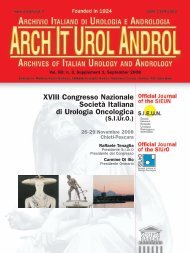
Foto di Bob Krieger<br />
Non esiste un farmaco privo di effetti collaterali e non si può affermare che se un farmaco dà un problema è solo<br />
colpa del medico e della sua ines<strong>per</strong>ienza. Eppure su molti giornali e siti internet si legge: “… il botulino usato<br />
<strong>per</strong> eliminare le rughe non ha nessun effetto collaterale, gli unici problemi sono legati all’ines<strong>per</strong>ienza del medico<br />
che effettua il trattamento…”. È incredibile! Ed è su questo che non sono d’accordo! La letteratura scientifica<br />
è piena di effetti collaterali, venuti alla luce solo dopo che certi farmaci primeggiavano dap<strong>per</strong>tutto come sicuri e<br />
miracolosi. Recentemente è stato pubblicato un articolo scritto dal Dott. Caleo del CNR di Pisa con cui si evidenzia<br />
un passaggio della tossina botulinica <strong>per</strong> via nervosa fino al cervello. Come <strong>tutti</strong> gli studi farmacologici l’es<strong>per</strong>imento<br />
è stato fatto su animali da laboratorio a cui si è sezionato il cervello dopo aver iniettato in alcuni muscoli<br />
il farmaco. Le quantità iniettate nel topo sono molto su<strong>per</strong>iori a quelle utilizzate nell’uomo ma la sco<strong>per</strong>ta importante<br />
fatta da Caleo è l’aver trovato e dimostrato che il botulino può diffondere a distanza <strong>per</strong> via nervosa. Nessuno<br />
sa con sicurezza se anche il botulino usato nell’uomo (alle dosi attuali) può arrivare nel cervello e con quali effetti.<br />
Però forse questo potrebbe spiegare i<br />
casi di botulismo e i vari effetti collaterali<br />
descritti in letteratura. Tutto questo non<br />
viene preso in considerazione, ma paradossalmente<br />
mistificato e denigrato da chi<br />
continua a sostenere che il botulino è sicuro<br />
e privo di rischi! Perché?<br />
Credo che molti effetti collaterali non vengano<br />
segnalati dal medico che inietta il<br />
botulino, o <strong>per</strong>ché ha paura di essere<br />
accusato di im<strong>per</strong>izia e incapacità o <strong>per</strong>ché<br />
pensa di diffondere il panico su un<br />
comodo ed utile status-quo terapeutico.<br />
Da alcuni anni sto cercando di far capire<br />
che il botulino è un farmaco che va rispettato<br />
con i suoi pro e contro, senza nulla<br />
nascondere, proprio come si fa con <strong>tutti</strong> gli<br />
altri farmaci. Ho sempre difeso e promosso<br />
il suo uso nell’ambito patologico (i<strong>per</strong>idrosi,<br />
spasmi, ecc.), tuttavia nutro grossi dubbi<br />
sul suo utilizzo come antiaging. Credo che<br />
la bellezza di un viso sia legata alla sua<br />
capacità di esprimere emozioni. Se si altera<br />
la mimica si <strong>per</strong>de non solo l’espressione<br />
ma anche la <strong>per</strong>sonalità che esprime ogni<br />
volto. Ogni terapia antiaging dovrebbe<br />
orientarsi verso la rigenerazione dei tessuti,<br />
stimolando e favorendo il metabolismo cellulare<br />
e il fisiologico turnover dei tessuti.<br />
Anche <strong>per</strong> questo non credo nell’utilità di<br />
paralizzare fibre muscolari con una tossina.<br />
In un recente convegno organizzato<br />
dall’Isplad ho ascoltato una relazione sui<br />
pro e contro del botulino fatta da un bravissimo<br />
collega di Venezia (il Dott. Patrizio<br />
Sedona), che stimo molto. È stata una relazione<br />
equilibrata e di grande valore.<br />
Finalmente non ho ascoltato uno spot pubblicitario.<br />
Vorrei fosse sempre così!<br />
There is no drug that is completely free from side effects. And when a drug does cause a<br />
problem, it can be very difficult to determine whether or not solely the physician and his<br />
inex<strong>per</strong>ience are at fault. It is also not uncommon to read something like this in medical<br />
journals and on websites: “Botulinum toxins that are used to eliminate wrinkles have no<br />
side effects, the only problems that occur are attributed to the inex<strong>per</strong>ience of the physician<br />
that administers the treatment...”. It’s unbelievable! And I disagree with these statements!<br />
Scientific literature widely documents the side effects of drugs. But these usually<br />
only come to light after the drugs have been accepted as being safe and miraculous by<br />
everyone. A recently-written article by Dott. Caleo of the National Research Centre in<br />
Pisa shows evidence which suggests that Botulinum toxins can be transferred to the brain<br />
via the nervous system. Like the majority of pharmacological ex<strong>per</strong>iments, this study was<br />
<strong>per</strong>formed on laboratory animals whose brains were dissected after the drug had been<br />
injected into their muscles. The quantity that was injected into the mice was much higher<br />
than that used in humans but the important discovery made by Caleo, nevertheless,<br />
demonstrates that Botulinum toxins can be diffused from a distance via the nervous system.<br />
Nobody knows for sure if Botulinum toxins used in humans at current dosages can,<br />
in fact, spread to the brain and what effects that would have. But <strong>per</strong>haps this could<br />
explain the cases of Botulinum toxin side effects that are found in scientific literature.<br />
Rarely is any of this ever taken into consideration. Quite paradoxically, it is mystified and<br />
discredited by those who continue to maintain that Botulinum toxins are safe and carry no<br />
risk! Why is that? I think many of the side effects are simply not pointed out to the patient<br />
by the physician injecting the Botulinum toxin. Perhaps it is the fear of being accused of malpratice<br />
and inablity that keeps him from doing so. Or maybe he thinks this will only serve<br />
to diffuse pointless panic about a convenient and useful therapeutic status quo.<br />
For years, I have tried to point out that Botulinum toxins should be used like any other<br />
drug. That is, with full disclosure of their pros and cons. I have always defended and promoted<br />
their clinical use (hy<strong>per</strong>hidrosis, spasms, etc.). However, I harbour great doubts<br />
about their use in anti-aging treatments. I believe that the capacity to express emotion is<br />
an essential part of facial beauty. When you alter facial mimics, you delete an expression,<br />
and thereby a part of someone’s <strong>per</strong>sonality as well.<br />
Every antiaging therapy should be oriented towards the regeneration of the tissues, stimulating<br />
and favouring the cellular metabolism and the physiological turnover of the tissues.<br />
Also for this reason I don’t believe it is useful to paralyze muscle fibres with a toxin.<br />
At a recent convention organized by Isplad, I listened to a presentation on the pros and<br />
cons of Botulinum toxins by Dott. Patrizio Sedona, a colleague of mine from Venice whom<br />
I highly respect. It was an objective and valuable presentation. “Finally”, I thought to<br />
myself, “I’m not listening to another advertising spot.” I wish this were always the case!<br />
Antonino Di Pietro<br />
Journal of Plastic Dermatology 2010; 6, 2 117
Journal of Plastic Dermatology<br />
Editor<br />
Antonino Di Pietro (Italy)<br />
Editor in Chief<br />
Francesco Bruno (Italy)<br />
Co-Editors<br />
Bernd Rüdiger Balda (Austria)<br />
Salvador Gonzalez (USA)<br />
Pedro Jaen (Spain)<br />
Associate Editors<br />
Francesco Antonaccio (Italy)<br />
Mariuccia Bucci (Italy)<br />
Franco Buttafarro (Italy)<br />
Ornella De Pità (Italy)<br />
Giulio Ferranti (Italy)<br />
Andrea Giacomelli (Italy)<br />
Alda Malasoma (Italy)<br />
Steven Nisticò (Italy)<br />
Elisabetta Perosino (Italy)<br />
Andrea Romani (Italy)<br />
Nerys Roberts (UK)<br />
Editorial Board<br />
Fabio Ayala (Italy)<br />
Lucio Andreassi (Italy)<br />
Kenneth Arndt (USA)<br />
Nicola Balato (Italy)<br />
H.S. Black (USA)<br />
Lucia Brambilla (Italy)<br />
Günter Burg (Switzerland)<br />
Stefano Calvieri (Italy)<br />
Michele Carruba (Italy)<br />
Aldo Di Carlo (Italy)<br />
Robin Eady AJ (UK)<br />
Paolo Fabbri (Italy)<br />
Giuseppe Micali (Italy)<br />
Martin Charles Jr Mihm (USA)<br />
Giuseppe Monfrecola (Italy)<br />
Joe Pace (Malta)<br />
Lucio Pastore (Italy)<br />
Gerd Plewig (Germany)<br />
Patrizio Sedona (Italy)<br />
Riccarda Serri (Italy)<br />
Adele Sparavigna (Italy)<br />
Abel Torres (USA)<br />
Stefano Veraldi (Italy)<br />
Umberto Veronesi (Italy)<br />
Managing Editor<br />
Antonio Di Maio<br />
English editing<br />
Rewadee Anujapad<br />
Direttore Responsabile Pietro Cazzola<br />
Direttore Generale Armando Mazzù<br />
Direttore Marketing Antonio Di Maio<br />
Consulenza grafica Piero Merlini<br />
Impaginazione Stefania Cacciaglia<br />
Registr. Tribunale di Milano n. 102 del 14/02/2005<br />
Scripta Manent s.n.c. Via Bassini, 41 - 20133 Milano<br />
Tel. 0270608091/0270608060 - Fax 0270606917<br />
E-mail: scriman@tin.it<br />
Abbonamento annuale (3 numeri) Euro 39,00<br />
Pagamento: conto corrente postale n. 20350682<br />
intestato a: Edizioni Scripta Manent s.n.c.,<br />
via Bassini 41- 20133 Milano<br />
Stampa: Arti Grafiche Bazzi, Milano<br />
Sommario<br />
pag. 121 Evaluation of the efficacy of a dietary supplement<br />
containing polyunsaturated fatty acids (pufas)<br />
and vitamins in the management of dermatitis<br />
Silvia Bottini, Silvia Ferrucci, Marinella Brambati,<br />
Barbara Riva, Paolo D. Pigatto<br />
pag. 127 External laser lipolisys in the treatment of cellulite: preliminary data<br />
Gianluca Campiglio<br />
pag. 135 Melatonin cream and melatonin-resveratrol Laa 15% serum<br />
and Quantum Molecular Resonance technology as ideal treatment<br />
for age related skin deseases<br />
Matteo Tutino, Adam Bodian, Ruben Oddenino<br />
pag. 145 Evaluation of cutaneous pH after chemical peel and its correction<br />
with amphoteric solutions<br />
Maurizio Cavallini, Valeria Puggioni, Riccardo Gazzola<br />
pag. 149 Case report: treatment of keloid with Intense pulsed Light<br />
Elisabetta Perosino, Eleonora Tati<br />
pag. 155 Valutazione clinica e strumentale dell’efficacia di un innovativo complesso antiage<br />
(ètas TimeBlock factor ® ) in un prodotto cosmetico <strong>per</strong> pelli mature<br />
Michela Quaglini<br />
pag. 167 Valutazione in vivo della interazione di un laser CO 2 frazionale sulla cute<br />
mediante microscopia confocale<br />
Elisabetta Sorbellini, Fabio Rinaldi<br />
pag. 177 La fisiopatologia della cicatrizzazione<br />
Valentina Bevelacqua, Giusy Schipani, Mauro Barone<br />
pag. 183 Your silent diener<br />
Gerd Plewig<br />
SLIDES<br />
pag. 191 La domanda e l’offerta dermatologica nel settore privato<br />
Delia Colombo, Antonino Di Pietro<br />
pag. 199 NEWS fROM THE COMpaNIES<br />
È vietata la riproduzione totale o parziale,<br />
con qualsiasi mezzo, di articoli, illustrazioni<br />
e fotografie senza l’autorizzazione scritta dell’Editore.<br />
L’Editore non risponde dell’opinione espressa dagli<br />
Autori degli articoli.<br />
Ai sensi della legge 675/96 è possibile in qualsiasi<br />
momento opporsi all’invio della rivista<br />
comunicando <strong>per</strong> iscritto la propria decisione a:<br />
Edizioni Scripta Manent s.n.c.<br />
Via Bassini, 41 - 20133 Milano<br />
Journal of Plastic Dermatology 2009; 5, 2 Journal of Plastic Dermatology 2010; 6, 12 119
Silvia Bottini<br />
Silvia Bottini ¹<br />
Silvia Ferrucci ¹<br />
Marinella Brambati ¹<br />
Barbara Riva ¹<br />
Paolo D. Pigatto 2<br />
¹ Istituto di Scienze Dermatologiche<br />
Fondazione IRCCS<br />
Ospedale Maggiore Policlinico,<br />
Mangiagalli e Regina Elena, Milano, Italy<br />
² Dipartimento di Tecnologie<br />
<strong>per</strong> la <strong>Salute</strong> IRCCS Istituto Galeazzi,<br />
Milano, Italy<br />
Evaluation of the efficacy<br />
of a dietary supplement containing<br />
polyunsaturated fatty acids (pufas)<br />
and vitamins in the management<br />
of dermatitis<br />
Summary<br />
I ntroduction<br />
Atopic dermatitis is an inflammatory<br />
skin disease characterized by intense pruritus<br />
with a chronic, relapsing course. The dermatitis<br />
is often resistant to traditional treatment and<br />
the use of systemic therapies is sometimes associated<br />
with a high incidence of recurrences and<br />
complications 1 .<br />
Many studies in the international literature suggest<br />
that atopic dermatitis is associated with a<br />
defect in the levels of essential polyunsaturated<br />
fatty acids (PUFAs) and vitamins 2 .<br />
The essential polyunsaturated fatty acids (PUFAs),<br />
Evaluation of the efficacy<br />
of a dietary supplement containing<br />
polyunsaturated fatty acids (pufas)<br />
and vitamins in the management<br />
of dermatitis<br />
The aim of this study was to evalute the effects of the dietary intake of PUFAs (essential<br />
polyunsaturated fatty acids) and vitamins on a group of atopic dermatitis patients<br />
in an open not standardized study. Supplementation of diet with PUFAs can be considered<br />
an adjunctive therapy in patients with eczema and it could be used as a preventive<br />
treatment. We took into consideration 120 atopic dermatitis patients divided into<br />
2 groups: group A took common therapy plus a diet supplementation (ADskin sachets)<br />
for at least two months, while the patients of the group B were treated only with the<br />
common therapy. They underwent regular visits at the base line and every 3 weeks till<br />
3 months (1 month of follow up) to evaluate clinical conditions scored by a well known<br />
scoring system for atopic dermatitis (SCORAD) and pruritus by a 100 mm Visual<br />
Analog Scale (VAS). They also completed a questionnaire giving subjective score for<br />
their condition. We recorded a statistically significant beneficial effect on the severity<br />
of the dermatitis evaluated by a decrese in Scorad values and VAS scale for pruritus,<br />
in patients treated with this diet supplementation as long as they took the diet supplementation.<br />
KEY WORDS: Atopic dermatitis, Dietary supplementation, Eczema, Gamma linoleic-acid,<br />
Pruritus, Antihistamines<br />
as linoleic acid n-6 (LA) and linolenic acid n-3<br />
(ALA), are obtained from the diet and are the precursors<br />
of the long chain polyunsaturated fatty<br />
acid (Lc-PUFAs) arachidonic acid (AA) 3 .<br />
PUFAs are considered essential fatty acids and<br />
they are found in vegetables oils (corn, sunflower,<br />
rapeseed, flaxseed and soybean oils) and<br />
derivates, such as margarines 4 .<br />
Many Authors have investigated the correlation<br />
between the severity of atopic dermatitis and<br />
the deficit of PUFAs. The deficit of PUFAs is<br />
linked to skin inflammation and loss of the bar-<br />
Journal of Plastic Dermatology 2010; 6, 2 121
122<br />
S. Bottini, S. Ferrucci, M. Brambati, B. Riva, P.D. Pigatto<br />
rier function 2 . PUFAs are important constituents<br />
of the cellular membrane and they assure<br />
the correct structure, function, fluidity and<br />
gene expression of its components 4 . Gammalinolenic<br />
acid is the precursor of arachidonic<br />
acid and its metabolites, such as prostaglandin<br />
E1 (PGE1) and other eicosanoids, are involved<br />
in the maturation and differentiation of T<br />
lymphocytes and in the production of IgE 5 . On<br />
the contrary, studies regarding the correlation<br />
between n-6 EFA metabolism and total IgE give<br />
conflicting opinions 2, 5 .<br />
Il problema maggiore nella somministrazione<br />
di PUFA consiste nel loro rapido degrado legato<br />
ad una facile ossidazione I tocoferoli ed altri<br />
anti ossidanti naturali presenti nella formulazione<br />
somministrata sono potenti antiossidanti<br />
e conferiscono la massima protezione verso<br />
il danno ossidativo cutaneo indotto dai radicali<br />
liberi.<br />
The aim of this study is to evaluate the influence<br />
of a dietary supplementation of antioxidative<br />
vitamins (E) and PUFAs in patients affected<br />
by atopic dermatitis.<br />
atients and methods<br />
p<br />
patients<br />
Over the last year we observed 120<br />
patients, 41 men and 79 women, with atopic<br />
eczema (Group A + B) elegible for this study.<br />
The median age of the patients at presentation<br />
was 64.47 years (range 17-79 years) Table 1.<br />
The diagnosis of eczema was clinical and based<br />
on the patients’ <strong>per</strong>sonal and familiar histories.<br />
All the patients had an history of severe dermatitis<br />
with uncontrollable pruritus and exacerbated<br />
skin lesions despite therapy with topical<br />
and systemic corticosteroids and oral antihistamines<br />
Table 1. Scorad value were taken at the<br />
base line as well as the VAS scale (Visual Analog<br />
Scale) for pruritus intensity which represents<br />
the visual evaluation of the symptom. We<br />
obtain a SCORAD baseline value of 41 + 14<br />
and a pruritus VAS baseline value of 54 + 13<br />
mm that mean that our patients were affected<br />
by a moderate dermatitis with a very important<br />
itching.<br />
We randomly divided the patients into two groups:<br />
group A (GA) took diet supplementation for<br />
a <strong>per</strong>iod of at least two months. The group B<br />
(GB) did not used diet supplementation.<br />
All patients (GA + GB) used a emollient cream<br />
Journal of Plastic Dermatology 2010; 6, 2<br />
and 75 (53.33%) (32 GA, 33 GB) used a topical<br />
corticosteroid cream and 6 (4 GB, 2 GA)<br />
(8.33%) applied a topical association of corticosteroids<br />
and antibiotics. Topical tacrolimus<br />
or pimecrolimus ointments were used in 47 (29<br />
GA e 18 GB) patients (51.66%) as a unique<br />
therapy (6 patients) or, generally, after failure<br />
on treatment with corticosteroid creams.<br />
Two patients (1 GA, 1 GB) with severe eczema<br />
required systemic corticosteroid drugs. In 7<br />
patients (3 GA, 4 GB) (8.33%) we suggested an<br />
association with narrow band-UVB phototherapy<br />
and lastly 58 patients (30 GA and 28 GB)<br />
also used an oral antihistamine (88.33%).<br />
methods<br />
For two months, patients of GA used<br />
a diet supplementation named ADskin. It is a<br />
mixture of PUFAs, vitamins (B, C, E), zinc,<br />
magnesium, selenium, silicon, manganese and<br />
Table 1.<br />
Patient characteristics.<br />
Group A Group B<br />
Median age 74.66 (17-79) 54.28 (17-91)<br />
Number of patients 60 60<br />
Male 15 (25%) 26 (43.33%)<br />
Female 45 (75%) 34 (56.66%)<br />
DA classical form 45 41<br />
Nummular eczema 12 14<br />
Nodular prurigo 3 5<br />
Table 2.<br />
Composition of ADskin.<br />
Nutrizional information In 100 g<br />
Energetic value 316.02 kcal/1321.21 kJ<br />
Proteins 14.08 g<br />
Carbohydrates 21.24 g<br />
Unsaturated fats 0.02 g<br />
Saturated fats 5.88 g<br />
Carbon hydrate 1.77 units<br />
Other compounds: vitamins (vit. C, niacin, vit. E, pantothenic acid, vit. B6, vit. B2, vit. B1,<br />
tocotrienol, tocopherol, betacarotene, folic acid, biotin, vit. B12), lactate, bicarbonate, K,<br />
Mg, Ca, Na, Si, Zn, Fe, Mn, Cu, Mo, Se, citrate, carbonate.
Evaluation of the efficacy of a dietary supplement containing polyunsaturated fatty acids (PUFAs) and vitamins in the management of dermatitis<br />
cop<strong>per</strong> (Table 2). It is a powder that can be swallowed<br />
with water, milk, fruit juice or yogurt.<br />
We suggested one packet/day, corresponding to<br />
twenty grams ƒof product, for 56 patients<br />
(93.33%), over a <strong>per</strong>iod of two months. Only<br />
four patients (6.66%) with a low body mass<br />
area (BMA < 17) used a reduced dosage of half<br />
a packet/day.<br />
Table 3.<br />
Clinical score system.<br />
Score Clinical estimation<br />
1 Worsening<br />
2 No improvement<br />
3 Slight or modest improvement<br />
4 Marked improvement<br />
Table 4.<br />
Results.<br />
Clinical score Group A Group B<br />
1 6 (10%) 2 (3.33 %)<br />
2 7 (11.66%) 20<br />
3 16 (20%) 22<br />
4 23 (38.33%) 12<br />
Lost to follow-up 8 4<br />
Table 5.<br />
Group A: results after one or more cycle of therapy with ADskin.<br />
Clinical score One cycle More than one cycle<br />
of therapy of therapy<br />
1 6 (10%) 2 (3.33%)<br />
2 7 (11.66%) 20<br />
3 16 (20%) 22<br />
4 23 (38.33%) 12<br />
Lost to follow-up 8 4<br />
Group A Group A after Rx Group A after follow up<br />
VAS 68 ± 18 46 ± 12 52 ± 20<br />
Scorad 38 ± 18 18 ± 8 29 ± 22<br />
Group B Group B after Rx<br />
VAS 69 ± 13 56 ± 17<br />
Scorad 37 ± 12 19 ± 14<br />
On the other hand, the GB patients used the<br />
same traditional eczema treatments, as for GA.<br />
Patients were subjected to clinical visits every 3<br />
weeks when they also showed a VAS score for<br />
the seriousness of their pruritus filled at home.<br />
At these moments patients were evaluated<br />
using a clinical scoring system (Table 3) and<br />
calculating SCORAD values.<br />
r esults<br />
We considered the effects of the dietary<br />
intake of PUFAs and vitamins on the severity<br />
of dermatitis, on pruritus.<br />
52 GA patients out of 60 completed the 2<br />
months therapy with this diet supplementation<br />
in addition to conventional eczema therapy.<br />
Eight patients decided to interrupt the therapy<br />
for <strong>per</strong>sonal problems (13.33%) 52 patients<br />
were followed up for one other month after<br />
withdrawl of this preparation.<br />
39 patients out of 52 of the GA (65%) using the<br />
clinical scoring system noticed a reduction in<br />
clinical symptoms especially pruritus In particular,<br />
after one cycle of treatment, 23 patients<br />
referred a marked improvement in their condition<br />
(score 4) and 8 patients declared a mild or<br />
modest improvement (score 3). Moreover, from<br />
this group of patients 14 (23.33%) declared an<br />
improvement in pruritus as well as a reduction<br />
in the skin inflammation. On the contrary, 7<br />
patients (21.66%) declared that there were no<br />
changes and 6 that they had noticed a worsening<br />
of their dermatitis. We observed a progressive<br />
improvement in the treated group as relieved<br />
by Scorad and by VAS versus the traditionally<br />
treated group but only after two months<br />
of treatment we obtain a statistically significant<br />
improvement. (p < 0,05 for both parameters).<br />
During the follow up patients symptoms behave<br />
as the control groups. 14 patients that obtained<br />
the better results <strong>per</strong>formed other cycles of<br />
therapy with a progressive clear improvement<br />
of pruritus in eight of them (Table 5). Three of<br />
these patients kept ADskin as their unique therapy<br />
now withdrawning antihistamines and<br />
other topical treatments but emollients. No<br />
side effects were recorded by these patients<br />
(Table 4).<br />
In GB 4 patients decided to interrupt therapy<br />
and in the remaining group of 56 we observed<br />
an improvement in pruritus in 34 patients; 12<br />
patients in particular declared a high reduction<br />
Journal of Plastic Dermatology 2010; 6, 2 123
124<br />
S. Bottini, S. Ferrucci, M. Brambati, B. Riva, P.D. Pigatto<br />
of symptom, while 22 reported a mild or modest<br />
improvement. There was no improvement in<br />
pruritus in 20 cases despite therapy and 2 noticed<br />
a worsening.<br />
D iscussion<br />
The importance of high levels of<br />
PUFAs in the diet is still a matter for debate.<br />
Our study detected the impact of a diet supplementation<br />
with PUFAs, vitamins and mineral<br />
salts on the clinical manifestations of atopic<br />
dermatitis especially on pruritus. Unlike most<br />
of the studies found in the international literature,<br />
we observed the impact of this diet integrator<br />
in classical atopic dermatitis, as well as in<br />
other kinds of atopic eczema like nummular<br />
and prurigo nodularis. We observed a reduction<br />
in the severity of the dermatitis with an amelioration<br />
of pruritus in particular, in more than<br />
half of the patients in the study (65%).<br />
In 1929 Burr demonstrated that a deficiency<br />
of linolenic acid can cause severe alterations in<br />
the structure of the skin and it manifests clinically<br />
with erythema, dryness and pruritus. An<br />
increase in transepidermal water loss was<br />
demonstrated in particular, as well as alterations<br />
in the skin’s im<strong>per</strong>meability to water, in<br />
epidermal proliferation with abnormal keratinocytes<br />
and, finally, in sterol esters synthesis 6 .<br />
Many studies have been conducted on the<br />
levels of linoleic acid and g-linolenic, dihomog-linolenic<br />
and arachidonic acid in atopics 4 .<br />
In the early 1980s Manku et al. demonstrated,<br />
by use of the gas chromatography technique,<br />
that the concentrations of linoleic acid are elevated<br />
in patients with atopic dermatitis with,<br />
on the contrary, a deficit of its metabolites (glinolenic<br />
acid GLA, dihomo-g-linolenic acid<br />
DGLA, arachidonic acid AA, adrenic acid and<br />
docosapentaenoic acid). Furthermore, a-linolenic<br />
acid levels are normal in patients with<br />
atopic dermatitis, but there is a deficiency of<br />
its metabolites.<br />
The reduced rate of activity of the enzyme D6desaturase,<br />
which converts linoleic acid to GLA<br />
and a-linolenic acid to stearidonic acid, can<br />
probably explain this condition 2, 5, 6 .<br />
On the contrary, Calder suggests that the lowered<br />
levels of arachidonic acid in atopy could be a<br />
consequence of an increased utilization in flogistic<br />
processes rather than a deficit in synthesis 4 .<br />
If a decrease in PUFAs worsens the atopic der-<br />
Journal of Plastic Dermatology 2010; 6, 2<br />
matitis, the correction of this defect should also<br />
lead to the clinical improvement of the dermatitis<br />
although we should remember that it is a<br />
multifactorial and complex disease 6 . Many studies<br />
have been conducted on the levels of linoleic<br />
acid and Kitz et al. ascribe a modulatory<br />
effect to GLA. Nevertheless, they affirm that<br />
GLA supplementation has no preventive effects<br />
for atopic dermatitis 5 .<br />
This all the bias of an open not standardized<br />
study we can extract some conclusions by<br />
means of the data we have assembled.<br />
First of all we obtained a statistically significant<br />
difference between the two groups for Vas and<br />
Scorad values indicating that ADskin has some<br />
proprieties that could be used to treat atopic<br />
dermatitis patients. The <strong>per</strong>centage of the<br />
patients who have achieved a clinical improvement<br />
(score 4 and score 3) are statistically significantly<br />
su<strong>per</strong>ior in group A than in group B.<br />
Moreover analysing the patients’ VAS carefully,<br />
we observed a greater number of patients who<br />
declared a marked improvement of pruritus<br />
after the therapy greater than that obtained with<br />
the antihistamines alone. For this reasons,<br />
many patients <strong>per</strong>formed other cycle of therapy<br />
with ADskin achieving a progressive clear<br />
improvement of pruritus without ex<strong>per</strong>iencing<br />
adverse effects.<br />
We underline that eight patients in group A and<br />
4 in group B were forced to interrupt the treatment<br />
before completing the suggested schema<br />
because of the high cost of the therapy as whole.<br />
Apparently, the patients of group A had a longer<br />
<strong>per</strong>iod of time without relapses in comparison<br />
with the patients of group B. These data<br />
are important because the use of a diet integrator<br />
enriched in PUFAs, as an adjunctive<br />
therapy in patients with severe or recurrent<br />
dermatitis, could reduce the daily administration<br />
of antihistamines and steroid, sparing<br />
them for cases of heavy exacerbation but<br />
should continued without stopping at least in<br />
autumn and winter time.<br />
In conclusion, we consider that diet supplementation<br />
is a safe coadjuvant therapy for eczema<br />
and we suggest combining it with the standard<br />
treatments.<br />
Specific therapy for eczema and atopic dermatitis<br />
cannot be replaced with dietetic supplementation;<br />
however the combined use of a diet<br />
integrator such as ADskin can be useful in<br />
reducing the severity of the symptoms, mainly<br />
in the case of severe dermatitis.
Evaluation of the efficacy of a dietary supplement containing polyunsaturated fatty acids (PUFAs) and vitamins in the management of dermatitis<br />
r eferences<br />
1. Tsoureli-Nikita E, Hercogova J, Lotti T, et al.<br />
Evaluation of dietary intake of vitamin E in the treatment of<br />
atopic dermatitis: a study of the clinical corse and evaluation<br />
of the immunoglobulin E serum levels. Int J Dermatol 2002;<br />
41:146-50.<br />
2. Yen Ch, Dai YS, Yang YH, et al. Linoleic acid metabolite<br />
levels and transepidermal water loss in children with<br />
atopic dermatitis. Ann Allergy Asthma Immunol 2008;<br />
100:66-73.<br />
2. Rodriguez-Cruz M, Tovar AR, del Prado M, et al.<br />
Molecular mechanisms of action and health benefits of<br />
polyunsaturated fatty acid. Rev Invest Clin 2005; 57:457-<br />
472.<br />
4. Calder PC. Abnormal fatty acid profiles occur in atopic<br />
dermatitis but what do they mean? Clin Exp Allergy 2006;<br />
36:138-141.<br />
5. Kitz R, Rose MA, Schönborn H, et al. Impact of early<br />
dietary gamma-linolenic acid supplementation on atopic<br />
eczema in infancy. Pediatr Allergy Immunol 2006; 17:112-7.<br />
6. Horrobin DF. Essential fatty acid metabolism and its modification<br />
in atopic eczema. Am J Clin Nutr 2000;<br />
71(Suppl 1):367S-72S.<br />
7. Burton GW, Joyce A, Ingold KU. First proof that vitamin E<br />
is major lipid-soluble, chain-breaking antioxidant in human<br />
blood plasma. Lancet 1982; 7:327.<br />
8. Fogarty A, Lewis S, Weiss S, et al. Dietary vitamin E, IgE<br />
concentration and atopy. Lancet 2000; 356:1573.<br />
Journal of Plastic Dermatology 2010; 6, 2<br />
125
Gianluca Campiglio<br />
External laser lipolisys<br />
in the treatment of cellulite:<br />
preliminary data<br />
Gianluca Campiglio<br />
Summary<br />
External laser lipolisys<br />
in the treatment of cellulite:<br />
preliminary data<br />
Board Certified Plastic Surgeon<br />
Milan, Italy<br />
In this pa<strong>per</strong> the preliminary data from the first Italian ex<strong>per</strong>ience with the use of a<br />
new device for the treatment of cellulite are reported. The machine is based on a<br />
innovative technology which couples the lipolytic effects of a dual wavelength laser<br />
with a suction assisted mechanical massage. A total of 11 patients have been enrolled<br />
in the study and concluded a cycle of treatments consisting in two sessions <strong>per</strong> week<br />
for a <strong>per</strong>iod of 4 weeks. Each patient filled in a satisfaction survey and was documented<br />
with digital photos before and after the treatment. The results showed a very<br />
high satisfaction rate, already after the first sessions, and also the comparison of the<br />
photos evidenced an improvement in the skin contour. These preliminary data confirm<br />
previous reports on the efficacy of the device but must be completed with further<br />
investigations using wider samples of patient, more reproducible protocols of<br />
treatment and more objective methods of evaluation of the results.<br />
KEY WORDS: Cellulite, Laser, Treatment<br />
I ntroduction<br />
Fat accumulation is an esthetic problem<br />
very often complained by the women. It is<br />
well demonstrated that this tissue can be accumulated<br />
in the human body in deep and su<strong>per</strong>ficial<br />
fat layers (Figure 1).<br />
Deep depots, such as flanks, thighs, abdomen<br />
and knees are located immediately above the<br />
muscular fascia and below the su<strong>per</strong>ficial fascia<br />
which is a structure present in almost all the<br />
human body under the subcutaneous fat.<br />
For these depots the ideal treatment is suctioned<br />
assisted lipectomy which allows to remove<br />
them in a very precise and definitive way.<br />
Modern liposuction is <strong>per</strong>formed with fine cannulas<br />
(diameter 2-3 mm) to reduce the rate of<br />
dimples or skin irregularities and using local<br />
anesthesia which has been demonstrated to<br />
significantly decrease the risk of major complications<br />
such as thromboembolism (Figure 2).<br />
Cellulite is a very common disease of the su<strong>per</strong>ficial<br />
fat layer which is immediately located<br />
under the dermis. It is characterized by engor-<br />
ged fat cells trapped in a fibrotic network of<br />
fibrous septae located in the hypodermis.<br />
According to Curri 1 cellulite can be clinically<br />
staged in four grades which are: Grade 1 =<br />
oedema, Grade 2 = fibrosis, Grade 3 = fibrosis<br />
Figure 1.<br />
Anatomic preparation showing the two different fat layers<br />
in the abdominal wall.<br />
Journal of Plastic Dermatology 2010; 6, 2 127
128<br />
G. Campiglio<br />
and sclerosis with micronodules and Grad 4 =<br />
macronodules.<br />
Etiology is multifactorial with hormones, diet<br />
and exercise playing an important role in its<br />
upset and evolution in the time. Interestingly<br />
cellulite is a fat problem which has nothing to<br />
do with weigh gain being often observed also in<br />
slim women.<br />
Many different types of treatment have been<br />
described to improve the cellulite. Some of<br />
them require topical agents but there is little<br />
peer-reviewed evidence about their efficacy and<br />
lasting effect. Also non-invasive devices have<br />
been proposed by various companies with<br />
varying results. Endermologie (LPG Systems),<br />
probably the most famous among them, is<br />
based on massage and suction of the skin.<br />
Many clinical studies reported a certain improvement<br />
in surface irregularities and also a<br />
reduction in circumference of thighs after<br />
cycles of treatment with this device.<br />
In this pa<strong>per</strong> we present the preliminary data<br />
with the use of a new technology, named<br />
photomology TM , which synergistically couples<br />
the lipolytic effects of a dual wavelength laser<br />
with a vacuum and mechanical rollers. This<br />
technology is the core of a new machine, which<br />
has both FDA and CE approval, specifically<br />
indicated for the non surgical treatment of cellulite<br />
(external laser lipolisys).<br />
aterials and methods<br />
m<br />
PhotomolgyTm module<br />
The photomologyTM module is constituted<br />
by:<br />
1) a 10W laser diodes with a wave length of<br />
915 nm;<br />
2) a 1W LED light of 650 nm;<br />
3) two contoured rollers;<br />
4) a vacuum system (Figure 3).<br />
915 nm laser diodes<br />
The choice of 915 nm wavelength was<br />
based on its preferential absorption by lipids 2 .<br />
This laser energy penetrates well into the tissue,<br />
with a minimal scattering, and leads to a moderate<br />
tem<strong>per</strong>ature elevation (< 40°C) inside the<br />
adipocytes promoting fat liquefaction.<br />
650 LED light<br />
This wavelenght was selected for its<br />
ability to interplay with the adipocytes mem-<br />
Journal of Plastic Dermatology 2010; 6, 2<br />
Figure 2.<br />
Pre and post o<strong>per</strong>ative views of a young patient<br />
with deep fat accumulation in the flanks,<br />
thighs and buttocks who underwent<br />
to suction assisted lipectomy.<br />
branes as previously demonstrated by Neira et<br />
al. in 2002 3 . In their work the authors showed<br />
that adipocytes in tissue samples taken from<br />
abdominoplasties, irradiated for 4 minutes and<br />
6 minutes with this wavelength, release respectively<br />
80 and 99 <strong>per</strong>cent of the fat.<br />
Transmission electron microscopy images of<br />
the adipose tissue taken at x 60,000 showed a<br />
transitory pore and complete deflation of the<br />
adipocytes with subsequent spillage of fat into<br />
the interstitial space.<br />
According to the Author the low-level laser<br />
Figure 3.<br />
Draw of the photomology module.
Figure 4.<br />
The photomology module produces a dual wavelength low level laser energy.<br />
energy affected the adipose cell by causing a<br />
transitory pore in the cell membrane to open<br />
which <strong>per</strong>mitted the fat content to go from inside<br />
to outside the cell. Interestingly the cells in<br />
the interstitial space and the capillaries remained<br />
intact.<br />
Light exposure and subsequent fat evacuation<br />
from cells do not destroy the adipocytes but left<br />
them intact and in good condition as also published<br />
by Jackson et al. in 2004 4 .<br />
The exact mechanism of this effect remains<br />
controversial, even if other authors reported<br />
that even small tem<strong>per</strong>ature elevation of 6°C<br />
above normal (i.e. 43°C) can lead to loss of the<br />
structural integrity of the lipidic bilayer of the<br />
cell membrane (Figure 4).<br />
Contoured rollers and vacuum<br />
The mechanical massage, obtained by<br />
two contoured rollers and a vacuum system,<br />
not only helps to evacuate the melted fat from<br />
the interstitial space but also assures a consistent<br />
interface of laser to tissue and improves<br />
blood and lymphatic circulation.<br />
Patient population and methods<br />
After a multicentric study conducted<br />
in 6 centers in USA and Europa on 83 patients<br />
demonstrating a significative improvement at 1<br />
External laser lipolisys in the treatment of cellulite: preliminary data<br />
and 3 months the device was introduced in<br />
Italy and first treatments started on April 2010.<br />
A total of 11 patients afflicted by cellulite has<br />
been treated with the same standard protocol<br />
consisting in two treatments <strong>per</strong> week for four<br />
weeks.<br />
Each session time ranged from 30 to 60 minutes<br />
according to the extension of the area to be<br />
treated. Maximun energy settings were used<br />
(1W, 650 nm and 10 W, 915 nm). Suction settings<br />
were medium intensity initially and then<br />
increased to maximum as tolerated by the<br />
patients.<br />
Preo<strong>per</strong>ative photos have been taken before the<br />
cycle of treatments and after one month from<br />
the end of the treatment. Body position was<br />
standardized using a foot position template. A<br />
survey was submitted to all the patients in order<br />
to judge the efficacy of the treatment.<br />
Exclusion criteria were: pregnancy, liver or kidney<br />
diseases, diabetes, dermatologic diseases,<br />
coagulation disorders, previous surgery in the<br />
treatment area less than 6 months before the<br />
enrollment and other anti-cellulite treatment 3<br />
months before the enrollment.<br />
r esults<br />
No complication was reported and<br />
any patient interrupted the treatment for adverse<br />
effects.<br />
Eighty two <strong>per</strong>cent of the patients (9/11) noted<br />
that their contour was improved after the first<br />
session of treatment and ninety-one (10/11)<br />
referred the same observation soon after the<br />
end of the cycle.<br />
All the patients wrote in the survey that they<br />
would have recommended this treatment to a<br />
friend.<br />
Some of them (3/11) referred a discomfort<br />
during the sessions due to the intensity of suction<br />
as demonstrated by the fact that this complication<br />
disappeared reducing it while maintaining<br />
constantly high the light energy.<br />
Even if some limitations in the standardization<br />
of the digital photographic recording of the<br />
patients before and after the treatment (minimal<br />
but significant light differences, reproducibility<br />
of the position of the patient despite of the footplate)<br />
this documentation showed a smoothering<br />
of the skin surface with improvement of<br />
the bulges and depressions especially in the<br />
thigh (Figures 5-8).<br />
Journal of Plastic Dermatology 2010; 6, 2 129
130<br />
G. Campiglio<br />
Figure 5.<br />
Pre and post treatment view of the thigh with improvement<br />
of the skin surface in the poster-lateral region.<br />
Figure 8.<br />
Same patient of the previous figure.<br />
In this view the improvement in the posterior thigh is shown.<br />
Journal of Plastic Dermatology 2010; 6, 2<br />
Figure 6.<br />
Pre and post treatment view of the thigh with reduction of the circumference and improvement<br />
of the skin contour.<br />
Figure 7.<br />
Pre and post treatment view of the inner thigh with smoothering of the skin contour<br />
in the suprapatellar area.
C onclusions<br />
The preliminary data obtained with<br />
the use of photomologyTM in the treatment of<br />
cellulite showed a positive effect on the most<br />
important and frequent symptom of this disease<br />
such as skin irregularity. Almost all the<br />
patient subjectively reported an improvement<br />
of the skin quality, already after the first session<br />
of treatment.<br />
This data confirm the results of previous and<br />
more sophisticated studies.<br />
In 2008 Lach reported the results of a vacuum<br />
massage and dual wavelength (650 and 915<br />
nm) low level laser energy to improve the<br />
appearance of cellulite 5 . In his cases each<br />
patient received a double treatment: one thigh<br />
was circumferentially treated with massage<br />
alone whereas the other thigh underwent to the<br />
dual beam laser energy treatment, followed by a<br />
mechanical massage. Each patient received a<br />
mean of 14 treatments, one to three <strong>per</strong> week<br />
for a total of 4-6 weeks. A series of magnetic<br />
resonance imaging measurements obtained in<br />
65 patients before and after the last treatment<br />
showed a significant reduction of fat thickness<br />
in the leg treated with laser and massage.<br />
More recently Kulick (2010) published an interesting<br />
study <strong>per</strong>formed on 20 women (BMI 29<br />
or less), aged 25-45 years, affected by mild to<br />
moderate cellulite of the postero-lateral aspect<br />
of the thigh 6 . The results were gathered using<br />
standard digital photographs and a complex<br />
three dimensional camera system (VECTRA,<br />
Canfield, USA) which depicts skin contour independent<br />
of incident lighting.<br />
The mechanism of this improvement could be<br />
probably secondary to the lipolityc effect of the<br />
laser energy on su<strong>per</strong>ficial fat tissue, located<br />
immediately below the dermis. In fact, the typical<br />
skin irregularities observed in the cellulite,<br />
are due to accumulation of fat at this level:<br />
enlargement of these adipocytes causes a stretching<br />
of the fibrous septa which connect the<br />
hypodermis to the deep tissue with formation<br />
External laser lipolisys in the treatment of cellulite: preliminary data<br />
of the classic dimples on the skin surfaces.<br />
Suction and massage are also important in this<br />
mechanism as showed by the fact that they are<br />
integral component of two other cellulite treatment<br />
devices worldwide used. In this case suction<br />
and massage help in the disruption of the<br />
sclerotic fibrous septa when cellulite evolves in<br />
its chronic phase.<br />
Although the positive results of our study further<br />
investigations are necessary in order to better<br />
verify the positive effects of external laser lipolysis<br />
in the treatment of cellulite, using a wider<br />
sample of patients, a more reproducible protocol<br />
of treatment (taking care for example also the<br />
total laser and mechanical energy transmitted to<br />
a defined treatment zone) and a more objective<br />
method of evaluation of the results.<br />
r eferences<br />
1. Curri S. Adiposità localizzata e panniculopatia<br />
edemato-fibrosclerotica. Sepem Ed. 1990.<br />
2. Anderson R, Farinelli W, Laubach H. Selective photothermolysis<br />
of lipid rich tissue: a free electron laser study.<br />
Laser Surg Med, 2006; 38:913-919.<br />
3. Neira R, Arroyave J, Ramirez M. Fat liquefaction: effect<br />
of low-level laser energy on adipose tissue. Plast Reconstr<br />
Surg 2002; 110:912-922.<br />
4. Jackson R, Roche G, Butterwick K, et al. Low level laser<br />
assisted liposuction: a 2004 clinical study of its effectiveness<br />
for enhancing ease of liposuction procedure and facilitating<br />
the recovery. Am J Cosmet Surg, 2004; 21:191-197.<br />
5. Lach E. Reduction of subcutaneous fat and improvement<br />
in cellulite by dual wavelength, low level laser energy combined<br />
with vacuum and massage. J Cosmet Laser Ther<br />
2008; 10:202-209.<br />
6. Kulick M. Evaluation of non invasive dual wavelength<br />
laser suction and massage device for the regional treatment<br />
of cellulite. Plast Reconstr Surg 2010; 125:1788-1796.<br />
D isclosure<br />
The author has not financial interest with<br />
the production or sale of the device.<br />
Journal of Plastic Dermatology 2010; 6, 2<br />
131
Matteo Tutino<br />
Matteo Tutino 1<br />
Adam Bodian 2<br />
Ruben Oddenino 3<br />
1 Clinic of Plastic Craniofacial Surgery,<br />
Department of ENT,<br />
University of Palermo, Italy<br />
2 Bodian Dermatology center<br />
New York, USA<br />
3 Plastic and Aesthetic Surgeon,<br />
President of European Association<br />
of Aesthetic Surgery, Milano, Italy<br />
melatonin cream and melatoninresveratrol<br />
laa 15% serum<br />
and Quantum molecular resonance<br />
technology as ideal treatment<br />
for age related skin deseases<br />
Summary<br />
I ntroduction<br />
The signs and symptoms of aging<br />
affect several organ systems. The skin is our largest<br />
organ. It functions as a barrier, protecting<br />
the body (internal environment) from the external<br />
environment. Skin problems may be the<br />
first symptoms of aging and a window to internal<br />
organ deterioration. Skin is our first organ<br />
to show signs of sun damage.<br />
We have developed a new melatonin multivitamin<br />
complex in combination with the purest<br />
vitamin C [L-ascorbic acid (LAA)] stabilized<br />
with resveratrol and Vitis vinifera, to topically<br />
treat sun damage, anti aging and actinic keratoses.<br />
We also treated the skin of sixty patients<br />
using a new revitalizing procedure based on<br />
Quantum Molecular Resonance (QMR) technology<br />
in order to value if and how skin face could<br />
melatonin cream and melatoninresveratrol<br />
laa 15% serum<br />
and Quantum molecular resonance<br />
technology as ideal treatment<br />
for age related skin deseases<br />
A new melatonin multivitamin complex in combination with the purest vitamin C [Lascorbic<br />
acid (LAA)] stabilized with resveratrol and Vitis vinifera has been developed<br />
to topically treat sun damage, anti aging and actinic keratoses. The skin of sixty patients<br />
using a new revitalizing procedure based on Quantum Molecular Resonance (QMR)<br />
technology has been also used in order to value if and how skin face could improve with<br />
both treatments. The study was conducted over three years with 264 patients, ages<br />
ranging from 28 to 85 years old. The new melatonin multivitamin complex in combination<br />
with vitamin C 15% stabilized with melatonin, resveratrol and Vitis vinifera,<br />
improves the intrinsic aging and photo-aged skin. QMR technology improves significantly<br />
the results increasing the amount of collagen and ameliorating the skin texture.<br />
KEY WORDS: Skin aging, Melatonin-resveratrol LAA 15% serum, Quantum Molecular Resonance<br />
improve with both treatments. Our study incorporated<br />
264 patients over a three year <strong>per</strong>iod<br />
and took an objective view on how this new<br />
topical product treats the aforementioned skin<br />
changes.<br />
tudy design<br />
S<br />
The study was conducted over three<br />
years with 264 patients, ages ranging from 28 to<br />
85 years old. We divided the patients into 2<br />
groups: control and study.<br />
The patients were selected randomly and based<br />
on their psychological examination, evaluation<br />
and clinical assessment. In addition, the study<br />
group received our treatment protocol and the<br />
Journal of Plastic Dermatology 2010; 6, 2 135
136<br />
M. Tutino, A. Bodian, R. Oddenino<br />
control group used vitamin C serum and simple<br />
moisturizer. All patients were allowed to continue<br />
their normal cosmetic treatments (botox, fillers,<br />
etc). Actinic keratoses was included among the<br />
pathologies as it may represent the natural evolution<br />
of sun damaged skin or a chronologic aged<br />
skin when the mitochondrial DNA has already<br />
expressed mutation 1-4 . Those suffering from actinic<br />
keratoses in both the study and control<br />
group, received cryotherapy or radiofrequency.<br />
aterials and method<br />
m<br />
Melatonin has been used ex<strong>per</strong>imentally<br />
in skin function such as hair growth<br />
cycling, pigmentation and melanoma control.<br />
Melatonin receptors are present in several skin<br />
cells including normal and malignant keratinocytes,<br />
melanocytes, and fibroblasts.<br />
Melatonin both suppresses ultraviolet rays (UV)<br />
that induce damage to skin cells and shows a<br />
strong antioxidant activity in UV exposed cells.<br />
Moreover, we recently uncovered activity in the<br />
skin of the biochemical processes involved in<br />
the sequential transformation of L-tryptophan<br />
to serotonin and melatonin 5-8 .<br />
A new cream formulation was developed mixing<br />
melatonin with a multivitamin complex and<br />
antioxidant derivative from the grape (resveratrol),<br />
in order to obtain a synergistic effect during<br />
the application and improve penetration of the<br />
substances through the skin barrier. The new<br />
creams were all based on a ph controlled system.<br />
Another formulation was based on melatonin,<br />
resveratrol and LAA which have important biological<br />
functions for skin and all cellular tissues<br />
as well as an antioxidant and an anti-inflammatory<br />
action. Moreover, they exert a noticeable<br />
action in the modulation of the immune response:<br />
by stimulating synthesis of collagen in fibroblasts<br />
of the human skin. LAA, in particular,<br />
plays a role in homeostasis of the connective tissue<br />
system of human cells. Furthermore, LAA<br />
increases the synthesis of proteins and collagen<br />
with anti-wrinkling effects. This process has a<br />
blocking action with respect to ferric ions, preventing<br />
skin damage due to excessive exposure<br />
to the sun 9-13 .<br />
The basic protocol used in our study was based<br />
on an acidic pH which enhanced the penetration<br />
and the action of the following compounds 14 :<br />
1) A cleanser for acidification of the skin to<br />
ph 3.5.<br />
Journal of Plastic Dermatology 2010; 6, 2<br />
2) Vitamin C serum 15% which has a strong<br />
penetration a ph 2.8.<br />
3) Melatonin resveratrol multivitamin complex<br />
cream a ph 4.5.<br />
The system required an application of the compounds<br />
twice/day. According to the skin condition,<br />
some patients were asked to double the<br />
application of products on the individual skin<br />
lesion in order to have a stronger action.<br />
We added a special formulation of tretinoin at a<br />
concentration of 0.1%, stabilized in vitis vinifera<br />
and resveratrol, to increase the intracellular<br />
action especially for actinic keratoses.<br />
The Control group (made up of 94 patients)<br />
was administered only topical vitamin C serum<br />
LAA and a custom made moisturizing cream<br />
containing petrolatum.<br />
4) QMR technology (fifty patients were included<br />
in the study group of Photo-Ageing and ten<br />
patients in the study group of Intrinsic<br />
Ageing): we used Rexon-Age technology plus<br />
the above-mentioned products. QMR protocol<br />
we used: 8 treatments to the whole face<br />
(1 a week). Exposure time: 50 minutes each<br />
treatment, 80 (mj/cmq)/S, electrode code n. 4<br />
(handle).<br />
The clinical application was conducted during<br />
the last 3 years, and each patient was selected<br />
according to a specific criteria based on clinical<br />
observations. Our study was carried out on a<br />
qualitative evaluation of the results, particularly<br />
on macroscopic evaluations. Histological examinations<br />
were realized only for those patients<br />
showing precancerous lesions. In photo-aged<br />
skin and intrinsic ageing, the histology was avoided<br />
in order not to create any discomfort to the<br />
patients since the clinical data did not justify any<br />
biopsies. Furthermore, every patient went<br />
through a psychological evaluation before,<br />
during and after the treatment <strong>per</strong>iod. Tests<br />
were <strong>per</strong>formed to evaluate the satisfaction of<br />
the patients. In order to do this a questionnaire<br />
was given. The following questions were asked:<br />
1. Have you noticed any improvements in the<br />
areas of the body that were treated?<br />
2. Do such improvements correspond to your<br />
expectations?<br />
3. Express a value from 1 to 6, where 1 = not<br />
satisfied and 6 = very satisfied your <strong>per</strong>ception<br />
of the improvement of the skin in relation to:<br />
• hy<strong>per</strong> pigmentation<br />
• wrinkles<br />
• scars
Melatonin cream and melatonin-resveratrol LAA 15% serum and Quantum Molecular Resonance technology as ideal treatment for age related skin deseases<br />
• acne<br />
• reduction of secretion of the sebum<br />
• compactness<br />
• pore reduction<br />
• hydration<br />
• brightness/luminosity.<br />
4. Express a value from 1 to 6, where 1 = not<br />
satisfied and 6 = very satisfied on the quality<br />
of the creams in comparison to:<br />
• ease of application<br />
• smell<br />
• penetration/absorption<br />
5. What was your state of mind when you first<br />
approached the treatment?<br />
6. Do you believe that you can have an active<br />
role in the results of the treatment?<br />
7. How much and to what extent do you feel<br />
that changes and/or improvements in your<br />
physical appearance has had or will have<br />
influence in your inter<strong>per</strong>sonal relationships?<br />
8. How do you view a medical approach that<br />
focuses on both the physical and psychological<br />
aspects of treatment?<br />
In order to have a better evaluation of our<br />
method and to achieve the best compliance of<br />
patients, this questionnaire was completed by<br />
each patient every year. To evaluate the comparison<br />
between the two groups, a statistics analysis<br />
regarding the achievement of expectations<br />
was conducted through the test.<br />
S tudy<br />
The clinical application of the abovementioned<br />
products was conducted in the 170<br />
patients of the study group who were affected<br />
by age-related skin diseases, intrinsic ageing<br />
and photo-damaged skin and was differentiated<br />
according to Table 1.<br />
Table 1.<br />
Expectations in patients with photo aging of the two groups and c2 value.<br />
Study group Control group c 2<br />
(n = 96) (n = 34)<br />
Expectations F % VA F % VA Extremely satisfied 44 45,8 8,7 0 1,7<br />
Satisfied<br />
Partially satisfied<br />
Not satisfied<br />
F = Observed values<br />
V = Awaited value<br />
A<br />
28<br />
19<br />
5<br />
29,1<br />
19,8<br />
5,2<br />
61,93<br />
10,47<br />
16,74<br />
9<br />
20<br />
5<br />
26,3<br />
59<br />
14,7<br />
15,72<br />
12,32<br />
4,25 }<br />
26,21<br />
The patients, including the study group, were<br />
selected according to skin deseases:<br />
1) Photo-ageing: 96 cases were enrolled after a<br />
macroscopic exam, in consideration of<br />
wrinkles, decreased skin tone (measured by<br />
pinch test), vascular collapse and signs of<br />
elastosis. The amount of collagen in the dermal<br />
skin of the fifty patients we tested using<br />
QMR technology have been previously evaluated<br />
with echograpy examination. The<br />
same ultrasound has been made to the<br />
patients of the control group. The patients’<br />
age varied from 28 to 65 years. The control<br />
group included 10 cases.<br />
2) Actinic keratosis: 34 cases, from 47 to 78<br />
years old.<br />
These cases where enrolled after clinical<br />
exam. Biopsies and histological examination<br />
where <strong>per</strong>formed before and after the treatment<br />
(Figures 1, 2).<br />
3) Intrinsic ageing: 40 cases, from 45 to 85<br />
years old.<br />
Figure 1.<br />
Histology of actinic keratosis biopsy.<br />
Figure 2.<br />
Histology after 1 year of treatment.<br />
Journal of Plastic Dermatology 2010; 6, 2 137
138<br />
M. Tutino, A. Bodian, R. Oddenino<br />
These cases were enrolled in the study concerning<br />
Intrinsic ageing since all the patients<br />
involved, because of their life style, rigorously<br />
avoided sun exposure.<br />
The control group includes:<br />
1) Photo ageing: 34 cases, from 28 to 65 years<br />
old;<br />
2) Actinic keratosis: 30 cases, from 47 to 78<br />
years old;<br />
3) Intrinsic ageing: 30 cases, from 67 to 85.<br />
The same principles of the study group were<br />
applied to enroll the control group patients.<br />
r esults<br />
The clinical and statistical evaluation<br />
of the patients of the study group has shown<br />
(Graphic 1, Tables 1-4):<br />
1) Patient with Intrinsic ageing (but patients<br />
simultaneously tested with QMR technology,<br />
who have been separated)<br />
• extremely satisfied: 24%<br />
• satisfied: 60%<br />
• partially satisfied: 10%<br />
• not satisfied: 6%<br />
2) Patient with Actinic keratoses<br />
• extremely satisfied: none<br />
• satisfied: 53%<br />
• partially satisfied: 29,4%<br />
• not satisfied: 17,6%<br />
3) Patient with Photo ageing (but patients<br />
simultaneously tested with QMR technology,<br />
who have been separated)<br />
• extremely satisfied: 43%<br />
• satisfied: 30%<br />
• partially satisfied: 20%<br />
• not satisfied: 7%<br />
4) Expectations in patients with Photo ageing<br />
and Intrinsic ageing tested with the abovementioned<br />
products plus QMR technology<br />
(Graphic 1.2)<br />
• extremely satisfied: 84%<br />
• satisfied: none<br />
• partially satisfied: 16%<br />
• not satisfied: none<br />
The partially satisfied patients report in the<br />
commentary that their expectations are based<br />
on the combination between creams containing<br />
vitamin C and melatonin combined and collateral<br />
procedures such us botox, fillers, thermage<br />
(RF Ellman Advanged Thermage), laser or in<br />
Journal of Plastic Dermatology 2010; 6, 2<br />
100 -<br />
90 -<br />
80 -<br />
70 -<br />
60 -<br />
% 50 -<br />
40 -<br />
30 -<br />
20 -<br />
10 -<br />
0 -<br />
90% -<br />
80% -<br />
70% -<br />
60% -<br />
50% -<br />
40% -<br />
30% -<br />
20% -<br />
10% -<br />
0% -<br />
Graphic 1.<br />
Perceptions of the improvement of the skin in the patients<br />
according to the skin problem after 3 years.<br />
After treatment<br />
Extremely satisfied<br />
Partially satisfied<br />
Extremely satisfied<br />
Satisfied<br />
Partially satisfied<br />
Not satisfied<br />
Photo aging Actinic keratosis Intrinsic aging<br />
Graphic 1.2.<br />
Perceptions of the improvement of the skin in patients<br />
who also underwent to QMR technology,<br />
according to the skin problem.<br />
Table 2.<br />
Expectations in patients with actinic keratosis among the two groups and c2 value.<br />
Study group Control group c 2<br />
(n = 34) (n = 30)<br />
Expectations F % V A F % V A<br />
Extremely satisfied<br />
Satisfied 18 53 10,68 8,43<br />
Partially satisfied 10 29,4 10,62 10 33,33 9,37 22,93<br />
Not satisfied 6 17 13,81 20 66,4 12,18<br />
F = Observed values<br />
V A = Awaited value<br />
}
100 -<br />
90 -<br />
80 -<br />
70 -<br />
60 -<br />
% 50 -<br />
40 -<br />
30 -<br />
20 -<br />
10 -<br />
0 -<br />
Melatonin cream and melatonin-resveratrol LAA 15% serum and Quantum Molecular Resonance technology as ideal treatment for age related skin deseases<br />
Table 3.<br />
Expectations in patients with intrinsic aging among the two groups and c2 value.<br />
Study group Control group c 2<br />
(n = 40) (n = 30)<br />
Expectations F % V A F % V A<br />
Extremely satisfied 10 25 1,33 0 1<br />
Satisfied 24 60 19,33 5 16,7 14,5 29,7<br />
Partially satisfied 4 10 11,33 15 50 8,5<br />
Not satisfied 2 5 8 10 33,3 6<br />
F = Observed values<br />
V A = Awaited value<br />
Table 4.<br />
c2 value (until 10 degree of freedom).<br />
G.L p = 0.10 p = 0.05 p = 0.01 p = 0.005<br />
1 2.705 3.841 6.635 7.879<br />
2 4.605 5.991 9.210 10.597<br />
3 6.251 7.815 11.345 12.838<br />
4 7.779 9.488 11.277 14.860<br />
5 9.236 11.07 15.086 16.749<br />
6 10.645 12.592 16.812 18.547<br />
7 12.017 14.067 18.475 20.278<br />
8 13.362 15.507 20.090 21.955<br />
9 14.684 16.919 21.666 23.589<br />
10 15.987 18.307 23.209 25.188<br />
Graphic 2.<br />
Perceptions of the improvement of the skin in the control group<br />
according to the skin problem after 3 years.<br />
Satisfied<br />
Partially satisfied<br />
Not satisfied<br />
Photo aging Actinic keratosis Intrinsic aging<br />
}<br />
certain cases surgery. The partially satisfied<br />
patients who underwent QMR report to be not<br />
fulfilled satisfied because their expectation was<br />
little bit higher than result could offer.<br />
The clinical and statistical evaluation of the<br />
patients of the control group has shown<br />
(Graphic 2, Tables 1-4):<br />
1) Expectations in patients with intrinsic ageing<br />
• 16,7% satisfied,<br />
• 50,0% partially satisfied,<br />
• 33,3% not satisfied yet.<br />
2) Expectations in patients affected by actinic<br />
keratoses<br />
• 33,3% partially satisfied,<br />
• 66,4% not satisfied yet.<br />
3) Expectations in patients with photo ageing<br />
• 26,3% satisfied,<br />
• 59,0% partially satisfied,<br />
• 14,7% not satisfied yet.<br />
tatistical analysis<br />
S<br />
We have employed the chi-squared test<br />
(c2 ) to demonstrate if the values of frequency we<br />
obtained are different in a significant way from<br />
the ones obtained through expected distribution;<br />
In this way we can demonstrate or refuse the<br />
hypothesis that the different treatments administered<br />
have the same effect or not.<br />
From our study we obtained statistically significant<br />
differences, level of probability of 0.005.<br />
D iscussion<br />
Both the study and control patients<br />
were given questionnaires asking them to<br />
express a judgment about their skin improvement<br />
during the three year <strong>per</strong>iod using their<br />
specific topical preparations. The questions were<br />
either yes or no or the scale of Likert (1 = “not at<br />
all satisfied” and 6 = “extremely satisfied”).<br />
Graph I demonstrates the compilation of the<br />
study group answers to the questionnaire.<br />
What stands out is that of those affected by<br />
intrinsic aging, 24% thought they were extremely<br />
satisfied, 60% felt satisfied, 10% partially<br />
satisfied and 6% not satisfied with the treatment<br />
regimen.<br />
All the patients treated with QMR technology<br />
plus products have been very satisfied and the<br />
sonography evaluation showed that two months<br />
after the end of the therapy the total amount of<br />
Journal of Plastic Dermatology 2010; 6, 2<br />
139
140<br />
M. Tutino, A. Bodian, R. Oddenino<br />
collagen increased up to 15% (average), that<br />
means about 5-6% more than patients who did<br />
not underwent to synergic procedure using QMR<br />
technology.<br />
In the study group of photoaging patients, 43%<br />
were extremely satisfied, 30% were satisfied,<br />
Journal of Plastic Dermatology 2010; 6, 2<br />
20% partially satisfied and 7% not satisfied<br />
with the treatment regimen.<br />
No one of the actinic keratosis patients felt<br />
extremely satisfied but 53% were satisfied,<br />
29.4% partially satisfied and 17.6% were not<br />
satisfied. Of note, these patients almost all wan-<br />
Figure 3.<br />
Pre treatment.<br />
Figure 4.<br />
Post treatment<br />
after 3 years.<br />
Case 1.<br />
A 64 year old patient heavy smoker, affected by skin aging, solar elastosis and in some areas actinic keratoses (Figure 1). The patient was 12 weeks under treatment<br />
with skin purify cleanser, purify toner, vitamin C fifteen serum with melatonin resveratrol and natural skin tone balancer with melatonin 1% and resveratrol cream<br />
in combination with restructurating melatonin 0.5% cream mixed with vitamins. Tretinoin 0.1% in a special formulation was added in the treatment once daily.<br />
The containing vitamin C and melatonin creams were applied twice daily for 12 weeks.<br />
After that <strong>per</strong>iod the patient was introduced in a weekly maintenance program (Figures 3, 4).<br />
Figure 5.<br />
Pre treatment.<br />
Figure 6.<br />
Post treatment<br />
after 3 years.<br />
Case 2.<br />
The new protocol, was applied in cases with actinic keratosis, and we bring to example this 75 old patient affected by recurrent actinic keratosis and basal cell<br />
carcinoma. The patient was o<strong>per</strong>ated several times and the success rate was poor since the skin was undefended against the UV DNA damage. The patient was<br />
o<strong>per</strong>ated and the carcinoma was removed with local anesthesia thanks to the use of radiofrequency technology (Ellman Surgitron 4 Mhrz) and a new special silver<br />
alloy which does not create thermal damage. Multiple flaps were needed to cover the cranium under local anesthesia. The small areas where the bone was<br />
exposed were covered with equine collagen and fibrin glue. The creams were applied systematically daily starting from the post o<strong>per</strong>ative <strong>per</strong>iod. The protocol<br />
included cleanser, vitamin C serum, melatonin cream and special regenerating cream containing aloe. The products were applied 2 times a day. Exfoliating cream<br />
and tretinoin was added once a day after the healing process. The treatment was carried out for 6 months and a light peel was done after the first 8 weeks from<br />
the surgical <strong>per</strong>iod. The patient did not have any recurrence of precancerous lesions and is still following a maintenance program (Figures 5, 6).
Melatonin cream and melatonin-resveratrol LAA 15% serum and Quantum Molecular Resonance technology as ideal treatment for age related skin deseases<br />
Figure 7.<br />
Pre treatment.<br />
Figure 8.<br />
Post treatment<br />
after 3 years.<br />
Case 3.<br />
This case represents a 70 years old patient affected by intrinsic aging. The patient has been treated for 3 years with our system which include vitamin C 15% serum<br />
containing melatonin and resveratrol, melatonin-resveratrol cream.<br />
The before and after pictures show the effect of the treatment on skin due to the antioxidant pro<strong>per</strong>ties of the system (Figures 7, 8).<br />
Figure 9. Pre treatment. Figure 10. Post treatment.<br />
Case 4.<br />
This case represents a 56 years old patient affected by intrinsic aging. The patient has been treated with vitamin C 15% serum containing melatonin and resveratrol,<br />
melatonin-resveratrol cream twice a day plus QMR technology (8 procedures). Figures 9 and 10 show the right cheek of the same patient before and after<br />
the treatment. Skin texture is really improved.<br />
ted additional surgical treatment (liquid nitrogen<br />
or curetting) in combination with the treatment<br />
regimen.<br />
Graph II illustrates the compilation of the control<br />
group answers to the questionnaire. Not one<br />
patient in all categories of the control group was<br />
extremely satisfied. Only about 20% of the intrinsic<br />
aging and photoaging patients were satisfied.<br />
About 50% of these patients were only partially<br />
satisfied and about 30% were not satisfied.<br />
The actinic keratoses patients showed that most<br />
were not satisfied with the control regimen and<br />
that about a third was partially satisfied. The<br />
difference between the study group and the<br />
control group has a statistical significance at a<br />
probability level of 0.5%.<br />
C onclusion<br />
As practicing clinicians most of us<br />
have seen the effects of using just moisturizers<br />
Journal of Plastic Dermatology 2010; 6, 2<br />
141
142<br />
M. Tutino, A. Bodian, R. Oddenino<br />
to help improve the skin. This idea is illustrated<br />
in that the two groups both felt some satisfaction<br />
with the treatment protocols. However,<br />
more patients were significantly satisfied in the<br />
study group. The new melatonin multivitamin<br />
complex in combination with vitamin C 15%<br />
stabilized with melatonin, resveratrol and Vitis<br />
vinifera, improves the intrinsic aging and<br />
photo-aged skin.<br />
In addition, combining this with tretinoin may<br />
be considered as an alternative treatment for the<br />
prevention of actinic keratoses and photo<br />
damaged skin. QMR technology improves<br />
significantly the results increasing the amount<br />
of collagen and ameliorating the skin texture.<br />
This new protocol may be used to treat photodamaged<br />
skin diseases and as an anti-aging<br />
cosmetic treatment. The three case studies<br />
point out how versatile the new products are for<br />
treating a variety of skin ailments. More studies<br />
need to be done to objectively show the effects<br />
these medications have on the skin.<br />
r eferences<br />
1. Azmi AS, Bhat SH, Hanif S, Hadi SM. Plant<br />
polyphenols mobilize endogenous cop<strong>per</strong> in human <strong>per</strong>ipheral<br />
lymphocytes leading to oxidative DNA breakage: a<br />
putative mechanism for anticancer pro<strong>per</strong>ties. FEBS Lett.<br />
2006; 580:533-8. Epub 2005 Dec 28.<br />
2. de la Lastra CA, Villegas I. Resveratrol as an antiinflammatory<br />
and anti-aging agent: mechanisms and clinical<br />
implications. Mol Nutr Food Res 2005; 49:405-30.<br />
3. Queille S, Luron L, Spatz A, et al. Analysis of skin cancer<br />
risk factors in immunosuppressed renal transplant<br />
patients shows high levels of UV-specific tandem CC to TT<br />
mutations of the p53 gene. Carcinogenesis 2007; 28:724-<br />
31. Epub 2006. 25. Altro.<br />
4. Slominski A, Wortsman J, Tobin. The cutaneous sero-<br />
Journal of Plastic Dermatology 2010; 6, 2<br />
toninergic/melatoninergic system: securing a place under<br />
the sun. FASEB J 2005; 19:176-94.<br />
5. Goskel Sener DJ, Guilten Sert, Ozer Sehirli A., et al.<br />
Melatonin Protects against pressure ulcer-induced oxidative<br />
injury of the skin and remote organs in rats. J Pineal Res<br />
2006; 40:280-287.<br />
6. Ekmekcioglu C. Melatonin receptors in humans: biological<br />
role and clinical relevance Biomed Pharmacother 2006;<br />
60:97-108. Epub 2006 Feb 20.<br />
7. Esrefoglu M, Gul M, Seyhan M, Parlakpinar H.<br />
Ultrastructural clues for the potent therapeutic effect of<br />
melatonin on aging skin in pinealectomized rats. Fundam<br />
Clin Pharmacol. 2006; 20:605-611. PMID: 17109654<br />
[PubMed - as supplied by publisher]<br />
8. Tebbe B, et al. L-ascorbic acid inhibits UVA-induced lipid<br />
<strong>per</strong>oxidation and secretion of IL-1 alpha and IL-6 in cultured<br />
human keratinocytes in vitro. Invest Dermatol 1997;<br />
108: 302-6.<br />
9. Jagetia GC, Rajanikant GK, Rao SK. Evaluation of the<br />
effect of ascorbic acid treatment on wound healing in mice<br />
exposed to different doses of fractionated gamma radiation.<br />
Radiat Res 2003; 159:371-80.<br />
10. Koo N, Cho D, Kim Y, et al. Effects of resveratrol on<br />
mast cell degranulation and tyrosine phosphorylation of the<br />
signaling components of the IgE receptor. Planta Med.<br />
2006; 72:659-61. Epub 2006 Apr 24.<br />
11. Moon SO, Kim W, Sung MJ, et al. Resveratrol suppresses<br />
tumor necrosis factor-alpha-induced fractalkine<br />
expression in endothelial cells. Mol Pharmacol 2006;<br />
70:112-9. Epub 2006 Apr 13.<br />
12. Yang JH, Lee HC, Chung JG, et al. Mitochondrial DNA<br />
mutations in light-associated skin tumors. Anticancer Res<br />
2004; 24:1753-8.<br />
13. Ribeiro GR, Francisco G, Teixeira LV, et al. Repetitive<br />
DNA alterations in human skin cancers J Dermatol Sci<br />
2004; 36:79-86.<br />
14. Yangxi Wang, Bryan Mackenzie, Hiroyasu Tsukaguchi, et<br />
al. Human Vitamin C (L-ascorbic acid) Transporter SVCT1<br />
Biochemical and Biophysical Research Communications<br />
2000; 267:488-494.
Maurizio Cavallini<br />
Maurizio Cavallini 1<br />
Valeria Puggioni 2<br />
Riccardo Gazzola 3<br />
1 Servizio di Chirurgia Plastica,<br />
Centro Diagnostico Italiano, Milano<br />
2 Unità di Chirurgia Plastica Ricostruttiva,<br />
IRCCS Istituto Ortopedico Galeazzi, Milano<br />
3 Unità di Chirurgia Plastica,<br />
IRCCS San Raffaele, Milano<br />
Evaluation of cutaneous pH<br />
after chemical peel and its correction<br />
with anphoteric solutions<br />
Summary<br />
I ntroduction<br />
Several mechanisms coo<strong>per</strong>ate in<br />
maintaining cutaneous pH at 5.5. The use of<br />
cosmetic products that alter this value are<br />
potentially harmful for skin health.<br />
Unfortunately this problem is often underestimated<br />
among the o<strong>per</strong>ators in aesthetic medicine.<br />
During the 13th national meeting of SIES<br />
(Italian society of aesthetic medicine and surgery)<br />
a questionnaire was administerd to the<br />
450 partecipants. In a multiple-choice question,<br />
the doctors were asked to point the cutaneous<br />
pH after chemical peel.<br />
Just 41.1% of partecipants answered correctly<br />
(pH ≈ 2), while the majority indicated higher<br />
pHs wrongly.<br />
In this study we evaluate the influence of products,<br />
employed chemical peel, on cutaneous<br />
pH. Finally we analyze the effectiveness of<br />
water and products based on amphoteric solution<br />
(Diphoterine ® , Prevor-France) in correcting<br />
extremely low pHs.<br />
The physiologic cutaneous low <strong>per</strong>meability to<br />
water is provided by the corneal layer 1 of the<br />
skin. This stratum maintains a constant pH of<br />
Evaluation of cutaneous pH<br />
after chemical peel and its correction<br />
with amphoteric solutions<br />
Human cutaneous pH is the result of a fine regulation, in fact several mechanisms<br />
provide for the maintainment of the physiological value of 5.5. Considerable oscillations<br />
in cutaneous pH could hesitate in damages to the skin barrier. In this article we<br />
analyze the effect of products employed in chemical peels on cutaneous pH and the<br />
results on pH correction with amphoteric solutions that are tested in association with<br />
water and alone. We conclude that amphoteric solutions alone are more effective in<br />
correcting cutaneous pH after chemical peel and that those solutions are preferable<br />
to basic solutions that could damage the skin barrier overcorrecting the pH.<br />
KEY WORDS: Chemical peel, Cutaneous pH, Anphoteric solution, Sodium bicarbonate<br />
7.4, thanks to an acidification process deriving<br />
from the phospholipids hydrolysis by a secretory<br />
phospholipase and a Na/H antiporter<br />
(NHE1) 2 . In this mechanism a considerable<br />
role is played by the trans-urocanic acid that<br />
acts as proton donor 3 . Also bacterial lipases are<br />
involved in this mechanisms, generating free<br />
fatty acids 4 .<br />
In some studies the correlation between melanosome<br />
secretion and pH (relatively to skin type<br />
IV-V) have been demonstrated 5 .<br />
aterials and methods<br />
m<br />
In this study we <strong>per</strong>formed chemical<br />
peels on 25 patients. The formulation of the<br />
product that we used to acidify the skin consists<br />
of a 70% glycolic acid solution. The glycolic<br />
acid solution was applied on both the forearm<br />
for 5 minutes. The measurements were divided<br />
in two groups: in group A were included the<br />
measurements <strong>per</strong>formed on the left forearm,<br />
while the measurements on the right forearm<br />
Journal of Plastic Dermatology 2010; 6, 2 145
146<br />
M. Cavallini, V. Puggioni, R. Gazzola<br />
were considered belonging to group B. In the<br />
group A the product was washed with water for<br />
1 minute and then amphoteric solution was<br />
employed with the suggested modalities. In the<br />
group B amphoteric solution was used directly<br />
without washing with water.<br />
The measurements were obtained with litmus<br />
pa<strong>per</strong> before any treatment, after the application<br />
of the glycolic acid-solution and after correction<br />
of pH (with water plus amphoteric<br />
solution or with amphoteric solution alone)<br />
(Figure 1).<br />
esults and discussion<br />
r<br />
The average pH of the skin before any<br />
treatment was 4.88, after the application of glycolic<br />
acid was 0.7. In the group A, after washing<br />
with water, the pH was 3.4, while in the group<br />
B, after washing with amphoteric solution, the<br />
pH was 4.03.<br />
The average increase of pH was respectively 2.7<br />
and 3.33. One-sample T test was <strong>per</strong>formed to<br />
compare the mean of group A with the values of<br />
group B. The results show a mean difference of<br />
4.025 with a p value < 0.001.<br />
These results confirm the effectiveness of an<br />
amphoteric solution in increasing the cutaneous<br />
pH and its su<strong>per</strong>iority in comparison<br />
with water washings.<br />
Alteration of cutaneous pH is the base for a<br />
further damage. The activity of NHE1 has been<br />
demonstrated to be strictly dependent on pH,<br />
having effects on the maturation of the lamellar<br />
membranes. More over in aged mice, changes<br />
of cutaneous pH affect the correct maturation of<br />
the skin, and restoring the physiological pH can<br />
correct the defect 6 .<br />
In chemical peels, pH fluctuations are huge and<br />
cutaneous stress is high. In fact, after applying<br />
of glycolic acid, skin pH decreases of 4.08. The<br />
correction of this value should have two aims:<br />
first, the physiological pH should be restored as<br />
soon as possible, on the other hand overcorrection<br />
should be absolutely avoided. Kim et al.<br />
demonstrated skin damage, concerning skin<br />
barrier and function of the stratum corneum<br />
with basic pH values 7 .<br />
For this reason we suggest the employment of<br />
amphoteric solutions which are effective in<br />
restoring physiological values without causing<br />
basic pH, contrary to sodium bicarbonate.<br />
Journal of Plastic Dermatology 2010; 6, 2<br />
C onclusion<br />
Among the substances employed for<br />
chemical peel, glycolic acid is widely popular.<br />
In our study, the pH of the skin has been<br />
demonstrated to decrease of 4.08 points after<br />
the procedure described earlier.<br />
The employment of pH-correcting substances<br />
aims to avoid alteration of skin <strong>per</strong>meability<br />
and abnormalities in the stratum corneum. An<br />
amphoteric solution has been demonstrated to<br />
be an useful product in correcting low pH, thus<br />
avoiding damages to the skin by glycolic acid.<br />
Moreover amphoteric solution has been<br />
demonstrated to be more effective than water in<br />
raising cutaneous pH 8 .<br />
Adjusting of cutaneous pH after chemical peel<br />
is of primary importance. In our opinion the<br />
neutralization of acidity with water and/or<br />
amphoteric solution is advisable, while pH correction<br />
through alcaline solution (e.g. sodium<br />
bicarbonate) is to avoid. In fact basic products<br />
could overcorrect the cutaneous acidity, leading<br />
to alkaline pH and thus providing further chemical<br />
stress and damages.<br />
Figura 1.<br />
Litmus pa<strong>per</strong> is employed to measure cutaneou pH.<br />
The combination of the three colours indicates the pH-value.
B ibliography<br />
1. Shade H, Marchionini A. Der sauremantel<br />
der haut. Klin Wochenschr 1928; 7:12-14.<br />
2. Fluhr JW, Behne MJ, Brown BE, et al. Stratum corneum<br />
acidification in neonatal skin: secretory phospholipase A2<br />
and the sodium/hydrogen antiporter-1 acidify neonatal rat<br />
stratum corneum. J Invest Dermatol 2004; 122:320-9.<br />
3. Krien PM, Kermici M. Evidence for the existence of a<br />
self-regulated enzymatic process within the human stratum<br />
corneum -an unexpected role for urocanic acid. J Invest<br />
Dermatol 2000; 115:414-20.<br />
4. Thueson DO, Chan EK, Oechsli LM, et al. The roles of<br />
pH and concentration in lactic acid-induced stimulation of<br />
epidermal turnover. Dermatol Surg 1998; 24:641-5.<br />
Evaluation of cutaneous pH after chemical peel and its correction with anphoteric solutions<br />
5. Gunathilake R, Schurer NY, Shoo BA, et al. pH-regulated<br />
mechanisms account for pigment-type differences in epidermal<br />
barrier function. J Invest Dermatol 2009;<br />
129:1719-29.<br />
7. Choi EH, Man MQ, Xu P, et al. Stratum corneum acidification<br />
is impaired in moderately aged human and murine<br />
skin. J Invest Dermatol 2007; 127:2847-56.<br />
6. Kim E, Kim S, Nam GW, et al. The alkaline pH-adapted<br />
skin barrier is disrupted severely by SLS-induced irritation.<br />
Int J Cosmet Sci 2009; 31:263-9.<br />
8. Mathieu L, Cavallini M, Corsi MM. Efficacy of diphoterine<br />
HCl decontamination in rats: a comparative study.<br />
Toxicology Letters 2002; 135:166-67.<br />
Journal of Plastic Dermatology 2010; 6, 2<br />
147
Elisabetta Perosino<br />
Elisabetta Perosino 1<br />
Eleonora Tati 2<br />
1 Medico Chirurgo Dermatologo<br />
Roma, Italy<br />
2 Medico Chirurgo, Roma, Italy<br />
Case report: treatment of keloid<br />
with Intense Pulsed Light<br />
Summary<br />
I ntroduction<br />
Hy<strong>per</strong>trophic scars and keloids are<br />
common problems especially in young women<br />
and dark skin types (Fitzpatrick VI) like in individuals<br />
of African, Hispanic, and Asian descent 1 .<br />
Hy<strong>per</strong>trophic scars and keloids occur as a result<br />
of excessive healing reactions to damage to the<br />
deep skin tissues after traumatic or surgical<br />
wounds that have a prolonged phase of inflammation<br />
and fibroplasias.<br />
This healing reaction leads to excessive fibroblast<br />
proliferation and collagen synthesis that result in<br />
benign fibrous outgrowths for both keloids and<br />
hy<strong>per</strong>trophic scars. Both have a similar clinical<br />
appearance; however, the growth reaction<br />
remains within the margins of the scar in hy<strong>per</strong>trophic<br />
scars, while in keloids the reaction has<br />
the ability to extend the boundaries of the initial<br />
scar and is likely to recur after treatment 2 .<br />
The exact pathogenesis of keloids and hy<strong>per</strong>trophic<br />
scar formation still remains unknown<br />
which makes treatment even more complicated<br />
3 ; however, an imbalance of matrix degradation<br />
and collagen biosynthesis, resulting in<br />
excess accumulation of collagen in the wound,<br />
has been postulated to be the primary biochemical<br />
feature of these skin lesions 4-6 .<br />
Case report: treatment of keloid<br />
with Intense Pulsed Light<br />
Keloids are scars, unique to humans that grow beyond the boundaries of a cutaneous<br />
injury, inflammation, burn or surgical incision. However spontaneous keloids sometimes<br />
develop without a preceding trauma, most commonly over the sternum. A wide<br />
range of therapies is available for keloids but the reported efficacy has been variable.<br />
These options include intralesional steroid injections, pressure dressing, silicone gel<br />
sheeting, topical retinoids, excisional surgery, radiotherapy and cryosurgery. Lasers<br />
are now being used to improve keloids. In this article we present a case of a young<br />
woman with keloid of the sternum region treated with Intense Pulsed Laser.<br />
KEY WORDS: Keloid, Intense Pulsed Laser<br />
Many <strong>per</strong>sons seek medical attention for the<br />
treatment of scars, not only for aesthetic reasons<br />
(an unsightly scar can be very distressing, especially<br />
to young females) but also for functional<br />
reasons; scar contracture over a joint can<br />
severely restrict movement of the affected area.<br />
Pain and pruritus are typical symptoms associated<br />
with a proliferative scar.<br />
A wide range of therapies is also available for<br />
keloids, but the reported efficacy has been variable.<br />
These options include intralesional steroid<br />
injections, pressure dressing, silicone gel sheeting,<br />
topical retinoids, excisional surgery, radiotherapy,<br />
and cryosurgery 7-10 . Lasers are now<br />
being used to improve hy<strong>per</strong>trophic scars and<br />
keloids 11 .<br />
Since their introduction for hy<strong>per</strong>trophic scars<br />
and keloids by Apfelberg et al. 12 and Castro et<br />
al. 13 in the mid-1980s, more and more lasers<br />
with different wavelengths have been studied.<br />
Q-switched lasers (e.g. Nd: YAG, alexandrite,<br />
ruby) can be used to treat hy<strong>per</strong>pigmented<br />
scars and to remove artificial pigment from a<br />
scar (e.g. tarmac or gun powder particles<br />
embedded in a scar after traffic or explosives<br />
accident, respectively).<br />
Journal of Plastic Dermatology 2010; 6, 2 149
150<br />
E. Perosino, E. Tati<br />
Continuous wave lasers (CO 2 , argon, Nd: YAG)<br />
in the 1970s and early 1980s were early lasers<br />
used in scar treatment. Due to the inherent<br />
problems of continuous wave lasers little<br />
improvement was achieved in most cases 14 .<br />
Pulsed ablative lasers (CO 2 , Er: YAG) are being<br />
used in the treatment of atrophic acneic scars<br />
and keloids, although in the latter recurrence<br />
rates are high 15 .<br />
Because of the limitations of currently available<br />
treatments, other options such as short-pulsed<br />
pulsed dye laser (PDL) and long-pulsed pulsed<br />
dye laser (LPDL) have been used with reported<br />
improvement in the long-term appearance of<br />
hy<strong>per</strong>trophic scars 16-19 .<br />
The 585 nm pulsed dye laser is used to treat<br />
erythematous, hy<strong>per</strong>trophic and keloid scars.<br />
Erythema, symptoms, and size of the scar are<br />
improved with this type of laser, which targets<br />
principally the scar’s vascularity 20 . Recent<br />
reports exist on the early use of pulsed dye laser<br />
to prevent unfavourable scars 21 .<br />
More recently, Intense Pulsed Light (IPL)<br />
applied following suture removal was found<br />
in a pilot study to be effective on surgically<br />
induced scars 22 , and on various types of scars<br />
in another study 23, 24 . The IPL systems have a<br />
broad wavelength spectrum (515-1200 nm).<br />
Unlike lasers, IPL systems are flash lamps<br />
with non-coherent pulsed light which<br />
employs variable pulse durations and intervals.<br />
Each pulse can be dived up to three subpulses.<br />
The duration of each subpulse (0,5-20<br />
ms), the duration of the delay (1-300 ms)<br />
between subpulses, and energy fluence of the<br />
whole pulse (3-90 J/cm 2 ) can also be adjusted.<br />
Several cut off filters are available (at 515,<br />
550, 570, 590, 615, 645, 695, and 755 nm)<br />
to block out from the emitted pulse photons<br />
of shorter wavelength.<br />
Due to its characteristics IPL can act on the pigmentation<br />
and vasculature of a scar and treat<br />
erythematous and proliferative scars 25 .<br />
ase report<br />
C<br />
In this pa<strong>per</strong> we present a case of a<br />
young woman with a keloid of the sternum<br />
(Figure 1). This patient had a story of sebaceous<br />
cysts when she was 16 years old localized<br />
over the sternum area. She didn’t treat the<br />
lesion with surgery excision neither with topical<br />
antibiotic therapy. The cysts in a few of time<br />
Journal of Plastic Dermatology 2010; 6, 2<br />
became quickly a hy<strong>per</strong>trophic scar and subsequently<br />
a keloid.<br />
She didn’t have familiarity for keloids or systemic<br />
disease. She used to treat the lesion only<br />
with silicone gel sheets 24 hours <strong>per</strong> day and<br />
intralesional corticosteroid infiltration.<br />
We decided to treat the keloid with IPL to test<br />
its efficiency on these types of lesions. We used<br />
three different cut-off filters: 550, 570 e 695<br />
nm. In combination with 550 nm filter we used<br />
a fluence of 30 J/cm 2 with single impulse and<br />
impulse duration of 4,0 ms. In combination<br />
with 570 nm filter we applied a fluence of 45<br />
J/cm 2 , a triple impulse, a duration pulse of 2,8<br />
ms and a delay’s duration of 20 ms. With a cutoff<br />
filter 695 nm the fluence was 50 J/cm 2 ,<br />
triple impulse, duration pulse of 4,5 ms and<br />
delay’s duration of 40 ms (Table 1).<br />
A total number of 5 treatments were given to<br />
the patient. Time interval were 4 weeks at least,<br />
enough time for the treated area to recover<br />
from side effects (if any) and to show its<br />
response to the treatment for the first three<br />
applications. Time intervals were 8 weeks for<br />
the remnant five sessions.<br />
Discomfort produced by the treatment was<br />
mild (as it is whenever an IPL device is used for<br />
any kind of treatment), and there is no need for<br />
the use of local anaesthesia. We used to mask<br />
the surrounding skin with adhesive stickers to<br />
protect it from side effects.<br />
D iscussion<br />
In the past IPL treatment has been<br />
used for photorejuvenation, to treat vascular<br />
lesions, sun-damaged skin, pigmented lesions<br />
and rhytids, to reduce the incidence of purpura,<br />
and for hair removal 22, 23, 27-31 ; only a few<br />
study reported its use on hy<strong>per</strong>trophic scars<br />
and keloids 22, 23 .<br />
Table 1.<br />
Cut off filter Fluence Impulse Duration pulse Duration of the delay<br />
550 nm 30 J/cm 2 single 4 ms -<br />
570 nm 45 J/cm 2 triple 3 ms 20 ms<br />
695 nm 50 J/cm 2 triple 5 ms 40 ms
Figure 1.<br />
Pre treatment.<br />
Figure 2.<br />
Post treatment.<br />
Bellew et al. 22 after IPL treatment of hy<strong>per</strong>throphic<br />
scars, observed significant effect on<br />
pliability of the scar and improvement in the<br />
textural smoothing component of scars due to<br />
Case report: treatment of keloid with Intense Pulsed Light<br />
IPL inclusion of near-infrared wavelengths.<br />
They hypothesized that IPL treatment might<br />
affect the neovascularization that plays a vital<br />
role in wound healing and scar formation.<br />
Kontoes et al. 23 reported improvement in pigmentation<br />
and scar’s size in hy<strong>per</strong>trophic scars;<br />
they hypothesized that IPL treatment might be<br />
applicable to and effective in almost all types of<br />
scars and could easily be combined with other<br />
treatment methods.<br />
In our case we reported an improvement of the<br />
scar of almost the 80% considering size, margins,<br />
colour, texture, contour and bulk. The<br />
scar’s size and bulk significantly reduced as is<br />
showed in Figure 2 and the erythema almost<br />
disappeared. The patient referred symptoms<br />
improvement. The result obtained was long<br />
lasting. The 585 nm pulsed dye laser is useful<br />
to treat keloids: it can act principally on the<br />
scar’s vascularity as it reported by Alster et al. 20<br />
due to the very short pulse duration (0,45 ms)<br />
otherwise its shallow penetrating wavelength<br />
are limited to su<strong>per</strong>ficial vessels and produce a<br />
long lasting purpura (approx. 2 weeks) as a<br />
side effect.<br />
We agree with Kontoes et al. 23 that IPL can act<br />
on multiple targets of a keloid due to the different<br />
deep penetrating wavelengths that the<br />
device emits.<br />
We used a wavelength of 550 nm to treat the<br />
pigmentation and the su<strong>per</strong>ficial layer of the<br />
keloid; a wavelength of 570 nm to treat the<br />
vasculature of the scar considering that IPL<br />
can penetrate on vessels lying dee<strong>per</strong> inside a<br />
bulky scar, without the side effect of purpura;<br />
a wavelength of 695 nm to obtain collagen<br />
remodelling.<br />
Considering other methods (intralesional and<br />
topical corticosteroids, surgery, cryosurgery,<br />
CO 2 , laser treatment, radiotherapy, intralesional<br />
injection of triamcinolone, interferon, and 5fluorouracil)<br />
3, 26 IPL treatment is noninvasive,<br />
i.e. it requires no injections, and therefore has<br />
few side effects. Two disadvantages of IPL treatment<br />
are the cost of the device and slight pain<br />
during administration, which can be made tolerable<br />
with cold and topical anaesthetic cream<br />
(we used only cold to relieve pain following IPL<br />
treatment if necessary).<br />
In conclusion, IPL sources induce significant<br />
clinical improvement in keloids scar and are an<br />
effective modality in the treatment of all almost<br />
type of scars depending on the multiple wavelengths<br />
that the device can emit.<br />
Journal of Plastic Dermatology 2010; 6, 2 151
152<br />
E. Perosino, E. Tati<br />
r eferences<br />
1. Kuo YR, Jeng SF, Wang FS, et al. Flashlamp<br />
pulsed dye laser (PDL) suppression of keloid proliferation<br />
through down-regulation of TGF-beta1 expression and<br />
extracellular matrix expression. Lasers Surg Med 2004;<br />
34:104-108.<br />
2. Alster TS, Tanzi EL. Hy<strong>per</strong>trophic scars and keloids: etiology<br />
and management. Am J Clin Dermatol 2003; 4:235-243.<br />
3. Alster TS, West TB. Treatment of scars: a review. Ann Plast<br />
Surg 1997; 39:418-432.<br />
4. Ketchum LD, Cohen IK, Masters FW. Hy<strong>per</strong>trophic scars<br />
and keloids: a collective review. Plast Reconstr Surg 1974;<br />
53:140-154.<br />
5. Urioste SS, Arndt KA, Dover JS. Keloids and hy<strong>per</strong>trophic<br />
scars: review and treatment strategies. Semin Cutan Med<br />
Surg 1999; 18:159-171.<br />
6. Manuskiatti W, Fitzpatrick RE. Treatment response of<br />
keloidal and hy<strong>per</strong>trophic sternotomy scars: comparison<br />
among intralesional corticosteroid, 5-fluorouracil, and 585nm<br />
flashlamp-pumped pulsed-dye laser treatments. Arch<br />
Dermatol 2002; 138:1149-1155.<br />
7. Kiil J. Keloids treated with topical injections of triamcinolone<br />
acetonide (Kenalog). Scand J Plast Reconstr Surg<br />
1977; 11:169-172.<br />
8. Pollack SV, Goslen JB. The surgical treatment of keloids. J<br />
Dermatol Surg Oncol 1982; 8:1045-1049.<br />
9. Gupta S, Kalra A. Efficacy and safety of intralesional 5fluorouracil<br />
in the treatment of keloids. Dermatology 2002;<br />
204:130-132.<br />
10. Ogawa R, Mitsuhashi K, Hyakusoku H, et al.<br />
Posto<strong>per</strong>ative electron-beam irradiation therapy for keloids<br />
and hy<strong>per</strong>trophic scars: retrospective study of 147 cases followed<br />
for more than 18 months. Plast Reconstr Surg 2003;<br />
111:547-553.<br />
11. Alster TS. Laser treatment of hy<strong>per</strong>trophic scars, keloids<br />
and striae. Dermatol Clin 1997; 15:419-429.<br />
12. Apfelberg DB, Maser MR, Lash H, et al. Preliminary<br />
results of argon and carbon dioxide laser treatment of keloid<br />
scars. Lasers Surg Med 1984; 4:283-290.<br />
13. Castro DJ, Abergel RP, Meeker C, et al. Effects of the<br />
Nd:YAG laser on DNA synthesis and collagen production in<br />
human skin fibroblast cultures. Ann Plast Surg 1983;<br />
11:214-222.<br />
14. Widgerow AD, Chait LA, Stals R., Stals PJ. New innovation<br />
in scar management. Aesthetic Plast Surg 2000;<br />
24:227-234.<br />
15. Sawcer D, Lee HR, Lowe NJ. Lasers and adjunctive treatments<br />
for facial scars: a review. J Cutan Laser Ther 1999;<br />
1:77-85.<br />
16. Alster TS. Laser treatment of hy<strong>per</strong>trophic scars, keloids,<br />
and striae. Dermatol Clin 1997; 15:419-429.<br />
Journal of Plastic Dermatology 2010; 6, 2<br />
17. Alster TS, Lewis AB, Rosenbach A.Laser scar revision:<br />
comparison of CO laser vaporization with and without<br />
2<br />
simultaneous pulsed dye laser treatment. Dermatol Surg<br />
1998; 24:1299-1302.<br />
18. Alster TS, Tanzi EL. Re: the use of pulsed dye laser for<br />
the prevention and treatment of hy<strong>per</strong>trophic scars in<br />
Chinese <strong>per</strong>sons. Dermatol Surg 2005; 31:252-253; author<br />
reply 253.<br />
19. Lupton JR, Alster TS. Laser scar revision. Dermatol Clin<br />
2001; 20:55-65.<br />
20. Aster TS, Handrick C. Laser treatment of hy<strong>per</strong>trophic<br />
scars, keloids and striae. Semin Cutan Med Surg 2000;<br />
19:287-292.<br />
21. McCraw JB, McCraw JA, McMellin A., Bettencourt N.<br />
Prevention of unfavorable scars using early pulse dye laser<br />
treatments: a preliminary report. Ann Plast Surg 1999;<br />
42:7-14.<br />
22. Bellew SG, Weiss MA, Weiss RA. Comparison of intense<br />
pulsed light to 595-nm long-pulsed pulsed dye laser for treatment<br />
of hy<strong>per</strong>trophic surgical scars: a pilot study. J Drugs<br />
Dermatol 2005; 4:448-452.<br />
23. Kontoes PP, Marayiannis KV, Vlachos SP. The use of<br />
intense pulsed light in the treatment of scars. Eur J Plast Surg<br />
2003; 25:374-377.<br />
24. Onur Erol O, Gurlek A, Agaoglu G, et al. Treatment of<br />
Hy<strong>per</strong>trophic Scars and Keloids Using Intense Pulsed Light.<br />
Aesth Plast Surg 2008; 32:902-909.<br />
25. Raulin C, Greve B, Grema H. IPL technology: a review.<br />
Lasers Surg Med 2003; 32:78-87.<br />
26. Chen MA, Davidson TM. Scar management: prevention<br />
and treatment strategies. Curr Opin Otolaryngol Head Neck<br />
Surg 2005; 13:242-247.<br />
27. Clementoni MT, Gilardino P, Muti GF, et al. Intense<br />
pulsed light treatment of 1,000 consecutive patients with<br />
facial vascular marks. Aesthetic Plst Surg 2006; 30:226-232.<br />
28. Lem<strong>per</strong>le G, Rullan PP, Gauthier-Hazan N. Avoiding and<br />
treating dermal filler complications. Plast Reconstr Surg<br />
2006; 118:92S-107S.<br />
29. Yamashita T, Negishi K, Hariya T, et al. Intense pulsed<br />
light therapy for su<strong>per</strong>ficila pigmented lesions evaluated<br />
by reflectance-mode confocal microscopy and optical<br />
coherence tomography. J invest Dermatol 2006; 126:2281-<br />
2286.<br />
30. Butterwick KJ, Butterwick LS, Han A. Laser and light<br />
therapies for acne rosacea. J Drugs Dermatol 2006; 5:35-39.<br />
31. Hurwitz DJ, Holland SW. The L brachioplasty: an innovative<br />
approach to correct excess tissue of the up<strong>per</strong> arm,<br />
axilla, and lateral chest. Plast Reconstr Surg 2006; 117:403-<br />
411; discussion 412-403.
Michela Quaglini<br />
Michela Quaglini<br />
Specialista in Dermatologia<br />
e Venereologia, Pavia, Italy<br />
Valutazione clinica e strumentale<br />
dell’efficacia di un innovativo complesso<br />
antiage (ètas TimeBlock Factor ® )<br />
in un prodotto cosmetico <strong>per</strong> pelli mature<br />
Summary<br />
I ntroduzione<br />
Nella società odierna è sempre maggiore<br />
l’esigenza di curare se stessi ed in particolare<br />
il proprio aspetto, in quanto “biglietto da<br />
visita” nelle relazioni inter<strong>per</strong>sonali e lavorative.<br />
Per questo motivo la ricerca della bellezza e<br />
della giovinezza ha portato allo sviluppo di<br />
nuove tecnologie in grado di donare levigatezza<br />
e turgore alla pelle e cercare in particolare di<br />
contrastare la lassità cutanea.<br />
Quale donna non teme la comparsa delle<br />
rughe? In effetti sono l’espressione più evidente<br />
degli effetti dell’età sulla pelle.<br />
L’invecchiamento cutaneo 1, 2 si suddivide in<br />
invecchiamento intrinseco e cronologico<br />
(processo genetico ed ormonale);<br />
invecchiamento estrinseco o foto-invecchiamento<br />
(provocato da fattori esterni come i<br />
raggi UV, il fumo e l’inquinamento).<br />
I principali segni dell’invecchiamento cutaneo<br />
sono:<br />
Clinical and instrumental evaluation<br />
of cosmetic efficacy of a cream based<br />
on an innovative anti-age complex<br />
(ètas TimeBlock Factor ® )<br />
This study provides a clinical and instrumental evaluation of the cosmetic efficacy of<br />
a cream based on an innovative anti-age complex Ètas TimeBlock Factor ® . The<br />
product was applied morning and night for 30 days by a group of volunteer subjects<br />
over 50 years of age (18 women, 7 men). At the end of treatment, a eutrophic effect<br />
was observed on the skin areas affected by chrono o photoaging, with an increase in<br />
elasticity and moisture levels. An analysis of skin relief demonstrated in addition a<br />
reduction in wrinkle volume. The volunteer subjects reported that the treatment<br />
results were good, with particular appreciation for the product’s cosmetic pro<strong>per</strong>ties.<br />
KEY WORDS: Antiage, Sericina, Fagus sylvatica extract<br />
La pelle che diventa sempre più secca e disidratata<br />
<strong>per</strong>de elasticità e si rilassa in quanto<br />
il rinnovamento cellulare rallenta e la coesione<br />
tra derma ed epidermide si attenua 3 ;<br />
Incominciano a formarsi piccole rughe, la<br />
pelle assume un colorito spento ed appare<br />
sciupata (le fibre collagene si degradano, il<br />
derma pian piano <strong>per</strong>de la sua densità e si<br />
assottiglia) 4 ;<br />
In trasparenza cominciano a intravedersi<br />
fini capillari;<br />
La pigmentazione appare irregolare con la<br />
comparsa di macchie scure al volto e alle<br />
mani;<br />
La pelle diventa sempre più sottile e tende<br />
al pallore;<br />
Si formano quindi le rughe che si distinguono in<br />
Rughe d’espressione: provocate dall’espressione<br />
del viso e dalla mimica, sono posizionate<br />
sulla fronte (gabella e fronte), ai lati<br />
Journal of Plastic Dermatology 2010; 6, 2 155
156<br />
M. Quaglini<br />
dell’occhio (zampe di gallina) e tra la guancia<br />
e la bocca (naso geniene);<br />
Rughe di involuzione: rughe sottili ed irregolari,<br />
ripartite in trame sulla pelle e che<br />
formano un rilievo;<br />
Rughe di cedimento: rughe profonde, come<br />
delle pieghe cutanee, che appaiono quando<br />
la pelle <strong>per</strong>de la sua tonicità si assottiglia e<br />
si rilassa.<br />
L’invecchiamento è geneticamente determinato,<br />
ma molto si può fare <strong>per</strong> cercare di rallentare<br />
questo processo; i segni del tempo si possono<br />
combattere innanzitutto conducendo una vita<br />
sana ed equilibrata, evitando il fumo,limitando<br />
il consumo di alcool, assumendo una alimentazione<br />
sana e variata, proteggendo la pelle dalle<br />
aggressioni del clima e dall’esposizione solare.<br />
I cosmetici antietà rientrano nelle strategie di<br />
prevenzione delle rughe<br />
In questo studio viene dimostrata l’efficacia del<br />
prodotto Ètas viso nel contrastare i segni di<br />
invecchiamento cutaneo.<br />
omposizione della crema<br />
C “ètas viso”<br />
Ètas viso crema TimeBlock 24h ad<br />
intensa attività antiage contiene l’esclusivo complesso<br />
antiage Ètas TimeBlock Factor ® che è<br />
composto da cinque diversi attivi:<br />
Fagus sylvatica extract: uno straordinario<br />
estratto vegetale 5 certificato Ecobio che si ottiene<br />
dalle gemme di Faggio (Fagus selvatica) caratterizzato<br />
da un alto contenuto di fitostimoline,<br />
fitosteroidi, flavonoidi e amminoacidi. Possiede<br />
un’importante azione antirughe, antiossidante e<br />
riepitelizzante. Grazie a questo estratto il metabolismo<br />
cellulare risulta rivitalizzato: +71% di<br />
consumo di ossigeno (testato in vitro mediante<br />
ossigrafia). Il micro-rilevo cutaneo risulta ridotto<br />
(-10% di profondità delle rughe) in 4 settimane<br />
di utilizzo (testato in vivo). Infine vi è un aumento<br />
della sintesi proteica dei cheratinociti (+42%<br />
testato in vitro allo 0.4%).<br />
Ecobio <strong>per</strong>ché le gemme di Faggio sono raccolte<br />
in Francia esclusivamente su alberi che crescono<br />
lontano da zone urbane ed industriali, la<br />
raccolta viene effettuata solo sui rami bassi <strong>per</strong><br />
non danneggiare lo sviluppo della pianta, le<br />
gemme vengono stabilizzate con i<strong>per</strong>frequenze<br />
<strong>per</strong> preservarne la freschezza.<br />
Journal of Plastic Dermatology 2010; 6, 2<br />
Humus lupulus: estratto vegetale di Luppolo<br />
con un alto contenuto di glucosidi, tannini<br />
(polifenoli di origine vegetale con attività antibatterica<br />
e vasocostrittrice), oli essenziali e acilfloroglucinoli<br />
(estratti oligomerici naturali<br />
humus lupulus e mirto con una intensa attività<br />
antiossidante). Possiede un’attività riepitelizzante,<br />
rigenerante ed emolliente.<br />
Acido fitico: è un sequestrante di origine naturale<br />
(deriva dalla crusca di grano e riso) esplica<br />
un’azione chelante verso il ferro, rame e calcio,<br />
contrastando gli stress ossidativi che inducono i<br />
processi infiammatori. Stimola inoltre il turnover<br />
cellulare e possiede un’azione esfoliante e<br />
schiarente.<br />
Sericina: è una proteina della seta. Ricerche<br />
recenti 6 hanno dimostrato che la sericina inibisce<br />
gli effetti degli enzimi responsabili dell’invecchiamento<br />
cutaneo riducendo le macchie e<br />
le rughe. Sicurezza, gradevolezza sensoriale ed<br />
efficacia, sono state ampiamente dimostrate <strong>per</strong><br />
la sericina. Prove s<strong>per</strong>imentali 7, 8 hanno dimostrato<br />
la sua affinità verso le proteine idrofobe<br />
(fibroina) e quindi la cheratina (effetto protettivo);<br />
le proprietà filmogene (effetto antirughe);<br />
la capacità di trattenere l’acqua (effetto idratante)<br />
9 . Inoltre la sericina in soluzione acquosa,<br />
applicata sulla pelle, forma inizialmente un film<br />
viscoso, che, dopo essersi asciugato, dà una<br />
sensazione di pelle liscia e vellutata 10 .<br />
Vitamina E: vitamina liposolubile composta da<br />
un gruppo di componenti chiamati tocoferoli<br />
(in natura ne esistono 7 tipi: alfa, beta, delta,<br />
ipsilon, eta, gamma e zeta). L’alfa-tocoferolo è la<br />
forma più potente di vitamina E. È la vitamina<br />
antiossidante <strong>per</strong> eccellenza. È una delle sostanze<br />
più attive contro i radicali liberi derivanti<br />
dall’ossigeno (quindi anche l’anione su<strong>per</strong>ossido).<br />
Topicamente l’alfa-tocoferilacetato, se ben<br />
veicolato, è assorbito dalla pelle esplicando<br />
un’azione idratante, antinfiammatoria, lenitiva e<br />
protettiva verso i raggi UV, riducendo la formazione<br />
di lipo<strong>per</strong>ossidi.<br />
In Ètas viso crema sono contenuti inoltre:<br />
Aloe vera nota <strong>per</strong> le sue proprietà idratanti,<br />
cicatrizzanti e lenitive, dovute all’interazione<br />
sinergica delle diverse molecole che la costituiscono<br />
(aloina, vitamine, polisaccaridi)<br />
Pantenolo vitamina essenziale sia <strong>per</strong> il<br />
metabolismo energetico sia <strong>per</strong> la rigenera
Valutazione clinica e strumentale dell’efficacia di un innovativo complesso antiage: Ètas TimeBlock Factor ® in un prodotto cosmetico <strong>per</strong> pelli mature<br />
zione. Ha anche proprietà lenitive ed una<br />
azione emolliente.<br />
Acido jaluronico è uno dei componenti fondamentali<br />
dei tessuti connettivi dell’uomo.<br />
Grazie alle sue proprietà conferisce alla<br />
pelle le classiche caratteristiche di elasticità<br />
e morbidezza. La sua concentrazione all’interno<br />
del tessuto connettivo cutaneo tende a<br />
diminuire con l’avanzare dell’età ed è tra i<br />
principali responsabili dell’invecchiamento<br />
cutaneo. Normalmente contenuto nei dermocosmetici<br />
ha proprietà idratanti ed elasticizzati.<br />
Betaina estratta dalla barbabietola da zucchero,<br />
impiegata in cosmesi <strong>per</strong> le sue caratteristiche<br />
ioniche che interessano la funzionalità<br />
idratante: idrocoordinazione, idratazione<br />
cutanea e protezione delle mucose<br />
Ossido di zinco esplica azione protettiva,<br />
lenitiva ed è un ottimo filtro solare fisico.<br />
atura e scopo dello studio<br />
N<br />
Lo studio ha il fine di valutare l’efficacia<br />
del prodotto cosmetico in oggetto, nell’attenuare<br />
i segni dell’invecchiamento cutaneo.<br />
Allo scopo è effettuato uno studio clinico-strumentale<br />
su 25 soggetti sani di ambo i sessi (18<br />
femmine, 7 maschi) che presentano segni di<br />
rugosità ed alterazioni cutanee tipiche del cronoinvecchiamento<br />
o del fotoinvecchiamento. La<br />
valutazione dell’efficacia del prodotto è stata<br />
effettuata mediante tecniche di bioingegneria<br />
cutanea non invasive in grado di quantificare l’idratazione<br />
dello strato corneo 11 , l’elasticità cutanea<br />
12, 13 , il volume della ruga 12 e la pigmentazione<br />
della macchia melanica 14 . La valutazione strumentale<br />
è completata dall’analisi clinica del dermatologo<br />
e dei soggetti partecipanti allo studio.<br />
oggetti partecipanti allo studio<br />
S<br />
Selezione dei soggetti<br />
I soggetti partecipanti allo studio sono selezionati<br />
dal medico dermatologo da un panel di<br />
soggetti sani (età media: 58.4 ± 8.9 anni) applicando<br />
i criteri di inclusione ed esclusione di<br />
seguito riportati.<br />
Criteri di inclusione<br />
Soggetti sani di sesso femminile e maschile (in<br />
rapporto 18:7).<br />
Età su<strong>per</strong>iore ai 50 anni.<br />
Razza: caucasica.<br />
Soggetti con evidente rugosità cutanea dovuta<br />
ai processi di invecchiamento sia di tipo cronologico<br />
sia attinico.<br />
Soggetti presentanti discromie cutanee (i<strong>per</strong>pigmentazione)<br />
dovute ai processi di invecchiamento.<br />
Soggetti che non hanno partecipato a studi<br />
simili.<br />
Impegno a non utilizzare altri prodotti topici<br />
nel corso dello studio.<br />
Impegno a non esporsi a radiazione UV artificiale<br />
e compatibilmente con la loro attività lavorativa<br />
quotidiana.<br />
Assenza di patologie cutanee.<br />
Anamnesi negativa <strong>per</strong> atopia.<br />
Soggetti informati sulle procedure dello studio<br />
e che hanno firmato un consenso informato.<br />
Criteri di esclusione<br />
Soggetti che non soddisfano i criteri di inclusione.<br />
Soggetti che normalmente utilizzano prodotti<br />
<strong>per</strong> la cura del viso con efficacia antirughe o<br />
antietà.<br />
Storia pregressa di allergia o sensibilità ai prodotti<br />
cosmetici, ai solari e/o a medicazioni topiche.<br />
Soggetti con problemi dermatologici nell’area<br />
del test.<br />
Soggetti in terapia farmacologica sia locale sia<br />
sistemica.<br />
ateriali a metodi<br />
m<br />
In questa sezione sono descritti i<br />
materiali e i metodi impiegati nello studio.<br />
Valutazione dell ’indice di idratazione cutanea<br />
La misurazione dell’idratazione cutanea è basata<br />
sul metodo Corneometer ® (Courage + Khazaka,<br />
electronic GmbH) internazionalmente riconosciuto.<br />
La misurazione si basa sulla valutazione<br />
della costante dielettrica dell’acqua. La sonda di<br />
misurazione mostra i cambiamenti del contenuto<br />
idrico del campione da misurare. Un campo<br />
elettrico diffuso, generato dalla testa della sonda,<br />
penetra i primi strati della pelle e determina la<br />
dielettricità.<br />
Valutazione dell’elasticità cutanea<br />
La valutazione dell’elasticità cutanea si basa sul<br />
metodo della suzione/elongazione e del successi-<br />
Journal of Plastic Dermatology 2010; 6, 2<br />
157
158<br />
M. Quaglini<br />
vo rilascio della pelle all’interno della sonda dello<br />
strumento di misurazione. Durante la fase di<br />
suzione/elongazione lo strumento genera una<br />
pressione negativa costante (450 mbar) in grado<br />
di aspirare la pelle all’interno della sonda. Alla<br />
fase di suzione segue la fase di rilascio, in cui la<br />
pressione all’interno della sonda viene portata a<br />
zero mbar <strong>per</strong>mettendo così alla pelle di ritornare<br />
allo stato di riposo. Un sistema di rilevazione<br />
ottico, valuta quindi la profondità raggiunta dalla<br />
pelle all’interno della sonda nelle due fasi di<br />
misurazione. I dati sono quindi rielaborati graficamente<br />
e numericamente allo scopo di calcolare<br />
le proprietà viscoelastiche della pelle. Lo strumento<br />
utilizzato è il Cutometer ® MPA 580<br />
(Courage + Khazaka, electronic GmbH).<br />
Valutazione del volume della ruga<br />
La valutazione del volume della ruga è stato effettuato<br />
mediante Videocamera Visioscan ® VC 98<br />
(Courage + Khazaka, electronic GmbH) <strong>per</strong> l’analisi<br />
in vivo della su<strong>per</strong>ficie cutanea. Mediante successiva<br />
analisi d’immagine viene calcolato un<br />
parametro semiquantitativo che correla alla<br />
profondità e all’ampiezza della ruga, ovvero al<br />
suo volume.<br />
Valutazione della pigmentazione cutanea<br />
La valutazione del colore cutaneo è effettuata<br />
mediante una sonda colorimetrica Mexame -<br />
Journal of Plastic Dermatology 2010; 6, 2<br />
ter ® MX 18 (Courage + Khazaka electronic<br />
GmbH). La valutazione avviene all’interno di<br />
una regione cutanea in cui sia evidente la<br />
discromia cutanea i<strong>per</strong>pigmentata. Lo strumento<br />
Mexameter ® MX 18 misura specificatamente<br />
il contenuto di melanina nella pelle. La<br />
misura si basa sul principio dell’assorbimento.<br />
La sonda dello strumento emette luce di tre<br />
definite lunghezze d’onda. Un ricevitore misura<br />
la luce riflessa dalla pelle. La posizione dell’emittente<br />
e del ricevitore assicura che venga<br />
misurata solo la luce diffusa e sparsa. Poiché la<br />
quantità di luce emessa è definita, può essere<br />
calcolata la quantità adsorbita dalla pelle. Per<br />
la misura dell’indice di melanina sono utilizzate<br />
due differenti lunghezze d’onda scelte in<br />
relazione ai picchi di assorbimento della me -<br />
lanina.<br />
Lo strumento calcola quindi un parametro noto<br />
come indice di melanina sulla base dei dati di<br />
assorbimento.<br />
Valutazione clinica della cute<br />
Il dermatologo valuta la rugosità e la luminosità<br />
cutanea utilizzando le scale cliniche di seguito<br />
riportate. La variazione dei parametri condotta<br />
sulle macrofotografie digitali acquisite prima e<br />
dopo trattamento.<br />
Le fotografie sono acquisite utilizzando una fotocamera<br />
reflex digitale professionale (Nikon D300<br />
Tabella 1.<br />
Classificazione clinica della rugosità cutanea cutanea a T0 Score Classificazione clinica della rugosità cutanea cutanea a T15/30 Score<br />
Nessuna rugosità. Rughe non visibili;<br />
tramatura cutanea regolare<br />
0 Nessuna variazione 0<br />
Rughe molto su<strong>per</strong>ficiali ma visibili 0.5 Lieve miglioramento 1<br />
Rughe sottili. Rughe visibili e leggera indentazione 1 Moderato miglioramento 2<br />
Rughe visibili e chiara indentazione 1.5 Notevole miglioramento 3<br />
Rughe moderate. Rughe chiaramente visibili 2<br />
Rughe prominenti e visibili 2.5<br />
Rughe profonde. Rughe e solchi profondi 3<br />
Tabella 2.<br />
Classificazione clinica della luminosità cutanea a T0 Score Classificazione clinica della luminosità cutanea a T15/30 Score<br />
Colorito molto spento 1 Nessuna variazione 0<br />
Colorito spento 2 Lieve miglioramento 1<br />
Cute leggermente asfittica (poco <strong>per</strong>fusa) 3 Moderato miglioramento 2<br />
Cute luminosa (ben ossigenata e ben <strong>per</strong>fusa) 4 Notevole miglioramento 3
Valutazione clinica e strumentale dell’efficacia di un innovativo complesso antiage: Ètas TimeBlock Factor ® in un prodotto cosmetico <strong>per</strong> pelli mature<br />
digital camera, Nikon Corpo ration Tokyo, Japan)<br />
dotata di un obbiettivo macro (AF-S Micro<br />
Nikkor 60 mm f/2.8G ED, Nikon Corporation<br />
Tokyo, Japan) e di un sistema illuminante autonomo<br />
(Kit R1C1, Nikon Corporation Tokyo,<br />
Japan).<br />
Valutazione soggettiva<br />
Al termine dello studio ai soggetti è stato chiesto<br />
di esprimere il loro <strong>per</strong>sonale giudizio alle<br />
domande di un questionario incentrato a valutare:<br />
la levigatezza cutanea;<br />
la vitalità e la luminosità cutanea;<br />
l’attenuazione delle macchie cutanee;<br />
il comfort cutaneo;<br />
l’intenzione di acquisto del prodotto.<br />
La risposta dei soggetti avviene su una scala da<br />
1 a 10. I dati sono quindi rielaborati utilizzando<br />
i seguenti descrittori di categoria:<br />
punteggio < 6: insufficiente<br />
punteggio = 6: sufficiente<br />
punteggio = 7: discreto<br />
punteggio = 8: buono<br />
punteggio = 9: molto buono<br />
punteggio = 10: ottimo.<br />
analisi statistica<br />
I risultati sono riportati nelle rispettive unità di<br />
misura in tabelle.<br />
1) I valori medi sono calcolati come:<br />
Dove: p è il valore del parametro in analisi.<br />
2) le variazioni medie dei parametri sono calcolate<br />
come:<br />
Dove:<br />
pt0 è il valore del parametro in analisi al<br />
momento dell’arruolamento.<br />
ptX è il valore del parametro in analisi al<br />
tempo s<strong>per</strong>imentale considerato.<br />
3) l’errore standard (SEM) dei dati calcolato<br />
come:<br />
I dati sono sottoposti a test della varianza <strong>per</strong><br />
misure ripetute (RM-ANOVA) seguita da posttest<br />
di Tukey-Kramer.<br />
r isultati<br />
Il prodotto è stato regolarmente applicato<br />
mattina e sera su viso, collo e decolleté<br />
dopo un’accurata detersione.<br />
L’utilizzo del medesimo determina un aumento<br />
dell’idratazione cutanea.<br />
I soggetti che a T0 presentavano una cute secca<br />
mostrano dopo 15 e 30 giorni di utilizzo, una<br />
pelle decisamente più idrata con una <strong>per</strong>centuale<br />
media del 4.8% a T15 e del 10.7% a T30.<br />
I dati ottenuti evidenziano l’efficacia del prodotto<br />
nel migliorare l’idratazione di cuti secche<br />
e/o tendenzialmente secche (Grafici 1 e 1a).<br />
A distanza di 15 e 30 giorni di utilizzo, un<br />
aumento tempo-dipendente e statisticamente<br />
significativo dell’elasticità cutanea è registrato<br />
rispettivamente nell’80% e nel 92% dei soggetti<br />
partecipanti allo studio. La variazione registrata<br />
è indicativa di una riduzione dell’“isteresi”<br />
(o deformazione residua) cutanea, ovvero di<br />
una sua maggiore capacità di ritornare allo stato<br />
originario di riposo.<br />
I dati ottenuti sono a supporto dell’efficacia<br />
elasticizzante e rassodante del prodotto (Gra -<br />
fici 2 e 2a).<br />
Le proprietà antirughe ed antietà del prodotto<br />
sono comprovate dalla valutazione della<br />
profondità della ruga. Si è evidenziato dopo 15<br />
giorni di trattamento una diminuzione dell’ampiezza<br />
e della profondità della ruga stessa pari a<br />
-7.1% e dopo 30 giorni -12.5% (Grafici 3 e 3a).<br />
Infine è stata registrata una buona riduzione<br />
della pigmentazione cutanea nelle aree discromiche<br />
prese in considerazione conferendo al<br />
prodotto una discreta efficacia schiarente<br />
(Grafici 4 e 4a).<br />
Journal of Plastic Dermatology 2010; 6, 2<br />
159
160<br />
M. Quaglini<br />
Indice di idratazione (u.c.)<br />
Elasticità cutanea (Ua/Uf)<br />
60 -<br />
55 -<br />
50 -<br />
45 -<br />
40 -<br />
35 -<br />
30 -<br />
25 -<br />
0,80 -<br />
0,75 -<br />
0,70 -<br />
0,65 -<br />
0,60 -<br />
0,55 -<br />
0,50 -<br />
0,45 -<br />
0,40 -<br />
Grafico 1.<br />
Il grafico riporta i dati ottenuti nel corso dello studio.<br />
I dati sono riportati come media ± SEM.<br />
T0 42.21 ± 1.30<br />
T30m 44.20 ± 1.29<br />
T6h 46.63 ± 1.61<br />
T0 T15 T30<br />
T0 0.5871 ± 0.0149<br />
T15 0.6447 ± 0.0168<br />
T30 0.7174 ± 0.0213<br />
T0 T15 T30<br />
Commento<br />
A distanza di 15 e 30 giorni di utilizzo, è stato registrato un aumento tempo-dipendente e statisticamente significativo dell’elasticità cutanea rispettivamente<br />
nell’80% e nel 92% dei soggetti partecipanti. La variazione registrata è indicativa di una riduzione dell’“isteresi” (o deformazione residua)<br />
cutanea ovvero di una sua maggiore capacità di ritornare allo stato originario di riposo.<br />
I dati ottenuti sono a supporto dell’efficacia elasticizzante/rassodante del prodotto.<br />
Journal of Plastic Dermatology 2010; 6, 2<br />
p < 0.001<br />
p < 0.05<br />
Test di Tukey-Kramer vs. T0<br />
Pelle ben idratata<br />
Pelle abbastanza<br />
idrata/idratata<br />
Pelle secca<br />
Pelle molto secca<br />
Tempo (gg)<br />
p < 0.01<br />
p < 0.001<br />
Test di Tukey-Kramer vs. T0<br />
Tempo (gg)<br />
% di soggetti<br />
Grafico 1a.<br />
Il grafico riporta la stratificazione dei risultati in relazione alla classificazione<br />
di pelle secca o idratata. I dati sono riportati come la % di soggetti<br />
con una determinata classificazione di cute secca o idratata.<br />
70 -<br />
60 -<br />
50 -<br />
40 -<br />
30 -<br />
20 -<br />
10 -<br />
0 -<br />
60% -<br />
50% -<br />
40% -<br />
30% -<br />
20% -<br />
10% -<br />
< 35 u.c. pelle molto secca<br />
≥ 35 e < 45 u.c. pelle secca<br />
< 55 u.c. pelle abbastanza idratata/idratata<br />
≥ 55 u.c. pelle ben idratata<br />
T0 T15 T30<br />
Il grafico mostra come la condizione di secchezza cutanea (frecce rosse) sia<br />
diminuita a favore di un aumento della condizione di cute abbastanza<br />
idratata/idratata (frecce verdi) o ben idratata (frecce blu).<br />
Commento<br />
Come è possibile notare, l’utilizzo del prodotto determina un aumento dell’idratazione cutanea.<br />
Tale miglioramento a livello clinico coincide con una riduzione della <strong>per</strong>centuale di soggetti che registra una variazione della classificazione originale<br />
di cute secca a favore di quella di cute idratata o ben idratata.<br />
I dati ottenuti supportano l’efficacia del prodotto nel migliorare l’idratazione di cuti secche e/o tendenzialmente secche.<br />
Grafico 2.<br />
Il grafico riporta i dati ottenuti nel corso dello studio.<br />
I dati sono riportati come media ± SEM.<br />
% di soggetti<br />
Grafico 2a.<br />
Il grafico riporta la stratificazione dei risultati in relazione<br />
alla variazione media rilevata.<br />
T15<br />
T30<br />
Percentuale di soggetti<br />
che registra un<br />
miglioramento:<br />
T15: 80%<br />
T30: 92%<br />
0% - Nessun effetto < effetto medio ≥ effetto medio
Volume (u.a.)<br />
Indice di melanina (u.a.)<br />
95 -<br />
90 -<br />
85 -<br />
80 -<br />
75 -<br />
70 -<br />
65 -<br />
60 -<br />
55 -<br />
280 -<br />
260 -<br />
240 -<br />
220 -<br />
200 -<br />
180 -<br />
160 -<br />
140 -<br />
Valutazione clinica e strumentale dell’efficacia di un innovativo complesso antiage: Ètas TimeBlock Factor ® in un prodotto cosmetico <strong>per</strong> pelli mature<br />
Grafico 3.<br />
Il grafico riporta i dati ottenuti nel corso dello studio.<br />
I dati sono riportati come media ± SEM.<br />
T0 87.6 ± 3.7<br />
T15 81.0 ± 3.7<br />
T30 76.2 ± 3.4<br />
T0 T15 T30<br />
Grafico 4.<br />
Il grafico riporta i dati ottenuti nel corso dello studio.<br />
I dati sono riportati come media ± SEM.<br />
T0 236.5 ± 14.3<br />
T15 214.3 ± 11.4<br />
T30 201.8 ± 10.2<br />
T0 T15 T30<br />
p < 0.01<br />
p < 0.001<br />
Test di Tukey-Kramer vs. T0<br />
u.a., unità arbitrarie<br />
Tempo (gg)<br />
p < 0.01<br />
p < 0.001<br />
Test di Tukey-Kramer vs. T0<br />
u.a., unità arbitrarie<br />
Tempo (gg)<br />
Commento<br />
A distanza di 15 e 30 giorni di utilizzo è stata registrata una riduzione statisticamente significativa delle discromie cutanee rispettivamente nell’84%<br />
e nell’88% dei soggetti partecipanti.<br />
La variazione registrata è indicativa di una riduzione dell’intensità della discromia cutanea, ovvero del suo contenuto.<br />
I dati ottenuti sono a supporto dell’efficacia antimacchia/antietà del prodotto.<br />
% di soggetti<br />
% di soggetti<br />
60% -<br />
50% -<br />
40% -<br />
30% -<br />
20% -<br />
10% -<br />
0% -<br />
60% -<br />
50% -<br />
40% -<br />
30% -<br />
20% -<br />
10% -<br />
0% -<br />
Grafico 3a.<br />
Il grafico riporta la stratificazione dei risultati in relazione<br />
alla variazione media rilevata.<br />
Grafico 4a.<br />
Il grafico riporta la stratificazione dei risultati in relazione<br />
alla variazione media rilevata.<br />
T15<br />
T30<br />
T15<br />
T30<br />
Percentuale di soggetti<br />
che registra un<br />
miglioramento:<br />
T15: 68%<br />
T30: 96%<br />
Nessun < effetto ≥ effetto<br />
effetto medio medio<br />
Commento<br />
A distanza di 15 e 30 giorni di utilizzo è stata registrata una riduzione tempo-dipendente e statisticamente significativa del volume della ruga rispettivamente<br />
nel 68% e nel 96% dei soggetti partecipanti.<br />
La variazione registrata è indicativa di una riduzione dell’ampiezza e della profondità della ruga.<br />
I dati ottenuti sono a supporto dell’efficacia antirughe/antietà del prodotto.<br />
Percentuale di soggetti<br />
che registra un<br />
miglioramento:<br />
T15: 84%<br />
T30: 88%<br />
Nessun effetto < effetto medio ≥ effetto medio<br />
Journal of Plastic Dermatology 2010; 6, 2 161
162<br />
M. Quaglini<br />
C onclusioni<br />
I consumatori oggi sono sempre più<br />
esigenti nell’acquisto di un prodotto cosmetico<br />
<strong>per</strong> la cura della propria pelle. Si pone molta<br />
attenzione ai principi contenuti in esso con una<br />
ricerca particolare di tutto ciò che sia “naturale”<br />
ed eco-compatibile.<br />
Questo nuovo prodotto con l’innovativo complesso<br />
Ètas TimeBlock Factor ® è una esclusiva<br />
combinazione di attivi naturali e biotecnologici<br />
che garantiscono un’azione rivitalizzante, elasticizzante,<br />
antirughe, antiossidante, idratante e<br />
protettiva. I dati s<strong>per</strong>imentali hanno confermato<br />
tali azioni (Figura 1).<br />
Dal punto di vista clinico si è riscontrato un<br />
miglioramento della <strong>per</strong>fusione cutanea con una<br />
cute più luminosa ed una riduzione della visibilità<br />
della ruga. Inoltre i risultati sono stati confermati<br />
dai giudizi dei volontari che hanno particolarmente<br />
apprezzato la buona efficacia e le proprietà<br />
cosmetiche del prodotto.<br />
B ibliografia<br />
Figura 1.<br />
L’immagine mostra l’effetto del prodotto nell’attenuare la visibilità delle rughe e nel ridurre i segni clinici del cronoaging.<br />
T0 T30 T0 T30<br />
1. Francès C. Smoker's wrinkles: epidemiological<br />
and pathogenic considerations. Clin Dermatol 1998; 16:565.<br />
2. Kligman LH. Photoaging. Manifestations, prevention,<br />
and treatment. Clin Geriatr Med 1989; 5:235.<br />
Journal of Plastic Dermatology 2010; 6, 2<br />
3. Smith L. Histhopathologic characteristics and ultrastructure<br />
of aging skin. Cutis 1989; 43:414.<br />
4. Bentley JP. Aging of collagen. J Invest Dermatol 1979;<br />
73:80.<br />
5. Benhäim M, Brun E. New technology fort he stabilization<br />
of fresh plants. Söfw-Journal Jahrgang 1/1995; 121:3:7.<br />
6. Griesbach U, Klingels M, et al. Proteins: Classic<br />
Additives and Actives for Skin and hair Care. Cosm Toil<br />
1998; 113:69-73.<br />
7. Voegeli R, Meier J, Blust R, et al. Sericin silk Protein:<br />
Unique Structure and Pro<strong>per</strong>ties. Cosm Toil 1993; 108:<br />
101-108.<br />
8. Rigano L, Leporatti R, et al. Sericina integra. Una molecola<br />
bio-adesiva della seta. Cosmetic Technol 2005; 8:15-22.<br />
9. Tsubouchi K, Igarashi Y, et al. Sericin enhances attachment<br />
of cultured human skin fibroblasts. Biosci Biotechnol<br />
Biochem 2005; 69:403-5.<br />
10. Zhang Y. Applications of natural silk protein sericin in<br />
biomaterials. Biotechnol Adv 2002; 20:91-100.<br />
11. Schueller R, Romanowski P. Multifunctional Cosmetic<br />
2002.<br />
12. Wilhelm KP. Bioengineering of the skin:skin surface<br />
imaging and analysis 1997.<br />
13. Agache PG, Humbert P, et al. Measuring the skin: Noninvasive<br />
investigations, physiology, normal constants 2004.<br />
14. Ortonne JP, Marks R. Photodamaged Skin Clinical<br />
Signs, Cause and Management-Matin Dunitz Ltd 1999.
Elisabetta Sorbellini<br />
Elisabetta Sorbellini<br />
Fabio Rinaldi<br />
International Hair Research Foundation,<br />
Milano, Italy<br />
Valutazione in vivo della interazione<br />
di un laser CO 2 frazionale sulla cute<br />
mediante microscopia confocale<br />
Es<strong>per</strong>ienza <strong>per</strong>sonale attraverso l’impiego di parametri conservativi<br />
Summary<br />
I ntroduzione<br />
L’impiego della fototermolisi ablativa<br />
frazionale mediante laser CO2 <strong>per</strong>mette di ottenere<br />
colonne microscopiche di danno termico a<br />
livello dell’epidermide e del derma, al fine di<br />
indurre una abrasione su<strong>per</strong>ficiale epidermica e<br />
un danno termico (sia in ablazione che in coagulazione)<br />
controllato fino agli strati cutanei<br />
che si devono trattare (tipicamente tra i 300 e i<br />
400 μm), che determinano una importante stimolazione<br />
del collagene 1-6, 10-20 . Gli effetti clinici<br />
sono significativi sia nel trattamento dell’invecchiamento<br />
cutaneo (di <strong>tutti</strong> i gradi della<br />
classificazione di Fitzpatrick), che nelle lesioni<br />
pigmentate epidermiche e negli esiti cicatriziali<br />
da acne. Il trattamento richiede una o più sedu-<br />
Evaluation of CO 2 fractional laser effect<br />
on the skin by confocal microscopy<br />
The physical principle of the efficacy and the reduced risk of CO 2 fractional lasers is<br />
the generation of controlled columns of thermal damage, the so-called Microthermal<br />
zones (MTZ) or DOT. These require certain specific characteristics of emission so to<br />
ensure the maximum efficacy and minimum unwanted skin damage. The stimulation<br />
effect of collagen and skin rejuvenation are due to the depth of the thermal damage<br />
in dermal layers, which triggers the generation of new collagen and repair tissue.<br />
The objective of this study has been to evaluate the effect of a CO 2 fractional laser<br />
(SmartXide, Deka) on the epidermal and dermal layers and the use of confocal<br />
microscopy together with the fractional mode (DOT on) system, to determine in<br />
vivo the maximum depth of the laser thermal effect. The fractional mode, with also<br />
an interlaced scan mode, uses the “Stack” parameter to multiply the depth of the<br />
pulses in tissues up to the deepest layers while the parameters of potency remain<br />
unchanged, and to determine the spot diameter in skin tissue from the surface to<br />
the depth.<br />
Our study has demonstrated that the SmartXide CO 2 fractional laser, with the use of<br />
conservative settings, emits pulses with a regular shape in the whole columns. By<br />
changing the emission to penetrate into depth (DOT On mode, Stack On), it is possible<br />
to highlight the thermal damage to a depth up to more than 400 μm.<br />
Key wOrDS: CO 2 franctional laser, Skin rejuvenation, Confocal microscopy<br />
te che possono essere effettuate anche senza<br />
anestesia o con l’applicazione di un anestetico<br />
topico nei soggetti con invecchiamento meno<br />
evidente. Il downtime è decisamente minimo<br />
(pochi giorni) e, in accordo con Geronemus 2 , gli<br />
effetti collaterali sono decisamente ridotti e<br />
significativamente inferiori a quelli provocati<br />
dall’uso di laser ablativi che impiegano tecniche<br />
di resurfacing tradizionale 10-20 .<br />
Il principio fisico che determina l’efficacia di un<br />
sistema laser CO 2 frazionale e il suo ridotto<br />
rischio di effetti collateral è la formazione di<br />
colonne controllate di danno termico, definite<br />
in letteratura Microthermal zones (MTZ) e denominate<br />
DOT nel sistema laser impiegato nello<br />
Journal of Plastic Dermatology 2010; 6, 2 167
168<br />
E. Sorbellini, F. Rinaldi<br />
studio, che devono avere delle caratteristiche di<br />
emissione specifiche <strong>per</strong> poter garantire il massimo<br />
dell’efficacia e il minimo del danno cutaneo<br />
indesiderato. Per questo le colonne devono<br />
avere un diametro di ablazione costante, dall’impatto<br />
con la su<strong>per</strong>ficie cutanea agli strati<br />
inferiori, e la propagazione del danno termico<br />
coagulativo, che circonda la zona sottoposta ad<br />
ablazione, a livello del derma deve mantenere<br />
una forma e un diametro costante <strong>per</strong> tutta la<br />
profondità di penetrazione, <strong>per</strong> evitare la diffusione<br />
incontrollata del calore nelle zone di tessuto<br />
circostante. La cute intorno ad ogni colonna<br />
non subisce il danno termico appena menzionato<br />
(ablazione e coagulazione) ma unicamente<br />
un effetto di biostimolazione, e la valutazione<br />
della distanza tra ogni singolo DOT è fondamentale<br />
<strong>per</strong> ottenere un processo di guarigione<br />
più veloce e il miglior risultato clinico.<br />
L’effetto di stimolazione del collagene e di ringiovanimento<br />
cutaneo è dato dalla profondità<br />
del danno termico a livello degli strati del<br />
derma, che innesca la formazione di nuovo collagene<br />
e di tessuto di riparazione 3 .<br />
Questa nuova tecnologia, che impiega il frazionamento<br />
della ben conosciuta ed efficace sorgente<br />
laser a CO 2, ha reso decisamente più semplice e<br />
più affidabile la metodica. La conoscenza della<br />
fisica del laser CO 2 frazionale, la manualità e l’es<strong>per</strong>ienza<br />
dell’o<strong>per</strong>atore sono tuttavia una variabile<br />
estremamente importante <strong>per</strong> ottenere i<br />
risultati più convincenti al minor rischio possibile.<br />
Scopo di questo studio è stato quello di valutare<br />
l’effetto di un laser CO 2 frazionale<br />
(SmartXide, Deka, Calenzano FI, Italia), sullo strato<br />
epidermico e dermico, e in particolare verificare<br />
in vivo mediante microscopia confocale:<br />
1) a che profondità massima era evidenziabile<br />
l’effetto termico (ablazione e coagulazione)<br />
del laser, utilizzando il sistema in modalità<br />
frazionata (DOT On), con scansione interlacciata,<br />
insieme al parametro “Stack” che<br />
<strong>per</strong>mette di moltiplicare la profondità dell’impulso<br />
nei tessuti, fino a gli strati più<br />
profondi, mantenendo invariati i parametri<br />
di potenza e di tempo di <strong>per</strong>manenza;<br />
2) verificare il diametro ablativo dello spot<br />
(DOT) nel tessuto cutaneo dalla su<strong>per</strong>ficie<br />
alla profondità.<br />
In letteratura esistono molteplici lavori che<br />
hanno valutato gli effetti istologici dell’impulso<br />
laser CO 2 frazionale sulla cute 1-5, 10, 16, 19 : abbiamo<br />
ritenuto interessante valutare in vivo l’interazione<br />
Journal of Plastic Dermatology 2010; 6, 2<br />
dell’impulso laser su epidermide e derma<br />
mediante l’uso del microscopio confocale che<br />
<strong>per</strong>mette di effettuare una valutazione quasi-istologica,<br />
con tecnica ormai standardizzata, in vivo<br />
senza alterazione artefatta dei tessuti e in modo<br />
assolutamente non invasivo. La metodica è servita<br />
<strong>per</strong> valutare la profondità massima raggiunta<br />
dall’impulso, la sua forma dal primo impatto con<br />
lo strato corneo agli strati più profondi, la reazione<br />
cellulare dei tessuti al danno termico. Lo studio,<br />
attraverso l’impiego di parametri conservativi,<br />
ha dimostrato che il laser CO 2 frazionale testato<br />
emette spot che mantengono una forma regolare<br />
<strong>per</strong> tutta la profondità delle colonne e che,<br />
variando l’emissione <strong>per</strong> favorire la penetrazione<br />
in profondità (modalità DOT On e Stack multipli),<br />
è possibile evidenziare il segno del danno<br />
termico ad una profondità di oltre 400 μm.<br />
ateriali e metodi<br />
m<br />
è stato utilizzato un laser CO2 frazionale<br />
SmartXide 30W (Deka, Calenzano FI, Italia)<br />
<strong>per</strong> verificare l’effetto termico dell’impulso sui<br />
tessuti cutanei, effettuando DOT a profondità<br />
crescente, con impulso di Stack da 1 a 5, mantenendo<br />
parametri di utilizzo costanti in potenza<br />
(13 W), tempi di <strong>per</strong>manenza del singolo impulso<br />
di 500 μs (parte destra del viso di ogni soggetto)<br />
e 1000 μs (parte sinistra del viso), spaziatura<br />
fissa tra i DOT di 700 μm, su 10 soggetti<br />
volontari sani, che hanno accettato di aderire alla<br />
s<strong>per</strong>imentazione, e hanno firmato il consenso<br />
informato preparato <strong>per</strong> l’occasione.<br />
è bene specificare il significato del parametro<br />
Stack: tale parametro consente di impostare il<br />
numero di impulsi erogati sul tessuto all’interno<br />
dello stesso DOT, prima di spostarsi al successivo<br />
secondo la modalità di scansione selezionata.<br />
Ad esempio, impostando il parametro Stack a 3,<br />
il sistema triplicherà automaticamente all’interno<br />
di una sola scansione l’effetto termico ed<br />
ablativo sul tessuto, senza cambiare sensibilmente<br />
il tempo della scansione. Questa modalità<br />
moltiplica l’effetto sul tessuto ed è un<br />
approccio completamente diverso da ripetere<br />
<strong>per</strong> tre volte, ovvero tre “passaggi”, (tipicamente<br />
premendo tipicamente <strong>per</strong> tre volte il pedale del<br />
sistema) la scansione del laser sul tessuto.<br />
Infatti in questa seconda metodologia il tessuto<br />
si muoverà durante la scansione e, nel successivo<br />
passaggio, il laser non potrà sovrapporre<br />
<strong>per</strong>fettamente ed omogeneamente <strong>tutti</strong> i DOT,
mentre ciò sarà possibile in modalità Stack con<br />
l’ulteriore rapidità di esecuzione delle scansioni.<br />
I parametri utilizzati in questo studio contengono<br />
valori di potenza e tempi di <strong>per</strong>manenza sul<br />
tessuto molto conservativi rispetto alle potenzialità<br />
della macchina ed all’uso riscontrato in altre<br />
es<strong>per</strong>ienze cliniche 11-14 , ma nella nostra pratica<br />
quotidiana riteniamo essere i più elettivi <strong>per</strong> le<br />
esigenze di guarigione, di tollerabilità e di risultati<br />
clinici che ci si aspetta da questa metodica<br />
frazionale 10 . Ogni singola valutazione è stata<br />
effettuata in un’area di 2 cm² in ogni soggetto.<br />
La costanza dei parametri di emissione è stata<br />
scelta <strong>per</strong> valutare la penetrazione dell’impulso<br />
nei tessuti indipendentemente dall’energia<br />
emessa e da altre interazioni. L’effetto dell’impulso<br />
sulla cute (al variare dello Stack) è stato valutato<br />
<strong>per</strong> ogni parametro con passaggio singolo,<br />
con 2 passaggi e con 3 passaggi. Nella modalità<br />
dei passaggi multipli, il manipolo è stato ruotato<br />
di 90° rispetto al primo passaggio.<br />
è stata valutata anche la penetrazione dell’impulso<br />
del CO 2 ad una potenza decisamente più<br />
Valutazione in vivo della interazione di un laser CO 2 frazionale sulla cute mediante microscopia confocale<br />
Figura 1.<br />
Metodica di valutazione:<br />
videoepiluminescenza e microscopia confocale della stessa area<br />
di ogni singola scansione <strong>per</strong> profondità da 10 a 412 µm.<br />
alta (e utilizzata più raramente nella nostra normale<br />
pratica dermatologica) di 20 W, 500 μs,<br />
700 μm, Stack 1 e 3 in cinque soggetti.<br />
etodica di valutazione<br />
m<br />
Per ogni soggetto, come mostrato in<br />
Figura 1, sono stati eseguiti le valutazioni obiettive<br />
nelle due aree trattate riportate di seguito,<br />
al tempo del reclutamento (basale, T0), subito<br />
dopo il trattamento (T1), e dopo 7 giorni dal<br />
trattamento (T2):<br />
• Foto digitale<br />
• Epiluminescenza<br />
• Ecografia cutanea<br />
• Microscopia confocale (RCM)<br />
r isultati<br />
I valori della massima profondità raggiunta<br />
dagli spot con tempo di <strong>per</strong>manenza di<br />
Videoepiluminescenza ... dall’immagine più su<strong>per</strong>ficiale (10 µm)<br />
di ogni singola scansione<br />
Microscopia confocale ... con foto a scansione in profondità fino all’ultima evidenziata<br />
di ogni singola scansione di danno termico (412 µm)<br />
Journal of Plastic Dermatology 2010; 6, 2<br />
169
170<br />
E. Sorbellini, F. Rinaldi<br />
500 μs sono riportati nel Grafico 1. Nel Grafico<br />
2 viene riportato lo stesso tipo di andamento del<br />
grafico 1 con 1, 2 e 3 passaggi, con durata dall’impulso<br />
di 1000 μs e stessi parametri di potenza<br />
(profondità apprezzabili tra 250 e 412 μm).<br />
Aumentando il tempo di <strong>per</strong>manenza dell’impulso<br />
si evidenzia un aumento della profondità raggiunta<br />
dal danno termico, in modo significativo<br />
nella modalità “1000 μs – 1 passaggio – Stack 1-4”<br />
rispetto alla profondità raggiunta con “500 μs - 1<br />
passaggio – Stack 1-4” (p > 0.001), ma non significativa<br />
nella modalità “1000 μs – 2/3 passaggi –<br />
Stack 1-4” rispetto alla profondità raggiunta con<br />
“500 μs – 2/3 passaggi – Stack 1-4”. Questo dato è<br />
razionale con la specificità della modalità “Stack”<br />
che <strong>per</strong>mette una penetrazione negli strati cutanei<br />
<strong>per</strong> la specificità dell’emissione descritta precedentemente<br />
e non in rapporto al numero dei<br />
passaggi effettuati.<br />
L’impiego della modalità Stack infatti, nella pratica<br />
quotidiana, ottimizza e sostituisce la metodica<br />
che utilizza più passaggi <strong>per</strong> ottenere la<br />
profondità desiderata.<br />
I valori ottenuti sono stati paragonati anche alla<br />
modalità DOT Off, ovvero tramite l’impiego del<br />
resurfacing tradizionale (senza aumento della<br />
capacità di penetrazione in profondità) che ha<br />
ovviamente evidenziato una minor capacità di<br />
penetrazione (200 μm).<br />
La microscopia confocale <strong>per</strong>mette di evidenziare<br />
il segno del danno termico fino agli strati<br />
profondi del derma, anche se la definizione dell’immagine<br />
diminuisce nettamente. è possibile<br />
individuare tuttavia il segno del danno attraverso<br />
la demarcazione della zona bianca che appare<br />
in tutte le scansioni di profondità nella stessa<br />
zona, ben distinta dalla mancanza di immagine<br />
nelle aree circostanti. In questo modo è stato<br />
possibile seguire la massima profondità di<br />
danno termico nelle varie misurazioni.<br />
Aumentando l’erogazione della potenza del<br />
sistema laser frazionale CO 2 a 20W non si nota<br />
un aumento significativo della profondità del<br />
danno termico nel derma. Si nota al contrario<br />
un aumento del danno termico a livello dello<br />
strato epidermico con segni della carbonizzazione<br />
più accentuati ai contorni dello spot.<br />
orma e diametro dello spot (DOT)<br />
F<br />
Con la tecnica di microscopia confocale<br />
è possibile valutare il diametro della ablazione<br />
dello spot del laser dalla su<strong>per</strong>ficie di impatto alla<br />
Journal of Plastic Dermatology 2010; 6, 2<br />
Micron<br />
Grafico 1.<br />
A parità di intensità di potenza (13 W) e di durata<br />
dell’impulso di 500 µs, variando sia il numero degli Stack che dei passaggi,<br />
la profondità aumenta di circa 100 µm (compresa tra ~ 300 e ~ 400 µm).<br />
450<br />
400<br />
350<br />
300<br />
250<br />
450<br />
400<br />
350<br />
300<br />
250<br />
200<br />
Profondità max dei DOT<br />
500/1 200/2 500/3 500/4<br />
Durata impulso µs/Stack<br />
1000/1 1000/2 1000/3 1000/4<br />
Durata impulso µs/Stack<br />
profondità del derma, in vivo, senza modificazioni<br />
artefatte provocate dalla preparazione istologica,<br />
come indicato in Figura 2.<br />
Con questa metodica è possibile seguire il diametro<br />
dello spot in profondità, fino al derma<br />
medio (Figura 3 e Figura 4).<br />
Con la <strong>per</strong>dita della definizione dell’immagine<br />
oltre i 300 μm della microscopia confocale, non<br />
è tuttavia più definibile l’esatto diametro dello<br />
spot nel derma profondo. Come si può vedere in<br />
Figura 3, il diametro rimane uniforme (con<br />
media di 185 μm ± 20 μm) in tutte le rilevazioni<br />
fino a 200 μm, dopo di che tende a ridursi.<br />
1 pass<br />
2 pass<br />
3 pass<br />
Grafico 2.<br />
A parità di intensità di potenza (13 W) e di durata<br />
dell’impulso di 1000 µs, variando sia il numero degli Stack che dei passaggi la profondità<br />
massima in ablazione e coagulazione aumenta in analogia al grafico precedente.<br />
Micron<br />
Profondità max dei DOT<br />
1 pass<br />
2 pass<br />
3 pass
Valutazione in vivo della interazione di un laser CO 2 frazionale sulla cute mediante microscopia confocale<br />
Figura 2.<br />
Microscopia confocale.<br />
Il diametro medio dello spot<br />
all’impatto col corneo risulta di<br />
185 µm ± 20 µm.<br />
* 195 µm<br />
** 208 µm<br />
*** 165 µm<br />
Figura 3.<br />
Microscopia confocale del DOT <strong>per</strong> profondità da 10 a 412 µm.<br />
Parametri: 15 W, 1000 µs, 700 µm, Stack 4. A partire dall’alto, da sinistra a destra:<br />
profondità 10 µm, 100 µm, 200 µm, 300 µm, 400 µm<br />
Figura 4.<br />
Microscopia confocale: profondita di 40 µm (A) e profondita di 280 µm (B).<br />
Parametri: 15W, 500 µs, 700 µm, Stack 4.<br />
La dimensione media dello spot, <strong>per</strong> Stack impostato<br />
a valori su<strong>per</strong>ori ad 1, appare di forma conica,<br />
con diametro massimo negli strati più su<strong>per</strong>ficiali<br />
del corneo. Il diametro rimane costante<br />
nella forma e nella dimensione <strong>per</strong> i primi 200<br />
µm, <strong>per</strong> poi apparire di diametro lievemente<br />
inferiore (mediamente di 130 µm). Nel grafico 3<br />
e nella semplificazione della figura 5 sono riportati<br />
il diametri degli spot dalla su<strong>per</strong>ficie al derma<br />
profondo, <strong>per</strong> Stack impostato a valori 3 e 4.<br />
La valutazione ecografica (con sonda a 13 MHz)<br />
non evidenzia nessuna variazione significativa<br />
della struttura epidermica e dermica dalle immagini<br />
basali a quelle successive al trattamento,<br />
salvo un lieve aumento del derma su<strong>per</strong>ficiale<br />
dato da una reazione di modesto edema.<br />
In questo studio non sono stati valutati gli effetti<br />
terapeutici del trattamento, ma abbiamo<br />
seguito gli eventuali effetti collaterali insorti<br />
sulle aree di cute trattate. In 13 soggetti non si<br />
è avuto nessun effetto collaterale (particolare<br />
dolore post-trattamento, esiti infiammatori, ipo<br />
o i<strong>per</strong>-pigmentazione, esiti cicatriziali) anche<br />
dopo 3 mesi dal trattamento. In 2 soggetti, l’area<br />
trattata con potenza di 20 W e 3 passaggi ha<br />
portato ad un esito lievemente atrofico e i<strong>per</strong>pigmentato<br />
evidenziabile dopo 30 giorni dal<br />
trattamento.<br />
Questo dato ha un valore puramente descrittivo,<br />
data la limitatezza dell’area trattata (2 cm²<br />
<strong>per</strong> ogni area testata, Figura 7) e considerando<br />
che tipicamente un trattamento utilizza un solo<br />
passaggio sul tessuto, ma è decisamente coerente<br />
con i dati della letteratura e della pratica quotidiana<br />
dell’utilizzo di un laser CO 2 frazionale. è<br />
<strong>per</strong>ò significativo notare che gli unici due casi<br />
di effetto collaterale si sono manifestati utilizzando<br />
una potenza decisamente alta e più passaggi.<br />
Il downtime è stato di 4 ± 1 giorni <strong>per</strong><br />
<strong>tutti</strong> i soggetti.<br />
D iscussione<br />
è indiscutibile che questa nuova<br />
metodica di emissione dell’impulso laser CO2 sia<br />
significativamente più sicura e maneggevole dei<br />
normali laser CO2 (ed Er:Yag) ablativi usati tradizionalmente<br />
nella tecnica di resurfacing 7÷9 ,<br />
senza <strong>per</strong>altro ridurne l’efficacia terapeutica.<br />
La riduzione dei tempi di recu<strong>per</strong>o <strong>per</strong> il<br />
paziente aumenta in modo importante la compliance<br />
e quindi la possibilità di effettuare un<br />
numero maggiore di trattamenti.<br />
Journal of Plastic Dermatology 2010; 6, 2<br />
171
172<br />
E. Sorbellini, F. Rinaldi<br />
Figura 5.<br />
Diametro medio dello spot<br />
dalla su<strong>per</strong>ficie al derma.<br />
Parametri: 15 W, 500 µs/1000 µs, 700 µm,<br />
<strong>per</strong> Stack impostato a valori 3 e 4.<br />
Per questo motivo abbiamo voluto verificare<br />
alcuni aspetti dell’interazione dell’impulso di un<br />
laser CO 2 frazionale (SmartXide, Deka) con emissione<br />
“Stack” <strong>per</strong> il controllo della profondità del<br />
danno termico con la cute. Riteniamo infatti che<br />
una precisa conoscenza dell’effetto del laser sui<br />
diversi target cutanei (epidermide, derma, ipoderma)<br />
<strong>per</strong>metta un miglior utilizzo della metodica,<br />
una razionalizzazione dell’impostazione dei<br />
parametri da utilizzare nelle diverse situazioni<br />
terapeutiche, una netta riduzione dei rischi o<strong>per</strong>atori<br />
e la previsione dei tempi di recu<strong>per</strong>o e di<br />
guarigione della pelle.<br />
La valutazione eseguita con la metodica descritta<br />
ha evidenziato dei dati a nostro avviso estremamente<br />
significativi.<br />
Profondità dell’effetto ablativo<br />
e termico del DOT<br />
L’impulso del sistema laser a CO 2<br />
SmartXide, con l’impiego dei parametri conservativi<br />
selezionati nelle diverse modalità di emissione,<br />
raggiunge gli strati profondi del derma<br />
fino ad una profondità di oltre 400 µm. Ciò<br />
implica che il potenziale di effetto di questo<br />
laser, ai parametri scelti, copre <strong>tutti</strong> i target<br />
cutanei necessari <strong>per</strong> effettuare trattamenti<br />
su<strong>per</strong>ficiali o profondi, sulle problematiche cliniche<br />
da affrontare.<br />
Le valutazioni effettuate con la microscopia<br />
confocale dimostrano che l’effetto ablativo inizia<br />
a manifestarsi mediamente nei primi 70 μm<br />
con parametri minimi, senza modalità Stack,<br />
potenze e tempi <strong>per</strong>manenza ridotti (13 W-500<br />
μs), mentre il danno termico totale si trasmette<br />
Journal of Plastic Dermatology 2010; 6, 2<br />
Grafico 3.<br />
Parametri: 15 W, 500 µs/1000 µs, 700 µm, <strong>per</strong> Stack impostato a valori 3 e 4.<br />
Oltre i 300 µm non è possibile determinare l’esatto diametro <strong>per</strong> la <strong>per</strong>dita di definizione.<br />
Diametro µm<br />
Diametro medio dello spot dalla su<strong>per</strong>ficie al derma profondo<br />
200 -<br />
150 -<br />
100 -<br />
50 -<br />
0 -<br />
5 50 150 200 250 300<br />
Profondità µm<br />
Figura 7.<br />
Segni di carbonizzazione ai margini del DOT:<br />
Minimi segni <strong>per</strong> 13 W, 500 o 1000 µs (A), più evidenti a 20W, 500 µs (B).<br />
Figura 6.<br />
Confronto istologico in H&E. Parametri: 15W, 1000 µs, 700 µm, 1 passaggio, Stack 1-4.<br />
(Per gentile concessione di PhotoBioLab, El. En. Calenzano FI, Italia).<br />
15 W 15 W 15 W 15 W<br />
1000 µs 1000 µs 1000 µs 1000 µs<br />
Stack 1 Stack 2 Stack 3 Stack 4
già agli strati più profondi del derma (circa 250-<br />
300 µm). Alzando il valore di Stack ed aumentando<br />
il tempo di <strong>per</strong>manenza, ma mantenendo<br />
fissi gli altri parametri, si ha un aumento dell’ablazione<br />
e del danno termico complessivo agli<br />
strati più profondi di oltre 400 µm. La carbonizzazione<br />
su<strong>per</strong>ficiale appare minima entro i<br />
primi 45 μm (Figura 7).<br />
I parametri con tempo di <strong>per</strong>manenza di 1000<br />
µs, rispetto a quelli di 500 µs, determinano<br />
un’area di ablazione di 125 μm senza l’utilizzo<br />
dello Stack e ad 1 passaggio, e la trasmissione<br />
dell’impulso leggermente più profonda nel<br />
derma, al variare dello Stack, in modo significativo<br />
(p > 0.01) solo con un passaggio singolo.<br />
L’esecuzione di 2 o 3 passaggi non modifica in<br />
modo significativo l’effetto termico sui tessuti.<br />
Ovviamente l’esecuzione di passaggi multipli<br />
determina un aumento del diametro dei singoli<br />
spot e una <strong>per</strong>dita della uniformità della forma<br />
(dovuto al movimento del manipolo).<br />
L’aumento della potenza di emissione dell’impulso<br />
del laser a 20 W non corrisponde ad un<br />
aumento proporzionale della profondità dell’effetto<br />
termico, ma solo a un lieve aumento della<br />
profondità dell’azione ablativa (85 μm a 500 μs<br />
e Stack 1).<br />
Dalla valutazione dei dati senza l’utilizzo dello<br />
Stack, con 1 singolo passaggio, emerge che<br />
all’aumentare del tempo di <strong>per</strong>manenza (da 500<br />
a 1000 μs) oppure all’aumentare della potenza<br />
(da 13 W a 20 W), che comporta un aumento<br />
dell’energia somministrata al tessuto, si riscontri<br />
un aumento significativo dell’effetto ablativo,<br />
mentre vi è un minore aumento della profondità<br />
dell’azione coagulativa (Grafici 1 e 2).<br />
Questo dato conferma le risultanze di Fulton et<br />
al. 21 in cui si ribadisce che l’interazione laser col<br />
tessuto è fortemente dipendente dall’energia<br />
somministrata: l’impiego di basse energie induce<br />
nel tessuto una maggiore i<strong>per</strong>termia, mentre<br />
con energie più alte si ha una inversione della<br />
dinamica in cui si riscontra una maggiore vaporizzazione<br />
del tessuto accompagnata da un<br />
danno termico coagulativo più contenuto.<br />
Questi dati sono estremamente significativi: l’effetto<br />
termico voluto dall’o<strong>per</strong>atore anche agli<br />
strati dermici più profondi può essere ottenuto<br />
anche a potenze relativamente basse, riducendo<br />
così il dolore del trattamento, il downtime e i<br />
rischi di effetti collaterali. Infatti il sistema di<br />
emissione di DOT con la modalità Stack <strong>per</strong>mette<br />
di controllare la profondità dell’effetto<br />
termico correlandosi anche alla potenza eroga-<br />
Valutazione in vivo della interazione di un laser CO 2 frazionale sulla cute mediante microscopia confocale<br />
ta, <strong>per</strong>mettendo così un maggior controllo del<br />
danno termico. Negli interventi dermatologici<br />
questa opportunità è da considerarsi estremamente<br />
importante. è interessante segnalare<br />
<strong>per</strong>altro che lo stesso effetto sul tessuto si nota<br />
già a 9 W, come evidenziato dal nostro gruppo<br />
in uno studio in corso, dove stiamo valutando<br />
l’efficacia di diversi parametri (potenza, tempo<br />
di <strong>per</strong>manenza, valutazione della modalità DOT<br />
On ed Off) a seconda delle diverse zone cutanee<br />
da trattare.<br />
Diametro di ogni singolo impulso<br />
con l’impatto con i tessuti<br />
Il diametro dello spot (DOT) mantiene<br />
un’uniformità di dimensione fino a circa 200<br />
µm di profondità, poi tende a ridursi di pochi<br />
micron (come evidenziato nel grafico 3 e figura<br />
5). A profondità maggiore il diametro diminuisce.<br />
è doveroso segnalare <strong>per</strong>ò che oltre i 300<br />
µm la definizione del microscopio confocale si<br />
riduce e non è possibile evidenziare il diametro<br />
con certezza. Questo comportamento dell’impulso<br />
del laser CO 2 frazionale SmartXide è <strong>per</strong>ò<br />
molto coerente con la fisica dell’assorbimento<br />
del CO 2 nel tessuto cutaneo: man mano che il<br />
target assorbe l’impulso e si verifica una <strong>per</strong>dita<br />
di potenza, il diametro tende a ridursi. Inoltre<br />
l’erogazione di impulsi multipli tramite la<br />
modalità Stack consente di modellare il cratere<br />
con una morfologia cilindrica alesando la struttura<br />
ad ogni passaggio. Inoltre vi è una netta<br />
corrispondenza con le evidenze istologiche<br />
riportate in figura 6, che conferma l’attendibilità<br />
della metodica confocale. La regolarità del diametro<br />
dell’impulso è fondamentale <strong>per</strong> ridurre i<br />
rischi di propagazione del calore nei tessuti circostanti:<br />
l’emissione scannerizzata degli impulsi<br />
DOT, <strong>per</strong>ciò, <strong>per</strong>mette di ottenere un danno<br />
termico molto controllato. L’impiego di passaggi<br />
multipli (non legati alla funzione Stack)<br />
ovviamente modifica il diametro di ogni singolo<br />
spot, anche se la manualità dell’o<strong>per</strong>atore<br />
<strong>per</strong>mette di controllare la sovrapposizione dell’emissione<br />
e quindi di ridurre una eccessiva<br />
alterazione del diametro degli impulsi.<br />
Effetti tissutali dell’emissione<br />
del CO 2 frazionale<br />
I segni dell’infiammazione dei tessuti<br />
(vasodilatazione reattiva, presenza di cellule<br />
dell’infiammazione come macrofaci, linfociti)<br />
sono più evidenti con l’aumentare della potenza<br />
e del tempo di <strong>per</strong>manenza dell’impulso. I<br />
Journal of Plastic Dermatology 2010; 6, 2<br />
173
174<br />
E. Sorbellini, F. Rinaldi<br />
danni provocati dalla carbonizzazione su<strong>per</strong>ficiale<br />
non provocano effetti particolarmente<br />
aggressivi, e la guarigione di ogni microferita è<br />
decisamente veloce.<br />
Il controllo dei diversi parametri (potenza, possibilità<br />
di penetrazione dell’impulso, uniformità<br />
del diametro dello spot e conseguente riduzione<br />
della trasmissione del calore alle zone limitrofe)<br />
<strong>per</strong>mettono di ridurre i danni infiammatori<br />
di riparazione di tutta l’area trattata. Le<br />
immagini ecografiche dimostrano solo un<br />
modesto edema del derma nei primi 2-3 giorni<br />
dal trattamento.<br />
L’aumento della potenza dell’emissione del CO 2<br />
e del tempo di <strong>per</strong>manenza determinano un<br />
aumento della profondità dell’ablazione cutanea<br />
e, di conseguenza, la reazione infiammatoria di<br />
riparazione della ferita e il downtime <strong>per</strong> il<br />
paziente. Vale la pena sottolineare, <strong>per</strong>altro, che<br />
dal punto di vista esclusivo della fisica dell’interazione<br />
del laser CO 2 con i tessuti cutanei, non<br />
è evidenziabile un aumento dell’efficacia con il<br />
solo aumento dei parametri di potenza. Questo<br />
dato è congruo con quanto riportato da<br />
Prignano et al . 10 , cioè che l’impiego di fluenze<br />
più basse determina una guarigione più “fisiologica”,<br />
anche se l’innalzamento dei parametri<br />
consente di raggiungere risultati più intensi<br />
sulla distensione delle rughe col conseguente<br />
aumento del downtime.<br />
Il meccanismo di azione del CO 2 frazionale, <strong>per</strong><br />
il trattamento dell’aging cutaneo, è la stimolazione<br />
del collagene e dei fibroblasti del derma,<br />
<strong>per</strong> ottenere l’effetto di rimodellamento e dello<br />
shrinking dei tessuti, e <strong>per</strong> questo razionale non<br />
parrebbe particolarmente utile il riscaldamento<br />
eccessivo del derma profondo che si ottiene con<br />
parametri particolarmente “forzati” (aumento<br />
della potenza, scelta di passaggi multipli,<br />
aumento del tempo di <strong>per</strong>manenza, Stack elevati).<br />
Va da sé che l’aumento progressivo della<br />
potenza determina un forte aumento del dolore<br />
soggettivo <strong>per</strong> il paziente che richiede, a nostro<br />
avviso, obbligatoria l’esecuzione di una anestesia<br />
locale o addirittura tronculare.<br />
Nel nostro studio abbiamo scelto parametri di<br />
uso “consueto” (13 W, 500/1000 μs, 700 μm di<br />
distanza tra gli spot) <strong>per</strong> studiare in vivo l’effetto<br />
del laser CO 2 frazionale SmatXide (Deka). è<br />
evidente che molti altri parametri interagiscono<br />
in un qualsiasi trattamento su un paziente: spessore<br />
della cute da trattare, età e sesso del soggetto,<br />
grado della patologia, dolorabilità, eccetera.<br />
Per questo motivo stiamo analizzando diverse<br />
Journal of Plastic Dermatology 2010; 6, 2<br />
interazioni di questo genere in uno studio su un<br />
numero decisamente alto di soggetti <strong>per</strong> cercare<br />
di individuare meglio le caratteristiche fisiche di<br />
questo laser da adattare alle situazioni cliniche<br />
che quotidianamente il dermatologo deve af -<br />
frontare.<br />
B ibliografia<br />
1. Sasaki GH. Fractional CO2 laser resurfacing<br />
of photoaged facial and non-facial skin: histologic and clinical<br />
results and side effects. Journal of Cosmetic and Laser<br />
Therapy 2009; 11:190-201.<br />
2. Geronemus RG. Fractional photothermolysis: current<br />
and future applications. Lasers Surg Med 2006; 38:169-76.<br />
3. Rinaldi F. Laser: a review. Clinics in Dermatology 2008;<br />
26: 590-601.<br />
4. Hantash BM, Bedi VP, Kapadia B, et al. In vivo histogical<br />
evaluation of a novel ablative fractional resurfacing<br />
device. Lasers Surg Med 2007; 39:96-107.<br />
5. Hantash BM, Bedi VP, Cha KF, Zachary CB. Ex vivo histological<br />
characterization of a novel ablative fractional<br />
resurfacing device. Lasers Surg Med 2007; 39:87-95.<br />
6. Levy JL, Fournier N, Mordon S. CO2 fractional resurfacing<br />
combined with air-cooling: Histologic investigation and<br />
optimal parameter determination. Laser Surg Med 2007;<br />
S19:51.<br />
7. Schwartz RJ, Burns AJ, Rohrich RJ, et al. Long-term<br />
assessment of CO2 facial laser resurfacing: Aesthetic results<br />
and complications. Plast Reconstr Surg 1999; 103:592-601.<br />
8. Nanni CA, Alser TS. Complications of carbon dioxide<br />
laser resurfacing. An evalutation of 500 subject. Dermatol<br />
Surg 1998; 24:315-20.<br />
9. Berwarld C, Levy JL, Magalon G. Complications of the<br />
resurfacing laser: Retrospective study of 749 patients. Ann<br />
Chir Plast Esthet 2004; 49:360-5.<br />
10. Prignano F, Campolmi P, Bonan P, et al. Fractional CO2 laser: a novel therapeutic device upon photobiomodulation<br />
of tissue remodeling and cytokine pathway of tissue repair.<br />
Dermatol Ther 2009; 22(Suppl 1):S8-15.<br />
11. Gotkin RH, Sarnoff DS, Cannarozzo G, et al. Ablative<br />
skin resurfacing with a novel microablative CO2 laser. J<br />
Drugs Dermatol 2009; 8:138-44.<br />
12. Waibel J, Beer K, Narurkar V, et al. Preliminary observations<br />
on fractional ablative resurfacing devices: clinical<br />
impressions. J Drugs Dermatol 2009; 8:481-5.<br />
13. Tierney EP, Hanke CW. Treatment of Poikiloderma of<br />
Civatte with ablative fractional laser resurfacing: prospective<br />
study and review of the literature. J Drugs Dermatol<br />
2009; 8:527-34.<br />
14. Ancona D, Katz BE. A prospective study of the improvement<br />
in <strong>per</strong>iorbital wrinkles and eyebrow elevation with a<br />
novel fractional CO2 laser- the fractional eyelift. J Drugs<br />
Dermatol 2010; 9:16-21.
15. Manuskiatti W, Triwongwaranat D, Varothai S, et al.<br />
Efficacy and safety of a carbon-dioxide ablative fractional<br />
resurfacing device for treatment of atrophic acne scars in<br />
Asians. J Am Acad Dermatol 2010; 63:274-83.<br />
16. Katz B. Efficacy of a new fractional CO2 laser in the<br />
treatment of photodamage and acne scarring. Dermatol<br />
Ther 2010 Jul; 23:403-6.<br />
17. Wang YS, Tay YK, Kwok C. Fractional ablative carbon<br />
dioxide laser in the treatment of atrophic acne scarring in<br />
Asian patients: a pilot study. J Cosmet Laser Ther 2010;<br />
12:61-4.<br />
18. Cho SB, Lee SJ, Kang JM, et al. The efficacy and safety<br />
Valutazione in vivo della interazione di un laser CO 2 frazionale sulla cute mediante microscopia confocale<br />
of 10,600-nm carbon dioxide fractional laser for acne scars<br />
in Asian patients. Dermatol Surg 2009; 35:1955-61.<br />
19. Saluja R, Khoury J, Detwiler SP, et al. Histologic and<br />
clinical response to varying density settings with a fractionally<br />
scanned carbon dioxide laser. J Drugs Dermatol 2009;<br />
8:17-20.<br />
20. Kim S. Clinical trial of a pinpoint irradiation technique<br />
with the CO2 laser for the treatment of atrophic acne scars.<br />
J Cosmet Laser Ther 2008; 10:177-80.<br />
21. Fulton JE, Shitabata PK. CO2 laser physics and tissue<br />
interactions in skin. Lasers Surg Med 1999; 24:113-21.<br />
Journal of Plastic Dermatology 2010; 6, 2<br />
175
Valentina Bevelacqua<br />
Valentina Bevelacqua 1<br />
Giusy Schipani 1<br />
Mauro Barone 2<br />
1 Dipartimento di Medicina Sociale<br />
del Territorio Sezione di Dermatologia,<br />
Università degli Studi di Messina, Italy<br />
2 Facoltà di Medicina e Chirurgia,<br />
Campus Bio-Medico di Roma, Italy<br />
La fisiopatologia della cicatrizzazione<br />
Summary<br />
La cicatrizzazione è l'insieme di eventi biologici<br />
che intervengono nella riparazione di un tessuto<br />
leso, attraverso un processo dinamico e finemente<br />
regolato, caratterizzato da complesse<br />
interazioni biochimiche, cellulari e tissutali che<br />
conducono al ripristino della continuità anatomica<br />
interrotta e al mantenimento dell’omeostasi<br />
dell’organismo.<br />
e fasi della riparazione<br />
L<br />
Il processo cicatriziale segue una catena<br />
di eventi ben precisa, che comprendono: l’emostasi,<br />
la flogosi, la migrazione e la proliferazione<br />
cellulare, la sintesi proteica, la contrazione<br />
della ferita e, infine, il rimodellamento della<br />
cicatrice (Tabella 1) 1 .<br />
La prima fase è quella dell’emostasi che si sviluppa,<br />
a sua volta, attraverso tre processi, avviati<br />
inizialmente in modo sequenziale, poi contemporanei.<br />
Subito dopo la sezione vascolare si produce una<br />
transitoria vasocostrizione arteriosa dovuta in<br />
larga misura ad un riflesso vasocostrittore neurogeno,<br />
ma in parte anche al rilascio locale di endotelina.<br />
Segue, poi, l’attivazione piastrinica (emostasi<br />
primaria) e, quindi, la fase coagulativa vera<br />
e propria che porta alla formazione del coagulo<br />
di fibrina (emostasi secondaria) 2 . Mentre si svolgono<br />
i processi emostatici e coagulativi ha inizio<br />
la flogosi, quale risposta aspecifica al danno con<br />
lo scopo di rimuovere porzioni danneggiate di<br />
tessuto e l’eventuale materiale esogeno 1 .<br />
Pathophysiology of wound healing<br />
The endpoint of the processes of wound healing, defined as scarring, is the complete<br />
regeneration with formation of new tissue with the same structural, aesthetic and<br />
functional characteristics than the original. This happens through a complex and<br />
articulated system that involves vessels, chemical mediators of inflammation and connective<br />
tissue cells. The author reviews the pathophysiological stages of scarring,<br />
knowledge of which ever dee<strong>per</strong> is indispensable for a correct management of wounds.<br />
KEY WORDS: Scarring, Wound, Tissue repair<br />
L’attivazione piastrinica nel coagulo determina il<br />
rilascio acuto e localizzato di mediatori in grado<br />
di stimolare e modulare l’infiammazione (serotonina,<br />
istamina, eparina, adrenalina, nucleotidi),<br />
nonché di fattori di crescita, come il platelet-derived<br />
growth factor (PDGF), ed il transforming<br />
growth factor-beta (TGF-beta) che agiscono<br />
non solo sui leucociti, a loro volta imprigionati<br />
nel coagulo, ma anche sulle cellule del connettivo<br />
sub-endoteliale circostante. Questi mediatori,<br />
oltre alla loro ben nota attività sul tono e<br />
sulla <strong>per</strong>meabilità vascolare, stimolano in<br />
sequenza temporale il reclutamento e l’attivazione<br />
di granulociti polimorfonucleati (PMN),<br />
fibroblasti, monociti/macrofagi e cellule dendritiche<br />
3 . I fibrinopeptidi, prodotti dall’azione<br />
della trombina sul fibrinogeno (e la trombina<br />
stessa), stimolano inoltre la chemiotassi dei leucociti,<br />
la produzione di specie radicaliche e l’esocitosi<br />
dei granuli secretori, mentre il fattore di<br />
Hageman attivato agisce sul chininogeno e sul<br />
sistema del complemento, <strong>per</strong> produrre chinine<br />
vasoattive e fattori attivati del complemento,<br />
come C5a e C3a. L’infiltrato leucocitario in sede<br />
di riparazione tissutale diviene una importantissima<br />
fonte addizionale, non solo di mediatori<br />
chimici preformati o di neosintesi, ma anche di<br />
fattori di crescita ed enzimi in grado di degradare<br />
la matrice extracellulare 4 . Il coagulo occlude<br />
la lesione e blocca rapidamente l’emorragia.<br />
La su<strong>per</strong>ficie esposta all’aria si disidrata e prende<br />
consistenza, accrescendo così la resistenza ai<br />
traumi esterni.<br />
Entro le prime ore i margini della ferita vengo-<br />
Journal of Plastic Dermatology 2010; 6, 2 177
178<br />
V. Bevelacqua, G. Schipani, M. Barone<br />
1. EMOSTASI<br />
Transitoria Vasocostrizione arteriosa.<br />
$<br />
Attivazione piastrinica.<br />
$<br />
Formazione coagulo di fibrina.<br />
Ruolo chiave:<br />
angiogenesi<br />
Journal of Plastic Dermatology 2010; 6, 2<br />
Tabella 1.<br />
CICATRIZZAZIONE<br />
3. MIGRAZIONE DEI NEUTROFILI IN SEDE DI COAGULO<br />
1° FASE:<br />
pruduzione di enzimi ad attività degradativa (elastasi, collagenasi, gelatinasi),<br />
TNF-a, IL 1a e 1b, MCP-1 e MCP-2, MIP 1 a.<br />
2° FASE:<br />
sostituzione dei polimorfonucleati da parte dei macrofagi e produzione di<br />
IL-1, TNF-a, TGF-b, PDGF, IGF.<br />
4. PROLIFERAZIONE CELLULARE<br />
2. FLOGOSI<br />
Liberazione di serotonina, istamina, eparina,<br />
adrenalina, nucletoidi, PDFG, TGF-beta.<br />
$<br />
Reclutamento e attivazione di granulociti PMN,<br />
fibroblasti, monociti/macrofagi<br />
e cellule dendritiche.<br />
Con la secrezione di uPA (prodotto dai macrofagi) ha inizio il processo di<br />
degradazione del coagulo e il differenziamento di cellule connettivali (fibroblasti<br />
e miofibroblasti), delle cellule endoteliali e dell’epitelio di rivestimento.<br />
$ $<br />
Formazione del tessuto di granulazione. Riepitelizzazione della ferita.<br />
5. SINTESI PROTEICA<br />
Deposizione fibrille collagene.<br />
$<br />
Regressione tessuto granulazione.<br />
$<br />
Scomparsa vasi neoformati.<br />
$<br />
Impallidimento ferita.<br />
6. CONTRAZIONE DELLA FERITA<br />
$ $<br />
Disidratazione coagulo. Azione miofibroblasti.<br />
7. RIMODELLAMENTO DELLA CICATRICE<br />
$<br />
Riorganizzazione del tessuto connettivale ad o<strong>per</strong>a dei fibroblasti.
no infiltrati dai neutrofili (migrazione) che<br />
costituiscono un denso aggregato cellulare,<br />
soprattutto alla <strong>per</strong>iferia del coagulo 1 . Oltre ad<br />
enzimi ad attività degradativa contenuti nei granuli<br />
specifici, azzurrofili o terziari (elastasi, collagenasi<br />
e gelatinasi), i neutrofili producono<br />
anche citochine e chemochine, quali fattore di<br />
necrosi tumorale (tumor necrosis factor-alfa,<br />
TNF-alfa), interleuchine-1a e 1b (IL-1a, IL-1b),<br />
proteine chemiotattiche <strong>per</strong> i monociti 1 e 2<br />
(monocyte chemiotactic protein, MCP-1 e MCP-2),<br />
proteina infiammatoria macrofagica 1 alfa<br />
(macrophage inflammatory protein, MIP 1 alfa).<br />
Entro 24-48 ore l’infiltrato cellulare polimorfonucleato<br />
viene gradatamente sostituito dai<br />
macrofagi; in particolare, oltre ad IL-1 e TNFalfa,<br />
i macrofagi producono il TGF-beta, il<br />
PDGF e il fattore di crescita insulino-simile<br />
(insulin-like growth factor, IGF) 5 .<br />
Di <strong>tutti</strong> questi fattori di crescita, il TGF-beta, a<br />
basse concentrazioni, ha una forte azione inibitoria<br />
sulla crescita cellulare, mentre ad alte concentrazioni<br />
stimola la proliferazione 6 ; ha, inoltre,<br />
potente effetto chemiotattico sulle cellule<br />
che prendono parte alla costituzione del tessuto<br />
di granulazione, stimola la deposizione di<br />
matrice extracellulare e ne inibisce la degradazione.<br />
In definitiva, il TGF-b emerge come uno<br />
dei fattori più importanti nel promuovere la formazione<br />
del tessuto di granulazione 7 .<br />
Un altro fattore chiave nella formazione del tessuto<br />
di granulazione sono le metallo-proteasi<br />
della matrice (MMPs: matrix metallo proteases),<br />
insieme di enzimi degradanti il connettivo con<br />
un ruolo chiave nell’infiammazione e nella riparazione<br />
tissutale, di cui i macrofagi sono una<br />
fonte rilevante.<br />
Nel corso del processo di riparazione delle ferite<br />
questi enzimi sono sottoposti ad un accurato<br />
processo di attivazione/disattivazione <strong>per</strong> <strong>per</strong>mettere<br />
il rimodellamento del tessuto neoformato<br />
e la formazione della cicatrice 8 .<br />
Una volta attivate, le MMPs vengono rapi -<br />
damente inibite dagli inibitori tissutali delle<br />
proteasi (tissue inhibitors of metallo proteases,<br />
TIMPs).<br />
Con la secrezione di urokinase-type plasminogen<br />
activator (uPA), precursore della plasmina e<br />
prodotto dai macrofagi, ha inizio il processo di<br />
degradazione del coagulo e contemporaneamente<br />
la proliferazione ed il differenziamento<br />
delle cellule connettivali (fibroblasti e miofibroblasti),<br />
delle cellule endoteliali e dell’epitelio di<br />
rivestimento che portano, da un lato, alla for-<br />
La fisiopatologia della cicatrizzazione<br />
mazione del tessuto di granulazione e, dall’altro,<br />
alla riepitelizzazione della ferita 9 .<br />
Nella formazione del tessuto di granulazione e,<br />
più in generale, nel processo di riparazione<br />
della ferita, una fase cruciale è costituita dall’angiogenesi,<br />
processo attraverso cui i vasi preesistenti<br />
producono <strong>per</strong> gemmazione, vasellini<br />
neoformati 10-12 . La ricca componente vascolare<br />
e le alte concentrazioni di mediatori vasoattivi,<br />
determinano la formazione nel tessuto di granulazione<br />
di un ricco essudato infiammatorio,<br />
che lo rende fortemente edematoso e funzionalmente<br />
diretto a detergere l’area danneggiata, a<br />
colmare la <strong>per</strong>dita di sostanza avvenuta durante<br />
la lesione e a nutrire il nuovo tessuto mesenchimale.<br />
La formazione di tessuto di granulazione procede<br />
ancora <strong>per</strong> i primi cinque-sei giorni successivi<br />
al trauma, <strong>per</strong> decrescere all’inizio della<br />
seconda settimana ed essere sostituito dalla<br />
deposizione di fibrille collagene (sintesi proteica),<br />
a disposizione prevalentemente parallela<br />
all’incisione e, <strong>per</strong>tanto, non ancora in grado di<br />
ristabilire la continuità tissutale. Alla regressione<br />
del tessuto di granulazione si accompagna la<br />
scomparsa dei vasi neoformati, che ne sono<br />
componente essenziale, e tale fenomeno è clinicamente<br />
segnalato dall’impallidimento della<br />
ferita. Il collagene costituisce più del 50% delle<br />
proteine presenti nel tessuto cicatriziale e la sua<br />
produzione è essenziale nel processo di guarigione.<br />
La concentrazione dei vari tipi di collagene<br />
è variabile: nel derma normale quello di<br />
tipo I predomina su quello di tipo III, mentre<br />
nel tessuto di granulazione il tipo III è prevalente.<br />
L’acido ialuronico, inoltre, facilita il movimento<br />
e la divisione cellulare all’interno della<br />
matrice extracellulare (ECM). La concentrazione<br />
di questa molecola normalmente aumenta<br />
subito dopo l’evento lesivo, diminuisce nei<br />
dieci giorni successivi, <strong>per</strong> poi rimanere costante<br />
nel tempo; i glicosaminoglicani solfatati invece,<br />
nello stesso <strong>per</strong>iodo, aumentano progressivamente<br />
la loro concentrazione.<br />
Un fenomeno rilevante del processo di riparazione,<br />
è la contrazione della ferita, tanto più<br />
importante quanto più estesa è stata la <strong>per</strong>dita<br />
di sostanza e la formazione di tessuto di granulazione.<br />
La contrazione è dovuta sia alla disidratazione<br />
del coagulo (soprattutto alla su<strong>per</strong>ficie<br />
esposta all’aria), sia all’azione dei miofibroblasti.<br />
Il processo di contrazione della ferita,<br />
della durata media di 12-15 giorni, è caratterizzata<br />
da movimenti centripeti da parte dei mar-<br />
Journal of Plastic Dermatology 2010; 6, 2<br />
179
180<br />
V. Bevelacqua, G. Schipani, M. Barone<br />
gini della lesione verso il centro. I fibroblasti<br />
sembrano avere il ruolo predominante, andando<br />
incontro, infatti, ad una serie di cambiamenti<br />
fenotipici, che modificano la loro interazione<br />
con la matrice extracellulare: prima hanno fenotipo<br />
migratorio, poi profibrotico e, infine, sviluppano<br />
gradualmente il fenotipo definitivo,<br />
cioè diventano miofibroblasti, arricchendo il<br />
proprio citoplasma con microfilamenti ricchi di<br />
actina.<br />
Quando la ferita è completamente riepitelizzata<br />
la contrazione si conclude e i miofibroblasti<br />
vanno incontro ad apoptosi. Entro 4-5 settimane<br />
la cicatrizzazione è ultimata, con la scomparsa<br />
pressoché completa dell’infiltrato infiammatorio,<br />
il <strong>per</strong>fezionamento della riepitelizzazione<br />
ed il rimodellamento.<br />
Il rimodellamento della cicatrice è un processo<br />
che prosegue <strong>per</strong> almeno due-tre mesi, ben<br />
oltre la fase morfologicamente più appariscente<br />
della sostituzione del tessuto di granulazione e<br />
della riepitelizzazione. Il tessuto connettivale,<br />
deposto nella fase di riassorbimento del tessuto<br />
di granulazione, va incontro ad estesa riorganizzazione,<br />
che ne modifica le proprietà biochimiche<br />
e meccaniche. I fibroblasti inizialmente<br />
depongono collagene di tipo III, successivamente<br />
sostituito da collagene di tipo I; la deposizione<br />
di acido ialuronico raggiunge un picco<br />
durante le prime 24-48 ore, <strong>per</strong> poi declinare<br />
ed essere rimpiazzato da quella dei proteoglicani<br />
solfatati che contengono glicosaminoglicani<br />
(catene di dermatan- e condroitin-solfato);<br />
modesta è, invece, la deposizione di fibre elastiche<br />
11 .<br />
In questo processo hanno un ruolo centrale le<br />
MMPs e varie citochine, tra cui il TGF-beta che<br />
stimola la sintesi, non solo di collagene, ma<br />
anche degli inibitori delle proteasi.<br />
odalità di guarigione<br />
m<br />
La guarigione delle ferite può aver<br />
luogo <strong>per</strong> rigenerazione (le cellule danneggiate<br />
sono sostituite con cellule dello stesso tipo),<br />
oppure <strong>per</strong> sostituzione con tessuto connettivo<br />
(fibrosi).<br />
Nel primo caso, in genere, la riparazione non dà<br />
luogo ad esiti cicatriziali di rilievo, mentre nel<br />
secondo si ha la formazione di una cicatrice<br />
<strong>per</strong>manente.<br />
Nella cicatrizzazione “<strong>per</strong> prima intenzione”,<br />
che può avvenire dopo incisione chirurgica, o<br />
Journal of Plastic Dermatology 2010; 6, 2<br />
in seguito a ferita accidentale da oggetto tagliente,<br />
la ferita è lineare e non c’è, o è minima, la<br />
<strong>per</strong>dita di sostanza; i margini sono vicini o<br />
accostabili mediante sutura chirurgica, cosicchè<br />
dopo circa 8 giorni gli stessi sono fermamente<br />
uniti.<br />
Il risultato è una cicatrice lineare, inizialmente<br />
rossa <strong>per</strong> la presenza di un cospicuo numero di<br />
capillari che, via via, si riducono ottenendo così<br />
un tessuto simile alla cute circostante.<br />
La cicatrizzazione “<strong>per</strong> seconda intenzione” si<br />
verifica spesso come conseguenza di ustioni di<br />
III grado, ferite suppuranti o cavità ascessuali.<br />
L'agente vulnerante provoca, frequentemente,<br />
<strong>per</strong>dita di sostanza notevole, con margini diastasati<br />
e notevole deposito di fibrina. La ferita è<br />
scarsamente detersa e va facilmente incontro a<br />
complicazioni infiammatorie e infettive.<br />
Il processo di cicatrizzazione è tanto lungo<br />
quanto il <strong>per</strong>iodo di rimodellamento e predispone<br />
così alla formazione di una cicatrice patologica.<br />
C onclusioni<br />
La cicatrizzazione è, dunque, un complesso<br />
processo basato sull’attività coordinata di<br />
numerose popolazioni cellulari e mediatori chimici<br />
al fine di raggiungere la restituito ad integrum<br />
del tessuto leso.<br />
L’approfondita conoscenza della fisiopatologia<br />
di questo fenomeno, potenziata dai recenti<br />
studi di biologia molecolare, rappresenta il presupposto<br />
imprescindibile <strong>per</strong> la comprensione<br />
della cicatrizzazione patologica e dei sistemi di<br />
prevenzione e cura.<br />
B ibliografia<br />
1. Bayat A, Mc Grouther DA, Ferguson MWJ.<br />
Skin scarring. BMJ 2003; 326:88-92.<br />
2. Niessen FB, Spauwen PH, Schalkwijk J, Kon M. On the<br />
Nature of Hy<strong>per</strong>trophic Scars and keloids: A Review. Plast<br />
Reconstr Surgery 1999; 104:1435-1458.<br />
3. Van Rijswijk L. Wound assessment and documentatio.<br />
Wounds 1996; 8:57-69.<br />
4. Monaco JL, Lawrence WT. Acute wound healing an<br />
overview. Clinics in Plastic Surgery 2003; 30:1-12.<br />
5. Clark RA. Basics of cutaneous wound repair. J Dermatol<br />
Surg Oncol 1993; 19:693.<br />
6. Seppä H, Grotendorst GR, Seppä S, et al. Platelet
derived growth factor in chemotactic for fibroblasts. J Cell<br />
Biol 1982; 92:584-588.<br />
7. Thredget EE, Nedelec B, Scott PG, et al. Hy<strong>per</strong>trophic<br />
scars, keloids, and contractures. The cellular and molecular<br />
basis for therapy. Surg Clin North Am 1997; 77:701.<br />
8.Weiss SJ. Tissue destruction by neutrophils. N Engl J Med<br />
1989; 320:365-76.<br />
9.Werner S, Grose R. Regulation of wound healing by growth<br />
factors and cytokines. Physiol Rev 2003; 83:835-870.<br />
La fisiopatologia della cicatrizzazione<br />
10. Guarneri C, Cannavò SP. Ruolo del derma nella<br />
riparazione tessutale. In “Ulcerazione e cicatrizzazione<br />
cutanea”. Lotti TM. Ed. UTET 2005.<br />
11. Barrientos S, Stojadinovic O, Golinko MS, et al. Growth<br />
factors and cytokines in wound healing. Wound Rep Reg<br />
2008; 16:585-601.<br />
12. Li J, Zhang YP, Kirsner RS. Angiogenesis in wound<br />
repair: angiogenic growth factors and the extracellular<br />
matrix. Microsc Res Tech 2003; 60:107-114.<br />
Journal of Plastic Dermatology 2010; 6, 2<br />
181
182<br />
Journal of Plastic Dermatology 2010; 6, 2<br />
Ricordo di Guido Porcu<br />
Aveva partecipato attivamente e con la consueta attenzione al suo “ultimo congresso” nella<br />
“sua” Cagliari. La sessione <strong>ISPLAD</strong> in particolare, ma anche le altre sessioni del Congresso<br />
ADOI, avevano trascinato Guido Porcu in una full immersion di dermatologia, la specialità<br />
che aveva scelto e che praticava costantemente, in parallelo con la medicina generale, l’altra<br />
specialità che lo portava a seguire quotidianamente <strong>tutti</strong> i pazienti che lo avevano scelto come<br />
“medico di famiglia” a Teulada, località del sulcis, nota <strong>per</strong> la base militare.<br />
Le lacrime e lo scrosciante applauso di tutto un paese che ha dato l’estremo saluto al “suo”<br />
dottore, hanno testimoniato l’affetto e la stima che Guido si era conquistato negli anni.<br />
Affetto e stima che in questi anni lo hanno circondato anche in ambito dermatologico dove si è fatto apprezzare come relatore<br />
in molti congressi. Nell’ambito <strong>ISPLAD</strong>, era diventato il referente <strong>per</strong> la terapia sclerosante. Quattro anni fa, appena sono stato<br />
eletto Presidente ADOI, ho voluto che al mio posto, come Delegato regionale <strong>ISPLAD</strong> della Sardegna, il Presidente Antonino<br />
Di Pietro nominasse Guido Porcu. Poi, grazie al suo carattere ed alle sue grandi capacità, l’anno successivo è stato rieletto<br />
Delegato dall’Assemblea dei Soci.<br />
La sua improvvisa scomparsa ha lasciato costernati e stupiti <strong>tutti</strong> gli amici e colleghi che hanno sempre trovato in lui una puntuale<br />
e tempestiva corrispondenza alla collaborazione professionale e all’amicizia.<br />
Ancora incredulo, mi è difficile anche trovare le parole giuste <strong>per</strong> ricordare un caro amico che non c’è più. Un abbraccio<br />
Patrizio Mulas<br />
A me è venuto a mancare il fraterno amico e Collega “speciale”, con cui ho condiviso undici lunghi, anzi brevissimi, anni di<br />
splendida vita professionale. Oggi c’è solo un grande vuoto, che non sarà facile colmare.<br />
Nella mia mente restano, indelebili ed unici: la sua sincera amicizia, i suoi preziosi consigli, lo spirito battagliero, la forza d’animo<br />
unita ad una grande dolcezza, la spinta e la forza <strong>per</strong> su<strong>per</strong>are le avversità della vita.<br />
Resta l’affetto, ancora più forte e vivo, <strong>per</strong> la moglie Patrizia e <strong>per</strong> il figlio Michele, laureando in medicina, studente modello<br />
e orgoglio del padre, di cui si accinge a raccogliere il testimone.<br />
Noi <strong>tutti</strong> sentiremo fortemente la mancanza di Guido ma, ricordando il suo valore ed il suo incitamento, troveremo la forza<br />
ed il coraggio <strong>per</strong> andare avanti. Ciao Guido<br />
Alessandra Orrù<br />
Guido lascia un grande vuoto.<br />
È stato un collega che ricorderemo sempre <strong>per</strong> la sua bravura, la sua disponibilità, la sua gentilezza d’altri tempi, la sua onestà<br />
e la capacità di essere amico di <strong>tutti</strong>. Ha dato molto all’<strong>ISPLAD</strong> nella sua veste di responsabile regionale <strong>per</strong> la Sardegna e si<br />
è sempre prodigato <strong>per</strong>ché la sua terra, la sua isola (come amava chiamarla) fosse sempre presente nelle iniziative intraprese.<br />
Mi legano molti ricordi cari a Guido. Ammiravo la sua pacatezza, la forza e la capacità con cui realizzava i progetti di cui a<br />
lungo parlavamo. Era appassionato ed es<strong>per</strong>to di flebologia e ha sempre cercato di trasmettere a <strong>tutti</strong> noi la sua es<strong>per</strong>ienza e i<br />
suoi consigli pratici come noi dell’<strong>ISPLAD</strong> amiamo.<br />
Conservo una sua bottiglia di mirto che mi regalò lo scorso Natale e che fece con le sue piante e con le sue mani.<br />
La conserverò preziosa come la sua amicizia e il ricordo del suo grande cuore.<br />
Ciao Guido!<br />
Antonino, il Consiglio Direttivo dell’<strong>ISPLAD</strong> e <strong>tutti</strong> i soci
Gerd Plewig<br />
Gerd Plewig<br />
Department of Dermatology<br />
Ludwig-Maximilian-University Munich,<br />
Germany<br />
Your silent diener<br />
I am honoured to publish this very original manuscript by Prof. Gerd Plewig in our journal. I have been<br />
coo<strong>per</strong>ating with this highly respected teacher of Dermatology since 1981. In fact, I owe much of my enthusiasm<br />
and modest research work on acne to him and his teachings. It is also a great privilege to be considered<br />
a friend by him. I do not believe there is a single dermatologist in Italy or in foreign countries who has<br />
not studied the textbook on Dermatology or the excellent textbooks about acne which he co-authored with<br />
Kligman. His research on acne and sebaceous glands and his 500 publications have significantly deepened<br />
our knowledge on such a complex subject. With this manuscript, Professor Plewig will surprise all of you<br />
with his unexpected and pleasant sense of humour. Who of us is not familiar with the bags that we are usually<br />
given at the congresses? Professor Plewig likes to call these bags “silent dieners”, which immediately<br />
brings them to life. The metamorphosis over the years of these specimens mirrors discernable changes in<br />
our habits. So please feel free to read metaphorically into the story. I apologize in advance if I was not able<br />
to fully preserve the emotional weight of some of the sentences or expressions when translating the article<br />
into Italian. This applies especially to places where the author uses subtle irony.<br />
Prof. Gerd Plewig is the author or editor of the following books: Plewig Kligman Acne and Rosacea,<br />
Springer. Braun Falco’s Dermatology (Burgdorf, Plewig, Wolff, Landthaler), Springer. Löser and Plewig:<br />
Pantheon der Dermatologie, Springer. The English edition of “Pantheon of Dermatology”, which has 209<br />
chapters and almost 1.900 illustrations in color, will be available very soon.<br />
Sono onorato di ospitare nella nostra rivista questo originalissimo manoscritto del Prof. Gerd Plewig. Dal<br />
1981 collaboro con questo grande maestro della dermatologia. Devo a lui la mia passione ed il mio modesto<br />
lavoro di ricerca sull’Acne. È <strong>per</strong> me un grande privilegio godere della sua amicizia. Non penso che ci sia un<br />
solo dermatologo in Italia e all’estero, che non abbia studiato sul suo trattato di Dermatologia o sui suoi<br />
splendidi libri sull’Acne scritti con Kligman. Le sue ricerche sull’acne, le ghiandole sebacee e le sue<br />
cinquecento pubblicazioni hanno contribuito in modo significativo al miglioramento delle nostre<br />
conoscenze su questo complesso argomento. In questa pubblicazione Gerd Plewig ci stupisce rivelando<br />
un’inaspettata, quanto gradita, vena di raffinato umorista. Chi di noi non conosce le borse congressuali<br />
che Plewig ama definire “Silent diener”, conferendo loro una vita e forse un’anima. Straordinarie le metamorfosi<br />
di questi “fedeli servitori” negli anni; segno e proiezione dell’evoluzione dei costumi. Sta alla piena<br />
libertà di voi lettori trovare significati metaforici ed allegorici. Chiedo scusa sin d’ora all’autore se non<br />
ho tradotto in italiano, con il giusto peso emozionale l’humor e l’ironia del manoscritto.<br />
Il Prof. Gerd Plewig è autore di Plewig Kligman Acne and Rosacea – Springer Braun Falco’s Dermatology<br />
(Burgdorf, Plewig, Wolff, Landthaler) – Springer Löser and Plewig: Pantheon der Dermatologie –<br />
Springer – Di prossima uscita la versione inglese dell’o<strong>per</strong>a magna “Pantheon of Dermatology” con 209<br />
capitoli e 1900 illustrazioni a colori.<br />
Have you ever thought of the origins of your<br />
silent diener [Diener: German; English: servant,<br />
butler]? I mean your old bag which you gathered<br />
at one of the recent meetings domestically<br />
or abroad. Actually, you must own many of<br />
these servants as I assume you are a frequent<br />
goer to meetings, as I am.<br />
It all has become routine business: Once we<br />
enter the convention facilities and registration<br />
area, greet our friends from here and there,<br />
pick up seminar tickets, attendance certificates,<br />
name tag, CME documents, and loads of color-<br />
Francesco Bruno<br />
Editor in Chief JPD<br />
Avete mai pensato alle origini del vostro fedele<br />
“diener”? (Diener in tedesco). In inglese: servant,<br />
butler. In italiano: servo, maggiordomo.<br />
Mi riferisco alla vostra vecchia borsa che avete<br />
“raccolto” in occasione di un recente congresso<br />
nazionale o internazionale. Sono sicuro che ne<br />
avete una bella scorta se, come me, frequentate<br />
molti convegni. Chi frequenta i congressi da<br />
molti anni, sa bene quali sono le solite consuetudini.<br />
Una volta giunti alla sede congressuale, ci<br />
iscriviamo, incontriamo gli amici, ritiriamo i vari<br />
ticket <strong>per</strong> i seminari, il certificato di frequenza, il<br />
Journal of Plastic Dermatology 2010; 6, 2 183
184<br />
G. Plewig<br />
ful advertisements (usually thrown away into<br />
the next trash bin), the final destination is the<br />
counter where the friendly staff hands out your<br />
<strong>per</strong>sonal bag. From here on our diener is with<br />
us all day, unless we lose him inadvertently, as<br />
he looks alike to all other bags, as if they were<br />
cloned. This accessoire accompanies us professional<br />
meeting-goers in the convention area,<br />
pick-up busses or hotel lobbies for days. Easily<br />
are we visitors to the hosting city identified by<br />
this regalia, even in the shopping malls.<br />
Have you ever thought when these fellows entered<br />
our lives? More precisely, where is the root?<br />
When was it offered to serve us for the first<br />
time? This is not a question for the residents,<br />
but rather for the elderly. I do not know it. A<br />
formal written enquiry to the headquarters of<br />
our beloved institution, the American Academy<br />
of Dermatology (AAD), yielded no information.<br />
Nobody there knew when it all started, at least<br />
with the AAD. Likewise was my attempt to no<br />
avail to hear the date when I asked the distinguished<br />
audience of a session of the Society of<br />
the History of Dermatology where I hoped to<br />
obtain the final truth. I introduced myself<br />
saying: “I am collecting old bags…” There was<br />
funny laughing, but not the information I was<br />
looking for. Some thought I had had an early<br />
afternoon drink already, but this was not the<br />
case.<br />
Maybe you readers are more educated and let<br />
the rest of us know when and where it started.<br />
Those bags are very practical indeed and are<br />
often used for years. I noticed that some ladies<br />
and gentlemen handle their beloved ones,<br />
which showing signs of wear and tear over the<br />
years, with tender loving care. The owners seem<br />
to love their favorite bag. I <strong>per</strong>sonally collect<br />
my bag at the convention, but do not use it thereafter.<br />
From early convention days on I have<br />
not parted from these utensils, have not made a<br />
precious gift to someone at home or in the<br />
department where I work, but maybe due to an<br />
inherited trait of collecting all kinds of goodies,<br />
I stored them simply away, for years and decades.<br />
One has to wait just long enough to make<br />
a story out of anything, like others collect safety<br />
pins, pa<strong>per</strong> clips, match boxes, autographs, or<br />
modern art for fun or revenues.<br />
My vernissage was at an international meeting,<br />
the Praktische Fortbildungswoche für Derma -<br />
tologie und Venerologie (Practical Educational<br />
Week for Dermatology and Venereology) where I<br />
exhibited my some 70 oldtimers and newco-<br />
Journal of Plastic Dermatology 2010; 6, 2<br />
badge, i punti ECM, dopodiché ci caricheremo<br />
di brochure pubblicitarie che butteremo regolarmente<br />
nel più vicino cestino. La meta finale è il<br />
desk della segreteria del congresso, dove delle<br />
amabilissime hostess finalmente, ci consegneranno<br />
la nostra amatissima borsa. Da questo<br />
momento la nostra “diener” ci terrà compagnia<br />
tutto il giorno, tranne che inavvertitamente non<br />
la smarriamo, poiché è assolutamente uguale,<br />
“clonata” con le altre borse congressuali.<br />
Noi “habitué” di convegni, siamo ben consapevoli<br />
che <strong>per</strong> alcuni giorni, questo accessorio ci<br />
terrà compagnia ovunque: nella sede congressuale,<br />
sul pullman o nella lobby dell’hotel.<br />
Nelle diverse città che ci ospitano, <strong>per</strong>sino nei<br />
grandi magazzini, diventiamo facilmente identificabili<br />
grazie a questa sorta di “distintivo”.<br />
Avete mai pensato quando questo “fedele compagno”<br />
è entrato nella nostra vita?<br />
Più precisamente, dov’è cominciata tutta la<br />
“vicenda”? Quando è stata la prima volta che ci<br />
è stata offerta <strong>per</strong> “servirci”? Non sto certo rivolgendo<br />
questa domanda ai giovani specializzandi,<br />
ma piuttosto ai dermatologi più attempati.<br />
Confesso che non lo so. Una richiesta formale<br />
rivolta ai “quartieri alti”, alle nostre beneamate<br />
istituzioni, come l’American Academy of<br />
Dermatology (AAD), non ci fornirebbe informazioni<br />
esaustive. Nessuno può quindi dire quando<br />
è iniziato tutto, neanche l’AAD. Non ho<br />
avuto il piacere di conoscere questa famosa<br />
data, neppure durante una riunione della<br />
Società di Storia della Dermatologia.<br />
Esordii dicendo: “Sto facendo collezione di vecchie<br />
borse…”, seguirono grandi risate, ma non ottenni<br />
l’informazione che cercavo. Qualcuno pensò<br />
che fossi già brillo sin dal primo pomeriggio.<br />
Credetemi, non era il mio caso!<br />
Forse voi da bravi lettori, sarete un po’ curiosi<br />
di sa<strong>per</strong>e quando e dove è cominciato tutto.<br />
Quelle borse sono veramente molto pratiche e<br />
le abbiamo usate <strong>per</strong> anni.<br />
Ho anche notato che a volte, uomini e donne<br />
trattano le loro antiche borse con tenera e amorevole<br />
cura. Essi danno l’impressione di amare<br />
una borsa più delle altre. Personalmente conservo<br />
le borse, ma non le uso mai dopo. Sin dai primissimi<br />
congressi, non ho mai avuto l’abitudine<br />
di portare a casa o in Clinica regali preziosi, ma<br />
forse, <strong>per</strong> un’innata tendenza a collezionare qualsiasi<br />
cosa, non facevo altro che mettere da parte e<br />
accumulare queste borse senza alcun ordine preciso.<br />
Ho fatto questo <strong>per</strong> decenni. È necessario<br />
attendere molti anni <strong>per</strong> dare un significato stori
mers on a rack about ten meters long, like a<br />
dealer offering his goodies in a bazar. My booth<br />
was placed between the technical and scientific<br />
exhibitors, an alien to all of them. A sympathetic<br />
anonymous friend from New England broke<br />
his heart and presented his rare specimen from<br />
a meeting on the study of itch to me. Due acknowledgement<br />
will be given to the donator in<br />
future exhibits. This was my occasion to present<br />
material, methods and design, and start a<br />
discussion.<br />
M aterials<br />
A good bag of today is made of<br />
synthetic fabrics, thin or heavy weave, of many<br />
varieties. They emanate their typical scent, or<br />
name it smell, stink, or odor, which stays with<br />
them for years. Is there an analogy to the<br />
markings in the animal kingdom, su<strong>per</strong>potent<br />
pheromones? A typical specimen is light yet stable,<br />
other fellows are really heavy as to stand<br />
the conquest of Mount Everest or the Moon.<br />
Congress organizations have offered us also<br />
chea<strong>per</strong> ones, discount bags, which fall apart<br />
before you use them. Others are manufactured<br />
of expensive materials. Simple ones are produced<br />
out of ordinary pa<strong>per</strong>, pa<strong>per</strong> maché, waxed<br />
pa<strong>per</strong>, linen or cotton. Politically speaking,<br />
these are the materials of the Greens, of which<br />
there are increasing party members in many<br />
parts of the world.<br />
The exquisite designer specimens are made of<br />
leather, flawless hide stained in beautiful shades<br />
of brown or black, with the holder’s name<br />
imprinted in golden letters, not showing on the<br />
outside, but rather understated on the inside.<br />
Politically speaking, an appropriate outfit for<br />
the pure conservative. Such trophies are not<br />
given to the ordinary or at mass meetings, but<br />
only to the up<strong>per</strong> elite. They seem to have gone,<br />
maybe for fiscal reasons. I can whis<strong>per</strong> into<br />
your ear that I own such a piece.<br />
ethods of design<br />
M<br />
The standard shape is that of a simple<br />
attaché-case, originally without a handle. With<br />
the next generation of dermatologists and the<br />
wave of modern trends came the rucksack, the<br />
backpack type. This was smart, as the younger<br />
and not so young generations loved this design.<br />
Your silent diener<br />
co ad articoli di ogni tipo. Questa è una regola<br />
che vale <strong>per</strong> qualsiasi collezione: spille da balia,<br />
mollette, statole di fiammiferi, autografi, o oggetti<br />
d’arte moderna, raccolti <strong>per</strong> hobby o business.<br />
Il mio primo “vernissage” ebbe luogo in occasione<br />
di un congresso internazionale, Praktische<br />
Fortbildungswoche für Dermatologie und Venerologie<br />
(Practical Educational Week for Dermatology and<br />
Venereology), durante il quale esibii, fra vecchie e<br />
nuove, 70 borse disposte su una mensola <strong>per</strong> 10<br />
metri. Mi sentivo come un mercante che esponeva<br />
la sua merce in un bazar. La mia “bancarella” si<br />
trovava tra gli stand delle varie aziende. Mi presero<br />
<strong>per</strong> un alieno!<br />
Un mio carissimo amico del New England del<br />
quale preferisco tacere il nome, durante un congresso<br />
sul prurito, mi regalò, distaccandosene<br />
con dolore, una rarissima borsa congressuale. In<br />
seguito, in occasione di altre mostre, questo anonimo<br />
donatore ebbe il giusto riconoscimento. Ho<br />
colto questa occasione <strong>per</strong> presentare materiali,<br />
metodi, “design” e cominciare la discussione.<br />
M ateriali<br />
Oggi una buona borsa è sempre di<br />
materiale sintetico, la stoffa può essere leggera o<br />
pesante. Esse emanano un caratteristico “odore”<br />
che potremmo anche chiamare olezzo, puzza, che<br />
comunque si conserva <strong>per</strong> molti anni. Potremmo<br />
azzardare un’analogia con la marcatura dei su<strong>per</strong>potenti<br />
feromoni nel regno animale. Il modello<br />
tipico è piuttosto leggero, non molto solido. A<br />
volte se ne incontrano di pesantissimi, più adatti<br />
forse, ad una conquista dell’Everest o della luna.<br />
Nei congressi ci hanno anche consegnato borse<br />
più a buon mercato, che mettiamo da parte<br />
ancor prima di usarle. Altri modelli sono invece<br />
costruiti con materiali più costosi.<br />
Le più semplici sono di cartone pressato, di cartapesta,<br />
di cerata, di tela, o cotone. Usando un<br />
linguaggio politico, questi sono i materiali dei<br />
“Verdi” (in Germania molto popolari n.d.t.), che<br />
hanno sempre più rappresentanti in molte parti<br />
del mondo. I modelli più esclusivi sono senz’altro<br />
in pelle, impeccabili e sobri nel colore: cuoio,<br />
marroni o neri. Il nome del possessore, in lettere<br />
dorate, è opportunamente fissato all’interno e<br />
non all’esterno della borsa. Politicamente parlando,<br />
il <strong>per</strong>fetto corredo del Conservatore “DOC”.<br />
Questa sorta di trofeo non è certo riservata alla<br />
massa, ma è appannaggio solo di una ristretta<br />
élite. “Vi sussurro in un orecchio che ne possiedo un<br />
Journal of Plastic Dermatology 2010; 6, 2<br />
185
186<br />
G. Plewig<br />
Another type was the shoulder bag, just to be<br />
flung over one shoulder. Yet another type turned<br />
out to be the winner: the long, wide, voluminous<br />
sports bag like the ones sport idols carry with<br />
ostentation when they go to their athlete’s club or<br />
fitness studio. Look at the many useful or useless<br />
accessoires on the outside or inside of your bags:<br />
Pockets or nets for the cellular (we say handy),<br />
specialized compartments for water bottles, tea<br />
or soft drinks (a modern fad of being afraid to<br />
suffer from dangerous dehydration within a couple<br />
of hours).<br />
There are many more adjuncts, like the handy<br />
telescopic umbrella (which we call knirps and<br />
others tot, dwarf, tiddler, hop-o’-my thumb or<br />
titch), specialized receptacula for your laptop,<br />
writing equipment, name tags and the like to<br />
accommodate our many modern office utensils<br />
of today.<br />
he colors<br />
T<br />
National and continental tastes have<br />
yielded a wide spectrum of colorful birds. Some<br />
are dark, black, grey, or deep blue, rather triste.<br />
Others pick up the colors of the fashion season,<br />
with shining spectral distribution as if to honor<br />
Joseph von Fraunhofer (1787-1826), the German<br />
physicist and describer of the color spectrum.<br />
One must admit that some dieners are beautiful,<br />
as if the top fellows from Milan or Paris had<br />
their hands on the design. Logos of societies,<br />
silhouettes from the hosting city, or state symbols<br />
often take a prominent position on the<br />
front or back side, with silver or gold embellishment.<br />
he imprimatur or affiliation<br />
T<br />
Like authors authorize their scientific<br />
pa<strong>per</strong>s, organizations or societies identify themselves<br />
on the bag. They give it a name to be<br />
known now and forever. Some of the insignia<br />
(an analogy to a book plate or ex libris) are really<br />
beautiful.<br />
The AAD, or the European Academy of<br />
Dermatology and Venerology (EADV), like many<br />
major organizations worldwide, produces every<br />
year another composition, printed in color on<br />
the material, or sometimes stitched with accuracy.<br />
It names the meeting, year, and dates of<br />
the convention, plus the hosting city.<br />
Journal of Plastic Dermatology 2010; 6, 2<br />
modello simile…”. Questi eleganti articoli, forse<br />
<strong>per</strong> ragioni fiscali, sono oramai “spariti” dai congressi.<br />
D esign<br />
La forma standard è quella della classica<br />
della “busta” o della “carpetta”, originariamente<br />
senza manici. Col passare degli anni, sull’onda<br />
delle più moderne tendenze, sono arrivati gli<br />
zaini. È stata una bella trovata, molto apprezzata<br />
sia dalle vecchie, sia dalle nuove generazioni. Un<br />
altro tipo di zaino è quello “a tracolla”, che lascia<br />
un braccio più libero nei movimenti.<br />
Un altro modello ancor più “à la page”, è la sacca<br />
sportiva di grandi dimensioni, simile a quelle<br />
usate dai divi dello sport che la portano con una<br />
certa ostentazione, quando si recano in palestra o<br />
nei loro club su<strong>per</strong>-esclusivi. È interessante<br />
curiosare fra gli accessori, più o meno utili, fuori<br />
e dentro le nostre borse: tasche e reti <strong>per</strong> il – o –<br />
i cellulari (in tedesco handy n.d.t.), speciali scomparti<br />
<strong>per</strong> le bottigliette d’acqua, <strong>per</strong> il tè o <strong>per</strong> le<br />
bibite (l’ultima moda impone l’assoluta necessità<br />
di avere la bottiglietta d’acqua sempre a portata<br />
di mano, <strong>per</strong> non incorrere in una grave disidratazione,<br />
se non si beve <strong>per</strong> un paio d’ore!). Altri<br />
accessori sono gli ombrelli portatili (hanno tanti<br />
nomignoli knirps, dwarf, tiddler, hop-o’-my thumb,<br />
titch). Oramai indispensabili sono gli speciali<br />
“receptacula” <strong>per</strong> il computer portatile, <strong>per</strong> le<br />
penne e le matite, <strong>per</strong> la targhetta col nome e <strong>per</strong><br />
<strong>tutti</strong> i più moderni accessori d’ufficio che si<br />
usano oggi.<br />
I colori<br />
I gusti nazionali ed internazionali forniscono<br />
un’ampia gamma di colori. La maggior<br />
parte delle borse sono scure: nere, grigie, blu<br />
scuro, invero piuttosto tristi. Altre si ispirano alla<br />
moda stagionale, con una scintillante rassegna di<br />
<strong>tutti</strong> i colori dello spettro, forse in onore di Joseph<br />
von Fraunhofer (1787-1826), il fisico tedesco scopritore<br />
dello spettro luminoso. Bisogna ammettere<br />
che alcuni “dieners” sono veramente straordinari,<br />
come quelli “su<strong>per</strong>griffati” di Milano o Parigi.<br />
Nella parte anteriore o posteriore del nostro “fedele<br />
servitore”, fanno bella mostra di sé i loghi delle<br />
società scientifiche, le immagini della città o i<br />
simboli della nazione ospitante, impreziositi di<br />
decori argentati o dorati.
Historians like these careful inscriptions as the<br />
object is clearly identifyable for future research.<br />
he commercials<br />
Tand the anonymous<br />
It is with lamento to say that some<br />
mute dieners do not know who they are. He (or<br />
she?) carries no name, no dates, no occcasion.<br />
They rank low in the hands of collectors, but<br />
are preferred shopping bags for others as they<br />
are void of advertisement. Others show the<br />
name of a company or a product with pride,<br />
and bold letters.<br />
Recently, commercials have been disappearing<br />
from the congress bags as there are upcoming<br />
problems with the financial support for medical<br />
congresses by pharmaceutical industries.<br />
This is surely a delicate situation and of ethical<br />
relevance. It is noteworthy that the black bag<br />
offered at the 19th Annual Meeting of EADV held<br />
in Berlin, October 7-10, 2009, identifies the meeting,<br />
but is void of commercials.<br />
uture trends<br />
F<br />
The future is already with us. Bags<br />
have metamorphosed into two – or four –<br />
wheeled trolleys, with aluminium bars and<br />
handles. This is to facilitate sample shopping.<br />
At recent international or national meetings<br />
literally hundreds or maybe thousands queue<br />
up, forming lines in the always crowded exhibition<br />
areas, make the elderly stumble over these<br />
buggies, block entrances, rows and seats in<br />
meeting rooms.<br />
At the end of the convention the caravan moves<br />
towards the airport and railway stations to carry<br />
home their heavy pounds of goodies. Nobody<br />
seems to be ashamed.<br />
all for information<br />
C<br />
The AAD passed on the information<br />
that approximately in 1988 the bags started<br />
their career. This needs verification, or falsification.<br />
Other organizations came earlier, as I<br />
own a fine bag from the 7th Regional Conference<br />
of Dermatology, Asian-Australasian, Bangkok,<br />
June 1-5, 1986. In my collection is a gift from a<br />
fine gentleman of Bavaria, former Chairman of<br />
a Department of Dermatology in the South West of<br />
Your silent diener<br />
mprimatur o affiliazione<br />
I<br />
Come gli autori in genere autorizzano<br />
la pubblicazione dei loro lavori scientifici, così le<br />
organizzazioni e le società dermatologiche,<br />
amano essere identificate e riconosciute dalle etichette<br />
sulla borsa. Devono mettere il loro nome,<br />
<strong>per</strong> essere ricordate ora e <strong>per</strong> sempre! Alcune<br />
“decorazioni” e loghi (analoghi alle co<strong>per</strong>tine dei<br />
libri o agli ex libris), sono veramente belli.<br />
L’AAD, o la European Academy of Dermatology and<br />
Venerology (EADV), come tante altre Società<br />
scientifiche nel mondo, producono ogni anno<br />
una nuova composizione stampata a colori sulla<br />
borsa, o talvolta cucita con cura. È indicato il<br />
Congresso, l’anno, la data e la città ospitante. Gli<br />
storici amano particolarmente queste accurate<br />
iscrizioni, chiaro oggetto d’identificazione nelle<br />
ricerche future.<br />
ubblicità ed anonimato<br />
P<br />
Vi informo, con vero rammarico, che i<br />
“fedeli servitori” non sanno chi sono veramente.<br />
Lui (o lei?) non porta nomi, date, occasioni.<br />
Sono esposte, disposte in fila da collezionisti, o<br />
messe in vendita in negozi, nel più completo<br />
anonimato, senza alcuna pubblicità. Altri diener<br />
mostrano il nome della compagnia o di un<br />
prodotto con orgoglio a chiare lettere.<br />
Recentemente le pubblicità stanno pian piano<br />
scomparendo dalle borse congressuali, <strong>per</strong> la<br />
crisi economica che ha penalizzato il settore farmaceutico.<br />
È un momento certamente molto delicato, di<br />
grande importanza sociale. È emblematico che<br />
la borsa nera del 19° Congresso dell’EADV di<br />
Berlino, (7-10 Ottobre, 2009) identificava il<br />
meeting, ma non conteneva alcuna pubblicità.<br />
uturi orientamenti, trend<br />
F<br />
Il futuro è già con noi. Le borse<br />
hanno subito una metamorfosi verso i trolley in<br />
alluminio leggero, manici, a due – o quattro –<br />
ruote. Questo facilita certamente lo shopping.<br />
Nei recenti congressi nazionali e internazionali<br />
centinaia, o forse migliaia di code si formano<br />
dinanzi agli affollatissimi stand, facendo spesso<br />
inciampare i colleghi più anziani in questi “passeggini”.<br />
Bloccano le entrate e i posti a sedere<br />
nelle sale.<br />
Journal of Plastic Dermatology 2010; 6, 2<br />
187
188<br />
G. Plewig<br />
Figura 1.<br />
La più vecchia borsa della collezione.<br />
Plastica leggera con semplice chiusura lampo.<br />
BMA Annual Meeting 21-30 Luglio, 1971.<br />
The oldest bag in the collection. Thin plastic, a simple<br />
zip<strong>per</strong>. BMA Annual Meeting July 21-30, 1971.<br />
Figura 3, 4.<br />
Due modelli dai congressi AAD: 56° 27 Febbraio-4 marzo 1988 Orlando, Florida, e 59°<br />
Congresso annuale 2-7 marzo 2001 Washington D.C.<br />
Two specimens from AAD meetings: 56th Annual Meeting, February 27, March 4, 1988<br />
in Orlando, Florida, and 59th Annual Meeting, March 2-7, 2001 in Washington D.C.<br />
Journal of Plastic Dermatology 2010; 6, 2<br />
Figura 2a-b.<br />
Borsa di pelle con iscrizione dorata all’interno. Congresso europeo negli anni 1980-1990.<br />
A designer leather bag with gold inscription on the inside. From a European meeting in the 1980-1990ies.<br />
Figura 6a-b.<br />
La borsa colibrì del 19° Congresso<br />
Mondiale di Dermatologia 15-20 Giugno,<br />
1997 Sydney, Australia.<br />
The colibri-bag from the 19 th World Congress<br />
of Dermatology (Congressus Mundi<br />
Dermatologiae), June 15-20, 1997 in Sydney,<br />
Australia.<br />
Figura 5.<br />
Figura 7.<br />
La Borsa “Tricon -<br />
tinentale”: Society of<br />
Investigative Dermatology<br />
(SID), European Society of<br />
Dermatological Research<br />
(ESDR), and Japanese<br />
Society of Investigative<br />
Dermatology (JSID) 1998<br />
Colonia (Germania).<br />
The Tricontinental bag:<br />
Society of Investigative<br />
Dermatology (SID),<br />
European Society of<br />
Dermatological Research<br />
(ESDR), and Japanese<br />
Society of Investigative<br />
Dermatology (JSID) 1998<br />
in Köln (Cologne),<br />
Germany.<br />
Borsa a tracolla dal<br />
17° Praktische<br />
Fortbildungswoche für<br />
Dermatologie und<br />
Venerologie, 23-28<br />
luglio 2002 Monaco,<br />
Germania. Logo del<br />
congresso nella parte<br />
interna.<br />
Shoulder-bag from the<br />
17th Praktische<br />
Fortbildungswoche für<br />
Dermatologie und<br />
Venerologie, July<br />
23-28, 2002 in Munich,<br />
Germany.<br />
Logo specifying<br />
meeting on the inside.
Figura 8.<br />
Borsa sportiva dal 19° Praktische Fortbildungswoche für<br />
Dermatologie und Venerologie, 25-30 luglio, 2004<br />
Monaco, Germania.<br />
Sportsbag from the 19th Praktische Fortbildungswoche<br />
für Dermatologie und Venerologie, July 25-30, 2004 in<br />
Munich, Germany.<br />
Figura 11.<br />
Silent dieners esposti.<br />
Silent dieners on display.<br />
Figura 9.<br />
Borsa in cotone liscio con lo stemma del<br />
Arbeitsgemeinschaft Dermatologische<br />
Forschung (ADF) (German Research Society).<br />
31° Meeting annuale, 26-28 febbraio, 2004<br />
Dresda, Germania.<br />
Plain cotton bag reflecting the budget of the<br />
Arbeitsgemeinschaft Dermatologische<br />
Forschung (ADF) (German Research Society).<br />
31st Annual Meeting, February 26-28, 2004 in<br />
Dresden, Germany.<br />
the United States, of a thin plastic bag, issued by<br />
our British colleagues, dated much earlier,<br />
from 1971.<br />
Your silent diener<br />
Figura 10.<br />
Borsa in cartoncino lucido trasparente dal<br />
17° Meeting della Società Dermatologica<br />
Tedesco-Giapponese 3-5 giugno 2005.<br />
Dresda Germania, decorata con foglia di<br />
gingko biloba.<br />
Plain glossy pa<strong>per</strong> bag from the 17 th Meeting<br />
of the German-Japanese Society of<br />
Dermatology, June 3-5, 2005 in Dresden,<br />
Germany, decorated with a gingko<br />
biloba leaf.<br />
Alla fine del convegno la carovana si trasferisce<br />
verso l’aeroporto e la stazione ferroviaria, <strong>per</strong><br />
portare a casa chili e chili di campioni. Nessuno<br />
sembra minimamente vergognarsene.<br />
hiamare <strong>per</strong> informazioni<br />
C<br />
L’AAD informa che le borse hanno iniziato<br />
la loro “carriera” nel 1988. Notizia <strong>per</strong>ò da<br />
verificare.<br />
Altre Società scientifiche hanno iniziato prima.<br />
Io possiedo una bellissima borsa presa al 7°<br />
Regional Conference of Dermatology, Asian-<br />
Australasian, Bangkok, 1-5 giugno 1986.<br />
Nella mia collezione ho un’altra borsa regalatami<br />
da un gentile signore bavarese, a quel tempo<br />
Direttore di una Divisione di Dermatologia nel<br />
sud-ovest degli Stati Uniti.<br />
La borsa, in plastica leggera, proveniva da al -<br />
cuni nostri colleghi inglesi ed era addirittura<br />
del 1971.<br />
Journal of Plastic Dermatology 2010; 6, 2<br />
189
190<br />
G. Plewig<br />
he beauty contest<br />
T<br />
Certainly some specimens are outstanding,<br />
like the complex design and vivid colors of<br />
the rucksack, competing with colibris, given<br />
away at the Congressus Mundi Dermatologiae, at<br />
the 19th World Congress of Dermatology, June 19-24,<br />
1999, in Sydney, Australia. I am sure many of you<br />
care particularly for your beloved one.<br />
F unding/support<br />
My premier exhibition was supported<br />
in part by the 20. Fortbildungswoche für<br />
Praktische Dermatologie und Venerologie, Munich,<br />
July 23-28, 2006.<br />
Journal of Plastic Dermatology 2010; 6, 2<br />
oncorso di bellezza<br />
C<br />
Alcuni modelli sono veramente stupendi,<br />
se pensiamo al complesso design e ai<br />
colori vividi dello zaino, quasi in competizione<br />
con il “colibrì” del 19° Congresso Mondiale di<br />
Dermatologia di Sydney, (19-24 giugno 1999).<br />
Sono certo che molti di voi hanno una borsa<br />
alla quale sono particolarmente affezionati.<br />
F inanziamenti/aiuti<br />
La mia prima mostra è stata sostenuta<br />
parzialmente dal 20° Fortbildungswoche für<br />
Praktische Dermatologie und Venerologie, Monaco<br />
di Baviera (23-28 luglio 2006).
Delia Colombo<br />
Delia Colombo<br />
Antonino Di Pietro<br />
Dermatologa, Milano<br />
Servizio di Dermatologia<br />
Ospedale L. Marchesi di Inzago (MI), Italy<br />
La domanda e l’offerta dermatologica<br />
nel settore privato<br />
I ntroduzione<br />
In ogni settore, e quindi anche nell’ambito<br />
medico, svolgere correttamente una professione<br />
non significa solo applicare sul campo<br />
quanto imparato durante gli studi, ma è necessario<br />
anche conoscere i cambiamenti che si stanno<br />
realizzando nella società relativamente a quel tipo<br />
di attività. A questo proposito vi siete mai chiesti<br />
quali siano gli attuali scenari professionali della<br />
moderna Dermatologia?<br />
La risposta a questo interrogativo ci serve a meglio<br />
comprendere le esigenze dei nostri pazienti e a<br />
meglio o<strong>per</strong>are, con risposte professionali immediate,<br />
nel complesso “mercato” della salute.<br />
L’<strong>ISPLAD</strong> si è fatta recentemente carico di analizzare<br />
meticolosamente i vari aspetti della domanda<br />
e offerta relative alla Dermatologia che o<strong>per</strong>a<br />
nel settore privato. I dati emersi sono molto interessanti<br />
e, a volte, sorprendenti e ci auguriamo<br />
possano aiutare il Dermatologo libero professionista<br />
a essere pronto <strong>per</strong> le sfide di oggi e di<br />
domani. L’indagine, molto articolata, ha <strong>per</strong>messo<br />
1<br />
Summary<br />
Demand and supply<br />
in private Dermatology<br />
The slide show presents the results obtained from a study <strong>per</strong>formed among private<br />
Italian Dermatologists. The aim was to know all the aspects of the private profession<br />
and the present and future requirements for better satisfying patients requests.<br />
KEY WORDS: Private Dermatology<br />
di tracciare un profilo dei Dermatologi italiani <strong>per</strong><br />
area geografica, sesso e età. Ora sappiamo dove<br />
prevalentemente svolgiamo la nostra professione<br />
e quanti di noi abbiano un’altra specializzazione.<br />
Inoltre ci è finalmente noto quando incida nel<br />
nostro lavoro quotidiano l'attività privata e quali<br />
siano le tariffe medie praticate. Altri aspetti della<br />
ricerca hanno preso in considerazione l’organizzazione<br />
e il grado tecnologico dei nostri studi e le<br />
caratteristiche dei nostri pazienti. Ovviamente<br />
non è mancato un confronto con i nostri colleghi<br />
di altri Paesi e un’attenta analisi delle altre specializzazioni<br />
che cercano di erodere il nostro campo<br />
professionale. Per ultimi, ma non meno importanti,<br />
sono stati presi in considerazione i fattori che<br />
aumentano la domanda nel settore privato, e quali<br />
sono i bisogni strutturali e logistici.<br />
Tutte queste informazioni sono schematizzate<br />
nello slide-show che segue. Confidiamo che vi<br />
siano utili <strong>per</strong> “settare” la nostra/vostra professione<br />
di Dermatologo informato.<br />
La domanda e l’offerta nel settore privato<br />
Delia Colombo, Antonino di Pietro<br />
––––––––––––––––––<br />
Grandangolo 2009: dove va la dermatologia?<br />
Bergamo 29-30 Marzo 2010<br />
Journal of Plastic Dermatology 2010; 6, 2 191<br />
S L I D E S
192<br />
D. Colombo, A. Di Pietro<br />
2 3<br />
4<br />
Il profilo<br />
dei dermatologi italiani:<br />
descrizione <strong>per</strong> area geografica<br />
Area geografica:<br />
Nord Ovest<br />
Nord Est<br />
Centro<br />
Sud e Isole<br />
Il profilo dei dermatologi italiani:<br />
descrizione <strong>per</strong> luogo<br />
di attività prevalente<br />
Luogo di attività prevalente:<br />
In che <strong>per</strong>centuale<br />
sul tuo lavoro<br />
incide l’attività privata<br />
Meno del 10%<br />
Tra il 10% e il 30%<br />
Più del 30%<br />
100%<br />
Reparto ospedaliero/clinica<br />
Ambulatorio in ospedale/clinica<br />
Ambulatorio territoria presso una ASL<br />
Ambulatorio privato/studio <strong>per</strong>sonale<br />
Non indica<br />
Journal of Plastic Dermatology 2010; 6, 2<br />
16,08%<br />
20,58%<br />
32,80%<br />
30,55%<br />
21%<br />
19%<br />
28%<br />
32%<br />
37%<br />
20%<br />
6%<br />
31%<br />
6%<br />
6 7<br />
Sesso:<br />
Età:<br />
Il profilo<br />
dei dermatologi italiani:<br />
descrizione <strong>per</strong> sesso ed età età<br />
Maschio<br />
Femmina<br />
Fino a 34 anni<br />
35-44 anni<br />
45-54 anni<br />
Oltre 55 anni<br />
Età media: 50 anni<br />
Il profilo dei dermatologi italiani:<br />
le altre specializzazioni<br />
specializzazioni<br />
dei dermatologi<br />
Almeno un’altra specializzazione:<br />
Chirurgia<br />
Oncologia<br />
Medicina estetica<br />
Altre specializzazioni<br />
Nessun’altra specializzazione<br />
Quale patologia<br />
vedi più frequentemente<br />
nella tua attività privata<br />
Acne<br />
Eczemi<br />
Allergie<br />
Psoriasi<br />
Patologie infiammatorie<br />
Tumori<br />
Ricerca Isplad Ricerca Isplad<br />
5<br />
32,68%<br />
21,73%<br />
10,29%<br />
8,33%<br />
11,76%<br />
15,20%<br />
69%<br />
31%<br />
3%<br />
24%<br />
50%<br />
23%<br />
58%<br />
42%<br />
32%<br />
28%<br />
6%<br />
42%
8 9<br />
10<br />
Quale rapporto incontri<br />
con il medico di base<br />
<strong>per</strong> la richiesta di farmaci ed esami<br />
Generalmente buono 79,10%<br />
Di scontro 0,64%<br />
Dipende dalla richiesta<br />
e dai farmaci da prescrivere 20,26%<br />
In che <strong>per</strong>centuale<br />
incide la dermatologia plastica<br />
sulla richiesta di attività privata<br />
Meno del 10% 39%<br />
Tra il 10% e il 30% 29,3%<br />
Più del 30% 19,6%<br />
Più del 50% 12,2%<br />
Qual’è il costo della tua visita visita privata privata<br />
Fino a 100 euro 81,6%<br />
Tra 100 e 150 euro 16,4%<br />
Oltre 150 euro 1,9%<br />
La domanda e l’offerta dermatologica nel settore privato<br />
Qual'è la tua lista d'attesa<br />
Non c'è lista d'attesa 36,98%<br />
Una settimana 32,48%<br />
Più di una settimana 22,19%<br />
Oltre il mese 8,36%<br />
Ricerca Isplad Ricerca Isplad<br />
Ricerca Isplad<br />
12 13<br />
Come acquisisci i pazienti privati<br />
Dal passa parola 61,1%<br />
Dai siti internet 2%<br />
Dall'ospedale/dai colleghi 4,9%<br />
Dai farmacisti 0,9%<br />
Da <strong>tutti</strong> 30,5%<br />
Da nessuno di questi 0,6%<br />
Ricerca Isplad<br />
Qual’è il costo dei controlli<br />
Fino a 50 euro 52,7%<br />
Tra 50 e 100 euro 44,3%<br />
Oltre 100 euro 2,8%<br />
Ricerca Isplad Ricerca Isplad<br />
11<br />
Journal of Plastic Dermatology 2010; 6, 2 193
194<br />
D. Colombo, A. Di Pietro<br />
14<br />
Hai attrezzature laser nel tuo studio<br />
Si 58,2%<br />
No 41,8%<br />
Fai della chirurgia<br />
Si 72,6%<br />
No 27,3%<br />
Ricerca Isplad<br />
16 17<br />
18<br />
Frequenti congressi<br />
Si 54,9%<br />
No 0,96%<br />
Da 1 a 3 congressi l’anno 17,6%<br />
Più di 3 congressi l’anno 26,1%<br />
Frequenti corsi di aggiornamento<br />
aggiornamento<br />
Si 63%<br />
No 1,9%<br />
Da 1 a 3 congressi l’anno 11,9%<br />
Più di 3 congressi l’anno 23,1%<br />
Ricerca Isplad<br />
I dermatologi all’estero all’estero<br />
In Inghilterra 1 dermatologo ogni 100.000 abitanti.<br />
In Francia, Germania ed Olanda 1 ogni 20-30.000<br />
abitanti.<br />
In Germania dal 22.5 al 45% degli individui sono<br />
affetti da patologie dermatologiche (3000 patologie<br />
distinte).<br />
H. Gollnick Berlino Ottobre 2009<br />
Journal of Plastic Dermatology 2010; 6, 2<br />
15<br />
19<br />
Usi il diatermocoagulatore<br />
Si 90,3%<br />
No 9,6%<br />
Usi la crioterapia<br />
Si 76,8%<br />
No 23,1%<br />
Ricerca Isplad<br />
I dermatologi in Italia<br />
In Italia c’è 1 dermatologo ogni 13.000 abitanti.<br />
Si calcola che il 30% delle prestazioni mediche<br />
generali riguardi un problema di carattere<br />
dermatologico.<br />
Il 30-55% della popolazione ha un problema<br />
cutaneo, di cui il 18-27% necessita di cure mediche.<br />
Congresso AIDA Trieste Settembre 2009<br />
Future<br />
of Dermatologic<br />
Surgery Education<br />
Valutazioni sulle caratteristiche e sulle motivazioni<br />
dei dermatologi che intraprendono la carriera<br />
Accademica o l’attività privata dopo il fellowship.<br />
Accademia 29%<br />
Attività Privata 60%<br />
Mix 10%<br />
Christofer Miller su Dermatologic Surgery
20<br />
22<br />
Future<br />
of Dermatologic<br />
Surgery Education<br />
Chi continua la carriera Accademica dopo il<br />
fellowship è più propenso alla ricerca di base e<br />
all’insegnamento e pubblica più original pa<strong>per</strong>.<br />
Chi opta <strong>per</strong> l’attività privata pubblica più case<br />
report.<br />
Non ci sono grosse differenze sociali o di ricchezza.<br />
Christofer Miller su Dermatologic Surgery<br />
Nel setting privato i pazienti erano:<br />
Prevalentemente femmine<br />
Più giovani<br />
Meno poveri<br />
Più sani<br />
I tumori erano in genere più piccoli<br />
Prevalentemente localizzati in aree nascoste<br />
del corpo<br />
Più interventi di Mohs surgery<br />
24 25<br />
Mercato<br />
Reumatologi: la psoriasi.<br />
Stomatologi: la patologia del cavo orale.<br />
Infettivologi: l’AIDS e le infezioni profonde della<br />
cute e dei tessuti molli.<br />
Oncologi: il melanoma e i NMSC.<br />
Pediatri: la dermatite atopica.<br />
Medici estetici: la dermatologia estetica.<br />
21<br />
23<br />
La domanda e l’offerta dermatologica nel settore privato<br />
Medical Care<br />
Mary Chren su Medical Care valuta con uno studio<br />
prospettico di coorte come vengono trattati i NMSC<br />
nel pubblico e nel privato.<br />
Pubblico Privato<br />
Distruzione 19% 23%<br />
Escissione 48% 25%<br />
Mohs surgery 25% 37%<br />
Mercato<br />
Le competenze vengono erose da altri specialisti:<br />
Allergologi: eczemi, orticaria, ecc.<br />
Angiologi: ulcere cutanee.<br />
Immunologi: malattie autoimmuni cutanee.<br />
Ginecologi ed urologi: malattie sessualmente<br />
trasmesse.<br />
Mercato<br />
Il 52% delle domande rivolte ai Farmacisti sono di<br />
<strong>per</strong>tinenza dermatologica.<br />
Per l’estetica la rilevanza del Dermatologo è diminuita<br />
del 25%<br />
assorbita <strong>per</strong> il 15% dai Medici Estetici<br />
e <strong>per</strong> il 10% dai Chirurghi Plastici.<br />
Journal of Plastic Dermatology 2010; 6, 2 195
196<br />
D. Colombo, A. Di Pietro<br />
26<br />
Mercato<br />
Muta il mercato: cambia il cliente.<br />
Più maturo: meno sprovveduto<br />
Più informatizzato<br />
Più documentato<br />
Sicuramente più difficile<br />
Giudica severamente<br />
(pregiudizi contro il medico: malasanità)<br />
28 29<br />
30<br />
Il coefficiente di domanda medica medica<br />
Si fonda su due premesse essenziali:<br />
a) la considerazione che la Domanda Medica è<br />
effetto del concatenamento di una serie di fattori<br />
di vario ordine e grado che condizionano lo<br />
“stato di salute” della popolazione oggetto di<br />
interesse.<br />
b) solo una adeguata conoscenza della Domanda<br />
Medica porta alla determinazione qualitativa e<br />
quantitativa dei medici necessari <strong>per</strong> soddisfare<br />
la Domanda Medica stessa.<br />
Fattori del pubblico<br />
che influenzano l’aumento<br />
della domanda <strong>per</strong> il privato<br />
In genere liste d’attesa troppo lunghe<br />
Mancanza di attrezzature speciali (laser, ecc.)<br />
Specialista a rotazione<br />
Complicanze burocratiche<br />
In genere scarsa accoglienza<br />
Ambienti e strutture in genere non curati<br />
Poco tempo a visita<br />
Ticket considerati alti<br />
Journal of Plastic Dermatology 2010; 6, 2<br />
27<br />
31<br />
Mercato<br />
Attese ed Aspettative (su 1200 intervistati)<br />
Radio e TV 7%<br />
Stampa di settore 22%<br />
Medico di fiducia 3%<br />
Internet 11%<br />
Bocca a bocca 51%<br />
Altro 6%<br />
Il coefficiente di domanda medica<br />
Lo Stato di <strong>Salute</strong> viene calcolato<br />
in base a precisi indicatori:<br />
Indicatori bio-demografici (24)<br />
Indicatori di capacità assistenziale (9)<br />
Indicatori di demografia medica (7)<br />
Indicatori di condizione sociale (5)<br />
Linee guida Segreteria Nazionale Sumai Settembre 2009<br />
Fattori del pubblico<br />
che influenzano l’aumento<br />
della domanda nel privato<br />
In genere liste d’attesa ridotte<br />
Attrezzature speciali (laser, ecc)<br />
Lo stesso specialista e non a rotazione<br />
Servizi medici complementari nello stesso studio<br />
(anatomopatologo, chirurgo, dietologo ecc. )<br />
Accoglienza e logistica più semplice<br />
Ambienti e strutture in genere più curati<br />
Più tempo a visita
32<br />
I bisogni strutturali/logistici<br />
Organizzazione di studi associati <strong>per</strong> il finanziamento<br />
e <strong>per</strong> l’acquisto degli strumenti e la divisione<br />
delle spese <strong>per</strong> il <strong>per</strong>sonale (segretarie, <strong>per</strong>sonale<br />
infermieristico) o di struttura.<br />
L’organizzazione di network <strong>per</strong> la messa in rete<br />
delle cartelle cliniche:<br />
Network di organizzazione di “<strong>per</strong>corsi intelligenti”<br />
dei pazienti.<br />
34 35<br />
I bisogni formativi<br />
Sull’impiego di strumenti diagnostici, di cura<br />
(lampade UV, laser, cavitazione, elettrobisturi, luce pulsata,<br />
infrarossi, radiofrequenza ecc.)<br />
Sulla gestione degli aspetti relazionali e di comunicazione<br />
con il paziente (<strong>per</strong> i medici dello studio e <strong>per</strong> il<br />
<strong>per</strong>sonale)<br />
Sugli aspetti marketing dell’attività medica<br />
Sugli aspetti medico legali<br />
Sugli aspetti commerciali<br />
36 37<br />
I bisogni relazionali<br />
Criticità con le Istituzioni:<br />
Se richiedi un’asportazione le Assl richiedono un’altra<br />
visita dermatologica, quindi quella chirurgica e poi<br />
eventualmente l’asportazione.<br />
Se richiedi degli esami allergologici (patch test, prick,<br />
prist rast test), le Assl richiedono una visita allergolo -<br />
gica ed il paziente segue la lista delle visite allergologiche,<br />
quindi forse verranno effettuati gli esami.<br />
Grave dispendio di tempo e denaro del tuo paziente<br />
Affollamento delle liste pubbliche<br />
33<br />
La domanda e l’offerta dermatologica nel settore privato<br />
I bisogni strutturali/logistici<br />
I <strong>per</strong>corsi “intelligenti” dei pazienti:<br />
Dal sospetto di una patologia<br />
Fino al trattamento<br />
Dalla gestione degli aspetti medici<br />
Fino a quelli psicologici, nutrizionali, riabilitativi,<br />
di fitness, estetici (make up), ecc.<br />
I bisogni relazionali<br />
Con le cliniche della zona o con centri di eccellenza:<br />
<strong>per</strong> ottimizzare l’invio e la gestione del paziente<br />
(cartelle cliniche condivise, numero telefonico dedicato <strong>per</strong><br />
lo scambio di informazioni)<br />
anche <strong>per</strong> definire le responsabilità, le attribuzioni<br />
di spesa<br />
Con le Istituzioni:<br />
organizzazione di network con le organizzazioni<br />
socio-assistenziali della propria zona a cui inviare i<br />
pazienti e i caregiver.<br />
Mezzi<br />
attraverso cui veicolare<br />
le risposte ai bisogni<br />
Le <strong>per</strong>sone: incontri <strong>per</strong> discutere sui casi clinici;<br />
corsi <strong>per</strong> imparare le tecniche, informatori <strong>per</strong> la<br />
comunicazione sul farmaco e cosmetici.<br />
La Stampa: libretti da consegnare ai pazienti sulle<br />
patologie e sui trattamenti, sul follow-up, sui centri di<br />
eccellenza, articoli sui media ecc.<br />
Il web: siti semplici o mirati dove trovare le linee<br />
guida <strong>per</strong> i trattamenti, i nuovi strumenti, i principali<br />
aspetti medico legali ecc.<br />
Il telefono e la televisione: numeri verdi, spot, video<br />
su argomenti specifici, rubriche ecc.<br />
Journal of Plastic Dermatology 2010; 6, 2 197
198<br />
D. Colombo, A. Di Pietro<br />
38<br />
Marketing<br />
Regola del 3:<br />
Processo di sensibilizzazione<br />
Reclutamento<br />
Fidelizzazzione<br />
40 41<br />
Marketing<br />
Reclutamento:<br />
Credibilità promossa dal medico<br />
in chiave di risultato<br />
42<br />
Journal of Plastic Dermatology 2010; 6, 2<br />
Marketing<br />
La Fidelizzazione:<br />
Empatia ed affidabilità 5%<br />
Fiducia 32%<br />
Chiarezza 19%<br />
Certezze tangibili 11%<br />
Costo beneficio 9%<br />
39<br />
Marketing<br />
Sensibilizzazione:<br />
Rapporto di empatia (trasmetto la mia <strong>per</strong>sonalità)<br />
Tecniche di approccio (offro la mia disponibilità)<br />
Professionalità espressa (manifesto la mia preparazione)<br />
Marketing<br />
La credibilità si evolve su 4 bisogni:<br />
Bisogno palese<br />
Bisogno latente<br />
Bisogno di conferme<br />
Bisogno di riconoscimento e gratificazione
News from the Companies<br />
SKiNiUS (dall’inglese skin pelle e dal latino ius diritto - legge) è una nuova linea dermocosmetica<br />
protettiva, nutriente, idratante, rigenerante adatta a tutte le pelli.<br />
La linea SKiNiUS utilizza nelle sue formule grandi quantità di fosfolipidi (componenti<br />
principali delle membrane cellulari) e acido linoleico (acido grasso costituente dei<br />
fosfolipidi di membrana che non è sintetizzabile dalle cellule).<br />
I primi due prodotti della linea Skinius sono:<br />
• FOSPID Gel Vitalizzante Dermatologico: ripara i danni delle membrane cellulari cutanee, stimola la<br />
biosintesi di acido ialuronico e promuove il turgore dei tessuti.<br />
• LIFTNES Crema Rigenerante Dermatologica: contrasta i cedimenti cutanei che penalizzano la zona<br />
di collo, seno e decolleté.<br />
ospid Gel Vitalizzante Dermatologico<br />
F<br />
Indicazioni<br />
È un innovativo “integratore esterno” sotto<br />
forma di “gel ri strutturante e antiaging”, frutto<br />
di una importante e rigorosa<br />
ricerca scientifica. Può essere<br />
usato tutto l'anno, <strong>per</strong> riparare i<br />
danni quotidiani di una pelle che<br />
invecchia stimolandone la formazione<br />
di nuovo collagene, elastina<br />
e acido ialuronico. Già dopo le<br />
prime ap plicazioni la pelle appare<br />
più tonica e idratata; nell'arco<br />
di po che settimane migliora notevolmente<br />
il turgore e l'elasticità.<br />
La distensione delle microrughe<br />
de termina, inoltre, una maggiore<br />
lu minosità e vitalità cutanea.<br />
Formulazione<br />
L’azione antiaging si basa su tre<br />
principi attivi tra loro in sinergia. I<br />
fosfolipidi riescono a penetrare<br />
molto velocemente (in pochi<br />
minuti) attraverso i primi strati<br />
cutanei e riparano le membrane<br />
cellulari dai danni della luce e dell'invecchiamento.<br />
I fosfolipidi grazie alla veloce<br />
capacità di penetrazione riescono a far arrivare in<br />
profondità altri componenti: i fitoestrogeni e la<br />
glucosamina.<br />
I fitoestrogeni stimolano le cellule<br />
a produrre collagene, elastina e<br />
acido ialuronico, svolgendo una<br />
potente azione antiaging mentre<br />
la glucosamina è uno zucchero<br />
naturale che le cellule utilizzano<br />
<strong>per</strong> fabbricare acido ialuronico,<br />
che è fondamentale <strong>per</strong> la giovinezza<br />
della pelle.<br />
Ingredienti<br />
Aqua, Alcohol, Glycerin, Glu -<br />
cosamine, Glycosamino gly cans,<br />
Glucamine, Phenoxyetha nol,<br />
A cry lates/C10-30 Alkyl Acry late<br />
Crosspolymer, Imidazo li di nyl<br />
Urea, Xanthan Gum, Bu tyl pa -<br />
raben, Propylparaben, E thyl pa -<br />
raben, Methylparaben, Pho spho -<br />
lipids.<br />
Modalità d’uso<br />
Il gel viene applicato una volta al<br />
giorno come maschera <strong>per</strong> 3-5 minuti e poi<br />
rimosso con acqua.<br />
Journal of Plastic Dermatology 2010; 6, 2 199
200<br />
iftnes Crema Rigenerante Dermatologica<br />
L<br />
Indicazioni<br />
È una crema rigenerante e ristrutturante <strong>per</strong> la<br />
cute del collo, decolleté e seno.<br />
L’applicazione mattino e sera <strong>per</strong>mette di mi -<br />
gliorare il turgore, l’elasticità e l’idratazione non<br />
solo delle pelli mature ma anche delle pelli giovani<br />
che prendono molto sole.<br />
Formulazione<br />
La sua spiccata azione antiaging<br />
si esplica grazie all’azione<br />
dell’enzima SOD (su<strong>per</strong>ossido-dismutasi)<br />
e dei fitoestrogeni,<br />
due principi attivi<br />
importantissimi <strong>per</strong> stimolare<br />
le cellule a produrre collagene<br />
ed elastina. La formula è frutto<br />
della più avanzata ricerca<br />
dermatologica e risolve i problemi<br />
estetici di una zona del corpo molto delicata,<br />
soggetta precocemente a subire gli insulti<br />
del tempo.<br />
Ingredienti<br />
Aqua, C12-18 Acid Peg-8 Ester, C12-15 Alkyl<br />
Journal of Plastic Dermatology 2010; 6, 2<br />
Benzoate, Glycerin, Cetyl Alcohol, Cyclomethi -<br />
cone, Caprylic/Capric Glyce rides, Phenoxye tha -<br />
nol, Dime thicone, Glycosaminoglycans, Su -<br />
<strong>per</strong>oxide Dismutase, Imi dazolidinyl Urea, Propyl -<br />
pa raben, Xanthan Gum, Butyl paraben, Disodium<br />
Edta, So dium Dehydroacetate, Ethyl paraben,<br />
Glu cosamine, Mea do wfoam<br />
Del talactone, Mea dowfoam E -<br />
stolide, Me thyl paraben, Pho -<br />
spha tidylcholine, Carthamus<br />
Tin ctorius, Limnanthes Alba<br />
Seed Oil, Buty ros<strong>per</strong>mum Par -<br />
kii Butter, Lecithin, Olea Euro -<br />
paea Unsaponifiables, Phy to -<br />
sterols, Alcohol, Gly ceryl Stea -<br />
rate, Parfum, Peg-100 Stearate,<br />
Pho spholipids.<br />
Modalità d’uso<br />
Applicare sulle zone da trattare, mattina e sera,<br />
massaggiando con movimenti dal basso verso<br />
l’alto, al fine di favorire la penetrazione del prodotto.