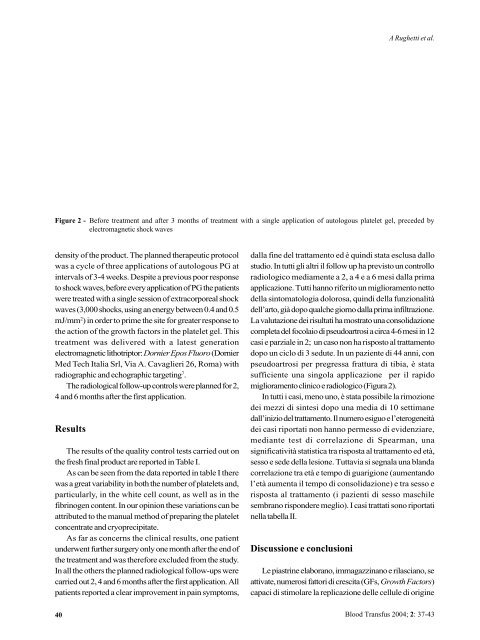
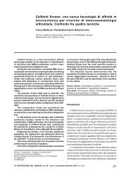
A Rughetti et al.Figure 2 - Before treatment and after 3 months of treatment with a single application of autologous platelet gel, preceded byelectromagnetic shock wavesdensity of the product. The planned therapeutic protocolwas a cycle of three applications of autologous PG atintervals of 3-4 weeks. Despite a previous poor responseto shock waves, before every application of PG the patientswere treated with a single session of extracorporeal shockwaves (3,000 shocks, using an energy between 0.4 and 0.5mJ/mm 2 ) in order to prime the site for greater response tothe action of the growth factors in the platelet gel. Thistreatment was delivered with a latest generationelectromagnetic lithotriptor: Dornier Epos Fluoro (DornierMed Tech Italia Srl, Via A. Cavaglieri 26, Roma) withradiographic and echographic targeting 7 .The radiological follow-up controls were planned for 2,4 and 6 months after the first application.ResultsThe results of the quality control tests carried out onthe fresh final product are reported in Table I.As can be seen from the data reported in table I therewas a great variability in both the number of platelets and,particularly, in the white cell count, as well as in thefibrinogen content. In our opinion these variations can beattributed to the manual method of preparing the plateletconcentrate and cryoprecipitate.As far as concerns the clinical results, one patientunderwent further surgery only one month after the end ofthe treatment and was therefore excluded from the study.In all the others the planned radiological follow-ups werecarried out 2, 4 and 6 months after the first application. Allpatients reported a clear improvement in pain symptoms,40dalla fine del trattamento ed è quindi stata esclusa dallostudio. In tutti gli altri il follow up ha previsto un controlloradiologico mediamente a 2, a 4 e a 6 mesi dalla primaapplicazione. Tutti hanno riferito un miglioramento nettodella sintomatologia dolorosa, quindi della funzionalitàdell’arto, già dopo qualche giorno dalla prima infiltrazione.La valutazione dei risultati ha mostrato una consolidazionecompleta del focolaio di pseudoartrosi a circa 4-6 mesi in 12casi e parziale in 2; un caso non ha risposto al trattamentodopo un ciclo di 3 sedute. In un paziente di 44 anni, conpseudoartrosi per pregressa frattura di tibia, è statasufficiente una singola applicazione per il rapidomiglioramento clinico e radiologico (Figura 2).In tutti i casi, meno uno, è stata possibile la rimozionedei mezzi di sintesi dopo una media di 10 settimanedall’inizio del trattamento. Il numero esiguo e l’eterogeneitàdei casi riportati non hanno permesso di evidenziare,mediante test di correlazione di Spearman, unasignificatività statistica tra risposta al trattamento ed età,sesso e sede della lesione. Tuttavia si segnala una blandacorrelazione tra età e tempo di guarigione (aumentandol’età aumenta il tempo di consolidazione) e tra sesso erisposta al trattamento (i pazienti di sesso maschilesembrano rispondere meglio). I casi trattati sono riportatinella tabella II.Discussione e conclusioniLe piastrine elaborano, immagazzinano e rilasciano, seattivate, numerosi fattori di crescita (GFs, Growth Factors)capaci di stimolare la replicazione delle cellule di origine<strong>Blood</strong> Transfus 2004; 2: 37-43
Platelet-gel for the treatment of pseudoarthrosisTable II - Patients with pseudoarthrosis of the long bones treated with autologous PGPatient n° Age (years) Sex (M/F) Site of fracture n° of session with PG Time of consolidation (days)1 36 M Humerus 5 1802 68 M Femur 2 453 46 M Tibia 2 904 52 M Humerus 3 905 83 F Humerus 3 No consolidation6 56 M Tibia 3 1807 44 M Tibia 1 608 39 F Femur 2 909 49 F Humerus 3 Out of follow-up10 37 M Tibia 2 6011 21 M Ulna 4 12012 31 F Femur 2 601 3 4 1 M Radius 3 12014 59 F Femur 4 1801 5 6 1 M Radius 5 18016 42 M Femur 3 180and thus in limb function, already only a few days after thefirst infiltration. An evaluation of the results showedcomplete consolidation of the focus of the pseudoarthrosisat about 4-6 months in 12 cases and partial consolidationin 2 other cases; one case did not respond to treatmentafter a cycle of 3 sessions. Finally, in one 44-year old patientwith pseudoarthrosis of a previous tibial fracture, a singleapplication was sufficient for a rapid clinical and radiologicalimprovement (Figure 2).In all cases, except one, the synthesis aid could beremoved a mean of 10 weeks after the start of the treatment.The small number and the heterogeneity of the casesreported do not allow Spearman's correlation analysis tohighlight any statistical significant associations betweenresponse to treatment and age, sex or site of lesion. However,a weak correlation was noted between age and time to heal(increasing age associated with longer consolidation times)and between sex and response to treatment (the malepatients seemed to respond better). The cases treated arereported in table II.Discussion and conclusionsPlatelets produce, store and release, if activated,numerous growth factors capable of stimulating thereplication of cells of mesenchymal origin such asfibroblasts, osteoblasts and endothelial cells 8 . Besidesosteoprogenitor cells (fibroblasts, osteoblasts andchondrocytes) the regeneration of bone tissue requires amatrix conducive to bone formation, including osteogenicgrowth factors (PDGF Platelet Derived Growth Factor;mesenchimale come fibroblasti, osteoblasti e celluleendoteliali 8 . La rigenerazione del tessuto osseo necessitaoltre che di cellule osteoprogenitrici (fibroblasti,osteoblasti e condrociti) e di una matrice osteoconduttiva,anche di fattori di crescita osteoinduttivi (PDGF PlateletDerived Growth Factor; TGF-b Trasforming GrowthFactor-b; IGF I e II Insulin Like Growth Factor I and II) diderivazione piastrinica e di proteine ad attivitàmorfogenetica (BPMs, Bone Morphogenetic Proteins)BPMs. Le BPMS sono peptidi osteoinduttivi di derivazionepiastrinica appartenenti al gruppo del TGF-b responsabiledei processi di generazione e rigenerazione dell’osso 6,9 .Fattori locali, citochine e fattori di crescita, sono necessariper l’innesco e il mantenimento dei processi di rigenerazionetessutale. Nel tessuto osseo, inoltre, i fattori di crescitaliberati localmente vengono incorporati nella matriceminerale e rilasciati lentamente durante la dissoluzione dellamatrice da parte degli osteoclasti o dei condroclasti 9 .È stato ipotizzato che nel sito di lesione dell’osso ci siaun rilascio iniziale di PDGF, TGF- b e IGF I e II per effettodella degranulazione delle piastrine presenti in loco 6,9,10 .Il PDGF stimola, da un lato, la mitosi delle cellulestaminali midollari presenti nell’osso e dall’altro ha uneffetto angiogenetico inducendo e potenziando laformazione di nuovi capillari nelle sedi di lesione 9 .Contemporaneamente, per effetto del TGF-b, si assiste allaproliferazione di fibroblasti e pro-osteoblasti e nelladifferenziazione di questi ultimi verso forme più mature(osteoblasti) che vengono stimolate a produrre matriceossea, mentre i fibroblasti a depositare matrice di collageneper sostenere la crescita vasale 9,10 . La presenza locale dipiastrine veicolate dal torrente ematico rifornisce dicontinuo l’area di rigenerazione ossea dei GFs necessari 6 .Successivamente il rilascio di IGF I e II agisce sugli osteoblasti<strong>Blood</strong> Transfus 2004; 2: 37-4341