Mitogen-stimulated DAT: a new method for the ... - Blood Transfusion
Mitogen-stimulated DAT: a new method for the ... - Blood Transfusion
Mitogen-stimulated DAT: a new method for the ... - Blood Transfusion
You also want an ePaper? Increase the reach of your titles
YUMPU automatically turns print PDFs into web optimized ePapers that Google loves.
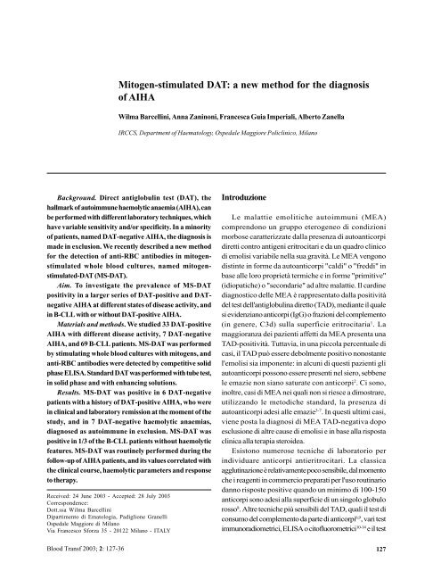
<strong>Mitogen</strong>-<strong>stimulated</strong> <strong>DAT</strong>: a <strong>new</strong> <strong>method</strong> <strong>for</strong> <strong>the</strong> diagnosisof AIHAWilma Barcellini, Anna Zaninoni, Francesca Guia Imperiali, Alberto ZanellaIRCCS, Department of Haematology, Ospedale Maggiore Policlinico, MilanoBackground. Direct antiglobulin test (<strong>DAT</strong>), <strong>the</strong>hallmark of autoimmune haemolytic anaemia (AIHA), canbe per<strong>for</strong>med with different laboratory techniques, whichhave variable sensitivity and/or specificity. In a minorityof patients, named <strong>DAT</strong>-negative AIHA, <strong>the</strong> diagnosis ismade in exclusion. We recently described a <strong>new</strong> <strong>method</strong><strong>for</strong> <strong>the</strong> detection of anti-RBC antibodies in mitogen<strong>stimulated</strong>whole blood cultures, named mitogen<strong>stimulated</strong>-<strong>DAT</strong>(MS-<strong>DAT</strong>).Aim. To investigate <strong>the</strong> prevalence of MS-<strong>DAT</strong>positivity in a larger series of <strong>DAT</strong>-positive and <strong>DAT</strong>negativeAIHA at different states of disease activity, andin B-CLL with or without <strong>DAT</strong>-positive AIHA.Materials and <strong>method</strong>s. We studied 33 <strong>DAT</strong>-positiveAIHA with different disease activity, 7 <strong>DAT</strong>-negativeAIHA, and 69 B-CLL patients. MS-<strong>DAT</strong> was per<strong>for</strong>medby stimulating whole blood cultures with mitogens, andanti-RBC antibodies were detected by competitive solidphase ELISA. Standard <strong>DAT</strong> was per<strong>for</strong>med with tube test,in solid phase and with enhancing solutions.Results. MS-<strong>DAT</strong> was positive in 6 <strong>DAT</strong>-negativepatients with a history of <strong>DAT</strong>-positive AIHA, who werein clinical and laboratory remission at <strong>the</strong> moment of <strong>the</strong>study, and in 7 <strong>DAT</strong>-negative haemolytic anaemias,diagnosed as autoimmune in exclusion. MS-<strong>DAT</strong> waspositive in 1/3 of <strong>the</strong> B-CLL patients without haemolyticfeatures. MS-<strong>DAT</strong> was routinely per<strong>for</strong>med during <strong>the</strong>follow-up of AIHA patients, and its values correlated with<strong>the</strong> clinical course, haemolytic parameters and responseto <strong>the</strong>rapy.Received: 24 June 2003 - Accepted: 28 July 2003Correspondence:Dott.ssa Wilma BarcelliniDipartimento di Ematologia, Padiglione GranelliOspedale Maggiore di MilanoVia Francesco S<strong>for</strong>za 35 - 20122 Milano - ITALYIntroduzioneLe malattie emolitiche autoimmuni (MEA)comprendono un gruppo eterogeneo di condizionimorbose caratterizzate dalla presenza di autoanticorpidiretti contro antigeni eritrocitari e da un quadro clinicodi emolisi variabile nella sua gravità. Le MEA vengonodistinte in <strong>for</strong>me da autoanticorpi "caldi" o "freddi" inbase alle loro proprietà termiche e in <strong>for</strong>me "primitive"(idiopatiche) o "secondarie" ad altre malattie. Il cardinediagnostico delle MEA è rappresentato dalla positivitàdel test dell'antiglobulina diretto (TAD), mediante il qualesi evidenziano anticorpi (IgG) o frazioni del complemento(in genere, C3d) sulla superficie eritrocitaria 1 . Lamaggioranza dei pazienti affetti da MEA presenta unaTAD-positività. Tuttavia, in una piccola percentuale dicasi, il TAD può essere debolmente positivo nonostantel'emolisi sia imponente: in alcuni di questi pazienti gliautoanticorpi possono essere presenti nel siero, sebbenele emazie non siano saturate con anticorpi 2 . Ci sono,inoltre, casi di MEA nei quali non si riesce a dimostrare,utilizzando le metodiche standard, la presenza diautoanticorpi adesi alle emazie 3-7 . In questi ultimi casi,viene posta la diagnosi di MEA TAD-negativa dopoesclusione di altre cause di emolisi e in base alla rispostaclinica alla terapia steroidea.Esistono numerose tecniche di laboratorio perindividuare anticorpi antieritrocitari. La classicaagglutinazione è relativamente poco sensibile, dal momentoche i reagenti in commercio preparati per l'uso routinariodanno risposte positive quando un minimo di 100-150anticorpi sono adesi alla superficie di un singolo globulorosso 8 . Altre tecniche più sensibili del TAD, quali il test diconsumo del complemento da parte di anticorpi 6,9 , vari testimmunoradiometrici, ELISA o citofluorometrici 10-14 e il test<strong>Blood</strong> Transf 2003; 2: 127-36127
W Barcellini et al.Conclusions. In <strong>the</strong> test described here mitogenstimulation seems able to disclose a latent anti-RBCautoimmunity, both in AIHA in clinical remission and inB-CLL. MS-<strong>DAT</strong> could be proposed as an additional test<strong>for</strong> <strong>the</strong> diagnosis and monitoring of AIHA, and <strong>for</strong> <strong>the</strong>screening of those pathological conditions in which anti-RBC autoimmunity is known to be potentially harmful.Key words: AIHA, B-CLL, autoimmunity, mitogen<strong>stimulated</strong><strong>DAT</strong>IntroductionAutoimmune haemolytic anaemias (AIHA) are aheterogeneous group of disorders characterised by <strong>the</strong> presenceof auto-antibodies directed against red cell surface antigens,and by a variable clinical picture and severity of haemolysis.AIHA can be distinguished in "warm" and "cold" basedon <strong>the</strong> <strong>the</strong>rmal properties of <strong>the</strong> antibody, and in "primary"(idiopathic) and "secondary" to underlying disease. Thehallmark of AIHA is a positive direct antiglobulin test (<strong>DAT</strong>),by which IgG and/or complement (C3d) are found on <strong>the</strong> redblood cell (RBC) surface 1 .The vast majority of patients with AIHA present apositive <strong>DAT</strong>. However, in a minority of patients with AIHA<strong>the</strong> <strong>DAT</strong> may be weakly positive in spite of fairly severehaemolysis in <strong>the</strong> patient. In some of <strong>the</strong>se patientsunbound auto-antibody may be present in <strong>the</strong> serumalthough <strong>the</strong> patients' red cells are clearly not saturatedwith antibodies 2 . Moreover <strong>the</strong>re are now many welldocumented cases of patients with <strong>the</strong> disease in whomno red cell-bound auto-antibody could be demonstratedby standard procedures 3-7 . In <strong>the</strong>se patients, <strong>the</strong> diagnosisof <strong>DAT</strong> negative AIHA is made on <strong>the</strong> basis of <strong>the</strong>exclusion of o<strong>the</strong>r causes of haemolysis and of <strong>the</strong> clinicalresponse to steroid <strong>the</strong>rapy.There are many laboratory techniques to detect anti-RBC antibodies. The traditional agglutination techniqueis relative insensitive, since commercially available reagentsprepared <strong>for</strong> routine use usually yield a positive reactionwhen a minimum of 100-150 antibodies attach to <strong>the</strong> surfaceof each erythrocyte 8 .More sensitive <strong>DAT</strong> techniques, such as <strong>the</strong>complement-fixing antibody consumption test 6,9 , variousimmunoradiometric ELISA and cytofluorimetric assays 10-14and <strong>the</strong> solid phase antiglobulin test 15 have been reportedto give positive results in some patients with negativeconventional <strong>DAT</strong>.However since all <strong>the</strong> <strong>method</strong>s available could give128dell'antiglobulina in fase solida 15 , si sono dimostratepositive in alcuni pazienti affetti da MEA TAD-negativacon i tradizionali test. Pertanto, si raccomanda di utilizzareper la diagnosi di MEA una batteria di esami piuttosto cheun singolo test, dal momento che tutte le metodiche adisposizione possono dare risposte falsamente positive ofalsamente negative 16 .Recentemente abbiamo descritto un nuovo metodoper la ricerca di anticorpi antieritrocitari, basato sullastimolazione con mitogeni di colture di sangue intero edenominato MS-<strong>DAT</strong> (<strong>Mitogen</strong> Stimulated-DirectAntiglobulin Test). Il test si è dimostrato in grado dirilevare nelle MEA la modulazione della produzioneanticorpale da parte delle citochine ed è risultato positivoin alcuni casi di MEA TAD-negative in remissione clinica,probabilmente perché la stimolazione con mitogeniinduce una produzione anticorpale in vitro 17 . Abbiamoinoltre valutato, mediante MS-<strong>DAT</strong>, la produzione in vitrodi anticorpi antieritrocitari in pazienti affetti da leucemialinfatica cronica a cellule B (B-LLC), malattia nella qualesi riscontra frequentemente una MEA 18 . MS-<strong>DAT</strong> si èdimostrato positivo non soltanto nei casi evidenti di MEAma anche in una frazione di pazienti con B-LLC senzaanemia emolitica, suggerendo l'esistenza di <strong>for</strong>me latentidi autoimmunità antieritrocitaria in corso di B-LLC, chepotrebbero essere evidenziate utilizzando la stimolazionein vitro con mitogeni 19 .Lo scopo di questo lavoro è di indagare l'incidenza diMS-<strong>DAT</strong> positività in un maggior numero di MEA sia TADpositiveche TAD-negative in fase diversa di attività clinicanonché in pazienti affetti da B-LLC con o senza MEA TADpositiva.Inoltre, è stato investigato il significato del test,eseguendolo routinariamente durante il monitoraggioclinico dei pazienti affetti da MEA e correlando i risultaticon il decorso clinico, i parametri emolitici e la risposta allaterapia.Materiali e metodiTAD, ricerca e identificazione degli anticorpiIl TAD in provetta è stato eseguito secondo la tecnicastandard, impiegando sieri antiglobuline poli emonospecifici. Si sono utilizzati reagenti monoclonali delcommercio (anti-IgG+C, anti-IgG, anti-C3d e anti-C3c), sieridi coniglio anti-IgA umane e sieri di capra anti-IgM umane,<strong>for</strong>niti dall'Ortho-Clinical Diagnostics (Raritan, NJ, USA),dalla Bioscot Ltd (Edimburgo, Scozia) e dalla GammaBiologicals Inc (Houston, TX, USA). I risultati vengono<strong>Blood</strong> Transf 2003; 2: 127-36
MS-<strong>DAT</strong>: <strong>new</strong> <strong>method</strong> <strong>for</strong> AIHAfalse-positive and false-negative results, a battery of testsra<strong>the</strong>r than a single test is recommended <strong>for</strong> <strong>the</strong> diagnosisof AIHA 16 .We recently described a <strong>new</strong> <strong>method</strong> <strong>for</strong> <strong>the</strong> detectionof anti-RBC antibodies in mitogen-<strong>stimulated</strong> whole bloodcultures, named mitogen-<strong>stimulated</strong>-<strong>DAT</strong> (MS-<strong>DAT</strong>). Thetest was suitable <strong>for</strong> revealing cytokine modulation of anti-RBC antibody production in AIHA, and was positive in asmall number of <strong>DAT</strong>-negative AIHA in clinical remission,probably because mitogen stimulation induced antibodyproduction in vitro 17 .Fur<strong>the</strong>rmore, we investigated by MS-<strong>DAT</strong> <strong>the</strong> invitro production of anti-RBC antibodies in B-ChronicLymphocytic Leukaemia (B-CLL), a disease in whichAIHA is frequently observed 18 . MS-<strong>DAT</strong> was positivenot only in overt AIHA, but also in a fraction of B-CLLpatients without haemolytic anaemia, suggesting <strong>the</strong>existence of a latent anti-RBC autoimmunity in B-CLL,that could be disclosed by in vitro mitogenstimulation 19 .The aim of this paper was to investigate <strong>the</strong>prevalence of MS-<strong>DAT</strong> positivity in a larger series of<strong>DAT</strong>-positive and <strong>DAT</strong>-negative AIHA at differentstates of disease activity, and in B-CLL with or without<strong>DAT</strong>-positive AIHA.Fur<strong>the</strong>rmore <strong>the</strong> significance of MS-<strong>DAT</strong> wasinvestigated by routinely per<strong>for</strong>ming <strong>the</strong> test during <strong>the</strong>follow-up of AIHA patients, and relating it to <strong>the</strong> clinicalcourse, haemolytic parameters and response to <strong>the</strong>rapy.Materials and <strong>method</strong>s<strong>DAT</strong> and antibody screening and identificationTube-<strong>DAT</strong> was per<strong>for</strong>med according to <strong>the</strong> standardtechniques using both polyspecific and monospecificsera. Commercial monoclonal reagents (anti-IgG+C, anti-IgG, anti-C3d, and anti-C3c), rabbit anti-human IgA andsheep anti-human IgM from Ortho (Raritan, NJ, USA),Bioscot (Edinburgh, UK) and Gamma (Houston, TX,USA) were used. The results expressed as titre and scoreby testing two-fold dilutions 20 .The cassette-<strong>DAT</strong> was carried out with monospecificantisera only, following <strong>the</strong> manufacturer's instructions.Cassettes (BioVue Ortho) with anti-IgG, anti-C andnegative control were used <strong>for</strong> cassettes-<strong>DAT</strong>.Antibody screening and identification in serum,eluates, and culture supernatants were per<strong>for</strong>med using<strong>the</strong> indirect antiglobulin test in tube (eluate or culture<strong>Blood</strong> Transf 2003; 2: 127-36espressi in titolo e in score valutando le diluizioni perraddoppio 20 . Il TAD su schedina è stato effettuato soltantocon sieri monospecifici seguendo le istruzioni deiproduttori. Si sono impiegate schedine (BioVue Ortho) conanti-IgG, anti-C e controllo negativo. L'individuazione el'identificazione degli anticorpi sono state condotte su sieri,eluati e supernatanti di colture, impiegando il test delleantiglobuline indiretto in provetta (eluati e supernatanti) oin schedina (sieri) utilizzando antiglobuline polispecifiche.Per l'individuazione e l'identificazione degli anticorpi sueluati, supernatanti e sieri si sono utilizzate soluzionifacilitanti-rin<strong>for</strong>zanti (PEG Gamma e Bliss Ortho). Le reazionipositive sono state registrate utilizzando una scala da 0,5+a 4+ 20 . Per lo screening e l'identificazione degli anticorpi sisono impiegati un pannello a tre cellule (Surgiscreen Ortho)e due a 11 (Ortho e Gamma), rispettivamente.Test dell'antiglobulina diretto stimolatocon mitogeni (MS-<strong>DAT</strong>)MS-<strong>DAT</strong> è stato condotto come descritto inprecedenza 15,17 . Brevemente, campioni di sangue frescoeparinato sono stati diluiti (1:6) con RPMI 1640 (Gibco,Grand Island, NY, USA) e posti in coltura senza stimoloo stimolati con 2 µg/mL di fitoemoagglutinina (PHA), 20ng/mL di Phorbol-12-myristate-13-acetate (PMA,Sigma-Aldrich Chemicals, St Louis, MO, USA) o 1% difitolacca americana (PWM, Gibco) in piastre a 24 pozzettie incubati per 48 h. La quantità di IgG adese alla superficieeritrocitaria è stata misurata con un testimmunoenzimatico competitivo in fase solida, comedescritto di seguito. Abbiamo rivestito con 50 µL di IgGumane (Sclavo, Siena, Italia), diluite alla concentrazionedi 50 µg/mL in tampone carbonato-bicarbonato a pH 9,piastre a 96 pozzetti (Falcon 3912, Becton Dickinson,Oxnard, CA , USA), incubando per una notte a +4 °C.Dopo tre lavaggi con fisiologica tamponata (PBS)contenente 0,2% di siero fetale bovino (FCS, HycloneLaboratories, Logan, UT, USA), in ciascun pozzetto sonostati seminati 200 µL di 2% FCS-PBS per 2 ore atemperatura ambiente, al fine di saturare i legamiaspecifici; infine, le piastre sono state lavate 3 voltecon 0,2%FCS-PBS. È stata costruita una curva standardcon emazie O CcDee, ottenute da un solo donatore, esensibilizzate con diluizioni seriali di anticorpi anti-D(Immuno AG, Vienna, Austria), come descrittoprecedentemente 17 . Cento µL sia dei campioni che deglistandard sono stati incubati, a 37 °C per 30', con uneguale volume di IgG anti-uomo da coniglio coniugatecon perossidasi (Dako, Glostrup, Danimarca), diluite129
W Barcellini et al.supernatant) or in cassettes (serum) with polyspecificanti-human globulin reagents. Enhancement solutions(PEG Gamma and Bliss Ortho) were used <strong>for</strong> screeningand identification of antibodies in eluates, culturesupernatants and serum, respectively. Positive resultswere recorded on a scale from 0.5+ to 4+ 20 . A 3-cell RBCpanel (Surgiscreen Ortho) and two 11-cell RBC panels(Ortho and Gamma) were used <strong>for</strong> serum antibodyscreening and identification, respectively.<strong>Mitogen</strong>-<strong>stimulated</strong> <strong>DAT</strong><strong>Mitogen</strong>-<strong>stimulated</strong> <strong>DAT</strong> was per<strong>for</strong>med as alreadydescribed 15,17 . Briefly, fresh heparinized blood sampleswere diluted 1:6 with RPMI 1640 medium (Gibco, GrandIsland, NY, USA) and ei<strong>the</strong>r un<strong>stimulated</strong> or <strong>stimulated</strong>with 2 µg/mL phytohaemoagglutinin (PHA) and 20 ng/mL Phorbol-12-myristate-13-acetate (PMA) (Sigma-Aldrich Chemicals, St. Louis, MO, USA), or 1%pokeweed (PWM) (Gibco) in 24 well-plates andincubated <strong>for</strong> 48 hr.The quantitation of IgG bound to <strong>the</strong> surface of RBCwas evaluated with a competitive solid-phase enzymeimmunoassay. Briefly, 96 well-plates (Falcon 3912, BectonDickinson, Oxnard, CA, USA) were coated with 50 µL ofhuman IgG (Sclavo, Siena, Italy) diluted at <strong>the</strong>concentration of 50 µg/mL in carbonate-bicarbonate buffer,pH 9.00, and incubated overnight at +4 °C.After washing three times with PBS containing 0.2%fetal calf serum (FCS, Hyclone Laboratories, Logan, UT,USA), plates were blocked with 200 µL of 2% FCS-PBS <strong>for</strong>2 hr at room temperature, and finally washed 3 times withPBS-0.2% FCS.A standard curve was constructed with group OCcDee RBC, obtained from a single blood donor,sensitised with serial dilutions of anti-D antibody(Immuno AG, Vienna, Austria) as previouslydescribed 17 .One hundred µL of both samples and standardswere incubated with an equal volume of peroxidaseconjugatedrabbit anti-human IgG (Dako, Glostrup,Denmark) diluted 1:3,000 in 0.2% FCS-PBS at 37 °C <strong>for</strong>30 min. One-hundred µL of this mixture were added to<strong>the</strong> IgG-coated plates in triplicate wells, and incubatedat +37 °C <strong>for</strong> 30 min.The plate was <strong>the</strong>n washed five times with PBScontaining 0.2% FCS and 0.05% Tween 20 (Merck,Darmstadt, Germany). Fifty µL of o-phenylenediamindihydrochloride (Sigma-Aldrich Chemicals, St. Louis,MO, USA) at a concentration of 0.5 mg/mL in 10 -1 M1:3.000 in 0,2%FCS-PBS. Le piastre venivano poi lavate 5volte con PBS contenente 0,2% di FCS e 0,05% di Tween20 (Merck, Darmstadt, Germania). A ciascun pozzetto sonostati poi aggiunti 50µL di una soluzione di ortofenilendiaminadiidrocloride (Sigma-Aldrich Chemicals, StLouis, MO, USA) alla concentrazione di 0,5 mg/mL in unasoluzione tampone 10 -1 M di acido citrico-citrato di sodio apH 4,8-5,0; è seguita incubazione a temperatura ambiente.Dopo 15 minuti, è stata misurata la reazione colorimetrica a450 nm, utilizzando uno spettrofotometro ELISA. Per ladeterminazione della presenza di anticorpi antieritrocitarisulle emazie autologhe poste in colture, le sospensionicellulari sono state trattate come emazie O CcDeesensibilizzate con anticorpi anti-D, mentre per ladeterminazione della presenza di anticorpi antieritrocitarisecreti, i supernatanti delle colture sono stati utilizzati persensibilizzare emazie O CcDee.Abbiamo costruito una curva standard su scalalogaritmica con i valori di densità ottica (OD) e dellaconcentrazione degli anticorpi anti-D (ng/mL); facendoriferimento a questa curva, sono stati calcolati i valori delleIgG legate ad emazie autologhe o la concentrazione deglianticorpi antiemazie nei supernatanti delle colture. È dasottolineare che il controllo positivo TAD standard davaun valore di IgG adese di 327±23 ng/mL (media±ES di 6esperimenti indipendenti).Al fine di fissare un cut-off per i valori positivi di MS-<strong>DAT</strong>, sono state calcolate le medie di 81 colture stimolatecon PHA, PMA e PWM di soggetti di controllo; la positivitàè stata definita come un valore che eccedesse la media più3 DS (220 IgG ng/mL).PazientiSono stati esaminati i seguenti soggetti: 1) 33 pazientiaffetti da MEA primitiva (idiopatica) con età media (±DS)di 50±12 anni, range 28-76, 20 femmine e 13 maschi e 2) 69pazienti affetti da B-LLC, età media 68±11 anni, range 41-89, 25 femmine e 44 maschi, la cui diagnosi era stata postaseguendo i criteri clinici, morfologici e immunologici diCheson et al. 21 e la cui stadiazione clinica era in accordocon le linee-guida proposte da Binet et al. 22 . Inoltre,abbiamo studiato altri 7 pazienti, con diagnosi presuntivadi MEA TAD-negativa posta in base all'esclusione di altrecause di emolisi e alla risposta clinica alla terapia consteroidi. Il test MS-<strong>DAT</strong>, insieme con la conta eritrocitariae i parametri emolitici (reticolociti, LDH e aptoglobina) èstato eseguito ogni mese su 8 pazienti affetti da MEA,seguiti regolarmente in regime ambulatoriale dal nostroDipartimento.130 <strong>Blood</strong> Transf 2003; 2: 127-36
MS-<strong>DAT</strong>: <strong>new</strong> <strong>method</strong> <strong>for</strong> AIHATable I - Effect of mitogen stimulation on in vitro autologous anti-RBC bound IgG in 48 hr whole blood cultures from patients andcontrolsUn<strong>stimulated</strong> PHA PMA PWMAIHA Patients 322+49 *** 623+122 ** 465+55 * 635+134 **B-CLL Patients 134+15 ** 207+29 *** 182+37 ** 183+25 ***Controls 75+7 75+9 70+6 76+14Values are expressed as IgG ng/mL, mean + SE of 33 AIHA and 69 B-CLL patients and 81 controls. * p
W Barcellini et al.Table II - Clinical and laboratory characteristics of <strong>DAT</strong>-negative, MS-<strong>DAT</strong> positive AIHA patientsPatient N° Gender Hb Total Reticulocytes Haptoglobin LDH Steroid <strong>DAT</strong>* MS-<strong>DAT</strong>g/dL bilirubin % mg/L U/L <strong>the</strong>rapy IgG ng/mL(unconjugated)µmol/L1 F 13.3 8 (5) 1.4 1,200 420 yes neg 2792 F 12.4 13.7 (12) 0.8 2,290 296 yes neg 3213 F 12.3 10.3 (7) 0.7 1,830 377 no neg 4334 M 13.1 10 (9) 1.8 1,200 472 no neg 3025 M 12 10 (8) 0.9 690 303 yes neg 2566 M 15 13.7 (8) 0.2 870 291 no neg 3227 M 12 27 (22) 3.7 200 480 no neg 8138 F 10.9 28 (22) 4.4 200 420 no neg 4339 M 11.1 20 (18) 1.1 200 550 no neg 1,23010 F 5.3 20 (19) 6.9 200 232 no neg 1,66011 M 8.8 n.d. 4.3 200 520 yes neg 51612 M 10.8 40 (27) 15.9 200 470 no neg 85613 F 11.1 9 (8) 0.9 500 338 no neg 314Normal range female 11.5-15 0-17 (0-12)
MS-<strong>DAT</strong>: <strong>new</strong> <strong>method</strong> <strong>for</strong> AIHAFigure 1 - MS-<strong>DAT</strong> in <strong>the</strong> clinical follow up of AIHA. Levels of Hb ( ), Haptoglobin ( ), and MS-<strong>DAT</strong> ( ) are expressed aspercentage of <strong>the</strong> values at <strong>the</strong> beginning of <strong>the</strong> follow-up. For <strong>the</strong> first patient (left panel) <strong>the</strong>se values were: Hb=5.8 g/dL,Haptoglobin=200 mg/L, MS-<strong>DAT</strong>=4,121 IgG ng/mL. For <strong>the</strong> second patient (right panel) <strong>the</strong>se values were: Hb=13.6 g/dL, Haptoglobin=590 mg/L, MS-<strong>DAT</strong>=41 IgG ng/mLmean plus 3 SD of controls. MS-<strong>DAT</strong> positivity wascompared with standard <strong>DAT</strong>-tube positivity. Both<strong>the</strong> standard <strong>DAT</strong> and MS-<strong>DAT</strong> were positive in 27AIHA at varying degree of clinical activity (data notshown). MS-<strong>DAT</strong> was positive in 6 <strong>DAT</strong>-negativepatients with a history of <strong>DAT</strong>-positive AIHA, whowere in clinical and laboratory remission at <strong>the</strong> momentof <strong>the</strong> study (Table II, patients 1-6).Finally, we studied 7 additional patients,tentatively diagnosed as <strong>DAT</strong>-negative AIHA on <strong>the</strong>basis of <strong>the</strong> exclusion of o<strong>the</strong>r causes of haemolysisand of <strong>the</strong> clinical response to steroid <strong>the</strong>rapy. Thesepatients showed a clear-cut positive MS-<strong>DAT</strong> (TableII, patients 7-13).MS-<strong>DAT</strong> was positive in 20 out of 69 B-CLL patients(10 in stage A, 4 in stage B, 6 stage C), correspondingto a prevalence of 28.9% (data not shown). Standard<strong>DAT</strong> tube was positive at <strong>the</strong> time of <strong>the</strong> study in 3stage C B-CLL patients, suffering from haemolyticanaemia, with a prevalence of 4.3%.momento dell'anemia manifesta (355 e 528 IgG ng/mL). Lamodesta ricaduta emolitica è stata controllata prontamentedalla terapia steroidea, che ha determinato unanormalizzazione dei livelli di emoglobina (da 10 a 13,4 g/dL)e aptoglobina (da 200 a 430 mg/L) e una diminuzione deivalori di MS-<strong>DAT</strong> (da 528 a 166 IgG ng/mL).DiscussioneI risultati descritti in questo lavoro dimostrano che MS-<strong>DAT</strong> è positivo in pazienti con anamnesi di MEA, cheerano in fase di remissione clinica e laboratoristica almomento dello studio e divenuti TAD negativi. Dato ancorpiù interessante, MS-<strong>DAT</strong> è risultato positivo in pazientinei quali è stata posta una diagnosi presuntiva di MEA,sulla base dell'esclusione di altre possibili cause di emolisie della risposta alla terapia steroidea. Per di più, MS-<strong>DAT</strong>si è dimostrato positivo in un terzo di pazienti affetti da B-LLC, con TAD negativo e senza segni clinici di emolisi,confermando precedenti risultati 19 . I valori assoluti di IgG<strong>Blood</strong> Transf 2003; 2: 127-36133
W Barcellini et al.MS-<strong>DAT</strong> in <strong>the</strong> clinical follow up of AIHAMS-<strong>DAT</strong> was per<strong>for</strong>med in 6 AIHA regularlyfollowed at <strong>the</strong> out-patient department. The test wasper<strong>for</strong>med at each medical examination, toge<strong>the</strong>r withblood counts and haemolytic parameters. Two typicalcases are shown in Figure 1.The first case (left panel) was a 70 years old manwith an acute onset of typical warm AIHA,subsequently treated with steroid. The <strong>the</strong>rapyinduced a progressive increase of haemoglobin levels(from 5.8 to 15.3 g/dL in 7 months), normalisation of<strong>the</strong> haptoglobin levels (from 200 to 1,370 mg/L),toge<strong>the</strong>r with a decrease of MS-<strong>DAT</strong> values (from 4,121to 374 IgG ng/mL).The second case (right panel) was a 50 years oldwoman with AIHA in clinical remission (Hb 13.6 g/dL)on low-dose steroid <strong>the</strong>rapy. During <strong>the</strong> follow-up ahaemolytic relapse occurred, with decrease ofhaemoglobin (from 13.6 to 10 g/dL) and haptoglobin(from 590 to 200 mg/L) levels. Interestingly, MS-<strong>DAT</strong>showed an increased positivity which preceded <strong>the</strong>clinical worsening (from 41 to 176 IgG ng/mL) and wasclearly positive at <strong>the</strong> time of overt anaemia (355 and528 IgG ng/mL).The mild haemolytic relapse was promptly controlledby steroid <strong>the</strong>rapy which induced a normalisation ofhaemoglobin (from 10 to 13.4 g/dL) and haptoglobin(from 200 to 430 mg/L) levels and a decrease of MS-<strong>DAT</strong> values (from 528 to 166 IgG ng/mL).DiscussionResults presented in this paper showed that MS-<strong>DAT</strong> was positive in <strong>DAT</strong>-negative patients with ahistory of AIHA, who were in clinical and laboratoryremission and, more interestingly, in patientstentatively diagnosed as <strong>DAT</strong>-negative AIHA on <strong>the</strong>basis of exclusion of o<strong>the</strong>r causes of haemolysis andof <strong>the</strong> clinical response to steroid <strong>the</strong>rapy. Moreover,MS-<strong>DAT</strong> was positive in 1/3 of <strong>DAT</strong>-negative B-CLLpatients without haemolytic features, confirmingprevious findings 19 .Absolute values of IgG bound to RBC are greaterin mitogen-<strong>stimulated</strong> cultures, as compared toun<strong>stimulated</strong> ones, both in AIHA and in B-CLLpatients 17,19 .On <strong>the</strong> contrary, mitogen stimulation has no effectadese alle emazie sono maggiori nelle colture di sangue interostimolate con mitogeni se paragonati a quelli rilevabili incolture non stimolate, sia nei pazienti affetti da MEA che inquelli affetti da B-LLC 17,19 . Al contrario, la stimolazione conmitogeni non ha alcun effetto nei controlli, dove non visono linfociti B potenzialmente autoreattivi, così da esserestimolati in vitro a produrre anticorpi antieritrocitari. Nelloro insieme, questi risultati indicano che la stimolazionecon mitogeni sembra essere in grado di svelareun'autoimmunità antieritrocitaria latente, sia nella MEA inremissione clinica che nella B-LLC. Questo dato potrebbeessere utile per continuare il monitoraggio clinico elaboratoristico dei pazienti con MEA in remissione e perprogrammare un follow-up più intensivo nei pazienti B-LLC,soprattutto in quelli in trattamento con fludarabina 23-26 .Tuttavia, per accertare il valore predittivo del test, si rendenecessario un controllo clinico a distanza di tempo deipazienti affetti da B-LLC con MS-<strong>DAT</strong> positivo. Abbiamo,inoltre, individuato 7 pazienti con diagnosi di MEA TADnegativa,presuntivamente posta sulla base dell'esclusionedi altre cause di emolisi: tale diagnosi è stata confermatadalla positività di MS-<strong>DAT</strong>. È noto che tutti i metodi adisposizione per la diagnosi di MEA possono dare risultatifalsamente negativi e che è necessario disporre di una batteriadi test, piuttosto che di un singolo tipo di esame 2,10,16 . Diconseguenza, MS-<strong>DAT</strong> potrebbe essere proposto come testsupplementare per la diagnosi di MEA. La negatività delTAD eseguito con metodiche standard è spesso dovuta adnumero esiguo di molecole immunoglobuliniche presentisulla superficie eritrocitaria 8 . Così come si osserva nella MEAin remissione e nella B-LLC, la stimolazione con mitogenipuò provocare, anche nelle <strong>for</strong>me di MEA TAD-negative, laproduzione in vitro di anticorpi e la loro conseguenteadesione alle emazie autologhe presenti nella coltura,determinando la positività del TAD. Infine, MS-<strong>DAT</strong> è statoeseguito routinariamente nel corso dei follow-up di pazientiaffetti da MEA e i valori ottenuti sono risultati correlarecon il decorso clinico, con i parametri emolitici e con larisposta alla terapia. Un dato di particolare interesse è cheMS-<strong>DAT</strong> ha rivelato una aumentata positività cheprecedeva il peggioramento clinico, indicando, una voltadi più, che la stimolazione con mitogeni è in grado di svelareuna autoimmunità latente e agire da "cassa di risonanza"per l'individuazione di una risposta autoimmunitaria.ConclusioniIl test che abbiamo descritto in questo lavoro sembra ingrado di evidenziare un'autoimmunità antieritrocitaria latente,sia in MEA in fase di remissione clinica sia in corso di B-LLC.134<strong>Blood</strong> Transf 2003; 2: 127-36
MS-<strong>DAT</strong>: <strong>new</strong> <strong>method</strong> <strong>for</strong> AIHAin controls, where no potentially auto-reactive B-lymphocytes exist, that could be <strong>stimulated</strong> in vitro toproduce anti-RBC antibodies.Altoge<strong>the</strong>r <strong>the</strong>se findings suggest that mitogenstimulation seems able to disclose a latent anti-RBCautoimmunity, both in AIHA in clinical remission andin B-CLL.In <strong>the</strong> <strong>for</strong>mer, this in<strong>for</strong>mation could be useful tocontinue <strong>the</strong> clinical and laboratory monitoring of patients,and, in B-CLL, to plan more intensive follow-up, particularlyin fludarabine-treated patients 23-26 . The clinical follow-upof <strong>the</strong>se MS-<strong>DAT</strong> positive B-CLL patients is needed toascertain <strong>the</strong> predictive value of MS-<strong>DAT</strong> positivity.Fur<strong>the</strong>rmore, we identified 7 patients in which <strong>the</strong>diagnosis of <strong>DAT</strong>-negative AIHA, previously made inexclusion, was confirmed by a positive MS-<strong>DAT</strong>. It isknown that all <strong>the</strong> <strong>method</strong>s available could give falsenegativeresults, and that a battery of tests ra<strong>the</strong>r thana single test is recommended <strong>for</strong> <strong>the</strong> diagnosis 2,10,16 .There<strong>for</strong>e, MS-<strong>DAT</strong> could be proposed as anadditional test <strong>for</strong> <strong>the</strong> diagnosis of AIHA. Thenegativity of <strong>DAT</strong> per<strong>for</strong>med with standard proceduresis often a problem of number of IgG molecules presenton <strong>the</strong> surface of <strong>the</strong> RBC 8 . As observed <strong>for</strong> AIHA inclinical remission and B-CLL, mitogen stimulationcould induce also in <strong>DAT</strong>-negative AIHA antibodyproduction in vitro and subsequent binding ofantibodies to autologous RBC present in culture,determining <strong>the</strong> positivity of <strong>the</strong> <strong>DAT</strong>.Finally, MS-<strong>DAT</strong> was routinely per<strong>for</strong>med during<strong>the</strong> follow-up of AIHA patients, showing that itsvalues correlated with <strong>the</strong> clinical course, haemolyticparameters and response to <strong>the</strong>rapy. Interestingly, MS-<strong>DAT</strong> showed an increased positivity, which preceded<strong>the</strong> clinical worsening, again indicating that mitogenstimulation could disclose a latent autoimmunity andper<strong>for</strong>m as a "resonance box" <strong>for</strong> <strong>the</strong> detection of anautoimmune response.ConclusionIn <strong>the</strong> test described here, mitogen stimulation seemsable to disclose a latent anti-RBC autoimmunity, both inAIHA in clinical remission and in B-CLL. MS-<strong>DAT</strong> couldbe proposed as an additional test <strong>for</strong> <strong>the</strong> diagnosis andmonitoring of AIHA, and <strong>for</strong> <strong>the</strong> screening of thosepathological conditions in which anti-RBC autoimmunityis known to be potentially harmful.Di conseguenza, MS-<strong>DAT</strong> potrebbe essere proposto comeun test aggiuntivo per la diagnosi e il monitoraggio di MEA eper lo screening di quelle condizioni patologiche nelle qualiun'autoimmunità antieritrocitaria può divenire pericolosa.RiassuntoPremessa. Il test dell'antiglobulina diretto (TAD),cardine diagnostico della malattia emolitica autoimmune(MEA), può essere eseguito con tecniche di laboratoriodifferenti, ognuna con sensibilità e/o specificità di variogrado. In una minoranza di casi, indicati come MEA TADnegative,la diagnosi viene posta per esclusione.Recentemente, abbiamo descritto una nuova metodica perl'individuazione di anticorpi antieritrocitari in colturedi sangue intero stimolate con mitogeni, denominata MS-<strong>DAT</strong> (<strong>Mitogen</strong>-Stimulated-Direct Antiglobulin Test).Scopo. Investigare l'incidenza di positività di MS-<strong>DAT</strong>in un'ampia serie di MEA TAD-positive o negative adifferente stato di attività della malattia e in pazienti affettida leucemia linfatica cronica a cellule B (B-LLC) con osenza MEA TAD-positiva.Materiali e metodi. Abbiamo studiato 33 MEA TADpositivecon malattia a diverso stato di attività, 7 MEA TADnegativee 69 pazienti affetti da B-LLC. MS-<strong>DAT</strong> è statoeseguito stimolando con mitogeni colture di sangue intero esono stati rilevati anticorpi antieritrocitari utilizzando untest ELISA competitivo, in fase solida. Il TAD è stato effettuatoanche con metodiche tradizionali, in provetta, in fase solidao utilizzando soluzioni facilitanti-rin<strong>for</strong>zanti.Risultati. MS-<strong>DAT</strong> è risultato positivo in 6 pazienti conMEA TAD-negativa, che avevano una storia clinica di MEATAD-positiva e che erano in fase di remissione al momentodello studio; MS-<strong>DAT</strong> è risultato positivo in 7 pazienti conanemia emolitica TAD-negativa, diagnosticata comeautoimmune per esclusione. Inoltre, MS-<strong>DAT</strong> è risultatopositivo in un terzo dei pazienti affetti da B-LLC senzasegni di emolisi. MS-<strong>DAT</strong> è stato routinariamente eseguitodurante il monitoraggio clinico dei pazienti affetti da MEAe i valori correlavano con il decorso clinico della malattia,con i parametri emolitici e con la risposta alla terapia.Conclusioni. La stimolazione con mitogeni utilizzatain MS-<strong>DAT</strong> sembra in grado di svelare casi diautoimmunità antieritrocitaria latente sia in corso di MEAin remissione clinica che in corso di B-LLC. Il test puòessere proposto come indagine supplementare per ladiagnosi e il monitoraggio di MEA e per lo screening diquelle condizioni patologiche nelle quali l'autoimmunitàantieritrocitaria può essere pericolosa.Parole chiave: MEA, B-LLC, autoimmunità, MS-<strong>DAT</strong><strong>Blood</strong> Transf 2003; 2: 127-36135
W Barcellini et al.References1) Biagi E, Assali G, Rossi F, et al. A persistent severe autoimmune hemolytic anemia despite apparent direct antiglobulin testnegativization. Haematologica 1999; 84: 1043-5.2) Garratty G. In: Immunobiology of <strong>Transfusion</strong> Medicine. New York:Marcel Dekker; 1994.3) Petz LD and Garratty G. Acquired Immune Hemolytic Anemias, New York, Churchill Livingstone; 1980.4) Dacie JV. The Autoimmune Haemolytic Anaemias. In: The Haemolytic Anaemias. Vol. 3, 3 rd Ed, New York, Churchill Livingstone1992.5) Sokol RJ, Hewitt S, Booker DJ, Stamps R. Enzyme linked direct antiglobulin tests in patients with autoimmune haemolysis. JClin Pathol 1985; 38: 912-4.6) Gilliland BC. Coombs-negative immune hemolytic anemia. Sem. Hematol 1976; 13: 267-75.7) Garratty G. In: Immune Destruction of Red <strong>Blood</strong> Cells, Arlington, VA, Am Assoc <strong>Blood</strong> Banks; 1989.8) Sokol RJ, Hewitt S, Booker DJ, Stamps R. Small quantities of erythrocyte bound immunoglobulins and autoimmune haemolysis.J Clin Pathol 1987; 40: 254-7.9) Gallagher MT, Branch DR, Mison A, Petz LD. Evaluation of reticuloendo<strong>the</strong>lial function in autoimmune hemolytic anemia usingan in vitro assay of monocyte-macrophage interaction with erythrocytes Exp Hematol 1983; 11: 82-9.10) Garratty G. The clinical significance (and insignificance) of red-cell-bound IgG and complement. In: Wallance ME, Levitt JS,editors. Current Applications and Interpretation of <strong>the</strong> Direct Antiglobulin Test, Arlington, Am Assoc <strong>Blood</strong> Banks; 1988; 1-24.11) Leikola J, Perkins HA. Enzyme-linked antiglobulin test: an accurate and simple <strong>method</strong> to quantify red cell antibodies. <strong>Transfusion</strong>1980; 20: 138-44.12) Rebulla P. A biotin-avidin micro-ELISA antiglobulin test. <strong>Transfusion</strong> 1988; 28 (Suppl): 41-4.13) Wang Z, Shi J, Zhou Y, Ruan C. Detection of red blood cell-bound immunoglobulin G by flow cytometry and its application in <strong>the</strong>diagnosis of autoimmune hemolytic anemia. Int J Hematol 2001; 73: 188-93.14) Schmitz N, Djibey I, Kretschmer V, et al. Assessment of red cell autoantibodies in autoimmune hemolytic anemia of warm typeby a radioactive anti-IgG test. Vox Sang 1981; 41: 224-30.15) Rachel JM, Sinor LT, Beck ML, Plapp FV. A solid phase antiglobulin test. <strong>Transfusion</strong> 1985; 25: 24-6.16) Engelfriet CP, Overbeeke MA, von dem Borne AE. The antiglobulin test (Coombs test) and <strong>the</strong> red cell. In: Cash JD, editor,Progress in <strong>Transfusion</strong> Medicine, Churchill Livingstone, 1987; 2:74-98.17) Barcellini W, Clerici G, Montesano R, et al. In vitro quantification of anti-red blood cell antibody production in idiopathicautoimmune haemolytic anaemia: effect of mitogen and cytokine stimulation. Br J Haematol 2000; 111: 452-60.18) Mauro FR, Foa R, Cerretti R, et al. Autoimmune hemolytic anemia in chronic lymphocytic leukemia: clinical, <strong>the</strong>rapeutic, andprognostic features. <strong>Blood</strong> 2000; 95: 2786-92.19) Barcellini W, Montesano R, Clerici G, et al. In vitro production of anti-RBC antibodies and cytokines in chronic lymphocyticleukemia. Am J Hematol 2002; 71: 177-83.20) Walker RH. Technical Manual, 12th ed., Arlington, VA, Am Ass <strong>Blood</strong> Banks; 1996.21) Cheson BD, Bennett JM, Grever M, et al. National Cancer Institute-sponsored working group guidelines <strong>for</strong> chronic lymphocyticleukemia: revised guidelines <strong>for</strong> diagnosis and treatment. <strong>Blood</strong> 1996; 87: 4990-7.22) Binet JL, Auquier A, Dighiero G, et al. A <strong>new</strong> prognostic classification of chronic lymphocytic leukemia derived from amultivariate survival analysis. Cancer 1981; 48: 198-206.23) Shvidel L, Shtarlid M, Klepfish A, et al. Evans syndrome complicating fludarabine treatment <strong>for</strong> advanced B-CLL. Br J Haematol1997; 99: 706.24) Tertian G, Cartron J, Bayle C, et al. Fatal intravascular autoimmune hemolytic anemia after fludarabine treatment <strong>for</strong> chroniclymphocytic leukemia. Hematol Cell Ther 1996; 38: 359-60.25) Gonzalez H, Leblond V, Azar N, et al. Severe autoimmune hemolytic anemia in eight patients treated with fludarabine. HematolCell Ther 1998; 40: 113-8.26) Vick DJ, Byrd JC, Beal CL, Chaffin DJ. Mixed-type autoimmune hemolytic anemia following fludarabine treatment in a patientwith chronic lymphocytic leukemia/small cell lymphoma. Vox Sang 1998; 74: 122-6136<strong>Blood</strong> Transf 2003; 2: 127-36


