Tumori della cresta neurale - Facoltà di Medicina e Chirurgia ...
Tumori della cresta neurale - Facoltà di Medicina e Chirurgia ...
Tumori della cresta neurale - Facoltà di Medicina e Chirurgia ...

Create successful ePaper yourself
Turn your PDF publications into a flip-book with our unique Google optimized e-Paper software.
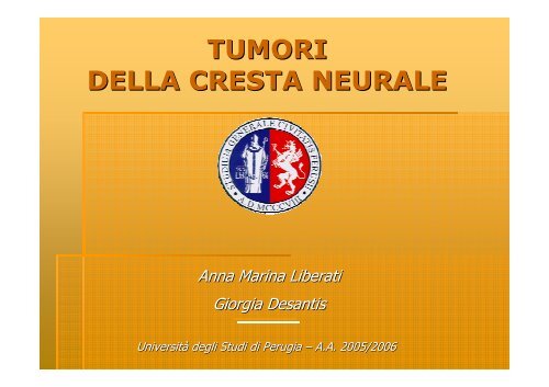
TUMORI<br />
DELLA CRESTA NEURALE<br />
Anna Anna Marina Marina Liberati Liberati<br />
Giorgia Giorgia Desantis Desantis<br />
Universitàà Universit degli degli Stu<strong>di</strong> Stu<strong>di</strong> <strong>di</strong> <strong>di</strong> Perugia Perugia –– A.A. A.A. 2005/2006 2005/2006
Solo una parte delle lezioni sui tumori originanti dalla <strong>cresta</strong> <strong>neurale</strong><br />
sarà sar oggetto <strong>di</strong> <strong>di</strong>scussione in aula<br />
In particolare verranno <strong>di</strong>scusse le <strong>di</strong>apositive<br />
contrassegnate con la lettera S<br />
Rinviate allo stu<strong>di</strong>o personale le altre<br />
per completezza del quadro
TUMORI DELLA CRESTA NEURALE<br />
PERCHÉ?<br />
PERCH<br />
Sono state tra le prime neoplasie<br />
<strong>di</strong> cui si sono stu<strong>di</strong>ati gli aspetti genetico-molecolari<br />
genetico molecolari, ,<br />
in cui sono state messe in evidenza le correlazioni<br />
tra le alterazioni citogenetiche e quelle molecolari<br />
nonché nonch la loro rilevanza patogenetica<br />
S
TUMORI DELLA CRESTA NEURALE<br />
PERCHÉ?<br />
PERCH<br />
Inoltre Inoltre con con i i tumori tumori <strong>di</strong> <strong>di</strong> derivazione derivazione <strong>della</strong> <strong>della</strong> <strong>cresta</strong> <strong>cresta</strong> <strong>neurale</strong> <strong>neurale</strong><br />
èè stato stato introdotto introdotto il il concetto concetto <strong>di</strong> <strong>di</strong> neoplasie neoplasie embrionarie<br />
embrionarie<br />
((medulloblastoma<br />
medulloblastoma e e neuroblastoma).<br />
neuroblastoma).<br />
Il Il modello modello patogenetico patogenetico <strong>di</strong> <strong>di</strong> origine origine intruterina intruterina dei dei tumori tumori èè stato stato<br />
successivamente applicato applicato a a neoplasie neoplasie <strong>di</strong> <strong>di</strong> <strong>di</strong>versa <strong>di</strong>versa istogenesi. istogenesi.<br />
Infine, Infine, queste queste neoplasie, neoplasie, sono sono state state tra tra le le prime prime<br />
in in cui cui le le indagini indagini<br />
immunoistochimiche si si sono sono rivelate rivelate <strong>di</strong> <strong>di</strong> in<strong>di</strong>spensabile in<strong>di</strong>spensabile utilitàà utilit<br />
S
TUMORI DELLA CRESTA NEURALE<br />
S
TUMORI DELLA CRESTA NEURALE<br />
TUBO NEURALE<br />
ENCEFALO<br />
MIDOLLO SPINALE<br />
CRESTA NEURALE<br />
SN PERIFERICO<br />
(n. sensitivi)<br />
C DI SCHWANN<br />
SN AUTONOMO<br />
(gangli simpatici e<br />
midollare surrene)<br />
MELANOCITI<br />
S<br />
SN ENTERICO<br />
STRUTTURE MUSC E CONNET<br />
DELLA TESTA
TUMORI DELLA CRESTA NEURALE<br />
�� Possono essere <strong>di</strong>visi in due gruppi:<br />
Derivanti dalle cellule <strong>della</strong> guaina <strong>neurale</strong> o dalle<br />
cellule <strong>di</strong> Schwann<br />
C. neuroendocrine<br />
C. gangliari<br />
SCHWANNOMA<br />
NEUROFIBROMA<br />
MPNSTs<br />
c. <strong>di</strong> schwann<br />
(Malignant Malignant Peripheral Nerve Sheath Tumor) Tumor<br />
Derivanti dalle cellule neuronali<br />
FEOCROMOCITOMA<br />
PARAGANGLIOMA<br />
c. <strong>di</strong> schwann/ schwann/<br />
c. perineurali<br />
GANGLIONEUROMA<br />
GANGLIONEUROBLASTOMA<br />
NEUROBLASTOMA<br />
SARCOMA DI EWING e<br />
pPNET (Primitive periferal<br />
S<br />
periferal neroectodermal tumor) tumor
TUMORI DELLA CRESTA NEURALE<br />
�� Vasto e complesso spettro <strong>di</strong> neoplasie<br />
�� Incidenza globale bassa<br />
�� <strong>Tumori</strong> più pi comuni del<br />
me<strong>di</strong>astino posteriore<br />
�� Comportamento clinico variabile<br />
da benigni (schwannoma<br />
( schwannoma) )<br />
ad altamente maligni (neuroblastoma<br />
( neuroblastoma) )<br />
S
TUMORI DELLA CRESTA NEURALE<br />
�� Possono essere <strong>di</strong>visi in due gruppi:<br />
Derivanti dalle c. <strong>di</strong> Schwann e/o dalle c. <strong>della</strong> guaina <strong>neurale</strong><br />
C. neuroendocrine<br />
C. gangliari<br />
SCHWANNOMA<br />
NEUROFIBROMA<br />
MPNSTs<br />
(Malignant Malignant Peripheral Nerve Sheath Tumor) Tumor<br />
Derivanti dalle cellule neuronali<br />
c. <strong>di</strong> schwann<br />
FEOCROMOCITOMA<br />
PARAGANGLIOMA<br />
GANGLIONEUROMA<br />
GANGLIONEUROBLASTOMA<br />
NEUROBLASTOMA<br />
SARCOMA DI EWING e<br />
pPNET (Primitive peripheral<br />
c. <strong>di</strong> schwann/ schwann/<br />
c. perineurali<br />
peripheral neroectodermal tumor) tumor<br />
N
TUMORI DELLA CRESTA NEURALE<br />
�� SCHWANNOMI, NEUROFIBROMI e MPNSTs<br />
65% dei tumori neurogeni Origine dalla guaina nervosa<br />
Cellule <strong>di</strong> Schwann Cellule perineurali<br />
Derivano dalla <strong>cresta</strong> <strong>neurale</strong><br />
Positività Positivit per S-100 S 100<br />
ISTOGENESI<br />
GUAINA NERVOSA<br />
Derivazione aracnoidea<br />
negative per S-100 S 100<br />
Marker <strong>di</strong> neoplasia<br />
<strong>di</strong> origine <strong>neurale</strong><br />
Proteina legante il calcio<br />
presente nel tessuto nervoso<br />
N.B. Per una migliore comprensione ve<strong>di</strong> allegato 1<br />
N
TUMORI DELLA CRESTA NEURALE<br />
�� SCHWANNOMI, NEUROFIBROMI e MPNSTs<br />
PATOGENESI<br />
Frequente associazione con le<br />
neurofibromatosi<br />
NF1 e NF2 sono sindromi genetiche a trasmissione autosomica<br />
dominante<br />
legate alla per<strong>di</strong>ta <strong>di</strong> attività attivit <strong>di</strong><br />
TUMOR SUPPRESSOR GENE NF1 E NF2<br />
Le sindromi NF1 e NF2 sono clinicamente e geneticamente <strong>di</strong>stinte<br />
Gli in<strong>di</strong>vidui affetti sviluppano tumori e amartomi multipli<br />
S
TUMORI DELLA CRESTA NEURALE<br />
�� SCHWANNOMI, NEUROFIBROMI e MPNSTs<br />
PATOGENESI<br />
I geni<br />
NF1 (NEUROFIBROMINA)<br />
NF2 (MERLINA O SCHWANNOMINA)<br />
sono implicati anche nella insorgenza <strong>di</strong><br />
tumori neurali spora<strong>di</strong>ci<br />
MPNTSTs<br />
neurofibromi plessiformi<br />
meningiomi<br />
epen<strong>di</strong>momi<br />
schwannomi<br />
N
TUMORI DELLA CRESTA NEURALE<br />
�� SCHWANNOMI, NEUROFIBROMI e MPNSTs<br />
PATOGENESI<br />
NF1 o NEUROFIBROMINA<br />
Gene oncosoppressore mutato localizzazione 17q11.2<br />
NON ATTIVO<br />
Mutazione germinale <strong>di</strong> 1 allele<br />
Mutazione somatica acquisita del secondo allele<br />
La NEUROFIBROMINA regola il protoncogene RAS<br />
NF1 GAP(GTPasi<br />
GAP( GTPasi)<br />
RAS GDP RAS GTP<br />
ATTIVO<br />
S
TUMORI DELLA CRESTA NEURALE<br />
�� SCHWANNOMI, NEUROFIBROMI e MPNSTs<br />
ASPETTI CLINICI<br />
NF1 o m. Di Von Recklinghausen<br />
1/3000 nati<br />
E’ una <strong>della</strong> malattie genetiche<br />
più pi frequenti <strong>della</strong> specie umana<br />
NEUROFIBROMI NODULARI MULTIPLI sottocutanei<br />
NEUROFIBROMI PLESSIFORMI<br />
→possono possono trasformare in tumori maligni delle guaine periferiche<br />
(MPNSTs MPNSTs) )<br />
Altre manifestazioni<br />
Neoplastiche: ASTROCITOMA PILOCITICO<br />
LEUCEMIE<br />
FEOCROMOCITOMA<br />
Non neoplastiche: macchie caffelatte, noduli <strong>di</strong> Lisch, Lisch,<br />
angiomi, malformazioni scheletriche scheletriche<br />
e ipertensione.<br />
S
TUMORI DELLA CRESTA NEURALE<br />
�� SCHWANNOMI, NEUROFIBROMI e MPNSTs<br />
PATOGENESI<br />
NF2 o MERLINA o SCHWANNOMINA<br />
Gene oncosoppressore mutato localizzazione 22q12.2<br />
Mutazione germinale <strong>di</strong> 1 allele<br />
Mutazione somatica acquisita del secondo allele<br />
coinvolta nel controllo <strong>della</strong> crescita e <strong>della</strong> proliferazione<br />
cellulare<br />
probabilmente induce alterazione <strong>della</strong> espressione<br />
dell’oncosoppressore<br />
dell oncosoppressore p53<br />
N
TUMORI DELLA CRESTA NEURALE<br />
�� SCHWANNOMI, NEUROFIBROMI e MPNSTs<br />
ASPETTI CLINICI<br />
1/35000 nati<br />
NF2 o n. Acustica bilaterale<br />
SCHWANNOMI VESTIBOLARI BILATERALI<br />
(neurinoma bilaterale dell’VIII dell VIII n.c.) n.c.)<br />
SCHWANNOMI MULTIPLI<br />
MENINGIOMI<br />
EPENDIMOMI INTRACRANICI<br />
TUMORI SPINALI<br />
T. DEI TESSUTI NERVOSI PERIFERICI<br />
S
TUMORI DELLA CRESTA NEURALE<br />
�� SCHWANNOMI, NEUROFIBROMI e MPNSTs<br />
ASPETTI CLINICI<br />
<strong>Tumori</strong> neuronali <strong>di</strong> più pi frquente insorgenza<br />
nel me<strong>di</strong>astino posteriore nell’adulto nell adulto<br />
MEDIASTINO POSTERIORE<br />
SCHWANNOMA<br />
NEUROFIBROMA<br />
MPNSTs<br />
Forme più pi frequenti nell’adulto<br />
nell adulto<br />
65% dei tumori neurogeni<br />
del me<strong>di</strong>astino posteriore<br />
N
TUMORI DELLA CRESTA NEURALE<br />
�� SCHWANNOMA o NEURILEMMOMA<br />
ASPETTI CLINICI e ISTOPATOLOGICI<br />
Neoplasia benigna<br />
Lenta crescita<br />
Capsulato,<br />
Capsulato,<br />
ovalare, ovalare,<br />
ben circoscritto<br />
Istopatologia → due popolazioni cellulari<br />
► Antoni A cellule densamente stipate a palizzata<br />
► Antoni B cellule meno addensate, <strong>di</strong>sposizione lassa<br />
Deriva dalle cellule <strong>di</strong> Schwann dei n. cranici, spinali e periferici<br />
Solitario eccetto quando associato alla<br />
neurofibromatosi 2<br />
N
TUMORI DELLA CRESTA NEURALE<br />
�� SCHWANNOMA o NEURILEMMOMA<br />
ASPETTI CLINICI<br />
Localizzazione più pi frequente<br />
Nervo acustico-vestibolare<br />
acustico vestibolare<br />
(10% dei tumori<br />
intracranici)<br />
intracranici<br />
Tipica <strong>della</strong> NF2<br />
Localizzazione bilaterale a<br />
livello dell’ dell ottavo nervo cranico<br />
SCHWANNOMA VESTIBOLARE<br />
BILATERALE<br />
N
TUMORI DELLA CRESTA NEURALE<br />
�� SCHWANNOMA o NEURILEMMOMA<br />
ASPETTI CLINICI e DIAGNOSTICI<br />
Altre localizzazioni<br />
n. cranici<br />
n. spinali<br />
A livello toracico si localizza<br />
solitamente nella parte più pi alta del<br />
me<strong>di</strong>astino posteriore<br />
Raramente infiltra attraverso i forami intervertebrali<br />
In genere asintomatico<br />
Raramente sintomi da compressione del nervo vago o frenico<br />
Positività Positivit per S-100 S 100<br />
Terapia chirurgica senza sacrificare le strutture nervose<br />
N
TUMORI DELLA CRESTA NEURALE<br />
�� NEUROFIBROMA<br />
ASPETTI CLINICI, DIAGNOSTICI e TERAPEUTICI<br />
Tumore benigno<br />
Lenta crescita<br />
Può insorgere da qualsiasi tronco nervoso<br />
Istopatologia misto <strong>di</strong> c. <strong>di</strong> schwann,cellule<br />
schwann,cellule<br />
perineurali,<br />
perineurali,<br />
fibroblasti<br />
N. cutaneo<br />
piccoli nervi<br />
non capsulato<br />
ben delimitato<br />
cellule fusate<br />
separate da<br />
materiale<br />
mucoide<br />
Varianti<br />
Origine<br />
N. Plessiforme<br />
grossi tronchi nervosi<br />
Infiltra i fascicoli dei nervi<br />
dai quali non è separabile<br />
Quasi esclusivo <strong>della</strong> NF1<br />
N
TUMORI DELLA CRESTA NEURALE<br />
�� NEUROFIBROMA<br />
ASPETTI CLINICI, DIAGNOSTICI e TERAPEUTICI<br />
Sintomatologia legata a<br />
aumento <strong>di</strong> <strong>di</strong>mensioni<br />
terapia<br />
compressione/infiltrazione del nervo<br />
Positività Positivit per S-100 S 100<br />
resezione chirurgica<br />
inferiore rispetto allo schwannoma<br />
N
TUMORI DELLA CRESTA NEURALE<br />
MPNSTs (Malignant Malignant Peripheral Nerve Sheath Tumor) Tumor<br />
DEFINIZIONE-ISTOGENESI<br />
DEFINIZIONE ISTOGENESI<br />
TUMORI MALIGNI DELLE GUAINE DEI NERVI PERIFERICI<br />
Prima definiti schwannomi maligni<br />
Diversi pattern morfologici<br />
Simil-neurofibroma<br />
Simil neurofibroma Simil-fibrosarcoma<br />
Simil fibrosarcoma<br />
Con alternanza <strong>di</strong> zone cellulari e mixoi<strong>di</strong><br />
N
TUMORI DELLA CRESTA NEURALE<br />
MPNSTs (Malignant<br />
Malignant Peripheral Nerve Sheath Tumor) Tumor<br />
Insorgenza da tronchi nervosi periferici<br />
ECCEZIONE<br />
ASPETTI PATOGENETICI e CLINICI<br />
Generalmente non derivanti dalla<br />
trasformazione dei neurofibromi<br />
Dotati <strong>di</strong> invasività invasivit locale e<br />
capaci <strong>di</strong> <strong>di</strong>ffusione metastatica<br />
Nella NF1 i neurofibromi<br />
possono andare incontro a<br />
degenerazione sarcomatosa<br />
Frequenti reci<strong>di</strong>ve multiple dopo terapia<br />
N
TUMORI DELLA CRESTA NEURALE<br />
MPNSTs (Malignant Malignant Peripheral Nerve Sheath Tumor) Tumor<br />
Immunoistochimica<br />
ASPETTI DIAGNOSTICI<br />
positività positivit variabile per S-100 S 100 (50-70%)<br />
(50 70%)<br />
Forme ben <strong>di</strong>fferenziate con cellule<br />
che ricordano le c. <strong>di</strong> schwann<br />
Forme poco <strong>di</strong>fferenziate con<br />
caratteristiche ultrastrutturali<br />
che ricordano le cellule perineurali<br />
forte positività positivit per S100<br />
S100 negative<br />
N
TUMORI DELLA CRESTA NEURALE<br />
MPNSTs (Malignant<br />
Localizzazione<br />
Malignant Peripheral Nerve Sheath Tumor) Tumor<br />
ASPETTI CLINICI,PROGNOSTICI e TERAPEUTICI<br />
Tronchi nervosi periferici<br />
Toracica Sintomi <strong>di</strong> massa<br />
Dolore<br />
Prognosi severa per la malignità malignit locale<br />
Terapia → resezione chirurgica<br />
Sintomi da compressione<br />
e infiltrazione<br />
del nervo interessato<br />
Anoressia<br />
Affaticabilità<br />
Affaticabilit<br />
Per<strong>di</strong>ta <strong>di</strong> peso<br />
Il tumore infiltra localmente cuore, grossi vasi,<br />
corpi vertebrali, forami intervertebrali<br />
Metastatizza a polmone, milza, midollo, cute<br />
N
TUMORI DELLA CRESTA NEURALE<br />
�� Possono essere <strong>di</strong>visi in due gruppi:<br />
Derivanti dalle c. <strong>di</strong> Schwann e/o dalle c. <strong>della</strong> guaina <strong>neurale</strong><br />
C. neuroendocrine<br />
C. gangliari<br />
SCHWANNOMA<br />
NEUROFIBROMA<br />
MPNSTs<br />
(Malignant Malignant Peripheral Nerve Sheath Tumor) Tumor<br />
Derivanti dalle cellule neuronali<br />
c. <strong>di</strong> schwann<br />
FEOCROMOCITOMA<br />
PARAGANGLIOMA<br />
GANGLIONEUROMA<br />
GANGLIONEUROBLASTOMA<br />
NEUROBLASTOMA<br />
SARCOMA DI EWING e<br />
pPNET (Primitive peripheral<br />
c. <strong>di</strong> schwann/ schwann/<br />
c. prineurali<br />
peripheral neroectodermal tumor) tumor<br />
S
TUMORI DELLA CRESTA NEURALE<br />
�� FEOCROMOCITOMA E PARAGANGLIOMA<br />
DEFINIZIONE e ISTOGENESI<br />
Origine dalle cellule cromaffini<br />
neuroectodermiche del SNA (origine dalla <strong>cresta</strong> <strong>neurale</strong>)<br />
In relazione alla sede <strong>di</strong> origine prende il nome <strong>di</strong><br />
Feocromocitoma Paraganglioma<br />
midollare del surrene tessuto cromaffine extrasurrenalico<br />
Addome<br />
Gangli simpatici <strong>di</strong><br />
Collo<br />
Vescica<br />
Torace<br />
Organo <strong>di</strong> Zuckerlandl<br />
S
TUMORI DELLA CRESTA NEURALE<br />
�� FEOCROMOCITOMA E PARAGANGLIOMA<br />
EPIDEMIOLOGIA e LOCALIZZAZIONI ANATOMICHE<br />
Feocromocitoma<br />
Feocromocitoma maligno<br />
Paraganglioma<br />
spora<strong>di</strong>co 90%<br />
familiare 10%<br />
Associato a MEN2<br />
10% dei surrenalici<br />
maggiore frequenza<br />
nei bambini (30%)<br />
che negli adulti (10%)<br />
In questi casi<br />
frequentemente<br />
bilaterale<br />
30% degli extrasurrenalici<br />
S
TUMORI DELLA CRESTA NEURALE<br />
�� FEOCROMOCITOMA E PARAGANGLIOMA<br />
CARATTERI ISTOPATOLOGICI<br />
Tumore capsulato<br />
Possibile presenza <strong>di</strong> aree necrotiche e cistiche<br />
Istologia variabile alveolare, trabecolare o mista<br />
No <strong>di</strong>fferenze istopatologiche importanti tra<br />
forme benigne e maligne<br />
invasività invasivit locale<br />
In<strong>di</strong>ci <strong>di</strong> malignità<br />
malignit<br />
angioinvasività<br />
angioinvasivit<br />
estese aree <strong>di</strong> necrosi<br />
N
TUMORI DELLA CRESTA NEURALE<br />
�� FEOCROMOCITOMA E PARAGANGLIOMA<br />
Adrenalina<br />
Possono essre<br />
secreti anche<br />
ASPETTI CLINICI<br />
La neoplasia sintetizza catecolamine<br />
Noradrenalina<br />
In modo continuo o intermittente<br />
VIP<br />
serotonina<br />
PTH<br />
encefaline<br />
CRH<br />
calcitonina<br />
ACTH<br />
Dopamina<br />
Da queste secrezioni <strong>di</strong>pende la sintomatologia clinica<br />
N
TUMORI DELLA CRESTA NEURALE<br />
�� FEOCROMOCITOMA E PARAGANGLIOMA<br />
Ipertensione stabile<br />
50%<br />
spesso associata a<br />
ipotensione ortostatica<br />
Car<strong>di</strong>omiopatia<br />
TIA<br />
Insorgenza<br />
spontanea<br />
ASPETTI CLINICI<br />
Ipertensione parossistica<br />
40%<br />
crisi ipertensive <strong>della</strong> durata<br />
<strong>di</strong> 15-60 15 60 min associate a<br />
Astenia<br />
Sudorazione<br />
Pallore<br />
Cefalea<br />
Nausea<br />
Palpitazioni<br />
Vampate<br />
Dispnea<br />
Tremori<br />
Dolore addominale<br />
provocata Alcool<br />
Fumo<br />
Stress<br />
Palpazione addominale ecc<br />
N
TUMORI DELLA CRESTA NEURALE<br />
�� FEOCROMOCITOMA E PARAGANGLIOMA<br />
Esami ematochimici<br />
Dosaggio plasma<br />
ASPETTI DIAGNOSTICI<br />
urine<br />
catecolamine<br />
adrenalina noradrenalina<br />
metanefrine<br />
(metaboliti delle catecolamine)<br />
catecolamine<br />
acido vanilmandelico<br />
N.B. Da eseguirsi quando<br />
il paziente è iperteso,<br />
negli intervalli tra le crisi<br />
le indagini potrebbero risultare normali<br />
N
TUMORI DELLA CRESTA NEURALE<br />
�� FEOCROMOCITOMA E PARAGANGLIOMA<br />
ASPETTI DIAGNOSTICI<br />
Esami ematochimici<br />
stimolazione<br />
Valori borderline<br />
test <strong>di</strong>namici<br />
inibizione<br />
GLUCAGONE iv CLONIDINA iv<br />
Induce secrezione<br />
adrenergica<br />
Attiva i recettori α2adrenergici adrenergici<br />
presinaptici con inibizione<br />
<strong>della</strong> secrezione <strong>di</strong><br />
Misurazioni seriate<br />
catecolamine<br />
delle catecolamine<br />
La normalizzazione o la<br />
Plasmatiche<br />
riduzione delle catecolamine>50%<br />
catecolamine>50%<br />
Diagnostico un incremento<br />
esclude la <strong>di</strong>agnosi<br />
delle catecolamine <strong>di</strong><br />
3 volte il valore basale<br />
<strong>di</strong> feocromocitoma<br />
Raramente impiegati nella pratica clinica per le complicanze che<br />
possono evocare e la scarsa atten<strong>di</strong>bilità atten<strong>di</strong>bilit clinica<br />
N
TUMORI DELLA CRESTA NEURALE<br />
�� FEOCROMOCITOMA E PARAGANGLIOMA<br />
Esami strumentali<br />
TC addome<br />
RM addome<br />
Scintigrafia<br />
surrenalica<br />
con MIBG<br />
ASPETTI DIAGNOSTICI<br />
Permettono la<br />
localizzazione <strong>di</strong> malattia<br />
RM T2 feocromocitoma<br />
surrenalico<br />
Scintigrafia whole-body<br />
whole body con MIBG<br />
N
TUMORI DELLA CRESTA NEURALE<br />
�� FEOCROMOCITOMA E PARAGANGLIOMA<br />
ASPETTI TERAPEUTICI<br />
Asportazione chirurgica<br />
ra<strong>di</strong>cale <strong>della</strong> neoplasia<br />
Il paziente va preparato con α-bloccanti bloccanti per<br />
il rischio <strong>di</strong> un eccesso <strong>di</strong> liberazione<br />
<strong>di</strong> catecolamine durante l’intervento<br />
l intervento<br />
Se insorge crisi ipertensiva<br />
somministrazione<br />
<strong>di</strong> nitroprussiato <strong>di</strong> so<strong>di</strong>o<br />
N
TUMORI DELLA CRESTA NEURALE<br />
�� FEOCROMOCITOMA E PARAGANGLIOMA<br />
Feoromocitomi familiari e mutazioni genetiche<br />
Gene<br />
RET<br />
RET<br />
VHL<br />
NF1<br />
Sindrome<br />
MEN2A<br />
MEN2B<br />
Von Hippel<br />
Lindau<br />
NF1<br />
Mutazioni linea germinale<br />
Clinica<br />
Cr midollare <strong>della</strong> tiroide,<br />
iperparatiroi<strong>di</strong>smo<br />
Cr midollare <strong>della</strong> tiroide,<br />
ganglioneuromi, ganglioneuromi,<br />
habitus marfanoide<br />
Emangioblastomi, Emangioblastomi,<br />
angiomi retinici, cr<br />
renale, cisti renali e pancreatiche<br />
Neurofibromi periferici, macchie<br />
caffelatte, noduli <strong>di</strong> lisch, lisch,<br />
gliomi e altri<br />
tumori del SNC<br />
%<br />
rischio<br />
tumore<br />
40%<br />
40%<br />
10- 10<br />
20%<br />
< 5%<br />
S
ALLEGATO 1 Electron Microscopy of Tumors of the Peripheral Nerve Sheath<br />
Tumors of the peripheral nerve are predominantly those of the nerve sheath but can also include<br />
some peripheral primitive neuroectodermal tumors,<br />
gastrointestinal autonomic nerve tumors, and peripheral neuroblastoma. neuroblastoma.<br />
This presentation<br />
will concern tumors of the peripheral nerve sheath - schwannoma,<br />
schwannoma,<br />
neurofibroma,<br />
neurofibroma,<br />
perineurioma, perineurioma,<br />
and malignant peripheral nerve sheath tumor, tumor,<br />
and their variants. variants.<br />
THE CELLS OF THE NERVE SHEATH<br />
The epineurium is a layer of connective tissue enclosing the other layers of the nerve.<br />
The nerve sheath cells are principally the Schwann<br />
cell and the perineurial cell, both characterized by cytoplasmic processes.<br />
Schwann cells, the inside layer of the endoneurium, surround the axon and produce myelin,<br />
external lamina and collagen. collagen.<br />
They have inter<strong>di</strong>gitating cytoplasmic processes and<br />
continuous external lamina. In myelinated nerves there is one Schwann Schwann<br />
cell per axon,<br />
and in non-myelinated non myelinated nerves there are several axon segments within a single single<br />
Schwann cell,<br />
but with very few layers of schwannian plasma membrane.<br />
The Schwann cell is neural-crest<br />
neural crest derived and is positive for S100 protein, protein,<br />
Leu7, laminin, laminin,<br />
and myelin basic protein, and negative for cytokeratin, epithelial epithelial<br />
membrane antigen (EMA), desmin and muscle actins. actins.<br />
Perineurial cells form a few circumferential layers outside the endoneurium.<br />
endoneurium.<br />
They are bipolar (rarely rarely tripolar) tripolar)<br />
cells with very long, thin processes. processes.<br />
The perineurial cell <strong>di</strong>ffers from a fibroblast by the presence of external lamina (continuous<br />
( continuous or interrupted),<br />
interrupted),<br />
pinocytosis, and intercellular junctions.<br />
Perineurial cells derive from the arachnoid, and <strong>di</strong>splay positivity for EMA, but not for cytokeratin, cytokeratin,<br />
S100 protein or (usually ( usually) ) Leu7,<br />
although perineurial-like perineurial like cells occasionally show actin positivity. positivity<br />
A third cell type has been identified within the endoneurium as a slender dendritic spindle cell which <strong>di</strong>splays CD34 positivity, positivity,<br />
and which appears to be <strong>di</strong>stinct from Schwann, Schwann,<br />
perineurial or endothelial cells. cells.<br />
It may be analogous to the dermal dendritic fibroblast. fibroblast.<br />
CD34 positive cells are found in increased numbers in neurofibromas and in Antoni B areas of schwannomas,<br />
schwannomas,<br />
but only in about 15% of MPNST.<br />
(Cyril Cyril Fisher, Fisher,<br />
London, London,<br />
United Kingdom)<br />
Kingdom
BIBLIOGRAFIA<br />
I presenti articoli si riferiscono a tutte le lezioni relative<br />
ai tumori <strong>della</strong> <strong>cresta</strong> <strong>neurale</strong><br />
1: Martin D. Abeloff, Abeloff,<br />
James O. Armitage, Armitage,<br />
John E. Niederhuber, Niederhuber,<br />
Michael B. Kastan, Kastan,<br />
W. Gilles Mckenna. Mckenna<br />
Clinical Oncology 3rd e<strong>di</strong>tion 2004:<br />
Tumors of the pleura and me<strong>di</strong>astinum pp. 1773-1774<br />
1773 1774<br />
Ewing’s Ewing s sarcoma family tumors pp. 2661-2678<br />
2661 2678<br />
Neuroblastoma pp. 2678-2684<br />
2678 2684<br />
Malignant pheochromocytoma pp. 1627-1629<br />
1627 1629<br />
Cancer of the central nervous system pp. 1393-1395<br />
1393 1395<br />
2: Wakamatsu Y. Understan<strong>di</strong>ng glial <strong>di</strong>fferentiation in vertebrate nervous system development.<br />
development<br />
Tohoku J Exp Med. Med.<br />
2004 Aug;203(4):233<br />
Aug;203(4):233-40.<br />
40.<br />
3: Farlie PG, McKeown SJ, Newgreen DF.<br />
The neural crest: basic biology and clinical relationships in the craniofacial and enteric nervous<br />
systems. systems<br />
Birth Defects Res C Embryo Today. Today.<br />
2004 Jun;72(2):173<br />
Jun;72(2):173-89.<br />
89.<br />
4: Stemmer-Rachamimov<br />
Stemmer Rachamimov AO, Louis DN, Nielsen GP, Antonescu CR, Borowsky AD,<br />
Bronson RT, Burns DK, Cervera P, McLaughlin ME, Reifenberger G, Schmale MC, MacCollin MacCollin<br />
M,<br />
Chao RC, Cichowski K, Kalamarides M, Messerli SM, McClatchey AI, Niwa-Kawakita Niwa Kawakita M,<br />
Ratner N, Reilly KM, Zhu Y, Giovannini M.<br />
Comparative pathology of nerve sheath tumors in mouse models and humans. humans<br />
Cancer Res. 2004 May 15;64(10):3718-24.<br />
15;64(10):3718 24.<br />
N
BIBLIOGRAFIA<br />
5: BIAN Liu-guan Liu guan, , SUN Qing-fang Qing fang, , Tirakotai Wuttipong, Wuttipong,<br />
ZHAO Wei-guo Wei guo, , SHEN Jian-kang Jian kang, , LUO Qi- Qi<br />
zhong and Bertalanffy Helmut<br />
Loss of heterozygosity on chromosome 22 in spora<strong>di</strong>c schwannoma and its relation to the<br />
proliferation of tumor cells<br />
Chin Med J 2005;118(18):1517-1524<br />
2005;118(18):1517 1524<br />
6: Electron Microscopy of Tumors of the Peripheral Nerve Sheath<br />
Cyril Fisher, Fisher,<br />
London, London,<br />
United Kingdom<br />
7: Hongtae Kim, Kim,<br />
Noh-Jin Noh Jin Kwak, Kwak,<br />
Joo Yong Lee, Lee,<br />
Byung Hyune Choi, Choi,<br />
Young Lim, Lim,<br />
Young Jin Ko, Ko,<br />
Young- Young<br />
Hoon Kim, Kim,<br />
Pil-Woo Pil Woo Huh, Huh,<br />
Kweon-Haeng<br />
Kweon Haeng Lee, Lee,<br />
Hyoung Kyun Rha, Rha,<br />
Young-Pil Young Pil Wang<br />
Merlin Neutralizes the Inhibitory Effect of Mdm2 on p53<br />
J. Biol. Chem., Vol. 279, Issue 9, 7812-7818,<br />
7812 7818, February 27, 2004<br />
8: Yaser Alderazi, Michael W Yeh, Bruce G Robinson, Diana E Benn, Benn,<br />
Mark S Sywak, Sywak,<br />
Diana L<br />
Learoyd, Leigh W Delbridge and Stan B Sidhu Phaeochromocytoma: Phaeochromocytoma:<br />
current concepts<br />
MJA 2005; 183 (4): 201-204 201 204<br />
9: Prieur A, Tirode F, Cohen P, Delattre O.<br />
EWS/FLI-1 EWS/FLI 1 silencing and gene profiling of Ewing cells reveal downstream oncogenic pathways<br />
and a crucial role for repression of insulin-like<br />
insulin like growth factor bin<strong>di</strong>ng protein 3.<br />
Mol Cell Biol. Biol.<br />
2004 Aug;24(16):7275<br />
Aug;24(16):7275-83.<br />
83.<br />
10: De Alava E, Gerald L. V,<br />
Molecular biology of the Ewing’s Ewing s Sarcoma/Primitive Neuroectodermal Tumor Family<br />
JCO 2000; 18: 204-213 204 213<br />
N
BIBLIOGRAFIA<br />
11: WANG Hua, Hua,<br />
ZHENG Jie, Jie,<br />
WANG Yu-ping Yu ping, , YANG Yu and YOU Jiang-feng Jiang feng<br />
Molecular detection of EWS-Ets EWS Ets fusion transcripts and their clinicopathologic significance in<br />
Ewing’s Ewing s sarcoma/peripheral<br />
sarcoma/ peripheral primitive neuroectodermal tumor<br />
Chin Med J 2005;118(16):1323-1329<br />
2005;118(16):1323 1329<br />
12: Lavoie JF, Lesauteur L, Kohn J, Wong J, Furtoss O, Thiele CJ, CJ,<br />
Miller FD, Kaplan DR<br />
TrkA induces apoptosis of neuroblastoma cells and does so via a p53-dependent<br />
p53 dependent mechanism.<br />
mechanism<br />
J Biol Chem. Chem.<br />
2005 Aug 12;280(32):29199-207.<br />
12;280(32):29199 207.<br />
13: Lucarelli E, Kaplan D, Thiele CJ.<br />
Activation of trk-A trk A but not trk-B trk signal transduction pathway inhibits growth of neuroblastoma<br />
cells. cells<br />
Eur J Cancer. Cancer.<br />
1997 Oct;33(12):2068<br />
Oct;33(12):2068-70.<br />
70.<br />
14: Casciano I, Banelli B, Croce M, De Ambrosis A, <strong>di</strong> Vinci A, Gelvi I, Pagnan G, Brignole C,<br />
Allemanni G, Ferrini S, Ponzoni M, Romani M.<br />
Caspase-8 Caspase 8 gene expression in neuroblastoma.<br />
Ann N Y Acad Sci. 2004 Dec;1028:157<br />
Dec;1028:157-67.<br />
67.<br />
15: Cui H, Li T, Ding HF.<br />
Linking of N-Myc N Myc to death receptor machinery in neuroblastoma cells. cells<br />
J Biol Chem. Chem.<br />
2005 Mar 11;280(10):9474-81.<br />
11;280(10):9474 81. Epub 2005 Jan 4.<br />
16: Bown N.<br />
Neuroblastoma tumour genetics: clinical and biological aspects. aspects.<br />
J Clin Pathol. Pathol.<br />
2001 Dec;54(12):897<br />
Dec;54(12):897-910.<br />
910.<br />
N