

Microbial keratitis in southeast Brazil Floppy eyelid syndrome ...
Microbial keratitis in southeast Brazil Floppy eyelid syndrome ...
Microbial keratitis in southeast Brazil Floppy eyelid syndrome ...

Create successful ePaper yourself
Turn your PDF publications into a flip-book with our unique Google optimized e-Paper software.
CORNEAL BIOMECHANICAL EVALUATION IN HEALTHY THIN CORNEAS COMPARED WITH MATCHED KERATOCONUS CASES<br />
Central corneal thickness (CCT) is a biometric entity (14-15) , and its<br />
biological variability <strong>in</strong> healthy eyes is believed to result from different<br />
amounts of collagen fibrils and <strong>in</strong>terfibrillary substance <strong>in</strong> the<br />
corneal stroma (7,16) . It is a measure of tissue mass and perhaps an<br />
estimator of corneal rigidity (7) . However, CCT varies among ethnic<br />
groups and also demonstrates strong heritability <strong>in</strong> nuclear families (17-18) .<br />
The <strong>in</strong> vivo measurement of corneal resistance to deformation<br />
was enabled by the development of the ocular response analyzer<br />
(ORA) by Luce (19) . The ORA (Reichert Ophthalmic Instruments, New<br />
York, USA) determ<strong>in</strong>es corneal biomechanical metrics (CH and CRF)<br />
us<strong>in</strong>g an applied force - displacement relationship.<br />
Our group has studied corneal biomechanical metrics <strong>in</strong> different<br />
scenarios. In a population of healthy subjects (5) , we have found<br />
that corneal biomechanical metrics and CCT are strongly associated,<br />
while show<strong>in</strong>g an <strong>in</strong>verse relationship with older age, and higher<br />
values <strong>in</strong> females. In a small sample of patients with unilateral /<br />
asymmetrical keratoconus (20) , CH and CRF values were not statistically<br />
different between the studied groups, although a trend for<br />
lower values was found for keratoconus and fellow eyes versus the<br />
control group. When compar<strong>in</strong>g 77 eyes from 43 patients with keratoconus<br />
and 86 eyes from 43 healthy controls (unpublished data), a<br />
statistical difference <strong>in</strong> CH and CRF was found between groups,<br />
but, when considered <strong>in</strong>dividually, the measures demonstrated<br />
low sensitivity, specificity, and test accuracy for keratoconus and<br />
healthy cornea differentiation. Similar f<strong>in</strong>d<strong>in</strong>gs were found by our<br />
group regard<strong>in</strong>g CH and CRF <strong>in</strong> patients with mild keratoconus (21)<br />
and also <strong>in</strong> cases of keratoconus with CCT ≥ 520 μm (8) . In the present<br />
study, we <strong>in</strong>vestigated corneal biomechanical metrics <strong>in</strong> healthy<br />
eyes with CCT ≤ 505 μm and compared them with gender-, age-,<br />
and CCT-matched keratoconus cases.<br />
METHODS<br />
We used a prospective, comparative case series design. The<br />
research followed the tenets of the Declaration of Hels<strong>in</strong>ki and was<br />
approved by the ethics committee of the Federal University of São<br />
Paulo, <strong>Brazil</strong> (protocol 0123/06). Subjects were <strong>in</strong>formed of the<br />
purpose of the study, and all gave <strong>in</strong>formed consent before <strong>in</strong>clusion.<br />
Each subject underwent a comprehensive ophthalmologic exam<strong>in</strong>ation,<br />
<strong>in</strong>clud<strong>in</strong>g a review of their medical history, best-corrected<br />
visual acuity, slit lamp biomicroscopy, fundoscopic exam<strong>in</strong>ation, Placido<br />
disc topography (Humphrey ATLAS, Carl Zeiss Meditec, Dubl<strong>in</strong>,<br />
CA, USA) Pentacam tomographic evaluation (Oculus, Wetzlar, Germany)<br />
and ORA measurements (Reichert Ophthalmic Instruments,<br />
New York, USA).<br />
Diagnosis of keratoconus was made by cl<strong>in</strong>ical (corneal stromal<br />
th<strong>in</strong>n<strong>in</strong>g, Vogt’s striae, Fleischer r<strong>in</strong>g, scissor<strong>in</strong>g of the red reflex or oil<br />
droplet sign identified by ret<strong>in</strong>oscopy) and topographic evaluation<br />
(<strong>in</strong>creased area of corneal power surrounded by concentric areas of<br />
decreas<strong>in</strong>g power, <strong>in</strong>ferior-superior power asymmetry, and skew<strong>in</strong>g of<br />
the steepest radial axis above and below the horizontal meridian (22-24) .<br />
Healthy eyes with CCT ≤ 505 μm were matched with keratoconus<br />
cases accord<strong>in</strong>g to CCT (± 15 μm), age (± 6 years) and gender.<br />
For analysis, we used CCT <strong>in</strong>stead of the th<strong>in</strong>nest po<strong>in</strong>t (given by<br />
the Pentacam) because the air-jet delivered by the ORA is directed<br />
at the corneal center. Additionally, a CCT match of ± 15 μm was<br />
chosen based on the work by Khachikian et al. (15) , which showed that<br />
the average pachymetric difference between fellow healthy eyes<br />
was 8.8 ± 7.2 μm at the corneal apex and 8.9 ± 8.3 μm at the pupil<br />
center. Our patients were divided <strong>in</strong> two groups for data comparison:<br />
the healthy th<strong>in</strong> cornea group and the keratoconus group.<br />
Exclusion criteria were age less than 18 years, any previous<br />
corneal or ocular surgery, eye disease other than keratoconus (<strong>in</strong><br />
particular, endothelial dysfunction or dystrophy), chronic or cont<strong>in</strong>uous<br />
use of topical medications, corneal scars or opacities, and<br />
refusal to provide <strong>in</strong>formed consent. Contact lenses had to be<br />
removed at least 72 hours before the exam<strong>in</strong>ation.<br />
Patients underwent test<strong>in</strong>g with the ORA, corneal topography,<br />
and Pentacam dur<strong>in</strong>g the same visit by a tra<strong>in</strong>ed ophthalmic technician.<br />
All measurements were made between 8 am and 6 pm. Two<br />
consecutive ORA measurements were made (only good-quality<br />
read<strong>in</strong>gs, as def<strong>in</strong>ed by the manufacturer, were recorded), and the<br />
results were averaged. CCT was determ<strong>in</strong>ed us<strong>in</strong>g the Pentacam<br />
rotat<strong>in</strong>g Scheimpflug camera.<br />
Previously, we published a detailed description of the Pentacam<br />
system (5) , as have other <strong>in</strong>vestigators (25-26) . Briefly, a rotat<strong>in</strong>g camera is<br />
set to take 25 - slit images of the anterior eye segment <strong>in</strong> approximately<br />
2 seconds with 500 true elevation po<strong>in</strong>ts <strong>in</strong>corporated <strong>in</strong><br />
each slit image. CCT is measured <strong>in</strong> each of the s<strong>in</strong>gle images of a<br />
scan, giv<strong>in</strong>g accurate, repeatable, and reproducible measurements.<br />
Details of the ORA have been described extensively (5,8,19,21) . Briefly,<br />
a precisely metered air pulse is delivered to the eye, caus<strong>in</strong>g the<br />
cornea to move <strong>in</strong>wards, past applanation, and <strong>in</strong>to slight concavity.<br />
Milliseconds after the <strong>in</strong>itial applanation, the air pump generat<strong>in</strong>g<br />
the air pulse is shut off and the pressure applied to the eye decreases<br />
<strong>in</strong> an <strong>in</strong>verse-time, symmetrical fashion. As the pressure decreases,<br />
the cornea passes through a second applanated state, while return<strong>in</strong>g<br />
from concavity to its normal convex curvature. The energy<br />
adsorbed dur<strong>in</strong>g the rapid corneal deformation delays the occurrence<br />
of the <strong>in</strong>ward and outward applanation signal peaks, result<strong>in</strong>g<br />
<strong>in</strong> a difference between the applanation pressures. The difference<br />
between these <strong>in</strong>ward and outward motion applanation<br />
pressures is the corneal hysteresis (CH) and is an <strong>in</strong>dication of viscous<br />
damp<strong>in</strong>g <strong>in</strong> the cornea, reflect<strong>in</strong>g the capacity of corneal tissue to<br />
absorb and dissipate energy. The corneal resistance factor (CRF) is a<br />
measure of the cumulative effects of both the viscous and elastic<br />
resistance encountered by the air jet while deform<strong>in</strong>g the corneal<br />
surface; it is an <strong>in</strong>dicator of the overall resistance of the cornea. The<br />
CRF was derived empirically to maximize the correlation with the<br />
central corneal thickness (D. Luce. Methodology for Cornea Compensated<br />
IOP and Corneal Resistance Factor for the Reichert Ocular<br />
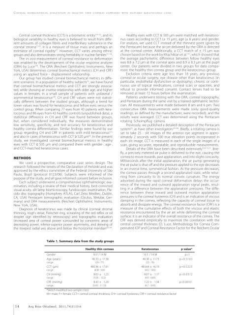
Table 1. Summary data from the study groups<br />
Healthy th<strong>in</strong> corneas Keratoconus p value*<br />
Gender 16 F / 14 M 16 F / 14 M p=1<br />
Age (years) 040.33 ± 17.38 040.30 ± 11.73 p=0.5103<br />
range (20 - 77) (22 - 76)<br />
CCT (μm) 485.96 ± 17.61 483.64 ± 16.19 p=0.5225<br />
range (438 - 505) (452 - 505)<br />
CH (mmHg) 008.63 ± 01.23 008.07 ± 01.17 p=0.0312<br />
range (5.95 - 12.2) (4.9 - 9.85)<br />
CRF (mmHg) 008.43 ± 01.29 007.22 ± 01.34 p


