

Microbial keratitis in southeast Brazil Floppy eyelid syndrome ...
Microbial keratitis in southeast Brazil Floppy eyelid syndrome ...
Microbial keratitis in southeast Brazil Floppy eyelid syndrome ...

Create successful ePaper yourself
Turn your PDF publications into a flip-book with our unique Google optimized e-Paper software.
AXONAL ELECTROVISIOGRAM AS AN ELECTROPHYSIOLOGICAL TEST TO EVALUATE OPTIC NERVE<br />
AND INNER RETINA ELECTRICAL POTENTIALS: FINDINGS IN NORMAL SUBJECTS<br />
Besides the orig<strong>in</strong>al studies dated from 1983 (8,10) , no additional<br />
reports on AxEvg have been published s<strong>in</strong>ce then. Thus, no technical<br />
standardization or normative values are known and no cl<strong>in</strong>ical<br />
use has been established so far.<br />
For cl<strong>in</strong>ical purposes, adequate knowledge about AxEvg<br />
electrical orig<strong>in</strong>s and wavelets characteristics is needed <strong>in</strong> order to<br />
<strong>in</strong>terpret results accurately. The goal of the present study is to<br />
validate the AxEvg as an electrophysiological test by standardiz<strong>in</strong>g<br />
the technique and establish<strong>in</strong>g the normative values, which will<br />
lead to the def<strong>in</strong>ition of its cl<strong>in</strong>ical <strong>in</strong>dications <strong>in</strong> the near future.<br />
METHODS<br />
PARTICIPANTS<br />
Normal <strong>in</strong>dividuals were selected from an ophthalmology<br />
tertiary center at Hospital de Base do Distrito Federal and from a<br />
private practice at Centro Brasileiro da Visão between March, 2007<br />
and March, 2009.<br />
All participants had a comprehensive ophthalmic exam<strong>in</strong>ation,<br />
<strong>in</strong>clud<strong>in</strong>g refraction and measurement of best-corrected visual<br />
acuity with Snellen charts, biomicroscopy, <strong>in</strong>traocular pressure measurement<br />
and funduscopy under pupil dilation. Patients were enrolled<br />
with the approval of the Ethics Committee <strong>in</strong> Research at<br />
University of Brasília (CEP-FM 009/2007), and all research procedures<br />
were performed <strong>in</strong> accordance with the tenets of the Declaration<br />
of Hels<strong>in</strong>ki. Informed consent was obta<strong>in</strong>ed from all studied<br />
subjects before their enrollment.<br />
We enrolled 140 normal <strong>in</strong>dividuals (280 eyes), disregard<strong>in</strong>g<br />
ethnicity and from both genders, with the aim to establish<br />
normative values for AxEvg. An <strong>in</strong>dividual was considered normal<br />
when best-corrected visual acuity with the Snellen chart was 20/20<br />
(or 1,0), present<strong>in</strong>g refraction between +3,00 spherical and -3,00<br />
spherical and/or a maximum of -1,50 cil<strong>in</strong>der. We excluded <strong>in</strong>dividuals<br />
with any ophthalmic condition that could reduce media<br />
transparency (such as corneal leucomas, cataracts, vitreous opacities<br />
and anterior or posterior uveitis), ret<strong>in</strong>al dystrophies or degenerations<br />
and neuro-ophthalmic disorders (such as glaucoma and optic<br />
neuropathies). We also excluded those with systemic chronic diseases<br />
that could cause ret<strong>in</strong>al or bra<strong>in</strong> ischemia (such as systemic<br />
arterial hypertension and diabetes mellitus) or that could present<br />
visual impairment due to medications (<strong>in</strong>clud<strong>in</strong>g those with autoimmune<br />
disorders).<br />
Participants were divided <strong>in</strong>to seven groups, each of them<br />
composed by 10 males and 10 females, accord<strong>in</strong>g to age: 1-10<br />
years-old, 11-20 years-old, 21-30 years-old, 31-40 years-old, 41-50<br />
years-old, 51-60 years-old, and older than 60 years-old.<br />
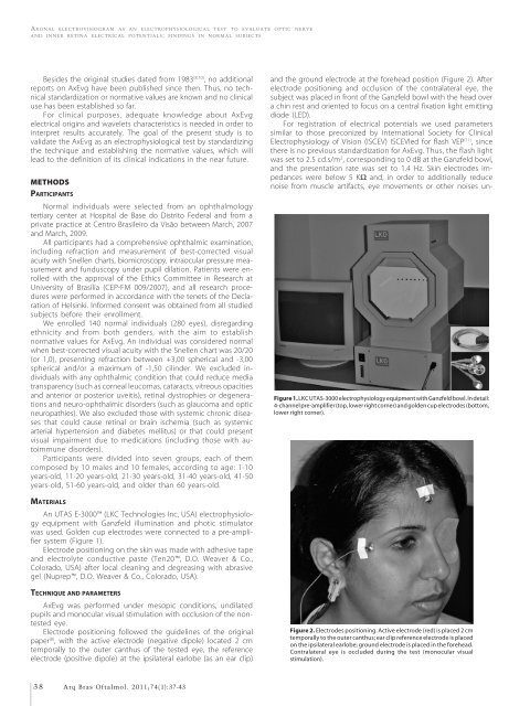
and the ground electrode at the forehead position (Figure 2). After<br />
electrode position<strong>in</strong>g and occlusion of the contralateral eye, the<br />
subject was placed <strong>in</strong> front of the Ganzfeld bowl with the head over<br />
a ch<strong>in</strong> rest and oriented to focus on a central fixation light emitt<strong>in</strong>g<br />
diode (LED).<br />
For registration of electrical potentials we used parameters<br />
similar to those preconized by International Society for Cl<strong>in</strong>ical<br />
Electrophysiology of Vision (ISCEV) ISCEVled for flash VEP (11) , s<strong>in</strong>ce<br />
there is no previous standardization for AxEvg. Thus, the flash light<br />
was set to 2.5 cd.s/m 2 , correspond<strong>in</strong>g to 0 dB at the Ganzfeld bowl,<br />
and the presentation rate was set to 1.4 Hz. Sk<strong>in</strong> electrodes impedances<br />
were below 5 KΩ and, <strong>in</strong> order to additionally reduce<br />
noise from muscle artifacts, eye movements or other noises un-<br />
Figure 1. LKC UTAS-3000 electrophysiology equipment with Ganzfeld bowl. In detail:<br />
4-channel pre-amplifier (top, lower right corner) and golden cup electrodes (bottom,<br />
lower right corner).<br />
MATERIALS<br />
An UTAS E-3000 (LKC Technologies Inc, USA) electrophysiology<br />
equipment with Ganzfeld illum<strong>in</strong>ation and photic stimulator<br />
was used. Golden cup electrodes were connected to a pre-amplifier<br />
system (Figure 1).<br />
Electrode position<strong>in</strong>g on the sk<strong>in</strong> was made with adhesive tape<br />
and electrolyte conductive paste (Ten20, D.O. Weaver & Co.,<br />
Colorado, USA) after local clean<strong>in</strong>g and degreas<strong>in</strong>g with abrasive<br />
gel (Nuprep, D.O. Weaver & Co., Colorado, USA).<br />
TECHNIQUE AND PARAMETERS<br />
AxEvg was performed under mesopic conditions, undilated<br />
pupils and monocular visual stimulation with occlusion of the nontested<br />
eye.<br />
Electrode position<strong>in</strong>g followed the guidel<strong>in</strong>es of the orig<strong>in</strong>al<br />
paper (8) , with the active electrode (negative dipole) located 2 cm<br />
temporally to the outer canthus of the tested eye, the reference<br />
electrode (positive dipole) at the ipsilateral earlobe (as an ear clip)<br />
Figure 2. Electrodes position<strong>in</strong>g. Active electrode (red) is placed 2 cm<br />
temporally to the outer canthus; ear clip reference electrode is placed<br />
on the ipsilateral earlobe; ground electrode is placed <strong>in</strong> the forehead.<br />
Contralateral eye is occluded dur<strong>in</strong>g the test (monocular visual<br />
stimulation).<br />
38 Arq Bras Oftalmol. 2011;74(1):37-43


