2019-03-27-PELO-workshop_for_participantsAT

Create successful ePaper yourself
Turn your PDF publications into a flip-book with our unique Google optimized e-Paper software.
DKTK<br />
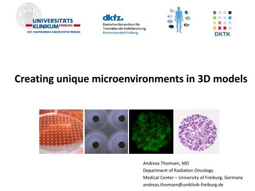
Creating unique microenvironments in 3D models<br />
Andreas Thomsen, MD<br />
Department of Radiation Oncology<br />
Medical Center – University of Freiburg, Germany<br />
andreas.thomsen@uniklinik-freiburg.de
Why 3D cell culture<br />
models?<br />
Monolayer<br />
culture (2D)<br />
To create<br />
microenvironments!
Microwell function<br />
Each conical microwell ≈ mini test tube<br />
Agarose<br />
Matrix 2x2<br />
mm<br />
Agarose gel<br />
- non-adhesive<br />
- open to diffusion<br />
- retains factors<br />
which are secreted by cells<br />
500 µm<br />
cells <strong>for</strong>m<br />
a micro-community<br />
+<br />
a micro-environment<br />
3
”Convincing“ cells to <strong>for</strong>m aggregates<br />
MIA Pa-Ca 2<br />
(human pancreatic adenocarcinoma)<br />
Hanging drop<br />
Liquid overlay<br />
3D CoSeedis TM<br />
Bright field<br />
Life / dead<br />
Aggregates harvested<br />
from microwells<br />
Paraffin sections<br />
proliferation gradient
3D CoSeedis TM cell culture in agarose microwells<br />
High definition geometry<br />
volumetry<br />
of cell<br />
aggregates<br />
embed into<br />
ECM<br />
or re-seed<br />
into 2D<br />
Single<br />
step<br />
seeding<br />
harvest cell<br />
aggregates<br />
(proteins,<br />
DNA …)<br />
Treatment<br />
(irradiation, drugs, hypoxia, cytokines …)<br />
imaging<br />
incubate<br />
Evaluation<br />
- several choices -<br />
[days to weeks]<br />
histology
Primary MSC from<br />
human bone marrow particles<br />
Bone marrow particles in T25 flask<br />
2D<br />
3D<br />
200 µm<br />
explant culture<br />
Outgrowth of MSC<br />
Bone marrow particle
Differentiation of bone marrow stromal cells<br />
A<br />
B C D<br />
Chondrogenic differentiation of hMSC in agarose microwells.<br />
A: Spheroids made from 180,000 hMSC per microwell on day 22<br />
B-D: Histology: Hyaline-like cartilage (B+C: H&E staining, D: alcian blue staining).<br />
Scale bars 2 mm (A), 200 µm (B) and 50 µm (C+D)
Primary MSC from<br />
human bone marrow particles<br />
Bone marrow particles in T25 flask<br />
2D<br />
200 µm<br />
3D<br />
MSC aggregates in basement membrane extract gel (BME)<br />
200 µm
Microwells customized<br />
with ECM particles<br />
*<br />
*<br />
no ECM particles BME particles Collagen type I particles
Re-Seeding MSC aggregates back into 2D culture<br />
200 µm<br />
200 µm<br />
Initial seeding densities 1,000 (left) and 10,000 MSC per microwell (right).
3D Contact Coculture<br />
T47D human breast cancer cells +/- human bone marrow stromal cells (bmMSC)<br />
T47D<br />
50 cells/microwell<br />
T47D<br />
50 cells/microwell<br />
+ bmMSC<br />
1000 cells/microwell<br />
* *<br />
*<br />
d7:<br />
- bmMSC assemble in the center (*) of co-spheroids and are soon overgrown by tumor cells<br />
- addition of bmMSC results in much larger T47D spheroids<br />
11
3D Contact Coculture<br />
T47D human breast cancer cells +/- human bone marrow stromal cells (bmMSC*)<br />
T47D only<br />
0 Gy<br />
T47D<br />
+ bmMSC<br />
*<br />
d 7<br />
d 14 d 19<br />
T47D only<br />
Growth inhibition<br />
through irradiation<br />
is mitigated by<br />
presence of<br />
bmMSC<br />
2 Gy<br />
T47D<br />
+ bmMSC<br />
*<br />
bmMSC<br />
aggregates (*)<br />
decrease in size<br />
over time.<br />
12
MSC number decreases in contact cocultures with cancer cells<br />
d 7 d 14<br />
MSC + T47D<br />
MSC + MIA PaCa 2<br />
cannibalism !?!
FDG-PET in Lung Cancer<br />
1. Diagnosis of lymph node involvement
FDG-PET in Lung Cancer<br />
1. Diagnosis of lymph node involvement<br />
2. Discrimination of malignant tissue from atelectasis
FDG-PET in Lung Cancer<br />
1. Diagnosis of lymph node involvement<br />
2. Discrimination of malignant tissue from atelectasis<br />
Metabolic Radiation Treatment Planning<br />
Schmuecking et al.
Eating sugar – track cellular metabolism of 3D microtumors<br />
Control<br />
No cells (empty agarose)<br />
Density 1<br />
~ 2.000 cells per microwell<br />
Density 2<br />
~ 8.000 cells per microwell<br />
Einstellung an Linearbeschleuniger<br />
Activity 1: 0,5 MBq<br />
17<br />
Activity 2: 5 MBq
Oxygen effect<br />
Hypoxia reduces<br />
cellular radiosensitivity<br />
by factor 2 - 3
Oxygen Effect<br />
Vaupel / Konerding
FMISO-PET: Hypoxia imaging<br />
Resolution of tumor hypoxia under chemoradiation<br />
MRI<br />
0 Gy<br />
be<strong>for</strong>e treatment<br />
14 Gy<br />
58 Gy<br />
after treatment<br />
Wiedenmann N, Grosu AL
F-MISO-PET: Hypoxia imaging<br />
HT-29<br />
Colorectal cancer<br />
cells/ microwell 1000 2000 4000 8000<br />
T47D<br />
Breast cancer<br />
100 µm<br />
cells/ microwell 1000 2000 4000 8000
Colony <strong>for</strong>mation assay<br />
<strong>for</strong> adhesion-independent cells<br />
‘Going digital’
Colony <strong>for</strong>mation assay – clonogenic survival<br />
confluent colonies<br />
inter-observer variation<br />
Intra-observer variation<br />
time<br />
secondary colonies<br />
≤ 50 cells?<br />
Survey among radiation biologists: ”Do you enjoy colony <strong>for</strong>mation assays?”<br />
5 % 20 % 30 % 45 %
Single cells in agarose microwells<br />
e.g. seeding 1 cell / microwell<br />
8 – 14 d
Single cells in agarose microwells<br />
500 µm<br />
HT-29,<br />
630 cells /cm², d0<br />
Small microwell <strong>for</strong>mat, 0.5 x 0.5 mm<br />
3D colonies, d14<br />
(different region of agarose array)
Image analysis (ImageJ)<br />
2 mm<br />
set threshold<br />
count particles<br />
only colonies >20 000<br />
µm² were counted<br />
colonies detected<br />
in 13 out of 25<br />
microwells
Why ‘digital’ colony <strong>for</strong>mation assay?<br />
1 0 0 0 1<br />
1 1 1 1 1<br />
0 0 1 1 0<br />
0 1 0 0 0<br />
2 mm<br />
1 1 1 0 0<br />
When a threshold is applied, readout of microwells may be done in<br />
a binary fashion
closeup: 2D vs. 3D colony <strong>for</strong>mation assay<br />
HT-29<br />
d14<br />
cells/cm² 10 16 25 40 63 100<br />
2D<br />
3D<br />
5 mm<br />
cells/cm² 160 250 400 630 1000 1600<br />
2D<br />
3D<br />
5 mm
Statistics<br />
HT-29 in 3D colony assay<br />
lambda<br />
Poisson distribution<br />
Residual standard error: 0.<strong>27</strong>38 on 11 degrees of freedom<br />
Multiple R-squared: 0.9853, Adjusted R-squared: 0.984<br />
F-statistic: 739.7 on 1 and 11 DF, p-value: 1.934e-11<br />
cells seeded per microwell<br />
[1] " Result: lambda = 0.36246* number of cells seeded per microwell"<br />
[1] " Number of colonies = 0.36* number of cells seeded per microwell"<br />
[1] " 0.36 = colony <strong>for</strong>mation efficiency"<br />
Dr. G. Rücker, Biometric Dept.
Readout (2)<br />
Clonal HT-29 spheroids harvested from microwells<br />
Paraffin section, H&E stained
CD34 + cells from cord blood<br />
Microwell <strong>for</strong>mat: 0.5 x 0.5 mm,<br />
Paraffin section, MIB-1 (Ki-67)
Paraffin section, H&E<br />
MIB-1 (Ki-67)
Microwells customized<br />
with ECM particles<br />
*<br />
*<br />
no ECM particles BME particles Collagen type I particles
Colony <strong>for</strong>mation using ”ECM nest“<br />
no ECM particles BME particles Collagen type I particles<br />
80<br />
70<br />
60<br />
50<br />
40<br />
30<br />
20<br />
10<br />
0<br />
Colonies > 8x 10 6 µm³<br />
ohne<br />
no ECM<br />
Einlage<br />
BME<br />
ECM<br />
particles Collagen<br />
Coll. Typ<br />
I<br />
I<br />
particles<br />
MIA Pa-Ca-2<br />
Seeding density: 0.3 cells / microwell<br />
d14, MTT staining
”ECM nest”<br />
100,0%<br />
Gy<br />
0 2 4 6 8 10<br />
*<br />
10,0%<br />
1,0%<br />
2D<br />
3D<br />
3D ECM<br />
ECM particles ( ) at bottom of microwells<br />
*<br />
0,1%<br />
Colony <strong>for</strong>mation efficiacy<br />
12000000<br />
10000000<br />
.<br />
Spheroid<br />
volume<br />
[µm³]<br />
8000000<br />
6000000<br />
4000000<br />
3D<br />
3D ECM<br />
2000000<br />
200 µm<br />
100 µm<br />
BME remodeled by cells<br />
0<br />
0 5 10<br />
Dose [Gy]
Thanks<br />
• Christine Aldrian<br />
• Nicolas Melin<br />
• Costas Zamboglou<br />
• Norbert Nanko<br />
• Nicole Wiedenmann<br />
• Simone Gaedicke<br />
• Elke Firat<br />
• Marie Follo (Core facility)<br />
• Gerta Rücker (Biometry Dept.)<br />
• Friederike Braun (Nuclear medicin)<br />
• Miriam Erlacher (Children‘s Dept.)<br />
• Irina Nararenko (IUK)<br />
• Gabriele Niedermann<br />
• Michael Henke<br />
• Ursula Nestle<br />
• Anca-Ligia Grosu<br />
• Marco Leu<br />
• Dr. Erwin Braun Foundation
The ”Raisin Roll Problem”<br />
probability<br />
Mean: 5 raisins per roll<br />
λ = 5<br />
Poisson distribution
Soft agar assay<br />
10 -14 d
Anchorage independence<br />
Anoikis resistance<br />
≈<br />
Soft agar<br />
Agarose microwells
Coherence of cells<br />
low<br />
high
Histology from 3D CoSeedis TM<br />
Paraffin block<br />
41 LMP = low melting point agarose
Volume<br />
measurement<br />
side view<br />
bottom view<br />
Inverted microscope<br />
MTT staining
Volume determination<br />
spherical or conical ?<br />
Volume calculation tool:<br />
https://biopply.com/products-applications/analysis-tool/<br />
43
.<br />
Thank you <strong>for</strong> your attention!<br />
Happy to hear your comments and questions!<br />
http://dx.doi.org/10.1<strong>03</strong>9/C7LC00832E<br />
andreas.thomsen@uniklinik-freiburg.de<br />
44