Import risk analysis: Llamas (Lama glama) and alpacas (Vicugna ...
Import risk analysis: Llamas (Lama glama) and alpacas (Vicugna ...
Import risk analysis: Llamas (Lama glama) and alpacas (Vicugna ...

You also want an ePaper? Increase the reach of your titles
YUMPU automatically turns print PDFs into web optimized ePapers that Google loves.
<strong>Import</strong> <strong>risk</strong> <strong>analysis</strong>: <strong>Llamas</strong><br />
(<strong>Lama</strong> <strong>glama</strong>) <strong>and</strong> <strong>alpacas</strong><br />
(<strong>Vicugna</strong> pacos) from<br />
specified countries<br />
FINAL<br />
ISBN 978-0-478-37526-8 (print)<br />
ISBN 978-0-478-37527-5 (online)<br />
14 December 2010
This page is intentionally blank
MAF Biosecurity New Zeal<strong>and</strong><br />
Pastoral House<br />
25 The Terrace<br />
PO Box 2526<br />
Wellington 6011<br />
New Zeal<strong>and</strong><br />
Tel: 64 4 894 0100<br />
Fax: 64 4 894 0731<br />
Policy <strong>and</strong> Risk<br />
MAF Biosecurity New Zeal<strong>and</strong><br />
<strong>Import</strong> <strong>risk</strong> <strong>analysis</strong>:<br />
<strong>Llamas</strong> (<strong>Lama</strong> <strong>glama</strong>) <strong>and</strong> <strong>alpacas</strong> (<strong>Vicugna</strong> pacos) from specified countries<br />
FINAL<br />
14 December 2010<br />
Approved for general release<br />
Christine Reed<br />
Manager, Risk Analysis<br />
MAF Biosecurity New Zeal<strong>and</strong>
This page is intentionally blank
CONTENTS<br />
Executive Summary 1<br />
1. Introduction 3<br />
2. Scope 4<br />
3. Commodity Definitions 4<br />
4. Risk Analysis Methodology 4<br />
5. Organisms of Potential Concern <strong>and</strong> the Preliminary Hazard List 7<br />
6. African horse sickness virus 17<br />
7. Bluetongue virus 19<br />
8. Borna virus 21<br />
9. Bovine viral diarrhoea virus 24<br />
10. Bovine herpesvirus type 1 31<br />
11. Epizootic haemorrhagic disease virus 37<br />
12. Eastern equine encephalitis virus 39<br />
13. Equine herpesvirus type 1 41<br />
14. Foot <strong>and</strong> mouth disease virus 46<br />
15. Louping-ill virus 51<br />
16. Rabies virus 53<br />
17. Vesicular stomatitis virus 57<br />
18. West Nile virus 64<br />
19. Anaplasma phagocytophilum 66<br />
20. Bacillus anthracis 69<br />
21. Brucella spp. 73<br />
22. Burkholderia pseudomallei 78<br />
23. Chlamydophila spp. 80<br />
i
24. Coxiella burnetii 85<br />
25. Leptospira serovars 89<br />
26. Mycobacterium bovis 95<br />
27. Mycoplasma haemolamae 100<br />
28. Pasteurella multocida types 6B <strong>and</strong> 6E 104<br />
29. Salmonella spp. 107<br />
30. Yersinia pestis 112<br />
31. Coccidioides immitis 114<br />
32. Trypanosoma spp. 115<br />
33. Echinococcus granulosus 120<br />
34. Internal parasites 125<br />
35. Mites, lice <strong>and</strong> fleas 131<br />
36. Ticks 136<br />
37. Screwworm <strong>and</strong> other myiasis infestations 142<br />
38. Weeds <strong>and</strong> seeds 149<br />
© Crown Copyright – Ministry of Agriculture <strong>and</strong> Forestry<br />
This publication is also available on the MAF website at<br />
http://www.biosecurity.govt.nz/regs/imports/ihs/<strong>risk</strong><br />
ii
Acronyms<br />
CDC United States Centers for Disease Control <strong>and</strong> Prevention<br />
CF(T) complement fixation (test)<br />
DNA deoxyribonucleic acid<br />
ELISA enzyme-linked immunosorbent assay<br />
IFA(T) indirect fluorescent antibody (test)<br />
IHS <strong>Import</strong> Health St<strong>and</strong>ard<br />
MAF New Zeal<strong>and</strong> Ministry of Agriculture <strong>and</strong> Forestry<br />
MAFBNZ Ministry of Agriculture <strong>and</strong> Forestry Biosecurity New Zeal<strong>and</strong><br />
OIE World Organisation for Animal Health<br />
PCR polymerase chain reaction<br />
PEQ pre-export quarantine<br />
RNA ribonucleic acid<br />
RT-PCR reverse transcriptase polymerase chain reaction<br />
WTO World Trade Organization<br />
iii
Contributors to this <strong>risk</strong> <strong>analysis</strong><br />
1. Authors<br />
Lincoln Broad<br />
Bob Worthington<br />
2. Internal Peer Review<br />
Howard Pharo<br />
Stuart MacDiarmid<br />
Stephen Cobb<br />
Marguerite<br />
Hern<strong>and</strong>ez<br />
3. External Scientific Review<br />
Fraser Hill<br />
James Gilkerson<br />
Senior Adviser Risk Analysis, MAF Biosecurity New Zeal<strong>and</strong>,<br />
Wellington<br />
Risk Analysis Contractor, MAF Biosecurity New Zeal<strong>and</strong>,<br />
Wellington<br />
Team Manager, Risk Analysis, MAF Biosecurity New Zeal<strong>and</strong>,<br />
Wellington<br />
Principal International Adviser Risk Analysis, MAF Biosecurity<br />
New Zeal<strong>and</strong>, Wellington<br />
Senior Adviser Risk Analysis, MAF Biosecurity New Zeal<strong>and</strong>,<br />
Wellington<br />
Senior Adviser Animal <strong>Import</strong>s <strong>and</strong> Exports, MAF Biosecurity<br />
New Zeal<strong>and</strong>, Wellington<br />
Registered Veterinary Specialist Anatomic Pathology, Gribbles<br />
Veterinary, Palmerston North<br />
Associate Professor in Veterinary Microbiology. Head, Equine<br />
Infectious Disease Laboratory, The University of Melbourne<br />
iv
Executive Summary<br />
This <strong>risk</strong> <strong>analysis</strong> examines the <strong>risk</strong>s involved with the importation of llamas (<strong>Lama</strong><br />
<strong>glama</strong>) <strong>and</strong> <strong>alpacas</strong> (<strong>Vicugna</strong> pacos) from specified countries: Australia, USA, Canada, the<br />
European Union <strong>and</strong> South America.<br />
An extensive hazard list of organisms of potential concern that could be associated with<br />
camelids has been collated in Table 1. Preliminary hazards are identified within Table 1 as<br />
those that have meet specified criteria. Mycobacterium bovis is the only endemic organism<br />
retained as a preliminary hazard since it is the subject of an official control programme<br />
under the Biosecurity Act 1993.<br />
The preliminary hazards identified from Table 1 are subjected to individual <strong>risk</strong> analyses,<br />
following the st<strong>and</strong>ard process. First, in the hazard identification step the epidemiology of<br />
the disease, including distribution, clinical signs, transmission, diagnosis <strong>and</strong> any available<br />
treatment, is considered. As a result of hazard identification, organisms are classified as<br />
either potential hazards in the commodity, or not.<br />
Organisms identified as potential hazards in the commodity are subjected to <strong>risk</strong><br />
assessment to provide a <strong>risk</strong> estimate by considering the likelihood of entry (the disease<br />
agent being present in an animal at the time of importation), exposure (likelihood of spread<br />
<strong>and</strong> establishment if imported) <strong>and</strong> any adverse consequences likely to follow these events.<br />
Risk management is not warranted for disease agents that are exclusively arthropod-borne,<br />
mainly through specific ticks, flies <strong>and</strong> mosquitoes that are not present in New Zeal<strong>and</strong>.<br />
However, if new vector species were to establish here, measures may then become<br />
necessary to prevent introduction of such organisms. For organisms that are classified as<br />
hazards as a result of <strong>risk</strong> estimation, in the <strong>risk</strong> management step the options that could be<br />
used to effectively manage the <strong>risk</strong> are presented. The <strong>risk</strong> management options include<br />
quarantine, testing, vaccination <strong>and</strong> treatment as appropriate.<br />
When drafting any <strong>Import</strong> Health St<strong>and</strong>ard (IHS) developed from this import <strong>risk</strong> <strong>analysis</strong>, <strong>risk</strong><br />
management measures selected by the Animal <strong>Import</strong>s <strong>and</strong> Exports Section of the Border<br />
St<strong>and</strong>ards Directorate of MAF Biosecurity New Zeal<strong>and</strong> will be the least trade restrictive<br />
whilst providing a level of protection that is considered to be appropriate.<br />
In the case of Babesia spp. of livestock, Anaplasma spp., bluetongue virus <strong>and</strong> epizootic<br />
haemorragic disease virus, the <strong>risk</strong> <strong>analysis</strong> concludes that the <strong>risk</strong>s posed by these<br />
organisms in <strong>alpacas</strong> <strong>and</strong> llamas are negligible. As a result, the testing requirements for<br />
these organisms in the currently issued IHSs are not justifiable.<br />
Thirty three individual organisms or groups of organisms were identified as preliminary<br />
hazards from the organisms of potential concern listed in Table 1. As a result of the<br />
individual <strong>risk</strong> assessments, 20 of these preliminary hazards were classified as hazards in<br />
the commodity <strong>and</strong> for each of these <strong>risk</strong> management measures are presented:<br />
Bovine viral diarrhoea virus type 2<br />
Bovine herpesvirus type 1<br />
Equine herpesvirus type 1<br />
Foot <strong>and</strong> mouth disease virus<br />
Rabies virus<br />
MAF Biosecurity New Zeal<strong>and</strong> <strong>Import</strong> <strong>risk</strong> <strong>analysis</strong>: <strong>Llamas</strong> <strong>and</strong> <strong>alpacas</strong> from specified countries ● 1
Vesicular stomatitis virus<br />
Bacillus anthracis<br />
Brucella spp.<br />
Chlamydophila abortus<br />
Coxiella burnetii<br />
Leptospira serovars<br />
Mycobacterium bovis<br />
Mycoplasma haemolamae<br />
Salmonella spp.<br />
Trypanosoma spp.<br />
Echinococcus granulosus<br />
Other internal parasites (trematodes <strong>and</strong> nematodes)<br />
External parasites (mites, fleas, lice <strong>and</strong> ticks)<br />
Screwworm <strong>and</strong> other myiasis infestations<br />
Hitch-hiker weeds <strong>and</strong> seeds<br />
2 ● <strong>Import</strong> <strong>risk</strong> <strong>analysis</strong>: <strong>Llamas</strong> <strong>and</strong> <strong>alpacas</strong> from specified countries MAF Biosecurity New Zeal<strong>and</strong>
1. Introduction<br />
The importation of <strong>alpacas</strong> <strong>and</strong> llamas is increasing each year, from around 50 animals<br />
several years ago to more than 550 during 2008 <strong>and</strong> 2009. This increase in the number of<br />
animals moving internationally is expected to continue, along with an increase in the<br />
number of countries from which <strong>alpacas</strong> <strong>and</strong> llamas are sourced.<br />
Conditions have been developed in the past to allow importations from Chile <strong>and</strong><br />
Argentina. However, importations only ever occurred from Chile <strong>and</strong> the conditions were<br />
not based on a <strong>risk</strong> <strong>analysis</strong>, but rather detailed bilateral negotiation between MAF <strong>and</strong> the<br />
Chilean Authority. The last consignment from Chile (1992-1993) had problems when<br />
seropositive animals to foot <strong>and</strong> mouth disease were detected in post-arrival quarantine<br />
(false-positive test results). Australia <strong>and</strong> Canada also experienced similar problems with<br />
foot <strong>and</strong> mouth disease seropositive animals being detected in post-arrival quarantine. In<br />
those cases, it was thought that vaccinated animals had been inadvertently included in the<br />
consignments. These episodes raised serious concerns regarding the ability of Chile to<br />
provide the necessary pre-export assurances required.<br />
Because of this history when importing from Chile, New Zeal<strong>and</strong> put in place a<br />
moratorium on the issuing of import health permits for llamas <strong>and</strong> <strong>alpacas</strong> from all of<br />
South America.<br />
Alpacas <strong>and</strong> llamas are presently imported under the following <strong>Import</strong> Health St<strong>and</strong>ards<br />
(IHSs):<br />
Alpacas <strong>and</strong> <strong>Llamas</strong> from United States of America (July 2005)<br />
Alpacas <strong>and</strong> <strong>Llamas</strong> from Australia (May 2006)<br />
The measures in these IHSs are based on the MAF document "Disease Risk Analysis for<br />
the <strong>Import</strong>ation of <strong>Llamas</strong> (<strong>Lama</strong> <strong>glama</strong>) <strong>and</strong> Alpacas (<strong>Lama</strong> pacos) into New Zeal<strong>and</strong>"<br />
that was finalised in 1997 <strong>and</strong> up-dated in 1998. That <strong>risk</strong> <strong>analysis</strong> examined the <strong>risk</strong>s<br />
involved with the importation of animals from the USA, Canada <strong>and</strong> Australia. The 1997<br />
<strong>risk</strong> <strong>analysis</strong> specifically excluded South American countries from consideration.<br />
The 1998 <strong>risk</strong> <strong>analysis</strong> was produced using procedures <strong>and</strong> policies that have meanwhile<br />
changed. The <strong>analysis</strong> identified potential hazards <strong>and</strong> provided a short discussion,<br />
including some recommendations from the Terrestrial Animal Health Code. The measures<br />
that were presented were based on other animal species, not specifically camelids. For the<br />
identified agents of concern, no <strong>risk</strong> assessment step was undertaken.<br />
A new <strong>risk</strong> <strong>analysis</strong> is therefore required because of the changes in the <strong>risk</strong> analyses<br />
process, technical advances over the last 12 years <strong>and</strong> the increased number of countries<br />
requiring assessment.<br />
Note that an importing country is entitled to expect validity in the veterinary certification<br />
of export. However, it must be made clear that an evaluation of an exporting country’s<br />
st<strong>and</strong>ards <strong>and</strong> performance is not made in this <strong>risk</strong> <strong>analysis</strong>. MAF may conduct an<br />
evaluation of veterinary services when drafting IHSs developed from this import <strong>risk</strong><br />
<strong>analysis</strong>, particularly for countries with which there is no existing trade.<br />
MAF Biosecurity New Zeal<strong>and</strong> <strong>Import</strong> <strong>risk</strong> <strong>analysis</strong>: <strong>Llamas</strong> <strong>and</strong> <strong>alpacas</strong> from specified countries ● 3
2. Scope<br />
This qualitative <strong>risk</strong> <strong>analysis</strong> examines the <strong>risk</strong>s involved with the importation of llamas<br />
(<strong>Lama</strong> <strong>glama</strong>) <strong>and</strong> <strong>alpacas</strong> (<strong>Vicugna</strong> pacos). <strong>Llamas</strong> <strong>and</strong> <strong>alpacas</strong> are hereafter referred to as<br />
“camelids”.<br />
This <strong>risk</strong> <strong>analysis</strong> is restricted to camelids imported from Australia, Canada, the United<br />
States of America (USA), Central <strong>and</strong> South America <strong>and</strong> the European Union (EU) A .<br />
Hereafter these countries are referred to as “relevant countries”.<br />
3. Commodity Definitions<br />
The Family Camelidae contains three genera: Camelus, <strong>Lama</strong> <strong>and</strong> <strong>Vicugna</strong>.<br />
Guanacos (<strong>Lama</strong> guanicoe) are a wild species <strong>and</strong> are not traded. The genus <strong>Vicugna</strong><br />
includes the species V. vicuna which is a wild ancestor of the alpaca <strong>and</strong> an endangered<br />
species that will not be traded.<br />
The dromedary (one-humped) <strong>and</strong> bacterian (two-humped) camel belong to the Camelus<br />
genus <strong>and</strong> are not part of the commodity definition.<br />
<strong>Llamas</strong> (<strong>Lama</strong> <strong>glama</strong>) <strong>and</strong> <strong>alpacas</strong> (<strong>Vicugna</strong> pacos) (domesticated species) that have been<br />
certified on the day of shipment to be showing no clinical signs of infectious or parasitic<br />
disease are the commodity to be traded.<br />
4. Risk Analysis Methodology<br />
The methodology used in this <strong>risk</strong> <strong>analysis</strong> follows the 2006 MAF Biosecurity New<br />
Zeal<strong>and</strong> Risk Analysis Procedures- Version 1. These procedures combine the guidelines in<br />
the Terrestrial Animal Health Code 2009 (hereafter referred to as the Code) of the World<br />
Organisation for Animal Health (OIE) <strong>and</strong> International Plant Protection Convention<br />
guidelines. The procedures provide a framework which adheres to the requirements set out<br />
under the World Trade Organization’s Agreement on the Application of Sanitary <strong>and</strong><br />
Phytosanitary (SPS) measures, 1995 <strong>and</strong> of the Biosecurity Act, 1993.<br />
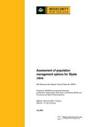
The process followed is shown in Figure 1 (overleaf).<br />
A The EU presently includes Austria, Belgium, Bulgaria, Cyprus, Czech Republic, Denmark, Estonia,<br />
Finl<strong>and</strong>, France, Germany, Greece, Hungary, Irel<strong>and</strong>, Italy, Latvia, Lithuania, Luxembourg, Malta,<br />
Netherl<strong>and</strong>s, Pol<strong>and</strong>, Portugal, Romania, Slovakia, Slovenia, Spain, Sweden, <strong>and</strong> the United Kingdom.<br />
4 ● <strong>Import</strong> <strong>risk</strong> <strong>analysis</strong>: <strong>Llamas</strong> <strong>and</strong> <strong>alpacas</strong> from specified countries MAF Biosecurity New Zeal<strong>and</strong>
Figure 1. The <strong>risk</strong> <strong>analysis</strong> process.<br />
4.1. Preliminary Hazard List (Organisms of Potential Concern)<br />
From consulting authoritative texts <strong>and</strong> electronic data-bases an extensive list of organisms<br />
known to infect or infest camelids has been collated. From all the potential organisms of<br />
concern listed, preliminary hazards are identified by applying specific criteria to each<br />
organism to eliminate those that clearly do not constitute any <strong>risk</strong>. The remaining<br />
organisms are collated into a preliminary hazard list. The organisms in this list are<br />
subjected to hazard identification.<br />
4.2. Hazard Identification<br />
Each organism in the preliminary hazard list is subjected to a hazard identification step.<br />
This includes formal identification of the organism, whether it is an OIE listed disease, its<br />
New Zeal<strong>and</strong> status, <strong>and</strong> a discussion on the relevant aspects of the epidemiology <strong>and</strong><br />
characteristics of the organism. The hazard identification section is concluded by an<br />
MAF Biosecurity New Zeal<strong>and</strong> <strong>Import</strong> <strong>risk</strong> <strong>analysis</strong>: <strong>Llamas</strong> <strong>and</strong> <strong>alpacas</strong> from specified countries ● 5
assessment of whether the organism is a potential hazard or not. All potential hazards are<br />
subjected to <strong>risk</strong> assessment.<br />
4.3. Risk Assessment<br />
Risk assessment consists of:<br />
a) Entry assessment: The likelihood of a potential hazard (pathogenic organism) being<br />
imported with the camelids.<br />
b) Exposure assessment: Describes the biological pathway(s) necessary for exposure of<br />
susceptible animals or humans in New Zeal<strong>and</strong> to the potential hazard. Further, a<br />
qualitative estimation of the probability of the exposure occurring is made.<br />
c) Consequence assessment: Describes the likely potential consequences of entry,<br />
exposure <strong>and</strong> establishment or spread of an imported potential hazard.<br />
d) Risk estimation: An estimation of the <strong>risk</strong> posed by the potential hazard associated with<br />
importing camelids. This is based on the entry, exposure <strong>and</strong> consequence assessments.<br />
If the <strong>risk</strong> estimate is non-negligible, then the potential hazard is classified as a hazard<br />
<strong>and</strong> <strong>risk</strong> management measures may be justified to reduce the level of <strong>risk</strong> to an<br />
acceptable level.<br />
Not all of the above steps may be necessary in all <strong>risk</strong> assessments. The OIE methodology<br />
makes it clear that if the likelihood of entry is negligible for a certain potential hazard, then<br />
the <strong>risk</strong> estimate is automatically negligible <strong>and</strong> the remaining steps of the <strong>risk</strong> assessment<br />
need not be carried out. The same situation arises when the likelihood of entry is nonnegligible<br />
but the exposure assessment concludes that the likelihood of susceptible species<br />
being exposed is negligible, or when both entry <strong>and</strong> exposure are non-negligible but the<br />
consequences of introduction are assessed to be negligible.<br />
4.4. Risk Management<br />
For each organism classified as a hazard, a <strong>risk</strong> management step is carried out, which<br />
identifies the options available for managing the <strong>risk</strong>. Where the Code lists<br />
recommendations for the management of a hazard, these are described alongside options of<br />
similar, lesser or greater stringency where available. In addition to the options presented,<br />
unrestricted entry or prohibition may also be considered for all hazards. Recommendations<br />
for the appropriate sanitary measures to achieve the effective management of <strong>risk</strong>s are not<br />
made in this document. These will be determined when an IHS is drafted.<br />
As obliged under Article 3.1 of the WTO’s SPS Agreement the measures adopted in IHSs<br />
will be based on international st<strong>and</strong>ards, guidelines <strong>and</strong> recommendations where they exist,<br />
except as otherwise provided for under Article 3.3 (where measures providing a higher<br />
level of protection than international st<strong>and</strong>ards can be applied if there is scientific<br />
justification, or if there is a level of protection that the member country considers is more<br />
appropriate following a <strong>risk</strong> assessment).<br />
4.5. Risk Communication<br />
MAFBNZ publishes draft import <strong>risk</strong> analyses for a six-week period of public consultation<br />
to verify the scientific basis of the <strong>risk</strong> assessment <strong>and</strong> to seek stakeholder comment on the<br />
<strong>risk</strong> management options presented. Stakeholders are also invited to present alternative <strong>risk</strong><br />
management options that they consider necessary or preferable.<br />
6 ● <strong>Import</strong> <strong>risk</strong> <strong>analysis</strong>: <strong>Llamas</strong> <strong>and</strong> <strong>alpacas</strong> from specified countries MAF Biosecurity New Zeal<strong>and</strong>
Following public consultation on the draft <strong>risk</strong> <strong>analysis</strong>, MAFBNZ produces a a review of<br />
submissions <strong>and</strong> determines whether any changes need to be made to the draft <strong>risk</strong> <strong>analysis</strong><br />
as a result of public consultation, in order to make it a final <strong>risk</strong> <strong>analysis</strong>.<br />
Following this process of consultation <strong>and</strong> review, the <strong>Import</strong>s St<strong>and</strong>ards team of<br />
MAFBNZ decides on the appropriate combination of sanitary measures to ensure the<br />
effective management of identified <strong>risk</strong>s. These are then presented in a draft IHS which is<br />
released for a six-week period of stakeholder consultation. Stakeholder submissions in<br />
relation to the draft IHS are reviewed before a final IHS is issued.<br />
5. Organisms of Potential Concern <strong>and</strong> the Preliminary<br />
Hazard List<br />
The first step in the <strong>risk</strong> <strong>analysis</strong> is hazard identification to ensure that all organisms of<br />
potential concern have been considered. For this <strong>risk</strong> <strong>analysis</strong> a list of organisms of<br />
potential concern was made comprising all the diseases/disease agents of <strong>alpacas</strong> <strong>and</strong><br />
llamas located from the following sources:<br />
� OIE (2009). Terrestrial Animal Health Code. Available at:<br />
http://www.oie.int/eng/normes/mcode/en_sommaire.htm<br />
� Taylor MA, Coop RL, Wall RL (2007). Veterinary Parasitology. Blackwell Publishing, Oxford. pp 874.<br />
� Fowler M (1998). Medicine <strong>and</strong> Surgery of South American Camelids. Iowa State University Press,<br />
Ames. pp 391.<br />
� Wernery U, Kaaden O-R (2002). Infectious Diseases in Camelids. 2 nd edition, Blackwell Science,<br />
Berlin. pp 404.<br />
� Ministry of Agriculture <strong>and</strong> Forestry (1997). Disease Risk Analysis for the <strong>Import</strong>ation of <strong>Llamas</strong><br />
(<strong>Lama</strong> <strong>glama</strong>) <strong>and</strong> Alpacas (<strong>Lama</strong> pacos) into New Zeal<strong>and</strong>. Amendments dated 21 st April 1998.<br />
� Australian Quarantine <strong>and</strong> Inspection Service (2000). <strong>Import</strong> Risk Analysis for the <strong>Import</strong>ation of<br />
Camelids from Chile <strong>and</strong> Peru. Canberra. pp 19.<br />
� PubMed electronic scientific journal data-base. Provides access to bibliographic information<br />
published in journals in the US <strong>and</strong> more than 80 other countries.<br />
� Additional diseases or disease agents. As suggested by experts employed by MAFBNZ <strong>and</strong><br />
interested parties that were consulted on the subject or were involved in reviewing this <strong>risk</strong> <strong>analysis</strong>.<br />
Organisms/diseases identified as of potential concern from the above sources are listed in<br />
Table 1 (below).<br />
The organisms of particular interest are those that are zoonotic <strong>and</strong> those that could be<br />
transmitted from camelids to domestic, feral or wild animals <strong>and</strong> humans.<br />
The table indicates whether the organisms are zoonotic <strong>and</strong> whether they are known to<br />
occur in New Zeal<strong>and</strong>. In Column 7 of the table, each organism is classified as to whether<br />
it is a preliminary hazard or not. An organism classified as a preliminary hazard meets the<br />
following criteria:<br />
� All disease agents that are exotic to New Zeal<strong>and</strong> <strong>and</strong> present in an exporting<br />
country or about which there is some uncertainty.<br />
MAF Biosecurity New Zeal<strong>and</strong> <strong>Import</strong> <strong>risk</strong> <strong>analysis</strong>: <strong>Llamas</strong> <strong>and</strong> <strong>alpacas</strong> from specified countries ● 7
� In addition, organisms that occur in New Zeal<strong>and</strong> for which there are known subspecies<br />
or strains or host associations that do not occur in New Zeal<strong>and</strong> but do<br />
occur in an exporting country <strong>and</strong> are potentially harmful.<br />
� Organisms that occur in New Zeal<strong>and</strong> <strong>and</strong> an exporting country <strong>and</strong> for which an<br />
eradication programme administered by a Pest Management Strategy under the<br />
Biosecurity Act is in place. However, measures taken to prevent entry of the<br />
organism must not be more stringent than the measures adopted in the control<br />
programme for the eradication of the disease.<br />
� Diseases that are of concern to human health.<br />
Disease agents are not preliminary hazards if:<br />
� After exhaustive searching no evidence is found that they are able to infect<br />
camelids or that they act as potential carriers of the pathogen concerned.<br />
� The disease agent is known to occur in New Zeal<strong>and</strong> <strong>and</strong> does not meet the criteria<br />
defined above for classification as a preliminary hazard.<br />
Table 1. Organisms of potential concern<br />
Disease agent<br />
Viruses<br />
OIE<br />
Listed<br />
Zoonotic Disease or<br />
potential<br />
carrier<br />
Present in<br />
NZ<br />
Present in<br />
any relevant<br />
country<br />
Adenovirus No No Yes Yes (MAF 1997) Yes<br />
(AQIS 2000)<br />
No<br />
African horse sickness<br />
virus<br />
Yes No Yes (OIE 2009b) No No (OIE 2009b) Yes+<br />
Bluetongue virus Yes No<br />
Borna disease virus No No<br />
Bovine viral diarrhoea virus Yes No<br />
Bovine herpesvirus type 1 No No<br />
Yes (OIE 2009c;<br />
OIE 2009a)<br />
Yes (Wernery &<br />
Kaaden 2002b)<br />
Yes (Puntel1997;<br />
Puntel et al 2002)<br />
Yes (Thedford &<br />
Johnson 1989)<br />
Preliminary<br />
hazard<br />
8 ● <strong>Import</strong> <strong>risk</strong> <strong>analysis</strong>: <strong>Llamas</strong> <strong>and</strong> <strong>alpacas</strong> from specified countries MAF Biosecurity New Zeal<strong>and</strong><br />
No<br />
No<br />
Some strains<br />
Some strains<br />
Yes<br />
(OIE 2009b)<br />
Yes (Wernery &<br />
Kaaden 2002b)<br />
Exotic strains<br />
(Potgieter 2004)<br />
Exotic strains<br />
(Babuik et al<br />
2004)<br />
Camelpox virus No No Yes (OIE 2009a) No No (OIE 2009b) No<br />
Contagious ecthyma virus No Yes Yes Yes (MAF 1997) Yes No<br />
Crimean Congo<br />
haemorrhagic fever virus<br />
Ephemeral fever virus No No No (Chiu 1984) No<br />
Epizootic haemorrhagic<br />
disease virus<br />
Equine herpesvirus type 1<br />
(Equine rhinopneumonitis)<br />
Eastern, Western <strong>and</strong><br />
Venezuelan equine<br />
encephalomyelitis viruses<br />
Yes Yes Yes (OIE 2009a) No No (OIE 2009b) No<br />
No No Yes (OIE 2009a) No<br />
Yes No<br />
Yes Yes<br />
Yes (Thedford &<br />
Johnson 1989)<br />
Yes for EEEV only<br />
(Nolen-Watson et al<br />
2007)<br />
Yes (Julian<br />
1992; Dunowska<br />
et al 2002)<br />
Yes (St George<br />
2004)<br />
Yes (The Center<br />
for Food Security<br />
<strong>and</strong> Public Health<br />
2006; Maclachlan<br />
& Osburn 2004)<br />
Yes (OIE 2009b;<br />
Allen et al 2004)<br />
No Yes (OIE 2009b)<br />
Yes<br />
Yes<br />
Yes<br />
Yes<br />
No<br />
Yes<br />
Yes #<br />
Yes for EEEV<br />
only
Disease agent<br />
Foot <strong>and</strong> mouth disease<br />
virus<br />
OIE<br />
Listed<br />
Influenza A viruses No Yes<br />
Zoonotic Disease or<br />
potential<br />
carrier<br />
Yes No Yes [Wernery &<br />
Kaaden 2004)<br />
No* (Wernery &<br />
Kaaden<br />
2002e;Fowler 1992)<br />
Present in<br />
NZ<br />
Present in<br />
any relevant<br />
country<br />
No Yes (OIE 2009b) Yes<br />
Yes Yes No<br />
Japanese encephalitis virus Yes Yes No No No (OIE 2009b) No<br />
Parainfluenza virus III No No Yes Yes (MAF 1997) Yes No<br />
Peste des petits ruminants<br />
virus<br />
Lumpy skin disease<br />
virus<br />
Yes No Yes (OIE 2009a) No No (OIE 2009b) No<br />
Yes<br />
No<br />
Louping ill virus No Yes<br />
Rabies virus Yes Yes<br />
Respiratory syncytial<br />
disease virus (bovine)<br />
No<br />
No<br />
Rift Valley fever virus Yes Yes<br />
No<br />
Yes (Cranwell et al<br />
2008)<br />
Yes (Wernery &<br />
Kaaden 2002h)<br />
Yes<br />
Yes (The Center for<br />
Food Security <strong>and</strong><br />
Public Health 2006)<br />
Preliminary<br />
hazard<br />
MAF Biosecurity New Zeal<strong>and</strong> <strong>Import</strong> <strong>risk</strong> <strong>analysis</strong>: <strong>Llamas</strong> <strong>and</strong> <strong>alpacas</strong> from specified countries ● 9<br />
No<br />
No<br />
No (OIE 2009)<br />
Yes (Cranwell et<br />
al 2008)<br />
No<br />
Yes<br />
No Yes (OIE 2009b) Yes<br />
Yes (MAF 1997)<br />
Yes<br />
No<br />
No No (OIE 2009b) No<br />
Rinderpest virus Yes No Yes (OIE 2009a) No No (OIE 2009b) No<br />
Rotavirus <strong>and</strong> Coronavirus No No<br />
Enzootic bovine leukosis<br />
virus<br />
Yes No<br />
Vesicular stomatitis virus Yes Yes<br />
West Nile virus Yes Yes<br />
BACTERIA, RICKETTSIA AND SPIROCHAETES<br />
Bacillus anthracis Yes Yes<br />
Yes (Parreno et al<br />
2001)<br />
Yes (MAF 1997) Yes No<br />
No (Wernery &<br />
Kaaden 2002j) Yes Yes No<br />
Yes (Wernery &<br />
Kaaden 2002m)<br />
Yes (Kutzler et al<br />
2004)<br />
Yes (Wernery &<br />
Kaaden 2002a)<br />
Actinomyces lamae No No Yes (MAF 1997) Yes (MAF 1997)<br />
Brucella spp. Yes Yes<br />
Burkholderia pseudomallei No Yes<br />
Yes (Wernery &<br />
Kaaden 2002c)<br />
Yes (Janmaat et al<br />
2004)<br />
No Yes (OIE 2009b) Yes<br />
No Yes (OIE 2009b) Yes<br />
No (Gill J 1992) Yes (OIE 2009b) Yes<br />
No (Mackereth G<br />
2003)<br />
No (Corkill &<br />
Cornere 1987)<br />
Yes (AQIS<br />
2000)<br />
Yes (OIE 2009b)<br />
Yes (Thomas<br />
1981)<br />
Clostridium tetani No No Yes Yes Yes No<br />
Escherichia coli No Yes Yes Yes Yes No<br />
Chlamydia spp. No Yes Yes (Fowler 1992) Some Some Yes<br />
Clostridium botulinum No No Yes Yes Yes No<br />
Clostridium perfringens No No Yes Yes (MAF 1997) Yes No<br />
Clostridium septicum No No Yes Yes (MAF 1997) Yes No<br />
Coxiella burnetii No Yes<br />
Yes (Wernery &<br />
Kaaden 2002k)<br />
No (Worthington<br />
2001)<br />
No<br />
Yes<br />
Yes<br />
Yes Yes<br />
Fusobacterium No Yes Yes Yes (MAF 1997) Yes No
Disease agent<br />
necrophorum<br />
Pasteurella multocida types<br />
6B <strong>and</strong> 6E<br />
OIE<br />
Listed<br />
Yes No<br />
Mannheimia haemolytica No No<br />
Leptospira spp. (exotic) Yes Yes<br />
Zoonotic Disease or<br />
potential<br />
carrier<br />
Yes (OIE 2009a;<br />
Wernery & Kaaden<br />
2002i)<br />
Yes (Dwan et al<br />
2008)<br />
Yes Wernery &<br />
Kaaden 2002g)<br />
Present in<br />
NZ<br />
Present in<br />
any relevant<br />
country<br />
No Yes (OIE 2009b) Yes<br />
Yes Yes No<br />
Few serovars<br />
(Midwinter 1999)<br />
Yes (OIE 2009b) Yes<br />
Listeria monocytogenes No Yes Yes Yes (MAF 1997) Yes No<br />
Anaplasma (Ehrlichia)<br />
phagocytophilum<br />
Yes No<br />
Mycobacterium avium<br />
subsp. paratuberculosis<br />
Mycobacterium bovis<br />
Corynebacterium<br />
Yes Yes<br />
pseudotuberculosis<br />
No No<br />
sheep/goat strain<br />
Mycoplasma capricolum<br />
subsp. capripneumoniae<br />
(biotype F-38)<br />
Mycoplasma<br />
(Eperythrozoon)<br />
haemolamae<br />
Mycoplasma mycoides<br />
subsp. mycoides SC<br />
Yes Wernery &<br />
Kaaden 2002k)<br />
Preliminary<br />
hazard<br />
10 ● <strong>Import</strong> <strong>risk</strong> <strong>analysis</strong>: <strong>Llamas</strong> <strong>and</strong> <strong>alpacas</strong> from specified countries MAF Biosecurity New Zeal<strong>and</strong><br />
No<br />
Yes<br />
(Grzeszczuk et al<br />
2004)<br />
Yes No Yes Yes (MAF 1997) Yes No<br />
Yes<br />
No<br />
No No<br />
Yes (Wernery &<br />
Kaaden 2002i)<br />
Yes (Wernery &<br />
Kaaden 2002f)<br />
No (OIE 2009a)<br />
Yes (Reagan et al<br />
1990)<br />
Yes<br />
Yes** Yes (OIE 2009b) Yes<br />
Yes (Radostits<br />
et al 2007)<br />
No (OIE 2009b)<br />
Yes (Radostits<br />
et al 2007)<br />
No (OIE 2009b)<br />
No<br />
No<br />
No Yes Yes<br />
Yes No No (OIE 2009a) No (OIE 2009b) No (OIE 2009b) No<br />
Nocardia asteroides No Yes Yes Yes (MAF 1997) Yes No<br />
Salmonella spp. No Yes<br />
Some spp. Yes Yes<br />
Yes (Wernery &<br />
Kaaden 2002d)<br />
Rhodococcus equi No Yes Yes Yes Yes No<br />
Streptococcus spp. No Yes Yes Yes Yes No<br />
Yersina pestis No Yes<br />
No (MAF 2009) Yes (CDC 2009) Yes<br />
FUNGI<br />
Aspergillus fumigatus<br />
Trichophyton verrucosum,<br />
T. mentagrophytes, <strong>and</strong><br />
Microsporum spp.<br />
No<br />
Yes<br />
Yes (Orloski &<br />
Lathrop 2003)<br />
Yes<br />
No Yes Yes Yes (MAF 1997) Yes No<br />
C<strong>and</strong>ida albicans No Yes Yes Yes Yes No<br />
Coccidioides immitis<br />
No Yes Yes No (MAF 1997) Yes Yes<br />
Rhizopus spp. No Yes Yes Yes Yes No<br />
Dermatophilus congolensis No Yes Yes Yes Yes<br />
Yes<br />
Yes<br />
No<br />
No
Disease agent<br />
PROTOZOA<br />
Eimeria alpacae, E. lamae,<br />
E. punoensis, E.<br />
macusaniensis, E.<br />
peruviana<br />
Anaplasma centrale<br />
<strong>and</strong> A. marginale<br />
OIE<br />
Listed<br />
No No<br />
Yes No<br />
Babesia spp. of livestock Yes No<br />
Zoonotic Disease or<br />
potential<br />
carrier<br />
Encephalitozoon cuniculi No Yes Yes<br />
Sarcocystis aucheniae <strong>and</strong><br />
S. lamacenis<br />
No Yes<br />
Toxoplasma gondii<br />
Neospora caninum,<br />
No Yes<br />
Cryptosporidium parvum,<br />
<strong>and</strong> Giardia intestinalis<br />
No Yes<br />
Theileria spp. No No<br />
Trypanosoma spp. Yes Yes<br />
INTERNAL PARASITES<br />
Cestodes: several species<br />
including Echinococcus<br />
granulosus<br />
Yes Yes<br />
Nematodes: many species No No<br />
Trematodes:<br />
Fasciola hepatica<br />
Fasciola gigantica<br />
Fascioloides magna<br />
Dicrocoelium dendriticum<br />
EXTERNAL PARASITES<br />
New World Screwworm<br />
Other myiasis infestations<br />
No<br />
Yes<br />
No<br />
Yes<br />
Yes<br />
Yes (Taylor , Coop<br />
& Wall 2007)<br />
No (Wernery &<br />
Kaaden 2002k;<br />
Taylor Coop & Wall<br />
2007)<br />
No (Taylor, Coop &<br />
Wall 2007, Wernery<br />
& Kaaden 2002l)<br />
Yes (Taylor , Coop<br />
& Wall 2007; More<br />
et al 2008)<br />
Yes (More et al<br />
2008)<br />
Yes (Taylor , Coop<br />
& Wall 2007; More<br />
et al 2008)<br />
No (Taylor, Coop &<br />
Wall 2007)<br />
Yes (Wernery &<br />
Kaaden 2002)<br />
Yes (Taylor , Coop<br />
& Wall 2007)<br />
Yes (Taylor, Coop<br />
& Wall 2007)<br />
Yes (Taylor , Coop<br />
& Wall 2007)<br />
Yes (Taylor , Coop<br />
& Wall 2007)<br />
Yes (Taylor , Coop<br />
& Wall 2007)<br />
Yes<br />
Yes (Taylor, Coop<br />
& Wall 2007)<br />
Yes<br />
(Mattoon et al 1997;<br />
Radostits et al 2007)<br />
Present in<br />
NZ<br />
Some species<br />
(McKenna 2006)<br />
No Yes<br />
Present in<br />
any relevant<br />
country<br />
Yes<br />
(OIE 2009b; MAF<br />
1997)<br />
Preliminary<br />
hazard<br />
MAF Biosecurity New Zeal<strong>and</strong> <strong>Import</strong> <strong>risk</strong> <strong>analysis</strong>: <strong>Llamas</strong> <strong>and</strong> <strong>alpacas</strong> from specified countries ● 11<br />
No<br />
No<br />
No Yes No<br />
Yes<br />
(Anonymous<br />
1980)<br />
Yes [(Mason &<br />
Orr 1993)<br />
Yes (McKenna<br />
2009)<br />
Yes (McKenna<br />
2009)<br />
Yes No<br />
Yes No<br />
Yes No<br />
Yes No<br />
Yes Yes No<br />
No Yes Yes<br />
No (MAF 1997)<br />
Yes<br />
Yes<br />
Some Yes Yes<br />
Yes (McKenna<br />
2009)<br />
No (McKenna<br />
2009)<br />
No (McKenna<br />
2009)<br />
No<br />
No (McKenna<br />
2009)<br />
Some spp.<br />
Yes<br />
Yes<br />
Yes<br />
Yes<br />
Yes<br />
Yes<br />
No<br />
Yes<br />
Yes<br />
Yes<br />
Yes<br />
Yes
Disease agent<br />
OIE<br />
Listed<br />
Exotic tick spp. No<br />
Mites:<br />
Sarcoptes scabiei<br />
Psoroptes ovis<br />
No<br />
Chorioptes bovis<br />
Lice:<br />
Microthoracius mazzai<br />
M. minor<br />
M. praelongiceps<br />
Bovicola breviceps<br />
Fleas:<br />
Vermipsylla spp.<br />
HITCH -HICKERS<br />
Zoonotic Disease or<br />
potential<br />
carrier<br />
Yes<br />
Yes (Taylor , Coop<br />
& Wall 2007)<br />
Yes (Taylor , Coop<br />
& Wall 2007)<br />
Yes (Taylor , Coop<br />
& Wall 2007)<br />
Yes (Fowler 1992)<br />
Present in<br />
NZ<br />
Present in<br />
any relevant<br />
country<br />
No Yes Yes<br />
Preliminary<br />
hazard<br />
12 ● <strong>Import</strong> <strong>risk</strong> <strong>analysis</strong>: <strong>Llamas</strong> <strong>and</strong> <strong>alpacas</strong> from specified countries MAF Biosecurity New Zeal<strong>and</strong><br />
Yes<br />
No<br />
Yes<br />
No<br />
No<br />
No<br />
Yes (Palma et al<br />
2006; Reagan et<br />
al 1990)<br />
Exotic weeds <strong>and</strong> seeds No No No No Yes Yes<br />
+ Outbreaks have occurred outside Africa in Portugal <strong>and</strong> Spain (OIE 2009b).<br />
* Exotic species or serotype/strains occur in humans <strong>and</strong> other animals overseas that are not known to be<br />
present here. Only a serological response detected, but no disease in camelids.<br />
** An official control programme (National Pest Management Strategy) exists for cattle <strong>and</strong> deer.<br />
# EHV-1 is a common endemic infection of horses. However, there is evidence to indicate exotic strains of<br />
greater pathogenicity occur in other countries.<br />
5.1.1.1. Preliminary hazard list<br />
Viruses<br />
African horse sickness virus<br />
Bluetongue virus<br />
Borna disease virus<br />
Bovine viral diarrhoea virus<br />
Bovine herpes virus type 1<br />
Epizootic haemorrhagic disease<br />
Eastern equine encephalomyelitis virus<br />
Equine herpesvirus type 1<br />
Foot <strong>and</strong> mouth disease virus<br />
Louping ill virus<br />
Rabies virus<br />
Vesicular stomatitis virus<br />
West Nile virus<br />
No<br />
Yes<br />
Yes<br />
Yes<br />
Yes<br />
No<br />
Yes<br />
No<br />
Yes<br />
Yes<br />
Yes<br />
No<br />
Yes
Bacteria, rickettsias <strong>and</strong> spirochaetes<br />
Anaplasma phagocytophilum<br />
Bacillus anthracis<br />
Brucella spp.<br />
Burkholderia pseudomallei<br />
Chlamydia spp.<br />
Coxiella burnetii<br />
Leptospira spp.<br />
Mycobacterium bovis<br />
Pasteurella multocida types 6B <strong>and</strong> 6E<br />
Mycoplasma haemolamae<br />
Salmonella spp.<br />
Yersinia pestis<br />
Fungi<br />
Coccidioides immitis<br />
Protozoa<br />
Trypanosoma spp.<br />
Internal parasites<br />
Echinococcus granulosus <strong>and</strong> other cestodes<br />
Nematodes <strong>and</strong> trematodes<br />
External parasites<br />
Mites, lice <strong>and</strong> fleas<br />
Ticks<br />
Screwworm <strong>and</strong> other myiasis infestations<br />
Hitch-hikers<br />
Weeds <strong>and</strong> seeds<br />
All organisms in the preliminary hazard list are subjected to hazard identification, <strong>and</strong><br />
those concluded to be potential hazards are subjected to <strong>risk</strong> assessment.<br />
References<br />
References marked * were sighted as abstracts in electronic data-bases.<br />
Allen GP, Kydd JH, Slater JD, Smith KC (2004). Equid herpes virus 1 <strong>and</strong> equid herpesvirus 4 infections. In:<br />
JAW Coetzer, RC Tustin (eds), Infectious Diseases of Livestock, Vol. 3, Oxford University Press, Cape Town.<br />
Pp. 829-59.<br />
Anonymous (1980). Rabbit diseases. Surveillance, 7(4), 14-5.<br />
AQIS (2000). <strong>Import</strong> <strong>risk</strong> <strong>analysis</strong> for the importation of camelids from Chile <strong>and</strong> Peru.<br />
http://www.daffa.gov.au/__data/assets/pdf_file/0015/14802/00-027a.pdf, downloaded 9/9/2009.<br />
MAF Biosecurity New Zeal<strong>and</strong> <strong>Import</strong> <strong>risk</strong> <strong>analysis</strong>: <strong>Llamas</strong> <strong>and</strong> <strong>alpacas</strong> from specified countries ● 13
Babuik TA, Van Drunen Littel-van den Hurk S, Tikoo SK (2004). Infectious bovine rhinotracheitis/pustular<br />
vulvovaginitis <strong>and</strong> infectious pustular balnoposthitis. In: JAW Coetzer, RC Tustin (Editors), Infectious Diseases<br />
of Livestock. Oxford University Press, Cape Town, pp. 875-886.<br />
CDC (2009). Plague. Available at: http://www.cdc.gov/ncidod/dvbid/plague/, downloaded 9/9/2009.<br />
Chiu SY (1984). The epidemiology of bovine ephemeral fever in Taiwan 1984. Taiwan Provincial Research<br />
Institute for Animal Health, 23, 41-9.*<br />
Corkill MM, Cornere B (1987). Melioidosis: a new disease in New Zeal<strong>and</strong>. New Zeal<strong>and</strong> Medical Journal,<br />
100, 106-7.<br />
Cranwell MP, Josephson M, Willoughby K, Marriott L (2008). Louping ill in an alpaca. Veterinary Record,<br />
162(1), 28.<br />
Dunowska M, Wilks CR, Studdert MJ, Meers J (2002). Viruses associated with outbreaks of equine<br />
respiratory disease in New Zeal<strong>and</strong>. New Zeal<strong>and</strong> Veterinary Journal, 50(4), 132-9.<br />
Dwan LW, Thompson H, Taylor DJ, Philbey AW (2008). Laryngeal abscessation due to Mannheimia<br />
haemolytica in an alpaca (<strong>Vicugna</strong> pacos) cria. Veterinary Record, 163(4), 124-5.<br />
Fowler ME (1992). Chapter 7. Infectious diseases. In: Medicine <strong>and</strong> Surgery of South American Camelids.<br />
Second edition. Blackwell Publishing, USA, Pp. 102-32.<br />
Gill J (1992). Anthrax - still history after all these years. Surveillance, 20(1), 21-2.<br />
Grzeszczuk A, Stanczak J, Kubica-Biernat B, Racewicz M, Kruminis-Lozowska W, Prokopowicz D<br />
(2004). Human anaplasmosis in north-eastern Pol<strong>and</strong>: seroprevalence in humans <strong>and</strong> prevalence in Ixodes<br />
ricinus ticks. Annals of Agricultural <strong>and</strong> Environmental Medicine, 11(1), 99-103.*<br />
Janmaat A, Choy JL, Currie BJ (2004). Melioidosis in an alpaca (<strong>Lama</strong> pacos). Australian Veterinary<br />
Journal, 82(10), 622-3.<br />
Julian AF (1992). Infectious respiratory disease of adult horses. Surveillance, 19(2), 18-9.<br />
Kutzler MA, Bildfell RJ, Gardner-Graff KK, Baker RJ, Delay JP, Mattson DE (2004). West Nile virus<br />
infection in two <strong>alpacas</strong>. Journal of the American Veterinary Medical Association, 225(6), 921-4, 880.<br />
MAF (1997). Disease <strong>risk</strong> <strong>analysis</strong> for the importation of llamas (<strong>Lama</strong> <strong>glama</strong>) <strong>and</strong> <strong>alpacas</strong> (<strong>Lama</strong> pacos) into<br />
New Zeal<strong>and</strong>, Wellington, New Zeal<strong>and</strong>.<br />
MAF (2009). Unwanted Organisms Register. http://mafuwsp6.maf.govt.nz/uor/searchframe.htm, downloaded<br />
9/9/2009.<br />
Mackereth G (2003). Reaffirming New Zeal<strong>and</strong>'s freedom from bovine brucellosis. Surveillance, 30(3), 3-6.<br />
Maclachlan NJ, Osburn BI (2004). Epizootic haemorrhagic disease of deer. In: JAW Coetzer, RC Tustin<br />
(eds), Infectious Diseases of Livestock, Vol. 2, Oxford University Press, Cape Town. Pp. 1227-30.<br />
Mason P, Orr M (1993). Sarcocystosis <strong>and</strong> hydatidosis in lamoids - diseases we can do without. Surveillance,<br />
20(1), 14.<br />
Mattoon JS, Gerros TC, Parker JE, Carter CA, LaMarche RM (1997). Upper airway obstruction in a llama<br />
caused by aberrant nasopharyngeal bots (Cephenemyia sp.). Veterinary Radiology & Ultrasound, 38(5), 384-6.*<br />
McKenna PB (2006). Eimeria macusaniensis in camelids - a brief review. Surveillance, 33(4), 8-10.<br />
14 ● <strong>Import</strong> <strong>risk</strong> <strong>analysis</strong>: <strong>Llamas</strong> <strong>and</strong> <strong>alpacas</strong> from specified countries MAF Biosecurity New Zeal<strong>and</strong>
McKenna PB (2009). An updated checklist of helminth <strong>and</strong> protozoan parasites of terrestrial animals in New<br />
Zeal<strong>and</strong>. New Zeal<strong>and</strong> Journal of Zoology, 36, 89-113.<br />
Midwinter A (1999). Spirochaetes in New Zeal<strong>and</strong>. Surveillance, 26(3), 10-2.<br />
More G, Pardini L, Basso W, Marin R, Bacigalupe D, Auad G, Venturini L, Venturini MC (2008).<br />
Seroprevalence of Neospora caninum, Toxoplasma gondii <strong>and</strong> Sarcocystis sp. in llamas (<strong>Lama</strong> <strong>glama</strong>) from<br />
Jujuy, Argentina. Parasitology, 155(1-2), 158-60.*<br />
Nolen-Watson R, Bedenice D, Rodriguez C, Rushton S, Bright A, Fecteau ME, Short D, Majdalany R,<br />
Tewari D, Pedersen D, Kiuipel M, Maes R, Del Piero F (2007). Eastern equine encephalitis in 9 south<br />
American camelids. Journal of Veterinary Internal Medicine, 21(4), 846-52.<br />
OIE (2009a). Report of the meeting of the ad hoc group on Camelidae diseases.<br />
http://www.oie.int/downld/SC/2008/A_Diseases_Camelides_july08.pdf, downloaded 1/7/2009.<br />
OIE (2009b). World Animal Health Information Database (WAHID) Interface. http://www.oie.int/wahidprod/public.php?page=home,<br />
downloaded 11/2/2009.<br />
OIE (2009c). Animal disease information summaries. Bluetongue. http://www.oie.int/eng/ressources/BLUET-<br />
EN.pdf, downloaded 9/9/2009.<br />
Orloski KA, Lathrop SL (2003). Plague: a veterinary perspective. Journal of the American Veterinary<br />
Medical Association, 222(4), 444-8.<br />
Palma RL, Mckenna PB, Aitken P (2006). Confirmation of the occurrence of the chewing louse Bovicola<br />
(lepikentron) breviceps (Insecta: Phthiratera: Trichodectiae) on <strong>alpacas</strong> (<strong>Lama</strong> pacos) in New Zeal<strong>and</strong>. New<br />
Zeal<strong>and</strong> Veterinary Journal, 54(5), 253-4.<br />
Parreno V, Constantini V, Cheetham S, Blanco Viera J, Saif LJ, Fern<strong>and</strong>ez F, Leoni L, Schudel A (2001).<br />
The first isolation of rotavirus associated with neonatal diarrhoea in guanacos (<strong>Lama</strong> guanicoe) in the<br />
Argentinean Patagonia region. Journal of Veterinary Medicine B, Infectious Diseases <strong>and</strong> Veterinary Public<br />
Health, 48(9), 713-20.*<br />
Potgieter LND (2004). Bovine viral diarrhoea <strong>and</strong> mucosal disease. In: JAW Coetzer, RC Tustin, (eds),<br />
Infectious Diseases of Livestock, Vol. 2, Oxford University Press, Cape Town. Pp. 946-69.<br />
Puntel M (1997). Seroprevalence of viral infections in llamas (<strong>Lama</strong> <strong>glama</strong>) in the republic of Argentina.<br />
Revista Argentina de Microbiologica, 29(1), 38-46.*<br />
Puntel M, Fondevilla NA, Blanco Viera J, O'Donnell JF, Marcovecchio JF, Carillo BJ, Schudel AA<br />
(2002). Serological survey for antibodies in llamas (<strong>Lama</strong> <strong>glama</strong>) in Argentina. Journal of Veterinary Medicine<br />
Series B, 46(3), 157-62.*<br />
Radostits O, Gay CC, Hinchcliff KW, Constable PD (2007). Caseous lymphangitis of sheep <strong>and</strong> goats. In:<br />
Veterinary Medicine. A textbbok of the diseases of cattle, horses, sheep, pigs, <strong>and</strong> goats. Saunders Elsevier,<br />
Edinburgh, Pp. 795-8.<br />
Reagan WJ, Garry F, Thrall M, A, Colgan S, Hutchison J, Weiser MG (1990). The clinicopathologic, light,<br />
<strong>and</strong> scanning electron microscopic features of eperythrozoonosis in four naturally infected llamas. Veterinary<br />
Pathology, 27(6), 426-31.*<br />
St George TD (2004). Bovine ephemeral fever. In: JAW Coetzer, RC Tustin (eds), Infectious Diseases of<br />
Livestock, Vol. 2, Oxford University Press, Cape Town. Pp. 1183-98.<br />
Taylor MA, Coop RL, Wall RL (2007). Camelid (llama, alpaca, guanaco, vicuna) parasite checklist. In: (eds)<br />
Veterinary Parasitology, 3rd edition, Blackwell Publishing, Oxford. Pp. 601-2.<br />
MAF Biosecurity New Zeal<strong>and</strong> <strong>Import</strong> <strong>risk</strong> <strong>analysis</strong>: <strong>Llamas</strong> <strong>and</strong> <strong>alpacas</strong> from specified countries ● 15
The Center for Food Security <strong>and</strong> Public Health (2006). Diseases caused by the epizootic hemorrhagic<br />
disease virus serogroup. Available at:<br />
http://www.cfsph.iastate.edu/Factsheets/pdfs/epizootic_hemorrhagic_disease.pdf, downloaded 9/9/2009<br />
Thedford TR, Johnson LW (1989). Infectious diseases of New-World camelids (NWC). The Veterinary<br />
Clinics of North America. Food <strong>and</strong> Animal Practice, 5(1), 145-57.*<br />
Thomas AD (1981). Prevalence of melioidosis in northern Queensl<strong>and</strong>. Australian Veterinary Journal, 57(3),<br />
146-8.<br />
Wernery U, Kaaden O-R (2002a). Anthrax. In: Infectious Diseases in Camelids. Second edition, Blackwell<br />
Science, Berlin-Vienna. Pp. 33-5.<br />
Wernery U, Kaaden O-R (2002b). Borna disease. In: Infectious Diseases in Camelids. Second edition,<br />
Blackwell Science, Berlin-Vienna. Pp. 174-6.<br />
Wernery U, Kaaden O-R (2002c). Brucellosis. In: Infectious Diseases in Camelids. Second edition, Blackwell<br />
Science, Berlin-Vienna. Pp. 109-16.<br />
Wernery U, Kaaden O-R (2002d). Digestive System. In: Infectious Diseases in Camelids. Second edition,<br />
Blackwell Science, Berlin-Vienna. Pp. 73-90.<br />
Wernery U, Kaaden O-R (2002e). Influenza. In: Infectious Diseases in Camelids. Second edition, Blackwell<br />
Science, Berlin-Vienna. Pp. 195-8.<br />
Wernery U, Kaaden O-R (2002f). Integumentum. In: Infectious Diseases in Camelids. Second edition,<br />
Blackwell Science, Berlin-Vienna. Pp. 134-48.<br />
Wernery U, Kaaden O-R (2002g). Leptospirosis. In: Infectious Diseases in Camelids. Second edition,<br />
Blackwell Science, Berlin-Vienna. Pp. 55-8.<br />
Wernery U, Kaaden O-R (2002h). Rabies. In: Infectious Diseases of Camelids. Second edition, Blackwell<br />
Science, Berlin-Vienna. Pp. 168-74.<br />
Wernery U, Kaaden O-R (2002i). Respiratory system. In: Infectious Diseases in Camelids. Second edition,<br />
Blackwell Science, Berlin-Vienna. Pp. 91-108.<br />
Wernery U, Kaaden O-R (2002j). Retroviral infections. In: Infectious Diseases in Camelids. Second edition,<br />
Blackwell Science, Berlin-Vienna. Pp. 217-9.<br />
Wernery U, Kaaden O-R (2002k). Rickettsial diseases. In: Infectious Diseases in Camelids. Second edition, ,<br />
Blackwell Science, Berlin-Vienna. Pp. 59-65.<br />
Wernery U, Kaaden O-R (2002l). Tick-borne diseases: babesiois, theileriosis. In: Infectious Diseases in<br />
Camelids. Second edition, Blackwell Science, Berlin-Vienna. Pp. 286.<br />
Wernery U, Kaaden O-R (2002m). Vesicular stomatitis. In: Infectious Diseases in Camelids. Second edition, ,<br />
Blackwell Science, Berlin-Vienna. Pp. 223-4.<br />
Wernery U, Kaaden O-R (2004). Foot <strong>and</strong> mouth disease in camelids: a review. Veterinary Journal, 168(2),<br />
134-42.<br />
Worthington RW (2001). New Zeal<strong>and</strong> is free from Q fever. Surveillance, 28(4), 3-4.<br />
16 ● <strong>Import</strong> <strong>risk</strong> <strong>analysis</strong>: <strong>Llamas</strong> <strong>and</strong> <strong>alpacas</strong> from specified countries MAF Biosecurity New Zeal<strong>and</strong>
6. African horse sickness virus<br />
6.1. HAZARD IDENTIFICATION<br />
6.1.1. Aetiological agent<br />
Family: Reoviridae; Genus: Orbivirus, Species: African horse sickness virus (AHSV).<br />
There are 9 serotypes of AHSV (Mertens et al 2005).<br />
6.1.2. OIE list<br />
Listed as a disease of equidae.<br />
6.1.3. New Zeal<strong>and</strong> status<br />
Listed on the Unwanted Organisms Register as a notifiable organism.<br />
6.1.4. Epidemiology<br />
African horse sickness (AHS) is an infectious but noncontagious viral disease affecting all<br />
species of equidae. Camels <strong>and</strong> dogs can also be infected. Humans are not natural hosts<br />
<strong>and</strong> it is not a zoonotic disease.<br />
The virus is transmitted by midges in the genus Culicoides with C. imicola <strong>and</strong> C. bolitinos<br />
considered to be the principal vectors (Sanchez-Vizcaino 2008).<br />
AHS is endemic in sub-Saharan central <strong>and</strong> east Africa <strong>and</strong> sometimes spreads to southern<br />
Africa <strong>and</strong> occasionally to northern Africa <strong>and</strong> Mediterranean countries. Outbreaks have<br />
occurred in Europe (Spain, 1966, 1987-1990 <strong>and</strong> Portugal, 1989) (Sanchez-Vizcaino<br />
2008). The most serious infections occur in horses <strong>and</strong> mules. Zebras often do not show<br />
clinical signs of infection <strong>and</strong> are considered to be the natural reservoir hosts in Africa<br />
(Coetzer & Guthrie 2004; CFSPH 2006).<br />
Dogs may become naturally infected by consuming contaminated horse meat but play no<br />
role in the epidemiology of the disease (Coetzer & Guthrie 2004). Reports of infection in<br />
camels appear to be uncommon <strong>and</strong> infection is not associated with clinical disease<br />
(CFSPH 2006). Few details are available as to the level <strong>and</strong> duration of viremia in camels<br />
<strong>and</strong> their role, if any, in the epidemiology of AHS (Guthrie 2008). The OIE ad hoc group<br />
on Camelidae diseases considers that camelids could potentially act as carriers of the virus<br />
(OIE 2009).<br />
A Culicoides surveillance programme has been operating in New Zeal<strong>and</strong> since 1991<br />
(Ryan et al 1991). To date, seroconversion to arboviruses has not been detected in sentinel<br />
cattle <strong>and</strong> no Culicoides have been trapped (Tana & Holder 2008).<br />
AHS has not been associated with disease in camelids or camels. It is not known if any<br />
Camelidae develop a viraemia sufficient to infect feeding Culicoides.<br />
<strong>Import</strong>ed camelids would not be able to infect other animals in New Zeal<strong>and</strong> since the<br />
virus can only be transmitted by vectors that are not present.<br />
MAF Biosecurity New Zeal<strong>and</strong> <strong>Import</strong> <strong>risk</strong> <strong>analysis</strong>: <strong>Llamas</strong> <strong>and</strong> <strong>alpacas</strong> from specified countries ● 17
6.1.5. Hazard identification conclusion<br />
Camelids are not known to play any role in the epidemiology of AHS. It is a<br />
noncontagious infection <strong>and</strong> transmission of the virus to other animals would not be<br />
possible due to New Zeal<strong>and</strong>’s freedom from Culicoides spp.<br />
Therefore, AHSV is not considered to be a hazard in the commodity.<br />
References<br />
CFSPH (2006). Center for Food Security <strong>and</strong> Public Health. African horse sickness. Available at:<br />
http://www.cfsph.iastate.edu/Factsheets/pdfs/african_horse_sickness.pdf, dowloaded 9/9/2009.<br />
Coetzer JAW, Guthrie AJ (2004). African horse sickness. In: JAW Coetzer, RC Tustin (eds) Infectious<br />
Diseases of Livestock, Vol. 2, Oxford University Press, Cape Town. Pp. 1231-46.<br />
Guthrie AJ (2008). African horse sickness. In: Foreign Animal Diseases (The Gray Book), 7th edition, United<br />
States Animal Health Association, St. Joseph. Pp. 103-9.<br />
Mertens PPC, Maan S, Samuel A, Attoui H (2005). Genus Orbivirus. In: CM Fauquet, MA Mayo, J<br />
Maniloff, U Desselberger, LA Ball (eds), Eighth Report of the International Committee on Taxonomy of<br />
Viruses. Elsevier Academic Press, Amsterdam, Pp. 466-83.<br />
OIE (2008). Report of the meeting of the ad hoc group on Camelidae diseases. Available at :<br />
http://www.oie.int/downld/SC/2008/A_Diseases_Camelides_july08.pdf, downloaded 1/7/2009.<br />
Ryan TJ, Frampton ER, Motha MXJ (1991). Arbovirus <strong>and</strong> arbovirus vector surveillance in New Zeal<strong>and</strong>.<br />
Surveillance, 18(5), 24-6.<br />
Sanchez-Vizcaino JM (2008). African horse sickness. In: OIE (ed) Manual of Diagnostic Tests <strong>and</strong> Vaccines<br />
for Terrestrial Animals, Vol. 2. OIE, Paris, Pp. 823-37.<br />
Tana T, Holder P (2008). Arbovirus surveillance programme. Surveillance, 34(2), 10-11.<br />
18 ● <strong>Import</strong> <strong>risk</strong> <strong>analysis</strong>: <strong>Llamas</strong> <strong>and</strong> <strong>alpacas</strong> from specified countries MAF Biosecurity New Zeal<strong>and</strong>
7. Bluetongue virus<br />
7.1. HAZARD IDENTIFICATION<br />
7.1.1. Aetiological agent<br />
Family: Reoviridae; Genus: Orbivirus, Species: Bluetongue virus (BTV). There are 24<br />
known serotypes of BTV (Mertens et al 2005).<br />
7.1.2. OIE list<br />
Listed as a disease of multiple species.<br />
7.1.3. New Zeal<strong>and</strong> status<br />
Listed on the Unwanted Organisms Register as an exotic, notifiable organism.<br />
7.1.4. Epidemiology<br />
BTV can infect many ruminant species <strong>and</strong> antibodies to the virus have been found in<br />
camelids. It is not a zoonotic disease therefore there is no threat to public health.<br />
Bluetongue occurs in most tropical <strong>and</strong> sub-tropical countries. The global BTV distribution<br />
is currently between latitudes of approximately 53°N <strong>and</strong> 34°S but is known to be<br />
exp<strong>and</strong>ing in the Northern hemisphere (OIE 2009).<br />
The virus causes disease mainly in sheep, occasionally in goats <strong>and</strong> rarely in cattle <strong>and</strong><br />
deer (Verwoerd & Erasmus 2004). In camelids, disease associated with BTV infection is<br />
extremely rare. Few reports of clinical signs suggestive of bluetongue disease in camelids<br />
could be found. During the recent epizootic spread of BTV in Europe, a fatal case was<br />
described in an alpaca in Germany (Heinrich 2007) <strong>and</strong> the death of two llamas in France<br />
was attributed to BTV infection (Meyer et al 2009). An acute, fatal infection in an alpaca<br />
has recently been reported in the USA (Ortega et al 2010). An earlier report describes a<br />
suspected case of bluetongue affecting a llama (Fowler 1992).<br />
BTV is transmitted by Culicoides spp. (midges) <strong>and</strong> several serological surveys have<br />
investigated seroconversion rates in South American <strong>alpacas</strong>. Prevalances of 20 % have<br />
been described in some reports, whereas other surveys have failed to detect any antibody.<br />
Camelids are susceptible to infection <strong>and</strong> seroconvert, however, there is no evidence that<br />
they act as a reservoir for the virus (Fowler 1992). The OIE ad hoc group on Camelidae<br />
diseases considers that camelids could potentially act as carriers of BTV (OIE 2007).<br />
A Culicoides surveillance programme has been operating in New Zeal<strong>and</strong> since 1991.<br />
Sentinel cattle are monitored for seroconversion to viruses transmitted by Culicoides spp.<br />
(bluetongue, epizootic haemorrhagic disease, Akabane <strong>and</strong> Palyam viruses). To date,<br />
seroconversion to arboviruses has not been detected in sentinel cattle <strong>and</strong> no Culicoides<br />
have been trapped (Tana & Holder 2008).<br />
Camelids are resistant to disease <strong>and</strong> unlikely to show clinical signs of infection with BTV.<br />
Even if viraemic they would not be able to infect other animals since the virus can only be<br />
transmitted by Culicoides vectors that are not present in New Zeal<strong>and</strong>.<br />
MAF Biosecurity New Zeal<strong>and</strong> <strong>Import</strong> <strong>risk</strong> <strong>analysis</strong>: <strong>Llamas</strong> <strong>and</strong> <strong>alpacas</strong> from specified countries ● 19
Furthermore, the OIE Terrestrial Animal Health Code states that “A BTV free country or<br />
zone in which surveillance has found no evidence that Culicoides likely to be competent<br />
BTV vectors are present will not lose its free status through the importation of vaccinated,<br />
seropositive or infective animals” (OIE 2009).<br />
7.1.5. Hazard identification conclusion<br />
Camelids are not thought to play any role in the epidemiology of bluetongue. BTV<br />
transmission to other animals would not be possible due to New Zeal<strong>and</strong>’s freedom from<br />
Culicoides spp. Even if an animal were discovered to be infected or seropositive, the Code<br />
states that New Zeal<strong>and</strong> would not lose its BTV-free status.<br />
Therefore, BTV is not considered to be a hazard in the commodity.<br />
References<br />
Fowler ME (1992). Chapter 7. Infectious diseases. In: Medicine <strong>and</strong> Surgery of South American Camelids.<br />
Second edition. Blackwell Publishing, USA, pp. 102-32.<br />
Mertens PPC, Maan S, Samuel A, Attoui H (2005). Genus Orbivirus. In: Fauquet CM, Mayo MA, Maniloff<br />
J, Desselberger U, Ball LA (eds) Eighth Report of the International Committee on Taxonomy of Viruses,<br />
Elsevier Academic Press, Amsterdam. Pp. 466-83.<br />
Meyer G, Lacroux C, Léger S, Top S, Goyeau K, Deplanche M (2009). Lethal bluetongue virus serotype 1<br />
infection in llamas [letter]. Emerging Infectious Diseases, 15(4). Available at:<br />
http://www.cdc.gov/EID/content/15/4/608.htm, downloaded 20/8/09.<br />
OIE (2007). Report of the meeting of the OIE Terrestrial Animal Health St<strong>and</strong>ards Commission, September<br />
2007, OIE, Paris. Available at:<br />
http://www.oie.int/downld/SC/2007/A_TAHSC_September%202007_introduction.pdf, downloaded 24/6/2009.<br />
OIE (2008). Terrestrial Animal Health Code. Available at:<br />
http://www.oie.int/eng/normes/MCode/en_sommaire.htm, downloaded 30/6/2009<br />
Ortega J, Crossley B, Dechant JE, Drew CP, Maclachlan NJ (2010). Fatal bluetongue virus infection in an<br />
alpaca (<strong>Vicugna</strong> pacos) in California. Journal Veterinary Diagnostic Investigation, 22(1), 134-6.<br />
Tana T, Holder P (2008). Arbovirus surveillance programme. Surveillance, 34(2), 10-1.<br />
Verwoerd DW, Erasmus BJ (2004). Bluetongue. In: Coetzer JAW, Tustin RC, (eds) Infectious Diseases of<br />
Livestock, Vol. 2, Oxford University Press, Oxford. Pp. 1201-20.<br />
20 ● <strong>Import</strong> <strong>risk</strong> <strong>analysis</strong>: <strong>Llamas</strong> <strong>and</strong> <strong>alpacas</strong> from specified countries MAF Biosecurity New Zeal<strong>and</strong>
8. Borna virus<br />
8.1. HAZARD IDENTIFICATION<br />
8.1.1. Aetiological agent<br />
Family: Bornaviridae: Genus: Bornavirus; Species; Borna disease virus (Schwemmle et al<br />
2005).<br />
8.1.2. OIE List<br />
Not listed.<br />
8.1.3. New Zeal<strong>and</strong>’s status<br />
Listed on the Unwanted Organisms Register as an exotic, unwanted organism.<br />
8.1.4. Epidemiology<br />
Classical Borna disease virus (BDV) encephalomyelitis, known as Borna disease (BD) in<br />
horses, cattle <strong>and</strong> sheep, is restricted to endemic regions in Germany, Switzerl<strong>and</strong> <strong>and</strong><br />
Austria (Staeheli 2000). A range of other animals from birds to primates, including cats,<br />
dogs <strong>and</strong> possibly humans, can be infected. The definitive host for BDV has not been<br />
identified, but rodents <strong>and</strong> birds are suspected (Greene & Berg 2006).<br />
Antibody to BDV has been found in humans suffering from psychiatric disorders.<br />
However, the significance of the virus in human infections <strong>and</strong> as a cause of psychiatric<br />
disorders remains controversial (Carbone 2001).<br />
Virus is excreted in nasal secretions, saliva <strong>and</strong> conjunctiva of infected horses <strong>and</strong> sheep.<br />
Natural transmission is presumed to occur by direct contact with contaminated fomites,<br />
including food, which leads to inhalation <strong>and</strong> ingestion of the agent. In recent studies,<br />
however, all attempts to demonstrate infectivity in secretions of horses have failed<br />
(Staeheli et al 2000). There is no clear evidence that transmission from horse to horse<br />
occurs. Infection does not appear to spread between cats either <strong>and</strong> there are no reports of<br />
vertical transmission occurring in any species (Staeheli et al 2000).<br />
The virus is highly neurotropic, similar to rabies virus, <strong>and</strong> reaches the central nervous<br />
system (CNS) by intraaxonal transport. Injecting virus into the feet of neurectomized rats<br />
fails to lead to infection as virus is prevented from reaching the CNS (Carbone & Duchala<br />
1987). Intravenous injection of rats also failed to establish infection, reinforcing the<br />
exclusiveness of the neural pathway. Experimentally the disease has been transmitted from<br />
infected rats <strong>and</strong> mice to naïve rats <strong>and</strong> mice through the olfactory route (Carbone &<br />
Duchala 1987). This lends support to the theory that rodents may be the reservoir hosts of<br />
BDV <strong>and</strong> that the olfactory nerves carry the virus to the brain. However, overall the<br />
transmission route(s) of BDV remain largely unknown (Kamhieh & Flower 2006).<br />
Despite the fact that the disease has been known for more than 250 years, there is<br />
controversy regarding diagnosis <strong>and</strong> relative significance of BDV in animals<br />
(Staeheli et al 2000).<br />
Borna disease was diagnosed in a group of camelids that died at two zoos in Erfurt,<br />
Germany in the mid 1970s. In these outbreaks both llamas <strong>and</strong> <strong>alpacas</strong> were affected <strong>and</strong><br />
MAF Biosecurity New Zeal<strong>and</strong> <strong>Import</strong> <strong>risk</strong> <strong>analysis</strong>: <strong>Llamas</strong> <strong>and</strong> <strong>alpacas</strong> from specified countries ● 21
died acutely or within 3 weeks. Clinical signs did not involve neurological signs, but<br />
anorexia <strong>and</strong> wasting was observed. Diagnosis of BD was confirmed from histological<br />
changes seen in the hippocampus <strong>and</strong> the presence of BDV demonstrated by<br />
immunohistochemistry (Wernery & Kaaden 2002).<br />
More recently, genetic detection techniques are employed to demonstrate the presence of<br />
viral RNA using a RT-PCR test. However, the accuracy <strong>and</strong> reliability of the RT-PCR test<br />
has been questioned (Staeheli et al 2000; Carbone 2001). Although viral RNA has been<br />
demonstrated in an increasing number of countries <strong>and</strong> animal species, the occurrence of<br />
the disease is still mainly confined to parts of Germany <strong>and</strong> surrounding countries. Since<br />
studies using RT-PCR have not generally been confirmed by viral isolation, it is not known<br />
whether closely related viruses occur <strong>and</strong> what role they might play in causing disease <strong>and</strong><br />
stimulating antibody production.<br />
Detection in the CNS of BDV antigen by immunohistochemistry, of BDV RNA by in situ<br />
hybridization, or both in combination with neurohistopathological alterations is considered<br />
the most reliable method of confirming active CNS classical Borna disease (Greene &<br />
Berg 2006). The sensitivity <strong>and</strong> specificity of serological assays varies considerably<br />
between laboratories. A reason for this is that titres are usually very low (1:5 to 1:320) as<br />
the immune response to viral antigens is weak <strong>and</strong> these antibodies may have been induced<br />
by infection with an antigenetically related agent of unknown identity or exposure to some<br />
other related immunogen (Staeheli et al 2000).<br />
8.1.5. Hazard identification conclusion<br />
The currently available diagnostic tests for BDV are not well suited to diagnosing intra<br />
vitam (during life) infections in animals or humans. The epidemiology of BDV remains<br />
unclear <strong>and</strong> several key questions, including whether it causes psychiatric disease in<br />
humans <strong>and</strong> the extent of its distribution worldwide, are controversial.<br />
Alpacas <strong>and</strong> llamas in German zoos have been diagnosed with BD with fatal<br />
consequences. Therefore BDV is considered to be a potential hazard in the commodity.<br />
8.2. RISK ASSESSMENT<br />
8.2.1. Entry assessment<br />
BD is a rare disease primarily affecting horses <strong>and</strong> sheep in recognised endemic regions of<br />
Europe (Kolodziejek et al 2005). It appears that reported disease in <strong>alpacas</strong> <strong>and</strong> llamas is<br />
limited to the one report from German zoos in the mid 1970s. Infection is difficult to<br />
diagnose, with a largely unknown epidemiology <strong>and</strong> distribution. Serology remains<br />
controversial since seropositivity does not necessarily mean the animal is carrying the<br />
virus.<br />
Since BDV is reported extremely rarely in <strong>alpacas</strong> <strong>and</strong> llamas, <strong>and</strong> death results quickly if<br />
affected, it is unlikely they are reservoir hosts. It is more likely that they are incidental<br />
hosts <strong>and</strong> are unlikely to be important in the epidemiology of BD.<br />
The likelihood of importing an infected alpaca or llama is remote therefore the likelihood<br />
of entry is assessed to be negligible.<br />
22 ● <strong>Import</strong> <strong>risk</strong> <strong>analysis</strong>: <strong>Llamas</strong> <strong>and</strong> <strong>alpacas</strong> from specified countries MAF Biosecurity New Zeal<strong>and</strong>
8.2.2. Risk estimation<br />
Since entry is assessed to be negligible, the <strong>risk</strong> of importing llamas or <strong>alpacas</strong> infected<br />
with BDV is estimated to be negligible. BDV is therefore not classified as a hazard in the<br />
commodity.<br />
References<br />
Carbone KM (2001). Borna disease virus <strong>and</strong> human disease. Clinical Microbiology Reviews, 14(3), 513-27.<br />
Carbone KM, Duchala CS (1987). Pathogenesis of Borna disease in rats: evidence that intra-axonal spread is<br />
the major route of virus dissemination <strong>and</strong> the determinant for disease incubation. Journal of Virology, 61(11),<br />
3431-40.<br />
Greene CE, Berg A (2006). Borna Disease meningoencephalomyelitis. In: Greene CE (ed) Infectious Diseases<br />
of the Dog <strong>and</strong> Cat. 3rd edition, Elsevier, St. Louis. Pp. 165-7.<br />
Kamhieh S, Flower RL (2006). Borna Disease virus (BDV) infection in cats. A concise review based on<br />
current knowledge. The Veterinary Quarterly, 28(2), 66-73.<br />
Kolodziejek J, Durrwald R, Herzog S (2005). Genetic clustering of Borna disease virus natural animal<br />
isolates laboratory <strong>and</strong> vaccine strains strongly reflects their regional geographical origin. Journal of General<br />
Virology, 86, 385-98.<br />
Schwemmle M, Carbone KM, Tomonago K, Nowatny M, Garten W (2005). Family Bornaviridae. In:<br />
Fauquet CM, Mayo MA, Maniloff J, Desselberger U, Ball LA (eds) Eighth Report of the International<br />
Committee on Taxonomy of Viruses. Elsevier Academic Press, Amsterdam, Pp. 615-22.<br />
Staeheli PC, Sauder J, Hausmann J (2000). Epidemiology of Borna disease virus. Journal of General<br />
Virology, 81, 2123-35.<br />
Wernery U, Kaaden O-R (2002). Borna disease. In: Infectious Diseases in Camelids. Second edition,<br />
Blackwell Science, Berlin-Vienna. Pp. 174-6.<br />
MAF Biosecurity New Zeal<strong>and</strong> <strong>Import</strong> <strong>risk</strong> <strong>analysis</strong>: <strong>Llamas</strong> <strong>and</strong> <strong>alpacas</strong> from specified countries ● 23
9. Bovine viral diarrhoea virus<br />
9.1. HAZARD IDENTIFICATION<br />
9.1.1. Aetiological agent<br />
Family: Flaviviridae; Genus: Pestivirus, Species: Bovine viral diarrhea virus (Thiel et al<br />
2005). There are two genotypes BVDV1 <strong>and</strong> BVDV2 (Booth et al 1995). In each genotype<br />
both cytopathic <strong>and</strong> non-cytopathic biotypes occur.<br />
9.1.2. OIE list<br />
Listed. However, it is not covered by a chapter in the Code.<br />
9.1.3. New Zeal<strong>and</strong> status<br />
Bovine viral diarrhoea virus genotype 1 (BVDV1) is endemic in New Zeal<strong>and</strong> but<br />
genotype 2 (BVDV2) is exotic ( Vilcek et al 1998; Horner 2000).<br />
9.1.4. Epidemiology<br />
BVDV1 has a world-wide distribution, including New Zeal<strong>and</strong> <strong>and</strong> Australia (Horner<br />
2000; Vilcek et al 1998). In New Zeal<strong>and</strong>, most cattle have been exposed to BVDV1 <strong>and</strong><br />
the prevalence of antibodies is around 60 % (Littlejohns & Horner 1990). BVDV2 occurs<br />
in North America (Potgieter 2004), Italy (Falcone et al 2001), the Netherl<strong>and</strong>s (Barkema et<br />
al 2001) <strong>and</strong> in the United Kingdom (David et al 1994; Barkema et al 2001; Drew et al<br />
2002; Nettleton & Gunn 2002; Cranwell et al 2005). The only isolation of a BVDV2 strain<br />
in New Zeal<strong>and</strong> was from a batch of foetal calf serum imported from the USA (Horner<br />
2000). The virus was contained in the laboratory. BVDV2 has not been described in<br />
Australia.<br />
BVDV is normally transmitted by direct contact between infected animals <strong>and</strong>/or possibly<br />
by aerosol transmission over short distances (Potgieter 2004). It is also transmitted in<br />
semen, particularly from persistently infected bulls which shed virus in their semen for<br />
years (Potgieter 2004).<br />
In cattle, the incubation period is usually about 3-7 days (Brownlie 2005) <strong>and</strong> the animals<br />
may remain viraemic for 4-15 days after initial infection (Potgieter 2004). Viraemia<br />
seldom exceeds 10-14 days (Brownlie 2005). Antibodies develop 2-4 weeks after<br />
infection. The incubation period <strong>and</strong> viraemic period of natural infections in camelids is<br />
not known. It is assumed that they are likely to be similar to those in cattle infections.<br />
BVDV1 infection of non-pregnant cattle usually results in a mild infection typified by<br />
pyrexia <strong>and</strong> leukopenia from about 3-7 days, with viraemia <strong>and</strong> nasal excretion of the virus<br />
occurring during this period (Brownlie 2005). The clinical signs are often so mild that they<br />
are not observed or only mild signs <strong>and</strong> occasionally diarrhoea is seen (Potgieter 2004).<br />
Since BVDV1 is widely distributed in most herds, cattle are commonly infected before<br />
they become pregnant, resulting in a population that is mostly immune <strong>and</strong> does not carry<br />
the virus. Infection of naïve pregnant animals, particularly during the first trimester, may<br />
result in death of the conceptus or full term, or near full term, delivery of immunotolerant<br />
persistently infected calves.<br />
24 ● <strong>Import</strong> <strong>risk</strong> <strong>analysis</strong>: <strong>Llamas</strong> <strong>and</strong> <strong>alpacas</strong> from specified countries MAF Biosecurity New Zeal<strong>and</strong>
It has been suggested that as many as 7 % of foetal deaths in Swiss dairy cattle may be<br />
caused by infection with BVDV (Rufenacht et al 2001). BVDV2 strains that cause a more<br />
severe form of the disease have been described in the USA (Pellerin et al 1994). In these<br />
cases the mortality rate was up to 10 % (Potgieter 2004) <strong>and</strong> the disease was characterised<br />
by severe leucopenia <strong>and</strong> haemorrhagic disease (Brownlie 2005).<br />
Immunotolerant persistently infected cattle may be clinically normal or may not thrive <strong>and</strong><br />
die within a year. They are always infected with non-cytopathic strains of the virus<br />
(Brownlie 2005). Superinfection of persistently infected animals with a cytopathic BVDV<br />
strain results in the development of mucosal disease (MD) (Potgieter 2004; Brownlie<br />
2005). The cytopathic strain that re-infects the persistent carrier animals may result from a<br />
mutation of the persistent non-cytopathic strain or from infection with a new extrinsic<br />
cytopathic virus (Potgieter 2004; Brownlie 2005). Mucosal disease is invariably fatal. In<br />
acute cases death occurs within 2-21 days while in chronic cases the animal may survive<br />
for up to 18 months (Potgieter 2004).<br />
An ELISA is available to detect BVDV antibody (Drew 2008). Despite the fact that<br />
serologically positive animals are usually no longer infected, exceptions are known to<br />
occur <strong>and</strong> a minority of persistently infected animals are also serologically positive. Some<br />
bulls that develop antibody titres continued to excrete infectious virus in semen for at least<br />
5 months after experimental infection (Givens et al 2003). Further, a single case of a bull<br />
that was serologically positive <strong>and</strong> had no detectable virus in its blood but consistently<br />
excreted virus in its semen (Voges et al 1998). This led to a change in the<br />
recommendations made in the Code. It is now necessary for bulls that are antibody positive<br />
when they enter an AI station to have their semen tested for virus <strong>and</strong> for bulls that<br />
seroconvert to have every batch of semen that they have produced since their last negative<br />
serological test, tested for BVDV.<br />
It is assumed that male camelids could similarly be persistently infected although<br />
seropositive when imported. The antibody ELISA will not detect persistently infected<br />
animals that are immunotolerant. Antigen detection ELISAs are available but less sensitive<br />
than the RT-PCR. Although there is no Code chapter for BVDV, the Manual lists virus<br />
isolation or antigen-detection ELISA as the prescribed tests for international trade. An RT-<br />
PCR is available to detect viral RNA in blood (Rufenacht et al 2001; Stokstad et al 2003).<br />
Kapil et al (2009) state that the commercial antigen ELISA can give false positive results<br />
when testing camelid serum. He notes “screening new world camelids by BVD viralspecific<br />
PCR on whole blood will detect active infection/viraemia”, although PCR testing<br />
is not validated for international trade.<br />
BVD <strong>and</strong> MD are primarily diseases of cattle. Historical reports describe several<br />
serological studies that confirm camelids are susceptible to infection with BVDV. In a<br />
serological survey conducted in Peru on 117 <strong>alpacas</strong> that grazed with cattle, the prevalence<br />
of antibodies to BVDV was 11 %. In 270 llamas from Oregon, USA, the seroprevalence<br />
was reported to be 4.4 % (Wernery & Kaaden 2002). A survey of crias (unweaned baby<br />
camelids) in the USA found that 25 % of the herds studied were seropositive to BVDV<br />
(Topliff et al 2009). Historically, disease caused by BVDV in camelids had been described<br />
only once, in 1994. The affected llamas suffered excessive nasal discharge <strong>and</strong> diarrhoea<br />
before death. Experimental infection of four pregnant llamas during gestation did not result<br />
in clinical signs of disease or foetal infection or persistent BVDV infection of crias (Wentz<br />
et al 2003).<br />
MAF Biosecurity New Zeal<strong>and</strong> <strong>Import</strong> <strong>risk</strong> <strong>analysis</strong>: <strong>Llamas</strong> <strong>and</strong> <strong>alpacas</strong> from specified countries ● 25
However, the first report describing a naturally persistently infected (PI) Canadian alpaca<br />
cria was published in 2005 (Carman et al 2005). Subsequently persistent infections have<br />
been reported by several workers (Mattson et al 2006; Foster et al 2007; Byers et al 2009;<br />
Kim et al 2009; Topliff et al 2009) Among 63 herds studied in the USA, herd prevalence<br />
for persistent infection of crias was found to be about 6.3 % (Topliff et al 2009). It is not<br />
known whether persistently infected crias go on to develop MD. Where virus isolation has<br />
been carried out, in all cases noncytopathogenic BVDV type 1 strains have been isolated<br />
from camelids. However, this is not unexpected since these strains are more prevalent than<br />
the cytopathogenic strains in cattle (Goyal et al 2002).<br />
Although there is no evidence that proves conclusively that BVD2 is present in camelids,<br />
the OIE has classified BVD viruses as significant pathogens of camelids (OIE 2008) <strong>and</strong><br />
recent studies suggest that persistent infections occur in these animals. Therefore BVD<br />
viruses could be considered an emerging disease in camelids.<br />
9.1.5. Hazard identification conclusion<br />
It is concluded that BVDV2 could infect camelids since they are susceptible to infection<br />
with BVDV1. Therefore BVDV2 is considered to be a potential hazard in camelids from<br />
countries where the virus occurs.<br />
9.2. RISK ASSESSMENT<br />
9.2.1. Entry assessment<br />
Although there is no direct evidence of BVD2 virus in camelids, it is assumed that, similar<br />
to BVD1 infections, animals either in the acute stage of infection or persistently infected<br />
could be excreting BVDV2 when imported into New Zeal<strong>and</strong>. Therefore, the likelihood of<br />
entry in the commodity is considered to be non-negligible.<br />
9.2.2. Exposure assessment<br />
After importation, infectious carriers of BVDV2 would be in contact with, <strong>and</strong> could<br />
infect, naïve New Zeal<strong>and</strong> camelids <strong>and</strong> cattle. The likelihood of exposure is therefore<br />
assessed to be non-negligible.<br />
9.2.3. Consequence assessment<br />
BVDV2 is exotic to New Zeal<strong>and</strong> <strong>and</strong>, if introduced, it would be expected to spread<br />
amongst susceptible camelids <strong>and</strong> to cattle. Even those immune to BVDV1 may not be<br />
fully protected. Although some BVDV2 strains are of low virulence, mortalities of up to 10<br />
% could result in cattle from initial infection with virulent BVDV2 strains (Potgieter<br />
2004). It is therefore considered that the consequences of introducing the virus would be<br />
non-negligible.<br />
As the virus does not infect humans, there would be no consequences for human health.<br />
BVDV1 is known to infect deer <strong>and</strong> goats (Horner 2000). Antibody to the virus is known<br />
to develop in these species but disease has not been described. It is therefore likely that<br />
BVDV2 would also infect deer <strong>and</strong> goats, but it is not known whether the virus would<br />
cause significant disease in these species. The virus is not a <strong>risk</strong> to birds <strong>and</strong> the likelihood<br />
that there would be any consequences for the environment is considered to be negligible.<br />
26 ● <strong>Import</strong> <strong>risk</strong> <strong>analysis</strong>: <strong>Llamas</strong> <strong>and</strong> <strong>alpacas</strong> from specified countries MAF Biosecurity New Zeal<strong>and</strong>
Therefore, since the the consequences to New Zeal<strong>and</strong> livestock, particularly cattle, could<br />
be significant if BVD2 were introduced in any species of imported animal, the<br />
consequences are assessed to be non-negligible.<br />
9.2.4. Risk estimation<br />
Under the assumption that BVDV2 may be present in imported camelids <strong>and</strong> is<br />
epidemiologically similar to BVDV1 in these animals, the entry, exposure, <strong>and</strong><br />
consequence assessments are all non-negligible, the <strong>risk</strong> estimate for BVDV2 is nonnegligible<br />
<strong>and</strong> it is classified as a hazard in the commodity. Therefore, <strong>risk</strong> management<br />
measures may be justified.<br />
9.3. RISK MANAGEMENT<br />
9.3.1. Options<br />
The following points were considered when drafting options for the effective management<br />
of BVDV2 in the commodity:<br />
� It is assumed that BVDV2 is present in camelids <strong>and</strong> is epidemiologically similar to<br />
BVDV1 in these animals.<br />
� Australia is the only relevant country considered to be free from BVDV2.<br />
� As there is no Code chapter for BVD, there are no international <strong>risk</strong> management<br />
st<strong>and</strong>ards for any species. This notwithst<strong>and</strong>ing, the Manual lists virus isolation <strong>and</strong><br />
antigen-detection ELISA as the prescribed tests for international trade.<br />
� Routine diagnostic tests are not available to distinguish BVDV1 <strong>and</strong> BVDV2.<br />
Therefore, any animal that is carrying BVDV <strong>and</strong> comes from a country in which<br />
BVDV2 is present could potentially be carrying BVDV2.<br />
� Serologically negative immunotolerant, viraemic camelids have been reported<br />
(Carman et al 2005). In addition, viraemia in cattle may persist for several months<br />
after infection. Since it is possible for both antibody positive <strong>and</strong> negative animals<br />
to be viraemic, serological tests alone are not effective for screening camelids for<br />
importation.<br />
� Antigen detection ELISAs are less sensitive than the RT-PCR. Further, the<br />
commercial antigen ELISA is known to give false positive results when testing<br />
camelid serum (Kapil et al 2009). Therefore the RT-PCR could be used for<br />
detecting camelids viraemic with BVD viruses in general. However this is not an<br />
OIE-prescribed test for international trade.<br />
One or a combination of the following measures could be considered in order to effectively<br />
manage the <strong>risk</strong>s.<br />
1. Camelids could be subjected to an antigen detection test within 14 days of<br />
shipment, with negative results. The test could be:<br />
o Virus isolation; or<br />
o Antigen ELISA; or<br />
o RT-PCR<br />
MAF Biosecurity New Zeal<strong>and</strong> <strong>Import</strong> <strong>risk</strong> <strong>analysis</strong>: <strong>Llamas</strong> <strong>and</strong> <strong>alpacas</strong> from specified countries ● 27
2. Camelids undergo a pre-export isolation period of 28 days, <strong>and</strong> within 14 days of<br />
shipment they are subjected to an an antigen detection test, with negative results.<br />
The test could be:<br />
o Virus isolation; or<br />
o Antigen ELISA; or<br />
o RT-PCR<br />
3. Animals eligible for importation into New Zeal<strong>and</strong> could be only from countries<br />
that are free from BVDV2 virus (Australia).<br />
References<br />
References marked * were sighted as abstracts in electronic data-bases.<br />
Barkema HW, Bartels CJ, van Wuijckhuise L, Hesselink JW, Holzhauer M, Weber MF, Franken P, Kock<br />
PA, Bruschke CJ, Zimmer GM (2001). Outbreak of bovine virus diarrhea on Dutch dairy farms induced by a<br />
bovine herpesvirus 1 marker vaccine contaminated with bovine virus diarrhea virus type 2. Tijdschrift voor<br />
Diergeneeskunde, 126(6), 158-65.<br />
Booth PJ, Stevens DA, Collins ME, Brownlie J (1995). Detection of bovine viral diarrhoea virus antigen <strong>and</strong><br />
RNA in oviduct <strong>and</strong> granulosa cells of persistently infected cattle. Journal of Reproduction <strong>and</strong> Fertility, 105(1),<br />
17-24.*<br />
Brownlie J (2005). Bovine virus diarrhoea virus -Strategic directions for diagnosis <strong>and</strong> control, BVDV<br />
Symposium 2005. VetLearn, Massey University, Palmerston North, Wellington, New Zeal<strong>and</strong>, pp. 1-19.<br />
Byers SR, Snekvik KR, Righter DJ, Evermann JF, Bradway DS, Parish SM, Barrington GM (2009).<br />
Disseminated bovine viral diarrhea virus in a persistently infected alpaca (<strong>Vicugna</strong> pacos) cria. Journal of<br />
Veterinary Diagnostic Investigation, 21(1), 145-8.<br />
Carman S, Carr N, DeLay J, Baxi M, Deregt D, Hazlett M (2005). Bovine viral diarrhea virus in alpaca:<br />
abortion <strong>and</strong> persistent infection. Journal of Veterinary Diagnostic Investigation, 17(6), 589-93.<br />
Cranwell MP, Jones JR, Wakeley PR (2005). BVD virus type 2 in British cattle. Veterinary Record, 156(8),<br />
257-8.<br />
David GP, Crawshaw TR, Gunning RF, Hibberd RC, Lloyd GM, Marsh PR (1994). Severe disease in adult<br />
dairy cattle in three UK dairy herds associated with BVD virus infection. Veterinary Record, 134(18), 468-72.<br />
Drew T (2008). Bovine viral diarrhoea. In: OIE (eds), Manual of Diagnostic Tests <strong>and</strong> Vaccines for Terrestrial<br />
Animals. OIE, Paris. Pp. 698-711.<br />
Drew T, S<strong>and</strong>vik T, Wakeley PR, Jones T, Howard P (2002). BVD virus genotype 2 detected in British cattle.<br />
Veterinary Record, 151(20), 551.<br />
Falcone E, Cordioli P, Sala G, Tarantino M, Tollis M (2001). Genotyping of bovine viral diarrhoea viruses<br />
isolated from cattle in northern Italy. Veterinary Research Communications, 25(2), 161-7.*<br />
Foster AP, Houlihan MG, Holmes JP, Watt EJ, Higgins RJ, Errington J, Ibata G, Wakeley PR (2007).<br />
Bovine viral diarrhoea virus infection of <strong>alpacas</strong> (<strong>Vicugna</strong> pacos) in the UK. Veterinary Record, 161(3), 94-9.<br />
28 ● <strong>Import</strong> <strong>risk</strong> <strong>analysis</strong>: <strong>Llamas</strong> <strong>and</strong> <strong>alpacas</strong> from specified countries MAF Biosecurity New Zeal<strong>and</strong>
Givens MD, Heath AM, Brock KV, Brodersen BW, Carson RL, Stringfellow DA (2003). Detection of bovine<br />
viral diarrhea virus in semen obtained after inoculation of seronegative postpubertal bulls. American Journal of<br />
Veterinary Research, 64(4), 428-34.<br />
Goyal SM, Bouljihad M, Haugerud S, Ridpath JF (2002). Isolation of bovine viral diarrhea virus from an<br />
alpaca. Journal of Veterinary Diagnostic Investigation, 14: 523-25.<br />
Horner GW (2000). Typing of New Zeal<strong>and</strong> strains of pestivirus. Surveillance, 27(3), 16.<br />
Kapil S, Yeary T, Evermann JF (2009). Viral diseases of New World Camelids. Veterinary Clinics of North<br />
America: Food Animal Practice. 25 (2), 323-37.<br />
Kim SG, Anderson RR, Yu JZ, Zylich NC, Kinde H, Carman S, Bedenice D, Dubovi EJ (2009). Genotyping<br />
<strong>and</strong> phylogenetic <strong>analysis</strong> of bovine viral diarrhea virus isolates from BVDV infected <strong>alpacas</strong> in North America.<br />
Veterinary Microbiology, 136(3-4), 209-16.*<br />
Littlejohns IR, Horner GW (1990). Incidence, epidemiology <strong>and</strong> control of bovine pestivirus infections <strong>and</strong><br />
disease in Australia <strong>and</strong> New Zeal<strong>and</strong>. Revue Scientifique et Technique, 9(1), 195-205.<br />
Mattson DE, Baker RJ, Catania JE, Imbur SR, Wellejus KM, Bell RB (2006). Persistent infection with bovine<br />
viral diarrhea virus in an alpaca. Journal of the American Veterinary Medical Association, 228(11), 1762-5.<br />
Nettleton PF, Gunn G (2002). BVD virus genotype 2 in British cattle. Veterinary Record, 151(20), 616.<br />
OIE (2008). Report of the meeting of the ad hoc group on Camelidae diseases. Available at :<br />
http://www.oie.int/downld/SC/2008/A_Diseases_Camelides_july08.pdf, downloaded 1/7/2009.<br />
Pellerin C, van den Hurk J, Lecomte J, Tussen P (1994). Identification of a new group of bovine viral diarrhea<br />
virus strains associated with severe outbreaks <strong>and</strong> high mortalities. Virology, 203(2), 260-8.<br />
Potgieter LND (2004). Bovine viral diarrhoea <strong>and</strong> mucosal disease. In: Coetzer JAW, Tustin RC (eds) Infectious<br />
Diseases of Livestock. Oxford University Press, Cape Town, Pp. 946-69.<br />
Rufenacht J, Schaller P, Audige L, Knutti B, Kupfer U, Peterhans E (2001). The effect of infection with<br />
bovine viral diarrhea virus on fertility of Swiss diary cattle. Theriogenology, 56(2), 199-210.<br />
Stokstad M, Niskanen R, Lindberg A, Thoren P, Belak S, Alenius S, Loken T (2003). Experimental infection<br />
of cows with bovine viral diarrhoea virus in early pregnancy - findings in serum <strong>and</strong> foetal fluids. Journal of<br />
Veterinary Medicine B. Infectious Diseases <strong>and</strong> Veterinary Public Health, 50(9), 424-9.*<br />
Thiel HJ, Collett MS, Gould EA, Heinz EX, Houghton M, Meyers G, Purcell RH, Rice CM (2005). Genus<br />
Pestivirus. In: Fauquet CM, Mayo MA, Maniloff J, Desselberger U, Ball LA (eds) Eighth Report of the<br />
International Committee on Taxonomy of Viruses. Elsevier Academic Press, Amsterdam, Pp. 988-92.<br />
Topliff CL, Smith DR, Clowser SL, Steffen DJ, Henningson JN, Brodersen BW, Bedenice D, Callan RJ,<br />
Reggiardo C, Kurth KL, Kelling CL (2009). Prevalence of bovine viral diarrhea virus infections in <strong>alpacas</strong> in the<br />
United States. Journal of the American Veterinary Medical Association, 234 (4), 519-29.<br />
Vilcek S, Bjorklund HV, Horner GW, Meers J, Belak S (1998). Genetic typing of pestiviruses from New<br />
Zeal<strong>and</strong>. New Zeal<strong>and</strong> Veterinary Journal, 46, 35-7.<br />
Voges H, Horner GW, Rowe S, Wellenberg GJ (1998). Persistent bovine pestivirus infection localized in the<br />
testes of an immuno-competent, non-viraemic bull. Veterinary Microbiology, 61(3), 165-75.<br />
Wentz PA, Belknap EB, Brock KV, Collins HK, Pugh DG (2003). Evaluation of bovine viral diarrhea virus in<br />
NWCs. Journal of the American Veterinary Medical Association, 223(2), 223-8.<br />
MAF Biosecurity New Zeal<strong>and</strong> <strong>Import</strong> <strong>risk</strong> <strong>analysis</strong>: <strong>Llamas</strong> <strong>and</strong> <strong>alpacas</strong> from specified countries ● 29
Wernery U, Kaaden O-R (2002). Bovine viral diarrhea. In: Infectious Diseases in Camelids. Second edition,<br />
Blackwell Science, Berlin-Vienna, Pp. 224-8.<br />
30 ● <strong>Import</strong> <strong>risk</strong> <strong>analysis</strong>: <strong>Llamas</strong> <strong>and</strong> <strong>alpacas</strong> from specified countries MAF Biosecurity New Zeal<strong>and</strong>
10. Bovine herpesvirus type 1<br />
10.1. HAZARD IDENTIFICATION<br />
10.1.1. Aetiological agents<br />
Family: Herpesviridae; Subfamily: Alphaherpesvirinae; Genus: Varicellovirus; Species:<br />
bovine herpesvirus 1 (BHV-1) (Davison et al 2005).<br />
BHV-1 is associated with infectious bovine rhinotracheitis (IBR) <strong>and</strong> infectious pustular<br />
vulvovaginitis/infectious pustular balanoposthitis (IPV/IPB). Subtypes BHV-1.1 <strong>and</strong> BHV-<br />
1.2 can be identified by restriction endonuclease <strong>analysis</strong> of DNA (Engels et al 1981;<br />
Wentink et al 1993; Babuik et al 2004). Rhinitis <strong>and</strong> respiratory signs are associated with<br />
subtype 1.1, pustular vulvovaginitis <strong>and</strong> balanoposthitis is associated with subtype 1.2.<br />
Subtype 1.2 strains can be further classified as BHV-1.2a <strong>and</strong> BHV-1.2b strains. Some<br />
subtype 1.1 <strong>and</strong> 1.2a strains are abortifacient (Miller et al 1991). Subtype 1.2b strains are<br />
associated with respiratory <strong>and</strong> genital infections but not with abortions (Miller et al 1991;<br />
van Oirschot 1995).<br />
Table 2 Bovine herpesviruses<br />
Type<br />
Syndrome<br />
IBR IPV/IPB Abortion<br />
BHV-1.1 + - +<br />
BHV-1.2a + + +<br />
BHV-1.2b + + -<br />
10.1.2. OIE list<br />
Infectious bovine rhinotracheitis <strong>and</strong> infectious pustular vulvovaginitis are listed as<br />
diseases of bovidae.<br />
10.1.3. New Zeal<strong>and</strong> status<br />
Only BHV-1.2b has been isolated in New Zeal<strong>and</strong> (Wang et al 2006). Abortions caused by<br />
bovine herpesviruses have not been seen in New Zeal<strong>and</strong> (Horner 1990; Fairley 1996), <strong>and</strong><br />
an attempt to cause abortion by experimental infection with the New Zeal<strong>and</strong> strain of the<br />
virus was unsuccessful (Durham et al 1975). At the present time identification of<br />
abortifacient strains of the virus from either subtype 1 or 2 strains would require<br />
experimental infection of pregnant cows <strong>and</strong> a more pragmatic approach is to regard BHV-<br />
1.1 <strong>and</strong> BHV-1.2a as exotic organisms. Abortifacient strains are classified as unwanted<br />
notifiable organisms (MAF 2009).<br />
10.1.4. Epidemiology<br />
IBR/IPV has a world-wide distribution. Australia reports that BHV-1.2b is present but<br />
BHV-1.1 <strong>and</strong> BHV-1.2a has never occurred (Animal Health Australia 2010).The virus is<br />
endemic <strong>and</strong> widespread in New Zeal<strong>and</strong> (Neilson & Grace 1988). Both the IBR <strong>and</strong> the<br />
IPV syndrome have been described (Horner 1990; Fairley 1996; Vermunt & Parkinson<br />
2000). However, in the vast majority of cases there are no, or only mild, clinical signs<br />
(Vermunt & Parkinson 2000).<br />
MAF Biosecurity New Zeal<strong>and</strong> <strong>Import</strong> <strong>risk</strong> <strong>analysis</strong>: <strong>Llamas</strong> <strong>and</strong> <strong>alpacas</strong> from specified countries ● 31
In cattle, the acute infections are of short duration <strong>and</strong> virus is excreted in nasal secretions<br />
for up to 14 days after infection. Viraemia is hard to detect (Babuik et al 2004) but can<br />
occasionally occur (van Oirschot 2004). Virus spreads to the conjunctiva <strong>and</strong> trigeminal<br />
ganglion by neuronal axonal transport (van Oirschot 2004). Many animals become<br />
chronically infected latent carriers of the virus in their trigeminal or sacral ganglia, <strong>and</strong><br />
may excrete the virus periodically when stressed (Babuik et al 2004; van Oirschot 2004).<br />
Camelids have been found to have antibody to BHV. Rarely BHV-1 has been isolated from<br />
diseased <strong>and</strong> dead llamas (Williams et al 1991; Wernery & Kaaden 2002). In Peruvian<br />
llamas <strong>and</strong> <strong>alpacas</strong> a seroprevalence of about 16% was found in herds grazed on the same<br />
pastures as cattle, sheep <strong>and</strong> goats. Alpacas that were managed separately from other<br />
ruminants were found to have a 5% seroconversion rate. In Oregon, a very low rate of<br />
0.7% was found in 270 <strong>alpacas</strong> surveyed (Wernery & Kaaden 2002). In another serosurvey<br />
of 390 llamas in Argentina an antibody prevalence of 0.77% was reported. The positive<br />
samples were from a herd where the seroprevalence in the cohabitating cattle was found to<br />
be 60% (Puntel et al 1999).<br />
Although camelids are susceptible to infection with BHV-1, disease is rare. It is not known<br />
whether natural transmission occurs in camelids <strong>and</strong> infection in camelids may be a spillover<br />
from cattle (Puntel et al 1999). Indeed, the prevalences in camelids would be expected<br />
to be higher if natural transmission was occurring within infected flocks. The studies with<br />
very low prevalences are likely to reflect false positive or non-specific reactions. Although<br />
BHV-1 is considered non-pathogenic in camelids (Wernery & Kaaden 2002), the<br />
possibility that they could be infectious cannot be excluded. In other species such as goats,<br />
clinical signs of infection are mild but natural transmission can occur (Straub 1990).<br />
The OIE ad hoc group lists BHV-1 as a significant disease of camelids <strong>and</strong> recommends<br />
investigation of their susceptibility to BHV-1 (OIE 2008). In view of the uncertainty<br />
around the epidemiology of this virus in camelids <strong>and</strong> in light of the evidence for<br />
transmission from infected goats, this <strong>risk</strong> <strong>analysis</strong> assumes that transmission from infected<br />
camelids can occur.<br />
10.1.5. Hazard identification conclusion<br />
Abortifacient strains of IBR/IPV virus are exotic, notifiable organisms that can be<br />
associated with camelids. Under the assumption that transmission to other susceptible<br />
animals may occur, these organisms are classified as potential hazards.<br />
Since tests suitable for export/import certification are not available to identify abortifacient<br />
strains in the laboratory, it is necessary to regard all BHV-1 strains as potential hazards.<br />
10.2. RISK ASSESSMENT<br />
10.2.1. Entry assessment<br />
In latently infected cattle, the virus remains in the neurons of the ganglia <strong>and</strong> can be<br />
periodically re-activated. It is assumed that this also occurs in camelids.<br />
Camelids that have become infected without showing clinical signs <strong>and</strong> are still shedding<br />
virus or are latently infected could be imported from endemic areas. Therefore the<br />
likelihood of entry of BHV-1 in the commodity is assessed to be non-negligible.<br />
32 ● <strong>Import</strong> <strong>risk</strong> <strong>analysis</strong>: <strong>Llamas</strong> <strong>and</strong> <strong>alpacas</strong> from specified countries MAF Biosecurity New Zeal<strong>and</strong>
10.2.2. Exposure assessment<br />
<strong>Import</strong>ed llamas <strong>and</strong> <strong>alpacas</strong> are likely to be kept close by, or to share pasture with, herds<br />
of susceptible camelids <strong>and</strong> cattle. It is assumed that transmission occurs between camelids<br />
<strong>and</strong> that they may act as a reservoir of infection for cattle. Recently infected cattle may<br />
excrete the virus in nasal secretions <strong>and</strong> aerosols for up to 14 days after infection (Babuik<br />
et al 2004). However, disease is rarely observed in camelids, so viral shedding is probably<br />
lower than that of infected cattle. Experimental studies conducted on goats showed that<br />
their clinical reaction was mild, but nevertheless natural transmission occurred (Straub<br />
1990). The same situation is assumed to be possible in camelids. Therefore, it is assumed<br />
that viral shedding can occur from camelids <strong>and</strong> that this is capable of infecting other<br />
animals.<br />
Further, in times of stress such as transport, parasite infestations, treatment with<br />
corticosteroids or normal parturition, latent infections may be reactivated in camelids as<br />
occurs in other species. This could result in aerosol viral shedding or contamination of the<br />
environment from infectious birth products.<br />
Therefore the likelihood of exposure of naïve indigenous camelids <strong>and</strong> cattle to potentially<br />
exotic strains of BHV-1 associated with the commodity is considered to be non-negligible.<br />
10.2.3. Consequence assessment<br />
Introduction of abortifacient strains of BHV-1.1 or 1.2a may result in rare sporadic<br />
outbreaks of abortion in llamas <strong>and</strong> <strong>alpacas</strong>. Reproductive wastage <strong>and</strong> economic losses<br />
would be significantly greater should infection be introduced from infected imported<br />
camelids into the country’s cattle herds.<br />
Since clinical disease is rare in camelids, the disease is unlikely to be of direct concern to<br />
other camelids but introduction of the virus could have significant consequences for the<br />
cattle industries. The consequences for the camelid <strong>and</strong> cattle industries are assessed to be<br />
non-negligible.<br />
There is no evidence to suggest that the virus would cause significant disease in deer, feral<br />
goats, thar or indeed camelids themselves. Other animal species, including birds are not<br />
known to be infected. Therefore, it is considered that the effect of introduction of the virus<br />
on the environment would be negligible.<br />
There would be no consequences for public health since the virus does not infect humans.<br />
10.2.4. Risk estimate<br />
Based on the assumption that transmission may occur from infected camelids, the entry,<br />
exposure, <strong>and</strong> consequence assessments are all non-negligible. Therefore the <strong>risk</strong> estimate<br />
for BHV-1 is non-negligible. As such, BHV-1 strains are classified as hazards in the<br />
commodity <strong>and</strong> <strong>risk</strong> management measures may be justified.<br />
10.3. RISK MANAGEMENT<br />
10.3.1. Options<br />
The following points were considered when drafting options for the effective management<br />
of BHV-1 in the commodity:<br />
MAF Biosecurity New Zeal<strong>and</strong> <strong>Import</strong> <strong>risk</strong> <strong>analysis</strong>: <strong>Llamas</strong> <strong>and</strong> <strong>alpacas</strong> from specified countries ● 33
� Acute infections are generally subclinical <strong>and</strong> chronically infected animals are<br />
likely to be latent carriers of the virus.<br />
� Latent infections may persist for the life of the animal. Therefore quarantine is not<br />
an option for preventing introduction of the virus.<br />
� The Manual recommends the virus neutralisation test <strong>and</strong> various ELISAs as<br />
prescribed tests for international trade. These have been developed for use in cattle,<br />
but have also been applied to camelids B . These tests do not distinguish between<br />
antibodies induced by different BHV-1 strains. As a result, any animal that is<br />
serologically positive could be considered infected with an exotic strain.<br />
� For trade, demonstrating absence of infection is necessary <strong>and</strong> positive animals<br />
should not be eligible for importation. The Manual states that the immune response<br />
develops in 7-10 days <strong>and</strong> it is presumed the immune response persists for life,<br />
although it may fall below the detectable limit of some tests. To detect recently<br />
infected animals, testing should be carried out after ata least 10 days in quarantine<br />
to allow for antibody development.<br />
� The Code considers the clinical syndromes of IBR <strong>and</strong> IPV in cattle, but there are<br />
no international st<strong>and</strong>ards that are applicable when trading camelids.<br />
One or a combination of the following measures could be considered in order to effectively<br />
manage the <strong>risk</strong>s.<br />
1. Since camelids appear to be very rarely affected by BHV-1, <strong>and</strong> it has not been<br />
conclusively shown that they are capable of transmitting infection to other<br />
susceptible animals, it could be considered that no measures are necessary.<br />
2. Camelids could be subjected to a period of PEQ with serological testing to be<br />
carried out at least 10 days after entering the facility, with negative results.<br />
3. Camelids could be imported from countries that are considered free from BHV-1.1<br />
<strong>and</strong> BHV- 1.2a.<br />
References<br />
Animal Health Australia (2010). Animal Health in Australia Report. Available at:<br />
http://www.animalhealthaustralia.com.au/status/ahia.cfm [Accessed 12th November 2010].<br />
Babuik TA, Van Drunen Littel-van den Hurk S, Tikoo SK (2004). Infectious bovine rhinotracheitis/pustular<br />
vulvovaginitis <strong>and</strong> infectious pustular balanoposthitis. In: Coetzer JAW, Tustin RC (eds) Infectious Diseases of<br />
Livestock. Oxford University Press, Cape Town, pp. 875-86.<br />
Barenfus M, Delliquadri CA, McIntyre RW, Schroeder RJ (1963). Isolation of infectious bovine<br />
rhinotracheitis virus from calves with meningoencephalitis. Journal of the American Veterinary Medical<br />
Association, 143, 725-8.<br />
B<br />
Personal communication with Wlodek Stanislawek, virology scientist at Wallaceville diagnostic laboratory<br />
13/8/10.<br />
34 ● <strong>Import</strong> <strong>risk</strong> <strong>analysis</strong>: <strong>Llamas</strong> <strong>and</strong> <strong>alpacas</strong> from specified countries MAF Biosecurity New Zeal<strong>and</strong>
Bartha A, Haidu G, Aldassy P, Paczolay G (1969). Occurrence of encephalmyelitis caused by infectious<br />
bovine rhinotracheitis virus in calves in Hungary. Acta veterinaria Academiae Scientiarum Hungaricae, 19,<br />
145-51.<br />
Brake F, Studdert MJ (1985). Molecular epidemiology <strong>and</strong> pathogenesis of ruminant herpesviruses including<br />
bovine, buffalo <strong>and</strong> caprine herpesviruses l <strong>and</strong> bovine encephalitis herpesvirus. Australian Veterinary Journal,<br />
62(10), 331-4.<br />
Davison AJ, Erberle R, Hayward GS, McGeogh DJ, Minson AC, Pellett PE, Roizman B, Studdert MJ,<br />
Thiry E (2005). Family Herpesvirinae. In: Fauquet CM, Mayo MA, Maniloff J, Desselberger U, Ball LA (eds)<br />
Eighth Report of the International Committee on Taxonomy of Viruses, Elsevier Academic Press, Amsterdam.<br />
Pp. 193-212.<br />
Delhon G, Moraes MP, Lu Z, Afonso CL, Flores EF, Weiblen R, Kutish GF, Rock DL (2003). Genome of<br />
bovine herpesvirus 5. Journal of Virology, 77(19), 10339-47.<br />
Durham PJK, Forbes-Faulkner JC, Poole WSH (1975). Infectious bovine rhinotracheitis: Experimental<br />
attempts at inducing bovine abortion with a New Zeal<strong>and</strong> isolate. New Zeal<strong>and</strong> Veterinary Journal, 23, 93-4.<br />
Engels M, Steck F, Wyler R (1981). Comparison of the genomes of infectious bovine rhinotracheitis (IBR)<br />
<strong>and</strong> infectious pustular vulvovaginitis (IPV) strains by restriction enzyme <strong>analysis</strong> of their genomes. Archives of<br />
Virology, 67(2), 169-74.<br />
Fairley RA (1996). Respiratory diseases of New Zeal<strong>and</strong> cattle. Surveillance, 23(4), 15-6.<br />
Gough A, James D (1975). Isolation of IBR virus from a heifer with meningoencephalitis. Canadian<br />
Veterinary Journal, 16(10), 313-4.<br />
Horner GW (1990). Infectious bovine rhinotracheitis in New Zeal<strong>and</strong>. Surveillance, 17(2), 25-6.<br />
MAF (2009). Unwanted Organisms Register. Available at: http://mafuwsp6.maf.govt.nz/uor/searchframe.htm,<br />
[Accessed 9/9/2009].<br />
Miller JM, Whetstone CA, Van der Maaten MJ (1991). Abortifacient property of bovine herpesvirus type 1<br />
isolates that represent three subtypes determined by restriction endonuclease <strong>analysis</strong> of viral DNA. American<br />
Journal of Veterinary Research, 52(2), 458-61.<br />
Moretti B, Orfei Z, Mondino G, Persechino A (1964). Isolation of the Infectious Bovine Rhinotracheitis<br />
(IBR) Virus in Italy. (Preliminary Note). Nuovi annali d'igiene e microbiologia, 15, 18-22.<br />
Neilson FJA, Grace PJ (1988). Infectious bovine rhinotracheitis is widespread in New Zeal<strong>and</strong>. Surveillance,<br />
15(2), 29.<br />
OIE (2008). Report of the meeting of the ad hoc group on Camelidae diseases. Available at:<br />
http://www.oie.int/downld/SC/2008/A_Diseases_Camelides_july08.pdf, [Accessed 1/7/2009].<br />
Puntel M, Fondevila NA, Blanco Viera J, O'Donnell VK, Marcovecchio JF (1999). Serological survey of<br />
viral antibodies in llamas in Argentina. Journal of Veterinary Medicine B, Infectious Diseases <strong>and</strong> Public<br />
Health, 46, 157-61.<br />
Straub OC (1990). Infectious bovine rhinotracheitis virus. In: Dinter Z, Morein B (eds) Virus Infections of<br />
Ruminants, Elsevier, Amsterdam, Pp. 71-108.<br />
van Oirschot JT (1995). Bovine herpesvirus in semen of bulls <strong>and</strong> the <strong>risk</strong> of transmission: a brief review.<br />
Veterinary Quarterly, 17(1), 29-33.<br />
MAF Biosecurity New Zeal<strong>and</strong> <strong>Import</strong> <strong>risk</strong> <strong>analysis</strong>: <strong>Llamas</strong> <strong>and</strong> <strong>alpacas</strong> from specified countries ● 35
van Oirschot JT (2004). Infectious bovine rhinotracheitis/infectious pustular vulvovaginitis. In: OIE (ed)<br />
Manual of Diagnostic Tests <strong>and</strong> Vaccines for Terrestrial Animals. OIE, Paris, Pp. 474-85.<br />
Vermunt JJ, Parkinson TJ (2000). Infectious diseases of cattle in New Zeal<strong>and</strong>. Part 2- adult animals.<br />
Surveillance, 27(3), 3-9.<br />
Wang J, Horner GW, O'Keefe JS (2006). Genetic characterisation of bovine herpesvirus 1 in New Zeal<strong>and</strong>.<br />
New Zeal<strong>and</strong> Veterinary Journal, 54(2), 61-6.<br />
Wentink GH, van Oirschot JT, Verhoeff J (1993). Risk of infection with bovine herpes virus 1 (BHV1): a<br />
review. Veterinary Quarterly, 15(1), 30-3.<br />
Wernery U, Kaaden O-R (2002). Respiratory viruses. In: Infectious Diseases in Camelids. Second edition,<br />
Blackwell Science, Berlin-Vienna, Pp. 209-11.<br />
Williams JR, Evermann JF, Beede RF, Scott ES, Dilbeck PM, Whetstone CA, Stone DM (1991).<br />
Association of bovine herpesvirus type 1 in a llama with bronchopneumonia. Journal of Veterinary Diagnostic<br />
Investigation, 3: 258-60.<br />
36 ● <strong>Import</strong> <strong>risk</strong> <strong>analysis</strong>: <strong>Llamas</strong> <strong>and</strong> <strong>alpacas</strong> from specified countries MAF Biosecurity New Zeal<strong>and</strong>
11. Epizootic haemorrhagic disease virus<br />
11.1. HAZARD IDENTIFICATION<br />
11.1.1. Aetiological agent<br />
Family: Reoviridae; Genus: Orbivirus, Species: Epizootic haemorrhagic disease virus.<br />
There are 10 serogroups/strains (Mertens et al 2005).<br />
11.1.2. OIE list<br />
Epizootic haemorrhagic disease is an OIE listed disease but there is no Code chapter (OIE<br />
2009).<br />
11.1.3. New Zeal<strong>and</strong> status<br />
Listed on the Unwanted Organisms Register as an “other exotic organism” (MAF 2009).<br />
11.1.4. Epidemiology<br />
Epizootic haemorrhagic disease (EHD) is primarily a disease of white tailed deer in the<br />
southeast USA. Outbreaks have also been reported in wild pronghorn antelope <strong>and</strong> bighorn<br />
sheep. Although infection with the virus has been reported in ruminant species in other<br />
regions of the world, it is not regarded as pathogenic in domestic ruminants (Maclachlan &<br />
Osburn 2004). Although the virus is listed as occurring in camelids (Mertens et al 2005;<br />
OIE 2008) no references were located to suggest that it causes disease in camelids or that<br />
camelids are maintenance hosts. The virus is transmitted by a variety of Culicoides spp.<br />
(Maclachlan & Osburn 2004; Paweska et al 2005), which are not present in New Zeal<strong>and</strong>.<br />
Evidence from other species indicates that animals infected with Orbiviruses are not<br />
infectious <strong>and</strong> that transmission is only by Culicoides spp. vectors.<br />
11.1.5. Hazard identification conclusion<br />
Epizootic haemorrhagic disease virus rarely infects camelids. Infected camelids would not<br />
be infectious <strong>and</strong> the vectors for the disease are not present in New Zeal<strong>and</strong> (Tana &<br />
Holder 2007; Frazer & Green 2008). Therefore, introduction of infected animals would not<br />
result in establishment of EHD virus in New Zeal<strong>and</strong> <strong>and</strong> it is not considered to be a<br />
hazard in the commodity.<br />
References<br />
References marked * were sighted as abstracts in electronic data-bases.<br />
Frazer J, Green O (2008). Arbovirus surveillance programme. Surveillance, 35(2), 14.<br />
Maclachlan NJ, Osburn BI (2004). Epizootic haemorrhagic disease of deer. In: Coetzer JAW, Tustin RC (eds)<br />
Infectious Diseases of Livestock Vol. 2 Oxford University Press, Cape Town. Pp. 1227-30.<br />
MAF (2009). Unwanted Organisms Register. Available at: http://mafuwsp6.maf.govt.nz/uor/searchframe.htm,<br />
down loaded 11/1/2009.<br />
MAF Biosecurity New Zeal<strong>and</strong> <strong>Import</strong> <strong>risk</strong> <strong>analysis</strong>: <strong>Llamas</strong> <strong>and</strong> <strong>alpacas</strong> from specified countries ● 37
Mertens PPC, Maan S, Samuel A, Attoui H (2005). Genus Orbivirus. In: Fauquet CM, Mayo MA, Maniloff<br />
J, Desselberger U, Ball LA (eds) Eighth Report of the International Committee on Taxonomy of Viruses.<br />
Elsevier Academic Press, Amsterdam, Pp. 466-83.<br />
OIE (2008). Report of the meeting of the ad hoc group on Camelidae diseases. Available at:<br />
http://www.oie.int/downld/SC/2008/A_Diseases_Camelides_july08.pdf, downloaded 1/7/2009.<br />
OIE (2009). Terrestrial Animal Health Code. Available at:<br />
http://www.oie.int/eng/normes/MCode/en_sommaire.htm, downloaded 30/6/2009.<br />
Paweska JT, Venter GJ, Hamblin C (2005). A comparison of the susceptibility of Culicoides imicola <strong>and</strong> C.<br />
bolitinos to oral infection with eight serotypes of epizootic haemorrhagic disease virus. Medical <strong>and</strong> Veterinary<br />
Entomology, 19(2), 200-7.*<br />
Tana T, Holder P (2007). Arbovirus surveillance programme. Surveillance, 34(2), 10-1.<br />
38 ● <strong>Import</strong> <strong>risk</strong> <strong>analysis</strong>: <strong>Llamas</strong> <strong>and</strong> <strong>alpacas</strong> from specified countries MAF Biosecurity New Zeal<strong>and</strong>
12. Eastern equine encephalitis virus<br />
12.1. HAZARD IDENTIFICATION<br />
12.1.1. Aetiological agent<br />
Family: Togaviridae; Genus: Alphaviridae; Species: Eastern equine encephalitis virus<br />
(EEEV) (Weaver et al 2005).<br />
12.1.2. OIE list<br />
Listed as a disease of equidae.<br />
12.1.3. New Zeal<strong>and</strong> status<br />
It is listed as notifiable on the Unwanted Organisms Register.<br />
12.1.4. Epidemiology<br />
Eastern equine encephalitis virus is an arthropod-borne virus transmitted by mosquitoes.<br />
The disease has remained confined to the southern United States, central <strong>and</strong> northern<br />
South America where the virus is maintained in a mosquito/bird cycle. Virus titres build up<br />
in birds during the summer with the ornithophilic mosquito Culiseta melanura acting as<br />
the primary vector. When high levels of virus are present, so called ‘bridge vectors’ that<br />
feed on both birds <strong>and</strong> mammals transmit the virus to mammals. Virus has been isolated<br />
from at least 20 species of mosquitoes from six genera (Gibbs 2004). Birds that are<br />
indigenous to infected areas are not susceptible to the disease but may act as carriers of the<br />
virus. Introduced species of birds <strong>and</strong> mammals including man, horses <strong>and</strong> South<br />
American camelids (Nolen-Watson et al 2007) are susceptible <strong>and</strong> may develop a usually<br />
fatal disease when infected. The overwintering mechanism in temperate parts of America<br />
remains obscure as transovarial transmission is not considered to be important. In tropical<br />
<strong>and</strong> subtropical areas the mosquito/bird cycle may be continuous (Gibbs 2004). As with<br />
horses <strong>and</strong> humans, infected camelids are considered to be non-contagious dead-end hosts.<br />
The disease has not established anywhere in the world outside of the known endemic<br />
regions, indicating that conditions required for the maintenance of the virus are specific to<br />
these geographic areas.<br />
12.1.5. Hazard identification conclusion<br />
Culisetia melanura is not present in New Zeal<strong>and</strong> <strong>and</strong> the likelihood that other critical<br />
elements for the maintenance of the virus to be able to establish in New Zeal<strong>and</strong> is<br />
considered to be negligible. Since infected camelids are not contagious <strong>and</strong> vectors are not<br />
present, the virus could not be transmitted from introduced camelids or establish here.<br />
Therefore, EEEV is not considered to be a hazard in the commodity.<br />
MAF Biosecurity New Zeal<strong>and</strong> <strong>Import</strong> <strong>risk</strong> <strong>analysis</strong>: <strong>Llamas</strong> <strong>and</strong> <strong>alpacas</strong> from specified countries ● 39
References<br />
References marked * were sighted as abstracts in electronic data-bases.<br />
Gibbs EPJ (2004). Equine encephalitides caused by alphaviruses. In: Coetzer JAW, Tustin RC (eds) Infectious<br />
Diseases of Livestock, Vol. 2, Oxford University Press, Cape Town, Pp. 1014-22.<br />
Nolen-Watson R, Bedenice D, Rodriguez C, Rushton S, Bright A, Fecteau ME, Short D, Majdalany R,<br />
Tewari D, Pedersen D, Kiuipel M, Maes R, Del Piero F (2007). Eastern equine encephalitis in 9 South<br />
American camelids. Journal of Veterinary Internal Medicine, 21(4), 846-52.*<br />
Weaver SC, Frey TK, Huang HV, Kinney RM, Rice CM, Roehrig JT, Shope RE, Strauss EG (2005).<br />
Genus Alphavirus. In: Fauquet CM, Mayo MA, Maniloff J, Desselberger U, Ball LA (eds) Eighth Report of the<br />
International Committee on Taxonomy of Viruses, Elsevier Academic Press, Amsterdam, Pp. 1003-5.<br />
40 ● <strong>Import</strong> <strong>risk</strong> <strong>analysis</strong>: <strong>Llamas</strong> <strong>and</strong> <strong>alpacas</strong> from specified countries MAF Biosecurity New Zeal<strong>and</strong>
13. Equine herpesvirus type 1<br />
13.1. HAZARD IDENTIFICATION<br />
13.1.1. Aetiological agent<br />
Family: Herpesviridae; Subfamily: Alphaherpesvirinae; Genus: Varicellovirus; Species:<br />
Equid herpesvirus 1 (EHV-1) (Davison et al 2005).<br />
13.1.2. OIE list<br />
“Equine rhinopneumonitis” is listed in the Code as a disease of horses, <strong>and</strong> the Code<br />
contains a chapter on this disease, which states in Article 12.9.1.:<br />
Equine rhinopneumonitis (ER) is a collective term for any one of several highly contagious, clinical<br />
disease entities of equids that may occur as a result of infection by either of two closely related<br />
herpesviruses, equid herpesvirus-1 <strong>and</strong> -4 (EHV-1 <strong>and</strong> EHV-4).<br />
The Code chapter <strong>and</strong> the Manual restrict their discussion to equids only.<br />
13.1.3. New Zeal<strong>and</strong> status<br />
Both EHV-1 <strong>and</strong> EHV-4 are present in New Zeal<strong>and</strong>, but reports of serious disease<br />
associated with these viruses are rare (MAF 2000). Since there are reports of EHV-1<br />
strains of greater pathogenicity in other countries (Nugent et al 2006), this chapter focuses<br />
on exotic strains of EHV-1 that are more pathogenic than strains already present in New<br />
Zeal<strong>and</strong>.<br />
13.1.4. Epidemiology<br />
EHV-1 infects equids <strong>and</strong> occurs world-wide causing a wide range of diseases ranging<br />
from inapparent respiratory infection to abortion <strong>and</strong> potentially fatal neurological disease.<br />
EHV-1 is highly infectious, <strong>and</strong> transmission is by the inhalation of infected droplets or by<br />
the ingestion of material contaminated by nasal discharges or aborted foetuses. Horses, <strong>and</strong><br />
presumably camelids recovering from EHV-1 infection are likely to become long-term<br />
latent carriers of the virus, <strong>and</strong> they would not show signs of infection <strong>and</strong> would not<br />
excrete the virus except when it is reactivated due to stress or steroid treatment at which<br />
time it is shed in nasal secretions (Allen et al 2004; Radostits et al 2007).<br />
Differences exist between EHV-1 viruses in their ability to disseminate <strong>and</strong> to establish<br />
infection at vascular endothelial sites, particularly within the endometrium <strong>and</strong> the central<br />
nervous system, <strong>and</strong> a sustained cell-associated viraemia appears to be responsible for the<br />
development of disease in EHV-1 infected horses (Goodman et al 2007). Outbreaks of<br />
neurological disease in horses caused by EHV-1 have been reported with increasing frequency<br />
in the USA in recent years (Allen 2008). A point mutation of a single amino acid of the DNA<br />
polymerase is strongly associated with these outbreaks of neurological disease (Nugent et al<br />
2006). Sophisticated DNA technology has been used to detect the virus in tissues of animals<br />
latently infected with the mutant strain (Allen 2007; Allen et al 2008). However, a recent<br />
investigation of archived EHV-1 isolates collected from equine abortions in Kentucky dating<br />
MAF Biosecurity New Zeal<strong>and</strong> <strong>Import</strong> <strong>risk</strong> <strong>analysis</strong>: <strong>Llamas</strong> <strong>and</strong> <strong>alpacas</strong> from specified countries ● 41
ack to 1951, has revealed that the so-called new mutant strain did, in fact, exist in the 1950s,<br />
but at a low prevalence (Smith et al 2010). The investigation concluded that since that time the<br />
prevalence of the neuropathogenic genotype has been increasing, at least in the USA. It is<br />
assumed that the neuropathogenic strain has some sort of selection advantage over nonpathogenic<br />
strains, but the mechanism of this is not understood (Smith et al 2010).<br />
Although EHV-1 is considered a disease of equids, it has been isolated from a natural<br />
disease outbreak that occurred in camelids. An outbreak in 1984 causing total blindness in<br />
22 of 100 <strong>alpacas</strong> <strong>and</strong> llamas occurred on a quarantine farm in New York state (USA). The<br />
animals had originated from Chile 6 months previously <strong>and</strong> over a 30 day period they<br />
developed blindness characterised by retinitis <strong>and</strong> optic neuritis (Rebhun et al 1988). Some<br />
of the affected animals developed a generalised encephalitis <strong>and</strong> died. A herpesvirus<br />
indistinguishable from EHV-1 was isolated from the brain or ocular tissues of 4 <strong>alpacas</strong><br />
<strong>and</strong> 1 llama. All but one of the affected animals had detectable antibody titres against<br />
EHV-1 <strong>and</strong> antibody testing of cohorts also detected EHV-1 antibody (Rebhun et al 1988).<br />
An experimental study carried out at the Plum Isl<strong>and</strong> Animal Disease Center, confirmed<br />
that EHV-1 infects <strong>alpacas</strong>, causing blindness, severe neurological signs <strong>and</strong> death (House<br />
et al 1991). Severe clinical signs <strong>and</strong> death occurred acutely by day 8 post-inoculation in 2<br />
of 3 llamas infected. The third animal developed a mild depression <strong>and</strong> fever between day<br />
6-8 post-inoculation with a decreased visual acuity. Except for the decrease in visual<br />
acuity, this animal remained clinically normal between days 10-32 post-infection when the<br />
study concluded.<br />
No vaccination efficacy trials have been carried out in camelids <strong>and</strong> there are no vaccines<br />
registered for use in these animals. Further, vaccination does not eliminate latent<br />
infections.<br />
13.1.5. Hazard identification conclusion<br />
The neurological form of EHV-1 disease is considered to be an emerging disease in horses,<br />
especially in the Northern hemisphere (Neubauer et al 2004; Perkins 2009). Reports of<br />
equine neurological disease in New Zeal<strong>and</strong> are rare <strong>and</strong> no cases have been attributed to<br />
the neuropathogenic strain of EHV-1. It is assumed that the neurological disease syndrome<br />
is caused by an exotic neuropathogenic strain of EHV-1 <strong>and</strong> that reports of EHV-1 in<br />
camelids could indicate the presence of that exotic neuropathogenic strain. EHV-1 is<br />
therefore considered to be a potential hazard in the commodity.<br />
13.2. RISK ASSESSMENT<br />
13.2.1. Entry assessment<br />
Acutely infected camelids do not necessarily show obvious clinical signs of disease<br />
(Rebhun et al 1988; House et al 1991). However, as with horses, camelids recovering from<br />
EHV-1 infection are considered likely to become long-term latent carriers of the virus.<br />
Therefore, animals that meet the commodity definition may be infected with exotic strains<br />
of EHV-1, <strong>and</strong> the likelihood of introduction is assessed to be non-negligible.<br />
42 ● <strong>Import</strong> <strong>risk</strong> <strong>analysis</strong>: <strong>Llamas</strong> <strong>and</strong> <strong>alpacas</strong> from specified countries MAF Biosecurity New Zeal<strong>and</strong>
13.2.2. Exposure assessment<br />
Assuming that camelids recovering from acute infection are likely to become long-term<br />
latent carriers of the virus, it is considered that such animals would not show signs of<br />
infection <strong>and</strong> would not excrete the virus except when it is reactivated due to stress or<br />
steroid treatment. Shedding of the virus would be primarily in nasal secretions (Allen et al<br />
2004; Radostits et al 2007).<br />
Any susceptible animals (other camelids <strong>and</strong> horses) exposed to imported animals that are<br />
shedding virus are likely to become infected, allowing establishment of potentially exotic<br />
strains of EHV-1.<br />
Exposure <strong>and</strong> establishment is therefore assessed to be non-negligible.<br />
13.2.3. Consequence assessment<br />
Equine herpes viruses predominantly affect equids. The literature is sparse of natural<br />
disease outbreaks occurring in camelids. However, severe disease in camelids does result<br />
from infection.<br />
Infection by EHV-1 is normally characterised by a primary respiratory tract disease of<br />
varying severity that is related to age <strong>and</strong> immunological status of the infected animal.<br />
Severe neurological clinical signs, including death can occur in the infected camelid or<br />
equid. Since it is not a zoonotic disease there are no consequences for human health.<br />
Consequences are assessed to be non-negligible.<br />
13.2.4. Risk estimation<br />
Since entry, exposure <strong>and</strong> consequence assessments for the introduction of exotic<br />
neurovirulent strains are non-negligible, the <strong>risk</strong> is assessed to be non-negligible <strong>and</strong> EHV-<br />
1 is classified as a hazard in the commodity. Therefore, <strong>risk</strong> management measures may be<br />
justified.<br />
13.3. RISK MANAGEMENT<br />
13.3.1. Options<br />
The following points were considered when drafting options for the effective management<br />
of exotic EHV-1 in the commodity.<br />
� There are no treatments or vaccines that can be used to eliminate infections from<br />
carrier camelids.<br />
� There are no practical measures that allow latently infected camelids to be<br />
identified. Therefore, it is not possible to restrict the importation of camelids to<br />
non-infected animals.<br />
� Requiring the premise of origin to have had no clinical cases is probably the only<br />
practical measure available to reduce the likelihood of imported camelids<br />
harbouring the virus.<br />
� There are no OIE-prescribed tests for equine rhinopneumonitis, however the virus<br />
neutralisation test is listed in the Manual as an alternative test.<br />
MAF Biosecurity New Zeal<strong>and</strong> <strong>Import</strong> <strong>risk</strong> <strong>analysis</strong>: <strong>Llamas</strong> <strong>and</strong> <strong>alpacas</strong> from specified countries ● 43
� The Code does not discuss strains of equine herpes viruses <strong>and</strong> considers them only<br />
under the clinical syndrome ‘equine rhinopneumonitis’. The Code makes<br />
recommendations for the safe trade in equids, but not camelids.<br />
� The Code recommends that equids should comply with the following:<br />
Article 12.9.2.<br />
Recommendations for the importation of equines<br />
Veterinary Authorities of importing countries should require the presentation of an international<br />
veterinary certificate attesting that the animals:<br />
1.showed no clinical sign of equine herpes virus type 1 infection (abortigenic <strong>and</strong> paralytic forms) on<br />
the day of shipment <strong>and</strong> during the 21 days prior to shipment;<br />
2.were kept for the 21 days prior to shipment in an establishment where no case of equine herpes<br />
virus type 1 infection (abortigenic <strong>and</strong> paralytic forms), was reported during that period.<br />
One or a combination of the following options could be considered in order to effectively<br />
manage the <strong>risk</strong>s.<br />
1. Camelids could be imported without measures for EHV-1.<br />
N.B: This recognises that camelids appear to be rarely affected by EHV-1 <strong>and</strong> are not<br />
implicated in the international spread of the virus.<br />
2. Camelids could be imported provided they comply with the Code recommendations<br />
for equine rhinopneumonitis.<br />
N.B. This implies that, although there are no international st<strong>and</strong>ards that are directly<br />
applicable to camelids, it is reasonable to apply the Code chapter on equine<br />
rhinopneumonitis since they are a susceptible species.<br />
3. Camelids could be subjected to a virus neutralisation test during PEQ with negative<br />
results.<br />
References<br />
References marked * were sighted as abstracts in electronic data bases<br />
Allen GP (2007). Development of a real-time polymerase chain reaction assay for rapid diagnosis of<br />
neuropathogenic strains of equine herpes virus-1. Journal of Veterinary Diagnostic Investigation 19, 69-72.<br />
Allen GP (2008). Risk factors for development of neurologic disease after experimental exposure to equine<br />
herpesvirus-1 in horses. American Journal of Veterinary Research 69(12), 1595-1600.<br />
Allen GP, Bolin DC, Bryant U, Carter CN, Giles RC, Harrison LR, Hong CB, Jackson CB, Poonacha K,<br />
Wharton R, Willimas NM (2008). Prevalence of latent, neuropathogenic equine herpesvirus-1 in the<br />
thoroughbred broodmare population of central Kentucky. Equine Veterinary Journal 40(2), 105-110.<br />
Allen GP, Kydd JH, Slater JD, Smith KC (2004). Equid herpesvirus 1 <strong>and</strong> equid herpresvirus 4 infections. In:<br />
Coetzer JAW, Tustin RC, (eds). Infectious Diseases of livestock, Oxford University Press, Cape Town. Pp.829-<br />
859.<br />
44 ● <strong>Import</strong> <strong>risk</strong> <strong>analysis</strong>: <strong>Llamas</strong> <strong>and</strong> <strong>alpacas</strong> from specified countries MAF Biosecurity New Zeal<strong>and</strong>
Davison AJ, Erberle R, Hayward GS, McGeogh DJ, Minson AC, Pellett PE, Roizman B, Studdert MJ,<br />
Thiry E (2005). Family Herpesvirinae. In: Fauquet CM, Mayo MA, Maniloff J, Desselberger U, Ball LA,<br />
(eds), Eighth Report of the International Committee on Taxonomy of Viruses, Pp. 193-212, Elsevier Academic<br />
Press, Amsterdam.<br />
Goodman LB, Loregian A, Perkins GA, Nugent J, Buckles EL, Mercorelli B, Kydd JH, Palu G, Smith<br />
KC, Osterrieder N, Davis-Poynter N (2007). A point mutation in a herpesvirus polymerase determines<br />
pathogenicity. PLoS Pathogens 3(11): e160. Available at:<br />
http://www.ncbi.nlm.nih.gov/pmc/articles/PMC2065875/?tool=pubmed<br />
House JA, Gregg DA, Lubroth J, Dubovi EJ, Torres A (1991). Experimental equine herpesvirus-1 infection<br />
in llamas (llama <strong>glama</strong>). Journal of Veterinary Diagnostic Investigation 3:137-143.<br />
MAF (2000). <strong>Import</strong> <strong>risk</strong> <strong>analysis</strong>: horses <strong>and</strong> horse semen.<br />
http://www.biosecurity.govt.nz/files/regs/imports/<strong>risk</strong>/horse-ra.pdf, downloaded 11/1/2009.<br />
Nugent J, Birch-Machin I, Smith KC, Mumford JA, Swann Z, Newton JR, Bowden RJ, Allen GP, Davis-<br />
Poynter N (2006). Analysis of equid herpesvirus 1 strain variation reveals a point mutation of the DNA<br />
polymerase strongly associated with neuropathogenic versus nonneuropathogenic disease outbreaks. Journal of<br />
Virology 80(8), 4047-4060.<br />
Neubauer A, Einem J von, Eichhorn W, Osterrieder N (2004). Comparison of restriction enzyme patterns of<br />
recent equine herpesvirus type 1 (EHV-1) isolates. Tierartzliche Umschau 59(9), 488-492 *<br />
Perkins G (2009). Therapy <strong>and</strong> prevention of equine herpesvirus-1 (EHV-1) induced disease. Zweig "Currently<br />
Funded Projects", http://www.vet.cornell.edu/PUBLIC/Research/Zweig/Perkins09.html<br />
Radostits OM, Gay CC, Hinchcliff KW, Constable PD (eds) (2007). Equine herpesvirus-1 (EHV-1) infections.<br />
In: Veterinary Medicine. 10 th Edition. Elsevier. Pp 1309.<br />
Rebhun WC, Jenkins DH, Riis RC, Dill SG, Dubovi EJ, Torres A (1988). An epizootic of blindness <strong>and</strong><br />
encephalitis associated with a herpesvirus indistinguishable from equine herpesvirus 1 in a herd of <strong>alpacas</strong> <strong>and</strong><br />
llamas. Journal of the American Veterinary Medical Association 192 (7): 953-6.<br />
Smith KL, Allen GP, Branscum A J, Cook FR, Vickers ML, Timoney PJ, Balasuriya UBR (2010). The<br />
increased prevalence of neuropathogenic strains of EHV-1 in equine abortions. Veterinary Microbiology 141(1-2):<br />
5-11.<br />
MAF Biosecurity New Zeal<strong>and</strong> <strong>Import</strong> <strong>risk</strong> <strong>analysis</strong>: <strong>Llamas</strong> <strong>and</strong> <strong>alpacas</strong> from specified countries ● 45
14. Foot <strong>and</strong> mouth disease virus<br />
14.1. HAZARD IDENTIFICATION<br />
14.1.1. Aetiological agent<br />
Family: Picornaviridae; Genus: Aphthovirus; Species: Foot <strong>and</strong> mouth disease virus<br />
(FMDV). There are seven serotypes of the virus: O, A, C, SAT 1, SAT 2, SAT 3, <strong>and</strong> Asia<br />
1 (Stanway et al 2005).<br />
14.1.2. OIE list<br />
Listed as a disease of multiple species.<br />
14.1.3. New Zeal<strong>and</strong> status<br />
Listed on the Unwanted Organisms Register as an exotic notifiable disease.<br />
14.1.4. Epidemiology<br />
Extensive reviews on foot <strong>and</strong> mouth disease are available (Sanson 1994; Thomson &<br />
Bastos 2004) <strong>and</strong> much of the information given below is taken from these. The disease<br />
has been eradicated from, or has not occurred in, countries relevant to this <strong>risk</strong> <strong>analysis</strong><br />
except for South American countries. It can infect all cloven hoofed animals <strong>and</strong> is<br />
considered to be the most contagious <strong>and</strong> economically devastating animal disease. The<br />
outbreaks of the disease in Britain in 2001 (Thompson et al 2002) <strong>and</strong> in Taiwan in 1997<br />
(Yang et al 1999) cost those countries billions of dollars.<br />
Infected animals excrete the virus in saliva, faeces, urine, milk, semen, ocular <strong>and</strong> nasal<br />
discharges (Sanson 1994; Thomson & Bastos 2004), <strong>and</strong> virus is also discharged in aerosol<br />
in expired air. The incubation period is usually 2-14 days (Sanson 1994). Viraemia usually<br />
continues from 1 day before until 11 days after signs of disease first appear. Transmission<br />
can be from direct contact, contact with infected fomites, ingestion of infected animal<br />
products or from inhaling aerosolized virus (Sanson 1994; Thomson & Bastos 2004).<br />
Long- term carriers excrete small amounts of virus from the pharynx. Cattle may excrete<br />
virus in this way for up to 3 years. However, the amount of virus excreted by persistent<br />
carriers is low <strong>and</strong> the ability of persistently infected cattle to spread the disease is<br />
controversial (Thomson & Bastos 2004).<br />
Camelids are susceptible to foot <strong>and</strong> mouth disease. However, several investigations<br />
indicate that they are much more resistant to the infection than cattle, carry the virus for<br />
only short periods <strong>and</strong> are not highly infectious (Fowler 1992; David et al 1994; Viera et al<br />
1995; Wernery & Kaaden 2002; Wernery & Kaaden 2004). According to Fowler (1992)<br />
Mancini infected South American camelids experimentally <strong>and</strong> was able to transmit the<br />
disease by injection into the epidermis of the tongue, intramuscular <strong>and</strong> intravenous<br />
injection <strong>and</strong> by cohabitation. The first lesions developed 48-72 hours after infection.<br />
Some llamas infected intramuscularly or intravenously died but transmission by<br />
cohabitation was not always successful. It was concluded that camelids are less susceptible<br />
than cattle <strong>and</strong> sheep. In another study only two out of 10 llamas developed slight lesions<br />
when placed in contact with infected pigs <strong>and</strong> infected llamas carried the virus in the<br />
oesophageal-pharyngeal region for only short periods compared to cattle (David et al<br />
46 ● <strong>Import</strong> <strong>risk</strong> <strong>analysis</strong>: <strong>Llamas</strong> <strong>and</strong> <strong>alpacas</strong> from specified countries MAF Biosecurity New Zeal<strong>and</strong>
1993). According to Fowler (1992), Lubroth demonstrated that the virus could be<br />
transmitted from cattle to llamas <strong>and</strong> vice versa, but could not be isolated from llamas 14<br />
days after infection.<br />
Inactivated virus vaccines are available. However, vaccination may mask clinical<br />
infections while animals may still be carrying virus. In addition, the efficacy of vaccination<br />
in camelids is not known. The OIE recognises freedom where vaccination is practised, but<br />
countries must cease vaccination for at least 12 months <strong>and</strong> not have introduced vaccinated<br />
animals before qualifying for inclusion in the list of FMD free countries where vaccination<br />
is not practised (OIE 2009).<br />
14.1.5. Hazard identification conclusion<br />
Foot <strong>and</strong> mouth disease is a debilitating highly contagious disease <strong>and</strong> the virus is an<br />
exotic, notifiable organism. Camelids can be infectious for short periods. Therefore, the<br />
virus is considered to be a potential hazard in the commodity.<br />
14.2. RISK ASSESSMENT<br />
14.2.1. Entry assessment<br />
Animals from infected countries (some South American countries) <strong>and</strong> those that can not<br />
comply with the definition for country freedom without vaccination in the Code could be<br />
carrying the organism <strong>and</strong> could be infectious at the time of importation. The likelihood of<br />
entry in the commodity is therefore assessed to be non-negligible.<br />
14.2.2. Exposure assessment<br />
Infected animals are contagious <strong>and</strong> excrete virus in all body discharges <strong>and</strong> in aerosols.<br />
They could infect animals they are in contact with, or via contaminated fomites. Long<br />
distance transmission by aerosols is probably unlikely in the case of camelids. However, if<br />
camelids were to infect pigs, the pigs could spread the virus over long distances by<br />
aerosols (Gloster et al 1982). Therefore, the likelihood of exposure is assessed to be nonnegligible.<br />
14.2.3. Consequence assessment<br />
Animals that become infected could become the focal point for a serious outbreak of foot<br />
<strong>and</strong> mouth disease. Such an outbreak would cause serious disruption to the livestock<br />
industries, economic losses to individual farmers, very large expenses for an eradication<br />
campaign, <strong>and</strong> severe disruption to export markets for both animals <strong>and</strong> animal products.<br />
The overall effects could be catastrophic as demonstrated dramatically by the losses that<br />
resulted from an outbreak of the disease in Britain where the costs to government were<br />
estimated at 3.1 billion pounds (Thompson et al 2002). A scenario <strong>analysis</strong> of the likely<br />
macroeconomic impacts of a limited FMD outbreak in New Zeal<strong>and</strong> estimates that the<br />
cumulative loss in nominal gross domestic product to be around $6 billion after 1 year, <strong>and</strong><br />
around $10 billion after 2 years. Further, it is considered likely that there would be a large<br />
initial drop in the dollar (around 20 %) with the exchange rate expected to remain below<br />
the baseline for at least 2 years (The Reserve Bank of New Zeal<strong>and</strong> <strong>and</strong> the Treasury<br />
2003).<br />
MAF Biosecurity New Zeal<strong>and</strong> <strong>Import</strong> <strong>risk</strong> <strong>analysis</strong>: <strong>Llamas</strong> <strong>and</strong> <strong>alpacas</strong> from specified countries ● 47
Foot <strong>and</strong> mouth disease infection of humans is extremely rare <strong>and</strong> of negligible importance<br />
(Sanson 1994). Therefore, there would be negligible consequences for human health.<br />
The virus infects cloven hoofed animals <strong>and</strong> could infect feral pigs, goats <strong>and</strong> deer thereby<br />
establishing the disease in feral populations which could constitute an ongoing source of<br />
infection for domestic stock. The virus does not infect birds so there would be no<br />
consequences for native species.<br />
14.2.4. Risk estimation<br />
Since entry, exposure <strong>and</strong> consequence assessments are all non-negligible for animals from<br />
countries where foot <strong>and</strong> mouth disease occurs, the <strong>risk</strong> is assessed as non-negligible <strong>and</strong><br />
FMDV is classified as a hazard in the commodity. Therefore, <strong>risk</strong> management measures<br />
may be justified.<br />
14.3. RISK MANAGEMENT<br />
14.3.1. Options<br />
The following points were considered when drafting options for the effective management<br />
of FMDV in the commodity:<br />
� Foot <strong>and</strong> mouth disease is an economically devastating disease.<br />
� Vaccination <strong>and</strong> treatment are not options for the effective control of the virus in<br />
the commodity.<br />
� Long-term carriers of virus occur in cattle <strong>and</strong> although camelids are considered to<br />
carry the virus for much shorter periods than cattle the length of time for which<br />
they can remain infected has not been adequately defined.<br />
� For the purposes of the Code chapter relating to foot <strong>and</strong> mouth disease, ruminants<br />
include animals of the family of Camelidae. The Code chapter relating to ruminants<br />
<strong>and</strong> therefore Camelidae is covered in Articles relating to the importation of<br />
animals from infected <strong>and</strong> free countries or zones, with or without the practice of<br />
vaccination. The options listed include the wording taken directly from the Code.<br />
One or a combination of the following measures could be considered in order to effectively<br />
manage the <strong>risk</strong>s:<br />
1. Consistent with Article 8.5.10. of the Code, camelids from FMD free countries or<br />
zones where vaccination is not practised could be required to:<br />
Veterinary Authorities should require the presentation of an international veterinary certificate attesting<br />
that the animals:<br />
1.showed no clinical sign of FMD on the day of shipment;<br />
2.were kept since birth or for at least the past 3 months in a FMD free country or zone where<br />
vaccination is not practised;<br />
3.have not been vaccinated.<br />
2. Consistent with Article 8.5.11. of the Code, camelids from FMD free countries or<br />
zones where vaccination is practised could be required to:<br />
48 ● <strong>Import</strong> <strong>risk</strong> <strong>analysis</strong>: <strong>Llamas</strong> <strong>and</strong> <strong>alpacas</strong> from specified countries MAF Biosecurity New Zeal<strong>and</strong>
Veterinary Authorities should require the presentation of an international veterinary certificate attesting<br />
that the animals:<br />
1.showed no clinical sign of FMD on the day of shipment;<br />
2.were kept in an FMD free country or zone since birth or for at least the past 3 months; <strong>and</strong><br />
3.have not been vaccinated <strong>and</strong> were subjected, with negative results, to tests for antibodies against<br />
FMD virus, when destined to an FMD free country or zone where vaccination is not practised.<br />
3. Consistent with Article 8.5.12. of the Code, camelids from FMD infected countries<br />
or zones could be required to:<br />
Veterinary Authorities should require the presentation of an international veterinary certificate<br />
attesting that the animals:<br />
1.showed no clinical sign of FMD on the day of shipment;<br />
2.were kept in the establishment of origin since birth, or<br />
a. for the past 30 days, if a stamping-out policy is in force in the exporting country, or<br />
b. for the past 3 months, if a stamping-out policy is not in force in the exporting country,<br />
<strong>and</strong> that FMD has not occurred within a ten-kilometre radius of the establishment of origin for the<br />
relevant period as defined in points a) <strong>and</strong> b) above; <strong>and</strong><br />
3.were isolated in an establishment for the 30 days prior to shipment, <strong>and</strong> all animals in isolation<br />
were subjected to diagnostic tests (probang <strong>and</strong> serology) for evidence of FMDV infection with<br />
negative results at the end of that period, <strong>and</strong> that FMD did not occur within a ten-kilometre radius of<br />
the establishment during that period; or<br />
4.were kept in a quarantine station for the 30 days prior to shipment, all animals in quarantine were<br />
subjected to diagnostic tests (probang <strong>and</strong> serology) for evidence of FMDV infection with negative<br />
results at the end of that period, <strong>and</strong> that FMD did not occur within a ten-kilometre radius of the<br />
quarantine station during that period;<br />
5.were not exposed to any source of FMD infection during their transportation from the quarantine<br />
station to the place of shipment.<br />
4. <strong>Import</strong>ation of camelids from countries that are infected with foot <strong>and</strong> mouth<br />
disease or where vaccination is practised could be prohibited. Therefore animals<br />
only from countries that are on the OIE list of FMD free countries where<br />
vaccination is not practised would be eligible for export.<br />
References<br />
References marked * were sighted as abstracts in electronic data-bases.<br />
Blanco Viera J, Marcovecchio F, Fondevilla N, Carillo B, Schudel A, David M, Torres A, Mebus C<br />
(1995). Epidemiology of foot <strong>and</strong> mouth disease in the llama (<strong>Lama</strong> <strong>glama</strong>). Veterinaria Argentina, 12(119),<br />
620-7.*<br />
David M, Torres A, Mebus C, Carrillo BJ, Schudel A, Fondevilla N, Viera JB, Marcovecchio FE (1993).<br />
Further studies on foot <strong>and</strong> mouth disease in the llama (<strong>Lama</strong> <strong>glama</strong>). Proceedings of the Annual Meeting of the<br />
United States Animal Health Association, 97, 280-5.<br />
David M, Torres A, Nebus C, Carillo BJ, Schudel A, Fondevilla N, Viera JB, Marcovecchio FE (1994).<br />
Further studies on foot <strong>and</strong> mouth disease in the llama (<strong>Lama</strong> <strong>glama</strong>). Proceedings of the Annual Meeting of the<br />
United States Animal Health Association, 97, 280-5.<br />
Fowler ME (1992). Chapter 7. Infectious diseases. In: Medicine <strong>and</strong> Surgery of South American Camelids.<br />
Second edition. Blackwell Publishing, USA, pp. 102-32.<br />
MAF Biosecurity New Zeal<strong>and</strong> <strong>Import</strong> <strong>risk</strong> <strong>analysis</strong>: <strong>Llamas</strong> <strong>and</strong> <strong>alpacas</strong> from specified countries ● 49
Gloster J, Sellers RF, Donaldson AI (1982). Long distance spread of foot <strong>and</strong> mouth disease virus over the<br />
sea. Veterinary Record, 110, 47-52.<br />
OIE (2009). Terrestrial Animal Health Code. http://www.oie.int/eng/normes/MCode/en_sommaire.htm,<br />
downloaded 16/9/2009.<br />
Sanson RL (1994). The epidemiology of foot-<strong>and</strong>-mouth disease: Implications for New Zeal<strong>and</strong>. New Zeal<strong>and</strong><br />
Veterinary Journal, 42, 41-53.<br />
Stanway G, Brown F, Christian P, Hovi T, Hyypia T, King AMQ, Knowles NJ, Lemon SM, Minor PD,<br />
Pallansch MA, Palmenberc AC, Skern T (2005). Family Picornaviridae. In: Fauquet CM, Mayo MA,<br />
Maniloff J, Desselberger U, Ball LA (eds), Eighth Report of the International Committee on Taxonomy of<br />
Viruses, Elsevier Academic Press, Amsterdam, Pp. 757-78.<br />
The Reserve Bank of New Zeal<strong>and</strong> <strong>and</strong> the Treasury (2003). The macroeconomic impacts of a foot-<strong>and</strong>mouth<br />
disease outbreak: an information paper for Department of the Prime Minister <strong>and</strong> Cabinet. Available<br />
at: http://www.rbnz.govt.nz/research/0130346_2.pdf. Accessed 18/01/10.<br />
Thompson D, Muriel P, Russell D, Osborne P, Bromley A, Rowl<strong>and</strong> M, Creigh-Tyte S, Brown C (2002).<br />
Economic costs of the foot <strong>and</strong> mouth disease outbreak in the United Kingdom in 2001. Revue Scientifique et<br />
Technique, OIE, 21(3), 675-87.<br />
Thomson GR, Bastos ADS (2004). Foot <strong>and</strong> mouth disease. In: Coetzer JAW, Tustin RC (eds), Infectious<br />
Diseases of Livestock, Vol. 2, Oxford University Press, Cape Town, Pp. 1324-65.<br />
Wernery U, Kaaden O-R (2004). Foot <strong>and</strong> mouth disease in camelids: a review. Veterinary Journal, 168(2),<br />
134-42.<br />
Wernery U, Kaaden O-R (2002). Foot <strong>and</strong> mouth disease. In: Infectious Diseases in Camelids. Second edition,<br />
Blackwell Science, Berlin-Vienna. Pp. 219-24.<br />
Yang PC, Chu RM, Chung WB, Sung HT (1999). Epidemiological characteristics <strong>and</strong> economic costs of the<br />
foot <strong>and</strong> mouth disease epidemic in Taiwan. Veterinary Record, 145(25), 731-4.<br />
50 ● <strong>Import</strong> <strong>risk</strong> <strong>analysis</strong>: <strong>Llamas</strong> <strong>and</strong> <strong>alpacas</strong> from specified countries MAF Biosecurity New Zeal<strong>and</strong>
15. Louping-ill virus<br />
15.1. HAZARD IDENTIFICATION<br />
15.1.1. Aetiological identification<br />
Family: Flaviviridae; Genus: Flavivirus; Species: Louping ill virus. British, Irish, Spanish<br />
<strong>and</strong> Turkish subtypes are recognised (Thiel et al 2005).<br />
The viruses causing tick-borne encephalitis (TBE) are a closely related group of viruses<br />
including the agents of Louping-ill, Central European TBE, Far Eastern TBE, Omsk<br />
haemorrhagic fever in Siberia, Kyasanur Forest disease in the Indian subcontinent, Langat<br />
in Malaysia, Negishi in Japan, Powassan in North America <strong>and</strong> parts of Russia, <strong>and</strong> four<br />
viruses from Asia that have no known veterinary or medical significance (Gresikova &<br />
Beran 1981; Korenberg & Kovalevskii 1999; Gilbert et al 2000).<br />
15.1.2. OIE list<br />
Not listed.<br />
15.1.3. New Zeal<strong>and</strong> status<br />
Listed as an unwanted organism (MAF 2009).<br />
15.1.4. Epidemiology<br />
Louping-ill is a tick-borne disease that occurs in the United Kingdom. Closely related<br />
viruses cause tick-borne encephalitis in various parts of Europe <strong>and</strong> the USA (Powassan<br />
virus).<br />
Louping-ill in the United Kingdom is primarily a disease of sheep but other species can be<br />
infected. It has been suggested that at least 32 vertebrate species <strong>and</strong> a wide variety of ticks<br />
can be infected with louping-ill virus (Reid 1990). The TBE viruses that occur in Eastern<br />
Europe <strong>and</strong> Russia are primarily pathogens of humans. In Russia, 11,000 cases occur<br />
annually <strong>and</strong> another 3,000 cases occur in the rest of Europe (Gritsun et al 2003a).<br />
In animals, transmission of TBE viruses is entirely by ticks (Gresikova & Beran 1981).<br />
The main tick vectors are Ixodes ricinius <strong>and</strong> Ixodes persulcatus, although other tick<br />
species may also be involved (Gresikova & Beran 1981; Korenberg & Kovalevskii 1999).<br />
Antibody has been demonstrated in, or virus has been isolated from, a wide range of<br />
animals including small rodents, wildlife, <strong>and</strong> domestic animals such as deer <strong>and</strong> cattle<br />
(Swanepoel & Laurenson 2004). TBE viruses of Russia <strong>and</strong> Eastern Europe are believed to<br />
be sustained mainly in a tick/small mammal cycle, although transovarial transmission<br />
through multiple generations of ticks also occurs (Gresikova & Beran 1981). In the case of<br />
louping-ill, small mammals are probably of lesser importance in maintaining the virus<br />
(Gilbert et al 2000). In sheep, louping-ill has an incubation period of 2-5 days. In<br />
experimental infection of sheep <strong>and</strong> goats viraemia lasts 1-5 days <strong>and</strong> shedding of virus in<br />
milk 2-7 days (Gresikova & Beran 1981).<br />
The disease has been described in camelids in the United Kingdom (Macaldowie et al<br />
2005; Cranwell et al 2008). Detailed studies in this species have not been carried out <strong>and</strong><br />
information must be extrapolated from data from sheep.<br />
MAF Biosecurity New Zeal<strong>and</strong> <strong>Import</strong> <strong>risk</strong> <strong>analysis</strong>: <strong>Llamas</strong> <strong>and</strong> <strong>alpacas</strong> from specified countries ● 51
15.1.5. Hazard identification conclusion<br />
Animals that are infected with louping ill <strong>and</strong> their related viruses are not contagious.<br />
These diseases are transmitted by tick species that do not occur in New Zeal<strong>and</strong>. Therefore,<br />
louping-ill <strong>and</strong> related viruses in camelids are not considered to be hazards in the<br />
commodity.<br />
N.B. measures to manage the <strong>risk</strong>s of introducing ticks associated with the commodities<br />
are discussed in the ticks section.<br />
References<br />
References marked * were sighted as abstracts in electronic data-bases.<br />
Cranwell MP, Josephson M, Willoughby K, Marriott L (2008). Louping ill in an alpaca. Veterinary Record,<br />
162(1), 28.<br />
Gilbert L, Jones LD, Hudson PJ, Gould EA, Reid HW (2000). Role of small mammals in the persistence of<br />
Louping-ill virus: field survey <strong>and</strong> tick co-feeding studies. Medical <strong>and</strong> Veterinary Entomology, 14(3), 277-82.*<br />
Gresikova M, Beran GW (1981). Tick-borne encephalitis (TBE). In: Beran GW (ed), H<strong>and</strong>book series in<br />
Zoonoses. Section B: Viral Zoonoses, Volume 1, CRC Press, Boca Raton, Florida, Pp. 201-8.<br />
Gritsun TS, Lashkevich VA, Gould EA (2003a). Tick-borne encephalitis. Antiviral Research, 57(1-2), 129-46.*<br />
Gritsun TS, Nuttall PA, Gould EA (2003b). Tick-borne flaviviruses. Advances in Virus Research, 61, 317-71.*<br />
Korenberg EI, Kovalevskii YV (1999). Main features of tick-borne encephalitis eco-epidemiology in Russia.<br />
Zentralblatt fur Bakteriologie, 289(5-7), 525-39.*<br />
Macaldowie C, Patterson IA, Nettleton PF, Low H, Buxton D (2005). Louping ill in llamas (<strong>Lama</strong> <strong>glama</strong>) in<br />
the Hebrides. Veterinary Record, 156(13), 420-1.<br />
MAF (2009). Unwanted Organisms Register. http://mafuwsp6.maf.govt.nz/uor/searchframe.htm, downloaded<br />
11/1/2009.<br />
Reid HW (1990). Louping-ill virus. In: Dinter Z, Morein B (eds), Virus Infections of Ruminants. Elsevier<br />
Science Publishers, Amsterdam, Pp. 279-89.<br />
Swanepoel R, Laurenson R (2004). Louping ill. In: Coetzer JAW, Tustin RC (eds), Infectious Diseases of<br />
Livestock, Vol. 2, Oxford University Press, Cape Town, Pp. 995-1003.<br />
Thiel H-J, S. CM, Gould EA, Heinz FX, Houghton M, Meyers G, H. PR, Rice CM (2005). Genus<br />
Flavivirus. In: Fauquet CM, Mayo MA, Maniloff J, Desselberger U, Ball LA (eds), Eighth Report of the<br />
International Committee on Taxonomy of Viruses, Elsevier Academic Press, Amsterdam, Pp. 982-98.<br />
52 ● <strong>Import</strong> <strong>risk</strong> <strong>analysis</strong>: <strong>Llamas</strong> <strong>and</strong> <strong>alpacas</strong> from specified countries MAF Biosecurity New Zeal<strong>and</strong>
16. Rabies virus<br />
16.1. HAZARD IDENTIFICATION<br />
16.1.1. Aetiological agent<br />
Family: Rhabdoviridae; Genus: Lyssavirus; Species: Rabies virus (Tordo et al, 2005).<br />
16.1.2. OIE list<br />
Listed as a disease of multiple species.<br />
16.1.3. New Zeal<strong>and</strong> status<br />
Listed as an exotic notifiable disease (MAF 2009).<br />
16.1.4. Epidemiology<br />
Rabies is a disease of all mammals including camelids (Fowler 1992; Wernery & Kaaden<br />
2002) <strong>and</strong> humans. It is characterised by severe neurological signs <strong>and</strong> is invariably fatal.<br />
Rabies occurs widely around the world but there are a number of countries that are free,<br />
mainly isl<strong>and</strong> <strong>and</strong> peninsular countries.<br />
In all endemically infected countries rabies virus is maintained in a population of domestic<br />
or wild carnivores or bats. True rabies in bats is confined to the Americas (Swanepoel<br />
2004). In South America vampire bats are important vectors (Swanepoel 2004). Infections<br />
of bats with related lyssaviruses occur in Europe (Fooks et al 2003), Africa (Swanepoel<br />
2004) <strong>and</strong> Australia (Thompson 1999). Australia, the United Kingdom <strong>and</strong> some European<br />
countries are free from rabies, but it occurs in both North <strong>and</strong> South America.<br />
The virus is carried mainly by carnivores. In the final stages of the disease they excrete the<br />
virus in their saliva <strong>and</strong> transmit it to other animals when they bite them. Other forms of<br />
transmission such as aerosol transmission in bat colonies (Swanepoel 2004) <strong>and</strong> per os<br />
infection of kudu (Hubschle 1988) are rare exceptions. Following deposition in a bite<br />
wound the virus enters peripheral nerves <strong>and</strong> is transported through the nerves to the<br />
central nervous system. After entering the peripheral nerves the virus is not found in any<br />
other body tissues or in the blood. Amputation of limbs of mice experimentally infected in<br />
the foot pads has been shown to prevent the virus from progressing to the brain<br />
(Swanepoel 2004). The passage of virus through the nervous system is slow <strong>and</strong>,<br />
depending on the site of infection, the dose of virus <strong>and</strong> the animal concerned, the<br />
incubation period before the appearance of clinical signs may vary from weeks to years.<br />
The occurrence of viraemia is an exceptional event other than in experimental infections of<br />
young mice with large doses of virus (Swanepoel 2004).<br />
The virus spreads to the salivary gl<strong>and</strong>s at about the stage that there is generalised<br />
dissemination of infection in the brain. It then multiplies in the salivary gl<strong>and</strong>s <strong>and</strong> is<br />
excreted in the saliva. The Code states that “For the purposes of the Terrestrial Code, the<br />
incubation period for rabies shall be 6 months, <strong>and</strong> the infective period in domestic<br />
carnivores starts 15 days before the onset of the first clinical signs <strong>and</strong> ends when the<br />
animal dies”. Typically, animals become ataxic <strong>and</strong> aggressive, or develop a paralytic form<br />
of the disease (Radostits et al 2007).<br />
MAF Biosecurity New Zeal<strong>and</strong> <strong>Import</strong> <strong>risk</strong> <strong>analysis</strong>: <strong>Llamas</strong> <strong>and</strong> <strong>alpacas</strong> from specified countries ● 53
Outbreaks of the disease have occurred in camelids (CDC 1990; Fowler 1992; Wernery &<br />
Kaaden 2002). In one outbreak, in which 20 <strong>alpacas</strong> were bitten by a rabid dog, 13 died or<br />
were euthanised. In these cases the incubation period varied from 15-34 days <strong>and</strong> animals<br />
died within 8 days of developing clinical signs. In another outbreak involving 29 cases the<br />
incubation period was from 15-30 days (Fowler 1992). In view of the protracted incubation<br />
periods sometimes seen in other animals it must be assumed those incubation periods of<br />
longer than 3 months are possible. Herbivores are almost invariably dead-end hosts as they<br />
seldom bite other animals.<br />
Killed vaccines are safe for use in camelids although their efficacy is unproven. Modified<br />
live vaccines have been reported to cause post-vaccine paralysis in 10 % of cases (Fowler<br />
1992).<br />
16.1.5. Hazard identification conclusion<br />
Rabies virus can infect camelids <strong>and</strong> causes an invariably fatal disease. Therefore, rabies<br />
virus is considered to be a potential hazard in the commodity.<br />
16.1.6. Entry assessment<br />
Camelids could be incubating the disease when imported. Incubation periods may be<br />
protracted <strong>and</strong> during this time animals show no signs of infection. Since the disease<br />
occurs in several countries relevant to this <strong>risk</strong> <strong>analysis</strong>, the likelihood of entry is assessed<br />
to be non-negligible.<br />
16.1.7. Exposure assessment<br />
Camelids are likely to be dead-end hosts <strong>and</strong> therefore exposure of other animals is<br />
unlikely. However, since there is a remote chance of transmission to animal attendants<br />
working with the camelids the likelihood of exposure is assessed to be non-negligible.<br />
16.1.8. Consequence assessment<br />
Establishment of rabies in New Zeal<strong>and</strong> would be unlikely unless a carnivore became<br />
infected. The likelihood that a carnivore would be bitten by a camelid or have a wound<br />
infected with camelid saliva is extremely low. Humans working with an infected animal<br />
could possibly be infected by contamination of a wound with saliva. Since the untreated<br />
disease is invariably fatal the consequences are assessed to be non-negligible.<br />
16.1.9. Risk estimation<br />
Since entry, exposure <strong>and</strong> consequence assessments are all non-negligible, the <strong>risk</strong> is<br />
assessed as non-negligible <strong>and</strong> rabies virus is classified as a hazard in the commodity.<br />
Therefore, <strong>risk</strong> management measures may be justified.<br />
16.2. RISK MANAGEMENT<br />
16.2.1. Options<br />
The following points were considered when drafting options for the effective management<br />
of rabies virus in the commodity:<br />
54 ● <strong>Import</strong> <strong>risk</strong> <strong>analysis</strong>: <strong>Llamas</strong> <strong>and</strong> <strong>alpacas</strong> from specified countries MAF Biosecurity New Zeal<strong>and</strong>
� Rabies is a serious zoonotic disease which is invariably fatal in untreated people.<br />
Post-exposure prophylactic treatment of humans exposed to rabies is expensive <strong>and</strong><br />
stressful for the patient.<br />
� There are no effective post-exposure prophylactic treatments that could be used in<br />
animals.<br />
� The incubation period may be long <strong>and</strong> quarantine should be for a protracted<br />
period.<br />
� Inactivated vaccines are safe but efficacy has not been proven for camelids.<br />
� The Code makes the following recommendations that are relevant to the<br />
importation of camelids:<br />
Article 8.10.2.<br />
Rabies free country<br />
A country may be considered free from rabies when:<br />
1.the disease is notifiable;<br />
2.an effective system of disease surveillance is in operation;<br />
3.all regulatory measures for the prevention <strong>and</strong> control of rabies have been implemented including<br />
effective importation procedures;<br />
4.no case of indigenously acquired rabies infection has been confirmed in man or any animal species<br />
during the past 2 years; however, this status would not be affected by the isolation of an Australian or<br />
European Bat Lyssavirus;<br />
5.no imported case in carnivores has been confirmed outside a quarantine station for the past<br />
6 months.<br />
Article 8.10.3.<br />
Recommendations for importation from rabies free countries<br />
for domestic mammals, <strong>and</strong> wild mammals reared under confined conditions<br />
Veterinary Authorities should require the presentation of an international veterinary certificate attesting that the<br />
animals:<br />
1.showed no clinical sign of rabies on the day of shipment;<br />
2.were kept since birth or for the 6 months prior to shipment in a rabies free country or were imported<br />
in conformity with the regulations stipulated in Articles 8.10.5., 8.10.6. or 8.10.7.<br />
Article 8.10.6.<br />
Recommendations for importation from countries considered infected with rabies<br />
for domestic ruminants, equines <strong>and</strong> pigs<br />
Veterinary Authorities should require the presentation of an international veterinary certificate attesting that the<br />
animals:<br />
1.showed no clinical sign of rabies on the day of shipment;<br />
2.were kept for the 6 months prior to shipment in an establishment where separation from wild <strong>and</strong><br />
feral animals was maintained <strong>and</strong> where no case of rabies was reported for at least 12 months prior<br />
to shipment.<br />
One or a combination of the following measures could be considered in order to effectively<br />
manage the <strong>risk</strong>s.<br />
1. Animals could be imported provided that they:<br />
i. showed no clinical sign of rabies on the day of shipment; <strong>and</strong><br />
MAF Biosecurity New Zeal<strong>and</strong> <strong>Import</strong> <strong>risk</strong> <strong>analysis</strong>: <strong>Llamas</strong> <strong>and</strong> <strong>alpacas</strong> from specified countries ● 55
ii. were kept since birth or for the 6 months prior to shipment in a rabies free<br />
country.<br />
N.B. This is equivalent to the Code recommendations made in Article 8.10.3.<br />
2. Animals from countries in which rabies occurs could be imported provided that<br />
they:<br />
i. showed no clinical sign of rabies on the day of shipment; <strong>and</strong><br />
ii. were kept for the 6 months prior to shipment in an establishment where<br />
separation from wild <strong>and</strong> feral animals was maintained <strong>and</strong> where no case of<br />
rabies was reported for at least 12 months prior to shipment.<br />
N.B. This is equivalent to Code Article 8.10.6.<br />
3. In addition to measures equivalent to Code Article 8.10.6. as in option 2 above, the<br />
animals could be vaccinated with an inactivated rabies vaccine at least 6 months<br />
prior to shipment.<br />
References<br />
CDC (1990). Rabies in a llama - Oklahoma. Morbidity <strong>and</strong> Mortality Weekly Report, 39(12), 203-4.<br />
Fooks AR, Brookes SM, Johnson N, McElhinney LM, Hutson AM (2003). European bat lyssaviruses: an<br />
emerging zoonosis. Epidemiology <strong>and</strong> Infection, 131(3), 1029-39.<br />
Fowler ME (1992). Infectious diseases. In: Medicine <strong>and</strong> Surgery of South American Camelids. Second edition.<br />
Blackwell Publishing, USA, Pp. 102-32.<br />
Hubschle OJ (1988). Rabies in kudu antelope. Reviews of Infectious Diseases, 19 (Supplement 4), S629-33.<br />
MAF (2009). Unwanted Organisms Register. http://mafuwsp6.maf.govt.nz/uor/searchframe.htm, downloaded<br />
11/1/2009.<br />
Radostits O, Gay CC, Hinchcliff KW, Constable PD (2007). Rabies. In: Veterinary Medicine. A Textbook of<br />
the Diseases of Cattle, Horses, Sheep, Pigs, <strong>and</strong> Goats. Saunders Elsevier, Edinburgh, Pp. 1384-94.<br />
Swanepoel R (2004). Rabies. In: Coetzer JAW, Tustin RC (eds), Infectious Diseases of Livestock, Vol. 2,<br />
Oxford University Press, Cape Town, Pp. 1123-82.<br />
Thompson GK (1999). Veterinary surgeon's guide to Australian bat lyssavirus. Australian Veterinary Journal,<br />
77(11), 710-2.<br />
Tordo N, Benmansour A, Calisher C, Dietzgen RG, Fang R-X, Jackson AO, Kurath G, Nadin-Davis S,<br />
Tesh RB, Walker PJ (2005). Genus Lyssavirus. In: Fauquet CM, Mayo MA, Maniloff J, Desselberger U, Ball<br />
LA (eds), Eighth Report of the International Committee on Taxonomy of Viruses, Elsevier Academic Press,<br />
Amsterdam, Pp. 630-3.<br />
Wernery U, Kaaden O-R (2002). Rabies. In: Infectious Diseases in Camelids. Second edition, Blackwell<br />
Science, Berlin-Vienna, Pp. 168-74.<br />
56 ● <strong>Import</strong> <strong>risk</strong> <strong>analysis</strong>: <strong>Llamas</strong> <strong>and</strong> <strong>alpacas</strong> from specified countries MAF Biosecurity New Zeal<strong>and</strong>
17. Vesicular stomatitis virus<br />
17.1. HAZARD IDENTIFICATION<br />
17.1.1. Aetiological agent<br />
Family: Rhabdoviridae; Genus: Vesiculovirus; Species: Vesicular stomatitis virus (Tordo<br />
et al 2005). There are two main types; Indiana <strong>and</strong> New Jersey. Indiana has three sub-types<br />
<strong>and</strong> New Jersey contains only a single sub-type.<br />
17.1.2. OIE list<br />
Listed as a disease of multiple species.<br />
17.1.3. New Zeal<strong>and</strong> status<br />
An exotic, notifiable disease.<br />
17.1.4. Epidemiology<br />
The disease occurs in horses, cattle <strong>and</strong> pigs <strong>and</strong>, more rarely, in sheep <strong>and</strong> goats (Swenson<br />
2008; CFSPH 2009). Naturally occurring infections in South American camelids are rare<br />
(APHIS 2007; Bridges et al 1995; Schmidtmann et al 1999). Gomez (1964) infected<br />
camelids by injection into the dorsum of the tongue (Fowler 1992).<br />
In addition to being a virus of vertebrates, the virus has also been shown to multiply in<br />
insects such as blackflies (Simulium spp.), s<strong>and</strong>flies (Lutzomyia spp.), mosquitoes (Aedes<br />
aegypti) <strong>and</strong> leafhoppers (Peregrinus maidis) (Mare & Mead 2004).<br />
Vesicular stomatitis (VS) is important mainly because it is clinically indistinguishable<br />
from foot <strong>and</strong> mouth disease (Sellers & Daggupaty 1990; Rodriguez 2002; Mare & Mead<br />
2004). Therefore, initial diagnosis of the disease before laboratory confirmation of the viral<br />
aetiology may trigger the massive initial response usually reserved for foot <strong>and</strong> mouth<br />
disease. Alternatively, if an outbreak of foot <strong>and</strong> mouth disease is incorrectly assumed to<br />
be VS, as occurred in Saskatchewan in 1951, the response to the foot <strong>and</strong> mouth disease<br />
outbreak can be delayed (Sellers & Daggupaty 1990).<br />
The disease is endemic in Central <strong>and</strong> South America <strong>and</strong> thous<strong>and</strong>s of outbreaks occur<br />
each year from southern Mexico to northern South America (Rodriguez 2002). In the USA<br />
the disease occurs sporadically in some southern states but is endemic in at least one<br />
location in Georgia (Stallknecht 2000). During 2009, 5 outbreaks occurred affecting horses<br />
resident in New Mexico <strong>and</strong> Texas (OIE 2009). In some seasons the disease spreads<br />
northward along riverbeds into northern locations in the USA (Schmidtmann et al 1999)<br />
<strong>and</strong> even as far as Canada (Wilks 1994).<br />
Despite the large numbers of livestock exported from North America, the disease has only<br />
been reported outside the Americas on one occasion <strong>and</strong> this was in a large consignment of<br />
horses exported from North America to France during the First World War. The disease<br />
failed to establish in Europe (Mare & Mead 2004).<br />
The most commonly held view is that the virus is transmitted by an insect vector. Virus has<br />
been isolated from the s<strong>and</strong> fly Lutzomyia shannoni, which is the most likely vector<br />
(Braverman 1994; Comer et al 1994; Rodriguez et al 1996; Schmidtmann et al 1999;<br />
MAF Biosecurity New Zeal<strong>and</strong> <strong>Import</strong> <strong>risk</strong> <strong>analysis</strong>: <strong>Llamas</strong> <strong>and</strong> <strong>alpacas</strong> from specified countries ● 57
Stallknecht 2000). Culicoides spp. are also possible vectors <strong>and</strong> have been infected<br />
experimentally (Nunamaker et al 2000). Blackflies (Simulium spp.) have also been<br />
incriminated in the transmission of the disease (Mead et al 2000). The virus can also be<br />
transmitted by teat cups during milking of cows with teat lesions or by infection of wounds<br />
<strong>and</strong> abrasions (Wilks 1994).<br />
The maintenance hosts of the virus have not yet been conclusively established, but deer,<br />
raccoon (Stallknecht 2000) <strong>and</strong> the cotton rat, Sigmodon hispidus (Jimenez et al 1996),<br />
have been found to have antibody to the virus. The white tailed deer has shown signs of<br />
infection <strong>and</strong> many other species of animals can be infected or develop antibodies against<br />
the virus (Hanson & McMillan 1990).<br />
VS is zoonotic <strong>and</strong> people are infected by direct contact or as a result of laboratory<br />
accidents (Letchworth et al 1999).<br />
The incubation period is 1-3 days (Wilks 1994), but for the purposes of international trade<br />
a period of 21 days is recommended by the Code.<br />
Lesions on teats <strong>and</strong> feet are primary lesions caused by entry of the virus directly at these<br />
sites (Wilks 1994). Similarly, in experimental infection of pigs, lesions occurred at the<br />
injection sites but there was no viraemia (Howerth et al 1997). In a description of the<br />
pathogenesis of the disease it is stated that virus replicates in the lower layers of the<br />
epidermis <strong>and</strong> there is no description of viraemia (Mare & Mead 2004). Mead states that<br />
viraemia does not occur in mammalian hosts but demonstrated transmission of the virus to<br />
non-infected blackfly when infected <strong>and</strong> non-infected blackfly co-fed on the same host<br />
(Mead et al 2000). If viraemia does not occur in mammals, introducing the disease through<br />
trading clinically healthy animals would not be possible. This may account for the failure<br />
of the disease to spread beyond the Americas.<br />
Serotype specific antibody develops within 5-8 days of infection. Blocking <strong>and</strong><br />
competitive ELISAs, virus neutralisation <strong>and</strong> complement fixation tests are recommended<br />
for international trade (Swenson 2008).<br />
17.1.5. Hazard identification conclusion<br />
VS virus is an important exotic pathogen of cattle, horses <strong>and</strong> pigs <strong>and</strong> more rarely of other<br />
animals including llamas <strong>and</strong> <strong>alpacas</strong>. Therefore, it is considered to be a potential hazard in<br />
the commodity.<br />
17.2. RISK ASSESSMENT<br />
17.2.1. Entry assessment<br />
There is a considerable body of opinion that suggests that viraemia does not occur in VS.<br />
Despite this, it is has been stated that “subclinical infection is frequent <strong>and</strong> subsequent<br />
excretion of the virus can occur with no clinical signs” (Mare & Mead 2004). The Code<br />
suggests that the incubation period for international trade “shall be 21 days” <strong>and</strong><br />
recommends that a quarantine period of 30 days should be imposed on animals for export<br />
from infected countries. As many facts relating to the transmission, pathogenesis <strong>and</strong><br />
excretion of the virus remain unknown for South American camelids, it is prudent to<br />
assume that llamas <strong>and</strong> <strong>alpacas</strong> could introduce the virus to New Zeal<strong>and</strong>, while in the<br />
58 ● <strong>Import</strong> <strong>risk</strong> <strong>analysis</strong>: <strong>Llamas</strong> <strong>and</strong> <strong>alpacas</strong> from specified countries MAF Biosecurity New Zeal<strong>and</strong>
incubation period of the disease. Therefore, the likelihood of entry of virus in the<br />
commodity is assessed to be low but non-negligible.<br />
17.2.2. Exposure assessment<br />
Infected animals introduced into New Zeal<strong>and</strong> could transmit the virus to other animals<br />
through contact exposure involving minor abrasions of the oral mucosa or skin. However<br />
this would be an inefficient method of transmission <strong>and</strong> is unlikely to lead to establishment<br />
of the disease. The disease has never spread outside of the Americas, suggesting that there<br />
are factors unique to endemic regions that are necessary for the establishment of the<br />
disease. The disease is transmitted primarily by insect vectors. Whether any competent<br />
vectors occur in New Zeal<strong>and</strong> is not known. However, blackflies, commonly known as<br />
‘s<strong>and</strong>flies’ belonging to the family Simuliidae are present. Therefore, the likelihood that<br />
insect vectors in New Zeal<strong>and</strong> could become infected <strong>and</strong> transmit the disease to naïve<br />
animals in New Zeal<strong>and</strong> is assessed to be very low but non-negligible.<br />
17.2.3. Consequence assessment<br />
If the virus became established in competent vectors in New Zeal<strong>and</strong>, sporadic cases of<br />
disease would be likely in animals, resulting in confusion with foot <strong>and</strong> mouth disease.<br />
Expensive control procedures normally reserved for cases of foot <strong>and</strong> mouth disease might<br />
be activated. There would also be losses due to interference with trade at least until foot<br />
<strong>and</strong> mouth disease could be ruled out. Individual farmers would also incur costs due to<br />
production losses.<br />
The virus can cause disease in humans, as a result of direct contact or laboratory accidents<br />
(Wilks 1994; Letchworth et al 1999; Swenson 2008). Many people in endemic areas have<br />
antibody against the virus. Most cases of the disease go undiagnosed as the symptoms are<br />
similar to influenza. It is likely that the establishment of the disease in New Zeal<strong>and</strong> would<br />
result in sporadic infections in humans during outbreaks of disease in livestock.<br />
The exact host range of the virus is not known but infection or antibody production has<br />
been described in pigs, white tailed deer, raccoon, skunk, bobtail, kinkajou, two- <strong>and</strong> threetoed<br />
sloths, night monkeys, marmosets, agoutis <strong>and</strong> rabbits (Hanson & McMillan 1990). In<br />
view of the wide host range it is possible that wild <strong>and</strong> feral animals could be infected but<br />
indigenous birds are unlikely to be susceptible. Infections in feral <strong>and</strong> wild species are<br />
likely to be subclinical. Therefore, the effects on the environment are likely to be<br />
negligible.<br />
In view of the above, the consequences of introduction <strong>and</strong> establishment are assessed to<br />
be non-negligible.<br />
17.2.4. Risk estimation<br />
Since entry, exposure, <strong>and</strong> consequence assessments are all non-negligible, the <strong>risk</strong><br />
estimate for VS is non-negligible. Therefore it is classified as a hazard in the commodity<br />
<strong>and</strong> <strong>risk</strong> management measures may be justified.<br />
MAF Biosecurity New Zeal<strong>and</strong> <strong>Import</strong> <strong>risk</strong> <strong>analysis</strong>: <strong>Llamas</strong> <strong>and</strong> <strong>alpacas</strong> from specified countries ● 59
17.3. RISK MANAGEMENT<br />
17.3.1. Options<br />
The following points were considered when drafting options for the effective management<br />
of VS in the commodity:<br />
� VS is a rare disease of camelids.<br />
� Quarantine in insect-free facilities for periods exceeding the incubation <strong>and</strong><br />
viraemic periods could be effective.<br />
� Conditional on a suitable quarantine period with protection from insect vectors,<br />
seropositive animals demonstrating a stable or declining titre could be safely<br />
imported. This is because viraemia has not been demonstrated in infected animals<br />
<strong>and</strong> there is no evidence for a carrier state in recovered animals (Pharo 1999).<br />
� Australia, Canada <strong>and</strong> EU countries are free from the virus. The disease has never<br />
established outside of the Americas.<br />
� Serological tests recommended by the OIE are available for diagnosis.<br />
� There are no treatments or vaccines.<br />
� The relevant sections from the Code are given below:<br />
Article 8.15.2.<br />
VS free country<br />
A country may be considered free from VS when:<br />
1.VS is notifiable in the country;<br />
2.no clinical, epidemiological or other evidence of VS has been found during the past 2 years.<br />
Article 8.15.4.<br />
Recommendations for importation from VS free countries<br />
for domestic cattle, sheep, goats, pigs <strong>and</strong> horses<br />
Veterinary Authorities should require the presentation of an international veterinary certificate<br />
attesting that the animals:<br />
1.showed no clinical sign of VS on the day of shipment;<br />
2.were kept in a VS free country since birth or for at least the past 21 days.<br />
Article 8.15.6.<br />
Recommendations for importation from countries considered infected with VS<br />
for domestic cattle, sheep, goats, pigs <strong>and</strong> horses<br />
Veterinary Authorities should require the presentation of an international veterinary certificate<br />
attesting that the animals:<br />
1.showed no clinical sign of VS on the day of shipment;<br />
2.were kept, since birth or for the past 21 days, in an establishment where no case of VS was<br />
officially reported during that period; or<br />
3.were kept in a quarantine station for the 30 days prior to shipment <strong>and</strong> were subjected to a<br />
diagnostic test for VS with negative results at least 21 days after the commencement of quarantine;<br />
4.were protected from insect vectors during quarantine <strong>and</strong> transportation to the place of shipment.<br />
N.B. The Code Article 8.15.6 offers the possibility for infected countries to provide<br />
certification similar to that recommended for VS free countries (by meeting points<br />
1 <strong>and</strong> 2 only of Article 8.15.6.). There is no requirement for animals to be<br />
60 ● <strong>Import</strong> <strong>risk</strong> <strong>analysis</strong>: <strong>Llamas</strong> <strong>and</strong> <strong>alpacas</strong> from specified countries MAF Biosecurity New Zeal<strong>and</strong>
quarantined with protection from insect vectors <strong>and</strong> diagnostic testing. Further, the<br />
infected country is not required to certify that no clinical, epidemiological or other<br />
evidence of VS has been found during the previous 2 years. This apparent<br />
difference in stringency is a reflection of the fact that while VS may be present,<br />
even widespread, in some countries, there are regions within endemically infected<br />
countries where the disease is absent. It is possible, therefore, for an establishment<br />
to have a good record of VS freedom.<br />
Finally, the Code recommends that animals be subjected to a diagnostic test for VS<br />
with negative results. Serologically positive animals would not be eligible for<br />
importation.<br />
One or a combination of the following measures could be considered in order to effectively<br />
manage the <strong>risk</strong>s.<br />
1. Since natural infections are rare in camelids, it could be considered that no<br />
measures are necessary.<br />
2. Animals from VS free countries could be imported provided the disease is<br />
notifiable <strong>and</strong> that no clinical, epidemiological or other evidence of VS has been<br />
found during the past 2 years; <strong>and</strong><br />
i. showed no clinical sign of VS on the day of shipment; <strong>and</strong><br />
ii. were kept in a VS free country since birth or for at least the past 21 days.<br />
N.B. This is equivalent to the Code Article 8.15.2.<br />
3. Animals from VS infected countries could be imported provided that they:<br />
i. showed no clinical sign of VS on the day of shipment; <strong>and</strong><br />
ii. were kept, since birth or for the past 21 days, in an establishment where no case<br />
of VS was officially reported during that period; <strong>and</strong><br />
iii. were protected from insect vectors during quarantine <strong>and</strong> transportation to the<br />
place of shipment.<br />
N.B. These requirements are equivalent to those in the Code Article 8.15.6. (VS<br />
infected country) except that no serological test is required.<br />
4. Animals from VS infected countries could be imported provided that they:<br />
i. showed no clinical sign of VS on the day of shipment; <strong>and</strong><br />
ii. were kept, since birth or for the past 21 days, in an establishment where no case<br />
of VS was officially reported during that period; or<br />
iii. were kept in a quarantine station for the 30 days prior to shipment <strong>and</strong> were<br />
subjected to a diagnostic test for VS with negative results at least 21 days after<br />
the commencement of quarantine; <strong>and</strong><br />
iv. were protected from insect vectors during quarantine <strong>and</strong> transportation to the<br />
place of shipment.<br />
N.B. These requirements are equivalent to those recommended in the Code,<br />
Article 8.15.6.<br />
MAF Biosecurity New Zeal<strong>and</strong> <strong>Import</strong> <strong>risk</strong> <strong>analysis</strong>: <strong>Llamas</strong> <strong>and</strong> <strong>alpacas</strong> from specified countries ● 61
5. Camelids could be subjected to a diagnostic test for VS during the PEQ period <strong>and</strong><br />
be protected from insect vectors. In the case of any positive result, all camelids<br />
could be re-tested not less than 14 days subsequently. The results of testing could<br />
indicate that all camelids have negative, stable or declining titres.<br />
References<br />
N.B. This testing option allow serologically positive animals to be eligible for<br />
importation, reflecting IHS requirements for horses.<br />
References marked * were sighted as abstracts in electronic data-bases.<br />
APHIS (2007). Factsheet. Vesicular stomatitis. Available at:<br />
http://www.aphis.usda.gov/publications/animal_health/content/printable_version/fs_vesicular_stomatitis_07.pdf<br />
Braverman Y (1994). Nematocera, Ceratopogonidae, Psychodidae, Simuliidae <strong>and</strong> Culicidae. Revue<br />
Scientifique et Technique, 13(4), 1175-99.<br />
Bridges VE, McCluskey BJ, Salman MD, Hurd HS, Dick J (1995). Review of the 1995 vesicular stomatitis<br />
outbreak in western United States. Journal of the American Veterinary Medical Association, 211(5), 556-60.<br />
CFSPH (2009). The Center for food security <strong>and</strong> Public Health. Vesicular stomatitis. Available at:<br />
http://www.cfsph.iastate.edu/Factsheets/pdfs/vesicular_stomatitis.pdf<br />
Comer JA, Irby WS, Kavanaugh DM (1994). Hosts of Lutzomyia shannoni (Diptera: Psychodidae) in<br />
relation to vesicular stomatitis virus on Ossabaw Isl<strong>and</strong>, Georgia, USA. Medical <strong>and</strong> Veterinary Entomology,<br />
8(4), 325-30.*<br />
Fowler ME (1992). Infectious diseases. In: Medicine <strong>and</strong> Surgery of South American Camelids. Second edition.<br />
Blackwell Publishing, USA, pp. 102-32.<br />
Hanson BP, McMillan B (1990). Vesicular stomatitis virus. In: Dinter Z, Morein B (eds), Virus Infections of<br />
Ruminants, Amsterdam, Pp. 381-91.<br />
Howerth EW, Stallknecht DE, Dorminy M, Pisell T, Clarke GR (1997). Experimental vesicular stomatitis in swine:<br />
effects of route of inoculation <strong>and</strong> steroid treatment. Journal of Veterinary Diagnostic Investigation, 9(2), 136-42.<br />
Jimenez AE, CJ, Castro L, Rodriguez L (1996). Serological survey of small mammals in a vesicular<br />
stomatitis virus enzootic area. Journal of Wildlife Diseases, 32(2), 274-9.<br />
Letchworth GJ, Rodriguez LL, Del Cbarrera J (1999). Vesicular stomatitis. Veterinary Journal, 157(3), 239-60.*<br />
Mare CJ, Mead DG (2004). Vesicular stomatitis <strong>and</strong> other vesiculovirus infections. In: Coetzer JAW, Tustin<br />
RC (eds), Infectious Diseases of Livestock, Vol. 2, Oxford University Press, Cape Town, Pp. 1194-8.<br />
Mead DG, Ramberg FB, Besselsen DG, Mare CJ (2000). Transmission of vesicular stomatitis virus from<br />
infected to noninfected blackflies co-feeding on nonviremic deer mice. Science, 287(5452), 485-7.<br />
Nunamaker RA, Perez De Leon AA, Campbell CL, Lonning SM (2000). Oral infection of Culicoides<br />
sonorensis (Diptera: Ceratopogonidae) by vesicular stomatitis virus. Journal of Medical Entomology, 37(5),<br />
784-6.*<br />
OIE (2009). WAHID Event summary: Vesicular stomatitis, United States of America. Available at:<br />
http://www.oie.int/wahis/public.php?page=event_summary&this_country_code=USA&reportid=8189<br />
62 ● <strong>Import</strong> <strong>risk</strong> <strong>analysis</strong>: <strong>Llamas</strong> <strong>and</strong> <strong>alpacas</strong> from specified countries MAF Biosecurity New Zeal<strong>and</strong>
Pharo HJ (1999). Vesicular stomatitis. In: <strong>Import</strong> Risk Analysis, <strong>Import</strong>ed Seropositive Animals: Assurance<br />
provided by serological tests. Ministry of Agriculture <strong>and</strong> Forestry, Wellington. Pp. 18-20.<br />
Rodriguez LL (2002). Emergence <strong>and</strong> re-emergence of vesicular stomatitis in the United States. Virus Research, 85(2),<br />
211-9.<br />
Rodriguez LL, Fitch WM, Nichol ST (1996). Ecological factors rather than temporal factors dominate the<br />
evolution of vesicular stomatitis virus. Proceedings of the National Academy of Sciences USA, 93(23), 130- 5.*<br />
Schmidtmann ET, Tabanchnick WJ, Hunt GJ, Thompson LH, Hurd HS (1999). 1995 epizootic of<br />
vesicular stomatitis (New Jersey serotype) in the western United States: an entomologic perspective. Journal of<br />
Medical Entomology, 1(1-7).<br />
Sellers RF, Daggupaty SM (1990). The epidemic of foot-<strong>and</strong>-mouth disease in Saskatchewan, Canada, 1951-<br />
1952. Canadian Journal of Veterinary Research, 45(4), 457-64.<br />
Stallknecht DE (2000). VSV-NJ on Ossabaw Isl<strong>and</strong>, Georgia. The truth is out there. Annals of the New York<br />
Academy of Sciences, 916, 431-6.*<br />
Swenson SL (2008). Vesicular stomatitis. In: OIE (ed) Manual of Diagnostic Tests <strong>and</strong> Vaccines for Terrestrial<br />
Animals. Vol. 1, OIE, Paris, Pp. 367-76.<br />
Tordo N, Benmansour A, Calisher C, Dietzgen RG, Fang R-X, Jackson AO, Kurath G, Nadin-Davis S, Tesh<br />
RB, Walker PJ (2005). Genus Vesulovirus. In: Fauquet CM, Mayo MA, Maniloff J, Desselberger U, Ball LA (eds),<br />
Eighth Report of the International Committee on Taxonomy of Viruses. Elsevier Academic Press, Amsterdam. Pp.<br />
629-30.<br />
Wilks CR (1994). Vesicular stomatitis <strong>and</strong> other vesiculovirus infections. In: Coetzer JAW, Tustin RC (eds)<br />
Infectious Diseases of Livestock, Vol. 1, Oxford University Press, Cape Town, Pp. 563-6.<br />
MAF Biosecurity New Zeal<strong>and</strong> <strong>Import</strong> <strong>risk</strong> <strong>analysis</strong>: <strong>Llamas</strong> <strong>and</strong> <strong>alpacas</strong> from specified countries ● 63
18. West Nile virus<br />
18.1. HAZARD IDENTIFICATION<br />
18.1.1. Aetiological agent<br />
Family: Flaviviridae; Genus: Flavivirus: Species: West Nile virus (Thiel et al 2005).<br />
18.1.2. OIE list<br />
West Nile fever is a listed disease of multiple species.<br />
18.1.3. New Zeal<strong>and</strong> status<br />
An exotic organism that is not listed as unwanted or notifiable by MAF (2009).<br />
18.1.4. Epidemiology<br />
West Nile virus was originally isolated in Ug<strong>and</strong>a in 1937. It is found all over Africa <strong>and</strong><br />
has also been found in France (1962), Romania (1996), <strong>and</strong> Russia (1999) (Bunning et al<br />
2004). The virus spread to the United States in 1999 <strong>and</strong> subsequently to adjoining<br />
countries. Disease is seen mainly in humans <strong>and</strong> horses but the virus also causes deaths in<br />
wild birds. Most cases in humans are asymptomatic but at the height of the US epidemic in<br />
2003, 9,862 cases were reported <strong>and</strong> there were 264 deaths (CDC 2009a).<br />
The virus is transmitted by mosquitoes <strong>and</strong> maintained in a mosquito/bird cycle (CDC<br />
2009b). At least 43 species of mosquitoes have been suspected of acting as vectors of the<br />
virus (Gingrich & Williams 2005). The virus can be transmitted from infected mosquitoes<br />
to non-infected mosquitoes when they feed together on non-infected hosts (Higgs et al<br />
2005). Animals other than birds may be infected. Clinical cases are seen predominantly in<br />
horses in which the infection proves fatal in about 40 % of cases, but many horses are<br />
subclinically infected <strong>and</strong> develop antibody. However, in viraemic horses the virus titre is<br />
too low to infect mosquitoes.<br />
According to CDC “ People, horses, <strong>and</strong> most other mammals are not known to develop<br />
infectious-level viremias very often, <strong>and</strong> thus are probably ‘dead-end or incidental-hosts’<br />
(CDC 2009b). Cattle are also dead-end hosts (Ilkal et al 1988). Particularly relevant is the<br />
Code statement Article 8.16.2. that “Members should not impose trade restrictions on<br />
dead-end hosts such as horses”. The Code chapter makes recommendations for other<br />
susceptible species (birds) <strong>and</strong> specifically excludes horses from them (Articles 8.16.5.,<br />
8.16.6. <strong>and</strong> 8.16.7.). The Code also states that a free country or zone will not lose its free<br />
status through the importation of seropositive animals whether from natural infection, or<br />
vaccination induced (OIE 2009).<br />
Fatal cases of the disease have been seen in <strong>alpacas</strong> (Dunkel et al 2004; Kutzler et al<br />
2004b; Yaeger et al 2004). Alpacas <strong>and</strong> llamas produced antibody to the virus after<br />
vaccination. The vaccine proved to be safe but no challenge tests were done to prove<br />
immunity (Kutzler et al 2004a). It is highly likely that camelids will be dead-end hosts like<br />
horses, cattle <strong>and</strong> other mammalian species.<br />
64 ● <strong>Import</strong> <strong>risk</strong> <strong>analysis</strong>: <strong>Llamas</strong> <strong>and</strong> <strong>alpacas</strong> from specified countries MAF Biosecurity New Zeal<strong>and</strong>
18.1.5. Hazard identification conclusion<br />
West Nile virus is transmitted by mosquitoes <strong>and</strong> maintained in a mosquito/bird cycle.<br />
Mammals are dead-end hosts. Therefore, the likelihood that the virus would be introduced<br />
into New Zeal<strong>and</strong> in imported camelids is considered to be negligible <strong>and</strong> the virus is not<br />
considered to be a hazard in the commodity.<br />
References<br />
References marked * were sighted as abstracts in electronic data-bases.<br />
Bunning MI, Wilson TM, Bowen RA (2004). West Nile virus infection. In: Coetzer JAW, Tustin RC (eds),<br />
Infectious Diseases of Livestock, Vol. 2, Pp. 1004-11, Oxford University Press, Cape Town.<br />
CDC (2009a). West Nile virus. Statistics, Surveillance, <strong>and</strong> Control. Available at:<br />
http://www.cdc.gov/ncidod/dvbid/westnile/surv&controlCaseCount06_detailed.htm.<br />
CDC (2009b). West Nile virus. Vetebrate Ecology. Available at:<br />
http://www.cdc.gov/ncidod/dvbid/westnile/birds&mammals.htm.<br />
Dunkel B, Del Piero F, Wotman KL, Johns IC, Beech J, Wilkins PA (2004). Encephalomyelitis from West<br />
Nile flavivirus in 3 <strong>alpacas</strong>. Journal of Veterinary Internal Medicine, 18(3), 365-7.*<br />
Gingrich JB, Williams GM (2005). Host-feeding patterns of suspected West Nile virus mosquito vectors in<br />
Delaware, 2001-2002. Journal of the American Mosquito Control Association, 21(2), 194-200.<br />
Higgs S, Schneider BS, Vanl<strong>and</strong>ingham DL, Klingler KA, Gould EA (2005). Nonviraemic transmission of<br />
West Nile virus. Proceedings of the National Academy of Science USA, 102(25), 8871-4.*<br />
Ilkal Ma, Dh<strong>and</strong>a V, Rao BU, George S, Mishra AC, Prasanna Y, Gopalkrishna S, Pavri KM (1988).<br />
Absence of viraemia in cattle after experimental infection with Japanese encephalitis virus. Transactions of the<br />
Royal Society of Tropical Medicine <strong>and</strong> Hygiene, 82(4), 628-31*.<br />
Kutzler MA, Baker RJ, Mattson DE (2004a). Humoral response to West Nile virus vaccination in <strong>alpacas</strong><br />
<strong>and</strong> llamas. Journal of the American Veterinary Medical Association, 225(3), 414-6.<br />
Kutzler MA, Bildfell RJ, Gardner-Graff KK, Baker RJ, Delay JP, Mattson DE (2004b). West Nile virus<br />
infection in two <strong>alpacas</strong>. Journal of the American Veterinary Medical Association, 225(6), 921-4, 880.<br />
OIE (2009). West Nile fever. In: Terrestrial Animal Health Code, OIE, Paris, Pp. 500-4. Available at:<br />
http://www.oie.int/eng/normes/mcode/en_chapitre_1.8.16.htm<br />
Thiel H-J, S. CM, Gould EA, Heinz FX, Houghton M, Meyers G, Rice CM (2005). Genus Flavivirus. In:<br />
Fauquet CM, Mayo MA, Maniloff J, Desselberger U, Ball LA (eds) Eighth Report of the International<br />
Committee on Taxonomy of Viruses, Elsevier Academic Press, Amsterdam, Pp. 982-98.<br />
Yaeger MY, K. J., Schwartz K, Berkl<strong>and</strong> L (2004). West Nile virus meningoencephalitis in a Suri alpaca <strong>and</strong><br />
Suffolk ewe. Journal of Veterinary Diagnostic Investigation, 16(1), 64-6.<br />
MAF Biosecurity New Zeal<strong>and</strong> <strong>Import</strong> <strong>risk</strong> <strong>analysis</strong>: <strong>Llamas</strong> <strong>and</strong> <strong>alpacas</strong> from specified countries ● 65
19. Anaplasma phagocytophilum<br />
19.1. HAZARD IDENTIFICATION<br />
19.1.1. Aetiological agent<br />
Anaplasma phagocytophilum (synonym Ehrlichia phagocytophilum) is a gram-negative<br />
bacterium that is an obligate intracellular blood parasite of neutrophils. It is very closely<br />
related to Ehrlichia equi (Hulinska et al 2004) <strong>and</strong> may be the same species. Some authors<br />
refer to a genogroup of similar species (Barlough et al 1997a; Barlough et al 1997b).<br />
19.1.2. OIE list<br />
Not listed.<br />
19.1.3. New Zeal<strong>and</strong> status<br />
Ehrlichia spp. are listed as exotic unwanted organisms (MAF 2009).<br />
19.1.4. Epidemiology<br />
Re-organisation of the taxonomy of the organisms in the family Anaplasmataceae as a<br />
result of new information on their genomic structure has resulted in significant changes in<br />
their classification (Dumler et al 2001). The family now contains four genera, Ehrlichia,<br />
Anaplasma, Neorickettsia, <strong>and</strong> Wolbachia. Changes to names <strong>and</strong> classification of the<br />
organisms in this group have been ongoing for several years. Uilenberg has suggested that<br />
classification of organisms based only on partial gene sequences may lead to<br />
misclassification of some species (Uilenberg et al 2004). The recent name changes have<br />
been summarised in an article on the internet (Anonymous undated). It has been suggested<br />
that knowledge about the Ehrlichia spp. is inadequate <strong>and</strong> that many new species may be<br />
found in the future. Attention has also been drawn to the problem of “perpetuation of many<br />
doubtful species names” (Sumption & Scott 2004).<br />
This section is restricted to Anaplasma phagocytophilum. The genus <strong>and</strong> species names<br />
used in this chapter are those used by the authors of the articles referred to above.<br />
A. phagocytophilum is the agent of tick-borne fever in animals <strong>and</strong> human granulocytic<br />
ehrlichiosis (McQuiston et al 2003; Grzeszczuk et al 2004). It has a world-wide<br />
distribution. In Europe the organism primarily affects young cattle <strong>and</strong> sheep <strong>and</strong> usually<br />
runs a mild course <strong>and</strong> inapparent infections occur. In the USA it causes subclinical<br />
infections in cattle <strong>and</strong> is the predominant cause of ehrlichiosis in horses (McQuiston et al<br />
2003). Experimental infections with A. phagocytophilum in sheep <strong>and</strong> goats caused a nonfatal<br />
disease (Gokce & Woldehiwet 1999a). Infections with A. phagocytophilum may make<br />
animals more susceptible to concurrent infections with other organisms (Woldehiwet 1983;<br />
Gokce & Woldehiwet 1999b). Infected animals may carry the infection for 2 years<br />
(Woldehiwet 1983). In the USA the vectors are Ixodes scapularis <strong>and</strong> Ixodes spinipalpis,<br />
while in Europe the main vector is Ixodes ricinus (Alberdi et al 1998; Telford et al 2002;<br />
McQuiston et al 2003). A. phagocytophilum DNA was identified in Haemaphysalis<br />
longicornis from Korea but the report does not confirm that the tick can transmit the<br />
organism (Kim et al 2003). It has been suggested that although natural infection of several<br />
genera of ticks by single species of Ehrlichia occurs, infected species of ticks may not<br />
66 ● <strong>Import</strong> <strong>risk</strong> <strong>analysis</strong>: <strong>Llamas</strong> <strong>and</strong> <strong>alpacas</strong> from specified countries MAF Biosecurity New Zeal<strong>and</strong>
necessarily be competent vectors, <strong>and</strong> each species of Ehrlichia is transmitted by a single<br />
genus of competent ticks only (Sumption & Scott 2004). Therefore, it seems likely that the<br />
competent vectors of A. phagocytophilum are Ixodes spp. <strong>and</strong> that Haemaphysalis spp. are<br />
not competent vectors.<br />
Antibody can be detected by immunofluorescent antibody tests (Petrovec et al 2002;<br />
Zeman et al 2004). Organisms can be detected by microscopic examination of bloodsmears<br />
or by conventional or real time PCR tests (Ahrens et al 2003; Courtney & Massung 2003;<br />
Courtney et al 2004; Hulinska et al 2004).<br />
A single case of A. phagocytophilum infection has been described in a llama. In this case<br />
the vector was apparently Ixodes pacificus (Barlough et al 1997a). Humans <strong>and</strong> domestic<br />
animals are regarded as largely accidental hosts of an organism that is maintained in<br />
wildlife hosts (McQuiston et al 2003). Therefore, this case is assumed to be a very rare<br />
incident of disease in an accidental host.<br />
19.1.5. Hazard identification conclusion<br />
Ehrlichiosis caused by A. phagocytophilum in camelids is considered to be a rare disease in<br />
an accidental host. In addition, the main vectors for the organism are Ixodes spp. ticks that<br />
are not present in New Zeal<strong>and</strong>. Therefore, A. phagocytophilum is not considered to be a<br />
hazard in the commodity.<br />
References<br />
References marked * were sighted as abstracts in electronic data-bases.<br />
Ahrens MQ, Liddell AM, Beuning G, Gaudreault-Keener M, Summer JW, Comer JA, Buller RS, Storch<br />
GA (2003). Detection of Ehrlichia sp. in the blood of white tailed deer in Missouri by PCR assay <strong>and</strong><br />
serological <strong>analysis</strong>. Journal of Clinical Microbiology, 41(3), 1263-5.<br />
Alberdi MP, Walker AR, Paxton EA, Sumption KJ (1998). Natural prevalence of infection with Ehrlichia<br />
(Cytoecetes) phagocytophila of Ixodes ricinus ticks in Scotl<strong>and</strong>. Veterinary Parasitology, 78(3), 203-13.*<br />
Anonymous (undated). Introduction to Ehrlicha spp. http://riki-lb1.vet.ohiostate.edu/ehrlichia/background.php,<br />
downloaded 9/9/2009.<br />
Barlough JE, Madigan JE, Turoff DR, Clover JR, Shelly SM, Dumler JS (1997a). An Ehrlichia strain from<br />
a llama (<strong>Lama</strong> <strong>glama</strong>) <strong>and</strong> llama-associated ticks (Ixodes pacificus). Journal of Clinical Microbiology, 35(4),<br />
1005-7.<br />
Barlough JE, Madigan JE, Kramer VL, Glover JR, Hui LT, Webb JP, Vredevoe Lk (1997b). Ehrlichia<br />
phagocytophilia genogroup rickettsiae in ixodid ticks from California collected in 1995 <strong>and</strong> 1996. Journal of<br />
Clinical Microbiology, 35(8), 2018-21.<br />
Courtney JW, Kostelnik LM, Zeidner NS, Massung RF (2004). Multiplex real-time PCR for detection of<br />
Anaplasma phagocytophilum <strong>and</strong> Borrelia burgdorferi. Journal of Clinical Microbiology, 42(7), 3164-8.<br />
Courtney JW, Massung RF (2003). Multiplex Taqman PCR assay for rapid detection of Anaplasma<br />
phagocytophila <strong>and</strong> Borrelia burgdorferi. Annals of the New York Academy of Science, 990, 369-70.*<br />
MAF Biosecurity New Zeal<strong>and</strong> <strong>Import</strong> <strong>risk</strong> <strong>analysis</strong>: <strong>Llamas</strong> <strong>and</strong> <strong>alpacas</strong> from specified countries ● 67
Dumler JS, Barbet AF, Bekker CP, Dasch GA, Palmer GH, Ray SC, Rikihisa Y, Rurangirwa FR (2001).<br />
Reorganization of genera in the families Rickettsiaceae <strong>and</strong> Anaplasmataceae in the order Rickettsiales:<br />
unification of some species of Ehrlichia with Anaplasma, Cowdria with Ehrlichia <strong>and</strong> Ehrlichia with<br />
Neorickettsia, descriptions of six new species combinations <strong>and</strong> designation of Ehrlichia equi <strong>and</strong> 'HGE agent'<br />
as subjective synonyms of Ehrlichia phagocytophila. International Journal of Systematic <strong>and</strong> Evolutionary<br />
Microbiology, 51(Pt 6), 2145-65.*<br />
Gokce HI, Woldehiwet Z (1999a). Differential haematological effects of tick-borne fever in sheep <strong>and</strong> goats.<br />
Zentralblatt fur Veterinarmedizin Reihe B, 46(2), 105-15.*<br />
Gokce HI, Woldehiwet Z (1999b). Ehrlichia (Cytoecetes) phagocytophila predisposes to severe contagious<br />
ecthyma (Orf) in lambs. Journal of Comparative Pathology, 121(3), 227-40.<br />
Grzeszczuk A, Stanczak J, Kubica-Biernat B, Racewicz M, Kruminis-Lozowska W, Prokopowicz D<br />
(2004). Human anaplasmosis in north-eastern Pol<strong>and</strong>: seroprevalence in humans <strong>and</strong> prevalence in Ixodes<br />
ricinus ticks. Annals of Agricultural <strong>and</strong> Environmental Medicine, 11(1), 99-103.*<br />
Hulinska D, Langrova K, Pejcoch M, Pavlasek I (2004). Detection of Anaplasma phagocytophilum in<br />
animals by real-time polymerase chain reaction. Acta Pathologica, Microbiologica, et Immunologica<br />
Sc<strong>and</strong>inavica, 112(4-5), 239-47.*<br />
Kim CM, Kim MS, Park MS, Park JH, Chae JS (2003). Identification of Ehrlichia chaffeensis, Anaplasma<br />
phagocytophilum, <strong>and</strong> A. bovis in Haemaphysalis longicornis <strong>and</strong> Ixodes persulcatus ticks from Korea. Vector<br />
Borne Zoonotic Diseases, 3(1), 17-26.*<br />
MAF (2009). Unwanted Organisms Register. Available at: http://mafuwsp6.maf.govt.nz/uor/searchframe.htm,<br />
downloaded 9/9/2009.<br />
McQuiston JH, McCall CL, Nicholson WL (2003). Ehrlichiosis <strong>and</strong> related infections. Journal of the<br />
American Veterinary Medical Association, 223(12), 1750-6.<br />
Petrovec M, Bidovec A, Sumner JW, Nicholson WL, Childs JE, Avsic-Zupanc T (2002). Infection with<br />
Anaplasma phagocytophila in cervids from Slovenia: evidence of two genotypic lineages. Wiener Klinische<br />
Wochenschrift, 114(13-14), 641-7.*<br />
Sumption KJ, Scott GR (2004). Lesser know rickettsias infecting livestock. In: Coetzer JAW, Tustin RC (eds),<br />
Infectious Diseases of Livestock, Vol. 1, Oxford University Press, Cape Town, Pp. 536-49.<br />
Telford SR, 3rd, Korenberg EI, Goethert HK, Kovalevskii Iu V, Gorelova NB, Spielman A (2002).<br />
Detection of natural foci of babesiosis <strong>and</strong> granulocytic ehrlichiosis in Russia. Zhurnal mikrobiologii,<br />
epidemiologii, immunobiologii(6), 21-5.*<br />
Uilenberg G, Thiaucourt F, Jongejan F (2004). On molecular taxonomy: what is in a name? Experimental<br />
<strong>and</strong> Applied Acarology, 32(4), 301-12.*<br />
Woldehiwet Z (1983). Tick-borne fever: a review. Veterinary Research Communications, 6(3), 163-75.*<br />
Zeman P, Januska J, Orolinova M, Stuen S, Struhar V, Jebavy L (2004). High seroprevalence of<br />
granulocytic ehrlichiosis distinguishes sheep that were the source of an alimentary epidemic of tick-borne<br />
encephalitis. Wiener Klinische Wochenschrift, 116(17-18), 614-6.*<br />
68 ● <strong>Import</strong> <strong>risk</strong> <strong>analysis</strong>: <strong>Llamas</strong> <strong>and</strong> <strong>alpacas</strong> from specified countries MAF Biosecurity New Zeal<strong>and</strong>
20. Bacillus anthracis<br />
20.1. HAZARD IDENTIFICATION<br />
20.1.1. Aetiological agent<br />
Bacillus anthracis is an aerobic, spore-forming bacillus that causes the disease anthrax.<br />
20.1.2. OIE list<br />
Listed as a disease of multiple species.<br />
20.1.3. New Zeal<strong>and</strong> status<br />
The last case of anthrax occurred in 1954 (Gill 1992). It is an unwanted, notifiable<br />
organism (MAF 2009).<br />
20.1.4. Epidemiology<br />
Anthrax is a bacterial disease of most warm-blooded vertebrates including camelids. It is<br />
primarily a natural disease of herbivores which are most susceptible, followed by humans<br />
<strong>and</strong> pigs. Carnivores such as the dog <strong>and</strong> cat are resistant to infection (Langston 2005;<br />
Coker 2008). The disease has occurred in recent years in many countries including<br />
Australia, Canada, the EU, the USA <strong>and</strong> many South American countries (OIE 2009).<br />
The infectious agent is a spore forming bacillus that can survive in the spore state in<br />
suitable soils for many decades. In 1999 an outbreak occurred in Australia on farms where<br />
the disease had not occurred for about 100 years. On these properties earthworks in<br />
relation to an irrigation scheme possibly resulted in disturbance of old burial sites of cattle<br />
(Turner et al 1999a; Turner et al 1999b).<br />
Bacillus anthracis is probably an obligate pathogen that multiplies only in animals, <strong>and</strong> if<br />
an infected carcass is opened, it sporulates resulting in contamination of soil <strong>and</strong> the<br />
environment. In unopened carcasses the organism does not sporulate <strong>and</strong> is destroyed by<br />
putrefaction (De Vos & Turnbull 2004). The disease is not directly transmissible from<br />
animal to animal <strong>and</strong> infection is believed to be associated with ingestion of soil or other<br />
infected material that is contaminated with spores. Biting flies may carry the infection but<br />
they were not considered to be important in the transmission of the disease in an outbreak<br />
in Australia (Turner et al 1999a). Blowflies may be important in the spread of the disease<br />
when they have been feeding on infected carcasses (De Vos & Turnbull 2004). Infection<br />
through skin wounds <strong>and</strong> abrasions may also occur <strong>and</strong> is a common route of infection for<br />
humans (De Vos & Turnbull 2004). In some circumstances human infection can occur by<br />
inhalation (so-called woolsorter's disease) but inhalation is not likely to be of importance in<br />
camelids.<br />
The incubation period probably ranges from 1-14 days. In the peracute form in susceptible<br />
species, animals may die without showing signs. In other cases animals may die in 1-3<br />
days after developing subcutaneous swellings on various parts of the body (Fowler 1998).<br />
Efficient live spore vaccines are available for control of the disease. The vaccine strain<br />
developed by Sterne (Sterne 1937) is used for most animals including camelids. It is a<br />
rough strain that has lost plasmid pX02 which codes for the bacterial capsule. The vaccine<br />
is non-pathogenic in most animal species <strong>and</strong> provides good immunity for about a year (De<br />
MAF Biosecurity New Zeal<strong>and</strong> <strong>Import</strong> <strong>risk</strong> <strong>analysis</strong>: <strong>Llamas</strong> <strong>and</strong> <strong>alpacas</strong> from specified countries ● 69
Vos & Turnbull 2004). However, one report attributes the death of two crias (young<br />
unweaned camelids) to the vaccine (Cartwright et al 1987).<br />
20.1.5. Hazard identification conclusion<br />
Anthrax is known to occur in camelids <strong>and</strong> the disease occurs in all countries relevant to<br />
this <strong>risk</strong> <strong>analysis</strong>. Therefore, B. anthracis is considered to be a potential hazard in the<br />
commodity.<br />
20.2. RISK ASSESSMENT<br />
20.2.1. Entry assessment<br />
Although anthrax is a rare disease it still occurs sporadically in a number of countries. The<br />
incubation period is short <strong>and</strong> camelids are susceptible, exhibiting obvious clinical signs of<br />
infection. Therefore, the likelihood of an imported animal being in the incubation period<br />
for the disease or carrying spores in its intestines is assessed to be very low but nonnegligible.<br />
20.2.2. Exposure assessment<br />
The Code chapter states: “There is no evidence that anthrax is transmitted by animals<br />
before the onset of clinical <strong>and</strong> pathological signs”. An imported animal that is in the<br />
incubation period for the disease would be likely to die. In the extremely unlikely event<br />
that an infected camelid is imported <strong>and</strong> dies from anthrax, it is highly improbable that its<br />
carcass would extensively contaminate the environment. The original outbreaks of anthrax<br />
in New Zeal<strong>and</strong> in the early 1900s resulted from the importation of thous<strong>and</strong>s of tons of<br />
unsterilised animal bones that were applied to pastures as fertiliser (Barry 1954). Despite<br />
this widespread practice <strong>and</strong> several outbreaks, B. anthracis never became established. An<br />
imported case of anthrax would not contaminate the environment to the same extent.<br />
However, other animals could become infected from contaminated soil or water, resulting<br />
in further spread of the organism <strong>and</strong> contamination of the environment. Therefore the <strong>risk</strong><br />
of exposure is considered to be very low but non-negligible.<br />
20.2.3. Consequence assessment<br />
Control measures would be introduced in response to an outbreak. Quarantine <strong>and</strong><br />
disinfection of infected areas <strong>and</strong> vaccination of animals would incur costs. In the long<br />
term, should the introduction lead to long term contamination of the environment,<br />
sporadic cases of anthrax could occur in humans <strong>and</strong> in animals. Therefore, the<br />
consequences of introduction are assessed as non-negligible.<br />
20.2.4. Risk estimation<br />
Since entry, exposure <strong>and</strong> consequence assessments are all non-negligible, the <strong>risk</strong> is<br />
assessed as non-negligible <strong>and</strong> B. anthracis is classified as a hazard in the commodity.<br />
Therefore, <strong>risk</strong> management measures may be justified.<br />
70 ● <strong>Import</strong> <strong>risk</strong> <strong>analysis</strong>: <strong>Llamas</strong> <strong>and</strong> <strong>alpacas</strong> from specified countries MAF Biosecurity New Zeal<strong>and</strong>
20.3. RISK MANAGEMENT<br />
20.3.1. Options<br />
The following points were considered when drafting options for the effective management<br />
of B. anthracis in the commodity:<br />
� Introduction of the organism would result in Government intervention to control<br />
<strong>and</strong> eradicate an outbreak.<br />
� Rare sporadic cases of animal <strong>and</strong> human infection could occur.<br />
� An efficient attenuated vaccine is available but there is some doubt about its safety<br />
when used in camelids.<br />
� Both the incubation period <strong>and</strong> course of clinical disease are short. Therefore,<br />
quarantine would be an effective measure to prevent introduction of the disease<br />
agent.<br />
� The recommendations made in the Code are for the safe trade in ruminants, equines<br />
<strong>and</strong> pigs, rather than camelids. Although there are no international st<strong>and</strong>ards that<br />
are directly applicable when trading camelids, it is reasonable to apply the Code<br />
chapter on anthrax for the same bacteria in other susceptible species. The Code<br />
Article relating to anthrax is given below:<br />
Article 8.1.2.<br />
Recommendations for the importation of ruminants, equines <strong>and</strong> pigs<br />
Veterinary Authorities of importing countries should require the presentation of an international<br />
veterinary certificate attesting that the animals:<br />
1.showed no clinical sign of anthrax on the day of shipment;<br />
2.were kept for the 20 days prior to shipment in an establishment where no case of anthrax was<br />
officially declared during that period; or<br />
3.were vaccinated, not less than 20 days <strong>and</strong> not more than 6 months prior to shipment<br />
One or a combination of the following measures could be considered in order to effectively<br />
manage the <strong>risk</strong>s.<br />
1. Camelids to be imported showed no clinical sign of anthrax on the day of shipment.<br />
2. Camelids to be imported could be quarantined for the 20 days before shipment<br />
where no case of anthrax has occurred.<br />
N.B. This measure more than covers the incubation period (1-14 days) <strong>and</strong><br />
when combined with clinical freedom on the day of shipment provides an<br />
effective <strong>risk</strong> management option.<br />
3. Camelids to be imported could be vaccinated with an attenuated live vaccine at<br />
least 20 days <strong>and</strong> not more than 6 months before shipment.<br />
N.B. Including a vaccination option is equivalent to those recommended in the<br />
Code, i.e. points 1 <strong>and</strong> 2 of Article 8.1.2., or 3. Additional measures beyond the<br />
Code are not necessary to suggest. However, if the vaccination option is applied<br />
to camelids, there may be associated health <strong>risk</strong>s (including death) for these<br />
species.<br />
MAF Biosecurity New Zeal<strong>and</strong> <strong>Import</strong> <strong>risk</strong> <strong>analysis</strong>: <strong>Llamas</strong> <strong>and</strong> <strong>alpacas</strong> from specified countries ● 71
References<br />
Barry WC (1954). The occurrence of anthrax in New Zeal<strong>and</strong>. The New Zeal<strong>and</strong> Veterinary Journal 2: 51-52.<br />
Cartwright ME, McChesney AE, Jones RL (1987). Vaccination related anthrax in three lamas. Journal of the<br />
American Veterinary Medical Association, 191(6), 715-6.<br />
Coker PR (2008). Anthrax. In: Manual of Diagnostic Tests <strong>and</strong> Vaccines for Terrestrial Animals. OIE, Paris,<br />
Pp. 135-44.<br />
De Vos V, Turnbull PCB (2004). Anthrax. In: Coetzer JAW, Tustin RC (eds), Infectious Diseases of<br />
Livestock, Vol. 3, Oxford University Press, Cape Town, Pp. 1788-818.<br />
Fowler ME (1998). Chapter 7. Infectious diseases. In: Medicine <strong>and</strong> Surgery of South American Camelids,<br />
Blackwell Publishing, USA, Pp. 148-94.<br />
Gill J (1992). Anthrax - still history after all these years. Surveillance, 20(1)21-2.<br />
Langston C (1995). Postexposure management <strong>and</strong> treatment of anthrax in dogs--executive councils of the<br />
American Academy of Veterinary Pharmacology <strong>and</strong> Therapeutics <strong>and</strong> the American College of Veterinary<br />
Clinical Pharmacology. The AAPS journal 7(2): E272-3.<br />
MAF (2009). Unwanted Organisms Register. Available at: http://mafuwsp6.maf.govt.nz/uor/searchframe.htm,<br />
downloaded 11/1/2009.<br />
OIE (2009). World Animal Health Information Database (WAHID) Interface. Available at:<br />
http://www.oie.int/wahid-prod/public.php?page=home, downloaded 11/2/2009.<br />
Sterne M (1937). The effect of different carbon dioxide concentrations on the growth of virulent anthrax<br />
strains. Onderstepoort Journal of Veterinary Science <strong>and</strong> Animal Industry, 9, 49-67.<br />
Turner AJ, Galvin JW, Rubira RJ, Condron RJ, Bradley T (1999a). Experiences with vaccination <strong>and</strong><br />
epidemiological investigations on an anthrax outbreak in Australia. Journal of Applied Microbiology, 87(2),<br />
294-7.<br />
Turner AJ, Galvin JW, Rubira RJ, Miller GT (1999b). Anthrax explodes in an Australian summer. Journal<br />
of Applied Microbiology, 87(2), 196-9.<br />
72 ● <strong>Import</strong> <strong>risk</strong> <strong>analysis</strong>: <strong>Llamas</strong> <strong>and</strong> <strong>alpacas</strong> from specified countries MAF Biosecurity New Zeal<strong>and</strong>
21. Brucella spp.<br />
21.1. HAZARD IDENTIFICATION<br />
21.1.1. Aetiological agent<br />
Brucella abortus, Brucella melitensis, Brucella suis, <strong>and</strong> Brucella ovis.<br />
21.1.2. OIE list<br />
Bovine (B. abortus), porcine (B. suis) <strong>and</strong> sheep <strong>and</strong> goat (B. ovis <strong>and</strong> B. melitensis)<br />
brucellosis are listed.<br />
21.1.3. New Zeal<strong>and</strong> status<br />
B. abortus was eradicated from New Zeal<strong>and</strong> by 1989 (Hellstrom 1991; Mackereth 2003).<br />
B. melitensis <strong>and</strong> B. suis are not present. B. abortus, B. melitensis <strong>and</strong> B. suis are unwanted,<br />
notifiable organisms (MAF 2009). B. ovis is endemic.<br />
21.1.4. Epidemiology<br />
Information on the global occurrence of Brucella species <strong>and</strong> brucellosis is available from<br />
the World Animal Health Information Database (OIE 2009). Bovine brucellosis formerly<br />
had a world-wide distribution but has now been eradicated from many developed<br />
countries. Canada <strong>and</strong> Australia are free from the disease. It still occurs, but at a low<br />
prevalence, in the USA <strong>and</strong> in some parts of the EU, Central <strong>and</strong> South America <strong>and</strong> Asia.<br />
B. melitensis affects goats primarily but also sheep <strong>and</strong> occurs in some countries in Europe<br />
<strong>and</strong> South <strong>and</strong> Central America but not in Australia, the USA or Canada. B. suis occurs in<br />
some European, Asian, South <strong>and</strong> Central American countries <strong>and</strong> at a low prevalence in<br />
the USA. The last occurrence of B. suis in Australia was reported in 2004. The UK is free<br />
from all Brucella spp.<br />
Brucellosis is a rare disease of camelids. Fowler (1998) reviewed a report in which an<br />
outbreak caused by B. melitensis is described. He also reviewed a paper describing the<br />
death of three llamas after exposure to camels recently imported from Russia. These<br />
animals had high antibody titres to Brucella. However, this report must be viewed with<br />
some scepticism since brucellosis never causes acute fatal infections in adults of other<br />
animal species. Nevertheless, Nielson (2008) reports brucellosis in camelids related to<br />
contact with large <strong>and</strong> small ruminants infected with B. abortus or B. melitensis. <strong>Llamas</strong><br />
have been experimentally infected with B. abortus. The only pregnant llama in the exposed<br />
group aborted <strong>and</strong> the organism could be isolated from the placenta, foetus <strong>and</strong> various<br />
organs in the dam (Gidlewski et al 2000). Antibody was detected in llamas experimentally<br />
exposed to B. abortus S19 (vaccine strain) <strong>and</strong> strain 2308 (Gilsdorf et al 2001). Exposed<br />
camelids developed antibody that was detectable by a range of conventional serological<br />
tests (Gilsdorf et al 2001; Rojas et al 2004) but difficulties were experienced when using<br />
the complement fixation test due to anticomplementary activity of the sera (Rojas et al<br />
2004).<br />
No evidence was found of natural infection of camelids with B. suis, but since this<br />
organism is closely related to other Brucella spp. (B. abortus <strong>and</strong> B. melitensis) it is likely<br />
that it could be infectious for camelids. It is unlikely that B. ovis would be infectious for<br />
MAF Biosecurity New Zeal<strong>and</strong> <strong>Import</strong> <strong>risk</strong> <strong>analysis</strong>: <strong>Llamas</strong> <strong>and</strong> <strong>alpacas</strong> from specified countries ● 73
camelids since it is found only in sheep <strong>and</strong> deer. In addition it occurs endemically in New<br />
Zeal<strong>and</strong>.<br />
Natural infections of camelids with Brucella species appear to be rare, since no<br />
descriptions of natural infections, other than those reported above could be found. In<br />
addition, antibodies were not found in serological surveys of South American camelids<br />
(Marin et al 2008; Rojas et al 2004).<br />
There is no evidence to indicate that attenuated strain vaccines used for B. abortus or B.<br />
melitensis are safe or effective in camelids. There are no effective methods for treating<br />
brucellosis in animals.<br />
B. abortus, B. melitenis <strong>and</strong> B. suis are zoonotic organisms that cause serious debilitating<br />
disease of humans. Humans can contract the disease by drinking unpasteurised milk or by<br />
contact with animals, or their infected discharges at parturition.<br />
21.1.5. Hazard identification conclusion<br />
Brucellosis is rare in camelids. However, since they are susceptible to experimental<br />
exposure <strong>and</strong> rare cases of natural infection have been reported (Nielsen 2008), it is<br />
possible for natural infections to occur. Therefore, Brucella spp. are considered to be<br />
potential hazards in the commodity.<br />
21.2. RISK ASSESSMENT<br />
21.2.1. Entry assessment<br />
Many countries from which camelids may be imported are free from brucellosis.<br />
<strong>Import</strong>ation from these countries would not involve any <strong>risk</strong>. However, several relevant<br />
countries are infected with brucellosis. Since camelids are susceptible to experimental<br />
infection <strong>and</strong> natural infection has been reported, the likelihood of introducing the<br />
organism in imported camelids is non-negligible.<br />
21.2.2. Exposure assessment<br />
It is assumed that brucellosis in camelids is spread in a similar manner to cattle. Therefore,<br />
infected camelids could excrete the organism in vaginal discharges <strong>and</strong> milk after<br />
parturition or abortion. Animals in contact with them could become infected <strong>and</strong> this could<br />
lead to transmission of the disease to cattle (B. abortus) or sheep <strong>and</strong> goats (B. melitensis).<br />
Infection of pigs with B. suis is possible but less likely.<br />
21.2.3. Consequence assessment<br />
Once introduced, movement of animals could lead to the spread of the disease <strong>and</strong> the<br />
organism could become endemic. This would lead to production losses affecting individual<br />
farmers which could eventually translate to significant deterioration in national production.<br />
Eradication of the disease would be necessary to prevent ongoing production losses <strong>and</strong><br />
preserve entry into our preferred markets. An eradication campaign could be expensive<br />
depending on how far the disease had spread. Further, since brucellosis is a zoonotic<br />
disease, sporadic cases of serious disease could occur in humans.<br />
74 ● <strong>Import</strong> <strong>risk</strong> <strong>analysis</strong>: <strong>Llamas</strong> <strong>and</strong> <strong>alpacas</strong> from specified countries MAF Biosecurity New Zeal<strong>and</strong>
21.2.4. Risk estimation<br />
Since entry, exposure <strong>and</strong> consequence assessments are all non-negligible, the <strong>risk</strong> is<br />
assessed as non-negligible <strong>and</strong> B. abortus, B. melitensis <strong>and</strong> B. suis are classified as<br />
hazards in the commodity. Therefore, <strong>risk</strong> management measures may be justified.<br />
21.3. RISK MANAGEMENT<br />
21.3.1. Options<br />
The following points were considered when drafting options for the effective management<br />
of Brucella spp. in the commodity:<br />
� The incubation period for Brucella infections can be long <strong>and</strong> long-term carriers are<br />
known to occur, therefore isolation in quarantine is not an effective option.<br />
� Vaccination has not been proven to be effective in camelids.<br />
� There are no effective treatments for brucellosis in animals.<br />
� There are several serological tests prescribed by the OIE for international trade of<br />
cattle. Since infection in camelids follows a course similar to that in cattle, the OIE<br />
Manual suggests that the same serological procedures may be used in camelids but<br />
they should be validated.<br />
� Little is known about brucellosis in camelids. The recommendations made in the<br />
Code are for the safe trade in cattle, sheep <strong>and</strong> goats, rather than camelids.<br />
Although there are no international st<strong>and</strong>ards that are directly applicable when<br />
trading camelids, it is reasonable to apply recommendations in the Code,<br />
acknowledging that they are only broadly applicable to camelids since there are no<br />
official accreditation schemes for, or recommendations for flock freedom for<br />
camelids.<br />
� The Code chapters relating to brucellosis in cattle, sheep <strong>and</strong> goats are given below:<br />
Article 11.3.5. (Cattle)<br />
Recommendations for the importation of cattle for breeding or rearing (except castrated<br />
males)<br />
Veterinary Authorities of importing countries should require the presentation of an international<br />
veterinary certificate attesting that the animals:<br />
1.showed no clinical sign of bovine brucellosis on the day of shipment;<br />
2.were kept in a herd in which no clinical sign of bovine brucellosis was officially reported during the<br />
6 months prior to shipment;<br />
3.were kept in a country or zone free from bovine brucellosis, or were from a herd officially free from<br />
bovine brucellosis <strong>and</strong> were subjected to a serological test for bovine brucellosis with negative results<br />
during the 30 days prior to shipment; or<br />
4.were kept in a herd free from bovine brucellosis <strong>and</strong> were subjected to buffered Brucella antigen<br />
<strong>and</strong> complement fixation tests with negative results during the 30 days prior to shipment;<br />
if the cattle come from a herd other than those mentioned above:<br />
5.were isolated prior to shipment <strong>and</strong> were subjected to a serological test for bovine brucellosis with<br />
negative results on two occasions, with an interval of not less than 30 days between each test, the<br />
second test being performed during the 15 days prior to shipment. These tests are not considered<br />
valid in female animals which have calved during the past 14 days.<br />
MAF Biosecurity New Zeal<strong>and</strong> <strong>Import</strong> <strong>risk</strong> <strong>analysis</strong>: <strong>Llamas</strong> <strong>and</strong> <strong>alpacas</strong> from specified countries ● 75
Article 14.1.5. (Sheep <strong>and</strong> goats)<br />
Recommendations for the importation of sheep <strong>and</strong> goats for breeding or<br />
rearing (except castrated males) destined for flocks officially free from<br />
caprine <strong>and</strong> ovine brucellosis<br />
Veterinary Authorities of importing countries should require the presentation of an international<br />
veterinary certificate attesting that the animals:<br />
1.showed no clinical sign of caprine <strong>and</strong> ovine brucellosis on the day of shipment;<br />
2.come from a sheep or goat flock officially free from caprine <strong>and</strong> ovine brucellosis;<br />
OR<br />
3.come from a sheep or goat flock free from caprine <strong>and</strong> ovine brucellosis; <strong>and</strong><br />
4.have not been vaccinated against brucellosis, or, if vaccinated, that the last vaccination was<br />
performed at least 2 years previously; <strong>and</strong><br />
5.were isolated in the establishment of origin, <strong>and</strong> were subjected during that period to a diagnostic<br />
test for caprine <strong>and</strong> ovine brucellosis with negative results on two occasions, at an interval of not less<br />
than 6 weeks.<br />
One or a combination of the following measures could be considered in order to effectively<br />
manage the <strong>risk</strong>s.<br />
1. Animals could be imported with no restrictions since brucellosis is rare in camelids.<br />
2. During PEI (at least 30 days) camelids could be subjected to a complement fixation<br />
test using B. abortus antigen or an ELISA, with a negative result.<br />
N.B. This measure reflects the current requirement for importing camelids from<br />
the US. The tests required in this option could be changed to include all<br />
prescribed serological tests listed in the Manual. Under the Code<br />
recommendations for cattle, this option implies that the animals were kept in a<br />
country or zone free from bovine brucellosis, or were from a herd officially free<br />
from bovine brucellosis. However, the US is not free from bovine brucellosis.<br />
3. Animals could be isolated prior to shipment <strong>and</strong> subjected to a prescribed<br />
serological test for bovine brucellosis with negative results on two occasions, with<br />
an interval of not less than 30 days between each test, the second test being<br />
performed during the 15 days prior to shipment. These tests could be considered<br />
invalid in female animals which have given birth during the past 14 days.<br />
N.B. The Manual states that the manifestations of brucellosis in camelids are<br />
similar to those in cattle. This measure is equivalent to those recommended in<br />
the Code for cattle.<br />
4. Animals could be imported only from countries that are free from B. abortus such<br />
as Australia <strong>and</strong> Canada.<br />
References<br />
References marked * were sighted as abstracts in electronic data-bases.<br />
Gidlewski T, Cheville NF, Rhyan JC, Miller LD, Gilsdorf MJ (2000). Experimental Brucella abortus<br />
induced abortion in a llama: pathologic effects. Veterinary Pathology, 37(1), 77-82.<br />
76 ● <strong>Import</strong> <strong>risk</strong> <strong>analysis</strong>: <strong>Llamas</strong> <strong>and</strong> <strong>alpacas</strong> from specified countries MAF Biosecurity New Zeal<strong>and</strong>
Gilsdorf MJ, Thoen CO, Temple RM, Gidlewski T, Ewalt D, Martin B, Henneger SB (2001).<br />
Experimental exposure of llamas (<strong>Lama</strong> <strong>glama</strong>) to Brucella abortus: humoral antibody response. Veterinary<br />
Microbiology, 81(1), 85-91.<br />
Hellstrom J (1991). New Zeal<strong>and</strong> is free from bovine brucellosis. Surveillance, 18(1), 14.<br />
Mackereth G (2003). Reaffirming New Zeal<strong>and</strong>'s freedom from bovine brucellosis. Surveillance, 30(3), 3-6.<br />
MAF (2009). Unwanted Organisms Register. http://mafuwsp6.maf.govt.nz/uor/searchframe.htm, downloaded<br />
9/9/2009.<br />
Marin RE, Brihuega B, Romero G (2008). Seroprevalence of infectious diseases in llamas from Jujuy<br />
province, Argentina. Veterinaria Agentina, 25(244), 281-7.*<br />
Nielsen K (2008). Bovine brucellosis. In: Manual of Diagnostic Tests <strong>and</strong> Vaccines for Terrestrial Animals,<br />
OIE, Paris, Pp. 624-59.<br />
OIE (2008). World Animal Health Information Database (WAHID) Interface. Available at:<br />
http://www.oie.int/wahid-prod/public.php?page=home, downloaded 11/2/2009.<br />
Rojas X, Munoz S, Otto B, Perez B, Nielsen K (2004). The use of the polarized fluorescent assay (PF) <strong>and</strong><br />
competitive ELISA test (C-ELISA) for the diagnosis of brucellosis in South American camelids. Archivos de<br />
Medicina Veterinaria, 36(1), 59-64.*<br />
MAF Biosecurity New Zeal<strong>and</strong> <strong>Import</strong> <strong>risk</strong> <strong>analysis</strong>: <strong>Llamas</strong> <strong>and</strong> <strong>alpacas</strong> from specified countries ● 77
22. Burkholderia pseudomallei<br />
22.1. HAZARD IDENTIFICATION<br />
22.1.1. Aetiological agent<br />
Burkholderia pseudomallei is a gram-negative bacterium.<br />
22.1.2. OIE list<br />
Not listed.<br />
22.1.3. New Zeal<strong>and</strong> status<br />
The organism does not occur in New Zeal<strong>and</strong>. A single introduced human case of<br />
melioidosis has been reported (Corkill & Cornere 1987).<br />
22.1.4. Epidemiology<br />
Melioidosis is a disease of humans <strong>and</strong> animals that occurs predominantly in tropical <strong>and</strong><br />
subtropical regions of Asia <strong>and</strong> northern Australia <strong>and</strong> in some foci in Africa (Groves &<br />
Harrington 1994; Inglis 2004; Inglis et al 2004). It has been reported to occur in pigs as far<br />
south as south eastern Queensl<strong>and</strong> (Ketterer et al 1986). The aetiological agent occurs in<br />
the environment <strong>and</strong> is widely distributed in water <strong>and</strong> soil (Sprague & Neubauer 2004). In<br />
Thail<strong>and</strong>, two thirds of paddy fields are infected with the organism <strong>and</strong> antibodies to the<br />
organisms are found in 80 % of children under 4 years old. However, avirulent cross<br />
reacting organisms are also found in the environment (Dance 2000). It has been transmitted<br />
to animals via oral mucosa, nasal mucosa, ingestion, parental inoculation, <strong>and</strong> skin<br />
scarification (Groves & Harrington 1994). Infection in natural cases is by contact with<br />
infected water <strong>and</strong> mud especially through abrasions <strong>and</strong> wounds. Water was implicated as<br />
a possible source of infections in six locations in one study (Dance 2000; Inglis et al 2004).<br />
In animals, clinical melioidosis is most commonly seen in sheep, goats <strong>and</strong> swine. The<br />
agent may cause a wide variety of signs <strong>and</strong> lesions, varying from septicaemia <strong>and</strong> acute<br />
respiratory infections to localized abscesses. In humans, B. pseudomallei primarily infects<br />
hosts with impaired immunity <strong>and</strong> is believed to have a low disease-causing potential in<br />
healthy hosts. Disease does not spread from person to person (Cheng & Currie 2005).<br />
Transmission from animal to animal has not been described.<br />
There are several reports of infection of dromedary camels in Australia (Bergin &<br />
Torenbeeck 1991; Choy et al 2000; Forbes-Faulkner et al 1992). According to Wernery<br />
<strong>and</strong> Kaaden (2002), Curasson reported in 1947 that camelids were susceptible. A case of<br />
melioidosis was reported in an alpaca in Australia (Janmaat et al 2004).<br />
22.1.5. Hazard identification conclusion<br />
B. pseudomallei is found very widely in the environment in tropical <strong>and</strong> subtropical areas,<br />
but has not established in temperate climates. It appears to be an opportunistic pathogen<br />
<strong>and</strong> direct transmission from animal to animal does not occur. Therefore, it is not<br />
considered to be a potential hazard in the commodity.<br />
78 ● <strong>Import</strong> <strong>risk</strong> <strong>analysis</strong>: <strong>Llamas</strong> <strong>and</strong> <strong>alpacas</strong> from specified countries MAF Biosecurity New Zeal<strong>and</strong>
References<br />
References marked * were sighted as abstracts in electronic data-bases.<br />
Bergin TJ, Torenbeeck LR (1991). Melioidosis in camels. Australian Veterinary Journal, 68(9), 309.<br />
Cheng AC, Currie BJ (2005). Melioidosis: epidemiology, pathophysiology <strong>and</strong> management. Clinical<br />
Microbiology Reviews, 18(2), 383-416.*<br />
Choy JL, Mayo M, Janmaat A, Currie BJ (2000). Animal melioidosis in Australia. Acta Tropica, 74(2-3), 153-<br />
8.*<br />
Corkill MM, Cornere B (1987). Melioidosis: a new disease in New Zeal<strong>and</strong>. New Zeal<strong>and</strong> Medical Journal, 100,<br />
106-7.<br />
Dance DAB (2000). Ecology of Burkholderia psuedomallei <strong>and</strong> the interaction between environmental<br />
Burkholderia spp. <strong>and</strong> human-animal hosts. Acta Tropica, 74(2-3), 159-68.*<br />
Forbes-Faulkner JC, Townsend WL, Thomas AD (1992). Pseudomonas pseudomallei infection in camels.<br />
Australian Veterinary Journal, 69(6), 148.<br />
Groves MG, Harrington KS (1994). Gl<strong>and</strong>ers <strong>and</strong> melioidosis. In: Beran GW (eds), H<strong>and</strong>book of Zoonoses<br />
Section A: Bacterial, Rickettsial, Chlamydial <strong>and</strong> Mycotic. CRC Press, Boca Raton, Pp. 149-65.<br />
Inglis TJJ (2004). Melioidosis in man <strong>and</strong> other animals: epidemiology, ecology <strong>and</strong> pathogenesis. Veterinary<br />
Bulletin, 74(10), 39N-48N.<br />
Inglis TJJ, Foster NF, Gal D, Powell K, Mayo M, Norton R, Currie BJ (2004). Preliminary report on the<br />
northern Australian melioidosis environmental surveillance project. Epidemiology <strong>and</strong> Infection, 132(5), 813-<br />
20.*<br />
Janmaat A, Choy JL, Currie BJ (2004). Melioidosis in an alpaca (<strong>Lama</strong> pacos). Australian Veterinary<br />
Journal, 82(10), 622-3.<br />
Ketterer PJ, Webster WR, Shield J, Arthur RJ, Blackall PJ, Thomas AD (1986). Melioidosis in intensive<br />
piggeries in south eastern Queensl<strong>and</strong>. Australian Veterinary Journal, 63(5), 146-9.<br />
Sprague LD, Neubauer H (2004). Melioidosis in animals: A review on epizootiology, diagnosis <strong>and</strong> clinical<br />
presentation. Journal of Veterinary Medicine B, Infectious Diseases <strong>and</strong> Public Health, 51(7), 305-20.*<br />
MAF Biosecurity New Zeal<strong>and</strong> <strong>Import</strong> <strong>risk</strong> <strong>analysis</strong>: <strong>Llamas</strong> <strong>and</strong> <strong>alpacas</strong> from specified countries ● 79
23. Chlamydophila spp.<br />
23.1. HAZARD IDENTIFICATION<br />
23.1.1. Aetiological agent<br />
Chlamydophila abortus <strong>and</strong> Chlamydophila psittaci. The taxonomy of the species was<br />
revised in 1999 (Everett et al 1999). The genus Chlamydophila was formerly Chlamydia<br />
<strong>and</strong> there is confusion in the older literature about the taxonomy of some species. The<br />
taxonomy of the family Chlamydiaceae is grouped into two genera (Chlamydia <strong>and</strong><br />
Chlamydophila) <strong>and</strong> nine species based on DNA sequence <strong>analysis</strong> (Everett et al 1999;<br />
Longbottom 2008).<br />
23.1.2. OIE list<br />
Enzootic abortion of ewes is listed, <strong>and</strong> the causative agent is C. abortus.<br />
23.1.3. New Zeal<strong>and</strong> status<br />
Chlamydophila abortus is exotic <strong>and</strong> is an unwanted, notifiable organism (MAF 2009).<br />
Chlamydophila pecorum has been isolated in New Zeal<strong>and</strong> (Mackereth & Stanislawek<br />
2002).<br />
23.1.4. Epidemiology<br />
C. abortus is primarily a pathogen of sheep <strong>and</strong> goats (Aitken 1983), but also infects cattle.<br />
In these respective species it causes the diseases called enzootic abortion of ewes <strong>and</strong><br />
epizootic bovine abortion.<br />
C. abortus does not occur in Australia but is endemic in North America <strong>and</strong> the EU. Chile<br />
recently reported confirmed infection to the OIE but without clinical signs (OIE 2009b).<br />
Transmission probably occurs by the faecal-oral <strong>and</strong> venereal routes. Persistent infections<br />
of male accessory gl<strong>and</strong>s <strong>and</strong> the presence of C. abortus in semen have been described<br />
(Storz et al 1976; Andersen 2004). Ewes that have aborted remain long-term intestinal<br />
carriers (Aitken 1983) <strong>and</strong> may also be chronically infected in their reproductive tracts<br />
(Papp et al 1994; Papp et al 1998; Andersen 2004; Teankum et al 2006). Bulls may remain<br />
carriers <strong>and</strong> excrete the organism in semen for at least 18 months (Domeika et al 1994).<br />
The incubation period is variable. Some animals become infected in one season <strong>and</strong> remain<br />
infected <strong>and</strong> abort in the subsequent season, while in other cases abortion may occur in the<br />
same season as infection (Aitken 1983).<br />
The disease is diagnosed by demonstration or isolation of the organism in placental<br />
material. Diagnostic techniques include examination of suitably stained smears, antigen<br />
detection ELISA, PCR, demonstration of organisms in tissue section by direct staining or<br />
immunostaining, or isolation of the organism in tissue culture or embryonated eggs<br />
(Dagnall & Wilsmore 1990; Thomas et al 1990; Domeika et al 1994; Szeredi & Bacsadi<br />
2002; Andersen 2004; Longbottom 2008; OIE 2009a). PCR tests are available for the<br />
detection of the organism in semen (Teankum et al 2006). Serological testing is unable to<br />
differentiate between the exotic C. abortus <strong>and</strong> endemic C. pecorum. They can be<br />
differentiated by sequence <strong>analysis</strong> of the 16S rRNA (Mackereth & Stanislawek 2002).<br />
80 ● <strong>Import</strong> <strong>risk</strong> <strong>analysis</strong>: <strong>Llamas</strong> <strong>and</strong> <strong>alpacas</strong> from specified countries MAF Biosecurity New Zeal<strong>and</strong>
Serological tests include the complement fixation test <strong>and</strong> ELISA, but specificity is not<br />
high <strong>and</strong> cross reactions occur between C. abortus <strong>and</strong> C. pecorum <strong>and</strong> some gramnegative<br />
organisms (Longbottom 2008). Competitive ELISAs using monoclonal antibodies<br />
<strong>and</strong> tests using specific recombinant antigens that discriminate between C. abortus <strong>and</strong> C.<br />
pecorum have been developed (Longbottom 2008). The Manual does not prescribe a test<br />
for international trade, but the complement fixation test is suitable for use when<br />
importing/exporting animals.<br />
There is little information on infection in camelids, however evidence was found that C.<br />
abortus occasionally affects <strong>alpacas</strong> (CFSPH 2009). C. abortus could not be demonstrated<br />
in vaginal swabs from 67 healthy llamas (Wittek 2008). Antibody to Chlamydophila was<br />
found in 32 % of Camelidae in 11 zoological institutions (Probst 2007). It has been stated<br />
that “leptospirosis, toxoplasmosis <strong>and</strong> chlamydiosis have been diagnosed as the major<br />
causes of abortion in llamas <strong>and</strong> <strong>alpacas</strong>” (Tibary et al 2005). Wernery <strong>and</strong> Kaaden (2002)<br />
reviewed reports of abortion <strong>and</strong> disease, characterised by eye infections <strong>and</strong> arthritis,<br />
caused by Chlamydia spp. Of 53 crias born over 12 years, 32 died from the infection<br />
(Wernery & Kaaden 2002). It is not clear what species of Chlamydophila was involved in<br />
these cases. Since there is little information about the infection in camelids extrapolation<br />
from what is known in other animal species is necessary.<br />
23.1.5. Hazard identification conclusion<br />
Since C. pecorum occurs in New Zeal<strong>and</strong> it is not considered to be a hazard in the<br />
commodity. The role of C. abortus in causing abortion <strong>and</strong> disease in camelids is<br />
uncertain. Therefore it is considered to be a potential hazard in the commodity.<br />
23.2. RISK ASSESSMENT<br />
23.2.1. Entry assessment<br />
Infections with C. abortus in sheep <strong>and</strong> cattle may be subclinical <strong>and</strong> chronic, <strong>and</strong> it is<br />
therefore considered possible that a similar situation could occur in camelids. Animals<br />
from any country in which the disease is endemic could be infected with the organism.<br />
Therefore the likelihood of introduction is assessed to be non-negligible.<br />
23.2.2. Exposure assessment<br />
Introduced camelids are likely to be mixed with New Zeal<strong>and</strong> camelids <strong>and</strong> other animals.<br />
Since the organisms could be excreted in vaginal secretions after partuition, transmission<br />
to in-contact animals would be possible. Therefore, the likelihood that New Zeal<strong>and</strong><br />
animals would be infected is non-negligible.<br />
23.2.3. Consequence assessment<br />
Introduction of the organism could cause serious production losses, particularly in sheep in<br />
which it causes an economically important disease. The organism is pathogenic for humans<br />
<strong>and</strong> abortions have been described in pregnant women working with sheep at lambing time<br />
(Longbottom 2008). Therefore, sporadic cases of abortion could occur in women. The<br />
consequences of introduction are assessed to be non-negligible.<br />
MAF Biosecurity New Zeal<strong>and</strong> <strong>Import</strong> <strong>risk</strong> <strong>analysis</strong>: <strong>Llamas</strong> <strong>and</strong> <strong>alpacas</strong> from specified countries ● 81
23.2.4. Risk estimation<br />
Since entry, exposure <strong>and</strong> consequence assessments are all non-negligible, the <strong>risk</strong> is<br />
assessed as non-negligible <strong>and</strong> C. abortus is classified as a hazard in the commodity.<br />
Therefore, <strong>risk</strong> management measures may be justified.<br />
23.3. RISK MANAGEMENT<br />
23.3.1. Options<br />
The following points were considered when drafting options for the effective management<br />
of C. abortus in the commodity:<br />
� Subclinically infected long term carriers occur. Therefore quarantine will not be<br />
effective in preventing the introduction of the organism.<br />
� There are no effective vaccines or treatments.<br />
� The Manual does not prescribe a test for international trade, but the complement<br />
fixation test is listed as a suitable alternative test for use when importing/exporting<br />
animals in general. This test is considered suitable for use in camelids.<br />
� The recommendations made in the Code are for the safe trade in sheep <strong>and</strong> goats.<br />
Although there are no international st<strong>and</strong>ards that are directly applicable when<br />
trading camelids, it is reasonable to apply the Code chapter on enzootic abortion of<br />
ewes (EAE). The Code gives definitions for enzootic abortion-free sheep flocks but<br />
there are no equivalent recommendations for camelids, <strong>and</strong> it is doubtful whether<br />
Chlamydia-free flocks of camelids are available in any country. Therefore, Article<br />
14.5.3. herds free from EAE, is unlikely to be applicable.<br />
� The relevant Code Article relating to the importation of sheep <strong>and</strong> goats is given<br />
below:<br />
Article 14.5.2.<br />
Recommendations for the importation of sheep <strong>and</strong>/or goats for breeding<br />
Veterinary Authorities of importing countries should require the presentation of an international veterinary<br />
certificate attesting that the animals:<br />
1.have remained since birth, or for the previous 2 years, in establishments where no EAE has been<br />
diagnosed during the past 2 years;<br />
2.showed no clinical sign of EAE on the day of shipment;<br />
3.were subjected to a diagnostic test for EAE with negative results within the 30 days prior to<br />
shipment.<br />
One or a combination of the following measures could be considered in order to effectively<br />
manage the <strong>risk</strong>s.<br />
1. Animals could be imported without restriction.<br />
N.B. This measure is based on the assumption that since no evidence was found<br />
to confirm that camelids can be infected with C. abortus, they are not carriers of<br />
the organism.<br />
2. Animals for importation could have remained since birth or the previous 2 years in<br />
a herd in which enzootic abortion has not been diagnosed for 2 years; <strong>and</strong> be tested<br />
82 ● <strong>Import</strong> <strong>risk</strong> <strong>analysis</strong>: <strong>Llamas</strong> <strong>and</strong> <strong>alpacas</strong> from specified countries MAF Biosecurity New Zeal<strong>and</strong>
with negative results using the complement fixation test within 30 days of<br />
shipment.<br />
3. Animals for importation could have remained since birth or the previous 2 years in<br />
a herd in which enzootic abortion has not been diagnosed for 2 years; <strong>and</strong> the herd<br />
of origin could be tested using the complement fixation test to a level that gives a<br />
high degree of confidence that the herd is free from chlamydial infections.<br />
4. Camelids for export to New Zeal<strong>and</strong> could be required to have been resident since<br />
birth in a country or zone that is free from C. abortus infection.<br />
References<br />
N.B. This measure would restrict importation to animals coming from Australia<br />
<strong>and</strong> some South American countries.<br />
References marked * were sighted as abstracts in electronic data-bases.<br />
Aitken ID (1983). Enzootic(Chlamydial) abortion. In: Martin WB (ed), Diseases of Sheep, Blackwell Scientific<br />
Publications, Oxford. Pp. 119-23.<br />
Andersen AA (2004). Chlamydiosis. In: Coetzer JAW, Tustin RC (eds), Infectious Diseases of Livestock, Vol.<br />
1, Oxford University Press, Cape Town. Pp. 550-64.<br />
CFSPH (2009). The Center for Food Security <strong>and</strong> Public Health. Zoonotic Chlamydiae from mammals.<br />
Available at: http://www.cfsph.iastate.edu/Factsheets/pdfs/chlamydiosis.pdf<br />
Dagnall GJ, Wilsmore AJ (1990). A simple staining method for the identification of chlamydial elementary<br />
bodies in the fetal membranes of sheep affected by ovine enzootic abortion. Veterinary Microbiology, 21(3),<br />
233-9.<br />
Domeika M, Ganusauskas A, Bassiri M, Froman G, Mardh PA (1994). Comparison of polymerase chain<br />
reaction, direct immunofluorescence, cell culture <strong>and</strong> enzyme immunoassay for the detection of Chlamydia<br />
psittaci in bull semen. Veterinary Microbiology, 42(4), 273-80.<br />
Everett KD, Bush RM, Andersen AA (1999). Amended description of the order Chlamydiales, proposal of<br />
Parachlamydiaceae fam. nov. <strong>and</strong> Simkaniaceae fam. nov., each containing one monotypic genus, revised<br />
taxonomy of the family Chlamydiaceae, including a new genus <strong>and</strong> five new species, <strong>and</strong> st<strong>and</strong>ards for the<br />
identification of organisms. International Journal of Systematic Bacteriology, 49 Pt 2, 415-40.*<br />
Longbottom D (2008). Enzootic abortion of ewes. In: OIE (ed), Manual of Diagnostic Tests <strong>and</strong> Vaccines for<br />
Terrestrial Animals, Pp. 1013-20, OIE, Paris.<br />
Mackereth GF, Stanislawek W (2002). First isolation of Chlamydophila pecorum in New Zeal<strong>and</strong>.<br />
Surveillance, 29(3), 17-8.<br />
MAF (2009). Unwanted Organisms Register. http://mafuwsp6.maf.govt.nz/uor/searchframe.htm, downloaded<br />
9/9/2009.<br />
Motha J, Reed C, Gibbons A (1995). The prevalence of Chlamydia in feral pigeons <strong>and</strong> native psittacines.<br />
Surveillance, 22(4), 20-2.<br />
OIE (2008a). Terrestrial Animal Health Code. Available at:<br />
http://www.oie.int/eng/normes/MCode/en_sommaire.htm, downloaded 30/6/2009.<br />
MAF Biosecurity New Zeal<strong>and</strong> <strong>Import</strong> <strong>risk</strong> <strong>analysis</strong>: <strong>Llamas</strong> <strong>and</strong> <strong>alpacas</strong> from specified countries ● 83
OIE (2008b). World Animal Health Information Database (WAHID) Interface. Available at:<br />
http://www.oie.int/wahid-prod/public.php?page=home, downloaded 11/2/2009.<br />
Papp JR, Shewen PE, Gartley CJ (1994). Abortion <strong>and</strong> subsequent excretion of chlamydiae from the<br />
reproductive tract of sheep during oestrus. Infection <strong>and</strong> Immunity, 62, 3786-92.<br />
Papp JR, Shewen PR, Thorn CE, Andersen AA (1998). Immunocytological detection of Chlamydium<br />
psittaci from cervical <strong>and</strong> vaginal samples of chronically infected ewes. Canadian Journal of Veterinary<br />
Research, 62, 72-4.<br />
Probst C (2007). Infectious agents in zoo ungulates: the first epidemiological study considering different types<br />
of husb<strong>and</strong>ry. Thesis. http://docs.google.com/gview?a=v&q=cache:jx2gKfcCH8cJ:www.diss.fuberlin.de/diss/servlets/MCRFileNodeServlet/FUDISS_derivate_000000003461/10_summ.pdf%3Fhosts%3D+%<br />
5BInfectious+agents+in+zoo+ungulates:+the+first+epidemiological+study+considering+different+types+of+hu<br />
sb<strong>and</strong>ry&hl=en&gl=nz, downloded 3/9/2009.<br />
Storz J, Carrol EJ, Stephenson EH, Ball L, Faulkner LC (1976). Urogenital infection <strong>and</strong> seminal excretion<br />
after inoculation of bulls <strong>and</strong> rams with Chlamydia. American Journal of Veterinary Research, 37, 517-20.<br />
Szeredi L, Bacsadi A (2002). Detection of Chlamydophila (Chlamydia) abortus <strong>and</strong> Toxoplasma gondii in<br />
smears from cases of ovine <strong>and</strong> caprine abortion by the streptavidin-biotin method. Journal of Comparative<br />
Pathology, 127(4), 257-63.<br />
Teankum K, Pospichil A, Janett F, Brugnera E, Hoelzle LE, Hoelzle K, Weilenmann R, Gerber A,<br />
Polkinghorne A, Borel N (2006). Prevalence of chlamydiae in semen <strong>and</strong> genital tracts of bulls, rams <strong>and</strong><br />
bucks. Theriogenology, 67(2), 303-10.<br />
Thomas R, Davison HC, Wilsmore AJ (1990). Use of the IDEIA ELISA to detect Chlamydia psittaci (ovis) in<br />
material from aborted fetal membranes <strong>and</strong> milk from ewes affected by ovine enzootic abortion. British<br />
Veterinary Journal, 146(4), 364-7.<br />
Tibary A, Fite C, Anouassi A, Sighiri A (2005). Infectious causes of reproductive loss in camelids.<br />
Theriogenology, 66(3), 633-47.<br />
Wernery U, Kaaden O-R (2002). Chlamydiosis. In: Infectious Diseases in Camelids. Second edition,<br />
Blackwell Science, Berlin-Vienna. Pp. 124-6.<br />
Wittek T (2008). Retrospective <strong>analysis</strong> of bacterial culture results form vaginal swab samples from clinically<br />
healthy alpaca mares. Tierarztliche Praxis, 35(5), 329-32.*<br />
84 ● <strong>Import</strong> <strong>risk</strong> <strong>analysis</strong>: <strong>Llamas</strong> <strong>and</strong> <strong>alpacas</strong> from specified countries MAF Biosecurity New Zeal<strong>and</strong>
24. Coxiella burnetii<br />
24.1. HAZARD IDENTIFICATION<br />
24.1.1. Aetiological agent<br />
Coxiella burnetii is an obligate intracellular gram-negative bacterium that causes the<br />
disease Q fever.<br />
24.1.2. OIE list<br />
Q fever is listed but there is no Code chapter.<br />
24.1.3. New Zeal<strong>and</strong> status<br />
Listed as an exotic <strong>and</strong> notifiable organism (MAF 2009).<br />
24.1.4. Epidemiology<br />
Q fever occurs world-wide with the exception of New Zeal<strong>and</strong> (Worthington 2001),<br />
Icel<strong>and</strong> (OIE 2009) <strong>and</strong> possibly Norway (Jensenius et al 1997).<br />
C. burnetii probably infects all mammalian species, birds <strong>and</strong> many arthropods (Marrie<br />
1990; Marin & Raoult 1999). In animals the infection is of minimal economic importance<br />
<strong>and</strong> rarely causes disease, but C. burnetii is a zoonotic organism that sometimes causes<br />
serious disease in humans. Most human infections are asymptomatic or present as a mild<br />
flu-like condition. Acute or chronic infections sometimes occur <strong>and</strong> sometimes result in<br />
serious complications such as myocarditis, endocarditis, hepatitis <strong>and</strong> renal failure (Marin<br />
& Raoult 1999; Woldehiwet 2004). C. burnetii causes sporadic abortions in both humans<br />
<strong>and</strong> animals (Raoult et al 2002; Hatchette et al 2003).<br />
Transmission frequently occurs from contact with infected uterine discharges <strong>and</strong><br />
placentae <strong>and</strong> probably by inhalation of dust contaminated by animals <strong>and</strong> their birth<br />
products (Behymer & Riemann 1989; Marrie 1990; Hawker et al 1998; Marin & Raoult<br />
1999; Tissot-Dupont et al 1999). Infected ticks may also play a role in spreading the<br />
disease. At least 40 species of ticks from 11 genera can be infected <strong>and</strong> their dried faeces<br />
form dust that can contaminate animal coats. Infected cattle shed the organism<br />
intermittently in their milk after successive parturitions (Kelly 2004).<br />
Infected animals generally show few clinical signs thus making the determination of the<br />
incubation period <strong>and</strong> the interval to the development of antibodies difficult. In humans the<br />
incubation period is given as 1-3 weeks <strong>and</strong> the development of detectable antibody titres<br />
takes 2-3 weeks after the onset of symptoms (Marin & Raoult 1999). It is assumed that<br />
infected camelids will develop antibody within a similar time interval after infection.<br />
The infection is diagnosed by serological tests or by identification or isolation of the<br />
organism. The ELISA is considered to be more sensitive than the complement fixation test<br />
(CFT) when testing cattle <strong>and</strong> sheep. However, for camelids the CFT is recommended<br />
since the ELISA has not been validated in this species (Kittelberger et al 2009). The<br />
Manual does not prescribe a test for international trade, but the CFT is listed as a suitable<br />
alternative test for use when importing/exporting animals.<br />
MAF Biosecurity New Zeal<strong>and</strong> <strong>Import</strong> <strong>risk</strong> <strong>analysis</strong>: <strong>Llamas</strong> <strong>and</strong> <strong>alpacas</strong> from specified countries ● 85
No information could be found about the occurrence of the disease or the presence of<br />
antibodies in camelids. However, antibodies have frequently been found in camels.<br />
Wernery <strong>and</strong> Kaaden (2002) reviewed 26 articles in which the prevalence of antibodies<br />
varied from 0 to 26 % (Wernery & Kaaden 2002). In another survey the seroprevalence of<br />
antibodies in camels in Chad was 80 % (Schelling et al 2003). Therefore, it is assumed that<br />
camelids could be subclinically infected, <strong>and</strong> potential carriers of C. burnetii.<br />
24.1.5. Hazard identification conclusion<br />
C. burnetii is an exotic, notifiable <strong>and</strong> zoonotic organism <strong>and</strong> camelids are potential<br />
chronic carriers. Therefore, it is considered to be a potential hazard in the commodity.<br />
24.2. RISK ASSESSMENT<br />
24.2.1. Entry assessment<br />
Since camelids are potential carriers of C. burnetii <strong>and</strong> the organism is globally distributed,<br />
the likelihood of entry is assessed to be non-negligible.<br />
24.2.2. Exposure assessment<br />
High seroprevalences of antibodies in camels indicates that they are susceptible to<br />
infection <strong>and</strong> that the organism is readily transmitted amongst them. It is reasonable to<br />
assume that C. burnetti could similarly be transmitted from camelids to other susceptible<br />
animals, including humans. Therefore, the likelihood of exposure of New Zeal<strong>and</strong> animals<br />
<strong>and</strong> people is assessed to be non-negligible.<br />
24.2.3. Consequence assessment<br />
Since the organism rarely causes clinical disease in animals, establishment of C. burnetii<br />
would have minimal economic consequences for animal industries. However, the organism<br />
has zoonotic potential <strong>and</strong> rare sporadic cases of serious disease could occur in humans.<br />
Therefore, the consequences of introduction are assessed as non-negligible.<br />
24.2.4. Risk estimation<br />
Since entry, exposure <strong>and</strong> consequence assessments are all non-negligible, the <strong>risk</strong> is<br />
assessed as non-negligible <strong>and</strong> C. burnetii is classified as a hazard in the commodity.<br />
Therefore, <strong>risk</strong> management measures may be justified.<br />
24.3. RISK MANAGEMENT<br />
24.3.1. Options<br />
The following points were considered when drafting measures for the effective<br />
management of C. burnetii in the commodity:<br />
� Long term carriers of C. burnetii occur. Quarantine alone is therefore not a suitable<br />
measure for preventing entry of the organism.<br />
86 ● <strong>Import</strong> <strong>risk</strong> <strong>analysis</strong>: <strong>Llamas</strong> <strong>and</strong> <strong>alpacas</strong> from specified countries MAF Biosecurity New Zeal<strong>and</strong>
� Although quarantine is not suitable as a means of preventing the introduction of the<br />
organism, it is useful in preventing the introduction of ticks that could be carrying<br />
the organism.<br />
� Testing while in quarantine could be used to ensure that animals in the incubation<br />
period are not imported. The Manual lists the CFT as a suitable alternative test<br />
when screening animals for importation.<br />
� There are no suitable vaccines or treatments.<br />
� Economic impacts of introduction are likely to be minimal.<br />
� Introduction could result in sporadic cases of severe human disease.<br />
� There is no chapter on Q fever in the Code.<br />
One or a combination of the following measures could be considered in order to effectively<br />
manage the <strong>risk</strong>s.<br />
1. Animals to be imported could be tested with a serological test such as the CFT with<br />
negative results within 7 days of shipment.<br />
N.B. This measure would not exclude the entry of animals that are incubating<br />
the disease when tested.<br />
2. Animals to be imported could be isolated in tick-free premises <strong>and</strong> maintained tickfree<br />
as specified in the tick section for at least 30 days <strong>and</strong> tested with a serological<br />
test with negative results within 7 days of shipment.<br />
N.B. This measure would ensure that animals do not become infected by ticks<br />
while in quarantine <strong>and</strong> ensures that if an animal were to be incubating the<br />
disease when introduced into quarantine it would be likely to be detected by the<br />
serological test.<br />
3. Animals could be isolated <strong>and</strong> maintained tick-free as described above <strong>and</strong> tested<br />
twice with an interval of 3 weeks while in PEQ with a serological test with negative<br />
results.<br />
References<br />
References marked * were sighted as abstracts in electronic data-bases.<br />
Behymer D, Riemann HP (1989). Coxiella burnetii infection (Q fever). Journal of the American Veterinary<br />
Medical Association, 194, 764-7.<br />
Hatchette T, Campbell N, Hudson R, Raoult D, Marrie TJ (2003). Natural history of Q fever in goats.<br />
Vector Borne Zoonotic Diseases, 3(1), 11-5.*<br />
Hawker Jl, Ayres JG, Blair L (1998). A large outbreak of Q fever in the West Midl<strong>and</strong>s, a windborne spread<br />
to a metropolitan area? Communicable Diseases <strong>and</strong> Public Health, 1(3), 180-7.*<br />
Jensenius M, Mael<strong>and</strong> A, Kvale D, Farstad IN, Vene S, Bruu AL (1997). Q-fever imported into Norway.<br />
Tidsskrift for den Norske laegeforening, 117(27), 3937-40.<br />
MAF Biosecurity New Zeal<strong>and</strong> <strong>Import</strong> <strong>risk</strong> <strong>analysis</strong>: <strong>Llamas</strong> <strong>and</strong> <strong>alpacas</strong> from specified countries ● 87
Kelly J (2004). Q fever. In: Coetzer JAW, Tustin RC (eds) Infectious Diseases of Livestock, Vol. 1, Oxford<br />
University Press, Cape Town. Pp. 565-72.<br />
Kittelberger R, Mars J, Wibberley G, Sting R, Henning K, Horner GW, Garnett KM, Hannah, MJ,<br />
Jenner JA, O'Keefe JS (2009). Comparison of the Q-fever complement fixation test <strong>and</strong> two commercial<br />
enzyme-linked immunosorbent assays for the detection of serum antibodies against Coxiella burnetii (Q-fever)<br />
in ruminants: Recommendations for use of serological tests on imported animals in New Zeal<strong>and</strong>. New Zeal<strong>and</strong><br />
Veterinary Journal, 57(5), 262-268.<br />
MAF (2009). Unwanted Organisms Register. http://mafuwsp6.maf.govt.nz/uor/searchframe.htm, downloaded<br />
9/9/2009.<br />
Marin M, Raoult D (1999). Q fever. Clinical Microbiology Reviews, 12, 518-53.<br />
Marrie TJ (1990). Q fever - a review. Canadian Veterinary Journal, 31, 551-63.<br />
OIE (2009). World Animal Health Information Database (WAHID) Interface. Available at:<br />
http://www.oie.int/wahid-prod/public.php?page=home, downloaded 01/2/2010.<br />
Raoult D, Fenollar F, Stein A (2002). Q fever during pregnancy: diagnosis, treatment, <strong>and</strong> follow-up. Archives<br />
of Internal Medicine, 162(6), 701-4.*<br />
Rousset E, Dusquesne V, Russo P, Aubert MF (2008). Q fever. In: Manual of Diagnostic Tests <strong>and</strong> Vaccines<br />
for Terrestrial Animals. Vol. 1. OIE, Paris, Pp. 292-303.<br />
Schelling E, Diguimbaye C, Daoud S, Nicolet J, Boerlin P, Tanner M, Zinsstag J (2003). Brucellosis <strong>and</strong><br />
Q-fever seroprevalences of nomadic pastoralists <strong>and</strong> their livestock in Chad. Preventive Veterinary Medicine,<br />
61(4), 279-93.*<br />
Tissot-Dupont H, Torres S, Nezri Mea (1999). Hyperepidemic focus of Q fever related to sheep <strong>and</strong> wind.<br />
American Journal of Epidemiology, 150 (1), 67-74.<br />
Wernery U, Kaaden O-R (2002). Rickettsial diseases. In: Infectious Diseases in Camelids. Second edition,<br />
Blackwell Science, Berlin-Vienna. Pp. 59-65.<br />
Woldehiwet Z (2004). Q fever (coxiellosis): epidemiology <strong>and</strong> pathogenesis. Research in Veterinary Science,<br />
77(2), 93-100.<br />
Worthington RW (2001). New Zeal<strong>and</strong> is free from Q fever. Surveillance, 28(4), 3-4.<br />
88 ● <strong>Import</strong> <strong>risk</strong> <strong>analysis</strong>: <strong>Llamas</strong> <strong>and</strong> <strong>alpacas</strong> from specified countries MAF Biosecurity New Zeal<strong>and</strong>
25. Leptospira serovars<br />
25.1. HAZARD IDENTIFICATION<br />
25.1.1. Aetiological agent<br />
Before 1989 in the taxonomic scheme accepted at that time the species Leptospira<br />
interrogans contained all pathogenic serovars. Now, over 200 serovars of Leptospira<br />
interrogans have been re-classified serologically into at least 23 new serogroups on the<br />
basis of antigenetic relatedness. Different strains with small antigenic differences can<br />
sometimes be found within certain serovars.<br />
For the purposes of this <strong>risk</strong> <strong>analysis</strong> serovars are written as if they were single species e.g.<br />
Leptospira hardjo, L. pomona etc.<br />
25.1.2. OIE list<br />
Leptospirosis is a listed disease of multiple species but the Code does not have a chapter on<br />
the disease. In 2007 the Terrestrial Animal Health St<strong>and</strong>ards Commission stated that<br />
“development of a chapter at this time is not a priority because the disease is virtually<br />
ubiquitious <strong>and</strong> international trade is not considered to increase <strong>risk</strong>s to human or animal<br />
health. Rather than leave the title <strong>and</strong> no chapter in the Code, the commission has decided<br />
to delete the title” (OIE 2007). At the OIE General Session in May 2009, the International<br />
Committee accepted the recommendation of the Terrestrial Animal Health St<strong>and</strong>ards<br />
Commission <strong>and</strong> as a result the empty chapter on leptospirosis was deleted from the Code.<br />
25.1.3. New Zeal<strong>and</strong> status<br />
Leptospira hardjo, L. pomona, L. balcanica, L. copenhageni, L. ballum, <strong>and</strong> L. tarrasovi<br />
have been isolated from animals in New Zeal<strong>and</strong> (Midwinter 1999). Five of the species<br />
endemic in farm animals infect humans but L. balcanica which is associated with possums<br />
has not been diagnosed in humans (ESR 2008). A single isolation of L. australis has been<br />
reported from a human (Thompson 1980). In humans there were 121 cases of leptospirosis<br />
in 2008, equating to an infection rate of 2.8 per 100,000 (ESR 2009). However,<br />
leptospirosis is a rural disease <strong>and</strong> humans involved in animal husb<strong>and</strong>ry are overrepresented.<br />
Other Leptospira spp. are classified by MAF as “other exotic organisms”.<br />
25.1.4. Epidemiology<br />
Leptospirosis is not a single disease but a complex of diseases caused by at least 200<br />
different serovars. Many Leptospira serovars are adapted to a particular host species in<br />
which infection is generally completely benign. Species other than the maintenance host<br />
may be more resistant to infection but, if infected, are more susceptible to disease. L.<br />
hardjo for example infects most cattle in an endemic situation but only causes occasional<br />
cases of disease in that species. However, it may be responsible for causing sporadic cases<br />
of disease in other species such as humans (accidental hosts). In maintenance hosts,<br />
Leptospira may localise in the kidneys <strong>and</strong> the animals may continue to excrete the<br />
organism in their urine for years. Cattle can remain carriers of L. hardjo for at least 450<br />
days (Hunter 2004). In New Zeal<strong>and</strong>, the prevalence of the disease in humans is relatively<br />
high for a temperate climate country <strong>and</strong> L. hardjo accounts for nearly half the cases<br />
MAF Biosecurity New Zeal<strong>and</strong> <strong>Import</strong> <strong>risk</strong> <strong>analysis</strong>: <strong>Llamas</strong> <strong>and</strong> <strong>alpacas</strong> from specified countries ● 89
(Thornley et al 2002). Leptospirosis occurs world-wide <strong>and</strong> in all countries covered by this<br />
<strong>risk</strong> <strong>analysis</strong>. The endemic serotypes in each country differ.<br />
Leptospires spread in water <strong>and</strong> mud contaminated with infected urine. Infection can occur<br />
by mouth or through the skin, particularly through abrasions <strong>and</strong> wounds. Diseased<br />
animals shed more organisms <strong>and</strong> are more important sources of infection than chronic<br />
carriers (Horsch 1989).<br />
In accidental hosts, the incubation period may be from 2-16 days <strong>and</strong> is followed by a<br />
period of bacteraemia. A variety of signs may be shown by diseased animals including<br />
abortion, haemolytic anaemia, icterus <strong>and</strong> nephritis. The disease can be diagnosed by the<br />
isolation of the organism, but because this is a difficult <strong>and</strong> lengthy process (taking up to 6<br />
months) infection is more usually diagnosed by serological methods, with a rising titre<br />
signifying recent infection <strong>and</strong> a stable, often low level titre indicating resolution or a<br />
chronic infection. Serological methods are available but are difficult to interpret due to<br />
cross reactions between various serovars <strong>and</strong> problems related to selection of suitable cutoff<br />
points for interpretation <strong>and</strong> reproducibility of results. The microscopic agglutination<br />
test is still the most commonly used herd test but a number of variations of ELISAs are<br />
also available. ELISAs generally lack serovar specificity (Bolin 2008).<br />
Leptospirosis is seldom the cause of economically serious disease in animals <strong>and</strong> is mainly<br />
of concern because it is zoonotic <strong>and</strong> occasionally causes serious disease in humans<br />
(Thornley et al 2002).<br />
Leptospira spp. are sensitive to several antibiotics (Oie et al 1983; Gerritsen et al 1993;<br />
Gerritsen et al 1994; Alt et al 2001; Murray & Hospenthal 2004). In particular<br />
streptomycin <strong>and</strong> penicillin have been extensively used for prophylaxis <strong>and</strong> treatment of<br />
live cattle, semen <strong>and</strong> embryos in international trade.<br />
Vaccination of animals against the main serovars occurring in New Zeal<strong>and</strong> is widely<br />
practised, with the aim of developing an immune population thereby reducing the <strong>risk</strong> to<br />
humans that are in contact with the cattle.<br />
There is little information about leptospirosis or Leptospira infections in camelids.<br />
Infections have been described in <strong>alpacas</strong> <strong>and</strong> a 3 month old guanaco (Wernery & Kaaden<br />
2002). Leptospirosis has been implicated as a cause of abortion (Chenoweth 2006). In<br />
North America reported infectious causes of abortion in camelids includes leptospirosis<br />
(Tibary et al 2006). In a serological survey in Argentina, the prevalence was 36 % (Marin<br />
et al 2008). In another study in Argentina the prevalence varied from 47 to 96 % in llamas,<br />
0 to 13 % in guanacos <strong>and</strong> 9 to 63 % in vicunas (Liorente et al 2002; Tibary et al 2006).<br />
Antibody was detected against L. ballum, L. bataviae, L. icterohaemorrhagiae, L. pomona,<br />
L. pyogenes (Marin et al 2008), L. copenhageni <strong>and</strong> L. castellonis (Liorente et al 2002). It<br />
can be concluded that leptospirosis is a rare disease of camelids but that subclinical<br />
infections are relatively common. Therefore, camelids can be accidentally infected <strong>and</strong><br />
could be carriers of particular Leptospira serovars, but it is not known which serovars, if<br />
any, they act as maintenance hosts for.<br />
In North America, llamas in some locations are routinely vaccinated against leptospirosis<br />
with commercially produced vaccines intended for cattle (off-label use). However, the<br />
serological response is inconsistent in vaccinated camelids. No serological response to any<br />
or, only some of the poly-valent serovars contained in the vaccine occurs. Generally the<br />
90 ● <strong>Import</strong> <strong>risk</strong> <strong>analysis</strong>: <strong>Llamas</strong> <strong>and</strong> <strong>alpacas</strong> from specified countries MAF Biosecurity New Zeal<strong>and</strong>
immune response is a low titre that is short in duration (Hill & Wyeth 1991; Pugha et al<br />
1995; Tibary et al 2006).<br />
25.1.5. Hazard identification conclusion<br />
Camelids may be infected with serovars that do not occur in New Zeal<strong>and</strong>. Therefore<br />
exotic Leptospira serovars are considered to be potential hazards in the commodity.<br />
25.2. RISK ASSESSMENT<br />
25.2.1. Entry assessment<br />
Since pathogenic Leptospira serovars are widely distributed around the world <strong>and</strong> camelids<br />
could be infected with serovars that do not occur in New Zeal<strong>and</strong>, the likelihood of entry in<br />
imported camelids is assessed to be non-negligible.<br />
25.2.2. Exposure assessment<br />
<strong>Import</strong>ed camelids will be mixed with New Zeal<strong>and</strong> camelids <strong>and</strong> other livestock <strong>and</strong> new<br />
serovars of leptospira could be transmitted to New Zeal<strong>and</strong> animals that contact their urine,<br />
or urine contaminated water. The likelihood of exposure is assessed to be non-negligible.<br />
25.2.3. Consequence assessment<br />
The consequences of introduction <strong>and</strong> establishment of new serovars of Leptospira are<br />
likely to be minimal for camelids <strong>and</strong> livestock since infections are usually subclinical or<br />
cause minor disease. The establishment of a new Leptospira serovar to which humans are<br />
susceptible could lead to sporadic occurrence of leptospirosis in humans. The number <strong>and</strong><br />
seriousness of the cases would depend on the serovars involved <strong>and</strong> the possibility for<br />
contact with infected animals. However, the OIE Terrestrial Animal Health St<strong>and</strong>ards<br />
Commission has stated that “the disease is virtually ubiquitious <strong>and</strong> international trade is<br />
not considered to increase <strong>risk</strong>s to human or animal health”.<br />
There are not likely to be noticeable consequences for feral or wild animals but some<br />
species such as L. grippotyphosa, L. canicola, L. sejroe, <strong>and</strong> L. saxkoebing could become<br />
established in mice <strong>and</strong> rats (Horsch 1989) <strong>and</strong> subsequently be responsible for infecting<br />
humans.<br />
Since human cases could result from the introduction of new serovars of Leptospira<br />
associated with camelids either directly (contact with infective urine) or indirectly<br />
(establishment in rodents), the consequences for human health are assessed to be nonnegligible.<br />
25.2.4. Risk estimation<br />
Since entry, exposure <strong>and</strong> consequence assessments are all non-negligible, the <strong>risk</strong> is<br />
assessed as non-negligible <strong>and</strong> exotic Leptospira serovars are classified as hazards in the<br />
commodity. Therefore, <strong>risk</strong> management measures may be justified.<br />
MAF Biosecurity New Zeal<strong>and</strong> <strong>Import</strong> <strong>risk</strong> <strong>analysis</strong>: <strong>Llamas</strong> <strong>and</strong> <strong>alpacas</strong> from specified countries ● 91
25.3. RISK MANAGEMENT<br />
25.3.1. Options<br />
The following points were considered when drafting measures for the effective<br />
management of exotic Leptospira serovars in the commodity:<br />
� There are a very large number of pathogenic Leptospira serovars <strong>and</strong> the species in<br />
each country vary.<br />
� Vaccines that protect against all serovars are not available. There is little<br />
information on the efficacy of vaccines in camelids, <strong>and</strong> none are registered for use<br />
in this species. Vaccination is generally unreliable; to be effective it must take place<br />
before exposure <strong>and</strong> immunity must be maintained by repeated vaccination.<br />
Vaccination is effective only against those serovars included in the particular<br />
vaccine.<br />
� Leptospirosis is a rare disease of camelids, but subclinical infections may occur.<br />
� The length of time that camelids may remain carriers is not known, but<br />
extrapolation from other species suggests that it may be a protracted period should<br />
they be maintenance hosts for a particular serovar(s).<br />
� Isolation of organisms is a difficult <strong>and</strong> lengthy process taking up to 6 months <strong>and</strong><br />
not suitable as a diagnostic method for international trade of animals.<br />
� There are problems with the use of serological tests as a screening test particularly<br />
relating to serovar specificity. The MAT screening test for the importation of<br />
animals has limitations due to its poor sensitivity in both early <strong>and</strong> chronic<br />
infections, particularly if the relevant antigens are not represented <strong>and</strong> testing is<br />
carried out on a single sample. Serological screening for leptospirosis is of limited<br />
value because animals may be carriers without evidence of antibody production <strong>and</strong><br />
the detection of antibody does not indicate carrier status.<br />
� Antibiotic treatment has been used for many years as the sole method for<br />
management of the introduction of the organisms when importing animals.<br />
Antibiotic treatment is generally effective but cannot be guaranteed to clear<br />
leptospires from the kidneys in all cases.<br />
� Recently, at the OIE General Session in May 2009, the International Committee<br />
accepted the recommendation of the Terrestrial Animal Health St<strong>and</strong>ards<br />
Commission that the empty Code chapter on leptospirosis should be deleted from<br />
the Code. Leptospirosis still remains an OIE listed disease.<br />
� The rationale for deletion was cited in the March 2009 report of the Terrestrial<br />
Animal Health St<strong>and</strong>ards Commission:<br />
“Leptospirosis is distributed globally; it is improbable that any country can,<br />
with any credibility, claim to be free from the disease. Further, it is unlikely<br />
that any country has an official control programme for leptospirosis. Current<br />
serological tests <strong>and</strong> culture techniques are not able, with any degree of<br />
confidence, to demonstrate that an animal is free from leptospirosis. Antibiotic<br />
treatment to clear renal carriage of leptospires is not consistently successful<br />
<strong>and</strong> has not been validated in all the species subject to international trade.<br />
92 ● <strong>Import</strong> <strong>risk</strong> <strong>analysis</strong>: <strong>Llamas</strong> <strong>and</strong> <strong>alpacas</strong> from specified countries MAF Biosecurity New Zeal<strong>and</strong>
Retention of this empty Chapter, with the words ‘under study’ gives the false<br />
impression that the OIE is able to formulate meaningful measures to manage<br />
the disease.”<br />
One or a combination of the following measures could be considered in order to effectively<br />
manage the <strong>risk</strong>s.<br />
1. It is not known which serovars, if any, they act as maintenance hosts for. No<br />
restrictions could be placed on the introduction of camelids.<br />
N.B. This measure reflects the view that the disease is virtually ubiquitous <strong>and</strong><br />
international trade is not considered to increase the <strong>risk</strong>s to human or animal<br />
health (OIE 2007).<br />
2. Camelids to be imported could be tested by serological methods that would detect<br />
antibodies to the serovars likely to occur in the exporting country.<br />
N.B. As discussed above, serological testing is not a reliable method for<br />
identifying infected animals <strong>and</strong> the specific serovars involved.<br />
3. Animals to be imported could be treated with an effective course of antibiotics<br />
shortly before shipment.<br />
References<br />
N.B. This is the method that has been most widely used for international trade<br />
to date. The treatment of choice is considered to be dihydrostreptomycin if<br />
available, otherwise oxytetracycline (these antibiotic treatments have been<br />
shown to be effective in other animals such as the pig <strong>and</strong> cattle for some<br />
serovars). It is appropriate to continue to recommend dihydrostreptomycin since<br />
this is the chemotherapy of choice <strong>and</strong> although availability is becoming<br />
restricted it may be available to exporters under permit where necessary for<br />
trade.<br />
References marked * were sighted as abstracts in electronic data-bases.<br />
Alt DP, Zuerner RL, Bolin CA (2001). Evaluation of antibiotics for treatment of cattle infected with<br />
Leptospira borgpetersenii serovar hardjo. Journal of the American Veterinary Medical Association, 219(5),<br />
636-9.<br />
Bolin CA (2008). Leptospirosis. In: Manual of Diagnostic Tests <strong>and</strong> Vaccines for Terrestrial Animals. Sixth<br />
edition, Vol. 1. OIE, Paris, Pp. 251-64.<br />
Chenoweth PJ (2006). Infectious causes of reproductive loss in camelids. Theriogenology, 66(3), 633-47.<br />
ESR (2008). Notifiable <strong>and</strong> other diseases in New Zeal<strong>and</strong>. Annual Report 2007. Available at:<br />
http://www.surv.esr.cri.nz/PDF_surveillance/AnnualRpt/AnnualSurv/2007AnnualSurvRpt.pdf, downloaded<br />
19/6/2009.<br />
ESR (2009). Notifiable disease rates for 2008 in all District Health Boards in New Zeal<strong>and</strong>.<br />
http://www.nzpho.org.nz/NotifiableDisease.aspx, downloaded 19/6/2009.<br />
MAF Biosecurity New Zeal<strong>and</strong> <strong>Import</strong> <strong>risk</strong> <strong>analysis</strong>: <strong>Llamas</strong> <strong>and</strong> <strong>alpacas</strong> from specified countries ● 93
Gerritsen MJ, Koopmans MJ, Dekker TC, De Jong MC, Moerman A, Olyhoek T (1994). Effective<br />
treatment with dihydrostreptomycin of naturally infected cows shedding Leptospira interrogans serovar hardjo<br />
subtype hardjobovis. American Journal of Veterinary Research, 55(3), 339-43.<br />
Gerritsen MJ, Koopmans MJ, Olyhoek T (1993). Effect of streptomycin treatment on the shedding of <strong>and</strong> the<br />
serologic responses to Leptospira interrogans serovar hardjo subtype hardjobovis in experimentally infected<br />
cows. Veterinary Microbiology, 38(1-2), 129-35.<br />
Hill FI, Wyeth TK (1991). Serological reactions against Leptospira interrogans serovars in <strong>alpacas</strong> after<br />
vaccination. New Zeal<strong>and</strong> Veterinary Journal, 39, 32-3.<br />
Horsch F (1989). Leptospirosis. In: Blaha T (ed), Applied Veterinary Epidemiology, Elsevier Science<br />
Publishers, Amsterdam, Pp. 95-102.<br />
Hunter P (2004). Leptospirosis. In: Coetzer JAW, Tustin RC (eds) Infectious Diseases of Livestock, Vol. 3,<br />
Oxford University Press, Cape Town, Pp. 1445-6.<br />
Liorente P, Leoni L, Martinez Vivot M (2002). Leptospirosis in South American camelids. A study on the<br />
serological prevalence in different regions of Argentina. Archivos de Medicina Veterinaria, 34(1) 59-68.<br />
Marin RE, Brihuega B, Romero G (2008). Seroprevalence of infectious diseases in llamas from Jujuy<br />
province, Argentina. Veterinaria Argentina, 25(244), 281-7.<br />
Midwinter A (1999). Spirochaetes in New Zeal<strong>and</strong>. Surveillance, 26(3), 10-2.<br />
Murray CK, Hospenthal DR (2004). Determination of susceptibilities of 26 Leptospira sp. serovars to 24<br />
antimicrobial agents by a broth microdilution technique. Antimicrobial Agents <strong>and</strong> Chemotherapy, 48(10),<br />
4002-5.<br />
OIE (2007). Report of the meeting of the OIE Terrestrial Animal Health St<strong>and</strong>ards Commission, September<br />
2007, OIE, Paris. Available at:<br />
http://www.oie.int/downld/SC/2007/A_TAHSC_September%202007_introduction.pdf, downloaded 24/6/2009.<br />
Oie S, Hironaga K, Koshiro A, Konishi H, Yoshii Z (1983). In vitro susceptibilities of five Leptospira strains<br />
to 16 antimicrobial agents. Antimicrobial Agents <strong>and</strong> Chemotherapy, 24(6), 905-8.<br />
Thompson A (1980). The first New Zeal<strong>and</strong> isolation of Leptospira interrogans serovar australis. New Zeal<strong>and</strong><br />
Medical Journal, 91(651), 28.<br />
Thornley CN, Baker MG, Weinstein P, Maas EW (2002). Changing epidemiology of human leptospirosis in<br />
New Zeal<strong>and</strong>. Epidemiology <strong>and</strong> Infection, 128(1), 29-36.<br />
Tibary A, Fite C, Anouassi A, Sghiri A (2006). Infectious causes of reproductive loss in camelids.<br />
Theriogenology, 66, 633-647.<br />
Wernery U, Kaaden O-R (2002). Pneumonia. In: Infectious Diseases in Camelids. Second edition, Blackwell<br />
Science, Berlin-Vienna, Pp. 97-104.<br />
94 ● <strong>Import</strong> <strong>risk</strong> <strong>analysis</strong>: <strong>Llamas</strong> <strong>and</strong> <strong>alpacas</strong> from specified countries MAF Biosecurity New Zeal<strong>and</strong>
26. Mycobacterium bovis<br />
26.1. HAZARD IDENTIFICATION<br />
26.1.1. Aetiological agent<br />
Mycobacterium bovis, an intracellular bacterium that causes bovine tuberculosis in several<br />
species of mammal, including humans.<br />
26.1.2. OIE list<br />
Listed.<br />
26.1.3. New Zeal<strong>and</strong> status<br />
Mycobacterium bovis, is an endemic organism that is subject to an official control<br />
programme in New Zeal<strong>and</strong>, in the form of a National Pest Management Strategy (NPMS)<br />
under the Biosecurity Act, 1993.<br />
26.1.4. Epidemiology<br />
Tuberculosis in camelids has been reviewed by Wernery & Kaaden (2002). It is primarily<br />
caused by M. bovis (Thoen et al 1977; Barlow et al 1999; Wernery & Kaaden 2002; Ryan<br />
et al 2008), <strong>and</strong> there are rare reports of M. tuberculosis (Wernery & Kaaden 2002) <strong>and</strong><br />
very rare reports of M. microti (Oevermann et al 2004; Emmanuel et al 2007; Lyashchenko<br />
et al 2007; Zanolari et al 2009).<br />
Camelids generally become infected with M. bovis by contact with other infected animal<br />
species (Twomey et al 2007). While there has been anecdotal evidence suggesting that<br />
camelids are not very susceptible to tuberculois <strong>and</strong> that transmission between camelids or<br />
to other animals from camelids is limited (Fowler 1992), in recent years tuberculosis in<br />
camelids has been recognised as a problem in a number of herds of farmed camelids in the<br />
UK, where there has been an increase in vector-related M. bovis infection in cattle herds<br />
(Defra 2008).<br />
Tests available for international trade purposes are described in the Manual. The<br />
prescribed test is the delayed hypersensitivity (tuberculin) skin test. There are a number of<br />
blood-based tests, including interferon-gamma <strong>and</strong> ELISA. Isolation of organisms or<br />
demonstration of DNA by PCR are reliable diagnostic tests but are not generally suitable<br />
for use in live animals for export testing. Several reports indicate that the tuberculin test is<br />
unreliable, with problems associated with both specificity <strong>and</strong> sensitivity (Fowler 1992;<br />
Stevens et al 1998; Everett et al 1999; Wernery & Kaaden 2002; Lyashchenko et al 2007;<br />
Twomey et al 2007; Connolly et al 2008; Ryan et al 2008). Although serological tests<br />
show some promise (Stevens et al 1998; Lyashchenko et al 2007), only limited information<br />
is available on their use. The best approach seems to be to use a combination of tuberculin<br />
testing <strong>and</strong> serological testing.<br />
26.1.5. Hazard identification conclusion<br />
M. bovis can infect camelids. It is an endemic organism that is the subject of an official<br />
eradication programme (a National Pest Management Strategy under the Biosecurity Act<br />
MAF Biosecurity New Zeal<strong>and</strong> <strong>Import</strong> <strong>risk</strong> <strong>analysis</strong>: <strong>Llamas</strong> <strong>and</strong> <strong>alpacas</strong> from specified countries ● 95
1993). It causes severe disease in a number of animal species including cattle <strong>and</strong> it may<br />
affect humans. Therefore, it is considered to be a potential hazard in the commodity.<br />
26.2. RISK ASSESSMENT<br />
26.2.1. Entry assessment<br />
M. bovis can infect camelids <strong>and</strong> this organism is present in many countries from which<br />
camelids could be imported. In clinically healthy animals the likelihood of entry is<br />
considered to be low. However, since subclinically infected animals could be imported, if<br />
the source country had a significantly higher rate of infection in livestock than New<br />
Zeal<strong>and</strong> then the likelihood of imported camelids harbouring M. bovis would be higher<br />
than animals from this country. Therefore the likelihood of entry is assessed as low but<br />
non-negligible.<br />
26.2.2. Exposure assessment<br />
There is some indication that camelids infected with tuberculosis do not show clinical<br />
signs <strong>and</strong> are not highly contagious. However, assuming that transmission by the<br />
respiratory route is possible from subclinically infected camelids, as with infected<br />
ruminants, the likelihood that imported camelids could transmit the disease to susceptible<br />
animals through contact is assessed to be low but non-negligible.<br />
26.2.3. Consequence assessment<br />
The introduction of infected camelids could result in a small increase in the number of<br />
cases of bovine tuberculosis in cattle <strong>and</strong> deer. However, it is unlikely that such effects<br />
would be noticed unless infected camelids were introduced directly into cattle herds that<br />
were free from bovine tuberculosis. There are no controls under the Tb NPMS to restrict<br />
the movement of camelids in this country, reflecting the low importance of these animals<br />
in the epidemiology of bovine tuberculosis in New Zeal<strong>and</strong>. Nevertheless, admission of<br />
camelids into Agricultural <strong>and</strong> Pastoral (A&P) shows frequently requires a negative skin<br />
test. Therefore, although the likelihood of significant consequences arising as a result of<br />
importation of camelids is remote, for the purposes of this <strong>risk</strong> <strong>analysis</strong> the consequences<br />
are assessed to be non-negligible.<br />
26.2.4. Risk estimation<br />
There is a low likelihood of imported camelids harbouring M. bovis, <strong>and</strong> although the<br />
likelihood of transmission from infected camelids is somewhat uncertain, it appears to be<br />
lower than for cattle <strong>and</strong> deer. While the possibility of additional consequences in animals<br />
covered by the NPMS cannot be completely discounted, it is considered that the <strong>risk</strong> posed<br />
by imported camelids is unlikely to be higher than for domestically sourced animals.<br />
Since camelids are not subjected to any control measures under the NPMS for M. bovis,<br />
under New Zeal<strong>and</strong>’s international obligations it is not possible to impose measures on<br />
imported camelids unless they originate from countries where the rate of M. bovis infection<br />
in livestock is higher than in this country.<br />
As a result, the <strong>risk</strong> estimate for M. bovis is non-negligible <strong>and</strong> it is classified as a hazard in<br />
the commodity. Therefore <strong>risk</strong> management measures may be justified.<br />
96 ● <strong>Import</strong> <strong>risk</strong> <strong>analysis</strong>: <strong>Llamas</strong> <strong>and</strong> <strong>alpacas</strong> from specified countries MAF Biosecurity New Zeal<strong>and</strong>
26.3. RISK MANAGEMENT<br />
26.3.1. Options<br />
The following points were considered when drafting measures for the effective<br />
management of the <strong>risk</strong> posed by Mycobacterium bovis in the commodity:<br />
� Camelids appear to be not very susceptible to infection.<br />
� Transmission from infected camelids does not appear to be common, but for the<br />
purposes of this <strong>risk</strong> <strong>analysis</strong> it has been assumed to be possible by the respiratory<br />
route from subclinically infected animals.<br />
� New Zeal<strong>and</strong> has a national control program for M. bovis in place under the<br />
Biosecurity Act, but this does not apply to camelids.<br />
� Ad hoc testing of camelids for M. bovis is carried out in New Zeal<strong>and</strong>, particularly<br />
of animals that are going to A&P shows.<br />
� Testing protocols for camelids have been developed in the context of the New<br />
Zeal<strong>and</strong> Alpaca Association’s voluntary control scheme. The primary test is the<br />
single intradermal tuberculin test, <strong>and</strong> the comparative tuberculin test may be used<br />
in the case of suspicious positives.<br />
� The ancilliary “blood tuberculosis test” (BTB), which is a combination of a<br />
lymphocyte transformation test <strong>and</strong> an ELISA, is unvalidated for camelids.<br />
� The Manual discusses a lateral flow-based rapid test (TB StatPak) has been shown<br />
to be useful for detecting tuberculous animals in several species of zoo animals<br />
including South American camelids. This test is now licensed in the USA by the<br />
USDA for species such as elephants <strong>and</strong> nonhuman primates <strong>and</strong> is approved for<br />
use in the UK for badgers. However, this is not a prescribed or alternative test for<br />
international trade, <strong>and</strong> the degree to which it has been validated for camelids is<br />
uncertain.<br />
There are no recommendations in the Code for tuberculosis in camelids. Article 11.6.5 has<br />
recommendations for bovine tuberculosis in cattle, water buffalo <strong>and</strong> wood bison, <strong>and</strong><br />
Article 11.7.5 has recommendations for farmed deer.<br />
The Code recommendations for Bovine tuberculosis are:<br />
Article 11.6.5.<br />
Recommendations for the importation of cattle, water buffalo <strong>and</strong> wood bison<br />
for breeding or rearing<br />
Veterinary Authorities of importing countries should require the presentation of an international<br />
veterinary certificate attesting that the animals:<br />
1.showed no signs of bovine tuberculosis on the day of shipment;<br />
2.originate from a herd free from bovine tuberculosis that is in a country, zone or compartment free<br />
from bovine tuberculosis; or<br />
MAF Biosecurity New Zeal<strong>and</strong> <strong>Import</strong> <strong>risk</strong> <strong>analysis</strong>: <strong>Llamas</strong> <strong>and</strong> <strong>alpacas</strong> from specified countries ● 97
3.were subjected to the tuberculin test for bovine tuberculosis with negative results during the 30 days<br />
prior to shipment <strong>and</strong> come from a herd free from bovine tuberculosis; or<br />
4.have been isolated for at least 90 days prior to entry into the herd including protection from contact<br />
with wildlife reservoirs of bovine tuberculosis <strong>and</strong> were subjected to at least two tuberculin tests<br />
carried out at a six-month interval with negative results with the second tuberculin test performed<br />
during the 30 days prior to entry into the herd.<br />
The following options could be considered in order to effectively manage the <strong>risk</strong>s.<br />
1. No measures for camelids from any country.<br />
NB: this reflects that infection in camelids <strong>and</strong> transmission by them is considered to<br />
be rare. It also reflects that there is no testing of camelids carried out in New Zeal<strong>and</strong><br />
under the NPMS <strong>and</strong> there are no international st<strong>and</strong>ards for bovine tuberculosis in<br />
camelids.<br />
2. For countries that are free from bovine tuberculosis or countries that have an<br />
official control program that has resulted in a prevalence in cattle <strong>and</strong> deer herds as<br />
low as or lower than that in New Zeal<strong>and</strong>, no measures.<br />
N.B. This measure would be the least trade restrictive measure for countries of an<br />
equivalent or higher bovine tuberculosis status than New Zeal<strong>and</strong>.<br />
3. For countries where the prevalence of bovine tuberculosis is higher than New<br />
Zeal<strong>and</strong>.<br />
a. Bovine tuberculosis must be a notifiable disease, <strong>and</strong> during the 3 months<br />
immediately prior to entering PEQ facilities the animals have not been on<br />
any property where M. bovis has been diagnosed occurred during that<br />
period.<br />
NB: this measure is the current New Zeal<strong>and</strong> requirement in the IHS for<br />
camelids from the USA<br />
OR<br />
b. The camelids originated from establishments where no case of bovine<br />
tuberculosis occurred during the past 5 years AND<br />
c. The camelids were tested for bovine tuberculosis by an approved single<br />
intradermal tuberculin test at the axillary site (using 0.1ml of tuberculin<br />
PPD), with negative results (negative being no swelling or a swelling not<br />
greater than 2mm at the site of injection 72 hours after injection), within 14<br />
days of entering PEQ but more than 90 days after any previous tuberculin<br />
test.<br />
NB: this measure reflects the Australian requirements for camelids from the<br />
USA, Canada <strong>and</strong> New Zeal<strong>and</strong>. The NZAA voluntary scheme has a similar<br />
measure, but the test must not be carried out with 60 days of any tuberculin test.<br />
OR<br />
d. Apply the Code recommendations for bovine tuberculosis in cattle <strong>and</strong><br />
farmed deer to camelids.<br />
98 ● <strong>Import</strong> <strong>risk</strong> <strong>analysis</strong>: <strong>Llamas</strong> <strong>and</strong> <strong>alpacas</strong> from specified countries MAF Biosecurity New Zeal<strong>and</strong>
References<br />
References marked * were sighted as abstracts in electronic data-bases.<br />
Barlow AM, Mitchel KA, Visram KH (1999). Bovine tuberculosis in a llama (<strong>Lama</strong> <strong>glama</strong>) in the UK.<br />
Veterinary Record, 145(22), 639-40.<br />
Connolly DJ, Dwyer PJ, Fagan J, Hayes M, G. RE, E. C, Kilroy A, More SJ (2008). Tuberculosis in alpaca<br />
(<strong>Lama</strong> pacos) on a farm in Irel<strong>and</strong>. 2. Results of an epidemiological investigation. Irish Veterinary Journal,<br />
61(8), 533-7.<br />
Defra (2008). Animal Health 2008: The Report of the Chief Veterinary Officer. Department for Environment,<br />
Food <strong>and</strong> Rural Affairs, UK. www.defra.gov.uk/corporate/about/who/cvo/documents/2008report.pdf<br />
Emmanuel FX, Seagar AL, Doig C, Rayner A, Claxton P, Laurenson I (2007). Human <strong>and</strong> animal<br />
infections with Mycobacterium microti, Scotl<strong>and</strong>. Emerging Infectious Diseases, 13(12), 1924-7.<br />
http://www.cdc.gov/eid/content/13/12/contents_v13n12.htm, downloaded 9/9/2009.<br />
Everett KD, Bush RM, Andersen AA (1999). Emended description of the order Chlamydiales, proposal of<br />
Parachlamydiaceae fam. nov. <strong>and</strong> Simkaniaceae fam. nov., each containing one monotypic genus, revised<br />
taxonomy of the family Chlamydiaceae, including a new genus <strong>and</strong> five new species, <strong>and</strong> st<strong>and</strong>ards for the<br />
identification of organisms. International Journal of Systematic Bacteriology, 49 Pt 2, 415-40.*<br />
Fowler ME (1992). Infectious diseases. In: Medicine <strong>and</strong> Surgery of South American Camelids. Second edition.<br />
Blackwell Publishing, USA, pp. 102-32.<br />
Lyashchenko KP, Greenwald R, Esf<strong>and</strong>ari J, Meylan M, Burri IH, Zanolari P (2007). Antibody responses<br />
in New World camelids with tuberculosis caused by Mycobacterium microti. Veterinary Microbiology,<br />
125(2/3), 265-73.<br />
Oevermann A, Pfyffer GE, Zanolari P, Meylan M, Robert N (2004). Generalized tuberculosis in llamas<br />
(<strong>Lama</strong> <strong>glama</strong>) due to Mycobacterium microti. Journal of Clinical Microbiology, 42(4), 1818-21.<br />
Ryan EG, Dwyer PJ, Connolly DJ, J. F, Costello E, More SJ (2008). Tuberculosis in alpaca (lama pacos) on<br />
a farm in Irel<strong>and</strong>. 1. A clinical report. Irish Veterinary Journal, 61(8), 527-31.<br />
Stevens JB, Thoen CO, Rohonczy EB, Tessaro S, Kelly HA, Duncan JR (1998). The immunological<br />
response of llamas (<strong>Lama</strong> <strong>glama</strong>) following experimental infection with Mycobacterium bovis. Canadian<br />
Journal of Veterinary Research, 62(2), 102-9.<br />
Thoen CO, Richards, W. D., Jarnigan JL (1977). Mycobacteria isolated form exotic animals. Journal of the<br />
American Veterinary Medical Association, 170(9), 987-90.<br />
Twomey DF, Crawshaw TR, Anscombe JE, Farrant L, Evans LJ et al (2007). TB in llamas caused by<br />
Mycobacterium bovis. The Veterinary Record. 160, 170.<br />
Wernery U, Kaaden O-R (2002). Tuberculosis. In: Infectious Diseases in Camelids. Second edition, Pp. 91-6,<br />
Blackwell Science, Berlin-Vienna.<br />
Zanolari P, Robert N, Lyashchenko KP, Pfyffer GE, Greenwald R, Esf<strong>and</strong>iari J, Meylan M (2009).<br />
Tuberculosis caused by Mycobacterium microti in South American camelids. Journal of Veterinary Internal<br />
Medicine.*<br />
MAF Biosecurity New Zeal<strong>and</strong> <strong>Import</strong> <strong>risk</strong> <strong>analysis</strong>: <strong>Llamas</strong> <strong>and</strong> <strong>alpacas</strong> from specified countries ● 99
27. Mycoplasma haemolamae<br />
27.1. HAZARD IDENTIFICATION<br />
27.1.1. Aetiological agent<br />
Mycoplasma haemolamae was formerly classified as a species of Eperythrozoon. The<br />
organism was initially considered to most likely be Eperythrozoon suis (McLaughlin et al<br />
1991). Further investigation by inoculating potential host animals led to the conclusion that<br />
the organism was not Haemobartonella felis, Eperythrozoon suis or Eperythrozoon ovis<br />
(Mclaughlin et al 1991). Recently, members of the Eperythrozoon <strong>and</strong> Haemobartonella<br />
genera have been reclassified as Mycoplasma spp. (Messick 2004; Messick et al 2002).<br />
27.1.2. OIE list<br />
Not listed.<br />
27.1.3. New Zeal<strong>and</strong> status<br />
Not listed on the Unwanted Organisms Register (MAF 2009). No reference could be found<br />
of the occurrence of the organism in New Zeal<strong>and</strong>. The related species Mycoplasma<br />
(Eperythrozoon) wenyoni, Mycoplasma (Eperythrozoon) ovis, Mycoplasma<br />
(Eperythrozoon) suis, Mycoplasma (Haemobartonella) canis <strong>and</strong> Mycoplasma<br />
(Haemobartonella) felis all occur in New Zeal<strong>and</strong> (Thompson 1998).<br />
27.1.4. Epidemiology<br />
Eperythrozoonosis in a llama was first described in the USA (McLaughlin et al 1991).<br />
While it has been associated with wasting <strong>and</strong> anaemia in heavily infected animals, the<br />
majority of infections appear to be subclinical. For example, 70% of several hundred blood<br />
samples obtained from llamas throughout North America were positive to an ELISA<br />
(Johnson et al 1991). In another study, 24 % of llamas were found to be ELISA positive<br />
(Johnson 1989).<br />
The mode of transmission has not been established but in line with other similar infections<br />
it is considered likely to be transmitted by arthropod parasites such as fleas, lice <strong>and</strong> ticks.<br />
It may also be transmitted in utero from subclinically infected dams (Almy et al 2006).<br />
Few transmission studies appear to have been carried out in camelids. Injection of infected<br />
blood into 2 splenectomised llamas <strong>and</strong> 4 cria resulted in infection in only one of the cria,<br />
although the rate was higher in animals immunosuppressed with dexamethasone (Johnson<br />
et al 1991). Available evidence in camelids <strong>and</strong> extrapolation from similar infections in<br />
other species suggests that chronic carriers may occur.<br />
Diagnosis can be confirmed by the demonstration of the organism in blood smears but this<br />
method is likely to be insensitive for diagnosis in subclinically infected carriers.<br />
Serological tests are available including an ELISA (Johnson et al 1991). The PCR is the<br />
most sensitive test for the diagnosis of subclinical infections (Tornquist et al 2002; Almy et<br />
al 2006).<br />
Although the organism has not been described in New Zeal<strong>and</strong>, there appears to have been<br />
no active surveillance for it. It is commonly present as a subclinical infection in camelids<br />
<strong>and</strong> since thous<strong>and</strong>s of camelids have been imported into New Zeal<strong>and</strong> over many years<br />
100 ● <strong>Import</strong> <strong>risk</strong> <strong>analysis</strong>: <strong>Llamas</strong> <strong>and</strong> <strong>alpacas</strong> from specified countries MAF Biosecurity New Zeal<strong>and</strong>
without any measures for detecting M. haemolamae, it is considered highly likely that this<br />
organism is already present in New Zeal<strong>and</strong>. A number of other vector-transmitted<br />
mycoplasmas are present in New Zeal<strong>and</strong>, indicating that suitable vectors may be present.<br />
However, for the purposes of this <strong>risk</strong> <strong>analysis</strong> it is regarded as an exotic organism.<br />
27.1.5. Hazard identification conclusion<br />
M. haemolamae is commonly present as a subclinical infection in camelids in the USA <strong>and</strong><br />
probably in other countries. It has not been diagnosed in New Zeal<strong>and</strong>. Therefore M.<br />
haemolamae is considered to be a potential hazard in the commodity.<br />
27.2. RISK ASSESSMENT<br />
27.2.1. Entry assessment<br />
M. haemolamae is a common subclinical infection in camelids <strong>and</strong> is likely to be present in<br />
any country from which camelids are to be imported. Therefore, the likelihood of entry is<br />
assessed as non-negligible.<br />
27.2.2. Exposure assessment<br />
The high prevalence of infected animals in countries where it is known to be present<br />
indicates that the infection is easily transmitted amongst camelids. Since vectors are<br />
apparently present in New Zeal<strong>and</strong> for transmission of a number of species of Mycoplasma<br />
found in the blood of other animal species, it is possible that competent vectors for M.<br />
haemolamae are present. Since imported camelids are likely to be mixed with New<br />
Zeal<strong>and</strong> animals the likelihood of transmission to susceptible animals is assessed as nonnegligible.<br />
27.2.3. Consequence assessment<br />
The establishment of the organism could lead to sporadic outbreaks of wasting <strong>and</strong><br />
anaemia in very young animals. However, in most animals, infection can be expected to be<br />
subclinical. Therefore, in most cases it is unlikely that significant losses will be<br />
experienced. The organism does not affect humans. Domestic, feral <strong>and</strong> wild animals are<br />
not known to be infected by the organism <strong>and</strong> it is anticipated that introduction of the<br />
organism would not have any effect on any species other than camelids. There are unlikely<br />
to be any trade implications. However, since rare sporadic losses could occur in camelids<br />
the consequences are assessed to be very low but non-negligible.<br />
27.2.4. Risk estimation<br />
Since entry, exposure <strong>and</strong> consequence assessments are all non-negligible, the <strong>risk</strong> is<br />
assessed as non-negligible <strong>and</strong> M. haemolamae is classified as a hazard in the commodity.<br />
Therefore, <strong>risk</strong> management measures may be justified.<br />
MAF Biosecurity New Zeal<strong>and</strong> <strong>Import</strong> <strong>risk</strong> <strong>analysis</strong>: <strong>Llamas</strong> <strong>and</strong> <strong>alpacas</strong> from specified countries ● 101
27.3. RISK MANAGEMENT<br />
27.3.1. Options<br />
The following points were considered when drafting options for the effective management<br />
of M. haemolamae:<br />
� The organism is a commonly transmitted chronic infection of camelids <strong>and</strong> the only<br />
<strong>risk</strong> management measure that has applied to date is absence of diagnosis on the<br />
premises over the previous 3 months.<br />
� Infection results in few cases of serious disease.<br />
� Long-term carriers occur. Therefore quarantine will not be effective to prevent<br />
entry of the disease.<br />
� There are no vaccines for the disease.<br />
� Treatment with tetracyclines is helpful in clinical cases but is unlikely to result in<br />
elimination of the organism from carriers.<br />
� There is no Code chapter relating to Mycoplasma haemolamae.<br />
One or a combination of the following measures could be considered in order to effectively<br />
manage the <strong>risk</strong>s.<br />
� Since the organism causes a minor disease <strong>and</strong> is likely to be present in New<br />
Zeal<strong>and</strong> already, camelids could be imported provided they meet the commodity<br />
definition <strong>and</strong> are free from ectoparasites.<br />
N.B. This recognises that a large number of camelids have been imported in the past with<br />
no measures in place to detect subclinical infections.<br />
� Camelids could be certified as not having resided in the past 3 months on any<br />
property where M. haemolamae has been diagnosed.<br />
N.B. This measure reflects the current requirement for <strong>alpacas</strong> <strong>and</strong> llamas imported from<br />
the USA.<br />
� Animals to be imported could be kept free from external parasites for 30 days<br />
before shipment <strong>and</strong> tested by microscopic examination of a blood smear within<br />
10 days of shipment with negative results.<br />
N.B. This screening method lacks sensitivity because mycoplasmas quickly detach from<br />
erythrocytes once blood is taken <strong>and</strong> bacteraemia is generally low <strong>and</strong> intermittent.<br />
� Animals to be imported could be maintained free from external parasites for the<br />
30 days before shipment <strong>and</strong> tested using a PCR test within 10 days of shipment,<br />
with negative results.<br />
102 ● <strong>Import</strong> <strong>risk</strong> <strong>analysis</strong>: <strong>Llamas</strong> <strong>and</strong> <strong>alpacas</strong> from specified countries MAF Biosecurity New Zeal<strong>and</strong>
References<br />
References marked * were sighted as abstracts in electronic data-bases.<br />
Almy FS, Ladd SM, Sponenberg DP, Crisman MV, Messick JB (2006). Mycoplasma haemolamae infection<br />
in a 4-day-old cria: support for in utero transmission by use of a polymerase chain reaction assay. Canadian<br />
Veterinary Journal, 47(3), 229-33.<br />
Johnson LW (1989). Eperythrozoonosis (EPE) in llamas: a new disease, Proceedings of the Annual Meeting of<br />
the United States Animal Health Association, pp. 240.*<br />
Johnson LW, Smith AR, McFarlane B, Garry F, Weiser G, Ellis R, Schultheiss P, Frye B, Jorgenson D<br />
(1991). Experimental eperythrozoonosis in llamas, Proceedings of the Annual Meeting of the United States<br />
Animal Health Association, pp. 135-7.*<br />
McLaughlin BG, Evans CN, Mclaughlin PS, Johson LW, Smith AR, Zachary JF (1991). An<br />
Eperythrozoon-like parasite in llamas. Journal of the American Veterinary Medical Association, 197(9), 1170-5.<br />
Mclaughlin BG, Mclaughlin PS, Evans CN (1991). An Erythrozoon-like parasite of llamas: attempted<br />
transmission to swine, sheep <strong>and</strong> cats. Journal of Veterinary Diagnostic Investigation, 3(4), 352.<br />
Messick JB (2004). Haemotrophic mycoplasmas (Hemoplasmas): a review <strong>and</strong> new insights into pathogenic<br />
potential. Veterinary Clinical Pathology, 33(1), 2-13.<br />
Messick JB, Walker P, Raphael W, Berent L, Shi. X (2002). 'C<strong>and</strong>idatus Mycoplasma haemolamae' sp. nov.,<br />
'C<strong>and</strong>idatus Mycoplasma haemocanis' sp. nov <strong>and</strong> 'C<strong>and</strong>idatus Mycoplasma haemolamae' comb.. nov.,<br />
haemotrophic parasites from a naturally infected opossum (Didelphis virginiana), alpaca (<strong>Lama</strong> pacos) <strong>and</strong> dog<br />
(Canis familiaris): Phylogenetic <strong>and</strong> secondary structural relatedness of their 16S rDNA genes to other<br />
mycoplasmas. International Journal of Systematic <strong>and</strong> Evolutionary Microbiology, 52(3), 693-8.*<br />
Thompson J (1998). Blood parasites of animals in New Zeal<strong>and</strong>. Surveillance, 25(1), 6-8.<br />
Tornquist SJ, Boeder LJ, Parker JE, Cebra C, K., Messick JB (2002). Use of a polymerase chain reaction<br />
assay to study the carrier state in infection with camelid Mycoplasma haemolamae, formerly Eperythrozoon<br />
spp. infecting camelids. Veterinary Clinical Pathology, 31, 153-4.*<br />
MAF Biosecurity New Zeal<strong>and</strong> <strong>Import</strong> <strong>risk</strong> <strong>analysis</strong>: <strong>Llamas</strong> <strong>and</strong> <strong>alpacas</strong> from specified countries ● 103
28. Pasteurella multocida types 6B <strong>and</strong> 6E<br />
28.1. HAZARD IDENTIFICATION<br />
28.1.1. Aetiological agent<br />
Pasteurella multocida strains are classified into five capsular antigen types (A, B, D, E <strong>and</strong><br />
F) <strong>and</strong> 16 somatic antigen types (De Alwis 1999). Capsular typing is done by various<br />
methods described by Carter, Heddleston, Namioka <strong>and</strong> Murata. If the Namioka-Carter<br />
classification method is used the types that cause haemorrhagic septicaemia are 6.B <strong>and</strong><br />
6.E. In the Heddleston-Carter system the synonyms for these strains are B:2 <strong>and</strong> E:2<br />
(Srivastava et al 2008). Other types are associated with pneumonic pasteurellosis.<br />
28.1.2. OIE list<br />
Haemorrhagic septicaemia is listed as a disease of bovidae (cattle <strong>and</strong> buffaloes).<br />
28.1.3. New Zeal<strong>and</strong> status<br />
Pasteurella mutocida types B:2 <strong>and</strong> E:2 (6.B <strong>and</strong> 6.E) are listed as unwanted, notifiable<br />
organisms.<br />
28.1.4. Epidemiology<br />
Haemorrhagic septicaemia is predominantly a disease of cattle <strong>and</strong> buffaloes <strong>and</strong> they are<br />
the reservoir hosts. It occurs in, but is not restricted to, tropical <strong>and</strong> sub-tropical countries<br />
of Asia <strong>and</strong> Africa. It does not occur in Australia or Canada, <strong>and</strong> has been suspected but<br />
not confirmed in the USA. It occurs in Brazil <strong>and</strong> has previously occurred in Argentina <strong>and</strong><br />
Panama (OIE 2009c). In the 27 countries that make up the EU it has occurred only in Italy<br />
<strong>and</strong> Portugal during the last 3 years (OIE 2009c). In Africa, it is caused by Pasteurella<br />
multocida types B <strong>and</strong> E <strong>and</strong> in Asia by type B (Carter 1998; Bastianello & Henton 2004).<br />
The incubation period in naturally acquired infections is from 1-3 days (De Alwis et al<br />
1990; De Alwis 1992; Carter 1998; Bastianello & Henton 2004). The course usually varies<br />
from peracute to subacute but inapparent infections also occur. Peracute infections are<br />
characterised by sudden death, while acute cases show fever, profuse salivation, nasal<br />
discharge, <strong>and</strong> rapid respiration. Firm subcutaneous swellings in the subm<strong>and</strong>ibular region<br />
are seen in subacute cases. Untreated cases usually end fatally (Bastianello & Henton<br />
2004). Animals that survive infection may be active carriers for 4-6 weeks <strong>and</strong> then<br />
become latent carriers. In herds recently exposed to the infection, up to 23 % of animals<br />
may be latent carriers for at least 229 days (De Alwis et al 1990; Bastianello & Henton<br />
2004) with the organism being harboured in the nasopharynx, retropharyngeal lymph<br />
nodes, <strong>and</strong> tonsils, from which it is periodically shed when the animal is stressed (De<br />
Alwis et al 1990; Bastianello & Henton 2004). The organism is excreted in respiratory<br />
aerosols, saliva, urine, faeces <strong>and</strong> milk. Transmission is by the respiratory route or on<br />
fomites.<br />
Resistance to antibiotics has not been described <strong>and</strong> treatment with sulphonamides <strong>and</strong><br />
antibiotics is effective in controlling outbreaks of the disease (Bastianello & Henton 2004).<br />
However, treatment is not effective in eliminating the carrier state (De Alwis et al 1990).<br />
104 ● <strong>Import</strong> <strong>risk</strong> <strong>analysis</strong>: <strong>Llamas</strong> <strong>and</strong> <strong>alpacas</strong> from specified countries MAF Biosecurity New Zeal<strong>and</strong>
Animals become septicaemic a few hours before death <strong>and</strong> culture from blood is possible<br />
only in this period (Srivastava et al 2008). Recovered animals <strong>and</strong> latently infected animals<br />
carry the organism in their tonsils. Repeated culturing of tonsillar swabs is recommended<br />
in the Code (OIE 2009b). 6.B <strong>and</strong> 6.E strains produce hyaluronidase <strong>and</strong> can be identified<br />
by various PCR methods as well as serological methods (Srivastava et al 2008).<br />
Serological tests using the indirect haemagglutination test are seldom used. High antibody<br />
titres are indicative of recent infection (Srivastava et al 2008) but are not useful for<br />
detecting latent carriers.<br />
Both live <strong>and</strong> dead vaccines have been used. Vaccination as a means of control has been<br />
reviewed (De Alwis 1999). Although most authorities believe that vaccination reduces<br />
mortality, accurate data are not available <strong>and</strong> there is no evidence that suggests it could be<br />
used in imported camelids to effectively control the entry of the organism.<br />
There is some doubt about the occurrence of the disease in camels. Some cases of natural<br />
infection have been described but attempts to transmit the disease with isolates have<br />
generally failed. The situation has been reviewed by Werney & Kaaden (2002). The<br />
meeting of the OIE ad hoc group on Camelidae diseases classified haemorrhagic<br />
septicaemia as a significant disease of camels. In camelids, haemorrhagic septicaemia is<br />
classified as a minor or non-significant disease <strong>and</strong> the group recommended that work be<br />
done on controversial data on the susceptibility <strong>and</strong> aetiology in camelids (OIE 2009a). No<br />
evidence was found that camelids are susceptible to haemorrhagic septicaemia.<br />
28.1.5. Hazard identification conclusion<br />
Since haemorrhagic septicaemia is predominantly a disease of cattle <strong>and</strong> buffalo <strong>and</strong> no<br />
evidence could be found of it occurring in camelids it is not considered to be a hazard in<br />
the commodity.<br />
References<br />
Bastianello SS, Henton MM (2004). Haemorrhagic septicaemia. In: Coetzer JAW, Tustin RC (eds), Infectious<br />
Diseases of Livestock, Vol. 3, Oxford University Press, Cape Town. Pp. 1689-94.<br />
Carter RGR (1998). Hemorrhagic septicaemia, Foreign animal diseases. The Gray Book. Available at:<br />
http://www.usaha.org/pubs/fad.pdf, downloaded 9/9/2009.<br />
De Alwis MC (1992). Haemorrhagic septicaemia-a general review. British Veterinary Journal, 148(2), 99-112.<br />
De Alwis MC, Wijewardana TG, Gomis AI, Vipulasiri AA (1990). Persistence of the carrier status in<br />
haemorrhagic septicaemia (Pasteurella multocida serotype 6:B infection) in buffaloes. Tropical Animal Health<br />
<strong>and</strong> Production, 22(3), 185-94.*<br />
De Alwis MCL (1999). Haemorrhagic septicaemia. Available at: http://www.aciar.gov.au/publication/MN57,<br />
downloaded 30/6/2009.<br />
OIE (2008a). Report of the meeting of the ad hoc group on Camelidae diseases. Available at:<br />
http://www.oie.int/downld/SC/2008/A_Diseases_Camelides_july08.pdf, downloaded 1/7/2009.<br />
OIE (2009b). Terrestrial Animal Health Code. Available at:<br />
http://www.oie.int/eng/normes/MCode/en_sommaire.htm, downloaded 30/6/2009.<br />
MAF Biosecurity New Zeal<strong>and</strong> <strong>Import</strong> <strong>risk</strong> <strong>analysis</strong>: <strong>Llamas</strong> <strong>and</strong> <strong>alpacas</strong> from specified countries ● 105
OIE (2008c). World Animal Health Information Database (WAHID) Interface. Available at:<br />
http://www.oie.int/wahid-prod/public.php?page=home, downloaded 11/2/2009.<br />
Srivastava SK, Kumar AA, Chaudhuri P, Yadav MP (2008). Haemorrhagic septicaemia. In: Manual of<br />
Diagnostic Tests <strong>and</strong> Vaccines for Terrestrial Animals, sixth edition, OIE, Paris, Vol. 2, Pp. 739-51.<br />
106 ● <strong>Import</strong> <strong>risk</strong> <strong>analysis</strong>: <strong>Llamas</strong> <strong>and</strong> <strong>alpacas</strong> from specified countries MAF Biosecurity New Zeal<strong>and</strong>
29. Salmonella spp.<br />
29.1. HAZARD IDENTIFICATION<br />
29.1.1. Aetiological agent<br />
There are approximately 2,500 known serovars in the Salmonella genus (Davies 2008). All<br />
organisms considered in this section belong to the species enterica <strong>and</strong> the subspecies<br />
enterica except for Salmonella arizonae, which belongs to the subspecies arizonae. Using<br />
correct conventions, the names of serovars such as Dublin <strong>and</strong> Typhimurium, which do not<br />
have species status, should not be italicised. The correct name for the serovar<br />
Typhimurium is Salmonella enterica subsp. enterica serovar Typhimurium. However, in<br />
this <strong>risk</strong> <strong>analysis</strong> the simplified form Salmonella Typhimurium is used. Phage typing of<br />
Salmonella serovars is also commonly used to classify strains. The definitive phage type<br />
number is given after the serovar name e.g. Salmonella Typhimurium DT104.<br />
29.1.2. OIE list<br />
Only Salmonella Abortusovis, Salmonella Pullorum <strong>and</strong> Salmonella Gallinarum are listed.<br />
29.1.3. New Zeal<strong>and</strong> status<br />
Salmonella Abortusovis, Salmonella arizonae, Salmonella Dublin, Salmonella Enteritidis<br />
DT4, Salmonella Gallinarum, Salmonella Pullorum, Salmonella Typhimurium DT44 <strong>and</strong><br />
DT104 <strong>and</strong> salmonellae (exotic affecting animals), are listed as unwanted organisms (MAF<br />
2009).<br />
Salmonellae isolated in New Zeal<strong>and</strong> are identified to serovar <strong>and</strong> phage type by the<br />
Environmental Science <strong>and</strong> Research (ESR) laboratory <strong>and</strong> recorded on a database (ESR<br />
2009). Isolations from both medical <strong>and</strong> animal health laboratories are included. In 2008<br />
there were 1399 isolates from humans <strong>and</strong> 1349 isolates from non-human sources (mainly<br />
animals). The most common serotype isolated from both human <strong>and</strong> non-human sources<br />
was S.Typhimurium. S. Typhimurium DT104 is of particular importance because it<br />
exhibits multiple resistance to the commonly used antibiotics <strong>and</strong> is a threat to human<br />
health (Hogue et al 1997; Jones et al 2002; Plagemann 1989). In 2008 the definitive phage<br />
types DT104 <strong>and</strong> DT44 were not isolated from humans or animals.<br />
29.1.4. Epidemiology<br />
There is little specific information about Salmonella infections in camelids. In an extensive<br />
review up to 2002, Wernery <strong>and</strong> Kaaden (2002) mention only one report of salmonellosis<br />
in camelids. No salmonellae were isolated from 45 crias with diarrhoea <strong>and</strong> 268 llamas<br />
from 29 properties (Rulofson et al 2001; Cebra et al 2003).<br />
The first report of enteric salmonellosis in New World camelids appeared in 2004, a case<br />
of suppurative hepatitis in an alpaca associated with S. Typhimurium (Saulez et al 2004).<br />
Other forms of salmonellosis in camelids include meningitis in a newborn alpaca caused<br />
by S. Newport (D'Alterio et al 2003) <strong>and</strong> two cases of septicaemic salmonellosis caused by<br />
S. Choleraesuis <strong>and</strong> S. Typhimurium (Anderson et al 1995). Salmonellosis has been<br />
suggested as a cause of neonatal mortality in crias (Tibary et al 2006).<br />
MAF Biosecurity New Zeal<strong>and</strong> <strong>Import</strong> <strong>risk</strong> <strong>analysis</strong>: <strong>Llamas</strong> <strong>and</strong> <strong>alpacas</strong> from specified countries ● 107
Since salmonellosis in camelids is rarely reported, much of the information given below is<br />
extrapolated from other species, particularly cattle, in which enteric salmonellosis caused<br />
by host adapted serovars is a very common form of the disease.<br />
In enteric salmonellosis transmission is mainly by the oral route <strong>and</strong> factors such as<br />
infecting dose, the particular strain <strong>and</strong> serovar, <strong>and</strong> various stress factors influence the<br />
outcome of infection (Fenwick & Collett 2004). The incubation period in camelids is not<br />
known, but in cattle it is as little as 15 minutes in newborn calves (Radostits et al 2007).<br />
After oral infection, salmonellae colonise the distal ileum. Initial infection may be<br />
followed by bacteraemia <strong>and</strong> dissemination to several organs. In the case of pregnant<br />
animals, abortion may occur. Animals that recover from Salmonella infections may<br />
become carriers for life, shedding organisms sporadically in their faeces. Excreted<br />
organisms contaminate the environment <strong>and</strong> become a source of infection (Radostits et al<br />
2007).<br />
Carriers of infections can be detected by culturing faeces but, because excretion is<br />
intermittent, repeated sampling <strong>and</strong> culture may be necessary. Serology may be useful but<br />
is best applied on a herd basis (Davies 2008; Veling et al 2002). No practical serological<br />
method exists for detecting individual carrier animals (Hansen et al 2006).<br />
29.1.5. Hazard identification conclusion<br />
Salmonellosis is very rarely reported in camels. However, assuming that they could, like<br />
other animals, become carriers of exotic Salmonella serovars such as S. Dublin or S.<br />
Typhimurium DT104 that could be harmful to animal industries or to humans, salmonellae<br />
are considered to be potential hazards in the commodity.<br />
29.2. RISK ASSESSMENT<br />
29.2.1. Entry assessment<br />
Salmonella spp. have a world-wide distribution <strong>and</strong> the range of serovars varies from one<br />
country to the next. Assuming that subclinical carriers can occur, the likelihood that<br />
imported camelids could introduce exotic Salmonella serovars is assessed to be nonnegligible.<br />
29.2.2. Exposure assessment<br />
<strong>Import</strong>ed camelids will be introduced onto premises where New Zeal<strong>and</strong> camelids <strong>and</strong><br />
other animals are present. Assuming that camelids that are enteric carriers of salmonellae<br />
will excrete organisms in their faeces as other carrier animals do, the likelihood of<br />
exposure <strong>and</strong> infection of indigenous animals is assessed to be non-negligible.<br />
29.2.3. Consequence assessment<br />
Introduction of infected animals would be likely to result in spread of the organisms<br />
throughout the country due to movement of animals <strong>and</strong> people. The potential for spread is<br />
illustrated by the spread of S. Br<strong>and</strong>enberg in sheep <strong>and</strong> humans (Clark et al 2004; Clarke<br />
& Tomlinson 2004). Another example was the spread of S. Typhimurium DT160 following<br />
its first detection in sparrows <strong>and</strong> humans (Alley et al 2002). It spread rapidly to become<br />
the most widely isolated Salmonella spp. as can be seen in the data reported in the ESR<br />
database (ESR 2009). Introduction of new serovars may result in production losses in<br />
108 ● <strong>Import</strong> <strong>risk</strong> <strong>analysis</strong>: <strong>Llamas</strong> <strong>and</strong> <strong>alpacas</strong> from specified countries MAF Biosecurity New Zeal<strong>and</strong>
animals <strong>and</strong> sporadic cases of salmonellosis in humans. Wild <strong>and</strong> feral animals <strong>and</strong> birds<br />
may also be susceptible to infection.The consequences of introduction are therefore<br />
considered to be non-negligible.<br />
29.2.4. Risk estimation<br />
Since entry, exposure <strong>and</strong> consequence assessments are all non-negligible, the <strong>risk</strong> is<br />
assessed as non-negligible <strong>and</strong> exotic salmonellae are classified as hazards in the<br />
commodity. Therefore, <strong>risk</strong> management measures may be justified.<br />
29.3. RISK MANAGEMENT<br />
29.3.1. Options<br />
The following points were considered when drafting options for the effective management<br />
of exotic salmonellae in the commodity:<br />
� Salmonellae are very rarely reported in camelids, <strong>and</strong> there is no evidence of host<br />
adapted serovars in camelids.<br />
� When considering imports of animals that may be long term carriers, quarantine<br />
without other supporting measures is not useful to prevent the introduction of<br />
exotic salmonellae.<br />
� Carriers of infections can be detected by culturing faeces but, because excretion is<br />
intermittent, repeated sampling <strong>and</strong> culture may be necessary.<br />
� There are no suitable vaccines for use in camelids.<br />
� Treatment is not a useful option since resistance of salmonellae to antibiotics is<br />
widespread.<br />
� Many serovars of salmonellae are present in New Zeal<strong>and</strong>, including most of the<br />
common <strong>and</strong> significant serovars.<br />
� There are large numbers of horses, cats <strong>and</strong> dogs imported into New Zeal<strong>and</strong><br />
without safeguards for salmonellae.<br />
� About 4 million people enter New Zeal<strong>and</strong> annually without any safeguards being<br />
applied for salmonellae. Up to 11 % are likely to be carrying Salmonellae. Direct<br />
person-to-person spread is estimated to be around 5 % of human cases in New<br />
Zeal<strong>and</strong> (MacDiarmid 2010).<br />
� There are no recommendations in the Code relating to Salmonella serovars for<br />
species other than poultry.<br />
The following options measures could be considered in order to effectively manage the<br />
<strong>risk</strong>s.<br />
1. Camelids could be imported without restrictions.<br />
NB. This measure recognises that salmonellae are very rarely reported in camelids <strong>and</strong><br />
assumes that importation of exotic salmonellae by camelids is an insignificant pathway of<br />
introduction when compared to other pathways. This option would be consistent with the<br />
fact that there are large numbers of horses, cats <strong>and</strong> dogs imported into New Zeal<strong>and</strong><br />
without safeguards.<br />
MAF Biosecurity New Zeal<strong>and</strong> <strong>Import</strong> <strong>risk</strong> <strong>analysis</strong>: <strong>Llamas</strong> <strong>and</strong> <strong>alpacas</strong> from specified countries ● 109
2. Camelids could be required to have originated from farms where there have been no<br />
laboratory-confirmed cases of salmonellosis due to Salmonella Dublin or Salmonella<br />
Typhimurium DT 104 in the last 1-3 years.<br />
N.B. This option rests on the assumption that camelids may be carriers of S. Dublin <strong>and</strong> S.<br />
Typhimurium DT104. The number of years property history required can be varied<br />
according to acceptability of this <strong>risk</strong>.<br />
3. While in PEQ, faecal samples from camelids could be cultured for S. Dublin <strong>and</strong> S.<br />
typhimurium DT104, with negative results.<br />
N.B. This option is partly consistent with measures in the current IHS, <strong>and</strong> rests on the<br />
assumption that camelids may be carriers of S. Dublin <strong>and</strong> S. Typhimurium DT104. Extra<br />
sensitivity could be achieved by carrying out two faecal culture tests at an interval of 10 to<br />
14 days.<br />
References<br />
References marked * were viewed as abstracts in electronic data-bases.<br />
Alley MR, Connolly JH, Fenwick SG, Mackereth GF, Leyl<strong>and</strong> MJ, Rogers LE, Haycock M, Nicol C,<br />
Reed CEM (2002). An epidemic of salmonellosis caused by Salmonella Typhimurium DT 160 in wild birds<br />
<strong>and</strong> humans in New Zeal<strong>and</strong>. New Zeal<strong>and</strong> Veterinary Journal, 50(5), 170-6.<br />
Anderson NV, Anderson DE, Leipold HW, Kennedy GA, Repenning L, Strathe, GA (1995). Septicemic<br />
salmonellosis in two llamas. Journal of the American Veterinary Medical Association 206(1), 75-76.<br />
Cebra CK, Mattson DE, Baker RJ, Sonn RJ, Dearing PL (2003). Potential pathogens in feces from<br />
unweaned llamas <strong>and</strong> <strong>alpacas</strong> with diarrhea. Journal of the American Veterinary Medical Association, 223(12),<br />
1806-8.<br />
Clark RG, Fenwick SG, Nicol CM, Marchant RM, Swanney S, Gill JM, Holmes JD, Leyl<strong>and</strong> M, Davies<br />
PR (2004). Salmonella Br<strong>and</strong>enburg - emergence of a new strain affecting stock <strong>and</strong> humans in the South<br />
Isl<strong>and</strong> of New Zeal<strong>and</strong>. New Zeal<strong>and</strong> Veterinary Journal, 52(1), 26-36.<br />
Clarke R, Tomlinson P (2004). Salmonella Br<strong>and</strong>enburg: changing patterns of disease in Southl<strong>and</strong> Province,<br />
New Zeal<strong>and</strong>.http://www.nzma.org.nz/journal/117-1205/1144/, downloaded 9/9/2009.<br />
D'Alterio GL, Bazeley KJ, Jones JR, Jose M, Woodward MJ (2003). Meningitis associated with Salmonella<br />
Newport in a neonatal alpaca (<strong>Lama</strong> pacos) in the United Kingdom. Veterinary Record, 152(2), 56-7.<br />
Davies R (2008). Salmonellosis. In: Manual of Diagnostic Tests <strong>and</strong> Vaccines for Terrestrial Animals, Vol. 2,<br />
OIE, Paris. Pp. 1267-83.<br />
ESR (2007). Human Salmonella isolates. Available at:<br />
http://www.surv.esr.cri.nz/enteric_reference/human_salmonella.php<br />
ESR (2009). Database of the enteric reference laboratory. Available at:<br />
http://www.surv.esr.cri.nz/enteric_reference/enteric_reference.php, downloaded 9/9/2009.<br />
Fenwick SG, Collett MG (2004). Bovine salmonellosis. In: Coetzer JAW, Tustin RC (eds), Infectious Diseases<br />
of Livestock, Vol. 3, Oxford University Press, Cape Town. Pp. 1582-93.<br />
110 ● <strong>Import</strong> <strong>risk</strong> <strong>analysis</strong>: <strong>Llamas</strong> <strong>and</strong> <strong>alpacas</strong> from specified countries MAF Biosecurity New Zeal<strong>and</strong>
Hansen KR, Nielsen LR, Lind P (2006). Use of IgG avidity ELISA to differentiate acute from persistent<br />
infection with Salmonella Dublin in cattle. Journal of Applied Microbiolology, 100(1), 144-52.<br />
Hogue A, Agula F, Johnson R, Petersen K, Saini P, Schlosser W (1997). Situation Assessment: Salmonella<br />
Typhimurium DT 104, United States Department of Agriculture, Food Safety <strong>and</strong> Inspection Service,<br />
Washington DC 20250. http://www.fsis.usda.gov/OPHS/stdt104.htm, downloaded 9/9/2009.<br />
Jones YE, Chappell S, McLaren IM, Davies RH, Wray C (2002). Antimicrobial resistance in Salmonella<br />
isolated from animals <strong>and</strong> their environment in Engl<strong>and</strong> <strong>and</strong> Wales from 1988 to 1999. Veterinary Record, 150,<br />
649-54.<br />
MacDiarmid S (2010). Principal International Adviser Risk Analyis, MAFBNZ. Personal communication with<br />
Broad L (01/02/2010).<br />
MAF (2009). Unwanted Organisms Register. http://mafuwsp6.maf.govt.nz/uor/searchframe.htm, downloaded<br />
9/9/2009.<br />
Nielsen LR, Toft N, Ersboll AK (2004). Evaluation of an indirect serum ELISA <strong>and</strong> a bacteriological faecal<br />
culture test for diagnosis of Salmonella serotype Dublin in cattle using latent class models. Journal of Applied<br />
Microbiology, 96(2), 311-9.<br />
Plagemann O (1989). Differential diagnosis of Salmonella Abortus ovis <strong>and</strong> Yersinia pseudotuberculosis from<br />
abortions in ewes. Journal of Veterinary Medicine. Series B, 36(7), 509-14*.<br />
Radostits O, Gay C, C,, Hinchcliff KW, Constable PD (2007). Diseases associated with Salmonella species.<br />
In: Veterinary Medicine. A textbook of the diseases of cattle, horses, sheep, pigs, <strong>and</strong> goats, Saunders Elsevier,<br />
Edinburgh. Pp. 896-921.<br />
Rulofson FC, Atwill ER, Holmberg CA (2001). Fecal shedding of Giardia duodenalis, Cryptosporidium<br />
parvum, Salmonella organisms, <strong>and</strong> Escherichia coli O157:H7 from llamas in California. American Journal of<br />
Veterinary Research, 62(4), 637-42.<br />
Saulez MN, Cebra CK, Valentine BA (2004). Necrotizing hepatitis associated with enteric salmonellosis in an<br />
alpaca. Canadian Veterinary Journal, 45(4), 321-3.<br />
Tibary A, Fite C, Anouassi A, Sghiri A (2006). Infectious causes of reproductive loss in camelids.<br />
Theriogenology, 66(3), 633-47.<br />
Veling J, Barkema HW, van der Schans J, van Zijderveld F, Verhoeff J (2002). Herd-level diagnosis for<br />
Salmonella enterica subsp. enterica serovar Dublin infection in bovine dairy herds. Preventitive Veterinary<br />
Medicine, 53(1-2), 31-42.*<br />
Wernery U, Kaaden O-R (2002). Salmonellosis. In: Infectious Diseases in Camelids. Second edition,<br />
Blackwell Science, Berlin-Vienna. Pp. 73-7.<br />
MAF Biosecurity New Zeal<strong>and</strong> <strong>Import</strong> <strong>risk</strong> <strong>analysis</strong>: <strong>Llamas</strong> <strong>and</strong> <strong>alpacas</strong> from specified countries ● 111
30. Yersinia pestis<br />
30.1. HAZARD IDENTIFICATION<br />
30.1.1. Aetiological agent<br />
Yersinia pestis is the causative agent of plague <strong>and</strong> is a gram-negative bacterium.<br />
30.1.2. OIE list<br />
Not listed.<br />
30.1.3. New Zeal<strong>and</strong> status<br />
The last case of plague in humans was reported in 1911. It is classified as an unwanted,<br />
other exotic organism (MAF 2009) <strong>and</strong> yersiniosis is a Section A disease notifiable to the<br />
Medical Officer of Health (MoH 2009).<br />
30.1.4. Epidemiology<br />
Plague, caused by Yersinia pestis, resulted in millions of human deaths in the middle ages.<br />
Modern treatment <strong>and</strong> management of vermin has reduced the threat but the World Health<br />
Organization still reports 1,000 to 3,000 cases of plague annually (CDC 2009). Plague is<br />
primarily transmitted by fleas from rodent hosts, particularly rats <strong>and</strong> wild rodents such as<br />
prairie dogs in the USA. It can also be transmitted by bites <strong>and</strong> scratches from infected<br />
animals or by the respiratory route in cases of pneumonic plague. It is carried by a large<br />
number of rodents <strong>and</strong> mammals <strong>and</strong> according to one reference about 200 species of<br />
rodents had been proved to be naturally infected (Davis et al 1975). The disease in humans<br />
presents as bubonic plague (lymph node infection at the site of infection), septicaemic or<br />
respiratory infection.<br />
The disease in camels has been reviewed (Wernery & Kaaden 2002). It is associated with<br />
contact with infected rodents <strong>and</strong> transmission by fleas or mechanically by ticks of the<br />
genera Hyalomma <strong>and</strong> Ornithodoros. The incubation period in camels is 1-6 days <strong>and</strong><br />
death occurs within 20 days. In a recent plague outbreak, deaths that occurred in<br />
dromedary camels were associated with transmission by fleas from jirds (desert dwelling<br />
rodents) living in the camel enclosure. The disease was transmitted to humans that ate raw<br />
liver from a camel that had died of the infection (Bin Saeed et al 2005).<br />
The OIE ad hoc group on Camelidae diseases does not list plague as a significant disease<br />
of camelids. Only one reference was found relating to plague in a llama. This was a<br />
personal communication only with no supporting information (Orloski 2003). Camelids are<br />
not considered to be maintenance hosts for Y. pestis.<br />
30.1.5. Hazard identification conclusion<br />
Camelids are not recognised maintenance hosts for Y. pestis. Only one report was found of<br />
the occurrence of plague in camelids <strong>and</strong> this was a second-h<strong>and</strong> report of a personal<br />
communication. The disease in camels is an acute disease with death occurring within 20<br />
days. Camelids that are not infested with fleas are unlikely to transmit the disease. Fleas<br />
are not mentioned as significant parasites of camelids by the OIE ad hoc group on<br />
112 ● <strong>Import</strong> <strong>risk</strong> <strong>analysis</strong>: <strong>Llamas</strong> <strong>and</strong> <strong>alpacas</strong> from specified countries MAF Biosecurity New Zeal<strong>and</strong>
Camelidae diseases, or as transmitting diseases in camelids (Wernery & Kaaden 2002).<br />
Therefore, Y. pestis is not considered to be a hazard in the commodity.<br />
References<br />
Bin Saeed AA, Al-Hamdan NA, Fontaine RE (2005). Plague from eating raw camel liver. Emerging<br />
Infectious Diseases, 11(9), 1456-7.<br />
CDC (2009). Plague. Available at: http://www.cdc.gov/ncidod/dvbid/plague/, downloaded 2/7/2009.<br />
Davis DHS, Hallet AF, Isaacson M (1975). Plague. In: Hubert WT, McCullogh CC, Schurrenberger R (eds),<br />
Diseases Transmitted from Animals to Man, 6th edition, Charles C Thomas, Springfield, Illinois, Pp. 147-73.<br />
MAF (2009). Unwanted Organisms Register. Available at: http://mafuwsp6.maf.govt.nz/uor/searchframe.htm,<br />
downloaded 11/1/2009.<br />
MoH (2009). Notifiable diseases. Available at: http://www.moh.govt.nz/moh.nsf/wpg_index/Aboutnotifiable+diseases,<br />
downloaded 2/7/2009.<br />
Orloski KA, Lathrop SL (2003). Plague: a veterinary perspective. Journal of the American Veterinary<br />
Medical, 222(4), 444-8.<br />
Wernery U, Kaaden O-R (2002). Infestation with Siphonapterida (Fleas). In: Infectious Diseases in Camelids.<br />
Second edition, Blackwell Science, Berlin-Vienna. Pp. 333.<br />
MAF Biosecurity New Zeal<strong>and</strong> <strong>Import</strong> <strong>risk</strong> <strong>analysis</strong>: <strong>Llamas</strong> <strong>and</strong> <strong>alpacas</strong> from specified countries ● 113
31. Coccidioides immitis<br />
31.1. HAZARD IDENTIFICATION<br />
31.1.1. Aetiological agent<br />
Coccidioides immitis is a soil-borne fungus.<br />
31.1.2. OIE list<br />
Not listed.<br />
31.1.3. New Zeal<strong>and</strong> status<br />
Not known to occur in New Zeal<strong>and</strong> <strong>and</strong> not listed as an unwanted or notifiable organism.<br />
31.1.4. Epidemiology<br />
Coccidioides immitis is a dimorphic soil-borne fungus confined to areas of North, Central<br />
<strong>and</strong> South America characterised by alkaline s<strong>and</strong>y soils <strong>and</strong> high environmental<br />
temperatures. In soil it is present as vegetative mycelia which consist of alternating<br />
arthroconidia separated by smaller thin walled non viable cells. When released into the<br />
atmosphere the arthroconidia can be inhaled <strong>and</strong> develop in an infected host into spherules.<br />
The organism typically resides in the soil <strong>and</strong> humans <strong>and</strong> animals are accidental hosts <strong>and</strong><br />
do not become infectious. The disease occurs most commonly in humans <strong>and</strong> dogs<br />
(Wernery & Kaaden 2002; Greene 2006). The disease has been described in llamas (Muir<br />
& Pappagianis 1982; Fowler et al 1992).<br />
31.1.5. Hazard identification conclusion<br />
The disease is confined to hot dry areas of the USA <strong>and</strong> Central <strong>and</strong> South America where<br />
the fungus is found in the soil. Infected animals <strong>and</strong> humans are not infectious. Therefore<br />
the disease could not be transmitted by camelids <strong>and</strong> could not establish in New Zeal<strong>and</strong>.<br />
Coccidioides immitis is not considered to be a hazard in the commodity.<br />
References<br />
Fowler ME, Pappagianis D, Ingram I (1992). Coccidioidomycosis in llamas in the United States: 19 cases<br />
(1981-1989). Journal of the American Veterinary Medical Association, 201(10), 1609-14.<br />
Greene RT (2006). Coccidioidomycosis <strong>and</strong> Paracoccidioidomycosis. In: Greene CE (ed), Infectious Diseases<br />
of the Dog <strong>and</strong> Cat. Section III, Saunders, Elsevier, St Louis. Pp. 598-608.<br />
Muir S, Pappagianis D (1982). Coccidioidomycosis in the llama: case report <strong>and</strong> epidemiologic survey.<br />
Journal of the American Veterinary Medical Association, 181(11), 1334-7.<br />
Wernery U, Kaaden O-R (2002). Coccidioidomycosis. In: Infectious Diseases in Camelids. Second edition,<br />
Blackwell Science, Berlin-Vienna. Pp. 255.<br />
114 ● <strong>Import</strong> <strong>risk</strong> <strong>analysis</strong>: <strong>Llamas</strong> <strong>and</strong> <strong>alpacas</strong> from specified countries MAF Biosecurity New Zeal<strong>and</strong>
32. Trypanosoma spp.<br />
32.1. HAZARD IDENTIFICATION<br />
32.1.1. Aetiological agents<br />
Trypanosoma evansi is the aetiological agent of surra in animals. Humans are not<br />
susceptible to infection.<br />
Trypanosoma cruzi infects humans <strong>and</strong> animals <strong>and</strong> is the aetiological agent of Chagas<br />
disease in humans.<br />
Trypanosoma vivax infects animals but not humans <strong>and</strong> is the aetiological agent of the<br />
disease nagana in animals.<br />
Other pathogenic trypanosomes such as Trypanosoma congolense, Trypanosoma brucei<br />
<strong>and</strong> Trypanosoma simiae are typically African parasites <strong>and</strong> do not occur in any of the<br />
countries relevant to this <strong>risk</strong> <strong>analysis</strong>.<br />
32.1.2. OIE list<br />
Surra is listed as a disease of multiple species <strong>and</strong> trypanosomosis (tsetse-transmitted) is<br />
listed as a disease of cattle. However, there are no Code chapters for either.<br />
Of the tsetse fly transmitted trypanosomes, only Trypanosoma vivax is relevant to this <strong>risk</strong><br />
<strong>analysis</strong> as it is the only species that is transmitted mechanically by biting flies in South<br />
America.<br />
32.1.3. New Zeal<strong>and</strong> status<br />
T. evansi <strong>and</strong> Trypanosoma spp. are classified as unwanted, notifiable organisms (MAF<br />
2009).<br />
32.1.4. Epidemiology<br />
T. evansi<br />
T. evansi is mechanically transmitted by biting flies. It occurs in Northern Africa, the<br />
Middle East, some areas of Russia, India, China, South-East Asia (as far east as Timor)<br />
<strong>and</strong> South America (DAFF 2007; Luckins 2008; Uilenberg 1998). It causes surra, most<br />
importantly in camels <strong>and</strong> horses, but also in cattle, buffaloes, sheep <strong>and</strong> other domestic<br />
<strong>and</strong> wild animals. The occurrence of the disease in camelids is controversial. Some authors<br />
list camelids as animals in which surra occurs (Australian Government 2007; Uilenberg<br />
1998). In other authoritative reviews, llamas are not mentioned as animals in which it<br />
occurs (DAFF 2007; Luckins 2008; Uilenberg 1998). Wernery & Kaaden (2002) state that<br />
“Trypanosomosis has not been reported in South American camelids despite the presence<br />
of T. evansi in cattle <strong>and</strong> horses” Fowler states that “It (surra) has also been reported from<br />
Central <strong>and</strong> South America, although trypanosomiasis has not been reported from South<br />
American camelids in these areas”. He also states that “ However, trypanosomes have been<br />
isolated from llamas imported into the United States from Chile” giving as reference a<br />
personal communication from a reputable source (Fowler 1992). However, the species of<br />
trypanosome that was isolated is not specified. One expert has stated that llamas may be<br />
infected with T. evansi but specific references to cases are not given (Uilenberg 1998). No<br />
MAF Biosecurity New Zeal<strong>and</strong> <strong>Import</strong> <strong>risk</strong> <strong>analysis</strong>: <strong>Llamas</strong> <strong>and</strong> <strong>alpacas</strong> from specified countries ● 115
eports were found in which T. evansi was positively identified in natural cases in camelids<br />
or transmitted experimentally to camelids. However, it is a major disease in camels <strong>and</strong><br />
since a wide variety of animal species are susceptible to the disease it is likely that<br />
camelids could also be susceptible.<br />
In camels, surra has an incubation period of 5-60 days (DAFF 2007). Acute cases may<br />
occur but the disease is more often chronic <strong>and</strong> although most cases end fatally infected<br />
animals may live for several years (DAFF 2007; Uilenberg 1998). The disease is<br />
characterised by recurrent bouts of fever <strong>and</strong> parasitaemia, loss of condition, oedema <strong>and</strong><br />
anaemia. It can be diagnosed by the demonstration of parasites in the blood by a number of<br />
microscopic techniques, by inoculation of laboratory rodents or by PCR. However PCR<br />
has not proven to be any more sensitive than mouse inoculation (Luckins 2008).<br />
Serological tests are available. The ELISA is probably the most reliable test <strong>and</strong> a card<br />
agglutination test can be used to confirm equivocal test results (Luckins 2008). Generally,<br />
the diagnosis of surra is based on the demonstration of parasites in the blood,<br />
supplemented by serological tests.<br />
T. cruzi<br />
T. cruzi is the aetiological agent of Chagas disease a common <strong>and</strong> serious disease of<br />
humans. The disease is restricted to South <strong>and</strong> Central America <strong>and</strong> occurs more rarely in<br />
the southern parts of the USA. Closely related trypanosome species such as T. rangeli also<br />
occur in these regions. The organisms can infect large numbers of animal species.<br />
The agent is transmitted by kissing bugs (Family: Reduviidae, Sub-family: Triatomatinae).<br />
Kissing bugs do not stay on the host but hide in cracks in walls <strong>and</strong> other places <strong>and</strong> attack<br />
humans or animals for brief periods, usually at night, during which time they take blood<br />
meals from their hosts (Krinsky 2009). Infected animals are not contagious.<br />
T. vivax<br />
T. vivax is an African trypanosome that is transmitted by tsetse flies. However, it can also<br />
be transmitted mechanically by biting flies <strong>and</strong> has become established in Central <strong>and</strong><br />
South America, Mauritius <strong>and</strong> some Carribean isl<strong>and</strong>s (Silva & Davila 1996; Silva &<br />
Davila 2001; Connor & Van den Bossche 2004).<br />
Trypanosoma vivax can infect camels but the position with regard to camelids remains<br />
uncertain <strong>and</strong> no specific descriptions of the disease in camelids were found. However,<br />
since the parasite infects a wide variety of animals including camels it is assumed that it<br />
could infect camelids. A diagnosis can be made by a variety of techniques for the<br />
microscopic examination of blood but these tests lack sensitivity in cases with low<br />
parasitaemia. PCR methods are available but species-specific primers are required for each<br />
Trypanosoma spp. By using a primer specific for T. vivax PCR should be highly sensitive.<br />
Antibody tests are also available <strong>and</strong> ELISAs for T. vivax have high sensitivity <strong>and</strong> genus<br />
specificity but species specificity is generally low (Desquesnes 2008).<br />
32.1.5. Hazard identification conclusion<br />
Since the vectors necessary for transmission of T. cruzi are not carried on animals, <strong>and</strong> do<br />
not occur in New Zeal<strong>and</strong>, the likelihood that the parasite could be introduced by camelids<br />
<strong>and</strong> become established in New Zeal<strong>and</strong> is negligible.<br />
116 ● <strong>Import</strong> <strong>risk</strong> <strong>analysis</strong>: <strong>Llamas</strong> <strong>and</strong> <strong>alpacas</strong> from specified countries MAF Biosecurity New Zeal<strong>and</strong>
T. evansi <strong>and</strong> T. vivax occur in South America <strong>and</strong> T. cruzi occurs rarely in southern parts<br />
of the USA, but not in other countries of relevance to this <strong>risk</strong> <strong>analysis</strong>. It is concluded that<br />
T. vivax <strong>and</strong> T. evansi are not hazards in camelids imported from Australia, Canada, the<br />
European Union or most parts of the United States, but they are considered to be potential<br />
hazards in camelids from Central <strong>and</strong> South America.<br />
32.2. RISK ASSESSMENT<br />
32.2.1. Entry assessment<br />
T. evansi <strong>and</strong> T. vivax occur in South America <strong>and</strong> may be capable of infecting camelids.<br />
Therefore, the likelihood that they could be introduced by camelids imported from South<br />
America is assessed to be non-negligible.<br />
32.2.2. Exposure assessment<br />
Surra is a tropical disease <strong>and</strong> the principal vectors are Tabanus spp. flies not present in<br />
New Zeal<strong>and</strong>. However, both trypanosomes are transmitted mechanically by biting flies<br />
such as Stomoxys spp. S. calcitrans is a competent vector typically found in greater<br />
numbers in warmer parts of New Zeal<strong>and</strong>. Therefore, north of Auckl<strong>and</strong> would be the most<br />
likely area for establishment to occur initially. The feeding hosts for S. calcitrans are<br />
cattle, horses, sheep, dogs <strong>and</strong> humans. However, S. calcitrans is widely distributed worldwide<br />
in countries where surra does not occur. This indicates that surra is unlikely to<br />
establish in New Zeal<strong>and</strong> as it has not done so in any other temperate climate despite the<br />
presence of S. calcitrans.<br />
Since Stomoxys spp. are present in New Zeal<strong>and</strong> the likelihood of transmission to<br />
susceptible New Zeal<strong>and</strong> animals <strong>and</strong> the establishment of T. evansi <strong>and</strong> T. vivax is<br />
assessed to be very low but non-negligible.<br />
32.2.3. Consequence assessment<br />
If T. evansi or T. vivax were to establish there could be serious consequences for a number<br />
of animal industries <strong>and</strong> companion animals since the organisms have a wide host range.<br />
Horses are particularly susceptible to T. evansi <strong>and</strong> are also susceptible to T. vivax <strong>and</strong><br />
cases of clinical disease <strong>and</strong> mortalities could cause economic losses. Ruminants are also<br />
susceptible to both trypanosomes. Wild animal species such as deer could also be infected.<br />
Humans are not susceptible <strong>and</strong> there would be no consequences for human health.<br />
32.2.4. Risk estimation<br />
Since entry, exposure <strong>and</strong> consequence assessments are all non-negligible for camelids<br />
imported from South America, the <strong>risk</strong> is assessed as non-negligible <strong>and</strong> T. evansi <strong>and</strong> T.<br />
vivax are classified as hazards in the commodity. Therefore, <strong>risk</strong> management measures<br />
may be justified.<br />
MAF Biosecurity New Zeal<strong>and</strong> <strong>Import</strong> <strong>risk</strong> <strong>analysis</strong>: <strong>Llamas</strong> <strong>and</strong> <strong>alpacas</strong> from specified countries ● 117
32.3. RISK MANAGEMENT<br />
32.3.1. Options<br />
The following points were considered when drafting options for the effective management<br />
of T. evansi <strong>and</strong> T. vivax in the commodity:<br />
� The disease may be chronic <strong>and</strong> during periods of remission infected animals may<br />
show no signs of disease. Therefore, quarantine alone is not a reliable method to<br />
prevent entry of the organisms. However, quarantine <strong>and</strong> testing in insect-free<br />
premises could help ensure that animals are not incubating the disease when<br />
imported.<br />
� There are no vaccines or treatments suitable for use when importing animals.<br />
� A number of test options are available for the diagnosis of infection in both acutely<br />
<strong>and</strong> chronically infected animals.<br />
� There are no Code chapters relating to surra or trypanosomosis caused by T. vivax,<br />
but T. evansi is covered in the Manual. Diagnosis of surra is usually based on the<br />
demonstration of the parasites in the blood, supplemented by serological tests.<br />
One or a combination of the following measures could be considered in order to effectively<br />
manage the <strong>risk</strong>s.<br />
1. Camelids could be introduced without restrictions.<br />
N.B. This measure assumes that camelids cannot carry either of the trypanosomes <strong>and</strong>/or<br />
that they could not establish in New Zeal<strong>and</strong>. This measure would be further supported by<br />
the fact that both T. evansi <strong>and</strong> T. vivax have remained confined to certain tropical <strong>and</strong> subtropical<br />
countries <strong>and</strong> have never occurred in temperate climates.<br />
2. As close as practicably possible to the date of departure, animals could undergo<br />
direct examination of the blood by a concentration method recommended in the<br />
Manual, with no parasites observed.<br />
3. Animals to be imported could undergo a sensitive serological test (e.g. ELISA) for T.<br />
evansi <strong>and</strong> T. vivax within 1 week of shipment.<br />
N.B. The above two measures ( 2 <strong>and</strong> 3) are usually combined to diagnose infection.<br />
4. Animals to be imported could be isolated in insect-free premises for 4 weeks <strong>and</strong><br />
tested by a sensitive serological test (e.g. ELISA) for T. evansi <strong>and</strong> T. vivax within 1<br />
week of shipment.<br />
N.B. In this case animals that are incubating the infection <strong>and</strong> are serologically negative<br />
when introduced into quarantine premises would seroconvert <strong>and</strong> be detected before<br />
shipment.<br />
118 ● <strong>Import</strong> <strong>risk</strong> <strong>analysis</strong>: <strong>Llamas</strong> <strong>and</strong> <strong>alpacas</strong> from specified countries MAF Biosecurity New Zeal<strong>and</strong>
5. Animals to be imported could be isolated in insect-free premises for 4 weeks <strong>and</strong><br />
tested by a sensitive serological test (e.g. ELISA) <strong>and</strong> by PCR for T. evansi <strong>and</strong> T.<br />
vivax within 1 week of shipment.<br />
N.B. Using a diagnostic test to detect antibody <strong>and</strong> one to detect parasite DNA would<br />
increase the sensitivity of the testing procedure.<br />
References<br />
References marked * were sighted as abstracts in electronic data-bases.<br />
DAFF (2007). Surra. Available at: http://www.daff.gov.au/animal-plant-health/pests-diseasesweeds/animal/surra,<br />
downloaded 9/9/2009.<br />
Connor RJ, Van den Bossche P (2004). African animal trypanosomoses. In: Coetzer JAW, Tustin RC (eds),<br />
Infectious Diseases of Livestock, Vol. 1, Oxford University Press, Cape Town. Pp. 251-96.<br />
Desquesnes M (2008). Trypanosomosis (Tsetse transmitted). In: Manual of Diagnostic Tests <strong>and</strong> Vaccines for<br />
Terrestrial Animals, OIE, Paris, Pp. 813-22.<br />
Fowler ME (1992). Infectious diseases. In: Medicine <strong>and</strong> Surgery of South American Camelids, Second edition.<br />
Blackwell Publishing, USA, Pp. 102-32.<br />
Krinsky WL (2009). True bugs (Hemiptera). In: Medical <strong>and</strong> Veterinary Entomology. Second edition,<br />
Elsevier, Amsterdam, Pp. 83-98.<br />
Luckins AG (2008). Trypanosoma evansi infections. In: Manual of Diagnostic Tests <strong>and</strong> Vaccines for<br />
Terrestrial Animals, OIE, Paris, Pp. 352-60.<br />
MAF (2009). Unwanted Organisms Register. Available at: http://mafuwsp6.maf.govt.nz/uor/searchframe.htm,<br />
downloaded 9/9/2009.<br />
Silva RAMS, Davila AMR (1996). Epizootics of Trypanosoma vivax in Bolivian lowl<strong>and</strong>s <strong>and</strong> Pantanal<br />
region, Brazil. Proceedings of the first internet conference on Salivarian trypanosomes (FAO animal production<br />
<strong>and</strong> health paper 136). Available at: http://www.fao.org/docrep/w5781e/w5781e00.HTM, downloaded<br />
9/9/2009.<br />
Silva RAMS, Davila AMR (2001). Bovine trypanosomosis due to Trypanosoma vivax in the German Bush<br />
province, Bolivia. Parasitología al día, 25(1-2).*<br />
Uilenberg G (1998). A field guide for the diagnosis, treatment <strong>and</strong> prevention of African animal<br />
trypanosomosis. Chapter 5, Non tsetse transmitted trypanosomes. Available at:<br />
http://www.fao.org/docrep/006/X0413E/X0413E00.HTM, downloaded 9/9/2009.<br />
MAF Biosecurity New Zeal<strong>and</strong> <strong>Import</strong> <strong>risk</strong> <strong>analysis</strong>: <strong>Llamas</strong> <strong>and</strong> <strong>alpacas</strong> from specified countries ● 119
33. Echinococcus granulosus<br />
33.1. HAZARD IDENTIFICATION<br />
33.1.1. Aetiological agent<br />
Echinococcus granulosus is a tapeworm (cestode) parasite.<br />
33.1.2. OIE list<br />
Echinococcosis/hydatidosis is a listed disease of multiple species.<br />
33.1.3. New Zeal<strong>and</strong> status<br />
New Zeal<strong>and</strong> was declared provisionally free from Echinococcus granulosus in 2002<br />
(Pharo 2002) with no cases having been found since. Echinococcus spp. are listed on the<br />
Unwanted Organisms Register as notifiable organisms <strong>and</strong> hydatid disease is notifiable to<br />
the Medical Officer of Health (MoH 2009).<br />
33.1.4. Epidemiology<br />
The adult parasite is a small tapeworm found in the intestines of dogs that have eaten offal<br />
from sheep infested with hydatid cysts. Hydatid cysts, the cystic form of Echinococcus<br />
granulosus, occur particularly in the lungs, liver <strong>and</strong> occasionally in other organs of sheep,<br />
goats <strong>and</strong> other susceptible host animals that have ingested tapeworm eggs that have been<br />
voided with dog faeces. Humans are accidental hosts infested by ingesting tapeworm eggs<br />
that rarely go on to develop into hydatid cysts. Echinococcus granulosus can cause a<br />
severe (potentially fatal) disease in humans when the cyst stage develops in vital organs.<br />
Humans are considered to be dead-end hosts. Dogs are infested by eating hydatid cyst<br />
infested offal from sheep or other intermediate hosts.<br />
Hydatidosis has been described in camelids <strong>and</strong> camels. The advent of molecular typing<br />
techniques has resulted in the identification of at least 10 genotypes of Echinococcus<br />
granulosus. Type G1 is the common sheep type, G2 is the Tasmanian sheep variant, G4 is<br />
the horse type, G5 is the cattle type, G6 is the camel type, G7 is the pig type, G8 is a cervid<br />
type <strong>and</strong> G10 a reindeer type (Lavikainen et al 2006). It has been suggested that types G6-<br />
10 may represent a separate species (Lavikainen et al 2006). Most human infestations are<br />
caused by the G1 type.<br />
There is little information about hydatidosis in camelids. However, a large number of<br />
human infestations in Argentina are caused by G6 (the camel type) <strong>and</strong> therefore it seems<br />
probable that camelids are infected with this type (Guarnera et al 2004). Evidence from<br />
several papers suggests that sheep type G1 <strong>and</strong> the camel type G6 exist in<br />
epidemiologically distinct sheep/carnivore <strong>and</strong> camels/carnivore cycles respectively<br />
(Ahmadi & Dalimi 2006; Oudni-M'rad et al 2006).<br />
New Zeal<strong>and</strong> is free from the parasite but it could be re-introduced through the importation<br />
of animals. These could be dogs, the definitive host, or any imported intermediate host<br />
animal that is infested with hydatid cysts <strong>and</strong> subsequently fed to dogs.<br />
Diagnosis of hydatids in live animals is difficult since they show no signs of infestation.<br />
Serological diagnosis in intermediate hosts has proved to be specific but insensitive<br />
120 ● <strong>Import</strong> <strong>risk</strong> <strong>analysis</strong>: <strong>Llamas</strong> <strong>and</strong> <strong>alpacas</strong> from specified countries MAF Biosecurity New Zeal<strong>and</strong>
(Kittelberger et al 2002) <strong>and</strong> is therefore unreliable in individual animals although it may<br />
have application as a flock test. Ultrasound scanning has been suggested as a means of<br />
diagnosis in live animals but it is in principle not suitable for diagnosis of recently infested<br />
cases with small lesions. There is no literature indicating that ultrasound scanning is<br />
reliable or has been validated.<br />
Immunisation with a recombinant vaccine has proved to be highly effective in lambs<br />
vaccinated at a young age according to the recommended regimen. It is not effective in<br />
sheep that are already infested <strong>and</strong> therefore, for control of infestation vaccination of<br />
young lambs is recommended (Heath & Holcman 1997; Heath et al 2003; Gauci et al<br />
2005). However, the vaccine is not yet produced commercially, is not registered for use<br />
<strong>and</strong> has not been used in camelids. Therefore, it is not an option at the present time.<br />
Legal requirements that apply domestically that relate directly to the control of<br />
Echinococcus granulosus/hydatidosis are:<br />
Biosecurity Declaration of a Controlled Area Notice- Echinococcus granulosus (Hydatids)<br />
declares the whole of New Zeal<strong>and</strong> to be a controlled area in which raw offal from<br />
livestock C shall not be accessible by dogs:<br />
i. Slaughter <strong>and</strong> dressing of livestock shall take place in a dog-proof<br />
enclosure.<br />
ii. Owners shall control their dogs at all times in such a manner as to prevent<br />
them from having access to raw offal of livestock.<br />
iii. Offal shall be cooked by boiling for a minimum of 30 minutes before<br />
feeding to dogs.<br />
33.1.5. Hazard identification conclusion<br />
Echinococcus granulosus is known to affect camelids <strong>and</strong> is present in all countries<br />
relevant to this <strong>risk</strong> <strong>analysis</strong>. Hydatidosis cannot be reliably diagnosed in live animals.<br />
Therefore E. granulosus is considered to be a potential hazard in the commodity.<br />
33.2. RISK ASSESSMENT<br />
33.2.1. Entry assessment<br />
Echinococcus has a global distribution <strong>and</strong> cannot be reliably diagnosed in live animals.<br />
Therefore, the likelihood of introducing the parasite in imported camelids is assessed to be<br />
non-negligible.<br />
33.2.2. Exposure assessment<br />
Introduced camelids will be kept on farms <strong>and</strong> may have contact with dogs. The slaughter<br />
of camelids at home kill facilities is probably an uncommon practice when compared to<br />
sheep. However, if camelid offal is fed to dogs, or if dogs eat offal from a dead infested<br />
camelid they could become infested <strong>and</strong> in turn infest herbivourous intermediate hosts.<br />
Although there is some evidence that the camelid/carnivore cycle may be<br />
epidemiologically distinct from the sheep/carnivore cycle this has not been definitively<br />
C The Notice interprets livestock to mean: animals kept for use or profit <strong>and</strong> includes, but are not limited to,<br />
sheep, goats, cattle, deer, horses, llamas, <strong>alpacas</strong> <strong>and</strong> camels.<br />
MAF Biosecurity New Zeal<strong>and</strong> <strong>Import</strong> <strong>risk</strong> <strong>analysis</strong>: <strong>Llamas</strong> <strong>and</strong> <strong>alpacas</strong> from specified countries ● 121
proven. It is assumed that introducing infested camelids could reintroduce the disease into<br />
the sheep population. Therefore, the likelihood of exposure is assessed to be low but nonnegligible.<br />
33.2.3. Consequence assessment<br />
E. granulosus can infest cattle, sheep <strong>and</strong> goats, pigs, camelids, wild <strong>and</strong> feral ruminants,<br />
<strong>and</strong> macropods such as wallabies that occur in New Zeal<strong>and</strong>. Wild <strong>and</strong> feral animals could<br />
be involved in maintaining <strong>and</strong> disseminating the parasite to dogs. The presence of the<br />
parasite in animals other than sheep could result in transmission to sheep <strong>and</strong> the reestablishment<br />
of a sheep/dog cycle <strong>and</strong> sporadic cases of human disease.<br />
Re-establishment of the parasite in a dog/sheep cycle in New Zeal<strong>and</strong> would have<br />
consequences for human health. Neither dogs nor intermediate hosts develop clinical signs<br />
of infestation, <strong>and</strong> control or re-eradication programmes would be implemented on human<br />
health grounds (Pharo 2002). This could be a lengthy <strong>and</strong> expensive process dependent on<br />
the extent to which the parasite has become dispersed.<br />
In view of the above, the consequence assessment is assessed to be non-negligible.<br />
33.2.4. Risk Estimation<br />
Since entry, exposure, <strong>and</strong> consequence assessments are all non-negligible, the <strong>risk</strong><br />
estimate is non-negligible <strong>and</strong> E. granulosus is classified as a hazard in the commodity.<br />
Therefore, <strong>risk</strong> management measures may be justified.<br />
33.3. RISK MANAGEMENT<br />
33.3.1. Options<br />
The following points were considered when drafting options for the effective management<br />
of Echinococcus granulosus in the commodity:<br />
� New Zeal<strong>and</strong> has eradicated E. granulosus.<br />
� Hydatid-free flocks or herds are not defined in the Code <strong>and</strong> it is doubtful if the<br />
disease is reportable in most countries.<br />
� No reliable tests are available for the ante-mortem diagnosis of hydatid cysts in<br />
intermediate hosts. Serological tests have low sensitivity <strong>and</strong> are not generally<br />
available.<br />
� Premise freedom is not a practical option because hydatids can be diagnosed in<br />
intermediate hosts only by post-mortem inspection of offal. Therefore, the only<br />
properties that could claim historical freedom would be those that regularly sent a<br />
significant number of animals to slaughter at a plant where hydatids was recorded if<br />
found. This is unlikely to be feasible, particularly for properties that have only<br />
camelids <strong>and</strong> where the disease is not notifiable.<br />
� Newly developed vaccines for ruminants show promise but are not yet available for<br />
general use.<br />
122 ● <strong>Import</strong> <strong>risk</strong> <strong>analysis</strong>: <strong>Llamas</strong> <strong>and</strong> <strong>alpacas</strong> from specified countries MAF Biosecurity New Zeal<strong>and</strong>
� The role of an IHS is to specify measures that must be complied with up to the<br />
point of biosecurity clearance for imported <strong>risk</strong> goods. An IHS cannot include<br />
obligations that apply post-clearance.<br />
� The parasite could not establish in New Zeal<strong>and</strong> unless cysts within carcasses were<br />
eaten by dogs. A Controlled Area Notice under s131 of the Biosecurity Act (issued<br />
by MAF on 5 th May 2010) prohibits the feeding of uncooked offal of camelids to<br />
dogs. If the provisions of that notice are followed by owners of camelids, the<br />
likelihood of hydatids transmission from imported camelids to dogs is negligible.<br />
There are no practical measures to effectively manage the <strong>risk</strong> of hydatids in camelids that<br />
are suitable for inclusion in an IHS on which to base a biosecurity clearance.<br />
However, there are several options for the effective management of the <strong>risk</strong> in a postborder<br />
setting. One or a combination of the following options could be considered:<br />
1. At the time of importation, importers could be provided with information to inform<br />
them of their obligations under the Controlled Area notice in regard to cooking<br />
offal of livestock prior to feeding it to dogs, <strong>and</strong> in regard to controlling dogs so<br />
that they cannot gain access to offal of animals that died on the premises. This<br />
information could be contained in the Guidance Document accompanying the IHS,<br />
or could be in another form.<br />
2. Obligations could be imposed on importers <strong>and</strong> subsequent owners of imported<br />
camelids (either by amending the current imported animals identification<br />
regulations D or by some other regulatory mechanism) to report imported animal<br />
deaths, thereby enabling oversight of appropriate carcass disposal.<br />
References<br />
References marked * were sighted as abstracts in electronic data-bases.<br />
Ahmadi N, Dalimi A (2006). Characterization of Echinococcus granulosus isolates from human, sheep <strong>and</strong><br />
camel in Iran. Infection, Genetics <strong>and</strong> Evolution, 6(2), 85-90.<br />
Gauci C, Heath D, Chow C, Lightowlers MW (2005). Hydatid disease: vaccinology <strong>and</strong> development of the<br />
EG95 recombinant vaccine. Expert Review of Vaccines, 4(1), 103-12.<br />
Guarnera EA, Parra A, Kamenetzky L, Garcia G, Gutierrez A (2004). Cystic echinococcosis in Argentina:<br />
evolution of metacestode <strong>and</strong> clinical expression in various Echinococcus granulosus strains. Acta Tropica, 92(2),<br />
153-9.*<br />
Heath DD, Holcman B (1997). Vaccination against echinococcus in perspective. Acta Tropica, 67(1-2), 37-41.<br />
Heath DD, Jensen O, Lightowlers MW (2003). Progress in control of hydatidosis using vaccination--a review<br />
of formulation <strong>and</strong> delivery of the vaccine <strong>and</strong> recommendations for practical use in control programmes. Acta<br />
Tropica, 85(2), 133-43.*<br />
D Biosecurity (<strong>Import</strong>ed Animals, Embryos, <strong>and</strong> Semen Information) Regulations 1999<br />
MAF Biosecurity New Zeal<strong>and</strong> <strong>Import</strong> <strong>risk</strong> <strong>analysis</strong>: <strong>Llamas</strong> <strong>and</strong> <strong>alpacas</strong> from specified countries ● 123
Kittelberger R, Reichel MP, Jenner J, Heath DD, Lightowlers MW, Moro P, Ibrahem MM, Craig PS,<br />
O'Keefe JS (2002). Evaluation of three enzyme-linked immunosorbent assays (ELISAs) for the detection of<br />
serum antibodies in sheep infected with Echinococcus granulosus. Veterinary Parasitology, 110(1-2), 57-76.<br />
Lavikainen A, Lehtinen MJ, Laaksonen S, Agren E, Oksanen A, Meri S (2006). Molecular characterization<br />
of Echinococcus isolates of cervid origin from Finl<strong>and</strong> <strong>and</strong> Sweden. Parasitology, 133(Pt 5), 565-70.*<br />
Oudni-M'rad M, Cabaret J, M'Rad S, Bouzid W, Mekki M, Belguith M, Sayadi T, Nouri A, Lahmar S,<br />
Azaiez R, Mezhoud H, Babba H (2006). Genetic differences between Tunisian camel <strong>and</strong> sheep strains of the<br />
cestode Echinococcus granulosus revealed by SSCP. Parasite, 13(2), 131-6.*<br />
Pharo H (2002). New Zeal<strong>and</strong> declares 'provisional freedom' from hydatids. Surveillance, 29(3), 3-7.<br />
MoH (2009). Notifiable diseases. Available at: http://www.moh.govt.nz/moh.nsf/wpg_index/Aboutnotifiable+diseases,<br />
downloaded 7/10/2009.<br />
124 ● <strong>Import</strong> <strong>risk</strong> <strong>analysis</strong>: <strong>Llamas</strong> <strong>and</strong> <strong>alpacas</strong> from specified countries MAF Biosecurity New Zeal<strong>and</strong>
34. Internal parasites<br />
34.1. HAZARD IDENTIFICATION<br />
34.1.1. Aetiological agent<br />
All nematode, trematode <strong>and</strong> cestode parasites (except Echinococcus granulosus) of<br />
camelids (see Section 34.1.4 for details).<br />
34.1.2. OIE list<br />
Internal parasites are not listed in the Code, except for E. granulosus which is considered<br />
in the previous section of this <strong>risk</strong> <strong>analysis</strong>.<br />
34.1.3. New Zeal<strong>and</strong> status<br />
The parasites known to occur in New Zeal<strong>and</strong> have recently been compiled into an updated<br />
check-list (McKenna 2009). This check-list is used in this section to determine which<br />
parasites are present in New Zeal<strong>and</strong>.<br />
34.1.4. Epidemiology<br />
34.1.4.1. Nematodes<br />
A large number of nematode parasites have been identified in camelids <strong>and</strong> undoubtedly<br />
more will be identified in the future. Three sources were consulted to make a list of<br />
parasites for consideration. Taylor et al (2007) list 18 species of parasites identified to<br />
species or genus level (Taylor et al 2007j), Wernery <strong>and</strong> Kaaden list 21 (Wernery &<br />
Kaaden 2002) <strong>and</strong> Fowler lists 25 (Fowler 1992). Some are parasites of camelids but many<br />
are also parasites of cattle, sheep <strong>and</strong> other animals. The parasites listed in the above<br />
sources were compared to the list of those identified in New Zeal<strong>and</strong> (McKenna 2009) to<br />
determine which are exotic. It was concluded that the following have not been identified in<br />
New Zeal<strong>and</strong>:<br />
Angiostrongylus cantonensis<br />
Graphinema aucheniae<br />
Marshallagia marshalli<br />
Nematodirus lamae<br />
Spiculopteragia peruvianus<br />
Thelazia californiensis<br />
Parelaphostrongylus tenuis<br />
Angiostrongylus cantonensis has been found in the lungs of an alpaca. It is described as a<br />
lung parasite of rats <strong>and</strong> sometimes humans. It has an indirect lifecycle requiring<br />
development in a snail <strong>and</strong> ingestion of a snail by the final host. Diagnosis of infection can<br />
be made by demonstration of eggs in faeces (Taylor et al 2007a). It is probably rare in<br />
camelids <strong>and</strong> of little importance since it is primarily a parasite of rats <strong>and</strong> only<br />
incidentally of other animals.<br />
MAF Biosecurity New Zeal<strong>and</strong> <strong>Import</strong> <strong>risk</strong> <strong>analysis</strong>: <strong>Llamas</strong> <strong>and</strong> <strong>alpacas</strong> from specified countries ● 125
Graphinema aucheniae is a parasite of the abomasum of <strong>alpacas</strong>. It has been reported only<br />
from South America <strong>and</strong> has a direct lifecycle similar to other Trichostrongyles (Fowler<br />
1992).<br />
Marshallagia marshalli is a common abomasal parasite of sheep, goats, deer <strong>and</strong> wild<br />
small ruminants <strong>and</strong> occurs in South America. It is not considered to be an important<br />
pathogen. Diagnosis is possible from faeces examination (Taylor et al 2007f).<br />
Nematodirus lamae is a parasite of the small intestine of camelids. It is found in South<br />
America <strong>and</strong> presumed to have a direct lifecycle <strong>and</strong> has not been reported as being<br />
pathogenic. Other members of the genus are pathogenic in young ruminants (Fowler 1992;<br />
Taylor et al 2007h).<br />
Spiculopteragia peruvianus is found in camelids in Peru. Little is known about the parasite<br />
<strong>and</strong> it must be assumed that it has a direct life cycle similar to other trichostrongyles<br />
(Fowler 1992). Since it has not been described as a significant pathogen it is probably of<br />
minor importance.<br />
Thelazia californiensis occurs in the eyes of a variety of animals including llamas. It is is<br />
transmitted by face flies that feed on the excretions of animals’ eyes. It may cause<br />
excessive lacrimation <strong>and</strong> mild conjunctivitis (Fowler 1992). Diagnosis is by careful direct<br />
examination of the eyes.<br />
Parelaphostrongylus tenuis is a nematode found commonly in the venous sinuses <strong>and</strong><br />
subdural space in the brain of white-tailed deer in eastern North America (Duffy et al<br />
2002). The nematode is not zoonotic.<br />
The infestation is subclinical in white-tailed deer that are the natural definitive host.<br />
However, many other animal species are susceptible to infestation, including all other<br />
North American cervids, cattle, sheep, goats, horses, llamas <strong>and</strong> <strong>alpacas</strong> (Tanabe et al<br />
2007; Reinstein et al 2010). All are aberrant hosts, exposed by living in close proximity to<br />
white-tailed deer.<br />
Infestation of abberant hosts may cause severe neurological signs <strong>and</strong> death. <strong>Llamas</strong><br />
particularly appear to be sensitive to the development of severe clinical signs (ADDL<br />
2008).<br />
In natural infestations, infective larvae have never been observed in the faeces of camelids<br />
(Tanabe et al 2007). Further, experimentally infested llamas did not produce patent<br />
infestations. The authors concluded that the <strong>risk</strong> of llamas transporting patent infestations<br />
to non-endemic areas was slight since it is highly unlikely that the life cycle is completed<br />
in a llama host (Rickard et al 1994). Camelids are therefore concluded to be aberrant deadend<br />
hosts <strong>and</strong> P. tenuis is not a hazard in the commodity.<br />
With the exception of Thelazia, all the parasites of concern can be diagnosed by<br />
demonstration of parasite eggs in faeces. All species can be treated with st<strong>and</strong>ard<br />
anthelmintic drenches. Although other nematodes will be identified in camelids in the<br />
future, diagnosis <strong>and</strong> treatments for these parasites will probably follow the st<strong>and</strong>ard<br />
methods used for nematodes (Taylor et al 2007e).<br />
126 ● <strong>Import</strong> <strong>risk</strong> <strong>analysis</strong>: <strong>Llamas</strong> <strong>and</strong> <strong>alpacas</strong> from specified countries MAF Biosecurity New Zeal<strong>and</strong>
34.1.4.2. Trematodes<br />
The following trematodes do not occur in New Zeal<strong>and</strong>, but could be associated with<br />
camelids:<br />
Dicrocoelium dendriticum<br />
Eurytrema pancreaticum<br />
Fasciola gigantica<br />
Fascioloides magna<br />
Dicrocoelium dendriticum is a small fluke found in the bile ducts of sheep goats, cattle<br />
deer <strong>and</strong> rabbits <strong>and</strong>, occasionally, horses <strong>and</strong> pigs. Two intermediate hosts are required; a<br />
snail <strong>and</strong> an ant. Infestation may be subclinical but if severe, anaemia emaciation <strong>and</strong><br />
oedema may be seen. Diagnosis is confirmed by identification of eggs in faeces. A number<br />
of effective drugs are available for treatment, these include netobimin, albendazole <strong>and</strong><br />
praziquantel (Taylor et al 2007b).<br />
Eurytrema pancreaticum is a small fluke found in pancreatic ducts <strong>and</strong> sometimes in bile<br />
ducts. It is found in cattle, buffaloes, sheep, goats, pigs, camels, <strong>and</strong> humans in South<br />
America, Asia <strong>and</strong> Europe. Snails <strong>and</strong> grasshoppers are required for completion of the<br />
lifecycle. Low <strong>and</strong> moderate infestations produce little effect on the host <strong>and</strong> diagnosis is<br />
usually made as an incidental finding at a post-mortem examination. There is no specific<br />
treatment for the parasite (Taylor et al 2007c).<br />
Fasciola gigantica is a liver parasite found in tropical <strong>and</strong> subtropical countries only. It has<br />
a lifecycle similar to that of the common liver fluke Fasciola hepatica <strong>and</strong> requires a snail<br />
intermediate host to complete its lifecycle. An important snail intermediate host for F.<br />
gigantica is Lymnae radix auricularia which is present in New Zeal<strong>and</strong> (Spencer 2009).<br />
However, the trematode is confined to tropical <strong>and</strong> subtropical countries with distinct wet<br />
<strong>and</strong> dry seasons (Asia, Africa, Southern Europe <strong>and</strong> the USA) <strong>and</strong> it seems improbable that<br />
it could establish in temperate climate countries such as New Zeal<strong>and</strong>. Diagnosis can be<br />
made by identification of eggs in faeces samples. Treatment is similar to that for F.<br />
hepatica using triclabendazole (Taylor et al 2007a).<br />
Fascioloides magna is a liver fluke of a variety of domestic farm animals. It is<br />
predominantly found in Canada, the USA <strong>and</strong> Mexico, southwestern Europe <strong>and</strong> South<br />
Africa. A number of freshwater snails act as intermediate hosts. Diagnosis is made from<br />
clinical signs <strong>and</strong> faeces examination. In Canada <strong>and</strong> the Great Lakes area, the parasite is<br />
commonly carried by deer. Cattle become infested when grazing the same pastures as deer.<br />
Treatment <strong>and</strong> diagnosis are similar to those used for other liver flukes (Taylor et al<br />
2007d).<br />
34.1.4.3. Cestodes<br />
Cestodes of camelids (except Echinococcus granulosus) that do not occur in New Zeal<strong>and</strong><br />
are:<br />
Monezia benedeni<br />
Thysaniezia spp.<br />
Monezia benedeni is not included in the list of parasites found in New Zeal<strong>and</strong> but<br />
Monezia sp. is listed as occurring in cattle, goats, red deer, fallow deer, alpaca <strong>and</strong> llama<br />
MAF Biosecurity New Zeal<strong>and</strong> <strong>Import</strong> <strong>risk</strong> <strong>analysis</strong>: <strong>Llamas</strong> <strong>and</strong> <strong>alpacas</strong> from specified countries ● 127
(McKenna 2009). Oribatid mites are the intermediate hosts for M. benedeni. The species is<br />
considered to be apathogenic. Diagnosis is based on the presence of proglotids <strong>and</strong><br />
demonstration of characteristic eggs voided with faeces. A number of drugs are available<br />
for treatment (Taylor et al 2007g).<br />
Thysaniezia spp. have been reported in llamas but little is known about them <strong>and</strong> they are<br />
not considered to be of clinical significance (Fowler 1992). Thysaniezia ovilla has been<br />
described in camels. Oribatid mites are intermediate hosts <strong>and</strong> the parasites are not<br />
considered to be pathogenic. Readily identifiable mature tapeworm segments can be found<br />
voided with faeces (Taylor et al 2007i).<br />
The parasites described above are unlikely to constitute a complete listing since the amount<br />
of literature on parasitology in camelids is limited. However, infestation with the vast<br />
majority of internal parasites can be diagnosed by examination of faeces. To provide the<br />
highest likelihood of identifying parasites, faeces should be examined carefully to find<br />
tapeworm segments or whole parasites <strong>and</strong> should also be examined by flotation <strong>and</strong><br />
sedimentation techniques <strong>and</strong> larval culture (Taylor et al 2007i).<br />
34.1.5. Hazard identification conclusion<br />
Full <strong>risk</strong> assessment for all species of parasites is not necessary. Instead general diagnostic<br />
methods should be adopted to identify parasites, larvae or their eggs in faeces. Since there<br />
are several parasites of camelids that do not occur in New Zeal<strong>and</strong> parasites are considered<br />
to be potential hazards in the commodity.<br />
34.2. RISK ASSESSMENT<br />
34.2.1. Entry assessment<br />
There are a large number of internal parasites that may be present in camelids. Since the<br />
animals for importation may not show any obvious clinical signs of infestation, the<br />
likelihood of entry is assessed to be non-negligible.<br />
34.2.2. Exposure assessment<br />
<strong>Import</strong>ed animals infested with internal parasites will void parasite eggs in their faeces <strong>and</strong><br />
thus contaminate the environment resulting in infestation of other animals including a<br />
variety of farm animal species. Therefore, the likelihood of exposure is assessed to be nonnegligible.<br />
34.2.3. Consequence assessment<br />
Introduction of new parasite species could result in infestations of animals that leads to<br />
reduced animal growth <strong>and</strong> production losses. Wild animals could also become infested<br />
with some parasite species. Fasciola gigantica is the only known parasite of camelids able<br />
to infest humans. Therefore, there would not be any consequences for human health since<br />
this trematode would not be able to establish here.<br />
128 ● <strong>Import</strong> <strong>risk</strong> <strong>analysis</strong>: <strong>Llamas</strong> <strong>and</strong> <strong>alpacas</strong> from specified countries MAF Biosecurity New Zeal<strong>and</strong>
34.2.4. Risk estimation<br />
Since entry, exposure <strong>and</strong> consequence assessments are all non-negligible, the <strong>risk</strong> is<br />
assessed as non-negligible <strong>and</strong> internal parasites are classified as hazards in the<br />
commodity. Therefore, <strong>risk</strong> management measures may be justified.<br />
34.3. RISK MANAGEMENT<br />
34.3.1. Options<br />
The following points were considered when drafting options for the effective management<br />
of internal parasites in the commodity:<br />
� Methods of diagnosis are similar for almost all internal parasites.<br />
� Parasites found in imported animals should be identified to species level, where<br />
possible.<br />
� Treatments are available for all parasites <strong>and</strong> the best possible drugs should be<br />
selected for each situation.<br />
� The only Code chapter relating to internal parasites is for Echinococcus granulosus,<br />
which is covered in a separate chapter in this <strong>risk</strong> <strong>analysis</strong>.<br />
One or a combination of the following measures could be considered in order to effectively<br />
manage the <strong>risk</strong>s.<br />
1. No restrictions could be placed on the importation of camelids provided they meet<br />
the commodity definition.<br />
N.B. This measure implies that internal parasites of camelids that are not already endemic<br />
are rare <strong>and</strong> are generally of minimal importance.<br />
2. Camelids could be held in quarantine for 4 weeks <strong>and</strong> treated twice, for nematode,<br />
trematode <strong>and</strong> cestode parasites shortly before introduction into quarantine <strong>and</strong><br />
again 2 weeks after introduction into quarantine. At the second treatment different<br />
anthelmintics to those used at the first treatment, could be used. Anthelmintics used<br />
could be known to be broadly effective against all classes of internal parasites.<br />
N.B. This measure relies solely on treatment without testing to determine whether it was<br />
efficacious.<br />
3. Camelids could be treated 2 weeks before entry into quarantine.<br />
4. Immediately after entry animals could be treated using different anthelmintics to<br />
those used at the first treatment.<br />
5. Two weeks after the second treatment faeces samples could be re-tested. Testing<br />
should include macrosopic examination of faeces <strong>and</strong> flotation, sedimentation <strong>and</strong><br />
larval culture methods.<br />
6. If any animals are still infected with parasites the procedure could be repeated until<br />
they are found to be parasite free.<br />
MAF Biosecurity New Zeal<strong>and</strong> <strong>Import</strong> <strong>risk</strong> <strong>analysis</strong>: <strong>Llamas</strong> <strong>and</strong> <strong>alpacas</strong> from specified countries ● 129
References<br />
ADDL (2008). Animal Disease Diagnostic Laboratory, Parelaphostrongylus tenuis infection in llamas.<br />
Available at: http://www.addl.purdue.edu/newsletters/2008/Spring/lama.htm (Accessed 27/07/10).<br />
Duffy MS, Greaves TA, Keppie NJ, Burt MDB (2002). Meningeal worm is a long-lived parasitic nematode<br />
in White-tailed deer. Journal of Wildlife Diseases, 38(2), 448-52.<br />
Fowler ME (1992). Parasites. In: Medicine <strong>and</strong> Surgery of South American Camelids. Second edition.<br />
Blackwell Publishing, USA, pp. 132-65.<br />
Spencer HG, Marshall BA et al (2009). Phylum Mollusca. In: Gordon DP (ed) New Zeal<strong>and</strong> Inventory of New<br />
Zeal<strong>and</strong> Biodiversity, Canterbury University Press, Christchurch, pp 214.<br />
McKenna PB (2009). An updated checklist of helminth <strong>and</strong> protozoan parasites of terrestrial animals in New<br />
Zeal<strong>and</strong>. New Zeal<strong>and</strong> Journal of Zoology, 36, 89-113.<br />
Reinstein SL, Lucio-Forster A, Bowmann DD, Eberhard ML, Hoberg EP, Pot SA, Miller PE (2010).<br />
Surgical extraction of an intraocular infection of Parelaphostrongylus tenuis in a horse. Journal of the American<br />
Veterinary Medical Association, 237(2), 196-9.<br />
Rickard LG, Smith BB, Gentz EJ, Frank AA, Pearson EG, Walker LL, Pybus MJ (1994). Experimentally<br />
induced meningeal worm (Parelaphostrongylus tenuis) infection in the llama (<strong>Lama</strong> <strong>glama</strong>): clinical evaluation<br />
<strong>and</strong> implications of parasite translocation. Journal of Zoo <strong>and</strong> Wildlife Medicine, 25(3), 390-402.<br />
Tanabe M, Kelly R, de Lahunta A, Duffy MS, Wade SE, Divers TJ (2007). Verminous encephalitis in a<br />
horse produced by nematodes in the family Protostrongylidae. Veterinary Pathology, 44(1), 119-22.<br />
Taylor MA, Coop RL, Wall RL (2007a). Angiostrongylus cantonensis. In: Veterinary Parasitology, 3rd<br />
edition, Blackwell Publishing, Oxford. Pp. 633-4.<br />
Taylor MA, Coop RL, Wall RL (2007b). Dicrocoelium dendriticum. In: Veterinary Parasitology, 3rd edition,<br />
Blackwell Publishing, Oxford. Pp. 208-10.<br />
Taylor MA, Coop RL, Wall RL (2007c). Eurytrema dendriticum. In: Veterinary Parasitology, 3rd edition,<br />
Blackwell Publishing, Oxford. Pp. 91.<br />
Taylor MA, Coop RL, Wall RL (2007d). Fascioloides magna. In: Veterinary Parasitology, 3rd edition,<br />
Blackwell Publishing, Oxford. Pp. 89-90.<br />
Taylor MA, Coop RL, Wall RL (2007e). The laboratory diagnosis of parasitism. In: Veterinary Parasitology,<br />
3rd edition, Blackwell Publishing, Oxford. Pp. 798-847.<br />
Taylor MA, Coop RL, Wall RL (2007f). Marshallagia marshalli. In: Veterinary Parasitology, 3rd edition,<br />
Blackwell Publishing, Oxford. Pp. 158.<br />
Taylor MA, Coop RL, Wall RL (2007g). Monezia benedeni. In: Veterinary Parasitology, 3rd edition,<br />
Blackwell Publishing, Oxford. Pp. 67-8.<br />
Taylor MA, Coop RL, Wall RL (2007h). Nematodirus lamae. In: Veterinary Parasitology, 3rd edition,<br />
Blackwell Publishing, Oxford. Pp. 584.<br />
Taylor MA, Coop RL, Wall RL (2007i). Thysaniezia ovilla. In: Veterinary Parasitology, 3rd edition,<br />
Blackwell Publishing, Oxford. Pp. 68.<br />
Taylor MA, Coop RL, Wall RL (2007j). Veterinary Parasitology, 3rd edition. Blackwell Publishing, Oxford.<br />
130 ● <strong>Import</strong> <strong>risk</strong> <strong>analysis</strong>: <strong>Llamas</strong> <strong>and</strong> <strong>alpacas</strong> from specified countries MAF Biosecurity New Zeal<strong>and</strong>
35. Mites, lice <strong>and</strong> fleas<br />
35.1. HAZARD IDENTIFICATION<br />
35.1.1. Aetiological agents<br />
The following from Table 1 Organisms of potential concern have been identified as<br />
preliminary hazards:<br />
Psoroptes ovis (mite)<br />
Microthoracius mazzai, Microthoracius minor, M. praelongiceps (sucking lice)<br />
Vermipsylla spp. (flea)<br />
35.1.2. OIE list<br />
None of these parasites are listed in the Code.<br />
35.1.3. New Zeal<strong>and</strong> status<br />
The mite Psoroptes ovis is the causative agent of the condition ‘sheep scab’ that was<br />
eradicated from New Zeal<strong>and</strong> many years ago (officially declared free in 1893). It is listed<br />
as a notifiable organism (MAF 2009).<br />
No record was found that sucking lice of the genus Microthoracius or fleas of the genus<br />
Vermipsylla occur in New Zeal<strong>and</strong>.<br />
35.1.4. Epidemiology<br />
The mite Psoroptes ovis infests a wide range of host animals including camelids, sheep,<br />
cattle, goats, horses <strong>and</strong> rabbit. It occurs world-wide <strong>and</strong> particularly in South America <strong>and</strong><br />
Europe. The notable exceptions are Australia <strong>and</strong> New Zeal<strong>and</strong> that are both officially free.<br />
The lifecycle of Psoroptes ovis from egg to adult takes about 10 days to complete. Larvae<br />
hatch from the egg, moult through two nymphal stages <strong>and</strong> finally moult to become the<br />
adult form. The mites are non-burrowing <strong>and</strong> feed superficially on the skin surface. A<br />
hypersensitivity reaction occurs in the host to the presence of the mite. This results in<br />
inflammation <strong>and</strong> scratching that causes self-trauma to the host <strong>and</strong> damage to the fleece.<br />
The constant irritation leads to weight-loss <strong>and</strong> in some cases death of the host (Taylor et al<br />
2007b).<br />
Populations of Psoroptes mites on the host animal decline in warm weather, leaving<br />
residual populations in sites such as the axilla, groin, infra-orbital fossa <strong>and</strong> inner surface<br />
of the pinna <strong>and</strong> auditory canal where mites localise (Taylor et al 2007b).<br />
Transmission is primarily through physical contact. However, transmission can occur via<br />
the environment. Survival off the host may be up to 18 days in favourable environmental<br />
conditions of ambient temperatures <strong>and</strong> high humidity. The potential re-introduction of the<br />
mite into New Zeal<strong>and</strong> could occur through the importation of animals (Taylor et al<br />
2007b).<br />
Treatment of animals infested with Psoroptes ovis is available <strong>and</strong> effective. These<br />
treatments include pour-on application of organophosphates, synthetic pyrethroids, or the<br />
use of injectable ivermectins (Taylor et al 2007b).<br />
MAF Biosecurity New Zeal<strong>and</strong> <strong>Import</strong> <strong>risk</strong> <strong>analysis</strong>: <strong>Llamas</strong> <strong>and</strong> <strong>alpacas</strong> from specified countries ● 131
Microthoracius mazzai (González-Acuña et al 2007), Microthoracius minor <strong>and</strong><br />
Microthoracius praelongiceps (González-Acuña et al 2007) have been described in<br />
camelids. Microthoracius cameli is found in camels (Wernery & Kaaden 2002a). Light<br />
infestations are said to have no obvious effects, heavier infestations may cause pruritus,<br />
dermatitis <strong>and</strong> hair loss. In young animals, heavy infestations may cause anaemia (Taylor<br />
et al 2007a). The lifecycle is typical of lice. Adults lay eggs which hatch <strong>and</strong> give rise to<br />
three nymphal stages before developing into adults. The whole cycle may be completed in<br />
as little as 3 weeks. Because the eggs are resistant to insecticides treatments should be<br />
repeated at 7-10 day intervals to kill newly hatched larvae before they reach maturity <strong>and</strong><br />
lay eggs. A large number of insecticides can be used for treatment including macrocyclic<br />
lactones, permethrin, chlorinated hydrocarbons, carbamates <strong>and</strong> organophosphates (Fowler<br />
1992; Taylor et al 2007a). Ivermectin is not effective against biting lice but effective<br />
against sucking lice. Pour-on organophosphates are considered to give poor results (Fowler<br />
1992). Effective penetration of the wool is necessary <strong>and</strong> treatment should preferably<br />
follow recent shearing.<br />
Information on the infestation of camelids by fleas is sparse. Wernery <strong>and</strong> Kaaden (2002a)<br />
states that Vermipsylla alacurt <strong>and</strong> V. ioffi infest camels <strong>and</strong> camelids <strong>and</strong> quotes Fowler as<br />
the source of the information regarding camelids. However, Fowler (1998) only states that<br />
“Llama owners have described flea infestation to the author” <strong>and</strong> does not identify the<br />
genus or species of the fleas involved. Wernery <strong>and</strong> Kaaden (2002b) also states that other<br />
species such as Ctenocephalides felis may infest camelids. Vermipsylla alakurt is listed as<br />
a species of flea in one textbook (Mullen & Durden 2009). Searching electronic data-bases<br />
yielded one reference to three species of Vermipsylla occurring in ungulates, including<br />
camels (Zedev 1976).<br />
Infestation of camelids by Vermipsylla spp. has not been well studied <strong>and</strong> is likely to be of<br />
limited significance. Infestations with other fleas such as Ctenocephalides spp. are not of<br />
significance to biosecurity since they are already present in New Zeal<strong>and</strong>.<br />
Typically fleas have lifecycles that can be completed in about 18 days or may take up to 6-<br />
12 months depending on temperature <strong>and</strong> humidity. Pupae can remain dormant in the<br />
environment for months. Adults can lay hundreds of eggs while on the host. Three larval<br />
<strong>and</strong> a single pupal stage occur off the host, often in the bedding material where the host<br />
lies. Treatments for fleas are similar to those for lice with the exception that since pupae<br />
may live for extended periods in the environment, elimination of infestations requires<br />
either treatment of both infested animals <strong>and</strong> their environment or treatment <strong>and</strong> removal<br />
of animals from an infected environment. It is assumed that effective control of fleas of the<br />
Vermipsylla genus will be similar to control measures for fleas of other genera <strong>and</strong> that<br />
they will be susceptible to the same insecticides.<br />
35.1.5. Hazard identification conclusion<br />
Psoroptes ovis, Microthoracius spp. <strong>and</strong> Vermipsylla spp. could infest camelids <strong>and</strong> are<br />
considered to be potential hazards on the commodity.<br />
132 ● <strong>Import</strong> <strong>risk</strong> <strong>analysis</strong>: <strong>Llamas</strong> <strong>and</strong> <strong>alpacas</strong> from specified countries MAF Biosecurity New Zeal<strong>and</strong>
35.2. RISK ASSESSMENT<br />
35.2.1. Entry assessment<br />
Since Psoroptes ovis, Microthoracius spp. <strong>and</strong> Vermipsylla spp. of mites, lice <strong>and</strong> fleas<br />
respectively are known to infest camelids the likelihood that they could be introduced on<br />
the commodity is assessed to be non-negligible.<br />
35.2.2. Exposure assessment<br />
Introduced camelids would be mixed with New Zeal<strong>and</strong> camelids <strong>and</strong> other potential host<br />
animals. Mites, fleas <strong>and</strong> lice could be transferred to other camelids <strong>and</strong> other species by<br />
close contact, in bedding <strong>and</strong> from fomites such as grooming <strong>and</strong> shearing equipment.<br />
Therefore, the likelihood of exposure is assessed to be non-negligible.<br />
35.2.3. Consequence assessment<br />
Re-introduction of the mite Psoroptes ovis could result in ‘sheep scab’ affecting llama,<br />
alpaca <strong>and</strong> sheep flocks with the resultant economic losses incurred from self trauma <strong>and</strong><br />
damaged fleeces. Should control <strong>and</strong> eradication be initiated, active surveillance,<br />
movement control <strong>and</strong> compulsory treatment regimes including the destruction of flocks<br />
may be necessary to stamp-out the mite.<br />
Lice are likely to be broadly host specific <strong>and</strong> unlikely to transfer to humans or animals<br />
other than camelids. In camelids they could be responsible for damaged fibre, skin<br />
irritation <strong>and</strong> dermatitis. Heavy infestations of young animals could cause anaemia <strong>and</strong><br />
poor growth. Louse infestation could be spread to other camelids by movement of animals.<br />
Vermipsylla spp. have been found on a variety of wild <strong>and</strong> domestic animals (Zedev 1976)<br />
<strong>and</strong> introduction of infested animals could lead to infestations of wild <strong>and</strong> domestic<br />
animals but it is unlikely to affect humans. Infestation of livestock could result in skin<br />
irritation <strong>and</strong> loss of condition but fleas of camelids have not been shown to be vectors of<br />
any disease agent (Wernery & Kaaden 2002b).<br />
Since new species of mite, lice <strong>and</strong> fleas could be introduced by camelids the<br />
consequences of introduction are assessed to be non-negligible.<br />
35.2.4. Risk estimation<br />
Since entry, exposure <strong>and</strong> consequence assessments are all non-negligible, the <strong>risk</strong> is<br />
assessed as non-negligible <strong>and</strong> mites, fleas <strong>and</strong> lice are classified as hazards in the<br />
commodity. Therefore, <strong>risk</strong> management measures may be justified.<br />
35.3. RISK MANAGEMENT<br />
35.3.1. Options<br />
The following points were considered when drafting options for the effective management<br />
of mites, fleas <strong>and</strong> lice associated with the commodity:<br />
� New Zeal<strong>and</strong> has eradicated the mite P. ovis that causes the condition “sheep scab”<br />
<strong>and</strong> should seek to preserve this status.<br />
MAF Biosecurity New Zeal<strong>and</strong> <strong>Import</strong> <strong>risk</strong> <strong>analysis</strong>: <strong>Llamas</strong> <strong>and</strong> <strong>alpacas</strong> from specified countries ● 133
� Mites, fleas <strong>and</strong> lice of camelids cause skin irritation <strong>and</strong> damage to fibre but are<br />
not known to cause serious disease or act as vectors for disease agents.<br />
� Steps should be taken to ensure that camelids do not introduce mites, lice <strong>and</strong> fleas<br />
into the quarantine station in which they will be isolated. Introduction of parasites<br />
into the facility could result in re-infestation of animals after successful treatment.<br />
� There are several efficacious insecticides that can be used for the elimination of the<br />
parasites.<br />
� There is no Code chapter relating to mites, fleas <strong>and</strong> lice.<br />
One or a combination of the following measures could be considered in order to effectively<br />
manage the <strong>risk</strong>s.<br />
1. Animals to be imported could be quarantined in isolation from other animals for at<br />
least 30 days before shipment; <strong>and</strong><br />
2. Within the 4 days before entry into the quarantine station animals could be treated<br />
with an insecticide known to be effective against mites, fleas <strong>and</strong> lice. Animals that<br />
have not been recently shorn could be shorn before treatment; <strong>and</strong><br />
3. Animals could again be treated with an effective insecticide 7-10 days after entry<br />
into isolation <strong>and</strong> again 10-14 days later. Different insecticides could be used at<br />
each treatment.<br />
N.B. These measures allow for treatment with insecticides thought to be effective against<br />
mites, lice <strong>and</strong> fleas but do not provide for confirmation of the effectiveness of the<br />
treatment undertaken.<br />
4. Animals for importation could undergo treatments as above. In addition they could<br />
be carefully inspected at the time of the second <strong>and</strong> third treatments to ensure that<br />
the previous treatments have been effective. If mites, lice or fleas are detected at the<br />
last inspection, treatments could be repeated until no parasites are found. Since<br />
populations of Psoroptes mites are known to leave residual populations in sites<br />
such as the axilla, groin, infra-orbital fossa <strong>and</strong> inner surface of the pinna <strong>and</strong><br />
auditory canal, these areas could require special attention when inspecting the<br />
animal.<br />
N.B. These measures allows for inspection as well as treatment.<br />
5. Treatment with 5ml of diluted ivermectin solution into each ear canal <strong>and</strong><br />
microscopic examination of saline flushings of both ear canals with negative results<br />
for Psoroptes ovis ear mites.<br />
N.B. This measure is specific for residual populations of mites in the ear canals of animals<br />
<strong>and</strong> reflects Australia’s level of protection for mitigating the <strong>risk</strong> of introducing Psoroptes<br />
ovis when importing camelids from Chile.<br />
6. Ten days after entering pre-export isolation saline flushings of both ear canals of<br />
each animal should be examined <strong>and</strong> found to be free of evidence of P. ovis mites.<br />
If found then the animal should be treated with an ectoparasiticide effective against<br />
ear mites <strong>and</strong> re-examined 10 days later.<br />
134 ● <strong>Import</strong> <strong>risk</strong> <strong>analysis</strong>: <strong>Llamas</strong> <strong>and</strong> <strong>alpacas</strong> from specified countries MAF Biosecurity New Zeal<strong>and</strong>
N.B. This measure reflects the current IHS conditions required when importing camelids<br />
from the USA.<br />
7. Animals for importation could undergo all treatments <strong>and</strong> inspections as in the<br />
measure above. In addition, all hygiene measures regarding removal of bedding <strong>and</strong><br />
cleaning of premises in the measures presented for the management of ticks could<br />
be carried out.<br />
N.B. This measure would reduce the possibility that eggs, larvae or pupae of mites, lice<br />
<strong>and</strong> fleas surviving in bedding or on fomites that could re-infest animals that are to be<br />
imported.<br />
References<br />
References marked * were sighted as abstracts in electronic data-bases.<br />
Fowler ME (1992). Chapter 8, Parasites. In: Medicine <strong>and</strong> Surgery of South American Camelids. Second<br />
edition. Blackwell Publishing, USA, pp. 132-65.<br />
González-Acuña D, Cabezas R, Moreno L, Castro D (2007). New records of Phthiraptera (Arthropoda:<br />
Insecta) in <strong>Lama</strong> pacos Linnaeus 1758, in Chile. Archivos de Medicina Veterinaria, 39(1). Available at:<br />
http://translate.google.co.nz/translate?hl=en&sl=es&u=http://www.scielo.cl/scielo.php%3Fpid%DS0301-<br />
732X2007000100011%26script%3Dsci_arttext&ei=cGRySuyyOYTKsQP79tXMCA&sa=X&oi=translate&resnum<br />
=2&ct=result&prev=/search%3Fq%3Dmicrothoracius%2B<strong>and</strong>%2Bllamas%26hl%3Den%26sa%3DN%26start%3D<br />
20.<br />
MAF (2009). Unwanted Organisms Register. Available at: http://mafuwsp6.maf.govt.nz/uor/searchframe.htm,<br />
downloaded 9/9/2009.<br />
Mullen GR, Durden LA (2009). Fleas (Siphonoptera). In: Medical <strong>and</strong> Veterinary Entomology. Second<br />
edition, Pp. 115-35, Elsevier, Amsterdam.<br />
Taylor MA, Coop RL, Wall RL (2007a). Microthoracius mazzai. In: Veterinary Parasitology, 3rd edition, Pp.<br />
588-9, Blackwell Publishing, Oxford.<br />
Taylor MA, Coop RL, Wall RL (2007b). Psoroptes ovis. In: Veterinary Parasitology, 3rd edition. Blackwell<br />
Publishing, Oxford. Pp. 232-5.<br />
Wernery U, Kaaden O-R (2002a). Infectious Diseases in Camelids. Second edition. Blackwell Science,<br />
Berlin-Vienna.<br />
Wernery U, Kaaden O-R (2002b). Infestation with Siphonapterida (Fleas). In: Infectious Diseases in<br />
Camelids. Second edition, Blackwell Science, Berlin-Vienna. Pp. 333.<br />
Zedev B (1976). Biology, occurrence <strong>and</strong> distribution of Vermipsylla spp, (Siphonaptera, Vermipsyllidae) in<br />
farm <strong>and</strong> wild animals in the Mongolian Peoples Republic. Monatshefte fur Veterinarmedizin, 31(20), 788-91.*<br />
MAF Biosecurity New Zeal<strong>and</strong> <strong>Import</strong> <strong>risk</strong> <strong>analysis</strong>: <strong>Llamas</strong> <strong>and</strong> <strong>alpacas</strong> from specified countries ● 135
36. Ticks<br />
36.1. HAZARD IDENTIFICATION<br />
36.1.1. Aetiological agent<br />
World-wide there are around 170 species of Argasidae or soft ticks <strong>and</strong> 650 species of<br />
Ixodidae or hard ticks (Allan 2001). Species known to infest camelids are discussed in<br />
Section 36.1.4.<br />
36.1.2. OIE list<br />
Ticks are not listed in the Code. However, several tick species are vectors of diseases<br />
included in the OIE list.<br />
36.1.3. New Zeal<strong>and</strong> status<br />
There are nine species of tick in New Zeal<strong>and</strong>, most of which are found on wild birds.<br />
Only one species of cattle tick, Haemaphysalis longicornis, is of economic importance to<br />
livestock. Five genera of ticks are listed as unwanted notifiable organisms on the<br />
Unwanted Organisms Register:<br />
Amblyomma spp.<br />
Boophilus spp.<br />
Dermacentor spp.<br />
Ixodes spp.<br />
Rhipicephalus spp.<br />
36.1.4. Epidemiology<br />
Ticks cause serious economic losses. Losses are worse in hot tropical climates but also<br />
occur in countries with temperate climates. Ticks are vectors for a large number of diseases<br />
<strong>and</strong> tick toxicoses. Norval <strong>and</strong> Horak list 33 diseases <strong>and</strong> toxicoses of livestock that occur<br />
in Southern Africa (Norval & Horak 2004). The list is not complete even for Africa <strong>and</strong><br />
does not include diseases of cats, dogs, wildlife species, <strong>and</strong> humans. Allan lists nine<br />
diseases that occur in North America (Allan 2001). Many other diseases occur in other<br />
countries. The livestock diseases carried by ticks include economically important diseases<br />
such as heartwater, babesiosis, anaplasmosis, theileriosis, <strong>and</strong> African swine fever.<br />
World-wide losses due to tick-borne diseases <strong>and</strong> tick control have been estimated to cost<br />
several billion dollars annually (Jongejan & Uilenberg 1994). Apart from losses due to<br />
diseases carried by ticks, infestations with ticks also cause significant production losses<br />
<strong>and</strong> losses for tick control (Jonsson et al 2000; Jonsson et al 2001).<br />
New Zeal<strong>and</strong> has only one livestock tick, Haemaphysalis longicornis, <strong>and</strong> no significant<br />
tick-borne diseases. Many important ticks such as Amblyomma spp. might not be able to<br />
establish themselves in the New Zeal<strong>and</strong> environment, but it is considered that New<br />
Zeal<strong>and</strong>’s mainly moist-temperate climate provides an ideal environment for all but the<br />
most strictly tropical or arid region tick species (Heath 2001).<br />
Consideration of the lifecycles of ticks is important when designing programmes to prevent<br />
the entry of ticks into New Zeal<strong>and</strong>.<br />
136 ● <strong>Import</strong> <strong>risk</strong> <strong>analysis</strong>: <strong>Llamas</strong> <strong>and</strong> <strong>alpacas</strong> from specified countries MAF Biosecurity New Zeal<strong>and</strong>
Hard ticks (Ixodidae) have a lifecycle that is divided into four stages: egg; larva with six<br />
legs; nymphs with eight legs <strong>and</strong> no genital pore; adults with eight legs <strong>and</strong> a genital pore.<br />
Adults lay batches of several thous<strong>and</strong> eggs that hatch <strong>and</strong> the larvae climb up grass stems<br />
or other vegetation <strong>and</strong> await a passing host animal. Larvae are only pin head sized <strong>and</strong> not<br />
easily seen in grass or on an animal’s body. Once they have found a host animal they move<br />
to a suitable site on the animal, attach <strong>and</strong> start ingesting blood. They are wasteful feeders<br />
<strong>and</strong> may ingest more than 100 times their own starting weight of blood (Allan 2001).<br />
Three-host tick larvae can be fully engorged within 3 days. When fully engorged the larvae<br />
moult to develop to the next stage. Three host ticks leave the host <strong>and</strong> moult off the host.<br />
Two- <strong>and</strong> one-host ticks moult on the host <strong>and</strong> then continue to feed on the same host.<br />
Mature nymphs of two-host ticks leave the host when engorged <strong>and</strong> moult off the host<br />
before finding a new host on which to develop to the adult stage. One-host ticks remain on<br />
the same host throughout larval, nymph, <strong>and</strong> adult feeding periods. Finally when the adult<br />
females are engorged they mate with a male tick while still on the host. Male ticks remain<br />
on the host <strong>and</strong> may mate repeatedly. Females are soft skinned <strong>and</strong> engorge till they are<br />
bloated, mature females of the larger species may weigh 4 grams. Male ticks have a hard<br />
dorsal shield <strong>and</strong> are much smaller. Three-host ticks such as Rhipicephalus appendiculatus<br />
may remain on the host animal for only 3 days while one host ticks such as Boophilus<br />
microplus may be on the host for about 3 weeks (Norval & Horak 2004).<br />
Soft ticks (Argasidae) are economically less important than hard ticks but there are still<br />
several undesirable species such as Otobius megnini (the spinous ear tick) <strong>and</strong><br />
Ornithodorus savigni. Many of the soft ticks live off the host in cracks, burrows or nests,<br />
or buried in the s<strong>and</strong> <strong>and</strong> take repeated short meals from a resting host. Therefore, soft<br />
ticks are less likely to be imported on live animals.<br />
Many species of ticks in several countries have developed resistance to acaricides used to<br />
control them (Jongejan & Uilenberg 1994; Jonsson et al 2000; Li et al 2003; Li et al 2004;<br />
Mekonnen et al 2002).<br />
There is not a lot of information on soft ticks that infest camelids. The spinous ear tick<br />
Otobius megnini is listed in two book sources (Fowler 1992; Wernery & Kaaden 2002a)<br />
<strong>and</strong> a case that resulted in brain abscessation has been recorded (Chigerwe et al 2005).<br />
Wernery <strong>and</strong> Kaaden 2002, lists Dermacentor spp. as infesting camelids <strong>and</strong> Fowler gives<br />
“various species of ixodid ticks”. Other ticks reported from llamas include Haemaphysalis<br />
juxtakochi (Guglielmone et al 2005), Amblyomma parvitarsam (Guglielmone et al 2005;<br />
Peralta et al 1994), Ixodes pacificus (Barlough et al 1997), Dermacentor accidentalis, D.<br />
variabilis, Dermacentor spp. (Cebra et al 1996). <strong>Llamas</strong> were shown to be competent hosts<br />
for the important cattle tick Boophilus microplus (Aguirre et al 2000). In addition many<br />
species of ticks have been reported from camels (James-Rugu & Jidayi 2004; Lawai et al<br />
2007; Loftis et al 2006; Wernery & Kaaden 2002b). Although extensive literature is not<br />
available on the subject it is apparent that many species of ticks can infest camelids.<br />
36.1.5. Hazard identification conclusion<br />
Many species of ticks that can infest camelids are competent vectors for serious tick-borne<br />
diseases. Even if they are not infected with disease agents when introduced, they would<br />
represent a threat to biosecurity. An established population of ticks would be a source of<br />
competent vectors if disease agents should be introduced at a later stage. Since a large<br />
number of tick species could be carried by camelids <strong>and</strong> tick species are widely distributed<br />
in the world they are considered to be potential hazards in the commodity.<br />
MAF Biosecurity New Zeal<strong>and</strong> <strong>Import</strong> <strong>risk</strong> <strong>analysis</strong>: <strong>Llamas</strong> <strong>and</strong> <strong>alpacas</strong> from specified countries ● 137
36.2. RISK ASSESSMENT<br />
36.2.1. Entry assessment<br />
A large number of tick species are known to be, or are potential parasites of camelids. Tick<br />
species are widely distributed in most countries of the world <strong>and</strong> even careful inspection<br />
may fail to detect tick larvae infested camelids. Therefore, the likelihood of entry is<br />
assessed to be non-negligible.<br />
36.2.2. Exposure assessment<br />
Once introduced, female ticks are likely to lay large numbers of eggs that could hatch <strong>and</strong><br />
infest animals of various species including humans. Therefore, the <strong>risk</strong> of exposure is<br />
assessed to be non-negligible.<br />
36.2.3. Consequence assessment<br />
If ticks establish they could be vectors or potential vectors for many diseases of domestic,<br />
wild <strong>and</strong> feral animals <strong>and</strong> humans. Apart from vector potential, there are also the direct<br />
effects of parasitism (anaemia) <strong>and</strong> toxicity (paralysis <strong>and</strong> death). The effects on human<br />
<strong>and</strong> animal health can be severe. If an exotic tick were to establish, eradication would be<br />
difficult <strong>and</strong> expensive. Therefore, the consequences of introducing ticks are assessed as<br />
non-negligible.<br />
36.2.4. Risk estimation<br />
Since entry, exposure <strong>and</strong> consequence assessments are all non-negligible, the <strong>risk</strong> is<br />
assessed as non-negligible <strong>and</strong> ticks are classified as hazards in the commodity. Therefore,<br />
<strong>risk</strong> management measures may be justified.<br />
36.3. RISK MANAGEMENT<br />
36.3.1. Options<br />
The following points were considered when drafting options for the effective management<br />
of ticks associated with the commodity:<br />
1. Ticks are vectors of many disease agents.<br />
2. Ticks can cause infested animals to lose condition resulting in production losses.<br />
3. Once ticks have been introduced, control measures are expensive <strong>and</strong> an ongoing<br />
cost to producers.<br />
4. Ticks have developed resistance to many acaricides. Measures to prevent<br />
introduction should not rely on acaricide treatments. Suitable management <strong>and</strong><br />
hygiene measures can also be used to prevent introduction.<br />
5. There is no Code chapter relating to ticks.<br />
One or a combination of the following measures could be considered in order to effectively<br />
manage the <strong>risk</strong>s.<br />
138 ● <strong>Import</strong> <strong>risk</strong> <strong>analysis</strong>: <strong>Llamas</strong> <strong>and</strong> <strong>alpacas</strong> from specified countries MAF Biosecurity New Zeal<strong>and</strong>
1. Camelids could be treated with pour-on acaricides, 7-10 days prior to entering<br />
quarantine where they could be held for 30 days; <strong>and</strong><br />
2. Animals for importation could be treated within 48 hours of entering quarantine<br />
with an insecticide/acaricide solution that is effective against ticks, applied to the<br />
animals by thoroughly wetting the entire animal including under the tail, ears, the<br />
axillary region, between the hind legs, <strong>and</strong> the interdigital spaces (e.g. using a back<br />
pack spray unit). A pour-on treatment should not be used. If unshorn, animals<br />
should be shorn before commencing treatment; <strong>and</strong><br />
3. Animals for importation could be treated with an acaricide within the 3 days prior<br />
to shipment.<br />
N.B. The above three combined measures rely on treatment alone without verification of<br />
its efficacy.<br />
4. Camelids could be quarantined <strong>and</strong> treated as above, <strong>and</strong> the animals for<br />
importation could be meticulously inspected for ticks <strong>and</strong> other ectoparasites, at<br />
least 10 days after entering PEI. If still infested, the treatment could be repeated <strong>and</strong><br />
animals inspected again at least 10 days later. Treatments <strong>and</strong> testing could be<br />
repeated until the animals are found to be free from evidence of ticks. The<br />
ectoparasiticide could be altered if the previously used treatment has not been<br />
effective: <strong>and</strong><br />
5. Animals for importation could be treated with an acaricide within the 3 days prior<br />
to shipment.<br />
N.B. The above two combined measures include visual inspection to verify that treatment<br />
has been effective.<br />
6. Treatments <strong>and</strong> inspections could be applied as above <strong>and</strong><br />
7. The quarantine premises could have impervious washable floor <strong>and</strong> walls or be<br />
fenced, impervious pad without walls <strong>and</strong> surrounded by a cleared area free from<br />
vegetation. Bedding should not be straw or plant material that could contain tick<br />
eggs <strong>and</strong> larvae. Inert materials such as wood shavings or sterilised peat could be<br />
considered suitable. The animals could be fed rations that are free from potential<br />
contamination with ticks, tick eggs, larvae or nymphs. Pelleted rations could be<br />
preferred; <strong>and</strong><br />
8. All bedding could be removed every ten days during the quarantine period <strong>and</strong>, at<br />
this time, the walls <strong>and</strong> floor could be thoroughly cleaned, steam cleaning could be<br />
recommended, <strong>and</strong> sprayed with an acaricide.<br />
N.B. The above three combined measures are designed to ensure that quarantine premises<br />
are free from ticks including eggs <strong>and</strong> larvae <strong>and</strong> that new parasites are not introduced in<br />
bedding or feed. Ticks on the animals will engorge <strong>and</strong> leave the hosts before the end of<br />
quarantine <strong>and</strong> be removed in bedding or destroyed by cleaning <strong>and</strong> acaricide treatment<br />
before eggs have been laid <strong>and</strong> hatched.<br />
MAF Biosecurity New Zeal<strong>and</strong> <strong>Import</strong> <strong>risk</strong> <strong>analysis</strong>: <strong>Llamas</strong> <strong>and</strong> <strong>alpacas</strong> from specified countries ● 139
References<br />
References marked * were sighted as abstracts in electronic data-bases.<br />
Aguirre DH, Cafrune MM, Guglielmone AA (2000). Experimental infestation of llamas (<strong>Lama</strong> <strong>glama</strong>) with<br />
Boophilus microplus (Acari:Ixodidae). Experimental <strong>and</strong> Applied Acarology, 24(8), 661-4.*<br />
Allan SA (2001). Ticks (Class Arachnida: Order Acarina). In: Samuel WM, Pybus MP, Kocan AA (eds)<br />
Parasitic Diseases of Wild Mammals, Manson Publishing Ltd, London. Pp. 72-106.<br />
Barlough JE, Madigan JE, Turoff DR, Clover JR, Shelly SM, Dumler JS (1997). An Ehrlichia strain from<br />
a llama (<strong>Lama</strong> <strong>glama</strong>) <strong>and</strong> Llama-associated ticks (Ixodes pacificus). Journal of Clinical Microbiology, 35(4),<br />
1005-7.<br />
Cebra CK, Garry FB, Cebra ML (1996). Tick Paralysis in eight NWCs. Veterinary Medicine, 91(7), 673-6.*<br />
Chigerwe M, Middleton JR, Pardo I, Johnson GC, Peters J (2005). Spinose ear ticks <strong>and</strong> brain abscessation<br />
in an alpaca (<strong>Lama</strong> pacos). Journal of Camel Practice <strong>and</strong> Research, 12(2), 145-7.*<br />
Fowler ME (1992). Chapter 8. Parasites. In: Medicine <strong>and</strong> Surgery of South American Camelids. Second<br />
edition. Blackwell Publishing, USA, pp. 132-65.<br />
Guglielmone AA, Romero J, M. VJ, Nava S, Mangold AJ, Villa vicenzio J (2005). First record of<br />
Haemaphysallis juxtakochi Cooley, 1946 (Acari: ixodidae) from Peru. Systematic <strong>and</strong> applied Acarolagy, 10,<br />
33-5.*<br />
Heath ACG (2001). Exotic tick interception 1980-2000. Surveillance, 28(4):13-15.<br />
James-Rugu NN, Jidayi S (2004). A survey on the ectoparasites of some livestock from areas of Borno <strong>and</strong><br />
Yobestates. Nigerian Veterinary Journal, 25(2), 48-55.*<br />
Jongejan E, Uilenberg G (1994). Ticks <strong>and</strong> tick control methods. Revue Scientifique et Technique. OIE, 13(4),<br />
1201-20.<br />
Jonsson NN, Davis R, De Witt M (2001). An estimate of the economic effects of cattle tick (Boophilus<br />
microplus) infestation on Queensl<strong>and</strong> dairy farms. Australian Veterinary Journal, 79(12), 826-31.<br />
Jonsson NN, Mayer DG, Green PE (2000). Possible <strong>risk</strong> factors on Queensl<strong>and</strong> dairy farms for acaricide<br />
resistance in cattle tick (Boophilus microplus). Veterinary Parasitology, 88(1-2), 79-92.*<br />
Lawai MD, Ameh IG, Ahmed A (2007). Some ectoparasites of Camelus dromedarius in Sokoto, Nigeria.<br />
Journal of Entomology, 4(2), 143-8.*<br />
Li AY, Davey RB, Miller RJ, George JE (2003). Resistance to coumaphos <strong>and</strong> diazinon in Boophilus<br />
microplus (Acari: Ixodidae) <strong>and</strong> evidence for the involvement of an oxidative detoxification mechanism.<br />
Journal of Medical Entomology, 40(4), 482-90.*<br />
Li AY, Davey RB, Miller RJ, George JE (2004). Detection <strong>and</strong> characterization of amitraz resistance in the<br />
southern cattle tick, Boophilus microplus (Acari: Ixodidae). Journal of Medical Entomology, 41(2), 193-200.*<br />
Loftis AD, Reeves WK, Szumlas De, Ambassy MM, Helmy IM, Moriarty JR, Dasch GA (2006).<br />
Rickettsial agents in Egyptian ticks collected from domestic animals. Experimental <strong>and</strong> Applied Acarology,<br />
40(1), 67-81.*<br />
Mekonnen S, Bryson NR, Fourie LJ, Peter RJ, Spickett AM, Taylor RJ, Strydom T, Horak IG (2002).<br />
Acaricide resistance profiles of single- <strong>and</strong> multi-host ticks from communal <strong>and</strong> commercial farming areas in<br />
140 ● <strong>Import</strong> <strong>risk</strong> <strong>analysis</strong>: <strong>Llamas</strong> <strong>and</strong> <strong>alpacas</strong> from specified countries MAF Biosecurity New Zeal<strong>and</strong>
the Eastern Cape <strong>and</strong> North-West Provinces of South Africa. Onderstepoort Journal of Veterinary Research,<br />
69(2), 99-105.<br />
Norval RAI, Horak IG (2004). Vectors: Ticks. In: Coetzer JAW, Tustin RC (eds) Infectious Diseases of<br />
Livestock, Vol. 1, Oxford University Press, Cape Town. Pp. 3-42.<br />
Peralta JL, Gervasoni S, Ferraro MDC, Pazo R, Viana Fuentes LG (1994). Isolation of Amblyomma<br />
parvitarsum in llamas in Catamarca, Argentina, first communication. Veterinaria Argentina, 11(105), 327-31.*<br />
Wernery U, Kaaden O-R (2002a). Ticks found on camelids. In: Infectious Diseases in Camelids. Second<br />
edition. Blackwell Science, Berlin-Vienna. Pp. 324-8.<br />
Wernery U, Kaaden O-R (2002b). Infestations with ectoparasites. In: Infectious Diseases in Camelids. Second<br />
edition, Blackwell Science, Berlin-Vienna. Pp. 312-46.<br />
MAF Biosecurity New Zeal<strong>and</strong> <strong>Import</strong> <strong>risk</strong> <strong>analysis</strong>: <strong>Llamas</strong> <strong>and</strong> <strong>alpacas</strong> from specified countries ● 141
37. Screwworm <strong>and</strong> other myiasis infestations<br />
37.1. HAZARD IDENTIFICATION<br />
37.1.1. Aetiological agent<br />
Myiasis is a disease caused by the invasion of the tissues or open cavities (e.g. external<br />
ears, mouth, nares) of animals by dipteran larvae (Acha & Szyfres 1987). There are many<br />
species of fly that cause myiasis <strong>and</strong> camelids are susceptible to attacks from these.<br />
The fly <strong>and</strong> blowfly species that (Wernery & Kaaden 2002) list as infesting camelids are:<br />
Cochliomyia hominivorax, Phormia spp., Calliphora spp., Phaenicia <strong>and</strong> Lucilia spp.<br />
The nasal bot fly usually associated with cervids, Cephenemyia spp. (family Oestridae) is<br />
also listed (Wernery & Kaaden 2002).<br />
Other flies that could cause opportunistic myiasis infestations in camelids include:<br />
Dermatobia hominis, (Acha & Szyfres 1987) <strong>and</strong> Wohlfahrtia spp.<br />
37.1.2. OIE list<br />
New World screwworm (Cochliomyia hominivorax) is listed under diseases of multiple<br />
species (OIE 2009).<br />
37.1.3. New Zeal<strong>and</strong> status<br />
Cochliomyia hominivorax is listed as an unwanted, notifiable organism.<br />
The following species are listed as exotic: Calliphora albifrontalis, C. auger, C. imperialis,<br />
<strong>and</strong> C. nociva.<br />
Atherigona poecilopoda, A. excisa <strong>and</strong> A. orientalis are listed as unwanted (Ministry of<br />
Agriculture & Forestry 2009).<br />
Cephenemyia spp. (cervid nasal bot fly), Dermatobia spp. <strong>and</strong> Wohlfahrtia spp. are not<br />
listed, but are considered to be exotic.<br />
37.1.4. Epidemiology<br />
The New World screwworm fly (NWS) Cochliomyia homnivorax is an obligate parasite of<br />
warm-blooded animals, including humans <strong>and</strong> rarely birds. It is a blowfly of the family<br />
Calliphoridae, but unlike most other species of blowfly, screwworms lay their eggs at the<br />
edges of wounds on living mammals or at their body cavities. The eggs hatch within 24<br />
hours <strong>and</strong> the larvae (maggots) that are screw-shaped burrow into the wound in a<br />
characteristic screwworm fashion. This results in severe tissue destruction <strong>and</strong> infested<br />
wounds emit an odour that is highly attractive to other gravid female flies (Acha & Szyfres<br />
1987). If untreated, the destructive activity of the larvae may lead to the death of the<br />
animal within a very short time.<br />
142 ● <strong>Import</strong> <strong>risk</strong> <strong>analysis</strong>: <strong>Llamas</strong> <strong>and</strong> <strong>alpacas</strong> from specified countries MAF Biosecurity New Zeal<strong>and</strong>
The larvae reach maturity about 4-8 days after hatching from the egg <strong>and</strong> leave the wound,<br />
falling to the ground into which they burrow <strong>and</strong> pupate. Adult flies emerge from the<br />
pupae in 1 week (at 28�C) to 2 months time dependent on temperature <strong>and</strong> humidity (Acha<br />
& Szyfres 1987; Ausvetplan 1996). Freezing or sustained soil temperatures of 8°C or less<br />
kill the pupae (Merck 2006). The optimal temperature range for the fly is 20-30�C <strong>and</strong> this<br />
has had a major influence on their distribution. Flies will not move at temperatures below<br />
10�C, <strong>and</strong> in the range 10-16�C they are very sluggish <strong>and</strong> probably will not mate. At no<br />
stage in the fly's lifecycle is it resistant to freezing <strong>and</strong> over-wintering in frost areas does<br />
not occur (Ausvetplan 1996).<br />
The NWS fly is endemic in parts of Central <strong>and</strong> South America as far south as Argentina.<br />
It has been eliminated from the USA, Mexico <strong>and</strong> several Central American countries,<br />
where it was previously endemic, by use of the sterile insect technique. NWS has never<br />
established in Australia.<br />
Dermatobia hominis, the tropical warble fly, lives in humid forested areas <strong>and</strong> is one of the<br />
most important parasites of cattle in Latin America, where it is distributed between<br />
southern Mexico <strong>and</strong> northern Argentina (Acha & Szyfres 1987). Larval stages are found<br />
in many hosts, including humans (Soulsby 1968). The adult fly fastens its eggs to different<br />
types of insects of which 49 (mostly mosquitoes <strong>and</strong> muscoid flies) have been described as<br />
vectors of D. hominis in Latin America. These vectors then transport the eggs to warmblooded<br />
hosts where they hatch as the insect vector feeds. The warble fly larvae penetrate<br />
the skin of the animal within a few minutes of hatching <strong>and</strong> remain in the subcutaneous<br />
tissue for 4-18 weeks (Acha & Szyfres 1987) where they form ‘warbles’ which are<br />
connected by breathing holes through the skin to the air. When mature, the larvae leave the<br />
host <strong>and</strong> drop to the ground, burrow, <strong>and</strong> pupate (Soulsby 1968).<br />
The gray flesh fly, Wohlfahrtia vigil causes cutaneous myiasis in North America. Larval<br />
stages are maggot-like in appearance <strong>and</strong> are adapted to maintain an attachment to living<br />
tissues with strongly developed oral hooks. Wohlfahrtia vigil is larviparous i.e. it deposits<br />
larvae (not eggs) on healthy, uninjured skin of suitable hosts. Larvae penetrate the<br />
unbroken skin <strong>and</strong> form a boil-like swelling. Development to the infective third-larval<br />
stage is usually completed in 9-14 days. The parasites then drop to the ground <strong>and</strong> pupate,<br />
approximately 11-18 days later, depending on temperature.<br />
W. magnifica occurs in the European <strong>and</strong> African Mediterranean area, the Middle East,<br />
Russia <strong>and</strong> China. The fly is attracted to open wounds <strong>and</strong>, being larviparous, deposits<br />
larvae in these wounds. It is an important disease of sheep in southern parts of Russia<br />
(Acha & Szyfres 1987).<br />
The following larval dipterans are often referred to as facultative myiasis-producing flies:<br />
Musca domestica (the house flies), Calliphora, Phaenicia, Lucilia, <strong>and</strong> Phormia spp. (the<br />
blowflies or bottleflies) <strong>and</strong> Sarcophaga spp. (the flesh flies). Their adult stages are<br />
synanthropic flies, i.e. they are often associated with human dwellings <strong>and</strong> readily fly from<br />
faeces to food. Larval stages are usually associated with skin wounds of any animal that<br />
has become contaminated with bacteria or with a matted hair coat contaminated with<br />
faeces. In facultative myiasis, the adult flies are attracted to a moist wound, skin lesion, or<br />
soiled hair coat. As adult female flies feed in these sites, they lay eggs. The eggs hatch,<br />
MAF Biosecurity New Zeal<strong>and</strong> <strong>Import</strong> <strong>risk</strong> <strong>analysis</strong>: <strong>Llamas</strong> <strong>and</strong> <strong>alpacas</strong> from specified countries ● 143
producing larvae that move independently about the wound surface, ingesting dead cells,<br />
exudate, secretions, <strong>and</strong> debris, but not live tissue. This condition is known as fly strike.<br />
Unless appropriate therapy is administered, the infested animal may die, generally from<br />
shock, intoxication, or infection. A distinct, pungent odour permeates the infested tissue<br />
<strong>and</strong> the affected animal (Merck 2006).<br />
Cephenemyia spp. found in North America are nasal bots of cervids belonging to the<br />
family Oestridae. Reported infestations in camelids are very rare <strong>and</strong> they are considered<br />
abberant hosts. It is not known if Cephenemyia spp. can complete their lifecycle in<br />
camelids (Wernery & Kaaden 2002).<br />
In New Zeal<strong>and</strong>, Cephenemyia trompe was reported from the nasopharynx of an imported<br />
Canadian wapiti in 1982. This parasite had not been found previously <strong>and</strong> presumably it<br />
was introduced from Canada, with the wapiti (Mason 1982). Other wapiti from the same<br />
shipment were treated with ivermectin, <strong>and</strong> establishment of the parasite did not occur.<br />
There have been no further reports of Cephenemyia spp. occurring in New Zeal<strong>and</strong>.<br />
37.1.4.1. Diagnosis<br />
Myiasis is easily diagnosed from a careful clinical examination of the skin, any open<br />
wounds <strong>and</strong> around body cavities.<br />
Cochliomyia homnivorax (NWS) produces a particularly vile myiasis. Female flies are<br />
attracted to open wounds, <strong>and</strong> larvae burrow deep into the wound which results in severe<br />
tissue destruction. Infested wounds emit an odour that attracts more flies.<br />
The presence of a superficially situated dermal swelling with a central opening, especially<br />
if more than one is present, may lead to a tentative diagnosis of myiasis due to D. hominis<br />
(Soulsby 1968).<br />
The first indication that an animal is infested with Wohlfahrtia vigil is exudation of serum<br />
<strong>and</strong> matting of the hair coat over the site of penetration. The presence of a dermal swelling<br />
with a central opening may lead to a tentative diagnosis of myiasis due to W. vigil. On the<br />
third or fourth day, the larvae produce abscess-like lesions. The hair coat often becomes<br />
parted over the summit of the lesions <strong>and</strong> reveals an opening 2-3 mm in diameter. The<br />
posterior aspect of the larva is visible in these openings, through which it breathes. The<br />
penetration of the skin by the larvae, their development in the subcutaneous tissues, <strong>and</strong><br />
secondary bacterial infection produce intense irritation <strong>and</strong> inflammation (Merck 2006).<br />
Camelids infested with Cephenemyia spp. show clinical signs of head shaking, sneezing<br />
<strong>and</strong> coughing with or without a nasal discharge. Granulomatous swellings may occur in the<br />
nasopharynx <strong>and</strong> nasal cavities in chronic cases forcing the animal to breathe through an<br />
open mouth (Wernery & Kaaden 2002).<br />
37.1.4.2. Treatment<br />
Treatment <strong>and</strong> control measures for myiasis in camelids are limited. With most myiasis<br />
infestations, removing maggots from existing deep tissue pockets may need surgical<br />
144 ● <strong>Import</strong> <strong>risk</strong> <strong>analysis</strong>: <strong>Llamas</strong> <strong>and</strong> <strong>alpacas</strong> from specified countries MAF Biosecurity New Zeal<strong>and</strong>
exploration, debriding <strong>and</strong> flushing. This would involve sedating or anaesthetising the<br />
animal (Merck 2006).<br />
Larvae of Wohlfahrtia species can be removed by coating the breathing pore with a thick,<br />
viscous compound, such as heavy oil, or liquid paraffin. Clogging the pore causes the larva<br />
to become hypoxic <strong>and</strong> leave the cavity in search of oxygen (Merck 2006).<br />
Camelids infested with Cephenemyia spp. have been treated with ivermectin administered<br />
subcutaneously, or with rafoxanide or trichlorfon administered as a drench which<br />
effectively removes the larvae (Wernery & Kaaden 2002).<br />
37.1.5. Hazard identification conclusion<br />
Myiasis is a debilitating, serious disease of warm-blooded animals. Cochliomyia<br />
hominivorax (NWS) is listed as an unwanted, notifiable organism. All the listed agents that<br />
cause myiasis are considered to be potential hazards.<br />
37.2. RISK ASSESSMENT<br />
37.2.1. Entry assessment<br />
Camelids coming from endemically affected countries could be infested with myiasis.<br />
Myiasis is generally clinically evident on careful examination of the skin, particularly<br />
under any matted fleece, open wounds <strong>and</strong> around body cavities. Some infested camelids<br />
may display clinical signs of respiratory disease, such as sneezing or nasal discharges<br />
indicating possible Cephenemyia infestation.<br />
Pre-export veterinary examination on the day of travel that certifies the animal is clinically<br />
healthy should exclude such infested animals from travel. However, the animal may be<br />
infested immediately prior to departure, or en-route to New Zeal<strong>and</strong> with clinically<br />
undetectable larvae. The likelihood that infested animals will be imported with myiasis is<br />
therefore assessed to be extremely low but non-negligible.<br />
37.2.2. Exposure assessment<br />
New Zeal<strong>and</strong> animals could become infested if larvae in infested imported animals were<br />
able to complete their lifecycle <strong>and</strong> the resulting adult flies mated sucessfully. However, it<br />
is unlikely larvae in imported camelids would leave their hosts naturally since infestation is<br />
clinically obvious, <strong>and</strong> veterinary treatment would most likely be sought. If veterinary<br />
intervention did not occur, New Zeal<strong>and</strong>’s climate is probably not suitable for the pupal<br />
development of the tropical myiasis fly species. Facultative myiasis-producing flies such as<br />
Lucilia spp, of which some are already present in New Zeal<strong>and</strong>, are more likely to<br />
establish. The likelihood that New Zeal<strong>and</strong> animals will be exposed to exotic myiasis is<br />
therefore assessed to be non-negligible.<br />
37.2.3. Consequence assessment<br />
If the parasites were to establish it would have severe economic effects on New Zeal<strong>and</strong>’s<br />
primary industries due to production losses <strong>and</strong> treatment costs.<br />
MAF Biosecurity New Zeal<strong>and</strong> <strong>Import</strong> <strong>risk</strong> <strong>analysis</strong>: <strong>Llamas</strong> <strong>and</strong> <strong>alpacas</strong> from specified countries ● 145
Occasional infestations of humans would require medical treatment. The consequences for<br />
feral <strong>and</strong> wild animals are likely to be non-negligible since parasites are not host specific,<br />
generally affecting any warm-blooded mammal <strong>and</strong> birds.<br />
Since there could be severe negative effects on animal production <strong>and</strong> cases of myiasis in<br />
many animal species, including humans, the consequences are assessed to be nonnegligible.<br />
37.2.4. Risk estimation<br />
Since entry, exposure <strong>and</strong> consequence assessments are all assessed to be non-negligible,<br />
<strong>risk</strong> is estimated to be non-negligible <strong>and</strong> myiasis infestations are classified as hazards in<br />
the commodity. Therefore, <strong>risk</strong> management measures may be justified.<br />
37.3. RISK MANAGEMENT<br />
37.3.1. Options<br />
The following points were considered when drafting options for the effective management<br />
of myiasis infestation in the commodity:<br />
� The establishment of flies that cause myiasis could affect many livestock species<br />
<strong>and</strong> result in production losses from loss of condition <strong>and</strong> in severe cases, death.<br />
� NWS is an OIE listed disease, <strong>and</strong> the Code makes recommendations for the safe<br />
importation of animals. Therefore, all camelids introduced from countries that are<br />
infested with screwworm could be subjected to measures that are based on those<br />
international recommendations.<br />
� The OIE recommendations would also mitigate the <strong>risk</strong>s from other dipteran larval<br />
infestations. The Code recommendations are:<br />
Article 8.8.1.<br />
Recommendations for importation from countries considered infested with new world or<br />
old world screwworm<br />
for domestic <strong>and</strong> wild mammals<br />
Veterinary Authorities should require the presentation of an international veterinary certificate<br />
attesting that:<br />
1.immediately prior to loading, the animals to be exported have been inspected, on the premises of<br />
origin, by an Official Veterinarian. After inspection for wounds with egg masses or larvae of new<br />
world or old world screwworm, any infested animal has been rejected for export;<br />
2.immediately prior to entering the quarantine pens in the exporting country:<br />
a. each animal has been thoroughly examined for infested wounds, under the direct<br />
supervision of an Official Veterinarian, <strong>and</strong> that no infestation has been found in any animal; <strong>and</strong><br />
b. any wounds have been treated prophylactically with an officially approved oily larvicide at<br />
the recommended dose; <strong>and</strong><br />
c. all animals have been dipped, sprayed, or otherwise treated, immediately after inspection,<br />
with a product officially approved by the importing <strong>and</strong> exporting countries for the control of new<br />
world or old world screwworm, under the supervision of an Official Veterinarian <strong>and</strong> in conformity<br />
with the manufacturer's recommendations;<br />
3. at the end of the quarantine <strong>and</strong> immediately prior to shipment for export:<br />
146 ● <strong>Import</strong> <strong>risk</strong> <strong>analysis</strong>: <strong>Llamas</strong> <strong>and</strong> <strong>alpacas</strong> from specified countries MAF Biosecurity New Zeal<strong>and</strong>
a. all animals have been re-examined for the presence of infestation <strong>and</strong> all animals have been<br />
found free of infestation;<br />
b. all wounds have been prophylactically treated with an approved oily larvicide under the<br />
supervision of an Official Veterinarian;<br />
c. all animals have been prophylactically treated again by dipping or spraying as in point 2 above.<br />
Article 8.8.2.<br />
Quarantine <strong>and</strong> transportation recommendations<br />
1.The floor of the quarantine area <strong>and</strong> the vehicles must be thoroughly sprayed with an officially<br />
approved larvicide before <strong>and</strong> after each use.<br />
2.The transit route must be the most direct, with no stopover without prior permission of the importing<br />
country.<br />
Post importation inspection<br />
Article 8.8.3.<br />
1.On arrival at the importation point, all animals must be thoroughly inspected for wounds <strong>and</strong><br />
possible new world or old world screwworm infestation under the supervision of an Official<br />
Veterinarian.<br />
2.The bedding material of the vehicle <strong>and</strong> the quarantine area should immediately be gathered <strong>and</strong><br />
burned following each consignment.<br />
One or a combination of the following measures could be considered in order to effectively<br />
manage the <strong>risk</strong>s.<br />
1. Animals for export could be subjected to a close inspection of the skin for wounds<br />
with egg masses or larvae immediately prior to shipment. Where necessary animals<br />
should be shorn. Only animals that are free from infestation <strong>and</strong> that have a dry,<br />
unsoiled <strong>and</strong> unmatted fleece would be eligible for shipment; <strong>and</strong><br />
2. The inspection could be repeated at the arrival point in New Zeal<strong>and</strong>. This<br />
inspection could identify any infestation acquired en route <strong>and</strong> be integrated with<br />
tick inspections.<br />
3. Camelids could be quarantined for a period of 30 days pre-export <strong>and</strong> treated in<br />
accordance with OIE recommendations (Articles 8.8.1. <strong>and</strong> 8.8.2.) followed by a<br />
post-importation inspection as recommended (Article 8.8.3.).<br />
References<br />
Acha P, Szyfres B (1987). Myiasis. In: Acha P, Szyfres B (eds) Zoonoses <strong>and</strong> Communicable Diseases<br />
Common to Man <strong>and</strong> Animals. Pan American Health Organization; Washington DC; pp 866-876.<br />
Ausvetplan (1996) Australian veterinary emergency plan disease strategy Screw-worm fly.<br />
Department of Primary Industries <strong>and</strong> Energy; Canberra; ACT.<br />
Mason PC (1982). Laboratory reports. Surveillance 9(4), 27-8.<br />
Merck (2006). Dipterans that produce myiasis. Available at:<br />
http://www.merckvetmanual.com/mvm/index.jsp?cfile=htm/bc/71716.htm<br />
MAF Biosecurity New Zeal<strong>and</strong> <strong>Import</strong> <strong>risk</strong> <strong>analysis</strong>: <strong>Llamas</strong> <strong>and</strong> <strong>alpacas</strong> from specified countries ● 147
Ministry of Agriculture <strong>and</strong> Forestry (2009). The Unwanted Organisms Register. Available at:<br />
http://www1.maf.govt.nz/uor/searchframe.htm<br />
OIE (2009). Terrestrial Animal Health Code. [Online] Available at:<br />
http://www.oie.int/eng/normes/mcode/en_sommaire.htm Accessed 14/10/09.<br />
Soulsby EJL (1968). Subfamily: Calliphorinae. In: Soulsby EJL (ed) Helminths Arthropods <strong>and</strong> Protozoa of<br />
Domesticated Animals. Bailliere Tindall <strong>and</strong> Cassell; London; pp 429-449.<br />
Wernery U, Kaaden O-R (2002b). Insects found on camelids. In: Infectious Diseases in Camelids. Second<br />
edition, Blackwell Science, Berlin-Vienna. Pp. 331-346.<br />
148 ● <strong>Import</strong> <strong>risk</strong> <strong>analysis</strong>: <strong>Llamas</strong> <strong>and</strong> <strong>alpacas</strong> from specified countries MAF Biosecurity New Zeal<strong>and</strong>
38. Weeds <strong>and</strong> seeds<br />
38.1. HAZARD IDENTIFICATION<br />
38.1.1. Aetiological agent<br />
All plant material including seeds.<br />
38.1.2. OIE list<br />
Not listed.<br />
38.1.3. New Zeal<strong>and</strong> status<br />
Organisms of concern are exotic plants <strong>and</strong> seeds.<br />
38.1.4. General considerations<br />
Weeds <strong>and</strong> seeds could be found attached to the fibre <strong>and</strong> hair of camelids. Large seed<br />
heads <strong>and</strong> pieces of plant material would be easily visible <strong>and</strong> could be removed before<br />
shipment but small seeds would be difficult to detect.<br />
Seeds are specifically adapted to survive unfavourable environmental conditions <strong>and</strong> most<br />
will at least survive from one growing season to another. Many will survive for several<br />
years <strong>and</strong> germinate when favourable conditions occur. Most seeds are highly resistant to<br />
dehydration, particularly those from plants adapted to survival in desert or hot dry climates<br />
<strong>and</strong> most seeds retain viability better in dry conditions but some are specifically adapted to<br />
remain viable in water. Mimosa glomerata seeds survived 221 years in the herbarium of<br />
the Museum National d’Histoire Naturelle in Paris. Lupinus arcticus seeds frozen in a<br />
lemming’s burrow that was dated as 10,000 years old germinated within 48 hours when<br />
placed in favourable conditions (Encyclopædia Britannica 2008). Some seeds are adapted<br />
to environments subjected to periodic fires <strong>and</strong> survive or are activated by fires. Others are<br />
adapted to be dispersed by water including those that are adapted to salt water.<br />
Weed seeds can survive passage through an animal’s digestive system <strong>and</strong> be passed out in<br />
faeces (Katovich et al undated). A review of passage times for weed seeds in the digestive<br />
tract of herbivores (Barton <strong>and</strong> Williams 2001) concluded that, to avoid the importation of<br />
most unwanted seeds in the digestive tracts of herbivorous animals destined for New<br />
Zeal<strong>and</strong>, they should be fed a seed-free diet for at least 10 days prior to their arrival in New<br />
Zeal<strong>and</strong>. Cattle passed about half the seeds ingested by 2.5 days <strong>and</strong> most of them by 7<br />
days. A few seeds were retained for up to 1 month in cattle. It is expected that passage<br />
times for weed seeds in the digestive tracts of camelids would not be longer than those for<br />
cattle. The wide variation around the mean seed-passage times was attributed to many<br />
factors such as individual animal effects, whether or not the animal was pregnant, <strong>and</strong> food<br />
intake. The most widely reported factor with potential applicability to quarantine protocol<br />
was faster seed-passage time in animals fed a high-quality diet.<br />
An import <strong>risk</strong> <strong>analysis</strong> of the importation of weed species by live animals recommended<br />
that animals should be held, pre-shipment, in areas free of weed species <strong>and</strong> fed on clean<br />
pasture or high quality feed (MAF 1999). During transport, provision of high quality feed<br />
with little or no weed species contamination, or feed that has been treated in such a way as<br />
to render seeds non-viable, would mitigate the <strong>risk</strong>s associated with the importation of live<br />
MAF Biosecurity New Zeal<strong>and</strong> <strong>Import</strong> <strong>risk</strong> <strong>analysis</strong>: <strong>Llamas</strong> <strong>and</strong> <strong>alpacas</strong> from specified countries ● 149
animals. Faeces produced during transport should be safely disposed of, either en route or<br />
on arrival in New Zeal<strong>and</strong>.<br />
38.1.5. Hazard identification conclusion<br />
It is concluded that weed seeds or plant material could be introduced attached to animal’s<br />
fibres/coats or in their faeces. Therefore weed seeds <strong>and</strong> plant material are considered to be<br />
potential hazards in the commodity.<br />
38.2. RISK ASSESSMENT<br />
38.2.1. Entry assessment<br />
Seeds <strong>and</strong> plant material could be introduced attached to the animal itself or within its<br />
digestive tract. The entry assessment is therefore considered to be non-negligible.<br />
38.2.2. Exposure assessment<br />
Weed seeds could become detached from hair or voided in faeces. They are generally<br />
resistant to most environmental conditions <strong>and</strong> may remain dormant until conditions are<br />
favourable for germination. Therefore, the likelihood that seeds could germinate <strong>and</strong> grow<br />
if released into a suitable environment is considered to be non-negligible.<br />
38.2.3. Consequence assessment<br />
As a result of the entry of seeds or plant material, exotic noxious weeds could be<br />
introduced <strong>and</strong> become established with subsequent deleterious effects on the environment<br />
<strong>and</strong> the economy. The consequence assessment is therefore considered to be nonnegligible.<br />
38.2.4. Risk estimation<br />
Because entry, exposure, <strong>and</strong> consequence assessments are non-negligible, the <strong>risk</strong><br />
estimate is non-negligible <strong>and</strong> weed seeds, plants, <strong>and</strong> plant material associated with the<br />
commodity are classified as hazards. Therefore, <strong>risk</strong> management measures may be<br />
justified.<br />
38.3. RISK MANAGEMENT<br />
38.3.1. Options<br />
The following points were considered when drafting options for the effective management<br />
of weeds <strong>and</strong> seeds associated with the commodity:<br />
� The <strong>risk</strong>s of introducing seeds <strong>and</strong> plant material attached to camelids <strong>and</strong> hair<br />
could be greatly reduced if they have been closely shorn <strong>and</strong>/or groomed <strong>and</strong> kept<br />
free from visible contaminating plant material.<br />
� The measures suggested to control the introduction of ticks could greatly reduce the<br />
likelihood of introducing weed seeds. Housing the animals for a period of 30 days<br />
in facilities with clean impervious flooring on bedding that is not made up of grass<br />
hay or straw will reduce the <strong>risk</strong> of contamination with weed seeds. Suitable<br />
150 ● <strong>Import</strong> <strong>risk</strong> <strong>analysis</strong>: <strong>Llamas</strong> <strong>and</strong> <strong>alpacas</strong> from specified countries MAF Biosecurity New Zeal<strong>and</strong>
edding materials include wood shavings, sawdust or sterilised peat. During the 30<br />
days in quarantine the plant material eaten by the animals before they were<br />
introduced into the quarantine facilities, will have been either digested or passed<br />
out in the faeces. Regular removal of faeces <strong>and</strong> soiled bedding will reduce the<br />
likelihood that weed seeds will be present in faeces that could contaminate animal<br />
coats.<br />
� Feeding of processed pellets that are essentially free of weed seeds could ensure<br />
that the animals do not ingest new burdens of weed seeds. Heat treatment used in<br />
the production of pellets will reduce the number of viable seeds.<br />
� There is nothing in the Code relating to hitch hiker weeds <strong>and</strong> plants associated<br />
with animals.<br />
One or a combination of the following measures could be considered in order to effectively<br />
manage the <strong>risk</strong>s.<br />
1. Animals that are presented for loading could be required to be short shorn <strong>and</strong> well<br />
groomed <strong>and</strong> free from any visible weeds, seeds or plant material.<br />
2. To ensure minimal <strong>risk</strong> of introducing weed species of concern to New Zeal<strong>and</strong>,<br />
animals could be held, pre-shipment, in areas free of weed species <strong>and</strong> fed on clean<br />
pasture or high quality feed. During transport, provision of high quality feed with<br />
little or or no weed species contamination or feed that has been treated in such a<br />
way as to render seeds non-viable will mitigate the <strong>risk</strong>s associated with the<br />
importation of live animals. Dung produced during transport should be safely<br />
disposed of, either enroute or on arrival in New Zeal<strong>and</strong>.<br />
N.B. This reflects the <strong>risk</strong> management section of the weed seeds import <strong>risk</strong> <strong>analysis</strong><br />
(MAF 1999).<br />
3. Animals could be fed a high quality, seed-free diet to speed passage time in the<br />
digestive tract, for at least 10 days prior to their arrival in New Zeal<strong>and</strong>.<br />
N.B. There may be deleterious health effects of such a diet in llamas <strong>and</strong> <strong>alpacas</strong>. They do<br />
not do well on pelleted rations <strong>and</strong> may lose weight <strong>and</strong> develop diarrhoea.<br />
4. Measures suggested in the Tick section for the management of <strong>risk</strong> associated with<br />
ticks could also be considered for the control of weeds, seeds, <strong>and</strong> plant material.<br />
References<br />
Barton K, Williams PA (2001). Passage time for weed seeds in the digestive tract of herbivorous livestock.<br />
L<strong>and</strong>care Research Contract Report: LC 0001/065.<br />
Encyclopædia Britannica (2008). Dormancy <strong>and</strong> life-span of seeds. Encyclopædia Britannica Online.<br />
Available at: http://britannica.com/eb/article-75927, downloaded 8/1/2008<br />
Katovich J, Becker R, Doll J (undated). Weed seed survival in livestock systems. A publication of University<br />
of Minnesota extension service. Available at: http://www.manure.umn.edu/assets/WeedSeedSurvival.pdf,<br />
downloaded 27/8/2009.<br />
MAF Biosecurity New Zeal<strong>and</strong> <strong>Import</strong> <strong>risk</strong> <strong>analysis</strong>: <strong>Llamas</strong> <strong>and</strong> <strong>alpacas</strong> from specified countries ● 151
MAF (1999). <strong>Import</strong> <strong>risk</strong> <strong>analysis</strong>: <strong>Import</strong>ation of weed species by live animals <strong>and</strong> unprocessed fibre of sheep<br />
<strong>and</strong> goats. Ministry of Agriculture <strong>and</strong> Forestry, Wellington. Pp. 25.<br />
152 ● <strong>Import</strong> <strong>risk</strong> <strong>analysis</strong>: <strong>Llamas</strong> <strong>and</strong> <strong>alpacas</strong> from specified countries MAF Biosecurity New Zeal<strong>and</strong>