Abstracts - congress-info.ch | Home
Abstracts - congress-info.ch | Home
Abstracts - congress-info.ch | Home

Sie wollen auch ein ePaper? Erhöhen Sie die Reichweite Ihrer Titel.
YUMPU macht aus Druck-PDFs automatisch weboptimierte ePaper, die Google liebt.
Kongresshaus Biel | Bild © Stadt Biel<br />
S O C I E T E S U I S S E D E R E E D U C A T I O N D E L A M A I N<br />
S O C I E T A S V I Z Z E R A P E R L A R I E D U C A Z I O N E D E L L A M A N O<br />
S C H W E I Z E R I S C H E G E S E L L S C H A F T F Ü R H A N D R E H A B I L I T A T I O N<br />
1<br />
<strong>Abstracts</strong><br />
45. Jahreskongress SGH<br />
45 ème Congrès annuel SSCM<br />
13. CH-Kongress SGHR<br />
13 ème Congrès suisse SSRM<br />
Kongresshaus Biel<br />
17.–18. November 2011<br />
Palais des Congrès Bienne<br />
17–18 novembre 2011<br />
www.<strong>congress</strong>-<strong>info</strong>.<strong>ch</strong>/sgh-sghr2011
Inhaltsverzei<strong>ch</strong>nis / Sommaire<br />
Kongressfakultät<br />
Corps scientifique du congrès 3<br />
Freie Mitteilungen SGH<br />
Communications libres SSCM 4–24<br />
Freie Mitteilungen SGHR<br />
Communications libres SSRM 25–28<br />
Workshops SGHR<br />
Ateliers SSRM 29–30<br />
Impulsreferat, Symposien, Lecture SGHR<br />
Exposé introductif, symposia, lecture SSRM 31–33<br />
2
Kongressfakultät / Corps scientifique du congrès<br />
Tagungsleitung / Direction d’organisation<br />
PD Dr. med. Esther Vögelin<br />
Präsidentin SGH<br />
Présidente SSCM<br />
Gabriele Versümer Bücker<br />
Präsidentin SGHR<br />
Présidente SSRM<br />
Administrative Organisation / Organisation administrative<br />
Dr. med. Dietmar Bignion<br />
Mitglied SGH<br />
Membre SSCM<br />
Dr. med. Mi<strong>ch</strong>aël Papaloïzos<br />
Vorstandsmitglied SGH<br />
Membre du comité SSCM<br />
PD Dr. med. Esther Vögelin<br />
Präsidentin SGH<br />
Présidente SSCM<br />
Nicole Grünert-Plüss<br />
Mitglied SGHR<br />
Membre SSRM<br />
Silke Pritzkow<br />
Mitglied SGHR<br />
Membre SSRM<br />
Wissens<strong>ch</strong>aftli<strong>ch</strong>e Leitung / Direction scientifique<br />
Healthworld (S<strong>ch</strong>weiz AG), Sennweidstrasse 46, 6312 Steinhausen<br />
Tel. 041 748 76 00, Fax 041 748 76 11, toni.vonwyl@healthworld.<strong>ch</strong>, www.<strong>congress</strong>-<strong>info</strong>.<strong>ch</strong>/sgh-sghr2011<br />
3<br />
Dr. med. Bettina Juon<br />
Mitglied SGH<br />
Membre SSCM<br />
Véronique van der Zypen<br />
Vizepräsidentin SGHR<br />
Vice-présidente SSRM<br />
Dr. med. Cesare Fusetti<br />
Mitglied SGH<br />
Membre SSCM<br />
PD Dr. med.<br />
Andreas S<strong>ch</strong>weizer<br />
Mitglied SGH<br />
Membre SSCM<br />
Dr. med. Urs von Wartburg<br />
Vorstandsmitglied SGH<br />
Membre du comité SSCM<br />
Pascale Jürgensen-Bersier<br />
Mitglied SGHR<br />
Membre SSRM
Freie Mitteilungen SGH<br />
Communications libres SSCM<br />
FM 1<br />
Die intraartikuläre Korrekturosteotomie am Radius<br />
Reinhold Stober (Aarau)<br />
Intraartikuläre Stufen na<strong>ch</strong> Radiusfraktur führen<br />
unweigerli<strong>ch</strong> zur Arthrose, wenn sie ni<strong>ch</strong>t korrigiert<br />
werden.<br />
Hauptsä<strong>ch</strong>li<strong>ch</strong> kommen drei Typen der intraartikulären<br />
Malunion vor: das na<strong>ch</strong> proximal versetzt angeheilte<br />
Styloidfragment, die zentrale Impression und das na<strong>ch</strong><br />
dorsal abgekippte ulnare Fragment.<br />
Te<strong>ch</strong>nik:<br />
Zur Planung der Korrektureingriffe bei sol<strong>ch</strong>en<br />
Fehlstellungen sind dreidimensionale Bilder oder<br />
Plastik-Modelle, hergestellt na<strong>ch</strong> dem CT-Datensatz<br />
dur<strong>ch</strong> Lasersinterverfahren, sinnvoll (Abb.1)<br />
Für die zentrale Impression brau<strong>ch</strong>t es einen speziellen<br />
transossären Zugang, wel<strong>ch</strong>er vorgestellt wird.<br />
Die intraoperative Kontrolldarstellung mit Hilfe 3dimensional<br />
abbildender Bildwandler wird gezeigt, sie<br />
ist aber (no<strong>ch</strong>?) umständli<strong>ch</strong> und nur wenig hilfrei<strong>ch</strong>.<br />
Zeitpunkt der Operation<br />
Da der Carpus si<strong>ch</strong> an die Stufen im Gelenk anpassen<br />
und Dehnungen des SL-Bandes zur skaphoidalen<br />
Instabilität führen können, (Abb2) sollte die Korrektur<br />
na<strong>ch</strong> Abheilung der Primärfraktur so s<strong>ch</strong>nell als mögli<strong>ch</strong><br />
dur<strong>ch</strong>geführt werden. Vor Ablauf von 2 Monaten trifft<br />
man allerdings auf so wei<strong>ch</strong>e Kno<strong>ch</strong>en, dass<br />
eine Osteosynthese sehr problematis<strong>ch</strong> wäre,<br />
weswegen diese Zeitspanne abgewartet werden sollte.<br />
Unser Patientengut:<br />
4 styloidale Korrekturen, 5 korrigierte ulnare Fragmente<br />
und 4 zentrale Impressionen konnten ausgewertet<br />
werden.<br />
Alle profitierten von der Korrekturosteotomie, die<br />
Handgelenke waren besser bewegli<strong>ch</strong>, s<strong>ch</strong>merzfrei und<br />
besser belastbar.<br />
S<strong>ch</strong>lussfolgerung:<br />
Die Korrektur einer Stufe in der Radiusgelenkflä<strong>ch</strong>e ist<br />
eine anspru<strong>ch</strong>svolle aber erfolgrei<strong>ch</strong>e Massnahme zur<br />
Wiederherstellung der normalen Handgelenksfunktion<br />
und eine e<strong>ch</strong>te Alternative zu Teil- oder<br />
Totalversteifungen.<br />
Plastikmodell eines dorsal abgekippt verheilten ulnaren<br />
Fragmentes<br />
4<br />
Carpale Anpassung mit Instabilität des Skaphoid bei<br />
Fehlstellung des dorso-ulnaren Fragmentes<br />
FM 2<br />
3D-kontrollierte Planung und Dur<strong>ch</strong>führung von<br />
Korrekturosteotomien am distalen Radius<br />
Andreas S<strong>ch</strong>weizer 1 , Ladislav Nagy 1 ( 1 Züri<strong>ch</strong>)<br />
Die Planung der Korrekturosteotomien der distalen<br />
extraartikulären Malunion am Radius anhand von<br />
konventionellen Röntgen ist bei<br />
Angulationsfehlstellungen in mehreren Ebenen<br />
ungenau, dazu können axiale Rotationsfehler ni<strong>ch</strong>t<br />
mitbeurteilt werden. Zusätzli<strong>ch</strong> sind mit den bekannten<br />
Operationste<strong>ch</strong>niken vor allem Translations- und<br />
Flexionsfehlstellungen s<strong>ch</strong>wierig zu kontrollieren. Wir<br />
stellen eine neue Methode vor, wel<strong>ch</strong>e anhand von 3D<br />
Te<strong>ch</strong>niken die Fehlstellung exakt zu quantifizieren und<br />
die Operation kontrolliert dur<strong>ch</strong>zuführen erlaubt.<br />
Dur<strong>ch</strong> CT aquirierte Daten werden 3D Modelle des<br />
fehlverheilten sowie des gespiegelten kontralateralen<br />
Radius virtuell angefertigt. Die beiden Modelle werden<br />
zuerst im proximalen identis<strong>ch</strong>en Berei<strong>ch</strong> übereinander<br />
gelegt, dana<strong>ch</strong> wird die Osteotomie virtuell dur<strong>ch</strong>geführt<br />
und das distale Fragment mittels eines Algorithmus mit<br />
bester volumetris<strong>ch</strong>er Passung in den distalen Teil des<br />
kontralateralen Radius eingepasst. Die Parameter der 6<br />
Freiheitsgrade für die Reposition werden automatis<strong>ch</strong><br />
erre<strong>ch</strong>net und auf die Bohrlehre übertragen. Mit dieser<br />
können die S<strong>ch</strong>raubenlö<strong>ch</strong>er im fehlverheilten no<strong>ch</strong><br />
ni<strong>ch</strong>t osteotomierten Kno<strong>ch</strong>en bereits an der ri<strong>ch</strong>tigen<br />
Position und mit der korrekten Angulation gesetzt<br />
werden. Na<strong>ch</strong> der Osteotomie wird die Platte im<br />
distalen Fragment mit winkelstabilen S<strong>ch</strong>rauben fixiert.<br />
Es erfolgt die Repositon an der Platte und die Fixation<br />
im proximalen Fragment.<br />
3 Patienten mit einer multiplanaren Malunion<br />
insbesodere au<strong>ch</strong> der Axialrotation bis 28° sowie<br />
ausgeprägter Translationsfehlstellungen na<strong>ch</strong> dorsal bis<br />
8mm wurden auf diese Weise behandelt. Die Patienten<br />
sind na<strong>ch</strong> 2 Monaten mittels eines CT untersu<strong>ch</strong>t<br />
worden. Die Differenz zur Planung (Fehler) betrug im<br />
Dur<strong>ch</strong>s<strong>ch</strong>nitt 2.7° für die Angulationsfehlstellungen und<br />
0.7mm für die Translationsfehler. Das kurze follow up<br />
von 2 Monaten zeigte bei allen Patienten eine fast<br />
vollständige S<strong>ch</strong>merzfreiheit und eine<br />
allseitigeVerbesserung der Bewegunsamplituden.
FM 3<br />
Malunion of the distal radius and wrist stability<br />
Gontran Sennwald 1 , Jean-Yves Beaulieu 2 ( 1 Bern;<br />
2 Genève)<br />
Wrist instability is often attributed to malunited distal<br />
radiusfracture. To verify this hypothesis, a<br />
biome<strong>ch</strong>anical study on 3 unembalmed human wrist<br />
cadaver specimen was performed. Stability of the<br />
proximal carpal row, i.e., scaphoid and lunate, was<br />
tested in various flexion positions of the wrist as done in<br />
a previous study. The results were compared to the<br />
stability measured with various dorsal tilt of the distal<br />
radius mimicking malunion. It appeared that a dorsal tilt<br />
of the distal radius induced a restriction of the carpal<br />
bones. The findings allow to conclude that a dorsal tilt<br />
of the distal radius does not primarily induce a <strong>ch</strong>ange<br />
of the position of the carpal bone related to instability,<br />
but to an increased intracarpal restriction, i.e., an<br />
increased stability. This restriction might induce<br />
secondary ligamentous tears inding up in a real<br />
instability. Our results suggest that malunion should not<br />
remain untreated in younger persons, they might also<br />
explain the pain presented by patients presenting with a<br />
dorsal tilt of the radius.<br />
Malunion with external fixator<br />
5<br />
FM 4<br />
Rapid Prototyping und Manufacturing: Innovative<br />
Anwendungsmögli<strong>ch</strong>keiten in der Hand<strong>ch</strong>irurgie<br />
Mathias Häfeli 1 , Ralf S<strong>ch</strong>uma<strong>ch</strong>er 2 , Philipp<br />
Honigmann 3 , Dirk Johannes S<strong>ch</strong>aefer 1 , Daniel Felix<br />
Kalbermatten 1 ( 1 Basel; 2 Muttenz; 3 Basel)<br />
Basierend auf segmentierten CT-Daten können mittels<br />
rapid prototyping Kno<strong>ch</strong>endefekte, Fehlstellungen und<br />
intraossäre Prozesse genau bezügli<strong>ch</strong> Ausmass und<br />
Lokalisation bere<strong>ch</strong>net werden. Anhand von Daten der<br />
gesunden Gegenseite kann die ursprüngli<strong>ch</strong>e Form<br />
rekonstruiert und die genauen Abmessungen eines<br />
Kno<strong>ch</strong>eninterponates bere<strong>ch</strong>net werden. Mit Hilfe von<br />
rapid manufacturing werden sterilisierbare Osteotomie-<br />
und Bohrs<strong>ch</strong>ablonen zur intraoperativen Anwendung<br />
angefertigt. Damit lassen si<strong>ch</strong> komplexe Operationen<br />
vereinfa<strong>ch</strong>en und die Präzision verbessern. Wir<br />
präsentieren zwei klinis<strong>ch</strong>e Fälle, die mit Hilfe dieser<br />
Te<strong>ch</strong>nologie behandelt wurden und weitere mögli<strong>ch</strong>e<br />
Anwendungen.<br />
Bei einem ersten Patienten mit Subamputation des 2.-4.<br />
Fingers erfolgte die Rekonstruktion eines<br />
Kno<strong>ch</strong>endefektes mit einem Graft aus dem ipsilateralen<br />
3. Metacarpale. Die Dimensionierung des Interponats<br />
wurde anhand der Gegenseite bere<strong>ch</strong>net und<br />
ans<strong>ch</strong>liessend die geeignetste Entnahmestelle ermittelt.<br />
Intraoperativ wurden für die Graftentnahme mittels rapid<br />
manufacturing produzierte Kno<strong>ch</strong>en- und Sägemodelle<br />
verwendet.<br />
Bei einem zweiten Patienten mit einer aseptis<strong>ch</strong>en<br />
Kno<strong>ch</strong>ennekrose des 3. Metacarpaleköpf<strong>ch</strong>ens wurde<br />
die Nekrosezone mit Hilfe einer Titan-Bohrs<strong>ch</strong>ablone,<br />
die aufgrund ihrer Form exakt auf dem MC-3<br />
positioniert wurde, aufgebohrt und mit Spongiosa<br />
gefüllt. Die S<strong>ch</strong>ablone ermögli<strong>ch</strong>te ein ras<strong>ch</strong>es präzises<br />
Vorgehen obwohl die Läsion unter BV ni<strong>ch</strong>t si<strong>ch</strong>tbar<br />
war.<br />
Ein aktuelles Projekt befasst si<strong>ch</strong> mit der<br />
dreidimensionalen Bere<strong>ch</strong>nung von fehlverheilten<br />
distalen Radiusfrakturen um die genaue<br />
Dimensionierung und Positionierung eines Interponates<br />
zu vereinfa<strong>ch</strong>en.<br />
Diese ersten Erfahrungen mit rapid prototyping zeigen<br />
diverse Anwendungsmögli<strong>ch</strong>keiten in der<br />
Hand<strong>ch</strong>irurgie. Operationen können vereinfa<strong>ch</strong>t<br />
werden, was eine kürzere Operationszeit bei glei<strong>ch</strong>er<br />
oder besserer Präzision ermögli<strong>ch</strong>t. Vor-und Na<strong>ch</strong>teile<br />
des Verfahrens werden dargestellt und diskutiert.<br />
FM 5<br />
A novel multi-planar less invasive approa<strong>ch</strong> to<br />
distal radius fracture fixation<br />
Daniel Rikli 1 , Mi<strong>ch</strong>ael Strassmair 2 , Andrew S<strong>ch</strong>midt 3 ,<br />
Thomas Walsh 3 , Steven Moran 4 ( 1 Basel; 2 Starnberg<br />
DE; 3 Minneapolis US; 4 Ro<strong>ch</strong>ester US)<br />
Purpose: Although volar plating is effective for most<br />
patients with distal radius fractures, the need still exists<br />
for a solution that minimizes surgical trauma and tendon<br />
irritation. This study introduces a novel fracture fixation<br />
system using a universal, intramedullary scaffold to<br />
whi<strong>ch</strong> fragments can be stabilized using screws placed
in any plane as dictated by the fracture pattern. The<br />
device has been previously shown to have comparable<br />
axial and bending stiffness to volar locked plates.<br />
Methods: A prospective case study was conducted.<br />
Study protocol was approved within the clinical<br />
investigators’ respective institutions. Patient outcome<br />
was assessed based on serial radiographs, adverse<br />
event reporting and DASH scores.<br />
Surgical te<strong>ch</strong>nique – Following provisional reduction<br />
with K-wires, a small incision and a 5mm diameter<br />
cortical hole are created on the lateral aspect of the<br />
radius. An expandable broa<strong>ch</strong> is inserted into the<br />
medullary canal and used to prepare the metaphysis for<br />
implant insertion. A compressed nitinol scaffold is then<br />
introduced and allowed to expand within the prepared<br />
metaphyseal site. Cannulated bone screws are inserted<br />
percutaneously placed through fracture fragments and<br />
into the expanded scaffold.<br />
Results: Thirteen patients have undergone surgery (10<br />
female; age range 54-90 years), with follow-up ranging<br />
from 1 to 35 weeks. In all cases, immediate postoperative<br />
radiographs showed acceptable reduction.<br />
Radiographs at subsequent follow-up revealed no loss<br />
of reduction compared to the immediate post-operative<br />
images. One postoperative adverse event was<br />
documented – irritation of the superficial bran<strong>ch</strong> of the<br />
radial nerve – whi<strong>ch</strong> resolved spontaneously. DASH<br />
scores for 8 patients at 12 week follow-up all exhibited<br />
improvements compared with scores at the time of<br />
screening, with a mean score improvement of 41.8<br />
points.<br />
Conclusions: Early clinical experience with this new<br />
te<strong>ch</strong>nique for intramedullary management of distal<br />
radius fractures is promising. This study demonstrates<br />
the te<strong>ch</strong>nique’s ability to deliver stable fixation through a<br />
tissue-preserving approa<strong>ch</strong> and maintain reduction<br />
throughout the healing phase. Additional study is<br />
warranted.<br />
FM 6<br />
Outcome after surgical treatment for scaphoid<br />
nonunions - A 10-year experience.<br />
Nikolaos Tzinieris 1 , Philippe Cuénod 1 , Thierry Glauser 1 ,<br />
Mi<strong>ch</strong>ael Papaloïzos 1 ( 1 Genève)<br />
Introduction: The aim of this study was primarily to<br />
evaluate the results after surgical treatment by different<br />
te<strong>ch</strong>niques for scaphoid nonunions during the last 10<br />
years. The second goal was to assess the efficacy of<br />
the various te<strong>ch</strong>niques applied to our cases with<br />
respect to the stage and location of the nonunion and to<br />
re-evaluate our indications in the light of the passed<br />
experience. Patients’ satisfaction, clinical and<br />
radiological outcomes were assessed.<br />
Material-Methods: 84 patients (62 ♂, 22 ♀) were<br />
controlled clinically and radiologically and their <strong>ch</strong>arts<br />
reviewed. The nonunions were localized in the middle<br />
(57.9%), proximal (31.6%) and distal (10.5%) third of<br />
the scaphoid. The te<strong>ch</strong>niques applied were: a) screw<br />
fixation, b) vascularized bone graft with stabilization, c)<br />
non-vascularized bone graft with stabilization, d) grafts<br />
without fixation e) partial resection with pyrocarbonate<br />
spacer interposition. The mean age at surgery was 35.3<br />
years old and the mean time interval between injury and<br />
diagnosis was 1.3 years. The mean follow-up period<br />
6<br />
was 5.1 years (min 4 months – max 11.2 years).<br />
Secondary operations were included in the review. All<br />
patients were asked to fill out the Quick-DASH and<br />
PRWE questionnaires and were clinically evaluated<br />
using standard measurements. The radiological<br />
evaluation included: nonunion, DISI deformity and the<br />
presence of arthritis at the radio-scaphoid, STT and<br />
mid-carpal levels.<br />
Results: The patients were overall satisfied with the<br />
postoperative result (PRWE 17.89, QUICK DASH<br />
17.85) and showed a mean postoperative hand grip<br />
strength of 40 kg improved by 30% after surgery. They<br />
presented an average wrist flexion-extension of<br />
53º/0/50º and radio-ulnar deviation of 16º and 24º<br />
respectively. Nonunion was found in 10.5% of the<br />
primary cases (before secondary operations), while lowgrade<br />
arthritis was present in 25% after surgery. The<br />
majority of our patients who presented a D3 (Herbert<br />
classification) nonunion in the middle third of the<br />
scaphoid, the use of vascularized bone graft with<br />
retrograde screw stabilisation led to the most satisfying<br />
results. All surgical te<strong>ch</strong>niques seem to show a shallow<br />
learning curve when applied by experienced hand<br />
surgeons with a measurable but not impressive<br />
improvement of the surgical outcome over time.<br />
FM 7<br />
Outcome of non-vascularised secondary scaphoid<br />
reconstruction – a single-center experience<br />
Jan Plock 1 , Joana Fereirinha 1 , Gustav Andreisek 1 , Ivan<br />
Tami 1 , Maurizio Calcagni 1 , Pietro Giovanoli 1 ( 1 Züri<strong>ch</strong>)<br />
Scaphoid fractures are frequently prone to<br />
complications during the healing process regardless of<br />
the <strong>ch</strong>oice of treatment. In this study we intended to<br />
assess the outcome of secondary scaphoid<br />
reconstructions. Procedures included closed and open<br />
reduction, fixation with K-wires and compression screws<br />
and facultative grafting of cancellous bone.<br />
Retrospectively medical <strong>ch</strong>arts were analysed from<br />
2005 to 2009. 15 patients with a long-term follow-up of<br />
2-5 years were included and underwent radiological and<br />
clinical follow-up examinations. CT and/or MRI scans<br />
were performed to determine bone healing and arthrotic<br />
deformity of the bone and wrist. Functional scores, pain,<br />
range of motion were measured semi-/quantitatively,<br />
social and professional life were assessed qualitatively.<br />
The overall result was good. In all patients bone healing<br />
was a<strong>ch</strong>ieved. 29% of the patients had an excellent<br />
functional result, 36% were rated good, 21% fair and<br />
14% poor using the Witwoet/Allieu and the<br />
Green/O’Brien score sheets. 80% of the patients were<br />
free of pain using a visual analogue scale (VAS).<br />
However only 20% did not observe any loss of power<br />
and strength of grip in the affected hand in daily life.<br />
Objectively none of the patients revealed instability of<br />
the wrist or the proximal row of the carpus.<br />
In conclusion the overall results of secondary scaphoid<br />
reconstruction were good to excellent concerning<br />
function and pain. Most of the patients were free of pain<br />
and could use their hand with minimal restrictions in<br />
daily life.
FM 8<br />
Mini-invasive screw fixation of scaphoid fractures –<br />
the third way.<br />
Mi<strong>ch</strong>aël Papaloïzos (Genève)<br />
Since the concept and te<strong>ch</strong>nique of mini-invasive<br />
fixation by screws of scaphoid fractures have been<br />
introduced and progressively accepted on a large scale,<br />
two main surgical approa<strong>ch</strong>es have been applied : the<br />
palmar retrograde approa<strong>ch</strong> through the scaphoid<br />
tubercle and the anterograde dorsal approa<strong>ch</strong> through<br />
the proximal pole. Both have pros, contras, and their<br />
defenders.<br />
However, in some instances, the fracture is not easily or<br />
optimally fixed either by a palmar or a dorsal approa<strong>ch</strong>.<br />
The final <strong>ch</strong>oice depends on the surgeon’s preferences,<br />
but is also determined 1) by the occasional features of<br />
ea<strong>ch</strong> specific fracture and 2) by the anatomic features<br />
of the scaphoid itself. Optimal placement of the screw<br />
with respect to the fracture line is of primary importance<br />
first to obtain compression and second, healing of the<br />
fracture.<br />
Among a series of more than hundred cases operated<br />
by one single surgeon, three su<strong>ch</strong> particular cases and<br />
the way they were dealt with by a third operative<br />
approa<strong>ch</strong> (the « third way ») are presented and<br />
discussed.<br />
The aim of this case presentation is to provide to wrist<br />
surgeon with a new and useful tool in special and welldefined<br />
circumstances of scaphoid fractures.<br />
FM 9<br />
Immobilisation after percutaneous osteosynthesis<br />
of non displaced scaphoid fractures, still needed?<br />
Daniele De Spirito 1 , Christian Candrian 1 ( 1 Lugano)<br />
Background<br />
Percutaneous osteosynthesis of scaphoid fractures is a<br />
well known te<strong>ch</strong>nique. Although in 1998 a pilot study<br />
with no immobilization after the procedure was<br />
published, the practice to postoperatively protect the<br />
hand with a splint is still often carried on. The purpose<br />
of this communication is to moot the need of<br />
postoperative immobilization.<br />
Patients and methods<br />
Eleven patients underwent percutaneous fixation of B2<br />
scaphoid fractures. In all cases osteosynthesis was<br />
carried out with compression screws. After the<br />
operation, patients were free to move the wrist within<br />
pain tolerance, without any specific rehabilitation<br />
program. Clinical and radiological evaluation was<br />
performed at 45, 90 and 180 days postoperatively.<br />
Results<br />
There were no significant post-operative complications<br />
and pain was pharmacologically controlled in all cases.<br />
All the patients were very satisfied about the procedure<br />
and they recovered a good range of motion within first<br />
45 days. All the fractures clinically and radiologically<br />
healed between 90 and 180 days.<br />
Conclusions and discussion<br />
The frequent use of immobilization after percutaneous<br />
fixation of the scaphoid is probably due to the well<br />
known difficulty of the scaphoid to heal. So far, there<br />
7<br />
are still no papers demonstrating the real advantage of<br />
a post-operative immobilization, besides, since 1998<br />
some studies report of scaphoid healing without it.<br />
Furthermore, the high level of compression strength of<br />
recently used implants affords a grate confidence of<br />
fracture stability. Our preliminary results of a small<br />
number of patients, even if without a statistical<br />
relevance, suggest that the treatment without an<br />
immobilisation after percutaneous osteosynthesis of the<br />
scaphoid can be carried out without an increase of the<br />
incidence of pseudarthritis or other complications, with<br />
all the advantages of a rapid functional recovery of the<br />
hand. These data should be of course confirmed by<br />
randomised trials with a higher number of patients.<br />
FM 10<br />
Die Rolle der Ulnavarianz in der Entstehung und<br />
Heilung von Scaphoidfrakturen<br />
Urs Hug 1 , Sita Rogalski 1 , Rik Osinga 1 , Urs von<br />
Wartburg 1 ( 1 Luzern)<br />
Einleitung<br />
Ramos-Escalona et al. publizierten 2010 in Ihrer Arbeit<br />
"Ulnar variance and scaphoid fractures" (J Hand Surg<br />
(Eur); 35E: 195-7), dass Scaphoidfrakturen bei Ulna<br />
minus-Varianten gehäuft auftreten. Leider ma<strong>ch</strong>te diese<br />
Arbeit keinen Unters<strong>ch</strong>ied zwis<strong>ch</strong>en Frakturen und<br />
Pseudarthrosen und hatte keine eigene<br />
Verglei<strong>ch</strong>sgruppe. Unsere Frage war, ob au<strong>ch</strong> in<br />
unserem Krankengut die negative Ulnavarianz ein<br />
Risikofaktor für eine Scaphoidfraktur ist und ob<br />
allenfalls au<strong>ch</strong> ein erhöhtes Risiko für die Ausbildung<br />
einer Pseudarthrose besteht.<br />
Patienten und Methoden<br />
Retrospektiv wurden standardisierte, anteroposteriore<br />
Röntgenbilder der Handgelenke von 300 Patienten<br />
ausgemessen.<br />
Gruppe 1 100 Patienten, Therapie einer fris<strong>ch</strong>en<br />
Scaphoidfraktur zwis<strong>ch</strong>en 2001-2010, (konservativ<br />
n=50, operativ n=50).<br />
Gruppe 2: 100 Patienten, operative Therapie einer<br />
Scaphoid-Pseudarthrose (n=84) oder einer verzögerten<br />
Frakturheilung na<strong>ch</strong> 3-6 Monaten (n=16) zwis<strong>ch</strong>en<br />
2001-2010.<br />
Gruppe 3: 100 Patienten mit Kontusion/Distorsion des<br />
Handgelenkes, im weiteren Verlauf keine<br />
weiterführende Diagnose.<br />
Die Ulnavarianz wurde dur<strong>ch</strong> zwei Autoren unter<br />
Verwendung der "method of perpendiculars" gemessen.<br />
Ergebnisse<br />
Gruppe 1: 100 Patienten mit Scaphoidfrakturen.<br />
Ulnavarianz: Median -0.9mm, Mittelwert -1.2mm,<br />
Berei<strong>ch</strong> -8.5mm bis 2.0mm. Ulna minus: 65, Ulna<br />
neutral: 17, Ulna plus: 18.<br />
Gruppe 2: 100 Patienten mit Scaphoidpseudarthrosen<br />
(bzw. verzögerter Frakturheilung). Ulnavarianz: Median<br />
-0.6mm, Mittelwert -0.9mm, Berei<strong>ch</strong> -4.7mm bis 2.5mm.<br />
Ulna minus: 68, Ulna neutral: 17, Ulna plus: 15.<br />
Gruppe 3: 100 Patienten mit Kontusion/Distorsion des<br />
Handgelenkes. Ulnavarianz: Median 0mm, Mittelwert -<br />
0.4mm, Berei<strong>ch</strong> -5.0mm bis 4.0mm. Ulna minus: 44,<br />
Ulna neutral: 32, Ulna plus: 24.<br />
Diskussion<br />
Obwohl die statistis<strong>ch</strong>e Auswertung zum Zeitpunkt der<br />
Einrei<strong>ch</strong>ung des abstracts no<strong>ch</strong> ni<strong>ch</strong>t vorliegt, zeigen
si<strong>ch</strong> klar mehr Fälle mit negativer Ulnavarianz (Ulna<br />
minus) bei Scaphoidfrakturen und -pseudarthrosen als<br />
in der Kontrollgruppe. Hingegen s<strong>ch</strong>eint keine<br />
signifikante Differenz zwis<strong>ch</strong>en den Gruppen der<br />
Frakturen und der Pseudarthrosen zu bestehen. Somit<br />
s<strong>ch</strong>eint eine negative Ulnavarianz (Ulna minus) ein<br />
Risikofaktor für eine Scaphoidfraktur, ni<strong>ch</strong>t aber für die<br />
Ausbildung einer Scaphoidpseudarthrose zu sein. Wir<br />
mö<strong>ch</strong>ten unsere Resultate inklusive der no<strong>ch</strong><br />
ausstehenden Statistik und unter Berücksi<strong>ch</strong>tigung der<br />
Literatur vorstellen.<br />
FM 11<br />
Dynamic tenodesis of scaphoid in the treatment of<br />
symptomatic scapholunate instability<br />
Peter Eugen Bleuler (Rüti)<br />
Symptomatic scapholunate instability is far more<br />
frequent than generally admitted. Many patients, often<br />
young adults at the beginning of a manual occupation,<br />
are complaining of wrist pain whi<strong>ch</strong> remains<br />
undiagnosed and untreated. Not few of them are<br />
suffering in fact from scapholunate instability in various<br />
degrees. A defined correlation between the degree of<br />
instability and the intensity of symptoms cannot be<br />
established. Static scapholunate instability is easily<br />
recognizable on x-rays, whereas the dynamic form,<br />
often not less symptomatic as the static condition, can<br />
be properly diagnosed by arthroscopy only.<br />
Scapholunate instability can be surgically corrected by<br />
limited wrist fusion or by various te<strong>ch</strong>niques of soft<br />
tissue repair. Limited wrist fusion reduces wrist motion<br />
to a significant extend and leads to osteoarthritis in the<br />
remaining mobile wrist segments. Most types of soft<br />
tissue repair are te<strong>ch</strong>nically demanding and have<br />
disappointing results, either regarding motion or<br />
stability. We propose a new te<strong>ch</strong>nique using extensor<br />
carpi radialis longus dynamic tenodesis of the scaphoid<br />
for correction of the instability. The method is in use in<br />
our group since 1995, and a preliminary report was<br />
published in the Journal of Hand Surgery in 2008. The<br />
present study investigates retrospectively the results of<br />
the operations performed between January 2000 and<br />
December 2008. 134 wrists were operated in 129<br />
patients with static or dynamic scapholunate instability.<br />
Preoperative evaluation included clinical and radiologic<br />
evaluation as well as wrist arthroscopy. Postoperative<br />
evaluation was performed routinely a year after surgery<br />
and was done by clinical and radiological examination.<br />
Only 3 Patients were lost for the follow up. The<br />
operation involves the fixation of the ECRL tendon on<br />
the dorsal aspect of the scaphoid by means of a<br />
cancellous screw and a special washer, in order to<br />
enhance the extension forces on the scaphoid in all<br />
wrist positions. 82% of the patients were satisfied of the<br />
operation because pain was significantly reduced and<br />
76% returned to their previous occupation. The grip<br />
strength improved by 25% or more in 85% of the<br />
patients. No patient had postoperatively less than 45° of<br />
wrist extension and less than 45° of wrist flexion; 61%<br />
of the patients had an arc of motion above 110°. Th is<br />
report discusses different aspects of scapholunate<br />
instability and describes the procedure and the results<br />
of dynamic ECRL tenodesis whi<strong>ch</strong> reveals to be a<br />
simple and effective treatment option.<br />
8<br />
FM 12<br />
Athletic injuries of the ECU subsheath: MR<br />
imaging findings and utility of sequences with wrist<br />
pronation and supination<br />
Fabio Becce 1 , Jeremy Jeantroux 2 , Nicolas Theumann 1 ,<br />
Henri Guerini 2 , Bernard Montalvan 2 , Dominique Le<br />
Viet 2 , Jean-Luc Drapé 2 ( 1 Lausanne; 2 Paris FR)<br />
OBJECTIVE:<br />
To report the magnetic resonance imaging (MRI)<br />
findings in athletic injuries of the extensor carpi ulnaris<br />
(ECU) subsheath, assessing the utility of gadoliniumenhanced<br />
(Gd) fat-saturated (FS) T1-weighted<br />
sequences with wrist pronation and supination.<br />
METHODS:<br />
Sixteen patients (13 male, three female; mean age 30.3<br />
years) with athletic injuries of the ECU subsheath<br />
sustained between January 2003 and June 2009 were<br />
included in this retrospective study. Initial and follow-up<br />
1.5-T wrist MRIs were performed with transverse T1weighted<br />
and STIR sequences in pronation, and Gd FS<br />
T1-weighted sequences with wrist pronation and<br />
supination. Two radiologists assessed the type of injury<br />
(A to C), ECU tendon stability, associated lesions and<br />
rated pulse sequences using a three-point scale:<br />
1=poor, 2=good and 3=excellent.<br />
RESULTS:<br />
Gd-enhanced FS T1-weighted transverse sequences in<br />
supination (2.63) and pronation (2.56) were most<br />
valuable, compared with STIR (2.19) and T1-weighted<br />
(1.94). Nine type A, one type B and six type C injuries<br />
were found. There were trends towards diminution in<br />
size, signal intensity and enhancement of associated<br />
pou<strong>ch</strong>es on follow-up MRI and tendon stabilisation<br />
within the ulnar groove.<br />
CONCLUSION:<br />
Gd-enhanced FS T1-weighted sequences with wrist<br />
pronation and supination are most valuable in<br />
assessing and follow-up athletic injuries of the ECU<br />
subsheath on 1.5-T MRI.<br />
FM 13<br />
L’implant radio-capitate en pyrocarbone dans les<br />
arthroses avancées du poignet<br />
Stéphane Kämpfen 1 , Jan Van Aaken 1 , Philippe Vostrel 1 ,<br />
Jean-Yves Beaulieu 1 ( 1 Genève)<br />
Introduction : Le traitement des arthroses médio et<br />
radio carpiennes est sujet à débat concernant<br />
l’utilisation de la te<strong>ch</strong>nique <strong>ch</strong>irurgicale la plus adaptée<br />
en vue d’apporter un soulagement de la<br />
symptomatologie douloureuse et de maintenir une<br />
fonction acceptable en termes de force et de mobilité.<br />
Nous reportons notre expérience quant à l’utilisation<br />
d’un implant radio capitate en pyrocarbone (RCPi) sur<br />
une série de 5 patients avec un recul de plus de 2 ans.<br />
Méthode : 7 patients ont bénéficié d’un RCPi dans<br />
notre service entre janvier 2006 et mars 2010. 5<br />
patients ont été revus pour contrôle. Un patient a<br />
rapidement quitté le pays et un autre patient a dû être<br />
réopéré après 1 ½ an (arthrodèse totale). L’indication<br />
opératoire pour ces 2 patients consistait<br />
respectivement en un collapsus du carpe sur
pseudarthrose du scaphoïde et une arthrite psoriasique.<br />
Concernant les patients revus, elle consistait en 2<br />
maladies de Kienböck avec destruction du lunatum, 2<br />
collapsus scapho-lunaires avancés post-traumatiques<br />
et une panarthrose post-traumatique. Les mobilités et<br />
force de serrage ont été mesurées en préopératoire et<br />
au contrôle <strong>ch</strong>ez tous les patients, le Quick DASH score<br />
et le Patient Wrist Ratio Evaluated (PWRE) <strong>ch</strong>ez 3<br />
patients sur 5.<br />
Résultats : 5 patients d’âge moyen de 55 ans (30-68)<br />
ont été revus à 28 mois postopératoires (12-56). La<br />
flexion préopératoire était de 54° (40°-90°). Elle a<br />
diminué de 14° en moyenne lors du contrôle (20°-70° ).<br />
En revan<strong>ch</strong>e, l’extension préopératoire, qui était de 27°<br />
(-10°-45°), a augmenté de 15° en moyenne (20°-60°).<br />
La prono-supination a également été globalement<br />
améliorée, avec un gain de 11° en moyenne concernan t<br />
la pronation, et de 12° pour la supination. La forc e de<br />
serrage n’a pratiquement pas <strong>ch</strong>angé, avec une valeur<br />
moyenne préopératoire de 11 kilos pour une valeur<br />
moyenne au contrôle à 13 kilos, soit environ la moitié<br />
de la force de serrage moyenne controlatérale (25<br />
kilos). Le Quick DASH score et le PWRE était en<br />
moyenne de 30 et 54 points respectivement.<br />
Conclusion : L’utilisation d’un implant radio capitate en<br />
pyrocarbone pour le traitement des arthroses avancées<br />
médio et radio carpiennes est une alternative<br />
intéressante en comparaison à l’arthrodèse totale du<br />
poignet en raison de la conservation de l’arc<br />
préopératoire de mobilité totale. Bien que la force de<br />
serrage ne soit pas améliorée significativement, elle<br />
demeure acceptable en comparaison au coté<br />
controlatéral (50%).<br />
FM 14<br />
Conversion d’une résection de la première rangée<br />
en arthroplastie totale du poignet. (5 cas)<br />
Christof Bollmann 1 , Daniel-V. Egloff 1 ( 1 Lausanne)<br />
Objectifs<br />
Evaluer l’évolution postopératoire de 8 cas après<br />
arthroplastie totale du poignet par une prothèse SBI,<br />
dont 5 cas après résection de la première rangée<br />
(PRC). Identifier les problèmes et discuter les solutions.<br />
Matériels et méthodes<br />
Etude rétrospective (2008-2010) incluant 5 patients qui<br />
avaient bénéficié d’une PRC avant la mise en place<br />
d’une arthroplastie totale du poignet. Evaluation clinique<br />
avec mesures de mobilité, force, retour au travail,<br />
douleurs résiduelles et DASH-score.<br />
Résultats<br />
Les 5 patients sont toutes des femmes. L’âge moyen<br />
est 59 ans (29 à 79 ans). L’arc de mobilité en flexion et<br />
extension du poignet est en moyenne à 61.4%, la force<br />
moyenne (Jamar) 47%, comparée au côté controlatéral.<br />
Le DASH-score varie entre 30 et 85.4. Les 2 patientes<br />
peu satisfait ont décompensé leur rhizarthrose en postopératoire.<br />
Conclusion<br />
L’arthroplastie totale du poignet est te<strong>ch</strong>niquement une<br />
possibilité et une alternative valable à une<br />
panarthrodèse du poignet avec une préservation de la<br />
mobilité et une diminution des douleurs. Comme<br />
9<br />
l’évolution à long terme n’est pas encore connue il<br />
subsiste donc un risque surtout <strong>ch</strong>ez les jeunes<br />
patients.<br />
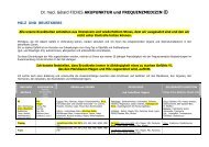
FM 15<br />
SPECT-CT bei unspezifis<strong>ch</strong>en Hand- und<br />
Handgelenksbes<strong>ch</strong>werden: eine diagnostis<strong>ch</strong><br />
sinnvolle Ergänzung ?<br />
Maja S<strong>ch</strong>ür<strong>ch</strong> 1 , Florian S<strong>ch</strong>lei<strong>ch</strong> 1 , Martin Hüllner 1 ,<br />
Patrick Veit-Haiba<strong>ch</strong> 1 , Urs Hug 1 , Urs von Wartburg 1 ,<br />
Klaus Strobel 1 ( 1 Luzern)<br />
Einleitung<br />
Das SPECT (Single Photon Emission Computed<br />
Tomography)-CT kombiniert eine biologis<strong>ch</strong> funktionelle<br />
Bildgebung (SPECT) mit einer morphologis<strong>ch</strong>en<br />
Darstellung (CT) im Sinne einer 3-D-Hybridmethode.<br />
Dadur<strong>ch</strong> ist eine genaue Lokalisation von aktiven<br />
Degenerationsvorgängen mögli<strong>ch</strong> was wiederum einen<br />
Einfluss auf die Wahl und Qualität der Therapie hat.<br />
Zweck<br />
Hypothese: die zusätzli<strong>ch</strong>e Untersu<strong>ch</strong>ung mittels<br />
SPECT-CT bei unspezifis<strong>ch</strong>en Handgelenks- und<br />
Handbes<strong>ch</strong>werden führt in einer bedeutenden Anzahl<br />
der Fälle zu einer Aenderung der Diagnose und damit<br />
der Therapie.<br />
Methode<br />
In einer retrospektiven Studie haben wir 51 Patienten<br />
(31 Frauen, 20 Männer, Dur<strong>ch</strong>s<strong>ch</strong>nittsalter 38 Jahre,<br />
range 17-73 Jahre) , wel<strong>ch</strong>e bei unspezifis<strong>ch</strong>en<br />
posttraumatis<strong>ch</strong>en oder <strong>ch</strong>ronis<strong>ch</strong>en<br />
Handgelenksbes<strong>ch</strong>werden ohne Trauma na<strong>ch</strong><br />
konventionellem Rx und oder MRI/CT zusätzli<strong>ch</strong> mittels<br />
SPECT-CT untersu<strong>ch</strong>t wurden, evaluiert. Die Häufigkeit<br />
einer Diagnose- respektive Therapieänderung wurde<br />
analysiert.<br />
Resultate<br />
Bei 20 Patienten (39%) führte das SPECT-CT zu einer<br />
Therapie-Aenderung.Bei 7 Patienten (14%) konnte das<br />
SPECT-CT die vermutete Diagnose bestätigen. Bei 9<br />
Patienten (18%) konnte eine vermutete Pathologie<br />
ausges<strong>ch</strong>lossen werden.<br />
S<strong>ch</strong>lussfolgerung<br />
Das SPECT-CT bei unspezifis<strong>ch</strong>en Hand- und<br />
Handgelenksbes<strong>ch</strong>werden bringt in vielen Fällen<br />
zusätzli<strong>ch</strong>e Informationen, was die Genauigkeit der<br />
Diagnose erhöht. Dadur<strong>ch</strong> wird die Therapie wesentli<strong>ch</strong><br />
beeinflusst. Wir era<strong>ch</strong>ten daher bei unklaren<br />
Handgelenks- und Handbes<strong>ch</strong>werden eine ergänzende<br />
Untersu<strong>ch</strong>ung mittels SPECT-CT als sinnvoll.
FM 16<br />
Direct MR arthrography of the wrist with axial<br />
traction: A feasibility study to assess joint cartilage<br />
Fabio Becce 1 , Daniel Guntern 2 , Delphine Ri<strong>ch</strong>arme 1 ,<br />
Reto Meuli 1 , Nicolas Theumann 1 ( 1 Lausanne; 2 Fribourg)<br />
Purpose:<br />
To assess the impact of axial traction during acquisition<br />
of direct magnetic resonance (MR) arthrography of the<br />
wrist with regard to joint space width and amount of<br />
contrast material between the opposing cartilage<br />
surfaces.<br />
Materials and Methods:<br />
Fifteen consecutive patients (12 male, mean age 38.1<br />
years) were included in this Institutional Review Boardapproved<br />
prospective study. Three-compartment wrist<br />
MR arthrographies were performed between October<br />
and December 2009 on a 3 T unit using a fatsuppressed<br />
T1-weighted isotropic high-resolution<br />
volumetric interpolated breathhold examination (VIBE)<br />
sequence in the coronal plane, with and without axial<br />
traction (3 kg). Two radiologists measured radiocarpal<br />
(radioscaphoid, radiolunate) and midcarpal<br />
(lunocapitate, hamatolunate) joint space widths, with<br />
and without traction, and assessed the amount of<br />
contrast material between the opposing cartilage<br />
surfaces using a three-point scale: 0 = absence, 1 =<br />
partial, 2 = complete.<br />
Results:<br />
With traction, joint space width increased significantly at<br />
the radioscaphoid (� = 0.78 mm, P < 0.01), radiolunate<br />
(� = 0.18 mm, P < 0.01), and lunocapitate (� = 0.45<br />
mm, P < 0.01) spaces, and both observers detected<br />
significantly more contrast material between the<br />
cartilage surfaces. At the hamatolunate space, the<br />
differences in joint space width (� = 0.14 mm, P = 0.54)<br />
and amount of contrast material were not significant.<br />
Conclusion:<br />
Direct wrist MR arthrography with axial traction of 3 kg<br />
increases joint space width at the radiocarpal and<br />
lunocapitate spaces, and prompts better coverage of<br />
the articular cartilage by the contrast material.<br />
FM 17<br />
Prospektive Multicenterstudie zur<br />
Handgelenksarthroskopie bei Discusverletzung<br />
Nicole S<strong>ch</strong>melzer-S<strong>ch</strong>mied 1 , Wolfgang Daecke 2 , Martin<br />
Jung 3 ( 1 Münsterlingen; 2 Frankfurt; 3 Heidelberg)<br />
Einleitung: Im Rahmen einer prospektiven<br />
multizentris<strong>ch</strong>en Studie der DGH, konnte ein großes<br />
Patientenkollektiv (n= 136) zur Ermittlung des<br />
klinis<strong>ch</strong>en Verlaufs na<strong>ch</strong> arthroskopis<strong>ch</strong>er Operation<br />
einer Diskusläsion am Handgelenk erfasst werden. Das<br />
Ziel dieser Studie war, den Verlauf na<strong>ch</strong><br />
Handgelenksarthroskopie zu dokumentieren, und das<br />
klinis<strong>ch</strong>e Ergebnis hinsi<strong>ch</strong>tli<strong>ch</strong> vers<strong>ch</strong>iedener<br />
Einflußgrößen zu untersu<strong>ch</strong>en<br />
Material/Methode: Einges<strong>ch</strong>lossen wurden Patienten,<br />
die klinis<strong>ch</strong> und radiologis<strong>ch</strong> den Verda<strong>ch</strong>t auf eine<br />
Discusverletztung hatten. Der Zeitraum der<br />
Untersu<strong>ch</strong>ungsphase pro Patient wurde auf 1 Jahr<br />
begrenzt. Die Untersu<strong>ch</strong>ung fand präoperativ, 3 und 12<br />
10<br />
Monate na<strong>ch</strong> der Operation statt und beinhaltete<br />
funktionelle Parameter, Kraft, S<strong>ch</strong>merzintensität sowie<br />
den DASH-Fragebogen.<br />
Ergebnisse: Wir präsentieren die Ergebnisse von 136<br />
Patienten, die aufgrund einer Discusverletzung<br />
arthroskopis<strong>ch</strong> operiert wurden.<br />
Folgerung: Insgesamt zeigen si<strong>ch</strong> beeindruckend gute<br />
Ergebnisse 1 Jahr na<strong>ch</strong> arthroskopis<strong>ch</strong>er Operation.<br />
Da es si<strong>ch</strong> bei dieser Studie um eine ni<strong>ch</strong>t kontrollierte<br />
Studie handelt, ist der Einfluss der Selbstheilung jedo<strong>ch</strong><br />
ni<strong>ch</strong>t klar trennbar. Weitere kontrollierte Studien sollten<br />
folgen.<br />
FM 18<br />
High failure rate treating CMC 1 osteoarthritis with<br />
PI2® pyrocarbon prosthesis<br />
Jan Van Aaken 1 , Nicolas Holzer 1 , Laurent Wehrli 2 ,<br />
François Delaquaize 1 , Jean-Yves Beaulieu 1 ( 1 Genève;<br />
2 Lausanne)<br />
Purpose One te<strong>ch</strong>nique for CMC 1 osteoarthritis is<br />
trapezium resection and interposition with a pyrocarbon<br />
prosthesis (PI 2 ®). This maintains the length of the first<br />
ray and is designed to generate powerful pin<strong>ch</strong>. The<br />
objective of this study was to evaluate the results<br />
following this surgical procedure.<br />
Patients and methods 41 patients (45 thumbs) who<br />
underwent this procedure between Mar<strong>ch</strong> 2004 and<br />
June 2009, with minimum 1 year follow-up, were<br />
included for review. 19 patients did not return for a<br />
variety of reasons, including 7 who refused because the<br />
prosthesis had been removed. This left 22 patients (26<br />
prostheses) with a median age of 60 years (45-80<br />
years) available for clinical and radiological review at a<br />
median follow up of 30 months (12- 78 months).<br />
Outcome included patient’s satisfaction and<br />
QuickDASH.<br />
Results At final review there were 5 patients (5<br />
prostheses) who had been re-operated upon with<br />
removal of the prosthesis at a median of 11 months<br />
postoperative (1 week- 25 months), and 3 of these<br />
patients were not satisfied with their result. 3 additional<br />
patients with retained prostheses were also dissatisfied.<br />
The remaining 17 patients were all very satisfied with<br />
the outcome. Their median QuickDASH was 11 (0-52).<br />
In comparison, the median QuickDASH for patients who<br />
were not satisfied was 45 (27-55).<br />
The clinical examination focused on those 17/22<br />
patients (21/26 prostheses) who still had their<br />
prosthesis. Global force measured on the second<br />
position with a Jamar dynamometer was in median<br />
22kg (5-56kg), as compared to a median of 22kg (4-<br />
44kg) before the operation. Lateral pin<strong>ch</strong> for all patients<br />
was in median 5.5kg (2-12 kg), as compared to a<br />
median of 4.5kg (0-10 kg) preoperatively. Opposition<br />
according to Kapandji was in median 10 (9-10),<br />
compared to a median of 9 (7-10) before the operation.<br />
In 12 of the retained 21 prostheses there was a dorsal<br />
subluxation or dislocation of the first ray together with<br />
the prosthesis, whi<strong>ch</strong> did not correlate with the surgical<br />
approa<strong>ch</strong>.<br />
Conclusion Three-quarters (74%) of patients were very<br />
satisfied with the operation. Mean QuickDASH was 11,<br />
pin<strong>ch</strong> force increased 22%, opposition of the thumb<br />
improved. Global force did not <strong>ch</strong>ange. However, one-
quarter of patients were not satisfied with their result.<br />
5/27 prostheses (19%) seen at follow-up had been<br />
removed, and an additional 7 prostheses in the 18<br />
patients that did not participate in the final review had<br />
been removed, for a total failure of 12/45 implants<br />
(27%).<br />
FM 19<br />
PIP - Arthroplastik mit einer neuen modularen<br />
Oberflä<strong>ch</strong>enprothese - Erste Ergebnisse<br />
Stephan S<strong>ch</strong>indele 1 , Daniel Herren 1 ( 1 Züri<strong>ch</strong>)<br />
Einleitung: Der primäre Gelenksersatz am proximalen<br />
Interphalangealgelenk (PIP) mit einem Silikonspacer<br />
stellt bei posttraumatis<strong>ch</strong>en oder degenerativen<br />
Arthrosen heute na<strong>ch</strong> wie vor den Goldstandart dar.<br />
Na<strong>ch</strong>teile sind eine mögli<strong>ch</strong>e laterale Instabilität,<br />
insbesondere an den radialen Fingerstrahlen bei<br />
manuell tätigen Patienten, und eine einges<strong>ch</strong>ränkte<br />
Bewegli<strong>ch</strong>keit. Bisherige Oberflä<strong>ch</strong>engleitprothesen<br />
haben si<strong>ch</strong> aufgrund einer problematis<strong>ch</strong>en<br />
Kno<strong>ch</strong>enverankerung und teils hohen Revisionsraten<br />
ni<strong>ch</strong>t dur<strong>ch</strong>setzen können.<br />
Ziel: Vorstellung der ersten klinis<strong>ch</strong>en und<br />
radiologis<strong>ch</strong>en Ergebnisse einer neuen modularen<br />
Oberflä<strong>ch</strong>engleitprothese am PIP-Gelenk (CapFlex-<br />
PIP © ) mit der Gleitpaarung Polyethylen-Metall und<br />
zementfreier Pressfit-Fixation. Der neue Prothesentyp<br />
benötigt nur eine sehr geringe ossäre Resektion und<br />
respektiert so den Bandapparat am PIP Gelenk.<br />
Zusammen mit der relativ hohen Formstabilität, sollte<br />
diese Kunstgelenklösung eine verbesserte laterale<br />
Stabilität ausweisen.<br />
Material und Methode: In einer prospektiven<br />
Pilotstudie werden die ersten Ergebnisse bei 5<br />
Patienten na<strong>ch</strong> Implantation einer CapFlex-Prothese an<br />
einem PIP-Gelenk über dorsalen Zugang mit einem<br />
minimalen follwo-up von 6 Monaten vorgestellt. Bei 5<br />
Patienten (4 Männer, 1 Frau) mit primärer<br />
Bou<strong>ch</strong>ardarthrose wurde 2x Zeigefinger und jeweils<br />
einmal Dig 3-5 operativ versorgt. Alle Patienten wurden<br />
na<strong>ch</strong> engen Ein- und Auss<strong>ch</strong>lusskriterien ausgewählt.<br />
Resultate: Der postoperative Verlauf war bei allen 5<br />
Patienten komplikationslos. Die S<strong>ch</strong>merzen (VAS-Skala<br />
1-10) konnten von präoperativ 7.8 auf 2.6 postoperativ<br />
reduziert werden. Der aktive Bewegungsumfang (ROM)<br />
betrug präoperativ 30° Grad (± 12.7°) und postop. 5 5°<br />
Grad (± 18.7°). Es zeigten si<strong>ch</strong> klinis<strong>ch</strong> keine<br />
Instabilitäten. Alle Patienten waren bis anhin sehr<br />
zufrieden. Radiologis<strong>ch</strong> konnte bei allen Patienten eine<br />
vollständige Osteointegration der Komponenten<br />
dokumentiert werden. Der Patient mit dem geringsten<br />
Bewegungsumfang zeigte postoperativ ausgeprägte<br />
Ossifikationen dorsal im Berei<strong>ch</strong> des Streckapparats.<br />
Zusammenfassung: Die ersten Ergebnisse dieser<br />
Pilotstudie mit einer neuen modularen<br />
Oberflä<strong>ch</strong>engleitprothese (Gleitpaarung PE-Metall)<br />
zeigen erfreuli<strong>ch</strong>e Ergebnisse bezügli<strong>ch</strong> der Funktion<br />
und Patientenzufriedenheit. Alle Komponenten zeigen<br />
radiologis<strong>ch</strong> in den ersten 6 Monaten keinerlei<br />
Lockerungszei<strong>ch</strong>en.<br />
11<br />
FM 20<br />
Wie die Sonographie unser Verständnis der<br />
Arthrose verändert<br />
Berndt Forster 1 , Ishilde Forster 2 ( 1 Winterthur; 2 Züri<strong>ch</strong>)<br />
Sonographis<strong>ch</strong> können wir arthrotis<strong>ch</strong>e Veränderungen<br />
an einem Gelenk der Hand erkennen, wel<strong>ch</strong>e uns die<br />
bisherigen bilddiagnostis<strong>ch</strong>en Verfahren ni<strong>ch</strong>t zeigen.<br />
Dur<strong>ch</strong> die Sonographie ist es mögli<strong>ch</strong>, degenerative<br />
Veränderungen von Gelenken na<strong>ch</strong>zuweisen, lange<br />
bevor das Röntgenbild oder das MRI sol<strong>ch</strong>e erkennen<br />
lässt.<br />
Kleinste Geoden und Exostosen sind sonographis<strong>ch</strong><br />
wesentli<strong>ch</strong> früher als in den anderen<br />
bilddiagnostis<strong>ch</strong>enVerfahren zu sehen.<br />
Der Abbau des Gelenksknorpels lässt si<strong>ch</strong> bis auf<br />
Bru<strong>ch</strong>teile eines Millimeters messen.<br />
Die Menge der synovialen Flüssigkeit und der Zustand<br />
der synovialen Membran eines Gelenkes sind weitere<br />
Parameter bei der sonographis<strong>ch</strong>en Beurteilungen<br />
eines arthrotis<strong>ch</strong>en Gelenkes.<br />
Dopplersonographis<strong>ch</strong> kann mit dem Erfassen der<br />
Gefässaktivität in der synovialen Membran eines<br />
Gelenkes der aktuelle Zustand der Arthrose erfasst<br />
werden. Dieser Befund ist essentiel für die Abgrenzung<br />
gegenüber den entzündli<strong>ch</strong>en Gelenkserkrankungen.<br />
FM 21<br />
A novel te<strong>ch</strong>nique detecting instability of the distal<br />
radioulnar joint in complete triangulo-fibrocartilage<br />
lesions<br />
Florian Hess 1 , Mazda Farshad 1 , Reto Sutter 1 , Ladislav<br />
Nagy 1 , Andreas S<strong>ch</strong>weizer 1 ( 1 Züri<strong>ch</strong>)<br />
Background: Discrepancy of bilateral dorsopalmar shift<br />
of the distal radioulnar joint (DRUJ) is a useful clinical<br />
hint for the diagnosis of DRUJ instability. Although<br />
several cadaver studies quantified the amount of the<br />
physiological dorsopalmar shift, up to here, there is no<br />
reliable method of quantification of DRUJ mobility in<br />
daily practice. The aim was to find a simple reliable<br />
sonographic te<strong>ch</strong>nique to quantify the physiological<br />
dorsopalmar shift of the DRUJ.<br />
Materials: Ulnar head translation between pressing the<br />
volar surface of the hand on a brick and unloaded lifting<br />
up the hand in a defined position was quantified by the<br />
means of ultrasonography with a cut in a transversal<br />
plane through the ulnar haed and Lister's tubecle . A<br />
quotient was built for reliable representation of the laxity<br />
of the DRUJ. Sonographic quantification was performed<br />
in both wrists in 40 healthy volunteers and 17 patients<br />
with subsequent intraoperative confirmation of complete<br />
rupture of the triangular fibrocartilage complex (TFCC).<br />
Results: The amount of dorsopalmar shift (quotient Q)<br />
of the 17 patients with unilateral TFCC lesion was<br />
significantly different between the pathological side and<br />
the contralateral wrist (1.19 vs. 0.54). Q>0.8 was found<br />
to be a reliable cut-off for pathological laxity of the<br />
DRUJ. There was no significant difference in the 40<br />
healthy volunteers between the right and left side or<br />
dominant and adominant wrist.<br />
Conclusions: This method is a reliable tool for the<br />
determination of an abnormal laxity of the DRUJ. Its
validity in detecting DRUJ-instability after traumatic or<br />
degenerative lesions is proven in this study. A cut-off of<br />
Q>0.8 allows a reliable discrimination between stable<br />
and unstable DRUJ.<br />
FM 22<br />
Prospective assessment of deep flexor tendon<br />
healing in Zone II by high-frequency ultrasound<br />
Nicole Lindenblatt 1 , Gilbert Puippe 1 , Ralph Gnannt 1 ,<br />
Daniela Meyer 1 , Gustav Andreisek 1 , Pietro Giovanoli 1 ,<br />
Maurizio Calcagni 1 ( 1 Züri<strong>ch</strong>)<br />
Purpose: Despite the establishment of early motion<br />
protocols flexor tendon repairs in Zone II remain a<br />
demanding task. However, evaluation of the healing<br />
process today still is limited to clinical inspection.<br />
Therefore it was the aim of this study to prospectively<br />
analyse postoperative morphological and functional<br />
<strong>ch</strong>anges during flexor tendon healing by ultrasound.<br />
Methods: Tendon repair was a<strong>ch</strong>ieved by a four-strand<br />
core suture and a epitendinous running suture.<br />
Postoperative a modified Kleinert protocoll (place-andhold)<br />
was applied. Tendon morphology was evaluated<br />
with high frequency ultrasound after 3, 7, 14, 21, 35, 70<br />
and 84 days. Tendon excursion over the PIP joint was<br />
assessed by a new non-invasive thread-and-scar<br />
tracking method. Data are given as means±SEM.<br />
Results: Ten patients with a mean age of 34 years and<br />
eleven tendons were studied. Morphologically all<br />
tendons exhibited a spindle-like appearance of the<br />
suture zone after 1 week, of whi<strong>ch</strong> 50% developed a<br />
normal band-like appearance after 12 weeks.<br />
Interestingly, a persisting spindle-like structure after 12<br />
weeks corresponded with a significantly increased FDP<br />
excursion at the PIP joint (2.86±0.61 mm vs. 1.14±0.27<br />
mm; (p< 0.05; CI 95% [0.20-3.25]). Tendons with<br />
increased power doppler perfusion exhibited a<br />
significantly better FDP excursion after 12 weeks<br />
(3.13±0.70 mm vs. 1.25±0.60 mm; p< 0.05 CI 95% [-<br />
3.33;-0.41]. Excursion of the flexor digitorum profundus<br />
tendon at PIP level was 1.48±0.3 mm at day 3 and<br />
increased steadily to 2.28±0.75 mm after 12 weeks<br />
(p=0.2; CI 95% [ -0.55; 3.25]). A persisting spindle-like<br />
shape after 12 weeks corresponded with a significantly<br />
increased FDP excursion and a better active motion of<br />
the PIP and DIP joints. Tendons with increased power<br />
Doppler signal intensity exhibited a significantly better<br />
FDP excursion after 12 weeks.<br />
Conclusions: The results of the present study indicate<br />
a reliable assessment of morphology and dynamics of<br />
flexor tendon healing by ultrasound. Correlation of<br />
ultrasonic morphology and flexor tendon excursion<br />
suggest a better tendon movement, if the tendon<br />
maintains a spindle-like shape and shows<br />
hyperperfusion at 12 weeks. This might indicate a<br />
predominantly intrinsic healing pattern with reduced<br />
adhesion formation in these patients Additionally, this<br />
new method may be a tool to rate the process of tendon<br />
healing in order to establish individually modified<br />
rehabilitation protocols.<br />
12<br />
FM 23<br />
Natural progression of CTS evaluated by<br />
sonography and electrophysiology<br />
Nicole Badur 1 , Christoph Klimsa 1 , Sebastian Kluge 2 , Kai<br />
Roesler 1 , Esther Voegelin 1 ( 1 Bern; 2 Züri<strong>ch</strong>)<br />
Purpose: We compared the diagnostic accuracy of<br />
electrodiagnostic testing (ENMG) with high-resolution<br />
ultrasound in carpal tunnel syndrome (CTS) and<br />
evaluated its natural progression.<br />
Methods: 64 patients (79 involved hands) with clinically<br />
suspected CTS were prospectively seen between<br />
2008/09. All patients were subject to a standard<br />
electrophysiologic evaluation by a neurologist and to a<br />
high-resolution sonography by a hand surgeon. Both<br />
examiners were blinded to the respective other results.<br />
Standard sensory nerve conduction and distal motor<br />
latencies were recorded by ENMG. Hands were<br />
classified into 6 grades of severity as proposed by<br />
Padua et al (1997). The cross-sectional area of the<br />
median nerve at the entrance of the carpal tunnel as<br />
well as 2 cm proximal was assessed and the ratio<br />
between the values was calculated. A receiver<br />
operating <strong>ch</strong>aracteristic (ROC) curve was used to<br />
document the sensitivity and specificity of the<br />
ultrasound with ENMG. Patients with false positive and<br />
negative CTS by ultrasound were followed up.<br />
Results: A cut-off value of 1.12 as distal/proximal ratio<br />
demonstrated a sensitivity of 78,6% and a specificity of<br />
71,4% for the diagnosis of CTS by sonography.<br />
According to the severity of CTS classified by ENMG<br />
there were 20 hands with negative, 4 minimal, 9 mild,<br />
38 moderate, 6 severe and 2 extreme CTS. There was<br />
no correlation between the severity and the crosssectional<br />
ratio. 18 of the 79 hands with<br />
electrodiagnostic positive CTS had a normal ratio of the<br />
cross sectional area of the median nerve. 8 of these 18<br />
hands underwent surgery and are symptom free, 2 are<br />
awaiting surgery. In 2 hands symptoms resolved<br />
spontaneously without treatment. One patient has<br />
persistent symptoms and refuses operative treatment. 5<br />
patients could not be contacted for follow up. 5 hands<br />
without evidence of CTS by ENMG had a ratio ≥ 1.12 in<br />
the sonography. Two patients underwent surgery and<br />
are symptom free. A third patient showed spontaneous<br />
relief of symptoms. Two patients could not be contacted<br />
for follow up.<br />
Conclusion: In patients with clinical CTS and a ratio ≥<br />
1.12 as evaluated by ultrasound no further ENMG<br />
testing is required. At a ratio ≤1.12 the ENMG is useful<br />
to ensure the diagnosis of CTS in patients with unclear<br />
clinical symptoms. Further evaluation of the natural<br />
progression of CTS by ultrasound might be useful since<br />
the sonography is straightforward and pain free in<br />
diagnosing CTS if performed by an experienced<br />
examiner.
FM 24<br />
Ultras<strong>ch</strong>allanatomie der palmaren Platte am MCP-<br />
und PIP- Gelenk der Langfinger<br />
Nadja Müller 1 , Tatjana Lanaras 1 , Maurizio Calcagni 1 ,<br />
Pietro Giovanoli 1 ( 1 Züri<strong>ch</strong>)<br />
Einleitung<br />
Verletzungen der palmaren Platte an den MCP- und<br />
PIP-Gelenken der Langfinger sind eine der häufigsten<br />
Verletzungen der Fingergelenke. Die aktuelle<br />
Standarddiagnostik verlässt si<strong>ch</strong> derzeit no<strong>ch</strong> auf eine<br />
Röntgenuntersu<strong>ch</strong>ung sowie die klinis<strong>ch</strong>e<br />
Untersu<strong>ch</strong>ung. Die Ultras<strong>ch</strong>alluntersu<strong>ch</strong>ung ist ein<br />
gutes Diagnose-Tool um das Ausmass der<br />
Wei<strong>ch</strong>teilverletzung und allenfalls ossärer<br />
Begleitverletzungen an den Langfingern darzustellen.<br />
Mit der exakten Bes<strong>ch</strong>reibung der normalen<br />
Ultras<strong>ch</strong>allanatomie kann eine korrekte und genaue<br />
Bes<strong>ch</strong>reibung von pathologis<strong>ch</strong>en Befunden erfolgen.<br />
Zielsetzungen der Studie<br />
Einerseits die Untersu<strong>ch</strong>ung der Anatomie mittels<br />
Ultras<strong>ch</strong>alldiagnostik und genaue Analyse der<br />
palmaren Platte am MCP sowie am PIP der Langfinger<br />
und na<strong>ch</strong>folgend der Verglei<strong>ch</strong> der sonographis<strong>ch</strong>en<br />
Befunde mit den anatomis<strong>ch</strong>en Befunden am Cadaver.<br />
Zudem sollte anhand der Daten die Erarbeitung eines<br />
Standardprotokolls zur sonographis<strong>ch</strong>en Untersu<strong>ch</strong>ung<br />
der palmaren Platte der MCP und PIP Gelenke erfolgen<br />
zur s<strong>ch</strong>nellen und exakten Diagnose einer Pathologie.<br />
Methoden<br />
Zur Ultras<strong>ch</strong>alldiagnostik wurde ein 14 MHZ Gerät der<br />
Firma Philipps verwendet.<br />
Die Untersu<strong>ch</strong>ungen wurden an 8 Cadaver Händen,<br />
wel<strong>ch</strong>e in Thiel Lösung fixiert waren dur<strong>ch</strong>geführt. Um<br />
eine höhergradige traumatis<strong>ch</strong>e Vorges<strong>ch</strong>i<strong>ch</strong>te<br />
auszus<strong>ch</strong>liessen wurden alle Hände einer<br />
Röntgenuntersu<strong>ch</strong>ung unterzogen. Na<strong>ch</strong> der<br />
Ultras<strong>ch</strong>alluntersu<strong>ch</strong>ung erfolgte die anatomis<strong>ch</strong>e<br />
Dissektion mit exaktem Ausmessen der palmaren<br />
Platten, die Ergebnisse wurden na<strong>ch</strong>folgend mit den<br />
Ultras<strong>ch</strong>allmessungen vergli<strong>ch</strong>en.<br />
In der Ultras<strong>ch</strong>alluntersu<strong>ch</strong>ung wurden vers<strong>ch</strong>iedene<br />
Transducer-Applikationen studiert: die beiden Zügel der<br />
palmaren Platte am PIP Gelenk sowie der Mittelanteil<br />
wurden in der Longitudinalebene dargestellt, zudem<br />
wurden mehrere Transversals<strong>ch</strong>nitte dargestellt um<br />
eine exakte Quers<strong>ch</strong>nittebene darzustellen und<br />
na<strong>ch</strong>folgend eine genaue Analyse und den Verglei<strong>ch</strong> zu<br />
den Resultaten der Dissektion herstellen zu können.<br />
Ergebnisse und S<strong>ch</strong>lussfolgerung<br />
Die palmaren Platten am MCP und PIP Gelenk der<br />
Langfinger können sehr gut mittels<br />
Ultras<strong>ch</strong>alluntersu<strong>ch</strong>ung dargestellt werden. Der<br />
Verglei<strong>ch</strong> der Ausmessung mittels Ultras<strong>ch</strong>all und<br />
anatomis<strong>ch</strong>er Ausmessung ergab keine grösseren<br />
Differenzen, die genauen Resultate werden am<br />
Kongress vorgestellt. Aufgrund der Studie ist es uns<br />
gelungen ein Protokoll zu erstellen, wel<strong>ch</strong>es si<strong>ch</strong>er und<br />
in wenigen S<strong>ch</strong>ritte die palmare Platte darstellt und die<br />
Diagnose und den Verlauf von Verletzungen ermögli<strong>ch</strong>t.<br />
13<br />
FM 25<br />
Dupuytren’s contracture: postoperative evaluation<br />
by the patients<br />
Mi<strong>ch</strong>aël Papaloïzos 1 , Cindy Bouvet 1 , Philippe Cuénod 1 ,<br />
Thierry Glauser 1 ( 1 Genève)<br />
Introduction: The aim of the present study was to<br />
generally evaluate the patient’s levels of satisfaction<br />
after surgical treatment for Dupuytren’s contracture,<br />
with respect to the stage of the disease and the type of<br />
operation used.<br />
Patients and methods : All patients operated by partial<br />
or total fasciectomy for Dupuytren’s disease from 2006<br />
to 2010 were included. Fasciotomies were excluded. A<br />
custom-made satisfaction questionnaire was sent to the<br />
207 patients (213 cases) included, investigating the<br />
following points: pain (postoperatively and at actual<br />
time), wound healing and scarring, complications, reoperations<br />
(in case it has been done elsewhere),<br />
trouble with sensitivity, trouble with activities of daily<br />
living, overall satisfaction, willingness to redo the same<br />
operation. Answers were binary (yes/no) or scaled from<br />
0 to 10. Tubiana’s stages were used for grading and<br />
patients operated for recurrencies recorded. Operation<br />
types were 1) open, 2) closed with plasties, 3) closed<br />
with skin grafts. Mean follow-up was 27 months and 3%<br />
of cases were lost to FU.<br />
Results: 34% of cases had some degree of pain for a<br />
mean duration of 3 months (min. 3 days, max. 12<br />
months). 20% still have pain at FU, grading from 1 to 9<br />
(mean 2.8). 96% report uncomplicated wound healing ;<br />
the 4% left complain mainly of unsightly scars. 4%<br />
report some degree of bothering due to the<br />
postoperative dressings. 18% reported complications,<br />
essentially recurrencies and restricted mobility. 14%<br />
had to be reoperated, mainly for recurrencies.<br />
Sensitivity troubles were reported in 15% of cases, and<br />
sensitivity to cold in 20%. At FU, ADL were affected in<br />
28% of cases at an average level of 3.5/10 (min. 1,<br />
max. 9). Overall, 7% were totally unsatisfied, 10%<br />
moderately satisfied, 20% satisfied and 63% totally<br />
satisfied. 85% would redo the operation. Complications<br />
and low satisfaction levels were correlated with<br />
advanced stages of the disease.<br />
Conclusions: From the patient’s point of view, surgery<br />
for Dupuytren’s contracture is not an ordinary procedure<br />
unfraught with problems and dissatisfaction. Residual<br />
pain, recurrencies and functional limitations are mostly<br />
encountered in high grades of the disease. In su<strong>ch</strong><br />
cases, caution should be exerted by the surgeon and a<br />
thorough preoperative <strong>info</strong>rmation should be given to<br />
the patient.
FM 26<br />
Dupuytren und Sonographie<br />
Berndt Forster 1 , Ishilde Forster 2 ( 1 Winterthur; 2 Züri<strong>ch</strong>)<br />
Ni<strong>ch</strong>t nur die fortges<strong>ch</strong>rittene Dupuytren-Erkrankung ist<br />
für den Patienten ein Problem.<br />
Au<strong>ch</strong> kleinste Knoten in der Hohlhand können bei<br />
gewissen handwerkli<strong>ch</strong>en Tätigkeiten zu erhebli<strong>ch</strong>en<br />
Bes<strong>ch</strong>werden führen. Sie imponieren klinis<strong>ch</strong> meist als<br />
Tendovaginitis von Beugesehnen.<br />
Diese Bes<strong>ch</strong>werden werden gelegentli<strong>ch</strong> dur<strong>ch</strong> einen<br />
kleinen Dupuytrenknoten verursa<strong>ch</strong>t, der am<br />
proximalen Ende des Ringbandes A1 entstanden ist.<br />
Die alleinige Ringbandspaltung führt in dieser Situation<br />
ni<strong>ch</strong>t zu Bes<strong>ch</strong>werdefreiheit.<br />
In einer präoperativen Sonographie sind sol<strong>ch</strong>e kleine<br />
Dupuytrenknoten lei<strong>ch</strong>t zu erkennen und der Eingriff<br />
kann entspre<strong>ch</strong>end ausgeweitet werden.<br />
Wir beri<strong>ch</strong>ten über unsere Erfahrung in 10 Fällen.<br />
FM 27<br />
Lambeau de glissement dans le traitement des<br />
atteintes digitales de la maladie de Dupuytren<br />
Philippe Cuénod (Genève)<br />
La couverture cutanée après excision des brides et<br />
correction de la rétraction digitale dans la maladie de<br />
Dupuytren est difficile. Diverses te<strong>ch</strong>niques de<br />
lambeaux et de greffes peuvent être utilisées.<br />
Le lambeau de glissement digital est déterminé par une<br />
incision médio-latérale se recourbant à angle droit dans<br />
le pli de flexion basal. Il est élevé sur toute la surface<br />
digitale sur son pédicule controlatéral, pour exciser les<br />
brides. Une fois le doigt étendu, le lambeau est glissé<br />
distalement et la perte de substance proximale<br />
consécutive est couverte par une greffe de peau totale.<br />
D’avril 2007 à février 2011, 13 doigts <strong>ch</strong>ez 12 patients<br />
(âge moyen : 62 ans) ont été traités par cette méthode.<br />
Le suivi moyen est de 25 semaines. Les doigts opérés<br />
ont été 10 fois le D5, 2 fois D3 et une fois D4. La<br />
rétraction totale préopératoire était de 58° en moy enne<br />
et celle de la PIP de 42°.<br />
Résultats : Cyanose transitoire du lambeau : 3 fois ;<br />
cicatrisation per primam de la greffe dans tous les cas;<br />
paresthésies transitoires avec résolution complète : 5<br />
cas. La rétraction résiduelle globale à été en moyenne<br />
de 16° et la rétraction de la PIP de 16°. Le gain moy en<br />
en degrés sur le flexum de la PIP a été de 25° et le<br />
pourcentage moyen d’amélioration de 60%. Nombre<br />
moyen de séances de rééducation : 10 séances. Dans<br />
3 cas : résultat sans rééducation. 10 patients sur 12<br />
étaient très satisfaits et deux moyennement.<br />
Conclusion : Ce type de lambeau permet un bon abord<br />
des brides pour leur excision et offre une couverture<br />
cutanée de qualité pour éviter une rétraction itérative<br />
par cicatrisation contractile.<br />
14<br />
Lamb D5 2<br />
FM 28<br />
Refraktur bei metaphysärer Pseudarthrose na<strong>ch</strong><br />
Doppelplattenosteosynthese einer C3 Fraktur des<br />
distalen Radius<br />
Philip Weber 1 , Urs Genewein 1 , Regula Steiger 1<br />
( 1 Liestal)<br />
Hintergrund<br />
Die Anlage einer Doppelplattenosteosynthese ist bei<br />
komplexen distalen Radiusfrakturen in einigen Fällen<br />
indiziert[1]. Sie ist jedo<strong>ch</strong> mit einem höheren Risiko<br />
einer Pseudarthrose assoziiert[2].<br />
Anhand der vorliegenden Kasuistik zeigen wir, wie na<strong>ch</strong><br />
einer dorsovolaren Plattenosteosynthese bei einer<br />
ni<strong>ch</strong>t erkannten delayed union eine Refraktur hätte<br />
verhindert werden können.<br />
Kasus<br />
Eine 60 jährige Patientin wird bei einer distalen,<br />
komplexen, mehrfragmentären, na<strong>ch</strong> palmar<br />
dislozierten intraartikulären Radiusfraktur (C3) mittels<br />
dorsovolarer Plattenosteosynthese (dorsale<br />
Rahmenplatte 2,5 mm und volare Platte 2,5 mm Aptus)<br />
behandelt. Zunä<strong>ch</strong>st zeitgere<strong>ch</strong>ter Heilungsverlauf: in<br />
der 4- und 8-Wo<strong>ch</strong>en Verlaufskontrolle unveränderte<br />
Stellungsverhältnisse, keine Aussagen zur<br />
Konsolidation bei Metallüberlagerung. Bei<br />
komplikationslosem klinis<strong>ch</strong>en Verlauf erfolgt die<br />
Vollbelastung na<strong>ch</strong> 8 Wo<strong>ch</strong>en mit Arbeitsaufnahme der<br />
selbständig erwerbenden Patientin. Anlässli<strong>ch</strong> der 4-<br />
Monatskontrolle inklusive konventionellem Röntgen fällt<br />
bei bes<strong>ch</strong>werdefreier Patientin der Ents<strong>ch</strong>eid zur<br />
vollständigen Metallentfernung, wel<strong>ch</strong>e 6 Monate na<strong>ch</strong><br />
Erstversorgung dur<strong>ch</strong>geführt wird. Perioperativ keine<br />
Hinweise auf eine Pseudarthrose.<br />
Im postoperativen Röntgen zeigt si<strong>ch</strong> nur ein<br />
einseitigen Dur<strong>ch</strong>bau der metaphysären Kortikalis,
was aber ni<strong>ch</strong>t speziell bea<strong>ch</strong>tet wurde. Anlässli<strong>ch</strong> der<br />
4-Wo<strong>ch</strong>en Kontrolle na<strong>ch</strong> Metallentfernung klagt die<br />
Patientin über eine S<strong>ch</strong>wellung und erneut S<strong>ch</strong>merzen<br />
im Handgelenksberei<strong>ch</strong>. Konventionell radiologis<strong>ch</strong><br />
besteht eine metaphysäre Refraktur. Im CT Na<strong>ch</strong>weis<br />
einer metaphysären Pseudarthrose.<br />
Diese wird mittels Beckenspaninterponat und palmarer<br />
Aptusplatte behandelt, der weitere Heilungsverlauf<br />
gestaltet si<strong>ch</strong> komplikationslos mit zeitgere<strong>ch</strong>ter<br />
Heilung. Die Patientin errei<strong>ch</strong>t annähernd einen<br />
seitenglei<strong>ch</strong>en Bewegungsumfang des Handgelenkes.<br />
Folgerung<br />
-Bei Zustand na<strong>ch</strong> Doppelplattenosteosynthese ist in<br />
den Röntgenkontrollen bei Metallüberlagerung des<br />
Radius der Na<strong>ch</strong>weis von Kallus/Konsolidation<br />
ers<strong>ch</strong>wert.<br />
- Eine weiterführende Diagnostik vor Plattenentferung<br />
ist notwendig (CT).<br />
- Im vorliegenden Fall wäre je na<strong>ch</strong> Klinik ein Zuwarten<br />
mit der Metallentfernung oder eine nur partielle<br />
Metallentfernung mit glei<strong>ch</strong>zeitiger Spongiosaplastik zu<br />
diskutieren.<br />
[1] Fernandez DL. Should anatomic reduction be<br />
pursued in distal radius fractures?, J Hand Surg 2000 ;<br />
25B : 523-7.<br />
[2] Sturzenegger M., L`ostésynthèse “sandwi<strong>ch</strong>” de<br />
l`extrémité inférieure du radius, Chirurgie de la Main<br />
2001 ; 20 : 447-3.<br />
FM 29<br />
Proximal row carpectomy in young patients<br />
Samia Guerid 1 , Christof Bollmann 1 , Wassim Raffoul 1<br />
( 1 Lausanne)<br />
ABSTRACT<br />
Purpose<br />
Proximal row carpectomy is a good motion-sparing<br />
procedure in case of painful arthrosis or important<br />
trauma of the wrist. The aim of this study is to evaluate<br />
the clinical results in patients under 44 years old at<br />
operation time at an average 32, 8-months follow-up.<br />
Method<br />
We reviewed retrospectively 13 patients operated in two<br />
institutions in Switzerland for PRC at the age of 43 or<br />
less. The patients underwent measures of their range of<br />
motion in flexion and extention and radial and ulnar<br />
deviation, grip strength and were asked about postoperative<br />
satisfaction. A Disabilites of the Arm,<br />
Shoulder and Hand score was also calculated.<br />
Results<br />
Thirteen patients were eligible for follow-up. Among<br />
them were 12 men and one woman, age ranging from<br />
23 to 43 years at operation time. Satisfaction was<br />
obtained by 69% of patients. Range of motion for wrist<br />
flexion and extention averaged 50, 5% (range 26 to 84)<br />
of the other side and radial and ulnar deviation 40,5%<br />
(range 9 to 55). Grip strength was 58.2% of healthy side<br />
(range 14 to 85) and the mean DASH score was 40.7<br />
(12.5 to 75). One patient was reoperated for total wrist<br />
denervation after 39 months, one for radial styloid<br />
resection after 4 months and one for wrist arthroplasty<br />
after 40 months, all of them because of strong residual<br />
pain.<br />
15<br />
Conclusion<br />
Proximal row carpectomy is an easy salvage procedure<br />
in young people, allowing good postoperative motion<br />
and relieving pain. Dissatisfaction seems to be related<br />
to lack of force in the postoperative period.<br />
FM 30<br />
Impingementsyndrom des Os pisiforme na<strong>ch</strong><br />
Proximal Row Carpectomy<br />
Sebastian Kluge 1 , Stephan S<strong>ch</strong>indele 1 , Daniel Herren 1<br />
( 1 Züri<strong>ch</strong>)<br />
Einleitung: Die Entfernung der proximalen<br />
Handwurzelreihe (PRC) stellt sowohl in Fällen einer<br />
Handgelenksarthrose, bei der das Midkarpalgelenk<br />
ausgespart ist, als au<strong>ch</strong> bei fortges<strong>ch</strong>rittenen<br />
aseptis<strong>ch</strong>en Kno<strong>ch</strong>ennekrosen wie der Lunatummalazie<br />
eine bewährte Operationsmethode dar, deren<br />
funktionelle Ergebnisse mit denen der mediokarpalen<br />
Teilarthrodese verglei<strong>ch</strong>bar sind. Im Gegensatz zur<br />
mediokarpalen Arthrodese tritt bei der PRC eine<br />
Proximalisierung der distalen Handwurzelreihe auf, die<br />
sekundäre Probleme, zum Beispiel ein radiokarpales<br />
Impingement, na<strong>ch</strong> si<strong>ch</strong> ziehen kann. Ein<br />
Impingementsyndrom dur<strong>ch</strong> das verbleibende Os<br />
pisiforme wurde bisher ni<strong>ch</strong>t bes<strong>ch</strong>rieben.<br />
Material und Methode: In den postoperativen<br />
Na<strong>ch</strong>kontrollen unserer Klinik fielen bei zwei mittels<br />
PRC versorgten Patienten bewegungsabhängige<br />
ulnokarpale S<strong>ch</strong>merzen auf. Die klinis<strong>ch</strong>e<br />
Untersu<strong>ch</strong>ung blieb sowohl bei der Flexion und<br />
Extension als au<strong>ch</strong> der Radialduktion unauffällig, zeigte<br />
jedo<strong>ch</strong> winkelabhängig in der na<strong>ch</strong> palmar geri<strong>ch</strong>teten<br />
Ulnarduktion ste<strong>ch</strong>ende S<strong>ch</strong>merzen. In dieser Position<br />
lag au<strong>ch</strong> eine Druckdolenz über dem Os pisiforme vor.<br />
Ergebnisse: Bereits die Nativröntgenbilder beider<br />
Patienten legten den Verda<strong>ch</strong>t auf eine<br />
Impingementsymptomatik des Os pisiforme nahe, die<br />
definitive Diagnosestellung erfolgte aber klinis<strong>ch</strong> und in<br />
der S<strong>ch</strong>nittbilddiagnostik. Bei beiden Patienten wurde<br />
eine CT-Untersu<strong>ch</strong>ung veranlasst, in einem der beiden<br />
Fälle au<strong>ch</strong> eine Kernspintomographie, die jedo<strong>ch</strong>,<br />
vermutli<strong>ch</strong> dur<strong>ch</strong> das nur positionsabhängige<br />
intermittierende Impingement, nur geringe<br />
Veränderungen mit allenfalls minimalem<br />
Kno<strong>ch</strong>enmarksödem zeigte. In der CT-Diagnostik<br />
konnten hingegen Osteolysen si<strong>ch</strong>tbar gema<strong>ch</strong>t<br />
werden, die die Verda<strong>ch</strong>tsdiagnose erhärteten. Sie<br />
waren in beiden Fällen dur<strong>ch</strong> ein Impingement am<br />
Processus styloideus ulnae, in einem Fall zusätzli<strong>ch</strong> am<br />
Os hamatum verursa<strong>ch</strong>t.<br />
S<strong>ch</strong>lussfolgerung: Eine symptomatis<strong>ch</strong>es<br />
Impingement des Os pisiforme na<strong>ch</strong> PRC ist eine<br />
seltene Entität, über die bisher keine publizierten Daten<br />
existieren. Denno<strong>ch</strong> sollte bei Patienten, die na<strong>ch</strong> einer<br />
Entfernung der proximalen Handwurzelreihe<br />
ulnokarpale Bes<strong>ch</strong>werden entwickeln, an ein<br />
Impingementsyndrom des Os pisiforme geda<strong>ch</strong>t<br />
werden. In diesen Fällen empfehlen wir eine Resektion<br />
des Os pisiforme, dessen sekundär stabilisierender<br />
Einfluss auf die proximale Handwurzelreihe na<strong>ch</strong> PRC<br />
ohnehin wegfällt.
FM 31<br />
Endoskopis<strong>ch</strong> assistierte Neurolyse einer<br />
spontanen Konstriktion des N. radialis<br />
Christoph Klimsa 1 , Dietmar Bignion 2 , Esther Vögelin 1<br />
( 1 Bern; 2 Thun)<br />
Einführung: Singuläre oder multiple, spontan<br />
aufgetretene Nervenkonstriktionen der oberen<br />
Extremität unklarer Ätiologie und ohne äussere<br />
Kompression oder Traumagenese sind im Rahmen von<br />
Fallberi<strong>ch</strong>ten bes<strong>ch</strong>rieben. Die Läsionen rei<strong>ch</strong>en von<br />
einer lei<strong>ch</strong>ten Eins<strong>ch</strong>nürung bis zur kompletten<br />
histologis<strong>ch</strong>en Kontinuitätsunterbre<strong>ch</strong>ung der Axone<br />
der betroffenen Nerven und betreffen sowohl die<br />
Hauptstämme als au<strong>ch</strong> Äste des N. medianus, N.<br />
radialis sowie N. axillaris. Meist zeigt si<strong>ch</strong> neben der<br />
Konstriktion eine fixierte Torsion von ungefähr 30 Grad<br />
zur Nervena<strong>ch</strong>se. Anamnestis<strong>ch</strong> weisen die betroffenen<br />
Patienten eine akute S<strong>ch</strong>merzepisode in der Dauer von<br />
2-3 Tagen mit folgender progredienter, meist isoliert<br />
motoris<strong>ch</strong>er Paralyse des entspre<strong>ch</strong>enden Nervens auf.<br />
Für dieses Krankheitsbild wird kein standardisiertes<br />
Behandlungsverfahren bes<strong>ch</strong>rieben. Die <strong>ch</strong>irurgis<strong>ch</strong>e<br />
Behandlung rei<strong>ch</strong>t von einer einfa<strong>ch</strong>en Epineurektomie<br />
bis zur Segmentresektion mit Interposition eines<br />
Nerventransplantates.<br />
Fallberi<strong>ch</strong>t: Wir beri<strong>ch</strong>ten über einen 37 Jahre alten<br />
linksdominanten Informatiker, wel<strong>ch</strong>er initial über eine 3<br />
Tage dauernde S<strong>ch</strong>merzperiode mit punctum maximum<br />
am linken lateralen Oberarm klagt. In der dritten Na<strong>ch</strong>t<br />
tritt eine motoris<strong>ch</strong>e Parese im Versorgungsberei<strong>ch</strong> des<br />
linken N. radialis auf. Die Sensibilität ist zu keinem<br />
Zeitpunkt einges<strong>ch</strong>ränkt. Mittels einer<br />
elektophysiologis<strong>ch</strong>en Untersu<strong>ch</strong>ung 3 Monate na<strong>ch</strong><br />
dem Ereignis können vereinzelte<br />
Denervierungspotentiale dokumentiert werden.<br />
Die Verlaufskontrolle na<strong>ch</strong> 9 Monaten bestätigt eine<br />
Denervation des distalen N. radialis. Die sonographishe<br />
Diagnostik weist einen Kalibersprung 12 cm proximal<br />
des Epikondylus humeri lateraleris auf. In einem<br />
weiteren Fall des N.radialis am Oberarm erweist si<strong>ch</strong><br />
das MRI als wenig hilfrei<strong>ch</strong><br />
Die operative Exploration und Epineurolyse des N.<br />
radialis auf Oberarmniveau wird in einem Fall<br />
endoskopis<strong>ch</strong> assistiert im anderen Fall offen<br />
dur<strong>ch</strong>geführt.<br />
S<strong>ch</strong>lussfolgerung: Die Verwendung der endoskopis<strong>ch</strong><br />
assistierten Methode minimiert den operativen Zugang,<br />
solange te<strong>ch</strong>nis<strong>ch</strong> eine Epineurektomie dur<strong>ch</strong>geführt<br />
werden kann. Zudem vereinfa<strong>ch</strong>t die optis<strong>ch</strong>e<br />
Vergrösserung des Endoskops die <strong>ch</strong>irurgis<strong>ch</strong>e<br />
Präparation.<br />
FM 32<br />
Aggressives digitales papilläres Adenokarzinom –<br />
ein Fallberi<strong>ch</strong>t<br />
Noémie Devaux 1 , Helen Segmüller 1 ( 1 Biel)<br />
Es handelt si<strong>ch</strong> um einen sehr seltenen Tumor,<br />
ausgehend von den S<strong>ch</strong>weissdrüsen der Haut an<br />
Fingern und Zehen. Eine Erstbes<strong>ch</strong>reibung erfolgte<br />
1979 dur<strong>ch</strong> Helwig, in der Folge wurden 1987 und 2000<br />
zwei Übersi<strong>ch</strong>tsarbeiten über insgesamt 73 Fälle aus<br />
16<br />
der Pathologie publiziert sowie einzelne Fallberi<strong>ch</strong>te.<br />
Die initiale Unters<strong>ch</strong>eidung in aggressives digitales<br />
papilläres Adenom und Adenokarzinom wurde<br />
aufgegeben, da au<strong>ch</strong> die vermeintli<strong>ch</strong>en Adenome<br />
metastasierten.<br />
Es handelt si<strong>ch</strong> um aggressive Tumoren mit Tendenz<br />
zu Metastasierung in 14% der Fälle, eine effiziente<br />
Behandlung der metastasierenden Krankheit ist ni<strong>ch</strong>t<br />
bekannt. Wi<strong>ch</strong>tig ist die grosszügige Resektion resp.<br />
Na<strong>ch</strong>resektion des Tumors mit Amputation.<br />
Der vorgestellte Fall betrifft einen 64 jährigen Mann mit<br />
einem 1.5 cm grossen, indolenten Tumor palmar in den<br />
Wei<strong>ch</strong>teilen der Endphalanx des Zeigfingers. Darüber<br />
besteht eine 1 x1 cm grosse alte knapp si<strong>ch</strong>tbare<br />
Hautnarbe na<strong>ch</strong> Verbrennung vor Jahren. Beim<br />
Patienten ist zudem seit 3 Jahren eine <strong>ch</strong>ronis<strong>ch</strong>e<br />
lymphatis<strong>ch</strong>e Leukämie bekannt, wel<strong>ch</strong>e ni<strong>ch</strong>t<br />
medikamentös behandelt wird. Die initiale Resektion<br />
erfolgte in toto, aufgrund des histologis<strong>ch</strong>en Befundes<br />
in der Folge Amputation Höhe PIP. Der Patient ist ein<br />
Jahr später rezidivfrei.<br />
Die Differentialdiagnose und Behandlung wird anhand<br />
der Literaturübersi<strong>ch</strong>t diskutiert.<br />
FM 33<br />
Is adipofascial flap the trick to prevent adhesions<br />
after proximal phalanx plating? A case report<br />
Stefano Luc<strong>ch</strong>ina 1 , Alexandru Nistor 1 , Pierluigi Tos 2 ,<br />
Cesare Fusetti 1 ( 1 Locarno; 2 Torino IT)<br />
Purpose: tendon adhesions occurring after proximal<br />
phalangeal fractures are not uncommon. After stable<br />
dorsal plating of the fracture, scar adhesions between<br />
the plate and the extensor apparatus restrict the<br />
postoperative range of motion of the finger. This is the<br />
first report describing the use of an adipofascial (ADF)<br />
flap to prevent adhesions after dorsal plating of the<br />
proximal phalanx.<br />
Methods: the case of a 42 yo man is presented,<br />
referred with a multifragmentary dislocated fracture of<br />
the proximal phalanx of the left index finger.<br />
Intraoperatively a radially based 1,0 mm wide and 2,5<br />
mm long ADF including a dorsal bran<strong>ch</strong> of the palmar<br />
artery was harvested superficial to the extensor<br />
apparatus. After tendon splitting of the central band,<br />
bone reduction was performed and fixation was<br />
a<strong>ch</strong>ieved with three interfragmentary screws and a 5holes<br />
1.5 mm plate. After tendon suture of the central<br />
slip a double window was created through opposite<br />
sides of the transverse fibers. The ADF was then<br />
interposed between the tendon and the plate and<br />
sutured with single suture on the subcutaneous fat of<br />
the ulnar side of the finger. One week postoperatively<br />
unrestricted recovery of the range of motion was started<br />
and exercises to recover the grip strength were delayed<br />
until the sixth week. Postoperative results were<br />
evaluated by a clinical and radiological evaluation with<br />
total active motion (TAM) of the affected finger and %<br />
TAM at 6 months. At the final follow-up a US-Doppler<br />
evaluation was also added.<br />
Results: prompt perfusion of the dorsal skin was<br />
obtained after tourniquet deflation with no infection at<br />
the fracture site. At six months the TAM was 234°,<br />
%TAM was 90% and the X-rays revealed a complete<br />
bone healing with no hardware dislocations. At 1-year
follow-up was completely symptoms-free and the<br />
dynamic US evaluation revealed the presence of living<br />
gliding tissue between the central slip and the dorsal<br />
side of the plate.<br />
Conclusions: the reconstruction of a biological gliding<br />
tissue preventing scar adhesions is useful to recover<br />
finger function after plating of the proximal phalangeal<br />
fractures.<br />
The ease of harvesting and performing, the presence of<br />
a double feeding system render the ADF a reliable tool<br />
to lessen scar adhesions between plates and the<br />
extensor apparatus of the proximal phalanx.<br />
FM 34<br />
Dynamis<strong>ch</strong>e Stabilisierung mit ECRL-<br />
Sehnentransfer bei scapholunärer Dissoziation -<br />
Erste Erfahrungen<br />
Lukas Fatzer 1 , Tomás Sán<strong>ch</strong>ez 1 ( 1 Olten)<br />
Einführung<br />
Die scapholunäre Bandläsion gehört zur häufigsten<br />
Ligamentverletzung des Carpus. Ausmass der<br />
Verletzung, Zeitpunkt der Versorgung, aber au<strong>ch</strong> die<br />
Stellungskorrektur des Scaphoid bestimmen den<br />
weiteren Verlauf einer radiocarpalen Arthrose.<br />
Vers<strong>ch</strong>iedenste Te<strong>ch</strong>niken zur Wiederherstellung und<br />
Stabilisierung des Scaphoid wurden publiziert. Tenound<br />
Capsulodesen stabilisieren das Scaphoid statis<strong>ch</strong><br />
und s<strong>ch</strong>ränken die Bewegli<strong>ch</strong>keit in Flexion meist ein.<br />
Arbeiten zur dynamis<strong>ch</strong>en Stabilisierung des Scaphoid<br />
mit Extensor carpi radialis longus Sehne wurden<br />
publiziert 1,2,3 . Die ECRL-Sehne ist im Allgemeinen zu<br />
kurz um dur<strong>ch</strong> das Scaphoid gezogen zu werden. Wir<br />
präsentieren erste Erfahrungen und Resultate einer<br />
modifizierten Variante mit zusätzli<strong>ch</strong>er Verlängerung<br />
mittels freier Palmaris longus Sehne.<br />
Methode<br />
Zwis<strong>ch</strong>en 6/2010 – 3/2011 wurden 5 Patienten mit<br />
scapholunärer Bandläsion und palmarer<br />
Scaphoidabkippung versorgt. Dabei wurde die Hälfte<br />
der ECRL Sehne von ihrem distalen Ansatz gelöst, mit<br />
freiem Palmaris longus-Transplantat verlängert und<br />
dur<strong>ch</strong> ein Bohrlo<strong>ch</strong> im distalen Scaphoidpol unter<br />
Spannung so fixiert, dass es zur dynamis<strong>ch</strong>en<br />
Aufri<strong>ch</strong>tung des Scaphoid kommt. Na<strong>ch</strong> Operation<br />
erfolgte eine Ruhigstellung im Scaphoidgips für 10<br />
Wo<strong>ch</strong>en, dana<strong>ch</strong> ein Belastungsaufbau während 4<br />
Wo<strong>ch</strong>en.<br />
Resultate<br />
Die palmare Abkippung betrug konventionell<br />
radiologis<strong>ch</strong> präoperativ im Mittel 73.6° (range 69- 87°)<br />
postoperativ 48.2° (range 40-54°). Intra- und<br />
postoperativ traten keine Komplikationen auf. Ein<br />
erneuter Arbeitsbeginn erfolgte im Mittel na<strong>ch</strong> 14<br />
Wo<strong>ch</strong>en (range 13-15 Wo<strong>ch</strong>en). Der mittlere ROM 14<br />
Wo<strong>ch</strong>en postoperativ war in Flexion 35° (range 25-50 °)<br />
und Extension 50° (range 30-60°).<br />
Diskussion<br />
Alle 5 Patienten wurden erfolgrei<strong>ch</strong> mittels dynamis<strong>ch</strong>er<br />
Scaphoid-Aufhängung versorgt. Es entsteht eine<br />
geführte Stabilität während des ganzen<br />
Bewegungsumfangs. Eine Arthrose der Fossa<br />
17<br />
scaphoidea wird verhindert. Der Follow up ist no<strong>ch</strong> sehr<br />
kurz und es ist der weitere Verlauf, sowie verglei<strong>ch</strong>ende<br />
Studien abzuwarten.<br />
Literatur<br />
1 Brunelli F. Dynamic correction of static scapholunate<br />
instability using an active tendon transfer of extensor<br />
brevi carpi radialis: preliminary report. Chirurgie de la<br />
main 23 (2004) 249 - 253<br />
2 Bleuler P. Dynamic Repair of Scapholunate<br />
Dissociation with dorsal extensor carpi radialis longus<br />
tenodesis. J Hand Surg 2008;33A:281-284<br />
3 Peterson S. L. Scapholunate stabilization with<br />
dynamic extensor carpi radialis longus tendon transfer.<br />
J Hand Surg 2010;35A:2093-2100<br />
FM 35<br />
Fractures dorsales du triquetrum : Avulsion ou<br />
conflit ulno-carpien ?<br />
Aspects IRM 3T et lésions associées.<br />
Christof Bollmann 1 , Fabio Becce 1 , Nicolas Theumann 1 ,<br />
Delphine Ri<strong>ch</strong>arme 1 , R. Meuli 1 ( 1 Lausanne)<br />
Objectifs<br />
Evaluer les lésions associées, notamment des<br />
ligaments extrinsèques dorsaux dans le cadre d’une<br />
fracture dorsale du triquetrum.<br />
Déterminer les caractéristiques en IRM 3T et discuter<br />
l'étiologie des fractures dorsales du triquetrum.<br />
Matériels et méthodes<br />
Etude retrospective (septembre 2007-décembre 2010)<br />
incluant 18 patients avec radiographies<br />
conventionnelles et IRM du poignet à 3T présentant<br />
une fracture dorsale du triquetrum. Lecture<br />
consensuelle par 2 radiologues évaluant les<br />
caractéristiques suivantes: index USPI (Garcia-Elias,<br />
JHS 1987;12A;266-8), taille et déplacement du<br />
fragment osseux, localisation de l'oedème médullaire,<br />
lésion des ligaments extrinsèques dorsaux (radiotriquetral,<br />
scapho-triquetral et ulno-triquetral), autres<br />
lésions associées. Traitement par immobilisation de 4<br />
semaines. Evolution clinique effectuée par un seul<br />
<strong>ch</strong>irurgien avec mesures de mobilité, force, retour au<br />
travail et douleurs résiduelles.<br />
Résultats<br />
Identification de 14 lésions ligamentaires scaphotriquetrales,<br />
12 ulno-triquetrales et 8 radio-triquetrales,<br />
corrélées à la localisation de l'oedème osseux.<br />
Absence d'oedème de la styloïde ulnaire évocatrice de<br />
conflit. Présence de 2 fractures associées occultes sur<br />
les radiographies standards: 1 radius distal et 1<br />
scaphoïde (pôle proximal). Evolution clinique favorable<br />
avec récupération de mobilité et force à 3 mois pour<br />
94% (17/18) des patients.<br />
Conclusion<br />
L'IRM 3T permet d'évaluer précisément les fractures<br />
dorsales du triquetrum et leurs lésions associées, y<br />
compris la distribution des oedèmes osseux. L’étiologie<br />
de ces fractures est probablement mixte, avec toutefois<br />
l’absence de signe de traumatisme direct de la styloïde<br />
ulnaire dans notre série.
FM 36<br />
Intramedullary splinting of metacarpal neck<br />
fractures with a single anterograde wire<br />
Thomas Giesen 1 , Nadja Müller 1 , Anna Krümbock 1 ,<br />
Pietro Giovanoli 1 , Maurizio Calcagni 1 ( 1 Züri<strong>ch</strong>)<br />
From February 2010 to April 2011 36 metacarpal<br />
fractures in 35 consecutive adult patients with a 30° to<br />
70° palmar displacement were treated with closed<br />
reduction and intramedullary splinting either with a<br />
single anterograde wire (16 patients) or 2 anterograde<br />
wires (19 patients). According to the medullary canal<br />
diameter a single wire of 1.5 mm to 2.0 mm of diameter<br />
was used in group one. In group two the wires were of<br />
0.8 mm to 1.2 mm of diameter. Functional mobilization<br />
in a metacarpal brace with twin tape was<br />
started between 1 week in both groups. A radiological<br />
and clinical assessment of flexion and extension of the<br />
metacarpophalangeal joint was done at two, four<br />
and eight weeks. Reduction was maintained in all<br />
patients until fracture healing, and there were no<br />
malunions. No statistically significant difference in<br />
consolidation time was found between the two groups.<br />
The operating time in group one was significantly<br />
shorter. One patient of group one had a slight<br />
mobilization of the wire after three weeks with no<br />
consequences on fracture reduction. This was due to a<br />
discrepancy between the diameter of the wire and the<br />
metacarpal medullary canal. All patients regained a full<br />
range of pain-free movement at 8 weeks and went back<br />
to work except one patient in group one that suffered of<br />
CRPS and regained full function after four months. We<br />
conclude that intramedullary splinting with a single wire<br />
for displaced fractures of the metacarpal neck is a<br />
simpler procedure that produces comparable results to<br />
the intramedullary splinting with 2 wires.<br />
FM 37<br />
Infektionen an der Hand - kleine Läsionen –<br />
fulminante Verläufe: die nekrotisierende Fasziitis<br />
Christina Ampofo 1 , Lukas Mathys 1 , Esther Vögelin 1<br />
( 1 Bern)<br />
Einleitung: Infektionen an der Hand sind häufig. Wenn<br />
Infektionen ni<strong>ch</strong>t mehr lokal abgrenzbar sind und si<strong>ch</strong><br />
diffus im Gewebe ausbreiten, entstehen<br />
lebensbedrohli<strong>ch</strong>e Situationen. Der Verlauf einer<br />
nekrotisierenden Fasziitis beginnt banal und umso<br />
wi<strong>ch</strong>tiger ist das Erkennen und das frühzeitige Einleiten<br />
einer adäquaten Therapie bei klinis<strong>ch</strong>em Verda<strong>ch</strong>t.<br />
Falldarstellung: Von Januar bis Juni 2011 haben wir<br />
an unserer Klinik 3 Fälle von foudroyant verlaufenden<br />
Handinfektionen behandelt. Alle Patienten waren<br />
anfängli<strong>ch</strong> in einem guten bis mässigen<br />
Allgemeinzustand, dekompensierten ras<strong>ch</strong> no<strong>ch</strong> in der<br />
Notaufnahme. Bei allen Patienten waren<br />
notfallmässige Faszienspaltungen der oberen und in<br />
zwei Fällen au<strong>ch</strong> der unteren Extremitäten sowie<br />
mehrere Débridements notwendig.<br />
Bei einem Patienten wurde na<strong>ch</strong> insgesamt 4 Eingriffen<br />
der Vorderarm amputiert.<br />
Ein weiterer Patient erhielt insgesamt 10<br />
hand<strong>ch</strong>irurgis<strong>ch</strong>e Eingriffe, kompliziert wurde der<br />
18<br />
Verlauf dur<strong>ch</strong> ein neu entdecktes Kolonkarzinom. Bei<br />
diesem Patienten trat im Verlauf ein<br />
Multiorganversagen mit kardialer, nephrologis<strong>ch</strong>er und<br />
neurologis<strong>ch</strong>er Dekompensation auf, insgesamt<br />
verbra<strong>ch</strong>te er 20 Tage auf der Intensivstation. Der<br />
Defekt an der linken Hand wurde am 49. Tag na<strong>ch</strong><br />
Aufnahme mittels Gracilis-Lappenplastik gedeckt.<br />
Die dritte Patientin erhielt bislang 10 Operationen an<br />
oberer sowie unterer Extremität, eine Gracilis-<br />
Lappenplastik an der re<strong>ch</strong>ten Hand ist bereits am 12.<br />
Tag na<strong>ch</strong> Aufnahme erfolgt, die definitive Deckung der<br />
mit Vakuum behandelten Wunden der Beine erfolgt im<br />
Juni 2011.<br />
Ergebnisse: Alle Patienten überlebten die Infektion.<br />
Sie erhielten alle eine resistenzgere<strong>ch</strong>te, systemis<strong>ch</strong>e<br />
Antibiotikatherapie. In zwei Fällen liessen si<strong>ch</strong><br />
Streptokokkeninfektionen na<strong>ch</strong>weisen, im dritten Fall<br />
Enterokokken. Die dur<strong>ch</strong>s<strong>ch</strong>nittli<strong>ch</strong>e Zahl der Eingriffe<br />
beträgt bis heute 8 Operationen pro Patient. Die<br />
dur<strong>ch</strong>s<strong>ch</strong>nittli<strong>ch</strong>e Dauer der Hospitalisation ist<br />
mindestens 2 Monate, aktuell befindet si<strong>ch</strong> eine<br />
Patientin no<strong>ch</strong> in stationärer Behandlung.<br />
S<strong>ch</strong>lussfolgerung: Foudroyant verlaufende Infektionen<br />
der Hand sind selten. Die Unters<strong>ch</strong>eidung zum banalen<br />
Infekt ist unabdingbar zur Eingrenzung des<br />
Organs<strong>ch</strong>adens, respektive für das Überleben des<br />
Patienten. Nur die unmittelbare Einleitung einer<br />
sofortigen <strong>ch</strong>irurgis<strong>ch</strong>en und systemis<strong>ch</strong>en adäquaten<br />
Therapie vermag die Menge der Bakterien bzw. Toxine<br />
zu reduzieren. Häufige Folgeoperationen sind<br />
notwendig um eine definitive Keimsanierung zu<br />
gewährleisten und so das Überleben der Patienten zu<br />
si<strong>ch</strong>ern.<br />
FM 38<br />
Kann eine Adhäsionsbarriere aus Cellulose die<br />
Fingerfunktion na<strong>ch</strong> P1-Plattenosteosynthese<br />
verbessern?<br />
Elisabeth Artemis Kappos 1 , Philipp Esenwein 1 , Jörg<br />
Grünert 1 ( 1 St. Gallen)<br />
Introduction: Bei der Osteosynthese von Langfinger-<br />
Grundphalanxfrakturen führen häufig postoperative<br />
Sehnenadhäsionen zu einem ni<strong>ch</strong>t befriedigenden<br />
Funktionsergebnis. Die Idee einer Adhäsionsbarriere<br />
(A) wurde s<strong>ch</strong>on vielfa<strong>ch</strong> aufgegriffen. Aufgrund ni<strong>ch</strong>t<br />
überzeugender Resultate haben diese bisher keine<br />
Akzeptanz in der Hand<strong>ch</strong>irurgie errei<strong>ch</strong>t. Das Problem<br />
der postoperativen Adhäsionsbildung ist damit weiterhin<br />
ni<strong>ch</strong>t gelöst. Im Rahmen einer randomisierten<br />
prospektiven Studie soll der Wert eines Produktes aus<br />
denaturierter Cellulose evaluiert werden. <br />
Methods: Zwis<strong>ch</strong>en Februar 2010 und Mai 2011<br />
konnten bisher 24 von geplanten 42 Patienten<br />
einges<strong>ch</strong>lossen und 17 davon abs<strong>ch</strong>liessend<br />
ausgewertet werden. Einges<strong>ch</strong>lossen wurden alle<br />
isolierten und ges<strong>ch</strong>lossenen P1-Frakturen, wel<strong>ch</strong>e<br />
eine Indikation zur Plattenosteosynthese darstellten.<br />
Auss<strong>ch</strong>lusskriterien sind Mehrfa<strong>ch</strong>verletzte und bereits<br />
vorbestehende Funktionsdefizite. Präoperative<br />
Randomisierung auf 2 Gruppen (mit und ohne<br />
Applikation der Adhäsionsbarriere). 6 Wo<strong>ch</strong>en und 6<br />
Monate postoperativ Aufzei<strong>ch</strong>nung des<br />
Bewegungsumfanges (ROM) als Ausruck der Funktion<br />
des betroffenen Fingers im Verglei<strong>ch</strong> zur Gegenseite
und Aufzei<strong>ch</strong>nung des DASH-Scores. Aufzei<strong>ch</strong>nung der<br />
Notwendigkeit einer intraoperativen Tenolyse bei der<br />
Metallentfernung (ME) 6 Monate na<strong>ch</strong> Osteosynthese.<br />
Results: Na<strong>ch</strong> 6 Wo<strong>ch</strong>en war in der Gruppe mit<br />
Adhäsionsbarriere die ROM 5.5% besser, der DASH<br />
Score 34% besser. 6 Monate postoperativ zeigten<br />
beide Gruppen beinahe identis<strong>ch</strong>e Messungen. Eine<br />
Patientin entzog si<strong>ch</strong> der Na<strong>ch</strong>kontrollen, zwei<br />
Patienten mussten wegen Infekt oder<br />
S<strong>ch</strong>raubenüberlänge ausges<strong>ch</strong>lossen werden. <br />
Conclusion: Interessant sind die Resultate na<strong>ch</strong> 6<br />
Wo<strong>ch</strong>en, da zu diesem Zeitpunkt die Gruppe mit<br />
Adhäsionsbarriere deutli<strong>ch</strong> besser abs<strong>ch</strong>neidet als die<br />
Gruppe ohne. Zu disskutieren bleibt warum na<strong>ch</strong> 6<br />
Monaten beide Gruppen beinahe identis<strong>ch</strong><br />
abs<strong>ch</strong>neiden. Spannend bleibt die Resultate des<br />
gesammten Kollektives abzuwarten um dann<br />
abs<strong>ch</strong>liessende Empfehlungen abgeben zu können.<br />
Kritis<strong>ch</strong> muss beoba<strong>ch</strong>tet werden ob die<br />
Cellulosematrix die Infektgefahr erhöht. Bisher ist im<br />
Kollektiv ein Infekt aufgetreten, dieser jedo<strong>ch</strong> bei einem<br />
Patienten mit Adhäsionsbarriere. Es muss betont<br />
werden dass aktuell no<strong>ch</strong> weniger als die Hälfte des<br />
finalen Studienkollektives ausgewertet werden konnte,<br />
weshalb allfällige Emfpehlungen in Bezug auf die<br />
standardisierte Anwendung der Adhäsionsbarriere no<strong>ch</strong><br />
ni<strong>ch</strong>t mögli<strong>ch</strong> sind.<br />
FM 39<br />
Conservative treatment of mallet fractures involving<br />
at least one third of the articular surface<br />
Lisa Reissner 1 , Andreas Platz 1 , Martin Kilgus 1 ( 1 Züri<strong>ch</strong>)<br />
Purpose. Operative treatment of mallet fractures is<br />
generally recommended in cases with more than onethird<br />
of the articular surface involved. 32 patients with<br />
mallet fractures involving at least one third of the<br />
articular surface of the distal interphalangeal (DIP) joint<br />
were treated between 02/05 and 08/09 in our hospital<br />
operatively using the K-wire te<strong>ch</strong>nique. 20 out of 32<br />
patients developed postoperative complications (63%).<br />
Because of this high rate of complications we <strong>ch</strong>anged<br />
our therapeutical concept. We present the clinical and<br />
radiological results of a single institution study after<br />
conservative treatment with a splint immobilisation.<br />
Methods. 16 consecutive patients, median age 41<br />
years (range 12-85 years) with mallet fractures<br />
involving at least one third of the articular surface of the<br />
DIP-joint between 05/10 and 05/11 were investigated. 3<br />
patients presented with subluxation of the DIP joint and<br />
were treated operatively using the K-wire te<strong>ch</strong>nique and<br />
were excluded of the study. 13 patients were treated<br />
conservatively with a splint immobilisation for 6 weeks.<br />
Clinical and radiological examinations were performed 3<br />
months after trauma.<br />
Results. All patients were pain-free and satisfied with<br />
there funcional and cosmetic results. There was no<br />
secondary palmar subluxation, skin maceration or nail<br />
deformity seen. The median extension lag was 0°<br />
(range 0-5°), the median flexion lag was 20° (range 0-<br />
40°). Radiological results showed a good remodellin g of<br />
the DIP joint with anatomic joint congruency.<br />
Conclusions. Our findings support the conclusion of<br />
Wehbé and S<strong>ch</strong>neider and Weber and Segmüller, that<br />
mallet fractures can be treated conservatively,<br />
19<br />
regardless of the size of fragment. Only mallet fractures<br />
of the thumb as well as subluxation of the DIP joint will<br />
be treated operatively.<br />
FM 40<br />
Cortical bone responses in the fingers after >10<br />
years of high level sport climbing<br />
Frederik Hahn 1 , Matthias Ers<strong>ch</strong>baumer 2 , Philippe<br />
Allenspa<strong>ch</strong> 2 , Kaspar Rufiba<strong>ch</strong> 2 , Andreas S<strong>ch</strong>weizer 2<br />
( 1 Bern; 2 Züri<strong>ch</strong>)<br />
Objective: Sports activity can induce bone modelling<br />
processes with apposition of new bone and <strong>ch</strong>anges in<br />
bone morphology. Sport climbing places extreme forces<br />
and stress on the hands, especially on the bones of the<br />
fingers. This study examines sports-induced<br />
physiological adaptations of the finger bones of<br />
climbers.<br />
Design: Prospective comparative case series.<br />
Methods: The radiographs of 31 high level (Fren<strong>ch</strong><br />
scale 7b+ - 9a+, median 8B) and experienced (median<br />
20 years climbing time) adult climbers were compared<br />
to a control group of 67 patients. The cortical<br />
dimensions and parameters were measured and<br />
analysed in a total of 330 fingers. An association<br />
analysis of climbing related variables was added.<br />
Results: The bones of the climbers showed a 25%<br />
higher cortical proportion than the control group. On<br />
average, the outer cortical width of the climbers’ bones<br />
was 6% larger and the medullary canal was 20%<br />
narrower than in the control group (p< 0.05). The<br />
differences between groups were more pronounced in<br />
the sagittal plane and more pronounced distally than<br />
proximally. There was no relationship between age,<br />
climbing experience, climbing level, or bone<br />
morphology.<br />
Conclusion: Differences in bone morphology can be<br />
observed in the finger bones of adult climbers if<br />
compared to controls. Because the differences are<br />
more pronounced at the palmar and dorsal cortices the<br />
analysis of the sagittal plane needs to be included in<br />
future investigations. To evaluate climbing-specific<br />
factors influencing these differences, further studies<br />
with larger, more specific study cohorts are needed.<br />
FM 41<br />
Late results after trans-scapho-perilunar<br />
dislocations: clinical, radiological and MRI findings<br />
Markus Cardell 1 , Urs Strobel 1 , Ivan Tami 2 , Mi<strong>ch</strong>ael<br />
Kleine 1 , Pietro Giovanoli 1 , Maurizio Calcagni 1 ( 1 Züri<strong>ch</strong>;<br />
2 Lugano)<br />
Trans-scapho-perilunar dislocations result after high<br />
energy trauma on the hyper extended wrist. The acute<br />
treatment of these often young patients is nowadays<br />
based on reposition and accurate reconstruction of all<br />
damaged structures. In the literature there are few<br />
reports on late results and any with a correlation<br />
between clinical findings and subjective perception on<br />
one side and radiological and MRI on the other. In the<br />
present study we analyze 20 consecutive patients<br />
treated in 1994-2008 in our clinic. All patients were<br />
treated with open reduction, scaphoid fixation and
temporary pinning of L-T band. Mean follow-up was 67<br />
months (25-145). The grip force was 86% and wrist<br />
extension-flexion was 82% of the opposite side. 16<br />
patients were subjective happy with the results (PRWE<br />
mean score 24.05; SF-36 mean score 77.58). The<br />
radiological evaluation showed little <strong>ch</strong>anges in S-L<br />
angle, carpal height, S-L gap, while in MRI in all cases<br />
were present signs of osteoarthritis.<br />
Accurate reconstruction of damaged structures allow for<br />
good clinical results whi<strong>ch</strong> remain stable over a longer<br />
period of time. Patients are satisfied and have good<br />
subjective scores despite poor appearance in MRI.<br />
FM 42<br />
Orthotopic transfer of prefabricated bone flaps in a<br />
rabbit model<br />
Alexandre Kaempfen 1 , René Largo 1 , Sinan Güven 1 ,<br />
Arnaud S<strong>ch</strong>erberi<strong>ch</strong> 1 , Ivan Martin 1 , Dirk Johannes<br />
S<strong>ch</strong>aefer 1 ( 1 Basel)<br />
Introduction:Segmental bone defects show a low<br />
healing potential due to infections, poor vascularisation<br />
and lack of bone to bone contact. The current gold<br />
standard of treatment is the transfer of vascularised<br />
bone. Our project aims on the creation of an artificial<br />
vascularised bone flap by tissue engineering to avoid<br />
functional and aesthetic donor site morbidity.Our<br />
previous studies showed that Bone Marrow Stromal<br />
Cells (BMSC) seeded onto a scaffold produce bone<br />
when being placed ectopically in a well vascularised<br />
bed of a muscle flap, while those under nonvascularised<br />
conditions do not. Questions were raised<br />
on the benefit of orthotopic implantation and<br />
implantation strategy.Methods: A cylinder shaped<br />
devitalized bone (Tutobone©) with fibrin glue. These<br />
tissue engineered constructs were placed in a critically<br />
sized (7mm) humeral defect in rabbits with three<br />
different strategies: Quantitative and qualitative analysis<br />
of bone formation and the time for construct integration<br />
were assessed with conventional x-rays, �CT and<br />
histology 12 weeks after in -vivo implantation.Results:<br />
The clonogenicity of aspirated BMSC was 0.02%.<br />
Distribution of loaded cells on the 3D construct at initial<br />
time point has been limited to the peripheral zone as<br />
expected. After 2 weeks of in vitro culture loaded cells<br />
are still present on the construct. In-vivo results showed<br />
callus formation on conventional radiographs in all<br />
animals 6 and 12 weeks after implantation. Prolonged<br />
2-3 weeks radial palsy was seen only in 2 of 9 animals.<br />
Conclusion: In our preclinical study, we were able to<br />
produce stable tissue engineered bone flaps in a new<br />
orthotopic model in small animals. Histologically the<br />
integration level of the tissue engineered construct to<br />
the host bone was similar in all groups. However �CT<br />
analysis showed better osteointegrationin Group C<br />
where callus formation was not peripherally retarded by<br />
the flap. Investigation of pre-vascularisation effect on<br />
integration of the construct and new bone formation is<br />
still ongoing. Engraftment and cell survival are the most<br />
critical limitations of large tissue engineered osteogenic<br />
constructs. With this model we tried to assess the<br />
natural and favorable environment for bone<br />
regeneration for critically sized osteogenic grafts.<br />
Future studies need to increase reliability and<br />
20<br />
effectiveness of the te<strong>ch</strong>nique before the gap from<br />
ben<strong>ch</strong> to bedside is fully filled.<br />
FM 43<br />
Resultate der Okklusivtherapie bei<br />
Fingerkuppenamputationen mit<br />
Kno<strong>ch</strong>enbeteiligung<br />
Dominik Hoigné 1 , Urs Hug 2 , Maja S<strong>ch</strong>ür<strong>ch</strong> 2 , Urs von<br />
Wartburg 2 ( 1 St. Gallen; 2 Luzern)<br />
Einleitung<br />
Zur Behandlung von Amputationsverletzungen im<br />
Finger-Endgliedberei<strong>ch</strong> ist au<strong>ch</strong> bei freiliegendem<br />
Kno<strong>ch</strong>en die Okklusivfolienbehandlung eine Option.<br />
Über das Ausmass der Fingerkuppen-Regeneration ist<br />
aber wenig bekannt.<br />
Methodik<br />
Von Oktober 2009 bis Oktober 2010 haben wir 24 Fälle<br />
mit freiliegendem und oder teilweise amputiertem<br />
Kno<strong>ch</strong>en (Zone II-III na<strong>ch</strong> Ishikawa) mit<br />
Okklusivfolienverbänden behandelt.<br />
6-18 Monate na<strong>ch</strong> Unfall standen 17 Patienten (9<br />
Frauen, 8 Männer, 21-69j.) mit 19 verletzten Fingern für<br />
eine Na<strong>ch</strong>untersu<strong>ch</strong>ung zur Verfügung. Die<br />
Wei<strong>ch</strong>teildicke wurde anhand einer Sonografie im<br />
Wasserbad (Philips, IU22, L17-5MHz) auf Höhe des<br />
distalen Endes der Endphalanx gemessen. Die<br />
Sensibilität wurde mit der 2-Punkte-Diskrimination na<strong>ch</strong><br />
Weber gemessen. Die Regeneration der Papillarlinie<br />
wurde mit Fingerprints dokumentiert. Die<br />
Untersu<strong>ch</strong>ungen erfolgten im Verglei<strong>ch</strong> zur Gegenseite.<br />
Ergebnisse<br />
Die regenerierte Wei<strong>ch</strong>teildicke im verletzten Finger in<br />
Ri<strong>ch</strong>tung palmar betrug im Dur<strong>ch</strong>s<strong>ch</strong>nitt 5.9mm (4.0-<br />
9.5mm, SD=1.6) und auf der unverletzten Gegenseite<br />
6.6mm, (5.0-9.5mm, SD=0.8); in Ri<strong>ch</strong>tung distal 3.9mm<br />
(2.5-9.3mm, SD=1.7) und auf der unverletzten<br />
Gegenseite 4.4mm (3.5-5.8mm, SD=0.8). In<br />
Einzelfällen kam es au<strong>ch</strong> zu übers<strong>ch</strong>iessender<br />
Regeneration, so dass die Wei<strong>ch</strong>teildeckung des<br />
Verletzen Fingers stellenweise grösser wurde als auf<br />
der ni<strong>ch</strong>t verletzten Gegenseite. Die Zweipunkte-<br />
Diskrimination im verletzten Finger betrug im<br />
Dur<strong>ch</strong>s<strong>ch</strong>nitt 4.3mm (2-9mm, SD 1.8) und auf der<br />
unverletzten Gegenseite 3.1mm (2-4mm, SD 0.8). Die<br />
Haut ist praktis<strong>ch</strong> narbenfrei geheilt, die Papillarlinien<br />
haben si<strong>ch</strong> wieder gebildet. Eine Patientin zeigt eine<br />
deutli<strong>ch</strong> vermehrte Berührungsempfindli<strong>ch</strong>keit, bei ihr<br />
wurde die dünnste regenerierte Wei<strong>ch</strong>teildicke von<br />
2.5mm gemessen. Bei einem Patienten hat si<strong>ch</strong> trotz<br />
makroskopis<strong>ch</strong> einwandfreier Heilung ein Neurom<br />
entwickelt. Alle übrigen Patienten sind mit dem Resultat<br />
sehr zufrieden. In keinem Fall ist es zu einer<br />
symptomatis<strong>ch</strong>en Infektion gekommen oder musste das<br />
Behandlungsverfahren gewe<strong>ch</strong>selt werden.<br />
S<strong>ch</strong>lussfolgerung<br />
Amputationsverlezungen im Endglied mit<br />
Kno<strong>ch</strong>enbeteiligung (Zone II und III) stellen keine<br />
Kontraindikation für eine Okklusivfolienbehandlung dar.<br />
Das Regenerationspotenzial der Wei<strong>ch</strong>teildicke ist mit<br />
fast 90% höher als erwartet. Um einen Verglei<strong>ch</strong> mit der<br />
Behandlung mit Lappenplastiken ma<strong>ch</strong>en zu können, ist<br />
eine prospektiv-randomisierte Studie geplant.
FM 44<br />
Traitement des pertes de substances distales des<br />
doigts longs par empaument selon arata : à propos<br />
de 5 cas<br />
Hugues Nouailles de Gorce 1 , Jan Van Aaken 2 ,<br />
Stéphane Kämpfen 2 , Jean-Yves Beaulieu 2 , Philippe<br />
Vostrel 2 ( 1 Toulouse FR; 2 Genève)<br />
Les amputations digitales distales au delà du pli interphalangien<br />
distal reste un defi <strong>ch</strong>irurgical. La prise<br />
<strong>ch</strong>arge <strong>ch</strong>irurgicale par lambeau, réimplantation<br />
micro<strong>ch</strong>irurgicale ou voir simple pansement doit<br />
maintenir la longueur digitale et assurer la couverture<br />
osseuse. Nous proposons ici une courte série<br />
rétrospective de réimplantation digitale selon la<br />
te<strong>ch</strong>nique originale de Arata.<br />
Matériel et méthode :<br />
5 patients d’une moyenne d’âge de 30 ans, 4 hommes<br />
et une femme ont bénéficié de cette <strong>ch</strong>irurgie. A 2<br />
reprises, il s’agissait de traumatisme par écrasement<br />
avulsion et 3 fois de sections nettes. Le niveau<br />
d’amputation est classé selon Ishikawa.<br />
Ils ont tous bénéficié du même protocole de<br />
réimplantation par désépidermisation ostéosynthèse<br />
puis empo<strong>ch</strong>ement sous cutané dans la paume de la<br />
main du doigt réimplanté puis libération secondaire.<br />
Résultats :<br />
On déplore un é<strong>ch</strong>ec complet <strong>ch</strong>ez le seul patient de la<br />
série tabagique. Tous les patients ont été revus avec un<br />
délai de 6 mois, le score de Quick DASH est de 34<br />
points. La consolidation osseuse est acquise dans 4<br />
cas. Il persiste un ongle dystrophique mais pas en<br />
griffe. La récupération de la sensibilité digitale au weber<br />
est de 6 mm et aux monofilaments de 4g. La trophicité<br />
des doigts réimplantés est analysé par au doigt<br />
adjacent, elle est jugé satisfaisante dans 4 cas.<br />
Il persiste, au dernier recul, un flessum moyen interphalangien<br />
proximal de 17° et la force est de 72% p ar<br />
rapport au côté controlatéral.<br />
4 patients sur 5 dans un délai moyen de 60 jours sont<br />
retournés à leur poste de travail initial.<br />
Conclusion : Avec 80 % de survie des doigts<br />
réimplantés dans notre série, nous sommes pro<strong>ch</strong>e des<br />
résultats de l’initiateur de la te<strong>ch</strong>nique. Il nous semble<br />
cohérent de combiner le repositionnement d’un greffon<br />
composite et la te<strong>ch</strong>nique d’empo<strong>ch</strong>ement, pour<br />
bénéficier d’imbibition plasmatique sur une surface de<br />
contact la plus grande possible de manière à favoriser<br />
une néovascularisation.<br />
FM 45<br />
Stellenwert der Emergency-Pollicisation bei<br />
fris<strong>ch</strong>en Handverletzungen<br />
Elmar Frits<strong>ch</strong>e (Luzern)<br />
Die Literatur bezügli<strong>ch</strong> sekundärer<br />
Daumenrekonstruktion ist sehr gross. Es finden si<strong>ch</strong><br />
aber<br />
kaum Arbeiten, wel<strong>ch</strong>e die Sofortrekonstruktion des<br />
Daumen bei fris<strong>ch</strong>en Verletzungen thematisieren. Wir<br />
mö<strong>ch</strong>ten anhand von 2 klinis<strong>ch</strong>en Fällen den<br />
Stellenwert einer Emergency-Index-Pollicisation<br />
aufzeigen und über die Ergebnisse beri<strong>ch</strong>ten. In beiden<br />
21<br />
Fällen war der verletzte Daumen so stark ges<strong>ch</strong>ädigt,<br />
dass eine Replantation bezw. Rekonstruktion ni<strong>ch</strong>t<br />
mögli<strong>ch</strong> war und si<strong>ch</strong> die Frage na<strong>ch</strong> einer<br />
Sofortrekonstruktion des Daumens stellte. In beiden<br />
Fällen wurde eine Sofortrekonstruktion des Daumen<br />
dur<strong>ch</strong> Pollicisation des Index dur<strong>ch</strong>gefürt. Vor- und<br />
Na<strong>ch</strong>teile des gewählten Verfahrens sollen aufgezeigt<br />
werden und mit den spärli<strong>ch</strong>en Angaben der Literatur<br />
vergli<strong>ch</strong>en werden.<br />
FM 46<br />
Regenerative cells used for tissue engineering<br />
Daniel F. Kalbermatten (Basel)<br />
Peripheral nerve injuries that lead to a gap defect are<br />
important traumas that cannot be addressed in a<br />
satisfactory way until nowadays. Usually patients do not<br />
regain the initial motor or sensate function.<br />
This work aims to improve these results using fibrin and<br />
regenerative cells from different sources.We have<br />
shown MSC are able to sustain morphology, adhere,<br />
differentiate and proliferate on the surface of fibrin glue<br />
in vitro and allow good cell integration in vivo.<br />
Furthermore we aim to identify new cell types whi<strong>ch</strong><br />
could be used to enhance the application of these<br />
conduit. Because artificial nerve conduits are suboptimal<br />
they do not replicate the cellular environment of<br />
an autologous nerve graft. Addition of SC, the<br />
endogenous mediators of regeneration, can enhance<br />
nerve repair. However, cultured SC have limited clinical<br />
application since the requirement for nerve donor<br />
material evokes additional morbidity and an extended<br />
period of time is required to culture and expand the<br />
cells thus delaying treatment. We show that an<br />
optimized matrix and an optimized seeding of cells are<br />
beneficial for nerve regeneration using MSC in conduit.<br />
MSC are an attractive cell source for the regeneration<br />
of nerve tissue as they are able to self-renew with a<br />
high growth rate. Bone marrow has been used as a<br />
source of MSC whi<strong>ch</strong> can be selectively differentiated<br />
into S<strong>ch</strong>wann cells to enhance nerve regeneration in<br />
experimental animal models.<br />
However, for humans the harvest of bone marrow MSC<br />
is a highly invasive and painful procedure and<br />
alternative sources from whi<strong>ch</strong> to isolate MSC should<br />
have been investigated. fat tissue has been identified<br />
as possessing a population of multi-potent stem cells<br />
whi<strong>ch</strong> are termed adipose-derived stem cells (ADSC).<br />
Humans have abundant subcutaneous fat deposits and<br />
ADSC can easily be isolated by conventional<br />
liposuction procedures, thus overcoming the tissue<br />
morbidity associated with bone marrow aspiration.Given<br />
the potential clinical advantages of<br />
human ADSC, we showed that superficial human<br />
abdominal wall fat has a greater<br />
neurotropic potential.
Principle<br />
harvest of Stem cells<br />
FM 47<br />
More frequent than was thought – Hereditary<br />
Neuropathy with liability to Pressure Palsies<br />
(HNPP)<br />
Urs Genewein 1 , Regula Steiger 1 ( 1 Liestal)<br />
Background<br />
HNPP is a locally limited recurring motor and sensory<br />
neuropathy with autosomal-dominant inheritance.<br />
The first symptoms seldom occur before the age of 20.<br />
Acute episodes of muscle weakness and paraesthesia<br />
in a defined supply region of a nerve bran<strong>ch</strong> are often<br />
not recognized in clinical routine.<br />
We have taken the following case as the starting point<br />
for a review of the literature and investigated the<br />
relevance of HNPP in routine hand surgery.<br />
Case<br />
31-year-old female patient, who first had operative<br />
treatment at age 24 for a neurologically confirmed<br />
bilateral carpal tunnel syndrome (CTS). After years free<br />
from symptoms, subjectively identical symptoms<br />
occurred on the right hand side. In neurological status<br />
and EMG evidence of a sulcus ulnaris syndrome right<br />
and a mild bilateral CTS. The genetic investigation<br />
showed the existence of an HNPP. After the<br />
22<br />
neural decompression (ulnar/median nerve) the patient<br />
is again free of symptoms.<br />
Method<br />
Literature review (1998 – 2010) in PubMed including<br />
the terms “neuropathy, hereditary, pressure palsies,<br />
surgery, treatment”. Classification of articles by<br />
specialty (surgical, molecular biological, genetic)<br />
relevance. Evaluation of the articles in accordance with<br />
<strong>info</strong>rmation on surgical intervention procedures.<br />
Derivation of recommendations.<br />
Results<br />
In the time under investigation, 210 articles were<br />
published, 30 of these as review articles. In 5 review<br />
contributions, there are recommendations on surgical<br />
procedure, in 8 molecular biological and in 17 articles<br />
references to genetic investigation methods. When<br />
HNPP is suspected, evidence of deletion (Chromosome<br />
region17p11.2) confirms the diagnosis. Treatment<br />
aims to prevent compression of nerves. The surgical<br />
recommendations (conservative/operative) are in part<br />
formulated in contrary ways.<br />
Conclusions<br />
HNPP (Relevance > 1:2500) are often overlooked in<br />
clinical routine[1]. Certainty of diagnosis is only<br />
obtainable molecular-genetically[2]. The symptoms<br />
often decline but may recur in the same or a different<br />
nerve. Comprehensive advice to and care of these<br />
patients is necessary[3].<br />
[1] Ann Neurol. 2011 Jan; 69 (1):22-33, Charcot-Marie-<br />
Tooth disease subtypes and genetic testing strategies.<br />
Saporta AS.<br />
[2] GeneReviews, Seattle (WA): 1993 – 1998 Sep 28;<br />
Hereditary Neuropathy with Liability to Pressure<br />
Palsies. Bird TD.<br />
[3] Am J Med Genet A. 2010 May;152A (5): 1101-10.<br />
Genomic and clinical <strong>ch</strong>aracteristics of<br />
microduplications in <strong>ch</strong>romosome 17. Sh<strong>ch</strong>elo<strong>ch</strong>kov<br />
OA.<br />
FM 48<br />
The interplay between extracellular matrix<br />
molecules and adipose-derived stem cells: new<br />
tools in nerve regeneration<br />
Pietro Di Summa 1 , Daniel F. Kalbermatten 1 , Wassim<br />
Raffoul 1 , Giorgio Terenghi 2 , Paul J. Kingham 3<br />
( 1 Lausanne; 2 Man<strong>ch</strong>ester UK; 3 Umeå SE)<br />
Introduction<br />
SC-like differentiated adipose-derived stem cells<br />
(dASC) have shown significant in vitro and in vivo<br />
neurotrophic potential closely following or even<br />
mat<strong>ch</strong>ing functional results of autografts, the actual gold<br />
standard in clinical practice. However, to effectively<br />
substitute the autologous nerve graft, dASC must be<br />
put in conditions to fully express their potential. The<br />
extracellular matrix (ECM) is one of the critical elements<br />
involved in cell migration and fiber guidance, and<br />
laminin is generally recognised for its key role in<br />
regulating nerve regeneration. We investigated the<br />
relationships between dASC and ECM molecules,<br />
notably fibronectin and laminin, and their effects on<br />
nerve regeneration.
Materials and Methods<br />
Influence of ECM molecules on dASC was analysed in<br />
terms of proliferation on fibrin as a nerve guide<br />
biomaterial and cell survival in stress conditions<br />
(apoptosis and necrosis models). Functional<br />
neurotrophic effects of ECM molecules was assessed in<br />
terms of neurotrophins secretion and neurite outgrowth<br />
of primary neurons (from Dorsal Root Ganglia, DRG)<br />
when cocultured with dASC.<br />
Results<br />
ECM coating of fibrin with both fibronectin and laminin<br />
resulted in a significantly improved proliferation rate of<br />
dASC, when compared to the uncoated biomaterial.<br />
Cell survival in live-dead assays was strongly improved<br />
on ECM coatings, suggesting better adhesion of dASC<br />
to fibronectin and laminin surfaces. Functional<br />
cocultures with DRG not only confirmed the<br />
neurotrophic influence of dASC on neurite outgrowth in<br />
vitro but suggested that laminin seems to interact with<br />
dASC to amplify this influence. This may be explained<br />
by the direct contact between dASC and neurons,<br />
whi<strong>ch</strong> could regulate integrin expression on dASC cell<br />
surface, resulting in an additive neurotrophic effect.<br />
Conclusion<br />
These results suggest that ECM molecules and<br />
especially laminin could improve proliferation, survival<br />
and the neurotrophic effect of transplanted SC-like<br />
differentiated ASC.<br />
FM 49<br />
Regenerative cell injection in the neuromuscular<br />
junction area of the gastrocnemius muscle<br />
Dominique S<strong>ch</strong>aakxs 1 , Paul J. Kingham 2 , Wassim<br />
Raffoul 3 , Mikael Wiberg 2 , Daniel F. Kalbermatten 4<br />
( 1 Pully; 2 Umeå SE; 3 Lausanne; 4 Basel)<br />
Introduction<br />
Functional recovery after a peripheral nerve injury is a<br />
clinical <strong>ch</strong>allenge due to <strong>ch</strong>ronic muscle atrophy and<br />
fibrosis of the target muscle caused by a long delay in<br />
nerve regeneration. Tissue engineering using stem cells<br />
is a novel approa<strong>ch</strong> to address this problem. We<br />
assessed the effect on muscle atrophy following<br />
S<strong>ch</strong>wann or stem cell injection into the neuromuscular<br />
junction area.<br />
Material & Method<br />
A rat sciatic nerve lesion was performed and cells were<br />
injected into the gastrocnemius muscles at the<br />
neuromuscular junction area.<br />
S<strong>ch</strong>wann cells and adipose stem cells, untreated or<br />
treated with a mixture of glial growth factors to turn<br />
them into “S<strong>ch</strong>wann cell-like” cells, were used.<br />
Three study groups received the different cell types and<br />
were evaluated after 1 month. A sham group received<br />
an injection of growth medium alone.<br />
Muscle atrophy was assessed based on gastrocnemius<br />
muscle weight and histology.<br />
Functionality was compared using the “walking track”<br />
test. Two bar patterns are used. The time to cross the<br />
ladder and the foot placement are assessed.<br />
Results<br />
Study groups showed significantly higher muscle weight<br />
than the sham group. The best results were obtained<br />
with the treated adipose stem cells and the S<strong>ch</strong>wann<br />
23<br />
cells. Histological and functional analysis confirmed<br />
these results.<br />
Conclusion<br />
Our results indicate that injecting stem or S<strong>ch</strong>wann cells<br />
prevents muscle atrophy and could be of benefit to<br />
nerve injury patients.<br />
FM 50<br />
Role of the <strong>info</strong>rmation available over internet:<br />
What are patient undergoing carpal tunnel release<br />
likely to find<br />
Cesare Fusetti 1 , Stefania Brunetti 1 , Guido Garavaglia 1 ,<br />
Stefano Luc<strong>ch</strong>ina 1 ( 1 Bellinzona)<br />
AIM<br />
The Internet has become a popular source of medical<br />
<strong>info</strong>rmation for patients but <strong>info</strong>rmation contained in the<br />
World Wide web is not screened or regulated to adhere<br />
to any standard for medical content or accuracy. The<br />
goal of the present study is to assess the type, quality,<br />
and authorship of internet Wed site regarding carpal<br />
tunnel syndrome and surgery.<br />
MATERIAL & METHODS<br />
Systematic quality assessment survey of web sites<br />
concerning carpal tunnel syndrome. The sear<strong>ch</strong> phrase<br />
"carpal tunnel syndrome" was entered into 3 commonly<br />
used World Wide Web sear<strong>ch</strong> engines. The sear<strong>ch</strong><br />
results then were given as an ordered list of universal<br />
resource locators, or web-site addresses. The top first<br />
50 web sites from ea<strong>ch</strong> of the three sear<strong>ch</strong>es were<br />
evaluated for authorship and content, and an<br />
<strong>info</strong>rmational value score ranging from 7 to 23 points<br />
was assigned to ea<strong>ch</strong>. The redability was analyzed with<br />
the Fles<strong>ch</strong>-Kincaid test.<br />
RESULTS:<br />
The majority of the sites sold commercial products for<br />
the evaluation or treatment of carpal tunnel syndrome.<br />
Ten percent were commercial web sites that did not sell<br />
products. 25 % contains accurate <strong>info</strong>rmation but in<br />
75% this was incomplete. Only 50% made references to<br />
reliable sources and 20% of the sites were authored by<br />
a physician or an academic organization. On the whole<br />
of the sites offered conventional and general<br />
<strong>info</strong>rmation but 10% offered unconventional or<br />
misleading <strong>info</strong>rmation. The mean <strong>info</strong>rmational value<br />
of the web sites was 17.<br />
CONCLUSIONS:<br />
There is a wide variability in web site quality with most<br />
of the web site failling to be sufficiently comprehensive<br />
and accurate. Physician treating patients with carpal<br />
tunnel syndrome must remain vigilant in guiding their<br />
patients to proper <strong>info</strong>rmation on the net.<br />
FM 51 – Microvascular monitoring with a novel laser<br />
doppler imaging system Easy LDI<br />
Wassim Raffoul 1 , Paolo Erba 1 , Saya S<strong>ch</strong>erer 1<br />
( 1 Lausanne)<br />
Laser Doppler Imaging (LDI) is a non-invasive and<br />
contactless optical method to visualize microcirculatory<br />
perfusion in the skin. So far, LDI systems have been<br />
slow and bulky, making them a useful tool in<br />
microcirculation resear<strong>ch</strong>, but limiting clinical adoption.<br />
In collaboration with the Swiss start-up company
Aïmago, we have developed a novel, easy-to-use LDI<br />
camera that works in real-time. Measures are<br />
repeatable and operator independent, and the camera<br />
can be used by surgeons and nurses alike.<br />
We report on first experiences using the camera during<br />
and after surgery to objectify the assessment and the<br />
monitoring of flaps and replanted tissue. We compare<br />
perfusion levels of neighboring tissue sites, and track<br />
their evolution over time, whi<strong>ch</strong> provides us with an<br />
instantaneous and reliable measure of tissue viability.<br />
Our initial experiences have shown that the Aïmago LDI<br />
system is extremely sensitive to <strong>ch</strong>anges in the arterial<br />
and venous blood flow whi<strong>ch</strong> is extremely important in<br />
the monitoring of microsurgical interventions.<br />
It allows with high reliability an early differentiation<br />
between superficial and deep burns<br />
It is an important tool in monitoring of extremity<br />
frostbite.<br />
Further work must still be conducted to evaluate the<br />
importance of this tool in some vascular diseases of the<br />
hand su<strong>ch</strong> as Reynaud's syndrome, and other<br />
microvascularangiopathy<br />
In conclusion, Aïmago LDI system is a reliable non<br />
invasive tool based on a new te<strong>ch</strong>nology. It is extremely<br />
sensitive to tissues arterial and venous blood flow<br />
variation and very easy to use in clinical and resear<strong>ch</strong><br />
applications in hand surgery.<br />
FM 52<br />
Forces required for package opening – a<br />
comparison of hand patients with normative data<br />
Miriam Marks 1 , Carina Muoth 1 , Stephan S<strong>ch</strong>indele 1 , Ina<br />
S<strong>ch</strong>reib 2 , Andrea Liebmann 2 , Jörg Goldhahn 1 ( 1 Züri<strong>ch</strong>;<br />
2 Dresden)<br />
Background: Patients with hand afflictions are limited<br />
in various activities of daily life, for example when<br />
opening consumer products 1 .<br />
Objectives: The objectives were (1) to quantify<br />
problems when opening consumer packaging<br />
experienced by patients with hand afflictions, (2) to<br />
compare these results with pin<strong>ch</strong> gauge and normative<br />
data, and (3) to provide an initial guideline for<br />
packaging producers.<br />
Methods: Pin<strong>ch</strong> strength of 100 consecutive patients<br />
with any hand affliction was measured with the pin<strong>ch</strong><br />
pull force tester (PPF-Tester, Fraunhofer AVV Institute<br />
Dresden, Germany) using three different flap lengths (7,<br />
14, 21mm) made of either aluminium or plastic as is<br />
usual in <strong>ch</strong>eese or yoghurt packages. Furthermore, the<br />
types of packaging whi<strong>ch</strong> patients have problems with,<br />
pin<strong>ch</strong> gauge measurements and subjective data<br />
(Patient Evaluation Measure, PEM) were recorded. The<br />
results were compared to normative data collected by<br />
the Fraunhofer AVV and those of Werle et al 2 .<br />
Results: A total of 82% of the patients reported<br />
problems with opening jars, 78% with <strong>ch</strong>eese/meat<br />
packaging and 69% with bottles. The force applied to<br />
the flaps differed significantly (p≤.05) with the larger<br />
flaps and flaps made from aluminium material showing<br />
the highest forces. Compared to normative data,<br />
patients could apply an average of 44% less force on a<br />
14mm plastic flap (p≤.001) than healthy individuals.<br />
80% of the patients could apply at least 9.5N on that<br />
flap compared to 24.8N in healthy people. Pin<strong>ch</strong> gauge<br />
24<br />
results correlated significantly (r=0.74; p≤.001) with<br />
PPF data whereas subjective data showed only weak<br />
correlations between self assessed pain, subjective<br />
strength and PPF results (r= -0.22 and -0.39<br />
respectively).<br />
Conclusions: The significant difference in strength<br />
between patients and healthy individuals of 44%<br />
explains the fact that many patients have major<br />
problems when opening consumer packaging. For<br />
producers, it can be recommended to design patientfriendly<br />
packaging whi<strong>ch</strong> need no more force than 9.5N<br />
so that at least 80% of the population with hand<br />
problems would be able to open these products. The<br />
significant correlation between pin<strong>ch</strong> gauge and PPF<br />
data suggest that a clinician can expect a patient to<br />
have problems with opening consumer products if the<br />
patient shows low values on the pin<strong>ch</strong> gauge.<br />
References:<br />
1<br />
Packaging Te<strong>ch</strong>nology and Science. 2009;22(4):187-<br />
197.<br />
2<br />
J Hand Surg Eur Vol. 2009;34(1):76-84.<br />
FM 53<br />
Small hook thread (Quill) increases the primary<br />
repair strength of lacerated rabbit a<strong>ch</strong>illes tendons<br />
Maurizio Calcagni 1 , Johanna Bus<strong>ch</strong>mann 1 , Angela<br />
Müller 1 , Jan Plock 1 , Pieto Giovanoli 1 , Nadja Müller 1<br />
( 1 Züri<strong>ch</strong>)<br />
Background: For the prevention of re-rupture during<br />
early healing phase, the primary repair strength of<br />
repaired lacerated tendons in hand surgery should be<br />
maximal and the reconstructed diameter minimal. Two<br />
new repair methods (small hook thread and internal<br />
splint) were assessed for strength and reconstructed<br />
diameter <strong>ch</strong>aracteristics.<br />
Methods: A<strong>ch</strong>illes tendons of 43 rabbits were harvested<br />
post-mortem and sectioned 2 cm above the calcaneus.<br />
Specimens were divided into 7 groups and repaired as<br />
follows: Kir<strong>ch</strong>mayr method 2-strand with 4.0<br />
polypropylene thread; Becker method 4-strand; 6strand;<br />
internal splint; Kir<strong>ch</strong>mayr method small hook 2strand;<br />
Becker method small hook 4-strand, nonoperated<br />
control tendons. Load until failure, load until<br />
gap formation, gap length, cross-sectional area and<br />
failure stress were determined.<br />
Results: The small hook 2-strand suture had 1.3 fold<br />
higher loads until failure compared to a conventional 2strand<br />
suture, P
Freie Mitteilungen SGHR<br />
Communications libres SSRM<br />
FM 60<br />
Dupuytren am Inselspital<br />
Beatrice Kobi (Bern)<br />
Es wird ein Überblick zur Na<strong>ch</strong>behandlung von<br />
Dupuytren in den Stadien II bis IV vermittelt. Wir stellen<br />
unser therapeutis<strong>ch</strong>es Konzept und die<br />
S<strong>ch</strong>ienenversorgung am Unispital Bern vor.<br />
Die Handtherapie startet mit aktiver und passiver<br />
Mobilisation, mit S<strong>ch</strong>wergewi<strong>ch</strong>t auf Extension, bereits<br />
am 1. postoperativen Tag. Zudem werden Patienten mit<br />
Dupuytren Stadium IV bereits vor der Operation für eine<br />
dynamis<strong>ch</strong>e S<strong>ch</strong>ienenna<strong>ch</strong>behandlung vorgemerkt und<br />
eingeplant. Die dynamis<strong>ch</strong>e S<strong>ch</strong>iene wird 2-3 Tage<br />
postoperativ angepasst. Bei den Patienten mit Stadium<br />
II und III beeinflusst der operative Befund oder der<br />
Verlauf der Therapie das S<strong>ch</strong>ienenkonzept. Anhand<br />
von Fallbeispieleen mö<strong>ch</strong>ten wir dies verans<strong>ch</strong>auli<strong>ch</strong>en.<br />
Le Dupuytren à l’Hôpital de l’Isle<br />
Beatrice Kobi (Berne)<br />
Aperçu des traitements possibles pour la maladie de<br />
Dupuytren pour les stades II à IV. Nous présentons<br />
notre concept thérapeutique, ainsi que les différentes<br />
orthèses utilisées dans ce contexte.<br />
La rééducation commence par de la mobilisation active<br />
et passive et a comme priorité l’extension, ceci à partir<br />
du premier jour postopératoire. En outre, les patients<br />
atteints d’un stade IV de la maladie de Dupuytren sont<br />
déjà planifiés avant l’opération pour un traitement<br />
postopératoire au moyen d’une attèle dynamique.<br />
Celle-ci sera confectionnée 2 ou 3 jour après<br />
l’intervention. Pour les patients atteints de Dupuytren au<br />
stade II ou III, le concept des attèles dépendra du<br />
résultat opératoire et du déroulement de la thérapie.<br />
Illustration par une présentation d’exemples de cas.<br />
FM 61<br />
Informationsblatt für Patienten mit M. Dupuytren<br />
Franca Casparin 1 , Sarah Pfister 2 ( 1 Basel; 2 Züri<strong>ch</strong>)<br />
In der Ergotherapie Handrehabilitation des<br />
Universitätsspitals Basel bieten wir die Patienten mit M.<br />
Dupuytren wenn mögli<strong>ch</strong> präoperativ auf. Dies gibt uns<br />
die Mögli<strong>ch</strong>keit, Bewegli<strong>ch</strong>keit und Eins<strong>ch</strong>ränkungen<br />
sowie die Lokalisation der M. Dupuytren Stränge zu<br />
erfassen. Bestehen Sensibilitätsausfälle, werden diese<br />
mit dem Semmes Weinstein Monofilment Test<br />
festgehalten. Mittels unseres internen Informationsblatts<br />
über Krankheitsbild, Operation und Na<strong>ch</strong>behandlung<br />
wird der Patient über den geplanten Therapieverlauf<br />
<strong>info</strong>rmiert.<br />
Dieses Informationsblatt stellen wir vor.<br />
25<br />
Feuille d’<strong>info</strong>rmation pour les patients atteints de<br />
Dupuytren<br />
Franca Casparin 1 , Sarah Pfister 2 ( 1 Bâle, 2 Zuri<strong>ch</strong>)<br />
Dans notre service d’ergothérapie en rééducation de la<br />
main de l’Hôpital Universitaire de Bâle, nous faisons<br />
venir si possible les patients atteints de Dupuytren<br />
avant l’intervention <strong>ch</strong>irurgicale. Ainsi nous pouvons<br />
faire une évaluation de la mobilité, des limitations<br />
articulaires et de la localisation des parties atteintes.<br />
D’éventuels troubles de la sensibilité ont évalués avec<br />
le « Semmes Weinstein Monofilament Test ». Au<br />
moyen notre feuille d’<strong>info</strong>rmation, nous montrons au<br />
patient ce qu’est la maladie de Dupuytren, nous leur<br />
expliquons le mode opératoire et le déroulement de la<br />
thérapie prévue.<br />
Présentation de cette feuille d’<strong>info</strong>rmation.<br />
FM 62<br />
Attelle palmaire ou dorsale ? L’attelle 2V (2Volets)<br />
Boubaker Lamamra 1 , Ann Croset 1 , Ra<strong>ch</strong>el Gaille 1 ,<br />
Fiona Gigon 1 ( 1 Yverdon-les-Bains)<br />
Il existe différents types d’attelles que l’on décrit par<br />
leurs objectifs, localisation et caractéristiques<br />
mécaniques. Atelle palmaire, dorsale, statique ou<br />
dynamique ; le Baron de Dupuytren opère la main d’un<br />
mar<strong>ch</strong>and de vin, après réduction des rétractions, il<br />
immobilisera la main sur une plan<strong>ch</strong>ette enroulée dans<br />
une gaze représentant ainsi la première attelle<br />
(statique). La deuxième attelle, dynamique, sera<br />
confectionnée par M. Lacroix. Elle présente « quatre<br />
tiges métalliques s’allongeant ou se raccourcissant à<br />
volonté ». Rappelons les objectifs de la prise en <strong>ch</strong>arge<br />
à atteindre. Nous devons répondre au maintien des<br />
amplitudes acquises suite à l’acte <strong>ch</strong>irurgical par la<br />
réalisation d’une attelle, l’optimisation de la mobilité<br />
fonctionnelle ainsi que le suivi cicatriciel. Paul Van Lede<br />
dans son ouvrage « orthèse thérapeutique de la main »<br />
reprend les principes biomécaniques et les indications<br />
du port orthétique. L’analyse des attelles existantes<br />
permet de dresser les avantages et inconvénients de<br />
<strong>ch</strong>acune d’entre elles. La création de l’attelle 2V s’est<br />
faite à partir de ces observations et la pratique<br />
quotidienne. L’intérêt de l’attelle 2V est de pouvoir<br />
respecter les temps de cicatrisation, et d’être évolutive.<br />
Elle vise le maintien des gains <strong>ch</strong>irurgicaux et traite la<br />
cicatrice. Elle permet l’application d’une force de<br />
pression ciblée dès la cicatrisation complète. Le point<br />
compressif « volontaire » est réalisé à l’aplomb de la<br />
cicatrice formant un angle de 45°. La traction à pa rtir du<br />
volet palmaire fait l’appui sur la cicatrice. Cette pression<br />
ou pressothérapie a pour but d'empê<strong>ch</strong>er l'hypertrophie<br />
de la cicatrice et sa rétraction. Elle doit être appliquée<br />
en continu et régulièrement adaptée. Il en est de même<br />
pour le silicone que nous appliquons sur la cicatrice<br />
complètement fermée. Son utilisation est courante ; son<br />
mode d’action est néanmoins plus énigmatigue. La<br />
forme adhésive de faible épaisseur a l’avantage de<br />
pouvoir être appliquée facilement même dans les plis
de flexion résiduels. Le port est préconisé plus de 12<br />
heures. Les mécanismes d’actions sont multiples…<br />
Palmare oder dorsale S<strong>ch</strong>iene? S<strong>ch</strong>iene 2V<br />
(2Volets)<br />
Boubaker Lamamra 1 , Ann Croset 1 , Ra<strong>ch</strong>el Gaille 1 ,<br />
Fiona Gigon 1 ( 1 Yverdon-les-Bains)<br />
Es gibt vers<strong>ch</strong>iedene Typen von S<strong>ch</strong>ienen, die man<br />
anhand ihres Zwecks, ihrer Lokalisierung und ihren<br />
me<strong>ch</strong>anis<strong>ch</strong>en Eigens<strong>ch</strong>aften bes<strong>ch</strong>reibt: Palmare,<br />
dorsale, statis<strong>ch</strong>e oder dynamis<strong>ch</strong>e S<strong>ch</strong>ienen; der<br />
Baron von Dupuytren operiert die Hand eines<br />
Weinhändlers; na<strong>ch</strong> der Reduktion der Beugekontraktur<br />
immobilisiert er die Hand auf einem Brett<strong>ch</strong>en, das er in<br />
Gaze eingewickelt hat, was somit die erste statis<strong>ch</strong>e<br />
S<strong>ch</strong>iene darstellt. Die zweite S<strong>ch</strong>iene, eine dynamis<strong>ch</strong>e,<br />
wird von M. Lacroix angefertigt. Sie weist "vier<br />
Metallstangen" auf, "die si<strong>ch</strong>, je na<strong>ch</strong> Wuns<strong>ch</strong>,<br />
verlängern oder verkürzen lassen". Erinnern wir uns an<br />
die zu errei<strong>ch</strong>enden Ziele der Behandlung. Dur<strong>ch</strong> das<br />
Anfertigen einer S<strong>ch</strong>iene müssen wir das dur<strong>ch</strong> den<br />
<strong>ch</strong>irurgis<strong>ch</strong>en Eingriff errei<strong>ch</strong>te Bewegungsausmass<br />
erhalten, die funktionelle Bewegli<strong>ch</strong>keit errei<strong>ch</strong>en sowie<br />
die Narben behandeln. Paul Van Lede greift in seinem<br />
Werk "Therapeutis<strong>ch</strong>e Orthesen der Hand" die<br />
biome<strong>ch</strong>anis<strong>ch</strong>en Prinzipien und die Indikationen des<br />
Tragens einer S<strong>ch</strong>iene auf. Die Analyse der<br />
bestehenden S<strong>ch</strong>ienen erlaubt es, die Vor- und<br />
Na<strong>ch</strong>teile einer jeden von ihnen aufzuzeigen. Die<br />
Entwicklung der S<strong>ch</strong>iene 2V verdanken wir diesen<br />
Beoba<strong>ch</strong>tungen und au<strong>ch</strong> der tägli<strong>ch</strong>en Praxis. Wi<strong>ch</strong>tig<br />
ist, dass die S<strong>ch</strong>iene 2V den Heilungsprozess der<br />
Narbe respektiert und dass sie angepasst werden kann.<br />
Der Erfolg des operativen Eingriffs wird dadur<strong>ch</strong><br />
si<strong>ch</strong>ergestellt und die Narbe behandelt. Sie erlaubt die<br />
Anwendung eines gezielten Drucks, sobald die<br />
Wundheilung vollständig ist. Der gewollte<br />
Kompressionspunkt kommt im Lot zur Narbe zu Stande,<br />
einen 45° Winkel bildend. Die Kompression erfolgt<br />
dur<strong>ch</strong> den Zug, der dur<strong>ch</strong> das in der Handflä<strong>ch</strong>e<br />
liegende S<strong>ch</strong>ienenteil auf die Narbe ausgeübt wird.<br />
Diese Kompression oder diese Kompressionstherapie<br />
hat zum Ziel, die Hypertrophie der Narbe und ihre<br />
Retraktion zu verhindern. Die S<strong>ch</strong>iene muss<br />
kontinuierli<strong>ch</strong> angewendet und regelmässig adaptiert<br />
werden. Dasselbe gilt für das Silikon, das wir auf der<br />
komplett ges<strong>ch</strong>lossenen Narbe anwenden; sein<br />
Gebrau<strong>ch</strong> ist geläufig, seine Wirkungsweise hingegen<br />
eher rätselhaft. Das selbstklebende Silikon von geringer<br />
Dicke hat den Vorteil, dass es lei<strong>ch</strong>t angewendet<br />
werden kann, selbst in den zurückbleibenden<br />
Beugefalten. Das Tragen wird für mehr als zwölf<br />
Stunden empfohlen. Die Me<strong>ch</strong>anismen der Wirkung<br />
sind vielfältig.<br />
FM 64<br />
Therapeutis<strong>ch</strong>es Management bei<br />
Wundheilungsstörung und kognitiven Defiziten des<br />
Patienten<br />
Aline Grond 1 , Silja Bütler 1 , Stephan S<strong>ch</strong>indele 1 ( 1 Züri<strong>ch</strong>)<br />
Die Dupuytren’s<strong>ch</strong>e Erkrankung bedarf im<br />
fortges<strong>ch</strong>rittenen Stadium einer operativen Behandlung,<br />
26<br />
die ni<strong>ch</strong>t selten zu Wundheilungsstörungen im<br />
postoperativen Verlauf führt. In sol<strong>ch</strong>en Fällen sind ein<br />
konsequentes Wundmanagement und die Instruktion<br />
des Patienten mit selbständiger passiver und aktiver<br />
Mobilisation der betroffenen Finger in der<br />
Handrehabilitation unerlässli<strong>ch</strong>. Eins<strong>ch</strong>ränkungen der<br />
kognitiven Funktionen eines Patienten, die teilweise<br />
präoperativ ni<strong>ch</strong>t klar zum Ausdruck kommen, können<br />
das Endergebnis einer operativen Behandlung<br />
erhebli<strong>ch</strong> beeinflussen.<br />
Anhand eines Fallberi<strong>ch</strong>tes wird der postoperative<br />
Verlauf eines 72-jährigen Patienten mit einer<br />
fortges<strong>ch</strong>rittenen Dupuytren’s<strong>ch</strong>en Kontraktur der<br />
Strahlen IV und V re<strong>ch</strong>ts (Stadium III-IV na<strong>ch</strong> Iselin)<br />
vorgestellt. Na<strong>ch</strong> problemlos dur<strong>ch</strong>geführter Operation<br />
mit partieller Aponeurektomie, Arthrolyse der PIP-<br />
Gelenke und Defektdeckung dur<strong>ch</strong> Hautlappenplastiken<br />
konnte wieder eine vollumfängli<strong>ch</strong>e Streckung errei<strong>ch</strong>t<br />
werden. Postoperativ wurde der Patient vom<br />
Hand<strong>ch</strong>irurgen der Handrehabilitation zugewiesen. Der<br />
initiale Verlauf der Wundheilung war gezei<strong>ch</strong>net dur<strong>ch</strong><br />
ausgeprägte Wundnekrosen mit grossflä<strong>ch</strong>igen<br />
Hautdefekten. Bei der weiteren Behandlung fiel zudem<br />
die s<strong>ch</strong>le<strong>ch</strong>te Mitarbeit des Patienten bezügli<strong>ch</strong><br />
Wundpflege und aktiver Mobilisation der Hand auf.<br />
Zunehmend wurde dann jedo<strong>ch</strong> eine deutli<strong>ch</strong>e<br />
Eins<strong>ch</strong>ränkung der kognitiven Fähigkeiten des<br />
Patienten erkannt, die den weiteren Therapieverlauf<br />
begleitete.<br />
Das Fallbeispiel zeigt retrospektiv das Management<br />
einer komplexen Wundsituation. Im Weiteren werden<br />
die individuell auf den Patienten abgestimmten<br />
therapeutis<strong>ch</strong>en Interventionen vorgestellt. Der mit<br />
Bildern s<strong>ch</strong>rittweise dokumentierte<br />
Rehabilitationsverlauf wird diskutiert.<br />
Der Vortrag soll einen Einblick in die therapeutis<strong>ch</strong>e<br />
Na<strong>ch</strong>behandlung eines Patienten na<strong>ch</strong> Operation einer<br />
Dupuytren’s<strong>ch</strong>en Erkrankung geben. Mögli<strong>ch</strong>e<br />
Massnahmen bei Wundheilungsstörungen sowie eine<br />
exemplaris<strong>ch</strong>e Führung eines Patienten mit kognitiven<br />
Defiziten werden thematisiert.<br />
Le management thérapeutique de problèmes de<br />
cicatrisation <strong>ch</strong>ez un patient présentant des déficits<br />
cognitifs<br />
Aline Grond 1 , Silja Bütler 1 , Stephan S<strong>ch</strong>indele 1 ( 1 Züri<strong>ch</strong>)<br />
La maladie de Dupuytren a besoin, dans un stade<br />
avancé, d’un traitement opératoire qui se complique<br />
souvent par des problèmes de cicatrisation. Il est donc<br />
impératif dans une telle situation que le patient reçoive<br />
des instructions pour les soins de la cicatrice, ainsi que<br />
des instructions pour la mobilisation passive et active<br />
des doigts concernés. Une limitation des facultés<br />
cognitives du patient qui n’était pas forcément apparues<br />
clairement en phase préopératoire peut influencer<br />
négativement un résultat postopératoire de façon<br />
significative.<br />
Présentation d’une étude de cas : patient de 72 ans,<br />
atteint d’un Dupuytren avancé au stade III-IV selon la<br />
classification d’Iselin aux doigts IV et V. Obtention d’une<br />
extension complète des doigts après une opération
sans problème comprenant une résection partielle de<br />
l’aponévrose, une arthrolyse de la PIP et un<br />
recouvrement par une plastie cutanée de la face<br />
palmaire. Rééducation postopératoire du patient en<br />
thérapie de la main. La première phase de la<br />
cicatrisation montre des nécroses importantes avec des<br />
déficits cutanés conséquents. La suite du traitement est<br />
marquée par une mauvaise collaboration du patient<br />
pour ce qui est des soins de la cicatrice et de la<br />
mobilisation de la main. Il est apparu clairement en<br />
court de traitement que le patient était limité dans ses<br />
facultés cognitives.<br />
Ce cas montre rétrospectivement le management d’un<br />
problème complexe de cicatrisation, ainsi que les<br />
différentes interventions thérapeutiques utilisées <strong>ch</strong>ez<br />
ce patient. Discussion du traitement documenté par des<br />
images.<br />
Cette conférence montre les aspects du traitement<br />
postopératoire d’un patient atteint de la maladie de<br />
Dupuytren, les différentes possibilités de traitement<br />
dans le cas d’une cicatrisation problématique et un<br />
exemple de l’accompagnement thérapeutique d’un<br />
patient dont les facultés intellectuelles sont limitées.<br />
FM 65<br />
Traitements autour d'une seule table de diverses<br />
pathologies de la main<br />
Atena Bosi 1 , Clémentine Geoffroy 1 , Sylvie Merolli 1 , Urs<br />
Wanner 1 ( 1 Payerne)<br />
Depuis 20 ans le service d’ergothérapie de Payerne<br />
prend en <strong>ch</strong>arge les patients atteints de pathologies de<br />
la main autour d’une table occupée par 4 couples<br />
thérapeutes/patients à <strong>ch</strong>aque coin. Chaque personne<br />
présente peut observer ce qui se passe autour d’elle et<br />
interagir si envie. Cela peut paraître intrusif et manquer<br />
de confidentialité, mais peut aussi favoriser des<br />
é<strong>ch</strong>anges, des prises de conscience et être favorable à<br />
l’apprentissage pour les nouveaux collègues. Plusieurs<br />
personnes nous ont questionné concernant cette<br />
disposition. Nos réponses étaient basées sur des<br />
impressions sans en avoir étudié les effets. Fin 2010<br />
nous avons donc décidé de mettre en place une étude.<br />
L’étude est réalisée en 2 temps. Nous avons constitué<br />
une fi<strong>ch</strong>e explicative et deux questionnaires pour le<br />
patient ainsi qu’un formulaire pour le thérapeute.<br />
Le 1 er questionnaire est axé sur son ressenti lors des<br />
1ères séances et le 2ème fait un bilan de la prise en<br />
<strong>ch</strong>arge.<br />
Le thérapeute doit donner son impression sur l’attitude<br />
du patient et s’il a interagit avec d’autres personnes. Il<br />
doit également mentionner s’il a eu des interactions<br />
avec les autres thérapeutes, son vécu par rapport aux<br />
remarques, aux regards et aux questions.<br />
Nous attendons la fin de l’étude (fin juin) pour pouvoir<br />
analyser nos résultats. A partir d’éléments théoriques<br />
sur la description des thérapies individuelle et de<br />
groupe nous situerons notre type de thérapie et<br />
essaierons d’extraire ses avantages et ses<br />
27<br />
inconvénients. Nous souhaitons également vous faire<br />
part des cas de figure les plus parlant.<br />
Die Behandlung von vers<strong>ch</strong>iedenen Erkrankungen<br />
der Hand an einem einzigen Tis<strong>ch</strong><br />
Atena Bosi 1 , Clémentine Geoffroy 1 , Sylvie Merolli 1 , Urs<br />
Wanner 1 ( 1 Payerne)<br />
Seit zwanzig Jahren s<strong>ch</strong>on kümmert si<strong>ch</strong> der<br />
Ergotherapeuten-Service von Payerne um Patienten mit<br />
Handerkrankungen an einem Tis<strong>ch</strong>, der an allen vier<br />
Ecken von je einem Paar Therapeut/Patient besetzt ist.<br />
Jede anwesende Person kann beoba<strong>ch</strong>ten, was um sie<br />
herum ges<strong>ch</strong>ieht, und interagieren, wenn sie will. Das<br />
könnte aufdringli<strong>ch</strong> ers<strong>ch</strong>einen und es könnte an<br />
Privatsphäre mangeln, aber glei<strong>ch</strong>zeitig begünstigt es<br />
den Austaus<strong>ch</strong>, die Erkenntnisse und dient der<br />
Berufsbildung neuer Kollegen. Mehrere Personen<br />
haben uns über diese Anordnung ausgefragt. Unsere<br />
Antworten basierten auf Eindrücken, ohne dass die<br />
Wirkung der Therapieform studiert worden wäre. Ende<br />
2010 haben wir also ents<strong>ch</strong>ieden, eine Studie zu<br />
ma<strong>ch</strong>en.<br />
Die Studie wird in zwei Etappen realisiert. Wir haben<br />
ein Erläuterungsblatt und zwei Fragebogen für den<br />
Patienten sowie ein Formular für den Therapeuten<br />
zusammengestellt. Der erste Fragebogen konzentriert<br />
si<strong>ch</strong> auf das Empfinden des Patienten während der<br />
ersten Sitzungen und der zweite ma<strong>ch</strong>t eine Bilanz der<br />
Betreuung.<br />
Der Therapeut muss seinen Eindruck von der<br />
Einstellung des Patienten formulieren und mitteilen, ob<br />
er mit andern Personen interagiert hat. Er muss au<strong>ch</strong><br />
erwähnen, ob er mit den andern Therapeuten<br />
interagiert und wie er ihre Bemerkungen, Blicke und<br />
Fragen erlebt hat.<br />
Wir warten auf das Ende der Studie (Ende Juni), um die<br />
Resultate analysieren zu können. Anhand von<br />
theoretis<strong>ch</strong>en Elementen zur Bes<strong>ch</strong>reibung von<br />
individuellen Therapien und Gruppentherapien werden<br />
wir unsere Therapieform einordnen und versu<strong>ch</strong>en, ihre<br />
Vor- und Na<strong>ch</strong>teile zu extrahieren. Wir mö<strong>ch</strong>ten eu<strong>ch</strong><br />
ebenfalls die ans<strong>ch</strong>auli<strong>ch</strong>sten Fälle mitteilen.<br />
FM 66<br />
Betätigungen erfahren mit einer bleibenden<br />
Handverletzung: Erzählungen von Betroffenen<br />
Bernadette Ammann (Steffisburg)<br />
Diese Masterthese war Teil der Absolvierung des<br />
�European Master of Science in Occupational Therapy“<br />
Studiums von 2008-2010.<br />
Abstract<br />
Das Ziel dieser narrativen Studie war es zu verstehen,<br />
wie Erwa<strong>ch</strong>sene mit einer bleibenden Handverletzung<br />
den Einfluss ihrer Behinderung auf ihre Betätigung im<br />
Verlauf der Zeit erleben. Dur<strong>ch</strong> ein gezieltes<br />
Auswahlverfahren mit Eins<strong>ch</strong>luss- und<br />
Auss<strong>ch</strong>lusskriterien konnten mit Hilfe von<br />
ergotherapeutis<strong>ch</strong>en Institutionen vier Teilnehmer für
die Studie rekrutiert werden. Ein Eins<strong>ch</strong>lusskriterium<br />
war, dass der Handunfall vor mindestens einem Jahr<br />
passiert war. So konnten die Veränderungen der<br />
Betätigungen im Verlaufe der Zeit besser dokumentiert<br />
werden. Alle Teilnehmer wurden vor Beginn der Studie<br />
über deren Ablauf <strong>info</strong>rmiert. Sie unters<strong>ch</strong>rieben zudem<br />
eine Einverständniserklärung. Pro Teilnehmer wurden<br />
zwei narrative Interviews dur<strong>ch</strong>geführt und mittels einer<br />
narrativen Strukturanalyse ausgewertet. Ein Teilnehmer<br />
verliess die Studie aufgrund seines s<strong>ch</strong>le<strong>ch</strong>ten<br />
Gesundheitszustandes frühzeitig.<br />
Alle Teilnehmer erlebten die Herausforderung,<br />
Betätigungen einhändig ausführen zu müssen. Sie<br />
wurden zudem mit Eins<strong>ch</strong>ränkungen in ihnen vertrauten<br />
Alltagsaktivitäten konfrontiert und mussten daher ihre<br />
Betätigungsabläufe ändern. Alle teilten sie den Wuns<strong>ch</strong>,<br />
so ras<strong>ch</strong> wie mögli<strong>ch</strong> zur Normalität zurückzukehren.<br />
Der Umgewöhnungsprozess in der Ausführung von<br />
Alltagsaktivitäten dauert bis heute an.<br />
Die Resultate der Studie zeigen, dass der<br />
Anpassungsprozess au<strong>ch</strong> mehr als ein Jahr na<strong>ch</strong> dem<br />
Unfall no<strong>ch</strong> ni<strong>ch</strong>t abges<strong>ch</strong>lossen ist. Ein ganzheitli<strong>ch</strong>er<br />
Therapieansatz für handverletzte Patienten ist daher<br />
von grosser Bedeutung. Die Aufgabe von uns<br />
Therapeuten ist es, die Patienten in dieser Zeit ni<strong>ch</strong>t nur<br />
in ihren Funktionen sondern au<strong>ch</strong> in ihren Betätigungen<br />
zu unterstützen.<br />
Keywords<br />
Bleibende Handverletzung, Betätigung, Erwa<strong>ch</strong>sene,<br />
qualitative Studie, narrative Fors<strong>ch</strong>ung<br />
(Chronic hand disability, occupations, adults, qualitative<br />
resear<strong>ch</strong>, narratives)<br />
Auszug aus den Referenzen<br />
Chan, J. & Spencer, J. (2004). Adaptation to Hand<br />
Injury: An Evolving Experience. American Journal of<br />
Occupational Therapy, 58, 128-139.<br />
Molineux, M. & Rickard, W. (2003). Storied approa<strong>ch</strong>es<br />
to understanding occupation. Journal of Occupational<br />
Science, vol. 10(1), 52-60.<br />
Wengraf, T. (2006). Qualitative resear<strong>ch</strong> interviewing:<br />
biographic narrative and semi-structured methods.<br />
London: Sage Publications.<br />
Refaire des activités après un traumatisme<br />
rémanent de la main : récits de patients<br />
Bernadette Ammann (Steffisburg)<br />
Thèse dans le cadre des études pour l’obtention d’un «<br />
European Master of Science in Occupational<br />
Therapy », 2008-2010.<br />
Abstract<br />
Le but de cette étude narrative était de comprendre<br />
comment des adultes percevaient l’influence de la<br />
persistance d’un handicap dû à un traumatisme de la<br />
main sur leurs activités. Nous avons pu recruter 4<br />
personnes concernées grâce au soutien de centres<br />
d’ergothérapie qui remplissaient des critères précis<br />
d’inclusion et d’exclusion. Un des critères d’inclusion<br />
était le fait que l’accident devait avoir eu lieu au moins<br />
une année avant l’étude, afin de mieux pouvoir<br />
documenter les modifications des activités au cours du<br />
temps. Tous les participants ont été <strong>info</strong>rmés au<br />
commencement de l’étude de son déroulement. Ils ont<br />
28<br />
signé un accord écrit. Nous avons fait 2 interviews<br />
semi-structurés et évaluer les données au moyen d’une<br />
analyse de structure narrative. Un participant a quitté la<br />
re<strong>ch</strong>er<strong>ch</strong>e pour des raisons de santé.<br />
Tous les participants ont dû vivre le <strong>ch</strong>allenge de<br />
réaliser des activités avec une main. En outre, ils ont<br />
tous été confrontés à devoir faire des activités leurs<br />
étant familiaires malgré des limitations physiques. Tous<br />
partageaient le désir de retourner aussi vite que<br />
possible à la normalité. Les processus d’adaptation à<br />
de nouvelles stratégies dans la vie quotidienne durent<br />
jusqu’à ce jour.<br />
Les résultats de l’étude montre que les processus<br />
d’adaptation ne sont pas encore terminés et ceci une<br />
année après l’accident. Une appro<strong>ch</strong>e thérapeutique<br />
holistique des patients de la main est donc de très<br />
grande importance. C’est donc notre devoir en tant que<br />
thérapeute de soutenir le patient non seulement au<br />
niveau fonctionnel, mais également dans ses activés en<br />
général.<br />
Keywords:<br />
Chronic handdisability, occupations, adults, qualitative<br />
resear<strong>ch</strong>, narratives<br />
Extraits des références de la re<strong>ch</strong>er<strong>ch</strong>e<br />
Chan, J. & Spencer, J. (2004). Adaptation to Hand<br />
Injury: An Evolving Experience. American<br />
Journal of Occupational Therapy, 58, 128-139.<br />
Molineux, M. & Rickard, W. (2003). Storied approa<strong>ch</strong>es<br />
to understanding occupation. Journal<br />
of Occupational Science, vol. 10(1), 52-60.<br />
Wengraf, T. (2006). Qualitative resear<strong>ch</strong> interviewing:<br />
biographic narrative and semistructured<br />
methods. London:
Workshops SGHR<br />
Ateliers SSRM<br />
WS 1<br />
Elastis<strong>ch</strong>es Taping - Einsatzmögli<strong>ch</strong>keiten in der<br />
Handtherapie<br />
Marlene Arnold 1 , Franziska Heimann 1 ( 1 Bern)<br />
Eine Einführung in die Arbeit mit dem elastis<strong>ch</strong>en Tape<br />
mit Beispielen aus dem handtherapeutis<strong>ch</strong>en Alltag.<br />
Das elastis<strong>ch</strong>e Tape gewinnt in den letzen Jahren<br />
immer mehr an Bedeutung. Die Einsatzmögli<strong>ch</strong>keiten<br />
rei<strong>ch</strong>en von der S<strong>ch</strong>merzbehandlung, der Förderung<br />
des Lymphabflusses, der Narbenbehandlung bis zur<br />
funktionellen Unterstützung.<br />
Taping élastique – possibilités d’application en<br />
rééducation de la main<br />
Marlene Arnold 1 , Franziska Heimann 1 ( 1 Berne)<br />
Introduction au travail avec le tape élastique au moyen<br />
d’exemples tirés du quotidien de la rééducation de la<br />
main.<br />
Le tape élastique a gagné de plus en plus d'importance<br />
ces dernières années. Les possibilités d'utilisation vont<br />
du traitement de la douleur à la stimulation du système<br />
lymphatique en passant par traitement des cicatrices et<br />
le soutien des fonctions<br />
WS 2<br />
Bildgebende Verfahren zur Diagnostik am<br />
Handgelenk<br />
Dietmar Bignion (Thun/Bern)<br />
112 Jahre na<strong>ch</strong> Erstellung des ersten Röntgenbildes<br />
der Hand haben si<strong>ch</strong> multiple Verfahren etabliert um die<br />
Diagnostik über die klinis<strong>ch</strong>e Untersu<strong>ch</strong>ung hinaus zu<br />
erweitern und eine hohe Zuverlässigkeit und<br />
Aussagekraft zu erzielen.<br />
29<br />
Die Indikationsstellung zum Einsatz der bildgebenden<br />
Verfahren wie Röntgen, MRI, CT, Szintigraphie und<br />
Ultras<strong>ch</strong>all, aber au<strong>ch</strong> die zu erwartenden Aussagen<br />
sollen in diesem Referat angespro<strong>ch</strong>en werden. Das<br />
breite Anwendungsspektrum wird si<strong>ch</strong> auf Fragen rund<br />
um das Handgelenk konzentrieren.<br />
Les procédés de diagnostic par images pour le<br />
poignet<br />
Dietmar Biginon (Thoune/Berne)<br />
112 ans après les premières radiographies de la main,<br />
divers procédés d’imagerie médicale ont fait leurs<br />
preuves et permettent d’établir un diagnostic<br />
complémentaire à l’examen clinique. Ces procédés<br />
fournissent des résultats fiables et pertinents.<br />
L’exposé vise à montrer les indications d’utilisation des<br />
procédés de diagnostic par images comme la<br />
radiographie, le MRI, le CT (scanner), la scintigraphie<br />
ou les ultrasons, mais également ce qu’ils peuvent<br />
apporter comme résultats. Étant donné le large spectre<br />
d’utilisation de ces procédés, l’exposé se concentre sur<br />
le sujet pécis du poignet.<br />
WS 3<br />
Eine innovative Neuentwicklung:<br />
�Gewebtes thermoplastis<strong>ch</strong>es S<strong>ch</strong>ienenmaterial“ -<br />
atmungsaktiv und mas<strong>ch</strong>inenwas<strong>ch</strong>bar.<br />
Uwe Bücker (Thun)<br />
Der Workshop <strong>info</strong>rmiert über Eigens<strong>ch</strong>aften und<br />
Mögli<strong>ch</strong>keiten in der Anwendung des neuen<br />
S<strong>ch</strong>ienenmaterials.<br />
Es werden vers<strong>ch</strong>iedene kleinere Fingers<strong>ch</strong>ienen<br />
(Fingerkännel, A<strong>ch</strong>ters<strong>ch</strong>ienen, Fingers<strong>ch</strong>laufen)<br />
hergestellt sowie weitere Einsatzgebiete des Materials<br />
erläutert.<br />
Die Teilnehmer erlernen die Handhabung des Materials<br />
dur<strong>ch</strong> eigene praktis<strong>ch</strong>e S<strong>ch</strong>ienenanpassungen.<br />
Une découverte innovatrice:<br />
Un matériel d’orthèse thermoplastique tissé pour<br />
la fabrication d‘orthèses, perspirant et lavable en<br />
ma<strong>ch</strong>ine<br />
Uwe Bücker (Thun)<br />
Le workshop montre les caractéristiques et l’utilisation<br />
de ce nouveau matériel thermoplastique.<br />
Confection de différentes petites attèle de doigt,<br />
discussion des <strong>ch</strong>amps d’application possibles. Les<br />
participants exercent la te<strong>ch</strong>nique de moulage en<br />
confectionnant eux-mêmes une attèle.
WS 4<br />
Belly Gutter - ein ernst zu nehmender Konkurrent<br />
für die dynamis<strong>ch</strong>e PIP-Extensionss<strong>ch</strong>iene<br />
Astrid S<strong>ch</strong>mid 1 , Véronique van der Zypen 2 ( 1 Bern-<br />
Brünnen; 2 Bern)<br />
Der Belly Gutter wurde 1991 das erste Mal in einem<br />
Artikel über optimale S<strong>ch</strong>ienentragdauer bei<br />
Kontrakturen von Wu SH. bes<strong>ch</strong>rieben (Am J Occup<br />
Ther. 1991 Sep;45(9):839-43). Trotzdem ist er bei uns<br />
eher unbekannt – zu Unre<strong>ch</strong>t, denn er verdient eine<br />
genauere Betra<strong>ch</strong>tung.<br />
2003 wurden in einem Artikel über die Verbesserung<br />
der Bewegli<strong>ch</strong>keit bei Kontrakturen sehr gute Resultate<br />
bei genügend langer Tragdauer publiziert (J Hand Ther.<br />
2003;16:207-218). In der Studie wurden ebenfalls eine<br />
lange dynamis<strong>ch</strong>e S<strong>ch</strong>iene für MP- oder PIP-Gelenke<br />
und au<strong>ch</strong> der Belly Gutter verwendet.<br />
Bei Kontrakturen im PIP-Gelenk wird bisher sehr oft<br />
eine dynamis<strong>ch</strong>e PIP-Extensionss<strong>ch</strong>iene verwendet –<br />
eigentli<strong>ch</strong> mit gutem Erfolg. Jedo<strong>ch</strong> ist der Aufwand an<br />
Zeit und Geld relativ ho<strong>ch</strong>, die Compliance der<br />
Patienten bezügli<strong>ch</strong> Tragdauer hingegen nur mässig.<br />
Genau hier ma<strong>ch</strong>t diese kleine Fingers<strong>ch</strong>iene den<br />
grossen Unters<strong>ch</strong>ied. Sie ist bei fast jeder Form von<br />
PIP-Kontrakturen anwendbar, jedo<strong>ch</strong> ras<strong>ch</strong>er<br />
hergestellt, preisgünstiger und aufgrund ihrer Grösse<br />
au<strong>ch</strong> beliebt beim Patienten, was auf die Tragdauer<br />
einen wesentli<strong>ch</strong>en Einfluss haben kann. Der Belly<br />
Gutter überzeugt dur<strong>ch</strong> die einfa<strong>ch</strong>e und einleu<strong>ch</strong>tende<br />
Theorie, praktis<strong>ch</strong>e Relevanz und gute Umsetzbarkeit.<br />
In den vergangenen vier Jahren haben wir die S<strong>ch</strong>iene<br />
in unserer Klinik resp. Praxis selber angewandt – unter<br />
anderem au<strong>ch</strong> bei Dupuytren – mit grossem Erfolg!<br />
Im Workshop werden nur kurz die theoretis<strong>ch</strong>en<br />
Hintergründe erläutert um na<strong>ch</strong> einer Demonstration<br />
den Teilnehmern genügend Zeit für die Herstellung<br />
eines Belly Gutters zu geben.<br />
Belly gutter- un concurrence sérieuse pour l’attèle<br />
dynamique d’extension de la PIP<br />
Astrid S<strong>ch</strong>mid 1 , Véronique van der Zypen 2 ( 1 Bern-<br />
Brünnen; 2 Berne)<br />
L’attèle dite Belly Gutter a été décrite pour la première<br />
fois en 1991 dans un article traitant la durée optimale<br />
du port d’attèle lors de contracture de Wu SH. (Am J<br />
Occup Ther. 1991 sep ; 45(9) : 839-43). Malgré tout,<br />
cette attèle reste méconnue <strong>ch</strong>ez nous, à tord du reste,<br />
car elle mérite plus d’attention.<br />
En 2003, une publication sur l’amélioration de la<br />
mobilité lors de raideurs articulaire montre de très bons<br />
résultat pours cette attèle moyennant qu’elle soit portée<br />
assez longtemps (J hand Ther. 2003 ; 16 :207-218).<br />
Dans cette étude, on a également utilisé une attèle<br />
dynamique longue pour la MCP ou la PIP et la Belly<br />
Gutter.<br />
30<br />
Jusqu’à maintenant, une attèle dynamique longue est<br />
très souvent utilisée pour les raideurs de la PIP et ceci<br />
avec de bons résultats. Ce type d’attèle prend<br />
beaucoup de temps, coûte relativement <strong>ch</strong>er et n’est<br />
pas forcément bien acceptée par les patients. C’est<br />
précisément sur ce point que cette petite orthèse de<br />
doigt fait la différence. Elle est utilisable pour presque<br />
tous les types de raideur de la PIP. Elle est<br />
confectionnée rapidement, elle est bon mar<strong>ch</strong>é et très<br />
appréciée des patients de par sa grandeur, ce qui a une<br />
influence considérable sur la durée du port de l’attèle<br />
par le patient. La Belly Gutter convainc en théorie<br />
comme en pratique, la théorie est claire, elle est facile à<br />
réaliser.<br />
Ces dernières années, nous avons utilisé cette attèle<br />
dans notre clinique et dans notre cabinet avec des<br />
patients atteints entre autre de Dupuytren et ceci avec<br />
succès.<br />
Nous présenterons brièvement les bases théoriques<br />
dans notre workshop et après une courte<br />
démonstration, nous laisserons assez de temps aux<br />
participants pour réaliser eux-mêmes cette orthèse.<br />
PIP Extensionss<strong>ch</strong>iene Belly Gutter<br />
Attèle de type Belly Gutter pour l’extension de la PIP
Impulsreferat (R), Symposien (S),<br />
Lecture (L) SGHR<br />
Exposé introductif (R), symposia (S),<br />
lecture SSRM<br />
R 1<br />
Fussen, Greifen, Graben, Fliegen – Die Polyvalenz<br />
der Gliedmassenspitze<br />
Mi<strong>ch</strong>ael Stoffel (Bern)<br />
Hinter der Vielfalt in der Ausgestaltung der<br />
Gliedmassen der Wirbeltiere verbirgt si<strong>ch</strong> ein<br />
gemeinsamer Grundbauplan. Getreu dem Prinzip �Form<br />
folgt Funktion“ wurde dieser im Laufe der Evolution so<br />
unters<strong>ch</strong>iedli<strong>ch</strong>en Aufgaben wie dem Fussen, Greifen,<br />
Graben oder Fliegen angepasst. Während si<strong>ch</strong> die<br />
Hand des Mens<strong>ch</strong>en aus morphologis<strong>ch</strong>er Si<strong>ch</strong>t no<strong>ch</strong><br />
alle Elemente der ursprüngli<strong>ch</strong>en Anlage bewahrt hat<br />
und vielseitig einsetzbar ist, weisen vers<strong>ch</strong>iedene<br />
Spezies eins<strong>ch</strong>liessli<strong>ch</strong> unserer Haustiere eine<br />
bemerkenswerte Spezialisierung der<br />
Gliedmassenspitze auf. Anhand (!) ausgewählter<br />
Beispiele sollen einige dieser Modifikationen aus<br />
verglei<strong>ch</strong>end-anatomis<strong>ch</strong>er Si<strong>ch</strong>t dargestellt werden.<br />
Mar<strong>ch</strong>er à pied, saisir un objet, creuser un trou,<br />
voler – la polyvalence de l’extrémité des membres<br />
Mi<strong>ch</strong>ael Stoffel (Berne)<br />
Derrière la variété de l’agencement des membres des<br />
vertébrés se ca<strong>ch</strong>e un plan de construction commun.<br />
Fidèles au principe « la forme suit la fonction », ils se<br />
sont adaptés au cours de l’évolution à des tâ<strong>ch</strong>es très<br />
différentes comme la mar<strong>ch</strong>e à pied, la saisie d’un<br />
objet, le creusement ou le vol. Alors que la main de<br />
l’homme morphologiquement parlant a gardé tous les<br />
éléments du plan originel et peut être utilisée de<br />
manière très polyvalente, les différentes espèces- nos<br />
animaux domestiques inclus- montrent une<br />
spécialisation de l’extrémité de leur membre. A l’aide<br />
d’exemples <strong>ch</strong>oisis, présentation de quelques<br />
modifications dans l’optique de l’anatomie comparée.<br />
S 5<br />
Klinis<strong>ch</strong>e Untersu<strong>ch</strong>ung des Handgelenkes<br />
Dietmar Bignion (Thun/Bern)<br />
Handtherapeutinnen und Therapeuten werden ni<strong>ch</strong>t nur<br />
von Hand<strong>ch</strong>irurgen konsultiert, die in aller Regel<br />
dezidierte Vorstellungen von deren Behandlung haben.<br />
Bei diesen Zuweisern kann eine zuvor erfolgte<br />
gründli<strong>ch</strong>e fa<strong>ch</strong>spezifis<strong>ch</strong>e Untersu<strong>ch</strong>ung vorausgesetzt<br />
werden.<br />
Häufig jedo<strong>ch</strong> erfolgt die Zuweisung über einen<br />
Hausarzt oder einen fa<strong>ch</strong>fremden Kollegen, der unter<br />
der vagen Vorstellung eines bestimmten<br />
Krankheitsbildes dem Ergotherapeuten die völlige<br />
Behandlungsfreiheit, somit aber au<strong>ch</strong> Verantwortung<br />
überlässt. Da in aller Regel bestenfalls eine Verordnung<br />
vorliegt, ni<strong>ch</strong>t jedo<strong>ch</strong> diagnostis<strong>ch</strong>e Unterlagen, ist es<br />
31<br />
au<strong>ch</strong> für den Therapeuten s<strong>ch</strong>wierig si<strong>ch</strong> ein eigenes<br />
korrektes Abbild der vorliegenden Problematik zu<br />
ma<strong>ch</strong>en.<br />
Ziel des Referates ist es einen Untersu<strong>ch</strong>ungsparcours<br />
zu erlernen, der den Ergo- und Physiotherapeutinnen<br />
und Therapeuten binnen weniger Minuten erlaubt die<br />
vom Patienten geäusserten Bes<strong>ch</strong>werden und<br />
Krankheitsbilder besser einzugrenzen und zu<br />
interpretieren und somit seinen eigenen<br />
Behandlungsplan zu festigen.<br />
Les examens cliniques du poignet<br />
Dietmar Bignion (Thoune/Berne)<br />
Les thérapeutes de la main ou les thérapeutes<br />
généralistes ne voient pas seulement des patients de la<br />
main dont le traitement a été ordonné par un <strong>ch</strong>irurgien<br />
qui, en règle générale, a une idée précise du traitement<br />
qui doit être appliqué. On peut s’attendre en effet de la<br />
part des <strong>ch</strong>irurgiens qu’ils aient fait auparavant un<br />
examen clinique rigoureux et spécifique.<br />
Très souvent, les patients sont envoyés par des<br />
généralistes ou des collègues qui ne sont pas des<br />
spécialistes en la matière et qui une vague idée des<br />
signes cliniques d’une certaine pathologie. Ils laissent<br />
totalement à l’ergothérapeute le libre <strong>ch</strong>oix du<br />
traitement et lui en laissent de par là la<br />
responsabilité. Étant donné qu’il y a, en général, au<br />
mieux un bon pour la thérapie, mais pas de dossier<br />
contenant les examens diagnostiques qui ont été<br />
effectués, il est difficile pour le thérapeute de se faire<br />
une idée exacte de la problématique présente.<br />
Le but de cet exposé est d’apprendre un protocole<br />
d’examen du patient qui permet dans un délai de<br />
quelques minutes aux thérapeutes, physio- et<br />
ergothérapeute de mieux définir et interpréter les<br />
douleurs que le patient décrit ainsi que les signes<br />
cliniques présents.<br />
S 6<br />
Die scapholunäre Instabilität des Handgelenkes<br />
Peter E. Bleuler 1 , Christa Greminger 1 ( 1 Rüti)<br />
Die scapholunäre Instabilität (SLI) ist eine häufige<br />
Ursa<strong>ch</strong>e von Handgelenksbes<strong>ch</strong>werden, die häufig<br />
ni<strong>ch</strong>t diagnostiziert und mit<br />
Sehnens<strong>ch</strong>eidenentzündungen, etc verwe<strong>ch</strong>selt wird.<br />
Sie kann in jedem Alter vorkommen und dur<strong>ch</strong> ein<br />
Trauma verursa<strong>ch</strong>t sein oder spontan auftreten.<br />
Betroffen sind oft junge Leute beim Beginn einer<br />
Berufslehre. Mit geeigneter konservativer oder<br />
<strong>ch</strong>irurgis<strong>ch</strong>er Behandlung kann den Patienten mit<br />
lei<strong>ch</strong>teren Stadien der SLI meist ermögli<strong>ch</strong>t werden, die<br />
berufli<strong>ch</strong>e Tätigkeit und die Freizeitaktivitäten ohne<br />
wesentli<strong>ch</strong>e Eins<strong>ch</strong>ränkung weiterzuführen.
Im Referat wird auf folgende Fragen eingegangen:<br />
• Prinzip der SLI<br />
• Bes<strong>ch</strong>werdebild<br />
• Abklärung<br />
• Spontanverlauf und Prognose<br />
• Konservative, d.h. handtherapeutis<strong>ch</strong>e<br />
Behandlung<br />
• Chirurgis<strong>ch</strong>e Behandlungsmögli<strong>ch</strong>keiten<br />
• Postoperative Handtherapie<br />
L’instabilité scapho-lunaire du poignet<br />
Peter E. Bleuler 1 , Christa Greminger 1 ( 1 Rüti)<br />
L’instabilité scapho-lunaire est une cause fréquente des<br />
douleurs du poignet, elle n’est souvent pas<br />
diagnostiquée et confondue avec l’inflammation des<br />
gaines tendineuses, etc.<br />
Elle peut survenir à tous les âges, être causée par un<br />
traumatisme ou apparaître de façon spontanée. Ce sont<br />
souvent de jeunes adultes en début de leur<br />
apprentissage professionnel qui sont concernés par<br />
cette pathologie. Un traitement conservateur adéquat<br />
ou un traitement <strong>ch</strong>irurgical approprié peut aider les<br />
patients atteints d’une instabilité légère à reprendre une<br />
activité professionnelle et des loisirs sans limitations<br />
majeures.<br />
Les questions suivantes seront traitées lors de<br />
l’exposé :<br />
• Les principes de l’instabilité scapho-lunaire<br />
• Tableau clinique des douleurs<br />
• Investigations<br />
• Évolution spontanée et Pronostic<br />
• Traitement conservateur c'est-à-dire<br />
rééducation de la main<br />
• Traitements <strong>ch</strong>irurgicaux possibles<br />
• Traitements postopératoires en rééducation de<br />
la main<br />
L7<br />
Von fris<strong>ch</strong> operiert bis wieder im Alltag: Der<br />
postoperative Verlauf bei<br />
Dupuytren Patienten in der Handrehabilitation<br />
Ruth Aukia-Lie<strong>ch</strong>ti (Züri<strong>ch</strong>)<br />
Dupuytren ist eine Erkrankung, die mehrheitli<strong>ch</strong> Männer<br />
ab dem mittleren bis hohen Lebensalter betrifft (8 von<br />
10 der Betroffenen sind Männer). Die Prävalenz dafür<br />
liegt in Deuts<strong>ch</strong>land bei 20% der über 50jährigen<br />
Männer; dabei sind au<strong>ch</strong> die ersten subklinis<strong>ch</strong>en<br />
Zei<strong>ch</strong>en mit eingere<strong>ch</strong>net. In S<strong>ch</strong>ottland sind 17% der<br />
Gesamtbevölkerung betroffen.<br />
Es ist ein progressiver Prozess, der bei<br />
Beeinträ<strong>ch</strong>tigung einen planbaren Eingriff erlaubt. Na<strong>ch</strong><br />
der Operation ist aber der Weg zurück bis zum<br />
adäquaten Einsatz der Hand im Alltag ni<strong>ch</strong>t immer<br />
standardisierbar. Gerade das Lebensalter und die damit<br />
verbundenen Veränderungen in der Arbeitstätigkeit und<br />
32<br />
somit der Rollen im Alltag können einen Einfluss auf<br />
den Rehabilitationsprozess haben. Die Literatur weist<br />
eine re<strong>ch</strong>t hohe Komplikationsrate für CRPS auf.<br />
Ziel dieses Vortrages ist es, die Perspektive der<br />
verändernden Rollen anzus<strong>ch</strong>auen und mögli<strong>ch</strong>e<br />
Komplikationen wie z.B. CRPS mit diesem Ansatz zu<br />
diskutieren. Zusammenhänge, aber au<strong>ch</strong> Mögli<strong>ch</strong>keiten<br />
sollen aufgezeigt werden, wie diesen<br />
Aspekten im Rehabilitationsprozess Re<strong>ch</strong>nung<br />
getragen werden können. Dazu<br />
gehören:<br />
- der Einbezug der sensokognitiven Veränderungen in<br />
dieser Altersgruppe (Si<strong>ch</strong>t, Gehör, Kurzzeitgedä<strong>ch</strong>tnis)<br />
- lange Lebenserfahrungen mit s<strong>ch</strong>on früher erlernten<br />
Copingstrategien berücksi<strong>ch</strong>tigen, aber au<strong>ch</strong><br />
Traumatisierungen, die es zu erkennen gilt<br />
- Ressourcen finden, die ni<strong>ch</strong>t mit Leistung<br />
zusammenhängen müssen.<br />
Das frühzeitige Erkennen mögli<strong>ch</strong>er Komplikationen<br />
mittels strukturierter Erfassung ist ein wi<strong>ch</strong>tiger Aspekt<br />
und soll helfen, die Therapie optimal den Bedürfnissen<br />
der Patienten anzupassen. Die Zielformulierung<br />
ges<strong>ch</strong>ieht gemeinsam mit den Patienten. Der Weg<br />
dorthin ist geprägt dur<strong>ch</strong> die zielgeri<strong>ch</strong>teten<br />
individuellen Massnahmen.<br />
Retour au quotidien après une intervention<br />
<strong>ch</strong>irurgicale : l’évolution postopératoire des<br />
patients atteints de Dupuytren en rééducation de la<br />
main.<br />
Ruth Aukia-Lie<strong>ch</strong>ti (Züri<strong>ch</strong>)<br />
La maladie de Dupuytren atteint la plupart du temps les<br />
hommes plus ou moins âgés (8 sur 10 patients<br />
concernés sont des hommes). En Allemagne, la<br />
prévalence est de 20 % pour les hommes âgés de plus<br />
de 50 ans, les premiers symptômes cliniques précoces<br />
inclus. En Écosse, 17 % de la population est atteinte de<br />
cette maladie.<br />
C’est un processus dégénératif qui, lors de gêne trop<br />
importante, permet de planifier une intervention<br />
<strong>ch</strong>irurgicale. Souvent, après l’opération, le <strong>ch</strong>emin<br />
menant au retour à la normalité n’est pas standard.<br />
C’est l’âge justement et les <strong>ch</strong>angements dans l’activité<br />
professionnelle qui en découlent ainsi que le rôle que<br />
ces <strong>ch</strong>angements jouent dans les activités quotidiennes<br />
qui influencent l’évolution du processus de rééducation.<br />
On trouve dans la littérature un haut pourcentage de<br />
complications CRPS.<br />
Le but de cette présentation est de voir l’évolution du<br />
traitement sous la perspective des modifications des<br />
rôles des patients en rapport aux complications comme<br />
le CRPS. Nous montrons les rapports entre ces 2<br />
facteurs, ainsi que les possibilités d’intégrer ce savoir<br />
dans la thérapie. Voici les points abordés :<br />
- Interaction des modifications sensoricognitives dans<br />
ce groupe d’âge (vue, ouïe, mémoire à court terme)
- Prendre en compte l’expérience de vie et les<br />
stratégies de coping utilisées auparavant, mais<br />
également reconnaître d’éventuels traumatismes<br />
antérieurs.<br />
- Trouver des ressources qui ne doivent pas être<br />
forcément en rapport avec un rendement<br />
Le fait de repérer rapidement les complications qui<br />
pourraient surgir grâce à une évaluation structurée est<br />
un aspect très important de la thérapie et devrait aider à<br />
adapter la rééducation de façon optimale aux besoins<br />
du patient. Les buts de la thérapie sont fixés avec le<br />
client. La voie pour y arriver est caractérisée par des<br />
mesures rééducatives adaptées individuellement.<br />
33