Download - DEGRO
Download - DEGRO
Download - DEGRO
Erfolgreiche ePaper selbst erstellen
Machen Sie aus Ihren PDF Publikationen ein blätterbares Flipbook mit unserer einzigartigen Google optimierten e-Paper Software.
Maligne GliomeGesichtsmaske und Lasersystem :präzise und reproduzierbare Bestrahlung
Maligne GliomeBestrahlung der Tumorregion / moderne Technologienventro-cranialTisch 270°Gantry 327°Individueller BlockEinfallender Strahl2- Felder Technik„non- coplanar“Dosisverteilunglinks-lateralTisch 0°Gantry 90°Individueller Block
Maligne GliomeIntensitätsmodulierte Radiotherapie(Reduktion der integralen Dosis außerhalb des Zielgebietes ?)Hermanto et al., 2007
Niedriggradige GliomeEntscheidungskriterienDerzeitige Fragestellungen‣Zeitpunkt der Bestrahlung‣Dosis-Wirkungsbeziehungen‣Risikofaktoren‣Stellenwert der Chemotherapie‣Stratifikation nachmolekulargenetischen Profilen?
Niedriggradige Gliome / RadiotherapieEORTC 22845 / RT versus „waitand see“Overall survival /up-date 2005Van den Bent et al., 2005
Niedriggradige Gliome / RadiotherapieMultizentrische StudienRTOG - Studie 9110Dosis – Wirkungsbeziehungen / Zeit zur Progression100Prozent progressionsfrei80604020Therapiezweig50.4 Gy, n = 10164.8 Gy, n = 1025 Jahre PFS : 55%5 Jahre PFS : 52%00 1 2 3 4 5 6 7 8 9 10Jahre nach Randomisation Shaw et al., 2002
Niedriggradige Gliome / RadiotherapieAnsprechraten auf Radiotherapie21 Patienten : MR Kontrollen undEvaluation neurologischer Defizite nach BestrahlungSymptomkontrolle : 16 von 21 Patienten (76%)Mediane Zeit bis zummaximalen Ansprechen : 2,8 Monate (1,5-11 Monate)5-Jahre progr.-freies ÜL : 43%Bauman et al., 1999
Niedriggradige Gliome / RadiotherapieSpätfolgen / Dosisverschreibung undBestrahlungstechnikModerne Techniken und moderate DosisapplikationenRT bedingte DefiziteTaphoorn, 1994 20 Pat. keine DefiziteVigliani, 1996 17 Pat. keine DefiziteLilja, 2001 30 Pat. keine DefiziteFrühere Techniken und hohe DosisapplikationenRT bedingte DefiziteKiebert, 1998 59,4 Gy DefiziteOlson, 2000 bis 65 Gy DefiziteSurma-aho, 2001 WB-RT, 60 Gy Defizite
Niedriggradige Gliome / RadiotherapieTherapieentscheidungenResezierbarNicht resezierbarVollständig Partiell Biopsie Alter > 65 ; KI < 60? ?Keine RTRisikoprofil !Postop. RTverlängertprogr.-fr. ÜLPrim. RTverlängert ÜL*KeineRT* Med. ÜL 9.8 Jahre bei 35 Pat.Biopsie / RT wegen ProgressionLunsford et al., 1995
Niedriggradige Gliome / Radiotherapie100RTOG - 9802: Prospective studies in adult LGGRisikogruppen : Progressionsfreies ÜLProzent überlebend ohne Progression7550250Progredient 5 Jahre PFÜLArm 1 (Observ.) 49/11 48%p = n.sArm 2 (RT) 70/126 42%Arm 3 (RT / PCV) 46/125 60%P = 0.270 1 2 3 4 5 6Jahre nach RegistrierungShaw, ASCO, 2005
Niedriggradige Gliome / RadiotherapieRTOG - 9802: Prospective studies in adult LGGRisikofaktoren Arm 1 (Observ.) : Progressionsfreies ÜLGruppe Progression Log Rankp-WertAlle Patienten 44%Präop. Tumorgröße< 4 cm>/= 4 cmHistologieOligodendrogliomAstrocytomBildgebung / Resttumor< 1 cm1 – 2 cm> 2 cm27%63%32%54%16%67%89%0.00020.02< 0.05Shaw, ASCO, 2005
Niedriggradige Gliome / Radiotherapie100RTOG - 9802: Prospective studies in adult LGGProgressionsfreies ÜL / up-date 2008Prozent überlebend ohne Progression7550250Progredient 5 Jahre PFÜLArm 2 (RT) 74/126 46%Arm 3 (RT / PCV) 46/125 63% Shaw, ASCO, 2008p = 0.0050 1 2 3 4 5 6 7Jahre nach Registrierung
Niedriggradige Gliome / Radiotherapie100RTOG - 9802: Prospective studies in adult LGGGesamtüberleben / up-date 200875Prozent überlebend50250Verstorben 5 Jahre ÜLArm 2 (RT) 51/126 63%Arm 3 (RT / PCV) 37/125 72% Shaw, ASCO, 2008p = 0.130 1 2 3 4 5 6 7Jahre nach Registrierung
Niedriggradige Gliome / RadiotherapieMultizentrische StudienNCCTG 94-72-53: Diagnostic and prognostic significance of 1p and 19q deletions inpatients (pts) with low-grade oligo.-glioma and astrocytoma.Treatment : 50.4 Gy vs 64.8 Gy RTOG 9110, and NCCTG 93-72-02 (RT+Chx)100Prozent9080706050403020100n = 625 Jahre progr.-fr. ÜL80%47%p = 0.25 (logrank)n = 381p & 19q VerlustKein VerlustAnsprechenauf RT (+Chx.)0 2 4 6 8 10 12 14 16 18JahreBuckner et al., ASCO 2005
Niedriggradige Gliome / RadiotherapieNatürlicher Verlauf / keine RT / ChxEinfluß 1p/19q Deletion auf progress.-fr. ÜL / Gliom NetzwerkProgr-freies Überlebenn=76Oligodendr. II 33Oligo-Astr. II 30Oligodendr. III 6Oligo-Astr. III 5JahreWeller et al., 2007
Niedriggradige Gliome / RadiotherapieHistologieMRAblaufPrüfungderEinschlußkriterienRegistrierungGenetischerTestQoLNeuropsychTest(ausgew.Zentren)TemozolomideRandomisationRadiotherapie
Niedriggradige Gliome / RadiotherapieFazit / Ausblick‣ Klinische prognostische FaktorenTherapieentscheidung / Zeitpunkt der TherapieRadiotherapie bei Progression oder Hoch-Risiko-Profil‣ Molekulargenetische prognostische Faktoren1p und 19q Verlust (MGMT – Expression ?)Auswahl der TherapiemodalitätenStratifizierung nach Therapiebedarf / -profit‣ Dosisfindung / Radiotherapie50,4 – 54 Gy (5 x 1.8 -2.0Gy) lokale RT‣ ChemotherapieStellenwert / Risikoprofile (progn. Faktoren, molek.- Muster)Auswahl der Protokolle (PFS und OS Vorteil / RTOG ?)‣ Endpunkt : Lebensqualität / neurokog. LeistungIntegrierung in Studien (EORTC)
Hochmaligne Gliome / RadiotherapieHochaggressive TumorenMedianes Überleben ca 9 - 12 MonateKurative oder palliative Therapie ?‣Intensität der Therapie‣Zusätzliche Systemtherapie‣Nebenwirkungen‣Lebensqualität
Hochmaligne Gliome / RadiotherapieResektionsausmass / 5 - AminolaevulinsäurestudieStummer et al., 2008
Hochmaligne Gliome / RadiotherapieRückfallmuster nachlokaler BestrahlungHess et al., 1993 Chan et al., 2002Technik 2D konventionell 3D konformal(2 cm Sich.-saum) (0.5 – 2.5 cm- Eskalation -)Dosis 60 Gy / 30 fr. 90 Gy / 45 Fr.Rate 58/66 23/34Lokal 86% 91%Rand 9% 9%Ausserh. 5% 0%
Hochmaligne Gliome / RadiotherapieGlioblastom / Dosis-WirkungsbeziehungenStudie / Autor Pat.-Zahl Gesamtdosis MedianesÜberlebenSignifikanzWalker et al.,1979194615633270keine
Hochmaligne Gliome / RadiotherapieGlioblastom / Dosis-Wirkungsbeziehungen / hfxAutor / Studie Pat.-ZahlGesamtdosis /FraktionierungMedianesÜberlebenSignifikanzFulton et al,19921007310761,4 Gy71,2 Gy80.0 Gy (3 x 0,89 Gy /Tag)46 Wo.38 Wo.45 Wo.n.sWerner-Wasik etal.,1996(RTOG 8302)(RT+ BCNU)781588612064,8 Gy72,0 Gy76,8 Gy81,6 Gy (2 x 1,2 Gy /Tag)11,4 Mo.12,7 Mo.12,0 Mo.11,7 Mo.n.s.
Hochmaligne Gliome / RadiotherapieGlioblastom / Dosis-Wirkungsbeziehungen / hfx mit Akzel.Autor / Studie Pat.-Zahl GesamtdosisFrakt.MedianesÜberlebenSignif.Werner-Wasik etal, 1996(RTOG 8302)16813748 Gy (BCNU)2 x 1,6 Gy /Tag54,4 Gy (BCNU)2 x 1,6 Gy /Tag11,9 Mon10,8 Mon.n.sGonzalez -Gonzalez, 1994(EORTC)1517181642 Gy48 Gy54 Gy60 Gy (3 x2Gy/Tag)8,3 Mon.8,1 Mon.10,3 Mon.7,2 Mon.n.s
Hochmaligne Gliome / RadiotherapieDosiseskalationPatienten :34 Pat / hochmaligne Gliome (33 Glioblastome, 1 anapl. Gliom)Medianes Alter : 55 JahreTechnik :3 D konformalDosisverschreibung :PTV 1 : (sichtb. Tumor + 0.5cm) : 90+/- 5 GyPTV 2 : (sichtb. Tumor + 1.5 cm) : 60 Gy (biol. eff. : 70Gy)PTV 3 : (sichtb. Tumor + 2.5 cm) : 44 Gy (biol. eff. : 60 Gy)Ergebnis : medianes Überleben : 11.7 Mon.1 / 2 Jahre Überl. : 47.1% / 12.9%Chan et al, 2002
Hochmaligne Gliome / RadiotherapieExterne fraktionierte RT + Brachytherapie („boost“)Medianes ÜLKeine : 58.8 Wo.n = 137Boost : 68.8 Wo.n = 133p=0.101 (n.s.)Selker et al., 2002
Hochmaligne Gliome / RadiotherapieGliaSite Brachytherapie als Boost (50Gy) / lokale perk. RT 60GyRetrospektive AnalyseMed. Gesamt ÜLRTOG : 8,5 Mon.Boost GliaSite : 11,4 MonBoost (n= 20)RTOGRPA data setWelsh et al., 2007
Hochmaligne Gliome / RadiotherapieRTOG 93-05 / glioblastomaPhase III : conv. RT/BCNU versus conv. RT/BCNU+stereot. Boost (15-24Gy)Souhami et al., 2004
Hochmaligne Gliome / RadiotherapiePhase I, Radiosensitizer Motexaphin – Gd, GlioblastomGesamtüberleben / matched pair analyse RTOG data base (nur RT)Med. ÜL : 16,1 MonP=0,03n=33n= k.A.Med. ÜL : 11,8 MonFord et al., 2007
Hochmaligne Gliome / RadiotherapieBesonderheiten bei älteren PatientenResektabilität des TumorsBegrenzte Wirkung der Bestrahlung(Therapiegewinn ca. 3-4 Monate)Behandlungsdauer der Standard-RT (6 Wo.)(ambulant / Hospitalisierung)Toxizitätsprofile der Therapien(25 % d. Pat. brachen die RTin einer Studie ab Roa et al., 2004)(Chemotherapieschema ?)
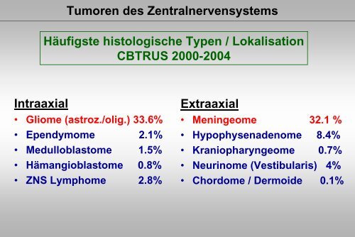
Hochmaligne Gliome / RadiotherapieGlioblastom / Alter als negativer prognostischer Faktor /cbtrus.orgAlter(Jahre)Pat. 1Jahr2Jahre3Jahre4Jahre5Jahre10Jahre0-14 181 47.5 24.4 20.0 18.8 17.5 14.60-19 271 50.9 27.9 21.5 19.8 19.0 15.620-44 1,869 60.0 29.8 20.7 16.0 13.3 8.745-54 2,632 42.2 11.6 5.8 3.8 3.3 1.655-64 4,075 31.3 6.0 2.6 1.6 1.3 0.565-74 4,307 16.8 2.7 1.0 0.5 0.4 0.175 + 2,639 8.1 1.4 0.7 0.5 0.5 0.3Total 15,793
Hochmaligne Gliome / RadiotherapieANOCEF / Phase III :RT 50 Gy / 28 fr. versus „best supportive care“Pat. >/= 70 Jahre / KI >/= 70 (anapl. Glioma, Gliobl.)Keine Einschr.der LQdurch RTMedianes ÜberlebenOp + RT (n=40) 28 Wochennur Op. (n=44) 17 Wochen2001 – 2004, 84 Pat.,med. Alter : 73 Jahre(70 - 85 Jahre)Abbruch der Studie nach1. InterimsanalyseKeime-Guibert, 2007
Autor Pat. Alter(Jahre)Bauman et al.,1994Hochmaligne Gliome / RadiotherapieGlioblastom beim älteren Pat. / Überleben / HypofraktionierungGesamtdosisFraktionierungÜberleben29 65,9-74,7 10 x 3.0 Gy (30 Gy ) 6 MonThomas et al., 1994 38 64,0 6 x 5.0 Gy (30 Gy) 6 Mon.Slotman et al., 1996 30 56 14 x 3.0 Gy (42 Gy) 36 WochenFord et al., 1997 32 65 12 x 3.0 Gy (36Gy) 16 WochenKleinberg et al., 1997 108 > 50 17 x 3.0 Gy (51Gy) RPA V : 8.0 Mon.RPA VI : 5 Mon.Jeremic et al., 1999 47 > 60 or poorKPSHulshof et al., 2000 66414854.060.060.0McAleese et al., 2003 92 7015 x 3.0 Gy (40Gy) 9 Mon.Philips et al., 2003 32 59 10 x 3.5 Gy (35 Gy) 8.7 Mon.33 x 2.0Gy (66 Gy)8 x 5.0 Gy (40 Gy)4 x 7.0 Gy (28 Gy)7.0 Mon.5.6 Mon.6.6 Mon.6 x 5.0 Gy (30 Gy) 5 Mon.Chang et al., 2003 59 65 20 x 2.5 Gy (50 Gy) 7 Mon.Lutterbach et al., 2005 50 67 12 x 3.5 Gy (42 Gy) 7.3 Mon.
Hochmaligne Gliome / RadiotherapiePhase III : 60 Gy in 30 Fraktionen versus 40 Gy in 15 FraktionenPatienten >/= 60 JahreMedianes ÜL6 Wochen 5.1 Monate3 Wochen 5.6 MonateRoa et al., 2004
Hochmaligne Gliome / RadiotherapieRadiotherapie / palliativer Effekt (neurologische Funktion)Autor Pat. Alter(Jahre)DosisFraktionierungBesser Stabil SchlechterBauman et al.,199421 65,9-74,7 30 Gy / 10 Fr. 38% 38% 24%Thomas et al., 1994 38 64,0 30 Gy / 6 Fr. 78 %Besser / stabil78 %Besser /stabil22 %Slotman et al., 1994 30 56 42 Gy / 14 Fr.(3.0Gy)KPS 70 :11/15KPS 80 : 3/6KPS 90: ---9/9-Meckling et al.,199666 75,6 median 50 Gymedian 35 Fr.18 % 67 % 15%Jeremic et al., 1999 47 > 60 orpoor KPS45 Gy(15 x 3.0 Gy)45% KeineAngabenKeineAngabenHulshof et al., 2000 414860,0 40 Gy (8 x 5.0 Gy)28 Gy (4 x 7.0 Gy)14%31%KeineAngabenKeineAngabenMcAleese et al.,200392 7030 Gy (6 x 5.0 Gy) 68%Besser / stabil68%Besser /stabil32%
Hochmaligne Gliome / RadiotherapieZusätzlicheChemotherapie?
Hochmaligne Gliome / RadiotherapieIntraop. Implantation von BCNU (Gliadel)Phase III StudieGliadel : 120 Pat.Placebo : 120 Pat.Westphal, et al., 2006
Hochmaligne Gliome / RadiotherapieHochmaligne Gliome / NOA-0110,751GesamtüberlebenszeitenACNU-VM26 : n = 183ACNU-AraC : n = 179Überleben (%)0,50,25017,34MedianesÜberleben(Monate)18,79MedianesÜberleben(Monate)0 3 6 9 12 15 18 21 24 27 30 33 36 39 42 45 48 51 54 57 60Zeit (Monate)NOA, J Clin Oncol 2003
Hochmaligne Gliome / RadiotherapiePatienten (%)1009080706050403020100O N281 286260 287Glioblastom / EORTC 29681 / 22981:progressionsfreies Überleben (PFS)RTTMZ/RTMedianes PFS (Monate(Monate) 5.0 6.91-Jahres-PFS9% 27%2-Jahres-PFS2% 11%HR [95% C.I.]: 0.54 [0.45-0.64]0.64]p
Patienten (%)1009080706050403020100Hochmaligne Gliome / RadiotherapieGlioblastom / EORTC 29681 / 22981:GesamtüberlebenRT(Monate)0 6 12 18 24 30 36 42RTTMZ/RTO N Anzahl der Patienten at risk :261 286 240 144 59 23 2 0219 287 246 174 109 57 27 4TMZ/RTMedian (Monate(Monate) 12.1 14.62-Jahres-Überleben10% 26%HR [95% C.I.] 0.63 [0.52-0.75]0.75]p
Hochmaligne Gliome / RadiotherapieMGMT Gen / MethylierungsstatusMGMT AusschüttungMGMT GenKaum Ansprechenauf BehandlungMGMT Promotor Regionaktiviertkeine MGMT AusschüttungMGMT GenAnsprechenauf Behandlung– Methyliertdeaktiviert
Hochmaligne Gliome / RadiotherapiePatienten (%)100908070605040302010Progressionsfreies ÜberlebenEinfluss des MGMT-Status in den StudienarmenOverall Wald-Test: p
Hochmaligne Gliome / RadiotherapieGlioblastom / Op / RT / Temoz. + Cilengitide (med. Überleben)Integrin Inhibitor (Anti-Tumoreffekt und Angiogenesehemmer)Methyl (n=23)Med. : nicht err.Unmethyl (n=22)Med. 12 Mon.Unbest (n=7)Cilengitide (n= 52)12 Mon. : 67%Med. : nicht err.Histor. Kontr. (n=64)(Lausanne)12 Mon.: 58%Stupp et al., ASCO 2007
Hochmaligne Gliome / Radiotherapie
Hochmaligne Gliome / Radiotherapie„Ältere“ Patienten / offene FragenAlleinige postop. Bestrahlung(geeignetes Dosis-Frakt.-Schema)Alleinige Chemotherapie(RT bei Progression)?Primär kombinierte Therapie
Hochmaligne Gliome / RadiotherapieNordic elderly trial (Sweden, Norway, Denmark, Finland)Phase III trial / role of up-front temozolomide +Assessment of appropriate RT scheduleGroup 1Conventional radiotherapy 60 Gy in 30 fr./ 6 weeksGroup 2Short-term radiotherapy 34 Gy in 10 fr. / 2 weeksGroup 3Chemotherapy alone(RT upon progression)Temozolomide 200 mg/m 2 day 1-5,every 4 weeks for max. 6 cyclesor until radiological and/or clinical progr.
Hochmaligne Gliome / RadiotherapieEORTC / Gesamt - ÜL RTOG class V (Alter >/= 50 / KPI 70-100)100908070605040302010p=0.054Subgruppenanalyse der EORTC 26981Alter 50-60 (105 RT, 98 RT/TMZ), HR=0.63 p
Hochmaligne Gliome / Radiotherapie
Hochmaligne Gliome / RadiotherapieTemozolomide als Alternative zur RT86 Patienten (Alter > 70): 60 Gy/30 Frakt. (54 Pat.)oder TMZ 5/28 (32 Pts.) (retrospective Analyse)– Mittleres Alter: 73.8 Jahre– Mittler KI: 70– 98% (84/86) Glioblastom– Medianes ÜLRT 4.1 MonateTMZ 6 MonateTMZ 150 mg/m 2 bei 11und 200 mg/m 2 bei 21 Patienten– Beherrschb. Toxizitätbei 15.6% d. Pat.Glantz et al. 2003
Operation (Resektion/Biopsie)Histologie: anaplastisches Astrozytom, GlioblastomAlter > 65 Jahre, Karnofsky Score ? 60RandomisierungArm AArm BNOA - 8Strahlentherapie54-60 Gy in Fraktionen zu 1.8 bis 2 Gyan 4 bis 5 Tagen/WocheDauer: ca. 6 WochenPDGesamtüberlebenTemozolomid100 mg/m 2 KOF an Tag 1-7 und 15 -21alle 28 TageDauer: bis zur ProgressionPDChemotherapie(n)nach Maßgabe desBehandlersStrahlentherapienach Maßgabe desBehandlers
Hochmaligne Gliome / RadiotherapieAnaplastische Oligodendrogliome / RTOG 94-02RT alone versus PCV anschl. RT (60 Gy) / Progressionsfreies ÜberlebenCairncross et al., 2006
Hochmaligne Gliome / RadiotherapieAnaplastische Oligodendrogliome / RTOG 94-02RT alone versus PCV anschl. RT (60 Gy) / GesamtüberlebenCairncross et al., 2006
Hochmaligne Gliome / RadiotherapieAnaplastische Oligodendrogliome / RTOG 94-02RT alone v. PCV anschl. RT (60 Gy) / Progr.-fr. ÜL / 1p-19q DeletionCairncross et al., 2006
NOA-04: Randomisierte Phase-III-Studie der sequentiellen Radiochx.oligoastrozytärer Tumoren des WHO-Grads III mit PCV oder TemozolomidResektion oder BiopsieHistologie: anaplastisches Oligodendrogliom, Oligoastrozytom, AstrozytomArm AArme B,CStrahlentherapiePrimäre ChemotherapieBCPCVTemozolomidA1PDA2PDPDPCVTemozolomidStrahlentherapieStrahlentherapiePDPDPDPDTemozolomidPCVTemozolomidPCV
Hochmaligne Gliome / Radiotherapie
Hochmaligne Gliome / RadiotherapiePseudoprogression nach RT – Chx / Teilres. Astrozytom III.12.01.07präop.16.04.07post op.
Hochmaligne Gliome / RadiotherapiePseudoprogression nach RT – Chx / Teilres. Astrozytom III.24.10.074 Mon.n.RT3. KursACNUVM 2609.05.0811 Mon.n. RT6. KursACNUVM 26
Hochmaligne Gliome / RadiotherapiePseudoprogression nach RT – Chx / Teilres. Astrozytom III.16.10.0816 Mon.n.RTACNUVM 2605.02.0921 Mon.n. RTACNUVM 26
Hochmaligne Gliome / RadiotherapieBildgebung / Ansprechen auf Therapie / PseudoprogressionOP / RT 60 Gy / Temoz.Medianes Gesamt-ÜLPseudoprogr. (n=32)frühe Progr. (n=18)weder / noch (n=53)38 Mon.10.2 Mon.20.2 Mon.Brandes, et al. 2008
Hochmaligne Gliome / RadiotherapieRezidivtherapie bei hochmalignen Gliomen- Operation-ErneuteBestrahlung?- Chemotherapie
Hochmaligne Gliome / RadiotherapieStrahlentherapie bei Rezidiv / hypofrakt. stereotakt.Autor Pat.-Zahl Technik / Dosierung MedianesÜberlebenShepherd et al., 1997 33 Hypofrakt. KonvergenztherapieEinzeldosis 5 Gy, Eskal. 20 -> 50 Gy11,0 Mon.Lederman et al., 2000 88 Stereotakt., hypofrakt. RTMed. 24 Gy in 4 Frakt.Voynov et al.,200210 Stereotakt. IMRT, med. 30 Gy(25-40 Gy), 5 Gy/Frakt.7 Monate10,1 Mon.Bartsch et al.,200522 Stereot. RT 14 Pat. 45-54 Gy, konv.Frakt. 8 Pat. 30 Gy hypofrakt. (6x5Gy)7,0 Mon.Grosu et al., 2005 44 Stereotakt. RT, hypofrakt.36 PET/SPECT, 30 Gy8 CT/MRI (6 x 5 Gy)Vordermark et al.200519 Stereotakt. RT, hypofrakt.(4-10 Gy Einzeldosis)30 Gy (20-30 Gy)9,0 Mon.5,0 Mon.9,3 Mon.
Hochmaligne Gliome / RadiotherapieStrahlentherapie bei Rezidiv / konv. frakt. stereotakt.AutorPat.-ZahlTechnik / DosierungMedianesÜberlebenArcicasa et al.,199931 Frakt. Konvent. 2 D RTEinzeldosis 1,5 Gy, 34,5 Gy13,7 Mon.Cho et al. 1999 25 Konv. Frakt. RT, 37.5/15Fraktionen12,0 Mon.Hudes et al.,1999Combs et al.,2005b20 Stereot. RT 3-3.5 Gy24.0->35 GyEskalationsprotok.54 GBM39WHO IIIStereotakt. RT36 Gy (15-62 Gy)5 x 2.0 Gy konv. frakt.10.5 Mon.8,0 Mon.12,0 Mon.
Re-Bestrahlung bei RezidivEffekt der FraktionierungSRS / n = 46, med. ÜL : 11 Mon.FSRS / n = 25, med. ÜL : 12 Mon.Dosiskonzepte :1. SRS :17 Gy50% Isodose2. FSRT37,5 Gy (ED 2,5 Gy)85% IsodoseCho et al., 1999
Re-Bestrahlung bei RezidivFraktionierung (n = 31)4 x 5 Gy 19 (61%) 5 x 4 Gy 10 (32%)5 x 5 Gy 2 (7%) Med. ÜL : 10.2 Mon.1,01,00,90,90,80,8Überleben0,70,60,50,40,30,2< 30 ml> 30 mlBp=0.010,70,60,50,40,30,2< 50 Jahre=/> 50 Jahrep=0.050,10,10,00,00 10 20 30 40 50 600 10 20 30 40 50 60MonateHenke et al., 2009
Re-Bestrahlung bei RezidivHochmalignes Gliom / Re-Bestrahlung bei Rückfall /stereotaktische KonvergenztherapiePat. 17 Jahre altHypofraktioniert , 4 x 5 GyVor RT 4 x 5 Gy CR 1 Jahr n. RT
Re-Bestrahlung bei Rezidiv3 D konformalefraktionierte RTRigide FixierungPräzision : ca 1 mmm
Hochmaligne Gliome / Radiotherapie„Targeting“Methionin-PET bei hochmalignem Gliom zur Definitiondes klinischen Zielvolumens / z.B. Rezidivbestrahlungen
Hochmaligne Gliome / RadiotherapieFunktionelle Bildgebung in der ZielvolumenbestimmungSpektroskopie
Hochmaligne Gliome / RadiotherapieFazit / Ausblick IPostop. RT Tumorregion 60 GyDosiseskalation : kein Vorteil (Stereot. / Brachyth.)Dosiseskalation mit Hyperfraktionierung /Akzelerierung : kein VorteilÄltere Patientenlokale RT Überlebensvorteilalternativ hypofraktioniertes Konzept möglichResponse MonitoringPseudoprogressionFunktionelle Bildgebung ?
Hochmaligne Gliome / RadiotherapieFazit / Ausblick IIIntegrierung von SystemtherapienStratifizierung nach molekulargenetischem Profil1p 19q Deletion (anapl. Gliome)MGMT Status (Glioblastom/anaplastische Gliome)Erneute Bestrahlungstereotaktische Techniken (geeign. Frakt.-Schema ?)Integrierung funktioneller Bildgebungerneute Kombinationstherapie (RT-Chx.)Weitere Forschung notwendig
Radiotherapie / MeningeomenOffene FragenZeitpunkt der BestrahlungSofortige postoperative RToderBestrahlung bei RezidivAuswahl der geeignetenBestrahlungstechnik?Einzeitbestrahlung(„Radiochirurgie“)oderfraktionierte Konformationstechnik
Radiotherapie / Meningeomen„Optimale Fraktionierungsschemata“- Stereotakt. Einzeit RT- Hypofraktionierte RT- Konventionelle Frakt.- Konvent. Frakt.+Dosiseskalation- Konv. RT – Technik (2 D)- „Konformierende“(3D) - TechnikKonv. 3 DIMRTProtonentherapieTumorkontrolleNebenwirkungsrisiko
Radiotherapie / MeningeomenErgebnisse nach alleiniger Operation und postop.RTAutor, Jahr Pat. Bestrahlungstechnik 5-J.-PFÜ nach Op. 5-J.-PFÜn. Op. + RTBarbaro et al., 1987 55 konv. fraktioniertmedian 54 GyTaylor et al., 1988 132 konv. fraktioniert50-63 Gy (1,8-2,0 Gy)Miralbell et al.,199296 konv. fraktioniertmedian 52 GyGoldsmith et al., 1994 117 konv. fraktioniertmedian 54 GyMaire et al., 1995 91 konv. fraktioniertmedian 52 Gy (ED 1,8 Gy)Matthiesen et al., 1996 315 - 96% (total)55% (subtotal)Stafford et al., 1998 581 - Kompl.Res.5 J.: 88%10 J.: 75%Nutting et al., 1999 82 konv. fraktionierte RT55-60 GyMendenhall et al., 2003 101 konv. fraktionierte RTmedian 54 Gy40% 68%43% 82%48% 88%- 89%10 J.: 77%- 71%Inkompl. Res. 5J.: 61%10 J.: 39%- 5 J.: 92%10 J.: 83%- 5 J. : 95%10 J. : 92%15. J. 92%--
Radiotherapie / MeningeomenErgebnisse nach fraktionierter 3 D konformaler RadiotherapieAutor, Jahr Pat. Med.DosisMed.Tumor-Vol. (ml)Tumorregression5-Jahres-PFÜKomplikationsrateAlheit et al.,199941 50-55Gy 17,9 22% 100%(3 Jahre)9,8%Selch et al., 2004 45 50,4 Gy 14,5 18% 97,4% 2%Milker-Zabel etal, 2001317 57,6 Gy 33,6 23% 90% 2,5%Henzel et al.,2006224 55,8Gy 9,1 45% 96,6% 2,5%
Radiotherapie / Meningeomen3-D konformale RT / Ansprechen (MR)n= 317Besser23%Progredienz7%Stabil70%Milker-Zabel, et al., 2005
Radiotherapie / MeningeomenErgebnisse nach stereotaktischer Einzeittherapie („Radiochirurgie“)Autor, Jahr Pat. Med.DosisStafford et al.,2001Med.Tumor-Vol. (ml)Tumorregression5-Jahres-PFÜKomplik.rate168 16 Gy k.A. 56% 93% 13%Lee et al., 2002 159 13 Gy 6.5 34% 93% 6,9%Nicolato et al, 2002 122 14,6Gy 8,1 61% 96,5% 4,1%DiBiase et al., 2004 162 14 Gy 4,5 28% 86% 8,3%Kreil et al., 2005 200 12 Gy 6,5 57% 98,5% 2,5%Malik et al., 2005 309 20 Gy 7,3 k.A. 87% 3%Kollova et al., 2007 325 12,6Gy 4,4 69,7% 97,9% 5,7%Kondziolka et al.,2008972 14,0 Gy 7,4 44,2% 10 J.95,0%7,7%
Radiotherapie / MeningeomenAtypische und anaplastische Meningeome /Dosis-Wirkungsbeziehungen (fraktionierte Radiotherapie)Autor, Jahr Pat. Dosis Ergebnis p-WertGoldsmithet al., 199423 < 53 Gy=/>53 Gy5 Jahre PFÜ :17%67%0,01Milosevicet al., 199659 < 50 Gy=/>50 Gy5 Jahre PFÜ :0%42%< 0,01Coke et al.,199817 < 54 Gy=/>54 Gy3 / 5 Pat. DOD1/12 Pat. DODk.A.
Radiotherapie / MeningeomenBesonderheiten beiMeningeomen der SchädelbasisHistologisch meistens benigneAufgrund der Lokalisation InoperabilitätKlinisch häufig HirnnervenausfälleEnge Nachbarschaft zu RisikoorganenHirnstamm, Chiasma, Hirnnerven(Risikoorgan im Zielvolumen)Irreguläre Konfiguration des TumorsNotw. Dosis / Toleranzdosis d. RisikoorganeMeningeome der Optikusscheide / Sonderfall
Radiotherapie / Meningeomen3 – D KonformationstechnikKeilbeinflügelOptikusscheide
Radiotherapie / MeningeomenErgebnisse nach frakt. 3 D konf. RTvon OptikusscheidenmeningeomenAutor, Jahr Pat. Med.DosisMed.Tumor-Vol. (ml)LokaleKontrolleBesserungdesSehvermögenTumorregressionKomplik.-rateAndrews200230 54Gy 2,9ml 13% 100% 42% 13%Becker200242 54 Gy k.A. 2,6% 100% 19% Visus31,6% GF9,5%Baumert200422 54 Gy 1,7ml 4,8% 100% 73% 4,3%
Radiotherapie / MeningeomenIMRT / ProtonentherapieIMRTProtonenProtonenInt.-moduliertBaumert et al., 2004
Radiotherapie / MeningeomenErgebnisse nach ProtonentherapieAutor, Jahr Pat. Bestrahlungstechnik Tumorkontrolle /ÜberlebenKomplikationenWenkel etal., 200046 frakt. Photonen +Protonen 59,0 CGEÜL 5/10 J. : 93% / 77%LK 5//10 J. : 100% / 88%9/46 schwereDefiziteHug et al.,200031(15 AM)(16 MM)frakt. Photonen +Protonentherapie62,0 CGE (AM),58 CGE (MM)LK 8 Jahre :AM : 19%, MM : 17%Dosis > 60 GyAM : 70%, MM : 83%3/31 RadionekroseVernimmenet al., 200127 Hypofrakt. Prot.-therapie16,3 CGE5 Jahre PFÜ : 88% 5.5%Weber et al.,200416 Frakt. Protonentherapie52,2 – 64 CGELK / 3 Jahre : 91,7%ÜL / 3 Jahre : 92,7%3/16 Grad III-IVToxizitätNoel et al.,200551 frakt. Photonen +Protonen 60,6 CGELK / 4 Jahre : 98%ÜL / 4 Jahre : 100%2/51WHO Gr. III Tox.
Radiotherapie / MeningeomenZusammenfassung / zukünftige StrategienKonventionelle Fraktionierung hocheffektivKontrolle von Hirnnervenausfällen / VisusKonformationstechnik praktikabel und nebenwirkungsarmStereotaktische Einzeit – RT hocheffektivHöheres NW – Potential ?Auswahl der Technik / KriterienTumorvolumen, Lokalisation, KonfigurationOffene FragenSofortige pop. RT nach inkompl. Res ? : (EORTC Studie)Tumorvolumen TumorkontrolleDosis-Wirkungsbez. (frakt. Techniken)Protonen / int.-moduliert Protonen
Vielen Dank fürIhre Aufmerksamkeit


