Kultur- und Kongresszentrum Thun (KK Thun) 8.–9. November 2012 ...
Kultur- und Kongresszentrum Thun (KK Thun) 8.–9. November 2012 ...
Kultur- und Kongresszentrum Thun (KK Thun) 8.–9. November 2012 ...
Erfolgreiche ePaper selbst erstellen
Machen Sie aus Ihren PDF Publikationen ein blätterbares Flipbook mit unserer einzigartigen Google optimierten e-Paper Software.
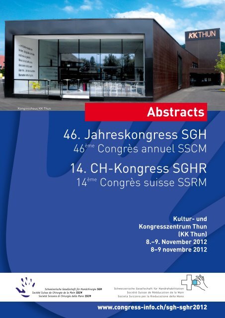
Kongresshaus <strong>KK</strong> <strong>Thun</strong><br />
1<br />
Abstracts<br />
46. Jahreskongress SGH<br />
46 ème Congrès annuel SSCM<br />
14. CH-Kongress SGHR<br />
14 ème Congrès suisse SSRM<br />
<strong>Kultur</strong>- <strong>und</strong><br />
<strong>Kongresszentrum</strong> <strong>Thun</strong><br />
(<strong>KK</strong> <strong>Thun</strong>)<br />
8.–9. <strong>November</strong> <strong>2012</strong><br />
8–9 novembre <strong>2012</strong><br />
www.congress-info.ch/sgh-sghr<strong>2012</strong>
Inhaltsverzeichnis / Sommaire<br />
Kongressfakultät<br />
Corps scientifique du congrès 3<br />
Freie Mitteilungen SGH<br />
Communications libres SSCM 4–23<br />
Freie Mitteilungen SGHR<br />
Communications libres SSRM 24–32<br />
Workshops SGHR<br />
Ateliers SSRM 33–36<br />
Impulsreferat (R), Symposien (S), Lecture (L) SGHR<br />
Exposé introductif (R), symposia (S), lecture (L) SSRM 37–44<br />
2
Kongressfakultät/Corps scientifi que du congrès<br />
Tagungsleitung/Direction d’organisation<br />
PD Dr. med. Esther Vögelin<br />
Präsidentin SGH<br />
Présidente SSCM<br />
Nora Käser<br />
Vorstandsmitglied SGHR<br />
Membre du comité SSRM<br />
Wissenschaftliche Leitung/Direction scientifi que<br />
Dr. med. Dietmar Bignion<br />
Mitglied SGH<br />
Membre SSCM<br />
Dr. med. Michaël Papaloïzos<br />
Vorstandsmitglied SGH<br />
Membre du comité SSCM<br />
PD Dr. med. Esther Vögelin<br />
Präsidentin SGH<br />
Présidente SSCM<br />
Gabriele Versümer Bücker<br />
Past-Präsidentin SGHR<br />
Ancienne présidente SSRM<br />
Administrative Organisation / Organisation administrative<br />
Healthworld (Schweiz AG), Sennweidstrasse 46, 6312 Steinhausen<br />
Tel. 041 748 76 00, Fax 041 748 76 11, toni.vonwyl@healthworld.ch, www.congress-info.ch/sgh-sghr<strong>2012</strong><br />
3<br />
Dr. med. Bettina Juon<br />
Mitglied SGH<br />
Membre SSCM<br />
Véronique van der Zypen<br />
Präsidentin SGHR<br />
Présidente SSRM<br />
Dr. med. Cesare Fusetti<br />
Mitglied SGH<br />
Membre SSCM<br />
PD Dr. med.<br />
Andreas Schweizer<br />
Mitglied SGH<br />
Membre SSCM<br />
Maja Drotschmann<br />
Mitglied SGHR<br />
Membre SSRM<br />
Katrin Voss<br />
Mitglied SGHR<br />
Membre SSRM
Freie Mitteilungen SGH<br />
Communications libres SSCM<br />
FM 1<br />
10 year follow-up of pyrocarbon implants for<br />
proximal interphalangeal joint replacement<br />
Lisa Reissner 1 , Stephan F. Schindele 1 , Stefanie<br />
Hensler 1 , Miriam Marks 1 , Daniel B. Herren 1 ( 1 Zürich)<br />
Introduction:<br />
Short and mid-term results of anatomical resurfacing<br />
using a pyrocarbon prosthesis (Ascension Orthopedics)<br />
showed reasonable clinical results with a high migration<br />
rate radiographically due to problematic implant fixation.<br />
The aim of the study was to investigate the subjective,<br />
clinical and radiographic long-term results after 10<br />
years, and to compare them with the 2 year follow-up.<br />
Methods:<br />
In a prospective, consecutive case series, 17 PIP-<br />
Pyrocarbon implants in 14 patients were assessed<br />
subjectively by a VAS pain scale, clinically and<br />
radiographically pre-operative, at a mean follow up of<br />
20.1 months (± 5.4) and 9.7 years (± 0.5). The<br />
indication for the joint replacement arthroplasty was<br />
degenerative osteoarthritis in 15 cases,<br />
chondrocalcinosis in one patient and post-traumatic in<br />
another patient.<br />
Results:<br />
One patient died before the 10 year follow up. One<br />
patient needed a revision surgery, due to implant<br />
failure. All other implants remain in situ. After a mean<br />
follow-up of 9.7 years no further migration of the<br />
implants were observed in comparison with the average<br />
20.1 month follow-up. Radiolucent lines remained<br />
identical with an average of 2.1mm (± 0.9). The average<br />
total range of motion of all replaced joints was 35° preoperatively,<br />
38° at the 2 year follow-up and decreased<br />
significantly to 27° at the final follow up (p≤0.01). The<br />
average grip strength was 21 kg pre-operatively, which<br />
increased to 26 kg at the 2 year follow-up and<br />
decreased again significantly at the final follow-up to 17<br />
kg (p=0.001). Significant pain relief was noted in all<br />
patients from 7.6 pre-operatively to 1.4 at 2 years of<br />
follow-up and 0.8 at final follow-up (p≤0.001).<br />
Discussion:<br />
To our knowledge this case series is the longest<br />
consecutive follow-up of PIP-Pyrocarbon implants.<br />
Compared to the results at the 2 year follow-up, the<br />
observed implant migration remained stable over time<br />
although no signs of secondary osteo-integration were<br />
observed. The range of motion decreased with longer<br />
follow-up and was, with less than 30°, disappointing for<br />
most patients. However overall patients’ satisfaction<br />
remained high due to permanent pain-relief.<br />
Nevertheless, we no longer perform this kind of<br />
arthroplasty due to only moderate clinical results and<br />
potential complications with implant migration.<br />
FM 2<br />
Arthroplastie unipolaire en pyrocarbone dans<br />
l’arthrose interphalagienne proximale<br />
Jean-Yves Beaulieu 1 , Stéphane Kämpfen 1 , Philippe<br />
Vostrel 1 , Gontran Sennwald 1 ( 1 Geneve)<br />
4<br />
Introduction : L’arthrose IPP quelle soit d’origine posttraumatique<br />
ou rhumatologique entraine une perte de<br />
fonction importante au niveau de la main. La prise en<br />
charge thérapeutique peut comprendre la réalisation de<br />
dénervation, d’arthrodèse ou encore d’arthroplastie. Les<br />
arthroplasties consistent en la mise en place d’implant<br />
devant permettre la mobilisation indolore des<br />
articulations. Il existe différents types d’implant : soit un<br />
spacer en silicone, soit un remplacement bipolaire<br />
articulaire. Nous vous présentons une courte série de 4<br />
patients ayant bénéficiés d’un remplassement<br />
unipolaire en pyrocarbone par un implant proximal IPP<br />
Ascencio®.<br />
Matériel et méthode : 5 implants condyliens proximaux<br />
IPP Ascencio® ont été posés chez 4 patients. Il<br />
s’agissait de 3 femmes et un homme. La moyenne<br />
d’âge était de 61 ans (38-74 ans). Dans 4 cas il<br />
s’agissait d’arthrose essentielle et dans un cas une<br />
arthrose post traumatique IPP. Tous les patients on<br />
bénéficié d’une voie d’abord palmaire. L’implant<br />
proximal était implanté après résection de la tête de P1<br />
et la base de P2 n’étant pas modifiée. L’évaluation c’est<br />
faite sur le score du quick dash la force de serrage les<br />
mobilités articulaires et l’analyse radiographique.<br />
Résultats : La revue moyenne des cas est à 24 mois<br />
(6-60mois). 3 index, 1 majeur et 1 annulaire furent<br />
implantés dans deux cas sur les mains dominantes.<br />
Aucun patient n’a présenté de complication majeure. A<br />
la revue le score moyen du quick dash était de 24 (9-<br />
31). Les patients ne présentaient plus de douleur au<br />
repos. Les mobilités en moyenne en flexion étaient de<br />
72° et un déficit d’extension de 5° en moyenne. Dans 3<br />
cas on retrouvait un clinodactisme latérale ulnaire en<br />
moyenne de 10° (0-20).<br />
Le contrôle radiographique permet de mettre en<br />
évidence dans tous les cas une densification corticale<br />
autour de la tige prothétique sans migration de<br />
l’implant. Tous les patents présentaient un<br />
remaniement de la base de P2 s’installant dans les<br />
premiers mois post opératoire mais stable au dernier<br />
recul.<br />
Conclusion : Le remplacement unipolaire de la tête de<br />
P1 dans les arthroses IPP par un implant en<br />
pyrocarbone donne des résultats prometteurs. Nous<br />
l’incluons désormais dans nos possibilités<br />
thérapeutiques.<br />
FM 3<br />
Case studies of a constrained finger joint system<br />
for interphalangeal (PIP) joints<br />
Eva Ståhl Wernersson 1 , Håkan Håkanson 1 , Göran<br />
L<strong>und</strong>borg 1 , Anders Björkman 1 ( 1 Malmö SE)<br />
Arthroplasty can be used for patients suffering from<br />
rheumatoid arthritis or arthrosis (both primary and<br />
posttraumatic) in the metacarpohalangeal (MCP) joints<br />
or in the interphalangeal (PIP) joints.<br />
A new design of a constrained finger implant system<br />
with a joint mechanism and bone anchorage screws<br />
has been developed. The system is designed for long<br />
endurance and to prevent luxation and instability. The<br />
joint resists the large forces applied occasionally to the<br />
joint.
The joint consists of two titanium screws which are<br />
fixated in the phalangeal bones. The joint has plates<br />
with pins that firmly fit into the screws. The plates are<br />
constrained with a ball of plastic material. The flexion of<br />
the joint is preset by the design of the ball. The PIP joint<br />
allow for 90 degrees of flexion, however, deviation is<br />
limited to a couple of degrees.<br />
The first patient was operated on using this arthroplasty<br />
system in June 2008. By May <strong>2012</strong>, 47 MCP joints and<br />
15 PIP joints have been implanted in 26 patients. We<br />
will present three patients operated on using this<br />
system in four PIP joints. The follow up period is 2 to 26<br />
months. The three patients show good clinical results<br />
with decreased pain and improved range of motion.<br />
We conclude that the titanium screws osseointegrate<br />
and the joint mechanism allows for a good range of<br />
motion resulting in considerable pain reduction and<br />
improved mobility in the fingers of the patients.<br />
Ledbo¦êj-bild.jpg<br />
FM 4<br />
Die PIP-Prothese -Was haben wir gelernt?-<br />
Nicole Schmelzer-Schmied 1 , Robert Vogt 1 , Mark Aerni 1<br />
( 1 Münsterlingen)<br />
Einleitung:<br />
Bei der Versorgung einer Bouchardarthrose durch eine<br />
PIP-Prothese gibt es Einiges zu beachten. Im Laufe der<br />
letzten 10 Jahre, konnten Erfahrungen gesammelt<br />
werden, die zur Verbesserung der Ergebnisse nach<br />
Operation <strong>und</strong> so zur Patientenzufriedenheit beitragen.<br />
Ziel dieser Studie war es, Fehlerquellen bei der<br />
Operation ausfindig zu machen <strong>und</strong> den Verlauf nach<br />
PIP-Implantation zu dokumentieren, sowie klinische <strong>und</strong><br />
radiologische Ergebnisse hinsichtlich verschiedener<br />
Einflussgrößen zu untersuchen.<br />
Material/Methode:<br />
Im Rahmen einer retrospektiven Studie, wurden 58<br />
Patienten mit 77 PIP-Prothesen (SBI-Avanta) im<br />
Hinblick auf klinische <strong>und</strong> radiologische Parameter<br />
untersucht. Unter anderem wurden die ROM, die<br />
Patientenzufriedenheit anhand des Quick-Dash <strong>und</strong> die<br />
Kraft gemessen. Dabei wurden 2 Gruppen gebildet.<br />
Patienten, die in den Jahren 2001-2007 operiert<br />
wurden, wurden mit Patienten, die zwischen 2008-<br />
2011 operiert wurden verglichen.<br />
Ergebnisse:<br />
Insgesamt sind die Ergebnisse nach Implantation einer<br />
PIP-Prothese sehr gut. Die radiologische Lockerung,<br />
die Schwanenhalsdeformität sowie die<br />
Patientenzufriedenheit konnte in den letzten Jahren<br />
verbessert werden. Unsere Ergebnisse zeigen, dass es<br />
5<br />
von Vorteil ist, auf Tricks zu achten. Unter anderem<br />
sollte die Kontaktfläche der Prothese genügend groß<br />
sein, das intraoperative Gelenkspiel leicht. Ein positiver<br />
Faktor scheint außerdem das Zementieren der<br />
Prothese zu sein. Dennoch sollte die Indikation zur<br />
Implantation einer PIP-Prothese gut überlegt sein.<br />
FM 5<br />
Hemi-hamate autograft arthroplasty for acute and<br />
chronic PIP joint fracture dislocations<br />
Nicole Lindenblatt 1 , Angelo Biraima 1 , Ivan Tami 2 , Pietro<br />
Giovanoli 1 , Maurizio Calcagni 1 ( 1 Zürich; 2 Gravesano-<br />
Lugano)<br />
Introduction: Treatment of fracture dislocations of the<br />
PIP joint represents a hand surgical challenge. In hemihamate<br />
arthroplasty, the palmar joint surface is<br />
reconstructed using an osteochondral graft from the<br />
hamate and the immediate stability permits early<br />
movement. Material and Methods: We performed hemihamate<br />
reconstructions in 10 patients (mean age 35<br />
years), who sustained fractures of the base of the<br />
middle phalanx of ≥ 50% surface and dorsal PIP<br />
dislocations. Outcomes were assessed by clinical exam<br />
and x-ray. Evaluation criteria were range of motion of<br />
PIP and DIP joints, grip strength, joint alignment,<br />
complications and donor site morbidity.<br />
Results: Mean follow-up was 8 months (range 3-14).<br />
Fractures mostly involved the 4th and 5th fingers, all<br />
patients were male. Indications were subluxation and<br />
comminution in acute cases of < 6 weeks (5 patients)<br />
and chronic pain and morning stiffness in chronic cases<br />
(3 patients). 2 patients had no pain and <strong>und</strong>erwent<br />
revision surgery to prevent secondary osteoarthritis.<br />
Operative treatment was performed in average after 67<br />
days (range 1-371 days) after injury. Average PIP<br />
motion was 71 degrees (range 0- 90), DIP motion was<br />
55 degrees (range 10-90) with a mean PIP flexion<br />
contracture of 5 degrees (range 0-20). Grip strength<br />
averaged 95% of the opposite hand. 3 patients had<br />
revision surgery (2x arthrolysis PIP joint, 1x neurolysis<br />
R. dorsalis N. ulnaris).<br />
Conclusions: Hemi-hamate autograft arthroplasty<br />
represents an effective procedure to address severe<br />
PIP joint fracture-dislocations. It restores the<br />
comminuted articular surface in chronic injuries and in<br />
the acute injury it is a challenging but valuable<br />
alternative to extension block spliniting. However, donor<br />
site morbidity and revision surgery have to be taken into<br />
account.<br />
PostopPIPHamatum.jpg
FM 6<br />
Ein Jahr Follow-up nach Rekonstruktion komplexer<br />
PIP Gelenks Defekte<br />
Thomas Meszaros 1 , Bettina Juon 1 , Nicole Badur 1 ,<br />
Esther Vögelin 1 ( 1 Bern)<br />
Einleitung: Komplexe Defekt-Läsionen des proximalen<br />
interphalangeal (PIP) Gelenkes bei Handwerkern sind<br />
für den behandelnden Handchirurgen eine<br />
Herausforderung. Falls immer möglich ist eine primäre<br />
Rekonstruktion der defekten Knorpel-Knochen-,<br />
Sehnen- <strong>und</strong> Bandstrukturen zum Erhalt der<br />
Beweglichkeit anzustreben.<br />
Material: 5 Handwerker im Alter von 21 bis 57 mit<br />
komplexen PIP Gelenkes Defekten wurden primär<br />
rekonstruiert. Der Zeigefinger war vier mal betroffen<br />
<strong>und</strong> der Kleinfinger einmal. Alle Patienten hatten einen<br />
osteochondralen Defekt (3x Gr<strong>und</strong>phalanx Kopf, 2x<br />
Mittelphalanx Basis) <strong>und</strong> zusätzlich eine Läsion des<br />
Streckapparates. Die Seitenbänder waren in 4 Fällen<br />
betroffen. Die Rekonstruktion des Knorpel-Knochen-<br />
Defektes wurde mit einem freien, autologen<br />
osteochondralen Graft durchgeführt: der Defekt der<br />
Gr<strong>und</strong>phalanx wurde mit einem Graft des Os<br />
Metatarsale III, der Basis Os Metacarpale IV oder dem<br />
Gr<strong>und</strong>phalanx Kopf der II. Zehe rekonstruiert. Die<br />
Rekonstruktion der Mittelphalanxbasis erfolgte in 2<br />
Fällen mit einem Hemihamatum-Graft. Eine<br />
Strecksehnenrekonstruktion mittels freiem<br />
Sehnentransplantat war in 3 Fällen nötig, die<br />
Bandläsionen wurden ebenfalls versorgt. Die<br />
Nachbehandlung wurde in allen Fällen nach Early<br />
Active Short Arc Motion (SAM) durchgeführt.<br />
Resultate: Durchschnittlich zeigte sich nach 3 Monaten<br />
ein Range-of-motion von 73° (Flexion/Extension: 90-16-<br />
0), nach 6 Monaten von 83° (F/E: 93-10-0) <strong>und</strong> nach 1<br />
Jahr von 88° (F/E: 91-3-0). Alle Patienten konnten ihren<br />
angestammten Beruf wieder im Vollzeitpensum<br />
ausführen <strong>und</strong> waren mit dem erreichten funktionellen<br />
<strong>und</strong> kosmetischen Resultat zufrieden.<br />
Zusammenfassung: Eine primäre Rekonstruktion<br />
komplexer PIP Gelenkes-Defektläsionen mit autologem<br />
osteochondralen Graft in Kombination mit Sehnen- <strong>und</strong><br />
Bandrekonstruktionen gefolgt vom intensiver<br />
frühfunktioneller Handtherapie erzielt auch bei<br />
Handwerkern ein gutes funktionelles Resultat.<br />
FM 7<br />
3D assisted planning and performance of corrective<br />
osteotomy<br />
Andreas Schweizer 1 , Ladislav Nagy 1 ( 1 Zürich)<br />
Introduction: Malunions at the hand may lead to pain,<br />
reduced range of motion, instability and joint<br />
degeneration justifying corrective osteotomy. The<br />
procedure is challenging due to the minuteness of the<br />
bones, restricted access and the aimed accurateness of<br />
Ziel: Vorstellung der ersten klinischen <strong>und</strong><br />
radiologische Resultate bei 6 Patienten nach<br />
dekomprimierender Radiusverkürzungsosteotomie bei<br />
primärer Ulna Minus Varianz. Das Operationsverfahren<br />
ermöglicht durch eine Keilosteotomie mit Verkürzung<br />
einen Radialshift. Die Inkongruenz im distalen<br />
Radioulnargelenk wird korrigiert im Sinne einer<br />
schmerzreduzierenden Dekompression.<br />
Material <strong>und</strong> Methoden: In einer retrospektiven Studie<br />
werden die ersten Ergebnisse nach dekomprimierender<br />
Radiusverkürzung über einen palmaren Zugang mit<br />
einem minimalen follow up von 6 Monaten vorgestellt.<br />
Resultate: Die Beweglichkeit im Handgelenk für Flexion<br />
<strong>und</strong> Extension war bei allen Patienten postoperativ<br />
uneingeschränkt. Ebenfalls ergab sich für die Pro- <strong>und</strong><br />
Suppination im distalen Radioulnargelenk keine<br />
Einschränkung. Der schmerzbedingte Leidensdruck<br />
(präoperativ VAS 7,8) konnte durch diese Methode 12<br />
Monate postoperativ deutlich reduziert werden (VAS<br />
1,8). Ein Patient war mit dem Vorgehen so zufrieden,<br />
dass die Gegenseite ebenfalls operiert wird.<br />
Zusammenfassung: Die Ergebnisse dieser<br />
Operationsmethode zeigen erfreuliche Resultate<br />
bezüglich Funktion, Schmerzen <strong>und</strong> der hieraus<br />
resultierenden Patientenzufriedenheit. Neben der<br />
Schmerzreduktion ist von einem präventiven Charakter<br />
dieses Vorgehens mit Vermeidung der langfristig zu<br />
erwartenden Arthrose auszugehen.<br />
Radiusverkuerzung1.jpg<br />
Radiusverkuerzung2.jpg<br />
7<br />
FM 9<br />
A radiolucent, carbon fibre reinforced synthetic<br />
plate for distal radius fractures: first experience<br />
Jürg Sonderegger 1 , Gerd Laub 1 , Marc Wieder 1 ( 1 Grabs)<br />
Introduction: Steel and titanium are widely and<br />
successfully used implant metals in traumatology.<br />
However, some disadvantages are related to metallic<br />
implants: They provoke artefacts in computed<br />
tomography (CT) and magnetic resonance imaging<br />
(MRI). Also, bone healing might be difficult to monitor in<br />
x-ray studies due to the invisibility of the bone<br />
<strong>und</strong>erlying the implant. Furthermore, hardware removal<br />
still remains an issue, especially with titanium implants.<br />
In order to avoid these problems carbon fibre reinforced<br />
polyetheretherketone (PEEK) implant material has been<br />
developed. The main advantages of this synthetic<br />
material are radiolucency, artefact-free imaging, good<br />
fatigue properties and biocompatibility. In spine surgery<br />
carbon fibre reinforced PEEK implants have been<br />
successfully used and documented for the last few<br />
years. Recently palmar plates for distal radius fractures<br />
have been developed.<br />
Material and Methods: Three consecutive cases of<br />
dorsally displaced extraarticular distal radius fracture<br />
were stabilized with a palmar semi-rigid carbon fibre<br />
reinforced PEEK plate with angular stable screw fixation<br />
(icotec AG, Altstätten, Switzerland). Integrated<br />
radiopaque tantalum filaments and metallic screw tips<br />
allow correct placement of the plate and make the<br />
implant visible <strong>und</strong>er image intensifier. A clinical and<br />
radiographic follow up was performed at 2 and 6 weeks,<br />
and at 3 and 6 months.<br />
Results: The range of motion at final follow up was<br />
comparable to the contralateral wrist and all patients<br />
were pain free. Bony union was achieved in all cases.<br />
We did not record any infections, secondary<br />
dislocations, tendon ruptures or other complications.<br />
Discussion: We present our first experience on a new<br />
carbon fibre reinforced synthetic palmar plate for distal<br />
radius fractures. The implant might represent an<br />
interesting alternative to metallic implants. Potential<br />
advantages are radiolucency, no adherence to<br />
surro<strong>und</strong>ing tissues and no artefacts in CT and MR<br />
imaging. A disadvantage of the implant is the missing<br />
multidirectional screw fixation, making it difficult to use<br />
in complex fracture situations.
ap.jpg<br />
lat.jpg<br />
8<br />
FM 10<br />
Corrective Osteotomy of the Distal Radius with or<br />
without Bone Grafts<br />
Alexander Kozlowski 1 , Vinh-Thuan Ly 1 , Martino Meoli 1 ,<br />
Jörg Grünert 1 ( 1 St Gallen)<br />
Malunion of distal radius fractures still represents a<br />
common complication. Corrective osteotomy of the<br />
distal radius improves function and can lead to<br />
significant pain relief. Early correction of the deformity<br />
should be preferred as it shortens the time of morbidity.<br />
The new anatomical, locking implants not only provide<br />
stable fixation and improve postoperative management,<br />
but also early functional rehabilitation. Based on these<br />
implants there is no more need for a prolonged<br />
immobilization. As the modern implants provide safe<br />
fixation, the discussion of whether to use interposition<br />
bone grafts (cortical or cancellous) is still open. Some<br />
illustrative cases are presented and recommendations<br />
are elaborated.<br />
Korrekturosteomie.jpg<br />
FM 11<br />
New radiographic method to determine screwlength<br />
using volar locking plate in distal radius fracture<br />
Luzian Haug 1 , Bernhard Glodny 2 , Christian Deml 2 ,<br />
René El Attal 2 , Martin Lutz 2 ( 1 Luzern; 2 Innsbruck AT)<br />
Verletzungen von Extensorensehnen nach volarer<br />
Plattenosteosynthese bei distalen Radiusfrakturen ist<br />
ein bekanntes Problem <strong>und</strong> in der Literatur bereits<br />
öfters beschrieben. Ursächlich sind hierfür meist zu<br />
lange Schrauben, welche die dorsale Kortikalis<br />
überragen <strong>und</strong> in die Strecksehnenfächer reichen.<br />
Intraoperativ ist es wegen der komplizierten Anatomie<br />
<strong>und</strong> dem Tuberculum Lister schwierig mit einer<br />
anteroposterior <strong>und</strong> lateralen Röntgenaufnahme die<br />
Schraubenlänge korrekt zu beurteilen. In einer axialen<br />
Röntgenaufnahme entlang der Achse des Radius kann<br />
man zu lange Schrauben genau erkennen. In welchem<br />
Winkel die Aufnahme durchgeführt werden soll, ist Ziel<br />
dieser Studie.<br />
Methode<br />
Bei sechs Kadaverarmen wurde jeweils eine volare<br />
distale Radiusplatte implantiert. Die Schraubenlänge
wurde durch einen dorsalen Zugang genau kontrolliert<br />
<strong>und</strong> unter Sicht die Überlänge gemessen. In einer<br />
ersten Serie wurde die Schraubenlänge so gewählt,<br />
dass die dorsale Kortikalis gerade nicht penetriert<br />
wurde. In der zweiten Serie wurden sämtliche<br />
Schrauben durch eine 2 mm längere Schraube ersetzt.<br />
Bei beiden Serien wurden axiale Röntgenaufnahmen<br />
des distalen Radius in 5°-Schritten durchgeführt -<br />
beginnend bei 0 Grad (Einfallswinkel α) [vgl. Bild].<br />
Zusätzlich fertigten wir bei jeder Serie eine<br />
Computertomographie an.<br />
Ergebnisse<br />
Der Vergleich zwischen den axialen<br />
Röntgenaufnahmen <strong>und</strong> der tatsächlich gemessenen<br />
Schraubenlänge zeigte eine statistisch signifikante<br />
Korrelation bei einem Einfallswinkel von 5-20° in beiden<br />
Serien. Der beste Einfallswinkel stellte sich jedoch bei<br />
15° heraus, wobei 24 der 25 Schrauben in der zweiten<br />
Serie als korrekt zu lang erkannt wurden. Die aus der<br />
Computertomographie angefertigen 3D-<br />
Rekonstruktionen mit identischem Versuchsaufbau,<br />
zeigten zwar eine gleiche Korrelation bei jedoch<br />
geringerer Messgenauigkeit, da die Streuungsartefakte<br />
in der CT mehr ins Gewicht fielen.<br />
Schlussfolgerung<br />
Die Schraubenlänge bei der volaren<br />
Plattenosteosynthese bei distalen Radiusfrakturen kann<br />
durch eine axiale Röntgenaufnahme exakt beurteilt<br />
werden. Die beste Korrelation mit der tatsächlichen<br />
Schraubenlänge konnte bei einem Einfallswinkel von<br />
15° gef<strong>und</strong>en werden.<br />
FIGURE1.jpg<br />
FM 12<br />
Versorgung veralteter Hamulusfrakturen mit<br />
kanülierter Schraube minimalinvasiv von dorsal<br />
Roman Blazek 1 , Tomas Kniess 2 , Sandra Vossen 1 ,<br />
Thomas Maurer 1 ( 1 Langenthal; 2 Solothurn)<br />
Einleitung:<br />
Die Fraktur des Hamulus ossis hamati ist eine seltene<br />
Fraktur der Handwurzel. Sie wird mit einer Häufigkeit<br />
von 2-4% der Handwurzelfrakturen angegeben. Auf den<br />
9<br />
Standardaufnahmen des Handgelenkes (a.p. <strong>und</strong><br />
seitlich) ist die Fraktur oft nicht sichtbar <strong>und</strong> wird<br />
deshalb häufig als Handgelenksdistorsion<br />
fehlinterpretiert. Zur Diagnosestellung <strong>und</strong><br />
Therapiebeginn kann es Wochen dauern.<br />
Die nicht erkannte oder nicht behandelte Fraktur des<br />
Hamulus heilt oft in Form<br />
einer Pseudarthrose aus <strong>und</strong> führt zur Persistenz der<br />
Beschwerden.<br />
Die Empfehlungen zur Therapie dieser Fraktur basieren<br />
auf retrospektiven Studien mit kleinen Fallzahlen.<br />
Standardgemäss werden bei der operativen Therapie<br />
der Fraktur des Hamulus die Resektion des Hamulus<br />
oder die offene Reposition <strong>und</strong> osteosynthetische<br />
Versorgung von einem palmaren Zugang aus<br />
durchgeführt. Die osteosynthetische Versorgung<br />
scheint aufgr<strong>und</strong> der erhaltenen besseren Grobkraft im<br />
Ergebnis günstiger zu sein.<br />
Material <strong>und</strong> Methoden<br />
Im Folgenden wird über zwei Fälle mit älteren (6,5 Wo<br />
<strong>und</strong> 7,5 Wo) Hamulusbasisfrakturen berichtet, die nach<br />
einem erfolglosen konservativen Therapieversuch,<br />
durch einen minimalinvasiven Zugang von dorsal mit<br />
einer kanülierten 2,2 mm Schraube versorgt wurden.<br />
Nach einer postoperativen Ruhigstellungszeit von<br />
sechs Wochen waren die Frakturen nach CT-Kriterien<br />
fest. Nach Remobilisierung des Handgelenkes fand sich<br />
keine Minderung der Beweglichkeit oder der groben<br />
Kraft. Sowohl die berufliche Tätigkeit mit hohem<br />
manuellen Belastungsprofil, als auch sportliche<br />
Tätigkeiten werden in beiden Fällen uneingeschränkt<br />
weitergeführt.<br />
Schlussfolgerung<br />
Die beiden dargestellten Fälle ermutigen dazu auch<br />
ältere, zu spät diagnostizierte, oder nicht heilende<br />
konservativ behandelte Frakturen des Hamulus<br />
osteosynthetisch zu versorgen.<br />
In beiden Fällen konnte die Fraktur mit einer kanülierten<br />
Schraube durch einen minimalinvasiven Zugang von<br />
dorsal bei sorgfältiger Plazierung der Schraube ohne<br />
die Gefährdung des motorischen Astes des N. ulnaris<br />
verschraubt werden.<br />
Weitere, aufgr<strong>und</strong> der geringen Fallzahlen ggf.<br />
multizentrische Studien sind nötig, um die Wertigkeit<br />
des Verfahrens festzustellen.
Bild 1. jpg<br />
Bild 2.jpg<br />
FM 13<br />
Die piezoelektrische Osteotomie<br />
Dominik Hoigné 1 , Jörg Hainich 1 , Jörg Grünert 1 ( 1 St.<br />
Gallen)<br />
Die piezoelektrische Osteotomie ermöglicht<br />
weichgewebeschonendes Operieren auf engstem<br />
Raum. Die Technik ist in der Kieferchirurgie etabliert.<br />
Inzwischen sind die Indikationen auf weitere Gebiete<br />
ausgeweitet worden. Wir haben zwei Geräte von zwei<br />
Anbietern (Synthes® <strong>und</strong> Piezosurgery®) in der<br />
Handchirurgie getestet <strong>und</strong> berichten von den<br />
Erfahrungen.<br />
In schwierigen Situationen haben sich die Vorteile der<br />
piezoelektrischen Technik deutlich gezeigt. So konnte<br />
eine Swanson-Prothese trotz Versagen der Reamer bei<br />
ausgeprägter Sklerose mit der piezoelektrischen<br />
Technik problemlos implantiert werden.<br />
Die Technik der piezoelektrischen Osteotomie sollte<br />
einem Handchirurgen bekannt sein, auch wenn<br />
dringende Indikationen eher selten sind.<br />
Piezo Abstract SGH.jpg<br />
10<br />
FM 14<br />
Is the Michigan Hand Questionnaire a valid tool to<br />
assess patients with 1st CMC OA?<br />
Miriam Marks 1 , Daniel Herren 1 , Stephan Schindele 1 ,<br />
Rob Nelissen 2 , Thea Vliet Vlieland 2 ( 1 Zürich; 2 Leiden<br />
NL)<br />
Backgro<strong>und</strong> The Michigan Hand Questionnaire (MHQ)<br />
is a self-administered questionnaire on hand function<br />
which, in contrast to other commonly used hand<br />
function questionnaires, yields results for each hand<br />
separately. The MHQ has been translated into<br />
German.1 Its reliability and validity in patients with<br />
carpometacarpal osteoarthritis (CMC OA) have not<br />
been proved for either the English original or the<br />
German version.<br />
Objectives To investigate the internal consistency and<br />
convergent construct validity of the German MHQ for<br />
patients with CMC OA.<br />
Methods Baseline data from consecutive patients<br />
included in a prospective, observational study on<br />
conservative and surgical treatment of CMC OA were<br />
used. All patients were asked to complete the MHQ,<br />
which consists of 37 items including six subscales:<br />
hand function, activities of daily living (ADL), pain, work<br />
performance, aesthetics, and satisfaction with hand<br />
function. The total score as well as the subscale scores<br />
are normalised and range from 0 to 100; higher scores<br />
indicate better performance, except for pain, where a<br />
higher score denotes more pain. Patients also filled in<br />
the Disabilities of Arm, Shoulder and Hand<br />
Questionnaire (DASH). Internal consistency was<br />
measured by calculating Crohnbach’s alpha. To<br />
investigate the convergent construct validity,<br />
correlations between the MHQ, its subscales and the<br />
DASH were calculated using Spearman’s correlation<br />
coefficient.<br />
Results 90 female and 19 male patients with a mean<br />
age of 64 years (SD ± 9.1) were included in the study.<br />
The total mean score of the MHQ was 55 (SD ± 14.1)<br />
for the affected hand and 67 (± 11.7) for the nonaffected<br />
hand, with 0.03% missing items in total. No<br />
patient obtained either the minimum or the maximum<br />
total score. Internal consistency ranged from α = 0.5 for<br />
the MHQ pain subscale to α = 0.89 for the ADL<br />
subscale. ADL (r=-0.74**), work (r=-0.64**) and<br />
satisfaction (r=-0.64**) as well as the total score (r=-<br />
0.72**), correlated well with the DASH, whereas hand<br />
function (r= -0.45**) and pain (r= 0.32**) showed<br />
modest, yet statistically significant correlations<br />
(**=p≤0.01). The aesthetics subscale did not correlate<br />
significantly with the DASH (r=-0.1).<br />
Conclusions The German MHQ seems to be an<br />
internally consistent and valid questionnaire for<br />
measuring hand function in patients with CMC OA. Its<br />
responsiveness in this particular patient group needs to<br />
be determined in future research.<br />
References<br />
1 Plast Reconstr Surg. 2011; 128: 39e-40e
FM 15<br />
One step treatment of type II and IIIA thumb<br />
hypoplasia: results at 4 years<br />
Thierry Christen 1 , Gilles Dautel 2 ( 1 Lausanne; 2 Nancy<br />
FR)<br />
Thumb hypoplasia treatment requires considering every<br />
component of the malformation. Type II and IIIA<br />
hypoplasia share common features such as first web<br />
space narrowing, hypoplasia or absence of thenar<br />
muscles and metacarpophalangeal (MP) instability. We<br />
believe that these three components can be effectively<br />
handled in a single procedure combining a first web<br />
enlarging plasty, a MP joint ligamentoplasty and an<br />
opponensplasty. Therefore, the goal of this<br />
retrospective study was to investigate the clinical results<br />
of this combination of surgical steps. The first web<br />
space was widened with either a four flap Z-plasty or a<br />
kite flap associated to a Dufourmentel flap for closure of<br />
t he donor site. The first web was kept abducted by an<br />
omega shaped K-wire between the first and second<br />
metacarpals. Opposition was restored by using a<br />
opponensplasty with the ring finger flexor digitorum<br />
superficialis. The same tendon was employed to<br />
stabilize the MP joint on its ulnar and/or radial side<br />
according to clinical testing findings. Abnormal tendon<br />
interconnexions (pollex abductus) were severed when<br />
present. Fifteen patients (18 thumbs) were included in<br />
the study. The mean follow-up was 45 months.<br />
Widening of the first web space was achieved in all<br />
cases. The mean post-operative opposition score was 6<br />
according to Kapandji’s scale. Stability of the MP joint<br />
was improved in all but one patient. Functional<br />
evaluation by the Manske standardized set of manual<br />
tasks showed effective gross grasp between the thumb<br />
and fingers while fine manipulation remained limited for<br />
three children. In conclusion, one-step surgical handling<br />
of type II and IIIA thumb hypoplasia yields favorable<br />
and durable results. Deficiencies considered minor such<br />
as radial collateral ligament laxity can be corrected<br />
without additional morbidity.<br />
FM 16<br />
Syndactylies in Cleft Hands: Is a routine angiogram<br />
needed before surgery?<br />
Daniel Weber 1 , Beat Simmen 1 , Paul Smith 2 ( 1 Zürich,<br />
2 London)<br />
Syndactyly of the first web can frequently be observed<br />
in cleft hands (Manske type III). Due to the unequal<br />
length of the thumb and index, adduction deformity of<br />
the thumb and angulation of the index finger can be<br />
progressive during growth. Therefore, most surgeons<br />
attempt early separation of these syndactylies. Many<br />
reports focus on the technique of opening the first web<br />
space and closure of the central cleft. However, little<br />
has been reported on possible vascular compromise of<br />
11<br />
the index during separation.<br />
We report on two children with Manske type III cleft<br />
hands who had intra-operative complications during<br />
attempted release of the 1st web space that was<br />
performed without previous angiography. The first<br />
patient had a well developed thumb and index with a<br />
simple complete syndactyly. During attempted<br />
separation, no palmar digital arteries of the index were<br />
fo<strong>und</strong> next to the intact digital nerves and the procedure<br />
had to be abandoned. The second patient had a similar<br />
anatomy: Intraoperatively, only a single minute digital<br />
artery to the index was fo<strong>und</strong>. After syndactyly release,<br />
the index had a prolonged poor recapillarisation.<br />
However, the patient had a good long-time outcome.<br />
The observation, that some patients with cleft hands<br />
have a syndactyly of the thumb and index on one hand<br />
and an absent or severely hypoplastic index on the<br />
other hand <strong>und</strong>erline, that the blood supply to the index<br />
may be precarious. Relatively few reports on<br />
angiography of cleft hands have been published. Upton<br />
mentioned in his 2004 publication a large series of 20<br />
hands with 16 cases who had a common digital artery<br />
from the palmar arch which supplied the radial side of<br />
the index and ulnar side of the thumb. This arcade<br />
became the limiting structure when opening the 1st web<br />
space.<br />
Our observations suggest that preoperative<br />
angiography may be warranted prior to separation of<br />
syndactyly release of the first web in Manske type III<br />
cleft hands.<br />
FM 17<br />
Treatment options for the upper extremity in<br />
children and adolescents with cerebral palsy<br />
Daniel Weber 1 , Claudia Rota 1 , Maurizio Calcagni 1 , Beth<br />
Padden 1 ( 1 Zürich)<br />
Cerebral Palsy (CP) can affect all four limbs. Since<br />
many pediatric and orthopedic colleagues are aware of<br />
the great potential of Botulinum toxin (BTX) and surgery<br />
to improve lower limb function, most children and<br />
adolescents with CP are offered an intensive<br />
rehabilitation program in Switzerland. However, only<br />
few children are enrolled in specialized programs for the<br />
upper limbs, because the potential of this treatment is<br />
not well known and patients are sometimes tired after<br />
extensive rehabilitation programs for the lower<br />
extremities.<br />
The aim is to present a series of children who received<br />
treatment with BTX, surgical muscle releases and<br />
muscle transfers to improve hand function and to<br />
systematically illustrate indications, contraindications<br />
and prerequisites for treatment.<br />
Between 2006 and 2011, 37 children and adolescents<br />
were assessed in our interdisciplinary upper extremity<br />
CP clinic. Most of them were referred from our<br />
rehabilitation clinic or our pediatric orthopaedic<br />
surgeons and therefore, in this highly selected group of<br />
patients, treatment with BTX was offered in 27 patients<br />
(no indication was seen due to dystonia in 3, no<br />
potential for improvement because function was too
poor in 5 and too good to be improved in 2 patients).<br />
Eight patients were operated, 2 operations are pending<br />
and 2 patients did not wish surgery although we saw an<br />
indication. No complications were seen after surgery<br />
and patients were satisfied with the result.<br />
Only few children and adolescents with CP classify for<br />
BTX treatment of the upper extremity and even fewer<br />
have an indication for surgery. However assessment by<br />
an interdisciplinary team may allow identifying a<br />
selected group of patients in whom function and<br />
positioning of the hand can be improved reliably.<br />
FM 18<br />
Can We Save the Arm? A 38-Year-Old Man<br />
withMorbus Ollier (Enchondromatosis) and<br />
Secondary Chondrosarcoma<br />
Elisabeth A Kappos 1 , Jörg Grünert 2 ( 1 Basel; 2 St. Gallen)<br />
Chondrosarcoma patients often have far progressed<br />
tumors which demand amputation of an extremity to<br />
fullfil the needs of an oncologically correct surgery. With<br />
this case we were able to show that preservation of<br />
functionality and body aesthetics is possible even in<br />
locally very far progressed tumors. To reach an optimal<br />
outcome, without unreasonable risks for the patients<br />
safety, sensitive preoperative planning and a well<br />
functioning interdisciplinary cooperation between<br />
surgeons, oncologists, radiologists and<br />
anesthesiologists is crucial. Following interdisciplinary<br />
agreement, radical surgery may sometimes be<br />
necessary to provide adequate treatment. Performed by<br />
a skilled surgical team, functionality can be preserved<br />
and ability to work regained. Radical surgery might not<br />
only be curative in this case but constitute a huge<br />
functional and aesthetic gain for the patient.<br />
intraoperative situs<br />
postoperative function<br />
12<br />
postoperative function<br />
postoperative Rx<br />
FM 19<br />
Macroreplantations of the Upper Extremity : A<br />
series of 11 patients<br />
Franck Marie Leclère 1 , Lukas Mathys 1 , Bettina Juon 1 ,<br />
Esther Vögelin 1 ( 1 Bern)<br />
Introduction: Depending on the level of amputation,<br />
distal or proximal to the wrist, it is classified as micro- or<br />
macroreplantation. In 1990 we presented a 10 ten year<br />
experience in macroreplantation. This complementary<br />
study was performed to review our experience with<br />
special attention to technical considerations and<br />
outcomes.<br />
Material and Methods: Between January 1990 and<br />
December 2010, eleven patients with a complete<br />
amputation of the upper extremity proximal to the wrist<br />
were referred for replantations to our department. The<br />
patients, one woman and ten men, had a mean age of<br />
43.4 ± 18.2 years (range, 19 to 76 years). There were<br />
two through elbow, two proximal forearm, four midforearm,<br />
and three distal forearm amputations. The<br />
mechanism of injury was crush in four, crush-avulsion in<br />
five and guillotine amputation in two patients. The Chen<br />
classification was used to assess the postoperative<br />
outcomes. The mean follow-up after macroreplantation<br />
was 7.5 ± 6.3 years (range 2-21 years)
Results: All but one were successfully replanted and<br />
regained limb function Chen I in 4 cases (36%), Chen II<br />
in 3 cases (27%), Chen III in 2 cases (18%), and Chen<br />
IV in one patient (9%). We discuss the steps of the<br />
macroreplantation technique, the need to minimize<br />
ischemic time and the risk of ischemia reperfusion<br />
injuries.<br />
Conclusion: Thanks to improvements in technique, the<br />
indications for limb preservation after amputation can<br />
be expanded. However, because of their rarity,<br />
replantations should be performed at replantation<br />
centers<br />
Leclère_11 REPLANTATIONS_Fig. 1_.doc_.jpg<br />
Leclère_11 REPLANTATIONS_Fig. 2_.doc_.jpg<br />
FM 20<br />
Primary functional and aesthetic restoration of<br />
distal fingertip amputations with eponychial flap<br />
Richard Fakin 1 , Angelo Biraima 1 , Klein Holger 1 , Pietro<br />
Giovanoli 1 , Maurizio Calcagni 1 ( 1 Zürich)<br />
Introduction: Fingertip injuries often include defects to<br />
fingernails and their reconstruction should be<br />
addressed within primary surgical approach. Present<br />
series retrospectively evaluates forty-five fingertip<br />
defects with at least a half of the fingernail involved<br />
where nail-complex salvage has been performed by<br />
dorsal eponychial plasty.<br />
Methods: Forty-five eponychial flaps were performed.<br />
In 33 cases, the procedure was combined with palmar<br />
island flaps for pulp reconstruction in the same<br />
operation. Form, sensitivity, nail growth, aesthetics and<br />
complication rate were analysed.<br />
13<br />
Results: Average follow-up was 5 months. All flaps<br />
healed uneventfully with no flap necrosis or infection. In<br />
44 cases, pulp’s volume was restored without<br />
disturbances of nail growth. Five complications (9%)<br />
were observed (pain, soft nail and nail deformities).<br />
Only nail deformity required reoperation. All patients<br />
were satisfied with the overall aesthetic and functional<br />
outcome, regardless of the amputation zone.<br />
Discussion: Shortening of fingernail leads not only to<br />
poor cosmetic result but also to reduced pulp stability<br />
and sensitivity. Distal fingertip amputations require<br />
combined reconstructions addressing both palmar and<br />
dorsal defects. The eponychial plasty permits nail<br />
salvage even in case of defects proximal to the lunula<br />
and is a simple, safe, time-effective technique that can<br />
per performed on an emergency basis.<br />
FM 21<br />
Okklusionsverband: Nachweis der Regeneration<br />
nach Amputation der Fingerkuppe<br />
Susanne Habelt 1 , Urs Hug 1 , Elvira Bodmer 1 , Luzian<br />
Haug 1 , Rik Osinga 1 , Elmar Fritsche 1 , Urs Von<br />
Wartburg 1 ( 1 Luzern)<br />
Einleitung<br />
Eine Fingerkuppenamputation definierten Ausmasses<br />
kann sowohl mit einer lokalen Lappenplastik als auch<br />
mit einem Okklusionsverband behandelt werden. Bisher<br />
ist nur wenig bezüglich der Bestimmung des<br />
regenerierten Weichteilvolumens nach Behandlung mit<br />
Okklusionsverbänden bekannt. Unsere Gruppe konnte<br />
letztes Jahr zeigen, dass die sonographisch<br />
gemessene Weichteildicke der regenerierten Kuppen<br />
durchschnittlich ca. 90% der ges<strong>und</strong>en Gegenseite<br />
erreicht. Unser nächstes Ziel ist, die Volumenzunahme<br />
des Weichteilgewebes während der Therapie mittels<br />
Wasserverdrängung quantitativ zu zeigen.<br />
Methode<br />
Als Pilotfall wurde bei einem 20-jährigen Schreiner eine<br />
Zeigefingerkuppenamputation mit ossärer Beteiligung<br />
der Okklusiv-Verband angewendet. Zu Beginn der<br />
Behandlung erfolgte eine Volumenbestimmung des<br />
verletzten Endgliedes. Dazu wurde der Finger bis zur<br />
DIP-Gelenksfalte in ein mit steriler Kochsalzlösung<br />
gefülltes Gefäss getaucht. Das aus dem Gefäss<br />
verdrängte Wasser entsprach also dem<br />
Endgliedvolumen. Anschließend wurde ein<br />
Okklusionsverband (OpSite Folie, selbstklebend<br />
Polyurethan-Folie, Smith and Nephew,UK) angelegt,<br />
der wöchentlich gewechselt wurde. Distal wurde ein<br />
kleines Reservoir belassen, über die Folie wurde ein<br />
TubeGaze-Verband angelegt. Nach 6 Wochen bzw.<br />
nach vollständiger Heilung mit intaktem Epithelüberzug<br />
der regenerierten Fingerkuppe erfolgte eine erneute<br />
Messung der verdrängten Flüssigkeit in gleicher Art <strong>und</strong><br />
Weise. Der Handwerker nahm seine Arbeit nach 7<br />
Wochen zu 100% wieder auf.<br />
Ergebnis<br />
Nach Abschluss der Behandlung wurden 3.5 ml mehr<br />
Wasser verdrängt als am Unfalltag.<br />
Zusammenfassung<br />
Zusammenfassend handelt es sich bei der Verwendung<br />
des Okklusionsverbandes weder um ein operatives<br />
noch um ein konservatives Behandlungsverfahren im<br />
herkömmlichen Sinne. Viel eher sollte von einem
egenerativen Konzept gesprochen werden, wobei es<br />
nicht zu einer Narbenbildung im Sinne der Reparation<br />
sondern eben zu einer Regeneration des<br />
Weichteilgewebes kommt. Wir planen, alle mit dem<br />
Okklusiv-Verband behandelten Patienten der<br />
anfänglichen <strong>und</strong> abschliessenden Endglied-<br />
Volumenmessung zu unterziehen <strong>und</strong> die<br />
entsprechende Serie am Kongress vorzustellen.<br />
FM 22<br />
Couverture par lambeau cutané de doigt sacrifié<br />
dans le traitement des rétraction digitales sévères<br />
après brûlures<br />
Philippe Cuénod (Genève)<br />
Les brûlures sévères de la main sont fréquentes,<br />
surtout chez les enfants, dans les pays en voie de<br />
développement. Elles surviennent fréquemment en<br />
raison de l’usage de la cuisine faite au feu et par le<br />
manque de précautions élémentaires dans la<br />
prévention des accidents. Le traitement initial<br />
traditionnel consiste souvent à draper la main fermée<br />
en poing dans un pansement serré, entraînant des<br />
cicatrices importantes et rétractiles. La correction<br />
secondaire est difficile, car la libération des brides<br />
laisse des pertes de substance cutanée étendues. Pour<br />
minimiser le recours à des greffes de peau totale, il est<br />
parfois possible d’utiliser en lambeau la peau d’un doigt<br />
sacrifié, qu’on aura amputé en désossant la peau. Le<br />
choix du doigt se porte naturellement sur celui qui est le<br />
plus rétracté et le moins fonctionnel.<br />
Deux exemples cliniques viennent illustrer l’usage de<br />
cette technique.<br />
FM 23<br />
Are health insurances and bureaucracy pulling<br />
back surgeons from patient care?A fight against<br />
time<br />
Stefano Lucchina 1 , Cesare Fusetti 1 ( 1 Locarno)<br />
Purpose: all over the world by the 1980s observers<br />
have continued to describe our profession in retreat,<br />
plagued by bureaucracy, diminished prestige and deep<br />
personal dissatisfaction. Previous surveys related to<br />
other specialities in other countries report an increasing<br />
dissatisfaction with virtually all aspects of practice,<br />
including income, workload and time consumed by<br />
administrative tasks. This is the first report comparing<br />
the time spent to complete both administrative and<br />
scientific tasks.<br />
Methods: the experience of a certified hand<br />
surgeon with a private practice located in Locarno’s<br />
Hospital is presented. During a period of one month all<br />
the activities <strong>und</strong>ertaken by the surgeon inside the<br />
hospital were recorded and divided into 14 tasks. Three<br />
were the places considered: the private office, the<br />
emergency room (ER) and the operating room (OR). All<br />
data records related to the time spent for each activity<br />
were collected permanently by the physician assistant.<br />
The time recorded was ro<strong>und</strong>ed off to the next 15<br />
minutes. The pure “scientific” tasks (ST group) included<br />
visits of inpatients, outpatients and in the ER, time<br />
spent for surgery, clinical research or phone calls. The<br />
pure “administrative” tasks (AT group) included the<br />
14<br />
reading, dictation or word processing or letters or<br />
reports, certificates for health insurances, requests for<br />
surgical instruments, writing or supervision of invoices,<br />
data collection sheets or statistics. The miscellaneous<br />
tasks (MT group) included replays to intramoenia emails<br />
and humanitarian activities.<br />
Results: the average time of work per week was 67<br />
hours. The average time spent for ST group was<br />
66,8%, for AT group was 27,4% and for MT group was<br />
5,8%. The time spent to address AT was 8-fold the time<br />
spent for clinical research and article readings. Writing<br />
certificates and invoices-related matters only require<br />
almost 15 hours per week.<br />
Conclusions: our data confirm previous surveys that<br />
health insurances and bureaucracy rob patients of<br />
doctor’s time. The reams of time-consuming paperwork<br />
that is out of proportion to time spent caring for patients,<br />
fights for reimbursement or loss of autonomy from<br />
insurers and fear of malpractice suits can push doctors<br />
to retire or cutting back their hours. In the long run for<br />
people attending the Medical school the choice of<br />
specialities with less demanding schedules, more<br />
regular hours and less bureaucracy could represent an<br />
unavoidable risk.<br />
FM 24<br />
Ausgedehnte Weichteilinfektion mit seltenem<br />
Schimmelpilz führt zu Oberarmamputation –<br />
Fallvorstellung<br />
Adrian Schneider 1 , Madeleine Rothen 1 , Helen<br />
Segmüller 1 , Lukas Mathys 2 , Esther Vögelin 2 ( 1 Biel;<br />
2 Bern)<br />
Opportunistische Weichteilinfekte mit Schimmelpilzen<br />
sind äusserst seltene <strong>und</strong> schwer therapierbare<br />
Erkrankungen, welche fast ausschliesslich bei<br />
immunsupprimierten Patienten vorkommen <strong>und</strong><br />
zunehmends an Bedeutung gewinnen. In der Literatur<br />
sind nur wenige Fälle beschrieben. Anhand eines<br />
konkreten Fallbeispieles mit protrahiertem<br />
Krankheitsverlauf, welcher schliesslich in einer<br />
Oberarmputation endete, möchten wir die Pathogenese<br />
<strong>und</strong> möglichen Therapieoptionen darstellen.<br />
FM 25<br />
Unexpected finding in a swollen<br />
metacarpophalangeal joint of the thumb after<br />
pinbrick injury<br />
Pascale Brei-Thoma 1 , Bettina Juon 1 , Hans-Rudolf<br />
Ziswiler 1 , Esther Vögelin 1 ( 1 Bern)<br />
Introduction:<br />
Synovial chondromatosis is a rare cause of swelling in<br />
the wrist or small joints in hands. In most cases it<br />
affects knees and hips. It is characterized by<br />
metaplastic cartilage formation of synovial membrane of<br />
joints, bursae or tendon sheath with rare tendency to<br />
malignancy. Nevertheless surgical excision often leads<br />
to recurrence in up to 60%. There is only little literature<br />
fo<strong>und</strong>. We present a case of a 32 year old patient with<br />
the disease affecting the MP joint of the thumb.
Case report:<br />
A 32 year old female nurse presented a swollen,<br />
occasionally aching metacarpophalangeal joint of the<br />
thumb for five months. Pain, swelling and constraint of<br />
motion were severely intensified after a pinprick injury<br />
close to the joint with a sterile needle during her job as<br />
a nurse. Radiography was without pathology.<br />
Ultrasonography showed a thickened synovial tissue<br />
without signs of synovitis. Aspiration was not<br />
successful. No pathologic serologic findings could be<br />
presented through our colleagues of the Rheumatologic<br />
Department. Conservative treatment with non-steroidalanti-rheumatics,<br />
splinting and intraarticular steroid<br />
injection were without success. Finally, one year after<br />
the accident, a diagnostic partial synovectomy with<br />
removal of loose bodies was performed. Microscopic<br />
examination confirmed the suspicion of a synovial<br />
chondromatosis. Meanwhile the patient suffers of<br />
recurrence. Therefore reoperation with postoperative<br />
synoviorthesis was planned as a next step.<br />
Conclusions:<br />
Synovial chondromatosis is a rare differential diagnosis<br />
of a swollen joint. When encountering this condition,<br />
complete excision is essential. However additional<br />
therapeutic steps like synoviorthesis have to be<br />
considered to further reduce recurrences.<br />
operative finding.jpg<br />
FM 26<br />
Riesenlipome des Daumens – einfach nur Fett?2<br />
Fallbeschreibungen <strong>und</strong> Literaturübersicht<br />
Philipp Honigmann 1 , Mathias Häfeli 1 , Doris Babst 1 ,<br />
Valerie Decrouy 1 , Daniel Kalbermatten 1 , Dirk J.<br />
Schaefer 1 ( 1 Basel)<br />
Lipome sind benigne Tumoren <strong>und</strong> können überall am<br />
<strong>und</strong> im Körper auftreten. Sie sind zwar die häufigsten<br />
benignen Weichteiltumore der Extremitäten, treten aber<br />
nur in 5% an den Händen <strong>und</strong> in 1% an den<br />
Langfingern <strong>und</strong> noch seltener am Thenar auf. Der<br />
klinische Verdacht kann einfach mittels Ultraschall oder<br />
MRI erhärtet werden. Gr<strong>und</strong>sätzlich werden lipomatöse<br />
Tumoren in benigne, intermediäre <strong>und</strong> maligne Formen<br />
unterteilt. Es ist wichtig, das gut-differenzierte vom<br />
atypischen Lipom zu unterscheiden, da letzteres bereits<br />
eine intermediäre maligne Form darstellt. Zur<br />
histologischen Differenzierung zwischen Lipom <strong>und</strong><br />
Liposarkom gehören mittlerweile auch zytogentische<br />
15<br />
Analyse, mit denen eine spezifischere Differenzierung<br />
möglich ist.<br />
Als Riesenlipome werden Lipome bezeichnet, die eine<br />
Grösse über 5 cm erreichen. Diese Grösse wird als<br />
kritisch bezeichnet, da Weichteiltumoren diesen<br />
Ausmasses malignitätssuspekt sind. Lipome, die<br />
ausserhalb des Fettmantels (z.B. intra- oder<br />
intrermuskuläre) auftreten, sind immer suspekt. Nicht<br />
selten kann hier ein Liposarkom verborgen sein.<br />
Wir berichten über zwei Fälle mit Riesenlipomen des<br />
Thenars, deren spezifische Diagnostik, Behandlung <strong>und</strong><br />
histologische Differenzierung mittels zytogenetischer<br />
Analyse.<br />
FM 27<br />
Transfer a Toe – Yes or No? Case Report<br />
Alexandre Kämpfen 1 , Dirk J. Schaefer 1 ( 1 Basel)<br />
In 2010 a 6 month old boy was presented to our clinic<br />
with a monodactylous symbrachydactyly of the left<br />
hand. The well informed parents asked for possibilities<br />
of improvement. Evaluation with x-ray showed a normal<br />
first metacarpal and a bony core in the capitate. No<br />
evidence of a syndromal aetiology was present.<br />
As symbrachydactyly is an agenesia and in itself<br />
completed. It is not an amputation and therefore it is<br />
acceptable because of adaptation. The biggest<br />
problems are cosmetics and aesthetics of gesture<br />
statically and dynamically. Contradictory to this, for two<br />
handed tasks, like opening a zipper, with total absence<br />
of a pinch grip our patient would have a handicap for<br />
lifetime.<br />
With informed consent of the parents we intended to<br />
upgrade the function of the left hand to help his<br />
dominant right. In order to allow an adaptation of the<br />
forming cortical representation to the new basic hand at<br />
18 months of age we transferred a toe and constructed<br />
a deeper “first web space”. Technically the anastomosis<br />
was feasible despite the youth of child. Tendon repair<br />
was performed by Pulvertaft sutures and nerves were<br />
coapted end-to-side on the median nerve.<br />
One year postoperative results are promising.<br />
Functionally the boy can now open packages to<br />
eat candies, hold toys with his left and manipulate with<br />
his right hand. The pinch grip is strong, normal pain<br />
reaction and sensibility. Range of Motion in the MTPJ is<br />
0-0-40°. The donor site is well tolerated and the patient<br />
was able to use his Bobby Car on the fourth<br />
postoperative day. One year postoperative his gait<br />
pattern is normal. In conclusion we prefer doing<br />
microsurgical toe transfer for creation of a basic hand<br />
early to maximize the cognitive imprinting along with the<br />
development of a controlled pinch in the contralateral<br />
hand. The optimum time in relation to technical<br />
feasibility in our opinion is 12-18 month of age.<br />
FM 28<br />
Ecthyma contagiosum – an Orf(ph)an disease?<br />
Rahel Meier 1 , Matthias Hoffmann 1 , Andreas<br />
Sommacal 1 , Michael Engels 2 , Katja Boggian 1 , Jörg<br />
Grünert 1 ( 1 St. Gallen; 2 Zürich)<br />
Introduction: Ecthyma contagiosum – or orf – is<br />
caused by a DNA parapoxvirus (Parapoxvirus ovis)
particularly adapted to epidermal cells. It is an<br />
uncommon human viral infection usually causing<br />
cutaneous lesions upon direct contact with an infected<br />
animal. Timely recognition of this usually benign and<br />
self-limiting infection avoids overtreatment. Careful<br />
patients’ history is crucial for accurate diagnosis.<br />
Case Presentation: A 48 year old woman presented<br />
herself with a bloodshot papulo-bulleous cutaneous<br />
lesion at her right index finger. Two weeks before she<br />
first noticed a fissure and developed a singular indolent<br />
papule with a progressive accompanying erythema after<br />
4 days. The patient’s history was significant for having<br />
fed lambs, but she could not remember having been<br />
injured. The lesion was sparingly debrided. Histological<br />
examination revealed inflammatory cellular infiltrates<br />
and eosinophilic intracytoplasmatic inclusion bodies in<br />
epithelial cells. Parapoxvirus was detected by real-time<br />
PCR confirming the diagnosis. Further evaluation of the<br />
lamb demonstrated typical mucous eschar, and<br />
diagnosis and epidemiological linkage was confirmed<br />
by Parapoxvirus rt-PCR positivity. The patient’s lesion<br />
healed within 3 weeks without sequelae.<br />
Discussion: Orf disease is endemic in sheeps and<br />
goats but artiodactyls in general are susceptible. In<br />
Switzerland orf seems to be rather sporadic but reliable<br />
surveillance data is lacking. Human infections may be<br />
more common than reported given the benign disease<br />
course. Transmission occurs usually by direct contact<br />
but the virus remains contagious in the environment for<br />
years. Ecthyma contangiosa clusters have been<br />
described in shepards’ communities. The disease<br />
course is usually self-limited with spontaneous healing.<br />
Nevertheless more severe cases due to disease<br />
dissemination have been described in<br />
immunocompromised individuals. Diagnosis is mainly<br />
made by the patients’ history and the disease course<br />
that is characterised by different stages (erythematous<br />
papule – ‘target’ nodule/pustule – crust). Timely<br />
recognition of human orf may prove beneficial since<br />
complications are mainly due to overtreatment.<br />
FM 29<br />
Die Daumenköcherschiene beim Skidaumen: das<br />
entscheidende Detail<br />
Noémie Devaux 1 , Esther Bohli 1 , Helen Segmüller 1<br />
( 1 Biel)<br />
Sowohl bei der konservativen Behandlung der<br />
Teilläsion des Ligamentum collaterale ulnare am MP-I-<br />
Gelenk als auch nach der operativen Bandnaht erfolgt<br />
die weitere Behandlung mit einer<br />
Daumenköcherschiene. Dabei wird Wert gelegt auf die<br />
freie Beweglichkeit des IP-Gelenkes in der Schiene. Bei<br />
den entsprechend kurzen Hebelarmen sind damit die<br />
Supinationskräfte beim Pinchgriff nicht neutralisiert. Um<br />
die Rotationsstabilität im MP-Gelenk beim Pinchgriff zu<br />
gewährleisten, wird eine radialseitige<br />
Schienenverlängerung vom IP-Gelenk bis zur<br />
Nagelwurzel angelegt. Die Schienenmodifikation wird<br />
vorgestellt.<br />
16<br />
FM 30<br />
Under pressure bilateral endoscopic forearm<br />
fasciotomy in chronic exertional compartment<br />
syndrome<br />
Beate Wilmink 1 , Ingo Eisenbarth 1 , Renato Fricker 1<br />
( 1 Bruderholz)<br />
Introduction: Chronic exertional compartment<br />
syndrome (CECS) in the upper limb is most often<br />
related to activities like motocross-racing, climbing,<br />
gymnastics, field hockey or heavy labour, including both<br />
forearms in almost every patient. Fasciotomy is the only<br />
therapeutic option so far; and open, minimally-invasive<br />
and endoscopic methods are described.<br />
Case: We report the case of a 33-year-old right-handed<br />
male patient with a medical history of juvenile-onset<br />
diabetes. He was working as an unskilled demolition<br />
worker and complained of labour-induced tenderness<br />
and pain in both forearms, recurrent swelling of the<br />
fingers and a loss of grip strength. Carpal tunnel<br />
release (CTR) was performed after neurophysiological<br />
confirmation of carpal tunnel syndrome. Symptoms<br />
diminished subjectively, but never disappeared<br />
completely. 3 months later, the patient`s complaints had<br />
reached initial intensity. Tenderness and induration of<br />
forearm flexor muscles was most obvious in clinical<br />
examination immediately after work. Symptoms<br />
persisted in spite of intensive ergotherapeutic<br />
treatment. CECS was suspected, and fasciotomy of the<br />
flexor compartement of both forearms was performed<br />
endoscopically. Intraoperative findings revealed a rigid<br />
fascia especially in the distal third.<br />
Results: Wo<strong>und</strong> healing was uneventful and after 4<br />
weeks the patient resumed his original work. At oneyear-follow-up<br />
he reported no limitations or pain in his<br />
daily activities or at work. Clinical examination revealed<br />
a bilaterally equal range of motion of elbow and wrist<br />
joints, a grip strength slightly higher on the right<br />
dominant side (Jamar: 50/50/46 kg on the right side,<br />
45/45/40 kg on the left side), and a slight widening of<br />
the otherwise unremarkable scars.<br />
Conclusion: Chronic exertional compartment<br />
syndrome of the forearm is usually a diagnosis of<br />
exclusion (due to missing diagnostic criteria) and was<br />
suspected in the present case with persistent symptoms<br />
after CTR and obvious induration of the forearm flexor<br />
muscles after exertion. Fasciotomy was perfomed<br />
endoscopically, using special instruments originally<br />
designed for ulnar nerve release at the elbow. This<br />
technique combines safety by providing full visual<br />
control and minimal invasiveness, and therefore<br />
presents a safe and low-risk treatment option with a<br />
short rehabilitation period, allowing even a heavy<br />
manual labourer to resume work soon.<br />
FM 31<br />
Case Report: Makroamputationen aller Extremitäten<br />
bei Waterhouse-Friedrichsen-Syndrom<br />
Thuan Ly 1 , Alexander Kozlowski 1 , Adrian Schneider 1 ,<br />
Jörg Grünert 1 ( 1 St. Gallen)<br />
Die Makroamputation einer Extremität führt zu einer<br />
sehr hohen psychischen <strong>und</strong> funktionellen<br />
Beeinträchtigung. Das Ziel der Rehabilitation ist - sofern
möglich - die Wiederherstellung der körperlichen<br />
Integrität mit Reintegration in den Lebensalltag <strong>und</strong> die<br />
Wiederaufnahme der Arbeitstätigkeit oder berufliche<br />
Umschulung.<br />
Anhand eines extremen Fallbeispiels mit gleichzeitigem<br />
Verlust aller Extremitäten bei Waterhouse-Friedrichsen<br />
Syndrom im Rahmen einer fulminanten Pneumokokken<br />
Sepsis wollen wir die Möglichkeiten <strong>und</strong> Grenzen der<br />
rekonstruktiven Chirurgie <strong>und</strong> prothetischen<br />
Versorgungen aufzeigen.<br />
FM 32<br />
Rezidivnerventumor am Handrücken versorgt durch<br />
ein Nerven-Conduit – ein Fallbeispiel<br />
Sandra Vossen 1 , Roman Blazek 1 ( 1 Langenthal)<br />
Einleitung: Seit wenigen Jahren sind resorbierbare<br />
kollagene Nerven Conduits zur Versorgung von<br />
Defektstrecken peripherer Nerven kommerziell<br />
erhältlich. In der Literatur finden sich kleine Fallserien,<br />
die gute bis sehr gute Ergebnisse sowohl bei der<br />
Versorgung von digitalen Defektverletzungen als auch<br />
bei der Versorgung von schmerzhaften Neuromen<br />
berichten.<br />
Material <strong>und</strong> Methoden: Wir berichten von einer 32<br />
jährigen Patientin, die zunächst wegen eines<br />
intraneuralen Neurofibroms am Handrücken operiert<br />
wurde. Nach Resektion des Tumors erfolgte eine<br />
Rekonstruktion des Nervens mit einer<br />
Nerventransplantation eines Seitenastes N. antebrachii<br />
medialis. 7 Monate später stellte sie sich mit wieder<br />
aufgetretenen deutlichen Schmerzen (VAS 4-6) <strong>und</strong><br />
einer Hyperpathie am Handrücken vor. Ein kleines<br />
Knötchen war im schmerzhaften Bereich tastbar. Bei<br />
der Revisionsoperation fand sich erneut ein kleiner<br />
Tumor, der in der histologischen Aufarbeitung einem<br />
Neurom entsprach. Die Rekonstruktion der Defektstelle<br />
erfolgte mit einem Nerven Conduit, das, obwohl der<br />
kleinste kommerziell erhältliche Durchmesser verwandt<br />
wurde, unter dem Mikroskop noch weiter verkleinert<br />
werden musste. Drei Monate postoperativ waren die<br />
Schmerzen <strong>und</strong> die Hyperpathie vollständig<br />
verschw<strong>und</strong>en. Es kam zu einer Resensibilisierung des<br />
Versorgungsgebietes des operierten Hautastes.<br />
Schlussfolgerung: Auch bei der Versorgung kleinster<br />
Nerventumore lässt sich ein Nerven Conduit erfolgreich<br />
verwenden. Eine Anpassung des Durchmessers auf<br />
kleinere Dimensionen kann problemlos durchgeführt<br />
werden.<br />
Intraoperatives Bild.JPG<br />
17<br />
FM 33<br />
Avascular necrosis of the metacarpal head – report<br />
of two cases and review of the literature<br />
Christoph Erling 1 , Thomas Buchegger 2 , Gautier<br />
Emanuel 1 , Georges Kohut 1 , Peter Wahl 1 ( 1 Fribourg;<br />
2 Lausanne)<br />
Following repetitive traction trauma, two industrial<br />
butchers presented avascular necrosis (AVN) of the<br />
third metacarpal head (MCH). Whereas this disease is<br />
well known since the first description by Mauclaire in<br />
1927, and is named after the description by Dieterich in<br />
1932, the circumstances of these two cases are<br />
particular and appear to be the first description of such<br />
association. One case was treated with debridement<br />
and bone grafting, whereas the other case had only<br />
conservative treatment. At one year, both had good<br />
functional results. However longer follow-up is not<br />
available yet. The literature regarding diagnostics and<br />
treatment option of this disease is reviewed and<br />
discussed considering our cases.<br />
FM 34<br />
Nerventransfer an der unteren Extremität<br />
Lukas Mathys 1 , Bettina Juon 1 , Nicole Badur 1 , Esther<br />
Vögelin 1 ( 1 Bern)<br />
Einleitung<br />
Nerventransfers an der oberen Extremität sind bereits<br />
eine akzeptierte Option für die Behandlung von<br />
entsprechenden Nervenläsionen nebst der<br />
Nervenrekonstruktion <strong>und</strong> den Sehnentransfers. Im<br />
Bereich der unteren Extremität ist die Literatur<br />
bezüglich Neurotisation noch spärlich, die bisherigen<br />
Behandlungsmodalitäten sind häufig unbefriedigend im<br />
Ergebnis.<br />
Material <strong>und</strong> Methode<br />
7 Patienten wurden bisher an unserer Klinik mittels<br />
Nerventransfers an der unteren Extremität behandelt, 6<br />
mal bei Peroneusläsion (viermal Neurotisation des N.<br />
peroneus durch Äste des N. tibialis, zweimal<br />
Neurotisation des lateralen Gastrocnemiuskopfes durch<br />
den N. peroneus <strong>und</strong> Transfer des entsprechenden<br />
Muskels auf die Strecksehnen), einmal bei hoher<br />
Femoralisläsion (Transfer des Gracilisastes des N.<br />
obturatorius auf einen Muskelast des N. femoralis).<br />
Resultate<br />
Die Mehrheit der Eingriffe liegt zwischen einem <strong>und</strong><br />
zwei Jahren zurück <strong>und</strong> lässt deswegen noch keine<br />
Aussagen zum abschliessenden Erfolg zu, da die<br />
Reinnervation bedeutend länger dauert als an der<br />
oberen Extremität. Ein Patient mit<br />
Gastrocnemiustransfer (welcher vier Jahre zurückliegt)<br />
zeigt ein funktionell gutes Ergebnis, er ist nicht mehr auf<br />
die Schiene angewiesen. Die restlichen Patienten,<br />
deren Operation noch keine zwei Jahre<br />
zurückliegt, zeigen Reinnervationszeichen, die<br />
Funktion des Streckapparates ist jedoch noch nicht<br />
ausreichend, um ohne Schiene auszukommen. Ein<br />
Patient zeigt eine progrediente Schädigung des<br />
Nervenstranges an der dorsalen unteren Extremität<br />
unbekannter Ursache.<br />
Diskussion
Ermutigt durch die positiven Ergebnisse der<br />
Neurotisation an der oberen Extremität <strong>und</strong> durch<br />
single-case-Ergebnisse in der Literatur haben wir<br />
dieses Verfahren auf die untere Extremität angewandt.<br />
Die bisherigen Verfahren (Arthrodese, Sehnentransfer)<br />
nach häufig erfolgloser Nervenrekonstruktion sind nicht<br />
befriedigend, die Patienten entsprechend auf ihre<br />
Schiene angewiesen. Der bisherige Verlauf zeigt, dass<br />
die Rehabilitation deutlich länger dauert (bei<br />
vergleichbarer Innervationsstrecke) als an der oberen<br />
Extremität <strong>und</strong> das Ergebnis weniger Nuancen in der<br />
Beurteilung zulässt (alles oder nichts).<br />
Image1.jpg<br />
FM 35<br />
Endoscopic decompression of the ulnar nerve in<br />
Cubitaltunnel syndrome.<br />
Stephan F. Schindele 1 , Miriam Marks 1 , Sebastian<br />
Kluge 1 , Andreas Eggspühler 1 ( 1 Zürich)<br />
Introduction: After carpal tunnel syndrome the<br />
Cubitaltunnel syndrome is the second most common<br />
compressions-syndom of a peripheral nerve. The site of<br />
compression in the area of the elbow can be localized<br />
electrophysiologically mostly accurate. After failed<br />
conservative treatment decompression with / without<br />
anterior translocation of the nerve is a common method<br />
to relieve the nerve over a long distance. Using a<br />
endoscopic technique the postoperative rehabilitation<br />
time should be shorter.<br />
Aim: In a prospective consecutive case series study<br />
with electrophysiological course, the results of<br />
endoscopic decompression of the ulnar nerve are<br />
presented in Cubitaltunnel syndrome.<br />
Material & Methods: Between 8 / 2008 to 3 / 2010 we<br />
performed at our institution an endoscopic<br />
decompression of the ulnar nerve in 22 patients.<br />
Preoperatively a clinical and electrophysiological<br />
examination was performed in all patients. Clinical<br />
follow up were performed 6 and 12 weeks and finally a<br />
clinical and electrophysiological examination after 6<br />
months. For the statistics for predominantly nonnormally<br />
distributed data we use the Spearman<br />
coefficient, Mann-Whitney U and Wilcoxon test.<br />
Results: The surgical procedure was performed<br />
without complications in all patients. 20 patients over 6<br />
months could be included for the entire follow-up. As<br />
early as 6 weeks postoperatively all patients showed a<br />
slight improvement of symptoms .At final follow-up 6<br />
months postop in 10 cases (50%) a normal sensibility in<br />
the ring- and smallfinger could be documented. The<br />
two-point discrimination averaged 4.9 (± 0.94). In 14<br />
cases (82%) the strengh of the ulnar innervated intrinsic<br />
muscles was normal.<br />
18<br />
Only in 3 cases (15%) there was no subjective<br />
improvement during the first 6 months. Satisfaction on<br />
a VAS scale from 0 (not satisfied) to 10 (very satisfied)<br />
was amounted 7.7 pts.<br />
Summary: Endoscopic decompression of the ulnar<br />
nerve does represent a simple and low complication<br />
procedure to relieve the nerve in the elbow area over a<br />
long distance. The recovery of the nerve takes place<br />
slowly and is dependent on the severity of nerve<br />
damage. Not in all cases, a complete nerve recovery<br />
could be expected.<br />
FM 36<br />
Anatomical landmarks for peripheral neural blocks<br />
of the forearm and the wrist: A cadaveric study<br />
Tatjana Lanaras 1 , Nadja Alexandra Müller 1 , Magdalena<br />
Vich 1 , Pietro Giovanoli 1 , Maurizio Calcagni 1 ( 1 Zürich)<br />
Purpose: Peripheral neural blocks are an important<br />
diagnostic tool for the treatment of pain associated with<br />
neuroma formation. A failed block may have a great<br />
impact on therapy and therefore on the development of<br />
chronic neuropathic pain syndromes. Literature still<br />
cannot provide sufficient data to ensure a standardized<br />
technique for peripheral neural blocks according to<br />
anatomical landmarks.<br />
Methods: This study was performed on five formalin-<br />
preserved cadaveric upper limbs. The superficial<br />
branch of the radial nerve (SBRN), the dorsal branch of<br />
the ulnar nerve (DBUN), the lateral, medial and dorsal<br />
antebrachial cutaneous nerve (LACN, MACN and<br />
DACN) as well as the palmar branch of the median<br />
nerve (PBMN) were dissected. As reference points<br />
served the ulnar and radial styloid process, the Lister<br />
tubercle, the medial and the lateral epicondylus of the<br />
humerus. The distance between the above mentioned<br />
nerves related to the reference points was measured<br />
with a digital caliper while keeping the wrist and the<br />
elbow in a fixed position.<br />
Results: For the SBRN the mean distance on a straight<br />
line from the Lister tubercle to the medial epicondylus<br />
was 85mm. Measuring from the styloid process of the<br />
radius to the lateral epicondylus the DBUN was fo<strong>und</strong><br />
on a mean distance of 27mm. On a line between the<br />
medial and lateral epicondylus the LACN was located<br />
on a mean distance of 50mm. On a mean distance of<br />
32mm from epicondylus lateralis to the medial one the<br />
DACN was located, while the MACN could be fo<strong>und</strong><br />
14mm radial from the medial epicondylus. For the<br />
PBMN the measurement included a mean distance of<br />
45mm from the radial styloid to the medial epicondylus<br />
and from this point perpendicularly a mean distance of<br />
21mm.<br />
Conclusion: Due to landmarks, identified by skin<br />
surface palpation combined with simple measurements<br />
the precise location of a peripheral neural block can be<br />
optimized.<br />
Clinical Relevance: The precise neural block of a<br />
certain peripheral nerve is of prime priority for distinct<br />
diagnosis and treatment of pain associated with<br />
neuroma formation.
FM 37<br />
Regenerative cell injection in denervated muscle<br />
reduces atrophy following nerve repair<br />
Dominique Schaakxs 1 , Daniel Kalbermatten 2 , Wassim<br />
Raffoul 1 , Mikael Wiberg 3 , Paul J. Kingham 3 ( 1 Lausanne;<br />
2 Basel; 3 Umeå SE)<br />
INTRODUCTION: Functional muscle recovery after a<br />
peripheral nerve injury is far from optimal due to atrophy<br />
of the muscle arising from prolonged denervation. We<br />
hypothesised that injecting regenerative cells in<br />
denervated muscle would reduce atrophy.<br />
METHODS: A rat sciatic nerve lesion was performed<br />
and Schwann cells (SC) or adipose derived stem cells,<br />
untreated and induced to a “Schwann cell-like”<br />
phenotype (dASC), were injected into the<br />
gastrocnemius muscles. Nerves were either repaired<br />
immediately or capped to prevent muscle reinnervation.<br />
One month later, functionality was measured using a<br />
walking track test and muscle atrophy was assessed by<br />
examining muscle weight and histology.<br />
RESULTS: In both experimental models (repair and<br />
capping of the nerve), cell injection groups displayed<br />
significantly higher muscle weight than the sham<br />
groups. Animals subjected to nerve injury followed by<br />
repair and injection of growth medium in the muscle<br />
showed greater than 60% weight reduction compared<br />
with the contra-lateral side. Injections of untreated ASC<br />
did not enhance muscle weights. However, significantly<br />
less muscle atrophy was observed in the dASC (p
avoid these complications, different tubulization<br />
techniques have been proposed and the use of muscle<br />
in vein is one of them.<br />
Patient and methods<br />
A 38 year old patient had a complex injury to his<br />
dominant right hand involving both soft tissues and<br />
bone. Even though the recovery of finger function was<br />
poor, his most important problem was the presence of<br />
an acute pain in the palm irradiating to the index finger,<br />
due to a big neuroma of ulnar collateral nerve. In order<br />
to eradicate pain as well as to improve peripheral<br />
sensibility, we performed the resection of the neuroma<br />
and we reconstructed the nerve by means of a musclein-vein<br />
tube of 3.5 cm in length.<br />
Results<br />
Immediately after the operation the pain disappeared.<br />
At 24 months follow up the patient recovered a good<br />
sensibility (according to modified American Society for<br />
Surgery of the Hand guidelines) in the area of previous<br />
complete anaesthesia and he obtained a better overall<br />
function of the hand, even if the movement limitations<br />
still remained.<br />
Conclusion and discussion<br />
Although muscle-in-vein technique is not so commonly<br />
adopted as a nerve reconstruction, it has several<br />
advantages, such as easy fashioning of the tube, a<br />
good recovery of sensibility, the absence of donor site<br />
morbidity and the absence of extra costs. Because of<br />
the encouraging results in several experimental and<br />
clinical applications as well as in our limited clinical<br />
experience, we think that this technique is a good<br />
option for sensitive nerve reconstruction in the hand as<br />
an alternative to autogenous nerve grafts, even with<br />
significant gap, as in the case we reported.<br />
FM 40<br />
Platelet rich plasma and prominin-1 peptide: A new<br />
treatment approach for lymphedema<br />
Paolo Erba 1 , Maximilian Ackermann 2 , Reto Wettstein 1 ,<br />
Christopher Senaldi 1 , Moritz Konerding 2 , Wassim<br />
Raffoul 1 ( 1 Lausanne; 2 Mainz DE)<br />
Backgro<strong>und</strong>: Lymphedema of the upper extremity is a<br />
very frequent pathology which affects up to 49% of<br />
breast cancer patients necessitating lymph node<br />
dissection and/or radiotherapy (Kissin MW et al, 1986).<br />
Currently no effective therapy is available so that<br />
patients present with a lifelong debilitating swelling of<br />
the hidden extremity. This leads to impaired function, is<br />
associated with an increased risk of severe soft tissue<br />
infections and may cause severe spine deformations<br />
due to body imbalance. Fibrosis is a key component of<br />
lymphedema and has been shown to impair lymphatic<br />
regeneration, lymphatic endothelial cell proliferation and<br />
migration, interfere with tubule formation and impair<br />
lymphatic function (Clavin NV et al, 2008). One of the<br />
mechanisms through which fibrosis acts is by impeding<br />
the interaction of Vascular endothelial growth factor-C<br />
(VEGF-C) with its receptors VEGFR-2 and VEGFR-3<br />
(Goldman J et al, 2007). With the aim to develop new<br />
treatment strategies for lymphedema, we studied the<br />
effect of VEGF-related therapies on the regeneration of<br />
lymph vessels.<br />
20<br />
Methods: Platelet-Rich Plasma (PRP) is an<br />
autologous cocktail of growth factors which includes<br />
VEGF. Prominin-1 Peptide (P-1) is a newly developed<br />
aminoacid sequence which has shown to improve the<br />
binding between VEGF in its receptors and thereby<br />
dramatically improve VEGF function. PRP and P-1 were<br />
studied in regard of angiogenesis (anti-CD31 staining),<br />
microcirculation (Laser Doppler Imaging),<br />
lymphangiogenesis (anti-LYVE1 staining),<br />
microvascular architecture (corrosion casting) and<br />
wo<strong>und</strong> healing (digital planimetry) in a murine tail<br />
lymphedema model. Saline was used as control group.<br />
Results: Wo<strong>und</strong>s treated by PRP and P-1 healed faster<br />
and showed a significantly increased epithelialization<br />
mainly from the proximal margin. Fourteen days after<br />
surgery wo<strong>und</strong> agiogenesis was comparable among<br />
groups whereby wo<strong>und</strong> microcirculation was fo<strong>und</strong> to<br />
be increased in PRP and P-1 groups. The application of<br />
PRP and P-1 induced a significantly increased<br />
lymphangiogenesis.<br />
Conclusions: PRP and P-1 represent a promising<br />
approach to improve regeneration of lymphatic vessels,<br />
restore disrupted lymphatic circulation and treat or<br />
prevent lymphedema alone or in combination with<br />
currently available lymphedema therapies.<br />
FM 41<br />
Approche chirurgicale des douleurs<br />
neuropathiques post-opératoires ou posttraumatiques.<br />
Valérie Decrouy-Duruz 1 , Thierry Christen 1 , Wassim<br />
Raffoul 1 ( 1 Lausanne)<br />
Le traitement chirurgical des douleurs neuropathiques<br />
est controversé voire méconnu, hormis dans les cas de<br />
syndromes canalaires typiques.<br />
Dans le service de chirugie plastique et reconstructive<br />
du CHUV, une étude rétrospective avait montré que sur<br />
une cohorte de 54 patients avec douleurs<br />
neuropathiques post-opératoires, 80% d’entre eux<br />
présentaient une diminution des douleurs après<br />
neurolyse et excision-enfouissement du névrome<br />
(Espinoza et al., 2010). Fort de cette expérience<br />
positive, le service s’est vu entrer en étroite<br />
collaboration avec les médecins spécialistes de<br />
l’antalgie : ceux-ci adressent des patients qu’ils ont euxmême<br />
sélectionnés pour des douleurs neuropathiques<br />
bien systématisées. Ainsi, entre 1996 et 2011, 548<br />
patients ont bénéficié d’une intervention chirurgicale<br />
portant sur un nerf: une partie d’entre eux (286 cas)<br />
présentaient un syndrome canalaire de localisation<br />
variable, les 262 autres patients souffraient de douleurs<br />
neuropathiques post-opératoires ou post-traumatiques.<br />
Une étude rétrospective portant sur ce dernier groupe<br />
de patients est menée, évaluant et confrontant les<br />
résultats du traitement chirurgical en fonction de divers<br />
paramètres tels que le type de lésion objectivée, sa<br />
localisation et l’étiologie en cause.<br />
Les résultats préliminaires sont extrêmement<br />
encourageants et montrent qu’une sélection rigoureuse<br />
des patients avec douleurs neuropathiques assure au<br />
traitement chirurgical par neurolyse un taux de succès<br />
important, particulièrement en présence d’une lésion de<br />
type névrome terminal.
Espinoza et al. Neurolysis using a carbohydrate<br />
polymer gel for the treatment of postoperative<br />
neuropathic pain. J Plast Surg Hand Surg, 2010; 44:<br />
12-16<br />
FM 42<br />
Acute versus late TFCC repair for distal radioulnar<br />
joint instability in complex wrist trauma<br />
Nicole Badur 1 , Lukas Mathys 1 , Esther Vögelin 1 ( 1 Bern)<br />
Introduction: Instability of the distal radioulnar joint<br />
(DRUJ) is often combined with complex wrist<br />
pathologies. Substantial ongoing disability can arise<br />
should these injuries go unrecognized and untreated. Is<br />
there a difference in functional outcome between<br />
primary fixation of the triangular fibrocartilage complex<br />
(TFCC) and late stabilization? Patients and Methods:<br />
Between 2009 and <strong>2012</strong> 8 patients were treated for<br />
complex wrist trauma and TFCC IB lesions resulting in<br />
DRUJ instability. 4 patients were treated within 6 weeks<br />
of the initial injury. An ulnar foveal detachment of the<br />
TFCC was verified by arthroscopy and open surgery in<br />
2 cases respectively. Concomitant wrist injuries were a<br />
Galeazzi type radius fracture, two comminuted<br />
intraarticular distal radius fractures, one with a<br />
scapholunate ligament lesion and a fracture of the<br />
lunate. The other 4 patients were treated between 7<br />
months and 13 years after the initial trauma. Those 4<br />
patients had a MRI and diagnostic arthroscopy showing<br />
a foveal detachment of the TFCC in 3 cases and a<br />
distal capsular detachment with rupture of the long<br />
radiolunate ligament in 1 patient. Initially treated wrist<br />
injuries were a distal radius fracture, a transstyloid<br />
radiocarpal fracture dislocation, a Galeazzi injury and a<br />
radiocarpal-midcarpal instability. Reinsertion of the<br />
TFCC at the fovea was performed to the bone with<br />
suture-anchors in 7 cases and a capsular suture repair<br />
in 1 case. Pronosupination was prohibited for 6 weeks<br />
in all patients. Results: The DRUJ remained stable in<br />
all positions after a minimum follow up of 6 months<br />
without difference in acute or late TFCC refixation.<br />
Postoperative range of motion did not differ within the 2<br />
groups. Grip strength, pain relief and patients’<br />
satisfaction improved in all patients that were treated<br />
late. One patient with acute TFCC refixation<br />
and Galeazzi injury required an arthrolysis of the DRUJ<br />
8 months later for decreased supination with an<br />
excellent result. No further complications occurred.<br />
Conclusion: When instability of the DRUJ was not<br />
detected initially or could not be treated adequately due<br />
to the complexity of the wrist injury a stabilizing<br />
procedure can still be performed within a year after the<br />
trauma with satisfactory stability, power grip and hand<br />
function. A prerequisite for a direct repair is a repairable<br />
TFCC and reducible and congruent DRUJ while<br />
arthroscopy is the best tool to evaluate the integrity of<br />
the TFCC.<br />
FM 43<br />
Wertigkeit des Arthro-SPECT/CT im Vergleich zum<br />
Arthro-MRI bei ulnokarpalen<br />
Handgelenksschmerzen<br />
Elvira Bodmer 1 , Rik Osinga 1 , Klaus Strobel 1 , Urs Hug 1 ,<br />
Urs Von Wartburg 1 ( 1 Luzern)<br />
21<br />
Fragestellung: Unsere letztes Jahr vorgestellte<br />
Pilotstudie zeigte, dass das SPECT-CT bei<br />
unspezifischen Handgelenksbeschwerden in 40% der<br />
Fälle zur Aenderung der Diagnose <strong>und</strong> der Therapie<br />
führte. Um die Indikationen für den Einsatz dieser<br />
jungen Modalität zu klären, wurde eine prospektive<br />
Studie bezüglich Wertigkeit des SPECT-CT im<br />
Vergleich zum Arthro-MRI bei ulnokarpalen<br />
Beschwerden initiiert.<br />
Methodik: Wir haben bisher 19 Patienten (4 Frauen <strong>und</strong><br />
15 Männer, Durchschnittsalter 43 Jahre) mit<br />
ulnokarpalen Handgelenksschmerzen eingeschlossen.<br />
Durch die intraartikuläre Injektion des MRI-<br />
Kontrastmittels wird das gleichentags durchgeführte<br />
SPECT-CT automatisch zum Arthro-SPECT-CT. Beide<br />
Untersuchungsmodalitäten werden regulär durch<br />
Fachärzte für Radiologie bzw Nuklearmedizin bef<strong>und</strong>et.<br />
Die detaillierte vergleichende Auswertung ist zum<br />
Zeitpunkt der Einreichung des abstracts erst teilweise<br />
erfolgt. Es sollen die Sensitivität <strong>und</strong> die Spezifität des<br />
(Arthro-) SPECT/CT <strong>und</strong> des Arthro-MRI im Bezug auf<br />
ulnokarpale Pathologien (speziell<br />
Ulnaimpaktionssyndrom, Knorpelschäden, TFCC-<br />
Läsionen <strong>und</strong> lunotriquetrale Bandläsionen) bestimmt<br />
werden. Schliesslich soll evaluiert werden, in wie vielen<br />
Fällen die Resultate des (Arthro-) SPECT-CT zur<br />
Aenderung der Diagnose bzw. der Therapie führen.<br />
Ergebnisse: In 4 Fällen (21%) mit unauffälligem MRI<br />
konnte mittels (Arthro-) SPECT/CT eine LT-Bandläsion<br />
nachgewiesen werden. Bezüglich TFCC-Läsionen<br />
zeigte sich in 3 Fällen (16%), dass das (Arthro-)<br />
SPECT/CT die im MRI beschriebenen Läsionen nicht<br />
erkannte. Die Sensitivität bezüglich Knorpelschaden im<br />
(Arthro-) SPECT/CT scheint grösser als im Arthro-MRI.<br />
Detailliertere Auswertungen liegen zum Zeitpunkt der<br />
abstract deadline noch nicht vor.<br />
Schlussfolgerung: Das (Arthro-) SPECT/CT erbrachte in<br />
der Beurteilung des Ulnaimpaktionssyndromes teilweise<br />
andere Bef<strong>und</strong>e als das Arthro-MRI. So scheint das<br />
Arthro-SPECT-CT sensitiver für LT-Läsionen, aber<br />
weniger sensitiv für TFCC-Läsionen zu sein. In einem<br />
Fall zeigte das Arthro-MRI keine Signalalterationen im<br />
Bereiche des Ulnakopfes <strong>und</strong> des Lunatums,<br />
währenddem sich im SPECT-CT bereits eine<br />
szintigraphische Aktivität in diesen Zonen zeigen liess.<br />
Aufgr<strong>und</strong> der noch nicht definitiven Auswertung können<br />
hier nur Tendenzen formuliert werden. Aufgr<strong>und</strong> des<br />
letztjährigen regen Interesses am SPECT-CT hoffen<br />
wir, dass dieses abstract in Erwartung einer noch<br />
erweiterten Fallzahl <strong>und</strong> der definitiven Auswertung als<br />
Vortrag angenommen werden kann.<br />
FM 44<br />
Ulnar Styloid Impaction Syndrome: <strong>und</strong>er-realized<br />
condition and frequent in adolescent tennis players<br />
Nicole Badur 1 , Alex Lluch 2 , Marc Garcia-Elias 2 ( 1 Bern;<br />
2 Barcelona ES)<br />
Introduction:<br />
Ulnar Styloid Impaction Syndrome (USIS) is an <strong>und</strong>erestimated<br />
cause for ulnar sided wrist pain and<br />
associated with a long ulnar styoid or pathologies that<br />
decrease the distance of the ulnar styloid and the<br />
triquetrum. In young professional tennis players specific<br />
grip positions can provoke USIS or a similar condition,
impingement of the meniscus homologue. Patients and<br />
Methods: 8 professional or semi-professional tennis<br />
players, 7 between 16 and 19 years and one 38 year<br />
old, presented with ulnar sided wrist pain. The pain was<br />
reproducible on wrist loading in extension, supination<br />
and ulnar deviation. 4 patients experienced the pain on<br />
their dominant single-handed forehand stroke and 4<br />
patients on their non-dominant hand with double<br />
handed backhand stroke. In each case the wrist<br />
position was in extreme ulnar deviation, extension and<br />
supination. Range of motion and grip strength was<br />
normal. X-ray revealed a large ulnar styloid with an<br />
ulnar styloid process index (USPI) above normal range<br />
in 4 patients. MRI was performed in 3 cases showing<br />
calcifications of the meniscus homologue. Clinically and<br />
radiographically the diagnosis of an ulnar styloid<br />
impaction syndrome or meniscoid impingement<br />
syndrome in cases where no osseous impaction of the<br />
ulnar styloid onto the triquetrum was apparent was<br />
established. In one patient and additional FCU tendinitis<br />
was apparent. Treatment consisted of open excision of<br />
the distal part of the ulnar styloid and the metaplastic<br />
meniscoid in 5 patients and resection of the fibrotic<br />
meniscoid alone in 2 cases. 1 patient is awaiting<br />
surgery. Results: All patients were symptom free two<br />
weeks after surgery and went back to playing tennis 4<br />
to 8 weeks after surgery. Tournaments were competed<br />
without pain after 3 months. No impairment in range of<br />
motion or grips strengths was noted after surgery<br />
compared to the unaffected wrist. Conclusions:<br />
Professional tennis players exhibit either with the<br />
dominant forehand stroke or non-dominant backhand<br />
stroke an axial load in extreme ulnar deviation with wrist<br />
extension and supination. With a long ulnar styloid this<br />
can result in an USIS. If the ulnar styloid length is<br />
normal this can lead to fibrotic changes with<br />
calcification of the meniscus homologue provoking an<br />
impingement between ulnar styloid and triquetrum.<br />
Excision of the fibrotic altered meniscus homologue<br />
with or without partial resection of the ulnar styloid<br />
relieves the symptoms reliably.<br />
FM 45<br />
L’option de la prothèse Amandys® dans les<br />
arthroses pancarpiennes<br />
Stéphane Kämpfen 1 , Philippe Vostrel 1 , Jan Van Aaken 1 ,<br />
Jean-Yves Beaulieu 1 ( 1 Genève)<br />
Introduction : Le traitement des arthroses<br />
pancarpiennes est sujet à débat concernant la<br />
technique chirurgicale la plus adaptée en vue<br />
d’apporter un soulagement de la symptomatologie<br />
douloureuse et de maintenir une fonction acceptable en<br />
termes de force et de mobilité. Nous reportons notre<br />
expérience quant à l’utilisation d’un implant de type<br />
espaceur d’interposition (Amandys®) sur une série de 8<br />
patients avec un recul de plus d’une année.<br />
Méthode : 8 patients ont bénéficié d’une prothèse<br />
d’interposition dans notre service entre avril 2010 et<br />
janvier <strong>2012</strong>. Tous ont été revus pour contrôle.<br />
L’indication opératoire consistait en 2 collapsus scapholunaires,<br />
2 collapsus sur non union scaphoïdienne, 3<br />
arthroses pancarpiennes post-traumatiques et 1<br />
panarthrose d’origine infectieuse. Les mobilités et force<br />
de serrage ont été mesurées en préopératoire et au<br />
contrôle chez tous les patients, le Quick DASH score et<br />
22<br />
le Patient Wrist Ratio Evaluated (PWRE) chez 4<br />
patients sur 8.<br />
Résultats : 4 patients ont dû être réopérés:2 patients en<br />
raison d’une instabilité prothétique ayant nécessité pour<br />
l’un une stabilisation temporaire par arthrorise et pour<br />
l’autre un repositionnement de l’implant et 2 autres<br />
patients en raison d’un conflit douloureux persistant<br />
ayant nécessité dans un cas un changement d’implant<br />
et dans l’autre cas une arthrodèse totale. Les 2 patients<br />
immobilisés (arthrorise et arthrodèse) sont exclus des<br />
résultats. 6 patients d’âge moyen de 55 ans (35-70) ont<br />
été revus à 15 mois postopératoires (5-25). La flexion<br />
préopératoire était de 31° (15°-70°). Elle a diminué de<br />
12° en moyenne lors du contrôle (0°-40°). L’extension a<br />
diminué de 17° après l’opération. Elle était de 41° (0°-<br />
70°) en préopératoire et de 24° (0°-50°) en<br />
postopératoire. La pronation est restée inchangée ; en<br />
revanche, la supination a été améliorée de 9° en<br />
moyenne. La force de serrage du poignet opéré<br />
représentait 30 % de la force controlatérale en<br />
préopératoire, pour augmenter à 38 % par rapport au<br />
côté controlatéral au dernier contrôle. Le Quick DASH<br />
score et le PWRE était en moyenne de 28 et 53 points<br />
respectivement.<br />
Conclusion : L’utilisation de l’implant Amandys® pour le<br />
traitement des arthroses avancées pancarpiennes est<br />
une option. Cependant, elle est grevée d’un taux<br />
important de reprises chirurgicales (50%) avec une<br />
diminution de l’arc de mobilité totale postopératoire de<br />
près de 50% et un gain mineur de force de serrage.<br />
FM 46<br />
Pyrocarbon Lunatum-Prothese bei M. Kienböck IIIB<br />
– Eine Alternative?Erste Erfahrungen aus 2 Fällen.<br />
Mathias Häfeli 1 , Philipp Honigmann 1 , Daniel<br />
Kalbermatten 1 , Dirk J. Schaefer 1 ( 1 Basel)<br />
Die Behandlung des M. Kienböck mit beginnendem<br />
carpalen Kollaps aber noch ohne wesentliche<br />
Gelenkdegeneration (Stadium IIIB nach Lichtman) zielt<br />
darauf ab, einen weiteren Kollaps des Carpus <strong>und</strong> eine<br />
damit verb<strong>und</strong>ene Degeneration der Gelenke zu<br />
verhindern. Eine Rekonstruktion des fragmentierten <strong>und</strong><br />
kollabierten Lunatums mittels vaskularisiertem<br />
Knochenspan ist i.R. nicht mehr möglich <strong>und</strong> Salvage-<br />
Operationen wie PRC oder STT-Arthrodese gehören zu<br />
den Therapien der Wahl. Mit neueren Implantaten aus<br />
Pyrocarbon, die entweder als reine Platzhalter oder als<br />
echte Lunatumprothesen mit Bandrekonstruktion<br />
eingesetzt werden können, sind neue Optionen<br />
hinzugekommen. Die verwendete Lunatum-Prothese ist<br />
der ursprünglichen Anatomie nachgebildet <strong>und</strong> wird mit<br />
einem FCR-Streifen zwischen Scaphoid <strong>und</strong> Triquetrum<br />
fixiert, was einer SL- <strong>und</strong> LT-Bandrekonstruktion<br />
entspricht. Theoretisch sollten sich so Anatomie <strong>und</strong><br />
Funktion möglichst gut wiederherstellen lassen. Wir<br />
berichten über unsere ersten Erfahrungen mit diesem<br />
Verfahren bei zwei Patienten.<br />
Bei einem 51jährigen im Büro tätigen Ingenieur zeigte<br />
sich auf der adominanten Seite ein carpaler Kollaps mit<br />
einem carpal height index (CHI) von 0.44 (Gegenseite<br />
0.53) <strong>und</strong> ein radioscaphoidaler Winkel (RSW) von 70°<br />
(55°). Eine diagnostische Arthroskopie bestätigte gute<br />
Knorpelverhältnisse radio- <strong>und</strong> midcarpal. Nach
Resektion des Lunatums wurde die Prothese eingesetzt<br />
<strong>und</strong> mittels FCR-Streifen, der zuerst durch das<br />
Scaphoid, dann durch die Prothese <strong>und</strong> schliesslich<br />
durch das Triquetrum gezogen wurde, fixiert. Der CHI<br />
konnte auf 0.5 erhöht, der RSW auf 55° vermindert<br />
werden. Bei der 1-Jahreskontrolle war der Patient im<br />
Alltag beschwerdefrei <strong>und</strong> fühlte sich nicht wesentlich<br />
eingeschränkt. F/E betrugen 45/0/45° (75%), P/S<br />
80/0/80° (100%), R/U 15/0/15° (95%) <strong>und</strong> die<br />
Faustschlusskraft 35kg (95%).<br />
Bei einem 56jährigen schwer handwerklich tätigen<br />
Arbeiter zeigte sich ein CHI von 0.39 (0.45) <strong>und</strong> ein<br />
RSW von 85° (65°). Fünf Monate nach identisch<br />
durchgeführter Operation betrug der CHI 0.43 <strong>und</strong> der<br />
RSW 70° <strong>und</strong> der Patient ist wieder 50% in leichterer<br />
Arbeit tätig, was gut toleriert wird. F/E betragen<br />
40/0/40° (85%), P/S 85/0/70° (100%), R/U 10/0/10<br />
(85%), Faustschlusskraft 20 kg (66%).<br />
Unsere ersten Erfahrungen mit dieser Prothese sind<br />
positiv. Auch wenn bisher die Langzeitergebnisse<br />
fehlen, sehen wir dieses Verfahren gerade bei jüngeren<br />
Patienten als Alternative, da damit alle Optionen für<br />
spätere salvage-Operationen offen bleiben.<br />
FM 47<br />
Korrelation der Ulnavarianz mit dem Scaphoid-<br />
Frakturtyp nach Herbert<br />
Rik Osinga 1 , Sita Rogalski 1 , Elvira Bodmer 1 , Björn<br />
Link 1 , Urs Hug 1 , Urs Von Wartburg 1 ( 1 Luzern)<br />
Fragestellung<br />
Während des letztjährigen SGH-Jahreskongresses<br />
konnten wir zeigen, dass die Ulnavarianz bei<br />
Scaphoidfrakturen signifikant kleiner ist als in der<br />
entsprechenden Kontrollgruppe. Die aktuelle<br />
Fragestellung lautet, ob auch der Scaphoid-Frakturtyp -<br />
gemäss Einteilung nach Herbert - von der Ulnavarianz<br />
abhängig ist?<br />
Methode<br />
Bei 200 Patienten mit frischen Scaphoidfrakturen wurde<br />
die Ulnavarianz mittels der Methode nach Gelbermann<br />
gemessen sowie der Frakturtyp des Scaphoids gemäss<br />
der Einteilung nach Herbert bestimmt. Perilunäre<br />
Luxationsfrakturen wurden ausgeschlossen.<br />
Resultat<br />
Es zeigte sich folgende Verteilung der Anzahl Frakturen<br />
mit entsprechender durchschnittlicher Ulnavarianz (in<br />
mm): 27 A1-Frakturen mit -1.02mm, 11 A2-Frakturen<br />
mit -1.30mm, 21 B1-Frakturen mit -1.32mm, 104 B2-<br />
Frakturen mit -0.84mm, 22 B3-Frakturen mit -0.47mm,<br />
6 B5-Frakturen mit -0.97mm.<br />
Beim Vergleich der Ulnavarianzwerte zwischen den<br />
verschiedenen Frakturtyp-Gruppen konnte kein<br />
statistisch signifikanter Unterschied gef<strong>und</strong>en werden.<br />
Diskussion<br />
Während die Ulnavarianz bei Scaphoidfrakturen im<br />
Allgemeinen deutlich kürzer ist, konnten wir keinen<br />
statistisch signifikanten Zusammenhang zwischen dem<br />
Frakturtyp <strong>und</strong> der Ulnavarianz zeigen. Ein möglicher<br />
23<br />
Gr<strong>und</strong> dafür könnte die relativ kleine Anzahl pro<br />
Frakturtypgruppe sein. Es scheint aber einen Trend<br />
zwischen Lokalisation der Fraktur <strong>und</strong> der Ulnavarianz<br />
zu geben; je weiter proximal die Scaphoidfraktur, desto<br />
grösser die Ulnavarianz.<br />
Die Resultate <strong>und</strong> die Verwendung der auf<br />
konventionell-radiologischer Aufnahmetechnik<br />
basierende Einteilung nach Herbert werden unter<br />
biomechanischen Aspekten diskutiert.<br />
FM 48<br />
3D-Titan-Schablone für die intraoperative Kontrolle<br />
der Kahnbein-Rekonstruktion bei Pseudarthrose<br />
Mathias Häfeli 1 , Philipp Honigmann 1 , Ralf<br />
Schumacher 2 , Daniel Kalbermatten 1 , Dirk Johannes<br />
Schaefer 1 ( 1 Basel; 2 Muttenz)<br />
Das Ziel der operativen Behandlung der Kahnbein-<br />
Pseudarthrose besteht einerseits darin, eine knöcherne<br />
Fusion zu erzielen, andererseits die komplexe<br />
dreidimensionale Form des Knochens<br />
wiederherzustellen. Die Abheilung in einer Fehlstellung<br />
kann zu Einschränkung der Beweglichkeit <strong>und</strong> Kraft im<br />
Handgelenk <strong>und</strong> zu vorzeitiger Arthrose führen. Die<br />
Planung der Rekonstruktion kann anhand der ges<strong>und</strong>en<br />
Gegenseite erfolgen, wobei das genaue Ausmass des<br />
zu resezierenden Anteiles häufig erst intraoperativ<br />
genau bestimmt werden kann. Zur Kontrolle der<br />
Rekonstruktion muss man sich intraoperativ auf die BV-<br />
Bilder verlassen wobei sich insbesondere allfällige<br />
Rotationsfehler nur schwer erfassen lassen.<br />
Ziel dieser Studie war es, eine individuell angefertigte<br />
Titanschablone zu entwickeln, die intraoperativ nach<br />
der Resektion der Pseudarthrose passgenau auf dem<br />
proximalen <strong>und</strong> distalen Pol befestigt werden kann <strong>und</strong><br />
so die Stellung der beiden Pole gegeneinander exakt<br />
definiert. Dies sollte die Dimensionierung des<br />
benötigten Knocheninterponates <strong>und</strong> das Erreichen<br />
einer möglichst anatomischen Rekonstruktion<br />
vereinfachen.<br />
Anhand von CT-Daten beider Handgelenke werden 3D-<br />
Rekonstruktionen der Kahnbeine angefertigt <strong>und</strong> virtuell<br />
übereinandergelegt, wodurch die Fehlstellung<br />
offensichtlich wird. Anschliessend wird das betroffene<br />
Kahnbein virtuell osteotomiert <strong>und</strong> der proximale <strong>und</strong><br />
distale Pol möglichst deckend auf die gespiegelte<br />
Gegenseite „reponiert“. Anhand des so virtuell<br />
rekonstruierten Knochens wird die Schablone entworfen<br />
<strong>und</strong> anschliessend mittels additiver Laser-<br />
Schmelztechnik aus Titan hergestellt. Anhand von<br />
Leichenhandgelenken <strong>und</strong> Sawbone-Modellen wurden<br />
verschiedene Schablonendesigns bezüglich<br />
Praktikabilität <strong>und</strong> intraoperativem Handling evaluiert.<br />
Das Resultat der Rekonstruktion wurde analog zur<br />
Planung anhand von 3D-Rekonstruktionen aus CT-<br />
Datensätzen mittels virtueller Überlagerung evaluiert.<br />
Es zeigte sich, dass sich in vitro mit den Schablonen<br />
gute dreidimensionale Rekonstruktionen erreichen<br />
lassen. Bisher konnten zwei Patienten mit dieser<br />
Technik operiert werden. Das intraoperative Handling<br />
zeigte sich dabei problemlos <strong>und</strong> es konnten weitere<br />
Anpassungen im Design vorgenommen werden. In<br />
beiden Fällen konnte eine gute Rekonstruktion erreicht<br />
werden. Vor- <strong>und</strong> Nachteile, mögliche Fehlerquellen bei<br />
der Planung <strong>und</strong> mögliche Weiterentwicklungen werden<br />
diskutiert.
FM 49<br />
Karpometakarpales Überlastungssyndrom - eine<br />
typische Erkrankung des Eishockeysports?<br />
Sebastian Kluge 1 , Stephan F. Schindele 1 , Daniel B.<br />
Herren 1 ( 1 Zürich)<br />
Einleitung: Kontusionsbedingte Schmerzen des<br />
Handgelenkes sind im Eishockeysport keine seltene<br />
Entität. Immer wieder werden aber auch<br />
Handgelenksschmerzen beobachtet, die das<br />
Karpometakarpalgelenk der radialen Langfingerstrahlen<br />
betreffen <strong>und</strong> im Sinne einer Überlastungssymptomatik<br />
auftreten.<br />
Material <strong>und</strong> Methoden: In der letzten<br />
Eishockeysaison wurden insgesamt 10 Profi-<br />
Eishockeyspieler in unserer handchirurgischen<br />
Sprechst<strong>und</strong>e behandelt. Sämtliche Spieler hatten eine<br />
Handgelenksproblematik, die in 7 Fällen das<br />
Karpometakarpalgelenk II bzw. III betraf. Nach<br />
Ausschluss der Patienten, die ein direktes Trauma<br />
erlitten hatten, verblieben vier, deren Schmerzen ohne<br />
erinnerliche Ursache aufgetreten waren <strong>und</strong> klinisch<br />
<strong>und</strong> radiologisch untersucht wurden.<br />
Ergebnisse: Sämtliche Schmerzen traten in den<br />
Karpometakarpalgelenken II <strong>und</strong> III auf <strong>und</strong> konnten in<br />
zwei Fällen auf ein Impingement (MRI) zurückgeführt<br />
werden. In einem der vier Fälle trat eine<br />
Traumatisierung eines Carpe bossu (CT) auf, bei einem<br />
Spieler kam es zu einer nicht dislozierten Basisfraktur<br />
des Os metacarpale III (CT).<br />
Diskussion: Die Überlastungssymptomatik des CMC-<br />
II- bzw. CMC-III-Gelenkes scheint eine typische Entität<br />
im Eishockeysport zu sein. Gründe mögen vielfälltig<br />
sein, liegen aber vermutlich überwiegend in Härte des<br />
Schlägermaterials <strong>und</strong> Schlagmechanismus begründet.<br />
Hierbei wird der Stock vor dem Puck auf dem Eis<br />
aufgeschlagen <strong>und</strong> erzeugt durch eine peitschenartige<br />
Vorwärtsbewegung des gebogenen Stockendes das<br />
Vorwärtsschleudern der Scheibe. Vermutlich aufgr<strong>und</strong><br />
der Tatsache, dass das CMC-II- <strong>und</strong> -III-Gelenk durch<br />
seine straffen Bandanbindungen immobiler ist als die<br />
CMC-Gelenke der beiden ulnaren Strahlen, wird ein<br />
repetitives Impingement im jeweiligen dorsalen<br />
Gelenkanteil angenommen. Inwiefern diese<br />
Überlastungssymptomatik zusätzlich durch repetitive<br />
Kontusionen verstärkt wird, ist unklar, aufgr<strong>und</strong> einer<br />
nicht unerheblichen Anzahl von Kontusions-bedingten<br />
Beschwerden in unserem Patientengut muss sie aber<br />
angenommen werden.<br />
24
Freie Mitteilungen SGHR<br />
Communications libres SSRM<br />
FM 60<br />
Akute PIP Verletzungen: gibt es einen Trend in der<br />
therapeutischen Nachbehandlung in der Schweiz?<br />
Sarah G. Ewald (Zürich)<br />
Hintergr<strong>und</strong>: Die Verletzung des Mittelgelenks ist<br />
häufig die Folge eines Hyperextensionstraumas, bei<br />
welchem die Mittel- in Bezug zur Gr<strong>und</strong>phalanx nach<br />
dorsal verschoben wird. Mögliche Schäden des<br />
Mittelgelenks bei dieser Verletzung reichen von<br />
Rupturen der palmaren Platte <strong>und</strong> des Kollateralbandes<br />
bis zu unstabilen Frakturen <strong>und</strong> Dislokationen. Diese<br />
Präsentation fokussiert auf die Behandlung von<br />
Verletzungen der palmaren Platte des Mittelgelenks<br />
ohne Instabilität. Diese Verletzungen werden<br />
konservativ behandelt <strong>und</strong> daher häufig in der<br />
Handtherapie zur Nachbehandlung angemeldet. Aktuell<br />
werden in der Literatur drei verschiedene<br />
Nachbehandlungsprotokolle beschrieben:<br />
Immobilisation, limitierte aktive Bewegung in einer<br />
dorsalen Extensions-Block-Schiene <strong>und</strong> die aktive<br />
Mobilisation mit einem Zwillingsverband. Unter den<br />
verschiedenen Nachbehandlungsprotokollen gibt es<br />
sowohl Unterschiede in der angestrebten Stellung des<br />
Mittelgelenks sowie der Dauer der verschiedenen<br />
Behandlungsphasen.<br />
Methoden: Ein Überblick verschiedener (sinnvollen)<br />
Nachbehandlungsmethoden, deren Variationen <strong>und</strong><br />
Evidenz, welche die verschiedenen<br />
Nachbehandlungsprotokolle unterstützt, wird<br />
präsentiert. Durch eine online-Befragung unter<br />
Schweizer HandtherapeutInnen wird zusätzlich<br />
erforscht, wie die Behandlung der akuten Verletzung<br />
des Mittelgelenks mit Beteiligung der palmaren Platte<br />
heute aussieht. Die Resultate zu der Umfrage zu den<br />
aktuellen Nachbehandlungsprotokollen stabiler<br />
Mittelgelenksverletzungen in der Schweiz werden<br />
präsentiert.<br />
Lésions aigues de l’IPP: existe-t-il une tendance<br />
dans le traitement thérapeutique en Suisse?<br />
Sarah G. Ewald (Zürich)<br />
Contexte: la lésion de l’IPP est souvent le résultat d’un<br />
traumatisme en hyperextension où P2 est déplacé<br />
dorsalement à P1. Les dommages de l’IPP dans ce<br />
type de blessure passent de la rupture de la plaque<br />
palmaire et du ligament collatéral à la fracture instable<br />
et la dislocation. Cette présentation met l’accent sur le<br />
traitement des lésions de la plaque palmaire de l’IPP<br />
sans instabilité. Ces lésions sont traitées de manière<br />
conservative et sont par conséquence souvent<br />
annoncées pour un traitement en thérapie de la main.<br />
On trouve actuellement dans la littérature trois<br />
différents protocoles de traitement: immobilisation,<br />
limitation du mouvement actif grâce à une orthèse avec<br />
un bloc d’extension et mobilisation active avec une<br />
sindactylisation au doigt voisin.<br />
On trouve des différences parmi les divers protocoles<br />
de traitement que ce soit au niveau de la position de<br />
25<br />
l’IPP ou dans la durée des différentes phases de<br />
traitement.<br />
Méthodes: un survol des différentes méthodes<br />
(sensées) de traitement sera présenté, leurs variantes<br />
et les évidences qui valident ces différents protocoles<br />
de traitement. Une enquête online auprès des<br />
ergothérapeutes suisses a été effectuée pour évaluer le<br />
traitement des lésions aigues de l’IPP avec lésion de la<br />
plaque palmaire. Seront également présentés les<br />
résultats de l’enquête sur les protocoles de traitement<br />
actuels des lésions de l’IPP en Suisse.<br />
FM 61<br />
Nachbehandlung bei Patienten mit einer<br />
Arthroplastik im proximalen Interphalangealgelenk<br />
Christina Furthmüller 1 , Silja Bütler 1 ( 1 Zürich)<br />
In der Ergotherapie der Schulthess Klinik werden sehr<br />
oft Patienten mit Bouchard Arthrose nach einer<br />
Gelenksersatzoperation behandelt. Anhand eines<br />
Fallbeispiels stellen wir das hausinterne<br />
Nachbehandlungsschema <strong>und</strong> die Besonderheiten bei<br />
der Behandlung des Zeigefingers vor.<br />
Die Handtherapie beginnt drei bis vier Tage nach der<br />
Operation. Dabei werden der Verband gewechselt, die<br />
Flexionsstützschiene für eine Tragezeit von sechs<br />
Wochen angepasst <strong>und</strong> die ersten Übungen instruiert.<br />
Die weitere Vorgehensweise unterscheidet sich je nach<br />
Finger, da den einzelnen Fingern in Ihrer Wertigkeit <strong>und</strong><br />
Funktion unterschiedliche Rollen zukommen. Alle<br />
Langfinger dürfen mit Ausnahme des Zeigefingers<br />
sofort aktiv mobilisiert werden. Nach zwei Wochen kann<br />
tagsüber auf die Schiene verzichtet werden. Die<br />
Mobilisation erfolgt dabei durch einen Zwillingsverband.<br />
Beim Zeigefinger hingegen steht die Stabilität im<br />
Vordergr<strong>und</strong>, weshalb eine Immobilisation über vier<br />
Wochen erfolgt. Nach diesem Zeitraum kann ebenfalls<br />
tagsüber auf die Flexionsstützschiene verzichtet <strong>und</strong><br />
ein Zwillingsverband angelegt werden. Bei allen<br />
Langfingern beginnt nach der Hautfadenentfernung die<br />
Narbenbehandlung. Im Anschluss an die Arztkontrolle,<br />
sechs Wochen postoperativ, darf die Hand je nach<br />
Situation für belastende Betätigungen eingesetzt<br />
werden.<br />
Das Therapieziel ist eine bestmögliche Funktion im<br />
Alltag mit einem Bewegungsausmass im PIP von<br />
mindestens 50 – 60 Grad sowie weitgehende<br />
Schmerzfreiheit.<br />
Ziel der Präsentation ist die Veranschaulichung eines<br />
möglichen Therapieverlaufes. Mittels<br />
Fotodokumentation beschreiben wir einen Ablauf der<br />
ersten drei Monate postoperativ. Dabei wird die<br />
besondere Nachbehandlung vom Zeigefinger im<br />
Vergleich zu den ulnaren Fingern verdeutlicht.<br />
Le traitement des patients avec une arthroplastie de<br />
l‘IPP<br />
Christina Furthmüller 1 , Silja Bütler 1 ( 1 Zürich)<br />
A la Schulthess Klinik les patients opérés de l’arthrose<br />
de Bouchard sont souvent traités en ergothérapie. Nous<br />
allons nous inspirer d’un exemple pour présenter le<br />
schéma de traitement interne à la clinique, ainsi que les<br />
particularités du traitement de l’index.
La thérapie de la main commence entre le 3ème et le<br />
4ème jour post-op. Le bandage est changé, l’orthèse de<br />
flexion à porter durant 6 semaines est confectionnée et<br />
les premiers exercices sont instruits. La suite du<br />
déroulement se différencie en fonction du doigt,<br />
puisque que chaque doigt a une importance, une<br />
fonction et un rôle différent. Tous les doigts longs, à<br />
l’exception de l’index, ont le droit d’être immédiatement<br />
mobilisés activement. Après 2 semaines l’orthèse peut<br />
être enlevée durant le jour. La mobilisation continue à<br />
l’aide d’une bague de syndactylie. Pour l’index par<br />
contre la stabilité est prioritaire, c’est pourquoi le doigt<br />
est immobilisé pendant 4 semaines. Après ce laps de<br />
temps on peut également abandonner l’orthèse et<br />
porter une bague de syndactylie. Pour tous les doigts<br />
longs le traitement commence après l’ablation des<br />
points de suture. Après la contrôle médical 6 semaines<br />
post-op. on peut, selon les situations, utiliser la main<br />
dans des activités avec résistance. Le but de la<br />
thérapie est d’arriver à la meilleure fonction possible<br />
dans le quotidien ainsi qu’une amplitude articulaire de<br />
l’IPP de minimum 50-60 degrés et l’absence de<br />
douleur.<br />
Le but de la présentation est d’illustrer le déroulement<br />
possible d’une thérapie. Nous décrivons à travers une<br />
documentation photo l’évolution des 3 premiers mois<br />
post-op. Le traitement particulier de l’index est comparé<br />
et à celui des doigts ulnaires.<br />
FM 62<br />
Postoperatives Behandlungskonzept bei PIP<br />
Kontraktur nach Strangresektion Dupuytren <strong>und</strong><br />
Arthrolyse<br />
Waltraud Knaus (Aarau)<br />
Das Behandlungskonzept wurde entwickelt, da sich in<br />
der Vergangenheit nach einer Arthrolyse der PIP-<br />
Gelenke häufig erneut Kontrakturen bzw. Bewegungseinschränkungen<br />
entwickelten.<br />
8 Patientinnen/Patienten entsprachen dem<br />
Anforderungsprofil <strong>und</strong> wurden nach dem Konzept<br />
behandelt. Bei 7 der 8 Patientinnen/Patienten hatten<br />
auch die MCP’s ein Extensionsdefizit. Alle<br />
Patientinnen/Patienten erhielten direkt postoperativ eine<br />
Nachtlagerungsschiene in der maximalen aktiv<br />
möglichen Extension <strong>und</strong> nach W<strong>und</strong>heilung eine<br />
dynamische PIP-Streckschiene für den Tag.<br />
Ausserdem erhielten alle Patientinnen/Patienten<br />
Narbenbehandlung <strong>und</strong> manuelle Mobilisation,<br />
fakultativ Ödembehandlung.<br />
Die Behandlung wurde bei 8 Patientinnen/Patienten<br />
erfolgreich eingesetzt. Es traten keine erneuten PIP<br />
Kontrakturen auf. Das Bewegungsausmass steigerte<br />
sich von durchschnittlich 20° präoperativ auf 57° bei<br />
Behandlungsabschluss. Die aktive Extension steigerte<br />
sich im Mittel von 61° auf 23°. Die volle aktive<br />
Extension wurde angestrebt, konnte jedoch bei keinem<br />
PIP dauerhaft erreicht werden.<br />
Die definitive Behandlungsdauer, gerechnet vom Tag<br />
der Operation bis zum Behandlungsabschluss durch<br />
den behandelnden Arzt lag im Schnitt bei r<strong>und</strong> 8<br />
Monaten.<br />
26<br />
Concept de traitement post-op. lors de<br />
contractures de l’IPP après résection des cordons<br />
de la maladie de Dupuytren et après arthrolyse<br />
Waltraud Knaus (Aarau)<br />
Le concept du traitement a été développé suite à la<br />
répétition fréquente dans le passé des contractures de<br />
l‘IPP après une arthrolyse, resp. des limitations du<br />
mouvement.<br />
8 patients-es ont répondu aux critères et ont été traités<br />
suivant ce concept. 7 des 8 patients-présentaient<br />
également un déficit d’extension de la MP. Tous les<br />
patients ont reçu directement après l’opération une<br />
orthèse pour la nuit en position d’extension active<br />
maximale et dès la guérison de la plaie une orthèse<br />
dynamique d’extension IPP pour le jour. En outre<br />
chaque patient a été suivi pour le traitement de la<br />
cicatrice et la mobilisation manuelle. Dans certains cas<br />
un traitement de l’œdème a été effectué.<br />
Le traitement a été un succès chez les 8 patients-tes.<br />
Aucune nouvelle contracture de l’IPP n’est apparue. Le<br />
mouvement est passé en moyenne de 20° pré opératif<br />
à 57° à la fin du traitement. L’extension active est<br />
passée en moyenne de 61° à 23°. L’extension active<br />
complète était visée, mais n’a pas pu être atteinte de<br />
façon durable dans aucune articulation traitée. La durée<br />
définitive du traitement, calculée à partir du premier jour<br />
de l’opération jusqu’à la conclusion du traitement par le<br />
médecin est estimée à environ 8 mois.<br />
FM 63<br />
Informelles Lernen am Arbeitsplatz: Peer Coaching<br />
in der Handtherapie<br />
Vera Beckmann-Fries 1 , Christine Meier 1 ( 1 Zürich)<br />
Hintergr<strong>und</strong>: Formale Weiterbildungen finden meist<br />
ohne die Beteiligung von Patienten <strong>und</strong> nicht am<br />
Arbeitsplatz statt. Lernerfahrungen sollten jedoch dort<br />
geschehen, wo sie später auch angewendet werden:<br />
am Arbeitsplatz. „To enhance clinical practice, learning<br />
needs to occur in clinical practice.“ [1]<br />
Methode: Peer Coaching wird als freiwillige <strong>und</strong> nichtbeurteilende<br />
Beziehung zwischen zwei<br />
Berufspraktikern, welche eine ähnliche Ausbildung <strong>und</strong><br />
Erfahrungen teilen <strong>und</strong> die Wissen <strong>und</strong> Fähigkeiten in<br />
die Praxis einbinden wollen, definiert. Als Instrumente<br />
für das Peer Coaching in der Handtherapie am<br />
UniversitätsSpital Zürich (USZ) werden das Mini-CEX<br />
(Mini-Clinical Evaluation Exercise) <strong>und</strong> die DOPS<br />
(Direct Observation of Procedural Skills) eingesetzt.<br />
Beide Instrumente wurden ursprünglich für die ärztliche<br />
Fort- <strong>und</strong> Weiterbildung entwickelt. [2] Mit dem Mini-<br />
CEX <strong>und</strong> der DOPS wird eine Momentaufnahme einer<br />
realen Therapeuten-Patienten-Interaktion erfasst. Beide<br />
sind evaluative Instrumente, welche eine direkte<br />
Beurteilung erlauben. In der Handtherapie am USZ<br />
werden diese Instrumente nur für die formative<br />
Evaluation eingesetzt; der Fokus wird auf das<br />
wohlwollende <strong>und</strong> unterstützende Feedback <strong>und</strong> den<br />
gemeinsam entwickelten Action Plan gelegt, welcher<br />
Stärken fördern <strong>und</strong> Schwächen beheben soll.<br />
Resultate: Alle am Peer Coaching teilnehmenden<br />
Handtherapeutinnen haben diese Form des informellen<br />
Lernens am Arbeitsplatz evaluiert: sie bewerten das<br />
erhaltene Feedback als nützlich. Der erste Einsatz
dieser Instrumente für das Peer Coaching in der<br />
Handtherapie am USZ kann als erfolgreich bezeichnet<br />
werden.<br />
Zusammenfassung: Das informelle Lernen am<br />
Arbeitsplatz wird durch Peer Coaching, mit<br />
Zuhilfenahme von definierten Instrumenten, besser<br />
strukturiert <strong>und</strong> stärker gewichtet.<br />
[1] Petty, N. J. et al. (2009). Clinical expertise: Learning<br />
together through observed practice. Manual Therapy,<br />
14(5), 461–462.<br />
[2] Norcini, J. et al. (1995). The Mini-CEX (Clinical<br />
Evaluation Exercise): A Preliminary Investigation.<br />
Annals of internal medicine, 123, 795–799.<br />
Apprentissage informel sur le lieu de travail : Peer<br />
coaching en thérapie de la main<br />
Vera Beckmann-Fries 1 , Christine Meier 1 ( 1 Zürich)<br />
Contexte: les formations continues formelles se<br />
déroulent généralement sans patients et loin du lieu de<br />
travail. Les expériences d’apprentissage devraient<br />
pourtant avoir lieu là où vous allez être amenés à<br />
l’utiliser. „To enhance clinical practice, learning needs to<br />
occur in clinical practice”. [1]<br />
Méthode: Le Peer Coaching est définit comme une<br />
relation libre et sans jugement entre 2 professionnels<br />
avec des formations et expériences similaires qui<br />
souhaitent intégrer leurs connaissances et leurs<br />
capacités dans la pratique. A l’Hôpital universitaire de<br />
Zurich (USZ) le CEX (Mini-Clinical Evaluation Exercise)<br />
et le DOPS (Direct Observation of Procedural Skills)<br />
sont utilisés comme instruments pour le Peer Coaching<br />
en thérapie de la main. Ces deux instruments ont été<br />
conçus à l’origine pour la recherche et la formation<br />
continue en médecine. [2] Le Mini-Cex et du DOPS<br />
permettent de recueillir les données d’une interaction<br />
réelle entre un thérapeute et son patient. Tous les 2<br />
sont des instruments d’évaluation qui permettent une<br />
appréciation directe. Ces instruments utilisés en<br />
thérapie de la main à l’USZ le sont uniquement pour<br />
des évaluations formatives ; le focus est mis sur le<br />
feed-back bienveillant et de soutient et sur le Plan<br />
d’Action commun de développement qui met en<br />
exergue les points forts et élimine les points faibles.<br />
Résultats: tous les participants ergothérapeutes au<br />
Peer Coaching ont testé cette forme d’apprentissage<br />
informel sur leur lieu de travail. Ils considèrent le<br />
feedback utile. La première utilisation de cet instrument<br />
pour le Peer Coaching en thérapie de la main à l’USZ<br />
peut être considérée une réussite.<br />
Résumé: L’apprentissage informel sur le lieu de travail<br />
à l’aide d’instruments définis comme le Peer Coaching<br />
est mieux structuré et a plus de poids.<br />
[1] Petty, N. J. et al. (2009). Clinical expertise: Learning<br />
together through observed practice. Manual Therapy,<br />
14(5), 461–462.<br />
[2] Norcini, J. et al. (1995). The Mini-CEX (Clinical<br />
Evaluation Exercise): A Preliminary Investigation.<br />
Annals of internal medicine, 123, 795–799.<br />
27<br />
FM 64<br />
Durch Kommunikation Verhaltensänderungen<br />
unterstützen - Auch in der Handrehabilitation<br />
zentral<br />
Muriel Lüthi 1 , Barbara Aegler 2 ( 1 Giswil; 2 Zürich)<br />
Ziel<br />
Die Klienten in der Handrehabilitation müssen wegen<br />
eines Unfalls oder einer Krankheit kurz- oder langfristig<br />
ihr Verhalten anpassen. Wenn Klienten in die<br />
Handtherapie kommen, dann erwarten wir, dass sie<br />
durch die Therapie positive Veränderungen erzielen<br />
wollen <strong>und</strong> eine Rückkehr an den Arbeitsplatz,<br />
grösstmögliche Selbständigkeit im Alltag oder<br />
Wiederaufnehmen von Freizeitaktivitäten anstreben.<br />
Um solche Ziele zu erreichen braucht es die Motivation<br />
der Klienten die nötigen Veränderungen anzugehen<br />
(Flinn&Jones, 2010). Diese Motivation kann aber<br />
fehlen, sei es zum Beispiel aus mangelndem Wissen,<br />
sehr tiefem Selbstwertgefühl oder Frustration wegen<br />
erlebter Rückfälle (Di Clemente&Prochaska,<br />
1998). Solchen, auf den ersten Blick als „unmotiviert“<br />
erscheinenden Klienten, begegnen wir in unserem<br />
Therapiealltag täglich. Die Frage, die wir uns stellten,<br />
ist: „Wie können wir Klienten mit gezielter<br />
Kommunikation motivieren Ihr Verhalten zu ändern <strong>und</strong><br />
sie im Veränderungsprozess unterstützen?“<br />
Methode<br />
Basierend auf einer Literaturrecherche sind wir auf das<br />
Motivational Interviewing (Miller&Rollnick, 2002)<br />
gestossen. Ziel dieses Vortrags ist es anhand eines<br />
Patientenbeispiels die Theorie des Motivational<br />
Interviewings praktisch darzustellen sowie mit Evidenz<br />
zu hinterlegen.<br />
Resultate<br />
Motivational Interviewing ist ein Kommunikationsstil,<br />
welcher nachweislich Therapeuten hilft<br />
Verhaltensänderungen der Klienten zu unterstützen<br />
(Miller&Rollnick, 2002). Mittels motivierender<br />
Gesprächsführung kann der Therapeut dem Klienten<br />
helfen Ambivalenzen bezüglich einer<br />
Verhaltensänderung zu überwinden, Diskrepanzen<br />
zwischen IST <strong>und</strong> SOLL zu erkennen <strong>und</strong><br />
Selbstwirksamkeit <strong>und</strong> Motivation zu fördern. Die Rolle<br />
des Therapeuten ist es, den Klienten zu einer<br />
Veränderung zu führen <strong>und</strong> diese nicht zu diktieren.<br />
Schlussfolgerung für Praxis<br />
Verhaltensänderungen zu erreichen ist eine grosse<br />
Herausforderung für Therapeuten. Selbstverantwortung<br />
<strong>und</strong> Mitbestimmung der Klienten sind entscheidend für<br />
eine erfolgreiche Behandlung. Für Ergotherapeuten<br />
bedeutet das, zu akzeptieren, dass<br />
Verhaltensänderungen letztendlich eigenverantwortlich<br />
vom Klienten umgesetzt werden, wir aber einen<br />
bedeutenden Beitrag leisten können durch gezieltes<br />
Kommunizieren den Weg in eine nachhaltige<br />
Verhaltensänderung zu ebnen.
Soutenir les changements de comportement à<br />
travers la communication – aussi important dans la<br />
réhabilitation de la main<br />
Muriel Lüthi 1 , Barbara Aegler 2 ( 1 Giswil; 2 Zürich)<br />
But<br />
Les clients de la réhabilitation de la main doivent, suite<br />
à un accident ou une maladie, adapter leur<br />
comportement que ce soit à court ou à long terme.<br />
Quand des clients viennent en thérapie, on s’attend à<br />
des changements positifs grâce à la thérapie et à ce<br />
que l’objectif du client soit un retour au travail,<br />
l’autonomie maximale dans le quotidien ou la reprise<br />
des loisirs. De tels buts ne sont pas possibles si le<br />
client n’est pas motivé à effectuer les changements<br />
nécessaires (Flinn & Jones, 2010).<br />
Cette motivation peut venir à manquer, que ce soit par<br />
exemple par un manque de connaissance, par un grand<br />
estime de soi ou par une frustration due à une rechute<br />
(DI Clemente&Prochask, 1998).Nous rencontrons tous<br />
les jours dans notre quotidien thérapeutique des clients<br />
qui d’un premier abord semblent démotivés. La<br />
question que nous nous posons est « comment<br />
pouvons-nous, à travers une communication ciblée,<br />
stimuler un changement de comportement chez le client<br />
et le soutenir dans le processus de changement ? ».<br />
Méthode<br />
En nous basant sur une recherche dans la littérature<br />
nous sommes tombés sur le Motivational Interviewing<br />
(Miller&Rollnick, 2002). Le but de cette conférence est<br />
de présenter pratiquement, à base d’un exemple, la<br />
théorie du Motivational Interviewing et ses évidences.<br />
Résultats<br />
Le MI est un style de communication qui soutient le<br />
client dans sa dynamique de changement de<br />
comportement (Miller&Rollnick, 2002). Grace à un<br />
entretien motivationnel, le thérapeute peut aider le<br />
client à aller au-delà de ses ambivalences dans un<br />
changement de comportement, à reconnaitre les<br />
divergences entre ETRE et DEVOIR et à stimuler<br />
l’auto-efficacité. Le rôle du thérapeute est de guider le<br />
client et non pas de l’obliger à changer.<br />
Conclusion pour la pratique<br />
Parvenir à un changement de comportement est un<br />
grand défit pour les thérapeutes. La responsabilisation<br />
et l’accord du client sont essentiels au succès du<br />
traitement. Cela signifie que les ergothérapeutes<br />
doivent accepter que les changements de<br />
comportement font partie de la responsabilité du client,<br />
mais qu’ils peuvent apporter une contribution<br />
importante à travers une communication ciblée et ouvrir<br />
le chemin d’un changement de comportement durable.<br />
FM 65<br />
Bewegst Du noch oder spürst Du schon?<br />
Fallbeispiel einer komplexen Handverletzung<br />
Christa Wyss (Basel)<br />
Eine komplexe Handverletzung reisst den Betroffenen<br />
aus seinem gewohnten Leben <strong>und</strong> lässt ihn über lange<br />
Zeit im Unklaren, welche Bedeutung dieses Ereignis für<br />
seine Handlungsfähigkeit <strong>und</strong> für sein Umfeld in Zukunft<br />
erlangen wird.<br />
Die Fallvorstellung zeigt die Art der Versorgung, die der<br />
Handchirurg (insbesondere für die Digitalnerven)<br />
28<br />
gewählt hat <strong>und</strong> welche vielfältigen Massnahmen in der<br />
Nachbehandlung der Ergotherapie eingesetzt wurden,<br />
um der Vielschichtigkeit einer Kreissägeverletzung<br />
gerecht zu werden.<br />
In der Regel folgt die Regeneration der Sensibilität der<br />
Erholung der motorischen Funktion. Das Erreichen<br />
eines befriedigenden Bewegungsausmasses ist<br />
allerdings noch kein Garant dafür, dass die behandelte<br />
Hand den hohen Anforderungen im alltäglichen<br />
Gebrauch (besonders bei feinmotorischen <strong>und</strong><br />
spürintensiven Tätigkeiten) genügen wird. Eine ebenso<br />
ergebnisrelevante Rolle spielt beim Wiedererlangen<br />
zielgerichteter Tätigkeiten die Erholung der<br />
durchtrennten Digitalnerven. Dies insbesondere im<br />
handwerklichen Umfeld. Aus diesem Gr<strong>und</strong> wird in dem<br />
Bericht ein besonderes Augenmerk auf die Sensibilität<br />
der Hand gelegt, die für den dosierten, sinnvollen <strong>und</strong><br />
schmerzfreien Handeinsatz von zentraler Bedeutung<br />
ist.<br />
Das Beispiel zeigt den Heilungsprozess der betroffenen<br />
Strukturen, die veränderten Aktivitäten des Betroffenen<br />
<strong>und</strong> welchen Einfluss die Umwelt <strong>und</strong> der Verunfallte<br />
selbst auf die Bewältigung eines solch einschneidenden<br />
Ereignisses haben können. Die Vorstellung zeigt<br />
Förderfaktoren <strong>und</strong> Barrieren im Umfeld auf, sowie<br />
Möglichkeiten <strong>und</strong> Grenzen der ergotherapeutischen<br />
Nachbehandlung.<br />
Est-ce que tu bouges encore ou est-ce que tu sens<br />
déjà? Exemple d’une lésion complexe de la main<br />
Christa Wyss (Basel)<br />
Une lésion complexe de la main arrache la personne<br />
concernée de son quotidien et la laisse pendant<br />
longtemps dans l‘incertitude sur la signification que peut<br />
avoir cet événement dans sa capacité de manipulation<br />
et dans son environnement dans le futur.<br />
La présentation du cas montre le choix des soins<br />
médicaux que le chirurgien de la main (en particulier<br />
pour les nerfs digitaux) a choisi et quels mesures ont<br />
été prises dans le traitement en ergothérapie afin de<br />
satisfaire les différentes facettes d’une lésion due à une<br />
scie circulaire.<br />
En général la régénération de la sensibilité suit la<br />
récupération de la fonction motrice. Arriver à une<br />
ampleur du mouvement satisfaisante ne veut pas dire<br />
que la main traité sera à même de résister aux<br />
exigences élevées de l’usage quotidien (en particulier<br />
dans les activités de motricité fine et de grande<br />
sensibilité tactile).<br />
Un autre rôle important joue la récupération du nerf<br />
digital coupé dans une reprise réussie des activités<br />
choisies. Ceci en particulier dans l’environnement de<br />
l’artisanat. C’est la raison pour laquelle une attention<br />
toute particulière sera mise dans le rapport sur la<br />
sensibilité de la main qui est primordiale pour une<br />
utilisation dosée, raisonnable et sans douleur de la<br />
main.<br />
L’exemple montre le processus de guérison des<br />
structures concernées, les modifications apportées aux<br />
activités des personnes touchées et la manière dont<br />
l’environnement et la personne accidentée ont pu<br />
surmonter un événement aussi traumatisant. La<br />
présentation montre les facteurs favorisants et les<br />
barrières environnementales, tout comme les<br />
possibilités et les limites du traitement<br />
ergothérapeutique.
FM 66<br />
Wiederaufnahme der Arbeit nach traumatischen<br />
Handverletzungen<br />
Elisabeth Oberfeld 1 , Esther Vögelin 1 , Marcel Zwahlen 1<br />
( 1 Bern)<br />
Ziel<br />
Die Studie untersucht anhand klinikbasierter Daten die<br />
Rückkehrrate von Menschen an den Arbeitsplatz <strong>und</strong><br />
die Dauer der Arbeitsunfähigkeit (AUF) nach offenen<br />
traumatischen Handverletzungen <strong>und</strong> wertet mögliche<br />
beeinflussende Faktoren quantitativ aus.<br />
Methode<br />
Eingeschlossen in die retrospektive Erhebung wurden<br />
18-65 Jährige mit akuten traumatischen<br />
Handverletzungen (n=435), (n=397) die 2008 <strong>und</strong> 2009 in der<br />
Klinik für Hand- <strong>und</strong> Plastische Chirurgie am<br />
Universitätsspital Bern operativ versorgt wurden. 2011<br />
wurde diesen PatientInnen ein Fragebogen zugesandt,<br />
um zusätzlich zu den Angaben aus der<br />
Krankengeschichte demografische,<br />
arbeitsplatzbezogene <strong>und</strong> persönliche Informationen zu<br />
erheben. Gruppenvergleiche <strong>und</strong> multivariable lineare<br />
Regressionen kamen zur Anwendung, um statistische<br />
Zusammenhänge zwischen möglichen beeinflussenden<br />
Faktoren <strong>und</strong> der Dauer der AUF zu ermitteln.<br />
Ergebnisse<br />
Die Stichprobe umfasste 290 PatientInnen mit einem<br />
durchschnittlichen Alter von 38.9 (SD 13.2) Jahren.<br />
98.6% der Handverletzten nahmen nach einer<br />
Zeitdauer von 45.5 Tagen (Median) ihre Tätigkeit<br />
wieder auf. Klinische, soziodemografische <strong>und</strong><br />
arbeitsbezogene Faktoren waren in den<br />
Gruppenvergleichen mit der Dauer der AUF assoziiert.<br />
Die Verletzungsregion, die Anzahl der betroffenen<br />
Regionen, das Aufkommen von Sek<strong>und</strong>äreingriffen, das<br />
Alter <strong>und</strong> die berufliche Tätigkeit waren aber die<br />
einzigen Variablen, die in der multivariablen linearen<br />
Regression einen statistisch signifikanten<br />
Zusammenhang mit der Dauer der AUF aufwiesen.<br />
Schlussfolgerung<br />
Verletzungsbezogene Faktoren <strong>und</strong> das Alter<br />
beeinflussen die Zeitdauer bis zur Wiederaufnahme der<br />
Arbeit nach einer traumatischen Handverletzung<br />
massgebend. Da sich beide nicht modifizieren lassen,<br />
kommt der Prävention von Handverletzungen, gefolgt<br />
von einer sachgemässen klinischen <strong>und</strong><br />
therapeutischen Rehabilitation grösste Bedeutung zu.<br />
Die jeweilige berufliche Tätigkeit spielt für die Zeitdauer<br />
bis zur Arbeitsaufnahme ebenfalls eine massgebende<br />
Rolle. Durch eine verbesserte Kommunikation zwischen<br />
medizinischem Behandlungsteam, Arbeitgebern,<br />
Versicherern <strong>und</strong> den Handverletzten liessen sich<br />
entscheidende Weichenstellungen in der Rehabilitation<br />
vornehmen. Ob diese eine graduelle <strong>und</strong> frühere<br />
Arbeitswiederaufnahme ermöglicht, bleibt Gegenstand<br />
weiterer Untersuchungen.<br />
Schlüsselwörter: Handverletzung, Wiederaufnahme der<br />
Arbeit, Rückkehr zur Arbeit, Zeitdauer der<br />
Arbeitsunfähigkeit<br />
29<br />
La reprise du travail après des lésions traumatiques<br />
de la main<br />
Elisabeth Oberfeld 1 , Esther Vögelin 1 , Marcel Zwahlen 1<br />
( 1 Bern)<br />
But<br />
L’étude analyse, sur la base de données cliniques, le<br />
taux de retour au travail des personnes, ainsi que la<br />
durée de leur incapacité de travail après des lésions<br />
traumatiques ouvertes de la main. Elle mesure<br />
quantitativement des possibles facteurs influençant.<br />
Méthode<br />
On été inclus dans l’enquête rétrospective, des patients<br />
entre 18 et 65 ans avec des lésions traumatiques<br />
aigues de la main (n (n= = 397)<br />
435) qui ont été opérés en 2008<br />
et 2009 à la Clinique de la chirurgie de la main et de la<br />
chirurgie plastique de l’Hôpital universitaire de Berne.<br />
Un questionnaire leur a été envoyé en 2011 pour<br />
compléter les données des antécédents personnels<br />
avec des informations tant démographiques, que de<br />
leur lieu de travail et des informations personnelles.<br />
Des comparaisons de groupes et des régressions à<br />
variables multiples linéaires ont été utilisés, afin de<br />
déterminer les points communs statistiques entre les<br />
possibles facteurs influents et la durée de l’incapacité<br />
de travail.<br />
Résultats<br />
L’échantillon comprenait 290 patients avec une<br />
moyenne d’âge de 38.9 (SD 13.2) ans. 98.6% des<br />
blessés de la main ont repris leur activité après 45.5<br />
jours (Median). Les facteurs cliniques, sociauxdémographiques<br />
et liés au travail ont été associés dans<br />
le groupe de comparaison avec la durée de l’incapacité<br />
de travail. La zone de la lésion, le nombre de zones<br />
touchées, l’apparition d’interventions chirurgicales<br />
secondaires, l’âge et l’activité professionnelle étaient<br />
les seules variables qui présentaient dans la régression<br />
linéaire multifactorielle un lien statistiquement significatif<br />
avec la durée de l’incapacité de travail.<br />
Conclusion<br />
Les facteurs se rapportant à la lésion et à l’âge<br />
influencent de façon déterminante le temps nécessaire<br />
à la reprise de travail après une lésion traumatique de<br />
la main. Comme on a aucune maitrise sur ces deux<br />
facteurs, l’accent sera mis sur la prévention des lésions<br />
de la main suivie par une réhabilitation clinque et<br />
thérapeutique adéquate.<br />
Le type d’activité professionnelle joue également un<br />
rôle significatif dans le temps nécessaire à la reprise du<br />
travail. Une meilleure communication entre l’équipe<br />
médicale soignante, l’employeur, l’assureur et les<br />
blessés de la main permet de procéder à un aiguillage<br />
décisif. Il reste à prouver si ceci permettra une reprise<br />
graduelle et plus précoce du travail.<br />
Mots clés : lésion de la main, reprise de travail, retour<br />
au travail, durée de l’incapacité de travail<br />
FM 67<br />
Phantomschmerzen nach Strahlresektion; am<br />
r<strong>und</strong>en Tisch mit drei Betroffenen<br />
Sarah Riedo 1 , Barbara Schwarz 2 ( 1 Aarberg; 2 <strong>Thun</strong>)<br />
„Ich habe kaum Phantomschmerzen; ich wollte den<br />
Finger schon lange nicht mehr <strong>und</strong> war froh, als er<br />
endlich weg war!“ (Aussage einer Patientin die
jahrelang für eine Strahlresektion gekämpft hat)<br />
Nach einer kurzen Einleitung über die Technik der<br />
Strahlresektion sowie einem theoretischen Teil zum<br />
Thema Phantomschmerzen, bieten wir einen Einblick in<br />
die Erlebnisse <strong>und</strong> Erfahrungen von drei verschiedenen<br />
Patienten.<br />
Anhand eines Fragebogens <strong>und</strong> im anschließenden<br />
gemeinsamen Gespräch, erfassen wir die drei<br />
unterschiedlichen Ereignisse, die zur Strahlresektion<br />
geführt haben.Unser Hauptinteresse gilt dabei der<br />
Entwicklung der Phantomschmerzen in den folgenden<br />
Stadien des Verlaufes:<br />
- direkt nach dem Ereignis bzw. der Strahlresektion<br />
- in der Rehabilitationsphase<br />
- nach Abschluss der Rehabilitation <strong>und</strong> im Alltag<br />
Geprägt durch die spannende Aussage der Patientin,<br />
setzen wir uns mit folgender Frage auseinander:<br />
Beeinflusst der Unfallhergang bzw. der Gr<strong>und</strong> für<br />
die Strahlresektion den Verlauf <strong>und</strong> das Ausmaß<br />
der Phantomschmerzen?<br />
In einem zweiten Schritt erfassen wir die Strategien der<br />
Patienten im Umgang mit den Phantomschmerzen <strong>und</strong><br />
vergleichen sie miteinander.<br />
Ziel ist es, allfällige Parallelen zu finden um eine<br />
Aussage zur Effizienz der gewählten Strategien zu<br />
machen <strong>und</strong> dadurch mögliche Rückschlüsse auf<br />
unsere handtherapeutischen Inhalte zu ziehen.<br />
Fachliteratur:<br />
Handtherapie 2. Auflage<br />
Birgit Schröder<br />
Rehabilitation of the Hand & Upper Limb<br />
Rosemary Prosser/ W.Bruce Conolly<br />
Chirurgische Techniken in Orthopädie <strong>und</strong><br />
Traumatologie<br />
Jacques Duparc<br />
Diverse Fachartikel aus dem Internet<br />
Les douleurs fantômes après résection d’un rayon :<br />
une table ronde avec 3 personnes concernées<br />
Sarah Riedo 1 , Barbara Schwarz 2 ( 1 Aarberg; 2 <strong>Thun</strong>)<br />
„Je n’ai presque plus de douleurs fantômes; il y a<br />
longtemps que je ne voulais plus du doigt et j’ai été<br />
soulagé quand finalement il n’était plus là“ (témoignage<br />
d’un patient qui a lutté pendant des années pour obtenir<br />
une résection du rayon)<br />
Nous proposons après une brève introduction sur la<br />
technique de la résection du rayon ainsi qu’une partie<br />
théorique sur le thème des douleurs fantômes, un<br />
aperçu dans le vécu et les expériences de 3 différents<br />
patients.<br />
Sur la base d’un questionnaire suivi d’un entretien<br />
commun, nous relevons les 3 différents évènements qui<br />
ont menés à une résection d’un rayon. Notre intérêt<br />
principal réside dans le développement des douleurs<br />
fantômes aux stades suivants du parcours :<br />
- directement après l’événement resp. la résection du<br />
rayon<br />
- dans la phase de rééducation<br />
- à la fin de la rééducation et dans la vie quotidienne<br />
30<br />
Marqués par les passionnantes réponses des patients<br />
nous nous sommes posé la question suivante:<br />
Est-ce que le déroulement des faits, resp. la raison<br />
de la résection du rayon influence le parcours et<br />
l’envergure des douleurs fantômes?<br />
Dans un deuxième temps nous avons récolté les<br />
stratégies des patients dans leur attitude face aux<br />
douleurs fantômes et nous les avons comparées entre<br />
elles. Le but étant de trouver des éventuels parallèles<br />
afin de faire un enoncé sur l’efficacité de la stratégie<br />
choisie et par ce fait aboutir à une conclusion sur le<br />
contenu de notre thérapie de la main.<br />
Littérature spécialisée :<br />
Handtherapie 2. Auflage<br />
Birgit Schröder<br />
Rehabilitation of the Hand & Upper Limb<br />
Rosemary Prosser/ W.Bruce Conolly<br />
Chirurgische Techniken in Orthopädie <strong>und</strong><br />
Traumatologie<br />
Jacques Duparc<br />
Divers articles spécialisés tirés d’internet<br />
FM 68<br />
Kompressionssyndrom der oberen Thoraxapertur<br />
(TOS) -Therapeutische Behandlungsmöglichkeiten<br />
Marlene Arnold-Wiebe 1 , ( 1 Bern)<br />
Das TOS- ist ein komplexes Problem mit oft<br />
irreführenden Beschwerden, die ein vernetztes Denken<br />
in der Therapie erfordern.<br />
Die operativen <strong>und</strong> medizinischen Interventionen finden<br />
nur kurz Erwähnung, während die therapeutischen<br />
Möglichkeiten näher betrachtet werden. Dies umfasst<br />
manuelle Techniken, Taping, Triggerpunkttherapie, Dry<br />
Needeling, Dehnung <strong>und</strong> Kräftigung,<br />
Patientenedukation <strong>und</strong> Ergonomie.<br />
Ziel dieses Vortrages ist es, aufzuzeigen, dass nicht<br />
immer die Operation die Therapie der Wahl ist. Oft führt<br />
eine spezialisierte Physiotherapie zur definitiven<br />
Schmerzlinderung.<br />
Syndrome de compression de l’ouverture supérieur<br />
du thorax (TOS) – possibilités thérapeutiques de<br />
traitement<br />
Marlene Arnold-Wiebe 1 , ( 1 Bern)<br />
Le TOS est un problème complexe qui présente<br />
souvent des symptômes trompeurs et qui requière<br />
donc un raisonnement complexe. Les interventions<br />
opératives et médicales sont brièvement mentionnées,<br />
alors que les possibilités thérapeutiques seront<br />
analysées plus en détail. Celles-ci comprennent les<br />
techniques manuelles, le taping, la thérapie des points<br />
trigger, le dry needeling, le stretching et le<br />
renforcement, l’éducation du patient et l’ergonomie.<br />
Le but de cette présentation est de montrer que<br />
l’opération n’est pas toujours le choix le plus judicieux.<br />
Une physiothérapie spécifique amène la plupart du<br />
temps à une diminution définitive des douleurs.
FM 69<br />
Early active Motion bei Strecksehnenverletzung der<br />
Langfinger Zone V <strong>und</strong> VI<br />
Andrea Bach (Winterthur)<br />
Um eine optimale Nachbehandlung bei<br />
Stecksehnenverletzungen gewährleisten zu können, ist<br />
es wichtig, sich mit neusten Nachbehandlungsschemen<br />
auseinanderzusetzen.<br />
Hall et al. (2010) konnten aufzeigen, dass Patienten,<br />
welche mit early active Motion nachbehandelt wurden<br />
eine signifikante Steigerung betreffend der<br />
Beweglichkeit, ein geringeres Extensionsdefizit <strong>und</strong><br />
einen besseren Score betreffend Selbsteinschätzung<br />
funktioneller Fertigkeiten aufwiesen, als Patienten<br />
welche immobilisiert oder mit dem early passive Motion<br />
Schema nachbehandelt wurden.<br />
Mit der Durchführung einer kleinen Case Serie am<br />
Kantonsspital Winterthur sollte der Frage<br />
nachgegangen werden, ob die Ergebnisse betreffend<br />
dem early active Motion in der Praxis bestätigt werden<br />
können.<br />
Im Rahmen der Projektarbeit des CAS Handtherapie an<br />
der zhaw in Winterthur wurden die Ergebnisse aus der<br />
Literatur <strong>und</strong> einer kleinen Case Serie bereits im März<br />
<strong>2012</strong> vorgestellt. Nun wurde die Case Serie<br />
weitergeführt <strong>und</strong> weitere Patienten in Anlehnung an<br />
das early active Motion Schema der Studie von Hall et<br />
al. (2010) nachbehandelt.<br />
Um eine aussagekräftige Bewertung treffen <strong>und</strong> einen<br />
Vergleich zu der Studie von Hall et al. (2010) herstellen<br />
zu können wurde zu festgelegten Zeitpunkten ein<br />
Bef<strong>und</strong> durchgeführt. Dabei wurden folgende Aspekte<br />
erhoben: Zufriedenheit mit der Schiene, aktive<br />
Beweglichkeit, DASH-Fragebogen <strong>und</strong> benötigte<br />
Therapiefrequenz. In allen Punkten konnten bei der<br />
Auswertung gute bis sehr gute Ergebnisse erzielt<br />
werden.<br />
Durch die Studie von Hall et al. (2010) <strong>und</strong> der<br />
Durchführung der Case Serie kann dargelegt werden,<br />
dass das early active Motion Schema gut geeignet ist<br />
zur Nachbehandlung von Strecksehnenverletzungen<br />
der Zone V <strong>und</strong> VI der Langfinger bei complianten<br />
Patienten. Zudem erweist sich das Schema als<br />
praktikabel in der Schienenherstellung <strong>und</strong> -<br />
handhabung.<br />
Hall B, Lee H, Page R, Rosenwax L, Lee AH. (2010)<br />
Comparing Three Postoperative Treatment Protocols<br />
for Extensor Tendon Repair in Zones V and VI of the<br />
Hand. Am J Occup Ther, 64(5): 682-8.<br />
Mobilisation active précoce dans les lésions des<br />
tendons extenseurs des doigts longs en zone V et<br />
VI<br />
Andrea Bach (Winterthur)<br />
Pour garantir un traitement optimal des lésions des<br />
tendons extenseurs, il est important de se pencher sur<br />
les derniers schémas de traitement. Hall et al. (2010)<br />
ont pu démontrer que des patients qui ont été traités<br />
avec la mobilisation active précoce ont bénéficié d’une<br />
31<br />
amélioration significative de la mobilité, d’un déficit<br />
d’extension mineur et d’un meilleur score à<br />
l’autoévaluation des capacités fonctionnelles par<br />
rapport aux patients qui ont été immobilisés ou qui ont<br />
été traités avec le schéma de la mobilisation passive<br />
précoce.<br />
La réalisation d’une petite étude de cas à l’Hôpital<br />
cantonal de Winterthur devait évaluer si les résultats<br />
concernant la mobilisation passive précoce valaient<br />
aussi dans la pratique.<br />
Les résultats provenant de la littérature et d’une petite<br />
étude de cas ont déjà été présentés en mars <strong>2012</strong> dans<br />
le cadre d’un projet de travail du CAS en thérapie de la<br />
main à la ZHAW de Winterthur. L’étude de cas a été<br />
poursuivie et d’autres patients ont été traités selon le<br />
schéma de la mobilisation active précoce de l’étude de<br />
Hall et al. (2010)<br />
Pour arriver à une évaluation solide et pour pouvoir<br />
établir une comparaison avec l’étude de Hall et al.<br />
(2010), une évaluation a été effectuée à un moment<br />
déterminé. Les aspects suivants ont été soulevés: le<br />
degré de satisfaction avec l’orthèse, la mobilité active,<br />
le questionnaire DASH et la fréquence nécessaire de la<br />
thérapie. De bons à très bons résultats ont été totalisés<br />
dans tous les points relevés lors de l’évaluation.<br />
L’étude de Hall et al. (2010) et la réalisation de l’étude<br />
de cas ont démontré que le schéma de la mobilisation<br />
active précoce se prête bien pour le traitement des<br />
lésions des tendons extenseurs des zones V et VI des<br />
doigts longs chez les patients compliants.<br />
Ce schéma se démontre en outre compatible avec la<br />
construction et le maniement des orthèses.<br />
Hall B, Lee H, Page R, Rosenwax L, Lee AH. (2010)<br />
Comparing Three Postoperative Treatment Protocols<br />
for Extensor Tendon Repair in Zones V and VI of the<br />
Hand. Am J Occup Ther, 64(5): 682-8.<br />
FM 70<br />
Manuell vs. standardisiert appliziertem Zug bei der<br />
Messung der circumference an den Langfingern<br />
Jürgen Busskamp Waldvogel (Birsfelden)<br />
Der Vortrag beschreibt, einen Vergleich zweier<br />
Messinstrumente zur Bestimmung der Circumference.<br />
Jansen, V., Radbourne, L., Fakis, A., Bradley, M.,<br />
Burke, F. & Ellis, J. (2010). Validity, responsiveness,<br />
intra- and inter- rater reliability of the weighted tape<br />
measure when measuring digital circumference. Hand<br />
Therapy, 15, 31-38. entwickelten ein Messinstrument<br />
zur Evaluation der Circumference an den Langfingern.<br />
Die Autorin erlaubte mir den Nachbau des in England<br />
patentierten Tools <strong>und</strong> das Verfassen eines<br />
Anwenderprotokolls. Anwendung <strong>und</strong> Ergebnisse<br />
werden mit dem Richardson Product® verglichen.<br />
Die Motivation zur Weiterntwicklung des bisher<br />
eingesetzten Richardson Product, beruht auf die<br />
Praxisbeobachtung, dass Patienten die Messmethode<br />
häufig hinterfragen.
Lewis, E. (2010). Finger circumference measurements:<br />
inter- and intra- rater reliability. Hand Therapy, 15, 69-<br />
72. hält in Ihrer Zusammenfassung fest, dass<br />
Unterschiede in den Messungen mit diesem Instrument<br />
von 5mm bei verschiedenen Therapeuten nicht<br />
akzeptabel sind. Daher empfiehlt sie einen<br />
standardisiert applizierten Zug. Die hier beschriebenen<br />
Daten wurden durch das Team der Handrehabilitation<br />
im Kantonsspital Baselland, Standort Bruderholz,<br />
erhoben. Patienten, aus der Traumatologie <strong>und</strong> nach<br />
operativen Eingriffen an den Phalangen P1/ P2 oder<br />
den PIP-Gelenken, wurden von je zwei Therapeuten mit<br />
dem Richardson Product <strong>und</strong> dem standardisiert<br />
applizierten Zug bef<strong>und</strong>et.<br />
Die zum Teil langwierigen Therapieverläufe der<br />
Patienten, werden häufig von zwei Therapeuten<br />
begleitet. Hierbei ist es unerlässlich ein Messinstrument<br />
zu verwenden, welches zuverlässige Ergebnisse bei<br />
einer hohen Intra- <strong>und</strong> Interraterreliabilität aufweist.<br />
Häufig begegnen wir Patienten, die der<br />
Umfangmessung mit Skepsis begegnen, <strong>und</strong> den<br />
manuell unterschiedlich applizierten Zug feststellen.<br />
Dies macht es uns im therapeutischen Team<br />
schwieriger dem Patienten professionell gegenüber zu<br />
stehen <strong>und</strong> ihn von den nächsten Interventionen,<br />
besonders dann, wenn sie als Einschränkung der<br />
momentanen Lebensqualität wahrgenommen werden,<br />
zu überzeugen. Um den Einsatz des von mir nach<br />
gebauten Tools zu rechtfertigen ist eine<br />
Gegenüberstellung der Messwerte mit der<br />
herkömmlichen Methode unerlässlich. Auf eine<br />
Interpretation der Daten kann noch nicht eingegangen<br />
werden, da die Datenerhebung noch nicht<br />
abgeschlossen ist. Beschreibung <strong>und</strong> Analyse der<br />
Daten ist Inhalt der Präsentation.<br />
Comparaison entre la tension appliquée<br />
manuellement et la tension standardisée dans la<br />
mesure de la circonférence des doigts longs<br />
Jürgen Busskamp Waldvogel (Birsfelden)<br />
La présentation compare 2 instruments de mesure pour<br />
déterminer la circonférence. Jansen, V. Radbourne, L.,<br />
Faks. A., Bradley, M., Burke, F. & Ellis, J. (2010).<br />
Validity, responsiveness, intra-and inter-raterreliability<br />
of the weighted tape measure when measuring digital<br />
circumference. Hand Therapy, 15, 31-38. Les auteurs<br />
cités ci-dessus ont développé un instrument de mesure<br />
qui permet d’évaluer la circonférence des doigts longs.<br />
L’auteur m’a donné l’autorisation de copier les Tools<br />
brevetés en Angleterre, ainsi que la rédaction du mode<br />
d’emploi. Mode d’emploi et résultats sont comparés<br />
avec le Richardson Product ®<br />
Le motif qui m’a poussé à continuer de développer le<br />
Richardson Product, qui était resté jusqu’à présent sur<br />
la touche, est que dans la pratique les patients<br />
demandent souvent des informations sur la méthode de<br />
mesure.<br />
Lewis, E. (2010), Finger circumference measurements :<br />
inter-and intra-rater reliability. HandTherapy, 15, 69-72.<br />
note que des différences entre thérapeutes de 5mm<br />
dans la prise de mesure avec cet instrument n’étaient<br />
pas acceptables. Il conseilla une tension standardisée.<br />
Les données décrites ici ont été relevées par l’équipe<br />
32<br />
de thérapie de la main de l’Hôpital cantonal de B^ale<br />
campagne, à Bruderholz. Des patients de la<br />
traumatologie et d’interventions chirurgicales aux<br />
phalanges P1/P2 ou à l’IPP ont chacun été évalués<br />
avec le Richardson Product par 2 thérapeutes.<br />
Les thérapies à longue durée sont souvent suivies par 2<br />
thérapeutes. En l’occurrence l’utilisation d’un instrument<br />
de mesure qui enregistre des résultats fiables avec une<br />
intra et Interrater-reliability élevée est essentielle. Nous<br />
rencontrons souvent des patients sceptiques quand on<br />
leur mesure la circonférence et qu’ils se rendent<br />
compte de la différence de tension appliquée<br />
manuellement. Ceci met en doute la crédibilité de<br />
l’équipe thérapeutique face au patient et complique sa<br />
position face à des opérations futures, surtout quand<br />
des limitations surviennent qui agissent sur la qualité de<br />
vie du patient. La confrontation entre la valeur mesurée<br />
et la méthode traditionnelle est indispensable pour<br />
pouvoir justifier l’investissement nécessaire à la<br />
reconstruction du Tool. On ne peut actuellement pas<br />
encore s’aventurer à interpréter les données puisque<br />
l’étude n’est pas encore terminée. La description et<br />
l’analyse des données constituent le contenu de la<br />
présentation.
Workshops SGHR<br />
Ateliers SSRM<br />
WS 1<br />
PIP Luxation/Verletzung der palmaren Platte - Tipps<br />
<strong>und</strong> Tricks in der Schienenversorgung<br />
Claudia Gerber 1 , Ruth Allemann 1 ( 1 Zürich)<br />
Bei einer Verletzung der palmaren Platte des<br />
proximalen Interphalangealgelenkes (PIP) (knöchern,<br />
nicht knöchern, mit oder ohne Bandbeteiligung,<br />
konservativ oder operativ versorgt) oder nach einer<br />
PIP-Luxation (offen oder geschlossen) trägt eine<br />
frühfunktionelle Nachbehandlung viel zur Prophylaxe<br />
von Kontrakturen im PIP- Gelenk bei.<br />
Ziel unseres Workshops ist die Vermittlung von<br />
praktischem Wissen zur Schienenbehandlung des PIPs<br />
welche die Frühmobilisation bei den obengenannten<br />
Verletzungen ermöglicht. Ergänzend erläutern wir kurz<br />
unser Behandlungsschema der Handtherapie am<br />
UniversitätsSpital Zürich.<br />
Der Schwerpunkt liegt bei der Vorstellung<br />
verschiedener Schienenmodelle. Alle Teilnehmende<br />
fertigen eine Extensionsblockschiene an. Wir geben<br />
„Tipps <strong>und</strong> Tricks“ bei der Anpassung dieser kniffligen<br />
kleinen Schiene.<br />
Die Zielgruppe sind TherapeutInnen <strong>und</strong> ÄrztInnen,<br />
welche Interesse daran haben, eigene Erfahrungen zu<br />
sammeln in der Anpassung des Extensionsblockes.<br />
Diverse RA klein doc 010.jpg<br />
Luxation IPP/Lésion de la plaque palmaire –<br />
conseils et astuces dans la confection des orthèses<br />
Claudia Gerber 1 , Ruth Allemann 1 ( 1 Zürich)<br />
Un traitement fonctionnel précoce lors d’une lésion de<br />
la plaque palmaire de l’articulation interphalangienne<br />
proximale (IPP) (os, non os, avec ou sans participation<br />
du ligament, avec traitement conservatif ou chirurgical)<br />
33<br />
ou après une luxation de l’IPP (ouverte ou fermée)<br />
contribue à la prévention des contractures de l’IPP.<br />
Le but de notre workshop est de transmettre le savoirfaire<br />
pour les traitements qui requièrent une orthèses<br />
pour l’articulation IPP. Cette dernière permet une<br />
mobilisation précoce dans les lésions citées ci-dessus.<br />
Accessoirement on illustrera brièvement le schéma de<br />
traitement de la thérapie de la main de l’Hôpital<br />
universitaire de Zurich.<br />
L’accent est mis sur la présentation des différents<br />
modèles d’orthèses. Tous les participants<br />
confectionnent une orthèse avec un bloc d’extension.<br />
Nous donnons des conseils et astuces pour l’ajustage<br />
de cette épineuse petite orthèse.<br />
Le groupe cible est composé de thérapeutes et<br />
médecins qui sont intéressés à acquérir une expérience<br />
personnelle dans l’adaptation du bloc d’extension.<br />
Diverse RA klein doc 010.jpg<br />
WS 2<br />
Workshop Konfliktsituationen in der Therapie. Ein<br />
Input zum Potential der „Gewaltfreien<br />
Kommunikation“<br />
Christine Meier (Zürich)<br />
Hintergr<strong>und</strong>:<br />
Einen Grossteil unserer Arbeitzeit verbringen wir im<br />
Kontakt mit Menschen, seien dies unsere PatientInnen,<br />
unsere ArbeitskollegInnen, Vorgesetzten oder im<br />
interdisziplinären Team. Dabei hat die Kommunikation<br />
einen bedeutenden Stellenwert – insbesondere, wenn<br />
wir in Konfliktsituationen stecken <strong>und</strong> einen<br />
konstruktiven Beitrag zur Entwicklung leisten möchten.<br />
Methode:<br />
Die Nonviolent Communication ist ein<br />
Gesprächsführungs- <strong>und</strong> Konfliktbearbeitungsmodell,<br />
das von Marshall B. Rosenberg in den 60er Jahren in<br />
einem gewaltgeprägten Umfeld in den USA entwickelt<br />
wurde. „Der Fokus liegt dabei auf Werten <strong>und</strong><br />
Bedürfnissen, die alle Menschen gemeinsam haben<br />
<strong>und</strong> wir werden zu einem Sprachgebrauch angeregt,<br />
der Wohlwollen verstärkt. Ein Sprachgebrauch, der zu<br />
Ablehnung <strong>und</strong> Abwertung führt, wird vermieden.“1 Der<br />
Fokus im Kommunizieren liegt dabei in der eigenen<br />
Klarheit, Transparenz <strong>und</strong> Verantwortung, da wir die<br />
Wahlmöglichkeit haben, uns <strong>und</strong> anderen entweder das<br />
Leben zu erschweren oder das Leben zu bereichern.<br />
„Es wird als machtvolles Werkzeug erlebt, um Konflikte
auf persönlicher, beruflicher <strong>und</strong> politischer Ebene auf<br />
friedliche Weise aufzulösen.“2<br />
Praxis:<br />
Die Gr<strong>und</strong>lagen der Gewaltfreien Kommunikation (GFK)<br />
<strong>und</strong> die vier Komponenten des Modells geben uns die<br />
Möglichkeit eigenes Kommunikationsverhalten zu<br />
analysieren <strong>und</strong> unseren Variantenreichtum in der<br />
Kommunikation zu erweitern. An eigenen Beispielen<br />
aus unserem Berufsalltag können wir das Potential der<br />
GFK erproben <strong>und</strong> unsere Fähigkeiten – <strong>und</strong> unsere<br />
Freude – am wertschätzenden <strong>und</strong><br />
verantwortungsvollen Kommunizieren stärken.<br />
Auszug der Referenzen:<br />
1 Rosenberg M. B., (2004). Gewaltfreie<br />
Kommunikation, Eine Sprache des Lebens. Junfermann<br />
Verlag, Paderborn.<br />
2 www.metapuls.ch, Gewaltfreie Kommunikation GFK<br />
Workshop situations de conflits en thérapie. Un<br />
Input au potentiel de « la communication nonviolente<br />
»<br />
Christine Meier (Zürich)<br />
Contexte:<br />
Nous passons la majorité de notre temps de travail en<br />
contact avec des gens, nos patients, nos collègues, des<br />
supérieurs ou l’équipe interdisciplinaire. En l’occurrence<br />
la communication occupe une place importante, en<br />
particuliers quand on est immobilisé dans une situation<br />
de conflit et qu’on souhaite contribuer à la croissance.<br />
Méthode:<br />
La communication non-violente est un modèle de<br />
conduite de dialogue et d’élaboration de conflit qui a été<br />
développé dans les années 60 par Marshall B.<br />
Rosenberg aux USA dans un environnement empreint<br />
de violence. « L’accent est mis sur les valeurs et<br />
besoins que tous les êtres humains ont et nous<br />
sommes stimulés vers une utilisation du langage qui<br />
renforce la serviabilité. Une utilisation du langage qui<br />
conduit au rejet et au dénigrement est évitée ». 1<br />
L’accent dans la communication est mis sur la clarté, la<br />
transparence et la responsabilité, puisque nous avons<br />
la possibilité de choisir : rendre la vie difficile aux autres<br />
et à soi-même, ou la faciliter. » Il est perçu comme un<br />
instrument puissant, qui permet de résoudre des<br />
conflits que ce soit au niveau personnel, professionnel<br />
ou politique ».2<br />
Pratique:<br />
Les bases de la communication non-violente et les 4<br />
composants du modèle nous donnent la possibilité<br />
d’analyser notre propre façon de communiquer et<br />
d’élargir nos possibilités de communiquer.<br />
Nous pouvons tester le potentiel du modèle sur notre<br />
propre quotidien professionnel et renforcer nos<br />
capacités et notre plaisir à communiquer de façon<br />
intelligente et responsable.<br />
Extrait des références:<br />
1 Rosenberg M. B., (2004). Gewaltfreie<br />
Kommunikation, Eine Sprache des Lebens. Junfermann<br />
Verlag, Paderborn.<br />
2 www.metapuls.ch, Gewaltfreie Kommunikation GFK<br />
34<br />
WS 3<br />
Neuromuskuläre Elektrostimulation nach<br />
peripheren Nervenverletzungen.<br />
Pieter van Kerkhof (Steckborn)<br />
Ziele <strong>und</strong> Inhalt.<br />
Die Teilnehmer/innen bekommen einen Einblick in die<br />
praktische Durchführung der Elektrostimulation nach<br />
peripheren Nervenverletzungen. Die theoretische<br />
Gr<strong>und</strong>lagen werden vermittelt <strong>und</strong> die<br />
Muskelstimulation wird nach einer Demonstration<br />
praktisch geübt.<br />
Gr<strong>und</strong>lagen der Elektrostimulation: Phasendauer,<br />
Phaseninterval, Frequenz, Impulsform<br />
Kontraindikationen, Risiken<br />
Parameter zur Stimulation denervierter Muskulatur<br />
Praktische Durchführung einer Elektrostimulation am<br />
Beispiel der Radialisparese<br />
Die Folien der Präsentation werden als Handout<br />
abgegeben.<br />
Electrostimulation neuromusculaire après lésions<br />
nerveuses périphériques<br />
Pieter van Kerkhof (Steckborn)<br />
Buts et contenu.<br />
Les participants-tes reçoivent un aperçu pratique de la<br />
stimulation électrique suite à une lésion nerveuse<br />
périphérique. Après une partie théorique sur les bases<br />
de la stimulation musculaire et une démonstration<br />
pratique, les participants auront la possibilité de<br />
s’exercer.<br />
Bases de la stimulation électrique: durée des phases,<br />
intervalle des phases, fréquence, forme de l’impulsion<br />
Contre-indications, risques<br />
Paramètres pour la stimualtion de la musculature<br />
denervée<br />
Réalisation pratique d’une stimulation électrique en<br />
prenant exemple sur la paralysie radiale<br />
Les feuilles de présentation sont données comme<br />
Handout.<br />
WS 4<br />
Muskelfunktionsprüfung bei peripheren<br />
Nervenläsionen<br />
Franziska Reding-Bähler (Nottwil), Cornelia Struchen<br />
(Winterthur)<br />
Der Muskelfunktionstest bildet ein wichtiges Instrument<br />
zur Bef<strong>und</strong>erhebung für Ergo- <strong>und</strong><br />
Physiotherapeutinnen in der Rehabilitation peripherer<br />
Nervenverletzungen. Er ermöglicht eine differenzierte<br />
Aussage über die tatsächliche Kraft der einzelnen<br />
Muskeln. Bei der Ermittlung des Wertes wird versucht<br />
jeder Muskel isoliert zu testen. Der Muskelfunktionstest<br />
ist maßgebend für den therapeutischen<br />
Behandlungsplan. Er bildet die Gr<strong>und</strong>lage für die<br />
Rehabilitation <strong>und</strong> den Weg zurück in Alltag <strong>und</strong> Beruf.<br />
An diesem Workshop gehen wir, in einem kurzen<br />
Theorieteil, auf die Gr<strong>und</strong>lagen des Muskelfunktionstest<br />
ein. In einem 2. Teil üben wir den Muskelfunktionstest<br />
im 2-er Team (N. Radialis).
Was nehmen Sie aus diesem Workshop mit?<br />
� Sie kennen mögliche Indikationen für den<br />
Muskelfunktionstest<br />
� Sie kennen die Muskelwerte <strong>und</strong> können diese<br />
einordnen<br />
� Sie üben den Muskeltest einzelner Muskeln,<br />
welche durch den Nervus Radialis innerviert<br />
sind.<br />
� Sie können ein Heimprogramm an Hand eines<br />
Fallbeispiels erstellen.<br />
Testing de la fonction musculaire dans les lésions<br />
nerveuses périphériques<br />
Franziska Reding-Bähler (Nottwil), Cornelia Struchen<br />
(Winterthur)<br />
Le test de la fonction musculaire constitue un<br />
instrument important dans la saisie des informations<br />
des ergos et physios dans la rééducation des lésions<br />
des nerfs périphériques. Il permet une assertion<br />
différenciée de la force effective de chaque muscle. On<br />
essaie d’évaluer chaque muscle de façon individuelle<br />
pour établir une valeur. Le test de la fonction<br />
musculaire fait fois pour le plan de traitement<br />
thérapeutique. Il constitue la base pour la rééducation<br />
et pour le retour à la vie quotidienne et au travail.<br />
Dans ce workshop nous allons aborder à travers une<br />
brève partie théorique les bases du test de la fonction<br />
musculaire. Dans un deuxième temps nous allons<br />
exercer le test de la fonction musculaire en groupe de 2<br />
(N. radialis).<br />
Qu’allez-vous retirer de ce workshop?<br />
� Vous connaissez des indications possibles<br />
pour le test de la fonction musculaire<br />
� Vous connaissez les valeurs des muscles et<br />
savez les identifier<br />
� Vous pratiquez le test musculaire de chaque<br />
muscle innervé par le nerf radial<br />
� Vous êtes capable à partir d’un exemple<br />
d’établir un programme à domicile<br />
WS 5<br />
Schienenworkshop: PIP- Kontrakturen Einsatz des<br />
biomechanisch optimierten Schienenauslegers<br />
ISOFORCE<br />
Ulla Jörn-Good 1 , Katrin Voss 1 ( 1 Zürich)<br />
In der Biomechanik von Schienen zur<br />
Kontrakturbehandlung können zwei Hauptprobleme<br />
identifiziert werden. Ein konstanter Zug von 90° <strong>und</strong><br />
eine konstante Kraftübertragung sind die<br />
Voraussetzung für eine geweberespektierende <strong>und</strong><br />
effektive Verbesserung des passiven <strong>und</strong> aktiven<br />
Bewegungsausmasses von kontrakten Gelenken<br />
(Boozer 1994, Brand 1995, Fess 1995, Van Lede<br />
1998).<br />
Diese Voraussetzungen werden jedoch von den bisher<br />
verwendeten Quengelschienen nicht erreicht, da der<br />
Weg, den ein Finger bei einer Extensions-<br />
/Flexionsbewegung beschreibt bogenförmig ist (Brand<br />
35<br />
1985). Dagegen beschreiben die Zugkomponenten<br />
einer Quengelschiene eine lineare Strecke.<br />
Zur Optimierung der biomechanischen Eigenschaften<br />
von Quengelschienen am PIP-Gelenk wurde der<br />
ISOFORCE-Ausleger, je in einer Extensions- <strong>und</strong><br />
Flexionsvariante, entwickelt <strong>und</strong> biomechanisch<br />
analysiert. Der ISOFORCE erreicht über die gesamte<br />
Strecke der Bewegungsverbesserung eine konstante<br />
Kraftübertragung <strong>und</strong> einen konstanten 90°-Zug.<br />
Ein Kreisausschnitt, dessen Zentrum deckungsgleich<br />
mit der PIP-Extensions-/Flexionsachse platziert wird,<br />
garantiert über den gesamten Weg der<br />
Bewegungsverbesserung einen konstanten 90°-Zug<br />
<strong>und</strong> konstante Kraftübertragung. Dies konnte in der<br />
biomechanischen Analyse belegt werden. Der<br />
ISOFORCE zeigte sich in allen Tests dem 2er-Ausleger<br />
(Vasques 2002, Jörn Good 2006, 2007) überlegen<br />
(p
force constante et une traction constante à 90° sur tout<br />
le trajet.<br />
Une boucle dont le centre est placé sur l’axe<br />
d’extension/flexion de l’IPP garantit sur tout le trajet du<br />
mouvement une traction à 90° et une distribution<br />
constante de la force. Ceci a pu être prouvé par une<br />
analyse biomécanique. L’ISOFORCE devance la<br />
deuxième potence dans tous les tests (p
Impulsreferat (R), Symposien (S), Lecture<br />
(L) SGHR<br />
Exposé introductif (R), symposia (S),<br />
lecutre SSRM<br />
R 1<br />
Motivierende Gesprächsführung<br />
Sonja Bächler-Mäder (Rosshäusern)<br />
Motivierende Gesprächsführung (MI) ist eine<br />
wissenschaftlich basierte patientenorientierte <strong>und</strong><br />
zielgerichtete Beratungsmethode, die sich hervorragend<br />
eignet, um Menschen bei Verhaltensveränderungen zu<br />
unterstützen. (Miller & Rollnick, 1991).<br />
Das Konzept kann in diversen Anwendungsfeldern<br />
eingesetzt werden, gerade weil im Beratungs-<br />
Behandlungsprozess Motivation nicht vorausgesetzt<br />
wird. Vielmehr wird davon ausgegangen, dass<br />
Menschen nie gänzlich unmotiviert sind, jedoch oft<br />
einer Veränderung ambivalent gegenüber stehen (Soll<br />
ich? Soll ich nicht?). Durch die gezielte Erk<strong>und</strong>ung<br />
dieser Ambivalenzen <strong>und</strong> mittels Entwicklung von<br />
Diskrepanz - zwischen tatsächlicher Gegenwart <strong>und</strong><br />
gewünschter Zukunft- kann dieses „Hin <strong>und</strong> Her<br />
gerissen“ sein überw<strong>und</strong>en werden. Tritt im<br />
Beratungsprozess Widerstand auf, wird davon<br />
ausgegangen, dass dieser „nicht bei ihm (Klienten),<br />
sondern zwischen ihm <strong>und</strong> mir (Berater) besteht“. Ein<br />
spezifisches Methodenset hilft hier weiter, um im<br />
Veränderungsprozess trotzdem voran zu kommen.<br />
MI zeichnet sich durch eine „Ziel- offene- Haltung“ aus,<br />
wahrt die Autonomie des Klienten <strong>und</strong> macht diesen<br />
zum Fürsprecher seiner eigenen Veränderung.<br />
Abschliessend kann gesagt werden, dass Menschen<br />
sich meist verändern wollen <strong>und</strong> ja auch sollen. Es ist<br />
nur wichtig, wie wir sie dabei unterstützen. MI liefert hier<br />
keine „Trickkiste“, die Menschen dazu bewegen will,<br />
etwas zu tun, was sie selber nicht wollen, sondern MI<br />
gibt Anregung mit verschiedensten Methoden,<br />
Einsichten bei Menschen hervorzulocken <strong>und</strong><br />
gemeinsam Lösungswege zu erforschen.<br />
Entretien motivationnel<br />
Sonja Bächler-Mäder (Rosshäusern)<br />
L’entretien motivationnel (MI) est une méthode de<br />
conseil scientifiquement basée, orientée sur le patient<br />
et sur l’objectif et qui a été conçue pour soutenir les<br />
personnes qui souhaitent changer leur comportement.<br />
(Miller & Rollnick, 1991).<br />
Le concept peut être appliqué dans différents domaines<br />
puisque la motivation n’est pas une condition cadre<br />
dans le processus de conseil-processus du traitement.<br />
Plus encore, on part du principe que les êtres humains<br />
ne sont jamais complètement immotivés, mais au<br />
contraire se montrent souvent ambivalents vis-à-vis du<br />
changement (Je me lance ? Je ne me lance pas ?). On<br />
peut surmonter ce tiraillement en partant à la<br />
reconnaissance de cette ambivalence et en affrontant<br />
les divergences – entre un présent réel et un future<br />
37<br />
désiré. Cette méthode part du principe qu’une<br />
résistance pendant le processus de conseil n’est pas à<br />
chercher chez lui (le client), mais entre lui et moi (le<br />
conseiller). Dans ce cas de figure un set de méthodes<br />
spécifiques aide à aller de l’avant dans le processus de<br />
changement.<br />
MI se caractérise par une „attitude ouverte à l’objectif“,<br />
respecte l’autonomie du client et en fait le porte-parole<br />
de son propre changement.<br />
On peut conclure en disant que les personnes<br />
souhaitent la plupart du temps changer et tireraient<br />
aussi profit du changement. L’importance est de quelle<br />
manière on peut les soutenir dans cette démarche. MI<br />
n’apporte pas de „boite magique“ qui pousserait les<br />
gens à faire ce qui ne veulent pas faire, mais au<br />
contraire MI stimule à travers les méthodes les plus<br />
diverses les gens à sortir de leur réserve et à explorer<br />
ensemble les voies possibles pour y arriver.<br />
S 5<br />
Periphere Nervenchirurgie - Chirurgische<br />
Möglichkeiten bei Nervenschädigungen<br />
Bettina Juon 1 , Esther Vögelin 1 (Bern)<br />
Einführung: Nervenschädigungen entstehen durch<br />
verschiedene äussere Einwirkungen. Je nach<br />
Krafteinwirkung handelt es sich um scharfe Läsionen,<br />
Traktions- oder Quetschverletzungen, welche oft mit<br />
Defekten einhergehen. Sie können im Rahmen von<br />
einfachen oder komplexen Verletzungen auftreten. Es<br />
können sensible, motorische oder gemischte Nerven<br />
betroffen sein. Je nach Lokalisation trifft die Schädigung<br />
proximal einen Stammnerven oder einen peripheren<br />
Endast . Neben frischen Verletzungen können auch<br />
chronische Nervenprobleme durch äussere Vernarbung<br />
eine Indikation für eine Operation sein.<br />
Die Klassifikation der peripheren Nervenläsionen erfolgt<br />
nach S<strong>und</strong>erland (I-V).<br />
Techniken:<br />
Neurolyse: bei erhaltener Kontinuität des Nerven wird<br />
eine Narbenlösung durchgeführt.<br />
Neurorrhapie: dabei wird eine epineurale oder epiperineurale<br />
Koadaptation des Nervens durchgeführt. Es<br />
handelt sich um eine direkte Nervennaht.<br />
Nervenrekonstruktion: bei Nervendefekten ist eine<br />
direkte Neurorrhapie nicht möglich. Die Rekonstruktion<br />
findet mit einem Transplantat statt. Als<br />
Nerventransplantat werden autologe Nerven verwendet<br />
(z.B. N. suralis, N. interosseus posterior). Eine<br />
alternative Möglichkeit ist z.B. der Allograft Avance®.<br />
Nerventransfer: ein Nerv wird mit einem anderen,<br />
ges<strong>und</strong>en Nerven, welcher entbehrt werden kann,<br />
verb<strong>und</strong>en. Dabei wird die Regenerationsstrecke des<br />
Nervens möglichst kurz gewählt. Als Beispiel kann der<br />
„Oberlin Transfer“ genannt werden. Nerventransfers<br />
können auch mit sensiblen Nerven durchgeführt<br />
werden.
Zusammenfassung: Frische Verletzungen, bei<br />
welchen die Nervenendigungen spannungsfrei adaptiert<br />
werden können, qualifizieren sich für eine direkte<br />
Neurorrhapie. Handelt es sich um Defekte, so wird in<br />
der Regel eine Rekonstruktion mit einem autologen<br />
Nerventransplantat oder einem Allograft durchgeführt.<br />
Im Falle einer eher proximalen Läsionen kann auch der<br />
primäre Nerventransfer geplant werden. Der primäre<br />
Nerventransfer hat bei proximalen, meistens<br />
geschlossenen Nervenschädigungen zunehmend an<br />
Bedeutung zugenommen <strong>und</strong> einen wichtigen<br />
Stellenwert erreicht, da eine gezieltere<br />
Nervenregeneration mit selektivem Nerventransfer als<br />
mit Nerventransplantaten erhofft wird. Auch bei<br />
chronischen Nervenproblemen hat die periphere<br />
Nervenchirurgie in selektionierten Fällen ihren Platz.<br />
Die Techniken variieren je nach Situation, so dass die<br />
gesamte Bandbreite der oben genannten Techniken zur<br />
Anwendung kommen kann.<br />
Chirurgie des nerfs périphériques – Possibilités<br />
chirurgicales lors de lésions nerveuses<br />
Bettina Juon 1 , Esther Vögelin 1 (Bern)<br />
Introduction: les lésions des nerfs périphériques ont<br />
des origines diverses. En fonction de l’effet de la force,<br />
il peut s’agir de lésions aigues, de tractions ou de<br />
contusions. Ces dernières sont souvent accompagnées<br />
de perte de substance (defect). Les nerfs sensibles,<br />
moteurs ou mixtes peuvent être touchés. En fonction de<br />
la localisation l’atteinte peut être proximale à un tronc<br />
nerveux ou une branche terminale périphérique. Non<br />
seulement les lésions fraiches, mais également les<br />
problèmes nerveux chroniques à cause de leur<br />
cicatrisation externe font partie des paramètres qui<br />
peuvent conduire à une opération.<br />
La classification des lésions nerveuses périphériques<br />
s’opère d’après S<strong>und</strong>erland (I-V).<br />
Techniques:<br />
Neurolise: si le nerf maintient sa continuité, on procède<br />
à une libération de la cicatrice.<br />
Neurorhapie: on procède à une coadaptation épineurale<br />
ou épi-périneurale du nerf. Il s’agit d’une suture directe<br />
du nerf.<br />
La reconstruction nerveuse: en cas de defect nerveux,<br />
on ne peut effectuer une neurorhapie directe. La<br />
reconstruction s’effectue à l’aide d’un greffon. Pour les<br />
greffons nerveux on emploie des nerfs autologues (par<br />
exemple : N. suralis, N. interosseus posterior). Une<br />
autre possibilité est l’Allograft Avance®.<br />
Transfert nerveux: un nerf est anastomosé avec autre<br />
nerf sain dont un peut se passer. De la sorte la distance<br />
de régénération du nerf sera courte. On peut citer<br />
comme exemple « l’Oberlin Transfer ». Les transferts<br />
nerveux sont aussi possibles avec des nerfs sensitifs.<br />
Résumé: les blessures fraiches dont les terminaisons<br />
nerveuses peuvent être adaptées sans tensions sont<br />
indiquées pour une neurorhapie directe. S’il s’agit d’un<br />
defect on effectue généralement une reconstruction<br />
avec un greffon nerveux autologue ou un allograft. Il est<br />
également possible de planifier un transfert nerveux<br />
primaire en cas de lésion plutôt proximale. Le transfert<br />
nerveux primaire a pris de l’importance dans les<br />
38<br />
atteintes nerveuses proximales, la plupart du temps<br />
fermées. Il a également son importance puisque une<br />
régénération nerveuse précise est espérée autant avec<br />
un transfert nerveux sélectif qu’avec un greffon<br />
nerveux. La chirurgie nerveuse périphérique a aussi sa<br />
place dans des cas électifs de problèmes nerveux<br />
chroniques. Les techniques varient en fonction de<br />
chaque situation. Ainsi l’ensemble de la gamme des<br />
techniques citées ci-dessus peut être appliqué.<br />
S 6<br />
Aspekte der neuromuskulären Elektrostimulation<br />
nach peripheren Nervenverletzungen.<br />
Pieter van Kerkhof (Steckborn)<br />
Das Stimulieren denervierter Muskulatur ist umstritten.<br />
Viele Erkenntnisse stammen aus Tierversuchen <strong>und</strong><br />
sind oft klinisch nicht umsetzbar. Es besteht Uneinigkeit<br />
über die anzuwendenden Behandlungsparameter.<br />
Ausserdem ist die Methode Arbeits- <strong>und</strong> Zeitaufwendig<br />
(Eberstein <strong>und</strong> Eberstein 1996, Spielholz 1999).<br />
Es scheint dennoch einleuchtend, nach einer<br />
Nervenschädigung die denervierte Muskulatur<br />
elektrisch zu stimulieren. Die Kontraktionsfähigkeit<br />
bleibt erhalten <strong>und</strong> eine Inaktivitätsatrophie wird<br />
gebremst (Arakawa 2010, Willand 2011).<br />
Möglicherweise läuft die Reinnervation effizienter ab<br />
(Asensio-Oinilla 2009,Wang 2009, Zealear 2002). Einen<br />
Einfluss auf das Bewegungsgefühl lässt sich nicht<br />
beweisen, ist aber plausibel.<br />
Ziel dieses Vortrages ist es, die Auswahl der<br />
Behandlungsparameter zur Stimulation denervierter<br />
Muskulatur zu erläutern. Ausserdem wird auf die<br />
Unterschiede zwischen der Stimulation von innervierten<br />
<strong>und</strong> denervierten Muskeln eingegangen.<br />
Arakawa T, Katada A, Shigyo H, Kishibe K, Adachi M,<br />
Nonaka S, Harabuchi Y. Electrical stimulation prevents<br />
apoptosis in denervated skeletal muscle.<br />
NeuroRehabilitation 2010;27(2):147-54<br />
Asensio-Oinilla E, Udina E, Jaramillo J, Navarro X.<br />
Electrical stimulation combined with exercise increase<br />
axonal regeneration after peripheral nerve injury. Exp<br />
Neurol 2009 Sep;219(1):258-65<br />
Eberstein A, Eberstein S. Electrical stimulation of<br />
denervated muscle: is it worthwhile? Med Sci Sports<br />
Exerc, 1996;28(12):1436-1469<br />
Spielholz NI. Electrical stimulation of denervated<br />
muscle. In: Nelson RM, Hayes KW, Currier DP. Clinical<br />
Electrotherapy. 1999;411-446 Appleton & Lange<br />
Stamford Connecticut. ISBN 0-8385-1491-X<br />
Wang WJ, Zhu H, Li F, Wan LD, Li HC, Ding WL.<br />
Electrical stimulation promotes motor nerve<br />
regeneration selectivity regardless of end-organ<br />
connection. J Neurotrauma 2009 Apr;26(4):641-9<br />
Willand MP, Holmes M, Bain JR, Fahnestock M, de<br />
Bruin H. Determining the effects of electrical stimulation<br />
on functional recovery of denervated rat gastrocnemius
muscle using motor unit number estimation.2 Conf Proc<br />
IEEE Eng Med Biol Soc 2011:1977-80<br />
Zealear DL, Rodriguez RJ, Kenny T, Billante MJ, Cho<br />
Y, Billante CR, Garren KC. Electrical stimulation of a<br />
denervated muscle promotes selective reinnervation by<br />
native over foreign motoneurons. J Neurophysiol,<br />
2002;87:2195-2199<br />
Les différentes facettes d l’électrostimulation<br />
neuromusculaire après lésions des nerfs<br />
périphériques.<br />
Pieter van Kerkhof (Steckborn)<br />
La stimulation de la musculature dénervée est<br />
controversée. Beaucoup de connaissances proviennent<br />
de la recherche sur les animaux et ne peuvent pas être<br />
mis en pratique cliniquement. Il existe un désaccord sur<br />
les paramètres de traitement à utiliser. La méthode<br />
nécessite par ailleurs énormément de travail et de<br />
temps (Eberstein et Eberstein 1996, Spielholz 1999).<br />
Il semble pourtant évident qu’il faille stimuler<br />
électriquement la musculature dénervée suite à une<br />
lésion nerveuse. La capacité de contraction est<br />
maintenue et l’atrophie due à l’inactivité est freinée<br />
(Arakawa 2010, Willand 2011). La réinnervation est<br />
susceptible d’être plus efficace (Asensio-Oinilla<br />
2009,Wang 2009, Zealear 2002). Même si on ne peut le<br />
prouver, il est plausible qu’elle ait une influence sur la<br />
sensation du mouvement.<br />
Le but de cette présentation est d’illustrer le choix des<br />
paramètres de traitement dans la stimulation de la<br />
musculature dénervée. La différence entre la<br />
stimulation des muscles innervés et dénervés sera par<br />
ailleurs abordée.<br />
Arakawa T, Katada A, Shigyo H, Kishibe K, Adachi M,<br />
Nonaka S, Harabuchi Y. Electrical stimulation prevents<br />
apoptosis in denervated skeletal muscle.<br />
NeuroRehabilitation 2010;27(2):147-54<br />
Asensio-Oinilla E, Udina E, Jaramillo J, Navarro X.<br />
Electrical stimulation combined with exercise increase<br />
axonal regeneration after peripheral nerve injury. Exp<br />
Neurol 2009 Sep;219(1):258-65<br />
Eberstein A, Eberstein S. Electrical stimulation of<br />
denervated muscle: is it worthwhile? Med Sci Sports<br />
Exerc, 1996;28(12):1436-1469<br />
Spielholz NI. Electrical stimulation of denervated<br />
muscle. In: Nelson RM, Hayes KW, Currier DP. Clinical<br />
Electrotherapy. 1999;411-446 Appleton & Lange<br />
Stamford Connecticut. ISBN 0-8385-1491-X<br />
Wang WJ, Zhu H, Li F, Wan LD, Li HC, Ding WL.<br />
Electrical stimulation promotes motor nerve<br />
regeneration selectivity regardless of end-organ<br />
connection. J Neurotrauma 2009 Apr;26(4):641-9<br />
Willand MP, Holmes M, Bain JR, Fahnestock M, de<br />
Bruin H. Determining the effects of electrical stimulation<br />
on functional recovery of denervated rat gastrocnemius<br />
muscle using motor unit number estimation.2 Conf Proc<br />
IEEE Eng Med Biol Soc 2011:1977-80<br />
39<br />
Zealear DL, Rodriguez RJ, Kenny T, Billante MJ, Cho<br />
Y, Billante CR, Garren KC. Electrical stimulation of a<br />
denervated muscle promotes selective reinnervation by<br />
native over foreign motoneurons. J Neurophysiol,<br />
2002;87:2195-2199<br />
S 7<br />
Gestützt auf die klinische Anatomie, ermöglicht die<br />
Forschungsrichtung der Hypästhesie die<br />
Verminderung von neuropathischen Schmerzen<br />
Claude Spicher (Fribourg)<br />
Kontext<br />
Die klinische Anatomie war Thema einer prospektiven<br />
topographischen Studie von 1528 Ästhesiographien,<br />
gekreuzt mit 94 Anatomie-Atlanten (Spicher et al.,<br />
2010). Es wurden fünf topographische Elemente der<br />
vibrotaktilen Sensibilität definiert.<br />
Fig. 1: Der ulnarste Punkt des Maximalterritoriums der<br />
Hautverteilung des oberflächlichen Astes des radialen<br />
Nervs. (grün: beobachtet, braun: veröffentlicht)<br />
Ziel<br />
Dank unserer Kenntnisse der klinischen Anatomie<br />
imstande sein<br />
� eine Hypoästhesie oder<br />
� eine paradoxerweise beim Berühren<br />
schmerzhafte Hypoästhesie<br />
zu finden.<br />
Patienten <strong>und</strong> Methoden<br />
Vom 1. 7. 2004 bis zum 30. 4. <strong>2012</strong> wurden im Centre<br />
de rééducation sensitive du corps humain 415<br />
Patienten (davon 61% Frauen) wegen Schmerzen an<br />
einer oberen Extremität beobachtet. Durchschnittsalter:<br />
47,6� SD =14,6 Jahre (Min: 7, Max: 98); die Hälfte von<br />
ihnen waren zwischen 33 <strong>und</strong> 62 Jahre alt. Am Tag der<br />
Ausgangsevaluation wiesen sie Schmerzen seit 38,5<br />
Monaten (Min: 0,25, Max: 523 Monate) auf. 25%<br />
zeigten Schmerzen seit < 6 Monaten. Die Evaluation<br />
erfolgte mittels Ästhesiographie oder Allodynographie.<br />
Resultate<br />
98 % (n=408) der Patienten weisen a priori die positive<br />
Diagnose von axonalen Läsionen auf (Ästhesiographie:<br />
40%) oder a posteriori die positive Diagnose von<br />
axonalen Läsionen (Allodynographie: 60%).<br />
Schlussfolgerung
Die neuro-anatomische Expertise ermöglicht es,<br />
axonale Läsionen der Hautnervenstränge erfolgreich zu<br />
erforschen.<br />
Basée sur l’anatomie clinique, la recherche de<br />
l’hypoesthésie permet de diminuer les douleurs<br />
neuropathiques.<br />
Claude Spicher (Fribourg)<br />
Contexte<br />
L’anatomie clinique a été le sujet d’une étude<br />
topographique prospective (Fig. 1) sur 1528<br />
esthésiographies croisées avec 94 atlas d’anatomie<br />
(Spicher et al., 2010). Cinq éléments topographiques de<br />
la sensibilité vibrotactile ont été définis.<br />
Fig. 1 : Le point le plus ulnaire du territoire maximal de<br />
distribution cutanée de la branche superficielle du nerf<br />
radial (vert : observé ; brun : publié)<br />
But<br />
Être capable, grâce à nos connaissances d’anatomie<br />
clinique, de trouver:<br />
� une hypoesthésie, ou<br />
� une hypoesthésie paradoxalement<br />
douloureuse au toucher.<br />
Patients et méthode<br />
415 patients (femmes: 61 %) ont été suivis pour un<br />
membre supérieur, du 1.7.2004 au 30.4.<strong>2012</strong>, au<br />
Centre de rééducation sensitive du corps humain. Âgés<br />
en moyenne de 47,6± SD=14,6 ans (min: 7; max: 98);<br />
la moitié d’entre eux avaient entre 33 et 62 ans. Le jour<br />
de l’évaluation initiale, ils présentaient des douleurs<br />
depuis 38,50 mois (min: 0,25; max: 523 mois) ; 25%<br />
présentaient des douleurs < 6 mois. L’évaluation a été<br />
pratiquée par esthésiographie ou par allodynographie.<br />
Résultats<br />
98 % (n=408) des patients présentent a priori un bilan<br />
diagnostique de lésions axonales positif<br />
(esthésiographie : 40 %) ou a posteriori un bilan<br />
diagnostique de lésions axonales positif<br />
(allodynographie : 60 %).<br />
Conclusion<br />
L’expertise en neuro-anatomie permet de rechercher<br />
avec succès des lésions axonales de branches<br />
cutanées.<br />
40<br />
S 8<br />
Hand Therapy after nerve injuries – motor reeducation<br />
Ton A.R. Schreuders (Rotterdam NL)<br />
In books and literature on hand rehabilitation sensory<br />
re-education is a well established therapy modality and<br />
goes back to Fess, Dellon and others. 1 More recently,<br />
the group in Malmo, Rosen and L<strong>und</strong>borg, have been<br />
advocates of intensive sensory re-education programs. 2<br />
For the recovery of muscle strength similar principles<br />
can be applied although they have not been called<br />
motor- re-education. Several important aspects of<br />
nerve recovery like the selective re-innervation of the<br />
nerve ( grouping) are the causes of poor muscle<br />
strength recovery after nerve injuries and more<br />
important poor hand dexterity due to slow and<br />
uncoordinated muscle performance and learned nonuse.<br />
This lecture will focus on muscle strength issues in the<br />
rehabilitation of the hand after nerve injuries which can<br />
also be applied to other injuries.<br />
After nerve injuries, i.e. radial, median and ulnar nerve<br />
lesions, the first concern is to prevent contractures<br />
which are for the ulnar nerve the PIP flexion<br />
contractures and in a later phase tightness, adaptive<br />
shortening, of the long flexors. The shorter they are the<br />
more flexion of the PIP. For the median nerve they are<br />
the thumb web shortening caused by tightness of the<br />
adductor pollicis muscle and its surro<strong>und</strong>ing tissue. In a<br />
high median nerve lesion the PIP extension contracture<br />
of the index finger can exists; the Pointing Finger. 3 For<br />
the radial nerve the main contracture is the wrist flexion<br />
contracture and in the same line the flexor tightness of<br />
the long flexors of the fingers and the wrist.<br />
When the nerve is innervating the muscles again and<br />
the muscles regain power we should be training the<br />
muscles, however, similar to new ideas about sensory<br />
re-education, this motor re-education can start before<br />
the re-innervation: have the patient attempt to move the<br />
hand as if normal and imagining it. Other possible tools<br />
can be the Rosen’s sensory glove 4 which makes use of<br />
“hearing what you feel” and mirror therapy. 5<br />
Some splints can assist in ADL but not all benefit from<br />
these splints, therefore, make sure the ADL hand<br />
function is really improved otherwise they can hinder<br />
the patient.<br />
An important phenomenon is the learned non-use or<br />
disuse- adaptation. This was discovered by studies in<br />
the brain of monkeys by Edward Taub and developed a<br />
therapy called constraint-induced movement therapy<br />
(CIMT) for patients after a stroke. 6 He argued that the<br />
patient stops using the affected limb because they are<br />
discouraged by the difficulty. Learned non-use is a type<br />
of negative feedback. Individuals are unable to move<br />
their affected limb or the movements are inefficient and<br />
clumsy and in response to this a suppression of<br />
movement occurs. It is this process that CIMT seeks to<br />
reverse. As a result of the patient engaging in repetitive<br />
exercises with the affected limb, the brain grows new
neural pathways. This change in the brain is referred to<br />
as cortical reorganization or neuroplasticity. Recently,<br />
the possible benefits of cortical reorganization has led<br />
to studies of CIMT on children because neuroplasticity<br />
is even greater among children than adults. Particular<br />
interest is growing in CIMT for children who have<br />
cerebral palsy where one arm is more affected than the<br />
other. 7<br />
In patient with nerve lesions a similar therapy might be<br />
useful where CIMT is applied by using a mitten or glove<br />
to the non injured hand. In concordance with these<br />
ideas Bjorkmans 8 used EMLA at the neighbouring area<br />
where there is normal sensation in the lower arm to<br />
improve sensation in the hand.<br />
Because of the long duration before recovery occurs a<br />
motivational interviewing approach might be<br />
valuable. This MI is a is developed d for eliciting<br />
behavior change by helping clients to explore and<br />
resolve ambivalence. Motivational Interviewing is a<br />
method that works on facilitating and engaging intrinsic<br />
motivation within the client in order to change behavior.<br />
The examination and resolution of ambivalence is a<br />
central purpose, and the counselor is intentionally<br />
directive in pursuing this goal.(source Wikipedia)<br />
Therapists need to recognize that even a short period of<br />
splint or immobilisation in a cast changes the cortical<br />
representation of the hand in our brain. 9<br />
Train intrinsic muscles properly and <strong>und</strong>erstand that<br />
they are important for gripping large objects and are for<br />
power. 10 The thumb in median nerve is mainly for<br />
position of the thumb rather than strength. Therapists<br />
need to get acquainted with compensation mechanisms<br />
such as the ; Thomas sign and the APL trick in APB<br />
weakness hands. 11<br />
Finally we need to use proper assessment tools and<br />
<strong>und</strong>erstand the MRC 0-5 scale. Studies have show<br />
that one can have a strong grip strength with poor<br />
intrinsic muscle strength. 12 MRC manual muscle testing<br />
is often misused as if it is a rational scale.<br />
References<br />
1. Skirven TM, Osterman AL, Fedorczyk J,<br />
Amadio PC. Rehabilitation of the hand and upper<br />
extremity. (6th ed.) Philadelphia: Mosby, Inc., 2011.<br />
(Terri M. Skirven ALO, Jane Fedorczyk and Peter C.<br />
Amadio, ed.<br />
2. L<strong>und</strong>borg G. A 25-year perspective of<br />
peripheral nerve surgery: evolving neuroscientific<br />
concepts and clinical significance. J Hand Surg [Am].<br />
2000; 25:391-414.<br />
3. Schreuders TAR. The quadriga phenomenon:<br />
a review and clinical relevance. J Hand Surg Eur. 2011;<br />
Vol. 2011 Dec 14. [Epub ahead of print].<br />
4. Rosen B, L<strong>und</strong>borg G. Early use of artificial<br />
sensibility to improve sensory recovery after repair of<br />
the median and ulnar nerve. Scand J Plast Reconstr<br />
Surg Hand Surg. 2003; 37:54-7.<br />
5. Rosen B, L<strong>und</strong>borg G. Training with a mirror in<br />
rehabilitation of the hand. Scand J Plast Reconstr Surg<br />
Hand Surg. 2005; 39:104-8.<br />
6. Taub E, Morris DM. Constraint-induced<br />
movement therapy to enhance recovery after stroke.<br />
Curr Atheroscler Rep. 2001; 3:279-86.<br />
41<br />
7. Aarts PB, van Hartingsveldt M, Anderson PG,<br />
van den Tillaar I, van der Burg J, Geurts AC. The Pirate<br />
Group Intervention Protocol: Description and a Case<br />
Report of a Modified Constraint-induced Movement<br />
Therapy Combined with Bimanual Training for Young<br />
Children with Unilateral Spastic Cerebral Palsy. Occup<br />
Ther Int. 19:76-87.<br />
8. Bjorkman A, Weibull A, Rosen B, Svensson J,<br />
L<strong>und</strong>borg G. Rapid cortical reorganisation and<br />
improved sensitivity of the hand following cutaneous<br />
anaesthesia of the forearm. Eur J Neurosci. 2009;<br />
29:837-44.<br />
9. Weibull A, Flondell M, Rosen B, Bjorkman A.<br />
Cerebral and clinical effects of short-term hand<br />
immobilisation. Eur J Neurosci. 33:699-704.<br />
10. Brand PW. Clinical Mechanics of the Hand. St.<br />
Louis, Toronto, Princetown: CV Mosby, 1999.<br />
11. Schreuders TAR, Brandsma JW, Stam HJ.<br />
The intrinsic muscles of the hand; Function,<br />
assessment and principles for therapeutic intervention.<br />
Phys Rehab Kur Med. 2007; 17:20-27.<br />
12. Schreuders TA, Selles RW, Roebroeck ME,<br />
Stam HJ. Strength measurements of the intrinsic hand<br />
muscles: a review of the development and evaluation of<br />
the Rotterdam intrinsic hand myometer. J Hand Ther.<br />
2006; 19:393-401; quiz 402.<br />
L 4<br />
Therapeutic management after peripheral nerve<br />
lesion: two case reports<br />
Isabelle Frank-Ziem (Bern)<br />
Nerve lesions are always a challenge in therapy<br />
management. Their appearance and their severity of<br />
symptoms can vary largely depending on many<br />
conditions: trauma, extent of injury, surgical possibilities<br />
of reconstruction, therapeutic management, pain<br />
components, compliance of the patient and many more.<br />
This lecture will show two case reports: one patient with<br />
digital nerve rehabilitation after a ring avulsion trauma<br />
and replantated finger, one patient after complete lesion<br />
and suture of the ulnar nerve at the elbow.<br />
The presentation will demonstrate the interaction<br />
between healing process and therapy management. It<br />
will discuss some characteristics for nerve treatment<br />
and will refer to concepts of current literature focusing<br />
on sensory and motor rehabilitation.<br />
Furthermore it will report our practical experiences with<br />
mobilization, splinting, scar management, sensory<br />
reeducation, motor function training, electrical<br />
stimulation, activities of daily life and work trials.<br />
The two highly motivated patients show amazing results<br />
in sensory and motor reeducation and functional use.<br />
We like to present some of these practical experiences.
L 7<br />
Neuroplastic aspects in hand rehabilitation after<br />
nerve injuries<br />
Vera Beckmann-Fries (Zürich)<br />
Backgro<strong>und</strong>: Peripheral nerve injuries have an<br />
obvious and immediate effect on hand function, less<br />
obvious is the effect on the brain. Pourrier (2010)<br />
describes the brain as “a dynamic, continuously<br />
changing neural organization in which every motor<br />
output, sensory input or environmental context induces<br />
neural plastic changes, such as sensitization,<br />
reorganization and remapping.” Thus the cortical<br />
representation of body parts is continuously modulated<br />
in response to activity, behavior and skill<br />
acquisition.[1] The acute deafferentation that occurs<br />
with peripheral nerve injuries has immediate and longlasting<br />
influences on the corresponding areas in the<br />
brain cortex as well in adjacent cortical territories. The<br />
cortical and sub-cortical functional reorganization is of<br />
great importance, as it is considered to be a key factor<br />
in explaining the poor recovery of motor and sensory<br />
functions of the hand following peripheral nerve<br />
injury.[2] The central plasticity can be beneficial by<br />
allowing to adapt to and to learn, but it can also be<br />
maladaptive and lead to profo<strong>und</strong> deficits.[3]<br />
Implications for Hand Therapy: Fortunately, the brain<br />
possesses the ability to reorganize and adapt. Based<br />
on the knowledge of the brain’s plasticity and our<br />
<strong>und</strong>erstanding of the central and peripheral<br />
ramifications of peripheral nerve injury new therapeutic<br />
strategies and intervention programs have been<br />
developed. These include: mirror visual feedback,<br />
selective ipsilateral cutaneous forearm anaesthesia and<br />
refined sensory re-educational programs.<br />
[1] Chen, R. et al.(2002). Nervous system<br />
reorganization following injury. Neuroscience, 111(4),<br />
761–773.<br />
[2] Pourrier, S. et al.(2010). Three cases of referred<br />
sensation in traumatic nerve injury of the hand:<br />
implications for <strong>und</strong>erstanding central nervous system<br />
reorganization. Journal of Rehabilitation Medicine, 42,<br />
357–361.<br />
[3] Davis, K., et al. (2011). Nerve Injury Triggers<br />
Changes in the Brain. The Neuroscientist, 17(4), 407–<br />
422.<br />
Lecture I: Functional anatomy and biomechanics of<br />
the PIP joint<br />
L 9<br />
Ton A.R. Schreuders (Rotterdam NL)<br />
The proximal inter-phalangeal (PIP) joint is a complex<br />
joints. In this presentation the important anatomical and<br />
biomechanical parts of the PIP joint from bone to<br />
capsule and ligaments will be presented, and in addition<br />
the tendons, blood supply and the pulley system. All of<br />
these structures can be damaged by injury or by<br />
overuse or due to diseases like rheumatoid arthritis.<br />
42<br />
The stiff and contracted PIP joint creates significant<br />
functional impairment. Even a seemingly harmless PIP<br />
joint injury such as a collateral ligament sprain can lead<br />
to surprisingly significant amounts of PIP joint pain,<br />
swelling, and stiffness. The difficulty in overcoming a<br />
PIP joint flexion contracture is due in part to the relative<br />
weakness of the extensors compared with the flexors.<br />
For this reason, the "safe position" of the hand<br />
requires that the PIP joint be positioned in full neutral<br />
extension unless contraindicated, depending on the<br />
injury. 1<br />
Considering the bone, there are several differences<br />
between MCP and PIP joints; e.g. the MCP joint is a<br />
ball-and-socket joint whereas the PIP joint is a hinge<br />
joint, however, flexion-extension does not take place<br />
along one axis of movement. The movement is more of<br />
a sliding rather than a rotary motion. This has<br />
consequences for how we mobilize the stiff joint<br />
especially with splinting.<br />
Secondly, the articular surfaces are different; MCP joint<br />
allows abduction, adduction, circumduction and rotation<br />
in addition to flexion and (hyper-) extension. In PIP joint<br />
mainly flexion and extension. Thirdly, the MCP joint the<br />
collateral ligaments are tight in flexion and slack in<br />
extension whereas in the PIP the lateral movements are<br />
restricted in all positions esp. in extension.<br />
The main ligament of the PIP is the proper collateral<br />
ligament (PCL) which is responsible for the lateral<br />
stability together with the accessory collateral ligament<br />
(ACL). The ACL is attached to the volar plate (VP) and<br />
in extension the ACL pulls the VP against the proximal<br />
phalanx adding another stabilising force to the PIP joint.<br />
When the stability of the PIP is tested there should be<br />
no lateral movement in full extension. In some flexion<br />
there is usually some lateral deviation possible.<br />
The volar plate of the PIP is bigger and has a firmer<br />
attachment compared to the VP of the MCP joint. It is a<br />
cartilage like structure which does “harmonica” in<br />
flexion making it somewhat shorter and lengthens a<br />
little when extended. After injury (hyperextension) the<br />
volar plate is protected by blocking the extension in<br />
approximately 20-30° and prevented from adhesions by<br />
allowing active flexion soon after the injury. If the VP is<br />
not protected or the patient exercises too aggressively a<br />
swan neck finger can be the result and, when we start<br />
flexing too late a stiff finger can be the result.<br />
The two flexor tendons are the FDS ad FDP. Just<br />
distal to the MCP joint, the FDP tendon<br />
starts to penetrate through the bifurcated FDS tendon.<br />
At the middle part of the proximal<br />
phalanx, the superficialis tendon divides into two slips<br />
that rotate laterally. The FDS tendon has a long<br />
insertion from the base of the middle phalanx toward<br />
the neck. The FDP tendon passes through the tunnel<br />
formed by the superficialis tendon at the level of the<br />
proximal phalanx.<br />
This intersection of fibres of the FDS tendon forms a<br />
plate beneath the flexor prof<strong>und</strong>us and is called the<br />
Chiasma tendinum of Camper, named after the Dutch<br />
physician Petrus Camper. The chiasma tendinum is an<br />
important biomechanical element of the gripping<br />
function of the hand. When longitudinal tension is<br />
applied to the FDP, the volar surface of the FDS is<br />
pushed against the A2 pulley. As grip is continued, the
FDP tendon is compressed by the two slips of the FDS<br />
tendon. These mechanisms provide stabilization of the<br />
joint, which aids in obtaining grip on an object. At the<br />
chiasms there is a higher resistance for the FDP<br />
together with the possibility a the FDS squeezed in by<br />
the FDS slips like in a Chinese finger, called the tendon<br />
locking mechanism. 2<br />
After an FDS rupture or when the tendon is used for a<br />
tendon transfer the flexion moment is decreased. This<br />
unbalance, where the extensor moment is now<br />
relatively larger, can cause a swan neck finger<br />
deformity.<br />
The vascular bands connect the dorsal side of the<br />
flexor tendons with the palmar side of the flexor sheath.<br />
They have a tendinous structure, and are named<br />
vincula tendinea. Several variations in the vincula<br />
tendinea have been observed.<br />
FDP tendons are attached at the carpal tunnel level by<br />
strong connective tissue creating, what is called the<br />
Quadriga phenomenon; thus when exercising for<br />
flexion, have the FDPs work together. 3<br />
The extensor tendon has several parts: the lateral<br />
bands (LB), the central slip (CS) and, at the PIP joint, a<br />
homologue to the sagittal bands originates from the<br />
volar aspect of the PIP joint. The transverse retinacular<br />
ligament maintains the extensor mechanism in position<br />
and pulls the LBs volarly, in flexion of the PIP joint. The<br />
LBs move up and down when moving from extension to<br />
flexion respectively. Distal to the insertion of the CS the<br />
dorsal hood continues toward the distal phalanx as a<br />
thin membrane between the LBs. This area covering<br />
the middle phalanx is called the triangular ligament<br />
which keep the LBs close to each other by their<br />
attachment to them over the dorsum of the middle<br />
phalanx.<br />
A narrow, tendinous band arises at the volar aspect of<br />
the distal end of the proximal phalanx. This oblique<br />
retinacular ligament (ORL) or Landsmeers ligament<br />
courses obliquely to the dorsal side of the middle<br />
phalanx, where it joins the lateral bands to the distal<br />
phalanx. It helps stabilizing the lateral bands and is<br />
also attributed to extend the DIP joint in harmony with<br />
the extension of the PIP joint. This "link" ligament<br />
passes volar to the PIP joint axis only when the joint is<br />
flexed; the same applies, in greater extend, to the DIP<br />
joint. The ORL was observed only to aid in extension<br />
when the DIP joint was flexed more than 70°. The test<br />
for tightness of the ORL is to keep passively in<br />
extension and assess the amount of flexion in this<br />
position compared to PIP flexion.<br />
After PIP joint implants operating by opening the central<br />
slip or after reconstructive surgery<br />
procedure for Boutonnière, centralization of the LBs can<br />
cause a decrease of the LB movement volarly resulting<br />
in less DIP flexion.<br />
The strengthening elements of the digital flexor<br />
sheaths, called pulley, consists of dense connective<br />
tissue and segmentally distributed along the digital<br />
sheath. They serve to restrain the tendons from<br />
bowstringing and to accommodate maximum<br />
mechanical efficiency of tendon excursion.<br />
The most important and largest pulley is the A2 pulley<br />
43<br />
which is positioned at the PIP joint. The A2 and A4<br />
pulleys remain nearly constant to the joint axis and<br />
shorten the least in finger flexion. 4<br />
In case of a rupture we need to protect the pulleys with<br />
a ring and also screen for the occurrence of a flexion<br />
contracture of the PIP joint.<br />
REFERENCES<br />
1. Skirven TM, Osterman AL, Fedorczyk J,<br />
Amadio PC. Rehabilitation of the hand and upper<br />
extremity. (6th ed.) Philadelphia: Mosby, Inc., 2011.<br />
(Terri M. Skirven ALO, Jane Fedorczyk and Peter C.<br />
Amadio, ed.<br />
2. Walbeehm ET, McGrouther DA. An anatomical<br />
study of the mechanical interactions of flexor digitorum<br />
superficialis and prof<strong>und</strong>us and the flexor tendon<br />
sheath in zone 2. J Hand Surg [Br]. 1995; 20:269-80.<br />
3. Schreuders TA. The quadriga phenomenon: a<br />
review and clinical relevance. J Hand Surg Eur 2011.<br />
4. Tang JB, Amadio PC, Guimberteau J-D,<br />
Chang JL. Tendon Surgery Of The Hand. Elsevier -<br />
Health Sciences Division <strong>2012</strong>.<br />
Lecture II: Kälteüberempfindlichkeit in der<br />
Handtherapie<br />
L 10<br />
Nicole Grünert-Plüss 1 , Manuela Hasler 1 , Dominik<br />
Hoigné 1 , Roger Frischknecht 1 , Elisabeth Rütsche-<br />
Strupler 1 , Lilian Santschi 1 , Adrian Schneider 1 , Pieter<br />
Van Kerkhof 2 , Dalia Vetterli 1 , Jörg Grünert 1 ( 1 St. Gallen;<br />
2 Steckborn)<br />
TEIL 1<br />
Theoretische Gr<strong>und</strong>lagen<br />
Kälteintoleranz, Kältesensitivität, Kälteunverträglichkeit<br />
sind nur ein paar Begriffe über eine ganze Problematik,<br />
die in der Literatur zu finden sind. Die Begriffe <strong>und</strong><br />
relevante Literatur werden in einer Übersicht vorgestellt.<br />
Der Gr<strong>und</strong> für eine Kälteüberempfindlichkeit liegt bei<br />
einer Schädigung der Vasoregulation <strong>und</strong> ist vermutlich<br />
das Resultat einer neurogenen Störung. Am häufigsten<br />
entwickelt sich diese Symptomatik nach Verletzungen<br />
von Nerven, Arterien, Knochen, Quetschverletzungen,<br />
Amputationen <strong>und</strong> Replantation. Pathophysiologische<br />
Hintergründe <strong>und</strong> Vorgänge werden beleuchtet.<br />
Objektive Verlaufsparameter (Hauttemperaturmessung,<br />
Videothermographie, Arteriographie, Laser Doppler<br />
Perfusionsbilder) <strong>und</strong> v.a subjektive Aussagen<br />
ermöglichen es, das Ausmass der<br />
Kälteüberempfindlichkeit <strong>und</strong> Veränderung der<br />
Thermoregulation zu erfassen. DASH, CSS (Cold<br />
Sensitivity Severity), CISS (Cold Intolerance Symptom<br />
Severity), PWES (Potential Work Exposure Scale) sind<br />
verschiedene validierte Patientenfragebögen, die den<br />
„Schweregrad“ der Kälteempfindlichkeit im Alltag <strong>und</strong><br />
Beruf untersuchen.<br />
TEIL 2<br />
Behandlungsmassnahmen<br />
Handtherapeutische Behandlungsmassnahmen gegen<br />
Kälteüberempfindlichkeit werden kurz erläutert,
Schwerpunkt bilden dabei die Patienten Edukation,<br />
Versorgung mit Handschuhen <strong>und</strong> andere<br />
therapeutische Interventionen.<br />
Die Stimulation nach Birger Kaada ist eine Low<br />
Frequency TENS-Anwendung, die bei Patienten mit<br />
peripheren Durchblutungsstörungen eine ausgeprägte<br />
Vasodilatation bewirkt. Es wird ein zentraler,<br />
serotonerger, sympathikushemmender Mechanismus<br />
vermutet (Kaada&Eielsen 1983). Wir haben festgestellt,<br />
dass die Behandlung bei manchen Patienten mit einer<br />
Kälteüberempfindlichkeit die Symptome lindert. Im<br />
Vortrag werden die Anwendung <strong>und</strong> die Ergebnisse<br />
anhand eines Fallbeispiels präsentiert.<br />
Den Abschluss der Vortragsreihe bildet die Vorstellung<br />
von zwei Fallbeispielen, die die Kälteempfindlichkeit<br />
<strong>und</strong> das Erleben von Patienten darstellen. Zusätzlich<br />
werden einige qualitative Studien das Thema ergänzen.<br />
Sensibilité au froid dans le symposium de la<br />
thérapie de la main avec 6 présentations<br />
Nicole Grünert-Plüss 1 , Manuela Hasler 1 , Dominik<br />
Hoigné 1 , Roger Frischknecht 1 , Elisabeth Rütsche-<br />
Strupler 1 , Lilian Santschi 1 , Adrian Schneider 1 , Pieter<br />
Van Kerkhof 2 , Dalia Vetterli 1 , Jörg Grünert 1 ( 1 St. Gallen;<br />
2 Steckborn)<br />
Première partie<br />
Bases théoriques<br />
L’intolérance au froid, la sensibilité au froid, la nonrésistance<br />
au froid sont seulement une partie des<br />
termes qui concernent une vraie problématique que l’on<br />
retrouve dans la littérature. Les termes et la littérature<br />
pertinente seront présentés dans un exposé sommaire.<br />
La raison d’une hypersensibilité au froid réside dans la<br />
lésion de la vasorégulation et est probablement d^u à<br />
un dysfonctionnement neurogène. Cette<br />
symptomatique se développe le plus souvent après des<br />
lésions nerveuses, d’artères, d’os, de contusions,<br />
d’amputations et de greffes. Les contextes<br />
physiopathologiques et les processus seront mis en<br />
lumière.<br />
Des paramètres objectifs du déroulement (mesure de la<br />
température de la peau, videothermographie,<br />
artériographie, images de perfusion au Laser Doppler )<br />
et les déclarations subjectives permettent de mesurer<br />
l’ampleur de l’hypersensibilité au froid et du<br />
changement de la thermorégulation. Le DASH, le CSS<br />
(Cold Sensitivity Severity), le CISS (Cold Intolerance<br />
Symptom Severity), le PWES (Potential Work Exposure<br />
Scale) sont des différents questionnaires validés<br />
destinés au patient et qui permettent d’analyser la<br />
« gravité » de la sensibilité au froid dans le quotidien,<br />
ainsi qu’au travail.<br />
Deuxième partie<br />
Mesures de traitement<br />
Les mesures thérapeutiques du traitement de la main<br />
dans l’hypersensibilité au froid seront citées<br />
brièvement. Les points forts sont constitués de<br />
l’éducation du patient, de l’approvisionnement en gants<br />
et d’autres interventions thérapeutiques. La stimulation<br />
d’après Birger Kaada est l’utilisation du Low Frequency<br />
TENS chez les patients atteints de problèmes de<br />
44<br />
circulation sanguine périphérique. Elle provoque une<br />
vasodilatation étendue. On suppose que le système<br />
agit à travers un mécanisme inhibiteur de sérotonine<br />
sympathique central (Kaada&Eielsen 1983). Nous<br />
avons constaté que le traitement atténuait les<br />
symptômes chez plusieurs patients souffrant<br />
d’hypersensibilité au froid. L’exposé présente, sur la<br />
base d’un exemple, les applications et les résultats. A la<br />
fin de la série d’exposés seront présentés deux<br />
exemples qui illustrent la sensibilité au froid et le vécu<br />
des patients. Le thème sera en outre complété par<br />
quelques études qualitatives.


