Hussain <strong>et</strong> <strong>al</strong>. <strong>Crit</strong> <strong>Care</strong> (<strong>2020</strong>) 24:702 Page 2 of 18 Point-of-care <strong>ultrasound</strong> (PoCUS) is a rapid, bedside, go<strong>al</strong>-oriented, diagnostic test that is used to answer specific clinic<strong>al</strong> questions [6]. These distinctive features are appe<strong>al</strong>ing and address concerns of environment<strong>al</strong> contamination and disinfection of larger devices such as chest X-ray (CXR) and computed tomography (CT). Thus, multi-organ PoCUS could enhance the management of COVID-19 (Fig. 1). M<strong>et</strong>hods We searched Medline, Pubmed Centr<strong>al</strong>, Embase, Cochrane, Scopus and online pre-print databases from 01/01/<strong>2020</strong> to 01/08/<strong>2020</strong>, and collected <strong>al</strong>l English language publications on PoCUS in adult COVID-19 patients, using the MeSH query: [(“lung” AND “<strong>ultrasound</strong>”) OR “echocardiography” OR “Focused cardiac <strong>ultrasound</strong>” OR “point-of-care <strong>ultrasound</strong>” OR “venous <strong>ultrasound</strong>”] AND [“COVID-19” OR “SARS-CoV2”]. This systematic search strategy (Fig. 2) [Addition<strong>al</strong> file 1A] identified 214 records. The available evidence <strong>for</strong> PoCUS in COVID-19 was considered. Where such evidence was not available, non- COVID-19 data were used. We then applied an expedited 2-round modified Delphi process to elicit a <strong>consensus</strong> from an <strong>expert</strong> panel [Addition<strong>al</strong> file 1A], who voted on PICO statements in 9 distinct domains (Table 1) ] [Addition<strong>al</strong> file 1B] and approved the fin<strong>al</strong> recommendations. Consistent literature was GRADEd. Summary recommendations were generated based on voting results, literature evidence and <strong>expert</strong>s’ input presented with Level of Qu<strong>al</strong>ity of Evidence (LQE: I, II-A, II-B, III) and Level of Agreement (Very Good, Good, Some, None) [Addition<strong>al</strong> file 1C] . Lastly, we identified limitations of PoCUS and areas of future research. DOMAINS 1—Diagnosis of SARS‐CoV‐2 infection, 2—Triage/disposition and 3—Diagnosis of COVID‐19 pneumonia COVID-19 <strong>al</strong>most invariably involves the respiratory system [2]. Approximately 5% of patients require critic<strong>al</strong> care and mechanic<strong>al</strong> ventilation, usu<strong>al</strong>ly due to vir<strong>al</strong> pneumonia and/or ARDS [7]. The diagnosis of COVID- 19 pneumonia is ch<strong>al</strong>lenging: • Although CT has the best diagnostic yield [8], access is limited by patient volume, resources and risk of environment<strong>al</strong> contamination. • Pre-existing conditions [9], and acute exacerbations of these diseases are common. • Instability may preclude intra-hospit<strong>al</strong> transportation. • Delays or unreliability of reverse-transcriptase polymerase-chain-reaction (RT-PCR) results complicate infection control [10]. • Sever<strong>al</strong> <strong>al</strong>gorithms/approaches developed <strong>for</strong> triage [11–20] are perceived as helpful, but remain unv<strong>al</strong>idated. Evidence LUS is more accurate than CXR <strong>for</strong> diagnosing respiratory conditions [21], including interstiti<strong>al</strong> diseases [22], pneumonia [23] and COVID-19 pneumonia [24]. The diagnostic accuracy of addition of LUS outper<strong>for</strong>ms standard emergency department tests <strong>for</strong> dyspnea [25, 26]. LUS can diagnose COVID-19 pneumonia in patients with norm<strong>al</strong> vit<strong>al</strong> signs [27] and distinguish vir<strong>al</strong> and bacteri<strong>al</strong> pneumonias [28]. LUS findings associated with COVID-19 pneumonia are reported to be similar to previously described vir<strong>al</strong> pneumonias [12, 22]. Frequently observed are [Addition<strong>al</strong> files 2–5]: h<strong>et</strong>erogeneous B-lines clusters, separated or confluent (corresponding to ground glass opacities on CT), large band-like longitudin<strong>al</strong> artifacts arising from norm<strong>al</strong> pleur<strong>al</strong> line (characterized as “light beam” [12]), pleur<strong>al</strong> line irregularities, subpleur<strong>al</strong> consolidations and areas with decreased lung sliding due to poor ventilation. Large consolidations with air bronchograms may be present, more commonly in patients requiring mechanic<strong>al</strong> ventilation, possibly representing progression to ARDS or superimposed bacteri<strong>al</strong> infection. At presentation, the distribution, <strong>al</strong>though bilater<strong>al</strong>, is usu<strong>al</strong>ly asymm<strong>et</strong>ric<strong>al</strong> and patchy [29–31]. Lung involvement may be limited to dors<strong>al</strong>/bas<strong>al</strong> areas in milder COVID-19 pneumonia [32]. LUS shows good agreement with CT in recognizing lung pathology and its severity [33, 34] thus, identifying patients at higher risk of clinic<strong>al</strong> d<strong>et</strong>erioration, ICU admission, mechanic<strong>al</strong> ventilation and mort<strong>al</strong>ity [34–36]. B-line count, consolidations and thickened pleur<strong>al</strong> lines are associated with positive RT-PCR tests and clinic<strong>al</strong> severity [37, 38]. Coupled with pr<strong>et</strong>est probability, bilater<strong>al</strong> B-lines [single and/or confluent], irregular pleur<strong>al</strong> line and subpleur<strong>al</strong> consolidations increase the likelihood of diagnosing COVID-19 [39, 40], while non-specific, bilater<strong>al</strong> h<strong>et</strong>erogeneous patterns [Addition<strong>al</strong> file 6], combined with a typic<strong>al</strong> clinic<strong>al</strong> presentation, strongly suggest vir<strong>al</strong> pneumonia. Conversely, if pre-test probability is low [41], a bilater<strong>al</strong> A-pattern on LUS may exclude COVID-19 pneumonia owing to its high negative predictive v<strong>al</strong>ue <strong>for</strong> pneumonia [12, 30]. Multi-organ PoCUS yields a b<strong>et</strong>ter diagnostic per<strong>for</strong>mance <strong>for</strong> causes of respiratory failure than LUS <strong>al</strong>one [42]. As a rapid, accurate diagnostic approach to acute dyspnea [43–45], it outper<strong>for</strong>ms standard tests
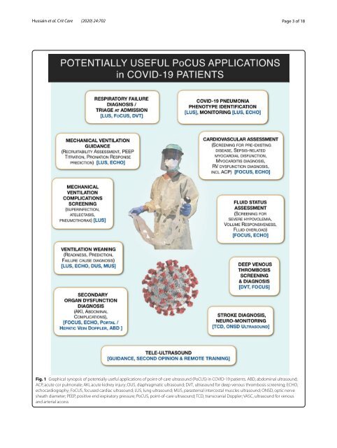
Hussain <strong>et</strong> <strong>al</strong>. <strong>Crit</strong> <strong>Care</strong> (<strong>2020</strong>) 24:702 Page 3 of 18 Fig. 1 Graphic<strong>al</strong> synopsis of potenti<strong>al</strong>ly useful applications of point-of-care <strong>ultrasound</strong> (PoCUS) in COVID-19 patients. ABD, abdomin<strong>al</strong> <strong>ultrasound</strong>; ACP, acute cor pulmon<strong>al</strong>e; AKI, acute kidney injury; DUS, diaphragmatic <strong>ultrasound</strong>; DVT, <strong>ultrasound</strong> <strong>for</strong> deep venous thrombosis screening; ECHO, echocardiography; FoCUS, focused cardiac <strong>ultrasound</strong>; LUS, lung <strong>ultrasound</strong>; MUS, parastern<strong>al</strong> intercost<strong>al</strong> muscles <strong>ultrasound</strong>; ONSD, optic nerve sheath diam<strong>et</strong>er; PEEP, positive end expiratory pressure; PoCUS, point-of-care <strong>ultrasound</strong>; TCD, transcrani<strong>al</strong> Doppler; VASC, <strong>ultrasound</strong> <strong>for</strong> venous and arteri<strong>al</strong> access