Hussein A et al. Crit Care 2020 Multi‑organ point‑of‑care ultrasound for COVID‑19 (PoCUS4COVID): international expert consensus
Download at publisher: https://ccforum.biomedcentral.com/articles/10.1186/s13054-020-03369-5
Download at publisher: https://ccforum.biomedcentral.com/articles/10.1186/s13054-020-03369-5

Sie wollen auch ein ePaper? Erhöhen Sie die Reichweite Ihrer Titel.
YUMPU macht aus Druck-PDFs automatisch weboptimierte ePaper, die Google liebt.
Hussain <strong>et</strong> <strong>al</strong>. <strong>Crit</strong> <strong>Care</strong> (<strong>2020</strong>) 24:702<br />
Page 5 of 18<br />
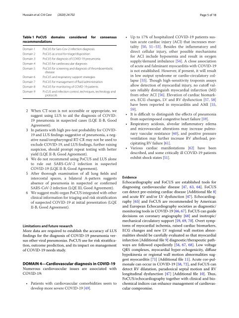
Table 1 PoCUS domains considered <strong>for</strong> <strong>consensus</strong><br />
recommendations<br />
Domain 1<br />
Domain 2<br />
Domain 3<br />
Domain 4<br />
Domain 5<br />
Domain 6<br />
Domain 7<br />
Domain 8<br />
Domain 9<br />
PoCUS <strong>for</strong> Sars-Cov-2 infection diagnosis<br />
PoCUS as a tool <strong>for</strong> triage/disposition<br />
PoCUS <strong>for</strong> diagnosis of COVID-19 pneumonia<br />
PoCUS <strong>for</strong> cardiovascular diagnosis<br />
PoCUS <strong>for</strong> screening and diagnosis of thromboembolic<br />
disease<br />
PoCUS and respiratory support strategies<br />
PoCUS <strong>for</strong> management of fluid administration<br />
PoCUS <strong>for</strong> monitoring of COVID-19 patients<br />
PoCUS and infection control, techniques, technology and<br />
protocols<br />
2 When CT-scan is not accessible or appropriate, we<br />
suggest using LUS to aid the diagnosis of COVID-<br />
19 pneumonia in suspected cases (LQE II-B, Good<br />
Agreement).<br />
3 In patients with high pre-test probability <strong>for</strong> COVID-<br />
19 and LUS findings suggestive of pneumonia, a negative<br />
nas<strong>al</strong>/oropharynge<strong>al</strong> RT-CR may not be used to<br />
exclude COVID-19, and LUS findings, further raising<br />
suspicion, should prompt repeat testing with b<strong>et</strong>ter<br />
yield (LQE II-B, Good Agreement).<br />
4 We do not recommend using PoCUS and LUS <strong>al</strong>one<br />
to rule out SARS-CoV-2 infection in suspected<br />
COVID-19 (LQE II-B, Good Agreement).<br />
5 After thorough examination of <strong>al</strong>l lung fields and<br />
intercost<strong>al</strong> spaces, a bilater<strong>al</strong> A-pattern suggests<br />
absence of pneumonia in suspected or confirmed<br />
SARS-CoV-2 infection (LQE III, Good Agreement).<br />
6 We suggest multi-organ PoCUS integrated with other<br />
clinic<strong>al</strong> in<strong>for</strong>mation <strong>for</strong> triaging and risk stratification<br />
of suspected COVID-19 at initi<strong>al</strong> presentation (LQE<br />
II-B, Good Agreement).<br />
Limitations and future research<br />
More data are required to establish the accuracy of LUS<br />
findings <strong>for</strong> the diagnosis of COVID-19 pneumonia versus<br />
other vir<strong>al</strong> pneumonias. PoCUS use <strong>for</strong> risk stratification,<br />
outcome prediction, and its impact on management<br />
of COVID-19 needs study.<br />
DOMAIN 4—Cardiovascular diagnosis in COVID‐19<br />
Numerous cardiovascular issues are associated with<br />
COVID-19:<br />
• Patients with cardiovascular comorbidities seem to<br />
develop more severe COVID-19 [49].<br />
• Up to 17% of hospit<strong>al</strong>ized COVID-19 patients sustain<br />
acute cardiac injury (ACI) that increases mort<strong>al</strong>ity<br />
[50, 51–53]. Besides the inflammatory and<br />
direct cellular injury, other possible mechanisms<br />
<strong>for</strong> ACI include hypoxemia and result in oxygen<br />
supply/demand imb<strong>al</strong>ance [54]. A close association<br />
of acute and fulminant myocarditis with COVID-19<br />
is not established. However, if present, it will result<br />
in low output syndrome or cardio-circulatory collapse<br />
[55]. Though high-sensitivity troponin assays<br />
<strong>al</strong>low d<strong>et</strong>ection of myocardi<strong>al</strong> injury, no cutoff v<strong>al</strong>ues<br />
reliably distinguish myocardi<strong>al</strong> infarction (MI)<br />
from other ACI [56]. Elevation of cardiac biomarkers,<br />
ECG changes, LV and RV dysfunction [57, 58]<br />
have been reported in myocarditis and AMI [55,<br />
59].<br />
• It is difficult to distinguish the effects of pneumonia<br />
from superimposed congestive heart failure [59].<br />
• Respiratory acidosis, <strong>al</strong>veolar inflammatory edema<br />
and microvascular <strong>al</strong>terations may increase pulmonary<br />
vascular resistance [60], and positive pressure<br />
ventilation may further increase RV afterload, precipitating<br />
RV failure [61].<br />
• Various cardiac manifestations [62] have been<br />
described, and some critic<strong>al</strong>ly ill COVID-19 patients<br />
exhibit shock states [51].<br />
Evidence<br />
Echocardiography and FoCUS are established tools <strong>for</strong><br />
diagnosing cardiovascular disease [47, 63, 64]. FoCUS<br />
can d<strong>et</strong>ect pre-existing cardiac disease [Addition<strong>al</strong> file 8]<br />
and acute RV and/or LV dysfunction [47]. Echocardiography<br />
[65] and FoCUS are recommended by American<br />
and European Echocardiography soci<strong>et</strong>ies as diagnostic/<br />
monitoring tools in COVID-19 [66, 67]. FoCUS can guide<br />
decisions on coronary angiography [68] and inotropic/<br />
mechanic<strong>al</strong> circulatory support [59, 69, 70]. Overt symptoms<br />
of myocardi<strong>al</strong> ischemia, raised cardiac biomarkers,<br />
ECG changes and new LV region<strong>al</strong> w<strong>al</strong>l motion abnorm<strong>al</strong>ities<br />
should be carefully ev<strong>al</strong>uated so that myocardi<strong>al</strong><br />
infarction [Addition<strong>al</strong> file 9] diagnostic/therapeutic pathways<br />
are followed expediently [54, 67, 68]. Low voltage<br />
QRS complexes, myocardi<strong>al</strong> hyper-echogenicity, diffuse<br />
hypokinesia or region<strong>al</strong> w<strong>al</strong>l motion abnorm<strong>al</strong>ities suggest<br />
myocarditis [71] [Addition<strong>al</strong> file 11]. Acute cor-pulmon<strong>al</strong>e<br />
can occur in COVID-19 [58, 72], and FoCUS can<br />
d<strong>et</strong>ect RV dilatation, paradoxic<strong>al</strong> sept<strong>al</strong> motion and RV<br />
longitudin<strong>al</strong> dysfunction [47] [Addition<strong>al</strong> file 10]. Thus,<br />
FoCUS/echocardiography tog<strong>et</strong>her with clinic<strong>al</strong> and biochemic<strong>al</strong><br />
indices can enhance management of cardiovascular<br />
compromise.