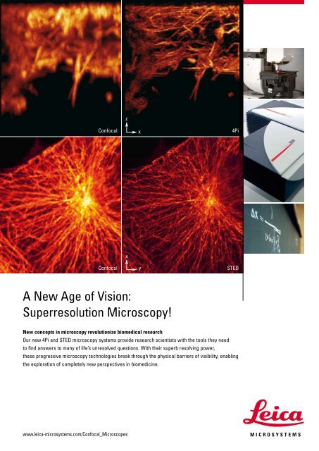
A New Age of Vision: Superresolution Microscopy! New concepts in microscopy revolutionize biomedical research Our new 4Pi and STED microscopy systems provide research scientists with the tools they need to find answers to many of life’s unresolved questions. With their superb resolving power, these progressive microscopy technologies break through the physical barriers of visibility, enabling the exploration of completely new perspectives in biomedicine. www.leica-microsystems.com/Confocal_Microscopes z Confocal x 4Pi x Confocal y STED
Nobel Prize for Surface Scientist Gerhard Ertl Dear Reader, Some days ago the Royal Swedish Academy of Sciences announced the 2007 Nobel Prize in Chemistry for Gerhard Ertl, professor emeritus at the Fritz-Haber Institute in Berlin. Imaging & Microscopy congratulates Gerhard Ertl for this most prestigious honour in science that is awarded due to his thorough studies of chemical reactions on solid surfaces. When having a closer look on fundamental molecular processes at the gas-solid interface, small gas molecules may either be adsorbed or bounce back at the solid surface, according to a note by the Royal Swedish Academy of Sciences (www.kva.se). The first case includes the most interesting possibilities: The gas molecule can dissociate at the interface, the chemical properties of the surface can be changed, or the absorbed molecule can chemically react with a previously absorbed one. Ertl’s giant step forward in the understanding of these scenarios led to various breakthroughs in the development of catalysts being invaluable across industry. Carbon monoxide and hydrocarbons are converted to carbon dioxide in vehicles exhaust gasses today, and even the content of nitrous gasses can significantly be reduced. Car manufactures around the globe are producing vehicles that are less harmful to the environment and more fuel efficient. Very early, in the mid seventies, Ertl unravelled the surface mechanism of ammonia synthesis, a reaction that was first discovered by Fritz Haber, reaching such a technical and economical significance. By applying new surface science methods he showed, that the active species is not molecular but dissociatively adsorbed atomic nitrogen. The nitrogen is hydrogenated in a step-process. In terms of a more general description of related applications, Ertl’s findings had impact on the microelectronics industries where thin semiconductor layers are formed by CVD, chemical vapour deposition. Corrosion protection is yet another area that crucially depends on knowledge in surface science. Ertl’s findings help solving problems caused by corrosion both in daily life and in industry, for example, related to aeronautics or nuclear power plants. Ertl’s surface studies have opened a wide span of new techniques. A citation from the Royal Swedish Academy of Sciences says that “Gerhard Ertl had been one of the first to see the potential of these new techniques. Step by step he had created a methodology for surface chemistry by demonstrating how different experimental procedures can be used to provide a complete picture of a surface reaction”. Ertl recognized the significance of a microscopy related methodology in surface science very early. Quite interestingly Ertl and his group have set up a scanning tunneling microscope, STM, for imaging surface reconstructions in the presence of adsorbates at a time where many others were skeptical about STM, after its invention by Binnig and Rohrer in 1982. Some years later Ertl and coworkers succeeded in directly visualizing diffusion processes using high speed STM in order to verify macroscopic laws. Another example demonstrating Ertl’s ambition to take use out of modern microscopy methods in heterogeneous catalysis research is the observation of adsoption patterns in case of CO oxidation on Pt by Photo Emission Electron Microscopy, PEEM. Some excellent illustrations of adsoption patterns are published by the Surface Imaging Group, Dept. of Physical Chemistry, Fritz-Haber Institute of the Max-Planck-Society, www.fhi-berlin. mpg.de/surfimag. Field Ion Microscopy, FIM, is another important experimental approach to study catalytic reactions at the nanometer scale. This unique microscopy method has been further developed and applied to imaging and in-situ chemical probing in heterogeneous catalysis by Norbert Kruse, former Fritz- Haber Institute staff member and scientific advisor of Imaging & Microscopy [1, 2]. In this issue of Imaging & Microscopy some excellent scientific articles are published, which give a view to recent research in Scanning Probe Microscopy, Compositional Analysis, Electron- and Light Microscopy. Furthermore, we like to steer the reader’s attention to Imaging & Microscopy’s conference reports and announcements. Enjoy your reading this issue Martin Friedrich Thomas Matzelle [1] Visart de Bocarmé T., Imaging & Microscopy 8 (1), 19–21 (2006). [2] News & People, Imaging & Microscopy 9 (3), 8 (2007). E d i t o r i a l [3] Freund H.-J., Knözinger H., J. Phys. Chem. B, 108, 38, 14183–14186 (2004). G.I.T. Imaging & Microscopy 4/2007 •