Huge Images? - GIT Verlag
Huge Images? - GIT Verlag
Huge Images? - GIT Verlag
Create successful ePaper yourself
Turn your PDF publications into a flip-book with our unique Google optimized e-Paper software.
camera. A frequency domain system for<br />
FLIM-TIRF is under development together<br />
with Lambert Instruments based<br />
on their LIFA system, which will offer enhanced<br />
time resolution compared to the<br />
TCSPC approach and sensitive detection<br />
of protein-protein interactions at the cell<br />
surface due to the optical sectioning of<br />
TIRF. A LIFA system coupled to a<br />
Yokogawa CSU22 spinning disk scanhead<br />
is also planned. Room 130 houses several<br />
upright stands for imaging fixed<br />
specimens, including histological stains,<br />
immunofluorescence, and FISH. Offline<br />
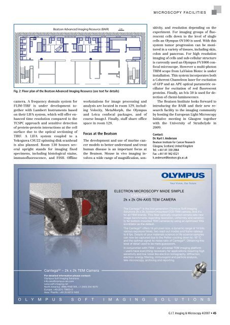
Beatson Advanced Imaging Resource (BAIR)<br />
rm 122 rm 123 rm 124<br />
rm 125 / rm 126 rm 127 rm 130 rm 129 rm 131<br />
C<br />
D E<br />
E<br />
Fig. 2: Floor plan of the Beatson Advanced Imaging Resource (see text for details)<br />
�<br />
������� �� ���������������������<br />
����������������������������������������<br />
�������������������������������<br />
�������������������������<br />
����������������������<br />
�������������������������������������������������<br />
��������������������������<br />
��������������������������������<br />
B<br />
A<br />
workstations for image processing and<br />
analysis are located in room 129, including<br />
Volocity, MetaMorph, the Olympus<br />
and Leica confocal packages, and of<br />
course ImageJ. Finally, staff share office<br />
space in room 128.<br />
Focus at the Beatson<br />
rm 128<br />
The development and use of murine cancer<br />
models to better understand and treat<br />
human disease is an important focus at<br />
the Beatson. Mouse in vivo imaging involves<br />
a wide range of magnification, sen-<br />
A<br />
2 m<br />
M i c r o s c o p y Fa c i l i t i e s<br />
sitivity, and resolution depending on the<br />
experiment. For imaging groups of fluorescent<br />
cells down to the level of single<br />
cells an Olympus OV100 is used. With this<br />
system tumor progression can be monitored<br />
in a variety of tissues, including skin,<br />
colon and pancreas. For high resolution<br />
imaging of cells and sub-cellular structure<br />
is currently used an Olympus FV1000 confocal<br />
microscope. However a multi-photon<br />
TRIM scope from LaVision Biotec is under<br />
installation. This system incorporates both<br />
a Coherent Chameleon laser for excitation<br />
of GFP and an APE optical parametric oscillator<br />
for excitation of red fluorescent<br />
proteins. Finally, an Ivis 50 is used for detection<br />
of chemi-luminescence.<br />
The Beatson Institute looks forward to<br />
introducing the BAIR and their new research<br />
facility to the imaging community<br />
by hosting the European Light Microscopy<br />
Initiative meeting in Glasgow together<br />
with the University of Strathclyde in<br />
2009.<br />
Contact:<br />
Dr. Kurt I. Anderson<br />
Beatson Institute for Cancer Research<br />
Glasgow, Scotland, United Kingdom<br />
Tel.: +44 141 330 2864<br />
Fax: +44 141 942 6521<br />
k.anderson@beatson.gla.ac.uk<br />
��������������������������������<br />
� � ������� ��<br />
� ��������������������������<br />
����������� �� ���������������������������������������������<br />
�����������������������������������������������������������<br />
����������������������������������������������������������������<br />
����������������������������������������������������������������<br />
������������������������������������������������������������<br />
����������������������������<br />
����������� �� ������������������������������������������������������<br />
�������������������������������������������������������������<br />
��������������������������������������������������������������������<br />
��������������������������������������������������������������<br />
������������������������������������������������ �� �����������������<br />
������������������������������������������<br />
��������������������������������������������������������������<br />
������������������������������������������������������������������<br />
�����������������������������������������������������������������<br />
�������������������������������������������������������������<br />
�����������������������������������������<br />
� � � � � � � � � � � � � � � � � � � � � � � � � � � � � �<br />
G.I.T. Imaging & Microscopy 4/2007 • 45


