The Early Signs of Right Bundle Branch Block - Chest
The Early Signs of Right Bundle Branch Block - Chest
The Early Signs of Right Bundle Branch Block - Chest

Create successful ePaper yourself
Turn your PDF publications into a flip-book with our unique Google optimized e-Paper software.
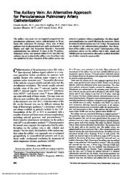
228 A. II _III<br />
AVR AVI. AVE<br />
25.8.1966,!<br />
FIGURE 7. Electrocardiograms <strong>of</strong> case 5.<br />
two beats <strong>of</strong> the bottom strips probably represent the same phe-<br />
nomenon.<br />
B.<br />
I II III AVR AVL AVE<br />
COMMENT<br />
<strong>The</strong> rhythm strips <strong>of</strong> all <strong>of</strong> the cases depicted above<br />
show intermittent right bundle branch block aberra-<br />
tion, during which a diminished S wave in right-on-<br />
ented leads appears as the earliest manifestation <strong>of</strong> in-<br />
complete right bundle branch block. This stimulated<br />
the search for this manifestation in serial 12-lead<br />
ECGs.<br />
CASES 5 TO 8<br />
An analysis <strong>of</strong> 11,000 ECGs <strong>of</strong>healthy aviation personnel yielded<br />
25 cases <strong>of</strong> right bundle branch block. Four <strong>of</strong> these cases had two<br />
preceding ECGs with normal (narrow) QRS configurations, pre-<br />
sented here as cases 5 to 8. <strong>The</strong>se ECGs preceding the complete<br />
right bundle branch block were recorded on two different dates, the<br />
intervening times ranging from one to seven years. An example is<br />
shown in Figure 7. <strong>The</strong> amplitudes <strong>of</strong>the S waves in leads V, and V2<br />
as well as the R waves in lead V, <strong>of</strong>all four cases are shown in Figure<br />
8. All four cases showed a diminution <strong>of</strong> the S wave in lead V2. Two<br />
cases had a diminution <strong>of</strong> the S wave in lead V,, one showed no<br />
change, and one had a minimal increase <strong>of</strong> 1 mm. <strong>The</strong> R wave<br />
amplitude in lead V5 was diminished in two cases and remained the<br />
same in the other two. <strong>Right</strong> bundle branch block was observed in<br />
the tracings <strong>of</strong> all four cases recorded one to five years later.<br />
DIsCUssIoN<br />
<strong>The</strong> right bundle branch block form <strong>of</strong>aberration in<br />
the rhythm strips <strong>of</strong> cases 1 to 4, and the sequential,<br />
time-spaced, 12-lead ECGs <strong>of</strong> cases 5 to 8 reflect the<br />
earliest manifestation <strong>of</strong>right bundle branch block as a<br />
diminution <strong>of</strong> the S wave amplitude in right-oriented<br />
leads, particularly lead V2.<br />
Mechanism<br />
Normal ventricular activation begins in the left side<br />
Downloaded From: http://journal.publications.chestnet.org/ on 03/27/2013<br />
S In<br />
VI<br />
S R In<br />
VS<br />
FIGURE 8. Graphic representation <strong>of</strong>the S wave amplitudes in leads<br />
V, and V2, and the R wave amplitude in lead V, <strong>of</strong> cases 5 to 8.<br />
<strong>of</strong>the interventricular septum and spreads from left to<br />
right through the septum (vector 16, Fig 9). This occurs<br />
near-synchronously with a smaller vector from the<br />
right side in the interventricular septum (vector la, Fig<br />
9). <strong>The</strong> dominant force is thus from left to right and is<br />
responsible for the normal small initial r wave in lead V,<br />
and the normal small initial q wave in lead V6. Septal<br />
activation is followed by paraseptal activation from<br />
endocardial to epicardial surfaces (vectors 2a and 2b,<br />
Fig 9). <strong>The</strong>se paraseptal vectors are larselv responsible<br />
FIGURE 9. Diagrammatic representations <strong>of</strong> normal ventricular<br />
depolarization.<br />
CHEST I 87 1 2 I FEBRUARY, 1985 183