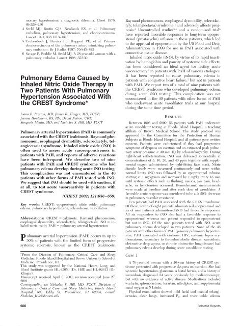
Pulmonary Edema Caused by Inhaled Nitric Oxide Therapy in Two ...
Pulmonary Edema Caused by Inhaled Nitric Oxide Therapy in Two ...
Pulmonary Edema Caused by Inhaled Nitric Oxide Therapy in Two ...

Create successful ePaper yourself
Turn your PDF publications into a flip-book with our unique Google optimized e-Paper software.
monary hypertension: a diagnostic dilemma. Chest 1976;<br />
69:229–230<br />
4 Seckl MJ, Rust<strong>in</strong> GJS, Newlands ES, et al. <strong>Pulmonary</strong><br />
embolism, pulmonary hypertension, and choriocarc<strong>in</strong>oma.<br />
Lancet 1991; 338:1313–1315<br />
5 Trubenbach J, Pereira PL, Huppert PE, et al. Primary<br />
choriocarc<strong>in</strong>oma of the pulmonary artery mimick<strong>in</strong>g pulmonary<br />
embolism. Br J Radiol 1997; 70:843–845<br />
6 Savage P, Roddie M, Seckl MJ. A 28-year-old woman with a<br />
pulmonary embolus. Lancet 1998; 352:30<br />
<strong>Pulmonary</strong> <strong>Edema</strong> <strong>Caused</strong> <strong>by</strong><br />
<strong>Inhaled</strong> <strong>Nitric</strong> <strong>Oxide</strong> <strong>Therapy</strong> <strong>in</strong><br />
<strong>Two</strong> Patients With <strong>Pulmonary</strong><br />
Hypertension Associated With<br />
the CREST Syndrome*<br />
Ioana R. Preston, MD; James R. Kl<strong>in</strong>ger, MD, FCCP;<br />
Jeanne Houtchens, BS, RN; David Nelson, CRT;<br />
Sangeeta Mehta, MD; and Nicholas S. Hill, MD, FCCP<br />
<strong>Pulmonary</strong> arterial hypertension (PAH) is commonly<br />
associated with the CREST (calc<strong>in</strong>osis, Raynaud phenomenon,<br />
esophageal dysmotility, sclerodactyly, telangiectasia)<br />
syndrome. <strong>Inhaled</strong> nitric oxide (iNO) is<br />
often used to assess acute vasoresponsiveness <strong>in</strong><br />
patients with PAH, and reports of adverse reactions<br />
have been <strong>in</strong>frequent. We describe two of n<strong>in</strong>e<br />
patients with PAH and CREST syndrome who had<br />
pulmonary edema develop dur<strong>in</strong>g acute iNO test<strong>in</strong>g.<br />
This complication was not encountered <strong>in</strong> the 46<br />
patients with other forms of PAH tested with iNO.<br />
We suggest that iNO should be used with caution, if<br />
at all, to test acute vasoreactivity <strong>in</strong> patients with<br />
CREST syndrome.<br />
(CHEST 2002; 121:656–659)<br />
Key words: CREST; epoprostenol; nitric oxide; pulmonary<br />
edema; pulmonary hypertension; scleroderma; vasodilators<br />
Abbreviations: CREST calc<strong>in</strong>osis, Raynaud phenomenon,<br />
esophageal dysmotility, sclerodactyly, telangiectasia; iNO <strong>in</strong>haled<br />
nitric oxide; PAH pulmonary arterial hypertension<br />
<strong>Pulmonary</strong> arterial hypertension (PAH) occurs <strong>in</strong> up to<br />
50% of patients with the limited form of progressive<br />
systemic sclerosis, known as the CREST (calc<strong>in</strong>osis,<br />
*From the Division of <strong>Pulmonary</strong>, Critical Care and Sleep<br />
Medic<strong>in</strong>e, Rhode Island Hospital and Brown University School of<br />
Medic<strong>in</strong>e, Providence, RI.<br />
This study was supported <strong>by</strong> the National Heart, Lung, and<br />
Blood Institute grants HL-45050 (Dr. Hill) and HL-02613 (Dr.<br />
Kl<strong>in</strong>ger).<br />
Manuscript received April 6, 2001; revision accepted June 27,<br />
2001.<br />
Correspond<strong>in</strong>g to: Nicholas S. Hill, MD, FCCP, Division of<br />
<strong>Pulmonary</strong>, Critical Care and Sleep Medic<strong>in</strong>e, Rhode Island<br />
Hospital, 593 Eddy St, Providence, RI 02903; e-mail:<br />
Nicholas_Hill@Brown.edu<br />
Raynaud phenomenon, esophageal dysmotility, sclerodactyly,<br />
telangiectasia) syndrome, 1 and adversely affects prognosis.<br />
2 Uncontrolled studies 3,4 and a randomized trial 5<br />
have reported favorable responses to long-term epoprostenol<br />
(prostacycl<strong>in</strong>) <strong>in</strong>fusion <strong>in</strong> these patients, which led<br />
to the approval of epoprostenol <strong>by</strong> the US Food and Drug<br />
Adm<strong>in</strong>istration <strong>in</strong> 1999 for use <strong>in</strong> PAH associated with<br />
connective tissue disease.<br />
<strong>Inhaled</strong> nitric oxide (iNO), <strong>by</strong> virtue of its rapid <strong>in</strong>activation<br />
<strong>by</strong> hemoglob<strong>in</strong> and paucity of systemic side effects,<br />
has been considered an ideal agent for test<strong>in</strong>g acute<br />
vasoreactivity 6 <strong>in</strong> patients with PAH of various etiologies.<br />
It has been reported to cause pulmonary edema <strong>in</strong><br />
patients with congestive heart failure, 7 but not <strong>in</strong> patients<br />
with PAH. We report two of a total of n<strong>in</strong>e patients with<br />
the CREST syndrome who developed pulmonary edema<br />
dur<strong>in</strong>g acute iNO test<strong>in</strong>g. This complication was not<br />
encountered <strong>in</strong> the 46 patients with other forms of PAH<br />
who underwent acute vasodilator trials at our hospital<br />
dur<strong>in</strong>g the same time period.<br />
Results<br />
Between 1996 and 2000, 56 patients with PAH underwent<br />
acute vasodilator test<strong>in</strong>g at Rhode Island Hospital, a teach<strong>in</strong>g<br />
affiliate of Brown Medical School. The study protocol was<br />
approved <strong>by</strong> the Committee for the Protection of Human<br />
Subjects at Rhode Island Hospital, and all patients gave written<br />
consent. Patients were catheterized if they had progressive<br />
symptoms of dyspnea on exertion and an estimated peak pulmonary<br />
artery pressure 40 mm Hg <strong>by</strong> echocardiography. Dur<strong>in</strong>g<br />
right-heart catheterization, iNO was delivered sequentially at<br />
concentrations of 5, 10, 20, and 40 ppm together with supplemental<br />
oxygen adm<strong>in</strong>istered <strong>by</strong> tight-fitt<strong>in</strong>g face mask. <strong>Nitric</strong><br />
dioxide levels were measured cont<strong>in</strong>uously and were with<strong>in</strong><br />
normal limits. iNO was followed <strong>by</strong> an epoprostenol <strong>in</strong>fusion<br />
start<strong>in</strong>g at 1 ng/kg/m<strong>in</strong> and <strong>in</strong>creased <strong>by</strong> 1 ng/kg every 15 m<strong>in</strong><br />
until systemic effects such as flush<strong>in</strong>g, nausea, jaw pa<strong>in</strong>, headache,<br />
or hypotension occurred. Hemodynamic measurements<br />
were made at basel<strong>in</strong>e and after each dose of vasodilator. A<br />
favorable acute response was considered to be a 20% decrease<br />
<strong>in</strong> pulmonary vascular resistance.<br />
Ten patients had PAH associated with the CREST syndrome.<br />
Of these, seven of eight patients adm<strong>in</strong>istered epoprostenol and<br />
six of n<strong>in</strong>e patients adm<strong>in</strong>istered iNO had favorable responses.<br />
All six responders to iNO also had a favorable response to<br />
epoprostenol, whereas one patient responded to epoprostenol<br />
but not to iNO. Of the n<strong>in</strong>e patients tested with iNO, acute<br />
pulmonary edema developed <strong>in</strong> two patients. None of the 46<br />
patients with other forms of PAH (primary pulmonary hypertension,<br />
PAH associated with cirrhosis, HIV, systemic lupus erythematosus,<br />
secondary to thromboembolic disease, sarcoidosis,<br />
obstructive sleep apnea, or chronic obstructive lung disease) had<br />
pulmonary edema develop dur<strong>in</strong>g acute vasodilator test<strong>in</strong>g.<br />
Case 1<br />
A 70-year-old woman with a 20-year history of CREST syndrome<br />
presented with progressive dyspnea on exertion. She had<br />
systemic hypertension, glaucoma, a hiatal hernia, and a history of<br />
sarcoidosis diagnosed 24 years previously <strong>by</strong> mediast<strong>in</strong>oscopy,<br />
but with no evidence of active disease. Medications <strong>in</strong>cluded<br />
warfar<strong>in</strong>, spironolactone, losartan, nifedip<strong>in</strong>e, and supplemental<br />
nasal oxygen at 5 L/m<strong>in</strong>.<br />
Physical exam<strong>in</strong>ation showed mild facial and manual telangiectasias,<br />
clear lungs, <strong>in</strong>creased P 2, and trace ankle edema.<br />
656 Selected Reports<br />
Downloaded From: http://journal.publications.chestnet.org/ on 04/02/2013
Spirometry results and lung volumes were normal on pulmonary<br />
function test<strong>in</strong>g, and diffusion capacity of the lung for carbon<br />
monoxide was 16% of predicted. Arterial blood gas measures<br />
obta<strong>in</strong>ed with the patient breath<strong>in</strong>g oxygen at 3 L/m<strong>in</strong> were as<br />
follows: pH, 7.34; Po 2, 52 mm Hg; and Pco 2, 39 mm Hg. CT of<br />
the chest showed m<strong>in</strong>imal ground-glass opacities.<br />
A left-heart catheterization revealed normal coronary arteries<br />
and left ventricular function. Hemodynamic measurements dur<strong>in</strong>g<br />
right-heart catheterization are shown <strong>in</strong> Table 1. The test was<br />
performed while the patient breathed 40% oxygen, and oxygen<br />
saturation was ma<strong>in</strong>ta<strong>in</strong>ed at 90%. Dur<strong>in</strong>g iNO adm<strong>in</strong>istration<br />
at 40 ppm, severe dyspnea, tachypnea, and tachycardia developed,<br />
and oxygen saturation fell to 85%. Lung auscultation<br />
revealed new bilateral rales. An ECG did not show ischemic<br />
changes. <strong>Pulmonary</strong> artery wedge pressure and pulmonary artery<br />
pressure rema<strong>in</strong>ed unchanged. iNO was immediately discont<strong>in</strong>ued,<br />
and subl<strong>in</strong>gual nitroglycer<strong>in</strong> and IV morph<strong>in</strong>e and furosemide<br />
were adm<strong>in</strong>istered, with rapid symptomatic improvement.<br />
A chest radiograph obta<strong>in</strong>ed after symptomatic<br />
improvement showed mild <strong>in</strong>terstitial edema. The next morn<strong>in</strong>g,<br />
epoprostenol <strong>in</strong>fusion was <strong>in</strong>itiated that was tolerated up to<br />
4 ng/kg/m<strong>in</strong> (Table 1). Treatment with epoprostenol was followed<br />
<strong>by</strong> an IV nitroglycer<strong>in</strong> <strong>in</strong>fusion up to 80 g/m<strong>in</strong> that was<br />
discont<strong>in</strong>ued because of systemic hypotension. The patient was<br />
discharged home with a cont<strong>in</strong>uous subcutaneous <strong>in</strong>fusion of<br />
prostacycl<strong>in</strong> (UT-15; United Therapeutics; Research Triangle<br />
Park, NC) and has rema<strong>in</strong>ed symptomatically improved for 6<br />
months.<br />
Case 2<br />
A 57-year-old woman with a 24-year history of CREST syndrome<br />
and a 2-year history of progressive dyspnea on exertion was referred<br />
for acute vasodilator test<strong>in</strong>g. Her medications <strong>in</strong>cluded nifedip<strong>in</strong>e<br />
and omeprazole and supplemental nasal oxygen at 4 to 5 L/m<strong>in</strong>.<br />
Physical exam<strong>in</strong>ation revealed facial telangiectasias, few bibasilar<br />
rales on lung auscultation, a prom<strong>in</strong>ent right ventricular impulse, an<br />
<strong>in</strong>creased P 2, a holosystolic murmur at the left lower sternal border,<br />
trace ankle edema, and sclerodactyly.<br />
<strong>Pulmonary</strong> function tests showed a total lung capacity of 52% of<br />
predicted and a diffusion capacity of the lung for carbon monoxide<br />
of 26% of predicted. Oxygen saturation of hemoglob<strong>in</strong> with the<br />
patient at rest and receiv<strong>in</strong>g oxygen supplementation at 4 L/m<strong>in</strong> was<br />
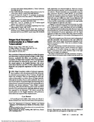
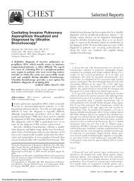
94%. Chest radiography revealed <strong>in</strong>creased mark<strong>in</strong>gs at the lung<br />
bases and small pleural effusions bilaterally (Fig 1, top). Echocardiography<br />
revealed a peak pulmonary artery pressure of 67 mm Hg<br />
and normal left ventricular function. Hemodynamic measurements<br />
dur<strong>in</strong>g heart catheterization with the patient breath<strong>in</strong>g 35% oxygen<br />
are shown <strong>in</strong> Table 1. While receiv<strong>in</strong>g iNO at 20 ppm, acute<br />
dyspnea, tachypnea, and <strong>in</strong>creased bilateral rales developed. Oxygen<br />
saturation was ma<strong>in</strong>ta<strong>in</strong>ed at 90% after the fraction of <strong>in</strong>spired<br />
oxygen was <strong>in</strong>creased to 100% <strong>by</strong> face mask. Chest radiography<br />
showed <strong>in</strong>creased <strong>in</strong>terstitial mark<strong>in</strong>gs compatible with pulmonary<br />
edema (Fig 1, middle), but pulmonary artery wedge pressure<br />
rema<strong>in</strong>ed unchanged. After prompt discont<strong>in</strong>uation of iNO, IV<br />
morph<strong>in</strong>e and furosemide were adm<strong>in</strong>istered and symptoms improved<br />
rapidly. A follow-up chest radiograph obta<strong>in</strong>ed 7 h after the<br />
episode showed improvement of the <strong>in</strong>terstitial mark<strong>in</strong>gs (Fig 1,<br />
bottom). The next day, epoprostenol <strong>in</strong>fusion was adm<strong>in</strong>istered only<br />
up to 2 ng/kg/m<strong>in</strong>, due to systemic hypotension. The patient was<br />
discharged home receiv<strong>in</strong>g epoprostenol at 1 ng/kg/m<strong>in</strong> and had a<br />
transient improvement <strong>in</strong> her dyspnea. She died 4 months later of<br />
<strong>in</strong>tractable right-heart failure. Pathologic exam<strong>in</strong>ation of the lungs<br />
showed <strong>in</strong>terstitial fibrosis and <strong>in</strong>timal thicken<strong>in</strong>g of pulmonary<br />
arteries, compatible with her diagnosis of CREST syndrome. There<br />
was no evidence of left-heart or pulmonary venous disease.<br />
Discussion<br />
We observed symptoms and signs of acute pulmonary<br />
edema dur<strong>in</strong>g iNO test<strong>in</strong>g <strong>in</strong> two patients with severe<br />
PAH associated with the CREST syndrome. No such<br />
reaction developed <strong>in</strong> 46 other patients with PAH of<br />
different etiologies undergo<strong>in</strong>g vasodilator test<strong>in</strong>g with<br />
iNO or epoprostenol.<br />
Acute pulmonary edema has previously been reported<br />
<strong>in</strong> patients with scleroderma dur<strong>in</strong>g short-term8–10 and<br />
Table 1—Hemodynamic Characteristics of the <strong>Two</strong> Patients at Basel<strong>in</strong>e and Dur<strong>in</strong>g Short-term Vasodilator Test<strong>in</strong>g*<br />
Patient No. Vasodilator<br />
SBP,<br />
mm Hg<br />
PAP,<br />
mm Hg<br />
mPAP,<br />
mm Hg<br />
PVR,<br />
dyne s cm 5<br />
PAWP,<br />
mm Hg CO, L/m<strong>in</strong><br />
SVR,<br />
dyne s cm 5<br />
1<br />
iNO basel<strong>in</strong>e 119/60 98/37 61 1,054 12 3.64 1,472<br />
iNO, 5 ppm 121/48 88/32 54 755 13 4.34 1,198<br />
iNO, 10 ppm 112/47 87/31 53 798 8 4.51 1,081<br />
iNO, 20 ppm 114/44 87/31 52 818 8 4.30 1,227<br />
iNO, 40 ppm† 120/68 88/30 52 7<br />
Epo basel<strong>in</strong>e 108/38 88/31 53 1,132 5 3.39 1,297<br />
Epo, 3 ng/kg/m<strong>in</strong> 96/52 70/26 43 690 6 4.17 1,208<br />
Epo, 4 ng/kg/m<strong>in</strong> 101/52 74/27 46 701 7 4.45 1,150<br />
2<br />
iNO basel<strong>in</strong>e 110/94 86/34 51 945 12 3.30 2,182<br />
iNO, 5 ppm 98/67 72/25 41 954 10 3.10 1,935<br />
iNO, 10 ppm 90/69 78/29 45 867 11 3.50 1,600<br />
iNO, 20 ppm† 135/73 85/34 51 945 12 3.30 2,109<br />
Epo basel<strong>in</strong>e 105/97 81/30 47 1,020 10 2.90 2,538<br />
Epo, 1 ng/kg/m<strong>in</strong> 97/66 81/47 47 921 9 3.30 1,503<br />
Epo, 2 ng/kg/m<strong>in</strong> 84/59 81/30 47 9<br />
*SBP systemic BP; PAP pulmonary artery pressure; mPAP mean pulmonary artery pressure; PVR pulmonary vascular resistance;<br />
PAWP pulmonary arterial wedge pressure; CO cardiac output; SVR systemic vascular resistance; Epo epoprostenol.<br />
†Dose of iNO at which pulmonary edema occurred.<br />
Downloaded From: http://journal.publications.chestnet.org/ on 04/02/2013<br />
CHEST / 121 /2/FEBRUARY, 2002 657
Figure 1. Chest radiographs of Case 2 obta<strong>in</strong>ed 2 h before (top),<br />
dur<strong>in</strong>g (middle), and after (bottom) the episode of acute pulmonary<br />
edema.<br />
long-term 4,11 epoprostenol adm<strong>in</strong>istration, but not with<br />
iNO. <strong>Two</strong> of the patients who had pulmonary edema<br />
develop dur<strong>in</strong>g short-term epoprostenol adm<strong>in</strong>istration 8,9<br />
had an <strong>in</strong>crease <strong>in</strong> pulmonary artery wedge pressure, and<br />
the third patient 10 had evidence of pulmonary venoocclusive<br />
disease at autopsy. Long-term epoprostenol <strong>in</strong>fusion<br />
was associated with the development of pulmonary<br />
edema <strong>in</strong> a patient with scleroderma and pulmonary<br />
capillary hemangiomatosis, 11 a condition <strong>in</strong> which pulmonary<br />
edema has been previously described <strong>in</strong> association<br />
with epoprostenol treatment. 12 The only previous report of<br />
pulmonary edema associated with acute iNO adm<strong>in</strong>istration<br />
was <strong>in</strong> three patients with severe refractory congestive<br />
heart failure and basel<strong>in</strong>e elevated pulmonary capillary<br />
wedge pressures that <strong>in</strong>creased further with iNO. 7 However,<br />
other <strong>in</strong>vestigators 13 have found no <strong>in</strong>fluence on left<br />
ventricular diastolic function <strong>in</strong> response to iNO <strong>in</strong> patients<br />
with mild congestive heart failure.<br />
Earlier reports 7,9 of pulmonary edema associated with<br />
vasodilators proposed either a cardiogenic mechanism, <strong>in</strong><br />
which <strong>in</strong>creased blood flow to the left heart raised left-sided<br />
fill<strong>in</strong>g pressure, or pulmonary capillary pressure <strong>in</strong>creased <strong>in</strong><br />
the presence of fixed venous obstruction, as <strong>in</strong> pulmonary<br />
veno-occlusive disease. 14 Although the presence of pulmonary<br />
veno-occlusive disease cannot be excluded <strong>in</strong> our first<br />
case <strong>in</strong> the absence of a postmortem exam<strong>in</strong>ation, we speculate<br />
that a noncardiogenic form of hydrostatic pulmonary<br />
edema developed <strong>in</strong> our patients. The normal pulmonary<br />
artery wedge pressures that rema<strong>in</strong>ed unchanged dur<strong>in</strong>g the<br />
acute episode support a noncardiogenic mechanism, and the<br />
rapid reversal of symptoms supports a hydrostatic cause.<br />
Furthermore, normal levels of nitrogen dioxide, as well as<br />
reversal of the pulmonary edema with<strong>in</strong> hours, makes an<br />
alteration of capillary permeability <strong>by</strong> toxic <strong>by</strong>products of<br />
iNO less likely. Therefore, we postulate that <strong>in</strong> our patients,<br />
iNO caused a temporary <strong>in</strong>crease <strong>in</strong> pulmonary capillary<br />
hydrostatic pressure, perhaps <strong>by</strong> dilat<strong>in</strong>g precapillary pulmonary<br />
arteries more than the postcapillary pulmonary venules.<br />
Larger vessels distal to the small pulmonary ve<strong>in</strong>s probably<br />
rema<strong>in</strong>ed unaffected, hence the normal pulmonary artery<br />
wedge pressure. Interest<strong>in</strong>gly, the nitrovasodilator nitroglycer<strong>in</strong><br />
and epoprostenol were tolerated, suggest<strong>in</strong>g a different<br />
distribution of their vascular effects <strong>in</strong> the pulmonary circulation.<br />
Consider<strong>in</strong>g that epoprostenol is at least as sensitive as<br />
iNO <strong>in</strong> detect<strong>in</strong>g acute pulmonary vasoreactivity <strong>in</strong> patients<br />
with scleroderma, 3 we suggest that it be used <strong>in</strong><br />
preference to iNO for vasodilator test<strong>in</strong>g. If it is to be used<br />
at all <strong>in</strong> these patients, iNO should be adm<strong>in</strong>istered at low<br />
concentrations ( 10 ppm) dur<strong>in</strong>g careful monitor<strong>in</strong>g.<br />
References<br />
1 Ungerer RG, Tashk<strong>in</strong> DP, Furst D, et al. Prevalence and<br />
cl<strong>in</strong>ical correlates of pulmonary arterial hypertension <strong>in</strong> progressive<br />
systemic sclerosis. Am J Med 1983; 75:65–74<br />
2 Salerni R, Rodnan GP, Leon DF, et al. <strong>Pulmonary</strong> hypertension<br />
<strong>in</strong> the CREST syndrome variant of progressive systemic<br />
sclerosis (scleroderma). Ann Intern Med 1977; 86:394–399<br />
3 Kl<strong>in</strong>gs ES, Hill NS, Ieong MH, et al. Systemic sclerosisassociated<br />
pulmonary hypertension: short- and long-term<br />
658 Selected Reports<br />
Downloaded From: http://journal.publications.chestnet.org/ on 04/02/2013
effects of epoprostenol (prostacycl<strong>in</strong>). Arthritis Rheum 1999;<br />
42:2638–2645<br />
4 Humbert M, Sanchez O, Fartoukh M, et al. Short-term and<br />
long-term epoprostenol (prostacycl<strong>in</strong>) therapy <strong>in</strong> pulmonary<br />
hypertension secondary to connective tissue diseases: results<br />
of a pilot study. Eur Respir J 1999; 13:1351–1356<br />
5 Badesch DB, Tapson VF, McGoon MD, et al. Cont<strong>in</strong>uous<br />
<strong>in</strong>travenous epoprostenol for pulmonary hypertension due to<br />
the scleroderma spectrum of disease: a randomized, controlled<br />
trial. Ann Intern Med 2000; 132:425–434<br />
6 Sitbon O, Humbert M, Jagot JL, et al. <strong>Inhaled</strong> nitric oxide as<br />
a screen<strong>in</strong>g agent for safely identify<strong>in</strong>g responders to oral<br />
calcium-channel blockers <strong>in</strong> primary pulmonary hypertension.<br />
Eur Respir J 1998; 12:265–270<br />
7 Bocchi EA, Bacal F, Auler JO Jr, et al. <strong>Inhaled</strong> nitric oxide<br />
lead<strong>in</strong>g to pulmonary edema <strong>in</strong> stable severe heart failure.<br />
Am J Cardiol 1994; 74:70–72<br />
8 Farber HW, Graven KK, Kokolski G, et al. <strong>Pulmonary</strong> edema<br />
dur<strong>in</strong>g acute <strong>in</strong>fusion of epoprostenol <strong>in</strong> a patient with<br />
pulmonary hypertension and limited scleroderma. J Rheumatol<br />
1999; 26:1195–1196<br />
9 Strange C, Bolster M, Mazur J, et al. Hemodynamic effects of<br />
epoprostenol <strong>in</strong> patients with systemic sclerosis and pulmonary<br />
hypertension. Chest 2000; 118:1077–1082<br />
10 Rub<strong>in</strong> LJ, Mendoza J, Hood M, et al. Treatment of primary<br />
pulmonary hypertension with cont<strong>in</strong>uous <strong>in</strong>travenous prostacycl<strong>in</strong><br />
(epoprostenol): results of a randomized trial. Ann<br />
Intern Med 1990; 112:485–491<br />
11 Gugnani MK, Pierson C, Vanderheide R, et al. <strong>Pulmonary</strong><br />
edema complicat<strong>in</strong>g prostacycl<strong>in</strong> therapy <strong>in</strong> pulmonary hypertension<br />
associated with scleroderma: a case of pulmonary capillary<br />
hemangiomatosis. Arthritis Rheum 2000; 43:699–703<br />
12 Humbert M, Maitre S, Capron F, et al. <strong>Pulmonary</strong> edema<br />
complicat<strong>in</strong>g cont<strong>in</strong>uous <strong>in</strong>travenous prostacycl<strong>in</strong> <strong>in</strong> pulmonary<br />
capillary hemangiomatosis. Am J Respir Crit Care Med<br />
1998; 157:1681–1685<br />
13 Hayward CS, Kaln<strong>in</strong>s WV, Rogers P, et al. Left ventricular<br />
chamber function dur<strong>in</strong>g <strong>in</strong>haled nitric oxide <strong>in</strong> patients with<br />
dilated cardiomyopathy. J Cardiovasc Pharmacol 1999; 34:<br />
749–754<br />
14 Palmer SM, Rob<strong>in</strong>son LJ, Wang A, et al. Massive pulmonary<br />
edema and death after prostacycl<strong>in</strong> <strong>in</strong>fusion <strong>in</strong> a patient with<br />
pulmonary veno-occlusive disease. Chest 1998; 113:237–240<br />
Diffuse Panbronchiolitis*<br />
A Treatable S<strong>in</strong>obronchial Disease<br />
<strong>in</strong> Need of Recognition <strong>in</strong> the<br />
United States<br />
Padmanabhan Krishnan, MBBS, FCCP;<br />
Rajeeve Thachil, MBBS; and Virgilio Gillego, MD<br />
Diffuse panbronchiolitis (DPB) is a progressive <strong>in</strong>flammatory<br />
disease, well recognized <strong>in</strong> Japan, characterized<br />
<strong>by</strong> s<strong>in</strong>usitis and obstructive small airway<br />
disease; if left untreated, it progresses to bronchiectasis,<br />
respiratory failure, and death. Treatment us<strong>in</strong>g<br />
low-dose erythromyc<strong>in</strong> has proven to be highly efficacious.<br />
Lack of familiarity with DPB <strong>in</strong> the United<br />
States may result <strong>in</strong> the failure to correctly diagnose<br />
Downloaded From: http://journal.publications.chestnet.org/ on 04/02/2013<br />
and treat this disorder. We describe a Cambodian<br />
man <strong>in</strong> whom the characteristic imag<strong>in</strong>g and histologic<br />
features of DPB were elicited but not recognized<br />
<strong>in</strong> spite of evaluation at a referral center.<br />
When DPB was diagnosed 6 years later, he was <strong>in</strong><br />
respiratory failure, but made an excellent recovery<br />
once erythromyc<strong>in</strong> therapy was <strong>in</strong>stituted. We report<br />
this case to <strong>in</strong>crease physician awareness of DPB as a<br />
cause of s<strong>in</strong>obronchial disease and discuss its diagnostic<br />
features so that the disease is recognized and<br />
treated without delay.<br />
(CHEST 2002; 121:659–661)<br />
Key words: diffuse panbronchiolitis; long-term low-dose macrolide<br />
therapy; s<strong>in</strong>obronchial disease<br />
Abbreviations: DPB diffuse panbronchiolitis; HLA human<br />
leukocyte antigen; HRCT high-resolution CT<br />
Diffuse panbronchiolitis (DPB) is an idiopathic <strong>in</strong>flammatory<br />
disease that is not uncommon <strong>in</strong> Japan,<br />
Korea, and Ch<strong>in</strong>a. It is characterized <strong>by</strong> progressive<br />
suppurative and obstructive airway disease, first <strong>in</strong>volv<strong>in</strong>g<br />
the s<strong>in</strong>uses and respiratory bronchioles, which, left untreated,<br />
progresses to bronchiectasis, respiratory failure,<br />
and death. 1–3 Its dist<strong>in</strong>ctive imag<strong>in</strong>g and histologic features,<br />
the presence of s<strong>in</strong>usitis, and the isolation of<br />
Haemophilus <strong>in</strong>fluenzae and Pseudomonas aerug<strong>in</strong>osa <strong>in</strong><br />
the sputum should enhance disease recognition. 1–3 If DPB<br />
is left untreated, only 12 to 25% of patients survive 10<br />
years. 4 The long-term use of low-dose erythromyc<strong>in</strong> therapy<br />
has proven to be highly effective <strong>in</strong> treat<strong>in</strong>g patients<br />
with DPB. Reported ma<strong>in</strong>ly among Asian patients, and<br />
primarily Japanese patients, the disease has been reported<br />
only rarely <strong>in</strong> Europe and the United States and is,<br />
therefore, unfamiliar to physicians <strong>in</strong> the West. 3<br />
This report demonstrates this fact and re-emphasizes<br />
that unless DPB is <strong>in</strong>cluded <strong>in</strong> the differential diagnosis of<br />
s<strong>in</strong>obronchial disorders, progressive bronchiolitis, bronchiectasis,<br />
and unexpla<strong>in</strong>ed progressive obstructive airway<br />
disease, this treatable disorder will rema<strong>in</strong> underrecognized<br />
<strong>in</strong> the United States. 3<br />
Case Report<br />
A 39-year-old unmarried Cambodian man compla<strong>in</strong>ed of progressive<br />
exertional dyspnea and productive cough, which he had first<br />
noticed 10 years ago <strong>in</strong> Cambodia. These symptoms progressively<br />
worsened while <strong>in</strong> the United States, and 6 years ago he received a<br />
diagnosis of chronic <strong>in</strong>terstitial pneumonitis after undergo<strong>in</strong>g an<br />
open lung biopsy, which was performed at another <strong>in</strong>stitution. He<br />
refused lung transplantation after treatment with prednisone and<br />
azathiopr<strong>in</strong>e proved unsuccessful. Exertional dyspnea and productive<br />
cough worsened over the next 4 years to the po<strong>in</strong>t that he was<br />
dyspneic on m<strong>in</strong>imal exertion when he was first seen <strong>by</strong> us.<br />
A physical exam<strong>in</strong>ation revealed a cachectic man who was<br />
*From the Departments of <strong>Pulmonary</strong> Medic<strong>in</strong>e (Drs. Krishnan<br />
and Thachil) and Radiology (Dr. Gillego), Coney Island Hospital,<br />
Brooklyn, NY.<br />
Manuscript received April 10, 2001; revision accepted July 25,<br />
2001.<br />
Correspondence to: Padmanabhan Krishnan, MBBS, FCCP, Associate<br />
Director, Department of <strong>Pulmonary</strong> Medic<strong>in</strong>e, Coney<br />
Island Hospital, 2601 Ocean Pkwy, Brooklyn, NY 11235<br />
CHEST / 121 /2/FEBRUARY, 2002 659