The Dissection of Vertebrates A Laboratory Manual
The Dissection of Vertebrates A Laboratory Manual
The Dissection of Vertebrates A Laboratory Manual

Create successful ePaper yourself
Turn your PDF publications into a flip-book with our unique Google optimized e-Paper software.
<strong>Dissection</strong><br />
area<br />
Falciform ligament<br />
Central tendon<br />
Liver, right lateral lobe<br />
Liver, caudate lobe<br />
Liver, right medial lobe<br />
Abdominal wall lined<br />
by visceral peritoneum<br />
Lateral ligament,<br />
filled with fat<br />
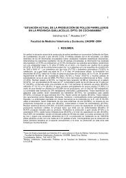
FIGURE 7.46 Abdominopelvic cavity <strong>of</strong> the cat in ventral view, with diaphragm reflected.<br />
Posterior to the stomach, the abdominal cavity is covered<br />
ventrally by the omental bursa, a large, double-layered,<br />
fat-laced mesentery that covers the intestines like an<br />
apron (Figure 7.46). <strong>The</strong> bursa is a sac-like structure<br />
formed from the greater omentum, which is part <strong>of</strong> the<br />
dorsal mesentery. Its structure will be discussed shortly.<br />
<strong>The</strong> omental bursa extends posteriorly to the urinary<br />
bladder, the light-colored, median, sac-like organ lying<br />
ventrally. If it is empty, it resembles a collapsed balloon.<br />
<strong>The</strong> mesentery passing from the bladder to the midventral<br />
wall, just to the left <strong>of</strong> the incision made to open the<br />
abdominopelvic cavity, is the median ligament (Figure<br />
7.46). <strong>The</strong> bladder is also supported by lateral ligaments,<br />
one on either side, that are <strong>of</strong>ten filled by wads <strong>of</strong> fat.<br />
Heart<br />
Round ligament<br />
Diaphragm<br />
Liver, left medial lobe<br />
Liver, quadate lobe<br />
Liver, left lateral lobe<br />
Stomach<br />
Gallbladder<br />
Visceral organs lined<br />
by visceral peritoneum<br />
Spleen<br />
Small intestine<br />
Omental bursa,<br />
ventral sheet,<br />
laced with fat<br />
Urinary bladder<br />
Median ligament<br />
Return to the anterior part <strong>of</strong> the abdominal cavity<br />
and spread apart the stomach and liver (Figure 7.48).<br />
<strong>The</strong> mesentery extending from the lesser curvature<br />
<strong>of</strong> the stomach and duodenum to the liver is the lesser<br />
omentum (another example <strong>of</strong> ventral mesentery),<br />
which is divided into two portions. One part, the<br />
hepatogastric ligament, passes from the lesser curvature<br />
to the liver. <strong>The</strong> other, passing from the proximal<br />
part <strong>of</strong> the duodenum to the liver, is the hepatoduodenal<br />
ligament, which appears to head toward the gall<br />
bladder. Various structures pass through the lesser<br />
omentum, including the common bile duct and<br />
the hepatic artery. <strong>The</strong>se will be considered<br />
below.<br />
SECTION IV—DIGESTIVE AND RESPIRATORY SYSTEMS 189