The Dissection of Vertebrates A Laboratory Manual
The Dissection of Vertebrates A Laboratory Manual
The Dissection of Vertebrates A Laboratory Manual

You also want an ePaper? Increase the reach of your titles
YUMPU automatically turns print PDFs into web optimized ePapers that Google loves.
first glance, however, it appears to form a connection<br />
between the uterine horn and ovary. Thus, carefully<br />
follow the uterine horn into the uterine tube.<br />
<strong>The</strong> union <strong>of</strong> the uterine horns posteriorly form the<br />
body <strong>of</strong> the uterus, a wider canal that then leads into<br />
the vagina. As there is no external division <strong>of</strong> these<br />
structures, their separation will be seen when they are<br />
opened. Note that the ureters pass dorsal to the uterine<br />
horns.<br />
<strong>The</strong> reproductive tract is supported by various mesenteries.<br />
<strong>The</strong> main supporting structure is the broad<br />
ligament, <strong>of</strong> which several portions are recognized. Lift<br />
the reproductive structures to clearly observe the following<br />
mesenteries. <strong>The</strong> portion <strong>of</strong> the broad ligament<br />
supporting the ovary is the mesovarium. <strong>The</strong> mesosalpinx<br />
attaches to the uterine tube, and the mesometrium<br />
attaches to the uterus, including the uterine<br />
horn. Lift the uterine horn near its center and tug it<br />
medially, as shown in Figure 7.71. <strong>The</strong> round ligament<br />
is the fibrous band in the mesometrium that extends<br />
diagonally from the uterine horn posterolaterally<br />
toward the body wall. Also, note the suspensory ovarian<br />
ligament, which supports the ovary anteriorly, as it<br />
extends to the dorsal body wall just lateral to the<br />
kidney.<br />
Open the pelvic canal by cutting through the pubic symphysis<br />
following the instructions given for the male on<br />
page 216. Pick away connective tissue to reveal and<br />
separate the structures passing through the canal (see<br />
Figure 7.71). <strong>The</strong> urethra and vagina unite toward the<br />
posterior end <strong>of</strong> the pelvic canal to form a common<br />
passageway, the urogenital sinus, which opens to the<br />
outside through the urogenital aperture. Dissect dorsal<br />
to the vagina and urogenital sinus to separate them from<br />
the rectum. On each side, dissect the tissue lateral to the<br />
rectum, very near the anus, to reveal the anal glands.<br />
<strong>The</strong>n completely separate the urogenital sinus from the<br />
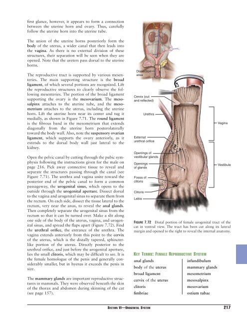
rectum so that it can be turned over. Make a slit along<br />
one side <strong>of</strong> the body <strong>of</strong> the uterus, vagina, and urogenital<br />
sinus, and spread the flaps apart (Figure 7.72). Find<br />
the urethral orifice, the entrance <strong>of</strong> the urethra. <strong>The</strong><br />
vagina extends anteriorly from this point to the cervix<br />
<strong>of</strong> the uterus, which is the distally tapered, sphincterlike<br />
portion <strong>of</strong> the uterus. Directly posterior to the<br />
urethral orifice, and just before the urogenital aperture,<br />
lies the small clitoris, which may be difficult to see. It is<br />
the female homologue <strong>of</strong> the penis and generally considerably<br />
smaller, but in hyenas it exceeds the penis in<br />
size.<br />
<strong>The</strong> mammary glands are important reproductive structures<br />
in mammals. <strong>The</strong>y were observed beneath the skin<br />
<strong>of</strong> the thorax and abdomen during skinning <strong>of</strong> the cat<br />
(see page 157).<br />
<strong>Dissection</strong><br />
area<br />
Cervix (cut<br />
and reflected)<br />
External<br />
urethral orifice<br />
Openings <strong>of</strong><br />
vestibular glands<br />
Openings<br />
<strong>of</strong> glands<br />
Fossa <strong>of</strong><br />
clitoris<br />
Clitoris<br />
Labia<br />
Urethra<br />
KEY TERMS: FEMALE REPRODUCTIVE SYSTEM<br />
anal glands<br />
infundibulum<br />
body <strong>of</strong> the uterus<br />
mammary glands<br />
broad ligament<br />
mesometrium<br />
cervix <strong>of</strong> the uterus mesosalpinx<br />
clitoris<br />
mesovarium<br />
fimbriae<br />
ostium tubae<br />
Vagina<br />
Vestibule<br />
FIGURE 7.72 Distal portion <strong>of</strong> female urogenital tract <strong>of</strong> the<br />
cat in ventral view. <strong>The</strong> tract has been cut along its lateral<br />
margin and opened to the right to reveal the internal anatomy.<br />
SECTION VI—UROGENITAL SYSTEM 217