- Page 2: The Dissection of Vertebrates A Lab
- Page 5 and 6: Acquisitions Editor: Tamsin Kent Ma
- Page 7 and 8: To our readers: Despite our best ef
- Page 9 and 10: Ear 67 Key Terms: Sensory Organs 68
- Page 11 and 12: C HAPTER 8 THE PIGEON Introduction
- Page 14 and 15: The past two decades have witnessed
- Page 16: students know they are not responsi
- Page 20 and 21: The study of vertebrate anatomy is
- Page 22: Anterior Anterodorsal (a) (b) Poste
- Page 25 and 26: PROTOSTOMATA ECHINODERMATA ENTEROPN
- Page 27 and 28: ately to attribute its cause to phy
- Page 29 and 30: UROCHORDATA SOMITICHORDATA CEPHALOC
- Page 31 and 32: the hagfishes considered the sister
- Page 33 and 34: PETROMYZONTOIDEA PLACODERMI GNATHOS
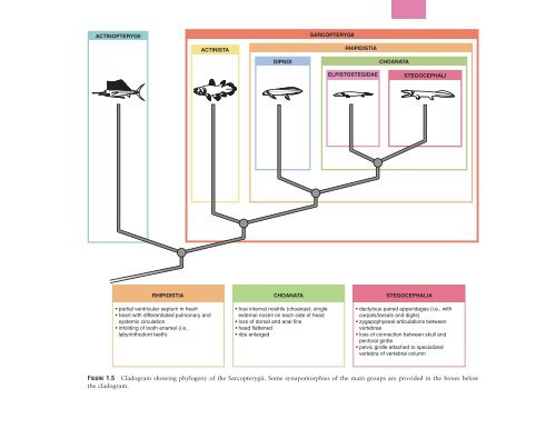
- Page 35: The sister group of the Actinoptery
- Page 39 and 40: AMPHIBIA REPTILIA AMNIOTA TESTUDINE
- Page 41 and 42: This page intentionally left blank
- Page 43 and 44: Anterior dorsal cartilage Annular c
- Page 45 and 46: Ovary Mesentery Kidney FIGURE 2.5 V
- Page 47 and 48: Pineal organ Olfactory sac Naris Or
- Page 49 and 50: and posterior cardinal veins join t
- Page 51 and 52: Examine a specimen of a dogfish ske
- Page 53 and 54: Precerebral cavity Precerebral fene
- Page 55 and 56: Neural canal Centrum Notochord Dors
- Page 57 and 58: Posterior Anterior Ceratotrichia Ra
- Page 59 and 60: (a) (b) (e) Epidermis Placoid scale
- Page 61 and 62: Interbranchial septum Spiracle Spir
- Page 63 and 64: Shark head, dorsal view (a) Pores o
- Page 65 and 66: Ampullae of Lorenzini Labial fold L
- Page 67 and 68: Gill ray Superficial constrictor m.
- Page 69 and 70: Oral cavity Hemibranch, with lamell
- Page 71 and 72: Falciform ligament Liver, right lob
- Page 73 and 74: Pylorus Stomach Line of attachment
- Page 75 and 76: Left coracohyoid m. (cut) Right cor
- Page 77 and 78: 1 2 3 4 5 6 Anterior intestinal a.
- Page 79 and 80: Falciform ligament Liver, right lob
- Page 81 and 82: The gastric vein accompanies the ga
- Page 83 and 84: Liver (cut) Esophagus Epididymis Le
- Page 85 and 86: parietal peritoneum lining the pleu
- Page 87 and 88:
Supraorbital canal passes onto vent
- Page 89 and 90:
Antorbital process (cut) Dorsal obl
- Page 91 and 92:
Anterior utriculus Membranous labyr
- Page 93 and 94:
superficial ophthalmic nerve suprac
- Page 95 and 96:
Olfactory sac Olfactory bulb Termin
- Page 97 and 98:
This sequence, however, does not ap
- Page 99 and 100:
hypophysis, just behind the optic n
- Page 101 and 102:
Cleithrum Operculum Orbit Premaxill
- Page 103 and 104:
Postcranial Skeleton The vertebral
- Page 105 and 106:
Anterior margin Growth rings Embedd
- Page 107 and 108:
Pharynx Heart Head kidney Liver Eso
- Page 109 and 110:
Pharynx Heart Duodenum Esophagus He
- Page 111 and 112:
This page intentionally left blank
- Page 113 and 114:
Procoracoid cartilage First trunk v
- Page 115 and 116:
Trabecular horn Antorbital process
- Page 117 and 118:
Neural canal Prezygapophysis Verteb
- Page 119 and 120:
Mouth Head Eye Neck Lips Gular fold
- Page 121 and 122:
urinary bladder. It is supported fr
- Page 123 and 124:
Stomach Liver Hepatic portal v. Pan
- Page 125 and 126:
Male Urogenital System In males the
- Page 127 and 128:
Right lung (reflected) Stomach (cut
- Page 129 and 130:
External carotid a. Left branch of
- Page 131 and 132:
108 CHAPTER 5 THE MUDPUPPY Internal
- Page 133 and 134:
Right lung Stomach Dorsal aorta Rig
- Page 135 and 136:
This page intentionally left blank
- Page 137 and 138:
Phalanges Metacarpals Carpals Prepo
- Page 139 and 140:
that helps form the roof of the bra
- Page 141 and 142:
Radio-ulna Radius Ulna Articular fo
- Page 143 and 144:
KEY TERMS: EXTERNAL ANATOMY cloaca
- Page 145 and 146:
Falciform ligament Right lobe of li
- Page 147 and 148:
Posterior vena cava (cut) Dorsolumb
- Page 149 and 150:
Right external carotid a. Right int
- Page 151 and 152:
Truncus arteriosus Right atrium Bul
- Page 153 and 154:
of the paired renal portal veins, w
- Page 155 and 156:
Skull Cervical vertebrae Hyoid appa
- Page 157 and 158:
group of jaw-closing muscles. Just
- Page 159 and 160:
Postorbital process Alisphenoid Fro
- Page 161 and 162:
Frontal b. Cribriform plate of ethm
- Page 163 and 164:
ceratohyoid choanae condyloid canal
- Page 165 and 166:
capitulum of a rib. Thoracics also
- Page 167 and 168:
Posterior Hemal process Hemal arch
- Page 169 and 170:
side of the trochlea. The supracond
- Page 171 and 172:
Head Neck Bicipital tuberosity Inte
- Page 173 and 174:
Left femur, anterior view Left femu
- Page 175 and 176:
Pinna Palperbrae Eye Rhinarium Exte
- Page 177 and 178:
154 TABLE 7.2 Muscles of the foreli
- Page 179 and 180:
TABLE 7.3 Continued Name Origin Ins
- Page 181 and 182:
The skin on the neck and throat may
- Page 183 and 184:
clavotrapezius, just anterior to th
- Page 185 and 186:
Mandibular gland Mylohyoid Parotid
- Page 187 and 188:
Splenius Parotid gland Mandibular g
- Page 189 and 190:
Sartorius Tensor fasciae latae Fasc
- Page 191 and 192:
verge and lie side by side, with th
- Page 193 and 194:
Tensor fasciae latae (cut & reflect
- Page 195 and 196:
Spermatic cord Pectineus Adductor l
- Page 197 and 198:
Muscles of the Head and Trunk Table
- Page 199 and 200:
176 TABLE 7.4 Continued Name Origin
- Page 201 and 202:
musculature here, you must also cut
- Page 203 and 204:
Examine the head. The larger muscle
- Page 205 and 206:
Premaxilla (cut) Vestibule Labia Or
- Page 207 and 208:
Anterior vena cava Internal mammary
- Page 209 and 210:
Internal mammary v. Anterior vena c
- Page 211 and 212:
Dissection area Anterior vena cava
- Page 213 and 214:
Dissection area Left lateral lobe o
- Page 215 and 216:
Dissection area Head of pancreas, w
- Page 217 and 218:
The pancreas consists of endocrine
- Page 219 and 220:
Dissection area Vagus n. Transversu
- Page 221 and 222:
Transverse scapular v. 198 CHAPTER
- Page 223 and 224:
Internal mammary a. (cut) Right sub
- Page 225 and 226:
Genioglossus m. Digastric m. Mylohy
- Page 227 and 228:
Hepatic v. Renal vv. Right internal
- Page 229 and 230:
Right medial lobe of liver Gallblad
- Page 231 and 232:
RIGHT ATRIUM RIGHT VENTRICLE Pulmon
- Page 233 and 234:
RIGHT ATRIUM RIGHT VENTRICLE Pulmon
- Page 235 and 236:
ase of heart brachial artery brachi
- Page 237 and 238:
the structures passing through the
- Page 239 and 240:
structure passes to the anterior en
- Page 241 and 242:
ovarian ligament ovary round ligame
- Page 243 and 244:
and of fibers, the olfactory tract,
- Page 245 and 246:
may have cut through it during expo
- Page 247 and 248:
section ventrally and posteriorly u
- Page 249 and 250:
This page intentionally left blank
- Page 251 and 252:
Second digit, phalanx 2 Second digi
- Page 253 and 254:
Pigeon skull with mandible and hyoi
- Page 255 and 256:
Ilium Dorsal iliac crest 6 free cau
- Page 257 and 258:
supra-angular supracoracoideus musc
- Page 259 and 260:
Barbs Anterior vane (a) Vaned fligh
- Page 261 and 262:
Nictitating membrane Trachea Esopha
- Page 263 and 264:
(a) (b) Carina of sternum Coracocla
- Page 265 and 266:
(a) the right lobe is considerably
- Page 267 and 268:
Pectoralis (cut) m. Right lung Righ
- Page 269 and 270:
are two main veins on each side but
- Page 271 and 272:
Right lung Posterior vena cava Righ
- Page 273 and 274:
limb is the ischiadic vein, but mos
- Page 275 and 276:
Maisey, J. G. 2001. Remarks on the
- Page 277 and 278:
Anterior mesenteric artery (continu
- Page 279 and 280:
Carpus, 147 Cat brain and cranial n
- Page 281 and 282:
Dentary bone in cat, 138-139 in fro
- Page 283 and 284:
Flocculonodular lobe, 222 Folia, of
- Page 285 and 286:
Hypophyseal pouch, 24 Hypophysis in
- Page 287 and 288:
Liver (continued) in mudpuppy, 96 i
- Page 289 and 290:
Neural crest, 8 Neural plate, 31 Ne
- Page 291 and 292:
Pelvis in cat, 147, 152 in frog, 11
- Page 293 and 294:
Pylorus, 49 Pyramids, 222 Pyriformi
- Page 295 and 296:
Squamosal bones in cat, 136 in frog
- Page 297 and 298:
Ulnare, 232 Umbilical artery/vein,