Squint Free Papers - aioseducation
Squint Free Papers - aioseducation
Squint Free Papers - aioseducation
Create successful ePaper yourself
Turn your PDF publications into a flip-book with our unique Google optimized e-Paper software.
<strong>Squint</strong> <strong>Free</strong> <strong>Papers</strong><br />
the macular area digitally covered by an opaque circle. He then assessed the<br />
torsion using the following vascular cues: (i) angle made by an imaginary<br />
line joining the superior and inferior branches of the central retinal artery<br />
as they emerge out from the centre of the optic disc, (ii) the orientation of the<br />
“axis of symmetry” passing through the centre of disc as the vertex, if the<br />
temporal vascular arcade is considered as a parabola (iii) the angle subtended<br />
by an imaginary line joining the arteries in the supero-temporal and inferotemporal<br />
quadrant at a distance of one disc diameter from the centre of the<br />
optic disc. For each fundus picture, three responses were noted based on each<br />
of the above 3 cues as “no torsion, intorsion or extorsion.” The final response<br />
for a particular fundus picture was the response that was repeated atleast<br />
twice. The final response was noted as “cannot be commented” if each of the<br />
3 responses were different.<br />
The readings of the two examiners were then compared and analyzed to test<br />
the sensitivity and specificity of the new method.<br />
RESULTS<br />
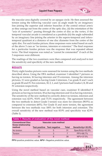
Thirty eight fundus pictures were assessed for torsion using the two methods<br />
described above. Using the DFA method, examiner I identified 7 pictures as<br />
having no torsion, 16 having intorsion and 15 extorsion. Among the intorsion<br />
pictures, 11 were graded as having Grade I intorsion, 3 as grade II and 1 each<br />
as grade III and IV. For extorsion, 10, 2, 1, and 2 pictures had Grades I, II, III and<br />
IV extorsion respectively.<br />
Using the novel method based on vascular cues, examiner II identified 8<br />
pictures as having no torsion, 18 as having intorsion and 12 as having extorsion.<br />
The sensitivity of the new method to correctly detect no torsion, intorsion and<br />
extorsion was 42.9%, 93.8% and 73.3% respectively. The agreement between<br />
the two methods to detect Grade I torsion was more for intorsion (90.9%) as<br />
compared to extorsion (60%). For Grade II and more torsion, the agreement<br />
between the two methods was 100% for both intorsion and extorsion. The<br />
overall sensitivity of the new method was 83.9% and specificity was 42.9%<br />
(Table 1).<br />
Table 1: Results of evaluation of the novel method<br />
Conventional Disc Vascular cues method<br />
fovea relationship<br />
method Test positive Test negative Total Sensitivity= 83.9%<br />
No torsion 26 5 31 Specificity=42.9%<br />
Torsion 4 3 7 PPV=86.7%<br />
30 8 38 NPV=37.5%<br />
PPV=positive predictive value. NPV= Negative predictive value<br />
813


