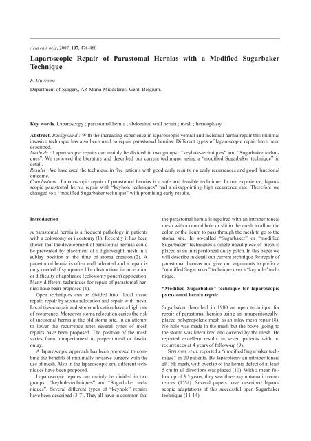
Laparoscopic Repair of Parastomal Hernias with a Modified - Belsurg
Laparoscopic Repair of Parastomal Hernias with a Modified - Belsurg
Laparoscopic Repair of Parastomal Hernias with a Modified - Belsurg

You also want an ePaper? Increase the reach of your titles
YUMPU automatically turns print PDFs into web optimized ePapers that Google loves.
Acta chir belg, 2007, 107, 476-480<br />
<strong>Laparoscopic</strong> <strong>Repair</strong> <strong>of</strong> <strong>Parastomal</strong> <strong>Hernias</strong> <strong>with</strong> a <strong>Modified</strong> Sugarbaker<br />
Technique<br />
F. Muysoms<br />
Department <strong>of</strong> Surgery, AZ Maria Middelares, Gent, Belgium.<br />
Key words. Laparoscopy ; parastomal hernia ; abdominal wall hernia ; mesh ; hernioplasty.<br />
Abstract. Background : With the increasing experience in laparoscopic ventral and incisonal hernia repair this minimal<br />
invasive technique has also been used to repair parastomal hernias. Different types <strong>of</strong> laparoscopic repair have been<br />
described.<br />
Methods : <strong>Laparoscopic</strong> repairs can mainly be divided in two groups : “keyhole-techniques” and “Sugarbaker techniques”.<br />
We reviewed the literature and described our current technique, using a “modified Sugarbaker technique” in<br />
detail.<br />
Results : We have used the technique in five patients <strong>with</strong> good early results, no early recurrences and good functional<br />
outcome.<br />
Conclusions : <strong>Laparoscopic</strong> repair <strong>of</strong> parastomal hernias is a safe and feasible technique. In our experience, laparoscopic<br />
parastomal hernia repair <strong>with</strong> “keyhole techniques” had a disappointing high recurrence rate. Therefore we<br />
changed to a “modified Sugarbaker technique” <strong>with</strong> promising early results.<br />
Introduction<br />
A parastomal hernia is a frequent pathology in patients<br />
<strong>with</strong> a colostomy or ileostomy (1). Recently it has been<br />
shown that the development <strong>of</strong> parastomal hernias could<br />
be prevented by placement <strong>of</strong> a lightweight mesh in a<br />
sublay position at the time <strong>of</strong> stoma creation (2). A<br />
parastomal hernia is <strong>of</strong>ten well tolerated and a repair is<br />
only needed if symptoms like obstruction, incarceration<br />
or difficulty <strong>of</strong> appliance (colostomy pouch) application.<br />
Many different techniques for repair <strong>of</strong> parastomal hernias<br />
have been proposed (1).<br />
Open techniques can be divided into : local tissue<br />
repair, repair by stoma relocation and repair <strong>with</strong> mesh.<br />
Local tissue repair and stoma relocation have a high rate<br />
<strong>of</strong> recurrence. Moreover stoma relocation caries the risk<br />
<strong>of</strong> incisional hernia at the old stoma site. In an attempt<br />
to lower the recurrence rates several types <strong>of</strong> mesh<br />
repairs have been proposed. The position <strong>of</strong> the mesh<br />
varies from intraperitoneal to preperitoneal or fascial<br />
onlay.<br />
A laparoscopic approach has been proposed to combine<br />
the benefits <strong>of</strong> minimally invasive surgery <strong>with</strong> the<br />
use <strong>of</strong> mesh. Also in the laparoscopic era, different techniques<br />
have been proposed.<br />
<strong>Laparoscopic</strong> repairs can mainly be divided in two<br />
groups : “keyhole-techniques” and “Sugarbaker techniques”.<br />
Several different types <strong>of</strong> “keyhole” repairs<br />
have been described (3-7). They all have in common that<br />
the parastomal hernia is repaired <strong>with</strong> an intraperitoneal<br />
mesh <strong>with</strong> a central hole or slit in the mesh to allow the<br />
colon or the ileum to pass through the mesh to go to the<br />
stoma site. In so-called “Sugarbaker” or “modified<br />
Sugarbaker” techniques a single uncut piece <strong>of</strong> mesh is<br />
placed as an intraperitoneal onlay patch. In this paper we<br />
will describe in detail our current technique for repair <strong>of</strong><br />
parastomal hernias and give our arguments to prefer a<br />
“modified Sugarbaker” technique over a “keyhole” technique.<br />
“<strong>Modified</strong> Sugarbaker” technique for laparoscopic<br />
parastomal hernia repair<br />
Sugarbaker described in 1980 an open technique for<br />
repair <strong>of</strong> parastomal hernias using an intraperitoneallyplaced<br />
polypropelene mesh as an inlay mesh repair (8).<br />
No hole was made in the mesh but the bowel going to<br />
the stoma was lateralized and covered by the mesh. He<br />
reported excellent results in seven patients <strong>with</strong> no<br />
recurrences at 4 years <strong>of</strong> follow-up (9).<br />
STELZNER et al. reported a “modified Sugarbaker technique”<br />
in 20 patients. By laparotomy an intraperitoneal<br />
ePTFE mesh, <strong>with</strong> overlap <strong>of</strong> the hernia defect <strong>of</strong> at least<br />
5 cm in all directions was placed (10). With a mean follow<br />
up <strong>of</strong> 3.5 years, they saw three asymptomatic recurrences<br />
(15%). Several papers have described laparoscopic<br />
adaptations <strong>of</strong> this successful open Sugarbaker<br />
technique (11-14).
<strong>Laparoscopic</strong> <strong>Parastomal</strong> Hernia <strong>Repair</strong> 477<br />
We would like to define the “<strong>Laparoscopic</strong> <strong>Modified</strong><br />
Sugarbaker technique for parastomal hernia” as : a<br />
repair by laparoscopic technique <strong>of</strong> a parastomal hernia<br />
by placement <strong>of</strong> an intact intraperitoneal mesh overlapping<br />
the hernia defect and <strong>with</strong> lateralisation <strong>of</strong> the<br />
bowel going to the stoma.<br />
Indications for repair are usually : obstruction, incarceration<br />
and difficulties in applying the stoma appliance<br />
material. It is common practice not to operate on oligoor<br />
asymptomatic patients. Care should be taken not to<br />
broaden the indications for repair <strong>of</strong> these hernias just<br />
because there is a new minimally invasive technique that<br />
we want to adopt. Not before we have long-term followup<br />
data <strong>with</strong> low recurrence rates and low complication<br />
rates, might we justify considering to operate on less<br />
symptomatic patients.<br />
Preoperatively some laxatives are given. A proper<br />
bowel preparation could be considered but is not given<br />
in our practice. One single dose <strong>of</strong> cefazoline is given<br />
during induction <strong>of</strong> anesthesia. Patients are operated<br />
under general anesthesia <strong>with</strong> tracheal intubation.<br />
Special attention should be given to patients operated for<br />
incarceration and obstruction to avoid aspiration <strong>of</strong><br />
stomach content resulting in pneumonitis by inhalation.<br />
The patient is in the supine position <strong>with</strong> both arms<br />
alongside the body. The arms should be tucked underneath<br />
the sides <strong>of</strong> the patients to allow access to the lateral<br />
border <strong>of</strong> the abdomen where the trocars will be<br />
placed. After desinfection, the operative field is draped.<br />
Again we emphasize the importance <strong>of</strong> keeping the<br />
operative field broad enough to expose bilaterally the<br />
lateral sides <strong>of</strong> the abdomen. We cover the stoma <strong>with</strong> a<br />
gauze and cover the stoma site and the whole operative<br />
field <strong>with</strong> a plastic drape. Doing this the stoma itself is<br />
during the operation never in contact <strong>with</strong> the operative<br />
field. Therefore we think the risk <strong>of</strong> contamination <strong>of</strong> the<br />
mesh should not be higher than for other laparoscopic<br />
incisional hernia repairs not involving a stoma.<br />
Positioning <strong>of</strong> surgeons, trocars and video equipment<br />
for a left sided colostomy is given in Fig. 1. Usually a<br />
pneumoperitoneum is created <strong>with</strong> a Verres needle subcostally<br />
at the anterior axillary line. We use an intraoperative<br />
abdominal pressure <strong>of</strong> 12 mmHg. When this<br />
pressure has been reached a trocar <strong>of</strong> 10 mm is inserted<br />
on the same anterior axillary line halfway between the<br />
costal margin and the superior iliac crest. A 30° angeled<br />
scope <strong>of</strong> 10 mm is introduced. The position <strong>of</strong> the Verres<br />
needle is checked and lesions to the bowel or the liver by<br />
the puncture are excluded. A second trocar <strong>of</strong> 10 mm is<br />
inserted subcostally. This will be the site <strong>of</strong> the scope<br />
during the operation. Finally a third trocar <strong>of</strong> 5 mm is<br />
placed just above the superior iliac crest. If needed the<br />
region where this trocar is to be inserted is released <strong>of</strong><br />
adhesions. If many intraperitoneal adhesions are suspected<br />
an open laparoscopy can be used to create the<br />
Fig. 1<br />
Positioning <strong>of</strong> trocars, surgeons and equipment for laparoscopic<br />
repair <strong>of</strong> a parastomal hernia <strong>of</strong> a left sided colostomy.<br />
pneumoperitoneum as an alternative for the Verres needle<br />
technique.<br />
Complete adhesiolysis <strong>of</strong> the anterior abdominal wall<br />
is performed, including release <strong>of</strong> the round ligament <strong>of</strong><br />
the liver if this is necessary. Care is taken to avoid accidental<br />
enterotomy. Indeed adhesiolysis can sometimes<br />
be difficult in these patients (15). It is a potentially dangerous<br />
step <strong>of</strong> the operation, certainly if a bowel lesion<br />
is missed and only recognised postoperatively. Delayed<br />
bowel lesions can result in intraabdominal sepsis and<br />
multiorgan failure. Therefore it is recommended to perform<br />
the adhesiolysis <strong>with</strong> a sharp dissection, only cutting<br />
under good visibility and <strong>with</strong> certainty about the<br />
structures being cut. We advise to use energy sources<br />
like coagulation or electronic scissors only scarcely. We<br />
always look for the avascular plane between the adhesions<br />
and the abdominal wall. This avascular plane is<br />
usually present and can be recognised by moderate traction<br />
on the adherent structures. In cases <strong>of</strong> recurrent<br />
hernias after previous mesh repair the avascular plane<br />
might not be present and adhesiolysis in these cases can<br />
be very difficult or even impossible.<br />
The content <strong>of</strong> the hernial sac is reduced after identifying<br />
the colon going to the colostomy and its mesocolon.<br />
The peritoneal sac is left in place. To be able to<br />
achieve adequate lateralisation <strong>of</strong> the colon, we recommend<br />
freeing all adhesions <strong>of</strong> the colon and its mesocolon<br />
from the margins <strong>of</strong> the hernia defect. The colon<br />
is pulled intraabdominally, thus reducing a prolaps if<br />
present. The colon is then pulled to the lateral side <strong>of</strong> the<br />
hernia defect. We fix the colon <strong>with</strong> some resorbable<br />
sutures between the serosa and the peritoneum lateral <strong>of</strong><br />
the hernia defect. Thus we close the opening lateral <strong>of</strong><br />
the colon.
478 F. Muysoms<br />
Fig. 2<br />
Intraoperative view showing the mesh used for a modified<br />
Sugarbaker repair <strong>of</strong> a parastomal hernia.<br />
We measure the hernia defect and check all incisions<br />
on the anterior abdominal wall for concomitant incisional<br />
hernias on these incisions. The hernia defect is drawn<br />
on to the abdomen as well as the concomitant hernias if<br />
present. A size <strong>of</strong> mesh is drawn outside these drawings<br />
so it will be large enough to cover all hernia defects by<br />
at least 5 cm in all directions (Fig. 2). If a concomitant<br />
incisional hernia is present we repair it <strong>with</strong> one large<br />
mesh covering both hernia defects.<br />
We recommend to use a mesh designed for intraperitoneal<br />
placement. Some authors have used regular<br />
polypropylene mesh intraperitoneally to repair parastomal<br />
hernias (12). Most authors think this harbours the<br />
risk for extensive adhesions, bowel fistula and mesh<br />
infection. We use an ePTFE mesh (Dualmesh Plus <strong>with</strong><br />
Holes ‚, WL Gore, Flagstaff, Arizona, USA) in our practice.<br />
When the mesh has been cut to the appropriate size<br />
and form, orientation marks are made on the mesh and<br />
on the abdominal wall to allow orientation <strong>of</strong> the mesh<br />
once it has been placed intraperitoneally. At the orientation<br />
marks the first sutures are placed before the mesh is<br />
inserted into the abdomen. When introducing the mesh,<br />
contact <strong>with</strong> the skin should be avoided. Therefore the<br />
meshes <strong>of</strong> moderate size are introduced trough a trocar.<br />
Larger meshes that are too big to introduce through a<br />
trocar are rolled inside the sterile plastic covering <strong>of</strong> the<br />
mesh and are introduced after removal <strong>of</strong> the trocar.<br />
Inside the abdomen the mesh is removed from its plastic<br />
covering, which is than removed from the abdomen.<br />
The mesh is orientated using the orientation marks and<br />
the sutures are extracted <strong>with</strong> a “suture passer” technique<br />
through separate small skin incisions at the orientation<br />
marks. The sutures are tied down to the anterior<br />
abdominal fascia, thus creating transabdominal fixation<br />
Fig. 3<br />
Intraoperative view <strong>of</strong> a laparoscopic parastomal hernia repair.<br />
The colon has been lateralised and the mesh is fixed <strong>with</strong> a<br />
transfascial suture below and above the colon.<br />
Fig. 4<br />
Fixation <strong>of</strong> the mesh in a laparoscopic parastomal hernia repair<br />
<strong>with</strong> a modified Sugarbaker technique. We use a combination<br />
<strong>of</strong> transfascial fixation sutures and spiral tackers in a double<br />
crown configuration.<br />
sutures. We place a transfascial fixation suture laterally<br />
in the mesh just above and just underneath the lateralised<br />
bowel (Fig. 3). Care is taken not to injure the<br />
bowel. Further sutures are placed all around the mesh at<br />
5 cm intervals at the margin <strong>of</strong> the mesh. Further fixation<br />
is done <strong>with</strong> a mechanical fixation device at the<br />
margin <strong>of</strong> the mesh <strong>with</strong> an interval <strong>of</strong> one to two cm<br />
between the fixations. We use spiral tackers (Protack®,<br />
Autosuture, Tyco Health Care Group, Norwalk,<br />
Connecticut, USA) as a fixation device. Then a second<br />
row <strong>of</strong> staplers is placed at the margin <strong>of</strong> the hernia
<strong>Laparoscopic</strong> <strong>Parastomal</strong> Hernia <strong>Repair</strong> 479<br />
defect like for a “double crown” fixation. With some<br />
additional staplers we connect these two rows <strong>of</strong> staplers<br />
at each side <strong>of</strong> the colon. Again care is taken not to put<br />
staplers into the colon or the mesocolon. Our method <strong>of</strong><br />
mesh fixation is explained graphically in Fig. 4.<br />
Results<br />
After having seen a live surgery session where Karl<br />
Leblanc performed a laparoscopic modified Sugarbaker<br />
technique to treat a parastomal hernia we adopted this<br />
technique since November 2005 in all patients presenting<br />
<strong>with</strong> an indication for repair.<br />
We have operated five consecutive patients <strong>with</strong> a<br />
symptomatic parastomal hernia. All patients had a left<br />
sided colostomy. Two for adenocarcinoma <strong>of</strong> the rectum<br />
and three for benign rectal disease leading to a permanent<br />
colostomy. For four patients this was the first time<br />
their parastomal hernia was repaired. One patient had<br />
two previous repairs, the last one including a mesh<br />
repair in the retromuscular position. In four patients a<br />
concomitant incisional hernia, either on the midline or at<br />
an old ileostomy site, was repaired. Indications were<br />
incarceration in one and recurrent obstructions in four.<br />
All patients were treated <strong>with</strong> one intact ePTFE mesh<br />
covering the parastomal hernia, the lateralised colon and<br />
the concomitant incisional hernia, if present. There were<br />
no intraoperative or postoperative complications. Till<br />
today, no recurrence has been observed, although follow-up<br />
is rather short. No problems <strong>with</strong> stenosis or<br />
obstructions <strong>of</strong> the colostomy have been encountered<br />
and colonic irrigation is either as good or better than preoperatively.<br />
We plan to do a prospective multicenter follow-up<br />
study <strong>of</strong> laparoscopic modified Sugarbaker repair to<br />
repair parastomal hernias to obtain long-term results <strong>of</strong><br />
this technique. We will focus on recurrences and functional<br />
outcome.<br />
Discussion<br />
Some reports <strong>with</strong> keyhole techniques have disappointing<br />
results <strong>with</strong> high recurrence rates. Safadi reported a<br />
recurrence rate <strong>of</strong> 56% (5/9 patients) <strong>with</strong>in 6 months <strong>of</strong><br />
the operation (7). They recommended changes in the<br />
technique <strong>of</strong> laparoscopic parastomal hernia repair to<br />
achieve better results.<br />
Initial results <strong>with</strong> laparoscopic parastomal hernia<br />
repair using a “Keyhole technique” in our experience<br />
were disappointing. Analysis <strong>of</strong> the recurrences showed<br />
that the weak point <strong>of</strong> the repair was the hole in the<br />
mesh. It is indeed difficult to estimate the appropriate<br />
size for the hole in the mesh, that optimally accommodates<br />
the colon going to the skin. From experience <strong>with</strong><br />
laparoscopic incisional hernia repair we know that the<br />
amount <strong>of</strong> overlap <strong>of</strong> the mesh beyond the hernia defect<br />
is an important determinant for the risk <strong>of</strong> hernia recurrence.<br />
In a “Keyhole technique” the amount <strong>of</strong> overlap<br />
at the hole is zero. Therefore this will always be a weak<br />
point for developing recurrences. Moreover we know<br />
that an implanted mesh shows a variable degree <strong>of</strong><br />
shrinkage. Shrinkage <strong>of</strong> a mesh <strong>with</strong> a central hole will<br />
result in enlargement <strong>of</strong> this hole. These disappointing<br />
results are similar to some results in the literature <strong>with</strong><br />
“Keyhole techniques”. Tessier analysed the literature on<br />
laparoscopic parastomal hernia repair and compared the<br />
“Sugarbaker” <strong>with</strong> the “Keyhole techniques” (16). He<br />
concluded that the “Sugarbaker technique” is superior to<br />
the “Keyhole technique” by <strong>of</strong>fering decreased operating<br />
time, lower morbidity, shorter length <strong>of</strong> stay and less<br />
recurrence rate. LEBLANC et al., in their most recent<br />
paper on laparoscopic parastomal hernia repair, feel that<br />
the single patch technique as an onlay repair is the better<br />
alternative compared to the keyhole techniques (14).<br />
We currently prefer the “modified Sugarbaker technique”<br />
to repair parastomal hernias laparoscopically.<br />
Not only are we convinced that the recurrence rates will<br />
be lower, but also that this technique is definitely much<br />
easier to perform. It is a laparoscopic adaptation <strong>of</strong> the<br />
open technique performed by Stelzner, that has proven<br />
its efficacy,<strong>with</strong> good long term results.<br />
Conclusion<br />
<strong>Laparoscopic</strong> repair <strong>of</strong> parastomal hernias is a safe and<br />
feasible technique. In our experience laparoscopic<br />
parastomal hernia repair <strong>with</strong> “keyhole techniques” had<br />
a disappointing high recurrence rate. Therefore we<br />
changed to a “laparoscopic Sugarbaker technique” <strong>with</strong><br />
promising early results.<br />
References<br />
1. CARNE P. W. G., ROBERTSON G. M., FRIZELLE F. A. <strong>Parastomal</strong><br />
hernia. Br J Surg, 2003, 90 : 784-93.<br />
2. JANES A., CENGIZ Y., ISRAELSSON L. A. Randomized clinical trial<br />
<strong>of</strong> the use <strong>of</strong> a prosthetic mesh to prevent parastomal hernia.<br />
Br J Surg, 2004, 91 : 280-82.<br />
3. BICKEL A., SHINKAREVSKY E., EITAN A. <strong>Laparoscopic</strong> repair <strong>of</strong><br />
paracolostomy hernia. J Laparoendosc Adv Surg Tech A, 1999, 9 :<br />
353-5.<br />
4. GOULD J. C., ELLISON E. C. <strong>Laparoscopic</strong> parastomal hernia repair.<br />
Surg Laparosc Endosc Percutan Tech, 2003, 13 : 51-4.<br />
5. LEBLANC K. A., BELLANGER D. E. <strong>Laparoscopic</strong> repair <strong>of</strong> parastomy<br />
hernias : early results. J Am Coll Surg, 2002, 194 : 232-9.<br />
6. HANSSON B. M., VAN NIEUWENHOVEN E. J., BLEICHRODT R. P.<br />
Promising new technique in the repair <strong>of</strong> parastomal hernia. Surg<br />
Endosc, 2003, 17 : 1789-91.<br />
7. SAFADI B. <strong>Laparoscopic</strong> repair <strong>of</strong> parastomal hernias : early<br />
results. Surg Endosc, 2004, 18 : 676-80.<br />
8. SUGARBAKER Ph. Prosthetic mesh repair <strong>of</strong> large hernias at the site<br />
<strong>of</strong> colonic stomas. Surg Gynecol Obstet 1980, 150 : 576-8.<br />
9. SUGARBAKER Ph. Peritoneal approach to prosthetic mesh repair <strong>of</strong><br />
paracolostomy hernias. Ann Surg 1985, 201 : 344-6.
480 F. Muysoms<br />
10. STELZNER S., HELLMICH G., LUDWIG K. <strong>Repair</strong> <strong>of</strong> paracolostomy<br />
hernias <strong>with</strong> a prosthetic mesh in the intraperitoneal onlay position<br />
: modified Sugarbaker technique. Dis Colon Rectum, 2004,<br />
47 : 185-91.<br />
11. PORCHERON J., PAYAN B., BALIQUE J. G. Mesh repair <strong>of</strong> paracolostomal<br />
hernia by laparoscopy. Surg Endosc, 1998, 12 : 1281.<br />
12. VOITK A. Simple technique for laparoscopic paracolostomy hernia<br />
repair. Dis Colon Rectum, 2000, 43 : 1451-3.<br />
13. BERGER D. <strong>Laparoscopic</strong> paraostomal hernia repair : indications,<br />
technique and results. In : Morales-Conde S. (ed.). <strong>Laparoscopic</strong><br />
ventral hernia repair. Springer Verlag France, 2002, pp. 383-7.<br />
14. LEBLANC K. A., BELLANGER D. E., WHITAKER J. M., HAUSMANN M. G.<br />
<strong>Laparoscopic</strong> parastomal hernia repair. Hernia, 2005, 9 : 140-4.<br />
15. HANSSON B. M., DE HINGH I. H., BLEICHRODT R. P. <strong>Laparoscopic</strong><br />
parastomal hernia repair is feasible and safe : early results <strong>of</strong> a<br />
prospective clinical study including 55 consecutive patients. Surg<br />
Endosc, 2007, Mar 13 : Epub ahead <strong>of</strong> print.<br />
16. TESSIER D. J. A comparison <strong>of</strong> laparoscopic parastomal hernia<br />
repair using the Sugarbaker and Keyhole techniques. 2006<br />
Abstract P-26, 3 rd International Hernia Congress, Boston.<br />
Dr. Filip Muysoms<br />
AZ Maria Middelares<br />
Kortrijksesteenweg 1026<br />
B-9000 Gent, Belgium<br />
Tel. : 003292607181<br />
Fax : 003292607175<br />
E-mail : filip.muysoms@azmmsj.be